polish science journal - Актуальные научные исследования ...
Upload
independentCategory
view
8download
0
HUMAN GENETICS · SHORT COMMUNICATION
AMPD1 gene mutations are associated with obesityand diabetes in Polish patients with cardiovascular diseases
Krzysztof Safranow & Janina Suchy & Katarzyna Jakubowska & Maria Olszewska &
Agnieszka Bińczak-Kuleta & Grzegorz Kurzawski & Ryszard Rzeuski &Edyta Czyżycka & Beata Łoniewska & Zdzisława Kornacewicz-Jach &
Andrzej Ciechanowicz & Dariusz Chlubek
Received: 1 February 2010 /Revised: 2 July 2010 /Accepted: 27 July 2010 /Published online: 25 November 2010# The Author(s) 2010. This article is published with open access at Springerlink.com
Abstract Previous studies showed an association of thecommon functional polymorphism (C34T, Gln12Stop) inthe adenosine monophosphate deaminase-1 (AMPD1) genewith survival in heart failure (HF) and/or coronary arterydisease (CAD). The aim of the study was to search for othermutations in selected regions of the AMPD1 gene in PolishCAD and HF patients, and to analyze their associationswith obesity and diabetes. Exons 2, 3, 5, and 7 of AMPD1were scanned for mutations in 97 patients with CADwithout HF (CAD+ HF−), 104 patients with HF (HF+), and200 newborns from North-Western Poland using denaturinghigh-performance liquid chromatography (DHPLC), polymerasechain reaction–restriction fragment length polymorphism (PCR-RFLP), and direct sequencing. Frequencies of AMPD1 C34T
mutation, as well as novel A99G, G512A, IVS4-6delT, andC784T sequence alterations, were similar in the three groups,but 860T mutated allele was less frequent in the combinedCAD+ HF− and HF+ groups than in the controls (1.7% vs.4.3%, p=0.040). Heterozygous 34CT genotype was associatedwith lower (odds ratio [OR]=0.32, 95% confidence interval[CI]=0.13–0.81) and 860AT with higher (OR=13.7, 95%CI=1.6–118) prevalence of diabetes or hyperglycemia in relation towild-type homozygotes. Abdominal obesity was more frequentin 860AT patients than in wild-type homozygotes and 34CTheterozygotes (86% vs. 40% vs. 29%, p<0.05). Nine genescontaining polymorphisms linked with AMPD1 C34T mutationwere found in the HapMap database. AMPD1 C34T nonsensemutation is associated with reduced prevalence of diabetes andobesity in patients with CAD or HF, but A860T substitutionseems to exert opposite metabolic effects and should always beaccounted for in the studies of the AMPD1 genotype.
Keywords AMP deaminase-1 . Coronary artery disease .
Denaturing high-performance liquid chromatography .
Diabetes . Heart failure . Human genetics . Obesity
Introduction
Previous studies showed an association of the commonC34T polymorphism in the AMPD1 gene with survival inpatients with heart failure (HF) and coronary arterydisease (CAD). Adenosine monophosphate deaminase(AMPD, EC 3.5.4.6) catalyzes the deamination ofadenosine monophosphate (AMP) to inosine monophosphate(IMP). The AMPD1 gene located at 1p13 encodes isoenzymewhich expresses the highest activity in skeletal muscles(Morisaki et al. 1990). The AMPD2 and AMPD3 genesencode liver and erythrocyte isoenzyme, respectively. The
K. Safranow (*) :K. Jakubowska :M. Olszewska :D. ChlubekDepartment of Biochemistry and Medical Chemistry,Pomeranian Medical University,Powstańców Wielkopolskich 72,70-111, Szczecin, Polande-mail: [email protected]
J. Suchy :G. KurzawskiDepartment of Genetics and Pathology,Pomeranian Medical University,Szczecin, Poland
A. Bińczak-Kuleta :A. CiechanowiczDepartment of Laboratory Diagnostics and Molecular Medicine,Pomeranian Medical University,Szczecin, Poland
R. Rzeuski : E. Czyżycka : Z. Kornacewicz-JachDepartment of Cardiology, Pomeranian Medical University,Szczecin, Poland
B. ŁoniewskaDepartment of Neonatology, Pomeranian Medical University,Szczecin, Poland
J Appl Genetics (2011) 52:67–76DOI 10.1007/s13353-010-0009-x
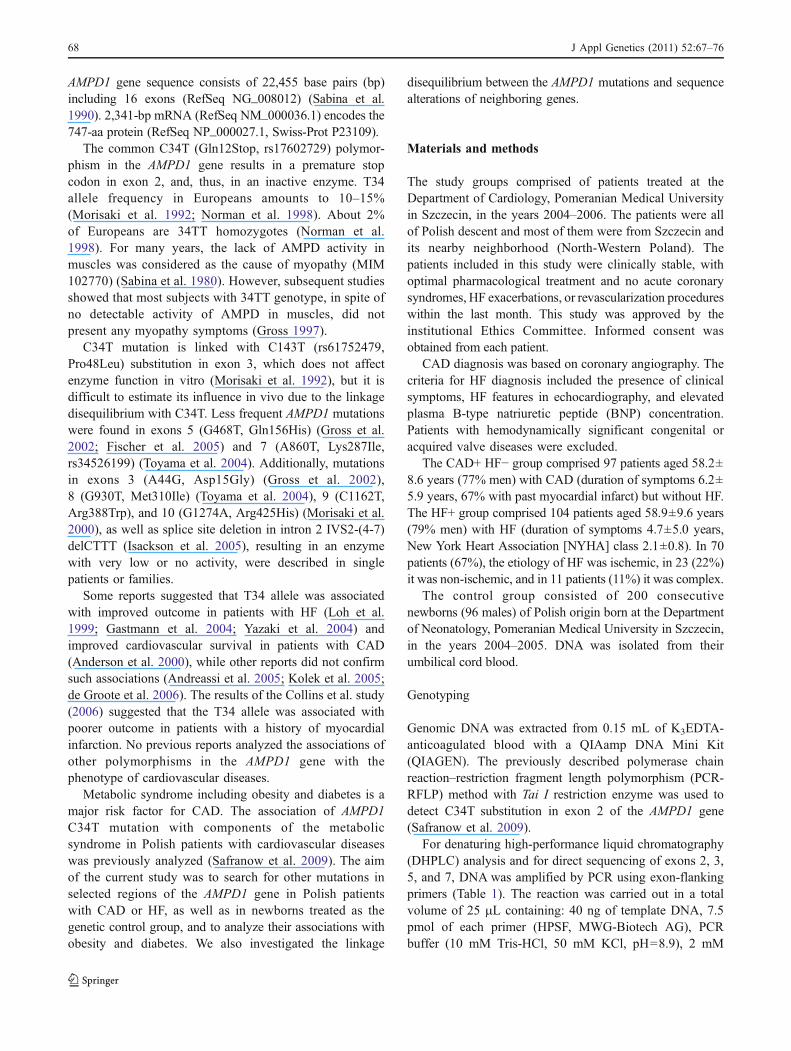
AMPD1 gene sequence consists of 22,455 base pairs (bp)including 16 exons (RefSeq NG_008012) (Sabina et al.1990). 2,341-bp mRNA (RefSeq NM_000036.1) encodes the747-aa protein (RefSeq NP_000027.1, Swiss-Prot P23109).
The common C34T (Gln12Stop, rs17602729) polymor-phism in the AMPD1 gene results in a premature stopcodon in exon 2, and, thus, in an inactive enzyme. T34allele frequency in Europeans amounts to 10–15%(Morisaki et al. 1992; Norman et al. 1998). About 2%of Europeans are 34TT homozygotes (Norman et al.1998). For many years, the lack of AMPD activity inmuscles was considered as the cause of myopathy (MIM102770) (Sabina et al. 1980). However, subsequent studiesshowed that most subjects with 34TT genotype, in spite ofno detectable activity of AMPD in muscles, did notpresent any myopathy symptoms (Gross 1997).
C34T mutation is linked with C143T (rs61752479,Pro48Leu) substitution in exon 3, which does not affectenzyme function in vitro (Morisaki et al. 1992), but it isdifficult to estimate its influence in vivo due to the linkagedisequilibrium with C34T. Less frequent AMPD1 mutationswere found in exons 5 (G468T, Gln156His) (Gross et al.2002; Fischer et al. 2005) and 7 (A860T, Lys287Ile,rs34526199) (Toyama et al. 2004). Additionally, mutationsin exons 3 (A44G, Asp15Gly) (Gross et al. 2002),8 (G930T, Met310Ile) (Toyama et al. 2004), 9 (C1162T,Arg388Trp), and 10 (G1274A, Arg425His) (Morisaki et al.2000), as well as splice site deletion in intron 2 IVS2-(4-7)delCTTT (Isackson et al. 2005), resulting in an enzymewith very low or no activity, were described in singlepatients or families.
Some reports suggested that T34 allele was associatedwith improved outcome in patients with HF (Loh et al.1999; Gastmann et al. 2004; Yazaki et al. 2004) andimproved cardiovascular survival in patients with CAD(Anderson et al. 2000), while other reports did not confirmsuch associations (Andreassi et al. 2005; Kolek et al. 2005;de Groote et al. 2006). The results of the Collins et al. study(2006) suggested that the T34 allele was associated withpoorer outcome in patients with a history of myocardialinfarction. No previous reports analyzed the associations ofother polymorphisms in the AMPD1 gene with thephenotype of cardiovascular diseases.
Metabolic syndrome including obesity and diabetes is amajor risk factor for CAD. The association of AMPD1C34T mutation with components of the metabolicsyndrome in Polish patients with cardiovascular diseaseswas previously analyzed (Safranow et al. 2009). The aimof the current study was to search for other mutations inselected regions of the AMPD1 gene in Polish patientswith CAD or HF, as well as in newborns treated as thegenetic control group, and to analyze their associations withobesity and diabetes. We also investigated the linkage
disequilibrium between the AMPD1 mutations and sequencealterations of neighboring genes.
Materials and methods
The study groups comprised of patients treated at theDepartment of Cardiology, Pomeranian Medical Universityin Szczecin, in the years 2004–2006. The patients were allof Polish descent and most of them were from Szczecin andits nearby neighborhood (North-Western Poland). Thepatients included in this study were clinically stable, withoptimal pharmacological treatment and no acute coronarysyndromes, HF exacerbations, or revascularization procedureswithin the last month. This study was approved by theinstitutional Ethics Committee. Informed consent wasobtained from each patient.
CAD diagnosis was based on coronary angiography. Thecriteria for HF diagnosis included the presence of clinicalsymptoms, HF features in echocardiography, and elevatedplasma B-type natriuretic peptide (BNP) concentration.Patients with hemodynamically significant congenital oracquired valve diseases were excluded.
The CAD+ HF− group comprised 97 patients aged 58.2±8.6 years (77% men) with CAD (duration of symptoms 6.2±5.9 years, 67% with past myocardial infarct) but without HF.The HF+ group comprised 104 patients aged 58.9±9.6 years(79% men) with HF (duration of symptoms 4.7±5.0 years,New York Heart Association [NYHA] class 2.1±0.8). In 70patients (67%), the etiology of HF was ischemic, in 23 (22%)it was non-ischemic, and in 11 patients (11%) it was complex.
The control group consisted of 200 consecutivenewborns (96 males) of Polish origin born at the Departmentof Neonatology, Pomeranian Medical University in Szczecin,in the years 2004–2005. DNA was isolated from theirumbilical cord blood.
Genotyping
Genomic DNA was extracted from 0.15 mL of K3EDTA-anticoagulated blood with a QIAamp DNA Mini Kit(QIAGEN). The previously described polymerase chainreaction–restriction fragment length polymorphism (PCR-RFLP) method with Tai I restriction enzyme was used todetect C34T substitution in exon 2 of the AMPD1 gene(Safranow et al. 2009).
For denaturing high-performance liquid chromatography(DHPLC) analysis and for direct sequencing of exons 2, 3,5, and 7, DNA was amplified by PCR using exon-flankingprimers (Table 1). The reaction was carried out in a totalvolume of 25 mL containing: 40 ng of template DNA, 7.5pmol of each primer (HPSF, MWG-Biotech AG), PCRbuffer (10 mM Tris-HCl, 50 mM KCl, pH=8.9), 2 mM
68 J Appl Genetics (2011) 52:67–76

MgCl2, 5 nmol of each dNTP, and 0.3 U of Taq polymerase(POLGEN). To minimize artifacts associated with replica-tion errors of the exon 5 sequence, which contains a tract of12 T nucleotides, high-fidelity Optimase (Transgenomic)polymerase with included buffer and 2.5 mM MgSO4 wasused for this amplicon. The amplification was performedusing the GeneAmp PCR System 9700 (Applied Biosystems)with initial denaturation at 94°C for 5 min and then 35 cyclesas follows: denaturation at 94°C (30 s), annealing at 58°C forthe first five cycles and 56°C for the subsequent 30 cycles(40 s), and extension at 72°C (45 s). The final 72°C incubationwas extended by 5 min. The quality of PCR products wascontrolled by electrophoresis on 2% agarose gel stained withethidium bromide, photographed in UV light.
DHPLC analyses were performed with the Hewlett-Packard 1050/1100 system and a Helix DNA column(CP28353, Varian) (Kurzawski et al. 2002). PCR productswere denatured (95°C, 5 min) and reannealed by slowlydecreasing the temperature (1°C/min) to allow the forma-tion of heteroduplexes. Then, a 5–10-mL sample wasinjected into the column. The optimal temperature for theanalysis of heteroduplexes was initially calculated with theDHPLC Melt Program (http://insertion.stanford.edu/melt.html) (Jones et al. 1999) and subsequently adjusted basedon experiments to achieve optimal separation of the homo-and heteroduplexes for each amplicon (Table 1).
Samples with DHPLC profiles different from wild-typehomozygote were directly sequenced in the forward andreverse directions with the ABI PRISM Dye TerminatorCycle Sequencing Ready Reaction (Applied Biosystems)according to the manufacturer’s protocols. Sequences wereread with the DNA 377 or 3130 analyzer (AppliedBiosystems).
Bioinformatics
Protein and DNA sequences of the AMPD1 gene in humanand 27 other vertebrates were compared using the Universityof California Santa Cruz (UCSC) Genome Browser (http://genome.ucsc.edu/). We analyzed the genomes of Homo
sapiens (NCBI sequence version 36.1), 19 mammals (Pantroglodytes, Macaca mulatta, Otolemur garnettii, Tupaiabelangeri, Mus musculus, Rattus norvegicus, Cavia porcel-lus, Oryctolagus cuniculus, Sorex araneus, Erinaceuseuropaeus, Canis familiaris, Felis catus, Bos taurus, Equuscaballus, Dasypus novemcinctus, Loxodonta africana,Echinops telfairi, Monodelphis domestica, Ornithorhynchusanatinus), lizard (Anolis carolinensis), chicken (Gallusgallus), frog (Xenopus tropicalis), and five fish species(Danio rerio, Tetraodon nigroviridis, Takifugu rubripes,Gasterosteus aculeatus, Oryzias latipes). Analyses of someloci were limited to a lower number of species when thesequence data were not available.
The BLASTP 2.2.18 program (Altschul et al. 1997) wasused to compare human amino acid sequences with inverte-brates and other species. Exonic splicing enhancer (ESE) andexonic splicing silencer (ESS) sequences were searched withthe ESEfinder (Cartegni et al. 2003) and FAS-ESS (Wang etal. 2004) programs. Linkage disequilibrium between AMPD1C34T mutation and polymorphisms of other genes located inits vicinity were analyzed using the HapMap database(version 23a) (International HapMap Consortium 2007).
Statistical analysis
Genotype and allele frequencies as well as other qualitativevariables were analyzed with Fisher’s exact test (two-sided)implemented in the SISA Tables program (QuantitativeSkills) (Agresti 1992). The exact test was also applied toassess the conformity of the genotype distribution to theHardy–Weinberg law (Guo and Thompson 1992). Whentables were too large for exact tests, the 2 test was used.Confidence intervals for allele frequencies were calculatedwith the exact test (Clopper and Pearson 1934). Quantitativevariables were compared between genotype groups with theMann–Whitney test.
Haplotype and linkage disequilibrium analyses wereperformed with the HaploView 4.0 program (Barrett et al.2005). Lewontin’s D value, its 95% confidence interval,r2, and LOD score were calculated for each loci pair.
J Appl Genetics (2011) 52:67–76 69
Table 1 Primer sequences and denaturing high-performance liquid chromatography (DHPLC) temperature for the analysis of the AMPD1 genefragments
AMPD1 amplicon Sense and antisense primer Product length (bp) Optimal temperature of DHPLC separation (°C)
Exon 2 5′-ATTCCCAAGCTTTCTGATGG-3′ 210 575′-CTCTGACAAATGGCAGCAAA-3′
Exon 3 5′-AGGGGCTTGAACACTAATATG-3′ 274 615′-GGCAGATACCCCTCCTTAG-3′
Exon 5 5′-TTTCGTGGGATTGACTCTGA-3′ 341 59.55′-GGGGCCAAAGATGATTATGA-3′
Exon 7 5′-GAATGCCTGAAACTTTTTGGA-3′ 222 615′-GAATTGTTTTTGCCCAGGAA-3′
χ
′

Results
Genotype data
No deviation from the Hardy–Weinberg equilibrium (p>0.05) was observed for the AMPD1 genotypes in the twostudy groups and in the controls (Table 2). Mutated 860Tallele was less frequent in both study groups than in thecontrols, and the difference between the combined CAD+HF− and HF+ group (n=201) and newborns reachedstatistical significance (1.7 vs. 4.3%, p=0.040). No othersignificant differences in the AMPD1 genotype and allelefrequencies were found between the three groups.
Due to linkage disequilibrium, the T allele in locus 34was almost always accompanied by the T allele in locus143, except for two subjects with the 34CC-143CTgenotype. There were two subjects with the 34CT-143CT genotype and concomitant G512A substitutionand one with a 34CT-143CT-860AT combination. All other512A and 860T alleles were detected in 34CC-143CChomozygotes.
Three new variants were found and confirmed by directsequencing. Synonymous substitution A99G (Gly33Gly) in
exon 3 was detected in two newborns (99G allelefrequency: 0.25%, 95%CI: 0.03–0.9%). It was added tothe dbSNP recently as rs61752480. Deletion IVS4-6delT inintron 4, which results in truncation of the T-tract locatedjust before the start of exon 5 from 12T to 11T, was foundin one allele of a newborn (IVS4-6delT allele frequency:0.12%, 95%CI: 0.003–0.7%). Substitution C784T(Arg262Trp) in exon 7 was detected in one allele of aCAD+ HF− patient (784T allele frequency: 0.12%, 95%CI:0.003–0.7%).
The haplotypes consisting of seven polymorphic sites,estimated using expectation–maximization algorithm, areshown in Table 3. No significant differences in the AMPD1haplotype distribution were found between groups. Thefrequency of the major haplotype was 79.1% in thecombined groups (n=401). The haplotype carrying the34T allele had a frequency of 16.2% and the remaininghaplotypes were at 4.7%.
Obesity and diabetes
Table 4 compares parameters associated with the metabolicsyndrome (obesity, history of diabetes, fasting glycemia)
Table 2 Frequency distribution of AMPD1 sequence alterations in patients with coronary artery disease without heart failure (CAD+ HF−, n=97),patients with heart failure (HF+, n=104), newborn controls (n=200), and all combined groups (n=401)
Polymorphism/group Genotype frequency (%) p-valuea Minor allele frequency (95%CI)b (%) p-valuec HWE p-valued
C34T CC CT TT T
CAD+ HF− 70.1 23.7 6.2 0.29 18.0 (12.9–24.2) 0.70 0.076
HF+ 71.1 26.0 2.9 15.9 (11.2–21.6) 0.72
Newborns 70.5 28.0 1.5 15.5 (12.1–19.4) 0.43
All 70.6 26.4 3.0 16.2 (13.7–19.0) 0.58
C143T CC CT TT T
CAD+ HF− 69.1 24.7 6.2 0.30 18.6 (13.4–24.8) 0.67 0.088
HF+ 71.1 26.0 2.9 15.9 (11.2–21.6) 0.72
Newborns 70.0 28.5 1.5 15.7 (12.3–19.7) 0.43
All 70.1 26.9 3.0 16.5 (14.0–19.2) 0.72
G512A GG GA AA A
CAD+ HF− 95.9 4.1 0 0.16 2.1 (0.6–5.2) 0.16 1.0
HF+ 98.1 1.9 0 1.0 (0.1–3.4) 1.0
Newborns 99.0 1.0 0 0.5 (0.1–1.8) 1.0
All 98.0 2.0 0 1.0 (0.4–2.0) 1.0
A860T AA AT TT
CAD+ HF− 95.9 4.1 0 0.12 2.1 (0.6–5.2) 0.13 1.0
HF+ 97.1 2.9 0 1.4 (0.3–4.2) 1.0
Newborn 91.5 8.5 0 4.3 (2.5–6.7) 1.0
All 94.0 6.0 0 3.0 (1.9–4.4) 1.0
a For all genotype frequencies in the three groupsb 95% confidence interval for minor allele frequencyc For allele frequencies in the three groupsd Exact test for deviation of genotype frequencies from the Hardy–Weinberg equilibrium
70 J Appl Genetics (2011) 52:67–76

among wild-type (WT) homozygotes for all of the analyzedloci, 34CT heterozygotes, and 860AT heterozygotes. Due tothe low number of 860T carriers (n=7), the CAD+ HF− andHF+ groups were combined. Higher body mass index(BMI), prevalence of diabetes (particularly when combinedwith hyperglycemia), and abdominal obesity were observedin 860AT compared to 34CT heterozygotes. There was alsoa high proportion of women among 860AT patients. Thepresence of C34T mutation is associated with lower (oddsratio [OR]=0.32, 95%CI=0.13-0–81) and the presence ofA860T with higher (OR=13.7, 95%CI=1.6–118) prevalence
of diabetes or hyperglycemia in relation to wild-typehomozygotes (34CT<WT<860AT).
Linkage disequilibrium analysis of AMPD1 C34Tand SNPs in neighboring genes
Table 5 presents the linkage disequilibrium analysis ofAMPD1 C34T mutation and 1,728 single-nucleotidepolymorphisms (SNPs) located in known genes at thedistance of up to 1,000,000 base pairs downstream andupstream from C34T. It is based on HapMap data for the
Table 3 Frequency distribution of haplotypes comprising C34T,A99G, C143T, IVS4-6delT, G512A, C784T, and A860T AMPD1gene sequence alterations in patients with coronary artery disease
without heart failure (CAD+ HF−, n=97), patients with heart failure(HF+, n=104), and newborn controls (n=200).
Haplotype Group
C34T A99G C143T IVS4 -6delT G512A C784T A860T CAD+ HF− HF+ Newborns
C A C T G C A 149 (76.8%) 170 (81.7%) 315 (78.8%)
T A T T G C A 35 (18.0%) 33 (15.9%) 62 (15.5%)
C A C T G C T 4 (2.1%) 3 (1.4%) 17 (4.2%)
C A C T A C A 4 (2.1%) 2 (1.0%) 2 (0.5%)
C A T T G C A 1 (0.5%) 0 (0%) 1 (0.25%)
C G C T G C A 0 (0%) 0 (0%) 2 (0.5%)
C A C T G T A 1 (0.5%) 0 (0%) 0 (0%)
C A C - G C A 0 (0%) 0 (0%) 1 (0.25%)
Alleles different from the wild type are shown in bold
p=0.34, Chi-square test for all haplotype frequencies in the three groups
J Appl Genetics (2011) 52:67–76 71
Table 4 Comparison of clinical data in a combined group of CAD+ HF− and HF+ patients stratified according to the AMPD1 genotype
AMPD1 genotype WT (n=128) 34CT (n=49) 860AT (n=7) Statistical significancea
34CT vs.WT
860AT vs.WT
860AT vs.34CT
Age (years) 59.3±8.3 57.8±9.7 59.1±8.7 0.25 0.88 0.66
Female gender 26 (20%) 9 (18%) 4 (57%) 0.84 0.043 0.043
BMI (kg/m2) 28.2±4.3 27.1±3.3 32.7±7.7 0.21 0.087 0.046
Obesity (BMI ≥30 kg/m2) 43 (34%) 11 (22%) 4 (57%) 0.20 0.24 0.074
Waist (cm) 97.0±11.3 93.8±11.2 104.3±16.0 0.067 0.18 0.072
Waist ≥102 cm (males) or ≥88 cm (females)b 51 (40%) 14 (29%) 6 (86%) 0.22 0.041 0.0064
Diabetes type 2 28 (22%) 4 (8%) 4 (57%) 0.048 0.054 0.0055
FPG ≥ 126 mg/dL 32 (25%) 6 (12%) 2 (29%) 0.069 1.0 0.26
Diabetes or FPG ≥ 126 mg/dL 39 (30%) 6 (12%) 6 (86%) 0.012 0.0056 0.00018
FPG – fasting plasma glucose; WT – wild-type for all analyzed locia Fisher’s exact test for qualitative variables and the Mann–Whitney test for quantitative variables; significant differences are shown in boldb Criterion of abdominal obesity according to NCEP ATP III (2001)
Data are given as mean ± standard deviation (SD) for quantitative variables or number (percentage) of patients with the indicated feature forqualitative variables
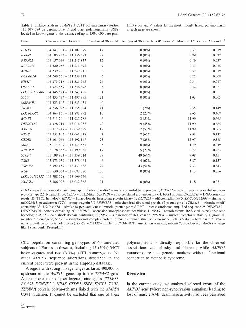
CEU population containing genotypes of 60 unrelatedsubjects of European descent, including 12 (20%) 34CTheterozygotes and two (3.3%) 34TT homozygotes. Noother AMPD1 sequence alterations described in thecurrent paper were present in the HapMap database.
A region with strong linkage ranges as far as 400,000 bpupstream of the AMPD1 gene, up to the TSPAN2 gene.After the exclusion of pseudogenes, nine genes (TRIM33,BCAS2, DENND2C, NRAS, CSDE1, SIKE, SYCP1, TSHB,TSPAN2) contain polymorphisms linked with the AMPD1C34T mutation. It cannot be excluded that one of these
polymorphisms is directly responsible for the observedassociations with obesity and diabetes, while AMPD1mutations are just genetic markers without functionalconnection to metabolic syndrome.
Discussion
In the current study, we analyzed selected exons of theAMPD1 gene (where non-synonymous mutations leading toloss of muscle AMP deaminase activity had been described
Table 5 Linkage analysis of AMPD1 C34T polymorphism (position115 037 580 on chromosome 1) and other polymorphisms (SNPs)located in known genes at the distance of up to 1,000,000 base pairs.
LOD score and r2 values for the most strongly linked polymorphismin each gene are shown
Gene Chromosome 1 location Number of SNPs Number (%) of SNPs with LOD score >2 Maximal LOD score Maximal r2
PHTF1 114 041 360 – 114 102 879 17 0 (0%) 0.57 0.019
RSBN1 114 105 977 – 114 156 593 27 0 (0%) 0.89 0.027
PTPN22 114 157 960 – 114 215 857 32 0 (0%) 0.89 0.037
BCL2L15 114 220 959 – 114 231 692 9 0 (0%) 0.47 0.016
AP4B1 114 239 201 – 114 249 215 8 0 (0%) 0.37 0.019
DCLRE1B 114 249 561 – 114 258 217 6 0 (0%) 0.22 0.008
HIPK1 114 273 519 – 114 321 945 24 0 (0%) 0.34 0.017
OLFML3 114 323 553 – 114 326 398 3 0 (0%) 0.42 0.021
LOC100132906 114 345 578 – 114 347 488 1 0 (0%) 0 0
SYT6 114 433 437 – 114 497 995 121 0 (0%) 1.03 0.063
MRP63P1 114 623 147 – 114 623 431 0 – – –
TRIM33 114 736 922 – 114 855 304 41 1 (2%) 2.55 0.149
LOC643586 114 864 161 – 114 881 992 10 2 (20%) 8.65 0.468
BCAS2 114 911 701 – 114 925 788 6 3 (50%) 11.99 0.665
DENND2C 114 928 719 – 115 014 255 42 19 (45%) 11.99 0.665
AMPD1 115 017 245 – 115 039 699 12 7 (58%) 11.99 0.665
NRAS 115 051 108 – 115 061 038 3 2 (67%) 8.93 0.332
CSDE1 115 061 060 – 115 102 147 25 7 (28%) 13.07 0.585
SIKE 115 113 623 – 115 124 831 3 0 (0%) 1.49 0.049
NR1H5P 115 178 857 – 115 199 038 17 5 (29%) 6.72 0.223
SYCP1 115 198 978 – 115 339 514 77 49 (64%) 9.08 0.45
TSHB 115 373 938 – 115 378 464 6 4 (67%) 3.87 0.157
TSPAN2 115 392 155 – 115 433 638 79 43 (54%) 7.33 0.343
NGF 115 630 060 – 115 682 380 100 0 (0%) 1.13 0.056
LOC100132332 115 908 526 – 115 909 576 0 – – –
VANGL1 115 986 097 – 116 042 368 91 0 (0%) 1.18 0.051
PHTF1 – putative homeodomain transcription factor 1; RSBN1 – round spermatid basic protein 1; PTPN22 – protein tyrosine phosphatase, non-receptor type 22 (lymphoid); BCL2L15 – BCL2-like 15; AP4B1 – adaptor-related protein complex 4, beta 1 subunit; DCLRE1B – DNA cross-linkrepair 1B (PSO2 homolog); HIPK1 – homeodomain interacting protein kinase 1; OLFML3 – olfactomedin-like 3; LOC100132906 – similar tomCG23455, pseudogene; SYT6 – synaptotagmin VI; MRP63P1 – mitochondrial ribosomal protein 63 pseudogene 1; TRIM33 – tripartite motif-containing 33; LOC643586 – similar to pyruvate kinase, muscle, pseudogene; BCAS2 – breast carcinoma amplified sequence 2; DENND2C –DENN/MADD domain containing 2C; AMPD1 – adenosine monophosphate deaminase 1; NRAS – neuroblastoma RAS viral (v-ras) oncogenehomolog; CSDE1 – cold shock domain containing E1; SIKE – suppressor of IKK epsilon; NR1H5P – nuclear receptor subfamily 1, group H,member 5 pseudogene; SYCP1 – synaptonemal complex protein 1; TSHB – thyroid stimulating hormone, beta; TSPAN2 – tetraspanin 2; NGF –nerve growth factor (beta polypeptide); LOC100132332 – similar to CCR4-NOT transcription complex, subunit 7, pseudogene; VANGL1 – vang-like 1 (van gogh, Drosophila)
72 J Appl Genetics (2011) 52:67–76

previously) in patients with cardiovascular diseases and innewborn controls. We chose exon 2 with the well-knownC34T (Gln12Stop) mutation, exon 3 with equally frequentC143T (Pro48Leu) substitution, exon 5 with G468T(Gln156His) mutation found in the German population (Grosset al. 2002), and exon 7 with A860T (Lys287Ile) (Toyama etal. 2004) described in Europeans. The presence of the samegenotype at loci 34 (RFLP method) and 143 (DHPLC) wasan additional proof of correct genotyping: when the resultswere discordant, direct sequencing was performed to confirmthe rare combination of genotypes at both loci.
Our results have shown that, in the Polish population,similarly to other Europeans, the most frequent alterations ofthe AMPD1 coding sequence are C34T and C143T, which arestrongly linked with each other. The most numerous group ofEuropeans genotyped for the AMPD1 C34T mutation so far isa cohort of 2,707 healthy British subjects (Webb et al. 2006)with the frequency of 34T allele equal to 13%, which is at thelower limit of confidence intervals for our groups (Table 2).The 34T frequency in a group of 721 healthy subjects fromsouth-western Germany was 14.5% (Frank et al. 2008), whilein 175 healthy Swedes, it was 13.7% (Norman et al. 1998). Itseems that the 34T allele frequency is similar in variousEuropean populations, including patients and newbornsexamined in the current study.
According to previous reports, A860T mutation ispresent in 3% alleles of healthy subjects of Europeandescent (Toyama et al. 2004). The 860T allele frequencywas 2.6% in healthy Americans of European origin(Isackson et al. 2005) and 2.8% in the healthy Germanpopulation (Hanisch et al. 2008). These values are inagreement with results of the current study (Table 2). Fourother detected sequence alterations (A99G in exon 3, IVS4-6delT in intron 4, G512A in exon 5, C784T in exon 7) havenot been described previously.
We have not found any case of G468Tmutation. This variantis probably very rare and limited to Germany, since it has notbeen detected in 230 subjects (healthy or with myopathy) fromvarious populations (Toyama et al. 2004) and in 704 healthySwedes (Fischer et al. 2007). Similarly, we have not founddel404T mutation in exon 5, which was detected in two of 879Swedes (Norman et al. 1998; Fischer et al. 2007).
The genotype combination 34TT+143CT was detected in0.5% of subjects, which is similar to the 0.7% frequencyfound in Americans of European origin (Isackson et al. 2005).Other previously described rare combinations (34CT+143CCand 34CT+143TT) (Fishbein et al. 1997) were not found inour population.
Functional impact of detected AMPD1 sequence alterations
Since our study did not involve the assessment of muscleAMP deaminase activity, we present the analysis of the
possible impact of the detected alterations on enzymefunction based on the previous reports and comparativegenomics. The C34T mutation in exon 2 (Gln12Stop)definitely leads to the termination of translation and resultsin the lack of immunoreactive protein (Morisaki et al. 1992)and enzyme activity (about 1% of normal) in homozygotes(Norman et al. 1998). Alternative splicing excluding exon 2may partly explain the residual AMPD activity andhypothetically protect from the metabolic consequences ofthe defect (Morisaki et al. 1993), but this theory needs to beproven.
The C143T transition (Pro48Leu) inmost cases accompaniesC34T and has no functional meaning, since translationterminates at codon 12. However, it could be introduced intothe protein if exon 2 was excluded due to alternative splicing. Astudy analyzing the expression of cDNAwith 143T and 34C inEscherichia coli did not show altered enzyme activity(Morisaki et al. 1992), but its stability and affinity were notanalyzed, and the influence on human muscle AMPD activityis unknown. Proline is exceptionally conservative and presentin all analyzed vertebrates at the position corresponding toPro48 in human AMPD1, together with adjacent amino acids,forming the sequence Cys-Pro-Ile. This sequence is alsopresent in the human AMPD3 protein and all of its orthologsin vertebrates, while in human AMPD2, the sequence is Ser-Pro-Ile. Proline is conservative also in AMPD proteins of non-vertebrates (Tyr-Pro-Ile sequence in Caenorhabditis elegans),and even in plants (Arabidopsis thaliana) (Han et al. 2006).The adjacent AMPD1 region encompassing AA 51-60 is aputative zinc-binding site (Martini et al. 2007) and has an -helix structure (Mangani et al. 2007). Proline-induced break ofthe -helix may be important for proper spatial conformationof the Zn2+-binding domain. The remarkable evolutionaryconservativeness of Pro48 needs further investigation, whichcould elucidate the effects of C143T substitution in the case ofalternative splicing excluding exon 2.
A860T transversion (Lys287Ile) affects the AMPD1region responsible for myosin binding. Lysine is totallyconservative in the corresponding position of AMPD1,AMPD2, and AMPD3 proteins in vertebrates and in mostnon-vertebrates and plants. In some protozoa, lysine issubstituted by arginine, a basic amino acid with similarproperties. The recombined mutated protein has decreasedboth the activity and affinity for AMP by half in relation towild-type enzyme (Toyama et al. 2004). Two reported casesconfirm impaired function of the mutated enzyme in vivo.The muscle AMPD activity in a subject with myopathysymptoms who turned out to be a compound heterozygotewith IVS2-(4-7)delCTTT and A860T mutations was 20–25%of normal (Isackson et al. 2005). Another compoundheterozygote with C34T and A860T mutations had theactivity equal to about 40% of common 34CT heterozygotes(Hanisch et al. 2008). It seems that the activity of enzyme
J Appl Genetics (2011) 52:67–76 73
α
α

encoded by 860T allele is 40–50% in relation to the enzymeencoded by wild-type allele.
G512A transition (Gly171Asp) is the most frequentAMPD1 alteration not described previously. Gly171 is fullyconservative in all of analyzed AMPD1 proteins inmammals, chicken, frog, and fish, but it is substituted byasparagine in lizard. Mammal AMPD2 contains alanine andAMPD3 may contain alanine, glycine, or threonine.AMPDs in non-vertebrates contain such different aminoacids as leucine, proline, and glutamate. These data suggestrelatively low conservation and, possibly, the lack offunctional impact of Gly171Asp alteration.
C784T (Arg262Trp) changes arginine residue, which isvery conservative in AMPD1, AMPD2, and AMPD3 proteinsof all 26 analyzed vertebrates, as well as in AMPD of insects,nematodes, and most fungi and plants (Arabidopsis thaliana).In some fungi and protozoa, arginine is substituted by similarbasic lysine or histidine. Only a few protozoa contain otheramino acids (glutamine, tyrosine, aspartate, glutamate). InFAC1 protein with AMPD activity in A. thaliana, thecorresponding Arg (position 350 in the Swiss-Prot O80452sequence) is located in an -helix structure in the middle ofthe His-Arg-Arg sequence, which forms a positively chargedflat surface (Han et al. 2006). These facts suggest the highprobability of functional impact of the C784T substitution.
The intron 4 region with IVS4-6delT alteration(cttttttttttttggcagGTT) contains a typical splice acceptorsequence: pyrimidine-rich tract and “ag” at the intron–exonboundary. The deletion shortens the tract from 12 to 11thymines. The chimpanzee AMPD1 intron 4–exon 5boundary is identical to the human sequence, but for thepresence of 11T instead of 12T, just like in the humanIVS4-6delT variant. In other primates, the pyrimidine-richtract may be as short as 7 bp. These facts are evidenceagainst the functional role of the deletion.
Synonymous A99G (Gly33Gly) transition in exon 3deletes one of five ESE motifs specific for the SF2/ASFprotein (AGGAGGT) with relatively low score (2.43) andcreates the ESS motif (AGGGGG), but it does not affect theESE motifs specific for other SR proteins (SC35, SRp40,SRp55). Adenine is present in eight of ten mammals ascoding nucleotide 99, while guanine is found in two (rabbitand tenrec). It suggests that the A99G variant lacksfunctional significance.
AMPD1 and cardiovascular diseases
Similarly to all earlier studies, we did not find significantdifferences in the C34T polymorphism genotype distributionsin the study groups (CAD+ HF− and HF +) and in a randomcontrol group consisting of consecutive newborns from thesame population as the study groups (Table 2). The T34 alleledoes not prevent the development of either CAD or HF,
though it seems to protect from the known risk factors ofCAD, such as abdominal obesity and diabetes (Safranowet al. 2009). We did not find significant differences betweengroups for the other detected sequence alterations, but the860T allele was less frequent in a combined group ofpatients than in newborns. It could be interpreted as aprotective effect of the T allele against cardiovasculardiseases, but such a hypothesis should be treated withcaution due to the low number of 860T carriers andmoderate statistical significance.
AMPD1 and diabetes
It was reported that variation in the AMPD1 gene is associatedwith insulin clearance and may participate in the syndromes ofinsulin resistance (Goodarzi et al. 2005). In the previous paper(Safranow et al. 2009), we demonstrated that the carriage ofT34 mutated allele in CAD patients without HF is associatedwith a lower prevalence of two features of metabolicsyndrome, diabetes and obesity, while in patients with HF, itis associated with lower fasting glucose. Hypothetically, theactivity of AMPD can influence the activity of AMP-activatedprotein kinase (AMPK), which controls cellular energy balance(e.g., by stimulating cellular glucose uptake), affecting thedevelopment of type 2 diabetes in many ways (Gerbitz et al.1996). In this study, we compared the influence of C34T andA860T mutations on obesity and diabetes in a combined groupof patients with and without HF. Surprisingly, their effectproved opposite: 34CT heterozygotes had a significantly lowerfrequency of diabetes than wild-type homozygotes, but in860AT heterozygotes, the prevalence of diabetes and obesitywas higher than in WT and 34CT patients. These differenceswere particularly significant when the presence of diabetes orfasting plasma glucose 126 mg/dL was analyzed (12% vs.30% vs. 86% prevalence for 34CT, WT, and 860AT,respectively). The difference in metabolic effects of the twomutations might be explained by the different ways of enzymeprotein modification: termination of translation by C34T andreduction of the activity by 40–50% in the case of A860T.Alternatively, the explanation might be the linkage disequilib-rium of AMPD1 mutations with functional variants inneighboring genes.
Linkage analysis of AMPD1 C34T polymorphism
Since the AMPD1 gene is expressed at a high level only inskeletal muscles, the mechanisms of genotype–phenotypeassociation between its mutations and clinical features ofpatients with cardiovascular diseases remain unclear.Therefore, a possibility of linkage with unknown sequencealteration within another gene should be taken into account.The nine genes containing polymorphisms linked withAMPD1 C34T mutation (Table 5) play various roles, and in
74 J Appl Genetics (2011) 52:67–76
α
≥

most cases, their function is poorly understood. TRIM33encodes transcription corepressor. BCAS2 is expressed inbreast tumors, interacting with estrogen receptors. DENND2Cencodes protein with the DENN domain and unknownfunction. Other proteins containing the DENN domainparticipate in mitogen-activated protein kinases (MAPK)pathways. Membrane protein encoded by NRAS oncogeneplays a part in MAPK pathways and in signaling throughinsulin receptor. Unr protein encoded by CSDE1 gene is anRNA chaperon, SIKE participates in the inhibition ofinterferon secretion during viral infection, and SYCP1 codesfor synaptonemal transverse filament protein. TSPAN2 enc-odes a membrane protein classified as one of the tetraspanins,which participate in the transduction of signals controllingcell development, activation, growth, and movement. TSHBencoding the beta subunit of thyrotropic hormone (TSH) isparticularly interesting, since its mutations lead to congenitalsecondary hypothyroidism (Karges et al. 2004), while bothhypo- and hyperthyroidism are recognized risk factors forcardiovascular diseases. We have not found any reportsanalyzing the association between polymorphisms of theabove-mentioned genes and cardiovascular diseases.
Conclusions
The most frequent mutation of the AMPD1 gene in the Polishpopulation is the C34T substitution, associated with reducedprevalence of diabetes and obesity in patients with coronaryartery disease (CAD) or heart failure (HF). The A860Tmutation seems to exert metabolic effects differing from C34Tand should always be accounted for in studies of the AMPD1genotype. The potential association of this mutation withreduced risk of cardiovascular disease, as well as withincreased prevalence of obesity and diabetes, merits furtherstudy in a larger population of patients. Due to the stronglinkage of AMPD1 mutations with sequence alterations in theTRIM33, BCAS2, DENND2C, NRAS, CSDE1, SIKE, SYCP1,TSHB, and TSPAN2 genes, it seems prudent to analyze theeffect of these alterations on the associations between clinicaland metabolic parameters and the AMPD1 genotype.
Acknowledgments This study was supported by grant 2 P05B 07926 from the Polish Committee for Scientific Research.
Open Access This article is distributed under the terms of the CreativeCommons Attribution Noncommercial License which permits anynoncommercial use, distribution, and reproduction in any medium,provided the original author(s) and source are credited.
References
Agresti A (1992) A survey of exact inference for contingency tables.Stat Sci 7:131–177
Altschul SF, Madden TL, Schäffer AA, Zhang J, Zhang Z, Miller W,Lipman DJ (1997) Gapped BLAST and PSI-BLAST: a newgeneration of protein database search programs. Nucleic AcidsRes 25:3389–3402
Anderson JL, Habashi J, Carlquist JF, Muhlestein JB, Horne BD, BairTL, Pearson RR, Hart N (2000) A common variant of theAMPD1 gene predicts improved cardiovascular survival inpatients with coronary artery disease. J Am Coll Cardiol36:1248–1252
Andreassi MG, Botto N, Laghi-Pasini F, Manfredi S, Ghelarducci B,Farneti A, Solinas M, Biagini A, Picano E (2005) AMPD1(C34T) polymorphism and clinical outcomes in patients under-going myocardial revascularization. Int J Cardiol 101:191–195
Barrett JC, Fry B, Maller J, Daly MJ (2005) Haploview: analysis andvisualization of LD and haplotype maps. Bioinformatics 21:263–265
Cartegni L, Wang J, Zhu Z, Zhang MQ, Krainer AR (2003)ESEfinder: a web resource to identify exonic splicing enhancers.Nucleic Acids Res 31:3568–3571
Clopper CJ, Pearson ES (1934) The use of confidence or fiduciallimits illustrated in the case of the binomial. Biometrika 26:404–413
Collins RP, Palmer BR, Pilbrow AP, Frampton CM, Troughton RW,Yandle TG, Skelton L, Richards AM, Cameron VA (2006)Evaluation of AMPD1 C34T genotype as a predictor of mortalityin heart failure and post-myocardial infarction patients. Am HeartJ 152:312–320
de Groote P, Lamblin N, Helbecque N, Mouquet F, Hermant X,Amouyel P, Dallongeville J, Bauters C (2006) The impact ofthe AMPD1 gene polymorphism on exercise capacity, otherprognostic parameters, and survival in patients with stablecongestive heart failure: a study in 686 consecutive patients.Am Heart J 152:736–741
Expert Panel on Detection, Evaluation, and Treatment of High BloodCholesterol in Adults (2001) Executive Summary of The ThirdReport of The National Cholesterol Education Program (NCEP)Expert Panel on Detection, Evaluation, and Treatment of HighBlood Cholesterol in Adults (Adult Treatment Panel III). JAMA285:2486–2497
Fischer S, Drenckhahn C, Wolf C, Eschrich K, Kellermann S, FrosterUG, Schober R (2005) Clinical significance and neuropathologyof primary MADD in C34-T and G468-T mutations of theAMPD1 gene. Clin Neuropathol 24:77–85
Fischer H, Esbjörnsson M, Sabina RL, Strömberg A, Peyrard-JanvidM, Norman B (2007) AMP deaminase deficiency is associatedwith lower sprint cycling performance in healthy subjects. J ApplPhysiol 103:315–322
Fishbein WN, Davis JI, Foellmer JW, Nieves S, Merezhinskaya N(1997) A competitive allele-specific oligomers polymerase chainreaction assay for the cis double mutation in AMPD1 that is themajor cause of myo-adenylate deaminase deficiency. Mol Diagn2:121–128
Frank B, Burwinkel B, Bermejo JL, Försti A, Hemminki K, HoulstonR, Mangold E, Rahner N, Friedl W, Friedrichs N, Buettner R,Engel C, Loeffler M, Holinski-Feder E, Morak M, Keller G,Schackert HK, Krüger S, Goecke T, Moeslein G, Kloor M,Gebert J, Kunstmann E, Schulmann K, Rüschoff J, Propping P;German HNPCC Consortium (2008) Ten recently identifiedassociations between nsSNPs and colorectal cancer could notbe replicated in German families. Cancer Lett 271:153–157
Gastmann A, Sigusch HH, Henke A, Reinhardt D, Surber R, GastmannO, Figulla HR (2004) Role of adenosine monophosphatedeaminase-1 gene polymorphism in patients with congestive heartfailure (influence on tumor necrosis factor-alpha level and outcome).Am J Cardiol 93:1260–1264
J Appl Genetics (2011) 52:67–76 75

Gerbitz KD, Gempel K, Brdiczka D (1996) Mitochondria anddiabetes. Genetic, biochemical, and clinical implications of thecellular energy circuit. Diabetes 45:113–126
Goodarzi MO, Taylor KD, Guo X, Quiñones MJ, Cui J, Li X, Hang T,Yang H, Holmes E, HsuehWA, Olefsky J, Rotter JI (2005) Variationin the gene for muscle-specific AMP deaminase is associated withinsulin clearance, a highly heritable trait. Diabetes 54:1222–1227
Gross M (1997) Clinical heterogeneity and molecular mechanisms ininborn muscle AMP deaminase deficiency. J Inherit Metab Dis20:186–192
Gross M, Rötzer E, Kölle P, Mortier W, Reichmann H, Goebel HH,Lochmüller H, Pongratz D, Mahnke-Zizelman DK, Sabina RL(2002) A G468-T AMPD1 mutant allele contributes to the highincidence of myoadenylate deaminase deficiency in the Caucasianpopulation. Neuromuscul Disord 12:558–565
Guo SW, Thompson EA (1992) Performing the exact test of Hardy–Weinberg proportion for multiple alleles. Biometrics 48:361–372
Han BW, Bingman CA, Mahnke DK, Bannen RM, Bednarek SY,Sabina RL, Phillips GN Jr (2006) Membrane association,mechanism of action, and structure of Arabidopsis embryonicfactor 1 (FAC1). J Biol Chem 281:14939–14947
Hanisch F, Joshi P, Zierz S (2008) AMP deaminase deficiency inskeletal muscle is unlikely to be of clinical relevance. J Neurol255:318–322
International HapMap Consortium (2007) A second generation humanhaplotype map of over 3.1 million SNPs. Nature 449:851–861
Isackson PJ, Bujnicki H, Harding CO, Vladutiu GD (2005) Myoadenylatedeaminase deficiency caused by alternative splicing due to a novelintronic mutation in the AMPD1 gene. Mol Genet Metab 86:250–256
Jones AC, Austin J, Hansen N, Hoogendoorn B, Oefner PJ, CheadleJP, O’Donovan MC (1999) Optimal temperature selection formutation detection by denaturing HPLC and comparison tosingle-stranded conformation polymorphism and heteroduplexanalysis. Clin Chem 45:1133–1140
Karges B, LeHeup B, Schoenle E, Castro-Correia C, Fontoura M, Pfäffle R,Andler W, Debatin KM, Karges W (2004) Compound heterozygousand homozygous mutations of the TSHbeta gene as a cause ofcongenital central hypothyroidism in Europe. Horm Res 62:149–155
Kolek MJ, Carlquist JF, Thaneemit-Chen S, Lazzeroni LC, WhitingBM, Horne BD, Muhlestein JB, Lavori P, Anderson JL (2005)The role of a common adenosine monophosphate deaminase(AMPD)-1 polymorphism in outcomes of ischemic and nonischemicheart failure. J Card Fail 11:677–683
Kurzawski G, Safranow K, Suchy J, Chlubek D, Scott RJ, Lubiński J(2002) Mutation analysis of MLH1 and MSH2 genes performed bydenaturing high-performance liquid chromatography. J BiochemBiophys Methods 51:89–100
Loh E, Rebbeck TR, Mahoney PD, DeNofrio D, Swain JL, Holmes EW(1999) Common variant in AMPD1 gene predicts improved clinicaloutcome in patients with heart failure. Circulation 99:1422–1425
Mangani S, Benvenuti M, Moir AJ, Ranieri-Raggi M, Martini D,Sabbatini AR, Raggi A (2007) Characterization of the metallocenter
of rabbit skeletal muscle AMP deaminase. Evidence for a dinuclearzinc site. Biochim Biophys Acta 1774:312–322
Martini D, Ranieri-Raggi M, Sabbatini AR, Moir AJ, Polizzi E,Mangani S, Raggi A (2007) Characterization of the metallocenterof rabbit skeletal muscle AMP deaminase. A new model forsubstrate interactions at a dinuclear cocatalytic Zn site. BiochimBiophys Acta 1774:1508–1518
Morisaki T, Sabina RL, Holmes EW (1990) Adenylate deaminase. Amultigene family in humans and rats. J Biol Chem 265:11482–11486
Morisaki T, Gross M, Morisaki H, Pongratz D, Zöllner N, Holmes EW(1992) Molecular basis of AMP deaminase deficiency in skeletalmuscle. Proc Natl Acad Sci USA 89:6457–6461
Morisaki H, Morisaki T, Newby LK, Holmes EW (1993) Alternativesplicing: a mechanism for phenotypic rescue of a commoninherited defect. J Clin Invest 91:2275–2280
Morisaki H, Higuchi I, Abe M, Osame M, Morisaki T (2000) Firstmissense mutations (R388W and R425H) of AMPD1 accompa-nied with myopathy found in a Japanese patient. Hum Mutat16:467–472
Norman B, Mahnke-Zizelman DK, Vallis A, Sabina RL (1998)Genetic and other determinants of AMP deaminase activity inhealthy adult skeletal muscle. J Appl Physiol 85:1273–1278
Sabina RL, Swain JL, Patten BM, Ashizawa T, O’Brien WE, HolmesEW (1980) Disruption of the purine nucleotide cycle. A potentialexplanation for muscle dysfunction in myoadenylate deaminasedeficiency. J Clin Invest 66:1419–1423
Sabina RL, Morisaki T, Clarke P, Eddy R, Shows TB, Morton CC,Holmes EW (1990) Characterization of the human and ratmyoadenylate deaminase genes. J Biol Chem 265:9423–9433
Safranow K, Czyzycka E, Binczak-Kuleta A, Rzeuski R, SkowronekJ, Wojtarowicz A, Jakubowska K, Olszewska M, Loniewska B,Kaliszczak R, Kornacewicz-Jach Z, Ciechanowicz A, Chlubek D(2009) Association of C34T AMPD1 gene polymorphism withfeatures of metabolic syndrome in patients with coronary arterydisease or heart failure. Scand J Clin Lab Invest 69:102–112
Toyama K, Morisaki H, Kitamura Y, Gross M, Tamura T, Nakahori Y,Vance JM, Speer M, Kamatani N, Morisaki T (2004) Haplotypeanalysis of human AMPD1 gene: origin of common mutantallele. J Med Genet 41:e74
Wang Z, Rolish ME, Yeo G, Tung V, Mawson M, Burge CB (2004)Systematic identification and analysis of exonic splicingsilencers. Cell 119:831–845
Webb EL, Rudd MF, Sellick GS, El Galta R, Bethke L, Wood W,Fletcher O, Penegar S, Withey L, Qureshi M, Johnson N,Tomlinson I, Gray R, Peto J, Houlston RS (2006) Search forlow penetrance alleles for colorectal cancer through a scan of1467 non-synonymous SNPs in 2575 cases and 2707 controlswith validation by kin-cohort analysis of 14 704 first-degreerelatives. Hum Mol Genet 15:3263–3271
Yazaki Y, Muhlestein JB, Carlquist JF, Bair TL, Horne BD, RenlundDG, Anderson JL (2004) A common variant of the AMPD1 genepredicts improved survival in patients with ischemic leftventricular dysfunction. J Card Fail 10:316–320
76 J Appl Genetics (2011) 52:67–76
Copyright © 2022 FDOKUMEN


![[Management of children and adolescents with severe obesity]](https://static.fdokumen.com/doc/165x107/633344bcb94d623842021d07/management-of-children-and-adolescents-with-severe-obesity.jpg)