Enzymatic Bioelectrocatalysis - MDPI
178
Edited by Enzymatic Bioelectrocatalysis Elisabeth Lojou and Xinxin Xiao Printed Edition of the Special Issue Published in Catalysts www.mdpi.com/journal/catalysts
-
Upload
khangminh22 -
Category
Documents
-
view
0 -
download
0
Transcript of Enzymatic Bioelectrocatalysis - MDPI
Edited by
Enzymatic Bioelectrocatalysis
Elisabeth Lojou and Xinxin Xiao
Printed Edition of the Special Issue Published in Catalysts
www.mdpi.com/journal/catalysts

Enzymatic Bioelectrocatalysis


Enzymatic Bioelectrocatalysis
Editors
Elisabeth Lojou
Xinxin Xiao
MDPI • Basel • Beijing • Wuhan • Barcelona • Belgrade • Manchester • Tokyo • Cluj • Tianjin

Editors
Elisabeth Lojou
BIP-CNRS-GLM
Aix Marseille University
Marseille
France
Xinxin Xiao
Department of Chemistry
Technical University of Denmark
Lyngby
Denmark
Editorial Office
MDPI
St. Alban-Anlage 66
4052 Basel, Switzerland
This is a reprint of articles from the Special Issue published online in the open access journal Catalysts
(ISSN 2073-4344) (available at: www.mdpi.com/journal/catalysts/special issues/enzy bioelect).
For citation purposes, cite each article independently as indicated on the article page online and as
indicated below:
LastName, A.A.; LastName, B.B.; LastName, C.C. Article Title. Journal Name Year, Volume Number,
Page Range.
ISBN 978-3-0365-3460-2 (Hbk)
ISBN 978-3-0365-3459-6 (PDF)
© 2022 by the authors. Articles in this book are Open Access and distributed under the Creative
Commons Attribution (CC BY) license, which allows users to download, copy and build upon
published articles, as long as the author and publisher are properly credited, which ensures maximum
dissemination and a wider impact of our publications.
The book as a whole is distributed by MDPI under the terms and conditions of the Creative Commons
license CC BY-NC-ND.

Contents
About the Editors . . . . . . . . . . . . . . . . . . . . . . . . . . . . . . . . . . . . . . . . . . . . . . vii
Elisabeth Lojou and Xinxin Xiao
Enzymatic BioelectrocatalysisReprinted from: Catalysts 2021, 11, 1373, doi:10.3390/catal11111373 . . . . . . . . . . . . . . . . . 1
Charlene Beaufils, Hiu-Mun Man, Anne de Poulpiquet, Ievgen Mazurenko and Elisabeth
Lojou
From Enzyme Stability to Enzymatic Bioelectrode Stabilization ProcessesReprinted from: Catalysts 2021, 11, 497, doi:10.3390/catal11040497 . . . . . . . . . . . . . . . . . . 3
Hongqi Xia and Jiwu Zeng
Rational Surface Modification of Carbon Nanomaterials for Improved Direct ElectronTransfer-Type Bioelectrocatalysis of Redox EnzymesReprinted from: Catalysts 2020, 10, 1447, doi:10.3390/catal10121447 . . . . . . . . . . . . . . . . . 49
Xiaomei Yan, Jing Tang, David Tanner, Jens Ulstrup and Xinxin Xiao
Direct Electrochemical Enzyme Electron Transfer on Electrodes Modified by Self-AssembledMolecular MonolayersReprinted from: Catalysts 2020, 10, 1458, doi:10.3390/catal10121458 . . . . . . . . . . . . . . . . . 65
Huijie Zhang, Rosa Catania and Lars J. C. Jeuken
Membrane Protein Modified Electrodes in BioelectrocatalysisReprinted from: Catalysts 2020, 10, 1427, doi:10.3390/catal10121427 . . . . . . . . . . . . . . . . . 91
Taiki Adachi, Yuki Kitazumi, Osamu Shirai and Kenji Kano
Recent Progress in Applications of Enzymatic BioelectrocatalysisReprinted from: Catalysts 2020, 10, 1413, doi:10.3390/catal10121413 . . . . . . . . . . . . . . . . . 121
Simin Arshi, Mehran Nozari-Asbemarz and Edmond Magner
Enzymatic Bioreactors: An Electrochemical PerspectiveReprinted from: Catalysts 2020, 10, 1232, doi:10.3390/catal10111232 . . . . . . . . . . . . . . . . . 141
v


About the Editors
Elisabeth Lojou
Elisabeth Lojou is a senior researcher in the Bioenergetic and Protein Engineering Laboratory
(Aix Marseille University, CNRS) in France. Her group conducts research in the domain of
fundamental bioelectrochemistry, with a special interest in electron transfers between functionalized
nanostructured electrodes and redox enzymes, such as hydrogenases and multicopper oxidases. For
8 years, the group has been engaged in the design of H2/O2 enzymatic fuel cells.
Xinxin Xiao
Xinxin Xiao achieved his PhD in 2019 at the University of Limerick, Ireland. Currently, he is a
researcher at the Technical University of Denmark. His research focuses on bioelectrochemistry, and
especially electrochemical sensors and electrochemical energy harvesters.
vii


catalysts
Editorial
Enzymatic Bioelectrocatalysis
Elisabeth Lojou 1,* and Xinxin Xiao 2,*
Citation: Lojou, E.; Xiao, X.
Enzymatic Bioelectrocatalysis.
Catalysts 2021, 11, 1373. https://
doi.org/10.3390/catal11111373
Received: 9 November 2021
Accepted: 12 November 2021
Published: 14 November 2021
Publisher’s Note: MDPI stays neutral
with regard to jurisdictional claims in
published maps and institutional affil-
iations.
Copyright: © 2021 by the authors.
Licensee MDPI, Basel, Switzerland.
This article is an open access article
distributed under the terms and
conditions of the Creative Commons
Attribution (CC BY) license (https://
creativecommons.org/licenses/by/
4.0/).
1 National Center for Scientific Research (CNRS), Aix Marseille University, BIP, UMR 7281, 31 Chemin Aiguier,13009 Marseille, France
2 Department of Chemistry, Technical University of Denmark, 2800 Lyngby, Denmark* Correspondence: [email protected] (E.L.); [email protected] (X.X.)
Enzymatic bioelectrocatalysis relies on immobilizing oxidoreductases on electrodesurfaces, leading to different applications, such as biosensors [1], biofuel cells [2], andbioelectrosynthesis [3]. Based on their intrinsic properties, i.e., high specificity and highaffinity for the substrate, enzymes may provide sustainable alternatives to currently usedchemical catalysts for human health monitoring, biopower generation or high-value prod-uct synthesis. However, enzyme bioelectrocatalysis suffers from low catalytic efficiency,imposing fundamental investigations on mechanisms of enzyme immobilization, includingmolecular basis knowledge of the efficient electronic communication between enzymes andthe electrode. Bioelectrocatalysis is also limited by long-term stability requiring ground-breaking strategies for enzyme protection and bioelectrode survival.
The “Enzymatic Bioelectrocatalysis” Special Issue comprises six reviews contributedby research groups from different countries, covering fundamentals and applications, aswell as the recent research progress in this field.
Enzyme immobilization and electron transfer mechanisms are two crucial and closelyinterrelated aspects, which ultimately determine the stability and efficiency of the bioelec-trode. On the basis of understanding the parameters governing protein stability, Beaufilset al. discuss the major strategies to improve redox enzyme stability, with a focus on theimmobilization as an important route [4]. The authors further discuss additional factorsspecific for bioelectrocatalysis, such as enzyme reorientation, effect of the electric field andprotection against reactive oxygen species (ROS) production, that should be consideredto achieve highly stable bioelectrodes undergoing electron transfer. For future directions,they emphasize the need to screen the diversity for the discovering of new outstandingenzymes with enhanced stability, as well as the requirement of in situ and in operandomethodologies to get new insights on enzyme behavior in the immobilized state.
Gold and carbon electrodes represent two key materials for enzyme immobilization,which are respectively reviewed by Xia et al. [5] and Yan et al. [6]. Strategies used to ratio-nalize surface modification for high-performance direct electron transfer (DET) of redoxenzymes are especially discussed. Yan et al. emphasizes the employment of self-assembledmonolayers (SAM) as tunable bridges between redox enzymes and gold electrode surfacespermitting DET [6]. The authors overview the characterization methods of SAMs andstructural properties of common enzymes, highlighting the strategic selection of a specificSAM to control proper enzyme orientation. Xia et al. highlight the key parameters allowingenzymatic DET on carbon nanomaterials, and review the various methods for the orientedimmobilization. Interestingly, they also present the tools currently developed to proberedox enzymes [5].
Membrane proteins, constituting 20–30% of all proteins secreted by living organisms, area major subject of bioelectrocatalysis. In the review by Zhang et al., various and eventuallycoupled techniques, i.e., electrochemistry, spectroscopy, microscopy, and quartz crystal mi-crobalance [7] are discussed toward the understanding and use of membrane enzymes activein bioenergy conversion. Electrode designs with a special focus on the specificity required formembrane proteins are highlighted. Two emerging directions: (i) the membrane protein based
1

Catalysts 2021, 11, 1373
hybrid vesicles for improved lifetime and (ii) microorganisms for microbial electrosynthesisand semi-artificial photosynthesis, are emphasized for future research.
Regarding to the applications of enzymatic bioelectrocatalysis, Adachi et al. describethe recent progress in emerging bioelectrochemical fields such as biosupercapacitor, bio-electrosynthesis and photo-bioelectrocatalysis [8]. The authors claim how crucial furtherresearch on protein-engineering, rational selection of electrode materials and mediators,immobilization of enzymes, and layout of electrodes to improve bioelectrocatalysis willbe. The review by Arshi et al. focuses on the development of electrochemically basedenzymatic reactors [9]. Together with the discussion on the mechanisms of electron transferinvolving immobilized enzyme, the authors review the usage of electrochemically basedbatch and flow bio-reactors. They highlight the importance of high surface area electrodes,enzyme engineering and enzyme cascades, joining the general opinion formulated in thereview by Adachi et al. [8]. Finally, both reviews provide some examples of enzyme-basedelectrosynthesis especially relevant in a sustainable world.
The common message conveyed from all the contributions of this special issue is thatenzymatic electrochemistry is expected to play an increasingly role towards electrocatalysisin mild conditions. We look forward to further new developments in this exciting field.
Conflicts of Interest: The authors declare no conflict of interest.
References
1. Schachinger, F.; Chang, H.; Scheiblbrandner, S.; Ludwig, R. Amperometric Biosensors Based on Direct Electron Transfer Enzymes.Molecules 2021, 26, 4525. [CrossRef] [PubMed]
2. Xiao, X.; Xia, H.-q.; Wu, R.; Bai, L.; Yan, L.; Magner, E.; Cosnier, S.; Lojou, E.; Zhu, Z.; Liu, A. Tackling the Challenges of Enzymatic(Bio)Fuel Cells. Chem. Rev. 2019, 119, 9509–9558. [CrossRef] [PubMed]
3. Chen, H.; Simoska, O.; Lim, K.; Grattieri, M.; Yuan, M.; Dong, F.; Lee, Y.S.; Beaver, K.; Weliwatte, S.; Gaffney, E.M.; et al.Fundamentals, Applications, and Future Directions of Bioelectrocatalysis. Chem. Rev. 2020, 120, 12903–12993. [CrossRef][PubMed]
4. Beaufils, C.; Man, H.-M.; de Poulpiquet, A.; Mazurenko, I.; Lojou, E. From Enzyme Stability to Enzymatic BioelectrodeStabilization Processes. Catalysts 2021, 11, 497. [CrossRef]
5. Xia, H.; Zeng, J. Rational Surface Modification of Carbon Nanomaterials for Improved Direct Electron Transfer-Type Bioelectro-catalysis of Redox Enzymes. Catalysts 2020, 10, 1447. [CrossRef]
6. Yan, X.; Tang, J.; Tanner, D.; Ulstrup, J.; Xiao, X. Direct Electrochemical Enzyme Electron Transfer on Electrodes Modified bySelf-Assembled Molecular Monolayers. Catalysts 2020, 10, 1458. [CrossRef]
7. Zhang, H.; Catania, R.; Jeuken, L.J.C. Membrane Protein Modified Electrodes in Bioelectrocatalysis. Catalysts 2020, 10, 1427.[CrossRef]
8. Adachi, T.; Kitazumi, Y.; Shirai, O.; Kano, K. Recent Progress in Applications of Enzymatic Bioelectrocatalysis. Catalysts 2020, 10,1413. [CrossRef]
9. Arshi, S.; Nozari-Asbemarz, M.; Magner, E. Enzymatic Bioreactors: An Electrochemical Perspective. Catalysts 2020, 10, 1232.[CrossRef]
2

catalysts
Review
From Enzyme Stability to Enzymatic BioelectrodeStabilization Processes
Charlène Beaufils, Hiu-Mun Man, Anne de Poulpiquet, Ievgen Mazurenko and Elisabeth Lojou *
Citation: Beaufils, C.; Man, H.-M.; de
Poulpiquet, A.; Mazurenko, I.; Lojou,
E. From Enzyme Stability to
Enzymatic Bioelectrode Stabilization
Processes. Catalysts 2021, 11, 497.
https://doi.org/10.3390/catal11040497
Academic Editor: Giovanni Gadda
Received: 17 March 2021
Accepted: 10 April 2021
Published: 14 April 2021
Publisher’s Note: MDPI stays neutral
with regard to jurisdictional claims in
published maps and institutional affil-
iations.
Copyright: © 2021 by the authors.
Licensee MDPI, Basel, Switzerland.
This article is an open access article
distributed under the terms and
conditions of the Creative Commons
Attribution (CC BY) license (https://
creativecommons.org/licenses/by/
4.0/).
Aix Marseille University, CNRS, BIP, Bioénergétique et Ingénierie des Protéines, UMR 7281, 31, Chemin JosephAiguier, CS 70071 13402 Marseille CEDEX 09, France; [email protected] (C.B.); [email protected] (H.-M.M.);[email protected] (A.d.P.); [email protected] (I.M.)* Correspondence: [email protected]; Tel.: +33-(0)491-164-144
Abstract: Bioelectrocatalysis using redox enzymes appears as a sustainable way for biosensing, elec-tricity production, or biosynthesis of fine products. Despite advances in the knowledge of parametersthat drive the efficiency of enzymatic electrocatalysis, the weak stability of bioelectrodes preventslarge scale development of bioelectrocatalysis. In this review, starting from the understanding of theparameters that drive protein instability, we will discuss the main strategies available to improve allenzyme stability, including use of chemicals, protein engineering and immobilization. Consideringin a second step the additional requirements for use of redox enzymes, we will evaluate how farthese general strategies can be applied to bioelectrocatalysis.
Keywords: enzyme; metalloenzyme; catalysis; stability; electrochemistry; bioelectrochemistry
1. Introduction
In the search for a more sustainable way of life, our society must adapt nowadaysto reduce the use of harmful products or high carbon energy sources deleterious for ourplanet, hence for our lives. Many industrial sectors are concerned by this revolution, fromfine product synthesis to environmental monitoring, clinical diagnosis or energy produc-tion. Catalysis of the involved chemical reactions is a key process towards sustainabilityby accelerating kinetics, or increasing selectivity. However, chemical catalysts themselvesare very often not produced and used in eco-friendly manner, requiring organic solventsfor their synthesis or being based on metals rare on earth. In that sense, enzymes can beregarded as sustainable alternatives with great advantages directly linked to their intrinsicproperties required to sustain life [1] In particular, many different enzymes are involvedin a variety of reactions in microorganisms where they operate in mild conditions, devel-oping high catalytic activity and specificity for their substrate. Their amazing functionaldiversity directly originates from the chemistry and polarity of amino acids that fold ina diversity of structures. In addition, they are produced in mild aqueous conditions andare totally biodegradable. However, their weak stability and ability to maintain biologicalactivity during storage and operation, freeze-thaw steps, and in non-physiological environ-ments lower their attractiveness. Development of strategies to enhance enzyme shelf lifewhile maintaining catalytic activity have been the subject of many researches during thelast decades [2–4].
Redox enzymes belong to a class of enzymes containing redox cofactors that arenecessary for their activity. These oxidoreductases transform their substrate by exchangingelectrons with their physiological partners in the metabolic chain. Intra-enzyme redoxcomponents can be organic cofactors such as flavin adenine dinucleotide (FAD) for sugaroxidation [5]. They can be metal centers, including copper for oxygen reduction [6], ironand nickel for hydrogen evolution and uptake or CO2 reduction [7–9], manganese for wateroxidation in photosynthesis [10], etc. Some others are dependent on nicotinamide adeninedinucleotide phosphate (NADPH) to realize enzymatic transformations [11]. Such enzymes
3

Catalysts 2021, 11, 497
have been envisioned as biocatalysts in biosensors [12–14], biosynthesis reactors [15,16],or biofuel cells [17,18]. This domain is referred as Bioelectrochemistry. As for non-redoxenzymes, one of the main limitations in the large-scale development of these biotechnolo-gies is the weak stability of bioelectrodes based on redox enzymes. In particular, in thedifferent biotechnological devices listed just before, enzymes may face high temperaturesor high salt concentrations. They may have to operate at gas-liquid interfaces or even atthe three phase boundaries. In addition, to exploit the catalytic properties of these redoxenzymes in biotechnology, a further step is to ensure electron exchange between the proteinand a conductive surface [19–21]. This prerequisite imposes the enzyme adopts certainconfigurations once immobilized, so that not only the structural conformation and dynamicremains for substrate accessibility and activity, but also enzyme positioning and orientationon the solid interface allows electron transfer.
The purpose of this review is to discuss whether general strategies available for allenzyme stabilization can be applied for the special case of redox enzymes used in bioelec-trochemistry. To reach this objective, the fundamental bases for all enzyme stabilizationby general reported strategies will be first detailed, with a focus on the immobilizationprocedure as an important way for stabilization. In a second step, we will emphasize theparticular features of redox enzymes that must be considered before applying any availablestabilization strategies. Ultimately, we will discuss which additional specific strategiesmust be set up for redox protein, and by extension for bioelectrode, stabilization. Becausethe topic is very large, this review will not provide an exhaustive list of references but willinstead aim to highlight major issues related to enzyme and redox enzyme stability withillustrations taken from papers published during the last few years.
2. Intrinsic (in)Stability of Enzymes
The metabolisms essential for life are catalyzed by enzymes. Over time, these proteinsmay have undergone different changes resulting from mutations, which cause enzymesto evolve to function in a particular cellular environment and will be very sensitive toexogenous conditions. In addition, in the cell, enzymes can be subjected to various stresses,which can lead to their denaturation or the formation of aggregates. Thanks to a perfectlydesigned proteolysis machine, new fresh proteins are produced to replace the deactivatedproteins. These two fundamental concepts mean that using enzymes outside a cell isexpected to induce some loss of activity and eventually progressive denaturation, whichcan be irreversible. The will to exploit enzymes for other works than predicted by evolution,although highly attractive, imposes finding out strategies to preserve activity withindurations compatible with fundamental laboratory studies and with applications [3]. Beforereaching that objective, the prerequisite is to know which factors affect enzyme activity andstability. A subtle balance between stability and flexibility is required to maintain activity,so that stability can be the price to pay against activity. Each enzyme is composed of alinear chain of amino acids that fold to produce a unique three-dimensional structure whichconfers specific and unique enzymatic properties. The overall structure of an enzyme isdescribed according to fours levels which contribute to its specific function and stability(Figure 1). The secondary structure refers to the arrangement of the polypeptide chain (theprimary structure) as random coil, α-helix, β-sheet and turn stabilized by hydrogen bondsbetween amino acid residues. While the primary structure is stable thanks to the strengthof the peptide bonds which are unlikely to be broken upon changes in the environmentalconditions, the secondary structure can be altered even during protein storage. Thetertiary structure is the three-dimensional arrangement of the amino acid residues, givingthe overall conformation of the polypeptidic chain. Hydrophobic interactions betweennonpolar chains as well as disulfide bonds and ionic interactions between charged residuesare involved in the stabilization of the tertiary structure. To fulfill the requirement ofenzyme localization in the cell, hydrophobic and hydrophilic groups are arranged withinthe protein. For soluble proteins, hydrophobic side groups tend to be hidden in the proteincore while hydrophilic groups are exposed to the surrounding environment. These amino
4

Catalysts 2021, 11, 497
acid residues on the surface of the enzyme are more likely to be sensitive to the externalenvironment than the core amino acids.
βα
Figure 1. The four levels of protein structures determine the enzyme function.
Denaturation occurs upon unfolding of the tertiary structure to a disordered polypep-tide where residues are no longer arranged for functional or structural stabilizing interac-tions [22]. This process may be reversible. Irreversible loss of activity may also occur uponexternal stresses. This leads to two different concepts to evaluate in vitro stability of a pro-tein. The thermodynamic stability refers to the ability for a protein to unfold/refold afterbeing subjected to stresses (elevated temperature, extreme pH, or high organic solvent con-centration). Methods to evaluate this stability range from scanning calorimetry to circulardichroism (CD) and fluorimetry. Kinetic stability represents the duration of enzyme activitybefore irreversible denaturation and is measured by activity assays. Actually, the nativeactive state of an enzyme is in equilibrium with the partially denatured, enzymaticallyinactive state, and the folded/unfolded transition involves mainly intramolecular hydro-gen bonds and hydrophobic interactions. This scheme is most certainly more complicatedsince it was shown that a protein can oscillate between many different folded/unfoldedconfigurations. Each oscillation is governed by the second thermodynamic law, the entropydecreasing as the protein folds, and hydrogen bond formation contributing to the enthalpy.Each strategy that tends to increase rigidity in the enzyme architecture decreases entropyand enhances stability [23]. Thermodynamic instability of enzymes can be partly attributedto the lack of rigidity within the tertiary structure, caused by flexible random coils incontrast to stable β-sheets [24]. In addition to higher rigidity and compact packing, thepresence of α-helices with antiparallel arrangement contributes to stability. Water is thenatural environment in which protein molecules do exist and operate, and water moleculesplay a key role in maintaining the entropic stability of the enzyme structure.
Any engineering towards enzyme structural rigidification must maintain the hydra-tion shell and flexibility required for activity. Based on enzyme structural features, manydifferent agents are potential enzyme “killers”. When temperature increases, the enzymetends to unfold in a cooperative process. Disulfide bridges can be broken by reducingagents, extreme pHs are going to affect hydrogen bonding and salt bridges, high saltconcentration may aggregate the protein, while charged components may form bondswith charged amino acids modifying the tertiary structure. Organic solvents or detergentswill alter hydrophobic interactions, and stability of enzymes in organic solvents will be
5

Catalysts 2021, 11, 497
dependent on the ability of the solvent to strip the essential hydration layer from theprotein surface [25,26]. Last but not least, mechanical stress or radiation may disrupt thedelicate balance of forces that maintain protein structure [27]. Stabilizing the enzymemeans preventing these changes.
3. Strategies for All Enzyme Stabilization
Enzyme stability involves a balance between intramolecular interactions of functionalgroups and their interaction with the protein environment. Water, as many protein naturalsolvent, plays a key role in enzyme stability by controlling the hydration shell structureand dynamics and by providing required plasticity for activity [28]. As a direct conse-quence, many stabilization strategies target water activity in the environment of the enzymeor inside the protein moiety, with the main objective of decreasing the enzyme motion(Figure 2). However, this enhanced stability should not be achieved at the expense ofactivity, so a delicate balance must be maintained between rigidification and sufficientplasticity controlled by water. As another mere general rule, stabilization over one stresstends to enhance stability against another stress.
Figure 2. Strategies for all enzyme stabilization.
3.1. Addition of Stabilizers in the Medium
3.1.1. Chemical Stabilizer Addition
It has been known for long time that addition of polyols and polysaccharides such astrehalose, glycerol, dextran, etc. helps stabilizing enzymes [29–32]. Protective effect of poly-ols was reported in many recent studies in different experimental situations and expositionto stresses (high pressure or high temperature, presence of non-aqueous solvents) [33–37].
Generally speaking, polyols stabilize proteins either in the dried state by serving aswater substitutes and preventing dehydration through hydrogen bonding, or in solution byaltering protein-solvent interactions [38]. Several mechanisms are discussed to account forthe effect of such chemical additives on the protein stabilization [23]. Hydrophilic polyolstend to strengthen hydrophobic interactions among nonpolar amino acid residues resultingin a more compact and spherical enzyme form with smaller surface area, preventing proteindynamics and enhancing stability. Compressibility of proteins, a crucial thermodynamicparameter that determines flexibility, is affected by polyol addition [39]. One acceptedmechanism is based on the different sizes between water and stabilizing molecules that
6

Catalysts 2021, 11, 497
preferentially exclude the latter from the protein surface. The preferential hydration ofproteins causes an unfavorable free energy change that the proteins tend to minimize byfavoring the more compact state over the structurally expanded state. Protective effectis also explained in terms of influence of additives on water activity that results in anincrease of structural organization of water molecules contributing to the conservation oflow energy interactions favoring native protein conformation [25].
Surfactants, maltodextrine, sodium azide, or special buffers have also been usedas additives to maintain the native structure of proteins through purification steps [25].Dimethyl sulfoxide (DMSO) acts as a stabilizer because it is preferentially excluded from theprotein surface [22,40,41]. Addition of DMSO but also of glycerol or ethylene glycol serve ascryoprotectants preventing protein solutions from freezing at −20 C and allowing multipleuse of a unique sample without freeze-thaw cycles [38,42]. Adding polymers in solutionsuch as poly(ethylene glycol) (PEG), alginate, or chitosan maintains a hydration shellaround the protein according to the exclusion mechanism [38,43–45]. Addition of polymersalso prevents protein aggregation by modifying protein-protein interactions and increasingmedium viscosity, thus decreasing enzyme motion. Macromolecular crowding, whichis the natural environment of enzymes in vivo, can explain the stabilizing effect of someadditives [46–49]. It can be mimicked in vitro through the addition of high concentrationsof macromolecules such as dextran or PEG [50] and would favor the folded state of proteinsand compact conformation through the excluded volume effect [51–55]. However, thestabilizing effect of in vitro crowding would not be universal as the properties of allmacromolecules in vivo are finely regulated under different physiological conditions [56].Aggregation under crowded conditions can be enhanced because the activity of water isdecreased, and the refolding rate of proteins as a consequence of increased viscosity isalso decreased.
3.1.2. Salt Effect
Salt addition affects protein stability according to a combination of binding effect,screening of protein surface electrostatic potential, and effect on protein/water interface.Salts may contribute to hydrophobic interaction strengthening in proteins, hence to sta-bilization, but salt binding to amino acids on the protein surface decreases the repulsionbetween proteins and induces aggregation. The salting out process, or salt-induced pre-cipitation, depends on the structure of the protein and in particular on the population ofhydrophilic amino acid residues. It also largely depends on the salt concentration. Theeffect of salts on protein stabilization has been tentatively explained on the basis of theFrank Hofmeister series [4,57–59] (Figure 3A). This series early ordered anions and cationscomposing salts according at first to their ability to precipitate lysozyme, and later totheir ability to stabilize protein secondary and tertiary structure. They were respectivelyclassified at that time as water-structure formers (kosmotropes) or water-structure breakers(chaotropes). Kosmotropes and chaotropes would be nowadays more understood as acharacterization of the degree of hydration, and different models are developed to considerthe various effects observed experimentally [60]. Ionic liquids (IL) would follow the sametendency as salts where kosmotropic anions and chaotropic cations stabilize the enzyme,while chaotropic anions and kosmotropic cations destabilize it [61]. While they can beconsidered as eco-friendly solvents, many proteins are inherently inactive in ILs, requiringthe addition of water for activity recovery. This suggests ILs could affect the internalwater shell [61,62].
Recent examples in the literature illustrate the effect of salts on enzyme stability, andmost of them agree with the Hofmeister series. Changes in the secondary structure oftwo proteins with helical and beta structural arrangement, respectively, were followed byFourier Transform Infra-Red spectroscopy (FTIR) in the presence of various salts. It wasshown that the stabilization effect of the salt follows the Hofmeister series of ions, althoughsome exceptions were observed with formation of intermolecular β-sheets typical of amor-phous aggregates [62]. Glucose oxidase (GOx) stability was studied using microcalorimetry
7

Catalysts 2021, 11, 497
in the presence of various salts [63]. At high salt concentrations (over 1 M), it was shownthat the Hofmeister effect on the temperature of inactivation was determined by the ion-specific effect on the protein/water interface. Correlation between stability and activity oflysozyme in the presence of various salts from the Hofmeister series suggested a role oflocal stability/flexibility in enzyme activity [64]. The thermal stability of Aspergillus terreusglucose dehydrogenase (GDH) was substantially improved by kosmotropic anions, retain-ing more than 90% activity after 60 min of heat treatment at 60 C. The stabilizing effectfollowed the Hofmeister series and was anion concentration-dependent and strongly re-lated to the structural stabilization of the enzyme, which involved enzyme compaction [65](Figure 3B). It was further shown that salts can stabilize proteins not only in vitro butalso in vivo or intracellularly, the stabilization level correlating with the Hofmeister seriesof ions [66].
β
Figure 3. Salt effect on enzyme stability. (A) Hofmeister series and effect on protein properties.(B) Stabilization effect of kosmotropic anions and cations on Aspergillus terreus GDH. Residualactivity after 1 h incubation at 50 C as a function of added anions and cations. Reproduced withpermission from [65].
3.2. Chemical Modification of Enzymes, Directed Evolution
More than 95% of all charged components are located on the surface of proteins,consisting mostly of hydrophilic moieties, while most of the hydrophobic ones are burieddeep inside the core. As already discussed above, the physico-chemical microenvironmentof the protein will be sensed intrinsically through those moieties, demonstrating the proteinsurface to be an attractive target for protein engineering to enhance protein stability. Theefforts toward engineering enzyme catalysts with increased stability can be divided intotwo groups: (i) chemical protein modification and (ii) protein engineering, such as site-directed mutagenesis and directed evolution. The combination of these two approaches isappealing for improving the catalytic properties of enzymes.
3.2.1. Chemical Modification of Enzymes
Chemical protein modification can be achieved either by modification of one sort ofamino acid on the protein surface or by polyfunctional modification using reticulatingagents or by conjugation to water-soluble polymers. One widely used method consistsin reticulation through cross linking via glutaraldehyde (GA) that increases the proteinrigidity and mimics disulfide physiological bonds or salt bridges [25]. Activity is howeververy often affected. Stabilization of proteins through cross-linked-enzyme-aggregates(CLEAs) will be further discussed below in Section 4 [67,68]. Conjugation through water-soluble polymers such as PEG, chitosan, alginate, dextran, etc. has also been widelyreported [38,69,70]. Those polymers present either one functionality or are bifunctional toenable reaction with N- or C-terminal or with one individual amino-acid residue on theprotein. Increase in the half-life of proteins is frequently observed [71,72]. Other polymersresponding to temperature stimuli can tailor the temperature dependence of enzymestability [73]. Two different methods can be carried out to synthesize the bioconjugate. In
8

Catalysts 2021, 11, 497
the “grafting to” strategy, the polymer is first synthetized; then, an end-group functionalityis attached to one amino-acid on the protein surface. As an example, a mono-PEGylatedarginase was constructed by linkage of PEG-maleimide to a cysteine residue on the enzymesurface [74]. The protein was protected against proteases thanks to the shielding effect ofPEG, allowing the modified arginase to operate as a promising anticancer drug candidate.In the “grafting from” method, radical polymerization occurs at sites attached to theprotein. A recent work describes the ligation of Atom Transfer Radical Polymerization(ATRP) initiators to lysine residues on the surface of a laccase [75]. The polymer-enzymehybrid obtained by this “grafting-from” procedure showed enhanced solvent and thermalstability, as well as a clear enhancement of activity in a much wider pH range than thefree enzyme. Combination of PEGylation and chemical modification by GA can also beefficient [76]. GOx was first PEGylated to provide steric protection; then, it was cross-linked with GA which stabilized the tertiary and quaternary structures. Intermolecularcrosslinking-induced aggregation was prevented by the PEG shield. The GA-modifiedPEGylated GOx retained 73% of activity after 4 weeks at 37 C against 8.2% for the control(Figure 4).
Figure 4. PEGylation of GOx (B) followed by chemical modification (C) enhances enzyme stability,while preventing intermolecular crosslinking contrary to direct cross-linking (A). Reprinted withauthorization from [76].
3.2.2. Enzyme Engineering
Protein engineering is another relevant strategy to enhance stability [4,77,78] (Figure 5).Rational design takes benefit of structures and sequences of already known stable proteins.Through identification of amino acids which are assumed to participate in (de)stabilization,new variants of proteins of interest are created through site-directed mutagenesis [79]. As anexample, asparagine is a thermolabile residue prone to deamination which can be mutatedinto threonine or isoleucine, with similar geometry but more thermostable. Replacementof lysines (or histidines) by arginine residues increases thermostability by increasing in-tramolecular or inter-subunit salt bridges. Otherwise, we will discuss further in this reviewthe interest in the screening of biodiversity to search for more stable enzymes such as inextremophilic organisms. However, because the production of extremophile enzymes maybe delicate, genes from hyperthermophiles can be implemented into suitable mesophilichosts, coupling advantages of high productivity and high thermostability [22,80]. Use ofdirected evolution to design more stable enzymes is now common. With this method,mutant libraries are created by random changes, and the most promising variants aresubjected to further rounds of evolution [81–85]. Rational approach based on molecular
9

Catalysts 2021, 11, 497
dynamic (MD) and QM/MM simulations may help in identifying and redesigning variantswith increased stability [86].
Figure 5. Protein engineering methods to improve enzyme stability. Reprinted with permissionfrom [87].
3.3. Screening of the Biodiversity and Outstanding Properties of Extremophiles
Screening in the biodiversity for “exotic” enzymes such as extremophiles presentingunusual and/or outstanding properties is an attractive strategy to be considered. Althoughthis research topic is increasingly growing, there is no doubt that Nature still retainsmany secrets that would allow new opportunities for stable biocatalysis. However, theharsh conditions required for the growth of extremophiles very often limit their laboratorystudies. Extremophiles have evolved to survive in ecological niches presenting extremetemperatures (thermophiles and psychrophiles living at high (>80 C) and low (<20 C)temperatures, respectively), extreme pH (acidophiles and alkalophiles), high pressures(barophiles), high salt concentrations (halophiles), or in the presence of heavy metals(metallophiles)). These extreme environments are found in deep-sea hydrothermal vents,hot springs, volcanic areas, or mine drainage [88–90]. Ancestral microorganisms areanother source of stable enzymes because distant ancestors of current organisms werethermophiles and would be composed of proteins that are more thermostable than theircurrent homologues [91–93].
Extremophilic enzymes present specific structural characteristics that afford themto resist in extreme conditions. Interestingly, the same structural features often induceenhanced stability of such enzymes at normal conditions or in the presence of non-aqueoussolvents. Moreover, “extremostable” enzymes, thermostable, halostable, acidostable, etc.isolated from extreme environments or obtained by protein engineering can supportlarge number of mutations due to their robustness, leading to eventually even morestable enzymes.
3.3.1. Thermophiles
Thermostability refers to two different concepts: thermotolerance, which is the tran-sient ability to maintain activity at high T, and thermostability, which is the ability toresist irreversible inactivation at high T [94]. In addition to the ability to resist to hightemperatures, thermostable enzymes usually also present higher resistance to chemicaldenaturants and extreme pHs. Such cross-adaptations are highly interesting to get stableenzymes in many different extreme conditions. Furthermore, due to their stability atelevated temperatures, enzymatic reactions are faster and less susceptible to microbialcontaminations. Many different structural characteristics have been demonstrated to be
10

Catalysts 2021, 11, 497
involved in thermostability. Compared to mesophiles, protein packing, a high number ofhydrophobic residues, increased helical fold content, a high number of disulfide bonds,density of internal hydrogen bonds and salt bridges, and distribution of charged residueson the surface are some of the features often shared by thermostable enzymes [95–97].Proportion of certain amino acids is also significantly different between extremophilesand mesophiles. For instance, in thermostable enzymes, lysines are replaced by arginines;asparagine/glutamine content is lower while proline content is higher [22,98–100]. Themost widespread explanation is that these structural features contribute to reducing theflexibility of the enzyme and to allowing optimal conformation at higher temperaturesthan mesophiles, with no denaturation [101]. MD simulations confirmed that hydrophobicpacking and electrostatic interaction network provided by salt bridges explain enzyme ther-mostability [102]. Dimerization can also be a key factor for enhanced thermostability [103].
3.3.2. Halophiles
Halophiles grow in the presence of high concentrations of salts. In order to avoidosmotic shock, they accumulate inorganic salts or small organic molecules in the cytoplasmuntil the intracellular osmolarity equals the extracellular ion concentration [101]. Theyalso require their proteins to operate under extreme ionic conditions. Actually, halophilicenzymes require high salt concentrations for activity and stability (1–4 M range) [104].Stabilization of proteins in high salinity environments is linked to the interaction of hy-drated ions with negatively charged surface residues. A highly ordered shell of watermolecules is formed that protects the protein and prevents denaturation [105]. Comparedto non-halophilic proteins, halophilic enzymes present less non-polar residues and morecharged residues on the protein surface, a higher frequency of acidic (Asp and Glu) overbasic residues (Lys), and low hydrophobicity [105] (Figure 6). This high surface chargeis neutralized mainly by tightly bound water dipoles [22]. Such excess of acidic overbasic amino acid residues makes them more flexible at high salt concentrations in con-ditions where non-halophilic proteins tend to aggregate [106,107]. Halophilic proteinsseem to be specially adapted to have multiple crossover adaptations, especially to pHand temperature.
Figure 6. Coulombic surface maps for both sides of the Salinibacter ruber (Sr), Haloarcula mori-
mortui (Hm), Haloferax volcanii (Hv) and Methylobacterium extorquens (Me) malate dehydrogenases.The halophilic structures display negative surface areas (in red), a common feature of halophilicproteins. Reprinted with permission from [101].
11

Catalysts 2021, 11, 497
3.3.3. Cross-Stabilities of Extremophiles
Many examples are reported in the recent literature that highlight the enhanced andcross-stabilities of extremophilic enzymes. Thermostability can be accompanied with pH,salts, metals, or solvent resistance [108,109]. Key enzymatic reaction may benefit fromthese cross-stabilities. Transformation of cellulose into biofuels is a promising eco-friendlyprocess. However, it requires the enzymes involved in the process to be thermostable,halostable and organic solvent-stable. A cellulase presenting all these characteristics wasidentified through metagenomic from a deep sea foil reservoir which could be a valuablecandidate [110]. Hydrogen is considered as an energy vector for a low carbon economy.Its production and conversion by enzymes should be promoted through operation on awide range of temperatures. The hyperthermophilic hydrogenase (HASE) from Aquifexaeolicus was demonstrated not only to be able to operate at high temperatures for H2oxidation but was also much more stable at room temperature than the HASE from Ralstoniaeutropha, a mesophilic homologue [111] (Figure 7). The structure of this hyperthermostableenzyme reveals more salt-bridges compared to mesophilic HASE, that contribute to itsthermostability [112].
Figure 7. A. aeolicus and R. eutropha are two homologous NiFe HASEs with different thermostability.(Left) Structural alignment of mesophilic R. eutropha (blue) and hyperthermophilic A. aeolicus (green)HASEs, (right) remaining activity of the two HASEs after 360 s incubation at increasing temperatures.Adapted with permission from [111].
4. Strategies for Enzyme Stabilization in the Immobilized State
The discussion above has emphasized that the less flexible enzymes are, the most stablethey are. In line with this assessment, it is quite straightforward that the immobilizationof an enzyme on a support will decrease the movement of the protein, tightening thestructure by single or multipoint binding and hence will increase its stability. The extent ofthe stabilization will depend on the enzyme, the nature of the support and the mode ofenzyme attachment to the support (simple adsorption, covalent attachment, entrapmentin pores, etc.) This means that the support must be amenable to surface modification forfurther enzyme attachment. The material acting as a support must also be biocompatible,stable, and able to host high enzyme loadings. A delicate balance between stability andactivity of the enzyme will be engaged, with an additional advantage of immobilizationbeing that the support itself can help in the stabilization by consumption of inhibitorsor by providing buffer properties as examples [47]. In the following, we will especiallydiscuss immobilization strategies that can enhance enzyme stability. We will not reportan exhaustive list of the coatings that can simply prevent enzyme leaching, but we willemphasize the role of the support structuration on the enzyme conformation, hence itsstability, although both concepts are closely linked. We will extend the discussion to modesof immobilization that do not require any carrier, like cross-linking of enzyme aggregates(Figure 8).
12

Catalysts 2021, 11, 497
Figure 8. Strategies for enzyme stabilization in the immobilized state.
4.1. Some Fundamentals on Enzyme Immobilization on a Solid Support
4.1.1. Various Types of Interactions
The key point to develop immobilization strategies able to enhance protein stabilityis to understand how the interactions between the support and the immobilized enzymemay modulate the enzyme internal motion and conformation. The specific structuralfeatures of enzymes make their immobilization process very different from rigid particlesthat simply attach to or detach from a support with certain adsorption and desorptionprobabilities. The interactions that drive protein adsorption range from high energycovalent bonds (disulfide bridges 320 kJ/mol) to electrostatic interactions (35–90 kJ/mol),hydrogen bonds (8–40 kJ/mol), van der Waals interactions and hydrophobic interactions(4–12 kJ/mol) [113]. Using the concept of “hard” and “soft” proteins based on structuralrigidity scale, it can be categorized that adsorption of “hard” proteins will be driven byelectrostatic interactions, while “soft” proteins will be able to adsorb either on hydrophobicor hydrophilic surfaces [114].
4.1.2. Enzyme Orientation on the Surface
Upon immobilization of an enzyme onto a support, several processes are going totake place: change in the state of hydration of the enzyme surface, enzyme structuralrearrangements, redistribution of charge groups, or surface aggregation [115]. These areslow processes since they involve a whole cascade of rotations. While proteins can rotatefreely in solution, they will adopt on a surface one preferential orientation that exposesone part of the enzyme to the surface and the other part to the bulk solution. However,the final orientation can be very different from requirements for optimal activity. Favoredorientation is linked to the minimum free energy resulting from attractive coulomb andvan der Waals interactions, hydrogen bonds, and the entropy gain of solvent moleculesor counter ion release [115]. In addition, rotation of the enzyme can occur even in theadsorption state when local conditions change [116]. In the case of immobilized enzyme, itwas expected that only the amino acids on the surface of the enzyme will sense the support.However, by looking at the rigidity profile obtained by Brownian dynamics simulations, itwas very recently shown that even amino acids in the core of the enzyme can be affecteddepending on the surface charge of the support [117].
13
Catalysts 2021, 11, 497
4.1.3. Conformation Changes upon Immobilization
While electrostatic interactions between the enzyme and the support may drive theadsorption process and prevent enzyme leaching, strong interactions between two chargedentities can affect the structure of the enzymes. Flattening of the structure was for exam-ple observed when proteins were immobilized on gold metal [118], while no change inthe secondary structure was detected when immobilization occurred on a gold surfacemodified by a self-assembled-monolayer (SAM) of thiol [116]. In the course of adsorption,protein density increases on the surface, and enzyme/enzyme interactions may surpassenzyme/support interactions. Low surface coverage can induce modification of the confor-mation because the enzyme molecules maximize their contact surface. At higher enzymeloading, it can be expected that protein-protein interactions decrease the interaction withthe surface and preserve enzyme conformation. Protein–protein interactions may otherwiseinduce repulsion between the neighbors which increases with the decrease of the distancesbetween the adsorbed proteins. A variety of methods exists to gain information on the con-formation of the enzyme in the immobilized state including CD, surface plasmon resonance(SPR), ellipsometry, fluorescence, and Raman and FTIR spectroscopies. However, charac-terization methods that can separate effects due to complex coupled mechanisms uponenzyme immobilization are lacking, and data often result in spatial and temporal averages.
4.1.4. Adsorption Versus Multipoint Attachment
Adsorption of proteins on surfaces is a straightforward protocol that is susceptible tomaintain high enzyme activity, but it may induce enzyme leaching. Covalent attachmentfixes the enzyme on the surface, thus preventing enzyme leaching, but must be carefullyconsidered because the mode of attachment can greatly affect the protein structure, henceboth stability and activity. Different protocols can be used to attach an enzyme covalently toa support [20]. The most widely used methods are maleimide and carbodiimide coupling,amine aldehyde condensation and various click chemistry reactions. They allow covalentcoupling between one available functional group naturally available or engineered on thesurface of the enzyme and a complementary function created on the support where theenzyme is immobilized [20]. A spacer can be added to induce some flexibility requiredfor the activity [119]. Multipoint attachment has been widely suggested to be more suit-able than one-point attachment for increased stability because of reduced dynamics ofthe protein [120–122]. On the other hand, a multipoint attachment is challenging, andboth the reactive groups on the support and on the enzyme must be carefully chosen toprevent steric hindrance. Furthermore, the decrease in the essential motion of the foldedenzyme state required for catalysis often affects the activity. Stabilization factors morethan 1000 were for example reported for formate dehydrogenase or alcohol dehydrogenaseafter immobilization on activated glyoxyl-agarose, while activity decreased by 50 and 10%,respectively [123].
4.2. Nanomaterials for Stabilizing Enzyme in the Immobilized State
Interest in nanomaterials is increasingly growing thanks to their intrinsic physicochem-ical properties that allow to envision many applications when combined with enzymes forenergy conversion, biosensing, drug delivery, etc. The most attractive feature of nanoma-terials is their large surface-to-volume ratio that enables enhanced catalysis thanks to anincreased loading of enzymes. Nanomaterials are easily functionalized for further enzymeattachment via classical covalent coupling or click chemistry. However, they also presentheterogeneity in terms of size of particles [124], number of sites on the surface available forenzyme attachment, or local curvature that will greatly influence enzyme immobilization.Particle agglomeration, caused by low colloidal stability of the particles in buffer, can alsohamper the storage stability of the immobilized biosystems [125].
Nanoparticles (NP) are widely used in medicine, cosmetics, or food industries. Hence,the effect of enzyme immobilization on NPs on both activity and stability has been largelystudied over the last ten years. Enhanced thermal or storage stability are reported, the
14

Catalysts 2021, 11, 497
reasons being often related to the mode of attachment of the enzyme [126,127]. As examples,enhanced thermal and long term stabilities, resistance to urea or to acidic conditions wereshown for GOx on ferritin [128] or on Fe3O4-based NPs [129], for lipase on Fe3O4-basedNPs [130], for galactosidase on ZnO-NPs [131], or for cellulase on magnetic NPs [132]. Thelinkage to the NP was assumed to prevent unfolding of the enzyme no matter of the NPintrinsic property.
Other aspects that account for enzyme stability enhancement on NPs need to bediscussed. One of the challenges is the control of the number of grafted enzymes, whichmay have a direct impact on enzyme flexibility, hence stability. Beyond conjugation of theenzyme to the NP that can affect aggregation, role of the charge, size, and morphology ofthe NP, local pH and ionic strength may be key parameters involved in the stabilizationprocess [127]. For instance, NP size was shown to have a direct effect on the stability of theenzyme [133]. Varying the size of NPs induces the variation of the NP surface curvature,that will offer distinguished surface of contact for the enzyme. High NP surface curvature(diameters of NP less than 20 nm) was shown to preserve enzyme native conformation [134].Smaller area for protein contact as well as suppression of unfavorable protein–proteinlateral interactions are most probably the reasons that can account for stabilization effectof nanomaterials [135,136]. Curvature-based stabilization of enzymes can be extendedto other nanomaterials such as carbon nanotubes (CNT) [4,135]. Morphology of the NPwas also reported to tune the enzyme stability. For example, switching from nanospheresto nanorods decreases enzyme stability, most probably because of the impact of the flatcylindrical axial surface of nanorods [137].
4.3. Encapsulation in Porous Materials
4.3.1. Key Role of Pore Size
Porous materials are attractive and versatile supports for enzyme stabilization. Onceencapsulated, the enzyme should be protected from the environment, while being able tobe attached via suitable chemical modification of the walls of the pores. The chemical con-ditions used for the porous material synthesis, as well as pore size and interconnectivity inthe matrix for diffusion of substrate and product in and out, while ensuring that the proteincannot diffuse out are main issues to be considered. Depending on the materials and thesynthetic procedure, the diameter of pores can be tuned and adapted to a specific enzyme.We should distinguish here micro, meso and macroporosity that are described by thediameter of pores being, respectively, less than 2 nm, between 2 and 50 nm and more than50 nm. While mesopores are required for enzyme encapsulation, macroporosity will enablesubstrate diffusion. Hence, getting a material with hierarchical porosity is highly desirablein most cases. In general, pore size close to enzyme size favors stabilization. Multipointinteractions may be one explanation for such a result [138]. It was also suggested that mod-ification of the water structure in nano-containers can induce higher enzyme stability [23].Given the average size of enzymes in the range of about 3–10 nm, some microporousmaterials with controlled pore size and geometry are unfortunately not suitable for enzymeencapsulation. This is the case of silica films presenting vertically aligned pores [139],or of the well-known silica SBA15 porous matrix [140]. However, porous materials, withpore size compatible with enzyme diameter, can be obtained by different strategies [141].Carbonization of MgO-templated precursors yields a pore size-tunable material (fromaround 30 to 150 nm) with interconnected mesopores [142]. Increased half-life stability wasshown when the pore size was the closest to the hydrodynamic enzyme diameter [143].
4.3.2. Metal-Based Porous Matrix
Nanoporous gold (NPG) can be obtained by acidic treatment of alloys containing20–50% gold, or by electrochemical treatment (dealloying process). NPG structure presentsinterconnected ligaments and pores of width between 10 and several hundred nm. NPGdisplays various surface curvatures that offer a different environment for enzyme im-mobilization. Different enzymes have been immobilized in NPG for various purposes
15

Catalysts 2021, 11, 497
showing a pore size-dependent enhanced stability as compared to enzyme in solution [144].Alternatively, enzymes can be encapsulated in gold nanocages within a 3D gold networkobtained by in situ reduction of gold salts in the presence of the enzyme [145]. Complexesformed between cations such as Cu(II) and protein molecules can serve as nucleation sitesfor micrometer-size particles with unique flower petal-like morphology. Enzymes confinedin such structures exhibit enhanced stability [146]. Other hierarchical materials presentingmesoporous structures can be obtained by using networks of CNT or nanofibers [19].
Polymeric metal-organic-frameworks (MOFs) are matrices currently under great con-sideration. They consist of metal ions or clusters coordinated to organic ligands to formtwo- or three-dimensional structures. Cavities of mesoporous MOFs are greatly suitable forenzyme immobilization. In addition to enhanced enzyme loading and reduced leaching,the strictly controlled pore size can provide selectivity for substrates. Enzymes can beimmobilized by infiltration process, requiring MOF pore size larger than protein size. En-zymes can alternatively be encapsulated in the lattices of the MOF structure by mixing theenzyme with the metal and the ligand. In this case, encapsulation proceeds through a nucle-ation mechanism where the enzyme acts as a nucleus for MOF growth, leading to enzymesprotected in pores with size close to the radius of gyration of the protein [147]. However,in the latter case, synthesis conditions must be mild enough to avoid protein denaturation.It is admitted that the framework around the enzyme maintains the conformation of theactive enzyme species [148]. Many studies report increased stability of various enzymeswhen embedded in the pores of MOFs. Enhanced thermal and pH stability was observedfor different enzymes, as well as protection against inhibitors, or maintaining of activity innon-aqueous solvents [149–154]. MOF can also be used to encapsulate cascade of enzymeswith enhanced stability and spatial control of the proteins inside the lattices [155]. However,care should be taken with solubility of MOFs in the presence of various compounds suchas amino acids, some organic acids or buffers that present high affinity for the MOF-metal,inducing leaching of the enzyme via MOF dissolution [156,157]. Covalent organic frame-works, only composed of light elements (H, C, B, N, and O) and covalent bonds, withhigher stability than MOFs, can be alternatives as efficient matrices for enzymes [158].
In MOFs, enzymes are statistically entrapped. It should be even more elegant toencage each individual enzyme with well-defined protecting shell surrounding it [159].This strategy, known as single enzyme nanoparticles (SEN), is based on the controlledformation of a shell of polymer around one enzyme by in situ polymerization from theenzyme surface. The shell is thin and permeable so that substrates can freely diffuse tothe enzyme core. SEN strategy is reported to enhance thermal stability, organic solvent,and acid/base tolerance even in aggressive environments [160,161]. Both protection bybiocompatible polymer shell and multipoint covalent attachments within the nanocapsulewere suggested to explain the enhanced enzyme stability. This last strategy recalls lessrecent examples reported in Section 2 of this review and based on polymer protectionaround the enzyme.
4.3.3. Other Polymeric Matrices
Hydrogels are polymeric networks that are of great interest for protein immobilizationbecause they present the double advantage of high water content and a tunable porousstructure [162]. Natural polysaccharides such as alginate can produce beads when cross-linked with certain metals. Alginate concentration can be tuned to control the porosity ofalginate beads (in the range 5 to 200 nm diameter) and to find the best compromise betweensubstrate diffusion and enzyme conformation preservation and leaching [163]. Enzymesembedded in DNA hydrogels often show enhanced thermal stability and improved stabilityduring freeze-thaw cycles or in the presence of denaturants such as organic solvents.Conformation of the enzyme is proposed to be maintained thanks to extensive inter- andintra-strand weaving of long DNA building blocks. The hydrophilicity of the hydrogelfurther protects the enzyme against organic solvent [164]. Gel nanofiber made of Zn2+ andadenosine monophosphate was also used to encapsulate an enzyme cascade for glucose
16

Catalysts 2021, 11, 497
detection. The biomaterial exhibited enhanced stability against temperature variation,protease attack, extreme pH, and organic solvents. Porosity and water content of the gelallowed to maintain 70% of the activity after 15 days storage while free enzymes lostactivity [165].
4.4. Cross-Linked Enzyme Aggregates (CLEAs)
The addition of salts, water miscible organic solvents, or non-ionic polymers to proteinsolution induces their precipitation as physical protein aggregates, held together by non-covalent bonding. Enzyme tertiary structure can be maintained in the aggregates. Note thisis also the traditional procedure for protein purification (in particular see in Section 3 thediscussion about salting-out processes) [166]. Subsequent cross-linking of these physicalaggregates renders them permanently insoluble while maintaining their pre-organizedstructure (Figure 9). Covalent binding or cross-linking of enzyme to enzyme to formaggregates is commonly referred to as CLEAs. CLEAs constitute a simple way of enzymeimmobilization which is carrier-free and can be used in any reaction medium [22,167,168].One more advantage of CLEAs is that they can be formed starting from crude enzymepreparations and do not require highly purified enzymes. Typical cross-linking agents areGA, ethylene glycol diglycidyl ether (EGDGE), and dextran, but also polyethyleneimine(PEI) that can generate covalent bonds between two or more biomolecules in the systemthrough the enzyme amino and/or carboxyl surface groups [169]. FTIR analysis of CLEAshighlighted changes in the secondary structure of enzymes, with an increase in β-sheetand α-helix components and a decrease in β-turns compared to free enzymes, showing theability of CLEAs to stabilize enzymes [169–171].β α β
Figure 9. (A) formation and redissolution of enzyme aggregates, (B) stable CLEA preparation.
A wide variety of enzymes, including hydrolases, oxidoreductases, lyases, and trans-ferases have been used as CLEAs especially for organic synthesis [172,173], displayingenhanced stability under various conditions: protection against organic solvents [174],enhanced thermostability [175], reduced substrate inhibition [176,177], enhanced stabilityin acidic or alkaline conditions [178,179] or under storage [180]. Magnetic CLEAs combinedthe advantages of CLEAs and covalent immobilization onto Fe3O4 magnetic particles andmay show increased stability to changes of pH and temperature [181,182]. Enzyme cascadecan also be combined as CLEAs showing enhanced thermal and pH stability [183,184].
17

Catalysts 2021, 11, 497
Despite a high enzyme concentration per unit volume, decrease in activity is oftenobserved using CLEAs. Diffusional limitation of mass transfer is one of the main issues.Alteration of the enzyme tertiary structure by one of the components required for CLEApreparation or steric hindrance within the aggregate of high particle size hampering innerenzyme activity can also explain the decrease in activity. Hence, the choice of precipitant,cross-linker, and ratio of cross-linker protein is a critical step for maintaining requiredflexibility of the structure for high activity [185]. These parameters strongly depend on theenzyme, making the CLEA technology delicate [174].
4.5. Bio-Surface Display of Enzymes
Bacteriophages, spores, yeast, and bacterial cells can be used as enzyme carriers in so-called surface protein display systems [186,187]. E. coli bacterium is one of the most studiedcells for enzyme surface display. Surface display of the targeted enzyme (the passenger) isrealized by genetically fusing it to a carrier protein, which facilitates export across the cellenvelope (inner membrane and outer membrane separated by the periplasm). The proteinof interest is ultimately secreted and immobilized on the cell wall. Interestingly, enzymesare thus simultaneously produced and immobilized on the bio-component. The anchoringmotif can be fused to the N- or C- end of the protein, or the protein can be inserted withinthe sequence of the anchor protein. The anchor protein must be able to ensure transportof the heterologous protein through the secretory pathway, proper folding of the fusionprotein, and strong binding at the cell wall surface. Contrary to purified immobilizedproteins, expressing enzymes on cell surfaces is considered advantageous as enzymes canbe used repeated for extended periods with no loss of enzyme activity. Stability of immobi-lized proteins by such a natural procedure against organic solvents, extreme pH, or hightemperature has been reported [188,189]. Yeast and spores are also currently explored asenzyme display surfaces allowing enzyme enhanced thermal stability [190,191]. However,the efficiency of the display systems in terms of immobilized enzyme amount and specificactivity needs to be improved for envisioning any biotechnological application [192].
5. Specificity of the Stabilization of Redox Enzymes on Electrodes for Bioelectrocatalysis
5.1. Bioelectrocatalysis: When Enzymes Meet a Foreign Conductive Surface
5.1.1. Electron Transfer in Metabolic Energy Chains
Most cellular functions, such as cell motility, key molecule biosynthesis or transportacross membranes, require energy. Life energy is generated by redox transformations ofnutrients present in the environment or by sunlight to create the proton gradient acrossthe membrane. This gradient is later used to produce ATP, the universal molecular “coin”driving metabolic reactions. In these processes, complex but highly specific molecularchains are involved that couple electron transfer (ET) to the diffusion of protons across cellmembranes. Focusing on bacteria, a wide range of energy substrates can be metabolized:lactate, glucose, H2, H2S, O2, sulfate, fumarate, etc. thanks to the presence of a largevariety of redox enzymes interacting with membrane lipids or with redox proteins acting aselectron shuttles. To enable metabolism achievement, ET must proceed fast through highlyspecific protein–protein interactions. According to Marcus theory, the distance betweenthe donor and the acceptor will drive the rate of ET [193]. It is admitted that electrostaticinteractions serve for pre-orientation of interacting partners; then, hydrophobic interactionsdrive a rearrangement of the transitory complex allowing ET [194]. Pre-orientation isguided by the heterogeneity of charge distribution on the protein surface which inducesdipole moments that can be as high as thousands of debyes [112]. As such, one or moreamino acids defining a charged patch on the surface of one protein partner are ofteninvolved for the specific interaction with the other partner.
5.1.2. Electron Transfer Mechanisms Involving Redox Enzymes at Electrochemical Interfaces
Redox enzymes can operate very efficiently in bioelectrocatalytic processes for theredox transformation of a variety of substrates. As stated in the introduction of this review,
18

Catalysts 2021, 11, 497
various applicative domains are concerned from bioelectrosensing, bioenergy to bioelec-trosynthesis. One prerequisite is the use of a conductive support, the electrode, to provideor accept the electrons for the redox transformation to take place. The question directlyarises whether the electrode would gently replace the physiological redox partner? Actually,many materials used as electrodes (carbon, gold, nano- or porous structures, etc.) can bechemically functionalized to provide an enzyme environment mimicking the physiologicalone. It was clearly demonstrated that the oriented approach of the enzyme to the electrodesurface was guided by electrostatic interactions driven by the enzyme dipole moment. Theexample of multicopper oxidases (MCO), the key enzymes for O2 reduction into water, ishighly relevant in that sense. Four copper sites are involved in the catalytic process, CuT1being the first electron acceptor, and electrons generated travelling intramolecularly to thetrinuclear Cu site where O2 binds and is reduced. Bilirubin oxidase (BOD) from the fungusMyrothecium verrucaria has a dipole moment of 910 D at pH 5 pointing positive towardsthe CuT1. At the same pH, BOD from the bacterium B. pumilus and laccase (Lac) fromThermus thermophilus have dipoles moments of 1830 and 860 D, respectively, but pointingpositive opposite to the CuT1. In accordance with this structural feature, ET was obtainedon negatively or positively charged electrodes in the case of M. verrucaria BOD or B. pumilusBOD and T. thermophilus LAC, respectively [116,195,196]. One specificity of bioelectrocatal-ysis stems from the substrate and product coming in and out to/from the active site thatmay be hampered by the required enzyme orientation for ET. Not only activity but alsostability in case of substrate or product accumulation may be affected [197]. This pointis however understudied in bioelectrochemistry. When direct ET (DET) is not possible,mainly because there is no surface electron relay identified, either because the enzyme 3Dstructure is not known or because the active site is electrically isolated in the protein moiety,mediated ET (MET) can be used instead. Typically, MCOs, enzymes with hemic cofactorsuch as cellulose dehydrogenase, and enzymes with surface FeS clusters such as HASEsare reported to undergo DET, while GOx is only electrochemically addressed through MET.In this latter case, a small molecule acting as a fast and reversible electrochemical systemwill make the electron shuttle between the electrode and the FAD cofactor in the enzyme.Redox potential of the redox mediator as well as its affinity for the enzyme will drive theelectrocatalysis. One important question is whether the enzyme will be more stable underDET or MET processes (Figure 10).
Figure 10. Scheme of a typical bioelectrochemical experiment showing the set up and the adsorptionof a protein on the electrode surface either in a MET or a DET mode. The active site of the enzyme,the electronic relays and the redox mediator are represented as red squares, dark blue squares,and light blue spheres, respectively. CE, W, and ref represent counter, working, and referenceelectrodes, respectively.
19

Catalysts 2021, 11, 497
5.2. Stabilization Strategies for Bioelectrocatalysis
5.2.1. How Far Can General Enzyme Stabilization Strategies Be Extended to Bioelectrocatalysis?
Table 1 reports bioelectrode stability for two main enzymatic reactions, i.e., O2 reduc-tion and H2 oxidation. These two enzymatic reactions have been chosen as relevant for thedevelopment of biofuel cells, devices currently developed as alternatives to platinum-basedfuel cells. As can be seen, many of the strategies detailed in the previous chapters of thisreview are involved in bioelectrode stabilization. It should be noted that in most casesstabilization refers to operational duration. Thus, the stability parameter is mainly evalu-ated through the percentage of preserved catalytic current. Although crucial for improvingbioelectrocatalysis, only rare studies report enzyme conformational changes upon redoximmobilization of enzymes on electrode.
From the survey of the literature reported in Table 1, general features of enzymestabilization inducing bioelectrode stability can be recognized. This is the case for redoxenzymes extracted from extremophiles yielding efficient bioelectrocatalysis while expectedto be more stable than their mesophilic homologues even at room temperature. We alreadyprovided in Section 3 one relevant example showing that the NiFe HASE from the hyper-thermophilic bacterium A. aeolicus was much more stable than its mesophilic homologuefrom R. eutropha [111]. This enhanced stability of the hyperthermophilic enzyme is fruit-fully exploited to design an H2/O2 fuel cell displaying at the same time high catalyticcurrents reported to the mass of enzymes (in the order 1 A/mg enzyme), delivering mWsof power in a range of temperature 20–60 C, and a half-life for the bioanode of one month,a quite encouraging stability compared to other reported devices [198]. It should be alsomentioned that at the cathodic side, the thermostable BOD from B. pumilus is used whichalso shows enhanced stability compared to the widely used M. verrucaria BOD. Otherrecent works proved the efficiency of extremophilic enzymes in bioelectrochemical systems,as illustrated in the two following examples. Bacillus sp. FNT thermophilic LAC showed60–80% of remaining activity after two weeks of storage at room temperature when theactivity of mesophilic T. versicolor LAC was zero in the same conditions [199]. The activityof cellobiose dehydrogenase from Corynascus thermophilus retained more than 50% activityafter 5 days of multicycle voltammetry mode and about 30% after 9 days [200].
As for non-redox enzymatic catalysis, stability of enzymatic electrodes requires toavoid enzyme leaching from the electrode. Entrapment in porous matrices and covalentimmobilization through suitable functionalization of the electrode are ways to improveredox enzyme-based bioelectrode stabilization. However, it must be considered that theredox enzyme will sense an electric field during a bioelectrocatalytic process and that somespecies can be generated through the electrochemical potential application, thus requiringstabilization strategies more specific to bioelectrochemistry. In addition, the electronexchange between the electrode and the enzyme which is the basis of bioelectrocatalysisimposes specific constraints that may prevent the use of the strategies developed above.Among these constraints, the host matrix must be conductive, cross-linking that yieldsheterogeneities in enzyme orientation can make a population of enzymes incapable ofelectron transferring; addition of polyols may electrically isolate the enzyme preventing anyelectron transfer, or use of high concentrations of salts in the electrolyte solution can favorthe leaching of the enzymes from the electrode surface. Hence, any stabilization protocolsdescribed above must be evaluated in light of their adaptability to bioelectrochemistrywith redox enzymes.
20

Catalysts 2021, 11, 497
Table 1. Bioelectrode stability: cases of multicopper oxidases (MCOs) and hyperthermophilic hydrogenase (HASEs).
Enzyme Bioelectrode Design Electrochemical Conditions Reaction Followed/Purpose Stability
Cerrena unicolor C-139 LAC Enzyme polymerized at a glassy carbonelectrode through cold plasma Acetate buffer pH 5, 25 C Rutin biosensing 42% of current is recovered after
8 days at pH 5. [201]
Streptomyces coelicor LACEnzyme adsorbed on zinc oxide NPscapped with p-amino thiophenol and
attached to graphene oxide0.1 M phosphate buffer pH 5 Sucralose sensing 83% and 73% of the initial activity after
5 and 10 days, respectively. [202]
Trametes hirsuta LACGreeDo mutant obtained by
combining computational design
Enzyme covalently attached to AuNPsgrafted on graphite electrode 0.1 M acetate buffer pH 4 O2 reduction
80% of initial activity after 96 hincubation at pH 4
60% of initial activity after 2 h ofcontinuous operation
[203]
LAC
Enzyme adsorbed on AuNPselectrodeposited on a screen-printed
carbon electrode (SPCE) modifiedwith polypyrrole
0.1 M acetate buffer pH 3.525 C Polyphenol sensing 85% of the initial activity after 1 month
(storage at 4 C) [204]
Trametes versicolor LAC
Enzyme and Cu2+co-adsorbed onpyrene-terminated block polymer on
pyrolytic graphite surface andgraphene papers
0.1 M acetic acid buffer pH 5 Pyrocatechol sensing
70% of original activity upon 90 daysof the freeze-dried powder.
80% of activity after 3 freeze-thawcycles.31% activity at 70 C.
96% of the initial activity after 30 daysstorage at 4 C.
[205]
Trametes versicolor LACEnzyme entrapped within
graphene-cellulose microfiber compositemodified SPCE.
0.1 M sodium phosphatebuffer pH 5 Catechol sensing 97% of the initial activity after
130 h storage. [206]
Trametes versicolor LAC Enzyme with Cu-nanoflowers mixedwith CNTs
0.01 M phosphate bufferpH 7.4
O2 reduction in a H2/O2hybrid biofuel cell
85% of biofuel cell initial powerdensity for 15 days at
room temperature.[207]
LACEnzyme absorbed on AuNPs-MoS2composite glued on a glassy carbon
electrode (GCE) by Nafion0.2 M acetate buffer pH 4 Catechol sensing 95% initial activity after 15 days of
storage stability. [208]
Trametes versicolor LAC Enzyme entrapped inchitosan-CNT matrix
0.1 M phosphate bufferpH 7.4, 20 C O2 reduction 82% of the initial activity within
10 days. [209]
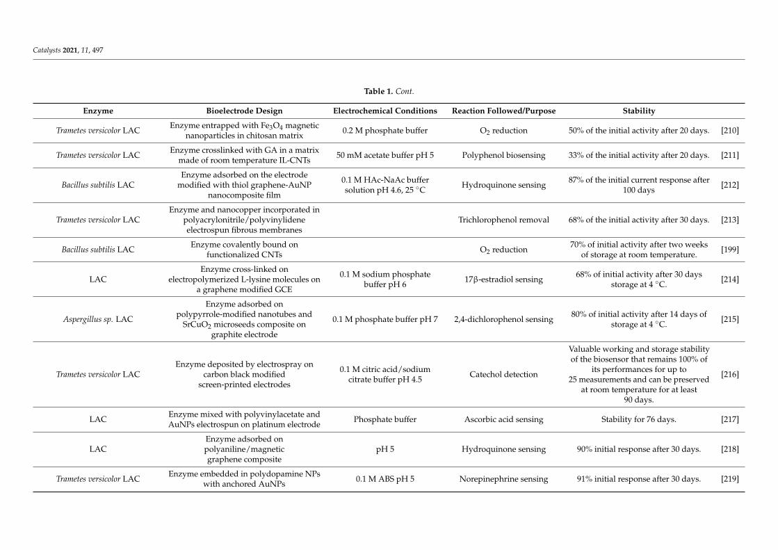
Catalysts 2021, 11, 497
Table 1. Cont.
Enzyme Bioelectrode Design Electrochemical Conditions Reaction Followed/Purpose Stability
Trametes versicolor LAC Enzyme entrapped with Fe3O4 magneticnanoparticles in chitosan matrix 0.2 M phosphate buffer O2 reduction 50% of the initial activity after 20 days. [210]
Trametes versicolor LAC Enzyme crosslinked with GA in a matrixmade of room temperature IL-CNTs 50 mM acetate buffer pH 5 Polyphenol biosensing 33% of the initial activity after 20 days. [211]
Bacillus subtilis LACEnzyme adsorbed on the electrode
modified with thiol graphene-AuNPnanocomposite film
0.1 M HAc-NaAc buffersolution pH 4.6, 25 C Hydroquinone sensing 87% of the initial current response after
100 days [212]
Trametes versicolor LACEnzyme and nanocopper incorporated in
polyacrylonitrile/polyvinylideneelectrospun fibrous membranes
Trichlorophenol removal 68% of the initial activity after 30 days. [213]
Bacillus subtilis LAC Enzyme covalently bound onfunctionalized CNTs O2 reduction 70% of initial activity after two weeks
of storage at room temperature. [199]
LACEnzyme cross-linked on
electropolymerized L-lysine molecules ona graphene modified GCE
0.1 M sodium phosphatebuffer pH 6 17β-estradiol sensing 68% of initial activity after 30 days
storage at 4 C. [214]
Aspergillus sp. LAC
Enzyme adsorbed onpolypyrrole-modified nanotubes and
SrCuO2 microseeds composite ongraphite electrode
0.1 M phosphate buffer pH 7 2,4-dichlorophenol sensing 80% of initial activity after 14 days ofstorage at 4 C. [215]
Trametes versicolor LACEnzyme deposited by electrospray on
carbon black modifiedscreen-printed electrodes
0.1 M citric acid/sodiumcitrate buffer pH 4.5 Catechol detection
Valuable working and storage stabilityof the biosensor that remains 100% of
its performances for up to25 measurements and can be preserved
at room temperature for at least90 days.
[216]
LAC Enzyme mixed with polyvinylacetate andAuNPs electrospun on platinum electrode Phosphate buffer Ascorbic acid sensing Stability for 76 days. [217]
LACEnzyme adsorbed onpolyaniline/magneticgraphene composite
pH 5 Hydroquinone sensing 90% initial response after 30 days. [218]
Trametes versicolor LAC Enzyme embedded in polydopamine NPswith anchored AuNPs 0.1 M ABS pH 5 Norepinephrine sensing 91% initial response after 30 days. [219]
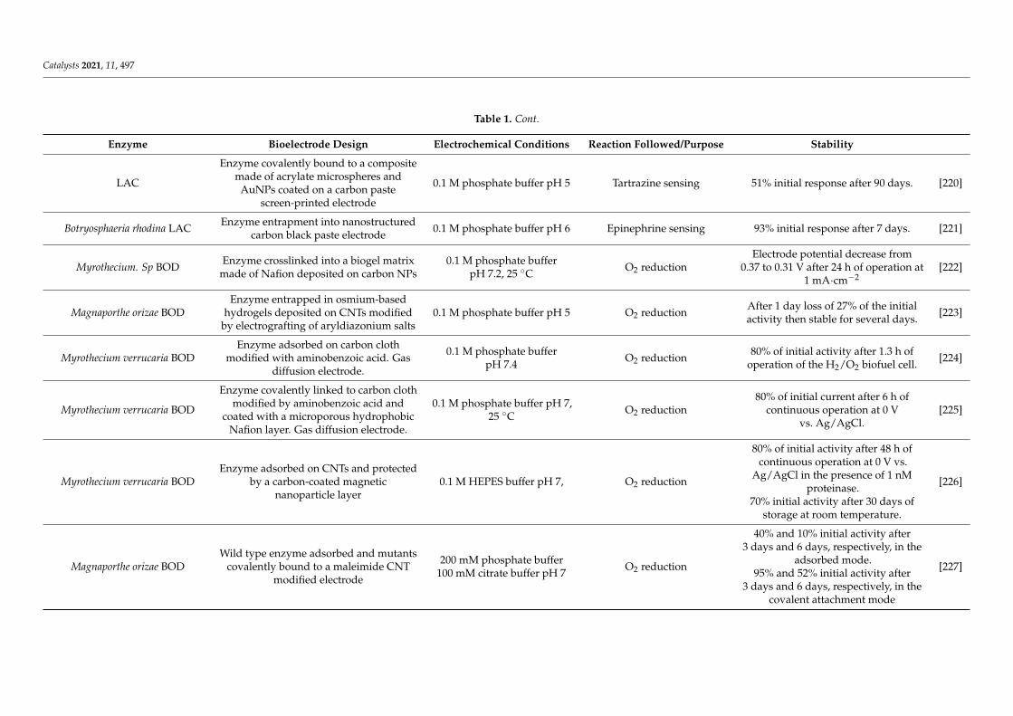
Catalysts 2021, 11, 497
Table 1. Cont.
Enzyme Bioelectrode Design Electrochemical Conditions Reaction Followed/Purpose Stability
LAC
Enzyme covalently bound to a compositemade of acrylate microspheres and
AuNPs coated on a carbon pastescreen-printed electrode
0.1 M phosphate buffer pH 5 Tartrazine sensing 51% initial response after 90 days. [220]
Botryosphaeria rhodina LAC Enzyme entrapment into nanostructuredcarbon black paste electrode 0.1 M phosphate buffer pH 6 Epinephrine sensing 93% initial response after 7 days. [221]
Myrothecium. Sp BOD Enzyme crosslinked into a biogel matrixmade of Nafion deposited on carbon NPs
0.1 M phosphate bufferpH 7.2, 25 C O2 reduction
Electrode potential decrease from0.37 to 0.31 V after 24 h of operation at
1 mA·cm−2[222]
Magnaporthe orizae BODEnzyme entrapped in osmium-based
hydrogels deposited on CNTs modifiedby electrografting of aryldiazonium salts
0.1 M phosphate buffer pH 5 O2 reduction After 1 day loss of 27% of the initialactivity then stable for several days. [223]
Myrothecium verrucaria BODEnzyme adsorbed on carbon cloth
modified with aminobenzoic acid. Gasdiffusion electrode.
0.1 M phosphate bufferpH 7.4 O2 reduction 80% of initial activity after 1.3 h of
operation of the H2/O2 biofuel cell. [224]
Myrothecium verrucaria BOD
Enzyme covalently linked to carbon clothmodified by aminobenzoic acid and
coated with a microporous hydrophobicNafion layer. Gas diffusion electrode.
0.1 M phosphate buffer pH 7,25 C O2 reduction
80% of initial current after 6 h ofcontinuous operation at 0 V
vs. Ag/AgCl.[225]
Myrothecium verrucaria BODEnzyme adsorbed on CNTs and protected
by a carbon-coated magneticnanoparticle layer
0.1 M HEPES buffer pH 7, O2 reduction
80% of initial activity after 48 h ofcontinuous operation at 0 V vs.
Ag/AgCl in the presence of 1 nMproteinase.
70% initial activity after 30 days ofstorage at room temperature.
[226]
Magnaporthe orizae BODWild type enzyme adsorbed and mutants
covalently bound to a maleimide CNTmodified electrode
200 mM phosphate buffer100 mM citrate buffer pH 7 O2 reduction
40% and 10% initial activity after3 days and 6 days, respectively, in the
adsorbed mode.95% and 52% initial activity after
3 days and 6 days, respectively, in thecovalent attachment mode
[227]

Catalysts 2021, 11, 497
Table 1. Cont.
Enzyme Bioelectrode Design Electrochemical Conditions Reaction Followed/Purpose Stability
Myrothecium verrucaria BOD Enzyme adsorbed or electrochemicallydeposited on Au polycrystalline electrode
0.1 M acetate buffer pH 5,25 C O2 reduction
Adsorption mode: 5% initial activityafter 100 potential cycles between
0 and 0.8 V vs. Ag/AgCl at 50 mV/s.Electrochemical deposition mode: 80%
initial activity after 200 cycles
[228]
Myrothecium verrucaria BOD Enzyme adsorbed on MgOC modifiedwith 6 amino 2-naphtoïque acid
0.1 M acetate buffer pH 5,24 C O2 reduction 50% of the initial activity after 5 days. [229]
Myrothecium verrucaria BOD
Enzyme covalently bound to reducedgraphene oxide modified by
4-aminobenzoic acid to reduceits aggregation
0.1 M phosphate buffer pH 7 O2 reduction 55 h half-life. [230]
Myrothecium verrucaria BOD Enzyme adsorbed on buckypaper CNTs Phosphate buffer pH 6.5,25 C O2 reduction 10 h half-life [231]
Magnaporthe orizae BODMolecular engineering for covalent
immobilization in macroporousgold electrode
0.1 M phosphate bufferpH 7.2, 25 C O2 reduction 75% of the initial activity after
incubation for 5 days. [232]
Myrothecium verrucaria BOD Mv BOD entrapped with ABTS inpolydopamine layer on CNTs
0.1 M phosphate buffer pH 7,25 C O2 reduction Mv BOD stable for 22 h of operation at
0.1 V vs. Ag/AgCl. [233]
Bacillus pumilus BOD Bp BOD: entrapped in pyrenebetaineon CNTs 50 C O2 reduction Rapid drop in activity, almost 0 after
3 h of operation at 0.1 V vs. Ag/AgCl [233]
Bacillus pumilus BOD Enzyme entrapment in a carbon feltmodified by pyrene-NH2 modified CNTs 0.2 M phosphate buffer pH 6, O2 reduction
7 days half-life at 25 C uponcontinuous operation of a H2/O2
biofuel cell.[198]
Pyrococcus furiosus HASE Enzyme adsorbed onto a CNT modifiedcarbon felt electrode
50 mM phosphate bufferpH 7, 50 C H2 oxidation Voltage retains 90% of the initial value
33 h operation in a hybrid fuel cell. [234]
Desulfovibrio vulgaris HASEEnzyme embedded in viologen-based
redox hydrogel modified porouscarbon cloth.
0.1 M phosphate bufferpH 7.4 H2 oxidation 60% of initial activity after 20 h at
0.16 V vs. SHE. [224]
Desulfomicrobium baculatumHASE
Oriented covalent immobilization of theenzyme on modified CNTs
50 mM phosphate bufferpH 7.6, 25 C H2 oxidation 80% of initial activity after 1 h in the
biofuel cell. [235]

Catalysts 2021, 11, 497
Table 1. Cont.
Enzyme Bioelectrode Design Electrochemical Conditions Reaction Followed/Purpose Stability
Desulfovibrio desulfuricans HASEEnzyme embedded in viologen-based
redox hydrogel on carboncloth-based electrode
0.1 M phosphate bufferpH 7.4 H2 oxidation 50% of initial activity after 11 h at
0.16 V vs. SHE. [236]
Desulfovibrio vulgaris HASEEnzyme embedded in viologen-based
redox hydrogel and protected bycatalase-GOx layer.
0.1 M phosphate bufferpH 7.4 H2 oxidation 60% initial activity after 8 h in the
presence of 5% O2 at 0.016 V vs. SHE. [143]
Aquifex aeolicus HASE Enzyme adsorption on CNT modified byπ–π stacking with PyrNH2
0.2 M phosphate buffer pH 7,25 C H2 oxidation
1 month half-life at 25 C uponcontinuous operation of a H2/O2
biofuel cell.[198]
[NiFeSe] Desulfovibrio vulgarisHildenborough HASE
Enzyme entrapped inmethylviologen-based redox polymer
films deposited on carbon cloths
0.1 M phosphate buffer pH7.4, room temperature H2 oxidation
25% and 50% of initial activitydepending on the variant after 7 h in
chronoamperometry experiments.[237]

Catalysts 2021, 11, 497
5.2.2. Engineering of Enzymes Seeking for Both Enhanced ET Efficiency and Stability
The general engineering procedures developed to enhance enzyme stability can beadapted for redox enzymes, including introduction of stronger bonds, removal of potentialdegradation sites, or oligomeric structure formation [238–240]. However, the most inter-esting strategies here are to obtain enhanced stability and ET efficiency at the same time.Then, the main targets for protein engineering will lie (i) in the vicinity of the active siteor (ii) at specific points of the enzyme surface able to anchor it with the best orientationfor the highest ET rate (Figure 11). For strategy (i), the vicinity of the heme cavity ofperoxidase [241] or of the CuT1 site of LAC has been targeted [242,243]. Two recent reportsillustrate this concept. Two alanine residues were mutated to one leucine and one valine.Not only did the introduction of hydrophobic residues increase the redox potential of theCuT1 site by 50 mV, but it also protected the CuT1 site from the solvent as well as enhancedtemperature and pH stability of the variant. The mutant was stabilized at the electrode. Itretained roughly 80% of its initial activity after 96 h incubation at pH 4.0 and presented ahalf-life of 60 min at 70 C against 23 min for the wild type protein [203]. With the aim ofelectricity production directly from seawater recycling, recombinant forms of CotA LACfrom B. licheniformis were produced [244]. A double mutation, one in a region close to theCuT1 and the other on the surface of the protein, was shown to induce a synergic effect inbioelectrocatalysis. Conformational changes in the double mutant were proved by CD andfluorescence that allow the variant to catalyze O2 reduction in seawater, a very unusualproperty for classical LACs. Protein leaching under such high ionic strength conditionsmust however be solved.
Figure 11. Enzyme engineering strategies for enhanced stability. (Left) Mutations in the vicinity ofthe active site; computer-guided mutagenesis and directed evolution of a fungal LAC in CuT1vicinity induce enhanced stability at pH 4 compared to the WT. Reproduced with permissionfrom [243]. (Right) Covalent immobilization of BOD through site-directed mutagenesis and reactionwith maleimide groups on the electrode surface induce enhanced stability of the mutant (black curve)compared to the WT (red curve). Adapted with permission from [227].
For strategy (ii), covalent immobilization of enzymes at electrodes was realized by pro-ducing site-directed variants with cysteine residues located at different sites of the enzymesable to react with maleimide groups immobilized on the electrode. This elegant methodcombines controlled orientation of the protein at the electrode for ET, sufficient flexibility
26

Catalysts 2021, 11, 497
for activity, and expected enhanced stability. In the case of cellulose dehydrogenase, 40% ofinitial electroactivity was recovered after 2 months of storage while the wild type enzymeloses progressively its activity within 20 days [245,246]. In addition, controlled orientationallowed investigating mechanistic aspects of the ET. The immobilization method wasextended to BOD from Magnaporthe oryzae showing an enhanced stability with almost noloss of electroactivity for 3 days [227] (Figure 11).
5.2.3. Conductive Porous Material as Redox Enzyme Host Matrices Favoring Fast ET
We discussed in the previous sections how porous materials can be beneficial forthe stabilization of embedded enzymes. The further requirements for their use as hostmatrices of redox enzymes are that either the porous material is conductive to ensureDET, or redox mediators can be added either diffusing or co-immobilized within thematrix. We emphasized that MOFs are nowadays considered as very attractive materials.To ensure electron conduction within MOFs, multicomponent systems can be synthetizedcomposed of MOFs, enzymes and conductive nanomaterials such as graphene nanosheetsor CNTs [147]. A nice example is provided by Li et al. who encapsulated a LAC in ZIF-8in the presence of CNTs (LAC@ZIF-8) [247]. Fluorescence measurements highlighted anegligible enzyme leaching. When compared to free enzyme or to enzyme embedded inMOF with no CNT, the LAC@ZIF-8 hybrid displayed improved thermal stability, enhancedresistance to solvents as well improved long-term storage stability. However, althoughDET was demonstrated, the catalytic efficiency was greatly enhanced by redox mediatoraddition, suggesting that a large enzyme population was not directly wired in the hybridmaterial. Mesoporous silica nanotubes are another example of a suitable matrix whencoated with a graphene sheet layer to enhance its electrical conductivity. BOD entrapped inthe material was able to reduce O2 in the absence of redox mediators, and the bioelectrodestored in water at 4 C for 15 days retained around 80% of initial current [248]. The stabilityof the bioelectrode was suggested to be linked to the size of the nano-channels (12 nm)being close to the size of the enzyme, limiting its mobility. Fructose dehydrogenase andHASE were immobilized in MgOC with pore size in the range 10–150 nm [249,250]. DEToccurred between enzymes and the material regardless of the pore size, but a pore diameterclose to the enzyme size enhanced the thermal and long-term stabilities.
The relationship between pore size and stability is shared by non-redox and redoxenzymes. However, for redox ones, the orientation of the enzyme toward the materialwall must also be considered for a direct wiring. In pores presenting a diameter close tothe enzyme size, the distance between electrical relays on the surface of enzymes and thematerial will be statistically minimized. Multi-point contacts are expected to occur at thesame time. This means that both stability and ET rates should be enhanced simultane-ously [238,251]. Porous gold obtained either by dealloying processes or anodization isincreasingly used for immobilization of redox enzymes. Their pore sizes are compatiblewith efficient enzyme entrapment [144]. Using electrochemistry, a narrow distributionof orientation of BOD favorable for enhanced DET was measured [252]. A model basedon close packing spheres explained why the curvature of pores was beneficial for DET.Unfortunately, no examination of the enzyme conformation in the porous gold materialwas provided. Effect of nanomaterial curvature was evocated in another work to explainLAC properties once immobilized in a CNT network in the presence of ethanol [253]. Animproved direct catalytic current for O2 reduction as well as reduced chloride inhibitionwas measured. CD and ATR-IR demonstrated a net difference in the secondary structurewhen the enzyme/CNT/ethanol system was immobilized at the electrode as comparedwith enzyme in solution. The structure stabilization suggested that ethanol favors contactbetween CNT and LAC in its native form with an orientation favorable for DET. Althoughnot deeply investigated, CNT curvature was suggested to help in the stabilization of theimmobilized dehydrated enzyme.
27
Catalysts 2021, 11, 497
5.2.4. Redox Enzyme Encapsulation in Hydrogels for Bioelectrocatalysis
Entrapping redox enzymes into hydrogels is expected to provide a suitable hydratedenvironment able to maintain the enzyme in a functional active state. Some examples ofuse of a sole polymer for direct electrocatalysis involving redox enzymes were recentlyreported [254]. Conductive polyaniline (PANi) forms a nanostructured hydrogel oncedeposited on an electrode [255]. The PANi hydrogel exhibits a continuously connectedhierarchical 3D network with pore diameter of 60 nm, allowing a short electron diffusionlength. The PANi-formate dehydrogenase electrode was efficient for catalytic reduction ofCO2 into formate. Although no exhaustive study of stability was made, conversion of CO2was efficient for 12 h. However, hydrogel matrices are very often poorly conductive, andconductive nanostructures or redox mediators must be co-immobilized in the gel to ensureelectron conduction. Chitosan is a natural polyamino-saccharide that forms thin hydrogelfilms at electrodes through controlled electrodeposition process. The amine groups areavailable for attachment to a functionalized electrode or for anchorage of the enzyme.CNTs mixed with chitosan render the material conductive. Hosting a LAC, the so-builtbioelectrode retained 70% initial current after 60 days of continuous measurement [209,256].The same authors reported an improved bioelectrode using genipin as a cross linker.The LAC-based electrode was implanted in rats and remained operational for 167 daysin vivo [257].
Alternatively, redox mediators can act as electron relays between the electrode andthe enzyme inside the hydrogel. In the search for enzyme stabilization, Matsumoto et al.prevented leaching of a biotinylated GDH by immobilizing it into a streptavidine-hydrogelprepared by click chemistry between sortase A and PEG [258]. In the presence of a ferrocenederivative as a redox mediator, the so-designed electrode showed improved stability whencompared to the electrode with no hydrogel. One issue was the weak stability of thehydrogel itself on the electrode. This last result points out one more requirement forredox enzyme-based electrode stabilization: Not only the enzyme must be stable but theinteraction between the electrode and the hybrid enzyme-host matrix must also be strongenough to avoid leaching of the whole biomaterial. To ensure stability of the bioelectrode,hydrogel can be combined within porous conductive material. As an illustration, a hydrogelformed by cross-linking bovine serum albumin, GOx and arginine was interlocked withinthe pores of a carbon cloth [259]. Leaching of enzyme was less than 0.5% of the total amountof embedded protein within 48 h. UV-Visible spectrometry and CD demonstrated that90% of the enzyme secondary structure was preserved in the network. In the presence offerricyanide as a redox mediator, the current density for glucose oxidation was maintainedfor 25 days.
Diffusing redox mediators are however undesirable in many cases, either because theylimit the rate of reaction or because they can be toxic when leaching to the environment inwhich the enzyme operates. Redox hydrogels, introduced 30 years ago by Adam Heller, arenow widely exploited for immobilization of a variety of enzymes [260]. These hydrogelscombine the advantages of a hydrated matrix ensuring protection of the enzyme and rapiddiffusion of the substrate with the presence of tethered redox moieties (they can be basedon osmium, ruthenium, ferrocene, colbaltocene, quinone, or viologen derivatives) thatcan electrically wire the enzyme by electron hopping inside the gel [261–263]. The redoxpotential of the hydrogel matrix can be adapted to the desired reaction to be catalyzedby tuning the ligands of the redox moieties. Ferrocene entity was tethered to linear PEIand used to cross-link GOx [264]. Of initial current, 70% and 36% was retained after21 days of storage and 6 h of continuous operation, respectively. This marked differenceunderlines the effect of applied potential on the stability of the bioelectrode, an issuethat we will further discuss in Section 5.3.2. Os-tethered hydrogel embedding GDH wasloaded in porous MgOC deposited by ink-drop-casting on an electrode [265]. The highcatalytic currents for glucose oxidation were linked to the controlled pore diameter of theMgOC matrix that allows high loading of redox polymer and rapid substrate diffusion.Moreover, the combined advantage of hydrated redox polymer and porous structure
28

Catalysts 2021, 11, 497
allowed improved stability. Of initial catalytic current, 44% was recovered after 10 daysof operation against 3% for the hydrogel-enzyme hybrid deposited on a glassy carbonelectrode. Direct oxidation of glucose by Corynascus thermophilus cellobiose dehydrogenasewas compared to the mediated one obtained when the CNT-based electrode was furthermodified by an Os-based redox hydrogel [200]. MET process generated higher catalyticcurrents than DET one proceeding at a lower potential through the FAD cofactor instead ofthe heme domain in the case of the DET process. Of the mediated electrocatalytic current,30% remained after 9 days of CV cycling. In such configuration, it will be interesting inthe future to get the comparative stability through the DET process. Last but not least,redox hydrogels have been shown in recent years to act as shields against O2 for sensitiveenzymes. This topic will be further developed below in Section 5.3.3.
5.2.5. When Aggregates or Partially Unfolded Enzymes Operate in Bioelectrocatalysis
We clearly stated in the introduction of this review that the secondary and tertiarystructure of enzymes control their activity. Any change in the conformation was thusexpected to modify the activity, yielding inactive states upon denaturation. We otherwiseunderlined that aggregated enzymes, for example in the form of CLEAs, could main-tain the native conformation showing a stable, although often lower, enzymatic activity.Efforts to maintain stable enzyme electroactivity are mainly directed toward enzymeswith unchanged conformations. However, and contrary to these strategies, examples ofaggregates or partially unfolded forms of proteins have been reported to promote bioelec-trocatalytic properties. Aggregated proteins immobilized on nanostructured carbon-basedelectrodes may enabled direct electrocatalysis showing enhanced stability compared tonon-precipitated proteins [266,267]. Pyranose oxidase was immobilized on a CNT-basedelectrode via three different procedures: (i) covalent attachment (CA), (ii) enzyme coating(EC), (iii) enzyme precipitate coating (CLEA). After 34 days at room temperature, CLEA re-tained 65% of initial electroactivity toward glucose oxidation, while CA and EC maintained9.2% and 26% of their initial activities, respectively. LAC aggregates were formed by com-putational design through production of a new dimeric interface that induces self-assemblyof the protein into crystalline-like assemblies [268]. Combined with CNT incorporated inthe aggregates, the bioelectrode showed high direct electroenzymatic rate for O2 reductionwith enhanced thermostability. LAC from T. versicolor was precipitated in the presenceof Cu2+ yielding flower-like particles [207]. The Cu/LAC nanoflowers were integratedin carbonaceous nanomaterials and tested as catalysts for O2 bioelectroreduction. A well-shaped direct electroenzymatic signal was observed, demonstrating the functionality ofthe electron pathway through CuT1 in the flower particles (Figure 12). The bioelectrodedelivered 72% initial current after 6 days of daily 2 h discharge, and the H2/O2 fuel cellconstructed based on the Cu/LAC flower-based biocathode maintained 85% initial powerafter 15 days of operation.
Figure 12. LAC assembled through Cu-flower like particles displays direct and stable electrocatalysisof O2 reduction. (A) SEM image of a Cu/LAC nanoflower, (B) Catalytic reduction of O2 (red curve)by LAC in Cu nanoflowers on CNTs, (C) Operational stability. Adapted with permission from [207].
29
Catalysts 2021, 11, 497
It was also recently shown that the large sub-unit of a NiFe HASE was able to sustainH2 oxidation by itself, showing that an incomplete structure still allows catalysis [269].As another illustration, LAC from Aspergillus sp. was immobilized on Fe2O3 NPs [270].Careful spectroscopic analysis, including FT-IR, fluorescence and EPR, demonstrated apartial unfolding of the protein which tends to a higher exposure of the CuT1. A largedirect catalytic current in the range of 3 mA/cm2 was observed, with an electrochemicalCV shape denoting fast ET. However, it should be stressed that heterogeneity of enzymepopulation certainly exists at the electrochemical interface, and methods are now requiredto distinguish whether the electrochemical activity is actually linked to the unfoldedproteins or to the small fraction of proteins remaining correctly folded.
5.3. Parameters Specific to Bioelectrocatalysis Stabilization
Bioelectrocatalysis implies not only the immobilization of the enzyme on a conductivesupport but also the application of a potential that the enzyme will sense. Hence, thestability of a bioelectrode will include the stability of the enzyme in the immobilized stateand the stability of the electrocatalytic response under an electric field which is reflected inmost cases by a measure of a current, a voltage or the impedance of the system. However,the variation of the electrochemical signal may have different origins. Progressive leachingof the enzymes from the electrochemical interface can explain a decrease in the catalyticcurrent, while changes in the conformation or in the reorientation of the enzyme can explainmodification in the catalytic response due to lower electron transfer rates. Methods probingthe relationship between electroactivity and amount of proteins on the electrochemicalinterface, as well as the relationship between electroactivity and changes in enzyme con-formation, are not readily available. Actually, many bioelectrocatalysis-related studiesreport a decrease of the catalytic current with time. This decrease has long been namedas “film loss”, suggesting that enzyme leaching from the electrode would be responsiblefor the current evolution. However, coupling SPR and quartz crystal microbalance withdissipation (QCM-D) to electrochemistry, it was shown that other phenomena than simpleenzyme desorption must account for bioelectrode instability [271,272]. In the following, wewould like to emphasize some critical parameters that will affect the overall electrocatalyticresponse stability and the methods developed to get more insights in the overall process.
5.3.1. Reorientation of the Immobilized Enzyme
We stated just before that one key issue in order to enhance the interfacial ET betweena redox enzyme and an electrode is the orientation of the enzyme on the solid surfaceso that the electronic relay on the surface of the protein can be positioned at a distancecompatible with fast ET. In many studies reporting enzyme orientation, hydrophobic orelectrostatic interactions are used to control the enzyme positioning. These interactionsare relevant because they mimic in vivo recognition with the substrate or between redoxpartners. LAC, as an example, is involved in transformation of many substrates such asphenolic compounds. In accordance, the Cu T1 is engaged in a hydrophobic pocket thatcan be recognized by the electrode modified by phenyl or anthracenyl groups, leading toan orientation favoring efficient electron transfer [273]. The recognition between HASE andits redox partner cytochrome c3 is another example illustrating electrostatic parameters tobe considered in ET. By NMR docking and by resolving the Poisson–Boltzmann equation,it was established that a functional complex is formed thanks to a small negatively chargedregion on the HASE surface that can interact with the positively charged cytochrome.Such local electrostatic interaction was able to overcome electrostatic repulsion of theoverall positive charges of the interacting proteins [274,275]. The same situation arisesin bioelectrocatalysis, considering the electrode as the redox partner. Local charges arereflected by charge heterogeneity on the surface of the enzyme and rationalization canbe obtained through the calculation of the dipole moment direction. This latter willdepend on the pH of the enzyme environment. Hence, it can be expected that an enzymetakes an orientation favorable to ET at one pH but rotates on the electrode when the
30

Catalysts 2021, 11, 497
pH of surrounding solution is modified. It is the most crucial when electrocatalysisinvolves proton consumption or production, as it is the case for many electroenzymaticreactions, thus inducing local changes in pH in the weakly buffered solution and potentialmodification of the catalytic signal.
Evidence of the rotation of BOD was demonstrated on gold electrodes modified bythiol-based SAMs [116]. Both the pH at which the enzyme was adsorbed and the pH of theelectrolytic solution were varied. By doing so, not only the charge of the electrode but alsothe charge of the enzyme varied. Coupling electrochemistry to ellipsometry, SPR, and PhaseModulation Infrared Reflection Absorption Spectroscopy (PMIRRAS), it was shown thatthe global charge of the protein controls the amount of adsorbed molecules as a function ofelectrostatic interactions tuned by the pH of adsorption. It was also demonstrated that thelocal charge in the vicinity of the CuT1 controls the orientation of the enzyme, hence thecatalytic current for O2 reduction. Hence, when the amount of enzyme and its orientationwere a priori fixed by adsorption at a fixed pH, the catalytic current recorded in buffersof various pHs nevertheless varied following rotation of the enzyme to adopt the mostfavorable orientation. It was clearly shown that orientation could be reversibly tuned bychanging the pH of the electrolyte, shifting the catalysis from a slow ET rate to a fast one(Figure 13).
Figure 13. Reorientation of enzymes on electrode may alter enzyme stability. (Top) Tuning the pHof the electrolyte induces rotation of the adsorbed bilirubin oxidase on the electrode surface, henceinduces modification in bioelectrocatalysis for O2 reduction. Adapted with permission from [116].(Down) Scheme of the reorientation of enzymes on an electrode induced by surface coverage. Thewhite sphere represents the active site. Adapted with permission from [115].
Enzyme reorientation, leading to potential bioelectrocatalysis instability, may haveanother origin. In the course of protein adsorption, protein-protein interactions are increas-ingly dominating. Hence, while the adsorption of the first proteins in a favorable orientationfor ET can be controlled by electrostatic interactions, the interactions between neighbor-ing adsorbed proteins may induce reorientation to decrease the repulsive forces [115](Figure 13). Coupling electrochemistry to QCM and interferometry for studying O2 reduc-
31

Catalysts 2021, 11, 497
tion by BOD on a gold electrode, Blanford et al. clearly demonstrated that an optimumsurface coverage exists yielding the highest stable electrocatalytic current [276]. Althoughproof of enzyme reorientation was not provided, QCM-D allowed to conclude that thisoptimum was linked to a balance between rate of enzyme adsorption and deformation.
5.3.2. Effect of the Electric Field
Compared to other catalytic processes, the special feature of bioelectrocatalysis liesin the electric field imposed at the electrode interface. Although partially shielded bythe ions present in the solution, the extent of the electric field can still reach the size ofimmobilized enzymes, especially in weakly supported electrolytes. It should be expectedto have a major impact, but only few studies report the effect of the electric field on enzymeor bioelectrode stability. This lack of information most probably comes from the difficultyto determine which effect among protein leaching, denaturation or reconformation can beresponsible for any variation in the electrocatalytic response. The recent use of coupledmethods with electrochemistry will certainly lead to major advancements in that field.Adsorption of MCOs on planar gold electrodes was taken as a model for such studies. Itwas shown using electrochemistry coupled to QCM-D that cycling the potential for O2reduction by M. verrucaria BOD accelerated mass decrease compared to applying a fixedpotential [272]. It was hypothesized that varying the electric field near the surface maydistort the adsorbed protein, expelling water and contributing to enzyme denaturation. Itwas further shown with the same enzyme that continuous cycling induced more stabilityof the bioelectrode than cycling during a same period but at regular intervals, the electrodebeing at the open circuit potential (OCP) when not cycling [116]. This result suggests amuch lower stability of the bioelectrode when the applied potential was close to the OCP.Considering the potential of zero charge (Epzc) of the electrode, it could be concludedthat the strong electric field generated when holding the potential far away from Epzcinduced bioelectrode destabilization. Kano’s group rationalized these experimental databy modeling the electrostatic interactions between enzymes and electrodes in the electricdouble layer [275]. Taking the copper efflux oxidase (CueO) enzyme as a model enzymeadsorbed either at bare or butanethiol-modified gold electrodes, the authors demonstratedhow the electrical double layer plays a key role in enzyme stability for bioelectrocatalysis.It can be argued that the diameter of the enzyme is much higher than the length of theelectrical double layer (5 nm vs. 1 nm at a typical ionic strength for electrocatalysis).However, the domain of CueO facing the electrode is indeed located in the double layerand will face an electric field whose force depends on the applied potential and on theEpzc of the electrode. Hence, at the bare gold electrode, the strong electric field inducedby an applied potential much more positive than Epzc of the electrode, in addition tothe complementary charges chosen to favor DET orientation, leads to denaturation ofthe protein.
Heterogeneity of the electric field inside the pores of a porous matrix otherwise affectsenzyme stabilization. A model developed in the group of Kano demonstrated the existenceof an inner pore region where the electric field remains low, and where weak electrostaticforces help in protein stabilization [238,277,278]. The higher electric field at the entry ofthe pore should enhance the ET, while inducing less stability [279,280]. This effect knownas the “edge effect” was recognized early in the history of bioelectrochemical studiesusing cytochrome c and graphite and may explain efficient bioelectrocatalysis at carbonnanofibers (CNFs) [281–284].
Finally, it should be mentioned the role of the electric field sensed by the enzyme withinthe course of mediated electrocatalysis against direct electrocatalysis. A relevant example isbioelectrocatalytic oxidation of hydrogen by HASEs. It is known that HASEs are reversiblyinactivated at high potentials [285]. In a typical cyclic voltammetry experiment where theenzyme is adsorbed on an electrode, this inactivation results in a decrease in the catalyticcurrent in the forward scan at a potential higher than ca. −0.4 V vs. Ag/AgCl at pH 7.Current recovers when the potential is reversed. It was shown that pushing the potential
32

Catalysts 2021, 11, 497
to even more positive values induces an irreversible inactivation of the enzyme [286]. Suchinactivation process may have consequences on the operation of fuel cells using HASEas anode catalyst, requiring regular steps of reducing voltages to reactivate the enzyme.Alternatively, it was shown that embedding the hydrogenase in a viologen-based hydrogelprotected the enzyme from high potential deactivation. In this MET process, the enzymeno longer senses the high potential of the electrode but the potential of the viologen redoxcouple which is sufficiently negative to prevent HASE inactivation [287–289]. Nevertheless,the use of redox mediators induces other detrimental effects towards enzyme stability, suchas production of reactive oxygen species (ROS) that will be discussed in the next section.
5.3.3. Protection against Reactive Oxygen Species (ROS) Production
Two electron reduction of O2 yields the formation of unstable superoxide O2•− which
is the precursor for other reactive species including H2O2 and •OH. Incomplete oxygenreduction can be encountered in the course of certain enzymatic reactions, or when O2is directly reduced at an electrochemical interface. A relevant example of ROS producedby enzymatic reactions is the oxidation of sugars by the flavin-based GOx. Here, O2 is aco-substrate of the enzyme and is reduced to H2O2 after the release of D-glucono-δ-lactonewithin glucose oxidation [290]. Identically, oxidation of lactate-by-lactate oxidase producesH2O2. To avoid any detrimental effect of H2O2, one solution is to electroenzymaticallyreduce this latter by peroxidases [291]. Alternatively, catalase has been used to decomposeH2O2 [292]. Engineering of flavoproteins is also largely reported to decrease O2 activ-ity [293,294]. Such strategy is particularly useful when the flavoprotein is embedded inredox hydrogels, where the competition between reaction of the enzyme with the redoxmediator or O2 occurs within the polymer, decreasing the efficiency of the bioelectrocataly-sis. An additional issue comes from the reduction of O2 directly by the redox entities ofthe polymer.
Reactions other than the oxidation of sugar by GOx embedded in Os-based polymerscan expose enzymes to ROS. Actually, low potential viologen-based redox polymers de-veloped to protect HASEs against high potential inactivation also serve as shields againstO2 [224,237,288]. However, the viologen-based matrix itself may generate ROS in thecourse of the HASE protection mechanism, inducing its progressive decomposition [295].To tackle this issue, a top polymer layer containing both GOx and catalase was used toensure a complete HASE protection [143]. Alternatively, iodide was added in the wholesystem to induce dismutation of H2O2. An improvement of the half-life of the HASE in thepresence of O2 was demonstrated [295]. In the case of O2-tolerant HASEs, their specificsensitivity/tolerance to O2 has been shown to lead to an even greater sensitivity to ROS.Hence, it was reported that ROS formed by applying low potentials to a HASE-basedcarbon felt electrode in the presence of O2 irreversibly deactivated the enzyme contraryto reversible O2 inactivation [111,198] (Figure 14). In this case, a protective upper layercomposed of a 3D porous carbon matrix increased the stability by 4–6 times against ROScompared to a 2D electrode. It was proposed that the 3D matrix could scavenge ROS beforereaching the enzymes inside the pores.
33

Catalysts 2021, 11, 497
Figure 14. Chronoamperometric response for H2 oxidation by A. aeolicus HASE embedded in aCNT-modified carbon felt electrode showing the inactivation of the enzyme by ROS electrochemicallyproduced (A) and protection by a porous matrix (B). Reproduced with permission from [198].
6. Conclusions and Future Directions
Electrocatalysis involving redox enzymes takes advantage of the high selectivity andaffinity of the biomolecules, as well as the large diversity of identified enzymes, hence alarge variety of substrates. Possible applications range from implantable medical devices(insulin pump, neurological stimulator, artificial organs, etc.), wearable sensors, and envi-ronmental diagnostic to energy converting devices or CO2 capture [16,17,296–299]. Manystrategies are available to enhance enzyme stability in general. However, the specificity ofredox enzymes that sense an electric field and require a controlled orientation to operate onelectrochemical interfaces makes the stability of redox enzymes one of the most importantissues to resolve before a large-scale commercial development of bioelectrocatalysis. Thesearch for a good balance between stability and activity of enzymes is a prerequisite, evencomplexified by the search for efficient ET in the case of redox enzymes. Recent examplesin the literature concerning MCOs revealed the difficulty of getting improved interfacialET parameters or improved redox properties while maintaining both the stability and theactivity of the enzyme [300,301]. No doubt that protein engineering, which is increasinglycarried out with the aid of innovative solutions based on emerging genetic tools, will offernew opportunities of producing stable enzymes. There is also no doubt that the screeningof the biodiversity will allow the discovery of redox enzymes with outstanding properties,which can be applied either directly in bioelectrochemical devices, or as model enzymesfor protein engineering for high stability.
Alternative strategies can be envisioned in case enzyme stability is limiting the targeteddevice. The first one would be to refresh the bioelectrodes regularly, so as to renewinactivated enzymes by new active ones. Increasing salt concentration in the mediumwas reported to allow LAC desorption, rendering the electrode available for fresh enzymeadsorption [209]. Refreshment of the bioelectrode can also be done by adsorbing theenzyme on magnetic nanoparticles [302]. Applying a suitable magnetic field may allowassembling or release of the biocatalyst on the electrode. Cosnier et al. proposed touse enzymes and redox mediators encapsulated in glyconoparticles, both diffusing insolution [303]. This strategy was supposed to allow protein and mediators to freely rotatefor an efficient ET and to offer in addition the possibility of easy refreshing of the biocatalyst
34

Catalysts 2021, 11, 497
by a simple exchange of the solutions. Although suitable to overcome enzyme instability,these procedures based on enzyme renewal, however, do not appear to be sustainableor cost effective. Nanozymes, or bioinspired catalysts, could be desirable alternativesto enzymes in the future [304–307]. Taking benefit of the fundamental understanding ofbioelectrocatalysis, these synthetic catalysts may provide higher stability while operatingthe closest to the enzymatic catalysis.
Should redox enzymes or biosynthetic catalysts be used, novel methodologies mustbe developed in order to get new insights in the parameters that limit the stability of bio-electrocatalysis. In situ and in operando methods are certainly the most appealing within theobjective of understanding and enhancing bioelectrode stability [308,309]. Recent reviewsby Reisner et al. and by Crespilho et al. discussed various methods, and especially coupledmethods, currently available to study bioelectrode interfaces [309,310]. Methods allowingmass or layer thickness quantification, i.e., SPR, QCM-D and ellipsometry, coupled toelectrochemistry will give access to the relationship between enzyme loading or releaseand catalytic response [21,116,271,272,311]. Electrochemistry coupled to spectroscopies(IR (including SEIRA, PMIRRAS), XAS, fluorescence, Raman (including SERRS), EPR, CD,mass spectrometry) will help in the determination of conformational change once enzymeis immobilized or as a function of applied potential or current [312–317]. Microscopy cou-pled to electrochemistry will allow the mapping of the electrocatalysis [318–320]. Finally,getting more and more accurate experimental data on enzyme behavior on electrodes maybe implemented in theoretical models that in return will help in bioelectrode rationaliza-tion [21,117,321–323].
Author Contributions: Writing, C.B.; review and editing, H.-M.M., A.d.P. and I.M.; writing, editing,and supervision, E.L. All authors have read and agreed to the published version of the manuscript.
Funding: This work was supported by ANR (ENZYMOR-ANR-16-CE05- 0024). H-M Man grant wasfunded by Région PACA, France, Centre National de la Recherche Scientifique, CNRS, France andHyseas company, Cannes, France. C. Beaufils grant was funded by Aix-Marseille University and by“Agence Innovation Defense” of the French army ministry.
Conflicts of Interest: The authors declare no conflict of interest.
References
1. Woodley, J.M. New frontiers in biocatalysis for sustainable synthesis. Curr. Opin. Green Sustain. Chem. 2020, 21, 22–26. [CrossRef]2. Reetz, M.T. What are the Limitations of Enzymes in Synthetic Organic Chemistry? Chem. Rec. 2016, 16, 2449–2459. [CrossRef]
[PubMed]3. Hauer, B. Embracing Nature’s Catalysts: A Viewpoint on the Future of Biocatalysis. ACS Catal. 2020, 10, 8418–8427. [CrossRef]4. Bommarius, A.S.; Paye, M.F. Stabilizing biocatalysts. Chem. Soc. Rev. 2013, 42, 6534–6565. [CrossRef] [PubMed]5. Mandpe, P.; Prabhakar, B.; Gupta, H.; Shende, P. Glucose oxidase-based biosensor for glucose detection from biological fluids.
Sens. Rev. 2020, 40, 497–511. [CrossRef]6. Mano, N.; de Poulpiquet, A. O-2 Reduction in Enzymatic Biofuel Cells. Chem. Rev. 2018, 118, 2392–2468. [CrossRef] [PubMed]7. Lojou, E. Hydrogenases as catalysts for fuel cells: Strategies for efficient immobilization at electrode interfaces. Electrochim. Acta
2011, 56, 10385–10397. [CrossRef]8. Lubitz, W.; Ogata, H.; Rudiger, O.; Reijerse, E. Hydrogenases. Chem. Rev. 2014, 114, 4081–4148. [CrossRef]9. Alfano, M.; Cavazza, C. Structure, function, and biosynthesis of nickel-dependent enzymes. Protein Sci. 2020, 29, 1071–1089.
[CrossRef]10. Najafpour, M.M.; Zaharieva, I.; Zand, Z.; Hosseini, S.M.; Kouzmanova, M.; Holynska, M.; Tranca, I.; Larkum, A.W.; Shen, J.R.;
Allakhverdiev, S.I. Water-oxidizing complex in Photosystem II: Its structure and relation to Manganese-oxide based catalysts.Coord. Chem. Rev. 2020, 409. [CrossRef]
11. Morello, G.; Megarity, C.F.; Armstrong, F.A. The power of electrified nanoconfinement for energising, controlling and observinglong enzyme cascades. Nat. Commun. 2021, 12, 340. [CrossRef] [PubMed]
12. Stolarczyk, K.; Rogalski, J.; Bilewicz, R. NAD(P)-dependent glucose dehydrogenase: Applications for biosensors, bioelectrodes,and biofuel cells. Bioelectrochemistry 2020, 135, 107574. [CrossRef] [PubMed]
13. Alvarez-Malmagro, J.; Garcia-Molina, G.; De Lacey, A.L. Electrochemical Biosensors Based on Membrane-Bound Enzymes inBiomimetic Configurations. Sensors 2020, 20, 3393. [CrossRef] [PubMed]
35

Catalysts 2021, 11, 497
14. Bollella, P.; Katz, E. Bioelectrocatalysis at carbon nanotubes. In Methods in Enzymology: Nanoarmoring of Enzymes with Carbon
Nanotubes and Magnetic Nanoparticles; Kumar, C.V., Pyle, A.M., Christianson, D.W., Eds.; Elsevier: Amsterdam, The Netherlands,2020; Volume 630, pp. 215–247.
15. Wu, R.R.; Ma, C.L.; Zhu, Z.G. Enzymatic electrosynthesis as an emerging electrochemical synthesis platform. Curr. Opin. Electrochem.
2020, 19, 1–7. [CrossRef]16. Yuan, M.W.; Kummer, M.J.; Minteer, S.D. Strategies for Bioelectrochemical CO2 Reduction. Chem. A Eur. J. 2019, 25, 14258–14266.
[CrossRef]17. Mazurenko, I.; Wang, X.; de Poulpiquet, A.; Lojou, E. H-2/O-2 enzymatic fuel cells: From proof-of-concept to powerful devices.
Sustain. Energy Fuels 2017, 1, 1475–1501. [CrossRef]18. Xiao, X.X.; Xia, H.Q.; Wu, R.R.; Bai, L.; Yan, L.; Magner, E.; Cosnier, S.; Lojou, E.; Zhu, Z.G.; Liu, A.H. Tackling the Challenges of
Enzymatic (Bio)Fuel Cells. Chem. Rev. 2019, 119, 9509–9558. [CrossRef] [PubMed]19. Mazurenko, I.; de Poulpiquet, A.; Lojou, E. Recent developments in high surface area bioelectrodes for enzymatic fuel cells.
Curr. Opin. Electrochem. 2017, 5, 74–84. [CrossRef]20. Mazurenko, I.; Hitaishi, V.P.; Lojou, E. Recent advances in surface chemistry of electrodes to promote direct enzymatic bioelectro-
catalysis. Curr. Opin. Electrochem. 2020, 19, 113–121. [CrossRef]21. Hitaishi, V.P.; Clement, R.; Bourassin, N.; Baaden, M.; de Poulpiquet, A.; Sacquin-Mora, S.; Ciaccafava, A.; Lojou, E. Controlling
Redox Enzyme Orientation at Planar Electrodes. Catalysts 2018, 8, 192. [CrossRef]22. Iyer, P.V.; Ananthanarayan, L. Enzyme stability and stabilization—Aqueous and non-aqueous environment. Process Biochemistry
2008, 43, 1019–1032. [CrossRef]23. Balcao, V.M.; Vila, M. Structural and functional stabilization of protein entities: State-of-the-art. Adv. Drug Deliv. Rev. 2015,
93, 25–41. [CrossRef] [PubMed]24. Polizzi, K.M.; Bommarius, A.S.; Broering, J.M.; Chaparro-Riggers, J.F. Stability of biocatalysts. Curr. Opin. Chem. Biol. 2007,
11, 220–225. [CrossRef] [PubMed]25. Gianfreda, L.; Scarfi, M.R. Enzyme Stabilization—State-Of-The-Art. Mol. Cell. Biochem. 1991, 100, 97–128. [CrossRef] [PubMed]26. Stepankova, V.; Bidmanova, S.; Koudelakova, T.; Prokop, Z.; Chaloupkova, R.; Damborsky, J. Strategies for Stabilization of
Enzymes in Organic Solvents. ACS Catal. 2013, 3, 2823–2836. [CrossRef]27. Lalande, M.; Schwob, L.; Vizcaino, V.; Chirot, F.; Dugourd, P.; Schlatholter, T.; Poully, J.C. Direct Radiation Effects on the Structure
and Stability of Collagen and Other Proteins. Chembiochem 2019. [CrossRef]28. Doster, W.; Settles, M. Protein-water displacement distributions. Biochim. Biophys. Acta Proteins Proteom. 2005, 1749, 173–186.
[CrossRef]29. Miyawaki, O. Hydration state change of proteins upon unfolding in sugar solutions. Biochim. Biophys. Acta Proteins Proteom. 2007,
1774, 928–935. [CrossRef] [PubMed]30. Ganjalikhany, M.R.; Ranjbar, B.; Hosseinkhani, S.; Khalifeh, K.; Hassani, L. Roles of trehalose and magnesium sulfate on structural
and functional stability of firefly luciferase. J. Mol. Catal. B Enzym. 2010, 62, 127–132. [CrossRef]31. Haque, I.; Singh, R.; Moosavi-Movahedi, A.A.; Ahmad, F. Effect of polyol osmolytes on Delta G(D), the Gibbs energy of
stabilisation of proteins at different pH values. Biophys. Chem. 2005, 117, 1–12. [CrossRef] [PubMed]32. Tiwari, A.; Bhat, R. Stabilization of yeast hexokinase A by polyol osmolytes: Correlation with the physicochemical properties of
aqueous solutions. Biophys. Chem. 2006, 124, 90–99. [CrossRef] [PubMed]33. Wlodarczyk, S.R.; Costa-Silva, T.A.; Pessoa, A.; Madeira, P.; Monteiro, G. Effect of osmolytes on the activity of anti-cancer enzyme
L-Asparaginase II from Erwinia chrysanthemi. Process Biochem. 2019, 81, 123–131. [CrossRef]34. Chen, G.; Zhang, Q.P.; Lu, Q.Y.; Feng, B. Protection effect of polyols on Rhizopus chinensis lipase counteracting the deactivation
from high pressure and high temperature treatment. Int. J. Biol. Macromol. 2019, 127, 555–562. [CrossRef]35. Pazhang, M.; Mardi, N.; Mehrnejad, F.; Chaparzadeh, N. The combinatorial effects of osmolytes and alcohols on the stability
of pyrazinamidase: Methanol affects the enzyme stability through hydrophobic interactions and hydrogen bonds. Int. J.
Biol. Macromol. 2018, 108, 1339–1347. [CrossRef]36. Khan, M.V.; Ishtikhar, M.; Rabbani, G.; Zaman, M.; Abdelhameed, A.S.; Khan, R.H. Polyols (Glycerol and Ethylene glycol)
mediated amorphous aggregate inhibition and secondary structure restoration of metalloproteinase-conalbumin (ovotransferrin).Int. J. Biol. Macromol. 2017, 94, 290–300. [CrossRef]
37. Kaushik, J.K.; Bhat, R. Why is trehalose an exceptional protein stabilizer? An analysis of the thermal stability of proteins in thepresence of the compatible osmolyte trehalose. J. Biol. Chem. 2003, 278, 26458–26465. [CrossRef]
38. Silva, C.; Martins, M.; Jing, S.; Fu, J.J.; Cavaco-Paulo, A. Practical insights on enzyme stabilization. Crit. Rev. Biotechnol. 2018,38, 335–350. [CrossRef]
39. Scharnagl, C.; Reif, M.; Friedrich, J. Local compressibilities of proteins: Comparison of optical experiments and simulations forhorse heart cytochrome-c. Biophys. J. 2005, 89, 64–75. [CrossRef]
40. Mangiagalli, M.; Carvalho, H.; Natalello, A.; Ferrario, V.; Pennati, M.L.; Barbiroli, A.; Lotti, M.; Pleiss, J.; Brocca, S. Diverse effectsof aqueous polar co-solvents on Candida antarctica lipase B. Int. J. Biol. Macromol. 2020, 150, 930–940. [CrossRef]
41. Mukherjee, A.; Sarkar, S.; Gupta, S.; Banerjee, S.; Senapati, S.; Chakrabarty, R.; Gachhui, R. DMSO strengthens chitin deacetylase-chitin interaction: Physicochemical, kinetic, structural and catalytic insights. Carbohydr. Polym. 2019, 223. [CrossRef] [PubMed]
36

Catalysts 2021, 11, 497
42. Woll, A.K.; Hubbuch, J. Investigation of the reversibility of freeze/thaw stress-induced protein instability using heat cycling as afunction of different cryoprotectants. Bioprocess Biosyst. Eng. 2020, 43, 1309–1327. [CrossRef] [PubMed]
43. Dal Magro, L.; Kornecki, J.F.; Klein, M.P.; Rodrigues, R.C.; Fernandez-Lafuente, R. Stability/activity features of the main enzymecomponents of rohapect 10L. Biotechnol. Prog. 2019, 35. [CrossRef] [PubMed]
44. Nolan, V.; Collin, A.; Rodriguez, C.; Perillo, M.A. Effect of Polyethylene Glycol-Induced Molecular Crowding on the EnzymaticActivity and Thermal Stability of beta-Galactosidase from Kluyveromyces lactis. J. Agric. Food Chem. 2020, 68, 8875–8882.[CrossRef] [PubMed]
45. Jain, E.; Flanagan, M.; Sheth, S.; Patel, S.; Gan, Q.; Patel, B.; Montano, A.M.; Zustiak, S.P. Biodegradable polyethylene glycolhydrogels for sustained release and enhanced stability of rhGALNS enzyme. Drug Deliv. Transl. Res. 2020, 10, 1341–1352.[CrossRef]
46. Sasahara, K.; McPhie, P.; Minton, A.P. Effect of dextran on protein stability and conformation attributed to macromolecularcrowding. J. Mol. Biol. 2003, 326, 1227–1237. [CrossRef]
47. Kuchler, A.; Yoshimoto, M.; Luginbuhl, S.; Mavelli, F.; Walde, P. Enzymatic reactions in confined environments. Nat. Nanotechnol.
2016, 11, 409–420. [CrossRef]48. Li, J.H.; Zheng, H.Y.; Feng, C.J. Effect of Macromolecular Crowding on the FMN-Herne Intraprotein Electron Transfer in Inducible
NO Synthase. Biochemistry 2019, 58, 3087–3096. [CrossRef]49. Shahid, S.; Hassan, M.I.; Islam, A.; Ahmad, F. Size-dependent studies of macromolecular crowding on the thermodynamic
stability, structure and functional activity of proteins: In vitro and in silico approaches. Biochim. Et Biophys. Acta-Gen. Subj. 2017,1861, 178–197. [CrossRef]
50. Christiansen, A.; Wang, Q.; Samiotakis, A.; Cheung, M.S.; Wittung-Stafshede, P. Factors Defining Effects of MacromolecularCrowding on Protein Stability: An in Vitro/in Silico Case Study Using Cytochrome c. Biochemistry 2010, 49, 6519–6530. [CrossRef]
51. Timasheff, S.N. Protein-solvent preferential interactions, protein hydration, and the modulation of biochemical reactions bysolvent components. Proc. Natl. Acad. Sci. USA 2002, 99, 9721–9726. [CrossRef]
52. Hasan, S.; Isar, M.; Naeem, A. Macromolecular crowding stabilises native structure of alpha-chymotrypsinogen-A againsthexafluoropropanol-induced aggregates. Int. J. Biol. Macromol. 2020. [CrossRef] [PubMed]
53. Ghosh, S.; Shahid, S.; Raina, N.; Ahmad, F.; Hassan, M.I.; Islam, A. Molecular and macromolecular crowding-induced stabilizationof proteins: Effect of dextran and its building block alone and their mixtures on stability and structure of lysozyme. Int. J.
Biol. Macromol. 2020, 150, 1238–1248. [CrossRef] [PubMed]54. Baykal, E.; Vardar, G.; Attar, A.; Yapaoz, M.A. Complexes of glucose oxidase with chitosan and dextran possessing enhanced
stability. Prep. Biochem. Biotechnol. 2020, 50, 572–577. [CrossRef] [PubMed]55. Das, N.; Sen, P. Size-dependent macromolecular crowding effect on the thermodynamics of protein unfolding revealed at the
single molecular level. Int. J. Biol. Macromol. 2019, 141, 843–854. [CrossRef] [PubMed]56. Mittal, S.; Chowhan, R.K.; Singh, L.R. Macromolecular crowding: Macromolecules friend or foe. Biochim. Et Biophys.
Acta-Gen. Subj. 2015, 1850, 1822–1831. [CrossRef] [PubMed]57. Lavelle, L.; Fresco, J.R. Stabilization of nucleic acid triplexes by high concentrations of sodium and ammonium salts follows the
Hofmeister series. Biophys. Chem. 2003, 105, 681–699. [CrossRef]58. Okur, H.I.; Hladilkova, J.; Rembert, K.B.; Cho, Y.; Heyda, J.; Dzubiella, J.; Cremer, P.S.; Jungwirth, P. Beyond the Hofmeister Series:
Ion-Specific Effects on Proteins and Their Biological Functions. J. Phys. Chem. B 2017, 121, 1997–2014. [CrossRef] [PubMed]59. Hofmeister, F. Zur Lehre von der Wirkung der Salze. Arch. Exp. Pathol. Pharmakol. 1888, 24, 247–260. [CrossRef]60. Salis, A.; Ninham, B.W. Models and mechanisms of Hofmeister effects in electrolyte solutions, and colloid and protein systems
revisited. Chem. Soc. Rev. 2014, 43, 7358–7377. [CrossRef]61. Ghobadi, R.; Divsalar, A. Enzymatic behavior of bovine liver catalase in aqueous medium of sugar based deep eutectic solvents.
J. Mol. Liq. 2020, 310. [CrossRef]62. Usoltsev, D.; Sitnikova, V.; Kajava, A.; Uspenskaya, M. FTIR Spectroscopy Study of the Secondary Structure Changes in Human
Serum Albumin and Trypsin under Neutral Salts. Biomolecules 2020, 10, 606. [CrossRef] [PubMed]63. Sedlak, E.; Sedlakova, D.; Marek, J.; Hancar, J.; Garajova, K.; Zoldak, G. Ion-Specific Protein/Water Interface Determines the
Hofmeister Effect on the Kinetic Stability of Glucose Oxidase. J. Phys. Chem. B 2019, 123, 7965–7973. [CrossRef] [PubMed]64. Garajova, K.; Balogova, A.; Dusekova, E.; Sedlakova, D.; Sedlak, E.; Varhac, R. Correlation of lysozyme activity and stability in
the presence of Hofmeister series anions. Biochim. Biophys. Acta Proteins Proteom. 2017, 1865, 281–288. [CrossRef] [PubMed]65. Nemoto, M.; Sugihara, K.; Adachi, T.; Murata, K.; Shiraki, K.; Tsujimura, S. Effect of Electrolyte Ions on the Stability of Flavin
Adenine Dinucleotide-Dependent Glucose Dehydrogenase. Chemelectrochem 2019, 6, 1028–1031. [CrossRef]66. Hani, F.M.; Cole, A.E.; Altman, E. The ability of salts to stabilize proteins in vivo or intracellularly correlates with the Hofmeister
series of ions. Int. J. Biochem. Mol. Biol. 2019, 10, 23–31.67. Banerjee, S.; Arora, A.; Vijayaraghavan, R.; Patti, A.F. Extraction and crosslinking of bromelain aggregates for improved stability
and reusability from pineapple processing waste. Int. J. Biol. Macromol. 2020, 158, 318–326. [CrossRef]68. Kulkarni, N.H.; Muley, A.B.; Bedade, D.K.; Singhal, R.S. Cross-linked enzyme aggregates of arylamidase from Cupriavidus
oxalaticus ICTDB921: Process optimization, characterization, and application for mitigation of acrylamide in industrial wastewater.Bioprocess Biosyst. Eng. 2020, 43, 457–471. [CrossRef]
37

Catalysts 2021, 11, 497
69. Mayolo-Deloisa, K.; Gonzalez-Gonzalez, M.; Simental-Martinez, J.; Rito-Palomares, M. Aldehyde PEGylation of laccase fromTrametes versicolor in route to increase its stability: Effect on enzymatic activity. J. Mol. Recognit. 2015, 28, 173–179. [CrossRef]
70. El-Sayed, A.S.A.; Shindia, A.A.; Abou Zeid, A.A.; Yassin, A.M.; Sitohy, M.Z.; Sitohy, B. Aspergillus nidulans thermostablearginine deiminase-Dextran conjugates with enhanced molecular stability, proteolytic resistance, pharmacokinetic properties andanticancer activity. Enzym. Microb. Technol. 2019, 131. [CrossRef]
71. Veronese, F.M. Peptide and protein PEGylation: A review of problems and solutions. Biomaterials 2001, 22, 405–417. [CrossRef]72. Huang, H.F.; Fang, L.; Xue, L.; Zhang, T.; Kim, K.B.; Hou, S.R.; Zheng, F.; Zhan, C.G. PEGylation but Not Fc-Fusion Improves in
Vivo Residence Time of a Thermostable Mutant of Bacterial Cocaine Esterase. Bioconjugate Chem. 2019, 30, 3021–3027. [CrossRef]73. Cummings, C.; Murata, H.; Koepsel, R.; Russell, A.J. Tailoring enzyme activity and stability using polymer-based protein
engineering. Biomaterials 2013, 34, 7437–7443. [CrossRef] [PubMed]74. Chung, S.F.; Kim, C.F.; Kwok, S.Y.; Tam, S.Y.; Chen, Y.W.; Chong, H.C.; Leung, S.L.; So, P.K.; Wong, K.Y.; Leung, Y.C.; et al.
Mono-PEGylation of a Thermostable Arginine-Depleting Enzyme for the Treatment of Lung Cancer. Int. J. Mol. Sci. 2020, 21, 4234.[CrossRef] [PubMed]
75. Kovaliov, M.; Zhang, B.; Konkolewicz, D.; Szczesniak, K.; Jurga, S.; Averick, S. Polymer grafting from a metallo-centered enzymeimproves activity in non-native environments. Poly. Int. 2020. [CrossRef]
76. Ritter, D.W.; Newton, J.M.; McShane, M.J. Modification of PEGylated enzyme with glutaraldehyde can enhance stability whileavoiding intermolecular crosslinking. RSC Adv. 2014, 4, 28036–28040. [CrossRef]
77. Sharma, A.; Gupta, G.; Ahmad, T.; Mansoor, S.; Kaur, B. Enzyme Engineering: Current Trends and Future Perspectives.Food Rev. Int. 2021. [CrossRef]
78. Korendovych, I.V. Rational and Semirational Protein Design. Methods Mol. Biol. 2018, 1685, 15–23. [CrossRef]79. de Morais, M.A.B.; Polo, C.C.; Domingues, M.N.; Persinoti, G.F.; Pirolla, R.A.S.; de Souza, F.H.M.; Correa, J.B.D.; dos Santos, C.R.;
Murakami, M.T. Exploring the Molecular Basis for Substrate Affinity and Structural Stability in Bacterial GH39 beta-Xylosidases.Front. Bioeng. Biotechnol. 2020, 8. [CrossRef]
80. Ito, Y.; Ikeuchi, A.; Imamura, C. Advanced evolutionary molecular engineering to produce thermostable cellulase by using asmall but efficient library. Protein Eng. Des. Sel. 2013, 26, 73–79. [CrossRef] [PubMed]
81. Minges, H.; Schnepel, C.; Bottcher, D.; Weiss, M.S.; Spross, J.; Bornscheuer, U.T.; Sewald, N. Targeted Enzyme EngineeringUnveiled Unexpected Patterns of Halogenase Stabilization. Chemcatchem 2020, 12, 818–831. [CrossRef]
82. Bulut, H.; Yuksel, B.; Gul, M.; Eren, M.; Karatas, E.; Kara, N.; Yilmazer, B.; Kocyigit, A.; Labrou, N.E.; Binay, B. ConservedAmino Acid Residues that Affect Structural Stability ofCandida boidiniiFormate Dehydrogenase. Appl. Biochem. Biotechnol. 2020.[CrossRef]
83. Alvarez, C.E.; Bovdilova, A.; Hoppner, A.; Wolff, C.C.; Saigo, M.; Trajtenberg, F.; Zhang, T.; Buschiazzo, A.; Nagel-Steger, L.;Drincovich, M.F.; et al. Molecular adaptations of NADP-malic enzyme for its function in C-4 photosynthesis in grasses. Nat. Plants
2019, 5, 755–765. [CrossRef] [PubMed]84. Zhang, Y.; Yang, J.; Yu, X.; Hu, X.; Zhang, H. Engineering Leuconostoc mesenteroides dextransucrase by inserting disulfide
bridges for enhanced thermotolerance. Enzym. Microb. Technol. 2020, 139. [CrossRef]85. Gomez-Fernandez, B.J.; Risso, V.A.; Sanchez-Ruiz, J.M.; Alcalde, M. Consensus Design of an Evolved High-Redox Potential
Laccase. Front. Bioeng. Biotechnol. 2020, 8. [CrossRef] [PubMed]86. Zhao, Z.X.; Lan, D.M.; Tan, X.Y.; Hollmann, F.; Bornscheuer, U.T.; Yang, B.; Wang, Y.H. How To Break the Janus Effect of H2O2
in Biocatalysis? Understanding Inactivation Mechanisms To Generate more Robust Enzymes. ACS Catal. 2019, 9, 2916–2921.[CrossRef]
87. Liu, Q.; Xun, G.H.; Feng, Y. The state-of-the-art strategies of protein engineering for enzyme stabilization. Biotechnol. Adv. 2019,37, 530–537. [CrossRef]
88. Roger, M.; Castelle, C.; Guiral, M.; Infossi, P.; Lojou, E.; Giudici-Orticoni, M.T.; Ilbert, M. Mineral respiration under extreme acidicconditions: From a supramolecular organization to a molecular adaptation in Acidithiobacillus ferrooxidans. Biochem. Soc. Trans.
2012, 40, 1324–1329. [CrossRef]89. Hassan, N.; Rafiq, M.; Rehman, M.; Sajjad, W.; Hasan, F.; Abdullah, S. Fungi in acidic fire: A potential source of industrially
important enzymes. Fungal Biol. Rev. 2019, 33, 58–71. [CrossRef]90. Jin, M.; Gai, Y.B.; Guo, X.; Hou, Y.P.; Zeng, R.Y. Properties and Applications of Extremozymes from Deep-Sea Extremophilic
Microorganisms: A Mini Review. Mar. Drugs 2019, 17, 656. [CrossRef]91. Guiral, M.; Prunetti, L.; Aussignargues, C.; Ciaccafava, A.; Infossi, P.; Ilbert, M.; Lojou, E.; Giudici-Orticoni, M.T. The hyper-
thermophilic bacterium Aquifex aeolicus: From respiratory pathways to extremely resistant enzymes and biotechnologicalapplications. Adv. Microb. Physiol. 2012, 61, 125–194. [CrossRef] [PubMed]
92. Furukawa, R.; Toma, W.; Yamazaki, K.; Akanuma, S. Ancestral sequence reconstruction produces thermally stable enzymes withmesophilic enzyme-like catalytic properties. Sci. Rep. 2020, 10, 15493. [CrossRef] [PubMed]
93. Abdollahi, P.; Ghane, M.; Babaeekhou, L. Isolation and Characterization of Thermophilic Bacteria from Gavmesh Goli HotSpring in Sabalan Geothermal Field, Iran: Thermomonas hydrothermalis and Bacillus altitudinis Isolates as a Potential Source ofThermostable Protease. Geomicrobiol. J. 2020. [CrossRef]
94. Hilden, K.; Hakala, T.K.; Lundell, T. Thermotolerant and thermostable laccases. Biotechnol. Lett. 2009, 31, 1117–1128. [CrossRef][PubMed]
38

Catalysts 2021, 11, 497
95. Sterner, R.; Liebl, W. Thermophilic adaptation of proteins. Crit. Rev. Biochem. Mol. Biol. 2001, 36, 39–106. [CrossRef]96. Maiello, F.; Gallo, G.; Coelho, C.; Sucharski, F.; Hardy, L.; Wurtele, M. Crystal structure of Thermus thermophilus methylenete-
trahydrofolate dehydrogenase and determinants of thermostability. PLoS ONE 2020, 15. [CrossRef]97. McManus, T.J.; Wells, S.A.; Walker, A.B. Salt bridge impact on global rigidity and thermostability in thermophilic citrate synthase.
Phys. Biol. 2020, 17. [CrossRef]98. Xie, Z.H.; Zhai, L.X.; Meng, D.; Tian, Q.P.; Guan, Z.B.; Cai, Y.J.; Liao, X.R. Improving the catalytic thermostability ofBacillus
altitudinisW3 omega-transaminase by proline substitutions. 3 Biotech 2020, 10. [CrossRef]99. Kargar, F.; Mortezavi, M.; Torkzadeh-Mahani, M.; Lotfi, S.; Shakeri, S. Evaluation of Luciferase Thermal Stability by Arginine
Saturation in the Flexible Loops. Curr. Proteom. 2020, 17, 30–39. [CrossRef]100. Enguita, F.J.; Martins, L.O.; Henriques, A.O.; Carrondo, M.A. Crystal structure of a bacterial endospore coat component—A
laccase with enhanced thermostability properties. J. Biol. Chem. 2003, 278, 19416–19425. [CrossRef]101. Brininger, C.; Spradlin, S.; Cobani, L.; Evilia, C. The more adaptive to change, the more likely you are to survive: Protein
adaptation in extremophiles. Semin. Cell Dev. Biol. 2018, 84, 158–169. [CrossRef] [PubMed]102. Cea, P.A.; Araya, G.; Vallejos, G.; Recabarren, R.; Alzate-Morales, J.; Babul, J.; Guixe, V.; Castro-Fernandez, V. Characterization
of hydroxymethylpyrimidine phosphate kinase from mesophilic and thermophilic bacteria and structural insights into theirdifferential thermal stability. Arch. Biochem. Biophys. 2020, 688. [CrossRef] [PubMed]
103. Maffucci, I.; Laage, D.; Stirnemann, G.; Sterpone, F. Differences in thermal structural changes and melting between mesophilicand thermophilic dihydrofolate reductase enzymes. Phys. Chem. Chem. Phys. 2020, 22, 18361–18373. [CrossRef] [PubMed]
104. Hou, J.; Yang, X.Y.; Xu, Q.; Cui, H.L. Characterization of a novel Cu-containing dissimilatory nitrite reductase from thehaloarchaeon Halorussus sp. YCN54. Extremophiles 2020, 24, 403–411. [CrossRef] [PubMed]
105. Graziano, G.; Merlino, A. Molecular bases of protein halotolerance. Biochim. Biophys. Acta Proteins Proteom. 2014, 1844, 850–858.[CrossRef] [PubMed]
106. Qin, H.M.; Gao, D.K.; Zhu, M.L.; Li, C.; Zhu, Z.L.; Wang, H.B.; Liu, W.D.; Tanokura, M.; Lu, F.P. Biochemical characterization andstructural analysis of ulvan lyase from marine Alteromonas sp. reveals the basis for its salt tolerance. Int. J. Biol. Macromol. 2020,147, 1309–1317. [CrossRef] [PubMed]
107. Zheng, C.; Li, Z.; Yang, H.; Zhang, T.; Niu, H.; Liu, D.; Wang, J.; Ying, H. Computation-aided rational design of a halophiliccholine kinase for cytidine diphosphate choline production in high-salt condition. J. Biotechnol. 2019, 290, 59–66. [CrossRef][PubMed]
108. Liu, Z.; Yuan, M.; Zhang, X.; Liang, Q.; Yang, M.; Mou, H.; Zhu, C. A thermostable glucose oxidase from Aspergillus heteromophusCBS 117.55 with broad pH stability and digestive enzyme resistance. Protein Expr. Purif. 2020, 176. [CrossRef]
109. Yi, Z.; Cai, Z.; Zeng, B.; Zeng, R.; Zhang, G. Identification and Characterization of a Novel Thermostable and Salt-Tolerant beta-1,3Xylanase from Flammeovirga pacifica Strain WPAGA1. Biomolecules 2020, 10, 1287. [CrossRef]
110. Zhu, T.; Li, R.; Sun, J.; Cui, Y.; Wu, B. Characterization and efficient production of a thermostable, halostable and organicsolvent-stable cellulase from an oil reservoir. Int. J. Biol. Macromol. 2020, 159, 622–629. [CrossRef]
111. Monsalve, K.; Mazurenko, I.; Gutierrez-Sanchez, C.; Ilbert, M.; Infossi, P.; Frielingsdorf, S.; Giudici-Orticoni, M.T.; Lenz, O.; Lojou,E. Impact of Carbon Nanotube Surface Chemistry on Hydrogen Oxidation by Membrane-Bound Oxygen-Tolerant Hydrogenases.Chemelectrochem 2016, 3, 2179–2188. [CrossRef]
112. Oteri, F.; Baaden, M.; Lojou, E.; Sacquin-Mora, S. Multiscale Simulations Give Insight into the Hydrogen In and Out Pathways ofNiFe -Hydrogenases from Aquifex aeolicus and Desulfovibrio fructosovorans. J. Phys. Chem. B 2014, 118, 13800–13811. [CrossRef]
113. Coglitore, D.; Janot, J.M.; Balme, S. Protein at liquid solid interfaces: Toward a new paradigm to change the approach to designhybrid protein/solid-state materials. Adv. Colloid Interface Sci. 2019, 270, 278–292. [CrossRef]
114. Arai, T.; Norde, W. The behavior of some model proteins at solid liquid interfaces 1. adsorption from single protein solutions.Adv. Colloid Interface Sci. 1990, 51, 1–15. [CrossRef]
115. Rabe, M.; Verdes, D.; Seeger, S. Understanding protein adsorption phenomena at solid surfaces. Adv. Colloid Interface Sci. 2011,162, 87–106. [CrossRef] [PubMed]
116. Hitaishi, V.P.; Mazurenko, I.; Harb, M.; Clement, R.; Taris, M.; Castano, S.; Duche, D.; Lecomte, S.; Ilbert, M.; de Poulpiquet,A.; et al. Electrostatic-Driven Activity, Loading, Dynamics, and Stability of a Redox Enzyme on Functionalized-Gold Electrodesfor Bioelectrocatalysis. ACS Catal. 2018, 8, 12004–12014. [CrossRef]
117. Bourassin, N.; Baaden, M.; Lojou, E.; Sacquin-Mora, S. Implicit Modeling of the Impact of Adsorption on Solid Surfaces for ProteinMechanics and Activity with a Coarse-Grained Representation. J. Phys. Chem. B 2020, 124, 8516–8523. [CrossRef] [PubMed]
118. Pankratov, D.; Sotres, J.; Barrantes, A.; Arnebrant, T.; Shleev, S. Interfacial Behavior and Activity of Laccase and Bilirubin Oxidaseon Bare Gold Surfaces. Langmuir 2014, 30, 2943–2951. [CrossRef] [PubMed]
119. Kienle, D.F.; Falatach, R.M.; Kaar, J.L.; Schwartz, D.K. Correlating Structural and Functional Heterogeneity of ImmobilizedEnzymes. ACS Nano 2018, 12, 8091–8103. [CrossRef] [PubMed]
120. Zhang, Y.F.; Ge, J.; Liu, Z. Enhanced Activity of Immobilized or Chemically Modified Enzymes. ACS Catal. 2015, 5, 4503–4513.[CrossRef]
121. Garcia-Garcia, P.; Guisan, J.M.; Fernandez-Lorente, G. A mild intensity of the enzyme-support multi-point attachment promotesthe optimal stabilization of mesophilic multimeric enzymes: Amine oxidase from Pisum sativum. J. Biotechnol. 2020, 318, 39–44.[CrossRef]
39

Catalysts 2021, 11, 497
122. Weltz, J.S.; Kienle, D.F.; Schwartz, D.K.; Kaar, J.L. Reduced Enzyme Dynamics upon Multipoint Covalent Immobilization Leadsto Stability-Activity Trade-off. J. Am. Chem. Soc. 2020, 142, 3463–3471. [CrossRef]
123. Mateo, C.; Palomo, J.M.; Fernandez-Lorente, G.; Guisan, J.M.; Fernandez-Lafuente, R. Improvement of enzyme activity, stabilityand selectivity via immobilization techniques. Enzym. Microb. Technol. 2007, 40, 1451–1463. [CrossRef]
124. Hitaishi, V.P.; Mazurenko, I.; Vengasseril Murali, A.; de Poulpiquet, A.; Coustillier, G.; Delaporte, P.; Lojou, E. NanosecondLaser-Fabricated Monolayer of Gold Nanoparticles on ITO for Bioelectrocatalysis. Front. Chem. 2020, 8. [CrossRef] [PubMed]
125. Sigurdardottir, S.B.; Lehmann, J.; Grivel, J.C.; Zhang, W.J.; Kaiser, A.; Pinelo, M. Alcohol dehydrogenase on inorganic powders:Zeta potential and particle agglomeration as main factors determining activity during immobilization. Colloids Surf. B-Biointerfaces
2019, 175, 136–142. [CrossRef] [PubMed]126. Johnson, B.J.; Algar, W.R.; Malanoski, A.P.; Ancona, M.G.; Medintz, I.L. Understanding enzymatic acceleration at nanoparticle
interfaces: Approaches and challenges. Nano Today 2014, 9, 102–131. [CrossRef]127. Sharifi, M.; Sohrabi, M.J.; Hosseinali, S.H.; Hasan, A.; Kani, P.H.; Talaei, A.J.; Karim, A.Y.; Nanakali, N.M.Q.; Salihi, A.; Aziz,
F.M.; et al. Enzyme immobilization onto the nanomaterials: Application in enzyme stability and prodrug-activated cancer therapy.Int. J. Biol. Macromol. 2020, 143, 665–676. [CrossRef]
128. Zhang, Y.; Tang, Z.; Wang, J.; Wu, H.; Lin, C.-T.; Lin, Y. Apoferritin nanoparticle: A novel and biocompatible carrier for enzymeimmobilization with enhanced activity and stability. J. Mater. Chem. 2011, 21, 17468–17475. [CrossRef]
129. Yang, Y.L.; Zhu, G.X.; Wang, G.C.; Li, Y.L.; Tang, R.K. Robust glucose oxidase with a Fe3O4@C-silica nanohybrid structure.J. Mater. Chem. B 2016, 4, 4726–4731. [CrossRef] [PubMed]
130. Suo, H.B.; Gao, Z.; Xu, L.L.; Xu, C.; Yu, D.H.; Xiang, X.R.; Huang, H.; Hu, Y. Synthesis of functional ionic liquid modified magneticchitosan nanoparticles for porcine pancreatic lipase immobilization. Mater. Sci. Eng. C Mater. Biol. Appl. 2019, 96, 356–364.[CrossRef] [PubMed]
131. Ansari, S.A.; Husain, Q.; Qayyum, S.; Azam, A. Designing and surface modification of zinc oxide nanoparticles for biomedicalapplications. Food Chem. Toxicol. 2011, 49, 2107–2115. [CrossRef]
132. Grewal, J.; Ahmad, R.; Khare, S.K. Development of cellulase-nanoconjugates with enhanced ionic liquid and thermal stability forin situ lignocellulose saccharification. Bioresour. Technol. 2017, 242, 236–243. [CrossRef]
133. Shang, W.; Nuffer, J.H.; Dordick, J.S.; Siegel, R.W. Unfolding of ribonuclease A on silica nanoparticle surfaces. Nano Lett. 2007,7, 1991–1995. [CrossRef]
134. Song, Y.; Zhong, D.; Luo, D.; Huang, M.; Huang, Z.; Tan, H.; Sun, L.; Wang, L. Effect of particle size on conformation andenzymatic activity of EcoRI adsorbed on CdS nanoparticles. Colloids Surf. B-Biointerfaces 2014, 114, 269–276. [CrossRef] [PubMed]
135. Asuri, P.; Karajanagi, S.S.; Yang, H.C.; Yim, T.J.; Kane, R.S.; Dordick, J.S. Increasing protein stability through control of thenanoscale environment. Langmuir 2006, 22, 5833–5836. [CrossRef] [PubMed]
136. Vertegel, A.A.; Siegel, R.W.; Dordick, J.S. Silica nanoparticle size influences the structure and enzymatic activity of adsorbedlysozyme. Langmuir 2004, 20, 6800–6807. [CrossRef] [PubMed]
137. Gagner, J.E.; Lopez, M.D.; Dordick, J.S.; Siegel, R.W. Effect of gold nanoparticle morphology on adsorbed protein structure andfunction. Biomaterials 2011, 32, 7241–7252. [CrossRef]
138. Bilal, M.; Asgher, M.; Cheng, H.R.; Yan, Y.J.; Iqbal, H.M.N. Multi-point enzyme immobilization, surface chemistry, and novelplatforms: A paradigm shift in biocatalyst design. Crit. Rev. Biotechnol. 2019, 39, 202–219. [CrossRef]
139. Vila, N.; Andre, E.; Ciganda, R.; Ruiz, J.; Astruc, D.; Walcarius, A. Molecular Sieving with Vertically Aligned Mesoporous SilicaFilms and Electronic Wiring through Isolating Nanochannels. Chem. Mater. 2016, 28, 2511–2514. [CrossRef]
140. Abdelbar, M.F.; Shams, R.S.; Morsy, O.M.; Hady, M.A.; Shoueir, K.; Abdelmonem, R. Highly ordered functionalized mesoporoussilicate nanoparticles reinforced poly (lactic acid) gatekeeper surface for infection treatment. Int. J. Biol. Macromol. 2020,156, 858–868. [CrossRef]
141. Rodriguez-Abetxuko, A.; Sanchez-deAlcazar, D.; Munumer, P.; Beloqui, A. Tunable Polymeric Scaffolds for Enzyme Immobiliza-tion. Front. Bioeng. Biotechnol. 2020, 8. [CrossRef]
142. Inagaki, M.; Toyoda, M.; Soneda, Y.; Tsujimura, S.; Morishita, T. Templated mesoporous carbons: Synthesis and applications.Carbon 2016, 107, 448–473. [CrossRef]
143. Ruff, A.; Szczesny, J.; Markovic, N.; Conzuelo, F.; Zacarias, S.; Pereira, I.A.C.; Lubitz, W.; Schuhmann, W. A fully protectedhydrogenase/polymer-based bioanode for high-performance hydrogen/glucose biofuel cells. Nat. Commun. 2018, 9. [CrossRef]
144. Stine, K.J. Enzyme Immobilization on Nanoporous Gold: A Review. Biochem. Insights 2017, 10. [CrossRef] [PubMed]145. Baruch-Shpigler, Y.; Avnir, D. Enzymes in a golden cage. Chem. Sci. 2020, 11, 3965–3977. [CrossRef]146. Ge, J.; Lei, J.D.; Zare, R.N. Protein-inorganic hybrid nanoflowers. Nat. Nanotechnol. 2012, 7, 428–432. [CrossRef] [PubMed]147. Liang, W.; Wied, P.; Carraro, F.; Sumby, C.J.; Nidetzky, B.; Tsung, C.-K.; Falcaro, P.; Doonan, C.J. Metal-Organic Framework-Based
Enzyme Biocomposites. Chem. Rev. 2021. [CrossRef]148. Lian, X.Z.; Fang, Y.; Joseph, E.; Wang, Q.; Li, J.L.; Banerjee, S.; Lollar, C.; Wang, X.; Zhou, H.C. Enzyme-MOF (metal-organic
framework) composites. Chem. Soc. Rev. 2017, 46, 3386–3401. [CrossRef] [PubMed]149. Li, P.; Moon, S.Y.; Guelta, M.A.; Harvey, S.P.; Hupp, J.T.; Farha, O.K. Encapsulation of a Nerve Agent Detoxifying Enzyme
by a Mesoporous Zirconium Metal-Organic Framework Engenders Thermal and Long-Term Stability. J. Am. Chem. Soc. 2016,138, 8052–8055. [CrossRef] [PubMed]
40

Catalysts 2021, 11, 497
150. Feng, Y.X.; Zhong, L.; Hou, Y.; Jia, S.R.; Cui, J.D. Acid-resistant enzyme@MOF nanocomposites with mesoporous silica shells forenzymatic applications in acidic environments. J. Biotechnol. 2019, 306, 54–61. [CrossRef]
151. He, H.M.; Han, H.B.; Shi, H.; Tian, Y.Y.; Sun, F.X.; Song, Y.; Li, Q.S.; Zhu, G.S. Construction of Thermophilic Lipase-EmbeddedMetal Organic Frameworks via Biomimetic Mineralization: A Biocatalyst for Ester Hydrolysis and Kinetic Resolution. ACS Appl.
Mater. Interfaces 2016, 8, 24517–24524. [CrossRef]152. Wu, X.L.; Yang, C.; Ge, J.; Liu, Z. Polydopamine tethered enzyme/metal-organic framework composites with high stability and
reusability. Nanoscale 2015, 7, 18883–18886. [CrossRef] [PubMed]153. Wu, X.L.; Yang, C.; Ge, J. Green synthesis of enzyme/metal-organic framework composites with high stability in protein
denaturing solvents. Bioresour. Bioprocess. 2017, 4. [CrossRef] [PubMed]154. Knedel, T.O.; Ricklefs, E.; Schlusener, C.; Urlacher, V.B.; Janiak, A.C. Laccase Encapsulation in ZIF-8 Metal-Organic Framework
Shows Stability Enhancement and Substrate Selectivity. Chemistryopen 2019, 8, 1337–1344. [CrossRef] [PubMed]155. Song, J.Y.; He, W.T.; Shen, H.; Zhou, Z.X.; Li, M.Q.; Su, P.; Yang, Y. Construction of multiple enzyme metal-organic frameworks
biocatalyst via DNA scaffold: A promising strategy for enzyme encapsulation. Chem. Eng. J. 2019, 363, 174–182. [CrossRef]156. Gao, Y.; Doherty, C.M.; Mulet, X. A Systematic Study of the Stability of Enzyme/Zeolitic Imidazolate Framework-8 Composites
in Various Biologically Relevant Solutions. Chemistryselect 2020, 5, 13766–13774. [CrossRef]157. Nadar, S.S.; Rathod, V.K. Magnetic-metal organic framework (magnetic-MOF): A novel platform for enzyme immobilization and
nanozyme applications. Int. J. Biol. Macromol. 2018, 120, 2293–2302. [CrossRef] [PubMed]158. Gan, J.; Bagheri, A.R.; Aramesh, N.; Gul, I.; Franco, M.; Almulaiky, Y.Q.; Bilal, M. Covalent organic frameworks as emerging host
platforms for enzyme immobilization and robust biocatalysis—A review. Int. J. Biol. Macromol. 2021, 167, 502–515. [CrossRef]159. Chapman, R.; Stenzel, M.H. All Wrapped up: Stabilization of Enzymes within Single Enzyme Nanoparticles. J. Am. Chem. Soc.
2019, 141, 2754–2769. [CrossRef]160. Chen, Y.Z.; Luo, Z.G.; Lu, X.X. Construction of Novel Enzyme-Graphene Oxide Catalytic Interface with Improved Enzymatic
Performance and Its Assembly Mechanism. ACS Appl. Mater. Interfaces 2019, 11, 11349–11359. [CrossRef]161. Wu, L.; Lu, X.; Niu, K.; Ma, D.; Chen, J. Tyrosinase nanocapsule based nano-biosensor for ultrasensitive and rapid detection of
bisphenol A with excellent stability in different application scenarios. Biosens. Bioelectron. 2020, 165. [CrossRef]162. Shen, X.T.; Yang, M.; Cui, C.X.; Cao, H. In situ immobilization of glucose oxidase and catalase in a hybrid interpenetrating
polymer network by 3D bioprinting and its application. Colloids Surf. A Physicochem. Eng. Asp. 2019, 568, 411–418. [CrossRef]163. Zhang, S.T.; Wu, Z.F.; Chen, G.; Wang, Z. An Improved Method to Encapsulate Laccase from Trametes versicolor with Enhanced
Stability and Catalytic Activity. Catalysts 2018, 8, 286. [CrossRef]164. Wan, L.; Chen, Q.S.; Liu, J.B.; Yang, X.H.; Huang, J.; Li, L.; Guo, X.; Zhang, J.; Wang, K.M. Programmable Self-Assembly of DNA-
Protein Hybrid Hydrogel for Enzyme Encapsulation with Enhanced Biological Stability. Biomacromolecules 2016, 17, 1543–1550.[CrossRef]
165. Liang, H.; Jiang, S.H.; Yuan, Q.P.; Li, G.F.; Wang, F.; Zhang, Z.J.; Liu, J.W. Co-immobilization of multiple enzymes by metalcoordinated nucleotide hydrogel nanofibers: Improved stability and an enzyme cascade for glucose detection. Nanoscale 2016,8, 6071–6078. [CrossRef] [PubMed]
166. Yamaguchi, H.; Kiyota, Y.; Miyazaki, M. Techniques for Preparation of Cross-Linked Enzyme Aggregates and Their Applicationsin Bioconversions. Catalysts 2018, 8, 174. [CrossRef]
167. Sheldon, R.A.; van Pelt, S. Enzyme immobilisation in biocatalysis: Why, what and how. Chem. Soci. Rev. 2013, 42, 6223–6235.[CrossRef] [PubMed]
168. Sheldon, R.A.; van Pelt, S.; Kanbak-Aksu, S.; Rasmussen, J.A.; Janssen, M.H.A. Cross-Linked Enzyme Aggregates (CLEAs) inOrganic Synthesis. Aldrichimica Acta 2013, 46, 81–93.
169. Qian, J.; Zhao, C.; Ding, J.; Chen, Y.; Guo, H. Preparation of nano-enzyme aggregates by crosslinking lipase with sodiumtripolyphosphate. Process Biochem. 2020, 97, 19–26. [CrossRef]
170. Nadar, S.S.; Muley, A.B.; Ladole, M.R.; Joshi, P.U. Macromolecular cross-linked enzyme aggregates (M-CLEAs) of alpha-amylase.Int. J. Biol. Macromol. 2016, 84, 69–78. [CrossRef] [PubMed]
171. Wang, S.; Zheng, D.; Yin, L.; Wang, F. Preparation, activity and structure of cross-linked enzyme aggregates (CLEAs) withnanoparticle. Enzym. Microb. Technol. 2017, 107, 22–31. [CrossRef] [PubMed]
172. Ren, S.Z.; Li, C.H.; Jiao, X.B.; Jia, S.R.; Jiang, Y.J.; Bilal, M.; Cui, J.D. Recent progress in multienzymes co-immobilization andmultienzyme system applications. Chem. Eng. J. 2019, 373, 1254–1278. [CrossRef]
173. Monteiro, R.R.C.; dos Santos, J.C.S.; Alcantara, A.R.; Fernandez-Lafuente, R. Enzyme-Coated Micro-Crystals: An AlmostForgotten but Very Simple and Elegant Immobilization Strategy. Catalysts 2020, 10, 891. [CrossRef]
174. Zerva, A.; Pentari, C.; Topakas, E. Crosslinked Enzyme Aggregates (CLEAs) of Laccases from Pleurotus citrinopileatus Inducedin Olive Oil Mill Wastewater (OOMW). Molecules 2020, 25, 2221. [CrossRef]
175. Kopp, W.; da Costa, T.P.; Pereira, S.C.; Jafelicci, M., Jr.; Giordano, R.C.; Marques, R.F.C.; Araujo-Moreira, F.M.; Giordano,R.L.C. Easily handling penicillin G acylase magnetic cross-linked enzymes aggregates: Catalytic and morphological studies.Process Biochem. 2014, 49, 38–46. [CrossRef]
176. Cui, J.D.; Zhang, S.; Sun, L.M. Cross-Linked Enzyme Aggregates of Phenylalanine Ammonia Lyase: Novel Biocatalysts forSynthesis of L-Phenylalanine. Appl. Biochem. Biotechnol. 2012, 167, 835–844. [CrossRef] [PubMed]
41

Catalysts 2021, 11, 497
177. Cui, J.D.; Jia, S.R. Optimization protocols and improved strategies of cross-linked enzyme aggregates technology: Currentdevelopment and future challenges. Crit. Rev. Biotechnol. 2015, 35, 15–28. [CrossRef]
178. Tandjaoui, N.; Tassist, A.; Abouseoud, M.; Couvert, A.; Amrane, A. Preparation and characterization of cross-linked enzymeaggregates (CLEAs) of Brassica rapa peroxidase. Biocatal. Agric. Biotechnol. 2015, 4, 208–213. [CrossRef]
179. Dinh, T.H.; Jang, N.Y.; McDonald, K.A.; Won, K. Cross-linked aggregation of glutamate decarboxylase to extend its activity rangetoward alkaline pH. J. Chem. Technol. Biotechnol. 2015, 90, 2100–2105. [CrossRef]
180. Gupta, K.; Jana, A.K.; Kumar, S.; Jana, M.M. Solid state fermentation with recovery of Amyloglucosidase from extract by directimmobilization in cross linked enzyme aggregate for starch hydrolysis. Agric. Biotechnol. 2015, 4, 486–492. [CrossRef]
181. Lucena, G.N.; dos Santos, C.C.; Pinto, G.C.; Piazza, R.D.; Guedes, W.N.; Jafelicci Junior, M.; de Paula, A.V.; Marques, R.F.C.Synthesis and characterization of magnetic cross-linked enzyme aggregate and its evaluation of the alternating magnetic field(AMF) effects in the catalytic activity. J. Magn. Magn. Mater. 2020, 516. [CrossRef]
182. Primozic, M.; Kravanja, G.; Knez, Z.; Crnjac, A.; Leitgeb, M. Immobilized laccase in the form of (magnetic) cross-linked enzymeaggregates for sustainable diclofenac (bio) degradation. J. Clean. Prod. 2020, 275. [CrossRef]
183. Xu, M.Q.; Wang, S.S.; Li, L.N.; Gao, J.; Zhang, Y.W. Combined Cross-Linked Enzyme Aggregates as Biocatalysts. Catalysts 2018,8, 460. [CrossRef]
184. Xu, M.Q.; Li, F.L.; Yu, W.Q.; Li, R.F.; Zhang, Y.W. Combined cross-linked enzyme aggregates of glycerol dehydrogenase andNADH oxidase for high efficiency in situ NAD(+) regeneration. Int. J. Biol. Macromol. 2020, 144, 1013–1021. [CrossRef] [PubMed]
185. Sellami, K.; Couvert, A.; Nasrallah, N.; Maachi, R.; Tandjaoui, N.; Abouseoud, M.; Amrane, A. Bio-based and cost effectivemethod for phenolic compounds removal using cross-linked enzyme aggregates. J. Hazard. Mater. 2021, 403. [CrossRef] [PubMed]
186. Mai-Lan, P.; Polakovic, M. Microbial cell surface display of oxidoreductases: Concepts and applications. Int. J. Biol. Macromol.
2020, 165, 835–841. [CrossRef]187. Ugwuodo, C.J.; Nwagu, T.N. Stabilizing enzymes by immobilization on bacterial spores: A review of literature. Int. J. Biol. Macromol.
2021, 166, 238–250. [CrossRef] [PubMed]188. Hsieh, H.-Y.; Lin, C.-H.; Hsu, S.-Y.; Stewart, G.C. A Bacillus Spore-Based Display System for Bioremediation of Atrazine.
Appl. Environ. Microbiol. 2020, 86. [CrossRef]189. Tang, X.J.; Liang, B.; Yi, T.Y.; Manco, G.; Palchetti, I.; Liu, A.H. Cell surface display of organophosphorus hydrolase for sensitive
spectrophotometric detection of p-nitrophenol substituted organophosphates. Enzym. Microb. Technol. 2014, 55, 107–112.[CrossRef]
190. Padkina, M.V.; Sambuk, E.V. Prospects for the Application of Yeast Display in Biotechnology and Cell Biology (Review).Appl. Biochem. Microbiol. 2018, 54, 337–351. [CrossRef]
191. Chen, H.; Chen, Z.; Ni, Z.; Tian, R.; Zhang, T.; Jia, J.; Chen, K.; Yang, S. Display of Thermotoga maritima MSB8 nitrilase on thespore surface of Bacillus subtilis using out coat protein CotG as the fusion partner. J. Mol. Catal. B Enzym. 2016, 123, 73–80.[CrossRef]
192. Lozancic, M.; Hossain, A.S.; Mrsa, V.; Teparic, R. Surface Display-An Alternative to Classic Enzyme Immobilization. Catalysts
2019, 9, 728. [CrossRef]193. Marcus, R.A. On the theory of oxidation-reduction reactions involving electron transfer. 1. J. Chem. Phys. 1956, 24, 966–978.
[CrossRef]194. Richardson, D.J.; Butt, J.N.; Fredrickson, J.K.; Zachara, J.M.; Shi, L.; Edwards, M.J.; White, G.; Baiden, N.; Gates, A.J.; Marritt,
S.J.; et al. The porin-cytochrome’ model for microbe-to-mineral electron transfer. Mol. Microbiol. 2012, 85, 201–212. [CrossRef][PubMed]
195. Hitaishi, V.P.; Clement, R.; Quattrocchi, L.; Parent, P.; Duche, D.; Zuily, L.; Ilbert, M.; Lojou, E.; Mazurenko, I. Interplay betweenOrientation at Electrodes and Copper Activation of Thermus thermophilus Laccase for O-2 Reduction. J. Am. Chem. Soc. 2020,142, 1394–1405. [CrossRef] [PubMed]
196. Mazurenko, I.; Monsalve, K.; Rouhana, J.; Parent, P.; Laffon, C.; Le Goff, A.; Szunerits, S.; Boukherroub, R.; Giudici-Orticoni,M.T.; Mano, N.; et al. How the Intricate Interactions between Carbon Nanotubes and Two Bilirubin Oxidases Control Direct andMediated O-2 Reduction. ACS Appl. Mater. Interfaces 2016, 8, 23074–23085. [CrossRef] [PubMed]
197. Oteri, F.; Ciaccafava, A.; de Poulpiquet, A.; Baaden, M.; Lojou, E.; Sacquin-Mora, S. The weak, fluctuating, dipole moment ofmembrane-bound hydrogenase from Aquifex aeolicus accounts for its adaptability to charged electrodes. Phys. Chem. Chem. Phys.
2014, 16, 11318–11322. [CrossRef]198. Mazurenko, I.; Monsalve, K.; Infossi, P.; Giudici-Orticoni, M.T.; Topin, F.; Mano, N.; Lojou, E. Impact of substrate diffusion and
enzyme distribution in 3D-porous electrodes: A combined electrochemical and modelling study of a thermostable H-2/O-2enzymatic fuel cell. Energy Environ. Sci. 2017, 10, 1966–1982. [CrossRef]
199. Atalah, J.; Zhou, Y.; Espina, G.; Blamey, J.M.; Ramasamy, R.P. Improved stability of multicopper oxidase-carbon nanotubeconjugates using a thermophilic laccase. Catal. Sci. Technol. 2018, 8, 1272–1276. [CrossRef]
200. Zafar, M.N.; Aslam, I.; Ludwig, R.; Xu, G.B.; Gorton, L. An efficient and versatile membraneless bioanode for biofuel cells basedon Corynascus thermophilus cellobiose dehydrogenase. Electrochim. Acta 2019, 295, 316–324. [CrossRef]
201. Malinowski, S.; Wardak, C.; Jaroszynska-Wolinska, J.; Herbert, P.A.F.; Panek, R. Cold Plasma as an Innovative ConstructionMethod of Voltammetric Biosensor Based on Laccase. Sensors 2018, 18, 4086. [CrossRef] [PubMed]
42

Catalysts 2021, 11, 497
202. Bathinapatla, A.; Kanchi, S.; Sabela, M.I.; Ling, Y.C.; Bisetty, K.; Inamuddin. Experimental and Computational Studies of aLaccase Immobilized ZnONPs/GO-Based Electrochemical Enzymatic Biosensor for the Detection of Sucralose in Food Samples.Food Anal. Methods 2020, 13, 2014–2027. [CrossRef]
203. Aleksejeva, O.; Mateljak, I.; Ludwig, R.; Alcalde, M.; Shleev, S. Electrochemistry of a high redox potential laccase obtained bycomputer-guided mutagenesis combined with directed evolution. Electrochem. Commun. 2019, 106. [CrossRef]
204. Mohtar, L.G.; Aranda, P.; Messina, G.A.; Nazaren, M.A.; Pereira, S.V.; Raba, J.; Bertolino, F.A. Amperometric biosensor based onlaccase immobilized onto a nanostructured screen-printed electrode for determination of polyphenols in propolis. Microchem. J.
2019, 144, 13–18. [CrossRef]205. Chen, T.; Xu, Y.H.; Peng, Z.; Li, A.H.; Liu, J.Q. Simultaneous Enhancement of Bioactivity and Stability of Laccase by
Cu2+/PAA/PPEGA Matrix for Efficient Biosensing and Recyclable Decontamination of Pyrocatechol. Anal. Chem. 2017,89, 2065–2072. [CrossRef] [PubMed]
206. Palanisamy, S.; Ramaraj, S.K.; Chen, S.M.; Yang, T.C.K.; Yi-Fan, P.; Chen, T.W.; Velusamy, V.; Selvam, S. A novel Laccase Biosensorbased on Laccase immobilized Graphene-Cellulose Microfiber Composite modified Screen-Printed Carbon Electrode for SensitiveDetermination of Catechol. Sci. Rep. 2017, 7. [CrossRef]
207. Lee, J.Y.Y.; Elouarzaki, K.; Sabharwal, H.S.; Fisher, A.C.; Lee, J.-M. A hydrogen/oxygen hybrid biofuel cell comprising anelectrocatalytically active nanoflower/laccase-based biocathode. Catal. Sci. Technol. 2020, 10, 6235–6243. [CrossRef]
208. Zhang, Y.N.; Li, X.; Li, D.W.; Wei, Q.F. A laccase based biosensor on AuNPs-MoS2 modified glassy carbon electrode for catecholdetection. Colloids Surf. B Biointerfaces 2020, 186. [CrossRef]
209. El Ichi-Ribault, S.; Zebda, A.; Tingry, S.; Petit, M.; Suherman, A.L.; Boualam, A.; Cinquin, P.; Martin, D.K. Performance andstability of chitosan-MWCNTs-laccase biocathode: Effect of MWCNTs surface charges and ionic strength. J. Electroanal. Chem.
2017, 799, 26–33. [CrossRef]210. Yang, Y.; Zeng, H.; Huo, W.S.; Zhang, Y.H. Direct Electrochemistry and Catalytic Function on Oxygen Reduction Reaction
of Electrodes Based on Two Kinds of Magnetic Nano-particles with Immobilized Laccase Molecules. J. Inorg. Organomet.
Polym. Mater. 2017, 27, 201–214. [CrossRef]211. Zappi, D.; Masci, G.; Sadun, C.; Tortolini, C.; Antonelli, M.L.; Bollella, P. Evaluation of new cholinium-amino acids based room
temperature ionic liquids (RTILs) as immobilization matrix for electrochemical biosensor development: Proof-of-concept withTrametes Versicolor laccase. Microchem. J. 2018, 141, 346–352. [CrossRef]
212. Zhang, Y.; Lv, Z.Y.; Zhou, J.; Fang, Y.; Wu, H.; Xin, F.X.; Zhang, W.M.; Ma, J.F.; Xu, N.; He, A.Y.; et al. Amperometric BiosensorsBased on Recombinant Bacterial Laccase CotA for Hydroquinone Determination. Electroanalysis 2020, 32, 142–148. [CrossRef]
213. Xu, R.; Cui, J.Y.; Tang, R.Z.; Li, F.T.; Zhang, B.R. Removal of 2,4,6-trichlorophenol by laccase immobilized on nano-copperincorporated electrospun fibrous membrane-high efficiency, stability and reusability. Chem. Eng. J. 2017, 326, 647–655. [CrossRef]
214. Wang, A.Q.; Ding, Y.P.; Li, L.; Duan, D.D.; Mei, Q.W.; Zhuang, Q.; Cui, S.Q.; He, X.Y. A novel electrochemical enzyme biosensorfor detection of 17 beta-estradiol by mediated electron-transfer system. Talanta 2019, 192, 478–485. [CrossRef]
215. Yashas, S.R.; Sandeep, S.; Shivakumar, B.P.; Swamy, N.K. A matrix of perovskite micro-seeds and polypyrrole nanotubes tetheredlaccase/graphite biosensor for sensitive quantification of 2,4-dichlorophenol in wastewater. Anal. Methods 2019, 11, 4511–4519.[CrossRef]
216. Castrovilli, M.C.; Bolognesi, P.; Chiarinelli, J.; Avaldi, L.; Cartoni, A.; Calandra, P.; Tempesta, E.; Giardi, M.T.; Antonacci,A.; Arduini, F.; et al. Electrospray deposition as a smart technique for laccase immobilisation on carbon black-nanomodifiedscreen-printed electrodes. Biosens. Bioelectron. 2020, 163. [CrossRef]
217. Lee, Y.G.; Liao, B.X.; Weng, Y.C. Ascorbic acid sensor using a PVA/laccase-Au-NPs/Pt electrode. RSC Adv. 2018, 8, 37872–37879.[CrossRef]
218. Lou, C.Q.; Jing, T.; Zhou, J.Y.; Tian, J.Z.; Zheng, Y.J.; Wang, C.; Zhao, Z.Y.; Lin, J.; Liu, H.; Zhao, C.Q.; et al. Laccase immobilizedpolyaniline/magnetic graphene composite electrode for detecting hydroquinone. Int. J. Biol. Macromol. 2020, 149, 1130–1138.[CrossRef] [PubMed]
219. Chen, T.; Xu, Y.H.; Wei, S.; Li, A.H.; Huang, L.; Liu, J.Q. A signal amplification system constructed by bi-enzymes and bi-nanospheres for sensitive detection of norepinephrine and miRNA. Biosens. Bioelectron. 2019, 124, 224–232. [CrossRef] [PubMed]
220. Mazlan, S.Z.; Lee, Y.H.; Abu Hanifah, S. A New Laccase Based Biosensor for Tartrazine. Sensors 2017, 17, 2859. [CrossRef][PubMed]
221. Moraes, J.T.; Salamanca-Neto, C.A.R.; Svorc, L.; Schirmann, J.G.; Barbosa-Dekker, A.M.; Dekker, R.F.H.; Sartori, E.R. Laccasefrom Botryosphaeria rhodina MAMB-05 as a biological component in electrochemical biosensing devices. Anal. Methods 2019,11, 717–720. [CrossRef]
222. Sedenho, G.C.; Hassan, A.; Macedo, L.J.A.; Crespilho, F.N. Stabilization of bilirubin oxidase in a biogel matrix for high-performance gas diffusion electrodes. J. Power Sources 2021, 482. [CrossRef]
223. Gentil, S.; Carriere, M.; Cosnier, S.; Gounel, S.; Mano, N.; Le Goff, A. Direct Electrochemistry of Bilirubin Oxidase fromMagnaporthe orizae on Covalently-Functionalized MWCNT for the Design of High-Performance Oxygen-Reducing Biocathodes.Chem. A Eur. J. 2018, 24, 8404–8408. [CrossRef]
224. Szczesny, J.; Markovic, N.; Conzuelo, F.; Zacarias, S.; Pereira, I.A.C.; Lubitz, W.; Plumere, N.; Schuhmann, W.; Ruff, A. A gasbreathing hydrogen/air biofuel cell comprising a redox polymer/hydrogenase-based bioanode. Nat. Commun. 2018, 9. [CrossRef][PubMed]
43

Catalysts 2021, 11, 497
225. Markovic, N.; Conzuelo, F.; Szczesny, J.; Garcia, M.B.G.; Santos, D.H.; Ruff, A.; Schuhmann, W. An Air-breathing CarbonCloth-based Screen-printed Electrode for Applications in Enzymatic Biofuel Cells. Electroanalysis 2019, 31, 217–221. [CrossRef]
226. Trifonov, A.; Stemmer, A.; Tel-Vered, R. Carbon-coated magnetic nanoparticles as a removable protection layer extending theoperation lifetime of bilirubin oxidase-based bioelectrode. Bioelectrochemistry 2021, 137, 107640. [CrossRef] [PubMed]
227. Al-Lolage, F.A.; Bartlett, P.N.; Gounel, S.; Staigre, P.; Mano, N. Site-Directed Immobilization of Bilirubin Oxidase for Electrocat-alytic Oxygen Reduction. ACS Catal. 2019, 9, 2068–2078. [CrossRef]
228. Takimoto, D.; Tsujimura, S. Oxygen Reduction Reaction Activity and Stability of Electrochemically Deposited Bilirubin Oxidase.Chem. Lett. 2018, 47, 1269–1271. [CrossRef]
229. Tsujimura, S.; Oyama, M.; Funabashi, H.; Ishii, S. Effects of pore size and surface properties of MgO-templated carbon on theperformance of bilirubin oxidase-modified oxygen reduction reaction cathode. Electrochim. Acta 2019, 322. [CrossRef]
230. Tang, J.; Yan, X.M.; Huang, W.; Engelbrekt, C.; Duus, J.O.; Ulstrup, J.; Xiao, X.X.; Zhang, J.D. Bilirubin oxidase oriented on noveltype three-dimensional biocathodes with reduced graphene aggregation for biocathode. Biosens. Bioelectron. 2020, 167. [CrossRef]
231. Walgama, C.; Pathiranage, A.; Akinwale, M.; Montealegre, R.; Niroula, J.; Echeverria, E.; McIlroy, D.N.; Harriman, T.A.; Lucca,D.A.; Krishnan, S. Buckypaper–Bilirubin Oxidase Biointerface for Electrocatalytic Applications: Buckypaper Thickness. ACS Appl.
Bio Mater. 2019, 2, 2229–2236. [CrossRef]232. Zhang, L.; Carucci, C.; Reculusa, S.; Goudeau, B.; Lefrancois, P.; Gounel, S.; Mano, N.; Kuhn, A. Rational Design of Enzyme-
Modified Electrodes for Optimized Bioelectrocatalytic Activity. Chemelectrochem 2019, 6, 4980–4984. [CrossRef]233. Mukha, D.; Cohen, Y.; Yehezkeli, O. Bismuth Vanadate/Bilirubin Oxidase Photo(bio)electrochemical Cells for Unbiased, Light-
Triggered Electrical Power Generation. Chemsuschem 2020, 13, 2684–2692. [CrossRef]234. Wang, Y.; Song, Y.; Ma, C.; Kang, Z.; Zhu, Z. A heterologously-expressed thermostable Pyrococcus furiosus cytoplasmic
[NiFe]-hydrogenase I used as the catalyst of H2/air biofuel cells. Int. J. Hydrogen Energy 2021, 46, 3035–3044. [CrossRef]235. Gentil, S.; Mansor, S.M.C.; Jamet, H.; Cosnier, S.; Cavazza, C.; Le Goff, A. Oriented Immobilization of NiFeSe Hydrogenases
on Covalently and Noncovalently Functionalized Carbon Nanotubes for H-2/Air Enzymatic Fuel Cells. ACS Catal. 2018,8, 3957–3964. [CrossRef]
236. Szczesny, J.; Birrell, J.A.; Conzuelo, F.; Lubitz, W.; Ruff, A.; Schuhmann, W. Redox-Polymer-Based High-Current-Density Gas-Diffusion H-2-Oxidation Bioanode Using FeFe Hydrogenase fromDesulfovibrio desulfuricansin a Membrane-free Biofuel Cell.Angew. Chem. Int. Ed. 2020, 59, 16506–16510. [CrossRef]
237. Ruff, A.; Szczesny, J.; Vega, M.; Zacarias, S.; Matias, P.M.; Gounel, S.; Mano, N.; Pereira, I.A.C.; Schuhmann, W. Redox-Polymer-Wired NiFeSe Hydrogenase Variants with Enhanced O-2 Stability for Triple-Protected High-Current-Density H-2-OxidationBioanodes. Chemsuschem 2020, 13, 3627–3635. [CrossRef] [PubMed]
238. Adachi, T.; Kitazumi, Y.; Shirai, O.; Kano, K. Direct Electron Transfer-Type Bioelectrocatalysis of Redox Enzymes at NanostructuredElectrodes. Catalysts 2020, 10, 236. [CrossRef]
239. Pereira, A.R.; Luz, R.A.S.; Lima, F.; Crespilho, F.N. Protein Oligomerization Based on Bronsted Acid Reaction. ACS Catal. 2017,7, 3082–3088. [CrossRef]
240. Bulutoglu, B.; Macazo, F.C.; Bale, J.; King, N.; Baker, D.; Minteer, S.D.; Banta, S. Multimerization of an Alcohol Dehydrogenaseby Fusion to a Designed Self-Assembling Protein Results in Enhanced Bioelectrocatalytic Operational Stability. ACS Appl.
Mater. Interfaces 2019, 11, 20022–20028. [CrossRef] [PubMed]241. Barbosa, C.; Silveira, C.M.; Silva, D.; Brissos, V.; Hildebrandt, P.; Martins, L.O.; Todorovic, S. Immobilized dye-decolorizing
peroxidase (DyP) and directed evolution variants for hydrogen peroxide biosensing. Biosens. Bioelectron. 2020, 153. [CrossRef][PubMed]
242. Mate, D.M.; Alcalde, M. Laccase engineering: From rational design to directed evolution. Biotechnol. Adv. 2015, 33, 25–40.[CrossRef]
243. Mateljak, I.; Monza, E.; Lucas, M.F.; Guallar, V.; Aleksejeva, O.; Ludwig, R.; Leech, D.; Shleev, S.; Alcalde, M. Increasing RedoxPotential, Redox Mediator Activity, and Stability in a Fungal Laccase by Computer-Guided Mutagenesis and Directed Evolution.ACS Catal. 2019, 9, 4561–4572. [CrossRef]
244. Lopes, P.; Koschorreck, K.; Pedersen, J.N.; Ferapontov, A.; Lorcher, S.; Pedersen, J.S.; Urlacher, V.B.; Ferapontova, E.E. BacillusLicheniformis CotA Laccase Mutant: ElectrocatalyticReduction of O-2 from 0.6 V (SHE) at pH 8 and in Seawater. Chemelectrochem
2019, 6, 2043–2049. [CrossRef]245. Al-Lolage, F.A.; Meneghello, M.; Ma, S.; Ludwig, R.; Bartlett, P.N. A Flexible Method for the Stable, Covalent Immobilization of
Enzymes at Electrode Surfaces. Chemelectrochem 2017, 4, 1528–1534. [CrossRef]246. Meneghello, M.; Al-Lolage, F.A.; Ma, S.; Ludwig, R.; Bartlett, P.N. Studying Direct Electron Transfer by Site-Directed Immobiliza-
tion of Cellobiose Dehydrogenase. Chemelectrochem 2019, 6, 700–713. [CrossRef] [PubMed]247. Li, X.; Li, D.W.; Zhang, Y.N.; Lv, P.F.; Feng, Q.; Wei, Q.F. Encapsulation of enzyme by metal-organic framework for single-enzymatic
biofuel cell-based self-powered biosensor. Nano Energy 2020, 68. [CrossRef]248. Itoh, T.; Shibuya, Y.; Yamaguchi, A.; Hoshikawa, Y.; Tanaike, O.; Tsunoda, T.; Hanaoka, T.A.; Hamakawa, S.; Mizukami, F.;
Hayashi, A.; et al. High-performance bioelectrocatalysts created by immobilization of an enzyme into carbon-coated compositemembranes with nano-tailored structures. J. Mater. Chem. A 2017, 5, 20244–20251. [CrossRef]
249. Funabashi, H.; Murata, K.; Tsujimura, S. Effect of Pore Size of MgO-templated Carbon on the Direct Electrochemistry of D-fructoseDehydrogenase. Electrochemistry 2015, 83, 372–375. [CrossRef]
44

Catalysts 2021, 11, 497
250. Mazurenko, I.; Clement, R.; Byrne-Kodjabachian, D.; de Poulpiquet, A.; Tsujimura, S.; Lojou, E. Pore size effect of MgO-templatedcarbon on enzymatic H-2 oxidation by the hyperthermophilic hydrogenase from Aquifex aeolicus. J. Electroanal. Chem. 2018,812, 221–226. [CrossRef]
251. Wanibuchi, M.; Kitazumi, Y.; Shirai, O.; Kano, K. Enhancement of the Direct Electron Transfer-type Bioelectrocatalysis of BilirubinOxidase at the Interface between Carbon Particles. Electrochemistry 2021, 89, 43–48. [CrossRef]
252. Takahashi, Y.; Wanibuchi, M.; Kitazumi, Y.; Shirai, O.; Kano, K. Improved direct electron transfer-type bioelectrocatalysis ofbilirubin oxidase using porous gold electrodes. J. Electroanal. Chem. 2019, 843, 47–53. [CrossRef]
253. Wu, F.; Su, L.; Yu, P.; Mao, L.Q. Role of Organic Solvents in Immobilizing Fungus Laccase on Single Walled Carbon Nanotubes forImproved Current Response in Direct Bioelectrocatalysis. J. Am. Chem. Soc. 2017, 139, 1565–1574. [CrossRef] [PubMed]
254. Hickey, D.P.; Lim, K.; Cai, R.; Patterson, A.R.; Yuan, M.W.; Sahin, S.; Abdellaoui, S.; Minteer, S.D. Pyrene hydrogel for promotingdirect bioelectrochemistry: ATP-independent electroenzymatic reduction of N-2. Chem. Sci. 2018, 9, 5172–5177. [CrossRef][PubMed]
255. Kuk, S.K.; Gopinath, K.; Singh, R.K.; Kim, T.D.; Lee, Y.; Choi, W.S.; Lee, J.K.; Park, C.B. NADH-Free Electroenzymatic Reductionof CO2 by Conductive Hydrogel-Conjugated Formate Dehydrogenase. ACS Catal. 2019, 9, 5584–5589. [CrossRef]
256. El Ichi, S.; Zebda, A.; Laaroussi, A.; Reverdy-Bruas, N.; Chaussy, D.; Belgacem, M.N.; Cinquin, P.; Martin, D.K. Chitosan improvesstability of carbon nanotube biocathodes for glucose biofuel cells. Chem. Commun. 2014, 50, 14535–14538. [CrossRef]
257. El Ichi, S.; Zebda, A.; Alcaraz, J.P.; Laaroussi, A.; Boucher, F.; Boutonnat, J.; Reverdy-Bruas, N.; Chaussy, D.; Belgacem, M.N.;Cinquin, P.; et al. Bioelectrodes modified with chitosan for long-term energy supply from the body. Energy Environ. Sci. 2015,8, 1017–1026. [CrossRef]
258. Matsumoto, T.; Isogawa, Y.; Tanaka, T.; Kondo, A. Streptavidin-hydrogel prepared by sortase A-assisted click chemistry forenzyme immobilization on an electrode. Biosens. Bioelectron. 2018, 99, 56–61. [CrossRef]
259. Ghimire, A.; Pattamrnattel, A.; Maher, C.E.; Kasi, R.M.; Kumar, C.V. Three-Dimensional, Enzyme Biohydrogel Electrode forImproved Bioelectrocatalysis. ACS Appl. Mater. Interfaces 2017, 9, 42556–42565. [CrossRef]
260. Heller, A. Electrical connection of enzyme redox centers to electrodes. J. Phys. Chem. 1992, 96, 3579–3587. [CrossRef]261. Cadoux, C.; Milton, R.D. Recent Enzymatic Electrochemistry for Reductive Reactions. Chemelectrochem 2020, 7, 1974–1986.
[CrossRef]262. Ruth, J.C.; Milton, R.D.; Gu, W.Y.; Spormann, A.M. Enhanced Electrosynthetic Hydrogen Evolution by Hydrogenases Embedded
in a Redox-Active Hydrogel. Chem. A Eur. J. 2020, 26, 7323–7329. [CrossRef]263. Xiao, X.X.; Conghaile, P.O.; Leech, D.; Magner, E. Use of Polymer Coatings to Enhance the Response of Redox-Polymer-Mediated
Electrodes. Chemelectrochem 2019, 6, 1344–1349. [CrossRef]264. Diaz-Gonzalez, J.C.M.; Escalona-Villalpando, R.A.; Arriaga, L.G.; Minteer, S.D.; Casanova-Moreno, J.R. Effects of the cross-linker
on the performance and stability of enzymatic electrocatalytic films of glucose oxidase and dimethylferrocene-modified linearpoly(ethyleneimine). Electrochim. Acta 2020, 337. [CrossRef]
265. Tsujimura, S.; Takeuchi, S. Toward an ideal platform structure based on MgO-templated carbon for flavin adenine dinucleotide-dependent glucose dehydrogenase-Os polymer-hydrogel electrodes. Electrochim. Acta 2020, 343. [CrossRef]
266. Huang, X.C.; Zhang, L.L.; Zhang, Z.; Guo, S.; Shang, H.; Li, Y.B.; Liu, J. Wearable biofuel cells based on the classification ofenzyme for high power outputs and lifetimes. Biosens. Bioelectron. 2019, 124, 40–52. [CrossRef]
267. Kim, J.H.; Hong, S.G.; Wee, Y.; Hu, S.; Kwon, Y.; Ha, S.; Kim, J. Enzyme precipitate coating of pyranose oxidase on carbonnanotubes and their electrochemical applications. Biosens. Bioelectron. 2017, 87, 365–372. [CrossRef]
268. Garcia, K.E.; Babanova, S.; Scheffler, W.; Hans, M.; Baker, D.; Atanassov, P.; Banta, S. Designed Protein Aggregates EntrappingCarbon Nanotubes for Bioelectrochemical Oxygen Reduction. Biotechnol. Bioeng. 2016, 113, 2321–2327. [CrossRef]
269. Caserta, G.; Lorent, C.; Ciaccafava, A.; Keck, M.; Breglia, R.; Greco, C.; Limberg, C.; Hildebrandt, P.; Cramer, S.P.; Zebger, I.; et al.The large subunit of the regulatory NiFe -hydrogenase from Ralstonia eutropha—A minimal hydrogenase? Chem. Sci. 2020,11, 5453–5465. [CrossRef]
270. Rodriguez-Padron, D.; Puente-Santiago, A.R.; Caballero, A.; Balu, A.M.; Romero, A.A.; Luque, R. Highly efficient direct oxygenelectro-reduction by partially unfolded laccases immobilized on waste-derived magnetically separable nanoparticles. Nanoscale
2018, 10, 3961–3968. [CrossRef] [PubMed]271. Gutierrez-Sanchez, C.; Ciaccafava, A.; Blanchard, P.Y.; Monsalve, K.; Giudici-Orticoni, M.T.; Lecomte, S.; Lojou, E. Efficiency of
Enzymatic O-2 Reduction by Myrothecium verrucaria Bilirubin Oxidase Probed by Surface Plasmon Resonance, PMIRRAS, andElectrochemistry. ACS Catal. 2016, 6, 5482–5492. [CrossRef]
272. Singh, K.; McArdle, T.; Sullivan, P.R.; Blanford, C.F. Sources of activity loss in the fuel cell enzyme bilirubin oxidase.Energy Environ. Sci. 2013, 6, 2460–2464. [CrossRef]
273. Zelechowska, K.; Stolarczyk, K.; Lyp, D.; Rogalski, J.; Roberts, K.P.; Bilewicz, R.; Biernat, J.F. Aryl and N-arylamide carbonnanotubes for electrical coupling of laccase to electrodes in biofuel cells and biobatteries. Biocybern. Biomed. Eng. 2013, 33, 235–245.[CrossRef]
274. Yahata, N.; Saitoh, T.; Takayama, Y.; Ozawa, K.; Ogata, H.; Higuchi, Y.; Akutsu, H. Redox interaction of cytochrome c(3) withNiFe hydrogenase from Desulfovibrio vulgaris Miyazaki F. Biochemistry 2006, 45, 1653–1662. [CrossRef]
45

Catalysts 2021, 11, 497
275. Sugimoto, Y.; Kitazumi, Y.; Shirai, O.; Nishikawa, K.; Higuchi, Y.; Yamamoto, M.; Kano, K. Electrostatic roles in electron transferfrom NiFe hydrogenase to cytochrome c(3) from Desulfovibrio vulgaris Miyazaki F. Biochim. Et Biophys. Acta Proteins Proteom.
2017, 1865, 481–487. [CrossRef]276. McArdle, T.; McNamara, T.P.; Fei, F.; Singh, K.; Blanford, C.F. Optimizing the Mass-Specific Activity of Bilirubin Oxidase Adlayers
through Combined Electrochemical Quartz Crystal Microbalance and Dual Polarization Interferometry Analyses. ACS Appl.
Mater. Interfaces 2015, 7, 25270–25280. [CrossRef] [PubMed]277. Kitazumi, Y.; Shirai, O.; Yamamoto, M.; Kano, K. Numerical simulation of diffuse double layer around microporous electrodes
based on the Poisson-Boltzmann equation. Electrochim. Acta 2013, 112, 171–175. [CrossRef]278. Olloqui-Sariego, J.L.; Calvente, J.J.; Andreu, R. Immobilizing redox enzymes at mesoporous and nanostructured electrodes.
Curr. Opin. Electrochem. 2021, 26, 100658. [CrossRef]279. Sakai, K.; Xia, H.-Q.; Kitazumi, Y.; Shirai, O.; Kano, K. Assembly of direct-electron-transfer-type bioelectrodes with high
performance. Electrochim. Acta 2018, 271, 305–311. [CrossRef]280. Sakai, K.; Kitazumi, Y.; Shirai, O.; Kano, K. Nanostructured Porous Electrodes by the Anodization of Gold for an Application as
Scaffolds in Direct-electron-transfer-type Bioelectrocatalysis. Electrochim. Acta 2018, 34, 1317–1322. [CrossRef]281. Armstrong, F.A.; Bond, A.M.; Hill, H.A.O.; Oliver, B.N.; Psalti, I.S.M. Electrochemistry of cytochrome-c, plastocyanin, and
ferredoxin at edge-plane and basal-plane graphite-electrodes interpreted via a model based on electron-transfer at electroactivesites of microscopic dimensions in size. J. Am. Chem. Soc. 1989, 111, 9185–9189. [CrossRef]
282. Komori, K.; Huang, J.; Mizushima, N.; Ko, S.; Tatsuma, T.; Sakai, Y. Controlled direct electron transfer kinetics of fructosedehydrogenase at cup-stacked carbon nanofibers. Phys. Chem. Chem. Phys. 2017, 19, 27795–27800. [CrossRef] [PubMed]
283. Kavetskyy, T.; Smutok, O.; Demkiv, O.; Mat’ko, I.; Svajdlenkova, H.; Sausa, O.; Novak, I.; Berek, D.; Cechova, K.; Pecz, M.; et al.Microporous carbon fibers as electroconductive immobilization matrixes: Effect of their structure on operational parameters oflaccase-based amperometric biosensor. Mater. Sci. Eng. C Mater. Biol. Appl. 2020, 109. [CrossRef] [PubMed]
284. de Poulpiquet, A.; Marques-Knopf, H.; Wernert, V.; Giudici-Orticoni, M.T.; Gadiou, R.; Lojou, E. Carbon nanofiber mesoporousfilms: Efficient platforms for bio-hydrogen oxidation in biofuel cells. Phys. Chem. Chem. Phys. 2014, 16, 1366–1378. [CrossRef][PubMed]
285. Pandelia, M.E.; Fourmond, V.; Tron-Infossi, P.; Lojou, E.; Bertrand, P.; Leger, C.; Giudici-Orticoni, M.T.; Lubitz, W. Membrane-Bound Hydrogenase I from the Hyperthermophilic Bacterium Aquifex aeolicus: Enzyme Activation, Redox Intermediates andOxygen Tolerance. J. Am. Chem. Soc. 2010, 132, 6991–7004. [CrossRef]
286. Ciaccafava, A.; De Poulpiquet, A.; Techer, V.; Giudici-Orticoni, M.T.; Tingry, S.; Innocent, C.; Lojou, E. An innovative powerfuland mediatorless H-2/O-2 biofuel cell based on an outstanding bioanode. Electrochem. Commun. 2012, 23, 25–28. [CrossRef]
287. Oughli, A.A.; Velez, M.; Birrell, J.A.; Schuhmann, W.; Lubitz, W.; Plumere, N.; Rudiger, O. Viologen-modified electrodes forprotection of hydrogenases from high potential inactivation while performing H-2 oxidation at low overpotential. Dalton Trans.
2018, 47, 10685–10691. [CrossRef]288. Plumere, N.; Rudiger, O.; Oughli, A.A.; Williams, R.; Vivekananthan, J.; Poller, S.; Schuhmann, W.; Lubitz, W. A redox hydrogel
protects hydrogenase from high-potential deactivation and oxygen damage. Nat. Chem. 2014, 6, 822–827. [CrossRef]289. Ruff, A.; Szczesny, J.; Zacarias, S.; Pereira, I.A.C.; Plumere, N.; Schuhmann, W. Protection and Reactivation of the NiFeSe
Hydrogenase from Desulfovibrio vulgaris Hildenborough under Oxidative Conditions. ACS Energy Lett. 2017, 2, 964–968.[CrossRef]
290. Mano, N. Engineering glucose oxidase for bioelectrochemical applications. Bioelectrochemistry 2019, 128, 218–240. [CrossRef]291. Elouarzaki, K.; Bourourou, M.; Holzinger, M.; Le Goff, A.; Marks, R.S.; Cosnier, S. Freestanding HRP-GOx redox buckypaper as
an oxygen-reducing biocathode for biofuel cell applications. Energy Environ. Sci. 2015, 8, 2069–2074. [CrossRef]292. Lopez, F.; Zerria, S.; Ruff, A.; Schuhmann, W. An O-2 Tolerant Polymer/Glucose Oxidase Based Bioanode as Basis for a
Self-powered Glucose Sensor. Electroanalysis 2018, 30, 1311–1318. [CrossRef]293. Sahin, S.; Wongnate, T.; Chuaboon, L.; Chaiyen, P.; Yu, E.H. Enzymatic fuel cells with an oxygen resistant variant of pyranose-2-
oxidase as anode biocatalyst. Biosens. Bioelectron. 2018, 107, 17–25. [CrossRef]294. Tremey, E.; Stines-Chaumeil, C.; Gounel, S.; Mano, N. Designing an O-2-Insensitive Glucose Oxidase for Improved Electrochemical
Applications. Chemelectrochem 2017, 4, 2520–2526. [CrossRef]295. Li, H.; Münchberg, U.; Oughli, A.A.; Buesen, D.; Lubitz, W.; Freier, E.; Plumeré, N. Suppressing hydrogen peroxide generation to
achieve oxygen-insensitivity of a [NiFe] hydrogenase in redox active films. Nat. Commun. 2020, 11, 920. [CrossRef]296. Zebda, A.; Alcaraz, J.P.; Vadgama, P.; Shleev, S.; Minteer, S.D.; Boucher, F.; Cinquin, P.; Martin, D.K. Challenges for successful
implantation of biofuel cells. Bioelectrochemistry 2018, 124, 57–72. [CrossRef] [PubMed]297. Xu, Z.H.; Liu, Y.C.; Williams, I.; Li, Y.; Qian, F.Y.; Wang, L.; Lei, Y.; Li, B.K. Flat enzyme-based lactate biofuel cell integrated
with power management system: Towards long term in situ power supply for wearable sensors. Appl. Energy 2017, 194, 71–80.[CrossRef]
298. Lv, J.; Jeerapan, I.; Tehrani, F.; Yin, L.; Silva-Lopez, C.A.; Jang, J.H.; Joshuia, D.; Shah, R.; Liang, Y.Y.; Xie, L.Y.; et al. Sweat-basedwearable energy harvesting-storage hybrid textile devices. Energy Environ. Sci. 2018, 11, 3431–3442. [CrossRef]
299. Ruff, A.; Conzuelo, F.; Schuhmann, W. Bioelectrocatalysis as the basis for the design of enzyme-based biofuel cells and semi-artificial biophotoelectrodes. Nat. Catal. 2020, 3, 214–224. [CrossRef]
46

Catalysts 2021, 11, 497
300. Clement, R.; Wang, X.; Biaso, F.; Ilbert, M.; Mazurenko, I.; Lojou, E. Mutations in the coordination spheres of T1 Cu affectCu2+-activation of the laccase from Thermus thermophilus. Biochimie 2021. [CrossRef]
301. Zhang, L.L.; Cui, H.Y.; Zou, Z.; Garakani, T.M.; Novoa-Henriquez, C.; Jooyeh, B.; Schwaneberg, U. Directed Evolution of aBacterial Laccase (CueO) for Enzymatic Biofuel Cells. Angew. Chem. Int. Ed. 2019, 58, 4562–4565. [CrossRef]
302. Herkendell, K.; Stemmer, A.; Tel-Vered, R. Extending the operational lifetimes of all-direct electron transfer enzymatic biofuel cellsby magnetically assembling and exchanging the active biocatalyst layers on stationary electrodes. Nano Res. 2019, 12, 767–775.[CrossRef]
303. Hammond, J.L.; Gross, A.J.; Giroud, F.; Travelet, C.; Borsali, R.; Cosnier, S. Solubilized Enzymatic Fuel Cell (SEFC) for Quasi-Continuous Operation Exploiting Carbohydrate Block Copolymer Glyconanoparticle Mediators. ACS Energy Lett. 2019, 4, 142–148.[CrossRef]
304. Wang, D.D.; Jana, D.L.; Zhao, Y.L. Metal-Organic Framework Derived Nanozymes in Biomedicine. Acc. Chem. Res. 2020, 53, 1389–1400.[CrossRef]
305. Navyatha, B.; Singh, S.; Nara, S. AuPeroxidase nanozymes: Promises and applications in biosensing. Biosens. Bioelectron. 2021,175, 112882. [CrossRef] [PubMed]
306. Ahmed, M.E.; Dey, A. Recent developments in bioinspired modelling of NiFe- and FeFe-hydrogenases. Curr. Opin. Electrochem.
2019, 15, 155–164. [CrossRef]307. Laureanti, J.A.; O’Hagan, M.; Shaw, W.J. Chicken fat for catalysis: A scaffold is as important for molecular complexes for energy
transformations as it is for enzymes in catalytic function. Sustain. Energy Fuels 2019, 3, 3260–3278. [CrossRef]308. Yang, Y.; Xiong, Y.; Zeng, R.; Lu, X.; Krumov, M.; Huang, X.; Xu, W.; Wang, H.; DiSalvo, F.J.; Brock, J.D.; et al. Operando Methods
in Electrocatalysis. ACS Catal. 2021, 11, 1136–1178. [CrossRef]309. de Souza, J.C.P.; Macedo, L.J.A.; Hassan, A.; Sedenho, G.C.; Modenez, I.A.; Crespilho, F.N. In Situ and Operando Techniques for
Investigating Electron Transfer in Biological Systems. Chemelectrochem 2020. [CrossRef]310. Kornienko, N.; Ly, K.H.; Robinson, W.E.; Heidary, N.; Zhang, J.Z.; Reisner, E. Advancing Techniques for Investigating the
Enzyme-Electrode Interface. Acc. Chem. Res. 2019, 52, 1439–1448. [CrossRef]311. Singh, K.; Blanford, C.F. Electrochemical Quartz Crystal Microbalance with Dissipation Monitoring: A Technique to Optimize
Enzyme Use in Bioelectrocatalysis. Chemcatchem 2014, 6, 921–929. [CrossRef]312. Sezer, M.; Kielb, P.; Kuhlmann, U.; Mohrmann, H.; Schulz, C.; Heinrich, D.; Schlesinger, R.; Heberle, J.; Weidinger, I.M. Surface
Enhanced Resonance Raman Spectroscopy Reveals Potential Induced Redox and Conformational Changes of Cytochrome cOxidase on Electrodes. J. Phys. Chem. B 2015, 119, 9586–9591. [CrossRef] [PubMed]
313. Dagys, M.; Laurynenas, A.; Ratautas, D.; Kulys, J.; Vidziunaite, R.; Talaikis, M.; Niaura, G.; Marcinkeviciene, L.; Meskys, R.; Shleev,S. Oxygen electroreduction catalysed by laccase wired to gold nanoparticles via the trinuclear copper cluster. Energy Environ. Sci.
2017, 10, 498–502. [CrossRef]314. Olejnik, P.; Pawlowska, A.; Palys, B. Application of Polarization Modulated Infrared Reflection Absorption Spectroscopy for
electrocatalytic activity studies of laccase adsorbed on modified gold electrodes. Electrochim. Acta 2013, 110, 105–111. [CrossRef]315. Macedo, L.J.A.; Hassan, A.; Sedenho, G.C.; Crespilho, F.N. Assessing electron transfer reactions and catalysis in multicopper
oxidases with operando X-ray absorption spectroscopy. Nat. Commun. 2020, 11. [CrossRef] [PubMed]316. Abdiaziz, K.; Salvadori, E.; Sokol, K.P.; Reisner, E.; Roessler, M.M. Protein film electrochemical EPR spectroscopy as a technique
to investigate redox reactions in biomolecules. Chem. Commun. 2019, 55, 8840–8843. [CrossRef]317. Harris, T.; Heidary, N.; Kozuch, J.; Frielingsdorf, S.; Lenz, O.; Mroginski, M.A.; Hildebrandt, P.; Zebger, I.; Fischer, A. In Situ
Spectroelectrochemical Studies into the Formation and Stability of Robust Diazonium-Derived Interfaces on Gold Electrodes forthe Immobilization of an Oxygen-Tolerant Hydrogenase. ACS Appl. Mater. Interfaces 2018, 10, 23380–23391. [CrossRef]
318. Abad, J.M.; Tesio, A.Y.; Martinez-Perinan, E.; Pariente, F.; Lorenzo, E. Imaging resolution of biocatalytic activity using nanoscalescanning electrochemical microscopy. Nano Res. 2018, 11, 4232–4244. [CrossRef]
319. Tassy, B.; Dauphin, A.L.; Man, H.M.; Le Guenno, H.; Lojou, E.; Bouffier, L.; de Poulpiquet, A. In Situ Fluorescence TomographyEnables a 3D Mapping of Enzymatic O-2 Reduction at the Electrochemical Interface. Anal. Chem. 2020, 92, 7249–7256. [CrossRef][PubMed]
320. Zigah, D.; Lojou, E.; de Poulpiquet, A. Micro- and Nanoscopic Imaging of Enzymatic Electrodes: A Review. Chemelectrochem 2019,6, 5524–5546. [CrossRef]
321. Barrozo, A.; Orio, M. Molecular Electrocatalysts for the Hydrogen Evolution Reaction: Input from Quantum Chemistry.Chemsuschem 2019, 12, 4905–4915. [CrossRef] [PubMed]
322. Qiu, S.Y.; Li, Q.Y.; Xu, Y.J.; Shen, S.H.; Sun, C.H. Learning from nature: Understanding hydrogenase enzyme using computationalapproach. Wiley Interdiscip. Rev. Comput. Mol. Sci. 2020, 10. [CrossRef]
323. Yang, S.J.; Liu, J.; Quan, X.B.; Zhou, J. Bilirubin Oxidase Adsorption onto Charged Self-Assembled Monolayers: Insights fromMultiscale Simulations. Langmuir 2018, 34, 9818–9828. [CrossRef] [PubMed]
47


catalysts
Review
Rational Surface Modification of CarbonNanomaterials for Improved Direct ElectronTransfer-Type Bioelectrocatalysis of Redox Enzymes
Hongqi Xia 1,2 and Jiwu Zeng 1,2,*
1 Key Laboratory of South Subtropical Fruit Biology and Genetic Resource Utilization (MOA),Institute of Fruit Tree Research, Guangdong Academy of Agricultural Sciences,Guangzhou 510640, China; [email protected]
2 Guangdong Province Key Laboratory of Tropical and Subtropical Fruit Tree Research,Guangzhou 510640, China
* Correspondence: [email protected]
Received: 27 October 2020; Accepted: 4 December 2020; Published: 10 December 2020
Abstract: Interfacial electron transfer between redox enzymes and electrodes is a key step forenzymatic bioelectrocatalysis in various bioelectrochemical devices. Although the use of carbonnanomaterials enables an increasing number of redox enzymes to carry out bioelectrocatalysisinvolving direct electron transfer (DET), the role of carbon nanomaterials in interfacial electrontransfer remains unclear. Based on the recent progress reported in the literature, in this mini review,the significance of carbon nanomaterials on DET-type bioelectrocatalysis is discussed. Strategiesfor the oriented immobilization of redox enzymes in rationally modified carbon nanomaterialsare also summarized and discussed. Furthermore, techniques to probe redox enzymes in carbonnanomaterials are introduced.
Keywords: direct electron transfer; bioelectrocatalysis; orientation; carbon nanomaterials;surface modification
1. Introduction
Bioelectrocatalysis, which couples an enzymatic catalysis with an electrode reaction and therebytransforms the chemical energy of the reactant into electrical energy (or vice versa), plays an importantrole in various applications, including biological fuel cells [1], biosensors [2], and bio-electrosynthesis [3].Redox enzymes in solutions usually show significantly high catalytic efficiency toward their naturalsubstrates. However, enzymes immobilized on the surface of a solid electrode usually do not enablesufficient electron transfer kinetics between the redox-active sites and the support owing to electricalinsulation of the redox-active site by the surrounding polypeptides.
In general, mechanisms of electron transfer between enzymes and electrodes are classified intomediated electron transfer (MET) and direct electron transfer (DET) [4] (Figure 1). In an MET-typesystem, an extrinsic redox-active species, referred to as a mediator, is utilized to shuttle electronsbetween the enzyme redox site and an electrode [5]. In this case, the redox enzyme catalyzes theoxidation or reduction of the mediator as a co-substrate. The reverse transformation (regeneration)of the mediator occurs reversibly or quasi-reversibly on the electrode surface. The use of small,low-molecular-weight electron mediators that require low overpotentials can be beneficial becausethey can enable rapid electron transfer between an enzyme and an electrode with low power loss.However, the cost, stability, selectivity, and ability to exchange electrons in the immobilized state ofsuch mediators must also be considered. In contrast, in a DET-type system, fast electron transfer toor from a solid electrode occurs through the intrinsic electron relay system in the protein [6] (such as
49

Catalysts 2020, 10, 1447
a series of ion-sulfur clusters [7], hemes [8], copper atoms [9], or some amino acid residues [10,11]).Accordingly, the electrode surface acts as a co-substrate of the redox enzyme, and the enzymaticand electrode reactions proceed simultaneously. From this viewpoint, a DET-type bioelectrocatalysisconstructed with only an enzyme and an electrode is mostly an ideal system that provides highselectivity for electrochemical biosensors and a high cell voltage for enzymatic biofuel cells without aproton exchange membrane.
π π
Figure 1. Schematic of electron transfer routes of (left) MET-type and (right) DET-type bioelectrocatalysis.
Over the past few decades, significant efforts have been devoted to improving the performance ofDET-type bioelectrocatalysis, including protein engineering, electrode materials, and bio-interfacialengineering [12,13]. The interfacial electron transfer rate constant (k), according to Macrus’stheory [14,15], is governed by the potential difference, re-organization energy, and most importantly,the distance between the active center of the enzymes and the electrode surface. The electron transferrate constant between the redox site and the electrode decreases exponentially with increasing distance.Because redox enzymes are sizable proteins with anisotropic properties and their active sites are notalways located in the central region, the distance between the active site and the electrodes shouldbe varied according to the orientation of the enzymes when immobilizing at the electrode surfaces.Therefore, controlling the orientation of the enzyme on the electrode surface is a key factor in realizinga fast interfacial electron transfer [16,17].
Carbon nanomaterials, such as carbon nanotubes (CNTs), graphene, carbon nanoparticles, and theircombinations thereof, have been widely utilized in DET-type bioelectrocatalysis owing to their highconductivity, chemical stability, and low cost. CNTs are nanowires constituted from one or more layersof seamlessly rolled graphene (single-walled and multi-walled CNTs, respectively) with large specificsurface areas (in a precise sense, with large values for surface-to-weight ratio) and behave electricallyas metals or as semiconductors [18]. Moreover, purified CNTs are usually electrochemically inert anddo not exhibit voltametric response in the potential window commonly used [19]. In contrary to CNTs,graphene is a two-dimensional sheet of sp2 bonded carbon atoms possess unique properties, like ballisticconductivity, high specific surface area, and rapid heterogeneous electron transfer [20]. For example,it has been reported that the specific surface area of graphene sheets is frequently larger than that ofsingle-walled CNTs. Unlike structurally more defined CNTs and graphene, structurally less-definedcarbon nanoparticles are considered more economical than other carbon nanomaterials because theyare produced commercially in bulk quantities and also often formed as waste byproducts during theformation of other nanomaterials, such as CNTs [21]. In addition to these intrinsic properties mentionedabove, carbon nanomaterials can be easily modified for different purposes through various approaches,including diazonium grafting, amine electrochemical oxidation, and π-π interactions. An increasingnumber of redox enzymes, including glucose dehydrogenase [22], fructose dehydrogenase (FDH) [23],cellobiose dehydrogenase [24], formate dehydrogenase (FoDH) [25], hydrogenase (H2ase), bilirubin
50

Catalysts 2020, 10, 1447
oxidase (BOD), laccase (Lac) [26], cooper efflux oxidase (CueO) [27], and peroxidase [28], have beenreported to be capable of DET at a suitable carbon nanomaterial-based electrode. Notably, althoughmany researchers in the previous decades have claimed that glucose oxidase, a well-known enzymeutilized in glucose biosensors and biofuel cells, achieves a DET capability in carbon nanomaterials,recent studies have concluded that no evidence supports the idea that native glucose oxidase undergoesDET in carbon nanotubes or graphene [29–31]. Understanding the mechanisms and roles of carbonnanomaterials in direct biolectrocatalysis is essential to identifying whether a DET reaction occurs andfor preparing a suitable platform for DET-type bioelectrocatalysis with a high performance.
In this mini review, we start with the significance of carbon nanomaterials in promoting directbioelectrocatalysis of redox enzymes. The effects of the pore distribution and surface chemicalproperties of carbon nanomaterials on DET-type bioelectrocatalysis are emphasized. Following asummarization of the surface modification of carbon nanomaterials, the oriented immobilization ofredox enzymes in rationally functionalized carbon nanomaterials via different strategies is reviewed.Techniques to probe the redox enzymes on the carbon nanomaterial surfaces are also described.
2. Significance of Carbon Nanomaterials on Direct Bioelectrocatalysis
2.1. Effect of Pore Distribution
The primary motivation of using carbon nanomaterials for direct electrocatalysis, at the early stage,is ascribed to the high enzyme loading because of its large specific surface area. Although we agreethat enzyme loading is essential to the performance of direct bioelectrocatalysis, several recent studieshave proposed that the microstructure and the surface physiochemistry of carbon nanomaterials alsoplay important roles in DET-type bioelectrocatalysis. For example, carbon cryogels with a controlledaverage pore radii of 5–40 nm have been utilized for FDH immobilization [32]. It has been found that,although the estimated Brunauer–Emmett–Teller (BET) specific surface area of a carbon cryogel withan average pore radius of 40 nm is the smallest, the DET-type bioelectrocatalytic current of fructoseoxidation in a carbon cryogel with an average pore radius of 40 nm is the highest (~5 mA cm−2),compared to those of carbon cryogels with radii of 5, 11, 16, and 26 nm, which have higher BET specificsurface areas. Similar results have been obtained for MgO-templated carbon nanomaterial electrodesusing different redox enzymes, including FDH [33], BOD [34], and H2ase [35]. Rather than a highspecific surface area, carbon nanomaterials with suitable pore size distributions are apparently moreessential for the DET-type bioelectrocatalysis of redox enzymes.
A theoretical model of a randomly oriented spherical enzyme adsorbed in a planar orthree-dimensional electrode was proposed [36,37] (Figure 2). In that model, spherical pores with aradius close to that of the enzyme improves the interfacial electron transfer kinetics thanks to increasedprobability of orientations with a short distance during the interfacial electron transfer (Figure 2).This effect is referred to as the curvature effect of porous structures [12,13]. These porous structurescan be found at the surface of mesoporous carbon materials or can be formed through primary carbonnanoparticle aggregation. For example, in a comparative study, three types of carbon materials—KetjenBlack EC300J (KB), Vulcan XC-72R (Vulcan), and high-purity exfoliated graphite J-SP (JSP)—wereutilized as scaffolds for the DET-type bioelectrocatalysis of BOD [38]. The results show that themicropores at the JSP surface are highly effective for the DET-reaction of BOD, whereas gaps betweenseveral primary particles in the KB and Vulcan aggregates play important roles as scaffolds for MvBOD.
51

Catalysts 2020, 10, 1447
μ −
μ −
−
− −
Figure 2. Schematic model of an adsorbed enzyme on (A) planar and (B) porous electrodes. Reprintedfrom [37]. Copyright (2016), with permission from American Chemical Society.
Notably, the optimization of carbon nanomaterials for DET-type bioelectrocatalysis requiresbalancing the ratio of macro-, meso-, and micropores for the construction of a hierarchicalthree-dimensional structure effective for the mass transport of an electrolyte solution and substrates aswell as the penetration of enzymes into the nanostructures, and enhancing the interfacial electron transferkinetics of macromolecular redox enzymes. The dispersion parameter of H2ase in MgO-templatedcarbon with a pore size of 35 nm is notably smaller than that in MgO-templated carbons with a pore sizeof 150 nm [35], which can be explained by the curvature effect [36,37], as mentioned above. However,the H2-oxidation bioelectrocatalytic current produced by H2ase in MgO-templated carbons with a poresize of 150 nm (~450 µA cm−2) is higher than that in MgO-templated carbons with a pore size of 35 nm(~50 µA cm−2) owing to greater enzyme penetration in carbon with larger pores [35]. Furthermore,a hierarchical dual MgO-templated carbon, with a mixture of 33% macropores (150 nm) and 67%mesopores (40 nm), was prepared for the DET-type bioelectrocatalysis of BOD [39]. The results showthat the oxygen reduction current in dual MgO-templated carbon reached to around 10 mA cm−2,which is much higher than that in a MgO-templated carbon electrode with only meso- or macro-porousstructures (~5.6 mA cm−2 or ~5.1 mA cm−2). These results clearly show the importance of the poredistribution in carbon nanomaterials in DET-type bioelectrocatalysis.
2.2. Effects of Surface Chemistry
Carbon nanomaterials are frequently formed with hydrophobic and chemically inactivate surfaces,whereas functional groups, such as hydroxyl, carboxyl, and carbonyl, can be formed at the surfacesof carbon nanomaterials, depending on the preparation and treatment approaches. For example,carboxyl groups are frequently found in oxidized CNTs, which can be produced by treatment withacid or peroxides. By contrast, redox enzyme surfaces usually consist of polar amino acids, some ofwhich can even be charged in a solution. Intricate interactions between carbon nanomaterials andredox enzymes have been proposed to play important roles in the orientation and interfacial electrontransfer rate of redox enzymes [40]. For example, an improved electron transfer has been reported inBOD on UV-ozone treated carbon black [41] and carbon nanofiber [42] surfaces. UV-ozone treatmentwas proposed to produce hydrophobically and negatively oxygenated functional groups in carbonnanomaterials. In addition, the O2-reduction current produced from the DET-type bioelectrocatalystof TrLac at the CNT decreases with increasing N dopant ratio [43]. Strong electrostatic interactionsbetween N-doped CNTs, which have a positive surface potential, and laccase has been proposedto cause the denaturation of redox enzymes and/or decrease the DET reaction rate. Furthermore,the zeta potential (i.e., surface charge density) of graphene oxide has been reported to affect the enzymeadsorption, direct electron transfer, and catalytic current [44]. In a recent study, three types of carbonnanotubes with different lengths were utilized as platforms for the DET-type bioelectrocatalysis ofthree redox enzymes, viz. BOD from Myrothecium verrucaria (MvBOD), CueO from Escherichia coli,and H2ase from Desulfovibrio vulgaris Miyazaki F (DvH2ase) [45]. As a result, diffusion-controlled O2
52

Catalysts 2020, 10, 1447
reduction reaction with catalytic current density of ~8 mA cm−2 in an O2-saturated neutral buffer wasrealized by BOD in CNTs of a length of 1 µm, but the catalytic current densities decreased as the lengthof CNTs increased [45] (Figure 3). However, in the cases of CueO and H2ase, the catalytic current(O2 reduction for CueO and H2 oxidation for DvH2ase) increases as the length of CNTs increased.This tendency can be weakened by the addition of Ca(NO3)2 to the electrolyte. The CNT surface isnegatively charged in neutral solutions owing to the dissociation of the carboxyl group on the CNTsurface, and the number of carboxy groups increases with a decreasing CNT length. It was concludedthrough this research that the electrostatic interaction between the region close to the active site ofthe enzymes and CNTs is one of the most important factors controlling the enzyme adsorption forDET-type bioelectrocatalysis.
− μ
ω υ −
π
Figure 3. (A) Rotating disk linear scan voltammograms (LSVs) of O2 reduction for MvBOD/CNT1(solid line), MvBOD/CNT2 (dashed–dotted line), and MvBOD/CNT3 (broken line). The dotted linerepresents an LSV on a CNT1 without enzyme. All measurements were carried out in O2-saturatedphosphate buffer (0.1 M, pH 7.0, and 25 C) at ω = 4000 rpm and υ = 5 mV s−1. (B) Relationshipsbetween the steady state current density and the relative amount of carboxy groups at different CNTsurfaces (N-COOH). Reprint from [45]. Copyright (2017), with permission from Elsevier.
3. Oriented Immobilization of Redox Enzymes in Modified Carbon Nanomaterials
Because the surface physiochemistry of carbon nanomaterials plays a key role in the orientationof redox enzymes, strategies for the rational surface modification of carbon nanomaterials to providesuitable platforms for DET-type bioelectrocatalysis of redox enzymes are highly expected. In general,functional groups at carbon nanomaterial surfaces can be introduced by different approaches, includingdiazonium electroreduction [46], amine electrooxidation [47], π-stacking [48], and in-situ chemicalmodification [49] (Figure 4). We do not discuss these approaches in detail herein, focusing instead onstrategies to control the orientation of redox enzymes in modified carbon nanomaterials for enhancedDET-type bioelectrocatalysis.
− μ
ω υ −
π
Figure 4. Typical strategies for surface modification of carbon nanomaterials. CNTs were utilized hereas representatives of carbon nanomaterials.
53

Catalysts 2020, 10, 1447
3.1. Modification of Carbon Nanomaterials with Charged Compounds
Electrostatic effects play major roles in enzyme-substrate interactions and in DET-typebioelectrocatalysis. For example, it has been found that the electrode potential may affect the adsorptionand orientation, and thus, the performance of DET-type bioelectrocatalysis of redox enzymes [50].In addition, improved O2-reduction reactions have been achieved through the adsorption of MvBODon negatively charged surfaces, which were produced by the electrooxidation of 4-aminobenzoic acidat the KB [12,47,51], reduced graphene oxide [52], or MgO-templated mesoporous carbon [34] modifiedelectrodes. Specially, the O2 reduction-current densities produced by MvBOD at 4-aminobenzoic acidfunctionalized KB modified electrodes is more than 2 times higher than that at pristine KB modifiedelectrodes [12,47] (Figure 5A). The net charge of MvBOD is negative at pH 7, whereas the surface closeto the type I copper, the site that is believed to accept electrons from the electrode in the DET reactioncase, is positive at pH 7. The electrostatic interaction between the 4-aminobenzoic acid-functionalizedcarbon surface and the positively charged T1 site is proposed to provide an effective orientation for theDET-type bioelectrocatalysis of MvBOD [47]. Such enhancement can also be found through the adsorptionof MvBOD at naphthoate-functionalized CNT-modified electrodes [53], which were prepared using theelectrochemical reduction of 6-carboxynaphthalenediazonium or by the in-situ chemical modification of6-amino-2-naphthoic acid onto CNTs. Similar phenomena have also been reported in Magnaporthe orizae
BOD (MoBOD), which illustrated improved oxygen reduction reactions at 6-carboxynaphthalenediazoniumfunctionalized CNTs [54]. In contrast to MvBOD and MoBOD, BOD from Bacillus pumilus (BpBOD) presentsa negatively charged T1 site. As expected, the O2-reduction current produced by BpBOD at a positivelycharged 1-pyrenemethylamine hydrochloride-functionalized CNT electrode is ~0.4 mA cm−2, which ismuch higher than that at a pristine CNT electrode (~0.05 mA cm−2) [55]. Similarly, CueO [56] andDvH2ase [51], which have negatively charged active sites for interfacial electron transfer, show enhancedDET-type bioelectrocatalysis at a p-phenylenediamine-functionalized KB electrode. Consequently,improved DET-type bioelectrocatalysis based on an electrostatic interaction has been applied to theconstruction of a H2/Air(O2) enzymatic biological fuel cell, which consists of a MvBOD-adsorbed4-aminobenzoic acid-modified KB biocathode and DvH2ase adsorbed p-phenylenediamine-functionalizedKB bioanode, with a high-power density of 6.1 mW cm−2 [51], which is the highest output of DET-typebiological fuel cells reported thus far.
3.2. Modification of Carbon Nanomaterials with Polycyclic Compounds
Contrary to the charged surface of redox enzymes, the active site of redox enzymes is usuallysurrounded by polypeptides to form a substrate-binding “pocket” that is often non-charged andhydrophobic. Based on this knowledge, by modifying an electrode surface with polycyclic aromaticcompounds, the hydrophobic interactions (π-π stacking) between the active center “pocket” of theenzyme and the modifier may induce a favorable orientation. Anthracene and its derivatives are themost common groups that are modified into carbon nanomaterials for the oriented immobilizationof redox enzymes. The concept was first proposed by Armstrong and his coworkers, who reportedan improved O2-reduction reaction by the adsorption of laccase at the anthracene-functionalizedpyrolytic graphite “edge” electrode [57]. This approach has been further developed for theorientated-immobilization of laccase at CNTs functionalized with anthracene-2-carbonyl chloride [58]or 1-(2-anthraquinonylaminomethyl) pyrene [59]. For example, the optimized O2-reduction currentdensity produced by TvLac at an anthracene-2-carbonyl chloride functionalized CNT electrodeis 140 µA cm−2, while no obvious catalytic current can be observed by TvLac at a pristine CNTelectrode [58]. In these studies, polycyclic anthracene groups tailored to carbon nanomaterialswere expected to insert the hydrophobic substrate-binding “pocket” to control the orientationof redox enzymes for an efficient interfacial electron transfer. Interestingly, CNTs modified by1-[bis(2-anthraquinonyl)aminomethyl] pyrene (PyrAQ2) with two anthraquinone groups showedgreater efficiency for improvement of DET-type bioelectrocatalysis of laccase than that modifiedby 1-(2-anthraquinonylaminomethyl) pyrene (PyrAQ) with only one anthraquinone group [60].
54

Catalysts 2020, 10, 1447
The O2-reduction current density produced by TvLac at PyrAQ2-functionalized CNT electrodesreached to 0.9 mA cm−2, which is much higher than that at PyrAQ-functionalized CNT electrodes(0.35 mA cm−2) [60]. The increased anthraquinone groups seem to increase the probability of laccaseswith a controlled orientation. In addition to anthracene, surfaces functionalized by pyrene [61] andadamantane [62] have been found to be efficient in controlling the orientation of Lac for the improvementof DET-type bioelectrocatalysis. (Figure 5B) Furthermore, such improved DET-type bioelectrocatalysisusing a similar approach has also been reported when using FDH in anthracene-functionalized CNTs [63]and Desulfomicrobium baculatum H2ase (DbH2ase) in anthraquinone- or adamantane-functionalizedCNTs [64].
3.3. Modification of Carbon Nanomaterials with Substrate Mimics
Compared to nonspecific electrostatic and hydrophobic interactions, interactions between redoxenzymes and their natural substrates are usually highly specific. A more effective approach isthe direct functionalization of carbon nanomaterials with a natural substrate (or its analogues) ofredox enzymes to control the orientation of the redox enzymes. A significant improvement in theO2-reduction current has been realized through the adsorption of MvBOD in bilirubin, the naturalsubstrate of MvBOD, which is a functionalized KB electrode [65]. (Figure 5C) A computational studythat combines the density functional theory with docking simulations showed that the modificationof the electrode surface with bilirubin provides an optimal orientation of BOD toward the supportand that bilirubin facilitates the interfacial electron transfer by decreasing the distance between theelectrode surface and the T1 Cu atom [66]. Similar improvements in DET-type bioelectrocatalysis can berealized through the adsorption of MvBOD at 2,5-dimethyl-1-phenyl-1H-pyrrole-3-carbaldehyde [67],syringaldazine [68], or protoporphyrin [69], analogues of bilirubin, and functionalized CNT electrodes.Syringaldazine-functionalized CNTs were also shown to be positively effective in improving theinterfacial electron transfer of laccase [68]. Moreover, an improved DET-type bioelectrocatalysis ofFDH at a methoxy-functionalized electrode surface [70] based on the specific interactions betweenFDH and methoxy substituents [8] has been reported. The fructose oxidation current produced byFDH at 2,4-dimethoxyaniline-functionalized KB electrodes reached to 23 ± 2 mA cm−2, which isthe largest measured to date. A recent study proposed that 4-mercaptopyridine functionalizedgold nanoparticle-embedded KB can act as a favorable platform for DET-type bioelectrocatalysis ofFoDH [71], probably owing to the attractive interactions between the pyridine moiety and the FeS siteof FoDH. These results suggest that site-specific interactions are also effective in inducing a suitableorientation of redox enzymes for DET-type bioelectrocatalysis.
3.4. Oriented Immobilization of Engineered Enzymes in Functionalized Carbon Nanomaterials
The conventional method for oriented immobilization of redox enzymes is dependent on the naturalproperties of redox enzymes. Engineered redox enzymes with specific residues for oriented immobilizationin modified carbon nanomaterials have recently been reported. Bartlett et al. constructed a site-specificvariant of MoBOD S362C, in which Ser362 at the MoBOD surface close to the TI Cu was replaced by a Cysresidue [72]. The distance between the T1 site and Ser362 is ~1.33 nm. A thiol-maleimide click reactionbetween the S362CMoBOD variant and maleimide-functionalized MWCNT was employed to construct astable bioelectrode. A clear DET-type bioelectrocatalytic O2 reduction wave with a higher current and moresigmoidal shape was obtained in a S362CMoBOD-modified electrode than that in a native MoBOD-modifiedelectrode. Such improvements in DET-type bioelectrocatalytic performance can be explained by theoriented immobilization of the enzyme with a short distance between the active site and the electrodes.A site-specific surface modification of a fungal Lac with a single covalently bound pyrene group close tothe T1 site showed improved DET-type bioelectrocatalysis in the CNTs [73]. (Figure 5D) The site-specificmodified pyrene was attached to the CNT through a π-π stacking interaction to control the orientation ofLac with the T1 site face to the CNT. As a result, a maximum O2-reduction current density produced bysite-specific pyrene-modified Lac at a CNT electrode is 1.15 mA cm−2, which is four times higher than that
55

Catalysts 2020, 10, 1447
produced by native Lac at a CNT electrode (0.27 mA cm−2). A further improved maximum O2-reductioncurrent density of 2.75 mA cm−2 was realized by immobilizing site-specific pyrene-modified Lac atβ-cyclodextrin-modified gold nanoparticles(β-CD-AuNPs)-functionalized CNT [73]. The β-CD-AuNPsoffer host−guest interactions between pyrene groups and β-CD moieties on the AuNPs surface to increasethe number of effectively wired enzymes. A recent study on the oriented immobilization of site-specificalkylated Lac in azido-modified CNTs through copper-catalyzed click reactions was reported by the samegroup [74]. Interestingly, it has been found that the variant with an alkyne group in the position close tothe T2/T3 site showed a higher O2-reaction current (~3 mA cm−2) than that with an alkyne group close tothe T1 site (~1.8 mA cm−2). Although several studies have reported a direct electron transfer from theelectrode to the T2/T3 cluster of Lac in gold electrodes [75], more experimental evidence is needed tosupport this finding.
−
−
−
β ββ ff −
β
− −
Figure 5. Examples of oriented immobilization of redox enzymes in modified carbon nanomaterials.(A) Cyclic voltammograms (CVs) of MvBOD adsorbed in pristine KB (dashed line) and 4-aminobenzoicacid-functionalized KB (solid line) electrodes in an air-saturated phosphate buffer (0.1 M, pH 7.0) witha rotating rate of 2000 rpm. The dotted line shows the cyclic voltammogram of a bare KB electrodewithout MvBOD. Reprinted from [47] Copyright (2016) with permission from Elsevier. (B) CVs of TvLacadsorbed in pristine CNTs (black solid line) and 1-pyrenebutyric acid adamantyl amide-functionalizedCNTs (ADA-CNT, blue solid line) in a stirred oxygen-saturated McIlvaine buffer (pH 5). The dottedline shows the cyclic voltammogram of the TvLac adsorbed CNT electrode in an argon-saturated buffer.Reprinted from [62] Copyright (2016) with permission from American Chemical Society. (C) CVs ofMvBOD adsorbed in pristine KB (dashed line) and bilirubin functionalized KB (solid lines) electrodesin an O2-saturated phosphate buffer (0.1 M, pH 7) with a rotating rate of 4000 rpm. The inset shows thestructure of bilirubin. Reprinted from [65] Copyright (2014) with permission from Royal Society ofChemistry. (D) CVs of the bare UNIK161 (dotted line), UNIK161–NHS–pyrene (dashed-dotted line),and UNIK161–pyrene (solid line) adsorbed in CNT electrodes in O2-saturated 0.1 M phosphate buffer(pH 5.0). The dotted line shows bare UNIK161 adsorbed CNT electrodes in an argon-saturated buffer.UNIK161 is a variant of the LAC3 enzyme (GENEBANK AAR00925.1) containing lysine, methionine,and histidine residues replacing arginine R161 (R > K161) and lysines K40 (K >M40) and K71 (K >H71)in the native sequence, respectively; in addition, UNIK161–NHS–pyrene indicates that the enzymewas modified with pyrene–NHS, which was randomly attached to UNIK161 through an activated estercoupling. UNIK161–pyrene is produced through the highly selective modifications of K161 with pyreneRreprinted from [73] Copyright (2016) with permission from American Chemical Society.
56

Catalysts 2020, 10, 1447
4. Techniques to Probe the Redox Enzymes in Carbon Nanomaterials
Although electrochemical investigations (e.g., rotating disk electrodes) coupled with computationalsimulations [66,76] and a theoretical analysis of current–potential curves [77,78] for an investigationinto DET-type bioelectrocatalysis have been well studied during the past 10 years, techniques todirectly probe the behavior of redox enzymes in carbon nanomaterials are still highly desired [79].Several techniques, particularly a quartz crystal microbalance (QCM) and infrared (IR) spectroscopy,used to analyze the enzyme adsorption, conformation, and orientation of redox enzymes in carbonnanomaterials have been proposed and developed during the past decades.
4.1. Quartz Crystal Microbalance
A quartz crystal microbalance (QCM) is a powerful technique commonly used to measure theadsorption of redox enzymes onto an electrode surface. In general, a QCM instrument operatesby measuring the resonance frequency of a piezoelectric quartz chip. According to the Sauerbreyequation [80], the frequency of a piezoelectric quartz chip is typically proportional to the mass onthe electrode (assuming a rigid thin film). By coating the piezoelectric quartz chip with carbonnanomaterials, the adsorption behaviors of redox enzymes at the corresponding surfaces can beobtained. Furthermore, considering the large size and structural flexibility of redox enzymes, QCM withdissipation (QCM-D) monitoring to further understand the rigidity of enzyme binding, desorption,reorientation, and conformational changes has been proposed [81]. For example, much greater decreasesin frequency and increases in the dissipation factor have been found for the adsorption of TvLac ontoadamantane-functionalized CNT electrodes than onto pristine CNT electrodes [62]. The results indicatehigher enzyme loading and stable immobilization of laccase in adamantane-functionalized CNTs than inpristine CNT electrodes. A similar study reported by the same group also showed increased frequencydecreases and dissipation factor increases for the adsorption of DbH2ase onto adamantane- andanthraquinone-functionalized CNT electrodes [63]. In combination with electrochemical measurements,these studies have concluded that the functionalization of CNTs with adamantane or anthraquinonegroups increase the effective adsorption of TvLac and DbH2ase for DET-type bioelectrocatalysisowing to the hydrophobic interaction between the active site pocket of redox enzymes and afunctionalized polycyclic modifier. A recent study revealed that upon adsorption of TrLac ontonaphthalene-functionalized CNTs, dissipation increased and resonance frequency decreased rapidly,followed by slow dissipation decrease [82].(Figure 6A) The first rapid dissipation increase and resonancefrequency decrease are typical behaviors of protein adsorption, whereas the subsequent slow decreasein dissipation was proposed to be caused by the rearrangement of adsorbed TrLac, which is importantfor an electric connection of TrLac to the naphthalene-functionalized CNTs by cross-comparing theresults of an open circuit potential monitoring and dissipation response.
4.2. Infrared Spectroscopy
Infrared (IR) spectroscopy is a well-established experimental technique for analyzing the secondarystructures of proteins [83]. IR vibrational spectra contain a wealth of information about the structureand environment of amino acid side chains, as well as protein conformation and the polypeptidebackbone. For in-situ investigation of the redox enzymes at the electrode surfaces, reflection-basedmethods, particularly attenuated total-reflectance infrared (ATR-IR) spectroscopy, are the mostcommonly used. The strongest features in the ATR-IR spectra of enzymes are typically amide bands(e.g., amide I at ~1650 cm−1 and amide II at ~1550 cm−1) that arise from the amide bonds in thepolypeptide backbone and sensitively encode the secondary structure of the enzyme. Mao et al.developed an ATR-IR spectroscopy-based method for investigating the effects of organic solventson the immobilization of TvLac on CNTs [84]. Six typical secondary structures in TvLac with andwithout ethanol treatment adsorbed on CNTs were resolved and compared. It has been found thatthe content of high-frequency (HF) β-sheets, which corresponds to the flexible β-extend, dropped
57

Catalysts 2020, 10, 1447
to just above zero after immobilization in the CNTs, while it increased by ~200% in ethanol-treatedTvLac. In addition, a stronger amide II peak was obtained with ethanol-treated TvLac-CNT electrodes,showing an increased amount of detectable N–H in-plane bending events. The ratio of the amideI/II peak intensity was reduced at the ethanol-treated TvLac-CNT electrode, compared to that atthe non-treated TvLac-CNT electrodes, indicating a significant orientation change of the adsorbedTvLac. With improved O2-reduction currents, ethanol-treated TvLac facilitating a favorable “end-on”orientation at the CNT surfaces for fast direct electron transfer was proposed. (Figure 6B) Basedon a similar ATR-IR technique, the orientations of the small Lac in carbon nanomaterials wereinvestigated [85]. Consequently, the effects of the surface curvature of carbon nanomaterials on theorientation of adsorbed small Lac in the view of DET-type bioelectrocatalysis was proposed.
Figure 6. (A) QCM-D response of naphthalene-functionalized CNTs modified gold surface before andafter TrLac injection. The inset shows the QCM-D response of pristine CNTs modified gold surfacebefore and after TrLac injection. Reprinted from [82] Copyright (2020) with permission from Elsevier.(B) Two orientations of β-strand at the surface of CNTs predicted from the Amide I/II peak intensityratios. Inset: Amide I and II band regions on difference IR spectra (corrected by a linear baseline between1700 and 1500 cm−1) of the untreated TvLac-CNT film (black line) and Ethanol-treated TvLac-CNT film(red line). Yellow arrows represent incident light. Amide bonds (ball and sticks) are in the plane ofβ-strand. Reprinted from [85] Copyright (2017) with permission from American Chemical Society.
5. Conclusions
DET-type bioelectrocatalysis is an ideal system that employs redox enzymes as biocatalysts forenergy conversion under extremely mild conditions. The properties of redox enzymes lead to reactionswith high specificity and high catalytic efficiency, which are suitable for various electrochemical devices,including biosensors, biofuel cells, and bioreactors. The major challenges for DET bioelectrocatalysisare the interfacial electron transfer between redox enzymes and electrodes. Carbon nanomaterials withhigh conductivity, large specific surface areas, and three-dimensional nanostructures have been widelyutilized for the construction of bioelectrodes with the capability of DET.
In this review, we focused on the rational surface modification of carbon nanomaterialsfor improving DET-type bioelectrocatalysis based on an understanding of the effects of carbonnanomaterials on the interfacial electron transfer between redox enzymes and electrodes. Althougha large specific surface area is important for increasing the enzyme loading, a suitable pore sizedistribution and specific surface chemistry of carbon nanomaterials have recently been proposed to playimportant roles in the promotion of interfacial electron transfer between redox enzymes and electrodes.Carbon nanomaterials, with hierarchical porosity that balance the enzyme adsorption, electron transfer,and mass transfer, are expected to be suitable for high-performance DET-type bioelectrocatalysis.By contrast, intricate interactions between redox enzymes and carbon nanomaterials play an important
58

Catalysts 2020, 10, 1447
role in the orientation of redox enzymes. From this perspective, rational surface modification based onan understanding of the interaction between enzymes and the specific modifier to control the orientationof the redox enzymes for improved DET-type bioelectrocatalysis has been developed during the pastdecades. Typically, charged compounds, polycyclic aromatics, and substrate mimics have usually beenutilized for the construction of functionalized carbon nanomaterials for controlling the orientationof redox enzymes. Unlike the random surface properties of native enzymes, recently developedengineered enzymes with specific sites that can be immobilized onto modified carbon nanomaterialswith controlled orientations have also attracted increased attention. To further understand the situationof redox enzymes in carbon nanomaterials, various techniques other than the use of electrochemicalmethods alone are highly desired. In particular, QCM-D and ATR-IR spectroscopy have been utilizedto investigate the adsorption, conformational change, and orientation distribution of redox enzymes incarbon nanomaterials.
Although significant progress has been made in the past decades, several important issuesregarding the DET-type bioelectrocatalysis are still waiting to be tackled. Firstly, detail mechanism ofDET reaction is still instinct. Advanced technology, for example coupling-technique as well as singlemolecule analysis, is desired to deeply understand the process of electron transfer between a redoxenzyme and electrode surfaces. Further development in theoretical discussion on this issue is alsoexpected. Secondly, redox enzyme that capable of DET-type bioelectrocatalysis is limited in number.Native redox enzymes have usually large and sophisticated three-dimensional molecular structures.Besides to electrode surface modification discussed in this review, protein engineering to reform redoxenzymes, for example shortening the distance between active site and enzyme surfaces by eliminationof the domains that are not related to the electron transfer, is proposed to improve the performanceof DET-type bioelectrocatalysis effectively. Thirdly, stability is continued to be an important issue inDET-type bioelectrocatalysis for practical applications. Strategies including optimization of enzymeimmobilization approaches, tuning enzyme properties and using suitable electrode materials havebeen proposed recently to improve the stability and increase the lifetime of redox enzymes at electrodesurfaces for rapid interfacial electron transfer.
Author Contributions: H.X.: Original manuscript preparation and revision; J.Z.: Revision and discussion.All authors have read and agreed to the published version of the manuscript.
Funding: This research is partially supported by Guangdong Basic and Applied Basic Research Foundation(No. 2019A1515111183) and President Foundation of Institute of Fruit Tree Research, Guangdong Academy ofAgricultural Science (No. 202005).
Conflicts of Interest: The authors declare no conflict of interest.
References
1. Xiao, X.; Xia, H.Q.; Wu, R.; Bai, L.; Yan, L.; Magner, E.; Cosnier, S.; Lojou, E.; Zhu, Z.; Liu, A. Tackling theChallenges of Enzymatic (Bio)Fuel Cells. Chem. Rev. 2019, 119, 9509. [CrossRef]
2. Adachi, T.; Kitazumi, Y.; Shirai, O.; Kano, K. Development Perspective of Bioelectrocatalysis-Based Biosensors.Sensors 2020, 20, 4826. [CrossRef]
3. Wu, R.; Ma, C.; Zhu, Z. Enzymatic Electrosynthesis as an Emerging Electrochemical Synthesis Platform.Curr. Opin. Electrochem. 2020, 19, 1–7. [CrossRef]
4. Karimian, N.; Hashemi, P.; Khanmohammadi, A.; Afkhami, A.; Bagheri, H. The Principles and RecentApplications of Bioelectrocatalysis. Anal. Bioanal. Chem. Res. 2020, 7, 281.
5. Kano, K.; Ikeda, T. Fundamentals and Practices of Mediated Bioelectrocatalysis. Anal. Sci. 2000, 16, 1013.[CrossRef]
6. Karyakin, A.A. Principles of Direct (Mediator Free) Bioelectrocatalysis. Bioelectrochemistry 2012, 88, 70–75.[CrossRef]
7. Sensi, M.; del Barrio, M.; Baffert, C.; Fourmond, V.; Léger, C. New Perspectives in Hydrogenase DirectElectrochemistry. Curr. Opin. Electrochem. 2017, 5, 135–145. [CrossRef]
59

Catalysts 2020, 10, 1447
8. Kawai, S.; Yakushi, T.; Matsushita, K.; Kitazumi, Y.; Shirai, O.; Kano, K. The Electron Transfer Pathway inDirect Electrochemical Communication of Fructose Dehydrogenase with Electrodes. Electrochem. Commun.
2014, 38, 28–31. [CrossRef]9. Kamitaka, Y.; Tsujimura, S.; Ikeda, T.; Kano, K. Electrochemical Quartz Crystal Microbalance Study of Direct
Bioelectrocatalytic Reduction of Bilirubin Oxidase. Electrochemistry 2006, 74, 642–644. [CrossRef]10. Bagdžiunas, G.; Ramanavicius, A. Towards Direct Enzyme Wiring: A Theoretical Investigation
of Charge Carrier Transfer Mechanisms between Glucose Oxidase and Organic Semiconductors.Phys. Chem. Chem. Phys. 2019, 21, 2968. [CrossRef]
11. Giese, B.; Graber, M.; Cordes, M. Electron Transfer in Peptides and Proteins. Curr. Opin. Chem. Biol. 2008, 12, 755.[CrossRef]
12. Sakai, K.; Xia, H.-Q.; Kitazumi, Y.; Shirai, O.; Kano, K. Assembly of Direct-Electron-Transfer-Type Bioelectrodeswith High Performance. Electrochim. Acta 2018, 271, 305–311. [CrossRef]
13. Adachi, T.; Kitazumi, Y.; Shirai, O.; Kano, K. Direct Electron Transfer-Type Bioelectrocatalysis of RedoxEnzymes at Nanostructured Electrodes. Catalysts 2020, 10, 236. [CrossRef]
14. Marcus, R.A. Chemical and Electrochemical Electron-Transfer Theory. Annu. Rev. Phys. Chem. 1964, 15, 155.[CrossRef]
15. Marcus, R.A. Electron Transfer Reactions in Chemistry: Theory and Experiment (Nobel Lecture).Angew. Chem. Int. Ed. 1993, 32, 1111. [CrossRef]
16. Sugimoto, Y.; So, K.; Xia, H.Q.; Kano, K. Encyclopedia of Interfacial Chemistry: Surface Science and Electrochemistry;Wandelt, K., Ed.; Elsevier: Amsterdam, The Netherlands, 2018; Volume 7.1.
17. Hitaishi, V.; Clement, R.; Bourassin, N.; Baaden, M.; de Poulpiquet, A.; Sacquin-Mora, S.; Ciaccafava, A.;Lojou, E. Controlling Redox Enzyme Orientation at Planar Electrodes. Catalysts 2018, 8, 192. [CrossRef]
18. Li, N.; Wang, J.; Li, M. Electrochemistry at Carbon Nanotube Electrodes. Rev. Anal. Chem. 2003, 22, 19.[CrossRef]
19. Gong, K.; Yan, Y.; Zhang, M.; Su, L.; Xiong, S.; Mao, L. Electrochemistry and Electroanalytical Application ofCarbon Nanotubes: A Review. Anal. Sci. 2005, 21, 1383. [CrossRef]
20. Pumera, M. Graphene-based Nanomaterials and Their Electrochemistry. Chem. Soc. Rev. 2010, 39, 4146.[CrossRef]
21. Lawrence, K.; Baker, C.L.; James, T.D.; Bull, S.D.; Lawrence, R.; Mitchels, J.M.; Opallo, M.; Arotiba, O.A.;Ozoemena, K.I.; Marken, F. Functionalized Carbon Nanoparticles, Blacks and Soots as Electron-TransferBuilding Blocks and Conduits. Chem. Asian J. 2014, 9, 1226. [CrossRef]
22. Flexer, V.; Durand, F.; Tsujimura, S.; Mano, N. Efficient Direct Electron Transfer of PQQ-GlucoseDehydrogenase on Carbon Cryogel Electrodes at Neutral pH. Anal. Chem. 2011, 83, 5721. [CrossRef]
23. Kamitaka, Y.; Tsujimura, S.; Kano, K. High Current Density Bioelectrolysis of D-Fructose at FructoseDehydrogenase-Adsorbed and Ketjen Black-Modified Electrodes without a Mediator. Chem. Lett. 2007, 36, 218.[CrossRef]
24. Tasca, F.; Zafar, M.N.; Harreither, W.; Nöll, G.; Ludwig, R.; Gorton, L. A Third Generation Glucose BiosensorBased on Cellobiose Dehydrogenase from Corynascus thermophilus and Single-Walled Carbon Nanotubes.Analyst 2011, 136, 2033. [CrossRef] [PubMed]
25. Sakai, K.; Sugimoto, Y.; Kitazumi, Y.; Shirai, O.; Takagi, K.; Kano, K. Direct Electron Transfer-TypeBioelectrocatalytic Interconversion of Carbon Dioxide/Formate and NAD+/NADH Redox Couples withTungsten-Containing Formate Dehydrogenase. Electrochim. Acta 2017, 228, 537–544. [CrossRef]
26. Tsujimura, S.; Kamitaka, Y.; Kano, K. Diffusion-Controlled Oxygen Reduction on Multi-CopperOxidase-Adsorbed Carbon Aerogel Electrodes without Mediator. Fuel Cells 2007, 7, 463–469. [CrossRef]
27. Tsujimura, S.; Miura, Y.; Kano, K. CueO-Immobilized Porous Carbon Electrode Exhibiting ImprovedPerformance of Electrochemical Reduction of Dioxygen to Water. Electrochim. Acta 2008, 53, 5716. [CrossRef]
28. Xia, H.Q.; Kitazumi, Y.; Shirai, O.; Kano, K. Direct Electron Transfer-Type Bioelectrocatalysis of Peroxidase atMesoporous Carbon Electrodes and Its Application for Glucose Determination Based on Bienzyme System.Anal. Sci. 2017, 33, 839–844. [CrossRef]
60

Catalysts 2020, 10, 1447
29. Yin, Z.; Ji, Z.; Zhang, W.; Taylor, E.W.; Zeng, X.; Wei, J. The Glucose Effect on Direct Electrochemistryand Electron Transfer Reaction of Glucose Oxidase Entrapped in a Carbon Nanotube-Polymer Matrix.ChemistrySelect 2020, 5, 12224. [CrossRef]
30. Wilson, G.S. Native Glucose Oxidase Does Not Undergo Direct Electron Transfer. Biosens. Bioelectron. 2016,82, vii–viii. [CrossRef]
31. Bartlett, P.N.; Al-Lolage, F.A. There Is No Evidence to Support Literature Claims of Direct ElectronTransfer (DET) for Native Glucose Oxidase (GOx) at Carbon Nanotubes or Graphene. J. Electroanal. Chem.
2018, 819, 26–37. [CrossRef]32. Tsujimura, S.; Nishina, A.; Hamano, Y.; Kano, K.; Shiraishi, S. Electrochemical Reaction of Fructose Dehydrogenase
on Carbon Cryogel Electrodes with Controlled Pore Sizes. Electrochem. Commun. 2010, 12, 446–449. [CrossRef]33. Funabashi, H.; Murata, K.; Tsujimura, S. Effect of Pore Size of MgO-Templated Carbon on the Direct
Electrochemistry of D-Fructose Dehydrogenase. Electrochemistry 2015, 83, 372–375. [CrossRef]34. Tsujimura, S.; Oyama, M.; Funabashi, H.; Ishii, S. Effects of Pore Size and Surface Properties of
MgO-Templated Carbon on the Performance of Bilirubin Oxidase-Modified Oxygen Reduction ReactionCathode. Electrochim. Acta 2019, 322, 134744. [CrossRef]
35. Mazurenko, I.; Clément, R.; Byrne-Kodjabachian, D.; de Poulpiquet, A.; Tsujimura, S.; Lojou, E. Pore SizeEffect of MgO-Templated Carbon on Enzymatic H2 Oxidation by the Hyperthermophilic Hydrogenase fromAquifex aeolicus. J. Electroanal. Chem. 2018, 812, 221–226. [CrossRef]
36. Sugimoto, Y.; Kitazumi, Y.; Shirai, O.; Kano, K. Effects of Mesoporous Structures on Direct ElectronTransfer-Type Bioelectrocatalysis: Facts and Simulation on a Three-Dimensional Model of Random Orientationof Enzymes. Electrochemistry 2017, 85, 82–87. [CrossRef]
37. Sugimoto, Y.; Takeuchi, R.; Kitazumi, Y.; Shirai, O.; Kano, K. Significance of Mesoporous Electrodes forNoncatalytic Faradaic Process of Randomly Oriented Redox Proteins. J. Phys. Chem. C 2016, 120, 26270.[CrossRef]
38. Wanibuchi, M.; Takahashi, Y.; Kitazumi, Y.; Shirai, O.; Kano, K. Significance of Nano-Structures of CarbonMaterials for Direct-Electron-Transfer-Type Bioelectrocatalysis of Bilirubin Oxidase. Electrochemistry
2020, 88, 374. [CrossRef]39. Funabashi, H.; Takeuchi, S.; Tsujimura, S. Hierarchical Meso/Macro-Porous Carbon Fabricated from Dual
MgO Templates for Direct Electron Transfer Enzymatic Electrodes. Sci. Rep. 2017, 7, 45147. [CrossRef]40. Hitaishi, V.P.; Clément, R.; Quattrocchi, L.; Parent, P.; Duché, D.; Zuily, L.; Ilbert, M.; Lojou, E.; Mazurenko, I.
IInterplay between Orientation at Electrodes and Copper Activation of Thermus thermophilus Laccase for O2
Reduction. J. Am. Chem. Soc. 2020, 142, 1394. [CrossRef]41. Tominaga, M.; Otani, M.; Kishikawa, M.; Taniguchi, I. UV—Ozone Treatments Improved Carbon Black
Surface for Direct Electron-Transfer Reactions with Bilirubin Oxidase under Aerobic Conditions. Chem. Lett.
2006, 35, 1174. [CrossRef]42. Xue, Q.; Kato, D.; Kamata, T.; Guo, Q.; You, T.; Niwa, O. Improved Direct Electrochemistry for Proteins
Adsorbed on a UV/Ozone-Treated Carbon Nanofiber Electrode. Anal. Sci. 2013, 29, 611–618. [CrossRef][PubMed]
43. Tominaga, M.; Togami, M.; Tsushida, M.; Kawai, D. Effect of N-Doping of Single-Walled Carbon Nanotubeson Bioelectrocatalysis of Laccase. Anal. Chem. 2014, 86, 5053. [CrossRef]
44. Filip, J.; Andicsová-Eckstein, A.; Vikartovská, A.; Tkac, J. Immobilization of Bilirubin Oxidase on GrapheneOxide Flakes with Different Negative Charge Density for Oxygen Reduction. The Effect of GO Charge Densityon Enzyme Coverage, Electron Transfer Rate and Current Density. Biosens. Bioelectron. 2017, 89, 384–389.[CrossRef] [PubMed]
45. Xia, H.Q.; Kitazumi, Y.; Shirai, O.; Ozawa, H.; Onizuka, M.; Komukai, T.; Kano, K. Factors Affectingthe Interaction between Carbon Nanotubes and Redox Enzymes in Direct Electron Transfer-TypeBioelectrocatalysis. Bioelectrochemistry 2017, 118, 70–74. [CrossRef] [PubMed]
46. Mohamed, A.A.; Salmi, Z.; Dahoumane, S.A.; Mekki, A.; Carbonnier, B.; Chehimi, M.M. Functionalization ofNanomaterials with Aryldiazonium Salts. Adv. Colloid Interface Sci. 2015, 225, 16–36. [CrossRef] [PubMed]
61

Catalysts 2020, 10, 1447
47. Xia, H.-Q.; Kitazumi, Y.; Shirai, O.; Kano, K. Enhanced Direct Electron Transfer-Type Bioelectrocatalysisof Bilirubin Oxidase on Negatively Charged Aromatic Compound-Modified Carbon Electrode.J. Electroanal. Chem. 2016, 763, 104–109. [CrossRef]
48. Tournus, F.; Latil, S.; Heggie, M.I.; Charlier, J.-C. π-Stacking Interaction between Carbon Nanotubes andOrganic Molecules. Phys. Rev. B 2005, 72, 075431. [CrossRef]
49. Lalaoui, N.; Le Goff, A.; Holzinger, M.; Mermoux, M.; Cosnier, S. Wiring Laccase on Covalently ModifiedGraphene: Carbon Nanotube Assemblies for the Direct Bio-Electrocatalytic Reduction of Oxygen. Chemistry
2015, 21, 3198. [CrossRef]50. Hoshikawa, Y.; Castro-Muñiz, A.; Tawata, H.; Nozaki, K.; Yamane, S.; Itoh, T.; Kyotani, T. Orientation
Control of Trametes Laccases on a Carbon Electrode Surface to Understand the Orientation Effect on theElectrocatalytic Activity. Bioconjug. Chem. 2018, 29, 2927. [CrossRef]
51. Xia, H.-Q.; So, K.; Kitazumi, Y.; Shirai, O.; Nishikawa, K.; Higuchi, Y.; Kano, K. Dual Gas-Diffusion Membrane-and Mediatorless Dihydrogen/Air-Breathing Biofuel Cell Operating at Room Temperature. J. Power Sources
2016, 335, 105–112. [CrossRef]52. Tang, J.; Yan, X.; Huang, W.; Engelbrekt, C.; Duus, J.Ø.; Ulstrup, J.; XIao, X.; Zhang, J. Bilirubin Oxidase
Oriented on Novel Type Three-Dimensional Biocathodes with Reduced Graphene Aggregation for Biocathode.Biosens. Bioelectron. 2020, 167, 112500. [CrossRef] [PubMed]
53. Lalaoui, N.; Holzinger, M.; Le Goff, A.; Cosnier, S. Diazonium Functionalisation of Carbon Nanotubes forSpecific Orientation of Multicopper Oxidases: Controlling Electron Entry Points and Oxygen Diffusion tothe Enzyme. Chemistry 2016, 22, 10494. [CrossRef] [PubMed]
54. Gentil, S.; Carrière, M.; Cosnier, S.; Gounel, S.; Mano, N.; Le Goff, A. Direct Electrochemistry ofBilirubin Oxidase from Magnaporthe Orizae on Covalently-Functionalized MWCNT for the Design ofHigh-Performance Oxygen-Reducing Biocathodes. Chemistry 2018, 24, 8404. [CrossRef] [PubMed]
55. Mazurenko, I.; Monsalve, K.; Rouhana, J.; Parent, P.; Laffon, C.; Goff, A.L.; Szunerits, S.; Boukherroub, R.;Giudici-Orticoni, M.T.; Mano, N.; et al. How the Intricate Interactions between Carbon Nanotubes and TwoBilirubin Oxidases Control Direct and Mediated O2 Reduction. ACS Appl. Mater. Interfaces 2016, 8, 23074.[CrossRef] [PubMed]
56. Adachi, T.; Kitazumi, Y.; Shirai, O.; Kawano, T.; Kataoka, K.; Kano, K. Effects of Elimination of α Helix Regionson Direct Electron Transfer-Type Bioelectrocatalytic Properties of Copper Efflux Oxidase. Electrochemistry
2020, 88, 185. [CrossRef]57. Blanford, C.F.; Heath, R.S.; Armstrong, F.A.; Stable, A. Electrode for High-Potential, Electrocatalytic O2
Reduction Based on Rational Attachment of a Blue Copper Oxidase to a Graphite Surface. Chem. Commun.
2007, 17, 1710. [CrossRef]58. Meredith, M.T.; Minson, M.; Hickey, D.; Artyushkova, K.; Glatzhofer, D.T.; Minteer, S.D. Anthracene-Modified
Multi-Walled Carbon Nanotubes as Direct Electron Transfer Scaffolds for Enzymatic Oxygen Reduction.ACS Catal. 2011, 1, 1683. [CrossRef]
59. Giroud, F.; Minteer, S.D. Anthracene-Modified Pyrenes Immobilized on Carbon Nanotubes for DirectElectroreduction of O2 by Laccase. Electrochem. Commun. 2013, 34, 157–160. [CrossRef]
60. Bourourou, M.; Elouarzaki, K.; Lalaoui, N.; Agnès, C.; Le Goff, A.; Holzinger, M.; Maaref, A.;Cosnier, S. Supramolecular Immobilization of Laccase on Carbon Nanotube Electrodes Functionalizedwith (Methylpyrenylaminomethyl) Anthraquinone for Direct Electron Reduction of Oxygen. Chemistry 2013,19, 9371. [CrossRef]
61. Lalaoui, N.; Elouarzaki, K.; Le Goff, A.L.; Holzinger, M.; Cosnier, S. Efficient Direct Oxygen Reductionby Laccases Attached and Oriented on Pyrene-Functionalized Polypyrrole/Carbon Nanotube Electrodes.Chem. Commun. 2013, 49, 9281. [CrossRef]
62. Lalaoui, N.; David, R.; Jamet, H.; Holzinger, M.; Le Goff, A.; Cosnier, S. Hosting Adamantane in theSubstrate Pocket of Laccase: Direct Bioelectrocatalytic Reduction of O2 on Functionalized Carbon Nanotubes.ACS Catal. 2016, 6, 4259. [CrossRef]
62

Catalysts 2020, 10, 1447
63. Bollella, P.; Hibino, Y.; Kano, K.; Gorton, L.; Antiochia, R. Enhanced Direct Electron Transfer of FructoseDehydrogenase Rationally Immobilized on a 2-Aminoanthracene Diazonium Cation Grafted Single-WalledCarbon Nanotube Based Electrode. ACS Catal. 2018, 8, 10279. [CrossRef]
64. Gentil, S.; Che Mansor, S.M.C.; Jamet, H.; Cosnier, S.; Cavazza, C.; Le Goff, A. Oriented Immobilization of[NiFeSe] Hydrogenases on Covalently and Noncovalently Functionalized Carbon Nanotubes for H2/AirEnzymatic Fuel Cells. ACS Catal. 2018, 8, 3957. [CrossRef]
65. So, K.; Kawai, S.; Hamano, Y.; Kitazumi, Y.; Shirai, O.; Hibi, M.; Ogawa, J.; Kano, K. Improvement of a DirectElectron Transfer-Type Fructose/Dioxygen Biofuel Cell with a Substrate-Modified Biocathode. Phys. Chem.
Chem. Phys. PCCP 2014, 16, 4823. [CrossRef] [PubMed]66. Matanovic, I.; Babanova, S.; Chavez, M.S.; Atanassov, P. Protein-Support Interactions for Rationally Designed
Bilirubin Oxidase Based Cathode: A Computational Study. J. Phys. Chem. B 2016, 120, 3634. [CrossRef]67. Lopez, R.J.; Babanova, S.; Ulyanova, Y.; Singhal, S.; Atanassov, P. Improved Interfacial Electron Transfer in
Modified Bilirubin Oxidase Biocathodes. ChemElectroChem 2014, 1, 241–248. [CrossRef]68. Ulyanova, Y.; Babanova, S.; Pinchon, E.; Matanovic, I.; Singhal, S.; Atanassov, P. Effect of Enzymatic
Orientation through the Use of Syringaldazine Molecules on Multiple Multi-Copper Oxidase Enzymes.Phys. Chem. Chem. Phys. PCCP 2014, 16, 13367. [CrossRef]
69. Lalaoui, N.; Le Goff, A.; Holzinger, M.; Cosnier, S. Fully Oriented Bilirubin Oxidase on Porphyrin-FunctionalizedCarbon Nanotube Electrodes for Electrocatalytic Oxygen Reduction. Chemistry 2015, 21, 16868. [CrossRef]
70. Xia, H.-Q.; Hibino, Y.; Kitazumi, Y.; Shirai, O.; Kano, K. Interaction between D-Fructose Dehydrogenase andMethoxy-Substituent-Functionalized Carbon Surface to Increase Productive Orientations. Electrochim. Acta
2016, 218, 41–46. [CrossRef]71. Sakai, K.; Kitazumi, Y.; Shirai, O.; Takagi, K.; Kano, K. Direct Electron Transfer-Type Four-Way
Bioelectrocatalysis of CO2/Formate and NAD+/NADH Redox Couples by Tungsten-Containing FormateDehydrogenase Adsorbed on Gold Nanoparticle-Embedded Mesoporous Carbon Electrodes Modified with4-Mercaptopyridine. Electrochem. Commun. 2017, 84, 75–79. [CrossRef]
72. Al-Lolage, F.A.; Bartlett, P.N.; Gounel, S.B.; Staigre, P.; Mano, N. Site-Directed Immobilization of BilirubinOxidase for Electrocatalytic Oxygen Reduction. ACS Catal. 2019, 9, 2068. [CrossRef]
73. Lalaoui, N.; Rousselot-Pailley, P.; Robert, V.; Mekmouche, Y.; Villalonga, R.; Holzinger, M.; Cosnier, S.;Tron, T.; Le Goff, A. Direct Electron Transfer between a Site-Specific Pyrene-Modified Laccase and CarbonNanotube/Gold Nanoparticle Supramolecular Assemblies for Bioelectrocatalytic Dioxygen Reduction.ACS Catal. 2016, 6, 1894. [CrossRef]
74. Gentil, S.; Rousselot-Pailley, P.; Sancho, F.; Robert, V.; Mekmouche, Y.; Guallar, V.; Tron, T.; Le Goff, A.Efficiency of Site-Specific Clicked Laccase-Carbon Nanotubes Biocathodes towards O2 Reduction. Chemistry
2020, 26, 4798. [CrossRef] [PubMed]75. Dagys, M.; Laurynenas, A.; Ratautas, D.; Kulys, J.; Vidžiunaite, R.; Talaikis, M.; Niaura, G.; Marcinkeviciene, L.;
Meškys, R.; Shleev, S. Oxygen Electroreduction Catalysed by Laccase Wired to Gold Nanoparticles via theTrinuclear Copper Cluster. Energy Environ. Sci. 2017, 10, 498–502. [CrossRef]
76. Yang, S.; Liu, J.; Quan, X.; Zhou, J. Bilirubin Oxidase Adsorption onto Charged Self-Assembled Monolayers:Insights from Multiscale Simulations. Langmuir 2018, 34, 9818. [CrossRef]
77. Hexter, S.V.; Grey, F.; Happe, T.; Climent, V.; Armstrong, F.A. Electrocatalytic Mechanism of Reversible HydrogenCycling by Enzymes and Distinctions between the Major Classes of Hydrogenases. Proc. Natl. Acad. Sci. USA
2012, 109, 11516. [CrossRef]78. Fourmond, V.; Léger, C. Modelling the Voltammetry of Adsorbed Enzymes and Molecular Catalysts.
Curr. Opin. Electrochem. 2017, 1, 110. [CrossRef]79. Kornienko, N.; Ly, K.H.; Robinson, W.E.; Heidary, N.; Zhang, J.Z.; Reisner, E. Advancing Techniques for
Investigating the Enzyme—Electrode Interface. Acc. Chem. Res. 2019, 52, 1439. [CrossRef]80. Sauerbrey, G. Verwendung von Schwingquarzen zur Wägung Dünner Schichten und zur Mikrowägung.
Z. Phys. 1959, 155, 206. [CrossRef]81. Singh, K.; Blanford, C.F. Electrochemical Quartz Crystal Microbalance with Dissipation Monitoring:
A Technique to Optimize Enzyme Use in Bioelectrocatalysis. ChemCatChem 2014, 6, 921–929. [CrossRef]
63

Catalysts 2020, 10, 1447
82. Tahar, A.B.; Zelechowska, K.; Biernat, J.F.; Paluszkiewicz, E.; Cinquin, P.; Martin, D.; Zebda, A.High Catalytic Performance of Laccase Wired to Naphthylated Multiwall Carbon Nanotubes.Biosens. Bioelectron. 2020, 151, 111961. [CrossRef] [PubMed]
83. Kong, J.; Yu, S. Fourier Transform Infrared Spectroscopic Analysis of Protein Secondary Structures.Acta Biochim. Biophys. Sin. 2007, 39, 549–559. [CrossRef] [PubMed]
84. Wu, F.; Su, L.; Yu, P.; Mao, L. Role of Organic Solvents in Immobilizing Fungus Laccase on Single-Walled CarbonNanotubes for Improved Current Response in Direct Bioelectrocatalysis. J. Am. Chem. Soc. 2017, 139, 1565.[CrossRef] [PubMed]
85. Han, Z.J.; Zhao, L.J.; Yu, P.; Chen, J.W.; Wu, F.; Mao, L.Q. Comparative Investigation of SmallLaccase Immobilized on Carbon Nanomaterials for Direct Bioelectrocatalysis of Oxygen Reduction.Electrochem. Commun. 2019, 101, 82–87. [CrossRef]
Publisher’s Note: MDPI stays neutral with regard to jurisdictional claims in published maps and institutionalaffiliations.
© 2020 by the authors. Licensee MDPI, Basel, Switzerland. This article is an open accessarticle distributed under the terms and conditions of the Creative Commons Attribution(CC BY) license (http://creativecommons.org/licenses/by/4.0/).
64

catalysts
Review
Direct Electrochemical Enzyme Electron Transfer onElectrodes Modified by Self-AssembledMolecular Monolayers
Xiaomei Yan, Jing Tang, David Tanner, Jens Ulstrup and Xinxin Xiao *
Department of Chemistry, Technical University of Denmark, 2800 Kongens Lyngby, Denmark;[email protected] (X.Y.); [email protected] (J.T.); [email protected] (D.T.); [email protected] (J.U.)* Correspondence: [email protected]
Received: 8 November 2020; Accepted: 10 December 2020; Published: 14 December 2020
Abstract: Self-assembled molecular monolayers (SAMs) have long been recognized as crucial“bridges” between redox enzymes and solid electrode surfaces, on which the enzymes undergodirect electron transfer (DET)—for example, in enzymatic biofuel cells (EBFCs) and biosensors.SAMs possess a wide range of terminal groups that enable productive enzyme adsorption andfine-tuning in favorable orientations on the electrode. The tunneling distance and SAM chain length,and the contacting terminal SAM groups, are the most significant controlling factors in DET-typebioelectrocatalysis. In particular, SAM-modified nanostructured electrode materials have recentlybeen extensively explored to improve the catalytic activity and stability of redox proteins immobilizedon electrochemical surfaces. In this report, we present an overview of recent investigations ofelectrochemical enzyme DET processes on SAMs with a focus on single-crystal and nanoporousgold electrodes. Specifically, we consider the preparation and characterization methods of SAMs,as well as SAM applications in promoting interfacial electrochemical electron transfer of redoxproteins and enzymes. The strategic selection of SAMs to accord with the properties of the core redoxprotein/enzymes is also highlighted.
Keywords: self-assembled molecular monolayers; electron transfer; direct electron transfer;bioelectrocatalysis; oxidoreductase; gold electrode; metallic nanostructures
1. Introduction
Self-assembled molecular monolayers (SAMs) are surface monolayers that spontaneously bindto metal surfaces on which, for example, the unique metal-S bonding between metal and thiolsoffers a versatile pathway to tailor interfacial properties for electrochemical and bioelectrochemicalapplications [1–4]. Thiol SAMs on metal surfaces are core targets to provide an understandingof self-organization and interfacial interactions at the molecular level in biological systems [5,6].Bigelow and associates were the first to demonstrate well-oriented SAMs adsorbed on a platinumwire [7]. However, SAMs did not attract much attention until Nuzzo and associates discovereddisulfide monolayers on gold substrates in solution, as a phenomenon different from conventionalLangmuir–Blodgett (LB) films [8]. Besides gold and platinum substrates, SAMs can also form on thesurfaces of other metals including silver, copper, palladium, and mercury [9–14]. Gold is, however,the most extensively investigated because of its chemical inertness, relatively easy handling, and widepotential window, suitable for a range of electrochemical studies [1,15].
In parallel, the nature of the surface Au-S bond has been under intense focus over a numberof years [1,2,4] and was recently overviewed [16,17]. In contrast to molecular Au(I)-S(-I) complexes,the “aurophilic” effect arising from collective interactions among surface Au atoms leads to displacement
65
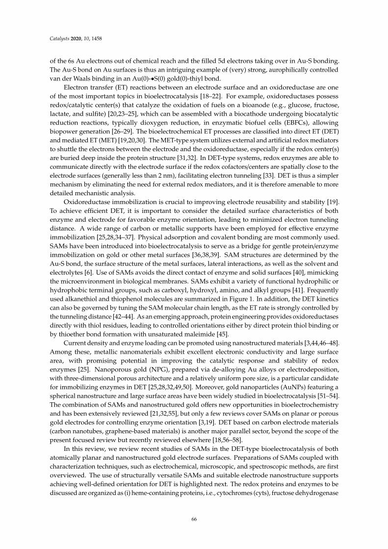
Catalysts 2020, 10, 1458
of the 6s Au electrons out of chemical reach and the filled 5d electrons taking over in Au-S bonding.The Au-S bond on Au surfaces is thus an intriguing example of (very) strong, aurophilically controlledvan der Waals binding in an Au(0)-•S(0) gold(0)-thiyl bond.
Electron transfer (ET) reactions between an electrode surface and an oxidoreductase are oneof the most important topics in bioelectrocatalysis [18–22]. For example, oxidoreductases possessredox/catalytic center(s) that catalyze the oxidation of fuels on a bioanode (e.g., glucose, fructose,lactate, and sulfite) [20,23–25], which can be assembled with a biocathode undergoing biocatalyticreduction reactions, typically dioxygen reduction, in enzymatic biofuel cells (EBFCs), allowingbiopower generation [26–29]. The bioelectrochemical ET processes are classified into direct ET (DET)and mediated ET (MET) [19,20,30]. The MET-type system utilizes external and artificial redox mediatorsto shuttle the electrons between the electrode and the oxidoreductase, especially if the redox center(s)are buried deep inside the protein structure [31,32]. In DET-type systems, redox enzymes are able tocommunicate directly with the electrode surface if the redox cofactors/centers are spatially close to theelectrode surfaces (generally less than 2 nm), facilitating electron tunneling [33]. DET is thus a simplermechanism by eliminating the need for external redox mediators, and it is therefore amenable to moredetailed mechanistic analysis.
Oxidoreductase immobilization is crucial to improving electrode reusability and stability [19].To achieve efficient DET, it is important to consider the detailed surface characteristics of bothenzyme and electrode for favorable enzyme orientation, leading to minimized electron tunnelingdistance. A wide range of carbon or metallic supports have been employed for effective enzymeimmobilization [25,28,34–37]. Physical adsorption and covalent bonding are most commonly used.SAMs have been introduced into bioelectrocatalysis to serve as a bridge for gentle protein/enzymeimmobilization on gold or other metal surfaces [36,38,39]. SAM structures are determined by theAu-S bond, the surface structure of the metal surfaces, lateral interactions, as well as the solvent andelectrolytes [6]. Use of SAMs avoids the direct contact of enzyme and solid surfaces [40], mimickingthe microenvironment in biological membranes. SAMs exhibit a variety of functional hydrophilic orhydrophobic terminal groups, such as carboxyl, hydroxyl, amino, and alkyl groups [41]. Frequentlyused alkanethiol and thiophenol molecules are summarized in Figure 1. In addition, the DET kineticscan also be governed by tuning the SAM molecular chain length, as the ET rate is strongly controlled bythe tunneling distance [42–44]. As an emerging approach, protein engineering provides oxidoreductasesdirectly with thiol residues, leading to controlled orientations either by direct protein thiol binding orby thioether bond formation with unsaturated maleimide [45].
Current density and enzyme loading can be promoted using nanostructured materials [3,44,46–48].Among these, metallic nanomaterials exhibit excellent electronic conductivity and large surfacearea, with promising potential in improving the catalytic response and stability of redoxenzymes [25]. Nanoporous gold (NPG), prepared via de-alloying Au alloys or electrodeposition,with three-dimensional porous architecture and a relatively uniform pore size, is a particular candidatefor immobilizing enzymes in DET [25,28,32,49,50]. Moreover, gold nanoparticles (AuNPs) featuring aspherical nanostructure and large surface areas have been widely studied in bioelectrocatalysis [51–54].The combination of SAMs and nanostructured gold offers new opportunities in bioelectrochemistryand has been extensively reviewed [21,32,55], but only a few reviews cover SAMs on planar or porousgold electrodes for controlling enzyme orientation [3,19]. DET based on carbon electrode materials(carbon nanotubes, graphene-based materials) is another major parallel sector, beyond the scope of thepresent focused review but recently reviewed elsewhere [18,56–58].
In this review, we review recent studies of SAMs in the DET-type bioelectrocatalysis of bothatomically planar and nanostructured gold electrode surfaces. Preparations of SAMs coupled withcharacterization techniques, such as electrochemical, microscopic, and spectroscopic methods, are firstoverviewed. The use of structurally versatile SAMs and suitable electrode nanostructure supportsachieving well-defined orientation for DET is highlighted next. The redox proteins and enzymes to bediscussed are organized as (i) heme-containing proteins, i.e., cytochromes (cyts), fructose dehydrogenase
66

Catalysts 2020, 10, 1458
(FDH), cellobiose dehydrogenase (CDH), glucose dehydrogenase (GDH), and sulfite oxidase (SOx);(ii) blue copper proteins, i.e., azurin, copper nitrite reductase (CuNiR), bilirubin oxidase (BOD),and laccase (Lac); (iii) [FeS]-cluster hydrogenases, i.e., [FeFe]-, [NiFe]-, and [NiFeSe]-hydrogenase.This classification is warranted primarily by the different nature of the core ET cofactor, but also withthe specific secondary and tertiary structures of the protein that envelopes the metallic or non-metalliccatalytic sites. Conclusions and further perspectives are offered and discussed in the final section.
Figure 1. Frequently used alkanethiols and thiophenols in the formation of self-assembled molecularmonolayers (SAMs) with alkyl, amino, hydroxyl, and carboxyl terminal groups.
67

Catalysts 2020, 10, 1458
2. Preparation and Characterization of SAM-Based Au- and Other Electrodes
2.1. Preparation of SAMs
A great merit of SAM-forming thiols is that they form spontaneously on electronically soft metalsubstrates such as gold surfaces from liquid or vapor media under mild conditions [5,59]. Well-definedSAMs are formed by immersing the clean metal surface into a thiol solution, commonly ethanol oraqueous solution, for a certain period of time (hours to days), followed by washing with the samesolvent. The quality of the SAMs is determined by the variety and concentration of thiol solution,temperature, soaking time, and the metal surface structure. Vapor deposition is also used to prepareSAMs, but the SAM morphology is here hard to control. Single-crystal, atomically planar goldelectrodes are the best substrates to investigate the SAM properties, which can then be characterizedusing several in situ techniques including electrochemical scanning tunneling microscopy (in situSTM). Nanomaterials with porous structure and high surface area are also used as substrates [40,60].It is noteworthy that a potential-assisted method to accelerate the SAM formation process has beendeveloped [9,61,62]. The potential range is then a key parameter that strongly affects the quality of theSAMs, which can be evaluated simply by the surface coverage.
2.2. Characterization Methods
2.2.1. Electrochemistry
SAMs can be characterized by a range of methods, among which electrochemical methods havebeen widely reported [59]. Electrochemical methods involve cyclic (CV) and linear sweep voltammetry(LSV), and electrochemical impedance spectroscopy (EIS). CV of non-redox active SAMs, i.e., capacitivevoltammetry, generally shows decreased double-layer capacitance after SAM adsorption on a metalsurface. LSV and CV records the irreversible reductive (and oxidative) desorption of SAMs at anegative (or positive) potential. Reductive desorption is well suited to evaluating the surface coverageand the thermodynamic stability of the SAM Au-S units. The shape and position of the desorptionpeaks are sensitive to the crystalline surface structure and the SAM species. EIS can disentangle the ETresistance from the mass transfer or diffusion resistance. As noted, the ET resistance increases withincreasing chain length because of the increased tunneling distance [63]. Electrochemical quartz crystalmicrobalance (EQCM) is an in situ technique, allowing real-time monitoring of the SAM adsorptionprocess with high sensitivity to the mass changes on the electrode [59].
2.2.2. Microscopy
Electrochemical scanning probe microscopies (SPMs), especially STM and atomic force microscopy(AFM), have been extensively used to provide structural SAM properties at the single-molecule level.The principle of STM is based on the quantum tunneling effect. A bias voltage is applied across the metalsupport and the extremely sharp STM probe, generating tunneling currents, which are transformed tohigh-resolution conductivity images when the STM tip is scanned across the SAM-modified metalsupport. Zhang and associates have reported extensive in situ STM investigations using a wide rangeof alkanethiols on single-crystal gold [6,64–66]. In situ STM combined with electrochemical controlcan also directly map real-time SAM dynamics and structural features. In situ AFM has also attractedattention in bioelectrochemistry [67]. AFM records complex forces, mapping the structural informationby allowing the tip to directly contact the redox protein/enzyme molecules. High-resolution STMhas been employed to record the SAM thickness and molecular orientations. Contact angle (CA)measurement is a simple and straightforward method for monitoring the hydrophobic/hydrophilicproperties of SAMs [1,5].
68

Catalysts 2020, 10, 1458
2.2.3. Spectroscopy
Various spectroscopic techniques are needed to map the complex interactions between SAMs andthe metal support [16,17]. X-ray photoelectron spectroscopy (XPS) records the element compositionand the chemical state of the SAMs. The high-resolution XPS spectra of S 2p usually show a doubletat the binding energy ranging from 160 to 165 eV, attributed to the formation of the Au-S bondbetween alkanethiol molecules and the metal support. The functional SAM groups can be identifiedby Fourier-transform infrared spectroscopy (FTIR), and Raman spectroscopy can be used to detectstructural changes in SAMs [68]. Surface plasmon resonance (SPR) spectroscopy is a powerful techniqueto measure the SAM thickness, showing a change in the tilt angle when the SAM thickness varies.Notably, the adsorption kinetics can be monitored online by SPR coupled with ellipsometry [62].
3. SAMs and Electrochemical DET of Redox Proteins/Enzymes
SAMs have received considerable early and recent attention as substrates for interfacialbioelectrochemical DET reactions, and a wide range of metalloproteins have been investigated in termsof catalytic mechanism and ET kinetics on various SAM-modified electrode surfaces. The redox activecenters of protein/enzymes in DET can be roughly categorized into two major groups: metal-based(iron, copper and molybdenum centers etc.) [20,69–71] and non-metal-based (e.g., flavin adeninedinucleotide (FAD) and pyrrolo-quinoline quinone (PQQ)) centers [44,72,73]. Most, although notall, DET-capable enzymes harbor multiple redox centers, with internal ET relays via heme groups,copper clusters, or Fe-S clusters, which shuttle electrons between the electrode surface and the catalyticcofactors [45,74,75]. In this section, we overview recent studies where SAMs have been used in DET-typeelectrocatalysis and discuss how to obtain favorable orientations using versatile SAMs, as well ashow to tune the interactions between the redox protein/enzyme and the SAM-modified electrodes.Redox proteins/enzymes which can be immobilized in well-defined orientations on SAM-modifiedsupports are illustrated in Figure 2 [4,15,19,32,64,67,70,76–81]. We shall overview and discuss someselected examples from each of these enzyme classes in Sections 3.1–3.3.
Figure 2. (a) Illustration of common proteins/enzymes capable of direct electron transfer (DET) onelectrode modified by self-assembled molecular monolayers (SAMs). The proteins include: horse heartcytochrome c (cyt c), PDB 1HRC; cellobiose dehydrogenase (CDH) from Neurospora crassa, PDB 4QI7;gamma-alpha subunit of FAD-dependent glucose dehydrogenase (FAD-GDH) from Burkholderia cepacia,PDB 6A2U; chicken liver sulfite oxidase (SOx), PDB 1SOX; Pseudomonas aeruginosa azurin T30R1,PDB 5I28; monomer of Achromobacter xylosoxidans copper nitrite reductase (AxCuNiR), PDB 1HAU;bilirubin oxidase from Myrothecium verrucaria (BOD), PDB 2XLL; laccase (Lac) from Trametes versicolor,PDB 1KYA; [NiFe]-hydrogenase from DesulfovVibrio Vulgaris Miyazaki F (DvMF), PDB 1UBU. Schematicview of representative DET processes of (b) mvBOD and (c) SOx on the electrode modified withnegatively and positively charged SAMs, respectively.
69

Catalysts 2020, 10, 1458
3.1. Heme-Containing Proteins
3.1.1. Cytochrome c
As an electrochemical paradigm redox metalloprotein target, cytochrome c (cyt c) is a solubleheme protein extensively studied also as a model metalloprotein on thiol SAM surfaces [64,82–87].Comprising 105 amino acid residues, cyt c (horse heart, ca. MW: 12.4 kDa) is an electron transportprotein largely present in eukaryotic cells [88]. Cyt c is an ideal model system enabling an understandingof protein ET mechanisms in electrochemistry and homogeneous solution. The interfacial ET rateconstant (kapp) can be obtained based on the Laviron equation [89]. Electroreflectance spectroscopy(ER) has been utilized to obtain more accurate kapp due to the elimination of the capacitive double-layercharging current [83]. Horse heart cyt c is the most studied cytochrome, containing a number ofpositively charged lysine residues around the heme edge. Cyt c docks electrostatically with naturalpartners including cyt c oxidases/peroxidases. To immobilize cyt c, SAMs with carboxyl terminalgroups are suitable due to favorable electrostatic binding [84]. Collinson and associates demonstratedthat horse heart cyt c shows similar orientations on the carboxyl terminated SAM-modified electrodefor both covalent bonding and electrostatic adsorption, but covalent bonding led to more stableimmobilization [85]. It was also noted that the formal redox potential (E) of electrostatically adsorbedhorse heart cyt c is shifted negatively due to the electrostatic interactions with the negatively chargedSAM surface.
SAMs, consisting of a mixture of long-chain pyridine alkanethiols and short-chain alkanethiols,enhance the interfacial kapp because of more favorable electronic coupling between cyt c and theelectrodes [84,86] than for pure SAMs. The effect of lysine residues on interfacial ET was exploredby substituting lysine residues at specific positions. Niki and associates reported that replacement oflysine-13 with alanine in rat cyt c (RC9-K13A) showed a more than five-fold ET rate decrease comparedwith replacing lysine-72 and lysine-79 [90], which suggests that lysine-13 exhibits optimized couplingwith the carboxyl SAM-modified electrode. Direct bonding to the heme group with axial pyridine orimidazole ligands onto the gold surfaces is another effective method for narrow orientation distributionof cyt c [84,87]. The tunneling distance-dependent ET was also investigated by surface-enhancedresonance Raman (SERR) spectroscopy, showing a declining signal with increasing SAM chain lengthfrom 2-mercaptoacetic acid to 16-mercaptohexadecanoic acid [91].
AuNPs enhance the interfacial ET rate of cyt c in bioelectrocatalysis. Insertion of 3–4 nm coatedAuNPs between cyt c and the a SAM-modified Au(111)-electrode surfaces was shown to increase kapp
by more than an order of magnitude [89] in spite of an ET distance increase exceeding 50 Å. This raisesissues relating to the mechanism of the AuNP promotion even of simple ET processes, discussed indetail recently [92,93]. Engelbrekt and associates reported ultra-stable starch-coated AuNPs, enablinga clear redox signal of yeast cyt c on AuNP-modified basal plane graphite (BPG) electrodes but nosignals on bare BPG and Au(111) electrode [94].
Other cytochromes, such as cyt b and cyt c4, have also been investigated. Della Pia and associatesreported that ET between the heme group in cyt b562 and the Au(111) electrode can be promoted byreplacing the original aspartic acid residue with a cysteine residue, which provided specific proteinorientation through a Au-S bond [95]. Chi and associates studied the interfacial and intramolecular ETkinetics of di-heme Pseudomonas stutzeri cyt c4 compared with horse heart cyt c (Figure 3) [64]. In situ
STM showed directly that the dipolar cyt c4 is vertically oriented on the carboxyl SAM-modified Au(111)electrode (Figure 3c), resulting in intriguing asymmetric CVs. The authors could show that electronswere first transferred to the heme with the higher potential and then to the second, low-potentialheme by fast intramolecular ET. Lisdat and coworkers reported extensive studies on a multilayeredprotein–enzyme system on SAM-modified gold electrodes [96–99]. For example, they described asulfite oxidase/cyt c (SOx/cyt c) multilayer system without polyelectrolyte, repeatedly incubating theprepared cyt c-modified Au electrode into a mixture of SOx/cyt c solution and pure cyt c solution [97].
70

Catalysts 2020, 10, 1458
A notable current density was observed even up to eight SOx/cyt c layers, which could be explained bythe direct electronic interactions between the two proteins.
ω
−
Figure 3. (a) Schematic illustration of P. stutzeri cyt c4 (left) and horse heart cyt c (right) on SAM-modifiedAu(111); in situ STM images of a ω-mercapto-decanoic acid SAM-modified Au(111)-electrode surface(b) without protein as a reference, (c) with the two-domain P. stutzeri cyt c4 vertically oriented (sharproughly circular spots), and (d) horse heart cyt c (sharp roughly circular spots) in 5 mM pH 7.0phosphate buffer under potential control in constant current mode; scan area, 60 × 60 nm2 Reproducedwith permission from [64]. Copyright 2010, American Chemical Society.
Overall, these reports highlight cyt c as a core electron carrier enabling efficient ET betweenredox enzymes and the electrode surface across suitably chosen SAMs and along with the blue ETprotein azurin as a case for characterization in unique detail, right down to the level of the singlemolecule. Many oxidoreductases furthermore rely on cytochrome domains or subunits as “built-in”ET relays between the catalytically active cofactor and the electrode surfaces and will be discussed inthe following sub-sections [100].
3.1.2. Fructose Dehydrogenase
Although not approaching the degree of detail associated with the simpler ET proteins cyt c andazurin, the molecular mechanistic mapping of several flavin dehydrogenases has reached an impressivelevel of detail over the last few years, represented by fructose dehydrogenase (FDH), CDH, and GDHin particular. The three enzymes display a common pattern, with a FAD catalytic center where fructose,cellobiose, and glucose, respectively, is oxidized, and an ET relays temporarily populated ET sitesthrough which the liberated electrons are transmitted to the electrode surface. As the first FAD enzymein our overview, Gluconobacter sp. FDH is a membrane-bound FAD-dependent oxidoreductase with amolecular mass around 140 kDa [25,30]. The protein holds three subunits: subunit I (67 kDa) containsa FAD cofactor, serving as the catalytic center for the two-electron oxidation of D-fructose to keto-Dfructose. Subunit II (51 kDa) has three heme groups with the formal potentials of 0.15, 0.06, and −0.01 Vvs. Ag/AgCl electrode (sat. KCl), respectively. Only two heme groups with the relatively lower redoxpotentials are proposed to participate in DET [30]. Subunit III (20 kDa) plays an important role inmaintaining the structural integrity of the enzyme complex. A number of recent studies illustrate theemployment of FDH for the development of biosensors and biofuel cells with high current density andoperational stability [30,81,101,102].
71

Catalysts 2020, 10, 1458
The three-dimensional crystal structure, especially the enzyme surface properties, is essentialfor rational tuning of the redox enzyme immobilization. The detailed crystallography of FDH is stillunclear, but homology models have helped to provide a clearer picture of intramolecular electrontransfer (IET) [103]. Kano and associates constructed FDH variants with glutamine instead of the axialmethionine ligands (M301, M450, or M578) of heme 1c, heme 2c, and heme 3c, respectively, illustratingthat the ET pathway leads from M578 to M450 bypassing M301 [104]. Heme 1c with the highest formalpotential of 0.15 V vs. Ag/AgCl electrode was evaluated not to be involved in DET, whereas heme2c was identified as the ET bridge between FDH and the electrode surface. Heme 3c with the lowestformal potential of −0.01 V vs. Ag/AgCl (sat. KCl) was suggested as a bridge between FAD and heme2c in the IET process (Figure 4) [30]. In addition, the catalytic current density of FDH was dramaticallyincreased by deleting the amino acid residues on the N- or C-terminus of subunit II [30,105,106].The deletion not only promotes enzyme loading but also provides more opportunities for favorableorientations on the electrode surface. Some researchers reported that hydrophobic anthracene groupsanchored on single-walled carbon nanotubes are favorable for enhanced catalytic activity and stabilityvia the hydrophobic C-terminal region of subunit II [101,107]. These studies suggest that orientationand enzyme loading are crucial for controlling the catalytic activity in DET-type bioelectrocatalysis.
−
Figure 4. Proposed ET pathway from D-fructose to the electrode surface in the DET of FDH. The ETroute involves FAD, heme 3c, heme 2c, but not heme 1c. Reproduced with permission from [30].Copyright 2019, Elsevier.
Favorable FDH SAM immobilization rests on the following consideration: the isoelectric point(IEP) of FDH is 6.59, which means that FDH is overall positively charged in slightly acidic electrolyte [81].Recent studies to improve the catalytic performance of FDH on various SAM-modified electrodes havebeen reported [25,41,44,81]. Bollella and associates reported extensive research on the catalytic activityof FDH on highly porous gold (h-PG) electrodes modified with 4-mercaptobenzoic acid (4-MBA),4-mercaptophenol (4-MPh), and 4-aminothiophenol (4-APh) SAMs. The data showed that highbioelectrocatalytic activity and stability of FDH was only observed with -OH terminated SAMs [81],suggesting optimized enzyme orientation on this particular SAM. Negatively charged SAMs mayhelp to favor FDH coverage due to preferred electrostatic interaction, but this is not necessarily themost favorable FDH orientation for DET. Murata and associates reached similar conclusions for FDHimmobilized on 2-mercaptoethanol (MET) SAM-modified AuNPs [41]. Considering the importanceof the gold nanostructure for the ET rate, the AuNP size is furthermore a crucial parameter. Kizlingand associates synthesized 1.0 to 3.5 nm AuNP clusters functionalized with 1,6-hexanedithiol and1-butanethiol for investigating the ET mechanism. A channel of “mediated” catalysis, i.e., electron“hopping”, was observed for the smallest AuNP clusters around 1 nm with the half-wave potentialclose to the first oxidation potential of the AuNP at the edge of the HOMO-LUMO gap [44]. Such amode accords with theoretical notions recently reported [92]. Siepenkoetter and associates reportedthe catalytic performance of covalently bonded FDH on NPG electrodes with varying pore sizes [25].A large number of findings thus show that FDH has high affinity for well-defined SAM-modifiedelectrodes with the polar but electrostatically neutral hydroxyl terminal group as the most efficient,
72

Catalysts 2020, 10, 1458
indicating the importance of hydrophilicity of the electrode surface towards the mixed surface chargedistribution of the FDH target enzyme. However, details of the underlying mechanism remainunknown.3.1.3. Cellobiose Dehydrogenase
The second FAD enzyme, cellobiose dehydrogenase (CDH), is a versatile oxidoreductase for directbioelectrocatalysis. CDH contains a catalytic dehydrogenase domain (DH) harboring a FAD as theredox center and an ET cytochrome b (CYT) domain to shuttle electrons from the FAD to the electrodesurface [23,45,108]. The two domains are separated in the crystalline structure, but an integrated IETpathway can be opened through a flexible and hydrophilic amino acid linker between the catalytic andCYT domains [80]. This motif is encountered also for the molybdenum sulfite oxidases, cf. Section 3.1.4.CDH is extracted from the phyla Basidiomycota and Ascomycota, divided into class I, class II, and class III,respectively. Class III CDH from Ascomycota remains uncertain compared to class I and class II CDH [23].
The natural substrates of CDH mainly include cellulose, lactose, and glucose [108]. Class I CDHwith short amino acid sequences shows direct, strongly pH-dependent catalytic activity only in solutionswith pH below 5.5 [23]. Harreither and associates reported extensive studies on class II CDH fromChaetomium attrobrunneum (CaCDH), Corynascus thermophiles (CtCDH), Dichomera saubinetii (DsCDH),Hypoxylon haematostroma (HhCDH), Neurospora crassa (NcCDH), and Stachybotrys bisbyi (SbCDH) [109].pH-dependent catalytic activity was observed for neutral and slightly alkaline electrolytes with cyt c
and 2,6-dichloroindophenol (DCIP) as electron acceptors. The different electrostatic environment inclass I and II CDH reveals different optimal IET processes, with optimum IET in acidic electrolyte forclass I CDH and neutral or slightly alkaline electrolyte for class II CDH [110]. Schulz and associatesdemonstrated turnover and non-turnover DET performance of CDH on polycrystalline gold modifiedwith MUO or 6-MHO [23]. A clear catalytic current between the FAD and the electrode surfacewas reported. The midpoint potential of bound FAD cofactor was −163 mV vs. SCE at pH 3.0,approximately 130 mV less than that of the heme b relay, leading to a much lower onset potentialfor lactose oxidation. The authors concluded that the tunneling distance between the FAD cofactorand the electrode was around 12–15 Å, thereby allowing direct electrochemical communication.The authors also demonstrated that only class I CDH from Trametes villosa (TvCDH) and Phanerochaete
sordida (PsCDH) displayed DET activity, whereas no DET signal was observed for class II CDHfrom reconstructed Myriococcum thermophilum (recMtCDH) and reconstructed Corynascus thermophilus
(recCtCDH) at low pH.The IEP of CDH (DH domain ~5, CYT domain ~3) gives a negatively charged surface around the
ET path exit, indicating that positively charged SAMs are suitable for favorable DET [110]. Lambergand associates anchored Humicola insolens CDH on various SAMs with different terminal groups forinvestigating the effects of charge and hydrophobicity [111] and found that hydrophilic SAMs werefavorable for high catalytic activities, with lower enzyme orientation variations than for hydrophobicSAMs. Tavahodi and associates reported a DET-type lactose biosensor of PsCDH on polyethyleneimine(PEI)-coated AuNP electrodes (PEI@AuNP) [112]. PEI with positively charged amino groups not onlyoptimized the orientation of PsCDH on the electrode to increase the IET rate but also gave high enzymestability and sensitivity. Bollella and associates reported a lactose biosensor based on DET of CtCDHon gold electrodes [113]. The authors demonstrated that BPDT SAMs with two thiol groups can beused to anchor metal NPs on a gold electrode surface by covalent bonding, showing the best ET rateon AuNPs/BPDT/Au electrode. A mediator-free HiCDH/MvBOD BFC using a positively charged MHPSAM for immobilization on AuNP-modified gold electrodes was also reported [114]. The half-life time,i.e., the time duration over which the activity decreased to half the initial value, was 13 and 44 h with5 mM glucose and 10 mM lactose in neutral buffer solution, respectively. Hirotoshi and associates foundan efficient ET process of Phanerochaete chrysosporium (PcCDH) on a mixed 11-AUT/MUO SAM-modifiedAuNP electrode [46]. Al-Lolage and coworkers addressed the catalytic performance and stabilityof MtCDH by introducing cysteine mutants (E522 and T701) on the protein surface (Figure 5) [45].The cysteine mutant with a surface thiol group made it possible to form stable thioether bonds withmaleimide groups via click-chemistry, thereby controlling the orientations of the MtCDH mutant on the
73

Catalysts 2020, 10, 1458
electrode surface and revealing efficient electrochemical glucose oxidation. Another example reportedby Meneghello and associates clearly indicated that the DET-type bioelectrocatalysis of CDH is highlysensitive to the cysteine residues introduced at particular positions [115]. Experimental results alsoshowed that divalent cations (i.e., Ca2+ and Mg2+) affect the IET kinetics [23,115].
Figure 5. (a) Structure of MtCDH with cysteine E522 and T701 mutations shown in blue and green,respectively; FAD and the heme group are highlighted in yellow and red, respectively. The flexiblechain linking the two domains is in blue; (b) the maleimide group anchored on an electrode surfacecan react with the mutant cysteine residue in the enzyme. Reproduced with permission from [45].Copyright 2017, John Wiley and Sons.
3.1.3. FAD-Dependent Glucose Dehydrogenase
Our final FAD enzyme target, glucose dehydrogenase (FAD-GDH), is one of the most widelyknown dehydrogenases with a tightly bound cofactor, making it different from NAD+-dependentGDH. FAD-GDH has been extensively studied as an emerging alternative to glucose oxidase (GOx)due to its favorable DET capability, insensitivity to dioxygen, and the fact that no hydrogen peroxide isgenerated [116–119]. The structure of GDH is analogous to that of FDH, comprising three subunits:an FAD-dependent catalytic subunit, an ET subunit with three heme groups, and a small “hitch-hiker”protein used for the flexibility of the catalytic subunit into the periplasm [116,120]. The catalyticsubunit harbors a 3Fe-4S cluster close to the ET subunit protein surface, allowing efficient DET onthe electrode without the need for mediators. Lee and associates demonstrated controlled DET ofbacterial FAD-GDH from Burkholderia cepacia on three different SAM-modified electrodes, on whichcatalytic current density decreases with increasing SAM chain length [118]. A glucose biosensorbased on FAD-GDH was reported recently by introducing a gold-binding peptide (GBP) for enzymeimmobilization on a screen-printed electrode (SPE) [24]. GBP composed of 12 amino acids exhibitinga strong binding affinity to the gold electrode surface was fused to the enzyme terminus, therebydetermining the enzyme orientation on the electrode. Around 10 times higher catalytic responsetoward 100 mM glucose was observed with FAD-GDH-GBP/Au compared with normal GDH/Au.
A DET-type glucose biosensor could also be fabricated coupled with electrochemicalimpedance spectroscopy (EIS) [120]. Three variable-length thiols, dithiobis(succinimidyl hexanoate)(DSH), dithiobis(succinimidyl octanoate) (DSO), and dithiobis(succinimidyl undecanoate) (DSU),were employed to modify the electrode surface. Charge transfer resistance (Rct) was a key parameterreflecting the DET efficiency, with the lowest resistance when FAD-GDH was immobilized on DSHSAMs because of the shortest tunneling distance. In addition, the steady-state catalytic currentdensity of FAD-GDH was dramatically increased on AuNPs assembled on the gold electrode [73,121].Ratautas and associates reported high glucose oxidation activities of FAD-GDH extracted from
74

Catalysts 2020, 10, 1458
Ewingella Americana without mediator [73,121]. They demonstrated that FAD-GDH immobilizedon 4-ATP-modified AuNPs displayed higher catalytic activity and lower overpotential than on4-MBA-modified electrodes. Notably, 4-ATP can be oxidized in neutral media and further converted to4-mercapto-N-phenylquinone monoimine (MPQM), the quinone groups of which could bind covalentlywith primary amino groups of the enzyme, thereby enhancing the ET rate. The catalytic responsewas found to be positively correlated to the ratio of 4-ATP/4-MBA for electrodes modified by mixedthiol SAMs [121]. PQQ-dependent GDH is still another promising GOx candidate, widely utilized forglucose biosensors and biofuel cells [35,95,96]. Kim and coworkers prepared a glucose biosensor byimmobilizing positively charged PQQ-GDH on MUA SAM-modified gold electrodes [122]. In thiscase, PQQ-GDH exhibited higher current density and detection sensitivity via electrostatic adsorptionthan via covalent bonding, due to less enzyme inactivation. Covalent bonding could thus enhancethe interactions between enzyme and electrode surface, but the surface enzyme characteristics are themore critical factors that determine the electrochemical behavior.
3.1.4. Sulfite Oxidase
Sulfite oxidase (SOx) is another heme-containing redox enzyme which has been studied for along time. Two kinds of SOx are mainly employed in DET-type bioelectrocatalysis: chicken liver,cSOx [123], and human, hSOx [124]. SOx acts by a principle resembling that of the FAD enzymes, with acatalytic center and an electronic relay, but also with some important differences. As a metalloprotein,the catalytic domain is composed of a pyranopterin molybdenum (Mo) cofactor effecting two-electronoxidation of sulfite to sulfate and connected to a N-terminal cytochrome b5 (cyt b5) domain by a flexiblelinker [123,125]. Although the crystallographic structure of cSOx shows that the distance betweenthe catalytic Mo domain and the cyt b5 domain is more than 32 Å, rapid internal ET in DET-typebioelectrocatalysis is still observed [126], effected by a conformational change on the electrode surfacevia the flexible tether. This “on-off” switch enables cyt b5 to be either adjacent or remote from the Mocofactor [70,127] in a gated ET mode, which is quite different from the rigidly bound ET relays in theFDH and GDH enzymes, but similar to the CDH operational mode.
The electrostatic surface charge distributions around both domains are complex and highlightthe importance of subtle tuning of the electrode surfaces [124]. A positively charged SAM surface isfavorable for immobilization of hSOx via the Mo domain, directing the smaller heme domain towardsthe electrode surface. As noted, a similar pattern with a flexible linker connecting the catalytic and ETdomain applies to CDH [80]; cf. Section 3.1.3. Sezer and associates investigated the catalytic activityof hSOx on a mixed SAM-modified silver electrode, showing a significantly increased SERR signalwhen increasing the ionic strength. High ionic strength is favored to shorten the distance betweenthe Mo cofactor and the heme domain, thereby facilitating both intramolecular and interfacial ET.Wollenberger and associates reported other studies of the electrochemical behavior of hSOx [127–130].In situ scanning tunneling microscopy and spectroscopy to single-molecule resolution has, finally beenreported quite recently and disclosed intriguing patterns of tunneling via the Mo- and heme groupredox centers [130]. AuNPs with a diameter less than 10 nm were covalently bonded on the MUA/MUOSAM-modified gold electrode, giving a significant enhancement of the interfacial ET rate of hSOx [129].The interfacial ET rate could be further increased by using SAMs with 3,3′-dithiodipropionic aciddi(N-hydroxysuccinimide ester) (DTSP) thinner than MUA/MUO SAMs [128]. They also reported thatthe introduction of BaSO4 nanoparticles played a significant role in the DET-type bioelectrocatalysis ofhSOx. The electrochemical communication between the active sites of hSOx and the electrode surfacecould be further enhanced by using a positively charged biopolymer. Kalimuthu and coworkers thusreported direct catalytic activity of hSOx on a chitosan-covered gold electrode [131]. Both non-catalyticredox signals corresponding to the heme group and catalytic signals in the presence of 4 mM sulfite,on chitosan-covered MPA-, MSA-, and 4-MBA-modified electrodes were observed. However, there wereno catalytic signals on MUA-based electrodes despite an observed non-turnover feature, highlightingthe importance of rational SAM selection.
75

Catalysts 2020, 10, 1458
3.2. Blue Copper Proteins
3.2.1. Azurin
Pseudomonas aeruginosa azurin is a simple blue copper protein that undergoes single-ET between theType 1 copper atom and the electrode, now developed as a single-molecule “core” target [6,132–136],as for cyt c characterized and mapped in unique detail. A hydrophobic patch and the disulfidegroup at opposite ends of the azurin molecule are both critical for well-defined orientations on theelectrode. Chi, Ulstrup, Zhang and associates conducted extensive investigations of the electrochemicalbehavior of azurin [39,43,51,137]. In situ STM disclosed arrays of well-organized self-assembledazurin monolayers on single-crystal Au(111)-electrodes mapped to single-molecule resolution [51].Hydrophobic alkanethiol monolayers were employed for gentle immobilization by hydrophobicinteractions with the hydrophobic patch of azurin [43]. Notably, exponential decay of the ET rateconstant with increasing chain length was observed for chain lengths longer than six carbon atoms,reflecting a dual mechanism, with tunneling dominating for the longer chains [39]. The ET kineticsand redox mechanism of azurin have been analyzed theoretically at different levels, as discussed indetail elsewhere [6,138]. Inserting 3–4 nm coated AuNPs as for horse heart cyt c [83] results in a 20-foldenhancement of kapp (220 ± 16 s−1) for azurin compared with the AuNP-free system (10.2 ± 0.4 s−1) [51].Armstrong and associates compared the ET kinetics of azurin on the electrode modified witha synthetic 3,5-diethoxy-4-[(E)-2-(4-ethylphenyl)vinyl]-phenyl methanethiol or commercial-CH3
terminal alkanethiol [139]. The front hydrophobic ethyl group of SAMs served as the protein-bindingbridge on the surface, yielding a very fast ET rate with a kapp over 1600 s−1.
3.2.2. Copper Nitrite Reductase
A common feature of the blue multi-copper oxidases is a blue T1 center for the electron inlet and a TII
or combined TII/TIII catalytic center for the electron “outlet” in the catalytic process (nitrite, or, dioxygenreduction). Such an electrochemical mode of action is only feasible via an efficient (short) intramolecularET channel. This feature has been mapped in considerable detail for three blue copper enzyme classes,the copper nitrite reductases, bilirubin oxidases, and the laccases. The trimeric blue copper nitritereductase (CuNiR) is crucial in the global nitrogen cycle, catalyzing the single-electron reductionof nitrite to nitrogen monoxide [140]. CuNiR effects direct bioelectrocatalysis, including an ETrelay (CuI) and a catalytic site (CuII) in each monomer. Ulstrup and associates reported DET-basedelectrocatalysis of CuNiR from Achromobacter xylosoxidans (AxCuNiR) on a cysteamine SAM-modifiedAu(111) electrode [141]. In situ STM displayed single AxCuNiR molecules but, intriguingly, only in thepresence of nitrite substrate. Further, the combination of varying alkanethiols with charged, neutral,hydrophilic, and hydrophobic properties showed that mixed hydrophilic/hydrophobic SAMs were themost favorable for facile AxCuNiR electrocatalysis [142]. In situ AFM is also reported and disclosedAxCuNiR conformational changes during catalytic reaction [67], with the apparent height of AxCuNiRrising from 4.5 nm in the resting state to 5.5 nm in the nitrite reduction state in the presence of nitrite.
3.2.3. Bilirubin Oxidase
Bilirubin oxidase (BOD) is another well-known blue multicopper oxidase class containingfour copper centers (T1, T2/T3), catalyzing the dioxygen reduction reaction (ORR) into water infour-electron direct bioelectrocatalysis [75,96]. The blue T1 center has a copper atom located closedto the protein surface for accepting electrons from the natural electron donor (bilirubin) or anelectrode. The external electrons are relayed via a short ligand-bound intramolecular peptide bridgeto the T2/T3 center, where ORR takes place. Target BODs are mainly from Myrothecium verrucaria
(MvBOD) [26,29,75,143–145], Trachyderma tsunodae (TtBOD) [146], Bacillus pumilus (BpBOD) [147],and Magnaporthe oryzae (MoBOD) [148]. Single-crystal gold electrodes modified with -NH2, -COOH,-OH, and -CH3 terminated SAMs have been studied and show that negatively charged SAMs arefavorable for proper MvBOD orientations due to the highly positively charged region close to the T1
76

Catalysts 2020, 10, 1458
center [42]. E values of T1 (0.69 V vs. NHE) and T2/T3 (0.39 V vs. NHE) in TtBOD have been observedunder aerobic conditions [146], but E of the T2/T3 centers drops to 0.36 V vs. NHE in the resting stateof the enzyme, indicative of an IET pathway from T1 to T2/T3 triggered by the ORR process.
Characterization of enzyme loading and conformation on the electrode is crucial [149]. Lojou andassociates studied the adsorption of MvBOD on both positively (NH3
+) and negatively (COO–) chargedSAM-modified electrodes with no significant difference in enzyme loading as disclosed by surfaceplasmon resonance (SPR) spectroscopy [26]. DET and MET processes were found to dominate on theCOO- and NH3
+ surfaces, respectively, consistent with the positively charged surroundings of theT1 Cu [26]. Polarization-modulated infrared reflection absorption spectroscopy (PMIRRAS) showedstrong electrostatic interactions between the negatively charged SAMs and the positively chargedMvBOD surface near the T1 Cu [26]. PMIRRAS and SPR ellipsometry were jointly used to demonstratethe effect of electrostatic interactions on the enzyme adsorption, catalytic performance, and stability ofMvBOD at four different pH values (Figure 6) [75]. The dipole moment of the enzyme is distinct atdifferent pH levels, showing a direction towards the T1 center in neutral or slightly acidic electrolytesbut significantly shifted in the strongly acid electrolyte (Figure 6a). This accords with the differentcharge distributions at the T1 Cu center. PMIRRAS spectra showed the amide I (ca. 1680 cm−1) andamide II (ca. 1550 cm−1) peaks, related to vibrational C=O and N-H modes in MvBOD, respectively,see Figure 6b. The wavelengths of the two peaks remained unchanged upon enzyme immobilization,which suggests that the secondary structure of the MvBOD is retained. In addition, amide I/amideII ratios were similar at different pH levels, further indicating that the orientations of MvBOD wereindependent of pH.
− −
Γ
Figure 6. (a) MvBOD structure and dipole moments in the pH range 3.6 to 7.5. CuT2/T3 contains threecopper atoms marked in blue and CuT1 with a single copper atom marked in gold; (b) PMIRRASsignals of MvBOD-modified bioelectrodes with 6-MHA SAM; (c) Enzyme coverage (Γ) and enzymelayer thickness at different pH recorded by SPR ellipsometry; (d) Cartoon illustration of the chargedistributions of MvBOD at different pH levels, with the CuT1 center, as well the 6-MHA and4-ATP SAM-modified electrode surfaces. The neutral electrode surface is indicated with greenstars. Reproduced with permission from [75]. Copyright 2018, American Chemical Society.
77

Catalysts 2020, 10, 1458
Enzyme loading, critical to catalytic activity, declines as pH increases (Figure 6c [75]). The authorsdemonstrated that MvBOD on a negatively charged 6-MHA SAM-modified electrode does not form asaturated monolayer at pH 7.5 but possibly more than a single monolayer at pH 3.6. The electrostaticinteractions between enzyme and SAMs thus not only strongly affect the enzyme adsorption, the dipolemoment of the enzyme, and the charge around the T1 center, but they also determine the enzymeorientation and catalytic rate (Figure 6d). Gholami and associates immobilized MvBOD on a goldmicrofilm by electropolymerization of TCA [150]. Molecular dynamics simulation has provided amore comprehensive understanding of MvBOD in DET-type bioelectrocatalysis [151]. In particular,MvBOD showed various orientations reflecting wide charge distributions, representing a “back-on”and “lying-on” state on positively and negatively charged electrodes, respectively.
Nanostructured gold surfaces are expected to improve the DET current density of BOD [29].However, the apparent and real current densities normalized to the geometric and real gold surfacearea, respectively, should be distinguished. In comparison to FDH on 1 nm AuNPs [44], this isparticularly important when the gold nanostructures are of larger size than BOD. Pankratov andassociates investigated the size effect of sub-monolayer AuNPs (diameter: 20, 40, 60, and 80 nm,significantly larger than MvBOD) on the bioelectrocatalytic performance of MvBOD [152]. Althoughthe apparent ORR current density increased with increasing size from 20 to 80 nm (proportionallyto the real surface area), similar values (15 ± 3 uA cm−2) for the real current density were obtained.This can be explained by the fact that the ET rate constant is independent of the AuNP size in thiscase, with similar values of 10.3 ± 0.5 s−1 and 10.7 ± 0.3 s−1 for the MvBOD-based bioelectrode withand without AuNPs, respectively. Siepenkoetter and associates immobilized MvBOD on NPG usingdiazonium grafting coupled with an MPA SAM [28]. NPG electrodes with average pore sizes between9 and 62 nm were finally investigated. The maximum apparent ORR current density was achievedwith 10 and 25 nm pores, slightly larger than the enzyme and likely due to a compromise between thereal gold surface area and enzyme loading, but similar real current densities were registered for thesepore sizes.
3.2.4. Laccase
The laccases (Lac) constitute a third important member class of the multicopper oxidases,containing T1 and T2/T3 centers [27,153,154]. Similar to BOD, T1 and T2/T3 Cu centers act as electronacceptance and ORR centers, respectively. E of the T1 center from tree Lac is lower than those fromfungal Lac, varying in the range 300 to 800 mV vs. NHE [155]. Bioelectrocatalysis of fungal Lac has beenextensively investigated [156]. SAMs with specific functional groups favor productive orientations ofLac at the electrode surface. Thorum and associates reported that the overpotential of ORR of Lac fromTrametes versicolor (TvLac) could be decreased by employing an anthracene-2-methanethiol SAM ongold. The aromatic anthracene presumably penetrates into the hydrophobic pocket close to the T1
center, facilitating DET [157]. Climent and associates investigated the DET-type catalytic activity ofthree different Lacs (Coprinus cinereus (CcLac), Myceliophthora thermophila (MtLac), and Streptomyces
coelicolor (ScLac)) [38]. In situ STM enabled single-molecule understanding of enzyme–electrodeelectronic interactions and the IET process on well-defined Au(111) surfaces with various SAMs.MPA SAMs with the carboxyl terminal group was best for CcLac, while alkyl and amino SAMs weremost suitable for ScLac. No catalytic signal was found for MtLac on any SAMs. As for AxCuNiR,single-molecule in situ STM contrasts were observed only in the presence of enzyme substrate, nitrite,and dioxygen, respectively. Traunsteiner and coworkers exploited DET-type bioelectrocatalysis ofTvLac using diluted MPA SAMs and a linker molecule of thiolated veratric acid (tVA) that couldapproach the T1 site [158]. Optimal catalytic activity was shown when the tVA and MPA SAMs weremixed uniformly, whereas the catalytic activity decreased dramatically due to the aggregation of tVA.Molecular dynamics simulations also showed that the positively charged SAMs were more favorablefor the DET of TvLac, with a narrow orientation distribution.
78

Catalysts 2020, 10, 1458
A recent study evaluated Thermus thermophilus (TtLac), exhibiting a methionine rich domain.Figure 7 shows the detailed three-dimensional structure and electrostatic charge distributions of thesurface amino acid residues of TtLac [69]. The T1 center, used for transferring electrons to an externalelectrode, showed a negatively charged zone. Gold electrodes modified with negatively (-COO−),uncharged (-OH), and positively charged groups (-NH3
+) were adopted, with the highest catalyticcurrent on the positively charged electrode and with no or only weak catalytic signals on the negativelycharged and uncharged electrodes, respectively [69]. Nanostructured materials have been developedrecently to optimize the Lac orientation. NPG modified with 4-ATP SAM could increase the catalyticperformance and stability at high temperatures [159]. Cristina and associates reported that AuNPs(particle size: 5 nm, comparable to the size of the enzyme) served as electronic bridges, thus promotingDET with a heterogeneous ET rate constant over 400 s−1 [54]. Nanostructured electrodes consisting oflow-density graphite (LDG) and gold nanorods (AuNRs, average length: 31 ± 6 nm, width: 5 ± 1 nm)have, finally, been prepared to orient Lac for improved ORR [27].
−
− −
Figure 7. (a) Model structure of TtLac (PDB 2XU9) showing the hairpin domain (magenta), the T1 andT2/T3 Cu centers (blue spheres) and all Met sulfurs (yellow spheres); (b) Electrostatic potentials at thesurface of TtLac in the same orientation as in the top panel at pH 5: positive charges in blue, negativecharges in red, and neutral in white. The positive end of the dipole moment vector is shown as a yellowstick. Reproduced with permission from [69]. Copyright 2020, American Chemical Society.
3.3. [FeS]-Cluster Hydrogenases
Hydrogenases are a class of [FeS] cluster-based metalloproteins which reversibly catalyze thetwo-electron reactions of dihydrogen oxidation and evolution [71,74]. The commonly reportedmembrane-bound hydrogenases can be classified based on their intrinsic catalytic cofactors ([FeFe],[NiFe], and [NiFeSe]), where hydrogen conversion is combined with [FeS] electron relay to accomplishthe entire ET process. It has been reported that the smallest [FeFe]-hydrogenase CrHydA1 isolatedfrom Chlamydomonas reinhardtii exhibits dioxygen insensitivity and shows high catalytic activity indihydrogen evolution [160]. The conditions of immobilized [FeFe]-hydrogenase CrHydA1 on aSAM-modified gold electrode were characterized by in situ surface-enhanced infrared absorptionspectroscopy (SEIRAS) and SPR spectroscopy [160]. Madden and associates investigated the catalyticactivity of [FeFe]-hydrogenase CaHydA from Clostridium acetobutylicum immobilized on negativelycharged MHA-modified Au(111) electrodes [74]. Electrochemical STM showed that the apparentheight of [FeFe]-hydrogenase CaHydA continuously increased when the potential of the STM substratewas shifted from −0.4 to −0.6 V (vs. Ag/AgCl electrode), in which the hydrogen evolution response
79
Catalysts 2020, 10, 1458
was observed by CV. Notably, the catalysis of hydrogenase is easily quenched by carbon monoxideand cyanide binding to the Fe active sites.
Membrane-bound [NiFe]-hydrogenase and [NiFeSe]-hydrogenase have attracted considerableattention due to their resistance to dioxygen, carbon monoxide, as well as to high temperature [161].Armstrong and associates reported a number of studies into the oxygen tolerance of[NiFe]-hydrogenase [162,163]. Typically, the catalytic sites of well-known hydrogenases are quenchedby dissolved oxygen gas due to the high oxygen sensitivity of the internal peptide chains. Modificationof Fe-S clusters by changing two oxygen-sensitive cysteine residues into glycines showed significantimprovements in the long-term dihydrogen oxidation performance [162]. The authors demonstratedthat the mechanism of oxygen tolerance mainly relates to removal of oxide species rather than preventingoxygen access into the protein. Lojou and associates demonstrated that carboxyl-terminated SAMswere favorable for optimizing the orientations of [NiFe]-hydrogenase from Aquifex aeolicus for efficientDET-type and MET-type bioelectrocatalysis of hydrogen oxidation, whereas hydrophobic SAMs onlyresulted in a MET process with the need for methylene blue mediator [71]. The electrochemicalbehavior of [NiFe]-hydrogenase from Allochromatium vinosum, DesulfoVibrio Vulgaris Miyazaki F(DvMF), and Ralstonia eutropha H16 has also been reported [164–166].
Immobilization of membrane-bound hydrogenase on the SAM-modified electrode surfaceappears promising, but the ET progress of membrane-bound hydrogenase is challenging due to thecomplex enzyme structure compared with soluble redox enzymes. Gutiérrez-Sánchez and coworkersdemonstrated that the introduction of a phospholipidic bilayer on positively charged 4-ATP modifiedAu electrode surfaces effectively controls the orientation of membrane-bound [NiFeSe]-hydrogenasefor hydrogen oxidation (Figure 8) [167]. The hydrophobic lipid tail of hydrogenase can be embeddedinto the phospholipidic bilayer, thereby reducing the orientation distribution and promoting the ETprocess. All these results suggest that SAM-modified electrodes are paramount to provide a versatileplatform for understanding how to tune the right enzyme orientation for DET.
Figure 8. Schematic illustration of [NiFeSe]-hydrogenase covalently inserted into a phospholipidicbilayer. Reproduced with permission from [167]. Copyright 2011, American Chemical Society.
4. Conclusions and Perspectives
Redox proteins and enzymes immobilized on solid surfaces are fragile biomolecular entities.With a few exceptions, they retain ET of enzyme function only in prepared microenvironmentsthat somehow emulate their natural aqueous/membrane reaction media. Self-assembled molecularmonolayers with multifariously functionalized thiols are close to ideal SAM building blocks whichhave emerged as the core class over the last couple of decades. The -SH thiol end ascertains robust Au-Slinking which is now increasingly well understood as dominated by the Au(0)-•S(0) gold-thiyl andstrong van der Waals Au-S interaction. The opposite functionalized end of the SAM thiol moleculesoffers the choice of hydrophilic or hydrophobic, electrostatically charged or neutral, and structurallylarge or small terminal groups, well suited for designed gentle protein/enzyme linking to the solid
80

Catalysts 2020, 10, 1458
SAM-modified electrochemical surface. The option of varying the length of the SAM-forming moleculesoffers additional control of the electron tunneling process as well as of the dielectric and other localenvironmental properties crucial in the overall control of the electrochemical activity of the immobilizedproteins or enzymes.
We have first overviewed the preparation and comprehensive characterization of thiol-based SAMson Au surfaces in particular, both per se and as developed in redox protein/enzyme electrochemicalresearch over the last couple of decades. Preparation is, in principle, straightforward, although theultimate SAM properties depend in subtle ways on thiol exposure time, temperature, and other externalcontrolling factors. A variety of sophisticated SAM surface techniques have brought the understandingof the fundamental molecular and electronic SAM structure to a high level, ranging all the wayfrom the ordered domain right down to the single molecule. The techniques include spectroscopy(XPS, FTIR, SPR, Raman, NEXUS, and others), microscopy (AFM, STM in the electrochemical insitu/operando modes), and mass balance techniques (QCM), supported by large-scale electronicstructure calculations [4,6,16,17,66].
Well-defined solid SAM-modified Au- and other electrode surfaces are a pre-requisite for theproductive immobilization of bioelectrochemically active DET enzymes. An outstanding challengeis that polycrystalline Au- and other metallic electrodes are nearly always used, with single-crystal,atomically planar, e.g. Au(111), electrodes only relatively recently introduced as electrochemicalbiomolecular target surfaces. A variety of local low- and higher-index surface structures aredistributed over the polycrystalline Au- and other surfaces, expectedly giving quite different surfaceET activities [92]. This presents a challenge to the microscopic characterization of the pure andSAM-modified electrode surfaces but at the same time also provides new openings in the way ofmore robust protein/enzyme monolayers and higher enzyme activity, if the polycrystallinity can becontrolled such as for NPG and other nanoporous metallic electrodes.
We have next overviewed and discussed adsorption and controlled protein and enzyme orientationon strategically chosen SAMs, of both simple ET metalloproteins (cyt. c and c4, azurin) and a varietyof much more complex redox metallo- and nonmetalloenzymes (blue copper oxidases, FAD-basedenzymes, cellobiose dehydrogenase, [FeS]-cluster dehydrogenases). We have shown that properlychosen SAMs can be brought to control efficiently the enzyme surface orientation in the ways mostfavorable both for productive bioelectrocatalysis and for detailed mapping of the molecular mechanismsinvolved. We have also shown that, although of more complex molecular/atomic surface structure,SAM-modified NPG and other nanoporous metallic electrochemical surfaces, structurally characterizedto intermediate levels of resolution, may offer other advantages. These extend to increased enzymestability and even enhanced catalytic efficiency compared to atomically planar electrode surfaces.
Overall, the present state of detailed structural and mechanistic protein and enzymebioelectrochemical mapping has now advanced in impressive detail, approaching the truelevel of the single molecule [3,4,6,168,169] and supported by large-scale electronic structurecomputations [16,17,92,138]. With new and rapidly increasing understanding of the complex,heterogeneous, and anisotropic electrode/SAM/protein/enzyme/aqueous interface, its real exploitationin the strategic design and development of enzyme biofuel cells, next-generation bioelectrochemicalsensors, and other high-technology applications is rapidly coming close.
Author Contributions: Writing—original draft, X.Y.; Supervision, D.T., J.U., X.X.; Writing—review and editing,J.T., D.T., J.U., X.X. All authors have read and agreed to the published version of the manuscript.
Funding: This project has received funding from the European Union’s Horizon 2020 research and innovationprogram under the Marie Skłodowska-Curie grant agreement No. 713683. Financial support was received alsofrom the Danish Council for Independent Research for the YDUN project (DFF 4093-00297), the Russian ScienceFoundation (project No. 17-13-01274), and from Villum Experiment (grant No. 35844).
Acknowledgments: X.Y. acknowledges support from the China Scholarship Council (No. 201806650009).
Conflicts of Interest: The authors declare no conflict of interest.
81

Catalysts 2020, 10, 1458
References
1. Love, J.C.; Estroff, L.A.; Kriebel, J.K.; Nuzzo, R.G.; Whitesides, G.M. Self-assembled monolayers of thiolateson metals as a form of nanotechnology. Chem. Rev. 2005, 105, 1103–1170. [CrossRef] [PubMed]
2. Nöll, T.; Nöll, G. Strategies for “wiring” redox-active proteins to electrodes and applications in biosensors,biofuel cells, and nanotechnology. Chem. Soc. Rev. 2011, 40, 3564–3576. [CrossRef] [PubMed]
3. Bollella, P. Porous Gold: A New Frontier for Enzyme-Based Electrodes. Nanomaterials 2020, 10, 722. [CrossRef][PubMed]
4. Chi, Q.; Ford, M.J.; Halder, A.; Hush, N.S.; Reimers, J.R.; Ulstrup, J. Sulfur ligand mediated electrochemistryof gold surfaces and nanoparticles: What, how, and why. Curr. Opin. Electrochem. 2017, 1, 7–15. [CrossRef]
5. Ulman, A. Formation and structure of self-assembled monolayers. Chem. Rev. 1996, 96, 1533–1554. [CrossRef]6. Zhang, J.; Kuznetsov, A.M.; Medvedev, I.G.; Chi, Q.; Albrecht, T.; Jensen, P.S.; Ulstrup, J. Single-molecule
electron transfer in electrochemical environments. Chem. Rev. 2008, 108, 2737–2791. [CrossRef]7. Bigelow, W.C.; Pickett, D.L.; Zisman, W.A. Oleophobic monolayers. I. Films adsorbed from solution in
non-polar liquids. J. Colloid Sci. 1946, 1, 513–538. [CrossRef]8. Nuzzo, R.G.; Allara, D.L. Adsorption of Bifunctional Organic Disulfides on Gold Surfaces. J. Am. Chem. Soc.
1983, 105, 4481–4483. [CrossRef]9. Muskal, N.; Mandler, D. Thiol self-assembled monolayers on mercury surfaces: The adsorption and
electrochemistry of v-mercaptoalkanoic acids. Electrochim. Acta 1999, 45, 537–548. [CrossRef]10. Williams, J.A.; Gorman, C.B. Alkanethiol Reductive Desorption from Self-Assembled Monolayers on Gold,
Platinum, and Palladium Substrates. J. Phys. Chem. C 2007, 111, 12804–12810. [CrossRef]11. Hoque, E.; Derose, J.A.; Houriet, R.; Hoffmann, P.; Mathieu, H.J.; Fe, E.P. Stable Perfluorosilane Self-Assembled
Monolayers on Copper Oxide Surfaces: Evidence of Siloxy-Copper Bond Formation. Chem. Mater. 2007, 19,798–804. [CrossRef]
12. Fischer, S.; Papageorgiou, A.C.; Marschall, M.; Reichert, J.; Diller, K.; Klappenberger, F.; Allegretti, F.;Nefedov, A.; Wöll, C.; Barth, J.V. L-Cysteine on Ag(111): A combined STM and X-ray spectroscopy study ofanchorage and deprotonation. J. Phys. Chem. C 2012, 116, 20356–20362. [CrossRef]
13. Alejandra, M.; Addato, F.; Rubert, A.A.; Benítez, G.A.; Fonticelli, M.H.; Carrasco, J.; Carro, P.; Salvarezza, R.C.Alkanethiol Adsorption on Platinum: Chain Length Effects on the Quality of Self-Assembled Monolayers.J. Phys. Chem. C 2011, 115, 17788–17798. [CrossRef]
14. Feliciano-ramos, I.; Caban-acevedo, M.; Scibioh, M.A.; Cabrera, C.R. Self-assembled monolayers of L-cysteineon palladium electrodes. J. Electroanal. Chem. 2010, 650, 98–104. [CrossRef]
15. Gooding, J.J.; Ciampi, S. The molecular level modification of surfaces: From self-assembled monolayers tocomplex molecular assemblies. Chem. Soc. Rev. 2011, 40, 2704–2718. [CrossRef]
16. Reimers, J.R.; Ford, M.J.; Marcuccio, S.M.; Ulstrup, J.; Hush, N.S. Competition of van der Waals and chemicalforces on gold-sulfur surfaces and nanoparticles. Nat. Rev. Chem. 2017, 1, 1–13. [CrossRef]
17. Reimers, J.R.; Ford, M.J.; Halder, A.; Ulstrup, J.; Hush, N.S. Gold surfaces and nanoparticles are protectedby Au(0)-thiyl species and are destroyed when Au(I)-thiolates form. Proc. Natl. Acad. Sci. USA 2016, 113,E1424–E1433. [CrossRef]
18. Tang, J.; Yan, X.; Engelbrekt, C.; Ulstrup, J.; Magner, E.; Xiao, X.; Zhang, J. Development of graphene-basedenzymatic biofuel cells: A minireview. Bioelectrochemistry 2020, 134, 107537. [CrossRef]
19. Hitaishi, V.P.; Clement, R.; Bourassin, N.; Baaden, M.; de Poulpiquet, A.; Sacquin-Mora, S.; Ciaccafava, A.;Lojou, E. Controlling redox enzyme orientation at planar electrodes. Catalysts 2018, 8, 192. [CrossRef]
20. Milton, R.D.; Minteer, S.D. Direct enzymatic bioelectrocatalysis: Differentiating between myth and reality.J. R. Soc. Interface 2017, 14, 20170253. [CrossRef]
21. Mano, N.; De Poulpiquet, A. O2 Reduction in Enzymatic Biofuel Cells. Chem. Rev. 2018, 118, 2392–2468.[CrossRef] [PubMed]
22. Moser, C.C.; Keske, J.M.; Warncke, K.; Farid, R.S.; Dutton, P.L. Nature of biological electron transfer. Nature
1992, 355, 796–802. [CrossRef] [PubMed]23. Schulz, C.; Kittl, R.; Ludwig, R.; Gorton, L. Direct Electron Transfer from the FAD Cofactor of Cellobiose
Dehydrogenase to Electrodes. ACS Catal. 2016, 6, 555–563. [CrossRef]
82

Catalysts 2020, 10, 1458
24. Lee, H.; Lee, Y.S.; Reginald, S.S.; Baek, S.; Lee, E.M.; Choi, I.G.; Chang, I.S. Biosensing and electrochemicalproperties of flavin adenine dinucleotide (FAD)-Dependent glucose dehydrogenase (GDH) fused to a goldbinding peptide. Biosens. Bioelectron. 2020, 165, 112427. [CrossRef]
25. Siepenkoetter, T.; Salaj-Kosla, U.; Magner, E. The Immobilization of Fructose Dehydrogenase on NanoporousGold Electrodes for the Detection of Fructose. ChemElectroChem 2017, 4, 905–912. [CrossRef]
26. Gutierrez-Sanchez, C.; Ciaccafava, A.; Blanchard, P.Y.; Monsalve, K.; Giudici-Orticoni, M.T.; Lecomte, S.;Lojou, E. Efficiency of Enzymatic O2 Reduction by Myrothecium verrucaria Bilirubin Oxidase Probed bySurface Plasmon Resonance, PMIRRAS, and Electrochemistry. ACS Catal. 2016, 6, 5482–5492. [CrossRef]
27. Di Bari, C.; Shleev, S.; De Lacey, A.L.; Pita, M. Laccase-modified gold nanorods for electrocatalytic reductionof oxygen. Bioelectrochemistry 2016, 107, 30–36. [CrossRef]
28. Siepenkoetter, T.; Salaj-Kosla, U.; Xiao, X.; Conghaile, P.; Pita, M.; Ludwig, R.; Magner, E. Immobilizationof Redox Enzymes on Nanoporous Gold Electrodes: Applications in Biofuel Cells. ChemPlusChem 2017,82, 553–560. [CrossRef]
29. Murata, K.; Kajiya, K.; Nakamura, N.; Ohno, H. Direct electrochemistry of bilirubin oxidase onthree-dimensional gold nanoparticle electrodes and its application in a biofuel cell. Energy Environ. Sci. 2009,2, 1280–1285. [CrossRef]
30. Adachi, T.; Kaida, Y.; Kitazumi, Y.; Shirai, O.; Kano, K. Bioelectrocatalytic performance of D-fructosedehydrogenase. Bioelectrochemistry 2019, 129, 1–9. [CrossRef]
31. Xiao, X.; Si, P.; Magner, E. An overview of dealloyed nanoporous gold in bioelectrochemistry.Bioelectrochemistry 2016, 109, 117–126. [CrossRef] [PubMed]
32. Xiao, X.; Xia, H.Q.; Wu, R.; Bai, L.; Yan, L.; Magner, E.; Cosnier, S.; Lojou, E.; Zhu, Z.; Liu, A. Tackling theChallenges of Enzymatic (Bio)Fuel Cells. Chem. Rev. 2019, 119, 9509–9558. [CrossRef] [PubMed]
33. Adachi, T.; Kitazumi, Y.; Shirai, O.; Kano, K. Direct Electron Transfer-Type Bioelectrocatalysis of RedoxEnzymes at Nanostructured Electrodes. Catalysts 2020, 10, 236. [CrossRef]
34. Vaz-Dominguez, C.; Campuzano, S.; Rüdiger, O.; Pita, M.; Gorbacheva, M.; Shleev, S.; Fernandez, V.M.;De Lacey, A.L. Laccase electrode for direct electrocatalytic reduction of O2 to H2O with high-operationalstability and resistance to chloride inhibition. Biosens. Bioelectron. 2008, 24, 531–537. [CrossRef]
35. Lindgren, A.; Gorton, L.; Ruzgas, T.; Baminger, U.; Haltrich, D.; Schülein, M. Direct electron transfer ofcellobiose dehydrogenase from various biological origins at gold and graphite electrodes. J. Electroanal. Chem.
2001, 496, 76–81. [CrossRef]36. Yan, Y.M.; Baravik, I.; Tel-Vered, R.; Willner, I. An ethanol/O2 biofuel cell based on an electropolymerized
bilirubin oxidase/Pt nanoparticle bioelectrocatalytic O2-reduction cathode. Adv. Mater. 2009, 21, 4275–4279.[CrossRef]
37. Schubert, K.; Goebel, G.; Lisdat, F. Bilirubin oxidase bound to multi-walled carbon nanotube-modified gold.Electrochim. Acta 2009, 54, 3033–3038. [CrossRef]
38. Climent, V.; Zhang, J.; Friis, E.P.; Østergaard, L.H.; Ulstrup, J. Voltammetry and single-molecule in situscanning tunneling microscopy of laccases and bilirubin oxidase in electrocatalytic dioxygen reduction onAu(111) single-crystal electrodes. J. Phys. Chem. C 2012, 116, 1232–1243. [CrossRef]
39. Chi, Q.; Farver, O.; Ulstrup, J. Long-range protein electron transfer observed at the single-molecule level:In situ mapping of redox-gated tunneling resonance. Proc. Natl. Acad. Sci. USA 2005, 102, 16203–16208.[CrossRef]
40. Xiao, X.; Li, H.; Wang, M.; Zhang, K.; Si, P. Examining the effects of self-assembled monolayers on nanoporousgold based amperometric glucose biosensors. Analyst 2013, 139, 488–494. [CrossRef]
41. Murata, K.; Suzuki, M.; Kajiya, K.; Nakamura, N.; Ohno, H. High performance bioanode based on directelectron transfer of fructose dehydrogenase at gold nanoparticle-modified electrodes. Electrochem. Commun.
2009, 11, 668–671. [CrossRef]42. Tominaga, M.; Ohtani, M.; Taniguchi, I. Gold single-crystal electrode surface modified with self-assembled
monolayers for electron tunneling with bilirubin oxidase. Phys. Chem. Chem. Phys. 2008, 10, 6928–6934.[CrossRef] [PubMed]
43. Chi, Q.; Zhang, J.; Andersen, J.E.T.; Ulstrup, J. Ordered assembly and controlled electron transfer of theblue copper protein azurin at gold (111) single-crystal substrates. J. Phys. Chem. B 2001, 105, 4669–4679.[CrossRef]
83

Catalysts 2020, 10, 1458
44. Kizling, M.; Dzwonek, M.; Wieckowska, A.; Bilewicz, R. Size Does Matter—Mediation of Electron Transferby Gold Clusters in Bioelectrocatalysis. ChemCatChem 2018, 10, 1988–1992. [CrossRef]
45. Al-Lolage, F.A.; Meneghello, M.; Ma, S.; Ludwig, R.; Bartlett, P.N. A Flexible Method for the Stable, CovalentImmobilization of Enzymes at Electrode Surfaces. ChemElectroChem 2017, 4, 1528–1534. [CrossRef]
46. Matsumura, H.; Ortiz, R.; Ludwig, R.; Igarashi, K.; Samejima, M.; Gorton, L. Direct electrochemistry ofphanerochaete chrysosporium cellobiose dehydrogenase covalently attached onto gold nanoparticle modifiedsolid gold electrodes. Langmuir 2012, 28, 10925–10933. [CrossRef]
47. Wang, X.; Falk, M.; Ortiz, R.; Matsumura, H.; Bobacka, J.; Ludwig, R.; Bergelin, M.; Gorton, L.;Shleev, S. Mediatorless sugar/oxygen enzymatic fuel cells based on gold nanoparticle-modified electrodes.Biosens. Bioelectron. 2012, 31, 219–225. [CrossRef]
48. Rauf, S.; Zhou, D.; Abell, C.; Klenerman, D.; Kang, D.J. Building three-dimensional nanostructures withactive enzymes by surface templated layer-by-layer assembly. Chem. Commun. 2006, 1721–1723. [CrossRef]
49. Xiao, X.; Siepenkoetter, T.; Conghaile, P.; Leech, D.; Magner, E. Nanoporous Gold-Based Biofuel Cells onContact Lenses. ACS Appl. Mater. Interfaces 2018, 10, 7107–7116. [CrossRef]
50. Siepenkoetter, T.; Salaj-Kosla, U.; Xiao, X.; Belochapkine, S.; Magner, E. Nanoporous Gold Electrodes withTuneable Pore Sizes for Bioelectrochemical Applications. Electroanalysis 2016, 28, 2415–2423. [CrossRef]
51. Jensen, P.S.; Chi, Q.; Zhang, J.; Ulstrup, J. Long-Range interfacial electrochemical electron transfer ofPseudomonas aeruginosa azurin-gold nanoparticle hybrid systems. J. Phys. Chem. C 2009, 113, 13993–14000.[CrossRef]
52. Xu, S.; Han, X. A novel method to construct a third-generation biosensor: Self-assembling gold nanoparticleson thiol-functionalized poly(styrene-co-acrylic acid) nanospheres. Biosens. Bioelectron. 2004, 19, 1117–1120.[CrossRef] [PubMed]
53. Murata, K.; Suzuki, M.; Nakamura, N.; Ohno, H. Direct evidence of electron flow via the heme c group forthe direct electron transfer reaction of fructose dehydrogenase using a silver nanoparticle-modified electrode.Electrochem. Commun. 2009, 11, 1623–1626. [CrossRef]
54. Gutiérrez-Sánchez, C.; Pita, M.; Vaz-Domínguez, C.; Shleev, S.; De Lacey, A.L. Gold nanoparticles as electronicbridges for laccase-based biocathodes. J. Am. Chem. Soc. 2012, 134, 17212–17220. [CrossRef]
55. Meredith, M.T.; Minteer, S.D. Biofuel Cells: Enhanced Enzymatic Bioelectrocatalysis. Annu. Rev. Anal. Chem.
2012, 5, 157–179. [CrossRef]56. Gross, A.J.; Holzinger, M.; Cosnier, S. Buckypaper bioelectrodes: Emerging materials for implantable and
wearable biofuel cells. Energy Environ. Sci. 2018, 11, 1670–1687. [CrossRef]57. Mazurenko, I.; de Poulpiquet, A.; Lojou, E. Recent developments in high surface area bioelectrodes for
enzymatic fuel cells. Curr. Opin. Electrochem. 2017, 5, 74–84. [CrossRef]58. Mazurenko, I.; Hitaishi, V.P.; Lojou, E. Recent advances in surface chemistry of electrodes to promote direct
enzymatic bioelectrocatalysis. Curr. Opin. Electrochem. 2020, 19, 113–121. [CrossRef]59. Chaki, N.K.; Vijayamohanan, K. Self-assembled monolayers as a tunable platform for biosensor applications.
Biosens. Bioelectron. 2002, 17, 1–12. [CrossRef]60. Xiao, X.; Ulstrup, J.; Li, H.; Wang, M.; Zhang, J.; Si, P. Nanoporous gold assembly of glucose oxidase for
electrochemical biosensing. Electrochim. Acta 2014, 130, 559–567. [CrossRef]61. Serleti, A.; Salaj-Kosla, U.; Magner, E. The spatial and sequential immobilisation of cytochrome c at adjacent
electrodes. Chem. Commun. 2013, 49, 8395–8397. [CrossRef] [PubMed]62. Meunier-Prest, R.; Legay, G.; Raveau, S.; Chiffot, N.; Finot, E. Potential-assisted deposition of mixed
alkanethiol self-assembled monolayers. Electrochim. Acta 2010, 55, 2712–2720. [CrossRef]63. Bradbury, C.R.; Zhao, J.; Fermín, D.J. Distance-independent charge-transfer resistance at gold electrodes
modified by thiol monolayers and metal nanoparticles. J. Phys. Chem. C 2008, 112, 10153–10160. [CrossRef]64. Chi, Q.; Zhang, J.; Arslan, T.; Borg, L.; Pedersen, G.W.; Christensen, H.E.M.; Nazmudtinov, R.R.; Ulstrup, J.
Approach to interfacial and intramolecular electron transfer of the diheme protein cytochrome c4 assembledon Au(111) surfaces. J. Phys. Chem. B 2010, 114, 5617–5624. [CrossRef]
65. Engelbrekt, C.; Nazmutdinov, R.R.; Zinkicheva, T.T.; Glukhov, D.V.; Yan, J.; Mao, B.; Ulstrup, J.; Zhang, J.Chemistry of cysteine assembly on Au(100): Electrochemistry, in situ STM and molecular modeling. Nanoscale
2019, 11, 17235–17251. [CrossRef]
84

Catalysts 2020, 10, 1458
66. Zhang, J.; Welinder, A.C.; Chi, Q.; Ulstrup, J. Electrochemically controlled self-assembled monolayerscharacterized with molecular and sub-molecular resolution. Phys. Chem. Chem. Phys. 2011, 13, 5526–5545.[CrossRef]
67. Hao, X.; Zhang, J.; Christensen, H.E.M.; Wang, H.; Ulstrup, J. Electrochemical single-molecule AFM of theredox metalloenzyme copper nitrite reductase in action. ChemPhysChem 2012, 13, 2919–2924. [CrossRef]
68. Marmisollé, W.A.; Capdevila, D.A.; De La Llave, E.; Williams, F.J.; Murgida, D.H. Self-assembled monolayersof NH2-terminated thiolates: Order, pKa, and specific adsorption. Langmuir 2013, 29, 5351–5359. [CrossRef]
69. Hitaishi, V.P.; Clément, R.; Quattrocchi, L.; Parent, P.; Duché, D.; Zuily, L.; Ilbert, M.; Lojou, E.; Mazurenko, I.Interplay between Orientation at Electrodes and Copper Activation of Thermus thermophilus Laccase for O2
Reduction. J. Am. Chem. Soc. 2020, 142, 1394–1405. [CrossRef]70. Tang, J.; Werchmeister, R.M.L.; Preda, L.; Huang, W.; Zheng, Z.; Leimkühler, S.; Wollenberger, U.; Xiao, X.;
Engelbrekt, C.; Ulstrup, J.; et al. Three-Dimensional Sulfite Oxidase Bioanodes Based on GrapheneFunctionalized Carbon Paper for Sulfite/O2 Biofuel Cells. ACS Catal. 2019, 9, 6543–6554. [CrossRef]
71. Ciaccafava, A.; Infossi, P.; Ilbert, M.; Guiral, M.; Lecomte, S.; Giudici-Orticoni, M.T.; Lojou, E. Electrochemistry,AFM, and PM-IRRA spectroscopy of immobilized hydrogenase: Role of a hydrophobic helix in enzymeorientation for efficient H2 oxidation. Angew. Chem. Int. Ed. 2012, 51, 953–956. [CrossRef] [PubMed]
72. Loew, N.; Scheller, F.W.; Wollenberger, U. Characterization of self-assembling of glucose dehydrogenase inmono- and multilayers on gold electrodes. Electroanalysis 2004, 16, 1149–1154. [CrossRef]
73. Yehezkeli, O.; Tel-Vered, R.; Raichlin, S.; Willner, I. Nano-engineered flavin-dependent glucosedehydrogenase/gold nanoparticle-modified electrodes for glucose sensing and biofuel cell applications.ACS Nano 2011, 5, 2385–2391. [CrossRef] [PubMed]
74. Madden, C.; Vaughn, M.D.; Díez-Pérez, I.; Brown, K.A.; King, P.W.; Gust, D.; Moore, A.L.; Moore, T.A.Catalytic turnover of [FeFe]-hydrogenase based on single-molecule imaging. J. Am. Chem. Soc. 2012, 134,1577–1582. [CrossRef]
75. Hitaishi, V.P.; Mazurenko, I.; Harb, M.; Clément, R.; Taris, M.; Castano, S.; Duché, D.; Lecomte, S.; Ilbert, M.;De Poulpiquet, A.; et al. Electrostatic-Driven Activity, Loading, Dynamics, and Stability of a Redox Enzymeon Functionalized-Gold Electrodes for Bioelectrocatalysis. ACS Catal. 2018, 8, 12004–12014. [CrossRef]
76. Guo, L.H.; Allen, H.; Hill, O. Direct electrochemistry of proteins and enzymes. Adv. Inorg. Chem. 1991,36, 341–375. [CrossRef]
77. Albery, W.J.; Eddowes, M.J.; Allen, H.; Hill, O.; Hillman, A.R. Mechanism of the Reduction and OxidationReaction of Cytochrome c at a Modified Gold Electrode. J. Am. Chem. Soc. 1981, 103, 3904–3910. [CrossRef]
78. Armstrong, F.A.; Hill, H.A.O.; Walton, N.J. Direct Electrochemistry of Redox Proteins. Acc. Chem. Res. 1988,21, 407–413. [CrossRef]
79. Armstrong, F.A.; Heering, H.A.; Hirst, J. Reaction of complex metalloproteins studied by protein-filmvoltammetry. Chem. Soc. Rev. 1997, 26, 169–179. [CrossRef]
80. Ma, S.; Laurent, C.V.F.P.; Meneghello, M.; Tuoriniemi, J.; Oostenbrink, C.; Gorton, L.; Bartlett, P.N.; Ludwig, R.Direct electron-transfer anisotropy of a site-specifically immobilized cellobiose dehydrogenase. ACS Catal.
2019, 9, 7607–7615. [CrossRef]81. Bollella, P.; Hibino, Y.; Kano, K.; Gorton, L.; Antiochia, R. Highly Sensitive Membraneless Fructose Biosensor
Based on Fructose Dehydrogenase Immobilized onto Aryl Thiol Modified Highly Porous Gold Electrode:Characterization and Application in Food Samples. Anal. Chem. 2018, 90, 12131–12136. [CrossRef] [PubMed]
82. Karlsson, J.J.; Nielsen, M.F.; Thuesen, M.H.; Ulstrup, J. Electrochemistry of cytochrome c4 from Pseudomonasstutzeri. J. Phys. Chem. B 1997, 101, 2430–2436. [CrossRef]
83. Avila, A.; Gregory, B.W.; Niki, K.; Cotton, T.M. An electrochemical approach to investigate gated electrontransfer using a physiological model system: Cytochrome C immobilized on carboxylic acid-terminatedalkanethiol self-assembled monolayers on gold electrodes. J. Phys. Chem. B 2000, 104, 2759–2766. [CrossRef]
84. Wei, J.; Liu, H.; Dick, A.R.; Yamamoto, H.; He, Y.; Waldeck, D.H. Direct wiring of cytochrome c’s heme unitto an electrode: Electrochemical studies. J. Am. Chem. Soc. 2002, 124, 9591–9599. [CrossRef] [PubMed]
85. Collinson, M.; Bowden, E.F.; Tarlov, M.J. Voltammetry of Covalently Immobilized Cytochrome c onSelf-Assembled Monolayer Electrodes. Langmuir 1992, 8, 1247–1250. [CrossRef]
86. El Kasmi, A.; Wallace, J.M.; Bowden, E.F.; Binet, S.M.; Linderman, R.J. Controlling interfacial electron-transferkinetics of cytochrome c with mixed self-assembled monolayers. J. Am. Chem. Soc. 1998, 120, 225–226.[CrossRef]
85

Catalysts 2020, 10, 1458
87. Yue, H.; Waldeck, D.H.; Schrock, K.; Kirby, D.; Knorr, K.; Switzer, S.; Rosmus, J.; Clark, R.A. Multiple sites forelectron tunneling between cytochrome c and mixed self-assembled monolayers. J. Phys. Chem. C 2008, 112,2514–2521. [CrossRef]
88. Scott, R.A.; Mauk, A.G. Cytochrome c: A Multidisciplinary Approach; Univ Science Books: Sausalito, CA,USA, 1996.
89. Jensen, P.S.; Chi, Q.; Grumsen, F.B.; Abad, J.M.; Horsewell, A.; Schiffrin, D.J.; Ulstrup, J. Gold nanoparticleassisted assembly of a heme protein for enhancement of long-range interfacial electron transfer. J. Phys.
Chem. C 2007, 111, 6124–6132. [CrossRef]90. Niki, K.; Hardy, W.R.; Hill, M.G.; Li, H.; Sprinkle, J.R.; Margoliash, E.; Fujita, K.; Tanimura, R.; Nakamura, N.;
Ohno, H.; et al. Coupling to lysine-13 promotes electron tunneling through carboxylate-terminatedalkanethiol self-assembled monolayers to cytochrome c. J. Phys. Chem. B 2003, 107, 9947–9949. [CrossRef]
91. Murgida, D.H.; Hildebrandt, P. Heterogeneous electron transfer of cytochrome c on coated silver electrodes.Electric field effects on structure and redox potential. J. Phys. Chem. B 2001, 105, 1578–1586. [CrossRef]
92. Shermukhamedov, A.S.; Nazmutdinov, R.R.; Zinkicheva, T.T.; Bronshtein, D.M.; Zhang, J.; Mao, B.; Tian, Z.;Yan, J.; Wu, D.-Y.; Ulstrup, J. Electronic Spillover from a Metallic Nanoparticle: Can Simple ElectrochemicalElectron Transfer Processes Be Catalyzed by Electronic Coupling of a Molecular Scale Gold NanoparticleSimultaneously to the Redox Molecule and the Electrode? J. Am. Chem. Soc. 2020, 142, 10646–10658.[CrossRef] [PubMed]
93. Chazalviel, J.-N.; Allongue, P. On the Origin of the Efficient Nanoparticle Mediated Electron Transfer acrossa Self-Assembled Monolayer. J. Am. Chem. Soc. 2010, 133, 762–764. [CrossRef] [PubMed]
94. Engelbrekt, C.; Sørensen, K.H.; Zhang, J.; Welinder, A.C.; Jensen, P.S.; Ulstrup, J. Green synthesis of goldnanoparticles with starch-glucose and application in bioelectrochemistry. J. Mater. Chem. 2009, 19, 7839–7847.[CrossRef]
95. Della Pia, E.A.; Chi, Q.; Jones, D.D.; MacDonald, J.E.; Ulstrup, J.; Elliott, M. Single-molecule mapping oflong-range electron transport for a cytochrome b562 variant. Nano Lett. 2011, 11, 176–182. [CrossRef]
96. Dronov, R.; Kurth, D.G.; Möhwald, H.; Scheller, F.W.; Lisdat, F. Communication in a protein stack: Electrontransfer between cytochrome c and bilirubin oxidase within a polyelectrolyte multilayer. Angew. Chem.
Int. Ed. 2008, 47, 3000–3003. [CrossRef]97. Dronov, R.; Kurth, D.G.; Möhwald, H.; Spricigo, R.; Leimkühler, S.; Wollenberger, U.; Rajagopalan, K.V.;
Scheller, F.W.; Lisdat, F. Layer-by-layer arrangement by protein-protein interaction of sulfite oxidase andcytochrome c catalyzing oxidation of sulfite. J. Am. Chem. Soc. 2008, 130, 1122–1123. [CrossRef]
98. Feifel, S.C.; Kapp, A.; Lisdat, F. Electroactive nanobiomolecular architectures of laccase and cytochrome c onelectrodes: Applying silica nanoparticles as artificial matrix. Langmuir 2014, 30, 5363–5367. [CrossRef]
99. Beissenhirtz, M.K.; Scheller, F.W.; Stöcklein, W.F.M.; Kurth, D.G.; Möhwald, H.; Lisdat, F. Electroactivecytochrome c multilayers within a polyelectrolyte assembly. Angew. Chem. Int. Ed. 2004, 43, 4357–4360.[CrossRef]
100. Ma, S.; Ludwig, R. Direct Electron Transfer of Enzymes Facilitated by Cytochromes. ChemElectroChem 2019,6, 958–975. [CrossRef]
101. Bollella, P.; Hibino, Y.; Kano, K.; Gorton, L.; Antiochia, R. Enhanced Direct Electron Transfer of FructoseDehydrogenase Rationally Immobilized on a 2-Aminoanthracene Diazonium Cation Grafted Single-WalledCarbon Nanotube Based Electrode. ACS Catal. 2018, 8, 10279–10289. [CrossRef]
102. Bollella, P.; Gorton, L.; Antiochia, R. Direct electron transfer of dehydrogenases for development of 3rdgeneration biosensors and enzymatic fuel cells. Sensors 2018, 18, 1319. [CrossRef] [PubMed]
103. Bollella, P.; Hibino, Y.; Kano, K.; Gorton, L.; Antiochia, R. The influence of pH and divalent/monovalentcations on the internal electron transfer (IET), enzymatic activity, and structure of fructose dehydrogenase.Anal. Bioanal. Chem. 2018, 410, 3253–3264. [CrossRef] [PubMed]
104. Hibino, Y.; Kawai, S.; Kitazumi, Y.; Shirai, O.; Kano, K. Mutation of Heme c Axial Ligands in D-FructoseDehydrogenase For Investigation of Electron Transfer Pathways and Reduction of Overpotential in DirectElectron Transfer-type Bioelectrocatalysis. Electrochem. Commun. 2016, 67, 43–46. [CrossRef]
105. Hibino, Y.; Kawai, S.; Kitazumi, Y.; Shirai, O.; Kano, K. Construction of a protein-engineered variant ofD-fructose dehydrogenase for direct electron transfer-type bioelectrocatalysis. Electrochem. Commun. 2017,77, 112–115. [CrossRef]
86

Catalysts 2020, 10, 1458
106. Sugimoto, Y.; Kawai, S.; Kitazumi, Y.; Shirai, O.; Kano, K. Function of C-terminal hydrophobic region infructose dehydrogenase. Electrochim. Acta 2015, 176, 976–981. [CrossRef]
107. Meredith, M.T.; Minson, M.; Hickey, D.; Artyushkova, K.; Glatzhofer, D.T.; Minteer, S.D. Anthracene-modifiedmulti-walled carbon nanotubes as direct electron transfer scaffolds for enzymatic oxygen reduction. ACS Catal.
2011, 1, 1683–1690. [CrossRef]108. Ludwig, R.; Harreither, W.; Tasca, F.; Gorton, L. Cellobiose dehydrogenase: A versatile catalyst for
electrochemical applications. ChemPhysChem 2010, 11, 2674–2697. [CrossRef]109. Harreither, W.; Nicholls, P.; Sygmund, C.; Gorton, L.; Ludwig, R. Investigation of the pH-dependent electron
transfer mechanism of ascomycetous class II cellobiose dehydrogenases on electrodes. Langmuir 2012, 28,6714–6723. [CrossRef]
110. Scheiblbrandner, S.; Ludwig, R. Cellobiose dehydrogenase: Bioelectrochemical insights and applications.Bioelectrochemistry 2020, 131, 107345. [CrossRef]
111. Lamberg, P.; Hamit-Eminovski, J.; Toscano, M.D.; Eicher-Lorka, O.; Niaura, G.; Arnebrant, T.; Shleev, S.;Ruzgas, T. Electrical activity of cellobiose dehydrogenase adsorbed on thiols: Influence of charge andhydrophobicity. Bioelectrochemistry 2017, 115, 26–32. [CrossRef]
112. Tavahodi, M.; Ortiz, R.; Schulz, C.; Ekhtiari, A.; Ludwig, R.; Haghighi, B.; Gorton, L. Direct Electron Transferof Cellobiose Dehydrogenase on Positively Charged Polyethyleneimine Gold Nanoparticles. ChemPlusChem
2017, 82, 546–552. [CrossRef] [PubMed]113. Bollella, P.; Mazzei, F.; Favero, G.; Fusco, G.; Ludwig, R.; Gorton, L.; Antiochia, R. Improved DET
communication between cellobiose dehydrogenase and a gold electrode modified with a rigid self-assembledmonolayer and green metal nanoparticles: The role of an ordered nanostructuration. Biosens. Bioelectron.
2017, 88, 196–203. [CrossRef] [PubMed]114. Krikstolaityte, V.; Lamberg, P.; Toscano, M.D.; Silow, M.; Eicher-Lorka, O.; Ramanavicius, A.; Niaura, G.;
Abariute, L.; Ruzgas, T.; Shleev, S. Mediatorless carbohydrate/oxygen biofuel cells with improved cellobiosedehydrogenase based bioanode. Fuel Cells 2014, 14, 792–800. [CrossRef]
115. Meneghello, M.; Al-Lolage, F.A.; Ma, S.; Ludwig, R.; Bartlett, P.N. Studying Direct Electron Transfer bySite-Directed Immobilization of Cellobiose Dehydrogenase. ChemElectroChem 2019, 6, 700–713. [CrossRef]
116. Yamashita, Y.; Lee, I.; Loew, N.; Sode, K. Direct electron transfer (DET) mechanism of FAD dependentdehydrogenase complexes ∼from the elucidation of intra- and inter-molecular electron transfer pathwayto the construction of engineered DET enzyme complexes∼. Curr. Opin. Electrochem. 2018, 12, 92–100.[CrossRef]
117. Korani, A.; Salimi, A.; Karimi, B. Guanine/Ionic Liquid Derived Ordered Mesoporous Carbon Decoratedwith AuNPs as Efficient NADH Biosensor and Suitable Platform for Enzymes Immobilization and BiofuelCell Design. Electroanalysis 2017, 29, 2646–2655. [CrossRef]
118. Lee, I.; Loew, N.; Tsugawa, W.; Lin, C.E.; Probst, D.; La Belle, J.T.; Sode, K. The electrochemical behaviorof a FAD dependent glucose dehydrogenase with direct electron transfer subunit by immobilization onself-assembled monolayers. Bioelectrochemistry 2018, 121, 1–6. [CrossRef]
119. Xiao, X.; Conghaile, P.; Leech, D.; Ludwig, R.; Magner, E. An oxygen-independent and membrane-lessglucose biobattery/supercapacitor hybrid device. Biosens. Bioelectron. 2017, 98, 421–427. [CrossRef]
120. Ito, Y.; Okuda-Shimazaki, J.; Tsugawa, W.; Loew, N.; Shitanda, I.; Lin, C.E.; La Belle, J.; Sode, K.Third generation impedimetric sensor employing direct electron transfer type glucose dehydrogenase.Biosens. Bioelectron. 2019, 129, 189–197. [CrossRef]
121. Ratautas, D.; Laurynenas, A.; Dagys, M.; Marcinkeviciene, L.; Meškys, R.; Kulys, J. High current, low redoxpotential mediatorless bioanode based on gold nanoparticles and glucose dehydrogenase from Ewingellaamericana. Electrochim. Acta 2016, 199, 254–260. [CrossRef]
122. Kim, Y.P.; Park, S.J.; Lee, D.; Kim, H.S. Electrochemical glucose biosensor by electrostatic binding ofPQQ-glucose dehydrogenase onto self-assembled monolayers on gold. J. Appl. Electrochem. 2012, 42, 383–390.[CrossRef]
123. Ferapontova, E.E.; Ruzgas, T.; Gorton, L. Direct electron transfer of heme- and molybdopterincofactor-containing chicken liver sulfite oxidase on alkanethiol-modified gold electrodes. Anal. Chem.
2003, 75, 4841–4850. [CrossRef] [PubMed]
87

Catalysts 2020, 10, 1458
124. Sezer, M.; Spricigo, R.; Utesch, T.; Millo, D.; Leimkuehler, S.; Mroginski, M.A.; Wollenberger, U.;Hildebrandt, P.; Weidinger, I.M. Redox properties and catalytic activity of surface-bound human sulfiteoxidase studied by a combined surface enhanced resonance Raman spectroscopic and electrochemicalapproach. Phys. Chem. Chem. Phys. 2010, 12, 7894–7903. [CrossRef] [PubMed]
125. Spricigo, R.; Dronov, R.; Lisdat, F.; Leimkühler, S.; Scheller, F.W.; Wollenberger, U. Electrocatalytic sulfitebiosensor with human sulfite oxidase co-immobilized with cytochrome c in a polyelectrolyte-containingmultilayer. Anal. Bioanal. Chem. 2009, 393, 225–233. [CrossRef]
126. Utesch, T.; Sezer, M.; Weidinger, I.M.; Mroginski, M.A. Adsorption of sulfite oxidase on self-assembledmonolayers from molecular dynamics simulations. Langmuir 2012, 28, 5761–5769. [CrossRef]
127. Zeng, T.; Leimkühler, S.; Wollenberger, U.; Fourmond, V. Transient Catalytic Voltammetry of Sulfite OxidaseReveals Rate Limiting Conformational Changes. J. Am. Chem. Soc. 2017, 139, 11559–11567. [CrossRef]
128. Zeng, T.; Frasca, S.; Rumschöttel, J.; Koetz, J.; Leimkühler, S.; Wollenberger, U. Role of ConductiveNanoparticles in the Direct Unmediated Bioelectrocatalysis of Immobilized Sulfite Oxidase. Electroanalysis
2016, 28, 2303–2310. [CrossRef]129. Frasca, S.; Rojas, O.; Salewski, J.; Neumann, B.; Stiba, K.; Weidinger, I.M.; Tiersch, B.; Leimkühler, S.;
Koetz, J.; Wollenberger, U. Human sulfite oxidase electrochemistry on gold nanoparticles modified electrode.Bioelectrochemistry 2012, 87, 33–41. [CrossRef]
130. Yan, J.; Frøkjær, E.; Engebrekt, C.; Leimkühler, S.; Ulstrup, J.; Wollenberger, U.; Xiao, X.; Zhang, J. Voltammetryand Single-molecule in Situ Scanning Tunnelling Microscopy of the Redox Metalloenzyme Human SulfiteOxidase. ChemElectroChem 2020. [CrossRef]
131. Kalimuthu, P.; Belaidi, A.A.; Schwarz, G.; Bernhardt, P.V. Chitosan-Promoted Direct Electrochemistry ofHuman Sulfite Oxidase. J. Phys. Chem. B 2017, 121, 9149–9159. [CrossRef]
132. Khoshtariya, D.E.; Dolidze, T.D.; Shushanyan, M.; Davis, K.L.; Waldeck, D.H.; Van Eldik, R. Fundamentalsignatures of short- And long-range electron transfer for the blue copper protein azurin at Au/SAM junctions.Proc. Natl. Acad. Sci. USA 2010, 107, 2757–2762. [CrossRef] [PubMed]
133. Vargo, M.L.; Gulka, C.P.; Gerig, J.K.; Manieri, C.M.; Dattelbaum, J.D.; Marks, C.B.; Lawrence, N.T.;Trawick, M.L.; Leopold, M.C. Distance dependence of electron transfer kinetics for azurin protein adsorbedto monolayer protected nanoparticle film assemblies. Langmuir 2010, 26, 560–569. [CrossRef] [PubMed]
134. Zhang, J.; Chi, Q.; Hansen, A.G.; Jensen, P.S.; Salvatore, P.; Ulstrup, J. Interfacial electrochemical electrontransfer in biology—Towards the level of the single molecule. FEBS Lett. 2012, 586, 526–535. [CrossRef][PubMed]
135. Alessandrini, A.; Facci, P. Electron transfer in nanobiodevices. Eur. Polym. J. 2016, 83, 450–466. [CrossRef]136. López-Martínez, M.; Artés, J.M.; Sarasso, V.; Carminati, M.; Díez-Pérez, I.; Sanz, F.; Gorostiza, P. Differential
Electrochemical Conductance Imaging at the Nanoscale. Small 2017, 13, 1–7. [CrossRef]137. Chin, Q.; Zhang, J.; Nielsen, J.U.; Friis, E.P.; Chorkendorff, I.; Canters, G.W.; Andersen, J.E.T.; Ulstrup, J.
Molecular monolayers and interfacial electron transfer of Pseudomonas aeruginosa azurin on Au(111). J. Am.
Chem. Soc. 2000, 122, 4047–4055. [CrossRef]138. Romero-Muñiz, C.; Ortega, M.; Vilhena, J.G.; Diéz-Pérez, I.; Cuevas, J.C.; Pérez, R.; Zotti, L.A. Mechanical
deformation and electronic structure of a blue copper azurin in a solid-state junction. Biomolecules 2019,9, 506. [CrossRef]
139. Armstrong, F.A.; Barlow, N.L.; Burn, P.L.; Hoke, K.R.; Jeuken, L.J.C.; Shenton, C.; Webster, G.R. Fast,long-range electron-transfer reactions of a ‘blue’ copper protein coupled non-covalently to an electrodethrough a stilbenyl thiolate monolayer. Chem. Commun. 2004, 4, 316–317. [CrossRef]
140. Chi, Q.; Zhang, J.; Jensen, P.S.; Christensen, H.E.M.; Ulstrup, J. Long-range interfacial electron transfer ofmetalloproteins based on molecular wiring assemblies. Faraday Discuss. 2006, 131, 181–195. [CrossRef]
141. Zhang, J.; Welinder, A.C.; Hansen, A.G.; Christensen, H.E.M.; Ulstrup, J. Catalytic Monolayer Voltammetryand In Situ Scanning Tunneling Microscopy of Copper Nitrite Reductase on Cysteamine-Modified Au(111)Electrodes. J. Phys. Chem. B 2003, 107, 12480–12484. [CrossRef]
142. Welinder, A.C.; Zhang, J.; Hansen, A.G.; Moth-Poulsen, K.; Christensen, H.E.M.; Kuznetsov, A.M.;Bjørnholm, T.; Ulstrup, J. Voltammetry and Electrocatalysis of Achromobacter Xylosoxidans Copper NitriteReductase on Functionalized Au(111)-Electrode Surfaces. Z. Phys. Chem. 2007, 221, 1343–1378. [CrossRef]
88

Catalysts 2020, 10, 1458
143. Tang, J.; Yan, X.; Huang, W.; Engelbrekt, C.; Duus, J.Ø.; Ulstrup, J.; Xiao, X.; Zhang, J. Bilirubin oxidaseoriented on novel type three-dimensional biocathodes with reduced graphene aggregation for biocathode.Biosens. Bioelectron. 2020, 167. [CrossRef] [PubMed]
144. Xiao, X.; Leech, D.; Zhang, J. An oxygen-reducing biocathode with “oxygen tanks. ” Chem. Commun. 2020,56, 9767–9770. [CrossRef] [PubMed]
145. Lopez, F.; Siepenkoetter, T.; Xiao, X.; Magner, E.; Schuhmann, W.; Salaj-Kosla, U. Potential pulse-assistedimmobilization of Myrothecium verrucaria bilirubin oxidase at planar and nanoporous gold electrodes.J. Electroanal. Chem. 2018, 812, 194–198. [CrossRef]
146. Ramírez, P.; Mano, N.; Andreu, R.; Ruzgas, T.; Heller, A.; Gorton, L.; Shleev, S. Direct electron transferfrom graphite and functionalized gold electrodes to T1 and T2/T3 copper centers of bilirubin oxidase.Biochim. Biophys. Acta Bioenerg. 2008, 1777, 1364–1369. [CrossRef]
147. Lalaoui, N.; De Poulpiquet, A.; Haddad, R.; Le Goff, A.; Holzinger, M.; Gounel, S.; Mermoux, M.; Infossi, P.;Mano, N.; Lojou, E.; et al. A membraneless air-breathing hydrogen biofuel cell based on direct wiring ofthermostable enzymes on carbon nanotube electrodes. Chem. Commun. 2015, 51, 7447–7450. [CrossRef]
148. Al-Lolage, F.A.; Bartlett, P.N.; Gounel, S.; Staigre, P.; Mano, N. Site-Directed Immobilization of BilirubinOxidase for Electrocatalytic Oxygen Reduction. ACS Catal. 2019, 9, 2068–2078. [CrossRef]
149. McArdle, T.; McNamara, T.P.; Fei, F.; Singh, K.; Blanford, C.F. Optimizing the Mass-Specific Activity ofBilirubin Oxidase Adlayers through Combined Electrochemical Quartz Crystal Microbalance and DualPolarization Interferometry Analyses. ACS Appl. Mater. Interfaces 2015, 7, 25270–25280. [CrossRef]
150. Gholami, F.; Navaee, A.; Salimi, A.; Ahmadi, R.; Korani, A.; Hallaj, R. Direct Enzymatic Glucose/O2 BiofuelCell based on Poly-Thiophene Carboxylic Acid alongside Gold Nanostructures Substrates Derived throughBipolar Electrochemistry. Sci. Rep. 2018, 8, 1–14. [CrossRef]
151. Yang, S.; Liu, J.; Quan, X.; Zhou, J. Bilirubin Oxidase Adsorption onto Charged Self-Assembled Monolayers:Insights from Multiscale Simulations. Langmuir 2018, 34, 9818–9828. [CrossRef]
152. Pankratov, D.; Sundberg, R.; Suyatin, D.B.; Sotres, J.; Barrantes, A.; Ruzgas, T.; Maximov, I.; Shleev, S.The influence of nanoparticles on enzymatic bioelectrocatalysis. RSC Adv. 2014, 4, 38164–38168. [CrossRef]
153. Cabrita, J.F.; Abrantes, L.M.; Viana, A.S. N-Hydroxysuccinimide-terminated self-assembled monolayers ongold for biomolecules immobilisation. Electrochim. Acta 2005, 50, 2117–2124. [CrossRef]
154. Balland, V.; Hureau, C.; Cusano, A.M.; Liu, Y.; Tron, T.; Limoges, B. Oriented immobilization of a fully activemonolayer of histidine-tagged recombinant laccase on modified gold electrodes. Chem. A Eur. J. 2008, 14,7186–7192. [CrossRef] [PubMed]
155. Pita, M.; Gutierrez-Sanchez, C.; Olea, D.; Velez, M.; Garcia-Diego, C.; Shleev, S.; Fernandez, V.M.; Lacey, A.L.D.High redox potential cathode based on laccase covalently attached to gold electrode. J. Phys. Chem. C 2011,115, 13420–13428. [CrossRef]
156. Shleev, S.; Christenson, A.; Serezhenkov, V.; Burbaev, D.; Yaropolov, A.; Gorton, L.; Ruzgas, T. Electrochemicalredox transformations of T1 and T2 copper sites in native Trametes hirsuta laccase at gold electrode. Biochem. J.
2005, 385, 745–754. [CrossRef]157. Thorum, M.S.; Anderson, C.A.; Hatch, J.J.; Campbell, A.S.; Marshall, N.M.; Zimmerman, S.C.; Lu, Y.;
Gewirth, A.A. Direct, electrocatalytic oxygen reduction by laccase on anthracene-2-methanethiol-modifiedgold. J. Phys. Chem. Lett. 2010, 1, 2251–2254. [CrossRef]
158. Traunsteiner, C.; Sek, S.; Huber, V.; Valero-Vidal, C.; Kunze-Liebhäuser, J. Laccase immobilized on a mixedthiol monolayer on Au(111)—Structure-dependent activity towards oxygen reduction. Electrochim. Acta
2016, 213, 761–770. [CrossRef]159. Hakamada, M.; Takahashi, M.; Mabuchi, M. Enhanced thermal stability of laccase immobilized on
monolayer-modified nanoporous Au. Mater. Lett. 2012, 66, 4–6. [CrossRef]160. Krassen, H.; Stripp, S.T.; Böhm, N.; Berkessel, A.; Happe, T.; Ataka, K.; Heberle, J. Tailor-made modification
of a gold surface for the chemical binding of a high-activity [FeFe] hydrogenase. Eur. J. Inorg. Chem. 2011,1138–1146. [CrossRef]
161. Gutiérrez-Sanz, Ó.; Tapia, C.; Marques, M.C.; Zacarias, S.; Vélez, M.; Pereira, I.A.C.; De Lacey, A.L. Inductionof a Proton Gradient across a Gold-Supported Biomimetic Membrane by Electroenzymatic H2 Oxidation.Angew. Chem. 2015, 127, 2722–2725. [CrossRef]
89

Catalysts 2020, 10, 1458
162. Goris, T.; Wait, A.F.; Saggu, M.; Fritsch, J.; Heidary, N.; Stein, M.; Zebger, I.; Lendzian, F.; Armstrong, F.A.;Friedrich, B.; et al. A unique iron-sulfur cluster is crucial for oxygen tolerance of a [NiFe]-hydrogenase.Nat. Chem. Biol. 2011, 7, 310–318. [CrossRef] [PubMed]
163. Cracknell, J.A.; Wait, A.F.; Lenz, O.; Friedrich, B.; Armstrong, F.A. A kinetic and thermodynamicunderstanding of O2 tolerance in [NiFe]-hydrogenases. Proc. Natl. Acad. Sci. USA 2009, 106, 20681–20686.[CrossRef] [PubMed]
164. Sezer, M.; Frielingsdorf, S.; Millo, D.; Heidary, N.; Utesch, T.; Mroginski, M.A.; Friedrich, B.; Hildebrandt, P.;Zebger, I.; Weidinger, I.M. Role of the HoxZ subunit in the electron transfer pathway of the membrane-bound[NiFe]-hydrogenase from Ralstonia eutropha immobilized on electrodes. J. Phys. Chem. B 2011, 115,10368–10374. [CrossRef] [PubMed]
165. Millo, D.; Pandelia, M.E.; Utesch, T.; Wisitruangsakul, N.; Mroginski, M.A.; Lubitz, W.; Hildebrandt, P.;Zebger, I. Spectroelectrochemical study of the [NiFe] hydrogenase from Desulfovibrio vulgaris miyazakiF in solution and immobilized on biocompatible gold surfaces. J. Phys. Chem. B 2009, 113, 15344–15351.[CrossRef]
166. Léger, C.; Jones, A.K.; Albracht, S.P.J.; Armstrong, F.A. Effect of a dispersion of interfacial electron transferrates on steady state catalytic electron transport in [NiFe]-hydrogenase and other enzymes. J. Phys. Chem. B
2002, 106, 13058–13063. [CrossRef]167. Gutiérrez-Sánchez, C.; Olea, D.; Marques, M.; Fernández, V.M.; Pereira, I.A.C.; Vélez, M.; Lacey, A.L.D.
Oriented immobilization of a membrane-bound hydrogenase onto an electrode for direct electron transfer.Langmuir 2011, 27, 6449–6457. [CrossRef]
168. Zhang, B.; Song, W.; Pang, P.; Lai, H.; Chen, Q.; Zhang, P.; Lindsay, S. Role of contacts in long-range proteinconductance. Proc. Natl. Acad. Sci. USA 2019, 116, 5886–5891. [CrossRef]
169. Lagunas, A.; Guerra-Castellano, A.; Nin-Hill, A.; Díaz-Moreno, I.; De la Rosa, M.A.; Samitier, J.; Rovira, C.;Gorostiza, P. Long distance electron transfer through the aqueous solution between redox partner proteins.Nat. Commun. 2018, 9, 3–9. [CrossRef]
Publisher’s Note: MDPI stays neutral with regard to jurisdictional claims in published maps and institutionalaffiliations.
© 2020 by the authors. Licensee MDPI, Basel, Switzerland. This article is an open accessarticle distributed under the terms and conditions of the Creative Commons Attribution(CC BY) license (http://creativecommons.org/licenses/by/4.0/).
90

catalysts
Review
Membrane Protein Modified Electrodesin Bioelectrocatalysis
Huijie Zhang †, Rosa Catania † and Lars J. C. Jeuken *
School of Biomedical Sciences and the Astbury Centre for Structural Molecular Biology, University of Leeds,Leeds LS2 9JT, UK; [email protected] (H.Z.); [email protected] (R.C.)* Correspondence: [email protected]; Tel.: +44-113-343-3829† These authors have contributed equally.
Received: 18 November 2020; Accepted: 3 December 2020; Published: 6 December 2020
Abstract: Transmembrane proteins involved in metabolic redox reactions and photosynthesis catalysea plethora of key energy-conversion processes and are thus of great interest for bioelectrocatalysis-based applications. The development of membrane protein modified electrodes has made it possibleto efficiently exchange electrons between proteins and electrodes, allowing mechanistic studies andpotentially applications in biofuels generation and energy conversion. Here, we summarise the mostcommon electrode modification and their characterisation techniques for membrane proteins involvedin biofuels conversion and semi-artificial photosynthesis. We discuss the challenges of applications ofmembrane protein modified electrodes for bioelectrocatalysis and comment on emerging methodsand future directions, including recent advances in membrane protein reconstitution strategies andthe development of microbial electrosynthesis and whole-cell semi-artificial photosynthesis.
Keywords: membrane protein; bioelectrocatalysis; electrode modification; biofuel cells; photosynthesis;liposomes; hybrid vesicles; microbial electrosynthesis
1. Introduction
Membrane proteins constitute 20–30% of all proteins encoded by both prokaryotic and eukaryoticcells. They perform a wide variety of functions, including material transport, signal transduction,catalysis, proton and electron transport (Figure 1) [1]. They are also key to a number of earth’s mostfundamental reactions, such as respiration and photosynthesis [2,3]. Redox enzymes in the respiratorychain catalyse a variety of fundamental processes for energy conversion and fuel production, includingH2 oxidation, O2 reduction, and carbon and nitrogen cycling. Membrane proteins that are involved inthe light reaction of photosynthesis harvest light and facilitate electron transfer essential for solar energyconversion. The amphiphilic nature of membrane proteins makes them difficult to isolate, study andmanipulate. Despite these challenges, membrane proteins have been widely advocated and studiedfor applications in bioelectrocatalysis, such as biofuel cells [4] and semi-artificial photosynthesis [5].Here, we will review electrochemical studies of membrane proteins with the view to using thesesystems for bioelectrocatalysis. To aid discussion later on, we will briefly introduce a small selection ofmembrane enzymes active in bioenergy conversion, although this is far from a comprehensive list.We will then summarise the main strategies to immobilise membrane proteins on electrodes and discusscommon techniques used to characterise these electrodes, including electrochemistry, spectroscopy,spectroelectrochemistry, microscopy and quartz crystal microbalance. Finally, some critical applicationchallenges and potential future research directions will be highlighted that might find application inbioelectrocatalysis. Specifically, we will focus on two emerging directions. One is the reconstitutionof membrane proteins into hybrid vesicles to extend their functional lifetime. The other is the use ofmicroorganisms for microbial electrosynthesis and semi-artificial photosynthesis.
91

Catalysts 2020, 10, 1427
− −
Figure 1. Representative functions of transmembrane proteins.
2. Membrane Proteins in Biofuel Conversion and Photosynthesis
2.1. Membrane Enzymes in Biofuel Conversion
Nature offers highly specialised enzyme machineries that can be exploited for (bio)fuel conversion.Among them, membrane-bound hydrogenases, found in many bacteria, archaea and lower eukaryotes,are metalloenzymes capable of catalysing the reversible oxidation of H2 to protons and electrons [6].Depending on the metal located in the active site, three phylogenetically unrelated classes can beidentified: [NiFe], [FeFe] and, less common, [Fe] hydrogenases. Most hydrogenases are rapidly andalmost completely inactivated by O2 [7]. However, aerobic or facultative aerobic H2-oxidising bacteriahave a particular subtype of O2-tolerant [NiFe] hydrogenase, which withstands the presence of O2 andare membrane-bound hydrogenases (MBHs) [8]. The most studied O2-tolerant [NiFe] hydrogenasesare found in Ralstonia eutropha, Ralstonia metallidurans, Aquifex aeolicus, Hydrogenovibrio marinus andEscherichia coli. MBHs are multimeric proteins with one large subunit and one small subunit, bound to theperiplasmic side of the cytoplasmic membrane through a transmembrane protein (b-type cytochrome) [8].The [NiFe] active site, with the same configuration as other [NiFe] hydrogenases, is deeply buried inthe large subunit. MBHs couple oxidation of hydrogen in the large subunit to the reduction of eithermenaquinone-7 or ubiquinone-8 in the b-type cytochrome in the membrane.
In the respiratory chain, terminal oxidases catalyse the reduction of molecular oxygen towater without formation of reactive oxygen species (ROS). They oxidise quinones (ubiquinone andmenaquinone oxidases) or cytochromes (cytochrome c oxidases) and can be classed into haem-copperoxidases [9], bd oxidases [10] or alternative oxidases [11]. For instance, cytochrome bo3 enzymes arehaem-copper oxidases in bacteria such as E. coli and couple oxidation of ubiquinol-8 to the reduction ofoxygen [12]. Cytochrome bd is a quinol-dependent terminal oxidase found exclusively in prokaryotesand is structurally unrelated to haem-copper oxidases [13].
Nitrate reductases are molybdoenzymes capable of reducing nitrate (NO3−) to nitrite (NO2
−).This enzyme family can be found in eukaryotic and prokaryotic cells. Eukaryotic nitrate reductases arepresent in plants, algae and fungi, and are involved in the assimilation of nitrate. Prokaryotic nitratereductases are classified into three classes: assimilatory nitrate reductases (Nas), periplasmic nitratereductases (Nap) and respiratory nitrate reductase (Nar). The latter are transmembrane enzymeswhich use nitrate as the electron acceptor of an anaerobic respiratory chain [14]. Facultative anaerobicbacteria use these alternative respiratory enzymes in oxygen depleted environments, replacing oxygenwith different electron acceptors [15]. For instance, under anaerobic conditions, E. coli expresses arespiratory membrane-bound NarGHI coupled with a membrane-bound formate dehydrogenases(FDH-N). These two membrane enzymes form the supermolecular formate:nitrate oxidoreductasesystem. FDH-N oxidises formate to CO2, after which electrons are transferred to nitrate with theformation of a proton-motive force across the cytoplasmic membrane [16].
92

Catalysts 2020, 10, 1427
2.2. Membrane Proteins in Photosynthesis
Photosynthesis is the natural process by which photosynthetic organisms convert light energy intochemical forms. The key steps, which include light harvesting, charge separation and electron transport,occur in the membrane. For oxygenic photosynthesis in algae, higher plants and cyanobacteria,two photosynthetic complexes are involved: photosystem I (PSI) and photosystem II (PSII) [17],each with a peripheral antenna system: light harvesting complex I (LH I) for PSI and light harvestingcomplex II (LH II) for PSII [18]. Anoxygenic photosynthesis in bacteria, such as purple bacteria,is conducted by just one type of photosystem [19]. There are two types of light-harvesting complexesin most purple bacteria. The light-harvesting complex I and reaction centre forms the RC-LH I complexwhich is surrounded by multiple light-harvesting complex II [20]. In both oxygenic and anoxygenicphotosynthesis, light-harvesting complexes exert the vital function to effectively absorb light andtransfer the energy to the reaction centres [21]. Light-harvesting complexes absorb a limited spectralrange depending on their natural pigment. The light-induced charge separation occurs at the reactioncentres: PSII catalyses the light-driven water oxidation to reduce quinones, while the reaction centre ofpurple bacteria only catalyses the reduction of quinones (part of the cyclic electron transfer process).PSI is not catalytically active but instead serves as an “electron pump” which can be potentially coupledto the other redox catalysts. Electrodes interfaced with photosynthetic proteins have found broadapplications in biosensors, biophotovoltaic cells and solar fuels generation [5,22,23].
3. Membrane Protein Electrode Design
Redox enzymes are molecular electrocatalysts that can function either in solution or whileimmobilised on an electrode surface. Immobilising enzymes on the electrodes has several advantagesfor electrocatalysis and this review will consider only immobilised systems. It is important that theenzymes retain their structural integrity and catalytic activity upon immobilisation [24]. Unlike solubleproteins, transmembrane proteins exist within lipid membranes and, consequently, are less stable in anaqueous environment where the amphiphilic properties of membrane proteins can lead to aggregationand denaturation. Therefore, it is challenging to retain the stability and function of membrane proteinsin in vitro studies [25]. Suitable detergents or mixed lipid/detergent systems are needed to maintainan amphiphilic environment surrounding the membrane protein and mimic the original membraneconditions [26,27]. Alternatively, for some multisubunit, heteromeric membrane proteins the solubilityproblem can be circumvented by either purifying only the soluble subunits or engineering the protein toexpress only the soluble subunits [28]. For instance, the large and small subunits of membrane-boundhydrogenases can be separated from the transmembrane “anchor” subunit (b-type cytochrome).More sophisticated approaches have been developed for the mammalian membrane-bound cytochromeP450 that has been bioengineered without the hydrophobic membrane anchor domain [29]. However,similar methods are not always applicable for other proteins, either because the catalytic centre islocated in a polytopic membrane subunit or because the water-soluble subunits on their own are notstable. It is worth mentioning that the activity of certain membrane proteins can be dependent on thepresence of particular annular lipids [30] and therefore the strategies that improve the solubility byremoving the need of the membrane environment can have some potential downsides.
Various immobilisation methods have been developed to achieve efficient electron transfer betweena membrane protein and an electrode. There are several aspects to consider when designing andassembling (membrane) protein modified electrodes for bioelectrocatalysis: (1) orientation of the proteinon the electrode surface, (2) preservation of the protein structural integrity and functionality, (3) lowoverpotential to minimise the energy loss, (4) protein loading of the electrode [24,31,32]. Some methodsdeveloped for electrocatalysis using soluble proteins can be adapted to detergent solubilised membraneproteins. An alternative strategy to the use of detergent is represented by reconstitution of membraneproteins within a lipid membrane on the electrode surface and several strategies have been developedto achieve this. We will discuss the most commonly used immobilisation methods.
93

Catalysts 2020, 10, 1427
3.1. Unmodified Electrode
Detergent solubilised membrane proteins can be absorbed directly on carbon, metal or semi-conductorelectrodes in an approach known as protein film electrochemistry (Figure 2a) [33]. A frequently usedelectrode material is pyrolytic graphite “edge” (PGE) which has a rough, negatively charged surface,which can be tailored with polycations such as polymixin B or poly-l-lysine if required. Respiratorynitrate reductase (NarGHI) isolated from Paracoccus pantotrophus in n-dodecyl β-D-maltoside (DDM)buffer was adsorbed onto PGE electrodes and showed direct electron transfer (DET) with theelectrode [34]. The catalytic activity of NarGHI from E. coli was studied over a wide pH range(5 < pH < 9), nitrate concentrations and in the presence of inhibitor [35]. Similarly, purple bacteriaRC-LH I was solubilised in 0.1% lauryldimethylamine N-oxide (LDAO) buffer and immobilised directlyon a bare Au electrode [36]. RC-LH I did not exhibit DET, but showed photocurrents (~64 nA/cm2)when ubiquinone-0 and cytochrome c were present in solution, indicating that electron transferneeds to be mediated by small redox compounds in this system (mediated electron transfer, MET).Like water-soluble proteins, membrane proteins on bare electrodes often lack a specific orientationand this can impede efficient electron transfer between electrode and proteins. Making use of theamphiphilic nature of membrane proteins, a densely packed protein monolayer with defined orientationcan be formed on electrode surfaces by Langmuir−Blodgett deposition and this was shown to increasethe electron transfer efficiency [37–41]. An example of this strategy can be found in Kamran et al.where detergent solubilised-RC-LH I was first pre-assembled in a monolayer at a water-air interfaceand then transferred onto a gold-coated electrode, reaching photocurrent values of ~45 µA/cm2 [37].
3.2. SAM Modified Electrode
An electrode surface can be modified to promote protein immobilisation and control the orientationof the protein on the surface. A common method to functionalise metallic electrode surfaces, in particulargold electrodes, is to form a self-assembly monolayer (SAM) of thiols (e.g., alkanethiols) (Figure 2b).By changing the length of the thiol compound (e.g., the alkyl chain), the distance between the proteinand electrode can be controlled. More importantly, by changing the terminal group of the SAM (e.g.,n-hydroxy-alkanethiol or n-amino-alkanethiol), the surface chemistry of the electrode can be controlled.As protein binding to surfaces is typically governed by van-der-Waals and electrostatic interactions,the surface chemistry strongly influences the orientation of proteins on the surface [42,43].
Besides tuning the chemistry of the electrode surface, proteins can be genetically engineered tocontrol their orientation on the electrode through affinity interactions. RCs from Rhodobacter sphaeroides
in LDAO detergent have been immobilised on SAM-modified electrodes terminated by Ni–NTAgroups. By genetically engineering a poly-histidine tag (His7) at the C-terminus of the M-subunitof the RC, the primary donor of RC was positioned to face the electrode [44]. The histidine tagcan also be engineered on H subunit of RCs to achieve the opposite orientation [45]. The Ni-NTAmodified gold electrodes have also been used to immobilise His-tagged PSII solubilised in DDMbuffer [46]. A different strategy that has been explored is to modify the gold electrode surface with anamine terminated SAM for further reaction with terephthaldialdehyde (TPDA). The TPDA modifiedSAM reacts with lysine residues from PSI to form covalent imine bonds [47]. Such an approachdoes not create the same orientational control compared to the His-tag/NTA coupling. Irrespectiveof selective orientation on the surface, mediators are often required for efficient electron exchangewith membrane proteins. Small proteins like cytochrome c are widely used as mediators for PSI andpurple bacteria RC [48]. Water-soluble redox mediators such as 2,6-dichloro-1,4-benzoquinone (PSII),2,6-dichlorophenolindophenol (PSI), sodium ascorbate (PSI) and ferricyanide (PSI) are also commonlyused to facilitate efficient electron transfer between the protein and electrode [49].
94

Catalysts 2020, 10, 1427
β
−
μ
Figure 2. Overview of the strategies for membrane protein modified electrodes (not to scale). Proteinsabsorbed on (a) an unmodified electrode, (b) a SAM modified electrode and (c) a nanoparticle modifiedelectrode. (d) Immobilisation of membrane proteins within a redox polymer. Examples of lipidmembrane-modified electrodes: (e) a hybrid bilayer lipid membrane (hBLM), (f) a solid supportedbilayer lipid membrane (sBLM), (g) a tethered bilayer lipid membrane (tBLM) and (h) a proteintethered bilayer lipid membrane. (ptBLM). (i) Layer-by-layer deposition of alternating charged films.(j) Multilayered lipid membrane stacks. (k) 3D structure electrode for protein immobilisation.
3.3. Nanoparticle Modified Electrode
Nanoparticles (NPs) have been shown to enhance interfacial electron transfer between a variety ofelectrodes and proteins [50] and gold NP-modified electrodes have been successfully used to studymembrane proteins (Figure 2c), such as terminal oxidases: E. coli cytochrome bd (a quinol oxidase) [51],Paracoccus denitrificans cytochrome aa3 [52] and Thermus thermophilus cytochrome ba3 [53] (two cytochromec oxidases). The use of NPs facilitated DET between the electrode and protein, enabling more in-depthstudies on enzyme activity. The size of the NPs significantly affects the electron transfer rates andsmaller particles reduced the requirement of overpotential for O2 reduction activity by E. coli cytochromebo3 [54]. The group of Hellwig has recently studied the role of the surface charge of thiol-modifiedgold NPs, the length of the thiols and the effect of phospholipid composition on the interaction andDET between NP and the membrane enzyme, cytochrome bd [55]. Both cytochromes bo3 and bd usedin the aforementioned studies were isolated and stabilised in DDM buffer. Besides gold NP, variousother particles have been used for protein immobilisation [56]. The Armstrong group used graphitemicroparticles to pair electron donor and acceptor membrane enzymes [57]. [NiFe] membrane-boundhydrogenase was reconstituted with either E. coli nitrate reductase (NarGHI) or E. coli fumarate reductase(FrdAB; not a membrane protein) on single microparticles; both systems catalysed the reductions ofnitrate or fumarate, respectively, by hydrogen. In 2015, Duca et al. [58] demonstrated a cascadeelectrochemical reduction of nitrate to ammonia by immobilising E. coli respiratory nitrate reductase(NarGHI) on an electrode (in polyoxyethylene 9-dodecyl ether detergent) with Pt or Rh nanoparticles.
95

Catalysts 2020, 10, 1427
3.4. Redox Polymers
Redox polymers are widely used for wiring redox proteins on electrode surface (Figure 2d) [59].Redox polymers act simultaneously as immobilisation matrix and as redox mediators. High coveragesof proteins can be achieved and the ubiquitous localisation of redox mediators bound to the polymermatrix negates the need to control protein orientation. These polymer-bound redox mediators overcomemass transport limitations, typically observed with freely diffusing redox mediators. The chemicaland physical properties of the redox polymers can be tailored by tuning the polymer backboneand the redox mediators. The Schuhmann group has extensively explored Os redox polymers to“wire” DDM-solubilised PSI [60] and PSII [61] on electrode surfaces for photocurrent generation.A two-compartment cell with a PSII photoanode and a PSI photocathode was constructed to mimic theZ-scheme of natural photosynthesis and an open-circuit voltage (OCV) of 90 mV was achieved [62].By tuning the redox potential of Os complexes in the redox polymers to match the redox sites ofthe proteins (PSII and PSI), the OCV could be increased from 90 mV to 372 mV [63]. Interestingly,by exploiting the pH-dependent properties of Os-modified polymer, the group of Plumeré improvedthe interfacial electron transfer rates for the PSI photocathode, even exceeding rates observed in naturalphotosynthesis [64].
3.5. Membrane Modified Electrode
Because of the nature and functions of the membrane proteins, it is important that their assemblyon electrodes preserves their structural integrity and functionality. In the examples provided so far,detergents are used to retain stability of the immobilised membrane enzymes. Although detergents arerequired to accommodate the amphiphilic nature of membrane proteins, they also have adverse effectsto membrane protein stability and are likely to influence the electrode-protein interaction. To mimicthe native environment of membrane proteins, electrodes have been modified with model membranes,as reviewed previously [65–67]. These membrane-modified electrodes can be categorised into hybridbilayer lipid membrane (hBLM) system (Figure 2e), solid supported bilayer lipid membrane (sBLM)system (Figure 2f), tethered bilayer lipid membrane (tBLM) system (Figure 2g), and protein tetheredbilayer lipid membrane (ptBLM) system (Figure 2h).
In hBLM systems a phospholipid layer is absorbed onto a self-assembled monolayer (SAM)of alkylthiols (Figure 2e). This strategy was used by the Hawkridge group to study cytochrome c
oxidase immobilised on gold electrode [68,69]. In sBLMs, a lipid bilayer is non-covalently bound tothe electrode surface (Figure 2f). Photosynthetic reaction centres including Rhodobacter sphaeroides
RC [70,71], spinach photosystem I [72] and photosystem II [73] have been integrated into sBLMs onpyrolytic graphite. Electron transfer between electrode and the aforementioned proteins was achievedand showed well-defined peaks in voltammetry which corresponded to the redox sites of the proteins.Noji et al. incorporated Rhodopseudomonas palustris RC-LH I into sBLMs on an ITO electrode. Anionicphospholipid like phosphatidylglycerol (PG) were shown to stabilise the charge-separated state ofRC-LH I and enhance the photocurrent [74]. In tBLM systems, the membrane is ‘tethered’ to thelipid-modified electrode surface via a linker (Figure 2g). Cytochrome bo3 has been incorporated intotBLMs and was shown to retain its catalytic activity [75]. We previously included the membrane-bound[NiFe]-hydrogenases (MBH) from R. eutropha into a tBLM approach and used electrochemistry to studythe activity of the entire heterotrimeric membrane-bound form of the enzyme [76]. In 2016, Pelsterand Minteer isolated mitochondrial electron transport chain (ECT) enzymes, reconstituted them intoliposomes and immobilised them onto a gold electrode in a tBLM. The authors used this reconstructedmitochondrial inner membrane biomimic to show the interdependence of the different complexes onbioelectrocatalytic activity [77]. In ptBLMs, the membrane protein is first anchored to an electrode viaa His-tag/NTA interaction and subsequently reconstituted into a lipid membrane (Figure 2h). Ataka etal. have immobilised the Rhodobacter sphaeroides cytochrome aa3 (a cytochrome c oxidase) by using aptBLM system [78–80]. Two opposite protein orientations were compared, by varying the positionof the His-tag. Cytochrome c bound and exchanged electrons with cytochrome c oxidase only when
96

Catalysts 2020, 10, 1427
the latter was orientated with its subunit II (the binding domain for cytochrome c) facing the bulkaqueous medium.
3.6. Multi-Layer Assembly, Multilayered Lipid Membrane Stacks, 3D Structure Electrode
An important aspect to improve the performance of bioelectrocatalysis is catalyst loading on theelectrode surface. For membrane proteins, this can be achieved via two alternative strategies: multi-layer(membrane) assembly (Figure 2d,i,j) and/or 3D electrode structure (Figure 2k). For a more detaileddiscussion on layer-by-layer assembly approach for redox proteins, we also refer to [65,81]. Multilayerimmobilisation within a redox polymer matrix was already discussed in Section 3.4. As an exampleof a layer-by-layer approach (Figure 2i), PSI has been co-assembled with cytochrome c as mediatorusing DNA as an anionic polymer [82]. The purple bacteria RC was also co-assembled into a multilayerarchitecture by alternating layers of RC with a cationic polymer poly(dimethyldiallylammoniumchloride) (PDDA) [83,84]. Our group used poly-l-lysine (PLL) as an electrostatic polymer to constructmultilayered lipid membrane stacks (Figure 2j) [85,86]. Two membrane proteins, E. coli cytochromebo3 and R. eutropha MBH, were incorporated into these lipid membranes stacks. Lipophilic quinones,the natural substrates of cytochrome bo3 and MBH, diffuse freely between the multilayered membranesand mediated electron transfer between the proteins and the electrode. Catalytic activity was shown toincrease linearly with the number of membrane layers for at least up to 5 layers [86].
The high surface area of so-called 3D electrodes can be used to immobilise proteins at highloading [87]. Mesoporous metal electrodes have been shown to increase protein loading compared toplanar electrodes, e.g., rough silver electrode for bacteria RC [88] and nanoporous gold electrode forPSI [89]. A mesoporous WO3-TiO2 film electrode has been reported for the entrapment for bacterialRCs [90] and mesoporous indium tin oxide (ITO) electrodes have been used to immobilise PSII [91].The ITO electrode can be modified with SAM to covalently bind and orientate PSII with the electronacceptor side of towards the electrode surface, enhancing electron transfer kinetics and electrodestability [92]. A hierarchically structured, inverse opal, mesoporous (IO-meso) ITO electrode waslater developed to provide even larger surface areas [93]. These electrodes were combined withredox polymers to electrically wire PSII with the 3D structure of IO-meso ITO, yielding photocurrentdensities of up to ~410 µA cm−2 [94]. Mesoporous ITO electrodes were also applied to co-immobilisecytochrome c and PSI and photocurrent densities >150 µA·cm−2 were achieved [95]. The effect of poresize was studied by comparing photocurrents between mesoporous (20–100 nm) and macroporous(5 µm) electrodes using 2,6-dichlorophenolindophenol (DCPIP) and ascorbate as redox mediatorfor PSI. The macroporous electrode showed three times higher photocurrent than the mesoporouselectrode [96]. The authors observed that the macroporous electrode increased the active surface areatwice compared to the mesoporous electrode with the same PSI mass loading. They concluded that theincrease in photocurrent was due to multilayers of PSI deposited along pore walls and the macroporesenhanced the MET within a single pore.
4. Methods to Characterise Membrane Protein Modified Electrode
The characterisation of electrodes and protein films is very often achieved through a combinationof powerful and advanced techniques and their combination with electrochemical tools. In this section,we will focus on the most commonly used techniques to study membrane proteins on electrode surfacesand we will report some representative works.
4.1. Electrochemical Methods
Protein film electrochemistry (PFE) is a well-established and important technique to studyprotein modified electrodes and has proven powerful to probe the thermodynamic, kinetic andcatalytic properties of redox proteins, typically with voltammetry [97–99]. PFE is also compatible withmembrane-modified electrode and allows to probe the membrane proteins within detergent solutions,
97

Catalysts 2020, 10, 1427
polymer matrices or model-membrane environment [100,101]. We refer to the following reviews for adetailed description of PFE methods [102–106].
Besides voltammetry, impedance spectroscopy has been used to study the electrode-membraneprotein interface. The quality and the structure of electrode surfaces modified with planar lipidmembranes (Figure 2e–h) are particularly suitable for investigation by electrochemical impedancespectroscopy (EIS). EIS can monitor the resistance and the capacitance of a planar membrane coveringthe electrode. For ideal planar lipid bilayers on the electrode, the capacitance value should be in therange of ~0.5 µF·cm−2 with a resistance of >MΩ cm2 [107]. Disorders and defects of these bilayerswill result in lower resistance and/or higher capacitance values. For instance, we monitored theformation of multilayer membrane stacks (Figure 2j) by EIS [86] and observed only small reductionsin capacitance upon the formation of each additional bilayer, indicating that the additional lipidbilayers permeable to ions and thus contain large or many defects. EIS can also provide informationon whether protein incorporation in the membranes affects the electrode structure. For instance,incorporation of cytochrome bo3 into tethered membranes (tBLM, Figure 2g) had almost no effect onthe capacitance, indicating that cytochrome bo3 did not induce large defects in the tBLM [75]. Similarly,only small changes in capacitance were observed of a hBLM (Figure 2e) after integrating human enzymecytochrome P450, indicating that the hBLM retains its integrity upon protein adsorption [108]. Besidesthe membrane, the quality of a SAM on metal electrodes is also often evaluated with EIS. Compact,well-formed SAMs act as ideal insulating layers with high resistance values. The compactness of theSAM will influence subsequent protein immobilisation steps and can determine the thickness of theSAM, impacting on interfacial electron transfer kinetics.
Protein-film photoelectrochemistry (PF-PEC) is a PFE technique which has been developedfor photosynthetic proteins [22,109–111]. PF-PEC combines illumination with PFE to investigatephotoactive proteins. Voltammetry and chronoamperometry can be used to illustrate the chargetransfer processes and the kinetics of the light driven reactions. Recently, other electrochemical setupshave been used to investigate photosynthetic proteins, including rotating ring disk electrode (RRDE)and scanning electrochemical microscopy (SECM). The set-up of RRDE includes a central rotating discelectrode and a ring electrode surrounding it. The potential on these two electrodes can be controlledindependently and products generated by a protein film at the central disk electrode are transferredto the ring electrode for electrochemical analysis. For instance, oxygenic photoreactivity of PSII wasstudied with RRDE [112] and products generated by PSII at the central disc, e.g., oxygen and radicalspecies, were detected by the ring electrode. This study revealed ET pathways that generate reactiveoxygen species and O2 by PSII. SECM employs a microelectrode as a tip above the protein modifiedelectrode to detect the products generated locally. It was used to monitor H2 evolution of a PSI-Ptcomplex within redox polymer under illumination [113]. SECM has also been used to quantify thereduction of charge carriers (methyl viologen) by PSI and compared this to the photocurrent [114].Methyl viologen is often used as a charge carrier to collect electron from PSI in biophotovoltaic systems,but reoxidation on the electrode leads to charge recombination. The authors compared PSI on goldand silicon surfaces, both with Os-based redox polymers. SECM showed that gold and silicon exhibitsdifferent photocurrents due to different charge recombination kinetics, i.e., electron transfer kinetics forthe reoxidation of the methyl viologen radical cation. Finally, by monitoring the photocurrent andH2O2 generation of a PSI photocathode with SECM, it was shown that light-induced formation ofreactive oxygen species caused degradation of PSI modified electrodes [115].
4.2. Spectroscopic Methods
While electrochemical methods provide information about redox reactions, in situ spectroscopicmethods can be applied to characterise the structure of the protein and the electrode assembly.Surface-enhanced Raman spectroscopy (SERS) is a surface-sensitive technique for molecules andproteins immobilised on roughened metallic surfaces, in particular Ag [116]. The SERS effect is due toa large increase in the Raman cross-section of molecules in contact with the metal surface, which is
98

Catalysts 2020, 10, 1427
enhanced at metallic nanostructured surfaces due to local surface plasmon resonance (LSPR) effects.SERS has been used to successfully characterise oxidised and reduced forms of adsorbed cytochromes(cytochrome c, haemoglobin and myoglobin) under precise potential control with low laser power [117].In 2005, Hrabakova et al. showed that no structural changes occurred to the haem sites (e.g., a and a3) ofcytochrome c oxidase embedded in a phospholipid bilayer tethered to a functionalised silver electrodevia a histidine-tag [118]. In 2011, Weidinger group used SERS to study the electron transfer of MBHfrom R. eutropha H16 [119]. Interestingly, the authors compared the behaviour of the entire enzymewith just the transmembrane “anchor” subunit (b-type cytochrome) and determined two independentpathways for the electrons from the active site of the enzyme to the electrode. A slow rate pathwaycrosses all the three subunits of the enzyme, whilst a faster pathway only crosses two subunits andleaves out the transmembrane anchor subunit which, however, contributes to the stabilisation of theenzyme on the electrode.
A different variation of conventional infrared spectroscopy is represented by surface-enhancedinfrared absorption spectroscopy (SEIRAS) in which the signal enhancement is due to plasmonresonance from a nanostructured metal thin film [120]. The Hellwig group used SEIRAS to characterisethe deposition of gold NPs on gold electrodes, the modification of gold NPs with thiols and, finally,the absorption of cytochrome bo3 on these modified electrodes [51,54]. Wiebalck et al. characterisedthe formation of a tBLM and incorporation of functional cytochrome bo3 from E. coli by SEIRAspectroscopy [121].
4.3. Spectroelectrochemistry
Spectroelectrochemistry combines electrochemistry and spectroscopic approaches to monitor aspecific molecular response while varying the electrode potential [122,123]. This can be done withproteins in solution [124] or immobilised on the electrode surface [125,126]. Insights into reactionmechanisms of membrane enzymes could be gained by methods such as UV-vis spectroelectrochemistryand infrared spectroelectrochemistry. UV-vis spectra of a protein are often specific for the redox stateof a cofactor, e.g., haems, FeS clusters or flavins. A common application is a spectroelectrochemicaltitration to determine the reduction potential of a cofactor in an enzyme in solution, where resultsare usually evaluated by fitting the titration data to the Nernst equation. An example is a studyof the cytochrome bc1 complex where the midpoint potentials of the cofactors were determinedby UV-vis and IR spectroelectrochemical titrations [127,128]. The redox potentials of the primaryelectron acceptor pheophytin a in photosystem II [129] and the primary electron donor P700 inphotosystem I [130] were also revealed by UV-vis spectroelectrochemistry with proteins in solution.UV-vis spectroelectrochemistry can also be used to study proteins on electrode surfaces. For instance,Haas et al. used UV-vis spectroelectrochemical titrations to determine the midpoint potentials forcytochrome c oxidase either in solution or in a Langmuir-Blodgett monolayer film [131]. A multi-proteinsystem was studied with UV-vis spectroelectrochemistry in which an outer-membrane cytochrome,MtrC from Shewanella oneidensis MR-1 (a peripheral membrane protein), was used as an electron conduitbetween an electrode and other redox enzymes. MtrC was shown to be an effectively transfer-electronconduit by monitoring the absorbance of reduced FeII-haems in MtrC [132].
Infrared (IR) spectroscopy is a powerful technique to detect structural changes of proteinsduring or in response to an electrochemical reaction although few studies have been reported formembrane proteins. Reactions can be triggered either by light or redox potential and infrareddifference spectroscopy of proteins is typically measured to investigate the reaction mechanisms.This approach has been developed for membrane proteins involved in photosynthesis, respirationand metabolic pathways [100]. Electrochemical SEIRA was used to study the conformational changesof the R. sphaeroides cytochrome c oxidase induced by direct electron transfer in ptBLM system [133].The cytochrome c oxidase was shown to change from a non-activated to an activated state after itinvolving enzymatic reaction. By applying periodic potential pulses switching between −800 mV andopen circuit potential to control the state of cytochrome c oxidase, the kinetics of the conformational
99

Catalysts 2020, 10, 1427
changes was monitored by time-resolved SEIRA spectroelectrochemistry [134]. The bacterial respiratoryubiquinol/cytochrome bo3 couple was incorporated into a tethered bilayer lipid membrane (tBLM)on SAM modified electrode. The transmembrane proton gradient was successfully monitored byspectroelectrochemical SEIRA [121].
4.4. Microscopy
4.4.1. Electron Microscopy
Electron microscopy (EM) techniques are available to investigate membrane proteins (typicallyby cryo transmission electron microscopy, TEM or cryo-EM) and electrode structures (typically byscanning electron microscopy, SEM) at various scales. However, EM has not been widely used tostudy membrane proteins immobilised on electrodes as TEM and SEM are based on different modes ofobservation [135]. In 2015, Monsalve et al. [136] used SEM and TEM to characterise the morphologyand size distribution of gold nanoparticles on a gold electrode used for direct absorption of Aquifex
aeolicus [NiFe] membrane-bound hydrogenase surrounded by DDM detergent.
4.4.2. Atomic Force Microscopy
Atomic force microscopy (AFM) is a scanning probe microscopic technique based on a nanoprobe,a tip placed at the end of a long and narrow cantilever, that interacts with the sample surface to measure thetopography of a surface [137]. Although AFM has been widely used to study lipid membranes [138–140],membrane proteins [141–144] and electrochemical systems with soluble proteins [145–147], AFM is lessoften used to study membrane proteins on electrode surfaces. In 2006, we used tapping-mode AFM toanalyse the distribution of cytochrome bo3 on a tethered bilayer lipid membrane (tBLM) on strippedgold electrode [75]. AFM combined with Polarisation Modulation Infrared Reflection-AbsorptionSpectroscopy (PM-IRRAS) was used to study the different orientation of membrane-bound Aquifex
aeolicus (Aa) [NiFe] hydrogenase immobilised on hydrophilic and hydrophobic SAM on gold electrodes.This work highlighted that on charged or hydrophilic interfaces, H2 oxidation proceeds throughboth direct and mediated electron transfer processes, while on hydrophobic surfaces, a mediatoris required [148]. In 2014, Gutiérrez-Sanz et al. [149] characterised the functional reconstitution ofrespiratory complex I on SAM on gold electrode. Their AFM study showed the formation of aphospholipid bilayer on SAM modified gold electrode and protrusions of 6–8 nm height were observedwhich were ascribed to the hydrophilic arm of complex I as this arm extends outside the membrane.
4.5. Quartz Crystal Microbalance
Quartz crystal microbalance (QCM) is a sensitive mass sensor which utilises acoustic wavesgenerated by oscillating a piezoelectric, single crystal quartz plate to measure mass changes in the orderof nanograms. The association of QCM with dissipation monitoring (QCM-D) allows to also measurethe energy loss or dissipation (∆D) of the system [150]. QCM was used to monitor the different stagesof the ptBLM formation with cytochrome c oxidase from Rhodobacter sphaeroides as model protein [151].The adsorption of cytochrome c oxidase at the surface decreased the resonance frequency whileincreased the dissipation. The Hawkridge group investigated the electrostatic association betweencytochrome c and cytochrome c oxidase immobilised in hBLM with QCM [68,69]. The QCM datarevealed the binding of cytochrome c and cytochrome c oxidase at different ionic strength which wasrelated to the mediated electron transfer. QCM-D was also used to characterise the formation of thecystamine–pyrroloquinoline quinone–thylakoids layers onto SAMs [152].
5. Membrane Protein Modified Electrodes for Bioelectrocatalysis
The increased demand to produce energy and value-added chemicals from cheap andenvironmentally friendly renewable resources has driven the recent advances in bioelectrocatalysisresearch towards the development of alternative systems. The applications of bioelectrocatalysis range
100

Catalysts 2020, 10, 1427
from biosensors, energy conversion devices and bioelectrosynthesis [153–156]. The majority of thestudies employ soluble proteins; however, some membrane proteins with suitable catalytic or electrontransfer properties also find applications in bioelectrocatalysis. Some of them show advantages oversoluble proteins or have unique functions. Here, we will discuss the membrane protein electrodeapplications in the field of bioelectrocatalysis.
5.1. Enzymatic Biofuel Cells (EBCs)
One of the most studied energy conversion devices is the enzymatic biofuel cell (EBC) which usesoxidoreductase enzymes as biocatalysts to convert chemical energy into electrical energy [157–159].A typical EBC consists of a two-electrode cell in which the biofuels (such as H2, formate) are oxidised at thebioanode and the oxidants are reduced at the biocathode (usually O2 is reduced to water) [160]. Amongthese, H2/O2 biofuel cells are one of the most investigated enzymatic systems [161]. Hydrogenases arepromising biocatalysts to fabricate high performance H2-oxidation bioanodes. However, the extremeoxygen sensitivity of highly active hydrogenases is one of the main limitations of hydrogenase basedH2 bioanodes [162]. The O2 sensitive hydrogenase can be protected by a low-potential viologen redoxpolymer matrix [163] or enzymatic O2 scavenger [164]. However, these protection mechanisms eitherconsume the electron from H2 oxidation or add chemicals for protection. O2-tolerant MBH thereforeshows great advantage for H2/O2 biofuel cell. The first membrane-less H2/O2 cell was assembled byArmstrong group. Two pyrolytic graphite electrodes, coated respectively with MBH from R. eutropha
and laccase from Trametes versicolor (Tv), were immersed in a H2/air flushed solution. The system reachedan OCV of ~970 mV and a maximum power output of ~5 µW [165]. High OCVs were achieved becauseMBH directly exchanged electrons with the electrode and no mediators were required. The Re MBHwas solubilised from the membrane extracts by 2% Triton-X 114 and isolated via a Strep-tag sequenceon the small subunit. It is possible that the MBH was devoid of the transmembrane cytochrome b
anchor and this could explain why there was no need to use mediators. The Re MBH also showedCO-tolerance [165]. The same group further improved the performance of the H2/O2 cell by replacingthe Re MBH with MBH (similarly Strep-tagged on the small subunit) from R. metallidurans, which ismore active and stable to O2 exposure (Figure 3a). The authors emphasised that such a system wouldhave the advantage to perform H2 conversion even in H2-poor mixtures. They also showed that threecells in series provided a total OCV of 2.7 V which was sufficient to power a wristwatch for 24 h [166].
As described earlier, we have presented a strategy for using the full heterotrimeric MBH as abiocatalyst (including the cytochrome b anchor) and have used multi-layer membrane stacks on goldelectrode to increase MBH loading [86]. In our approach, we used cytoplasmic membrane extractsof R. eutropha and created interconnected layers of membranes with each layer containing anchoredMBHs. Lipophilic quinones were used as mediator, shuttling electron between electrode and protein.However, the irreversible electrochemical behaviour of the quinone redox reaction increases therequired overpotential for H2 oxidation, which will limit the power output of the devices and moreresearch is needed to resolve this.
To optimise EBCs, gas diffusion electrodes have been investigated and enzyme coverage optimised.The porous structure of the gas diffusion electrode can increase the mass loading of the biocatalyst andovercome the mass transport limitation of gases. In 2016, Kano group used an O2-tolerant MBH fromHydrogenovibrio marinus and an O2-sensitive [NiFe]-hydrogenase from Desulfovibrio vulgaris “MiyazakiF” to create DET-type gas diffusion electrodes [167]. The authors did not specifically comment onwhether their enzyme purification methods might have affected the presence of the cytochrome b
anchor subunits of MBH. The MBHs were isolated through two different procedures. The large andsmall subunits of the O2-tolerant MBH were isolated through detergent solubilisation and maintainedin 0.025% Triton-X. The O2-sensitive MBH went through a trypsinisation process which could lead tothe separation of the transmembrane cytochrome b anchor. These H2 oxidation electrodes generateda current density of 10 mA·cm−2 in the half-cell configuration. Contrary to the O2-tolerant MBH,the O2-sensitive MBH did not show overpotential for H2 oxidation and, on this basis, was selected
101

Catalysts 2020, 10, 1427
by the authors as bioanode of a H2/O2 EBC for a further study. Coupling this O2 sensitive MBHanode with bilirubin oxidase (BOD) from Myrothecium verrucaria immobilised on Ketjen black-modifiedwaterproof carbon papers (KB/WPCC) electrode, a dual gas-diffusion membrane- and mediator-lessH2/air-breathing biofuel cell was constructed (Figure 3b) which showed maximum power density inthe range of 6.1 mW·cm−2 at 0.72 V [168].
− −
−
− −
−
−
Figure 3. Schematic representations of enzymatic biofuel cells (EBC). (a) EBC comprises of graphiteelectrodes modified with O2-tolerant MBH of R. metallidurans CH34 (anode) and fungal laccase (cathode)in aqueous electrolyte exposed to 3% H2 in air. Reprinted with the permission from ref [166]. Copyright(2006), The Royal Society of Chemistry. (b) A dual gas diffusion membrane-free H2/air powered EBCcomprises of a [NiFe]-MBH (anode) and a bilirubin oxidase (cathode). Reprinted with the permissionfrom ref [168]. Copyright (2016), Elsevier B.V.
The O2-reduction biocathodes for EBC are usually based on multi-copper oxidases like bilirubinoxidase or laccase which can reduce O2 almost without overpotential [169]. Among the membraneproteins, cytochrome c oxidase has been studied as O2 reduction catalyst. Katz and Willner assembleda membrane-less glucose/O2 biofuel cell with cytochrome c/cytochrome c oxidase as O2 reducingcathode [170]. The cytochrome c was assembled on a maleimide modified gold electrode througha cysteine residue to link cytochrome c oxidase. In a follow-up work, the authors developed anelectroswitchable and tunable biofuel cell. In this case, the cathode was modified with poly(acrylic acid)loaded with Cu2+ to covalently attach cytochrome c and link the latter to cytochrome c oxidase [171].Although cytochrome c was able mediated electron transfer to cytochrome c oxidase for O2 reduction,only an OCV of 0.12 V was obtained, likely limited by the redox potential of cytochrome c (~0.25 V vs.SHE), which is much lower than that of H2O/O2 (0.82 V vs. SHE at pH 7). Although cytochrome c
oxidase is not commonly used for O2 reducing cathode in biofuel cell, the recent work with gold NPsby the group of Hellwig has shown renewed possibilities for cytochrome c oxidase as catalyst (seeSection 3.3). Recently, Wang et al. [172] reported that cytochrome c oxidase from acidophilic bacteriumAcidithiobacillus ferrooxidans can reduce O2 at exceptionally high electrode potentials (+700 to +540 mVvs. NHE). The low overpotential for O2 reduction of this cytochrome c oxidase makes it an attractivebiocatalyst as cathode of biofuel cells in the future.
Besides the use of H2 as fuel for EBC, formate is also a valuable feedstock for biofuel cells becauseits redox potential is similar to H2/H+. The Kano group reported a mediated electron transfer typeformate/O2 biofuel cell by coupling formate dehydrogenase modified bioanode with BOD modifiedbiocathode [173]. In nature, some formate dehydrogenases can catalyse the inverse reaction to reduceCO2 to formate [174]. However, to the best of our knowledge, the research in this field has beenconducted only on soluble formate dehydrogenases. The ability of CO2 reduction and the applicationsof membrane-bound formate dehydrogenase need to be further explored.
Respiratory nitrate reductase (Nar) catalyses NO3− reduction to NO2
−, this is an essential step toproduce NH3. Today there is no enzyme known to reduce NO3
− to NH3 directly [175]. DET with nitratereductase has been shown to support the electrochemical reduction of NO3
− to NO2− [34,35]. A full
reduction of NO3− to NH3 was demonstrated by a cascade electrocatalysis process which combined
nitrate reductase and noble metal catalyst to reduce NO2− to NH3 [58].
102

Catalysts 2020, 10, 1427
5.2. Biophotoelectrocatalysis (PEC)
Biophotoelectrodes fabricated on planar carbon or SAM-modified metal electrode usually show lowphotocurrent density which limits their applications in bioelectrocatalysis. However, the development ofredox polymer electrodes, layer-by-layer assembly and 3D architectures has enhanced the performanceof biophotoelectrodes [176,177]. PSII is the only natural protein able to catalyse the photooxidationof water. The electron flow can be blocked by herbicide compounds since they bind to the terminalplastoquinone QB of PSII. This inhibition effect can be exploited for designing PSII light-drivenbiosensor to detect herbicides [178]. A similar approach to detect herbicides was taken with purplebacteria RCs [179].
As water oxidation biophotocatalyst, PSII attracts more attention for solar energy conversionto generate electricity or fuels. As mentioned above, PSII photoanodes have been connected toPSI photocathodes (both in Os redox polymers) to mimic natural photosynthesis Z-scheme forsolar-to-electricity generation [62,63]. In a similar approach using a benzoquinone redox polymer, a PSIIphotoanode (O2 evolution) has been connected to a bilirubin oxidase cathode for O2 reduction [180].Recently, PSII was integrated together with PbS quantum dots within a TiO2 inverse opal electrode toperform H2O photooxidation. This was combined with an inverse opal antimony-doped tin oxide(ATO) cathode modified with bilirubin oxidase to catalyse oxygen reduction. This system achieved ahigh open-circuit voltage of about 1V under illumination (Figure 4a,b) [181].
A full water splitting process can be realised by combining a PSII photoanode with a cathodemodified with hydrogenases. In contrast to EBCs (Section 5.1), water-soluble hydrogenases (typically[FeFe] hydrogenases, but also [NiFe] hydrogenases; H2ases) are most commonly used for water splittingsystems. When combining PSII and H2ases, there is an energy gap between the terminal electronacceptor QB within PSII and one of the FeS cluster within H2ase. A biophotoelectrochemical cell witha PSII photoanode and a H2ase cathode thus requires an applied bias voltage of 0.8 V to drive H2Osplitting [93]. This was improved by wiring H2ase and p-Si on an inverse opal TiO2 photocathode,lowering the required applied bias voltage to 0.4 V for H2O splitting [182]. Finally, by integrating PSIIon a diketopyrrolopyrrole dye-sensitised TiO2 photoanode and connecting it with a H2ase cathode,a bias-free photoelectrochemical cell for H2O splitting was developed by the group of Reisner [183].Coupling a dye-sensitised PSII photoanode with a W-dependent formate dehydrogenase (FDH) cathode,a biophotoelectrochemical cell was constructed for CO2 reduction at a small bias voltage of 0.3 V(Figure 4c,d) [184]. The latter study showed the possibility for rational design of biophotoelectrochemicalcells for value-added chemicals generation beyond H2.
Unlike PSII, PSI does not directly catalyse technologically valuable reactions such as wateroxidation. However, photoexcitation of PSI provides the reductive potential to drive reactions withother catalysts, such as Pt and H2ase [185]. Recent studies show that it is possible to drive H2 productionfrom light with PSI and H2ase by electrode design. Photoelectrodes were manufactured in a ‘layered’fashion using an Os redox polymer, PSI and, finally, a polymer/H2ase mix [186]. PhotoelectrochemicalH2 production is achieved at an onset potential of +0.38 V vs. SHE. In this study, the PSI was randomlyorientated and did not form a compact layer, likely limiting efficiency by charge recombinationbetween the carrier and mediator or electrode. In a more recent study, an anisotropically oriented PSImonolayer was formed using Langmuir-Blodgett deposition [187]. A compact and oriented PSI layerminimises charge recombination and enables unidirectional electron transfer to H2ase for H2 evolution.Combining this PSI/H2ase photocathode with a PSII photoanode created a system able of bias-freelight-driven water splitting [187]. Langmuir-Blodgett deposition transfers only a monolayer of PSI andthis might limit the performance of the biophotoelectrode as this limits the loading or coverage of PSI.
One of the limitations with biophotoelectrodes is the limited lifetime of the isolated proteins,especially PSII [154]. Light-induced formation of reactive oxygen species can further limit the lifespanof proteins [115]. Another limitation of biophotoelectrodes is that photosynthetic proteins only usea limited range of the solar spectrum which reduces solar conversion efficiency. The absorptionspectral range can be enhanced by attaching complementary chromophores to light-harvesting complex
103

Catalysts 2020, 10, 1427
proteins [188]. It also can be improved by integrating biological light-harvesting antenna complexes ororganic dyes/synthetic compounds to the RCs [189,190].
Figure 4. Photoelectrochemical (PEC) cell for electricity or biofuels generation. (a) Schematic ofthe electron transfer steps and energetic level of the components of the light-driven signal chaincomposed of TiO2, PbS QDs, redox polymer (POs), and PSII. (b) A scheme of a PEC cell consisting ofan IO-TiO2|PbS|POs|PSII anode and an IO-ATO|PC|BOD cathode. Reprinted with the permission fromref [181]. Copyright (2019), Wiley. (c) A schematic representation of a semi-artificial photosynthetictandem PEC cell coupling CO2 reduction to water oxidation. A blend of POs and PSII adsorbed on adpp-sensitized photoanode (IO-TiO2|dpp|POs-PSII) is wired to an IO-TiO2|FDH cathode. (d) Energylevel diagram showing the electron-transfer pathway between PSII, the redox polymer (POs), the dye(dpp), the conduction band (CB) of IO-TiO2 electrodes, four [Fe4S4] clusters, and the [WSe]-active sitein FDH. All potentials are reported vs. SHE at pH 6.5. Reprinted with the permission from ref [184].Copyright (2018), American Chemical Society.
6. Emerging Methods and Future Considerations
6.1. Extending the Lifetime of Membrane Enzymes
A major drawback of enzymes in EBC is their limited active lifetime, which usually ranges fromfew hours to several days [191]. This applies particularly when membrane enzymes in detergentsolutions are used, and functional reconstitution of membrane enzymes into an amphiphilic bilayer,such as liposome or polymersome vesicles, could represent a strategy to extend the enzyme lifetime.Liposomes offer great biocompatibility because they mimic the natural environment of membraneproteins, but they lack chemical and physical long-term stability [192]. Polymersomes offer a morerobust amphiphilic polymer environment with increased chemical and physical stability [193,194].However, this non-native polymeric environment might be limiting the functional incorporation of awider range of membrane proteins [195]. Recently, hybrid vesicle systems composed of a mixture oflipids and block copolymers, have been developed with the rationale to provide a compromise betweenthe biocompatibility of liposomes and the stability and robustness of polymersomes. In 2016 we showedthat hybrid vesicles, composed of biocompatible lipids and stable PBd–PEO copolymer, supportedhigher activity of reconstituted cytochrome bo3 than the proteopolymersomes and significantly extendedthe functional lifetime of the membrane enzyme when compared to standard proteoliposomes [196](Figure 5). In 2018, we achieved increased stability of cytochrome bo3 reconstituted in hybrid vesiclesup to 500 days [197]. Similarly, recent work from Dimova group showed that functional integrationof cytochrome bo3 oxidase in synthetic membranes made of PDMS-g-PEO was capable of lumen
104

Catalysts 2020, 10, 1427
acidification and such reconstituted system showed to increase the active lifetime and resistance tofree radicals [198]. In 2017, Otrin et al. [199] demonstrated a similar ability to store gradients byreconstituting cytochrome bo3 together with an ATP synthase in hybrid vesicles constituted of thesame copolymer, PDMS-g-PEO. In 2018, Smirnova et al. presented a method that allowed transfer of afunctional membrane protein, cytochrome c oxidase (cytochrome aa3 or yeast Complex IV), with adisc of its native lipids into pre-formed liposomes of well-defined lipid composition and size usingamphipathic styrene maleic acid (SMA) copolymer [200]. This recent advance in using SMA copolymerfor membrane enzymes isolation and reconstitution offers the advantage to maintain the nativephospholipids environment surrounding the proteins and, moreover, could reduce time and cost forenzyme isolation and reconstitution processes by avoiding detergent mediated extraction [201]. Furtherresearch into affordable purification strategies and extending the stability of commercially-relevantmembrane enzymes is required for membrane enzymes to find applications in bioelectrocatalysis.An alternative approach would be to omit purification altogether and exploit the regenerative capacityof micro-organisms in microbial electrosynthesis.
Figure 5. Stability of reconstituted cytochrome bo3 in hybrid vesicles. (a) Ribbon diagram ofcytochrome bo3. (b) Schematic representation of cytochrome bo3 reactions (c) schematic representationof proteo-phospholipid/block copolymer hybrid vesicles. (d) Comparison of cytochrome bo3 activity inreconstituted hybrid vesicles with increasing polymer content over a period of 41 days. Reprinted withthe permission from ref [196]. Copyright (2016), The Royal Society of Chemistry.
6.2. Microbial Electrosynthesis and Whole-Cell Semi-Artificial Photosynthesis
Microorganisms are of great interest for electrochemical applications because of their self-reproducenature and diverse metabolic processes. The possibility to use directly the microorganisms overcomesthe need to purify and manipulate the proteins. Many microorganisms are exoelectrogens and cancommunicate with electrodes through extracellular electron transfer (EET), either (1) directly throughmembrane-bound cytochromes and conductive filaments (nanowires) or (2) via a soluble redox species,such as flavins, either natively secreted by the microorganism or added as a mediator [202].
6.2.1. Microbial Electrosynthesis
Microbial electrosynthesis is an electricity-driven process which generates chemicals usingmicroorganisms as catalysts [203]. To date, the most well-studied exoelectrogens are Geobacter spp. andS. oneidensis [204]. They are dissimilatory metal-reducing bacteria that can reduce extracellular metalions (and bulk electrodes) as part of their anaerobic respiration. These electron transfer steps can bereversed to gain electron from electrode for reductive synthesis, some catalysed by membrane enzymesin the microorganisms (Figure 6) [205,206]. It has been shown that Geobacter spp. can reduce nitrate tonitrite [207], fumarate to succinate [207] and proton to hydrogen [208] using electrode as the electrondonor. S. oneidensis was also shown to electrocatalytically reduce fumarate to succinate [209]. It also hasbeen reported that S. oneidensis can catalyse CO2 reduction into formic acid with electron input from
105

Catalysts 2020, 10, 1427
the cathode [210]. The cathodic CO2 reduction in S. oneidensis is associated with the electron uptakethrough outer-membrane c-type cytochromes [211]. The efficiency of the microbial electrosynthesis ismainly limited by the EET. With the genetic engineering of the microorganisms, the EET efficiencycan be improved [212–214]. Advances in synthetic biology allow the rational design of non-naturalfunctions in order to increase the diversity of products obtainable from microbial electrosynthesis [215].For instance, by engineering genes for an ATP-dependent citrate lyase into Geobacter sulfurreducens,the microorganism is able to fix CO2 through a reverse TCA cycle using an electrode as electrondonor [216]. A genetically engineered S. oneidensis with heterologous Ehrlich pathway genes wasshown to produce isobutanol by supplying electricity [217]. It has been demonstrated that S. oneidensis
can use electrons supplied by an electrode to reduce O2. This study also showed that the cathodicreaction can reduce NAD+ via proton-pumping NADH oxidase complex I [218]. With addition ofa light-driven proton pump (proteorhodopsin) in the S. oneidensis to generate proton-motive force,the electron transferred from cathode to quinone pool can be used to reduce NAD+ to NADH by nativeNADH dehydrogenases. This was demonstrated by the reduction of acetoin to 2,3-butanediol via aheterologous butanediol dehydrogenase (Bdh) which is a NADH-dependent enzyme [219].
Figure 6. Conceptual model of the bidirectional EET pathways in S. oneidensis MR-1. The solid arrowsindicate the verified electron flow path, and the dashed arrows indicate the possible electron flow.The blue arrows indicate the outward EET pathway, the red arrows indicate the inward EET pathway,and the green arrows indicate the H+ flow pathway. (a) The outward EET toward extracellular acceptorsutilizes the metal-reducing (Mtr) pathway. (b) The inward EET toward intracellular terminal reductaseinvolves MtrCBA complex, periplasmic FccA, and inner membrane CymA. The cycle of oxidation (Ox)and reduction (Red) states of self-secreted flavins mediated both outward and inward EET. DMSO,dimethyl sulphoxide; Omc, outer-membrane cytochrome. Reprinted with the permission from ref [220].Copyright (2020), Taylor & Francis.
6.2.2. Whole-Cell Based Semi-Artificial Photosynthesis
Semi-artificial photosynthesis has attracted great attention for harvesting solar energy forelectricity or chemical generation [5]. Instead of isolated photosynthetic proteins (e.g., PSII),photosynthetic microorganisms such as algae [221], cyanobacteria [222–225] and purple bacteria [226],or extracted organelles such as thylakoids [227] have been studied for biophotoelectrochemicalsystems. The development of 3D structured electrodes increased the loading capacity which enhances
106

Catalysts 2020, 10, 1427
photocurrents [228]. However, low EET rates still limit the efficiency of these photoelectrodes. It hasbeen shown that redox polymers can improve electron transfer kinetics between microorganismsand electrode [221,226]. Furthermore improvement in EET kinetics and new electrode designs foraccommodating microorganisms are required to boost the performance of photoelectrochemical devicesthat are based on photosynthetic microorganisms [111].
An emerging direction in the semi-artificial photosynthesis is to interface synthetic light-harvestingmaterials with non-photosynthetic microorganisms for value-added chemicals [229,230]. As a modelmicroorganism, S. oneidensis has been widely studied for integration with artificial light-harvestingmaterials for solar-driven microbial synthesis. The transmembrane cytochrome MtrCAB, helped byOmcA located on the outside of the membrane, are known to transfer electrons between the interiorof the cell and extracellular materials (Figure 6) [231,232]. In the presence of a sacrificial electrondonor, MtrC and OmcA can be photoreduced by water-soluble photosensitisers including eosin Y,fluorescein, proflavine, flavin, and adenine dinucleotide, riboflavin and flavin mononucleotide [233].In an in vitro approach, it was showed that dye-sensitised TiO2 nanoparticles can photoreduce MtrC orOmcA, either in solution or one an electrode surface [233–235] and it might be possible to extend thisprocess to reductive photosynthesis in S. oneidensis [236]. To provide a proof-of-concept, we recentlyreconstituted MtrCAB into proteoliposomes encapsulating a redox dye, Reactive Red 120 (RR120),which can be reductively bleached. Using light-harvesting nanoparticles including dye-sensitised TiO2
nanoparticles, amorphous carbon dots and nitrogen doped graphitic carbon dots, we observed RR120reductive decomposition inside the lumen of the MtrCAB proteoliposomes, confirming that electronswere transferred from the nanoparticles via transmembrane MtrCAB complex into the liposomelumen [237]. These results show that the rational design of light-harvesting nanoparticles and proteinhybrids can lead to the development of semi-artificial photosynthetic systems for solar fuels synthesis.
7. Conclusions
Membrane proteins play an important role for biofuel conversion and photosynthesis, and thecatalytic and electron transfer ability of the membrane proteins makes them attractive for applicationsin bioelectrocatalysis. Recent developments in bioelectrochemistry have provided the means tocontrol and improve electron transfer rate between immobilised proteins and electrodes, stabilising theactivity of immobilised enzymes. High surface areas of so-called 3D electrodes are able to increasethe protein loading, which is desired for bioelectrocatalysis. Despite these enhanced performances,there is still the need to further optimise this technology for practical use in bioelectrocatalysis devices,especially for membrane enzymes. Combining several immobilisation strategies would providepossible solutions. Advances in characterisation techniques enable detailed characterisation of thestructure of the electrode, the electron transfer process, catalytic activity and protein structure, whichin turn provides an in-depth understanding of the catalytic reaction mechanism, aiding the rationaldesign of the electrode interface for bioelectrocatalysis. Membrane proteins are known to be difficult tostudy and manipulate due to their amphiphilic nature. Preserving their stability and functionality iscrucial for successful application of this technology. Recently developed redox polymers and hybridvesicles, composed of lipids and block copolymers, are promising platforms to stabilise membraneproteins on the electrode surface. Using exoeletrogenic microorganisms for electrosynthesis andsemi-artificial photosynthesis is an encouraging research direction, which will simplify time andcost related with protein purification processes. The ability of microorganisms to regenerate andself-replicate (at an energy cost), also removes the need to stabilise the biocatalyst. With syntheticbiology, the microorganisms can be further engineered for diverse biofuel and chemical synthesis.However, the extracellular electron transfer kinetics might still be limiting the efficiency of the whole-cellbased systems, especially when organisms are used that have not been optimised by evolution forextracellular electron transfer.
107

Catalysts 2020, 10, 1427
Funding: This research was funded by the Biotechnology and Biological Sciences Research Council (BBSRC),grant numbers BB/T000546/1 and BB/S000704/1.
Conflicts of Interest: The authors declare no conflict of interest.
References
1. von Heijne, G. Membrane-protein topology. Nat. Rev. Mol. Cell Biol. 2006, 7, 909–918. [CrossRef] [PubMed]2. Hedin, L.E.; Illergård, K.; Elofsson, A. An introduction to membrane proteins. J. Proteome Res. 2011, 10,
3324–3331. [CrossRef] [PubMed]3. Tan, S.; Tan, H.T.; Chung, M.C.M. Membrane proteins and membrane proteomics. Proteomics 2008, 8,
3924–3932. [CrossRef] [PubMed]4. Kumar, G.; Kim, S.-H.; Lay, C.-H.; Ponnusamy, V.K. Recent developments on alternative fuels, energy and
environment for sustainability. Bioresour. Technol. 2020, 317, 124010. [CrossRef] [PubMed]5. Kornienko, N.; Zhang, J.Z.; Sakimoto, K.K.; Yang, P.; Reisner, E. Interfacing nature’s catalytic machinery with
synthetic materials for semi-artificial photosynthesis. Nat. Nanotechnol. 2018, 13, 890–899. [CrossRef]6. Vignais, P.M.; Billoud, B.; Meyer, J. Classification and phylogeny of hydrogenases. FEMS Microbiol. Rev.
2001, 25, 455–501. [CrossRef]7. Shima, S.; Pilak, O.; Vogt, S.; Schick, M.; Stagni, M.S.; Meyer-Klaucke, W.; Warkentin, E.; Thauer, R.K.;
Ermler, U. The crystal structure of Fe-hydrogenase reveals the geometry of the active site. Science 2008, 321,572–575. [CrossRef]
8. Fritsch, J.; Lenz, O.; Friedrich, B. Structure, function and biosynthesis of O2-tolerant hydrogenases. Nat. Rev.
Microbiol. 2013, 11, 106–114. [CrossRef]9. Sousa, F.L.; Alves, R.J.; Ribeiro, M.A.; Pereira-Leal, J.B.; Teixeira, M.; Pereira, M.M. The superfamily of
heme-copper oxygen reductases: Types and evolutionary considerations. Biochim. Biophys. Acta Bioenerg.
2012, 1817, 629–637. [CrossRef]10. Borisov, V.B.; Gennis, R.B.; Hemp, J.; Verkhovsky, M.I. The cytochrome bd respiratory oxygen reductases.
Biochim. Biophys. Acta Bioenerg. 2011, 1807, 1398–1413. [CrossRef]11. Vanlerberghe, G.C. Alternative oxidase: A mitochondrial respiratory pathway to maintain metabolic and
signaling homeostasis during abiotic and biotic stress in plants. Int. J. Mol. Sci. 2013, 14, 6805–6847.[CrossRef] [PubMed]
12. García-Horsman, J.A.; Barquera, B.; Rumbley, J.; Ma, J.; Gennis, R.B. The superfamily of heme-copperrespiratory oxidases. J. Bacteriol. 1994, 176, 5587–5600. [CrossRef] [PubMed]
13. Safarian, S.; Hahn, A.; Mills, D.J.; Radloff, M.; Eisinger, M.L.; Nikolaev, A.; Meier-Credo, J.; Melin, F.;Miyoshi, H.; Gennis, R.B.; et al. Active site rearrangement and structural divergence in prokaryoticrespiratory oxidases. Science 2019, 366, 100–104. [CrossRef] [PubMed]
14. Moreno-Vivián, C.; Cabello, P.; Martínez-Luque, M.; Blasco, R.; Castillo, F. Prokaryotic nitrate reduction:Molecular properties and functional distinction among bacterial nitrate reductases. J. Bacteriol. 1999, 181,6573–6584. [CrossRef] [PubMed]
15. Berks, B.C.; Ferguson, S.J.; Moir, J.W.B.; Richardson, D.J. Enzymes and associated electron transport systemsthat catalyse the respiratory reduction of nitrogen oxides and oxyanions. Biochim. Biophys. Acta Bioenerg.
1995, 1232, 97–173. [CrossRef]16. Bertero, M.G.; Rothery, R.A.; Palak, M.; Hou, C.; Lim, D.; Blasco, F.; Weiner, J.H.; Strynadka, N.C.J. Insights
into the respiratory electron transfer pathway from the structure of nitrate reductase A. Nat. Struct. Biol.
2003, 10, 681–687. [CrossRef]17. Nelson, N.; Ben-Shem, A. The complex architecture of oxygenic photosynthesis. Nat. Rev. Mol. Cell Biol.
2004, 5, 971–982. [CrossRef]18. Gao, J.; Wang, H.; Yuan, Q.; Feng, Y. Structure and Function of the Photosystem Supercomplexes. Front. Plant.
Sci. 2018, 9, 357. [CrossRef]19. Hu, X.; Damjanovic, A.; Ritz, T.; Schulten, K. Architecture and mechanism of the light-harvesting apparatus
of purple bacteria. Proc. Natl. Acad. Sci. USA 1998, 95, 5935–5941. [CrossRef]20. Bahatyrova, S.; Frese, R.N.; Siebert, C.A.; Olsen, J.D.; van der Werf, K.O.; van Grondelle, R.; Niederman, R.A.;
Bullough, P.A.; Otto, C.; Hunter, C.N. The native architecture of a photosynthetic membrane. Nature 2004,430, 1058–1062. [CrossRef]
108
Catalysts 2020, 10, 1427
21. Mirkovic, T.; Ostroumov, E.E.; Anna, J.M.; van Grondelle, R.; Govindjee; Scholes, G.D. Light Absorption andEnergy Transfer in the Antenna Complexes of Photosynthetic Organisms. Chem. Rev. 2017, 117, 249–293.[CrossRef] [PubMed]
22. Plumeré, N.; Nowaczyk, M.M. Biophotoelectrochemistry of Photosynthetic Proteins. In Biophotoelectrochemistry:
From Bioelectrochemistry to Biophotovoltaics; Jeuken, L., Ed.; Springer: Cham, Switzerland, 2016; Volume 158,pp. 111–136.
23. Yehezkeli, O.; Tel-Vered, R.; Michaeli, D.; Willner, I.; Nechushtai, R. Photosynthetic reactioncenter-functionalized electrodes for photo-bioelectrochemical cells. Photosynth. Res. 2014, 120, 71–85.[CrossRef] [PubMed]
24. Chen, H.; Simoska, O.; Lim, K.; Grattieri, M.; Yuan, M.; Dong, F.; Lee, Y.S.; Beaver, K.; Weliwatte, S.;Gaffney, E.M.; et al. Fundamentals, Applications, and Future Directions of Bioelectrocatalysis. Chem. Rev. 2020.[CrossRef] [PubMed]
25. Carpenter, E.P.; Beis, K.; Cameron, A.D.; Iwata, S. Overcoming the challenges of membrane proteincrystallography. Curr. Opin. Struct. Biol. 2008, 18, 581–586. [CrossRef]
26. Seddon, A.M.; Curnow, P.; Booth, P.J. Membrane proteins, lipids and detergents: Not just a soap opera.Biochim. Biophys. Acta 2004, 1666, 105–117. [CrossRef]
27. Palazzo, G. Colloidal aspects of photosynthetic membrane proteins. Curr. Opin. Colloid Interface Sci. 2006, 11,65–73. [CrossRef]
28. Rawlings, A.E. Membrane proteins: Always an insoluble problem? Biochem. Soc. Trans. 2016, 44, 790–795.[CrossRef]
29. Gillam, E.M.J. Engineering cytochrome P450 enzymes. Chem. Res. Toxicol. 2008, 21, 220–231. [CrossRef]30. Hunte, C.; Richers, S. Lipids and membrane protein structures. Curr. Opin. Struct. Biol. 2008, 18, 406–411.
[CrossRef]31. Mazurenko, I.; Hitaishi, V.P.; Lojou, E. Recent advances in surface chemistry of electrodes to promote direct
enzymatic bioelectrocatalysis. Curr. Opin. Electrochem. 2020, 19, 113–121. [CrossRef]32. Yates, N.D.J.; Fascione, M.A.; Parkin, A. Methodologies for “Wiring” Redox Proteins/Enzymes to Electrode
Surfaces. Chem. Eur. J. 2018, 24, 12164–12182. [CrossRef] [PubMed]33. Sucheta, A.; Ackrell, B.A.; Cochran, B.; Armstrong, F.A. Diode-like behaviour of a mitochondrial
electron-transport enzyme. Nature 1992, 356, 361–362. [CrossRef] [PubMed]34. Anderson, L.J.; Richardson, D.J.; Butt, J.N. Catalytic protein film voltammetry from a respiratory nitrate
reductase provides evidence for complex electrochemical modulation of enzyme activity. Biochemistry 2001,40, 11294–11307. [CrossRef] [PubMed]
35. Elliott, S.J.; Hoke, K.R.; Heffron, K.; Palak, M.; Rothery, R.A.; Weiner, J.H.; Armstrong, F.A. Voltammetricstudies of the catalytic mechanism of the respiratory nitrate reductase from Escherichia coli: How nitratereduction and inhibition depend on the oxidation state of the active site. Biochemistry 2004, 43, 799–807.[CrossRef]
36. den Hollander, M.-J.; Magis, J.G.; Fuchsenberger, P.; Aartsma, T.J.; Jones, M.R.; Frese, R.N.Enhanced photocurrent generation by photosynthetic bacterial reaction centers through molecular relays,light-harvesting complexes, and direct protein-gold interactions. Langmuir 2011, 27, 10282–10294. [CrossRef]
37. Kamran, M.; Delgado, J.D.; Friebe, V.; Aartsma, T.J.; Frese, R.N. Photosynthetic protein complexes asbio-photovoltaic building blocks retaining a high internal quantum efficiency. Biomacromolecules 2014, 15,2833–2838. [CrossRef]
38. Yasuda, Y.; Sugino, H.; Toyotama, H.; Hirata, Y.; Hara, M.; Miyake, J. Control of protein orientation inmolecular photoelectric devices using Langmuir—Blodgett films of photosynthetic reaction centers fromRhodopseudomonas viridis. Bioelectrochem. Bioenerg. 1994, 34, 135–139. [CrossRef]
39. Uphaus, R.A.; Fang, J.Y.; Picorel, R.; Chumanov, G.; Wang, J.Y.; Cotton, T.M.; Seibert, M. Langmuir-Blodgettand X-ray diffraction studies of isolated photosystem II reaction centers in monolayers and multilayers:Physical dimensions of the complex. Photochem. Photobiol. 1997, 65, 673–679. [CrossRef]
40. Kernen, P.; Gruszecki, W.I.; Matuła, M.; Wagner, P.; Ziegler, U.; Krupa, Z. Light-harvesting complex II inmonocomponent and mixed lipid-protein monolayers. Biochim. Biophys. Acta Biomembr. 1998, 1373, 289–298.[CrossRef]
41. Pepe, I.M.; Nicolini, C. Langmuir-Blodgett films of photosensitive proteins. J. Photochem. Photobiol. B 1996,33, 191–200. [CrossRef]
109

Catalysts 2020, 10, 1427
42. Ko, B.S.; Babcock, B.; Jennings, G.K.; Tilden, S.G.; Peterson, R.R.; Cliffel, D.; Greenbaum, E. Effect of surfacecomposition on the adsorption of photosystem I onto alkanethiolate self-assembled monolayers on gold.Langmuir 2004, 20, 4033–4038. [CrossRef] [PubMed]
43. Lee, I.; Lee, J.W.; Greenbaum, E. Biomolecular Electronics: Vectorial Arrays of Photosynthetic ReactionCenters. Phys. Rev. Lett. 1997, 79, 3294–3297. [CrossRef]
44. Trammell, S.A.; Wang, L.; Zullo, J.M.; Shashidhar, R.; Lebedev, N. Orientated binding of photosyntheticreaction centers on gold using Ni-NTA self-assembled monolayers. Biosens. Bioelectron. 2004, 19, 1649–1655.[CrossRef] [PubMed]
45. Nakamura, C.; Hasegawa, M.; Yasuda, Y.; Miyake, J. Self-Assembling Photosynthetic Reaction Centerson Electrodes for Current Generation. In Twenty-First Symposium on Biotechnology for Fuels and Chemicals;Finkelstein, M., Davison, B.H., Eds.; Humana Press: Totowa, NJ, USA, 2000; Volume 84–86, pp. 401–408.
46. Maly, J.; Krejci, J.; Ilie, M.; Jakubka, L.; Masojídek, J.; Pilloton, R.; Sameh, K.; Steffan, P.; Stryhal, Z.; Sugiura, M.Monolayers of photosystem II on gold electrodes with enhanced sensor response-effect of porosity andprotein layer arrangement. Anal. Bioanal. Chem. 2005, 381, 1558–1567. [CrossRef]
47. Faulkner, C.J.; Lees, S.; Ciesielski, P.N.; Cliffel, D.E.; Jennings, G.K. Rapid assembly of photosystem Imonolayers on gold electrodes. Langmuir 2008, 24, 8409–8412. [CrossRef]
48. Friebe, V.M.; Millo, D.; Swainsbury, D.J.K.; Jones, M.R.; Frese, R.N. Cytochrome c Provides anElectron-Funneling Antenna for Efficient Photocurrent Generation in a Reaction Center Biophotocathode.ACS Appl. Mater. Interfaces 2017, 9, 23379–23388. [CrossRef]
49. Badura, A.; Kothe, T.; Schuhmann, W.; Rögner, M. Wiring photosynthetic enzymes to electrodes.Energy Environ. Sci. 2011, 4, 3263. [CrossRef]
50. Xiao, Y.; Patolsky, F.; Katz, E.; Hainfeld, J.F.; Willner, I. “Plugging into Enzymes”: Nanowiring of redoxenzymes by a gold nanoparticle. Science 2003, 299, 1877–1881. [CrossRef]
51. Fournier, E.; Nikolaev, A.; Nasiri, H.R.; Hoeser, J.; Friedrich, T.; Hellwig, P.; Melin, F. Creation of a goldnanoparticle based electrochemical assay for the detection of inhibitors of bacterial cytochrome bd oxidases.Bioelectrochemistry 2016, 111, 109–114. [CrossRef]
52. Meyer, T.; Melin, F.; Richter, O.-M.H.; Ludwig, B.; Kannt, A.; Müller, H.; Michel, H.; Hellwig, P.Electrochemistry suggests proton access from the exit site to the binuclear center in Paracoccus denitrificans
cytochrome c oxidase pathway variants. FEBS Lett. 2015, 589, 565–568. [CrossRef]53. Meyer, T.; Melin, F.; Xie, H.; von der Hocht, I.; Choi, S.K.; Noor, M.R.; Michel, H.; Gennis, R.B.; Soulimane, T.;
Hellwig, P. Evidence for distinct electron transfer processes in terminal oxidases from different origin bymeans of protein film voltammetry. J. Am. Chem. Soc. 2014, 136, 10854–10857. [CrossRef] [PubMed]
54. Melin, F.; Meyer, T.; Lankiang, S.; Choi, S.K.; Gennis, R.B.; Blanck, C.; Schmutz, M.; Hellwig, P.Direct electrochemistry of cytochrome bo3 oxidase at a series of gold nanoparticles-modified electrodes.Electrochem. Commun. 2013, 26, 105–108. [CrossRef] [PubMed]
55. Nikolaev, A.; Makarchuk, I.; Thesseling, A.; Hoeser, J.; Friedrich, T.; Melin, F.; Hellwig, P. Stabilizationof the Highly Hydrophobic Membrane Protein, Cytochrome bd Oxidase, on Metallic Surfaces for DirectElectrochemical Studies. Molecules 2020, 25, 3240. [CrossRef]
56. Asadian, E.; Ghalkhani, M.; Shahrokhian, S. Electrochemical sensing based on carbon nanoparticles: A review.Sens. Actuators B 2019, 293, 183–209. [CrossRef]
57. Vincent, K.A.; Li, X.; Blanford, C.F.; Belsey, N.A.; Weiner, J.H.; Armstrong, F.A. Enzymatic catalysis onconducting graphite particles. Nat. Chem. Biol. 2007, 3, 761–762. [CrossRef]
58. Duca, M.; Weeks, J.R.; Fedor, J.G.; Weiner, J.H.; Vincent, K.A. Combining Noble Metals and Enzymes forRelay Cascade Electrocatalysis of Nitrate Reduction to Ammonia at Neutral pH. ChemElectroChem 2015, 2,1086–1089. [CrossRef]
59. Ruff, A. Redox polymers in bioelectrochemistry: Common playgrounds and novel concepts. Curr. Opin.
Electrochem. 2017, 5, 66–73. [CrossRef]60. Badura, A.; Guschin, D.; Kothe, T.; Kopczak, M.J.; Schuhmann, W.; Rögner, M. Photocurrent generation by
photosystem 1 integrated in crosslinked redox hydrogels. Energy Environ. Sci. 2011, 4, 2435. [CrossRef]61. Badura, A.; Guschin, D.; Esper, B.; Kothe, T.; Neugebauer, S.; Schuhmann, W.; Rögner, M. Photo-Induced
Electron Transfer Between Photosystem 2 via Cross-linked Redox Hydrogels. Electroanalysis 2008, 20,1043–1047. [CrossRef]
110

Catalysts 2020, 10, 1427
62. Kothe, T.; Plumeré, N.; Badura, A.; Nowaczyk, M.M.; Guschin, D.A.; Rögner, M.; Schuhmann, W. Combinationof a photosystem 1-based photocathode and a photosystem 2-based photoanode to a Z-scheme mimic forbiophotovoltaic applications. Angew. Chem. Int. Ed. 2013, 52, 14233–14236. [CrossRef]
63. Hartmann, V.; Kothe, T.; Pöller, S.; El-Mohsnawy, E.; Nowaczyk, M.M.; Plumeré, N.; Schuhmann, W.;Rögner, M. Redox hydrogels with adjusted redox potential for improved efficiency in Z-scheme inspiredbiophotovoltaic cells. Phys. Chem. Chem. Phys. 2014, 16, 11936–11941. [CrossRef] [PubMed]
64. Kothe, T.; Pöller, S.; Zhao, F.; Fortgang, P.; Rögner, M.; Schuhmann, W.; Plumeré, N. Engineeredelectron-transfer chain in photosystem 1 based photocathodes outperforms electron-transfer rates innatural photosynthesis. Chem. Eur. J. 2014, 20, 11029–11034. [CrossRef] [PubMed]
65. Laftsoglou, T.; Jeuken, L.J.C. Supramolecular electrode assemblies for bioelectrochemistry. Chem. Commun.
2017, 53, 3801–3809. [CrossRef] [PubMed]66. Jeuken, L.J.C. Electrodes for integral membrane enzymes. Nat. Prod. Rep. 2009, 26, 1234–1240. [CrossRef]
[PubMed]67. Alvarez-Malmagro, J.; García-Molina, G.; López De Lacey, A. Electrochemical Biosensors Based on
Membrane-Bound Enzymes in Biomimetic Configurations. Sensors 2020, 20, 3393. [CrossRef] [PubMed]68. Burgess, J.D.; Rhoten, M.C.; Hawkridge, F.M. Cytochrome c Oxidase Immobilized in Stable Supported Lipid
Bilayer Membranes. Langmuir 1998, 14, 2467–2475. [CrossRef]69. Rhoten, M.C.; Hawkridge, F.M.; Wilczek, J. The reaction of cytochrome c with bovine and Bacillus
stearothermophilus cytochrome c oxidase immobilized in electrode-supported lipid bilayer membranes.J. Electroanal. Chem. 2002, 535, 97–106. [CrossRef]
70. Kong, J.; Lu, Z.; Lvov, Y.M.; Desamero, R.Z.B.; Frank, H.A.; Rusling, J.F. Direct Electrochemistry of CofactorRedox Sites in a Bacterial Photosynthetic Reaction Center Protein. J. Am. Chem. Soc. 1998, 120, 7371–7372.[CrossRef]
71. Munge, B.; Pendon, Z.; Frank, H.A.; Rusling, J.F. Electrochemical reactions of redox cofactors in Rhodobacter
sphaeroides reaction center proteins in lipid films. Bioelectrochemistry 2001, 54, 145–150. [CrossRef]72. Munge, B.; Das, S.K.; Ilagan, R.; Pendon, Z.; Yang, J.; Frank, H.A.; Rusling, J.F. Electron transfer reactions of
redox cofactors in spinach photosystem I reaction center protein in lipid films on electrodes. J. Am. Chem. Soc.
2003, 125, 12457–12463. [CrossRef]73. Alcantara, K.; Munge, B.; Pendon, Z.; Frank, H.A.; Rusling, J.F. Thin film voltammetry of spinach photosystem
II. Proton-gated electron transfer involving the Mn4 cluster. J. Am. Chem. Soc. 2006, 128, 14930–14937.[CrossRef] [PubMed]
74. Noji, T.; Matsuo, M.; Takeda, N.; Sumino, A.; Kondo, M.; Nango, M.; Itoh, S.; Dewa, T.Lipid-Controlled Stabilization of Charge-Separated States (P+QB
−) and Photocurrent Generation Activityof a Light-Harvesting-Reaction Center Core Complex (LH1-RC) from Rhodopseudomonas palustris. J. Phys.
Chem. B 2018, 122, 1066–1080. [CrossRef] [PubMed]75. Jeuken, L.J.C.; Connell, S.D.; Henderson, P.J.F.; Gennis, R.B.; Evans, S.D.; Bushby, R.J. Redox enzymes in
tethered membranes. J. Am. Chem. Soc. 2006, 128, 1711–1716. [CrossRef] [PubMed]76. Radu, V.; Frielingsdorf, S.; Evans, S.D.; Lenz, O.; Jeuken, L.J.C. Enhanced oxygen-tolerance of the full
heterotrimeric membrane-bound NiFe-hydrogenase of Ralstonia eutropha. J. Am. Chem. Soc. 2014, 136,8512–8515. [CrossRef]
77. Pelster, L.N.; Minteer, S.D. Mitochondrial Inner Membrane Biomimic for the Investigation of ElectronTransport Chain Supercomplex Bioelectrocatalysis. ACS Catal. 2016, 6, 4995–4999. [CrossRef]
78. Ataka, K.; Giess, F.; Knoll, W.; Naumann, R.; Haber-Pohlmeier, S.; Richter, B.; Heberle, J. Oriented attachmentand membrane reconstitution of His-tagged cytochrome c oxidase to a gold electrode: In situ monitoring bysurface-enhanced infrared absorption spectroscopy. J. Am. Chem. Soc. 2004, 126, 16199–16206. [CrossRef]
79. Ataka, K.; Richter, B.; Heberle, J. Orientational control of the physiological reaction of cytochrome c oxidasetethered to a gold electrode. J. Phys. Chem. B 2006, 110, 9339–9347. [CrossRef]
80. Friedrich, M.G.; Giebeta, F.; Naumann, R.; Knoll, W.; Ataka, K.; Heberle, J.; Hrabakova, J.; Murgida, D.H.;Hildebrandt, P. Active site structure and redox processes of cytochrome c oxidase immobilised in a novelbiomimetic lipid membrane on an electrode. Chem. Commun. 2004, 2376–2377. [CrossRef]
81. Lisdat, F. Trends in the layer-by-layer assembly of redox proteins and enzymes in bioelectrochemistry.Curr. Opin. Electrochem. 2017, 5, 165–172. [CrossRef]
111

Catalysts 2020, 10, 1427
82. Stieger, K.R.; Ciornii, D.; Kölsch, A.; Hejazi, M.; Lokstein, H.; Feifel, S.C.; Zouni, A.; Lisdat, F. Engineering ofsupramolecular photoactive protein architectures: The defined co-assembly of photosystem I and cytochromec using a nanoscaled DNA-matrix. Nanoscale 2016, 8, 10695–10705. [CrossRef]
83. Zhang, Y.; LaFountain, A.M.; Magdaong, N.; Fuciman, M.; Allen, J.P.; Frank, H.A.; Rusling, J.F. Thin filmvoltammetry of wild type and mutant reaction center proteins from photosynthetic bacteria. J. Phys. Chem. B
2011, 115, 3226–3232. [CrossRef] [PubMed]84. Mallardi, A.; Giustini, M.; Lopez, F.; Dezi, M.; Venturoli, G.; Palazzo, G. Functionality of photosynthetic
reaction centers in polyelectrolyte multilayers: Toward an herbicide biosensor. J. Phys. Chem. B 2007, 111,3304–3314. [CrossRef] [PubMed]
85. Heath, G.R.; Li, M.; Polignano, I.L.; Richens, J.L.; Catucci, G.; O’Shea, P.; Sadeghi, S.J.; Gilardi, G.;Butt, J.N.; Jeuken, L.J.C. Layer-by-Layer Assembly of Supported Lipid Bilayer Poly-L-Lysine Multilayers.Biomacromolecules 2016, 17, 324–335. [CrossRef] [PubMed]
86. Heath, G.R.; Li, M.; Rong, H.; Radu, V.; Frielingsdorf, S.; Lenz, O.; Butt, J.N.; Jeuken, L.J.C. MultilayeredLipid Membrane Stacks for Biocatalysis Using Membrane Enzymes. Adv. Funct. Mater. 2017, 27, 1606265.[CrossRef]
87. Sun, H.; Zhu, J.; Baumann, D.; Peng, L.; Xu, Y.; Shakir, I.; Huang, Y.; Duan, X. Hierarchical 3D electrodes forelectrochemical energy storage. Nat. Rev. Mater. 2019, 4, 45–60. [CrossRef]
88. Friebe, V.M.; Delgado, J.D.; Swainsbury, D.J.K.; Gruber, J.M.; Chanaewa, A.; van Grondelle, R.; von Hauff, E.;Millo, D.; Jones, M.R.; Frese, R.N. Plasmon-Enhanced Photocurrent of Photosynthetic Pigment Proteins onNanoporous Silver. Adv. Funct. Mater. 2016, 26, 285–292. [CrossRef]
89. Ciesielski, P.N.; Scott, A.M.; Faulkner, C.J.; Berron, B.J.; Cliffel, D.E.; Jennings, G.K. Functionalized nanoporousgold leaf electrode films for the immobilization of photosystem I. ACS Nano 2008, 2, 2465–2472. [CrossRef]
90. Lu, Y.; Yuan, M.; Liu, Y.; Tu, B.; Xu, C.; Liu, B.; Zhao, D.; Kong, J. Photoelectric performance of bacteriaphotosynthetic proteins entrapped on tailored mesoporous WO3-TiO2 films. Langmuir 2005, 21, 4071–4076.[CrossRef]
91. Kato, M.; Cardona, T.; Rutherford, A.W.; Reisner, E. Photoelectrochemical water oxidation with photosystemII integrated in a mesoporous indium-tin oxide electrode. J. Am. Chem. Soc. 2012, 134, 8332–8335. [CrossRef]
92. Kato, M.; Cardona, T.; Rutherford, A.W.; Reisner, E. Covalent immobilization of oriented photosystem II on ananostructured electrode for solar water oxidation. J. Am. Chem. Soc. 2013, 135, 10610–10613. [CrossRef]
93. Mersch, D.; Lee, C.-Y.; Zhang, J.Z.; Brinkert, K.; Fontecilla-Camps, J.C.; Rutherford, A.W.; Reisner, E. Wiringof Photosystem II to Hydrogenase for Photoelectrochemical Water Splitting. J. Am. Chem. Soc. 2015, 137,8541–8549. [CrossRef] [PubMed]
94. Sokol, K.P.; Mersch, D.; Hartmann, V.; Zhang, J.Z.; Nowaczyk, M.M.; Rögner, M.; Ruff, A.; Schuhmann, W.;Plumeré, N.; Reisner, E. Rational wiring of photosystem II to hierarchical indium tin oxide electrodes usingredox polymers. Energy Environ. Sci. 2016, 9, 3698–3709. [CrossRef]
95. Stieger, K.R.; Feifel, S.C.; Lokstein, H.; Hejazi, M.; Zouni, A.; Lisdat, F. Biohybrid architectures for efficientlight-to-current conversion based on photosystem I within scalable 3D mesoporous electrodes. J. Mater.
Chem. A 2016, 4, 17009–17017. [CrossRef]96. Wolfe, K.D.; Dervishogullari, D.; Stachurski, C.D.; Passantino, J.M.; Kane Jennings, G.; Cliffel, D.E.
Photosystem I Multilayers within Porous Indium Tin Oxide Cathodes Enhance Mediated Electron Transfer.ChemElectroChem 2020, 7, 596–603. [CrossRef]
97. Armstrong, F.A.; Butt, J.N.; Sucheta, A. [18] Voltammetric studies of redox-active centers in metalloproteinsadsorbed on electrodes. Methods Enzymol. 1993, 227, 479–500. [PubMed]
98. Léger, C.; Elliott, S.J.; Hoke, K.R.; Jeuken, L.J.C.; Jones, A.K.; Armstrong, F.A. Enzyme electrokinetics:Using protein film voltammetry to investigate redox enzymes and their mechanisms. Biochemistry 2003, 42,8653–8662. [CrossRef]
99. Armstrong, F.A. Recent developments in dynamic electrochemical studies of adsorbed enzymes and theiractive sites. Curr. Opin. Chem. Biol. 2005, 9, 110–117. [CrossRef]
100. Melin, F.; Hellwig, P. Recent advances in the electrochemistry and spectroelectrochemistry of membraneproteins. Biol. Chem. 2013, 394, 593–609. [CrossRef]
101. Vacek, J.; Zatloukalova, M.; Novak, D. Electrochemistry of membrane proteins and protein–lipid assemblies.Curr. Opin. Electrochem. 2018, 12, 73–80. [CrossRef]
112

Catalysts 2020, 10, 1427
102. Armstrong, F.A.; Heering, H.A.; Hirst, J. Reaction of complex metalloproteins studied by protein-filmvoltammetry. Chem. Soc. Rev. 1997, 26, 169–179. [CrossRef]
103. Léger, C.; Bertrand, P. Direct electrochemistry of redox enzymes as a tool for mechanistic studies. Chem. Rev.
2008, 108, 2379–2438. [CrossRef] [PubMed]104. Butt, J.N.; Armstrong, F.A. Voltammetry of Adsorbed Redox Enzymes: Mechanisms in the Potential
Dimension. In Bioinorganic Electrochemistry; Hammerich, O., Ulstrup, J., Eds.; Springer: Dordrecht,The Netherlands, 2008; Volume 101, pp. 91–128.
105. Fourmond, V.; Léger, C. Protein Electrochemistry: Questions and Answers. In Biophotoelectrochemistry:
From Bioelectrochemistry to Biophotovoltaics; Jeuken, L., Ed.; Springer: Cham, Switzerland, 2016; Volume 158,pp. 1–41.
106. Fourmond, V.; Léger, C. Modelling the voltammetry of adsorbed enzymes and molecular catalysts. Curr. Opin.
Electrochem. 2017, 1, 110–120. [CrossRef]107. Lang, H.; Duschl, C.; Vogel, H. A new class of thiolipids for the attachment of lipid bilayers on gold surfaces.
Langmuir 1994, 10, 197–210. [CrossRef]108. Millo, D.; Bonifacio, A.; Moncelli, M.R.; Sergo, V.; Gooijer, C.; van der Zwan, G. Characterization of hybrid
bilayer membranes on silver electrodes as biocompatible SERS substrates to study membrane-proteininteractions. Colloids Surf. B 2010, 81, 212–216. [CrossRef] [PubMed]
109. Kato, M.; Zhang, J.Z.; Paul, N.; Reisner, E. Protein film photoelectrochemistry of the water oxidation enzymephotosystem II. Chem. Soc. Rev. 2014, 43, 6485–6497. [CrossRef] [PubMed]
110. Kornienko, N.; Ly, K.H.; Robinson, W.E.; Heidary, N.; Zhang, J.Z.; Reisner, E. Advancing Techniques forInvestigating the Enzyme-Electrode Interface. Acc. Chem. Res. 2019, 52, 1439–1448. [CrossRef] [PubMed]
111. Zhang, J.Z.; Reisner, E. Advancing photosystem II photoelectrochemistry for semi-artificial photosynthesis.Nat. Rev. Chem. 2020, 4, 6–21. [CrossRef]
112. Kornienko, N.; Zhang, J.Z.; Sokol, K.P.; Lamaison, S.; Fantuzzi, A.; van Grondelle, R.; Rutherford, A.W.;Reisner, E. Oxygenic Photoreactivity in Photosystem II Studied by Rotating Ring Disk Electrochemistry.J. Am. Chem. Soc. 2018, 140, 17923–17931. [CrossRef]
113. Zhao, F.; Conzuelo, F.; Hartmann, V.; Li, H.; Nowaczyk, M.M.; Plumeré, N.; Rögner, M.; Schuhmann, W.Light Induced H2 Evolution from a Biophotocathode Based on Photosystem 1–Pt Nanoparticles ComplexesIntegrated in Solvated Redox Polymers Films. J. Phys. Chem. B 2015, 119, 13726–13731. [CrossRef]
114. Zhao, F.; Plumeré, N.; Nowaczyk, M.M.; Ruff, A.; Schuhmann, W.; Conzuelo, F. Interrogation of a PS1-BasedPhotocathode by Means of Scanning Photoelectrochemical Microscopy. Small 2017, 13, 1604093. [CrossRef]
115. Zhao, F.; Hardt, S.; Hartmann, V.; Zhang, H.; Nowaczyk, M.M.; Rögner, M.; Plumeré, N.; Schuhmann, W.;Conzuelo, F. Light-induced formation of partially reduced oxygen species limits the lifetime of photosystem1-based biocathodes. Nat. Commun. 2018, 9, 1973. [CrossRef] [PubMed]
116. Demirel, G.; Usta, H.; Yilmaz, M.; Celik, M.; Alidagi, H.A.; Buyukserin, F. Surface-enhanced Ramanspectroscopy (SERS): An adventure from plasmonic metals to organic semiconductors as SERS platforms.J. Mater. Chem. C 2018, 6, 5314–5335. [CrossRef]
117. Grytsyk, N.; Boubegtiten-Fezoua, Z.; Javahiraly, N.; Omeis, F.; Devaux, E.; Hellwig, P. Surface-enhancedresonance Raman spectroscopy of heme proteins on a gold grid electrode. Spectrochim. Acta Part A 2020, 230,118081. [CrossRef] [PubMed]
118. Hrabakova, J.; Ataka, K.; Heberle, J.; Hildebrandt, P.; Murgida, D.H. Long distance electron transfer incytochrome c oxidase immobilised on electrodes. A surface enhanced resonance Raman spectroscopic study.Phys. Chem. Chem. Phys. 2006, 8, 759–766. [CrossRef]
119. Sezer, M.; Frielingsdorf, S.; Millo, D.; Heidary, N.; Utesch, T.; Mroginski, M.-A.; Friedrich, B.; Hildebrandt, P.;Zebger, I.; Weidinger, I.M. Role of the HoxZ subunit in the electron transfer pathway of the membrane-boundNiFe-hydrogenase from Ralstonia eutropha immobilized on electrodes. J. Phys. Chem. B 2011, 115, 10368–10374.[CrossRef]
120. Ataka, K.; Stripp, S.T.; Heberle, J. Surface-enhanced infrared absorption spectroscopy (SEIRAS) to probemonolayers of membrane proteins. Biochim. Biophys. Acta 2013, 1828, 2283–2293. [CrossRef]
121. Wiebalck, S.; Kozuch, J.; Forbrig, E.; Tzschucke, C.C.; Jeuken, L.J.C.; Hildebrandt, P. Monitoring theTransmembrane Proton Gradient Generated by Cytochrome bo3 in Tethered Bilayer Lipid Membranes UsingSEIRA Spectroscopy. J. Phys. Chem. B 2016, 120, 2249–2256. [CrossRef]
113

Catalysts 2020, 10, 1427
122. López-Lorente, Á.I.; Kranz, C. Recent advances in biomolecular vibrational spectroelectrochemistry.Curr. Opin. Electrochem. 2017, 5, 106–113. [CrossRef]
123. Zhai, Y.; Zhu, Z.; Zhou, S.; Zhu, C.; Dong, S. Recent advances in spectroelectrochemistry. Nanoscale 2018, 10,3089–3111. [CrossRef]
124. Melin, F.; Hellwig, P. Redox Properties of the Membrane Proteins from the Respiratory Chain. Chem. Rev.
2020, 120, 10244–10297. [CrossRef]125. Białek, R.; Friebe, V.; Ruff, A.; Jones, M.R.; Frese, R.; Gibasiewicz, K. In situ spectroelectrochemical investigation
of a biophotoelectrode based on photoreaction centers embedded in a redox hydrogel. Electrochim. Acta
2020, 330, 135190. [CrossRef]126. Ash, P.A.; Vincent, K.A. Spectroscopic analysis of immobilised redox enzymes under direct electrochemical
control. Chem. Commun. 2012, 48, 1400–1409. [CrossRef] [PubMed]127. Ritter, M.; Anderka, O.; Ludwig, B.; Mäntele, W.; Hellwig, P. Electrochemical and FTIR spectroscopic
characterization of the cytochrome bc1 complex from Paracoccus denitrificans: Evidence for protonationreactions coupled to quinone binding. Biochemistry 2003, 42, 12391–12399. [CrossRef] [PubMed]
128. Baymann, F.; Robertson, D.E.; Dutton, P.L.; Mäntele, W. Electrochemical and spectroscopic investigations ofthe cytochrome bc1 complex from Rhodobacter capsulatus. Biochemistry 1999, 38, 13188–13199. [CrossRef]
129. Kato, Y.; Sugiura, M.; Oda, A.; Watanabe, T. Spectroelectrochemical determination of the redox potentialof pheophytin a, the primary electron acceptor in photosystem II. Proc. Natl. Acad. Sci. USA 2009, 106,17365–17370. [CrossRef]
130. Nakamura, A.; Suzawa, T.; Kato, Y.; Watanabe, T. Species dependence of the redox potential of theprimary electron donor P700 in photosystem I of oxygenic photosynthetic organisms revealed byspectroelectrochemistry. Plant. Cell Physiol. 2011, 52, 815–823. [CrossRef]
131. Haas, A.S.; Pilloud, D.L.; Reddy, K.S.; Babcock, G.T.; Moser, C.C.; Blasie, J.K.; Dutton, P.L. Cytochrome c
and Cytochrome c Oxidase: Monolayer Assemblies and Catalysis. J. Phys. Chem. B 2001, 105, 11351–11362.[CrossRef]
132. Lee, C.-Y.; Reuillard, B.; Sokol, K.P.; Laftsoglou, T.; Lockwood, C.W.J.; Rowe, S.F.; Hwang, E.T.;Fontecilla-Camps, J.C.; Jeuken, L.J.C.; Butt, J.N.; et al. A decahaem cytochrome as an electron conduit inprotein-enzyme redox processes. Chem. Commun. 2016, 52, 7390–7393. [CrossRef]
133. Nowak, C.; Laredo, T.; Gebert, J.; Lipkowski, J.; Gennis, R.B.; Ferguson-Miller, S.; Knoll, W.; Naumann, R.L.C.2D-SEIRA spectroscopy to highlight conformational changes of the cytochrome c oxidase induced by directelectron transfer. Metallomics 2011, 3, 619–627. [CrossRef]
134. Steininger, C.; Reiner-Rozman, C.; Schwaighofer, A.; Knoll, W.; Naumann, R.L.C. Kinetics of cytochrome c
oxidase from R. sphaeroides initiated by direct electron transfer followed by tr-SEIRAS. Bioelectrochemistry
2016, 112, 1–8. [CrossRef]135. Bogner, A.; Jouneau, P.-H.; Thollet, G.; Basset, D.; Gauthier, C. A history of scanning electron microscopy
developments: Towards “wet-STEM” imaging. Micron 2007, 38, 390–401. [CrossRef] [PubMed]136. Monsalve, K.; Roger, M.; Gutierrez-Sanchez, C.; Ilbert, M.; Nitsche, S.; Byrne-Kodjabachian, D.; Marchi, V.;
Lojou, E. Hydrogen bioelectrooxidation on gold nanoparticle-based electrodes modified by Aquifex aeolicus
hydrogenase: Application to hydrogen/oxygen enzymatic biofuel cells. Bioelectrochemistry 2015, 106, 47–55.[CrossRef] [PubMed]
137. Alsteens, D.; Gaub, H.E.; Newton, R.; Pfreundschuh, M.; Gerber, C.; Müller, D.J. Atomic forcemicroscopy-based characterization and design of biointerfaces. Nat. Rev. Mater. 2017, 2, 17008. [CrossRef]
138. Connell, S.D.; Smith, D.A. The atomic force microscope as a tool for studying phase separation in lipidmembranes. Mol. Membr. Biol. 2006, 23, 17–28. [CrossRef] [PubMed]
139. Goodchild, J.A.; Walsh, D.L.; Connell, S.D. Nanoscale Substrate Roughness Hinders Domain Formation inSupported Lipid Bilayers. Langmuir 2019, 35, 15352–15363. [CrossRef] [PubMed]
140. Aufderhorst-Roberts, A.; Chandra, U.; Connell, S.D. Three-Phase Coexistence in Lipid Membranes. Biophys. J.
2017, 112, 313–324. [CrossRef] [PubMed]141. Frederix, P.L.T.M.; Bosshart, P.D.; Engel, A. Atomic force microscopy of biological membranes. Biophys. J.
2009, 96, 329–338. [CrossRef]142. Carvalho, F.A.; Connell, S.; Miltenberger-Miltenyi, G.; Pereira, S.V.; Tavares, A.; Ariëns, R.A.S.; Santos, N.C.
Atomic force microscopy-based molecular recognition of a fibrinogen receptor on human erythrocytes.ACS Nano 2010, 4, 4609–4620. [CrossRef]
114

Catalysts 2020, 10, 1427
143. Heath, G.R.; Roth, J.; Connell, S.D.; Evans, S.D. Diffusion in low-dimensional lipid membranes. Nano Lett.
2014, 14, 5984–5988. [CrossRef]144. Heath, G.R.; Scheuring, S. Advances in high-speed atomic force microscopy (HS-AFM) reveal dynamics of
transmembrane channels and transporters. Curr. Opin. Struct. Biol. 2019, 57, 93–102. [CrossRef]145. Hao, X.; Zhang, J.; Christensen, H.E.M.; Wang, H.; Ulstrup, J. Electrochemical single-molecule AFM of the
redox metalloenzyme copper nitrite reductase in action. ChemPhysChem 2012, 13, 2919–2924. [CrossRef][PubMed]
146. Tang, J.; Yan, X.; Huang, W.; Engelbrekt, C.; Duus, J.Ø.; Ulstrup, J.; Xiao, X.; Zhang, J. Bilirubin oxidaseoriented on novel type three-dimensional biocathodes with reduced graphene aggregation for biocathode.Biosens. Bioelectron. 2020, 167, 112500. [CrossRef] [PubMed]
147. González Arzola, K.; Gimeno, Y.; Arévalo, M.C.; Falcón, M.A.; Hernández Creus, A. Electrochemical and AFMcharacterization on gold and carbon electrodes of a high redox potential laccase from Fusarium proliferatum.Bioelectrochemistry 2010, 79, 17–24. [CrossRef] [PubMed]
148. Ciaccafava, A.; Infossi, P.; Ilbert, M.; Guiral, M.; Lecomte, S.; Giudici-Orticoni, M.T.; Lojou, E. Electrochemistry,AFM, and PM-IRRA spectroscopy of immobilized hydrogenase: Role of a hydrophobic helix in enzymeorientation for efficient H2 oxidation. Angew. Chem. Int. Ed. 2012, 51, 953–956. [CrossRef]
149. Gutiérrez-Sanz, O.; Olea, D.; Pita, M.; Batista, A.P.; Alonso, A.; Pereira, M.M.; Vélez, M.; de Lacey, A.L.Reconstitution of respiratory complex I on a biomimetic membrane supported on gold electrodes. Langmuir
2014, 30, 9007–9015. [CrossRef]150. Neupane, S.; de Smet, Y.; Renner, F.U.; Losada-Pérez, P. Quartz Crystal Microbalance with Dissipation
Monitoring: A Versatile Tool to Monitor Phase Transitions in Biomimetic Membranes. Front. Mater. 2018, 5,46. [CrossRef]
151. Giess, F.; Friedrich, M.G.; Heberle, J.; Naumann, R.L.; Knoll, W. The protein-tethered lipid bilayer: A novelmimic of the biological membrane. Biophys. J. 2004, 87, 3213–3220. [CrossRef]
152. Lam, K.B.; Irwin, E.F.; Healy, K.E.; Lin, L. Bioelectrocatalytic self-assembled thylakoids for micro-power andsensing applications. Sens. Actuators B 2006, 117, 480–487. [CrossRef]
153. Chen, H.; Dong, F.; Minteer, S.D. The progress and outlook of bioelectrocatalysis for the production ofchemicals, fuels and materials. Nat. Catal. 2020, 3, 225–244. [CrossRef]
154. Ruff, A.; Conzuelo, F.; Schuhmann, W. Bioelectrocatalysis as the basis for the design of enzyme-based biofuelcells and semi-artificial biophotoelectrodes. Nat. Catal. 2020, 3, 214–224. [CrossRef]
155. Grattieri, M.; Beaver, K.; Gaffney, E.M.; Dong, F.; Minteer, S.D. Advancing the fundamental understandingand practical applications of photo-bioelectrocatalysis. Chem. Commun. 2020, 56, 8553–8568. [CrossRef][PubMed]
156. Milton, R.D.; Minteer, S.D. Direct enzymatic bioelectrocatalysis: Differentiating between myth and reality.J. R. Soc. Interface 2017, 14, 20170253. [CrossRef] [PubMed]
157. Xiao, X.; Xia, H.-Q.; Wu, R.; Bai, L.; Yan, L.; Magner, E.; Cosnier, S.; Lojou, E.; Zhu, Z.; Liu, A. Tackling theChallenges of Enzymatic (Bio)Fuel Cells. Chem. Rev. 2019, 119, 9509–9558. [CrossRef] [PubMed]
158. Cooney, M.J.; Svoboda, V.; Lau, C.; Martin, G.; Minteer, S.D. Enzyme catalysed biofuel cells. Energy Environ.
Sci. 2008, 1, 320–337. [CrossRef]159. Minteer, S.D.; Liaw, B.Y.; Cooney, M.J. Enzyme-based biofuel cells. Curr. Opin. Biotechnol. 2007, 18, 228–234.
[CrossRef]160. Sheldon, R.A.; Woodley, J.M. Role of Biocatalysis in Sustainable Chemistry. Chem. Rev. 2018, 118, 801–838.
[CrossRef]161. Mazurenko, I.; Wang, X.; de Poulpiquet, A.; Lojou, E. H2/O2 enzymatic fuel cells: From proof-of-concept to
powerful devices. Sustain. Energy Fuels 2017, 1, 1475–1501. [CrossRef]162. Vincent, K.A.; Parkin, A.; Armstrong, F.A. Investigating and exploiting the electrocatalytic properties of
hydrogenases. Chem. Rev. 2007, 107, 4366–4413. [CrossRef]163. Plumeré, N.; Rüdiger, O.; Oughli, A.A.; Williams, R.; Vivekananthan, J.; Pöller, S.; Schuhmann, W.; Lubitz, W.
A redox hydrogel protects hydrogenase from high-potential deactivation and oxygen damage. Nat. Chem.
2014, 6, 822–827. [CrossRef]164. Ruff, A.; Szczesny, J.; Markovic, N.; Conzuelo, F.; Zacarias, S.; Pereira, I.A.C.; Lubitz, W.; Schuhmann, W. A
fully protected hydrogenase/polymer-based bioanode for high-performance hydrogen/glucose biofuel cells.Nat. Commun. 2018, 9, 3675. [CrossRef]
115

Catalysts 2020, 10, 1427
165. Vincent, K.A.; Cracknell, J.A.; Lenz, O.; Zebger, I.; Friedrich, B.; Armstrong, F.A. Electrocatalytic hydrogenoxidation by an enzyme at high carbon monoxide or oxygen levels. Proc. Natl. Acad. Sci. USA 2005, 102,16951–16954. [CrossRef] [PubMed]
166. Vincent, K.A.; Cracknell, J.A.; Clark, J.R.; Ludwig, M.; Lenz, O.; Friedrich, B.; Armstrong, F.A. Electricityfrom low-level H2 in still air-an ultimate test for an oxygen tolerant hydrogenase. Chem. Commun. 2006,5033–5035. [CrossRef] [PubMed]
167. So, K.; Kitazumi, Y.; Shirai, O.; Nishikawa, K.; Higuchi, Y.; Kano, K. Direct electron transfer-type dual gasdiffusion H2/O2 biofuel cells. J. Mater. Chem. A 2016, 4, 8742–8749. [CrossRef]
168. Xia, H.-Q.; So, K.; Kitazumi, Y.; Shirai, O.; Nishikawa, K.; Higuchi, Y.; Kano, K. Dual gas-diffusion membrane-and mediatorless dihydrogen/air-breathing biofuel cell operating at room temperature. J. Power Sources 2016,335, 105–112. [CrossRef]
169. Mano, N.; de Poulpiquet, A. O2 Reduction in Enzymatic Biofuel Cells. Chem. Rev. 2018, 118, 2392–2468.[CrossRef]
170. Katz, E.; Willner, I.; Kotlyar, A.B. A non-compartmentalized glucose|O2 biofuel cell by bioengineered electrodesurfaces. J. Electroanal. Chem. 1999, 479, 64–68. [CrossRef]
171. Katz, E.; Willner, I. A biofuel cell with electrochemically switchable and tunable power output. J. Am.
Chem. Soc. 2003, 125, 6803–6813. [CrossRef]172. Wang, X.; Clément, R.; Roger, M.; Bauzan, M.; Mazurenko, I.; de Poulpiquet, A.; Ilbert, M.; Lojou, E. Bacterial
Respiratory Chain Diversity Reveals a Cytochrome c Oxidase Reducing O2 at Low Overpotentials. J. Am.
Chem. Soc. 2019, 141, 11093–11102. [CrossRef]173. Sakai, K.; Kitazumi, Y.; Shirai, O.; Takagi, K.; Kano, K. High-Power Formate/Dioxygen Biofuel Cell Based on
Mediated Electron Transfer Type Bioelectrocatalysis. ACS Catal. 2017, 7, 5668–5673. [CrossRef]174. Reda, T.; Plugge, C.M.; Abram, N.J.; Hirst, J. Reversible interconversion of carbon dioxide and formate by an
electroactive enzyme. Proc. Natl. Acad. Sci. USA 2008, 105, 10654–10658. [CrossRef]175. Milton, R.D.; Minteer, S.D. Enzymatic Bioelectrosynthetic Ammonia Production: Recent Electrochemistry
of Nitrogenase, Nitrate Reductase, and Nitrite Reductase. ChemPlusChem 2017, 82, 513–521. [CrossRef][PubMed]
176. Teodor, A.H.; Bruce, B.D. Putting Photosystem I to Work: Truly Green Energy. Trends Biotechnol. 2020, 38,1329–1342. [CrossRef] [PubMed]
177. Friebe, V.M.; Frese, R.N. Photosynthetic reaction center-based biophotovoltaics. Curr. Opin. Electrochem.
2017, 5, 126–134. [CrossRef]178. Koblizek, M.; Masojidek, J.; Komenda, J.; Kucera, T.; Pilloton, R.; Mattoo, A.K.; Giardi, M.T. A sensitive
photosystem II-based biosensor for detection of a class of herbicides. Biotechnol. Bioeng. 1998, 60, 664–669.[CrossRef]
179. Swainsbury, D.J.K.; Friebe, V.M.; Frese, R.N.; Jones, M.R. Evaluation of a biohybrid photoelectrochemical cellemploying the purple bacterial reaction centre as a biosensor for herbicides. Biosens. Bioelectron. 2014, 58,172–178. [CrossRef]
180. Yehezkeli, O.; Tel-Vered, R.; Wasserman, J.; Trifonov, A.; Michaeli, D.; Nechushtai, R.; Willner, I. Integratedphotosystem II-based photo-bioelectrochemical cells. Nat. Commun. 2012, 3, 781. [CrossRef]
181. Riedel, M.; Wersig, J.; Ruff, A.; Schuhmann, W.; Zouni, A.; Lisdat, F. A Z-Scheme-InspiredPhotobioelectrochemical H2O/O2 Cell with a 1 V Open-Circuit Voltage Combining Photosystem II and PbSQuantum Dots. Angew. Chem. Int. Ed. 2019, 58, 801–805. [CrossRef]
182. Nam, D.H.; Zhang, J.Z.; Andrei, V.; Kornienko, N.; Heidary, N.; Wagner, A.; Nakanishi, K.; Sokol, K.P.;Slater, B.; Zebger, I.; et al. Solar Water Splitting with a Hydrogenase Integrated in PhotoelectrochemicalTandem Cells. Angew. Chem. Int. Ed. 2018, 57, 10595–10599. [CrossRef]
183. Sokol, K.P.; Robinson, W.E.; Warnan, J.; Kornienko, N.; Nowaczyk, M.M.; Ruff, A.; Zhang, J.Z.; Reisner, E.Bias-free photoelectrochemical water splitting with photosystem II on a dye-sensitized photoanode wired tohydrogenase. Nat. Energy 2018, 3, 944–951. [CrossRef]
184. Sokol, K.P.; Robinson, W.E.; Oliveira, A.R.; Warnan, J.; Nowaczyk, M.M.; Ruff, A.; Pereira, I.A.C.; Reisner, E.Photoreduction of CO2 with a Formate Dehydrogenase Driven by Photosystem II Using a Semi-artificialZ-Scheme Architecture. J. Am. Chem. Soc. 2018, 140, 16418–16422. [CrossRef]
185. Lubner, C.E.; Grimme, R.; Bryant, D.A.; Golbeck, J.H. Wiring photosystem I for direct solar hydrogenproduction. Biochemistry 2010, 49, 404–414. [CrossRef] [PubMed]
116

Catalysts 2020, 10, 1427
186. Tapia, C.; Milton, R.D.; Pankratova, G.; Minteer, S.D.; Åkerlund, H.-E.; Leech, D.; De Lacey, A.L.; Pita, M.;Gorton, L. Wiring of Photosystem I and Hydrogenase on an Electrode for Photoelectrochemical H2 Productionby using Redox Polymers for Relatively Positive Onset Potential. ChemElectroChem 2017, 4, 90–95. [CrossRef]
187. Zhao, F.; Wang, P.; Ruff, A.; Hartmann, V.; Zacarias, S.; Pereira, I.A.C.; Nowaczyk, M.M.; Rögner, M.;Conzuelo, F.; Schuhmann, W. A photosystem I monolayer with anisotropic electron flow enables Z-schemelike photosynthetic water splitting. Energy Environ. Sci. 2019, 12, 3133–3143. [CrossRef]
188. Hancock, A.M.; Meredith, S.A.; Connell, S.D.; Jeuken, L.J.C.; Adams, P.G. Proteoliposomes as energytransferring nanomaterials: Enhancing the spectral range of light-harvesting proteins using lipid-linkedchromophores. Nanoscale 2019, 11, 16284–16292. [CrossRef]
189. Liu, J.; Friebe, V.M.; Frese, R.N.; Jones, M.R. Polychromatic solar energy conversion in pigment-proteinchimeras that unite the two kingdoms of (bacterio)chlorophyll-based photosynthesis. Nat. Commun. 2020,11, 1542. [CrossRef]
190. Liu, J.; Mantell, J.; Jones, M.R. Minding the Gap between Plant and Bacterial Photosynthesis within aSelf-Assembling Biohybrid Photosystem. ACS Nano 2020, 14, 4536–4549. [CrossRef]
191. Moehlenbrock, M.J.; Minteer, S.D. Extended lifetime biofuel cells. Chem. Soc. Rev. 2008, 37, 1188–1196.[CrossRef]
192. Rideau, E.; Dimova, R.; Schwille, P.; Wurm, F.R.; Landfester, K. Liposomes and polymersomes: A comparativereview towards cell mimicking. Chem. Soc. Rev. 2018, 47, 8572–8610. [CrossRef]
193. Discher, D.E.; Eisenberg, A. Polymer vesicles. Science 2002, 297, 967–973. [CrossRef]194. Poschenrieder, S.T.; Schiebel, S.K.; Castiglione, K. Stability of polymersomes with focus on their use as
nanoreactors. Eng. Life Sci. 2018, 18, 101–113. [CrossRef]195. Beales, P.A.; Khan, S.; Muench, S.P.; Jeuken, L.J.C. Durable vesicles for reconstitution of membrane proteins
in biotechnology. Biochem. Soc. Trans. 2017, 45, 15–26. [CrossRef] [PubMed]196. Khan, S.; Li, M.; Muench, S.P.; Jeuken, L.J.C.; Beales, P.A. Durable proteo-hybrid vesicles for the extended
functional lifetime of membrane proteins in bionanotechnology. Chem. Commun. 2016, 52, 11020–11023.[CrossRef] [PubMed]
197. Seneviratne, R.; Khan, S.; Moscrop, E.; Rappolt, M.; Muench, S.P.; Jeuken, L.J.C.; Beales, P.A. A reconstitutionmethod for integral membrane proteins in hybrid lipid-polymer vesicles for enhanced functional durability.Methods 2018, 147, 142–149. [CrossRef]
198. Marušic, N.; Otrin, L.; Zhao, Z.; Lira, R.B.; Kyrilis, F.L.; Hamdi, F.; Kastritis, P.L.; Vidakovic-Koch, T.; Ivanov, I.;Sundmacher, K.; et al. Constructing artificial respiratory chain in polymer compartments: Insights intothe interplay between bo3 oxidase and the membrane. Proc. Natl. Acad. Sci. USA 2020, 117, 15006–15017.[CrossRef] [PubMed]
199. Otrin, L.; Marušic, N.; Bednarz, C.; Vidakovic-Koch, T.; Lieberwirth, I.; Landfester, K.; Sundmacher, K.Toward Artificial Mitochondrion: Mimicking Oxidative Phosphorylation in Polymer and Hybrid Membranes.Nano Lett. 2017, 17, 6816–6821. [CrossRef]
200. Smirnova, I.A.; Ädelroth, P.; Brzezinski, P. Extraction and liposome reconstitution of membrane proteinswith their native lipids without the use of detergents. Sci. Rep. 2018, 8, 14950. [CrossRef]
201. Lee, S.C.; Knowles, T.J.; Postis, V.L.G.; Jamshad, M.; Parslow, R.A.; Lin, Y.-P.; Goldman, A.; Sridhar, P.;Overduin, M.; Muench, S.P.; et al. A method for detergent-free isolation of membrane proteins in their locallipid environment. Nat. Protoc. 2016, 11, 1149–1162. [CrossRef]
202. Kumar, A.; Hsu, L.H.-H.; Kavanagh, P.; Barrière, F.; Lens, P.N.L.; Lapinsonnière, L.; Lienhard V, J.H.;Schröder, U.; Jiang, X.; Leech, D. The ins and outs of microorganism–electrode electron transfer reactions.Nat. Rev. Chem. 2017, 1, 24. [CrossRef]
203. Rabaey, K.; Rozendal, R.A. Microbial electrosynthesis—Revisiting the electrical route for microbial production.Nat. Rev. Microbiol. 2010, 8, 706–716. [CrossRef]
204. Logan, B.E.; Rossi, R.; Ragab, A.; Saikaly, P.E. Electroactive microorganisms in bioelectrochemical systems.Nat. Rev. Microbiol. 2019, 17, 307–319. [CrossRef]
205. Jiang, Y.; Zeng, R.J. Bidirectional extracellular electron transfers of electrode-biofilm: Mechanism andapplication. Bioresour. Technol. 2019, 271, 439–448. [CrossRef] [PubMed]
206. Shi, M.; Jiang, Y.; Shi, L. Electromicrobiology and biotechnological applications of the exoelectrogens Geobacter
and Shewanella spp. Sci. China Technol. Sci. 2019, 62, 1670–1678. [CrossRef]
117

Catalysts 2020, 10, 1427
207. Gregory, K.B.; Bond, D.R.; Lovley, D.R. Graphite electrodes as electron donors for anaerobic respiration.Environ. Microbiol. 2004, 6, 596–604. [CrossRef] [PubMed]
208. Geelhoed, J.S.; Stams, A.J.M. Electricity-assisted biological hydrogen production from acetate by Geobacter
sulfurreducens. Environ. Sci. Technol. 2011, 45, 815–820. [CrossRef] [PubMed]209. Ross, D.E.; Flynn, J.M.; Baron, D.B.; Gralnick, J.A.; Bond, D.R. Towards electrosynthesis in shewanella:
Energetics of reversing the Mtr pathway for reductive metabolism. PLoS ONE 2011, 6, e16649. [CrossRef]210. Le, Q.A.T.; Kim, H.G.; Kim, Y.H. Electrochemical synthesis of formic acid from CO2 catalyzed by Shewanella
oneidensis MR-1 whole-cell biocatalyst. Enzyme Microb. Technol. 2018, 116, 1–5. [CrossRef]211. La Cava, E.; Guionet, A.; Saito, J.; Okamoto, A. Involvement of Proton Transfer for Carbon Dioxide Reduction
Coupled with Extracellular Electron Uptake in Shewanella oneidensis MR-1. Electroanalysis 2020, 32, 1659–1663.[CrossRef]
212. Shin, H.J.; Jung, K.A.; Nam, C.W.; Park, J.M. A genetic approach for microbial electrosynthesis system asbiocommodities production platform. Bioresour. Technol. 2017, 245, 1421–1429. [CrossRef]
213. Min, D.; Cheng, L.; Zhang, F.; Huang, X.-N.; Li, D.-B.; Liu, D.-F.; Lau, T.-C.; Mu, Y.; Yu, H.-Q. EnhancingExtracellular Electron Transfer of Shewanella oneidensis MR-1 through Coupling Improved Flavin Synthesisand Metal-Reducing Conduit for Pollutant Degradation. Environ. Sci. Technol. 2017, 51, 5082–5089. [CrossRef]
214. Yang, Y.; Ding, Y.; Hu, Y.; Cao, B.; Rice, S.A.; Kjelleberg, S.; Song, H. Enhancing Bidirectional Electron Transferof Shewanella oneidensis by a Synthetic Flavin Pathway. ACS Synth. Biol. 2015, 4, 815–823. [CrossRef]
215. Glaven, S.M. Bioelectrochemical systems and synthetic biology: More power, more products. Microb. Biotechnol.
2019, 12, 819–823. [CrossRef] [PubMed]216. Ueki, T.; Nevin, K.P.; Woodard, T.L.; Aklujkar, M.A.; Holmes, D.E.; Lovley, D.R. Construction of a Geobacter
Strain With Exceptional Growth on Cathodes. Front. Microbiol. 2018, 9, 1512. [CrossRef] [PubMed]217. La, J.A.; Jeon, J.-M.; Sang, B.-I.; Yang, Y.-H.; Cho, E.C. A Hierarchically Modified Graphite Cathode with Au
Nanoislands, Cysteamine, and Au Nanocolloids for Increased Electricity-Assisted Production of Isobutanolby Engineered Shewanella oneidensis MR-1. ACS Appl. Mater. Interfaces 2017, 9, 43563–43574. [CrossRef][PubMed]
218. Rowe, A.R.; Rajeev, P.; Jain, A.; Pirbadian, S.; Okamoto, A.; Gralnick, J.A.; El-Naggar, M.Y.; Nealson, K.H.Tracking Electron Uptake from a Cathode into Shewanella Cells: Implications for Energy Acquisition fromSolid-Substrate Electron Donors. mBio 2018, 9, e02203-17. [CrossRef]
219. Tefft, N.M.; TerAvest, M.A. Reversing an Extracellular Electron Transfer Pathway for Electrode-DrivenAcetoin Reduction. ACS Synth. Biol. 2019, 8, 1590–1600. [CrossRef]
220. Xie, Q.; Lu, Y.; Tang, L.; Zeng, G.; Yang, Z.; Fan, C.; Wang, J.; Atashgahi, S. The mechanism and applicationof bidirectional extracellular electron transport in the field of energy and environment. Crit. Rev. Environ.
Sci. Technol. 2020, 6, 1–46.221. Hasan, K.; Çevik, E.; Sperling, E.; Packer, M.A.; Leech, D.; Gorton, L. Photoelectrochemical Wiring of
Paulschulzia pseudovolvox (Algae) to Osmium Polymer Modified Electrodes for Harnessing Solar Energy.Adv. Energy Mater. 2015, 5, 1501100. [CrossRef]
222. Bombelli, P.; Müller, T.; Herling, T.W.; Howe, C.J.; Knowles, T.P.J. A High Power-Density, Mediator-Free,Microfluidic Biophotovoltaic Device for Cyanobacterial Cells. Adv. Energy Mater. 2015, 5, 1–6. [CrossRef]
223. McCormick, A.J.; Bombelli, P.; Scott, A.M.; Philips, A.J.; Smith, A.G.; Fisher, A.C.; Howe, C.J. Photosyntheticbiofilms in pure culture harness solar energy in a mediatorless bio-photovoltaic cell (BPV) system.Energy Environ. Sci. 2011, 4, 4699. [CrossRef]
224. Sekar, N.; Umasankar, Y.; Ramasamy, R.P. Photocurrent generation by immobilized cyanobacteria via directelectron transport in photo-bioelectrochemical cells. Phys. Chem. Chem. Phys. 2014, 16, 7862–7871. [CrossRef]
225. Zhang, J.Z.; Bombelli, P.; Sokol, K.P.; Fantuzzi, A.; Rutherford, A.W.; Howe, C.J.; Reisner, E.Photoelectrochemistry of Photosystem II in Vitro vs in Vivo. J. Am. Chem. Soc. 2018, 140, 6–9. [CrossRef][PubMed]
226. Grattieri, M.; Patterson, S.; Copeland, J.; Klunder, K.; Minteer, S.D. Purple Bacteria and 3D Redox Hydrogelsfor Bioinspired Photo-bioelectrocatalysis. ChemSusChem 2020, 13, 230–237. [CrossRef] [PubMed]
227. Pankratov, D.; Pankratova, G.; Gorton, L. Thylakoid membrane–based photobioelectrochemical systems:Achievements, limitations, and perspectives. Curr. Opin. Electrochem. 2020, 19, 49–54. [CrossRef]
228. Wenzel, T.; Härtter, D.; Bombelli, P.; Howe, C.J.; Steiner, U. Porous translucent electrodes enhance currentgeneration from photosynthetic biofilms. Nat. Commun. 2018, 9, 1299. [CrossRef]
118

Catalysts 2020, 10, 1427
229. Sakimoto, K.K.; Wong, A.B.; Yang, P. Self-photosensitization of nonphotosynthetic bacteria forsolar-to-chemical production. Science 2016, 351, 74–77. [CrossRef]
230. Cestellos-Blanco, S.; Zhang, H.; Kim, J.M.; Shen, Y.-X.; Yang, P. Photosynthetic semiconductor biohybrids forsolar-driven biocatalysis. Nat. Catal. 2020, 3, 245–255. [CrossRef]
231. Hartshorne, R.S.; Reardon, C.L.; Ross, D.; Nuester, J.; Clarke, T.A.; Gates, A.J.; Mills, P.C.; Fredrickson, J.K.;Zachara, J.M.; Shi, L.; et al. Characterization of an electron conduit between bacteria and the extracellularenvironment. Proc. Natl. Acad. Sci. USA 2009, 106, 22169–22174. [CrossRef]
232. Shi, L.; Deng, S.; Marshall, M.J.; Wang, Z.; Kennedy, D.W.; Dohnalkova, A.C.; Mottaz, H.M.; Hill, E.A.;Gorby, Y.A.; Beliaev, A.S.; et al. Direct involvement of type II secretion system in extracellular translocationof Shewanella oneidensis outer membrane cytochromes MtrC and OmcA. J. Bacteriol. 2008, 190, 5512–5516.[CrossRef]
233. Ainsworth, E.V.; Lockwood, C.W.J.; White, G.F.; Hwang, E.T.; Sakai, T.; Gross, M.A.; Richardson, D.J.;Clarke, T.A.; Jeuken, L.J.C.; Reisner, E.; et al. Photoreduction of Shewanella oneidensis Extracellular Cytochromesby Organic Chromophores and Dye-Sensitized TiO2. Chembiochem 2016, 17, 2324–2333. [CrossRef]
234. Hwang, E.T.; Sheikh, K.; Orchard, K.L.; Hojo, D.; Radu, V.; Lee, C.-Y.; Ainsworth, E.; Lockwood, C.;Gross, M.A.; Adschiri, T.; et al. A Decaheme Cytochrome as a Molecular Electron Conduit in Dye-SensitizedPhotoanodes. Adv. Funct. Mater. 2015, 25, 2308–2315. [CrossRef]
235. Hwang, E.T.; Orchard, K.L.; Hojo, D.; Beton, J.; Lockwood, C.W.J.; Adschiri, T.; Butt, J.N.; Reisner, E.;Jeuken, L.J.C. Exploring Step-by-Step Assembly of Nanoparticle:Cytochrome Biohybrid Photoanodes.ChemElectroChem 2017, 4, 1959–1968. [CrossRef] [PubMed]
236. Rowe, S.F.; Le Gall, G.; Ainsworth, E.V.; Davies, J.A.; Lockwood, C.W.J.; Shi, L.; Elliston, A.; Roberts, I.N.;Waldron, K.W.; Richardson, D.J.; et al. Light-Driven H2 Evolution and C=C or C=O Bond Hydrogenationby Shewanella oneidensis: A Versatile Strategy for Photocatalysis by Nonphotosynthetic Microorganisms.ACS Catal. 2017, 7, 7558–7566. [CrossRef]
237. Stikane, A.; Hwang, E.T.; Ainsworth, E.V.; Piper, S.E.H.; Critchley, K.; Butt, J.N.; Reisner, E.; Jeuken, L.J.C.Towards compartmentalized photocatalysis: Multihaem proteins as transmembrane molecular electronconduits. Faraday Discuss. 2019, 215, 26–38. [CrossRef] [PubMed]
Publisher’s Note: MDPI stays neutral with regard to jurisdictional claims in published maps and institutionalaffiliations.
© 2020 by the authors. Licensee MDPI, Basel, Switzerland. This article is an open accessarticle distributed under the terms and conditions of the Creative Commons Attribution(CC BY) license (http://creativecommons.org/licenses/by/4.0/).
119


catalysts
Review
Recent Progress in Applications ofEnzymatic Bioelectrocatalysis
Taiki Adachi, Yuki Kitazumi, Osamu Shirai and Kenji Kano *,†
Division of Applied Life Sciences, Graduate School of Agriculture, Kyoto University, Sakyo,Kyoto 606-8502, Japan; [email protected] (T.A.); [email protected] (Y.K.);[email protected] (O.S.)* Correspondence: [email protected]† Present address: Center for Advanced Science and Innovation, Kyoto University, Gokasho, Uji,
Kyoto 611-0011, Japan.
Received: 15 November 2020; Accepted: 1 December 2020; Published: 3 December 2020
Abstract: Bioelectrocatalysis has become one of the most important research fields in electrochemistryand provided a firm base for the application of important technology in various bioelectrochemicaldevices, such as biosensors, biofuel cells, and biosupercapacitors. The understanding and technologyof bioelectrocatalysis have greatly improved with the introduction of nanostructured electrodematerials and protein-engineering methods over the last few decades. Recently, the electroenzymaticproduction of renewable energy resources and useful organic compounds (bioelectrosynthesis) hasattracted worldwide attention. In this review, we summarize recent progress in the applications ofenzymatic bioelectrocatalysis.
Keywords: bioelectrocatalysis; nanostructured electrodes; protein engineering; bioelectrosynthesis;photo-bioelectrocatalysis
1. Introduction
Oxidoreductases catalyze redox reactions between two sets of redox substrate couples and areconsidered industrially useful catalysts due to their high activities and substrate specificities undermild conditions (room temperature, normal pressure, and neutral pH). However, most oxidoreductases,in addition to nicotinamide cofactor (NAD(P))-dependent enzymes, show low substrate specificities forone of the substrates. Such redox enzymes can accept or donate electrons from or to electrodes directlyor via artificial redox mediators. The coupled reaction is called bioelectrocatalysis, and the catalyticfunction of the redox enzymes provides a variety of specific and strong catalytic activities to nonspecificelectrode reactions. [1–5]. Bioelectrocatalysis provides a firm base for characterizing redox enzymereactions and applying the concept and related technologies to useful bioelectrochemical devices suchas biosensors [6–14], biofuel cells [11,15–21], biosupercapacitors [22], and other bioreactors [23].
Bioelectrocatalytic reactions are classified into two types according to the mode of the electrontransfer described above: direct electron transfer (DET) and mediated electron transfer (MET), as shown inFigure 1. These reactions proceed in the following schemes in the simple case of the substrate’s oxidation.On the other hand, we note here that some redox enzymes that have a catalytic site alone, without anyother redox site(s), are also able to show DET-type bioelectrocatalysis, e.g., cytochrome c peroxidase [24],horseradish peroxidase (HRP) [25–27], ferredoxin-NADP+ reductase [28], flavin adenine dinucleotide(FAD)-dependent glucose oxidase (FAD-GOD) [29,30], and FAD-dependent glucose dehydrogenase(FAD-GDH) [31].
121

Catalysts 2020, 10, 1413
S + 𝑛S𝑛E EO Enzyme→ 𝑛S𝑛P P + 𝑛S𝑛E ER,ER Electrode EO + 𝑛Ee−,
S + 𝑛S𝑛MMO Enzyme→ 𝑛S𝑛P P + 𝑛S𝑛MMR,MR Electrode MO + 𝑛Me−,
–
– – –
Figure 1. Schematic of electron transfer processes in direct electron transfer (DET)- and mediatedelectron transfer (MET)-type bioelectrocatalysis for substrate oxidation. In this scheme, the enzyme isassumed to have both catalytic and electron-donating sites in the molecules.
The DET-type reaction is given by Equations (1) and (2):
S +nS
nEEO
Enzyme−−−−−−→
nS
nPP +
nS
nEER, (1)
and
ER
Electrode EO + nEe−, (2)
On the other hand, the MET-type reaction is given by Equations (3) and (4):
S +nS
nMMO
Enzyme−−−−−−→
nS
nPP +
nS
nMMR, (3)
and
MR
Electrode MO + nMe−, (4)
where S, P, E, and M indicate a substrate, a product, an enzyme, and a mediator, respectively. nX isthe number of electrons of the chemical species X. XO and XR are the oxidized and reduced forms ofX, respectively. In DET-type bioelectrocatalysis, it is easy to construct relatively simple systems withminimum overpotentials in the electron transfer between the electrode-active redox center of an enzymeand an electrode from the thermodynamic perspectives. In this review, the electrode-active redox centermeans the site that can directly communicate with electrodes and is assigned to the catalytic activesite (especially for redox enzymes that have the catalytic site alone) or an electron-donating/acceptingsite (other than the catalytic center) that constitutes the intramolecular electron transfer. However,the reported redox enzymes enable DET-type reactions are increasing but still limited in numberbecause the electrochemical communication between an enzyme and an electrode occurs only whenthe electrode-active cofactor of the enzyme is close in distance to the electrode surface [32–35].In MET-type bioelectrocatalysis, on the other hand, a variety of enzymes can be used in principle,and a suitable selection of mediators makes it possible to construct realistic systems. In addition,once both enzymes and mediators are stably immobilized on electrodes, the measurement systems work aspseudo-DET-type systems [10,11,36]. Particularly, redox polymers anchoring osmium complexes [3,37–45],ferricyanide [46,47], metallocenes [48–51], and viologen units [52–55] are constructed as polymericmediators immobilized on electrodes. In summary, a DET-type system is often more ideal thana MET-type system, whereas it seems to be practical to utilize an MET-type system for several objectives.
122
Catalysts 2020, 10, 1413
The overpotential in bioelectrocatalysis has two components: (1) thermodynamics; the difference inthe formal potential between the substrate and the electron-donating site, and (2) kinetics; slow kineticsin the heterogeneous electron transfer (Figure 1). There is no way to avoid the problem concerning thefirst issue as long as one utilizes natural enzymes. Slow kinetics in heterogeneous electron transfer iscompensated by the so-called overpotential of the electrode in the DET-type reaction and by the largedriving force (that is, increased difference in the formal potential) between the enzyme and mediator.In this review, we will describe several techniques for improving the performance of enzymaticbioelectrocatalytic systems and summarize recent studies on their applications.
2. Tuning of Nanostructured Electrodes and Protein-Engineering of Redox Enzymes
2.1. Electrode Nanomaterials
In DET-type and enzyme/mediator-immobilized MET-type bioelectrocatalysis, immobilization ofas many amounts of enzymes and mediators as possible on the electrode surface is, in principle, able toincrease the current density of bioelectrocatalytic systems. Therefore, nanostructured electrodes witha large ratio of the effective surface area against the projective surface area are useful and frequentlyutilized for bioelectrocatalytic systems. In addition, it is suggested that the heterogeneous electrontransfer kinetics at the top edge of the microstructures of these electrodes is accelerated by the electricfield strengthened by the expansion of the electric double layer [56] and the charge accumulation asexpected by the Poisson equation [4]. This effect is very useful to decrease the overpotential due to theheterogeneous electron transfer in the DET-type reaction.
Electrode nanomaterials utilized in bioelectrocatalysis are roughly divided into two classes:carbon and metal. Carbon nanomaterials, such as carbon nanotubes [57–59], carbon blacks [60],carbon cryogels [61,62], and MgO-templated carbons [43,63,64], have properties to physically adsorba large amount of enzymes and mediators at hydrophobic sites and are generally used as platformsfavorable for bioelectrocatalysis. On the other hand, nanoporous gold constructed by anodization [27,65–67]or dealloying [68–70] and metallic nanoparticles of gold [57,71–78], silver [79–81], platinum [29–31],titanium oxide (TiO2) [80,81], iron oxide (Fe2O3) [82,83], and indium tin oxide (ITO) [84] are also widelyused. Compared to carbon nanomaterials, the pore and particle sizes of metallic nanomaterials canbe easily controlled according to several manufacturing methods. In addition, conductive supports,such as polymer hydrogels, act as nanostructured electrodes [85].
Furthermore, nanostructured electrodes exert another positive effect on DET-type bioelectrocatalysisfrom the viewpoint of the enzyme’s orientation. The limited catalytic current density (jcat) in DET-typereactions is expressed by Equation (5):
jcat = ±nEFkc,DETΓE,eff, (5)
where F is the Faraday constant, kc,DET is the catalytic constant in DET-type bioelectrocatalysis(with kc,DET = (nS/nE)kc,DET(1), kc,DET(1) being the catalytic constant for single turnover of the enzyme),and ΓE,eff is the surface concentration of the effective enzyme. Here, “effective” means being able toelectrochemically communicate with electrodes. In other words, enzymes of which the electrode-activesites face away from the electrode are not included because the long-range electron transfer kineticconstant (k) exponentially decreases with an increase in the distance between the electrode surfaceand the electrode-active site in an enzyme (d) [86–88], as described by Equation (6):
k = kmax exp(−βd), (6)
where kmax is the rate constant at the closest approach (d = dmin) and β is the decay coefficient.Based on the random orientation model in which enzymes are assumed to be randomly orientedon electrodes [32,89,90], the enzymes can penetrate into nanostructured electrodes with mesopores,and the mesoporous electrodes can adsorb a large amount of enzymes suitable for DET-type reactions
123

Catalysts 2020, 10, 1413
compared with planar electrodes, as illustrated in Figure 2. This is called the curvature effect ofmesoporous electrodes for DET-type bioelectrocatalysis and is also very effective in decreasing theoverpotential of DET-type bioelectrocatalysis. In practical applications, it is necessary to select andoptimize electrode nanomaterials based on the estimated size, shape, and hydrophobicity of enzymes.
d
dd
(B) Mesoporous electrode
(A) Planar electrode
d d
d
Enzyme
Active site
Figure 2. Schematic of the enzyme orientations at the (A) planar and (B) mesoporous electrodes.
On the other hand, chemical modifications of electrode surfaces are very effective in controlling theenzyme orientation by electrostatic or other specific interactions between the electrode-active site andthe electrode surface; therefore, they are useful to increase the population of the enzyme orientationswith minimum distances between the electrode-active site and the electrode surface [91]. Such favoriteorientations also contribute to decreasing the overpotential in the DET-type reaction. For example,[NiFe]-hydrogenase (H2ase) from Desulfovibrio vulgaris Miyazaki F and copper efflux oxidase (CueO)expressed in Escherichia coli showed strong DET-type bioelectrocatalysis activity at (positively charged)p-phenylenediamine-functionalized Ketjen-Black-modified glassy carbon electrodes compared withKetjen-Black-modified electrode without any chemical functionalization and artificially introducedcharged groups. The surfaces near the electrode-active sites of the enzymes are estimated to benegatively charged and the attractive electrostatic interaction with positively charged electrode surfacesincreases the probability of enzyme orientations favorable for the interfacial electron transfer [92,93].
In contrast, bilirubin oxidase (BOD) from Myrothecium verrucaria showed strong DET-typebioelectrocatalytic activity at negatively charged electrodes. The surface near the electrode-activesite of the enzyme is positively charged and the electrostatic interaction with negatively chargedelectrodes increases the probability of favorable orientations of the enzyme [94]. On the other hand,the DET-type bioelectrocatalysis of BOD was also improved by modifying an electrode with bilirubin(as the natural electron donor), which seemed to attractively interact with the electrode-active typeI copper site as the electron-accepting site. The interaction increases the probability of favorableorientations of the enzyme [95]. In addition, membrane-bound d-fructose dehydrogenase (FDH)from Gluconobacter japonicus NBRC3260 showed strong DET-type bioelectrocatalytic activity ata 4-mercaptophenol-modified porous gold electrode, probably due to the stabilization of the enzymelayer by the hydroxy groups of 4-mercaptophenol [96]. FDH also showed the attractive and specificinteraction with methoxy substituents on the electrode surface, which resulted in the favorableorientation [97].
124
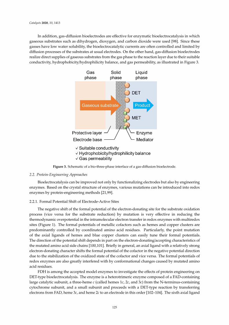
Catalysts 2020, 10, 1413
In addition, gas-diffusion bioelectrodes are effective for enzymatic bioelectrocatalysis in whichgaseous substrates such as dihydrogen, dioxygen, and carbon dioxide were used [98]. Since thesegasses have low water solubility, the bioelectrocatalytic currents are often controlled and limited bydiffusion processes of the substrates at usual electrodes. On the other hand, gas-diffusion bioelectrodesrealize direct supplies of gaseous substrates from the gas phase to the reaction layer due to their suitableconductivity, hydrophobicity/hydrophilicity balance, and gas permeability, as illustrated in Figure 3.
–
Figure 3. Schematic of a bio-three-phase interface of a gas-diffusion bioelectrode.
2.2. Protein-Engineering Approaches
Bioelectrocatalysis can be improved not only by functionalizing electrodes but also by engineeringenzymes. Based on the crystal structure of enzymes, various mutations can be introduced into redoxenzymes by protein-engineering methods [21,99].
2.2.1. Formal Potential Shift of Electrode-Active Sites
The negative shift of the formal potential of the electron-donating site for the substrate oxidationprocess (vice versa for the substrate reduction) by mutation is very effective in reducing thethermodynamic overpotential in the intramolecular electron transfer in redox enzymes with multiredoxsites (Figure 1). The formal potentials of metallic cofactors such as hemes and copper clusters arepredominantly controlled by coordinated amino acid residues. Particularly, the point mutationof the axial ligands of hemes and blue copper clusters can easily tune their formal potentials.The direction of the potential shift depends in part on the electron-donating/accepting characteristics ofthe mutated amino acid side chains [100,101]. Briefly in general, an axial ligand with a relatively strongelectron-donating character shifts the formal potential of the cofactor in the negative potential directiondue to the stabilization of the oxidized state of the cofactor and vice versa. The formal potentials ofredox enzymes are also greatly interfered with by conformational changes caused by mutated aminoacid residues.
FDH is among the accepted model enzymes to investigate the effects of protein engineering onDET-type bioelectrocatalysis. The enzyme is a heterotrimeric enzyme composed of a FAD-containinglarge catalytic subunit, a three-heme c (called hemes 1c, 2c, and 3c) from the N-terminus-containingcytochrome subunit, and a small subunit and proceeds with a DET-type reaction by transferringelectrons from FAD, heme 3c, and heme 2c to an electrode in this order [102–104]. The sixth axial ligand
125

Catalysts 2020, 10, 1413
(methionine 450) of heme 2c was then replaced by glutamine with electron-donating characteristicsto shift the formal potential of heme 2c in the direction of the negative potential. The FDH variant(M450Q_FDH) provided DET-type bioelectrocatalytic waves for the oxidation of fructose at a half-wavepotential of approximately 50 mV, more negative than that of the recombinant native enzyme, as shownin Figure 4A [103,104]. A drastic change in the limiting catalytic current was not observed. This suggeststhat the rate constant of the intramolecular electron transfer from FAD to heme 2c in the enzymeis sufficiently large compared with the catalytic reaction rate constant at the catalytic center (FAD);the latter predominantly determines kc,DET in Equation (5).
type bioelectrocatalysis of “substrate oxidation ” (
ΓΓ
Δ
Δ
Γ Δ
E
j Increase in ΓE,eff
E
j
Potential shift
E
j Increase in kc,DET
Potential shift
BA C
Figure 4. Illustrative drawing of the effect of mutations on steady-state bioelectrocatalytic wavesin the DET-type bioelectrocatalysis of “substrate oxidation”. (A) Negative potential shift in theformal potential of the electron-donating site of the DET-type redox enzyme reactions, in which theintramolecular electron transfer is not the rate-determining process in the enzymatic catalytic reaction.(B) Positive potential shift in the formal potential of the electron-donating site of the DET-type redoxenzyme reactions, in which the intramolecular electron transfer is the rate-determining process in theenzymatic catalytic reaction, and (C) downsizing without any change in the intrinsic enzymatic activity.
On the other hand, such replacement is also effective for the axial ligand of the electron-acceptingsite (type I blue copper site) to negatively shift the formal potential. Replacement of the axial ligandmethionine 467 in the type I copper site of BOD with glutamine (M467Q_BOD) caused a large negativeshift (approximately 0.23 V) in the half-wave potential of the DET-type bioelectrocatalytic waves.This means an increase in the overpotential in the intramolecular electron transfer in the enzyme.Fortunately, in this case, the catalytic limiting current density increased compared with that of therecombinant native BOD, as shown in Figure 4B (note here that the catalytic wave in Figure 4B isillustrated for substrate oxidation) [105]. Most probably, the catalytic rate constant of the dioxygenreduction at the type II/III catalytic site is sufficiently large compared with the rate constant of theintramolecular electron transfer from the electron-accepting type I site to the dioxygen-reducing typeII/III site; the intramolecular electron transfer is the rate-determining step to determine kc,DET inEquation (5). In addition, the electron transfer kinetics seems to obey the linear free energy relationship(LFER); the increase in the formal potential difference between type I and II/III sites (that is, the drivingforce of the reaction) results in an increase in the intramolecular electron transfer rate constant.
Even in MET-type bioelectrocatalysis, the redox potential shift of the electron-donating site(for substrate oxidation) can tune the overpotential in the intramolecular electron transfer process.In addition, the intermolecular electron transfer kinetics between the electron-donating site in the enzymeand the mediator seem to be improved in theory by the potential shift mutation of the electron-donatingsite (for substrate oxidation) on the basis of the concept of LFER, though there is no report on thismatter. Therefore, the mutational tuning of the redox potential of the electron-donating site (for substrateoxidation) may also expand the strategy of the mediator selection for MET-type bioelectrocatalysis.
2.2.2. Downsizing
As described by Equation (5), an increase in ΓE,eff is essential to getting a large bioelectrocatalyticcurrent density. ΓE,eff can be then increased by downsizing enzymes from which the regions not deeplyinvolved in bioelectrocatalysis are deleted, as shown in Figure 4C.
126

Catalysts 2020, 10, 1413
Downsizing effects were also investigated in FDH. Heme 1c of FDH is suggested to be uninvolved inthe DET-type reaction, and the downsized FDH without the heme 1c region (∆1c_FDH) showed a largerDET-type bioelectrocatalytic current density than the recombinant native FDH (r_FDH) [104,106].In addition, a further downsized FDH without the heme 1c and 2c regions (∆1c2c_FDH) alsoshowed DET-type bioelectrocatalytic activity with reduced overpotential due to direct electricalcommunication between an electrode and heme 3c with the most negative formal potential [104,107].However, whereas ΓE,eff was suggested to be increased, jcat of ∆1c2c_FDH was as large as that ofr_FDH (smaller than that of ∆1c_FDH), which seemed to be ascribed to a decrease in enzymatic activity(≈ kc,DET) of ∆1c2c_FDH by excessive deletion [104,107]. Thus, it is important to avoid conformationalchanges due to deletion and retain enzymatic activity as high as possible. Furthermore, a doublemutant of downsizing and the potential shift (M450Q∆1c_FDH) accomplished both an increase in jcat
and an overpotential reduction [104,108,109].
2.2.3. Surface Amino Acid Mutation
It is desirable to tightly immobilize enzymes on electrodes in both DET- and MET-type bioelectrocatalyticreactions. Cross-linkers such as glutaraldehyde [110], carbodiimide [29], and maleimide [71] are oftenused for the covalent immobilization of enzymes [111]. The mutation of amino acid residue(s) locatedon the enzyme surface also enhances the cross-coupling reactions and can control the orientation of theenzyme on a suitable electrode surface. For example, cysteine introduction onto the enzyme surfaceclose to the electrode-active site can increase the enzyme orientations suitable for DET-type reactions atthiol- and maleimide-functionalized electrodes by forming (di)sulfide bonds. Holland et al. reportedDET-type bioelectrocatalysis of cysteine-introduced GOD conjugated with maleimide-modified goldnanoparticles on a gold electrode [71]. In addition, Ferapontova et al. revealed that cysteine mutationof HRP was effective for its orientation on gold electrodes to improve DET-type bioelectrocatalyticproperties [112].
2.2.4. Fusion Protein
DET-type bioelectrocatalysis is sometimes achieved by introducing an electrode-active domaininto a native enzyme. Cellobiose dehydrogenase (CDH) is often used as a model of DET-typefusion enzymes. CDH has two domains: a larger catalytic dehydrogenase domain and a smallerelectrode-active cytochrome domain. The domains are linked by a flexible polypeptide [113]. The fusedcytochrome domain mediates the electron transfer from the catalytic domain to electrodes, such asa “built-in mediator.” Utilizing the fusion protein-engineering methods, DET-type reactions byFAD-GDH [114,115], pyrroloquinoline quinone (PQQ)-dependent GDH [116], and flavodoxin [117]were reported.
3. Novel Bioelectrochemical System
Major applications in enzymatic bioelectrocatalysis are biosensors and biofuel cells. Biosensors areutilized as analytical devices for food analyses and clinical diagnoses [6–14], and biofuel cellsare ecofriendly energy conversion devices in which the chemical energy of fuels is electricallyextracted [11,15–21]. The characteristics, properties, and progress of these devices were previouslysummarized in the corresponding reviews cited above. Recently, in addition, novel methods forbioelectrocatalytic applications are reported as described below.
3.1. Biosupercapacitor
Biosupercapacitors are self-powered energy storage devices using bioelectrocatalysis to chargecapacitors [22]. In the biosupercapacitor, electric power is generated by a biocathode and/or a bioanode.The fundamental difference between biosupercapacitors and biofuel cells is the separation of the outputof the current and bioelectrocatalytic reaction. Expressed as an equivalent circuit, a biosupercapacitor isa biofuel cell and a capacitor connected in parallel. Regardless of the connection to the external circuit,
127

Catalysts 2020, 10, 1413
the bioelectrocatalytic reaction injects the electrons into the supercapacitor. When the external circuitrequires the current, the biosupercapacitor provides the current. An advantage of biosupercapacitorsis that a large current can flow even if the kinetics of the bioelectrocatalysis is inadequate. Additionally,biosupercapacitors are compatible with the self-powered biosensors [118,119].
The supercapacitor is classified into two types: the electrical double layer capacitor (EDLC) andthe electrochemical pseudocapacitor (EPC). EDLC is the electrode with a large surface area, the electriccharge is accumulated in the electrical double layer at the interface between the electrode and theelectrolyte solution. EPC is constructed with the electrode and the reversible electrode-active redoxspecies. The externally applied voltage shifts the electrode potential at the anode and a cathode in thenegative and positive directions, respectively, as expressed in the Nernst equation.
Generally, redox-active species are immobilized at the electrode surface. Since an electrochemicalcapacitor contains two electrodes and an electrolyte solution, electrochemical capacitors are alsoclassified into three types: EDLC-EDLC, EPC-EPC, and EDLC-EPC [120]. Therefore, biosupercapacitorsare basically classified into the three types. The EDLC is compatible with DET-type bioelectrocatalysisand MET-type bioelectrocatalysis is adapted to the EPC. In order to avoid the mixing of mediators,EPCs in the biosupercapacitor are frequently employed redox polymers as mediators to immobilize atthe electrode surface.
Electrochemically inactive porous electrodes in an electrolyte solution work as the EDLC.Additionally, DET-type bioelectrocatalytic activity is required for the EDLC-type biosupercapacitor.The reported materials suitable for the EDLC and the bioelectrode are carbon nanotubes [121–123],porous gold [124], gold nanoparticles [125], and indium tin oxide nanoparticles [126]. Porous electrodesoften show high activity for DET-type bioelectrocatalysis. Therefore, it is considered that theinvestigation of bioelectrodes for DET-type biofuel cells will be directly useful for the biosupercapacitor.
The separation of oxidant and reductant is important in EPCs, since the mixing of the twospecies causes the cross-reaction discharge. Therefore, in the case of biosupercapacitors using theMET-type bioelectrocatalysis, careful attention is required to head off the outflow of mediators.As mentioned above, redox polymers are employed as mediators in biosupercapacitors [42,127–129].The immobilization of redox-active proteins at the electrode surface has been investigated for theimprovement of the capacitance of EPC-type biosupercapacitors [130,131].
3.2. Bioelectrosynthesis
Bioelectrosynthesis is a coined term for the generation of renewable energy resources and useful organiccompounds [132]. Particularly, stereoselective enzymatic bioelectrocatalysis is desirable for pharmaceuticaluse because pharmaceutical precursors are required to have high enantiomeric purity [133,134].
Some useful compounds are reduced forms of redox couples, and reductive conversion is often theopposite reaction in metabolisms in vivo. In bioelectrocatalysis, on the other hand, several enzymes canproceed bidirectional electroenzymatic reactions (both the oxidation and reduction of substrate redoxcouples), in which the driving forces are reversed depending on pH. For example, H2ase [135,136],formate dehydrogenase (FoDH) [75,136–139], carbon monoxide dehydrogenase (CODH) [136],diaphorase (DI) [136,140], ferredoxine-NADP+ reductase (FNR) [28], and some NAD(P)+-dependentdehydrogenases [28,140–143] show bidirectional bioelectrocatalytic activities. The characteristic ofthese enzymes is that the formal potentials of the substrate redox couples and the cofactors in theenzymes are relatively close to each other, and thus, the small uphill intramolecular electron transferproceeds in acceptable velocity [34]. Bidirectional bioelectrocatalysis is a key reaction for constructingbioelectrochemical energy/compound conversion systems.
There are mainly two types of bioelectrosynthetic systems: a fuel-cell-type [142,144–149] and anelectrolysis-type [28,53,141,143,150–154]. The former realizes a spontaneous production of compoundswithout any external power supplies, whereas the latter proceeds relatively rapid reactions due toan optimally controlled electrode potential to realize diffusion-controlled conditions. We will showexamples of bioelectrosyntheses in the following sections.
128

Catalysts 2020, 10, 1413
3.2.1. Dihydrogen Production
Dihydrogen (H2) is a typical clean energy source that emits no harmful products in combustion.H2ase, a common anodic bioelectrocatalyst for H2 oxidation, can also catalyze proton (H+)reduction [135,136]. The reversible reaction is biased to favor H2 oxidation, and it is difficult torealize a large cathodic current density of H+ reduction in DET-type systems. H+ reduction witha large current density was achieved in a H2ase MET-type system using a viologen polymer as a redoxmediator [53]. On the other hand, a dual DET-type bioelectrocatalytic water–gas shift reaction usingCODH and H2ase (H2O + CO → H2 + CO2) was reported [146]. The system requires no externalpower supplies.
3.2.2. Formate Production
Formate/formic acid (HCOO−/HCOOH) is a liquid energy resource with a high energy density dueto its property of being completely oxidized to CO2. FoDH interconverts HCOO− and CO2 both in DET-and MET-type systems [75,136–139]. Sakai et al. reported efficient bioelectrocatalytic CO2 reductionusing FoDH from Methylobacterium extorquens AM1 and a synthesized redox mediator with the lowformal potential on a gas-diffusion electrode [150]. In addition, spontaneous interconversion betweenHCOO− and H2 without any external power supplies was achieved by coupling two bioelectrocatalysesby FoDH and H2ase (H2 + CO2 → HCOO− + H+) [147]. The direction of the reaction is controlled bypH- and concentration-dependent equilibrium potentials of the 2H+/H2 and CO2/HCOO− couples.
3.2.3. Ammonia Production
Ammonia (NH3) is an industrially beneficial compound used as a chemical raw material, fuel,hydrogen storage, and so on [155,156]. Nitrogenase (N2ase) is an enzyme that catalyzes a nitrogenfixation from dinitrogen (N2) to NH3 using adenosine triphosphate (ATP) hydrolysis energy inmicroorganisms under an anaerobic condition [155,156]. Particularly, molybdenum-dependent N2ase,which comprises a catalytic MoFe protein and a homodimeric MgATP-binding Fe protein, has beenmainly investigated in view of bioelectrocatalysis [156,157]. N2ase reduces not only N2 but also nitrite(NO2
−) and azide (N3−) to NH3 on electrodes, and the MoFe protein disassociated with the Fe protein
shows the DET-type bioelectrocatalytic activity without ATP [154]. Furthermore, ATP-independent NH3
bioelectrosynthesis was improved in the MET-type system using N2ase and a cobaltocene-functionalizedpolymeric mediator [51]. On the other hand, Milton et al. reported a NH3-producing H2/N2 biofuel cellusing MET-type reactions of N2ase and H2ase [148]. If the fatal weakness of N2ase, a lack of oxygentolerance, is improved, N2ase will be expected to have further industrial applications.
3.2.4. NAD(P)+-Dependent Bioelectrosynthesis
Nicotinamide coenzymes (NAD(P)+/NAD(P)H) play essential roles in the function of manyNAD(P)+-dependent dehydrogenases, but the dehydrogenases need suitable redox mediators insingle-step enzymatic bioelectrocatalysis because the NAD(P)+/NAD(P)H interconversion requireshigh overpotentials at conventional electrodes due to its hydride ion-transfer-type characteristicsthat are completely different from two-step single-electron transfer-type electrode reactions. On theother hand, the introduction of dipaphorase (DI) and FNR, which are bioelectrocatalysts for theinterconversion of the NAD+/NADH and NADP+/NADPH redox couples, respectively, can realizevarious NAD(P)+-dependent bioelectrocatalyses with relatively low overpotentials [28,140].
The bienzymatic MET-type system of NAD+-dependent dehydrogenase and DI is kineticallyand thermodynamically investigated and often applied to biosensors [40,140,158,159]. In addition,the coulometric electrooxidation of organic substances was achieved in the same multienzymaticsystem mimicking the tricarboxylic acid cycle [151]. On the other hand, reductive production ofchiral compounds is useful in many cases. Minteer’s group reported various NAD+-dependentmultienzymatic cascade reactions for generating chiral compounds, as shown in Figure 5 [149,152,153].
129

Catalysts 2020, 10, 1413
They also demonstrated the bioelectrosynthesis of polyhydroxybutarate with NADH regenerationby DI [143].
β
]. Particularly, Armstrong’s group investigated NADP
B
A
Figure 5. Schematic representation of multienzymatic bioelectrosyntheses of (A) chiral amineand (B) β-hydroxy nitrile. Abbreviations; DH: diaphorase, AdhS: alcohol dehydrogenase, HHDH:halohydrin dehalogenase, COBE: 4-chloroacetoacetate, CHBE: 4-chloro-3-hydroxybutanoate, CHCN:ethyl-4-cyano-3-hydroxybutyrate. Reprinted from Ref. [152,153], Copyright (2019,2020), with permissionfrom ACS Publications.
In an NADP+-dependent system, on the other hand, FNR showed DET-type bioelectrocatalyticactivity [28]. Particularly, Armstrong’s group investigated NADP+-dependent bienzymaticbioelectrocatalysis using FNR. They reported the reductive carboxylation of pyruvate to malateby malate dehydrogenase [142], reductive amination of 2-oxoglutamate to l-glutamate by glutamate
130

Catalysts 2020, 10, 1413
dehydrogenase [28,144], and enantioselective interconversion of the secondary alcohol/ketone coupleby engineered alcohol dehydrogenase [141,145].
3.3. Photo-Bioelectrocatalysis
Photo-bioelectrocatalysis enables reducing a substrate with a lower formal potential byoxidizing a sacrificial reagent with a higher formal potential, which cannot spontaneously occurwithout solar energy. Electrons that photosensitizers accept from sacrificial reagents are excitedby solar energy, and the electric potential is shifted in the negative direction, corresponding tothe wavelength of the adsorbed light. The electrons are then donated to substrates via enzymesand mediators, as shown in Figure 6. Photosensitizers such as TiO2 [80,81,160–162], PbS quantumdots [162], silver nanoclusters [80,81], and organic dyes [160,161] are incorporated in anodes oftransparent electrode bases (ITO in general) with or without other catalysts. Particularly, in addition,the photosystem II (PSII) complex in the thylakoid membrane of cyanobacteria and higher plants isoften used as a water-splitting anodic photo-bioelectrocatalyst [129,160–181]. Biosolar cells [163–173]and solar biosupercapacitors [129,164,168,174], using PSII/I, thylakoid membranes, or cyanobacteria inanodes, and BOD or laccase in cathodes, realized the conversion from solar to electric energy withoutany sacrificial reagents in total. On the other hand, photo-bioelectrosyntheses, also called artificialphotosyntheses, are reported. H2 is generated by H2ase [80,160], and CO2 is fixed by FoDH andCODH [81,161]. These cathodic reactions proceed at quite low potentials. Thus, in order to improvethe Faradaic efficiency of these photo-bioelectrosyntheses, it is essential to reduce electron leakage todissolved oxygen as much as possible, especially when oxygen-generating photosynthetic proteins ororganisms are used as anodic photo-bioelectrocatalysts.
–
– –
——
E
Reductant
Enzyme
Photosensitizer
Substrate
Anode Cathode
(Mediator)hνProduct
e
e
e
Figure 6. Potential profile in a simple photo-bioelectrocatalytic system.
4. Conclusions
Enzymatic bioelectrocatalysis is advancing day by day due to the finding of novel enzymesand the development of electrode nanomaterials. However, there are still issues to be solved suchas thermostability, oxygen tolerance, and physical fragility, compared to inorganic electrocatalysts.Further understanding and consideration of the electrode interface and the interaction betweenenzymes and electrodes from kinetics and thermodynamics are required. Other perspectives, such asthe improvement of redox enzymes by protein-engineering approaches, the selection of electrodematerials and mediators (including redox hydrogels), the immobilization of enzymes, and the layoutof electrodes will be also required for the improvement of present bioelectrochemical devices.
131

Catalysts 2020, 10, 1413
Author Contributions: Conceptualization, T.A. and K.K.; data curation, T.A.; writing—original draft preparation,T.A.; writing—review and editing, Y.K., O.S. and K.K.; supervision, K.K.; project administration, K.K. All authorshave read and agreed to the published version of the manuscript.
Funding: This research received no external funding.
Conflicts of Interest: The authors declare no conflict of interest.
References
1. Bartlett, P.N. Bioelectrochemistry: Fundamentals, Experimental Techniques and Applications; John Wiley & Sons:Chichester, UK, 2008.
2. Wilson, G.S.; Johnson, M.A. In-Vivo Electrochemistry: What Can We Learn about Living Systems? Chem. Rev.
2008, 108, 2462–2481. [CrossRef]3. Heller, A. Electrical connection of enzyme redox centers to electrodes. J. Phys. Chem. 1992, 96, 3579–3587.
[CrossRef]4. Kano, K. Fundamentals and Applications of Redox Enzyme-functionalized Electrode Reactions. Electrochemistry
2019, 87, 301–311. [CrossRef]5. Scheller, F.; Kirstein, D.; Schubert, F.; Wollenberger, U.; Ollson, B.; Gorton, L.; Johansson, G. Enzyme electrodes
and their application. Philos. Trans. R. Soc. B Biol. Sci. 1987, 316, 85–94. [CrossRef]6. Thevenot, D.R.; Tóth, K.; Durst, R.A.; Wilson, G.S. Electrochemical Biosensors: Recommended Definitions
and Classification. Pure Appl. Chem. 1999, 71, 2333–2348. [CrossRef]7. Martinkova, P.; Kostelnik, A.; Valek, T.; Pohanska, M. Main streams in the Construction of Biosensors and
Their Applications. Int. J. Electrochem. Sci. 2017, 12, 7386–7403. [CrossRef]8. Bollella, P.; Gorton, L. Enzyme based amperometric biosensors. Curr. Opin. Electrochem. 2018, 10, 157–173.
[CrossRef]9. Kucherenko, I.S.; Soldatkin, A.; Kucherenko, D.Y.; Soldatkina, O.V.; Dzyadevych, S.V. Advances in
nanomaterial application in enzyme-based electrochemical biosensors: A review. Nanoscale Adv. 2019, 1,4560–4577. [CrossRef]
10. Nguyen, H.H.; Lee, S.H.; Lee, U.J.; Fermin, C.D.; Kim, M. Immobilized Enzymes in Biosensor Applications.Materials 2019, 12, 121. [CrossRef]
11. Pinyou, P.; Blay, V.; Muresan, L.M.; Noguer, T. Enzyme-modified electrodes for biosensors and biofuel cells.Mater. Horiz. 2019, 6, 1336–1358. [CrossRef]
12. Bollella, P.; Katz, E. Enzyme-Based Biosensors: Tackling Electron Transfer Issues. Sensors 2020, 20, 3517.[CrossRef] [PubMed]
13. Willner, I.; Katz, E.; Willner, B. Electrical contact of redox enzyme layers associated with electrodes: Routes toamperometric biosensors. Electroanalysis 1997, 9, 965–977. [CrossRef]
14. Scheller, F.; Schubert, F.; Pfeiffer, D.; Hintsche, R.; Dransfeld, I.; Renneberg, R.; Wollenberger, U.; Riedel, K.;Pavlova, M.; Kühn, M.; et al. Research and development of biosensors. A review. Analyst 1989, 114, 653–662.[CrossRef] [PubMed]
15. De Poulpiquet, A.; Ranava, D.; Monsalve, K.; Giudici-Orticoni, M.-T.; Lojou, E. Biohydrogen for a NewGeneration of H2/O2Biofuel Cells: A Sustainable Energy Perspective. ChemElectroChem 2014, 1, 1724–1750.[CrossRef]
16. Barton, S.C.; Gallaway, J.; Atanassov, P. Enzymatic Biofuel Cells for Implantable and Microscale Devices.Chem. Rev. 2004, 104, 4867–4886. [CrossRef]
17. Cracknell, J.A.; Vincent, K.A.; Armstrong, F.A. Enzymes as Working or Inspirational Electrocatalysts for FuelCells and Electrolysis. Chem. Rev. 2008, 108, 2439–2461. [CrossRef]
18. Meredith, M.T.; Minteer, S.D. Biofuel Cells: Enhanced Enzymatic Bioelectrocatalysis. Annu. Rev. Anal. Chem.
2012, 5, 157–179. [CrossRef]19. Mazurenko, I.; De Poulpiquet, A.; Lojou, E. Recent developments in high surface area bioelectrodes for
enzymatic fuel cells. Curr. Opin. Electrochem. 2017, 5, 74–84. [CrossRef]20. Mano, N.; De Poulpiquet, A. O2Reduction in Enzymatic Biofuel Cells. Chem. Rev. 2018, 118, 2392–2468.
[CrossRef]21. Xiao, X.; Xia, H.-Q.; Wu, R.; Bai, L.; Yan, L.; Magner, E.; Cosnier, S.; Lojou, E.; Zhu, Z.; Liu, A. Tackling the
Challenges of Enzymatic (Bio)Fuel Cells. Chem. Rev. 2019, 119, 9509–9558. [CrossRef]
132

Catalysts 2020, 10, 1413
22. Shleev, S.; González-Arribas, E.; Falk, M. Biosupercapacitors. Curr. Opin. Electrochem. 2017, 5, 226–233.[CrossRef]
23. Krieg, T.; Sydow, A.; Schröder, U.; Schrader, J.; Holtmann, D. Reactor concepts for bioelectrochemicalsyntheses and energy conversion. Trends Biotechnol. 2014, 32, 645–655. [CrossRef] [PubMed]
24. Paddock, R.M.; Bowden, E.F. Electrocatalytic reduction of hydrogen peroxide via direct electron transferfrom pyrolytic graphite electrodes to irreversibly adsorbed cytochrome c peroxidase. J. Electroanal. Chem.
Interfacial Electrochem. 1989, 260, 487–494. [CrossRef]25. Xia, H.; Kitazumi, Y.; Shirai, O.; Kano, K. Direct Electron Transfer-type Bioelectrocatalysis of Peroxidase at
Mesoporous Carbon Electrodes and Its Application for Glucose Determination Based on Bienzyme System.Anal. Sci. 2017, 33, 839–844. [CrossRef] [PubMed]
26. Gu, T.; Wang, J.; Xia, H.; Wang, S.; Yu, X. Direct Electrochemistry and Electrocatalysis of HorseradishPeroxidase Immobilized in a DNA/Chitosan-Fe3O4 Magnetic Nanoparticle Bio-Complex Film. Materials
2014, 7, 1069–1083. [CrossRef]27. Sakai, K.; Kitazumi, Y.; Shirai, O.; Kano, K. Nanostructured Porous Electrodes by the Anodization of Gold for
an Application as Scaffolds in Direct-electron-transfer-type Bioelectrocatalysis. Anal. Sci. 2018, 34, 1317–1322.[CrossRef]
28. Siritanaratkul, B.; Megarity, C.F.; Roberts, T.G.; Samuels, T.O.M.; Winkler, M.; Warner, J.H.; Happe, T.;Armstrong, F.A. Transfer of photosynthetic NADP+/NADPH recycling activity to a porous metal oxide forhighly specific, electrochemically-driven organic synthesis. Chem. Sci. 2017, 8, 4579–4586. [CrossRef]
29. Yehezkeli, O.; Raichlin, S.; Tel-Vered, R.; Kesselman, E.; Danino, D.; Willner, I. Biocatalytic Implant of PtNanoclusters into Glucose Oxidase: A Method to Electrically Wire the Enzyme and to Transform It from anOxidase to a Hydrogenase. J. Phys. Chem. Lett. 2010, 1, 2816–2819. [CrossRef]
30. Trifonov, A.; Stemmer, A.; Tel-Vered, R. Enzymatic self-wiring in nanopores and its application in directelectron transfer biofuel cells. Nanoscale Adv. 2019, 1, 347–356. [CrossRef]
31. Adachi, T.; Fujii, T.; Honda, M.; Kitazumi, Y.; Shirai, O.; Kano, K. Direct electron transfer-typebioelectrocatalysis of FAD-dependent glucose dehydrogenase using porous gold electrodes and enzymaticallyimplanted platinum nanoclusters. Bioelectrochemistry 2020, 133, 107457. [CrossRef]
32. Léger, C.; Bertrand, P. Direct Electrochemistry of Redox Enzymes as a Tool for Mechanistic Studies. Chem. Rev.
2008, 108, 2379–2438. [CrossRef] [PubMed]33. Milton, R.D.; Minteer, S.D. Direct enzymatic bioelectrocatalysis: Differentiating between myth and reality.
J. R. Soc. Interface 2017, 14, 20170253. [CrossRef] [PubMed]34. Adachi, T.; Kitazumi, Y.; Shirai, O.; Kano, K. Direct Electron Transfer-Type Bioelectrocatalysis of Redox
Enzymes at Nanostructured Electrodes. Catalysts 2020, 10, 236. [CrossRef]35. Kitazumi, Y.; Shirai, O.; Kano, K. Catalyst Materials for Bioelectrochemical Systems: Fundamentals and Applications;
ACS Publications: Washington, DC, USA, 2020; pp. 147–163.36. Gorton, L.; Jönsson-Pettersson, G.; Csöregi, E.; Johansson, K.; Domínguez, E.; Marko-Varga, G. Amperometric
biosensors based on an apparent direct electron transfer between electrodes and immobilized peroxidases.Plenary lecture. Analyst 1992, 117, 1235–1241. [CrossRef]
37. Degani, Y.; Heller, A. Electrical communication between redox centers of glucose oxidase and electrodes viaelectrostatically and covalently bound redox polymers. J. Am. Chem. Soc. 1989, 111, 2357–2358. [CrossRef]
38. Ohara, T.J.; Rajagopalan, R.; Heller, A. Glucose Electrodes Based on Cross-Linked (Os(bpy)2Cl)+/2+Complexed Poly(1-vinylimidazole) Films. Anal. Chem. 1993, 65, 3512–3517. [CrossRef]
39. Timur, S.; Yigzaw, Y.; Gorton, L. Electrical wiring of pyranose oxidase with osmium redox polymers.Sens. Actuators B Chem. 2006, 113, 684–691. [CrossRef]
40. Nikitina, O.; Shleev, S.; Gayda, G.; Demkiv, O.; Gonchar, M.; Gorton, L.; Csöregi, E.; Nistor, M. Bi-enzymebiosensor based on NAD+- and glutathione-dependent recombinant formaldehyde dehydrogenase anddiaphorase for formaldehyde assay. Sens. Actuators B Chem. 2007, 125, 1–9. [CrossRef]
41. Nieh, C.-H.; Kitazumi, Y.; Shirai, O.; Yamamoto, M.; Kano, K. Potentiometric coulometry based on chargeaccumulation with a peroxidase/osmium polymer-immobilized electrode for sensitive determination ofhydrogen peroxide. Electrochem. Commun. 2013, 33, 135–137. [CrossRef]
42. Alsaoub, S.; Conzuelo, F.; Gounel, S.; Mano, N.; Schuhmann, W.; Ruff, A. Introducing PseudocapavitiveBioelectrodes into a Biofuel Cell/Biosupercapacitor Hybrid Device for Optimized Open Circuit Voltage.ChemElectroChem 2019, 6, 2080–2087. [CrossRef]
133

Catalysts 2020, 10, 1413
43. Tsujimura, S.; Takeuchi, S. Toward an Ideal Platform Structure Based on MgO-Templated Carbon for FlavinAdenine Dinucleotide-Dependent Glucose Dehydrogenase-Os Polymer-Hydrogel Electrodes. Electrochim. Acta
2020, 343, 136110. [CrossRef]44. Xiao, X.; Conghaile, P.Ó.; Leech, D.; Magner, E. Use of Polymer Coatings to Enhance the Response of
Redox-Polymer-Mediated Electrodes. ChemElectroChem 2019, 6, 1344–1349. [CrossRef]45. Cenas, N.K.; Pocius, A.K.; Kulys, J.J. Electron Exchange between Flavin- and Heme-Containing Enzymes
and Electrodes Modified by Redox Polymers. Bioelectrochem. Bioenerg. 1983, 11, 61–73. [CrossRef]46. Nieh, C.-H.; Kitazumi, Y.; Shirai, O.; Kano, K. Sensitive d-amino acid biosensor based on oxidase/peroxidase
system mediated by pentacyanoferrate-bound polymer. Biosens. Bioelectron. 2013, 47, 350–355. [CrossRef][PubMed]
47. Nieh, C.-H.; Tsujimura, S.; Shirai, O.; Kano, K. Amperometric biosensor based on reductive H2O2 detectionusing pentacyanoferrate-bound polymer for creatinine determination. Anal. Chim. Acta 2013, 767, 128–133.[CrossRef]
48. Merchant, S.A.; Meredith, M.T.; Tran, T.O.; Brunski, D.B.; Johnson, M.B.; Glatzhofer, D.T.; Schmidtke, D.W.Effect of Mediator Spacing on Electrochemical and Enzymatic Response of Ferrocene Redox Polymers. J. Phys.
Chem. C 2010, 114, 11627–11634. [CrossRef]49. Tran, T.O.; Lammert, E.G.; Chen, J.; Merchant, S.A.; Brunski, D.B.; Keay, J.C.; Johnson, M.B.; Glatzhofer, D.T.;
Schmidtke, D.W. Incorporation of Single-Walled Carbon Nanotubes into Ferrocene-Modified LinearPolyethylenimine Redox Polymer Films. Langmuir 2011, 27, 6201–6210. [CrossRef]
50. Milton, R.D.; Abdellaoui, S.; Khadka, N.; Dean, D.R.; Leech, D.; Seefeldt, L.C.; Minteer, S.D. Nitrogenasebioelectrocatalysis: Heterogeneous ammonia and hydrogen production by MoFe protein. Energy Environ. Sci.
2016, 9, 2550–2554. [CrossRef]51. Lee, Y.S.; Yuan, M.; Cai, R.; Lim, K.; Minteer, S.D. Nitrogenase Bioelectrocatalysis: ATP-Independent
Ammonia Production Using a Redox Polymer/MoFe Protein System. ACS Catal. 2020, 10, 6854–6861.[CrossRef]
52. Eng, L.H.; Elmgren, M.; Komlos, P.; Nordling, M.; Lindquist, S.-E.; Neujahr, H.Y. Viologen-Based RedoxPolymer for Contacting the Low-Potential Redox Enzyme Hydrogenase at an Electrode Surface. J. Phys. Chem.
1994, 98, 7068–7072. [CrossRef]53. Shiraiwa, S.; So, K.; Sugimoto, Y.; Kitazumi, Y.; Shirai, O.; Kano, K. Reactivation of Standard (NiFe)-Hydrogenase and
Bioelectrochemical Catalysis of Proton Reduction and Hydrogen Oxidation in a Mediated-Electron-TransferSystem. Bioelectrochemistry 2018, 123, 156–161. [CrossRef]
54. Sakai, K.; Kitazumi, Y.; Shirai, O.; Kano, K. Bioelectrocatalytic formate oxidation and carbon dioxidereduction at high current density and low overpotential with tungsten-containing formate dehydrogenaseand mediators. Electrochem. Commun. 2016, 65, 31–34. [CrossRef]
55. Sakai, K.; Kitazumi, Y.; Shirai, O.; Takagi, K.; Kano, K. High-Power Formate/Dioxygen Biofuel Cell Based onMediated Electron Transfer Type Bioelectrocatalysis. ACS Catal. 2017, 7, 5668–5673. [CrossRef]
56. Kitazumi, Y.; Shirai, O.; Yamamoto, M.; Kano, K. Numerical simulation of diffuse double layer aroundmicroporous electrodes based on the Poisson–Boltzmann equation. Electrochim. Acta 2013, 112, 171–175.[CrossRef]
57. Serafin, V.; Hernández, P.; Agüí, L.; Yáñez-Sedeño, P.; Pingarrón, J.M. Electrochemical Biosensorfor Creatinine Based on the Immobilization of Creatininase, Creatinase and Sarcosine Oxidase ontoa Ferrocene/Horseradish Peroxidase/Gold Nanoparticles/Multi-Walled Carbon Nanotubes/Teflon CompositeElectrode. Electrochim. Acta 2013, 97, 175–183. [CrossRef]
58. Holzinger, M.; Le Goff, A.; Cosnier, S. Carbon nanotube/enzyme biofuel cells. Electrochim. Acta 2012, 82,179–190. [CrossRef]
59. Tominaga, M.; Sasaki, A.; Togami, M. Laccase Bioelectrocatalyst at a Steroid-Type Biosurfactant-ModifiedCarbon Nanotube Interface. Anal. Chem. 2015, 87, 5417–5421. [CrossRef]
60. Tsujimura, S.; Nishina, A.; Kamitaka, Y.; Kano, K. Coulometric d-Fructose Biosensor Based on Direct ElectronTransfer Using D-Fructose Dehydrogenase. Anal. Chem. 2009, 81, 9383–9387. [CrossRef]
61. Flexer, V.; Durand, F.; Tsujimura, S.; Mano, N. Efficient Direct Electron Transfer of PQQ-glucose Dehydrogenaseon Carbon Cryogel Electrodes at Neutral pH. Anal. Chem. 2011, 83, 5721–5727. [CrossRef]
62. Hamano, Y.; Tsujimura, S.; Shirai, O.; Kano, K. Micro-cubic monolithic carbon cryogel electrode for directelectron transfer reaction of fructose dehydrogenase. Bioelectrochemistry 2012, 88, 114–117. [CrossRef]
134

Catalysts 2020, 10, 1413
63. Murata, K.; Akatsuka, W.; Tsujimura, S. Bioelectrocatalytic Oxidation of Glucose on MgO-templatedMesoporous Carbon-modified Electrode. Chem. Lett. 2014, 43, 928–930. [CrossRef]
64. Mazurenko, I.; Clément, R.; Byrne-Kodjabachian, D.; de Poulpiquet, A.; Tsujimura, S.; Lojou, E. Pore SizeEffect of MgO-Templated Carbon on Enzymatic H2 Oxidation by the Hyperthermophilic Hydrogenase fromAquifex aeolicus. J. Electroanal. Chem. 2018, 812, 221–226. [CrossRef]
65. Takahashi, Y.; Wanibuchi, M.; Kitazumi, Y.; Shirai, O.; Kano, K. Improved direct electron transfer-typebioelectrocatalysis of bilirubin oxidase using porous gold electrodes. J. Electroanal. Chem. 2019, 843, 47–53.[CrossRef]
66. Mie, Y.; Yasutake, Y.; Ikegami, M.; Tamura, T. Anodized gold surface enables mediator-free and low-overpotentialelectrochemical oxidation of NADH: A facile method for the development of an NAD+-dependent enzymebiosensor. Sens. Actuators B Chem. 2019, 288, 512–518. [CrossRef]
67. Miyata, M.; Kitazumi, Y.; Shirai, O.; Kataoka, K.; Kano, K. Diffusion-limited biosensing of dissolved oxygenby direct electron transfer-type bioelectrocatalysis of multi-copper oxidases immobilized on porous goldmicroelectrodes. J. Electroanal. Chem. 2020, 860, 113895. [CrossRef]
68. Siepenkoetter, T.; Salaj-Kosla, U.; Xiao, X.; Belochapkine, S.; Magner, E. Nanoporous Gold Electrodes withTuneable Pore Sizes for Bioelectrochemical Applications. Electroanalysis 2016, 28, 2415–2423. [CrossRef]
69. Siepenkoetter, T.; Salaj-Kosla, U.; Magner, E. The Immobilization of Fructose Dehydrogenase on NanoporousGold Electrodes for the Detection of Fructose. ChemElectroChem 2017, 4, 905–912. [CrossRef]
70. Xiao, X.; Siepenkoetter, T.; Conghaile, P.Ó.; Leech, D.; Magner, E. Nanoporous Gold-Based Biofuel Cells onContact Lenses. ACS Appl. Mater. Interfaces 2018, 10, 7107–7116. [CrossRef] [PubMed]
71. Holland, J.T.; Lau, C.; Brozik, S.; Atanassov, P.; Banta, S. Engineering of Glucose Oxidase for Direct ElectronTransfer via Site-Specific Gold Nanoparticle Conjugation. J. Am. Chem. Soc. 2011, 133, 19262–19265. [CrossRef][PubMed]
72. Gutiérrez-Sánchez, C.; Pita, M.; Vaz-Domínguez, C.; Shleev, S.; De Lacey, A.L. Gold Nanoparticles asElectronic Bridges for Laccase-Based Biocathodes. J. Am. Chem. Soc. 2012, 134, 17212–17220. [CrossRef]
73. Suzuki, M.; Murata, K.; Nakamura, N.; Ohno, H. The Effect of Particle Size on the Direct Electron TransferReactions of Metalloproteins Using Au Nanoparticle-Modified Electrodes. Electrochemistry 2012, 80, 337–339.[CrossRef]
74. Monsalve, K.; Roger, M.; Gutierrez-Sanchez, C.; Ilbert, M.; Nitsche, S.; Byrne-Kodjabachian, D.; Marchi, V.;Lojou, E. Hydrogen bioelectrooxidation on gold nanoparticle-based electrodes modified by Aquifex aeolicushydrogenase: Application to hydrogen/oxygen enzymatic biofuel cells. Bioelectrochemistry 2015, 106, 47–55.[CrossRef] [PubMed]
75. Sakai, K.; Kitazumi, Y.; Shirai, O.; Takagi, K.; Kano, K. Direct Electron Transfer-Type Four-WayBioelectrocatalysis of CO2/Formate and NAD+/NADH Redox Couples by Tungsten-Containing FormateDehydrogenase Adsorbed on Gold Nanoparticle-Embedded Mesoporous Carbon Electrodes Modified with4-Mercaptopyridine. Electrochem. Commun. 2017, 84, 75–79. [CrossRef]
76. Takahashi, Y.; Kitazumi, Y.; Shirai, O.; Kano, K. Improved direct electron transfer-type bioelectrocatalysis ofbilirubin oxidase using thiol-modified gold nanoparticles on mesoporous carbon electrode. J. Electroanal. Chem.
2019, 832, 158–164. [CrossRef]77. Hitaishi, V.P.; Mazurenko, I.; Murali, A.V.; De Poulpiquet, A.; Coustillier, G.; Delaporte, P.; Lojou, E.
Nanosecond Laser–Fabricated Monolayer of Gold Nanoparticles on ITO for Bioelectrocatalysis. Front. Chem.
2020, 8, 431. [CrossRef]78. Kizling, M.; Dzwonek, M.; Wieckowska, A.; Bilewicz, R. Gold nanoparticles in bioelectrocatalysis—The role
of nanoparticle size. Curr. Opin. Electrochem. 2018, 12, 113–120. [CrossRef]79. Murata, K.; Suzuki, M.; Nakamura, N.; Ohno, H. Direct evidence of electron flow via the heme c group for
the direct electron transfer reaction of fructose dehydrogenase using a silver nanoparticle-modified electrode.Electrochem. Commun. 2009, 11, 1623–1626. [CrossRef]
80. Zhang, L.; Beaton, S.E.; Carr, S.B.; Armstrong, F.A. Direct visible light activation of a surfacecysteine-engineered [NiFe]-hydrogenase by silver nanoclusters. Energy Environ. Sci. 2018, 11, 3342–3348.[CrossRef]
81. Zhang, L.; Can, M.; Ragsdale, S.W.; Armstrong, F. Fast and Selective Photoreduction of CO2 to CO Catalyzedby a Complex of Carbon Monoxide Dehydrogenase, TiO2, and Ag Nanoclusters. ACS Catal. 2018, 8,2789–2795. [CrossRef]
135

Catalysts 2020, 10, 1413
82. Nakamura, R.; Kamiya, K.; Hashimoto, K. Direct electron-transfer conduits constructed at the interfacebetween multicopper oxidase and nanocrystalline semiconductive Fe oxides. Chem. Phys. Lett. 2010, 498,307–311. [CrossRef]
83. Kizling, M.; Rekorajska, A.; Krysinski, P.; Bilewicz, R. Magnetic-field-induced orientation of fructosedehydrogenase on iron oxide nanoparticles for enhanced direct electron transfer. Electrochem. Commun. 2018,93, 66–70. [CrossRef]
84. Rozniecka, E.; Jonsson-Niedziolka, M.; Sobczak, J.W.; Opallo, M. Mediatorless bioelectrocatalysis of dioxygenreduction at indium-doped tin oxide (ITO) and ITO nanoparticulate film electrodes. Electrochim. Acta 2011,56, 8739–8745. [CrossRef]
85. Willner, I.; Katz, E. Integration of Layered Redox Proteins and Conductive Supports for BioelectronicApplications. Angew. Chem. Int. Ed. 2000, 39, 1180–1218. [CrossRef]
86. Moser, C.C.; Keske, J.M.; Warncke, K.; Farid, R.S.; Dutton, P.L. Nature of biological electron transfer.Nat. Cell Biol. 1992, 355, 796–802. [CrossRef] [PubMed]
87. Marcus, R.; Sutin, N. Electron transfers in chemistry and biology. Biochim. Biophys. Acta BBA Rev. Bioenerg.
1985, 811, 265–322. [CrossRef]88. Marcus, R.A. Electron Transfer Reactions in Chemistry: Theory and Experiment (Nobel Lecture). Angew. Chem.
Int. Ed. 1993, 32, 1111–1121. [CrossRef]89. Sugimoto, Y.; Takeuchi, R.; Kitazumi, Y.; Shirai, O.; Kano, K. Significance of Mesoporous Electrodes for
Noncatalytic Faradaic Process of Randomly Oriented Redox Proteins. J. Phys. Chem. C 2016, 120, 26270–26277.[CrossRef]
90. Sugimoto, Y.; Kitazumi, Y.; Shirai, O.; Kano, K. Effects of Mesoporous Structures on Direct ElectronTransfer-Type Bioelectrocatalysis: Facts and Simulation on a Three-Dimensional Model of Random Orientationof Enzymes. Electrochemistry 2017, 85, 82–87. [CrossRef]
91. Hitaishi, V.P.; Clement, R.; Bourassin, N.; Baaden, M.; De Poulpiquet, A.; Sacquin-Mora, S.; Ciaccafava, A.;Sacquin-Mora, S. Controlling Redox Enzyme Orientation at Planar Electrodes. Catalysts 2018, 8, 192. [CrossRef]
92. Xia, H.; So, K.; Kitazumi, Y.; Shirai, O.; Nishikawa, K.; Higuchi, Y.; Kano, K. Dual gas-diffusion membrane-and mediatorless dihydrogen/air-breathing biofuel cell operating at room temperature. J. Power Sources 2016,335, 105–112. [CrossRef]
93. Adachi, T.; Kitazumi, Y.; Shirai, O.; Kawano, T.; Kataoka, K.; Kano, K. Effects of Elimination of α Helix Regionson Direct Electron Transfer-type Bioelectrocatalytic Properties of Copper Efflux Oxidase. Electrochemistry
2020, 88, 185–189. [CrossRef]94. Xia, H.; Kitazumi, Y.; Shirai, O.; Kano, K. Enhanced direct electron transfer-type bioelectrocatalysis of
bilirubin oxidase on negatively charged aromatic compound-modified carbon electrode. J. Electroanal. Chem.
2016, 763, 104–109. [CrossRef]95. So, K.; Kawai, S.; Hamano, Y.; Kitazumi, Y.; Shirai, O.; Hibi, M.; Ogawa, J.; Kano, K. Improvement of a direct
electron transfer-type fructose/dioxygen biofuel cell with a substrate-modified biocathode. Phys. Chem.
Chem. Phys. 2014, 16, 4823–4829. [CrossRef] [PubMed]96. Bollella, P.; Hibino, Y.; Kano, K.; Gorton, L.; Antiochia, R. Highly Sensitive Membraneless Fructose Biosensor
Based on Fructose Dehydrogenase Immobilized onto Aryl Thiol Modified Highly Porous Gold Electrode:Characterization and Application in Food Samples. Anal. Chem. 2018, 90, 12131–12136. [CrossRef] [PubMed]
97. Xia, H.; Hibino, Y.; Kitazumi, Y.; Shirai, O.; Kano, K. Interaction between d-fructose dehydrogenase andmethoxy-substituent-functionalized carbon surface to increase productive orientations. Electrochim. Acta
2016, 218, 41–46. [CrossRef]98. So, K.; Sakai, K.; Kano, K. Gas diffusion bioelectrodes. Curr. Opin. Electrochem. 2017, 5, 173–182. [CrossRef]99. Wong, T.S.; Schwaneberg, U. Protein engineering in bioelectrocatalysis. Curr. Opin. Biotechnol. 2003, 14,
590–596. [CrossRef]100. Battistuzzi, G.; Borsari, M.; Cowan, J.A.; Ranieri, A.; Sola, M. Control of cytochrome C redox potential: Axial
ligation and protein environment effects. J. Am. Chem. Soc. 2002, 124, 5315–5324. [CrossRef]101. Li, H.; Webb, S.P.; Ivanic, J.; Jensen, J.H. Determinants of the Relative Reduction Potentials of Type-1 Copper
Sites in Proteins. J. Am. Chem. Soc. 2004, 126, 8010–8019. [CrossRef]102. Kawai, S.; Yakushi, T.; Matsushita, K.; Kitazumi, Y.; Shirai, O.; Kano, K. The electron transfer pathway in
direct electrochemical communication of fructose dehydrogenase with electrodes. Electrochem. Commun.
2014, 38, 28–31. [CrossRef]
136

Catalysts 2020, 10, 1413
103. Hibino, Y.; Kawai, S.; Kitazumi, Y.; Shirai, O.; Kano, K. Mutation of heme c axial ligands in d-fructosedehydrogenase for investigation of electron transfer pathways and reduction of overpotential in directelectron transfer-type bioelectrocatalysis. Electrochem. Commun. 2016, 67, 43–46. [CrossRef]
104. Adachi, T.; Kaida, Y.; Kitazumi, Y.; Shirai, O.; Kano, K. Bioelectrocatalytic performance of d-fructosedehydrogenase. Bioelectrochemistry 2019, 129, 1–9. [CrossRef] [PubMed]
105. Kamitaka, Y.; Tsujimura, S.; Kataoka, K.; Sakurai, T.; Ikeda, T.; Kano, K. Effects of axial ligand mutation ofthe type I copper site in bilirubin oxidase on direct electron transfer-type bioelectrocatalytic reduction ofdioxygen. J. Electroanal. Chem. 2007, 601, 119–124. [CrossRef]
106. Hibino, Y.; Kawai, S.; Kitazumi, Y.; Shirai, O.; Kano, K. Construction of a protein-engineered variant of d-fructose dehydrogenase for direct electron transfer-type bioelectrocatalysis. Electrochem. Commun. 2017, 77,112–115. [CrossRef]
107. Kaida, Y.; Hibino, Y.; Kitazumi, Y.; Shirai, O.; Kano, K. Ultimate downsizing of d-fructose dehydrogenase forimproving the performance of direct electron transfer-type bioelectrocatalysis. Electrochem. Commun. 2019,98, 101–105. [CrossRef]
108. Hibino, Y.; Kawai, S.; Kitazumi, Y.; Shirai, O.; Kano, K. Protein-Engineering Improvement of Direct ElectronTransfer-Type Bioelectrocatalytic Properties of d-Fructose Dehydrogenase. Electrochemistry 2019, 87, 47–51.[CrossRef]
109. Kaida, Y.; Hibino, Y.; Kitazumi, Y.; Shirai, O.; Kano, K. Discussion on Direct Electron Transfer-TypeBioelectrocatalysis of Downsized and Axial-Ligand Exchanged Variants of d-Fructose Dehydrogenase.Electrochemistry 2020, 88, 195–199. [CrossRef]
110. Matsui, Y.; Hamamoto, K.; Kitazumi, Y.; Shirai, O.; Kano, K. Diffusion-Controlled Mediated ElectronTransfer-Type Bioelectrocatalysis Using Ultrathin-Ring and Microband Electrodes as Ultimate AmperometricGlucose Sensors. Anal. Sci. 2017, 33, 845–851. [CrossRef]
111. Mazurenko, I.; Hitaishi, V.P.; Lojou, E. Recent advances in surface chemistry of electrodes to promote directenzymatic bioelectrocatalysis. Curr. Opin. Electrochem. 2020, 19, 113–121. [CrossRef]
112. Ferapontova, E.E.; Schmengler, K.; Börchers, T.; Ruzgas, T.; Gorton, L. Effect of cysteine mutations on directelectron transfer of horseradish peroxidase on gold. Biosens. Bioelectron. 2002, 17, 953–963. [CrossRef]
113. Bollella, P.; Ludwig, R.; Gorton, L. Cellobiose dehydrogenase: Insights on the nanostructuration of electrodesfor improved development of biosensors and biofuel cells. Appl. Mater. Today 2017, 9, 319–332. [CrossRef]
114. Algov, I.; Grushka, J.; Zarivach, R.; Alfonta, L. Highly Efficient Flavin–Adenine Dinucleotide GlucoseDehydrogenase Fused to a Minimal Cytochrome C Domain. J. Am. Chem. Soc. 2017, 139, 17217–17220.[CrossRef] [PubMed]
115. Ito, K.; Okuda-Shimazaki, J.; Mori, K.; Kojima, K.; Tsugawa, W.; Ikebukuro, K.; Lin, C.-E.; La Belle, J.T.;Yoshida, H.; Sode, K. Designer fungus FAD glucose dehydrogenase capable of direct electron transfer.Biosens. Bioelectron. 2019, 123, 114–123. [CrossRef] [PubMed]
116. Okuda, J.; Sode, K. PQQ glucose dehydrogenase with novel electron transfer ability. Biochem. Biophys.
Res. Commun. 2004, 314, 793–797. [CrossRef]117. Gilardi, G.; Meharenna, Y.T.; Tsotsou, G.E.; Sadeghi, S.J.; Fairhead, M.; Giannini, S. Molecular Lego: Design
of molecular assemblies of P450 enzymes for nanobiotechnology. Biosens. Bioelectron. 2002, 17, 133–145.[CrossRef]
118. Hanashi, T.; Yamazaki, T.; Tsugawa, W.; Ferri, S.; Nakayama, D.; Tomiyama, M.; Ikebukuro, K.; Sode, K.BioCapacitor-A Novel Category of Biosensor. Biosens. Bioelectron. 2009, 24, 1837–1842. [CrossRef]
119. Conzuelo, F.; Ruff, A.; Schuhmann, W. Self-powered bioelectrochemical devices. Curr. Opin. Electrochem.
2018, 12, 156–163. [CrossRef]120. Falk, M.; Shleev, S. Hybrid Dual-functioning Electrodes for Combined Ambient Energy Harvesting and
Charge Strage: Towards Self-Powered Systems. Biosens. Bioelectron. 2019, 126, 275–291. [CrossRef]121. Agnès, C.; Holzinger, M.; Le Goff, A.; Reuillard, B.; Elouarzaki, K.; Tingry, S.; Cosnier, S. Supercapacitor/biofuel
cell hybrids based on wired enzymes on carbon nanotube matrices: Autonomous reloading after high powerpulses in neutral buffered glucose solutions. Energy Environ. Sci. 2014, 7, 1884–1888. [CrossRef]
122. Pankratov, D.; Blum, Z.; Suyatin, D.B.; Popov, V.O.; Shleev, S. Self-Charging Electrochemical Biocapacitor.ChemElectroChem 2013, 1, 343–346. [CrossRef]
123. Pankratov, D.; Blum, Z.; Shleev, S. Hybrid Electric Power Biodevices. ChemElectroChem 2014, 1, 1798–1807.[CrossRef]
137

Catalysts 2020, 10, 1413
124. Xiao, X.; Magner, E. A quasi-solid-state and self-powered biosupercapacitor based on flexible nanoporousgold electrodes. Chem. Commun. 2018, 54, 5823–5826. [CrossRef] [PubMed]
125. Pankratov, D.; Shen, F.; Ortiz, R.; Toscano, M.D.; Thormann, E.; Zhang, J.; Gorton, L.; Chi, Q. Fuel-independentand membrane-less self-charging biosupercapacitor. Chem. Commun. 2018, 54, 11801–11804. [CrossRef][PubMed]
126. Bobrowski, T.; González-Arribas, E.; Ludwig, R.; Toscano, M.D.; Shleev, S.; Schuhmann, W. Rechargeable,Flexible and Mediator-Free Biosupercapacitor Based on Transparent ITO Nanoparticle Modified ElectrodesActing in µM Glucose Containing Buffers. Biosens. Bioelectron. 2018, 101, 84–89. [CrossRef]
127. Pankratov, D.; Conzuelo, F.; Pinyou, P.; Alsaoub, S.; Schuhmann, W.; Shleev, S. A Nernstian Biosupercapacitor.Angew. Chem. Int. Ed. 2016, 55, 15434–15438. [CrossRef]
128. Alsaoub, S.; Ruff, A.; Conzuelo, F.; Ventosa, E.; Ludwig, R.; Shleev, S.; Schuhmann, W. An IntrinsicSelf-Charging Biosupercapacitor Comprised of a High-Potential Bioanode and a Low-Potential Biocathode.ChemPlusChem 2017, 82, 576–583. [CrossRef]
129. Zhao, F.; Bobrowski, T.; Ruff, A.; Hartmann, V.; Nowaczyk, M.M.; Rögner, M.; Conzuelo, F.; Schuhmann, W.A light-driven Nernstian biosupercapacitor. Electrochim. Acta 2019, 306, 660–666. [CrossRef]
130. González-Arribas, E.; Falk, M.; Aleksejeva, O.; Bushnev, S.; Sebastián, P.; Feliu, J.M.; Shleev, S. A ConventionalSymmetric Biosupercapacitor Based on Rusticyanin Modified Gold Electrodes. J. Electroanal. Chem. 2018,816, 253–258. [CrossRef]
131. Shen, F.; Pankratov, D.; Pankratova, G.; Toscano, M.D.; Zhang, J.; Ulstrup, J.; Chi, Q.; Gorton, L.Supercapacitor/Biofuel Cell Hybrid Device Employing Biomolecules for Energy Conversion and ChargeStroge. Bioelectrochemistry 2019, 128, 94–99. [CrossRef]
132. Wu, R.; Ma, C.; Zhu, Z. Enzymatic electrosynthesis as an emerging electrochemical synthesis platform.Curr. Opin. Electrochem. 2020, 19, 1–7. [CrossRef]
133. Schmid, A.I.; Dordick, J.S.; Hauer, B.; Kiener, A.; Wubbolts, M.; Witholt, B. Industrial biocatalysis today andtomorrow. Nat. Cell Biol. 2001, 409, 258–268. [CrossRef] [PubMed]
134. Zaks, A. Industrial biocatalysis. Curr. Opin. Chem. Biol. 2001, 5, 130–136. [CrossRef]135. Vincent, K.A.; Parkin, A.; Armstrong, F.A. Investigating and Exploiting the Electrocatalytic Properties of
Hydrogenases. Chem. Rev. 2007, 107, 4366–4413. [CrossRef] [PubMed]136. Armstrong, F.; Hirst, J. Reversibility and efficiency in electrocatalytic energy conversion and lessons from
enzymes. Proc. Natl. Acad. Sci. USA 2011, 108, 14049–14054. [CrossRef] [PubMed]137. Sakai, K.; Hsieh, B.-C.; Maruyama, A.; Kitazumi, Y.; Shirai, O.; Kano, K. Interconversion between formate and
hydrogen carbonate by tungsten-containing formate dehydrogenase-catalyzed mediated bioelectrocatalysis.Sens. Bio Sens. Res. 2015, 5, 90–96. [CrossRef]
138. Reda, T.; Plugge, C.M.; Abram, N.J.; Hirst, J. Reversible interconversion of carbon dioxide and formate by anelectroactive enzyme. Proc. Natl. Acad. Sci. USA 2008, 105, 10654–10658. [CrossRef]
139. Bassegoda, A.; Madden, C.; Wakerley, D.W.; Reisner, E.; Hirst, J. Reversible Interconversion of CO2 andFormate by a Molybdenum-Containing Formate Dehydrogenase. J. Am. Chem. Soc. 2014, 136, 15473–15476.[CrossRef]
140. Takagi, K.; Kano, K.; Ikeda, T. Mediated bioelectrocatalysis based on NAD-related enzymes with reversiblecharacteristics. J. Electroanal. Chem. 1998, 445, 211–219. [CrossRef]
141. Megarity, C.F.; Siritanaratkul, B.; Heath, R.S.; Wan, L.; Morello, G.; Fitzpatrick, S.R.; Booth, R.L.; Sills, A.J.;Robertson, A.W.; Warner, J.H.; et al. Electrocatalytic Volleyball: Rapid Nanoconfined Nicotinamide Cyclingfor Organic Synthesis in Electrode Pores. Angew. Chem. Int. Ed. 2019, 58, 4948–4952. [CrossRef]
142. Morello, G.; Siritanaratkul, B.; Megarity, C.F.; Armstrong, F.A. Efficient Electrocatalytic CO2 Fixation byNanoconfined Enzymes via a C3-to-C4 Reaction That Is Favored over H2 Production. ACS Catal. 2019, 9,11255–11262. [CrossRef]
143. Alkotaini, B.; Abdellaoui, S.; Hasan, K.; Grattieri, M.; Quah, T.; Cai, R.; Yuan, M.; Minteer, S.D. SustainableBioelectrosynthesis of the Bioplastic Polyhydroxybutyrate: Overcoming Substrate Requirement for NADHRegeneration. ACS Sustain. Chem. Eng. 2018, 6, 4909–4915. [CrossRef]
144. Wan, L.; Megarity, C.F.; Siritanaratkul, B.; Armstrong, F. A hydrogen fuel cell for rapid, enzyme-catalysedorganic synthesis with continuous monitoring. Chem. Commun. 2018, 54, 972–975. [CrossRef] [PubMed]
138

Catalysts 2020, 10, 1413
145. Wan, L.; Heath, R.S.; Siritanaratkul, B.; Megarity, C.F.; Sills, A.J.; Thompson, M.P.; Turner, N.J.; Armstrong, F.Enzyme-catalysed enantioselective oxidation of alcohols by air exploiting fast electrochemical nicotinamidecycling in electrode nanopores. Green Chem. 2019, 21, 4958–4963. [CrossRef]
146. Lazarus, O.; Woolerton, T.W.; Parkin, A.; Lukey, M.J.; Reisner, E.; Seravalli, J.; Pierce, E.; Ragsdale, S.W.;Sargent, F.; Armstrong, F.A. Water−Gas Shift Reaction Catalyzed by Redox Enzymes on Conducting GraphitePlatelets. J. Am. Chem. Soc. 2009, 131, 14154–14155. [CrossRef] [PubMed]
147. Adachi, T.; Kitazumi, Y.; Shirai, O.; Kano, K. Construction of a bioelectrochemical formate generating systemfrom carbon dioxide and dihydrogen. Electrochem. Commun. 2018, 97, 73–76. [CrossRef]
148. Milton, R.D.; Cai, R.; Abdellaoui, S.; Leech, D.; De Lacey, A.L.; Pita, M.; Minteer, S.D. BioelectrochemicalHaber-Bosch Process: An Ammonia-Producing H2 /N2 Fuel Cell. Angew. Chem. Int. Ed. 2017, 56, 2680–2683.[CrossRef] [PubMed]
149. Chen, H.; Prater, M.B.; Cai, R.; Dong, F.; Chen, H.; Minteer, S.D. Bioelectrocatalytic Conversion from N2
to Chiral Amino Acids in a H2/α-Keto Acid Enzymatic Fuel Cell. J. Am. Chem. Soc. 2020, 142, 4028–4036.[CrossRef]
150. Sakai, K.; Kitazumi, Y.; Shirai, O.; Takagi, K.; Kano, K. Efficient bioelectrocatalytic CO2 reduction ongas-diffusion-type biocathode with tungsten-containing formate dehydrogenase. Electrochem. Commun. 2016,73, 85–88. [CrossRef]
151. Fukuda, J.; Tsujimura, S.; Kano, K. Coulometric bioelectrocatalytic reactions based on NAD-dependentdehydrogenases in tricarboxylic acid cycle. Electrochim. Acta 2008, 54, 328–333. [CrossRef]
152. Chen, H.; Cai, R.; Patel, J.; Dong, F.; Chen, H.; Minteer, S.D. Upgraded Bioelectrocatalytic N2 Fixation: FromN2 to Chiral Amine Intermediates. J. Am. Chem. Soc. 2019, 141, 4963–4971. [CrossRef]
153. Dong, F.; Chen, H.; Malapit, C.A.; Prater, M.B.; Li, M.; Yuan, M.; Lim, K.; Minteer, S.D. BiphasicBioelectrocatalytic Synthesis of Chiral β-Hydroxy Nitriles. J. Am. Chem. Soc. 2020, 142, 8374–8382. [CrossRef][PubMed]
154. Hickey, D.P.; Cai, R.; Yang, Z.-Y.; Grunau, K.; Einsle, O.; Seefeldt, L.C.; Minteer, S.D. Establishinga Thermodynamic Landscape for the Active Site of Mo-Dependent Nitrogenase. J. Am. Chem. Soc. 2019, 141,17150–17157. [CrossRef] [PubMed]
155. Smith, B.E. STRUCTURE: Nitrogenase Reveals Its Inner Secrets. Science 2002, 297, 1654–1655. [CrossRef][PubMed]
156. Milton, R.D.; Minteer, S.D. Enzymatic Bioelectrosynthetic Ammonia Production: Recent Electrochemistry ofNitrogenase, Nitrate Reductase, and Nitrite Reductase. ChemPlusChem 2016, 82, 513–521. [CrossRef]
157. Cai, R.; Minteer, S.D. Nitrogenase Bioelectrocatalysis: From Understanding Electron-Transfer Mechanisms toEnergy Applications. ACS Energy Lett. 2018, 3, 2736–2742. [CrossRef]
158. Antiochia, R.; Gallina, A.; Lavagnini, I.; Magno, F. Kinetic and Thermodynamic Aspects of NAD-RelatedEnzyme-Linked Mediated Bioelectrocatalysis. Electroanalysis 2002, 14, 1256–1261. [CrossRef]
159. Lobo, M.J.; Miranda, A.J.; Tuñón, P. Amperometric Biosensors Based on NAD(P)-Dependent DehydrogenaseEnzymes. Electroanalysis 1997, 9, 191–201. [CrossRef]
160. Sokol, K.P.; Robinson, W.E.; Warnan, J.; Kornienko, N.; Nowaczyk, M.M.; Ruff, A.; Zhang, J.Z.; Reisner, E.Bias-free photoelectrochemical water splitting with photosystem II on a dye-sensitized photoanode wired tohydrogenase. Nat. Energy 2018, 3, 944–951. [CrossRef]
161. Sokol, K.P.; Robinson, W.E.; Oliveira, A.R.; Warnan, J.; Nowaczyk, M.M.; Ruff, A.; Pereira, I.A.C.; Reisner, E.Photoreduction of CO2 with a Formate Dehydrogenase Driven by Photosystem II Using a Semi-artificialZ-Scheme Architecture. J. Am. Chem. Soc. 2018, 140, 16418–16422. [CrossRef]
162. Riedel, M.; Wersig, J.; Ruff, A.; Schuhmann, W.; Zouni, A.; Lisdat, F. A Z-Scheme-Inspired PhotobioelectrochemicalH2O/O2 Cell with a 1V Open-Circuit Voltage Combining Photosystem II and PbS Quantum Dots. Angew. Chem.
Int. Ed. 2019, 58, 801–805. [CrossRef]163. Adachi, T.; Kataoka, K.; Kitazumi, Y.; Shirai, O.; Kano, K. A Bio-solar Cell with Thylakoid Membranes and
Bilirubin Oxidase. Chem. Lett. 2019, 48, 686–689. [CrossRef]164. Pankratov, D.; Pankratova, G.; Dyachkova, T.P.; Falkman, P.; Åkerlund, H.-E.; Toscano, M.D.; Chi, Q.; Gorton, L.
Supercapacitive Biosolar Cell Driven by Direct Electron Transfer between Photosynthetic Membranes andCNT Networks with Enhanced Performance. ACS Energy Lett. 2017, 2, 2635–2639. [CrossRef]
165. Rasmussen, M.; Wingersky, A.; Minteer, S.D. Improved Performance of a Thylakoid Bio-Solar Cell byIncorporation of Carbon Quantum Dots. ECS Electrochem. Lett. 2013, 3, H1–H3. [CrossRef]
139

Catalysts 2020, 10, 1413
166. Efrati, A.; Tel-Vered, R.; Michaeli, R.; Nechushtai, R.; Willner, I. Cytochrome c-coupled photosystem I andphotosystem II (PSI/PSII) photo-bioelectrochemical cells. Energy Environ. Sci. 2013, 6, 2950. [CrossRef]
167. Kirchhofer, N.D.; Rasmussen, M.; Dahlquist, F.W.; Minteer, S.D.; Bazan, G.C. The photobioelectrochemicalactivity of thylakoid bioanodes is increased via photocurrent generation and improved contacts bymembrane-intercalating conjugated oligoelectrolytes. Energy Environ. Sci. 2015, 8, 2698–2706. [CrossRef]
168. Pankratova, G.; Pankratov, D.; Hasan, K.; Åkerlund, H.-E.; Albertsson, P.-Å.; Leech, D.; Shleev, S.; Gorton, L.Supercapacitive Photo-Bioanodes and Biosolar Cells: A Novel Approach for Solar Energy Harnessing.Adv. Energy Mater. 2017, 7, 1602285. [CrossRef]
169. Yehezkeli, O.; Tel-Vered, R.; Wasserman, J.; Trifonov, A.; Michaeli, D.; Nechushtai, R.; Willner, I. Integratedphotosystem II-based photo-bioelectrochemical cells. Nat. Commun. 2012, 3, 742. [CrossRef]
170. Calkins, J.O.; Umasankar, Y.; O’Neill, H.; Ramasamy, R.P. High photo-electrochemical activity ofthylakoid–carbon nanotube composites for photosynthetic energy conversion. Energy Environ. Sci. 2013, 6,1891–1900. [CrossRef]
171. Tsujimura, S.; Wadano, A.; Kano, K.; Ikeda, T. Photosynthetic bioelectrochemical cell utilizing cyanobacteriaand water-generating oxidase. Enzym. Microb. Technol. 2001, 29, 225–231. [CrossRef]
172. Mimcault, M.; Carpentier, R. Kinetics of Photocurrent Induction by a Thylakoid Containing ElctrochemicalCell. J. Electroanal. Chem. 1989, 276, 145–158. [CrossRef]
173. Carpentier, R.; Lemieux, S.; Mimeault, M.; Purcell, M.; Goetze, D.C. A Photoelectrochemical Cell UsingImmobilized Photosynthetic Membranes. J. Electroanal. Chem. Interfacial Electrochem. 1989, 276, 391–401.[CrossRef]
174. González-Arribas, E.; Aleksejeva, O.; Bobrowski, T.; Toscano, M.D.; Gorton, L.; Schuhmann, W.; Shleev, S.Solar biosupercapacitor. Electrochem. Commun. 2017, 74, 9–13. [CrossRef]
175. Rasmussen, M.; Minteer, S.D. Investigating the Mechanism of Thylakoid Direct Electron Transfer forPhotocurrent Generation. Electrochim. Acta 2014, 126, 68–73. [CrossRef]
176. Saboe, P.O.; Conte, E.; Chan, S.; Feroz, H.; Ferlez, B.; Farell, M.; Poyton, M.F.; Sines, I.T.; Yan, H.; Bazan, G.C.;et al. Biomimetic wiring and stabilization of photosynthetic membrane proteins with block copolymerinterfaces. J. Mater. Chem. A 2016, 4, 15457–15463. [CrossRef]
177. Kanso, H.; Pankratova, G.; Bollella, P.; Leech, D.; Hernandez, D.; Gorton, L. Sunlight photocurrent generationfrom thylakoid membranes on gold nanoparticle modified screen-printed electrodes. J. Electroanal. Chem.
2018, 816, 259–264. [CrossRef]178. Hamidi, H.; Hasan, K.; Emek, S.C.; Dilgin, Y.; Åkerlund, H.-E.; Albertsson, P.-. Åke; Leech, D.; Gorton, L.
Photocurrent Generation from Thylakoid Membranes on Osmium-Redox-Polymer-Modified Electrodes.ChemSusChem 2015, 8, 990–993. [CrossRef] [PubMed]
179. Hasan, K.; Milton, R.D.; Grattieri, M.; Wang, T.; Stephanz, M.; Minteer, S.D. Photobioelectrocatalysis of IntactChloroplasts for Solar Energy Conversion. ACS Catal. 2017, 7, 2257–2265. [CrossRef]
180. Takeuchi, R.; Suzuki, A.; Sakai, K.; Kitazumi, Y.; Shirai, O.; Kano, K. Construction of photo-driven bioanodesusing thylakoid membranes and multi-walled carbon nanotubes. Bioelectrochemistry 2018, 122, 158–163.[CrossRef]
181. Hasan, K.; Dilgin, Y.; Emek, S.C.; Tavahodi, M.; Åkerlund, H.-E.; Albertsson, P.-Å.; Gorton, L.Photoelectrochemical Communication between Thylakoid Membranes and Gold Electrodes through DifferentQuinone Derivatives. ChemElectroChem 2014, 1, 131–139. [CrossRef]
Publisher’s Note: MDPI stays neutral with regard to jurisdictional claims in published maps and institutionalaffiliations.
© 2020 by the authors. Licensee MDPI, Basel, Switzerland. This article is an open accessarticle distributed under the terms and conditions of the Creative Commons Attribution(CC BY) license (http://creativecommons.org/licenses/by/4.0/).
140

catalysts
Review
Enzymatic Bioreactors: An Electrochemical Perspective
Simin Arshi 1 , Mehran Nozari-Asbemarz 2 and Edmond Magner 1,*
1 Department of Chemical Sciences and Bernal Institute, University of Limerick, V94 T9PX Limerick, Ireland;[email protected]
2 Department of Chemistry, University of Mohaghegh Ardabili, Ardabil 5619911367, Iran; [email protected]* Correspondence: [email protected]; Tel.: +353-61-202629
Received: 30 September 2020; Accepted: 18 October 2020; Published: 24 October 2020
Abstract: Biocatalysts provide a number of advantages such as high selectivity, the ability to operateunder mild reaction conditions and availability from renewable resources that are of interest inthe development of bioreactors for applications in the pharmaceutical and other sectors. The useof oxidoreductases in biocatalytic reactors is primarily focused on the use of NAD(P)-dependentenzymes, with the recycling of the cofactor occurring via an additional enzymatic system. The useof electrochemically based systems has been limited. This review focuses on the development ofelectrochemically based biocatalytic reactors. The mechanisms of mediated and direct electron transfertogether with methods of immobilising enzymes are briefly reviewed. The use of electrochemicallybased batch and flow reactors is reviewed in detail with a focus on recent developments in the use ofhigh surface area electrodes, enzyme engineering and enzyme cascades. A future perspective onelectrochemically based bioreactors is presented.
Keywords: bioelectrocatalysts; oxidoreductases; biocatalytic reactors; electrochemical reactors
1. Introduction
Biocatalysts represent an alternative to conventional catalysts, providing a number of advantagesthat include availability from renewable resources, biodegradability and high selectivity [1]. Enzymesare proteins and catalyse a wide variety of reactions that have applications in a range of industrialprocesses [2,3]. They are extremely effective biological catalysts, highly selective and can operate undermild conditions (ambient temperatures, physiological pH and atmospheric pressure). When used inorganic synthesis, enzymes can obviate the need to protect and activate functional groups. Enzymes aregenerally soluble in water and can avoid the use of organic solvents, resulting in the generation of lesswaste [4–6]. Oxidoreductases are a class of enzymes that catalyse redox reactions [7] such as oxygenation,dehydrogenation, oxidative bond formation and electron transfer reactions [8]. Over the last fourdecades, oxidoreductases have been coupled successfully with electrochemistry in biofuel cells [9–12]and biosensors [13–16]. The use of oxidoreductases in biocatalysis has gained significant attention in thesynthesis of a variety of chemicals, such as chiral compounds [17–19], biofuels [20] and ammonia [21].While biocatalysts are most commonly used in batch reactors, the development of biocatalytic flowreactors is of increasing interest as it can bring significant advantages that include improved massand heat transfer, lower costs and higher yields [22,23]. This review describes oxidoreductasesand bioreactors from an electrochemical perspective, with a focus on the electrocatalytic activity ofoxidoreductases and methods to improve their use.
2. Bioelectrochemistry and Bioelectrocatalysts
Oxidoreductases undergo electron transfer with the surface of an electrode via two mechanisms [24];mediated [25–28] and direct electron transfer [29–31] (Figure 1). As the redox active centres of
141

Catalysts 2020, 10, 1232
oxidoreductases can be placed deep within the enzymes, the rate of electron transfer between the redoxcentre and the electrode can be restricted and only a limited number of redox enzymes can undergodirect electron transfer (DET) [32]. Direct electron transfer relies on the appropriate orientation of anenzyme on the electrode surface, the location of the active site within the enzyme and the distancebetween the redox centre and the electrode. Efficient rates of electron transfer require that the redoxactive site in the enzyme be close to the surface of the electrode, with the appropriate orientation ofthe enzyme on the electrode. When the redox site is inaccessible, mediators can be used as electronshuttles between the electrode and the active site of enzymes (Figure 1a) [33,34].
⁺−
⁺
Figure 1. Schematic diagram of (a) mediated and (b) direct electron transfer between an enzyme andan electrode.
2.1. Mediated Electron Transfer
The redox properties of the mediator and of the enzymes need to match in order to have anefficient rate of electron transfer. The reduction potential of the mediator (E
mediator) should be morepositive than the redox potential of the prosthetic group of the enzyme (E
enzyme) for oxidation tooccur, while for reduction, E
mediator should be more negative than E
enzyme. The mediator shouldundergo fast electron transfer both at the surface of the electrode and with the active site of theenzyme [34,35]. Properties such as mediator stability, selectivity and the electrochemical reversibilityof the redox couple also need to be considered [33]. Approximately 90% of oxidoreductases utilisednicotinamide coenzymes to catalyse reactions. For example, alcohol dehydrogenase (ADH) uses thecofactor nicotinamide adenine dinucleotide (NAD+) to catalyse the oxidation of substrates such ascyclohexanol [36], primary and secondary alcohols [37] and methionine [38]. Due to the high cost ofNAD(P)+, it is important to use an efficient regeneration system. The direct regeneration of nicotinamidecoenzymes at unmodified electrode surfaces requires the use of high overpotentials that can result inthe formation of dimers of NAD(P)+ that are enzymatically inactive. Mediators such as methylenegreen, methylene blue [39–42], 2,2-azino-bis-(3-ethyl-benzo-thiazoline-6-sulfonic acid (ABTS) [43],naphthoquinone [44], ferrocenes [45,46], and viologens [47] are widely used to regenerate NAD+,etc. For example; an indirect NAD(P)+ regeneration system utilised ABTS to oxidise alcohols with aturnover of 1200 h−1 [48]. A number of enzymes such as dihydrolipoamide dehydrogenase (DLD),ferredoxin-NADP+ reductase (FNR) have been used to regenerate NAD [49–51]. Chen et al. usedreduced methyl viologen (MV•+) and diaphorase for effective NADH regeneration in the productionof NH3 and for asymmetric amination [52]. The mediated electrochemical regeneration of NADHthrough methyl viologen was also used in the enzymatic reduction of ketones [53]. Badalyan et al.used the negatively charged viologen derivative [(SPr)2V•]¯ as an effective mediator for nitrogenaseelectrocatalysis [54]. Tosstorff et al. used three different mediators, cobalt sepulchrate, safranin T, and[Cp*Rh(bpy)(H2O)]2+ in an NADH regeneration system that was combined with old yellow enzymefor the asymmetric reduction of C=C [55].
142

Catalysts 2020, 10, 1232
Redox polymers are used to wire enzymes and also act as an immobilisation matrix facilitatingelectron transfer between the electrode and enzyme by transferring electrons within the polymermatrix. Redox polymers possess a polymeric backbone with attached redox mediators [56,57]. Earlyreports focused on redox polymers based on osmium complexes with polymer backbones comprisedof poly(vinyl imidazole)s and poly(vinyl pyridine)s [58,59]. A range of other redox polymers basedon poly(vinylalcohol), poly(vinyl imidazole), poly(vinyl pyridine) and poly(ethylenimine) modifiedwith redox species such as ferrocene [60], cobaltocene [61], viologen [62] and quinone [44] have beendescribed. For example, Alkotaini et al. used a redox polymer N-benzyl-N′-propyl-4,4′-bipyridinium-modified linear polyethylenimine benzylpropylviologen (BPV-LPEI) and diaphorase for effectivecofactor regeneration for the bioelectrosynthesis of the bioplastic, polyhydroxybutyrate (PHB) [62].Szczesny et al. used a viologen-modified redox polymer for the electrical wiring of W-dependentformate dehydrogenase to reduce CO2 gas to formate [63], whereas Yuan et al. described theelectrochemical regeneration of NADH using a cobaltocene-modified poly(allylamine) redox polymerand diaphorase. The system produced 1,4-NADH with high yields close to 100% and turnoverfrequencies between 2091 and 3680 h−1 at different temperatures. The system was coupled with ADHto produce methanol and propanol (Figure 2) [57].
∗
′ ′
−
−β
β
Figure 2. A schematic of the designed system for the electrochemical regeneration of NADH [57].
2.2. Direct Electron Transfer
Direct electron transfer has a significant advantage in comparison to mediated electron transfer [64]as the use of mediators can cause voltage losses stemming from the difference between the redoxpotentials of the enzyme and the mediator. In addition, the development and miniaturization of biofuelcells can be easier as no membrane or compartment is required [51,64,65]. Some oxidoreductases,such as multi copper oxidases, can undergo direct electron transfer with the electrode. In direct electrontransfer, the distance (d) between the electrode and cofactor plays a key role in the rate of electrontransfer. In an electron transfer reaction, the heterogeneous rate constant k depends on the distancebetween the mediator and the electrode, (Equation (1)) [66,67]:
k = k maximum exp(−βd) (1)
where β is the decay coefficient, d is the distance between the redox centre of the enzyme and theelectrode and kmaximum is the rate constant at the closest approach. The orientation of enzymeson the electrode surface can affect d so it is crucial that enzymes have the optimal orientation onthe surface for direct electron transfer [67]. In general, the distance should be less than 2 nm.At longer distances, the rate of electron transfer between the electrode and cofactor is too low [35].DET studies of multi-copper oxidases such as laccase, cellobiose dehydrogenase and pyrroloquinolinequinone-dependent glucose dehydrogenases have been widely reported [34]. Interprotein electron
143

Catalysts 2020, 10, 1232
transfer occurs readily in multi-copper oxidases, minimizing the distance for electron transfer andincreasing the rate of electron transfer [68]. For example; the copper T1 site is the primary electronacceptor site and then transfers electrons via an intermolecular mechanism to other copper sites,where O2 is reduced [69]. The greatest challenge in direct electron transfer is to enable electricalcommunication between the electrode and enzyme. Some methods of enzyme immobilisation canimprove DET [35]. For example, Lee et al. used a gold-binding peptide (GBP) to bind glucosedehydrogenase which facilitates enzyme orientation on the surface of the gold electrode, decreasingthe distance between flavin adenine dinucleotide (FAD) and the electrode surface [70]. Cross-linkedhydrogels can be used for enzyme immobilisation on the surface of an electrode. Liu et al. usedan agarose hydrogel to immobilise haemoglobin, myoglobin and horseradish peroxidase on theelectrode surface whereby the proteins could undergo direct electron transfer [71]. Kuk et al. reportedthe electroenzymatic reduction of CO2 using NADH-free formate dehydrogenase immobilised on aconductive polyaniline hydrogel. The hydrogel amplified the electron transfer between the electrodeand enzyme and moreover, it increased enzyme loading [72]. Hickey et al. reported successful directelectron transfer between enzymes (laccase and nitrogenase) and an electrode using carbon electrodesmodified with multi-walled carbon nanotubes and poly(ethylenimine) attached to pyrene moieties.The system was used for the catalytic reduction of O2 and the production of NH3 from N2 [73]. Much ofthe work on the direct electron transfer of redox enzymes has focused on biosensors and biofuel cellsas DET offers a number of advantages. DET enables the use of reagentless biosensors, an importantadvantage for such devices [74] while with biofuel cells, the absence of a mediator reduces voltagelosses arising from differences in the redox potentials of the mediator and the redox enzyme.
3. Immobilisation Strategies
There are five general methods of immobilising enzymes; physical adsorption, covalent binding,immobilisation via ionic interactions, cross linking and entrapment in a polymeric gel or capsule [75].Physical adsorption is the easiest method of immobilisation [33,34]. Sakai et al. used a glassy carbonelectrode modified with 4-mercaptopyridine and gold nanoparticles to adsorb formate dehydrogenasefor the oxidation of HCOO− oxidation and the reduction of CO2 [76]. However, due to weak interactions,physisorbed enzymes can leach from the electrode surface. Covalent binding onto electrode surfacesprovides very stable enzyme immobilisation [75,77]. The most commonly used electrodes in thismethod are gold and carbon electrodes [34]. The immobilisation of bilirubin oxidases on nanoporousgold electrodes was performed by attaching -COOH via diazonium surface coupling to the electrodesurface which was then used for the covalent coupling of bilirubin oxidases [78]. Immobilisation viaionic interactions is commonly used and is dependent on parameters such as pH and the concentrationof salt [75]. The crosslinking of enzymes occurs via a bifunctional agent such as glutaraldehyde whichresults in enzyme aggregation, with the enzymes acting as their own carrier [75]. Encapsulationtechniques entail the entrapment of enzymes in the pores of polymers, hydrogels and sol–gels. Polymersare crossed linked in the presence of enzymes, encapsulating the enzymes in the polymers [34,79].Redox polymers (Section 2.1) were widely used for the immobilisation of enzymes, with immobilisationoccurring via electrostatic interactions, cross linking or/and encapsulation. These polymers are usedfor the construction of cathodes and anodes in biofuel cells as they enable the successful electricalconnection of enzymes, providing a stable means of attachment and can be miniaturized [80,81].On account of these advantages, redox polymers are successfully and widely used for the constructionof biofuel cells [82], biosensors [83], biosupercapacitors [84] and bioelectrosynthesis [85]. As an exampleof a biocatalytic system, Alsaoub et al. constructed a biosupercapacitor using an Os complex modifiedpolymer to immobilize glucose oxidase and flavin adenine dinucleotide (FAD)-dependent glucosedehydrogenase at the anode and bilirubin oxidase at the cathode [86].
4. Biocatalytic Reactors
Biocatalytic reactors consist of one of two types of reactor, batch and flow reactors [87,88].
144

Catalysts 2020, 10, 1232
4.1. Batch Reactors
Batch reactors are simple and flexible in terms of manufacture. They can vary in size fromsmall (mL) to large (m3). Immobilised enzymes can be recovered and reused in batch reactors,while additional amounts of enzyme can be added if required [87,89]. Markle et al. developed a batchreactor in which deracemization, stereoinversion and the asymmetric synthesis of l-leucine were carriedout by combining enzymatic oxidation using d-amino acid oxidase with an electrochemical reductionstep [90]. Mazurenko et al. used membrane-bound (S)-mandelate dehydrogenase encapsulated insilica film on an electrode surface to prepare phenylglyoxylic acid from an (S)-mandelic acid in a batchreactor [91]. Ali et al. developed an electrochemical batch reactor for the regeneration of 1,4-NADHvia platinum and nickel nano-particles deposited on the electrode surface [92]. While batch systemsare in widespread use in industrial applications, the development of flow reactors has been the subjectof significant research as outlined below [93].
4.2. Flow Reactors
The use of flow can increase rates of mass transfer with subsequent increases in the rates ofreaction, decreasing the reaction time and making it feasible to perform reactions at a large-scale usingrelatively small scale equipment [89]. Kundu et al. constructed a packed microreactor to investigatethe polymerization of polycaprolactone from ε-caprolactone in batch and continuous flow modes.Polymerization in continuous flow mode was faster than in batch mode and higher molecular masspolymers were obtained in continuous flow mode [94]. Using lipase, the time for the resolution of(±)-1,2-propanediol decreased significantly from 6 h (batch) to 7 min (flow) [95]. Using enzymesas a catalyst for the production of chemicals on a large scale is limited due to the requirement forhigh concentrations of enzyme, a limitation that does not necessarily apply to flow reactors [96].For example, Cosgrove et al. used galactose oxidase to investigate the oxidation of lactose on a largescale in batch and flow systems. In the batch reactor rate of reaction of 0.74 mmol L−1 h−1 was reported,a rate that increased 224-fold in the flow reactor (167 mmol L−1 h−1). Moreover, the concentration ofenzyme decreased considerably from 2.5 mg mL−1 (batch reactor) to 0.5 mg mL−1 (flow reactor) [96].Flow reactors possess additional advantages over batch reactors such as better mixing and thermalcontrol, improved stability and life time [23,97]. A range of studies have described the use of flowreactors to prepare intermediates for the preparation of pharmaceutical drugs [98], agrochemicalssuch as fluorinated and chlorinated organic compounds [99] and electronic materials such as ironsilicide–carbon nanotubes used in Li-ion batteries [100,101]. A flow reactor is based on the principle ofusing a pump to pump the substrate into a reactor where a product is formed and is then pumpedout of the reactor. Pumps can be divided into two groups, continuous and semi-continuous systems.Semi-continuous systems need to be refilled while continuous flow systems do not require refilling.Generally, reactors are fabricated from glass, polymers (e.g., polytetrafluoroethylene, polyfluoroacetateand polyether ether ketone), stainless steel and metals [23,102].
In general, enzyme immobilisation brings about a number of benefits. Lower amounts of enzymeare required, making the process more cost effective [103], while the removal of product from the reactoris simple and easy. In addition, immobilised enzymes show high stability, selectivity in comparisonwith free enzymes [23]. As described earlier, there are five general methods of immobilising enzymes.A number of challenges can arise when enzymes are immobilised in a microreactor. The enzymes needto be stable under conditions of fluid flow. In addition, during the immobilisation process, cloggingand solid formation should be minimized [104]. Enzymes can be immobilised in flow reactors in anumber of ways. Enzymes can be attached to beads, loaded into the reactor. This approach can resultin high enzyme loadings but requires high back pressures [105]. The enzyme can be immobilised on thewalls of reactors [89,106,107], attached to inorganic and polymeric monoliths in the channels [108–110]immobilised on a membrane (a method that is outside the scope of this paper) [111–114].
In order to select the optimal conditions for the immobilisation of an enzyme, the support needs tobe selected. The type of support chosen will determine the immobilisation procedure [106]. The process
145

Catalysts 2020, 10, 1232
of enzyme immobilisation should be cost-effective and technologically efficient. Flow reactors canbe divided into three groups, packed-bed microreactors, monolithic microreactors and wall-coatedmicroreactors (Figure 3).
ωń
−
Figure 3. A schematic of (a) packed-bed; (b) monolithic; and (c) wall-coated microreactors.
4.2.1. Packed-Bed Reactors
Conventional packed-bed reactors suffer from disadvantages such as inefficient heat and masstransfer due to the use of large particles (mm in size) in the channels [97]. The diameter of the particlesshould be less than 1/20th of the channel diameter in order to prevent channelling and ensure even flowsolution. For microreactors, the diameters of the particles should be less than 50 nm [115]. Enzymescan be immobilised on porous beads, streptavidin-coated magnetic microbeads, porous resins orvarious hydrogels. Oglio et al. designed a pack-bed flow reactor using immobilised ketoreductaseand glucose dehydrogenase on aldehyde agarose for different ketones reduction. Both enzymesshowed good stability in DMSO and the system was used continuously for a number of weeks [116].A packed bed bioreactor with high selectivity for the continuous production of a range of chiral alcoholswas prepared by immobilising alcohol dehydrogenase fused with HaloTagTM on a resin containingsepharose beads [117]. A cascade reaction for the synthesis of (1S,2S)-1-phenylpropane-1,2-diol usinga packed bed reactor containing immobilised fusion alcohol dehydrogenase and benzoylformatedecarboxylase was described [118]. Peschke et al. immobilised (R)-stereoselective ketoreductaseLbADH (alcohol dehydrogenase from Lactobacillus brevis) and glucose dehydrogenase (GDH) onmagnetic beads for the preparation of (R)-alcohols. Glucose dehydrogenase was used to recycle thecofactor. The packed-bed reactor operated continuously for four days [119]. However, using beads inthe reactors has disadvantages such as the need for high backpressures to achieve adequate flow rates.Moreover, it is difficult to control a multiphase flow inside the microreactor [120].
4.2.2. Monolithic Reactors
To address the disadvantages of packed-bed microreactors, monolithic microreactors havebeen prepared [93,121]. Monolithic reactors include a network of micro- or mesoporous materialsthat can be divided into two groups: inorganic monoliths (such as silica-based monoliths [122]) andpolymeric monoliths (such as acrylamide copolymers [123] and glycidyl methacrylate copolymers [124]).The porosity of monolithic microreactors enables rapid rates of mass transfer with high flow ratespossible at low pressures, resulting in improved performance when compared to conventional packedbeds [93]. Biggelaar et al. immobilised ω-transaminases on silica monoliths for the enantioselectivetransamination reaction [125]. Szymanska et al. developed a silica monolithic flow reactor for theesterification of a primary diol by immobilising acyltransferase on silica monoliths [126]. Sandig et al.used novel hybrid monolith materials (monolithic polyurethane matrix containing cellulose beads)for the immobilisation of lipase. The use of the continuous flow resulted in an efficient reactor with ahigh turnover number of 5.1 × 10⁶ [127]. Logan et al. immobilized three enzymes (invertase, glucoseoxidase, and horseradish peroxidase) on polymer monoliths in different regions of a microfluidicdevice [128]. Qiao et al. prepared a monolithic and a wall-coated reactor, using L-asparaginaseimmobilised on poly(GMA-co-EDMA). A lower value of KM was obtained with the monolith reactor
146

Catalysts 2020, 10, 1232
(3.8 µM) in comparison to the wall-coated microreactor (7.7 µM) with a lower value for Vmax alsoreported (106.2 vs. 157 µMmin−1, respectively), indicating that at a lower substrate concentration,a higher reaction rate was achieved in a monolithic reactor. The monolithic reactor showed a betteraffinity between substrate and enzyme than wall-coated reactors [129].
4.2.3. Wall-Coated Reactors
In wall-coated microreactors, the mass transfer resistance was decreased and fluid flow through thechannel can occur without difficulties such as pressure drop and channel blocking. Fluid dynamics aswell as heat transfer can be controlled more easily than in other microreactors [130]. A disadvantage ofwall-coated microreactors is that lower enzyme loadings occur in comparison with other microreactors.In order to increase enzyme loadings, different strategies can be used, including the deposition ofnanostructured materials such as gold nanoparticles [107], nanosprings [131], graphene oxide [132]and dopamine [133,134] on the wall as well as using multiple layers immobilised with enzymesattached to the surface of the wall [106,135,136]. For example, Valikhani et al. constructed a wall-coatedmicroreactor with the use of silica nanosprings, comprised of helical silicon dioxide (SiO2) structuresgrown via a chemical deposition process. The nanosprings were attached on to the channel wall andused to immobilise sucrose phosphorylase. In comparison with the unmodified surface, the loading ofenzyme was significantly increased [137]. Bi et al. developed a wall-coated micro reactor in whichpolyethyleneimine (PEI) and Candida Antarctica lipase B were alternatively absorbed. The loading ofthe enzyme increased with the increasing number of layers, showing good stability and performancefor the synthesis of wax ester [138]. An interesting study was carried out by Britton et al. in whichenzymes were attached on the wall in specific separated zones. This type of reactor can be usedfor multi-step reactions for the synthesis of products such as alpha-d-glucose1-phosphate [139].For example, Valikhani et al. described the use of sucrose phosphorylase to attach enzymes on thewalls of glass microchannels for the synthesis of alpha-d-glucose1-phosphate [140].
4.3. Electrochemical Reactors
The coenzyme nicotinamide adenine dinucleotide is required as a co-substrate for over300 dehydrogenases [141]. A number of studies have been carried out to use electrochemicalmethods to regenerate the cofactor. Yoon et al. prepared an electrochemical laminar flow microreactorto regenerate NADH in the synthesis of chiral L-lactate from the achiral substrate pyruvate (Figure 4).For this purpose, gold electrodes were deposited on the inside of a Y-shaped reactor, with two separatestreams, one with buffer and the second with reagents (FAD, NAD+, enzymes and substrate), with theflow directed to the cathode. The reduced cofactor, FADH2 was produced at the cathode and used forthe reduction of NAD+ to NADH, resulting in a 41% yield of L-lactate (theoretical yield of 50%) [142].
Figure 4. Schematic diagram of a laminar Y-shaped microreactor used for the electrochemicalregeneration of NAD+ using formate dehydrogenase (FDH) [142].
A filter-press microreactor with semi-cylindrical channels on the electrode surface resulting inhigh surface areas was used to electrochemically regenerate FADH2 (Figure 5) [143].
147

Catalysts 2020, 10, 1232
−
− −
−
Figure 5. A schematic of a filter-press microreactor. Reprinted from [143] Copyright (2020),with permission from John Wiley and Sons.
As electrochemical cofactor regeneration has, by definition, to occur at the surface of the electrode,scaling up reactions is likely to cause diffusion limitations resulting in reduced rates of reaction [23].This challenge can be addressed by using three-dimensional electrodes in continuous flow reactors.Kochius et al. designed a system for the efficient electrochemical regeneration of NAD+, based on 3Delectrodes with a high working surface area 24 m2. The anode, comprised of a packed bed of glassycarbon particles, was surrounded by two cathodes (titanium net) (Figure 6). The oxidation of NADHwas carried out using ABTS as a mediator, with turnover numbers of 1860 and 93 h−1 for the mediatorand the cofactor, respectively, rates that were significantly higher than previously reported. A spacetime yield 1.4 g L−1 h−1 was achieved for the three-dimensional electrochemical reactor, a value higherthan that at a two-dimensional cell [48].
−
− −
−
Figure 6. Schematic diagram of the three-dimensional electrode [48].
Ruinatscha et al. fabricated a reactor with three-dimensional porous carbon electrodes with asurface area to volume ratio of 19,685 m2m−3 (Figure 7).
The reactor was used to produce FADH2, which was then coupled with styrene monooxygenasesfor the synthesis of styrene oxide. The rate of mass transfer increased in this system and FAD wasreduced at a rate of 93 mM h−1 producing styrene oxide at a rate of 1.3 mM h−1 [144]. Due to the benefitsof using microreactors for cofactor regeneration, the direct regeneration of NAD+ was examined.Rodríguez-Hinestroza et al. designed an electrochemical filter press microreactor for the direct anodicregeneration of NAD+, via the horse-liver alcohol dehydrogenase-catalysed synthesis of β-alanine(Figure 8). The platinum and gold electrodes used had 150 microchannels, with a high surface area
148

Catalysts 2020, 10, 1232
of 250 cm−2. NADH was directly oxidized at the gold electrode, with a 92% conversion of NADHto NAD+. The produced NAD+ was used for the enzymatic oxidation of carboxybenzyl-β-aminopropanol [145].
−
− −
−
Figure 7. Schematic diagram of system for a reactor for the regeneration of FAD. Reprinted from [144]Copyright (2020), with permission from Elsevier.
− −
β
−
β
βFigure 8. Schematic diagram of an electrochemical microreactor system and the electroenzymaticoxidation of carboxybenzyl-β-amino propanol. Reprinted from [145]. Copyright (2020), with permissionfrom Elsevier.
Fisher et al. prepared a multichannel segmented flow bioreactor in which the oxidoreductasepentaerythritol tetranitrate reductase (PETNR) was regenerated electrochemically with the use ofmethyl viologen as a mediator (Figure 9), replacing NADPH as cofactor. The use of methyl viologenresulted in rates of substrate reduction that were 15–70% of those observed with NADPH [146].
Mazurenko et al. carried out mediated regeneration NAD+ via poly(methylene green) in aflow reactor for the synthesis of D-fructose from D-sorbitol using D-sorbitol dehydrogenase [142].Electrochemical methods can also be used to immobilise enzymes on the surface of electrodes forsubsequent use in a flow reactor. For example; Xiao et al. designed a continuous flow bioreactor basedon lipase immobilised by the electrochemical generation of a silica film nanoporous gold-modifiedglassy carbon electrodes (Figure 10a). In addition, −1.1 V vs. Ag/AgCl was applied on the electrodesurface immersed in tetraethoxysilane and lipase solution. This triggered hydroxyl ion productionthat caused tetraethoxysilane condensation on the electrode surface and lipase was entrapped intosilica structure.
149

Catalysts 2020, 10, 1232
− −
β
−
β
β
Figure 9. (a) Schematic diagram of a segmented flow bioreactor system, (b) 3D representation ofthe bioreactor. Reprinted from [146] Copyright (2020), with permission from The Royal Societyof Chemistry.
−
Figure 10. (a) Schematic diagram of the electrodeposition of a silica layer with entrapped enzyme onan electrode and (b) an image of the continuous flow reactor [107].
The modified electrodes were inserted into the flow reactor (Figure 10b) and used to preparep-nitrophenol from 4-nitrophenyl butyrate (4-NPB), with the full conversion of the substrate (0.075 mMin 2 mL) after eight cycles [107].
5. Strategies for Improving Bioelectrocatalysts
5.1. High Surface Area Electrodes
One method of improving the rate of reaction in electrochemical bioreactors is to use electrodeswith high surface areas to enable high enzyme loadings and consequently higher current densities.Electrodes can be designed with a range of three-dimensional (3D) architectures. Parameters such asthe method of enzyme immobilisation, the orientation of the enzyme on the support, the porosity ofthe support, and diffusional limitations within the pores need to be taken into account [147].
Due to their ease of preparation, porous metal electrodes have been used for a range ofapplications. For example, dealloyed nanoporous gold (NPG) can be prepared on the electrodesurface by electro/chemically dissolving Ag from Au/Ag alloys. The resultant pores have diametersthat are sufficiently large to accommodate the enzymes and to enable substrates and products to readilydiffuse into and out of the pores. Through changing parameters such as the alloy composition and thedealloying conditions, the pore diameters can vary over the range of 5–700 nm [148]. For example,dealloyed nanoporous gold (NPG) electrodes modified with osmium polymer as a mediator were used
150

Catalysts 2020, 10, 1232
for the determination of glucose and lactose [149]. Dealloyed nanoporous gold (NPG) electrodes havealso been used for the direct electron transfer of enzymes such as laccase [69]. The immobilisationof FAD-dependent glucose dehydrogenase, bilirubin oxidases [78] and fructose dehydrogenase [150]were also investigated on porous gold electrodes. Nanoporous gold electrodes are widely used as abiofuel cell [78,151] and biosensor [152]. In the construction of biofuel cells, gold nanoparticles havebeen attached to the electrode surface to enhance the efficiency of the cells [153,154].
Carbon nanotubes have been widely used to modify electrodes [155–158]. Carbon nanotubespossess properties such as high conductivity and surface areas. A simple method for the modificationof electrodes with carbon nanotubes and enzymes is to place a drop of a suspension of enzymeand carbon nanotubes onto the surface of the electrode surface [77]. Electrode modification withcarbon nanotubes can facilitate direct electron transfer between the electrode and enzymes suchas fructose dehydrogenase [159], or horseradish peroxidase [160], resulting in the preparation ofefficient biofuel cells [161,162]. Jourdin et al. used multiwalled carbon nanotubes to improve theperformance of a microbial system for the bioreduction of carbon dioxide. Multiwalled carbonnanotube electrodes considerably increased the rate of electron transfer between electrode andmicroorganisms by 1.65-fold and the acetate production rate by 2.6-fold in comparison with thegraphite plate electrodes [163]. The microbial electrosynthesis of acetate from CO2 was describedusing a vitreous carbon electrode modified with multi-walled carbon nanotubes [164]. Bulutoglu et al.immobilised fused alcohol dehydrogenase on multi-walled carbon nanotubes for the electrocatalyticoxidation of 2,3-butanediol [165]. Bucky papers are flexible, light materials prepared from carbonnanotubes [147,166]. Zhang et al. developed a bioelectrode for electroenzymatic synthesis using abucky paper electrode on which [Cp*Rh(bpy)Cl]+was immobilized as a mediator for the regeneration ofNADH. A turnover frequency of 1.3 s−1 was achieved for the regeneration of NADH and the system wasused for the preparation of D-sorbitol from D-fructose using immobilized D-sorbitol dehydrogenase.
The high porosity of 3D graphene materials can enable higher enzyme loadings, increasingthe performance of the electrodes. Choi et al. used a combination of graphitic carbon nitride andreduced graphene oxide as an efficient cathode for the reduction of O2. The H2O2 produced at thesurface of the cathode surface was used for the peroxygenase-catalysed selective hydroxylation ofethylbenzene to (R)-1-phenylethanol [167]. Enzymes can be covalently attached on the graphenehybrid electrodes through the controlled functionalization of graphene causing high stability. Effectiveenzyme–graphene conjugation can pave the way for direct electron transfer on the electrode surface.Seelajaroen et al. used enzyme–graphene hybrids for the electrochemical preparation of methanol fromCO2 using NAD(P)-linked enzymes including formate dehydrogenase, formaldehyde dehydrogenaseand alcohol dehydrogenase (Figure 11). Enzymes were covalently bound on to carboxylate-modifiedgraphene surfaces.
−
−
Figure 11. The cofactor-free biosynthesis of methanol using formate dehydrogenase, formaldehydedehydrogenase and alcohol dehydrogenase. Reprinted from [168] Copyright (2020), with permissionfrom the American Chemical Society.
151

Catalysts 2020, 10, 1232
Due to the effective rates of electron transfer from the graphene-modified electrode to the enzymes,considerable improvement was observed with a high faradaic efficiency of 12%. A higher productionrate of 0.6 µmol·h−1 was achieved and the current was stable for 20 h. This shows the advantage ofusing conductive graphene carboxylic acid as support [168].
5.2. Enzyme Engineering
Protein engineering can be used to improve the catalytic performance of enzymes [169,170].Rational design, directed evolution and combinations of these approaches are widely used to preparemore active and stable enzymes [170]. In rational design, mutations are inserted into specific locationsin the protein through site-directed mutagenesis. The lack of a full understanding of the relationshipbetween the protein structure and the rate of electron transfer makes it difficult to improve the catalyticactivity using rational design. The structural knowledge of proteins is not required for directedevolution and catalytic activity can be improved in the absence of a detailed crystal structure of theenzyme by mimicking Darwinian evolution [170]. Modifying the heme centre of myoglobin increasedits dehalogenation activity to over 1000 times that of a native dehaloperoxidase [171]. Recently,Chen et al. prepared cytochrome P450 enzymes through the directed evolution of serine-ligated P450variants for the preparation of cyclopropene with high efficiency (with a total turnover number (TTN)of up to 5760) and high selectivity (>99.9% ee) [172]. Brandenberg et al. reported a cytochrome P450variant that was able to catalyse the C2-amidation of indole. Both the heme and reductase domainswere modified, improving the catalytic activity with the total turnover number increasing from 100to 8400 and the product yield from 2.1 to 90% [173]. Mateljak et al. used computational design withdirected evolution to design fungal high-redox-potential laccases that exhibited high stability andactivity toward redox mediators [174]. Further work was performed to investigate the use of theselaccases for the reduction of O2 in the presence and absence of ABTS. The designed laccases couldreduce O2 at low overpotentials [175]. Protein engineering has been used to adjust the properties ofNAD(P)H-dependent oxidoreductases [176]. For example, Liu et al. used directed evolution to preparean NADH-dependent alcohol dehydrogenase from Lactococcus lactis for the production of isobutanol.The catalytic efficiency of the engineered enzyme increased by a factor of 160 in comparison with thewild-type enzyme [177]. Li et al. used directed evolution to improve the catalytic activity of puritativeoxidreductase in the production of 1,3-propanediol [178].
5.3. Enzyme Cascades
Typically, a biocatalytic cascade reaction is a reaction system in which two or more transformationsare performed simultaneously [179]. Through biocatalytic cascades, the isolation of reactionintermediates is circumvented, saving time and the use of reagents. The synthesis of chemicals withunstable intermediates is feasible as the isolation of the intermediates is not necessary [180]. Enzymecascades can enhance the current density as the oxidation of the substrate in a sequential manner enablesmore electrons to be extracted [34]. Dong et al. designed a biphasic bioelectrocatalytic system that useda reaction cascade to prepare (R)-ethyl-4-cyano-3-hydroxybutyrate from ethyl 4-chloroacetoacetateusing alcohol dehydrogenase and halohydrin dehalogenase (Figure 12). Ethyl 4-chloroacetoacetatewas reduced to (S)-4-chloro-3-hydroxybutanoate using alcohol dehydrogenase which was thenconverted to its R enantiomer using halohydrin dehalogenase. Efficient NADH regenerationwas performed using diaphorase with the redox polymer cobaltocene-modified poly-(allylamine)as a mediator on the electrode surface [181]. In total, 85% of substrate was converted into(R)-ethyl-4-cyano-3-hydroxybutyrate, an 8.8 higher yield than that achieved with a single-phase system.
152

Catalysts 2020, 10, 1232
Figure 12. Schematic diagram of the biphasic bioelectrocatalytic reaction cascade for the synthesis of(R)-ethyl-4-cyano-3-hydroxybutyrate. Reprinted from [181] Copyright (2020) with permission fromAmerican Chemical Society.
Abdellaoui et al. combined aldehyde deformylating oxygenase, immobilised on the surface ofan electrode, with NAD+-dependent alcohol dehydrogenase to create an enzymatic cascade reactionfor the conversion of fatty alcohols to aldehydes followed by the decarbonylation to produce alkenes(Figure 13) [182].
ω
Figure 13. Schematic diagram of a bioelectrochemical enzymatic cascade for the preparation ofalkenes [182].
Kuk et al. constructed a photoelectrochemical system for the production of methanol by amultienzyme cascade (a three-dehydrogenase cascade system) and the efficient regeneration of theNADH via visible-light assistance (Figure 14) [183].
Chen et al. developed an enzyme cascade to produce chiral amines from nitrogen. NH3 wasproduced from N2 via nitrogenase and then used, with L-alanine dehydrogenase, to produce alaninefrom pyruvate. The desired chiral amines were produced via the ω-transaminase transfer of an aminogroup from alanine. Pyruvate was generated as a by-product and converted to alanine using L-alaninedehydrogenase. The system was used for the successful amination of the substrates, 4-phenyl-2butanone, 4-methyl methoxy phenyl acetone, phenoxyacetone, 2-pentanone, methoxyacetone and2-octanone with an enantiomeric excess >99% (Figure 15) [52].
153

Catalysts 2020, 10, 1232
ω
Figure 14. Schematic diagram of the light-assisted synthesis of methanol from CO2 via an enzymecascade (formaldehyde dehydrogenase, formate dehydrogenase, and alcohol dehydrogenase) [183].
β
α
Figure 15. A schematic of bioelectrochemical asymmetric amination [52].
In a sequential catalytic cascade, noble metal catalysts can also be used. For example,the biocatalytic reduction of nitrite to ammonia is slow in comparison with metal catalysts [184].Duca et al. fabricated a hybrid electrochemical cascade that combined nitrate reductase and Ptnanoparticles on a carbon electrode surface. The reduction of nitrate to nitrite and nitrite to ammoniawas carried out by the Pt nanoparticles and nitrate reductase, respectively [185].
6. Bioelectrosynthesis
A variety of oxidoreductases have been used for the asymmetric synthesis of amines, aminoacids and alcohols [186–188]. Dong et al. developed a biphasic bioelectrocatalysis system for thesynthesis of (R)-CHCN (81.2%), (R)-3-hydroxy-3-Phenylpropanenitrile (96.8% ee), (S)-3-hydroxy-4-phenylbutanenitrile (94.6% ee) using alcohol dehydrogenase and halohydrin dehalogenase. The systemshowed high selectivity for the synthesis of chiralβ-hydroxy nitriles. The NADH cofactor was efficientlyregenerated using diaphorase and a cobaltocene-modified poly(allylamine) redox polymer [181].Combining enzymatic electrosynthesis with biofuel cells can pave the way for the self-poweredbioelectrosynthesis of valuable chemicals. Wu et al. designed an electroenzymatic system by combininga bioelectrosynthesis cell and an enzymatic fuel cell to prepare L-3,4-dDihydroxyphenylalanine witha coulombic efficiency of 90% [189]. Chen et al. combined a H2/α-keto acid enzymatic fuel cellswith bioelectrosynthesis to synthesis chiral amino acids with high efficiency. An enzymatic cascade
154

Catalysts 2020, 10, 1232
consisting of nitrogenase, diaphorase and L-leucine dehydrogenase was used at the cathode to convertnitrogen to chiral amino acids [190]. The NH3 and NADH prepared in the reaction were used for thesynthesis of L-norleucine from 2-ketohexanoic acid via leucine dehydrogenase (LeuDH). A high NH3
conversion ratio (92%) and a high faradaic efficiency (87.1%) was observed.Carbon dioxide emissions are increasing, dramatically bringing about an environmental crisis.
Therefore, electrochemical CO2 reduction to useful chemicals has gained great interest to reduce CO2
emissions. Cai et al. reduced CO2 to ethylene and propene via the VFe protein of vanadium nitrogenasethat was contacted with the electrode mediated by cobaltocene/cobaltocenium (Figure 16). A studyrepresented a new approach for preparing C–C bonds through a single metalloenzyme [191].
−
−
−
−
Figure 16. A schematic of C–C bond preparation via bioelectrocatalysts, Amperometric i−t trace carriedout for enzymatic electroreduction of CO2 via VFe nitrogenase [191].
Yuan et al. reported CO2 reduction producing formate with a high faradaic efficiency of 99%at a low potential −0.66 V vs. The standard hydrogen electrode (SHE). They used immobilisedmolybdenum-dependent formate mediated by cobaltocene on the electrode surface [192]. In somestudies, CO2 reduction was carried out via enzymatic cascade, using formate dehydrogenases,formaldehyde dehydrogenase and alcohol dehydrogenase to produce CH3OH [193]. Bioplastics canbe prepared from CO2 reduction via bioelectrocatalysts. For example, Sciarria et al. synthesizedpolyhydroxyalkanoates (PHA) from CO2 through a bioelectrochemical reactor. CO2 was convertedinto bioplastics successfully in a way that 0.41 kg of carbon as PHA was achieved per 1 kg of CCO2 inthe whole system [194].
Nitrogenase catalyses the reduction of N2 to ammonia. Nitrogenase can also be used for thereduction of CO, nitrite, azide and cyanide that can then be used for the synthesis of a range ofproducts [195]. Lee et al. wired nitrogenase to a redox-polymer (neutral red-modified poly(glycidylmethacrylate-co-methylmethacrylate-co-poly(ethyleneglycol)methacrylate)) for the conversion ofnitrogen to ammonia. [196]. Milton et al. reported on the use of nitrogenase to prepare ammoniafrom azide and nitrite using cobaltocene as a mediator. The system operated at a potential of −1.25 Vwith 70 and 234 nmol of NH3 produced from the reduction of N3
− and NO2−, respectively [197].
An ATP-free-mediated electron-transfer system operating at a potential of −0.58 V was developed byLee et al. for the synthesis of ammonia using cobaltocene-functionalized poly(allylamine) (Cc-PAA) asa mediator. The amount of ammonia prepared was 30 ± 5 and 7 ± 2 nmol from the reduction of N3
−
and NO2−, respectively [198].
7. Conclusions
Oxidoreductases catalyse redox reactions and have been successfully used to prepare biofuelcells and biosensors. The enzymes undergo electron transfer with the surface of the electrode viamediated or direct electron transfer mechanisms. However, relatively few oxidoreductases can undergo
155

Catalysts 2020, 10, 1232
direct electron transfer because their redox active centres are placed deep within their structures.While enzymes have been widely used in batch and flow reactors, the use of oxidoreductases inbioreactors has been mainly confined to cofactor regeneration. A number of strategies that entailthe use of high surface area electrodes, enzyme engineering and enzyme cascades have been usedto improve the performance of bioelectrocatalysts. As an example of the scope of bioelectrocatalysts,a variety of oxidoreductases has been used in asymmetric synthesis reactions. While the scope ofbioelectrocatalysts can be broadened, the successful use of bioelectrocatalysts will lie initially in thedevelopment of small-scale reaction systems that focus on high-value fine chemicals. The scale up ofthe reaction systems to the size required for bulk manufacture will be a significant challenge. Due tothis challenge, the development of electrocatalytic bioreactors will likely focus on the preparation ofproducts at a small scale taking advantage of the relatively low-cost systems that can be assembled in arelatively simple and rapid manner. Such systems are adaptable and are particularly suited for systemsthat may be required in process optimization or in the preparation of products or intermediates on asmall (g) scale. Screening the performance of mutant enzymes is also more feasible given the relativelysmall amounts of enzymes required in such reactors. A particular area of interest lies in the use ofmultiple enzymes for cascade reactions, where rapid prototyping is particularly useful in screeningand selecting the optimal operational conditions.
Author Contributions: Writing, S.A.; editing and review, M.N.-A.; writing—review and editing, E.M. All authorshave read and agreed to the published version of the manuscript.
Funding: This research was funded by the Science Foundation Ireland (SFI) Research Centre for Pharmaceuticalsunder Grant Number 12/RC/2275.
Conflicts of Interest: The authors declare no conflict of interest.
References
1. Jemli, S.; Ayadi-Zouari, D.; Hlima, H.B.; Bejar, S. Biocatalysts: Application and engineering for industrialpurposes. Crit. Rev. Biotechnol. 2016, 36, 246–258. [CrossRef] [PubMed]
2. van Beilen, J.B.; Li, Z. Enzyme technology: An overview. Curr. Opin. Biotechnol. 2002, 13, 338–344. [CrossRef]3. Otten, L.G.; Quax, W.J. Directed evolution: Selecting today′s biocatalysts. Biomol. Eng. 2005, 22, 1–9.
[CrossRef] [PubMed]4. Hartmann, M.; Kostrov, X. Immobilization of enzymes on porous silicas–benefits and challenges.
Chem. Soc. Rev. 2013, 42, 6277–6289. [CrossRef]5. Schulze, B.; Wubbolts, M.G. Biocatalysis for industrial production of fine chemicals. Curr. Opin. Biotechnol.
1999, 10, 609–615. [CrossRef]6. Davis, B.G.; Boyer, V. Biocatalysis and enzymes in organic synthesis. Nat. Prod. Rep. 2001, 18, 618–640.7. Martinez, A.T.; Ruiz-Dueñas, F.J.; Camarero, S.; Serrano, A.; Linde, D.; Lund, H.; Vind, J.; Tovborg, M.;
Herold-Majumdar, O.M.; Hofrichter, M. Oxidoreductases on their way to industrial biotransformations.Biotechnol. Adv. 2017, 35, 815–831. [CrossRef]
8. Schmitz, L.M.; Rosenthal, K.; Lütz, S. Enzyme-based electrobiotechnological synthesis. In Bioelectrosynthesis;Springer: Cham, Switzerland, 2017; pp. 87–134.
9. Giroud, F.; Sawada, K.; Taya, M.; Cosnier, S. 5, 5-Dithiobis (2-nitrobenzoic acid) pyrene derivative-carbonnanotube electrodes for NADH electrooxidation and oriented immobilization of multicopper oxidases forthe development of glucose/O2 biofuel cells. Biosens. Bioelectron. 2017, 87, 957–963. [CrossRef]
10. Sakai, K.; Kitazumi, Y.; Shirai, O.; Takagi, K.; Kano, K. High-power formate/dioxygen biofuel cell based onmediated electron transfer type bioelectrocatalysis. ACS Catal. 2017, 7, 5668–5673. [CrossRef]
11. Kang, Z.; Jiao, K.; Yu, C.; Dong, J.; Peng, R.; Hu, Z.; Jiao, S. Direct electrochemistry and bioelectrocatalysis ofglucose oxidase in CS/CNC film and its application in glucose biosensing and biofuel cells. RSC Adv. 2017, 7,4572–4579. [CrossRef]
12. Ruff, A.; Conzuelo, F.; Schuhmann, W. Bioelectrocatalysis as the basis for the design of enzyme-based biofuelcells and semi-artificial biophotoelectrodes. Nat. Catal. 2019, 1–11. [CrossRef]
13. Sekretaryova, A.N.; Eriksson, M.; Turner, A.P. Bioelectrocatalytic systems for health applications.Biotechnol. Adv. 2016, 34, 177–197. [CrossRef] [PubMed]
156

Catalysts 2020, 10, 1232
14. Gomes, F.O.; Maia, L.B.; Loureiro, J.A.; Pereira, M.C.; Delerue-Matos, C.; Moura, I.; Moura, J.J.; Morais, S.Biosensor for direct bioelectrocatalysis detection of nitric oxide using nitric oxide reductase incorporated incarboxylated single-walled carbon nanotubes/lipidic 3 bilayer nanocomposite. Bioelectrochemistry 2019, 127,76–86. [CrossRef] [PubMed]
15. Voitechovic, E.; Vektariene, A.; Vektaris, G.; Janciene, R.; Razumiene, J.; Gureviciene, V. 1, 4-Benzoquinonederivatives for enhanced bioelectrocatalysis by fructose dehydrogenase from Gluconobacter japonicus:Towards promising D-fructose biosensor development. Electroanalysis 2020. [CrossRef]
16. Miyata, M.; Kitazumi, Y.; Shirai, O.; Kataoka, K.; Kano, K. Diffusion-limited biosensing of dissolved oxygenby direct electron transfer-type bioelectrocatalysis of multi-copper oxidases immobilized on porous goldmicroelectrodes. J. Electroanal. Chem. 2020, 860, 113895. [CrossRef]
17. Höllrigl, V.; Otto, K.; Schmid, A. Electroenzymatic asymmetric reduction of rac-3-methylcyclohexanone to(1S, 3S)-3-methylcyclohexanol in organic/aqueous media catalyzed by a thermophilic alcohol dehydrogenase.Adv. Synth. Catal. 2007, 349, 1337–1340. [CrossRef]
18. Kohlmann, C.; Lütz, S. Electroenzymatic synthesis of chiral sulfoxides. Eng. Life Sci. 2006, 6, 170–174.[CrossRef]
19. Lütz, S.; Steckhan, E.; Liese, A. First asymmetric electroenzymatic oxidation catalyzed by a peroxidase.Electrochem. Commun. 2004, 6, 583–587. [CrossRef]
20. Atsumi, S.; Hanai, T.; Liao, J.C. Non-fermentative pathways for synthesis of branched-chain higher alcoholsas biofuels. Nature 2008, 451, 86–89. [CrossRef]
21. Liu, C.; Sakimoto, K.K.; Colón, B.C.; Silver, P.A.; Nocera, D.G. Ambient nitrogen reduction cycle using ahybrid inorganic–biological system. Proc. Natl. Acad. Sci. USA 2017, 114, 6450–6455. [CrossRef]
22. Cooney, C.L. Bioreactors: Design and operation. Science 1983, 219, 728–733. [CrossRef]23. Britton, J.; Majumdar, S.; Weiss, G.A. Continuous flow biocatalysis. Chem. Soc. Rev. 2018, 47, 5891–5918.
[CrossRef] [PubMed]24. Milton, R.D.; Minteer, S.D. Direct enzymatic bioelectrocatalysis: Differentiating between myth and reality.
J. R. Soc. Interface 2017, 14, 20170253. [CrossRef] [PubMed]25. Nguyen, K.V.; Holade, Y.; Minteer, S.D. DNA redox hydrogels: Improving mediated enzymatic
bioelectrocatalysis. ACS Catal. 2016, 6, 2603–2607. [CrossRef]26. Shiraiwa, S.; So, K.; Sugimoto, Y.; Kitazumi, Y.; Shirai, O.; Nishikawa, K.; Higuchi, Y.; Kano, K. Reactivation of
standard [NiFe]-hydrogenase and bioelectrochemical catalysis of proton reduction and hydrogen oxidationin a mediated-electron-transfer system. Bioelectrochemistry 2018, 123, 156–161. [CrossRef] [PubMed]
27. Gross, A.J.; Chen, X.; Giroud, F.; Travelet, C.; Borsali, R.; Cosnier, S. Redox-active glyconanoparticles aselectron shuttles for mediated electron transfer with bilirubin oxidase in solution. J. Am. Chem. Soc. 2017,139, 16076–16079. [CrossRef] [PubMed]
28. Elouarzaki, K.; Cheng, D.; Fisher, A.C.; Lee, J.-M. Coupling orientation and mediation strategies for efficientelectron transfer in hybrid biofuel cells. Nat. Energy 2018, 3, 574–581. [CrossRef]
29. Sakai, K.; Sugimoto, Y.; Kitazumi, Y.; Shirai, O.; Takagi, K.; Kano, K. Direct electron transfer-typebioelectrocatalytic interconversion of carbon dioxide/formate and NAD+/NADH redox couples withtungsten-containing formate dehydrogenase. Electrochim. Acta 2017, 228, 537–544. [CrossRef]
30. Sakai, K.; Xia, H.-q.; Kitazumi, Y.; Shirai, O.; Kano, K. Assembly of direct-electron-transfer-type bioelectrodeswith high performance. Electrochim. Acta 2018, 271, 305–311. [CrossRef]
31. Kaida, Y.; Hibino, Y.; Kitazumi, Y.; Shirai, O.; Kano, K. Ultimate downsizing of D-fructose dehydrogenase forimproving the performance of direct electron transfer-type bioelectrocatalysis. Electrochem. Commun. 2019,98, 101–105. [CrossRef]
32. Guzik, U.; Hupert-Kocurek, K.; Wojcieszynska, D. Immobilization as a strategy for improving enzymeproperties-application to oxidoreductases. Molecules 2014, 19, 8995–9018. [CrossRef]
33. Cooney, M.; Svoboda, V.; Lau, C.; Martin, G.; Minteer, S.D. Enzyme catalysed biofuel cells. Energy Environ.
Sci. 2008, 1, 320–337. [CrossRef]34. Hickey, D.P.; Milton, R.D.; Rasmussen, M.; Abdellaoui, S.; Nguyen, K.; Minteer, S.D. Fundamentals and
applications of bioelectrocatalysis. Electrochemistry 2015, 13, 97.35. Chen, H.; Dong, F.; Minteer, S.D. The progress and outlook of bioelectrocatalysis for the production of
chemicals, fuels and materials. Nat. Catal. 2020, 3, 1–20. [CrossRef]
157

Catalysts 2020, 10, 1232
36. Hilt, G.; Lewall, B.; Montero, G.; Utley, J.H.; Steckhan, E. Efficient In-Situ Redox Catalytic NAD (P)+Regeneration in Enzymatic Synthesis Using Transition-Metal Complexes of 1, 10-Phenanthroline-5, 6-dioneand Its N-Monomethylated Derivative as Catalysts. Liebigs Annalen 1997, 1997, 2289–2296. [CrossRef]
37. Kroutil, W.; Mang, H.; Edegger, K.; Faber, K. Biocatalytic oxidation of primary and secondary alcohols.Adv. Synth. Catal. 2004, 346, 125–142. [CrossRef]
38. Findrik, Z.; Šimunovic, I.; Vasic-Racki, Ð. Coenzyme regeneration catalyzed by NADH oxidase fromLactobacillus brevis in the reaction of L-amino acid oxidation. Biochem. Eng. J. 2008, 39, 319–327. [CrossRef]
39. Tahar, A.B.; Szymczyk, A.; Tingry, S.; Vadgama, P.; Zelsmanne, M.; Tsujumura, S.; Cinquin, P.; Martin, D.;Zebda, A. One-year stability of glucose dehydrogenase confined in a 3D carbon nanotube electrode withcoated poly-methylene green: Application as bioanode for a glucose biofuel cell. J. Electroanal. Chem. 2019,847, 113069. [CrossRef]
40. Bonfin, C.S.; Franco, J.H.; de Andrade, A.R. Ethanol bioelectrooxidation in a robust poly (methylenegreen-pyrrole)-mediated enzymatic biofuel cell. J. Electroanal. Chem. 2019, 844, 43–48. [CrossRef]
41. Karyakin, A.A.; Karyakina, E.E.; Schuhmann, W.; Schmidt, H.L.; Varfolomeyev, S.D. New amperometricdehydrogenase electrodes based on electrocatalytic NADH-oxidation at poly (methylene blue)-modifiedelectrodes. Electroanalysis 1994, 6, 821–829. [CrossRef]
42. Yuan, M.; Minteer, S.D. Redox polymers in electrochemical systems: From methods of mediation to energystorage. Curr. Opin. Electrochem. 2019. [CrossRef]
43. Carucci, C.; Salis, A.; Magner, E. Specific Ion Effects on the Mediated Oxidation of NADH. ChemElectroChem
2017, 4, 3075–3080. [CrossRef]44. Abdellaoui, S.; Milton, R.D.; Quah, T.; Minteer, S.D. NAD-dependent dehydrogenase bioelectrocatalysis:
The ability of a naphthoquinone redox polymer to regenerate NAD. Chem. Commun. 2016, 52, 1147–1150.[CrossRef] [PubMed]
45. Zhang, W.; Hollmann, F. Nonconventional regeneration of redox enzymes–a practical approach for organicsynthesis? Chem. Commun. 2018, 54, 7281–7289. [CrossRef] [PubMed]
46. Kwon, S.J.; Yang, H.; Jo, K.; Kwak, J. An electrochemical immunosensor using p-aminophenol redox cycling byNADH on a self-assembled monolayer and ferrocene-modified Au electrodes. Analyst 2008, 133, 1599–1604.[CrossRef] [PubMed]
47. Kim, S.; Yun, S.-E.; Kang, C. Electrochemical evaluation of the reaction rate between methyl viologenmediator and diaphorase enzyme for the electrocatalytic reduction of NAD+ and digital simulation for itsvoltammetric responses. J. Electroanal. Chem. 1999, 465, 153–159. [CrossRef]
48. Kochius, S.; Park, J.B.; Ley, C.; Könst, P.; Hollmann, F.; Schrader, J.; Holtmann, D. Electrochemical regenerationof oxidised nicotinamide cofactors in a scalable reactor. J. Mol. Catal. B Enzym. 2014, 103, 94–99. [CrossRef]
49. Hollmann, F.; Arends, I.W.; Buehler, K. Biocatalytic redox reactions for organic synthesis: Nonconventionalregeneration methods. ChemCatChem 2010, 2, 762–782. [CrossRef]
50. Weckbecker, A.; Gröger, H.; Hummel, W. Regeneration of nicotinamide coenzymes: Principles andapplications for the synthesis of chiral compounds. In Biosystems Engineering I; Springer: Cham, Switzerland,2010; pp. 195–242.
51. Kochius, S.; Magnusson, A.O.; Hollmann, F.; Schrader, J.; Holtmann, D. Immobilized redox mediators forelectrochemical NAD (P)+ regeneration. Appl. Microbiol. Biotechnol. 2012, 93, 2251–2264. [CrossRef]
52. Chen, H.; Cai, R.; Patel, J.; Dong, F.; Chen, H.; Minteer, S.D. Upgraded bioelectrocatalytic N2 fixation: FromN2 to chiral amine intermediates. J. Am. Chem. Soc. 2019, 141, 4963–4971. [CrossRef]
53. Kashiwagi, Y.; Yanagisawa, Y.; Shibayama, N.; Nakahara, K.; Kurashima, F.; Anzai, J.; Osa, T. Preparative,electroenzymatic reduction of ketones on an all components-immobilized graphite felt electrode. Electrochim.
Acta 1997, 42, 2267–2270. [CrossRef]54. Badalyan, A.; Yang, Z.-Y.; Hu, B.; Luo, J.; Hu, M.; Liu, T.L.; Seefeldt, L.C. An Efficient Viologen-Based Electron
Donor to Nitrogenase. Biochemistry 2019, 58, 4590–4595. [CrossRef] [PubMed]55. Tosstorff, A.; Kroner, C.; Opperman, D.J.; Hollmann, F.; Holtmann, D. Towards electroenzymatic processes
involving old yellow enzymes and mediated cofactor regeneration. Eng. Life Sci. 2017, 17, 71–76. [CrossRef][PubMed]
56. Ruff, A. Redox polymers in bioelectrochemistry: Common playgrounds and novel concepts. Curr. Opin.
Electrochem. 2017, 5, 66–73. [CrossRef]
158

Catalysts 2020, 10, 1232
57. Yuan, M.; Kummer, M.J.; Milton, R.D.; Quah, T.; Minteer, S.D. Efficient NADH regeneration by a redoxpolymer-immobilized enzymatic system. ACS Catal. 2019, 9, 5486–5495. [CrossRef]
58. Heller, A. Electrical wiring of redox enzymes. Acc. Chem. Res. 1990, 23, 128–134. [CrossRef]59. Heller, A. Electrical connection of enzyme redox centers to electrodes. J. Phys. Chem. 1992, 96, 3579–3587.
[CrossRef]60. Campbell, A.S.; Murata, H.; Carmali, S.; Matyjaszewski, K.; Islam, M.F.; Russell, A.J. Polymer-based protein
engineering grown ferrocene-containing redox polymers improve current generation in an enzymatic biofuelcell. Biosens. Bioelectron. 2016, 86, 446–453. [CrossRef]
61. Tapia, C.; Milton, R.D.; Pankratova, G.; Minteer, S.D.; Åkerlund, H.E.; Leech, D.; De Lacey, A.L.; Pita, M.;Gorton, L. Wiring of photosystem I and hydrogenase on an electrode for photoelectrochemical H2 productionby using redox polymers for relatively positive onset potential. ChemElectroChem 2017, 4, 90–95. [CrossRef]
62. Alkotaini, B.; Abdellaoui, S.; Hasan, K.; Grattieri, M.; Quah, T.; Cai, R.; Yuan, M.; Minteer, S.D. Sustainablebioelectrosynthesis of the bioplastic polyhydroxybutyrate: Overcoming substrate requirement for NADHregeneration. ACS Sustain. Chem. Eng. 2018, 6, 4909–4915. [CrossRef]
63. Szczesny, J.; Ruff, A.; Oliveira, A.R.; Pita, M.; Pereira, I.A.; De Lacey, A.L.; Schuhmann, W. ElectroenzymaticCO2 Fixation Using Redox Polymer/Enzyme-Modified Gas Diffusion Electrodes. ACS Energy Lett. 2020, 5,321–327. [CrossRef]
64. Falk, M.; Blum, Z.; Shleev, S. Direct electron transfer based enzymatic fuel cells. Electrochim. Acta 2012, 82,191–202. [CrossRef]
65. Newman, J.D.; Turner, A.P. Home blood glucose biosensors: A commercial perspective. Biosens. Bioelectron.
2005, 20, 2435–2453. [CrossRef] [PubMed]66. Allen, J.B.; Larry, R.F. Electrochemical Methods Fundamentals and Applications; John Wiley & Sons: New York,
NY, USA, 2001.67. Adachi, T.; Kitazumi, Y.; Shirai, O.; Kano, K. Direct Electron Transfer-Type Bioelectrocatalysis of Redox
Enzymes at Nanostructured Electrodes. Catalysts 2020, 10, 236. [CrossRef]68. Shleev, S.; Tkac, J.; Christenson, A.; Ruzgas, T.; Yaropolov, A.I.; Whittaker, J.W.; Gorton, L. Direct electron
transfer between copper-containing proteins and electrodes. Biosens. Bioelectron. 2005, 20, 2517–2554.[CrossRef]
69. Salaj-Kosla, U.; Pöller, S.; Schuhmann, W.; Shleev, S.; Magner, E. Direct electron transfer of Trametes hirsutalaccase adsorbed at unmodified nanoporous gold electrodes. Bioelectrochemistry 2013, 91, 15–20. [CrossRef]
70. Lee, Y.S.; Baek, S.; Lee, H.; Reginald, S.S.; Kim, Y.; Kang, H.; Choi, I.-G.; Chang, I.S. Construction of UniformMonolayer-and Orientation-Tunable Enzyme Electrode by a Synthetic Glucose Dehydrogenase withoutElectron-Transfer Subunit via Optimized Site-Specific Gold-Binding Peptide Capable of Direct ElectronTransfer. ACS Appl. Mater. Interfaces 2018, 10, 28615–28626. [CrossRef] [PubMed]
71. Feng, J.-J.; Zhao, G.; Xu, J.-J.; Chen, H.-Y. Direct electrochemistry and electrocatalysis of heme proteinsimmobilized on gold nanoparticles stabilized by chitosan. Anal. Biochem. 2005, 342, 280–286. [CrossRef]
72. Kuk, S.K.; Gopinath, K.; Singh, R.K.; Kim, T.-D.; Lee, Y.; Choi, W.S.; Lee, J.-K.; Park, C.B. NADH-FreeElectroenzymatic Reduction of CO2 by Conductive Hydrogel-Conjugated Formate Dehydrogenase. ACS Catal.
2019, 9, 5584–5589. [CrossRef]73. Hickey, D.P.; Lim, K.; Cai, R.; Patterson, A.R.; Yuan, M.; Sahin, S.; Abdellaoui, S.; Minteer, S.D. Pyrene
hydrogel for promoting direct bioelectrochemistry: ATP-independent electroenzymatic reduction of N 2.Chem. Sci. 2018, 9, 5172–5177. [CrossRef]
74. Bollella, P.; Gorton, L.; Antiochia, R. Direct electron transfer of dehydrogenases for development of 3rdgeneration biosensors and enzymatic fuel cells. Sensors 2018, 18, 1319. [CrossRef] [PubMed]
75. Hanefeld, U.; Gardossi, L.; Magner, E. Understanding enzyme immobilisation. Chem. Soc. Rev. 2009, 38,453–468. [CrossRef] [PubMed]
76. Sakai, K.; Kitazumi, Y.; Shirai, O.; Takagi, K.; Kano, K. Direct electron transfer-type four-way bioelectrocatalysisof CO2/formate and NAD+/NADH redox couples by tungsten-containing formate dehydrogenaseadsorbed on gold nanoparticle-embedded mesoporous carbon electrodes modified with 4-mercaptopyridine.Electrochem. Commun. 2017, 84, 75–79. [CrossRef]
77. Yates, N.D.; Fascione, M.A.; Parkin, A. Methodologies for “wiring” redox proteins/enzymes to electrodesurfaces. Chem. Eur. J. 2018, 24, 12164–12182. [CrossRef] [PubMed]
159

Catalysts 2020, 10, 1232
78. Siepenkoetter, T.; Salaj-Kosla, U.; Xiao, X.; Conghaile, P.Ó.; Pita, M.; Ludwig, R.; Magner, E. Immobilizationof redox enzymes on nanoporous gold electrodes: Applications in biofuel cells. ChemPlusChem 2017, 82,553–560. [CrossRef]
79. Besic, S.; Minteer, S.D. Micellar Polymer Encapsulation of Enzymes. In Enzyme Stabilization and Immobilization;Springer: Cham, Swtzerland, 2017; pp. 93–108.
80. Xiao, X.; Xia, H.-Q.; Wu, R.; Bai, L.; Yan, L.; Magner, E.; Cosnier, S.; Lojou, E.; Zhu, Z.; Liu, A. Tackling thechallenges of enzymatic (bio) fuel cells. Chem. Rev. 2019, 119, 9509–9558. [CrossRef]
81. Leech, D.; Kavanagh, P.; Schuhmann, W. Enzymatic fuel cells: Recent progress. Electrochim. Acta 2012, 84,223–234. [CrossRef]
82. Milton, R.D.; Hickey, D.P.; Abdellaoui, S.; Lim, K.; Wu, F.; Tan, B.; Minteer, S.D. Rational design of quinonesfor high power density biofuel cells. Chem. Sci. 2015, 6, 4867–4875. [CrossRef]
83. Prévoteau, A.; Mano, N. Oxygen reduction on redox mediators may affect glucose biosensors based on“wired” enzymes. Electrochim. Acta 2012, 68, 128–133. [CrossRef]
84. Pankratov, D.; Conzuelo, F.; Pinyou, P.; Alsaoub, S.; Schuhmann, W.; Shleev, S. A Nernstian biosupercapacitor.Angew. Chem. Int. Ed. 2016, 55, 15434–15438. [CrossRef]
85. Ruff, A.; Szczesny, J.; Zacarias, S.N.; Pereira, I.S.A.; Plumeré, N.; Schuhmann, W. Protection and reactivationof the [NiFeSe] hydrogenase from Desulfovibrio vulgaris Hildenborough under oxidative conditions.ACS Energy Lett. 2017, 2, 964–968. [CrossRef] [PubMed]
86. Alsaoub, S.; Ruff, A.; Conzuelo, F.; Ventosa, E.; Ludwig, R.; Shleev, S.; Schuhmann, W. An IntrinsicSelf-Charging Biosupercapacitor Comprised of a High-Potential Bioanode and a Low-Potential Biocathode.ChemPlusChem 2017, 82, 576–583. [CrossRef] [PubMed]
87. Spahn, C.; Minteer, S.D. Enzyme immobilization in biotechnology. Recent Pat. Eng. 2008, 2, 195–200.[CrossRef]
88. Jankowska, K.; Bachosz, K.; Zdarta, J.; Jesionowski, T. Application of Enzymatic-Based Bioreactors. In Seminar
on Practical Aspects of Chemical Engineering; Springer: Cham, Switzerland, 2019; pp. 110–121.89. Tamborini, L.; Fernandes, P.; Paradisi, F.; Molinari, F. Flow bioreactors as complementary tools for biocatalytic
process intensification. Trends Biotechnol. 2018, 36, 73–88. [CrossRef]90. Märkle, W.; Lütz, S. Electroenzymatic strategies for deracemization, stereoinversion and asymmetric synthesis
of amino acids. Electrochim. Acta 2008, 53, 3175–3180. [CrossRef]91. Mazurenko, I.; Ghach, W.; Kohring, G.-W.; Despas, C.; Walcarius, A.; Etienne, M. Immobilization
of membrane-bounded (S)-mandelate dehydrogenase in sol–gel matrix for electroenzymatic synthesis.Bioelectrochemistry 2015, 104, 65–70. [CrossRef]
92. Ali, I.; Gill, A.; Omanovic, S. Direct electrochemical regeneration of the enzymatic cofactor 1, 4-NADHemploying nano-patterned glassy carbon/Pt and glassy carbon/Ni electrodes. Chem. Eng. J. 2012, 188,173–180. [CrossRef]
93. Zhu, Y.; Chen, Q.; Shao, L.; Jia, Y.; Zhang, X. Microfluidic immobilized enzyme reactors for continuousbiocatalysis. React. Chem. Eng. 2020, 5, 9–32. [CrossRef]
94. Kundu, S.; Bhangale, A.S.; Wallace, W.E.; Flynn, K.M.; Guttman, C.M.; Gross, R.A.; Beers, K.L. Continuousflow enzyme-catalyzed polymerization in a microreactor. J. Am. Chem. Soc. 2011, 133, 6006–6011. [CrossRef]
95. Aguillón, A.R.; Avelar, M.N.; Gotardo, L.E.; de Souza, S.P.; Leão, R.A.; Itabaiana Jr, I.; Miranda, L.S.; deSouza, R.O. Immobilized lipase screening towards continuous-flow kinetic resolution of (±)-1, 2-propanediol.Mol. Catal. 2019, 467, 128–134. [CrossRef]
96. Cosgrove, S.C.; Mattey, A.P.; Riese, M.; Chapman, M.R.; Birmingham, W.R.; Blacker, A.J.; Kapur, N.;Turner, N.J.; Flitsch, S.L. Biocatalytic oxidation in continuous flow for the generation of carbohydratedialdehydes. ACS Catal. 2019, 9, 11658–11662. [CrossRef]
97. Yue, J. Multiphase flow processing in microreactors combined with heterogeneous catalysis for efficient andsustainable chemical synthesis. Catal. Today 2018, 308, 3–19. [CrossRef]
98. Zhang, P.; Russell, M.G.; Jamison, T.F. Continuous flow total synthesis of rufinamide. Org. Process Res. Dev.
2014, 18, 1567–1570. [CrossRef]99. Cantillo, D.; Kappe, C.O. Halogenation of organic compounds using continuous flow and microreactor
technology. React. Chem. Eng. 2017, 2, 7–19. [CrossRef]
160
Catalysts 2020, 10, 1232
100. Jo, C.; Groombridge, A.S.; De La Verpilliere, J.; Lee, J.T.; Son, Y.; Liang, H.-L.; Boies, A.M.; De Volder, M.Continuous Flow Synthesis of Carbon Coated Silicon/Iron Silicide Secondary Particles for Li-Ion Batteries.ACS Nano 2019, 14, 698–707. [CrossRef]
101. Rubio-Martinez, M.; Hadley, T.D.; Batten, M.P.; Constanti-Carey, K.; Barton, T.; Marley, D.; Mönch, A.;Lim, K.S.; Hill, M.R. Scalability of Continuous Flow Production of Metal–Organic Frameworks. ChemSusChem
2016, 9, 938–941. [CrossRef]102. Pastre, J.C.; Browne, D.L.; Ley, S.V. Flow chemistry syntheses of natural products. Chem. Soc. Rev. 2013, 42,
8849–8869. [CrossRef]103. Sheldon, R.A.; van Pelt, S. Enzyme immobilisation in biocatalysis: Why, what and how. Chem. Soc. Rev. 2013,
42, 6223–6235. [CrossRef]104. Valikhani, D.; Bolivar, J.M.; Nidetzky, B. Enzyme Immobilization in Wall-Coated Flow Microreactors.
In Immobilization of Enzymes and Cells; Springer: Cham, Switzerland, 2020; pp. 243–257.105. Thompson, M.P.; Peñafiel, I.; Cosgrove, S.C.; Turner, N.J. Biocatalysis using immobilized enzymes in
continuous flow for the synthesis of fine chemicals. Org. Process Res. Dev. 2018, 23, 9–18. [CrossRef]106. Bolivar, J.M.; Nidetzky, B. Smart enzyme immobilization in microstructured reactors. Chim Oggi 2013, 31,
50–54.107. Xiao, X.; Siepenkoetter, T.; Whelan, R.; Salaj-Kosla, U.; Magner, E. A continuous fluidic bioreactor utilising
electrodeposited silica for lipase immobilisation onto nanoporous gold. J. Electroanal. Chem. 2018, 812,180–185. [CrossRef]
108. Svec, F. Less common applications of monoliths: I. Microscale protein mapping with proteolytic enzymesimmobilized on monolithic supports. Electrophoresis 2006, 27, 947–961. [CrossRef] [PubMed]
109. Wohlgemuth, R.; Plazl, I.; Žnidaršic-Plazl, P.; Gernaey, K.V.; Woodley, J.M. Microscale technology andbiocatalytic processes: Opportunities and challenges for synthesis. Trends Biotechnol. 2015, 33, 302–314.[CrossRef] [PubMed]
110. Szymanska, K.; Pietrowska, M.; Kocurek, J.; Maresz, K.; Koreniuk, A.; Mrowiec-Białon, J.; Widłak, P.;Magner, E.; Jarzebski, A. Low back-pressure hierarchically structured multichannel microfluidic bioreactorsfor rapid protein digestion–Proof of concept. Chem. Eng. J. 2016, 287, 148–154. [CrossRef]
111. Hong, J.; Tsao, G.; Wankat, P. Membrane reactor for enzymatic hydrolysis of cellobiose. Biotechnol. Bioeng.
1981, 23, 1501–1516. [CrossRef]112. Rios, G.; Belleville, M.; Paolucci, D.; Sanchez, J. Progress in enzymatic membrane reactors—A review.
J. Membr. Sci. 2004, 242, 189–196. [CrossRef]113. Lopez, C.; Mielgo, I.; Moreira, M.; Feijoo, G.; Lema, J. Enzymatic membrane reactors for biodegradation of
recalcitrant compounds. Application to dye decolourisation. J. Biotechnol. 2002, 99, 249–257. [CrossRef]114. Prazeres, D.; Cabral, J. Enzymatic membrane bioreactors and their applications. Enzyme Microb. Technol.
1994, 16, 738–750. [CrossRef]115. Tanimu, A.; Jaenicke, S.; Alhooshani, K. Heterogeneous catalysis in continuous flow microreactors: A review
of methods and applications. Chem. Eng. J. 2017, 327, 792–821. [CrossRef]116. Dall’Oglio, F.; Contente, M.L.; Conti, P.; Molinari, F.; Monfredi, D.; Pinto, A.; Romano, D.; Ubiali, D.;
Tamborini, L.; Serra, I. Flow-based stereoselective reduction of ketones using an immobilized ketoreductase/glucose dehydrogenase mixed bed system. Catal. Commun. 2017, 93, 29–32. [CrossRef]
117. Döbber, J.; Pohl, M.; Ley, S.; Musio, B. Rapid, selective and stable HaloTag-Lb ADH immobilizationdirectly from crude cell extract for the continuous biocatalytic production of chiral alcohols and epoxides.Reaction Chem. Eng. 2018, 3, 8–12. [CrossRef]
118. Döbber, J.; Gerlach, T.; Offermann, H.; Rother, D.; Pohl, M. Closing the gap for efficient immobilizationof biocatalysts in continuous processes: HaloTag™ fusion enzymes for a continuous enzymatic cascadetowards a vicinal chiral diol. Green Chem. 2018, 20, 544–552. [CrossRef]
119. Peschke, T.; Bitterwolf, P.; Rabe, K.S.; Niemeyer, C.M. Self-Immobilizing Oxidoreductases for Flow Biocatalysisin Miniaturized Packed-Bed Reactors. Chem. Eng. Technol. 2019, 42, 2009–2017. [CrossRef]
120. Bolivar, J.M.; Wiesbauer, J.; Nidetzky, B. Biotransformations in microstructured reactors: More than flowingwith the stream? Trends Biotechnol. 2011, 29, 333–342. [CrossRef]
121. Russell, M.G.; Veryser, C.; Hunter, J.F.; Beingessner, R.L.; Jamison, T.F. Monolithic Silica Support forImmobilized Catalysis in Continuous Flow. Adv. Synth. Catal. 2020, 362, 314–319. [CrossRef]
161

Catalysts 2020, 10, 1232
122. Liu, X.; Zhu, X.; Camara, M.A.; Qu, Q.; Shan, Y.; Yang, L. Surface modification with highly-homogeneousporous silica layer for enzyme immobilization in capillary enzyme microreactors. Talanta 2019, 197, 539–547.[CrossRef]
123. Zhao, X.; Fan, P.-R.; Mo, C.-E.; Huang, Y.-P.; Liu, Z.-S. Green synthesis of monolithic enzyme microreactorbased on thiol-ene click reaction for enzymatic hydrolysis of protein. J. Chromatogr. A 2020, 1611, 460618.[CrossRef]
124. Meller, K.; Pomastowski, P.; Szumski, M.; Buszewski, B. Preparation of an improved hydrophilic monolith tomake trypsin-immobilized microreactors. J. Chromatogr. B 2017, 1043, 128–137. [CrossRef] [PubMed]
125. Van den Biggelaar, L.; Soumillion, P.; Debecker, D.P. Enantioselective transamination in continuous flowmode with transaminase immobilized in a macrocellular silica monolith. Catalysts 2017, 7, 54. [CrossRef]
126. Szymanska, K.; Odrozek, K.; Zniszczoł, A.; Torrelo, G.; Resch, V.; Hanefeld, U.; Jarzebski, A.B. MsAcT insiliceous monolithic microreactors enables quantitative ester synthesis in water. Catal. Sci. Technol. 2016, 6,4882–4888. [CrossRef]
127. Sandig, B.; Buchmeiser, M.R. Highly productive and enantioselective enzyme catalysis under continuoussupported liquid–liquid conditions using a hybrid monolithic bioreactor. ChemSusChem 2016, 9, 2917–2921.[CrossRef] [PubMed]
128. Logan, T.C.; Clark, D.S.; Stachowiak, T.B.; Svec, F.; Frechet, J.M. Photopatterning enzymes on polymermonoliths in microfluidic devices for steady-state kinetic analysis and spatially separated multi-enzymereactions. Anal. Chem. 2007, 79, 6592–6598. [CrossRef] [PubMed]
129. Qiao, J.; Qi, L.; Mu, X.; Chen, Y. Monolith and coating enzymatic microreactors of L-asparaginase: Kineticsstudy by MCE–LIF for potential application in acute lymphoblastic leukemia (ALL) treatment. Analyst 2011,136, 2077–2083. [CrossRef]
130. Irfan, M.; Glasnov, T.N.; Kappe, C.O. Heterogeneous catalytic hydrogenation reactions in continuous-flowreactors. ChemSusChem 2011, 4, 300–316. [CrossRef] [PubMed]
131. Schilke, K.F.; Wilson, K.L.; Cantrell, T.; Corti, G.; McIlroy, D.N.; Kelly, C. A novel enzymatic microreactorwith Aspergillus oryzae β-galactosidase immobilized on silicon dioxide nanosprings. Biotechnol. Progr. 2010,26, 1597–1605. [CrossRef]
132. Li, X.; Yin, Z.; Cui, X.; Yang, L. Capillary electrophoresis-integrated immobilized enzyme microreactor withgraphene oxide as support: Immobilization of negatively charged l-lactate dehydrogenase via hydrophobicinteractions. Electrophoresis 2020, 41, 175–182. [CrossRef] [PubMed]
133. Feng, H.; Zhang, B.; Zhu, X.; Chen, R.; Liao, Q.; Ye, D.-d.; Liu, J.; Liu, M.; Chen, G. Multilayered Pdnanocatalysts with nano-bulge structure in a microreactor for multiphase catalytic reaction. Chem. Eng. Res.
Des. 2018, 138, 190–199. [CrossRef]134. Liu, J.; Zhu, X.; Liao, Q.; Chen, R.; Ye, D.; Feng, H.; Liu, M.; Chen, G. Layer-by-layer self-assembly of
palladium nanocatalysts with polyelectrolytes grafted on the polydopamine functionalized gas-liquid-solidmicroreactor. Chem. Eng. J. 2018, 332, 174–182. [CrossRef]
135. Munirathinam, R.; Huskens, J.; Verboom, W. Supported catalysis in continuous-flow microreactors. Adv.
Synth. Catal. 2015, 357, 1093–1123. [CrossRef]136. Bi, Y.; Zhou, H.; Jia, H.; Wei, P. A flow-through enzymatic microreactor immobilizing lipase based on
layer-by-layer method for biosynthetic process: Catalyzing the transesterification of soybean oil for fattyacid methyl ester production. Process Biochem. 2017, 54, 73–80. [CrossRef]
137. Valikhani, D.; Bolivar, J.M.; Viefhues, M.; McIlroy, D.N.; Vrouwe, E.X.; Nidetzky, B. A spring in performance:Silica nanosprings boost enzyme immobilization in microfluidic channels. ACS Appl. Mater. Interfaces 2017,9, 34641–34649. [CrossRef] [PubMed]
138. Bi, Y.; Zhou, H.; Jia, H.; Wei, P. Polydopamine-mediated preparation of an enzyme-immobilized microreactorfor the rapid production of wax ester. RSC Adv. 2017, 7, 12283–12291. [CrossRef]
139. Britton, J.; Dyer, R.P.; Majumdar, S.; Raston, C.L.; Weiss, G.A. Ten-Minute Protein Purification and SurfaceTethering for Continuous-Flow Biocatalysis. Angew. Chem. 2017, 129, 2336–2341. [CrossRef]
140. Valikhani, D.; Bolivar, J.M.; Pfeiffer, M.; Nidetzky, B. Multivalency effects on the immobilization of sucrosephosphorylase in flow microchannels and their use in the development of a high-performance biocatalyticmicroreactor. ChemCatChem 2017, 9, 161–166. [CrossRef]
141. Dixon, M.; Webb, E. Enzyme techniques. In Enzymes, 3rd ed.; Academic Press: New York, NY, USA, 1979;pp. 7–11.
162

Catalysts 2020, 10, 1232
142. Yoon, S.K.; Choban, E.R.; Kane, C.; Tzedakis, T.; Kenis, P.J. Laminar flow-based electrochemical microreactorfor efficient regeneration of nicotinamide cofactors for biocatalysis. J. Am. Chem. Soc. 2005, 127, 10466–10467.[CrossRef]
143. Cheikhou, K.; Tzédakis, T. Electrochemical microreactor for chiral syntheses using the cofactor NADH.AlChE J. 2008, 54, 1365–1376. [CrossRef]
144. Ruinatscha, R.; Buehler, K.; Schmid, A. Development of a high performance electrochemical cofactorregeneration module and its application to the continuous reduction of FAD. J. Mol. Catal. B Enzym. 2014,103, 100–105. [CrossRef]
145. Rodríguez-Hinestroza, R.A.; López, C.; López-Santín, J.; Kane, C.; Benaiges, M.D.; Tzedakis, T.HLADH-catalyzed synthesis of β-amino acids, assisted by continuous electrochemical regeneration ofNAD+ in a filter press microreactor. Chem. Eng. Sci. 2017, 158, 196–207. [CrossRef]
146. Fisher, K.; Mohr, S.; Mansell, D.; Goddard, N.J.; Fielden, P.R.; Scrutton, N.S. Electro-enzymaticviologen-mediated substrate reduction using pentaerythritol tetranitrate reductase and a parallel, segmentedfluid flow system. Catal. Sci. Technol. 2013, 3, 1505–1511. [CrossRef]
147. Mano, N. Recent advances in high surface area electrodes for bioelectrochemical applications. Curr. Opin.
Electrochem. 2020, 19, 8–13. [CrossRef]148. Xiao, X.; Si, P.; Magner, E. An overview of dealloyed nanoporous gold in bioelectrochemistry.
Bioelectrochemistry 2016, 109, 117–126. [CrossRef] [PubMed]149. Salaj-Kosla, U.; Scanlon, M.D.; Baumeister, T.; Zahma, K.; Ludwig, R.; Conghaile, P.Ó.; MacAodha, D.;
Leech, D.; Magner, E. Mediated electron transfer of cellobiose dehydrogenase and glucose oxidase at osmiumpolymer-modified nanoporous gold electrodes. Anal. Bioanal. Chem. 2013, 405, 3823–3830. [CrossRef][PubMed]
150. Siepenkoetter, T.; Salaj-Kosla, U.; Magner, E. The immobilization of fructose dehydrogenase on nanoporousgold electrodes for the detection of fructose. ChemElectroChem 2017, 4, 905–912. [CrossRef]
151. Xiao, X.; Siepenkoetter, T.; Conghaile, P.O.; Leech, D.n.; Magner, E. Nanoporous gold-based biofuel cells oncontact lenses. ACS Appl. Mater. Interfaces 2018, 10, 7107–7116. [CrossRef] [PubMed]
152. Bhattarai, J.K.; Neupane, D.; Nepal, B.; Mikhaylov, V.; Demchenko, A.V.; Stine, K.J. Preparation,modification, characterization, and biosensing application of nanoporous gold using electrochemicaltechniques. Nanomaterials 2018, 8, 171. [CrossRef]
153. Wang, X.; Falk, M.; Ortiz, R.; Matsumura, H.; Bobacka, J.; Ludwig, R.; Bergelin, M.; Gorton, L.;Shleev, S. Mediatorless sugar/oxygen enzymatic fuel cells based on gold nanoparticle-modified electrodes.Biosens. Bioelectron. 2012, 31, 219–225. [CrossRef]
154. Kizling, M.; Dzwonek, M.; Nowak, A.; Tymecki, Ł.; Stolarczyk, K.; Wieckowska, A.; Bilewicz, R.Multi-Substrate Biofuel Cell Utilizing Glucose, Fructose and Sucrose as the Anode Fuels. Nanomaterials 2020,10, 1534. [CrossRef]
155. Kumar, S.A.; Chen, S.-M. Electroanalysis of NADH using conducting and redox active polymer/carbonnanotubes modified electrodes—A review. Sensors 2008, 8, 739–766. [CrossRef]
156. Gooding, J.J. Nanostructuring electrodes with carbon nanotubes: A review on electrochemistry andapplications for sensing. Electrochim. Acta 2005, 50, 3049–3060. [CrossRef]
157. Wang, J. Carbon-nanotube based electrochemical biosensors: A review. Electroanal. Int. J. Devoted Fundam.
Pract. Asp. Electroanal. 2005, 17, 7–14. [CrossRef]158. Zhu, Z.; Garcia-Gancedo, L.; Flewitt, A.J.; Xie, H.; Moussy, F.; Milne, W.I. A critical review of glucose
biosensors based on carbon nanomaterials: Carbon nanotubes and graphene. Sensors 2012, 12, 5996–6022.[CrossRef] [PubMed]
159. Bollella, P.; Hibino, Y.; Kano, K.; Gorton, L.; Antiochia, R. Enhanced direct electron transfer of fructosedehydrogenase rationally immobilized on a 2-aminoanthracene diazonium cation grafted single-walledcarbon nanotube based electrode. ACS Catal. 2018, 8, 10279–10289. [CrossRef]
160. Feizabadi, M.; Ajloo, D.; Soleymanpour, A.; Faridnouri, H. Study of electron transport in the functionalizednanotubes and their impact on the electron transfer in the active site of horseradish peroxidase. J. Phys.
Chem. Solids 2018, 116, 313–323. [CrossRef]161. Gross, A.J.; Chen, X.; Giroud, F.; Abreu, C.; Le Goff, A.; Holzinger, M.; Cosnier, S. A high power
buckypaper biofuel cell: Exploiting 1, 10-phenanthroline-5, 6-dione with FAD-dependent dehydrogenase forcatalytically-powerful glucose oxidation. ACS Catal. 2017, 7, 4408–4416. [CrossRef]
163

Catalysts 2020, 10, 1232
162. Sim, H.J.; Lee, D.Y.; Kim, H.; Choi, Y.-B.; Kim, H.-H.; Baughman, R.H.; Kim, S.J. Stretchable fiber biofuel cellby rewrapping multiwalled carbon nanotube sheets. Nano Lett. 2018, 18, 5272–5278. [CrossRef] [PubMed]
163. Jourdin, L.; Freguia, S.; Donose, B.C.; Chen, J.; Wallace, G.G.; Keller, J.; Flexer, V. A novel carbon nanotubemodified scaffold as an efficient biocathode material for improved microbial electrosynthesis. J. Mater.
Chem. A 2014, 2, 13093–13102. [CrossRef]164. Barin, R.; Biria, D.; Rashid-Nadimi, S.; Asadollahi, M.A. Enzymatic CO2 reduction to formate by formate
dehydrogenase from Candida boidinii coupling with direct electrochemical regeneration of NADH. J. CO2
Util. 2018, 28, 117–125. [CrossRef]165. Bulutoglu, B.; Macazo, F.C.; Bale, J.; King, N.; Baker, D.; Minteer, S.D.; Banta, S. Multimerization of an Alcohol
Dehydrogenase by Fusion to a Designed Self-Assembling Protein Results in Enhanced BioelectrocatalyticOperational Stability. ACS Appl. Mater. Interfaces 2019, 11, 20022–20028. [CrossRef]
166. Gross, A.; Holzinger, M.; Cosnier, S. Buckypaper bioelectrodes: Emerging materials for implantable andwearable biofuel cells. Energy Environ. Sci. 2018, 11, 1670–1687. [CrossRef]
167. Choi, D.S.; Lee, H.; Tieves, F.; Lee, Y.W.; Son, E.J.; Zhang, W.; Shin, B.; Hollmann, F.; Park, C.B. Bias-Free In SituH2O2 Generation in a Photovoltaic-Photoelectrochemical Tandem Cell for Biocatalytic Oxyfunctionalization.ACS Catal. 2019, 9, 10562–10566. [CrossRef]
168. Seelajaroen, H.; Bakandritsos, A.; Otyepka, M.; Zboril, R.; Sariciftci, N.S. Immobilized Enzymes on Grapheneas Nanobiocatalyst. ACS Appl. Mater. Interfaces 2019, 12, 250–259. [CrossRef]
169. Chen, K.; Arnold, F.H. Engineering new catalytic activities in enzymes. Nat. Catal. 2020, 3, 1–11. [CrossRef]170. Wong, T.S.; Schwaneberg, U. Protein engineering in bioelectrocatalysis. Curr. Opin. Biotechnol. 2003, 14,
590–596. [CrossRef]171. Yin, L.L.; Yuan, H.; Liu, C.; He, B.; Gao, S.-Q.; Wen, G.-B.; Tan, X.; Lin, Y.-W. A rationally designed myoglobin
exhibits a catalytic dehalogenation efficiency more than 1000-fold that of a native dehaloperoxidase. ACS Catal.
2018, 8, 9619–9624. [CrossRef]172. Chen, K.; Arnold, F.H. Engineering cytochrome P450s for enantioselective cyclopropenation of internal
alkynes. J. Am. Chem. Soc. 2020, 142, 6891–6895. [CrossRef]173. Brandenberg, O.F.; Miller, D.C.; Markel, U.; Ouald Chaib, A.; Arnold, F.H. Engineering Chemoselectivity in
Hemoprotein-Catalyzed Indole Amidation. ACS Catal. 2019, 9, 8271–8275. [CrossRef] [PubMed]174. Mateljak, I.; Monza, E.; Lucas, M.F.; Guallar, V.; Aleksejeva, O.; Ludwig, R.; Leech, D.; Shleev, S.; Alcalde, M.
Increasing redox potential, redox mediator activity, and stability in a fungal laccase by computer-guidedmutagenesis and directed evolution. ACS Catal. 2019, 9, 4561–4572. [CrossRef]
175. Aleksejeva, O.; Mateljak, I.; Ludwig, R.; Alcalde, M.; Shleev, S. Electrochemistry of a high redox potentiallaccase obtained by computer-guided mutagenesis combined with directed evolution. Electrochem. Commun.
2019, 106, 106511. [CrossRef]176. Vidal, L.S.; Kelly, C.L.; Mordaka, P.M.; Heap, J.T. Review of NAD (P) H-dependent oxidoreductases:
Properties, engineering and application. Biochim. Biophys. Acta Proteins Proteom. 2018, 1866, 327–347.[CrossRef]
177. Liu, X.; Bastian, S.; Snow, C.D.; Brustad, E.M.; Saleski, T.E.; Xu, J.-H.; Meinhold, P.; Arnold, F.H.Structure-guided engineering of Lactococcus lactis alcohol dehydrogenase LlAdhA for improved conversionof isobutyraldehyde to isobutanol. J. Biotechnol. 2013, 164, 188–195. [CrossRef]
178. Li, H.; Chen, J.; Li, Y. Enhanced activity of yqhD oxidoreductase in synthesis of 1, 3-propanediol by error-pronePCR. Prog. Natl. Sci. 2008, 18, 1519–1524. [CrossRef]
179. Guterl, J.K.; Sieber, V. Biosynthesis “debugged”: Novel bioproduction strategies. Eng. Life Sci. 2013, 13, 4–18.[CrossRef]
180. Schrittwieser, J.H.; Velikogne, S.; Hall, M.l.; Kroutil, W. Artificial biocatalytic linear cascades for preparationof organic molecules. Chem. Rev. 2018, 118, 270–348. [CrossRef] [PubMed]
181. Dong, F.; Chen, H.; Malapit, C.A.; Prater, M.B.; Li, M.; Yuan, M.; Lim, K.; Minteer, S.D. Biphasicbioelectrocatalytic synthesis of chiral β-hydroxy nitriles. J. Am. Chem. Soc. 2020, 142, 8374–8382. [CrossRef]
182. Abdellaoui, S.; Macazo, F.C.; Cai, R.; De Lacey, A.L.; Pita, M.; Minteer, S.D. Enzymatic electrosynthesis ofalkanes by bioelectrocatalytic decarbonylation of fatty aldehydes. Angew. Chem. Int. Ed. 2018, 57, 2404–2408.[CrossRef]
164

Catalysts 2020, 10, 1232
183. Kuk, S.K.; Singh, R.K.; Nam, D.H.; Singh, R.; Lee, J.K.; Park, C.B. Photoelectrochemical reduction of carbondioxide to methanol through a highly efficient enzyme cascade. Angew. Chem. Int. Ed. 2017, 56, 3827–3832.[CrossRef]
184. Hickey, D.P.; Gaffney, E.M.; Minteer, S.D. Electrometabolic pathways: Recent developments inbioelectrocatalytic cascades. In Electrocatalysis; Springer: Cham, Switzerland, 2020; pp. 149–165.
185. Duca, M.; Weeks, J.R.; Fedor, J.G.; Weiner, J.H.; Vincent, K.A. Combining noble metals and enzymes for relaycascade electrocatalysis of nitrate reduction to ammonia at neutral pH. ChemElectroChem 2015, 2, 1086–1089.[CrossRef]
186. Chen, Q.; Hu, Y.; Zhao, W.; Zhu, C.; Zhu, B. Cloning, expression, and characterization of a novel (S)-specificalcohol dehydrogenase from Lactobacillus kefir. Appl. Biochem. Biotechnol. 2010, 160, 19. [CrossRef]
187. Vedha-Peters, K.; Gunawardana, M.; Rozzell, J.D.; Novick, S.J. Creation of a broad-range and highlystereoselective D-amino acid dehydrogenase for the one-step synthesis of D-amino acids. J. Am. Chem. Soc.
2006, 128, 10923–10929. [CrossRef]188. Richter, N.; Neumann, M.; Liese, A.; Wohlgemuth, R.; Weckbecker, A.; Eggert, T.; Hummel, W.
Characterization of a whole-cell catalyst co-expressing glycerol dehydrogenase and glucose dehydrogenaseand its application in the synthesis of l-glyceraldehyde. Biotechnol. Bioeng. 2010, 106, 541–552. [CrossRef]
189. Wu, R.; Zhu, Z. Self-powered enzymatic electrosynthesis of l-3, 4-Dihydroxyphenylalanine in a hybridbioelectrochemical system. ACS Sustain. Chem. Eng. 2018, 6, 12593–12597. [CrossRef]
190. Chen, H.; Prater, M.B.; Cai, R.; Dong, F.; Chen, H.; Minteer, S.D. Bioelectrocatalytic Conversion from N2
to Chiral Amino Acids in a H2/α-keto Acid Enzymatic Fuel Cell. J. Am. Chem. Soc. 2020, 142, 4028–4036.[CrossRef]
191. Cai, R.; Milton, R.D.; Abdellaoui, S.; Park, T.; Patel, J.; Alkotaini, B.; Minteer, S.D. Electroenzymatic C–C bondformation from CO2. J. Am. Chem. Soc. 2018, 140, 5041–5044. [CrossRef] [PubMed]
192. Yuan, M.; Sahin, S.; Cai, R.; Abdellaoui, S.; Hickey, D.P.; Minteer, S.D.; Milton, R.D. Creating a Low-PotentialRedox Polymer for Efficient Electroenzymatic CO2 Reduction. Angew. Chem. 2018, 130, 6692–6696. [CrossRef]
193. Schlager, S.; Dumitru, L.M.; Haberbauer, M.; Fuchsbauer, A.; Neugebauer, H.; Hiemetsberger, D.; Wagner, A.;Portenkirchner, E.; Sariciftci, N.S. Electrochemical reduction of carbon dioxide to methanol by direct injectionof electrons into immobilized enzymes on a modified electrode. ChemSusChem 2016, 9, 631–635. [CrossRef]
194. Sciarria, T.P.; Batlle-Vilanova, P.; Colombo, B.; Scaglia, B.; Balaguer, M.; Colprim, J.; Puig, S.; Adani, F.Bio-electrorecycling of carbon dioxide into bioplastics. Green Chem. 2018, 20, 4058–4066. [CrossRef]
195. Cadoux, C.M.; Milton, R.D. Recent enzymatic electrochemistry for reductive reactions. ChemElectroChem
2020. [CrossRef]196. Lee, Y.S.; Ruff, A.; Cai, R.; Lim, K.; Schuhmann, W.; Minteer, S.D. Electroenzymatic Nitrogen Fixation Using
an Organic Redox Polymer-Immobilized MoFe Protein System. Angew. Chem. 2020. [CrossRef]197. Milton, R.D.; Abdellaoui, S.; Khadka, N.; Dean, D.R.; Leech, D.; Seefeldt, L.C.; Minteer, S.D. Nitrogenase
bioelectrocatalysis: Heterogeneous ammonia and hydrogen production by MoFe protein. Energy Environ.
Sci. 2016, 9, 2550–2554. [CrossRef]198. Lee, Y.S.; Yuan, M.; Cai, R.; Lim, K.; Minteer, S.D. Nitrogenase Bioelectrocatalysis: ATP-Independent
Ammonia Production Using a Redox Polymer/MoFe Protein System. ACS Catal. 2020, 10, 6854–6861.[CrossRef]
Publisher’s Note: MDPI stays neutral with regard to jurisdictional claims in published maps and institutionalaffiliations.
© 2020 by the authors. Licensee MDPI, Basel, Switzerland. This article is an open accessarticle distributed under the terms and conditions of the Creative Commons Attribution(CC BY) license (http://creativecommons.org/licenses/by/4.0/).
165


MDPISt. Alban-Anlage 66
4052 BaselSwitzerland
Tel. +41 61 683 77 34Fax +41 61 302 89 18
www.mdpi.com
Catalysts Editorial OfficeE-mail: [email protected]
www.mdpi.com/journal/catalysts


MDPI
St. Alban-Anlage 66
4052 Basel
Switzerland
Tel: +41 61 683 77 34
Fax: +41 61 302 89 18
www.mdpi.com ISBN 978-3-0365-3459-6