Extreme point characterizations for infinite network flow problems
ORIGINAL PAPER
Enzymatic synthesis of oligoesculin: structure and biologicalactivities characterizations
Julie Anthoni • Catherine Humeau • Elaine Rose Maia •
Latifa Chebil • Jean-Marc Engasser • Mohamed Ghoul
Received: 25 February 2010 / Revised: 17 May 2010 / Accepted: 21 May 2010 / Published online: 20 June 2010
� Springer-Verlag 2010
Abstract The oligomerization of esculin, catalyzed by
the laccase from Trametes versicolor, was realized in an
attempt to improve the properties of this glycosidic cou-
marin. MALDI-TOF analyses showed a degree of oligo-
merization up to 9, whereas NMR spectra revealed the
formation of C–C and C–O bridges, which involve both
the phenolic and the glucosidic part of the coumarin. The
solubility of oligomers is 189-fold higher than esculin’s
solubility. Moreover, antioxidant properties of oligomers
were correlated with their mass; the more the mass, the
more the xanthine oxidase inhibitory activity and the rad-
ical scavenging activity are important. These experimental
results were completed by in silico structural investiga-
tions, which suggested the preferential formation of C8–C8
linkages between esculin units during the oligomerization
reaction.
Keywords Oligomerization � Esculin �Laccase from Trametes versicolor � Solubility �Antioxidant activity � Molecular modeling
Introduction
Esculin is a glycosidic coumarin belonging to a group of
phenolic compounds widely distributed in natural plants.
Coumarins are naturally occurring derivatives of benzo-
pyrene. They are used in beverages [1], food [2], perfumes
[3], cosmetics [4] and as additives in polymers for their
fluorescence, photodimerization, photocleavage or liquid
crystalline properties. They have recently attracted much
attention due to their antiradical [5], xanthine oxidase
inhibitory [6], anti-inflammatory [7], anticarcinogenic [8]
and anti-HIV [9] activities. The magnitude of coumarin
activities depends upon the degree and the localization of
hydroxyl, glycosyl or methoxy groups on their backbone.
Esculin or aesculin (Fig. 1) is a monosaccharide of
esculetin characterized by a fine blue fluorescent solution.
It is extracted from the Aesculus hippocastanum L. (Hip-
pocastanaceae), or horse-chestnut tree. It exhibits an
UV-B-protective effect and antiflogistic, cytostatic and
antimutagenic properties [10]. Contrary to the majority of
phenolic species, esculin is soluble in water and relatively
stable in this medium.
The enzymatic polymerization of several phenolic spe-
cies like hydroxybenzene [11–13], cresol [13, 14], cardanol
[15], catechol [16], catechin [17] or rutin [18–20] was
already reported. In some cases, these modifications led to
a higher solubility in water, thermal stability and an
increase in antioxidant and xanthine oxidase inhibitory
activities, and in other cases, a loss of these activities was
observed. This behavior is due to the fact that such activ-
ities are correlated with the structure and the functional
groups of the molecules. Different studies have mentioned
that an ortho-catechol moiety [21], a 1,2-pyrone in B ring
[22], an O-glucopyranoside group and more globally
hydroxyl functions [23] increase the antioxidant activity of
J. Anthoni � C. Humeau � L. Chebil � J.-M. Engasser �M. Ghoul (&)
Laboratoire d’Ingenierie des Biomolecules, Ecole Nationale
Superieure d’Agronomie et des Industries Alimentaires,
Institut National Polytechnique de Lorraine ENSAIA-INPL,
54505 Vandoeuvre les Nancy, France
e-mail: [email protected]
E. R. Maia
Laboratorio de Estudos Estruturais Moleculares (LEEM),
Instituto de Quımica, Universidade de Brasılia, Campus Darcy
Ribeiro, C.P. 4478, Brasılia 70904-970, DF, Brazil
e-mail: [email protected]
123
Eur Food Res Technol (2010) 231:571–579
DOI 10.1007/s00217-010-1298-3

coumarins. Moreover, a substitution in position 8 (hydroxyl
group or glucosyl group), a methyl substitution in positions
3, 4 and 8 or a double bond on positions 4, 3 increases the
xanthine oxidase inhibitory activity [23]. For the oligomers
having interesting properties, their use in food formulation
is easier and more efficient than for monomers.
In spite of the interesting properties of esculin, the
polymerization of this compound was not yet studied. This
knowledge could favor the development of oligomers’ use
as antioxidant, antimicrobial or colorant for several appli-
cations in food and cosmetics areas. Moreover, availability
of oligomers with enhanced activities and stability can
facilitate their use in a wide range of operating conditions
(pH, temperature, etc). The aim of this paper is to inves-
tigate the relationships between the structure of the syn-
thesized esculin oligomers and their solubility, antioxidant
and xanthine oxidase inhibitory activities. In order to
attempt this goal, the spectroscopic analyses of UV, FTIR,
MALDI-TOF and NMR, and molecular modeling approa-
ches were used. Properties’ results (stacking, solubility and
antioxidant activity) were correlated with structural
descriptors, which were calculated by molecular modeling
under vacuum and in solvent environment.
Results and discussion
Production of esculin oligomers
The oligomerization of esculin was performed in a meth-
anol/water medium with an initial pH of 7.0, at 20 �C, by
laccase from Trametes versicolor. After 72 h of incubation,
the Mw of esculin oligomers and the residual concentration
of esculin were, respectively, equal to 1800 g mol-1
(corresponding to a degree of oligomerization of 5) and
0.7 g L-1 (Fig. 2). These values of Mw were reached after
12 h of reaction, whereas the consumption of esculin was
stabilized after 48 h of reaction. The polydispersity was
stabilized around 0.6 after 12 h of reaction. The limit value
of Mw is due neither to the limitation of the substrate,
which is not totally consumed, nor to the denaturation of
enzyme, which remains active after 12 h. After 12 h of
reaction, esculin concentration continued to diminish
showing that the enzyme was always active, but the size of
the oligomers has reached its ceiling. Such behavior could
be due to a lower reactivity of large oligomers comparing
to smaller ones. The enzyme continues to catalyze the
formation of esculin radical species, but these radicals are
supposed to react preferentially with each other, rather than
with large oligomers, which are less reactive.
To establish the structure of esculin oligomers, MALDI-
TOF, UV, FTIR and NMR analyses were realized both on the
bulk medium and on the enriched fractions of oligomers.
Structural investigation by MALDI-TOF, UV and FTIR
The determination of the absolute masses of oligomers by
MALDI-TOF showed that, under the used conditions, the
longest oligomer was a nonamer (Fig. 3). The gap of mass
due to the addition of one esculin monomer indicated the
abstraction of two hydrogen atoms, showing that the con-
nection mode between esculin units was a simple bridge.
The UV–visible spectra of esculin and reaction medium
containing the mixture of oligoesculins revealed two peaks
around 204 and 336 nm. However, oligomer peaks were
broader than those of esculin, which could be attributed to
conjugated oligomeric structure [19]. The FTIR spectra of
esculin and oligoesculin were similar; no new character-
istic band was observed.
Structural investigation by NMR and molecular
modeling
The bulk medium after 24 h of incubation and oligo-
mers’ enriched fractions were analyzed by NMR. The
Fig. 2 Kinetic study of the oligomerization reaction of esculin
catalyzed by laccase from Trametes versicolor, in methanol/water
(30:70, v/v), at 20 �C. Open square concentration of esculin, blackcircle weight-average mass
Fig. 1 Structure and chemical numbering system of esculin
572 Eur Food Res Technol (2010) 231:571–579
123

1H-NMR spectra revealed broad signals in both aromatic
and sugar zones and the apparition of new peaks around
7.5 ppm (Fig. 4). These peaks could correspond to a
downfield shift of the 5 H signal, due to the formation of
a C4–O7 bridge, or an upfield shift of the 4 H signal due
to the formation of a C3–C bridge. This profile suggests
that several types of bridges should occur during the
oligomerization of esculin, involving both the aromatic
and the sugar parts of this compound. Moreover, NMR
spectra indicated that, on the one hand, the presence of
residual esculin (340 g mol-1) even in the retentat
obtained with a 50 kDa membrane and, on the other
hand, a signal shift in the aromatic region for sample
issued from 1 kDa membrane retentat. The presence of
residual esculin for high cut-off membrane and the signal
shift suggests a stacking phenomenon between aromatic
rings. The stacking of phenolic species due to hydro-
phobic interactions is well known [24] and has already
been observed in the case of quercetin by Desentis-
Mendoza et al. [18].
This stacking phenomenon was studied by molecular
modeling using systems of four and of six molecules of
esculin immersed in a mixture of methanol/water cell. At
the equilibrium, the system having four esculin molecules
was clearly piled up and positioned head to tail (Fig. 5).
The solvent molecules formed a dense network around
esculin molecules, stabilized by at least eight hydrogen
bonds formed among esculins’ hydroxyl or carbonyl
groups and water or methanol unities. The distances
between the center of mass of their aromatics rings
were 4.9 A, in average. Besides, the aromatic rings
stayed closer when system had six esculin molecules
(\d[ = 4.3 A).
Solubility study
The solubility of esculin was 3.70 g L-1 in water and
5.96 g L-1 in methanol/water (30:70, v/v, 20 �C). The
solubility of esculin oligomers was around 700 g L-1 in
water and 0.4 g L-1 in methanol/water. In order to
understand such variations of solubility and especially the
low solubility of oligomers in methanol/water mixture,
the interactions between esculin, diesculin (O7–C4) and the
solvent (water or methanol/water) were studied by molec-
ular modeling and then compared. The stable conformation
of esculin was shown to form around seven to nine
H-bonds with water molecules and eight to eleven H-bonds
in the case of a methanol/water medium. These observations
could explain why esculin is slightly less soluble in water
than in methanol/water medium. For esculin dimer (based
on a O7–C4 linkage), twenty-two H-bonds (Fig. 6) can be
formed with water molecules, while only a few H-bonds
were observed with methanol/water medium (around four
H-bonds). This behavior is consistent with experimental
results and those obtained by Saidman et al. [25].
Study of xanthine oxidase inhibitory activity
Figure 7 shows xanthine oxidase inhibition activity of
esculin and its oligomers, assessed by measuring uric acid
formation from xanthine oxidase. IC 50 of oligomers
appeared to be lower than IC 50 of esculin (770 lM). The
IC50 fell when increasing Mw and stabilized around 150 lM.
The increase in the xanthine oxidase inhibition by oligomers
is reported by Kurisawa et al. in the case of poly(cathechin)
[19]. These authors attributed this increase to the effective
multivalent interactions between xanthine oxidase and the
Fig. 3 MALDI-TOF positive
ion spectrum ([M ? Na or K]?)
in the reflectron mode of esculin
oligomerization media and
isotopic distribution of tetramer.
Dihydroxybenzoic acid was
used as matrix
Eur Food Res Technol (2010) 231:571–579 573
123

polymeric chains of poly(catechin). The inhibition of xan-
thine oxidase activity by coumarins with different substitu-
tions is well investigated by Chang and Chiang [23] and Lin
et al. [26]. These authors have shown that the xanthine oxi-
dase inhibitory activity is strongly affected by both the
position and the nature of substitution on the benzene and
a-pyrone rings. The presence of hydroxyl groups on position
6 and 7 enhances the activity of coumarins against xanthine
oxidase. In the case of esculin, the position 6 is already
substituted by the sugar part. So, the observed increase in the
Fig. 4 1H-NMR spectra
(600 MHz by cryoprobe) of
media containing esculin and its
oligomers’ enriched fraction.
Esculin (3 g L-1)
oligomerization was catalyzed
by laccase from Trametes
versicolor (3 U mL-1) in
methanol/water (30:70, v/v),
at 20 �C
Fig. 5 Graphical representation
of a system comprising 4 and 6
esculin monomers in a
methanol/water cell (30:70,
v:v). For esculin molecules,
carbon atoms are displayed in
blue and oxygen atoms in red.
For clarity, solvent oxygen
atoms are colored in green and
methanol carbon atoms in blue.
a Snapshot of hydrogen bonds
distribution among water or
methanol solvent molecules and
esculin; b Distances between
aromatic ring center of mass of
four esculin molecules;
c Distances between aromatic
ring center of six esculin
molecules
574 Eur Food Res Technol (2010) 231:571–579
123

activity for the esculin oligomers means that position 7 is not
involved in the linkage between esculin units. Lin et al. [26]
investigated by molecular modeling the interaction between
xanthine oxidase and several coumarin derivatives. They
observed that there is a great interaction between coumarins
and molybdopterin region of xanthine oxidase. They also
reported that 6 and 7 hydroxyl groups on benzene ring play a
major role in coumarins affinity toward xanthine oxidase
binding site.
The molecular modeling study realized in this work
indicated that one probable linkage between esculin units
to form oligomers could be C8–C8. This result is in
agreement with the observation of Kurisawa et al. [19] but
not with those of Chang and Chiang [23] who observed a
decrease in the activity when a substitution is occurred on
C8. However, these authors did not study oligomers; they
only compared the activities of different coumarins
exhibiting different substitutions.
Study of antiradical activity
The antiradical activity for each oligomeric fraction was
evaluated. Results are reported on Fig. 8.
The IC50 of esculin (9 mM) is 18-fold higher than the
IC50 of oligomers (0.5 mM). However, the degree of
polymerization does not affect this activity. Different
authors reported that the absence of the catechol moiety
[21] and/or the hydroxyl groups [21, 22] led to a decrease
in the antiradical activity. Results obtained in the present
study show that these essential groups are not affected
during esculin oligomerization. Moreover, the high scav-
enging activity of oligomers indicates a high capacity to
stabilize radical species, which can be evaluated by the
bond dissociation energy (BDE) [27].
To verify these assumptions, molecular systems formed
by esculin and esculin dimers were studied, by molecular
modeling, under vacuum. Four different dimer structures
corresponding to four linkage modes were modeled: O7–C4,
C4–C5, C4–O20 and C8–C8 (Fig. 9).
The dimers composed by O7–C4 and C4–C5 linkages
were chosen because they come from the association of the
most stable radicals (Table 1).
NMR analysis of oligomers’ enriched fractions showed
that linkage between aromatic part and glycosidic part of
esculin units could occur. That’s why the dimer C4–O20
was modeled. Finally, the dimer C8–C8 was studied
because all essential groups remain free. The dimers were
minimized as described in methodology. Their BDE was
estimated and compared to that of esculin (Table 2).
At least one BDE value of the dimers (C4–C5),
(C8–C8) and (C4–O20) is lower than that of esculin. The
dimer C8–C8 is characterized by two low BDE values,
which suggest a high scavenging activity. The dimer
Fig. 6 Snapshot of esculin dimer (based on a O7–C4 linkage) in
water. An extensive hydrogen bonds network stabilizes diesculin into
water. Aromatic rings still oriented in a P-stacking interaction
Fig. 7 Influence of weight-average mass of esculin oligomers on
their xanthine oxidase inhibition activity. Esculin activity is indicated
as reference
Fig. 8 Influence of weight-average mass of esculin oligomers on
their antiradical activity determined by DPPH test. Esculin activity is
indicated as reference
Eur Food Res Technol (2010) 231:571–579 575
123

O7–C4 seems to be the less probable structure because
its BDE is higher than that of esculin. The high antiox-
idant activity measured for esculin oligomers suggests
that the dimer C8–C8 is the most probable and the dimer
O7–C4 the less one. This assumption is in accordance
with data reported by Ncanana et al. [28], Intra et al. [29]
and Mita et al. [12] who observed a majority of C–C
linkages, in the case of totarol, tetrahydro-2-naphtol and
phenol polymerization.
Experimental section
Materials
Laccase from Trametes versicolor (E.C. 1.10.3.2., 21.4
U mg-1) and esculin sesquihydrate ([98%) were
purchased from Fluka, and xanthine and xanthine oxidase
(1.17.3.2., grade IV from bovine milk, 0.2 U mg-1 protein)
from Sigma. All solvents were HPLC grade from VWR.
Oligomerization reaction
Reactions were carried out on a Chemspeed ASW1000
automated synthesizer (Chemspeed Ltd). Eight parallel
reactions were realized simultaneously. Esculin (3 g L-1)
was suspended in 27-mL vessels in 10 ml of methanol/
water (30:70, v/v). Laccase solution (3 U mL-1) was added
to the mixture. The reactions were stirred at 600 rpm, for
24 h, at 293 K (or 20 �C).
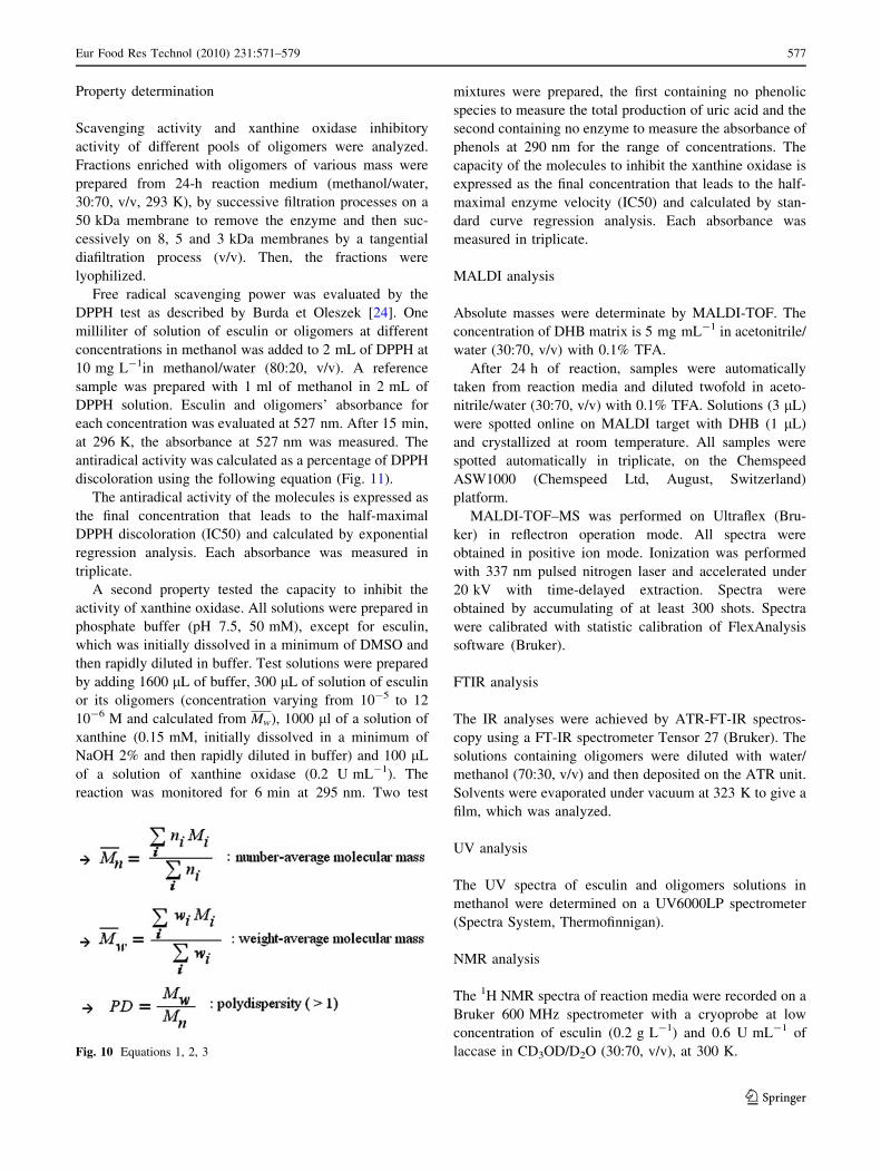
SEC analysis
Relative masses of oligomers were evaluated online by size
exclusion chromatography (SEC) (HPLC LaChrom
(Merck), UV 280 nm LaChrom L-7400, Tosoh TSK Gel a3000 column, at 333 K). DMF with 1% LiBr was used as
the mobile phase, at a flow rate of 1 mL min-1. Polysty-
rene standards were used to define molecular mass cali-
bration. Average molecular masses were defined by
(Fig. 10), where ni represents the number of oligomer
molecules having a mass of Mi at a polymerization degree
of i and wi the weight fraction of chain at a polymerization
degree of i. The quantity of oligomers was expressed in
arbitrary units of area (a.u.). Error values were determined
by three repetitions of the same reaction.
Fig. 9 Schematic
representation of esculin
oligomerization catalyzed by
laccase and molecular structure
of four esculin dimers studied
by molecular simulations.
(R: glucose)
Table 1 Heat of formation of esculin and its radicals in different positions, obtained from molecular minimization in vacuum, calculated by
AM1 semi-empirical quantum method
Esculin Radical species: abstraction of a hydrogen atom from
C3 C4 C5 O7 C8
DH (kcal.mol-1) -350.17 -295.49 -298.34 -295.84 -321.74 -292.37
DHesculin-DHradical = BDEa (kcal.mol-1) 54.68 51.83 54.33 28.43 57.80
a Bond dissociation energy
Table 2 Bond dissociation energy (BDE) of the O7-H bond of
esculin and its dimers, obtained from molecular minimization under
vacuum, calculated by AM1 semi-empirical quantum method
Esculin Diesculin with linkage between
O7–C4 C4–C5 C8–C8 C4–O20
DH (kcal.mol-1) -350.17 -677.30 -688.15 -690.59 -681.71
DDH (kcal.mol-1)
BDE1 28.43 30.54 30.65 25.98 30.65
BDE2 22.34 27.43 27.46
576 Eur Food Res Technol (2010) 231:571–579
123
Property determination
Scavenging activity and xanthine oxidase inhibitory
activity of different pools of oligomers were analyzed.
Fractions enriched with oligomers of various mass were
prepared from 24-h reaction medium (methanol/water,
30:70, v/v, 293 K), by successive filtration processes on a
50 kDa membrane to remove the enzyme and then suc-
cessively on 8, 5 and 3 kDa membranes by a tangential
diafiltration process (v/v). Then, the fractions were
lyophilized.
Free radical scavenging power was evaluated by the
DPPH test as described by Burda et Oleszek [24]. One
milliliter of solution of esculin or oligomers at different
concentrations in methanol was added to 2 mL of DPPH at
10 mg L-1in methanol/water (80:20, v/v). A reference
sample was prepared with 1 ml of methanol in 2 mL of
DPPH solution. Esculin and oligomers’ absorbance for
each concentration was evaluated at 527 nm. After 15 min,
at 296 K, the absorbance at 527 nm was measured. The
antiradical activity was calculated as a percentage of DPPH
discoloration using the following equation (Fig. 11).
The antiradical activity of the molecules is expressed as
the final concentration that leads to the half-maximal
DPPH discoloration (IC50) and calculated by exponential
regression analysis. Each absorbance was measured in
triplicate.
A second property tested the capacity to inhibit the
activity of xanthine oxidase. All solutions were prepared in
phosphate buffer (pH 7.5, 50 mM), except for esculin,
which was initially dissolved in a minimum of DMSO and
then rapidly diluted in buffer. Test solutions were prepared
by adding 1600 lL of buffer, 300 lL of solution of esculin
or its oligomers (concentration varying from 10-5 to 12
10-6 M and calculated from Mw), 1000 ll of a solution of
xanthine (0.15 mM, initially dissolved in a minimum of
NaOH 2% and then rapidly diluted in buffer) and 100 lL
of a solution of xanthine oxidase (0.2 U mL-1). The
reaction was monitored for 6 min at 295 nm. Two test
mixtures were prepared, the first containing no phenolic
species to measure the total production of uric acid and the
second containing no enzyme to measure the absorbance of
phenols at 290 nm for the range of concentrations. The
capacity of the molecules to inhibit the xanthine oxidase is
expressed as the final concentration that leads to the half-
maximal enzyme velocity (IC50) and calculated by stan-
dard curve regression analysis. Each absorbance was
measured in triplicate.
MALDI analysis
Absolute masses were determinate by MALDI-TOF. The
concentration of DHB matrix is 5 mg mL-1 in acetonitrile/
water (30:70, v/v) with 0.1% TFA.
After 24 h of reaction, samples were automatically
taken from reaction media and diluted twofold in aceto-
nitrile/water (30:70, v/v) with 0.1% TFA. Solutions (3 lL)
were spotted online on MALDI target with DHB (1 lL)
and crystallized at room temperature. All samples were
spotted automatically in triplicate, on the Chemspeed
ASW1000 (Chemspeed Ltd, August, Switzerland)
platform.
MALDI-TOF–MS was performed on Ultraflex (Bru-
ker) in reflectron operation mode. All spectra were
obtained in positive ion mode. Ionization was performed
with 337 nm pulsed nitrogen laser and accelerated under
20 kV with time-delayed extraction. Spectra were
obtained by accumulating of at least 300 shots. Spectra
were calibrated with statistic calibration of FlexAnalysis
software (Bruker).
FTIR analysis
The IR analyses were achieved by ATR-FT-IR spectros-
copy using a FT-IR spectrometer Tensor 27 (Bruker). The
solutions containing oligomers were diluted with water/
methanol (70:30, v/v) and then deposited on the ATR unit.
Solvents were evaporated under vacuum at 323 K to give a
film, which was analyzed.
UV analysis
The UV spectra of esculin and oligomers solutions in
methanol were determined on a UV6000LP spectrometer
(Spectra System, Thermofinnigan).
NMR analysis
The 1H NMR spectra of reaction media were recorded on a
Bruker 600 MHz spectrometer with a cryoprobe at low
concentration of esculin (0.2 g L-1) and 0.6 U mL-1 of
laccase in CD3OD/D2O (30:70, v/v), at 300 K.Fig. 10 Equations 1, 2, 3
Eur Food Res Technol (2010) 231:571–579 577
123

A diffusion-ordered spectroscopy (DOSY) NMR tech-
nique was also used to aid the deconvolution of the system.
A diffusion delay time of 100 ms was applied, in CD3OD/
D2O (30:70, v/v), at 293 K.
Esculin 1H NMR
(D2O/CD3OD, 70:30, v/v): d = 3.52 (2d JH30/H40/H50 H40),3.62 (m JH20/H30/H40/H50/H60 H20,H30,H50), 3.77 (d,d JH50/H60/H60
H60 5.8 ? 12.5 Hz), 3.95 (dd JH60/H60/H50 H60 2.0 ?
12.5 Hz), 5.06 (d JH10/H20 H10b 7.5 Hz), 6.31 (d JH3/H4 H3
9.6 Hz), 6.91 (s H8), 7.37 (s H5), 7.93 (d JH4/H3 H4 9.6 Hz)
ppm.
Molecular modeling simulations
Semi-empirical quantum Austin model (AM1) method
Enthalpic study was carried out using the semi-empirical
AM1 method [30], with HyperChem software [31]. Opti-
mization was made with conjugated gradient (algorithm
Polak-Ribiere) with a self-consistent field (SCF) of
1.0 9 10-3 kcal mol-1 A-1, under vacuum. The bond
dissociation energy (BDE) was calculated as the energy of
the radical resulting from hydrogen atom abstraction minus
the energy of the neutral molecule. For the dimers, the
BDE was calculated with the radical species formed in
position 7. For dimers, which present two hydroxyls in
position 7, two radical species and so two BDE were
obtained.
Molecular mechanics and molecular dynamics approach
The molecular systems have been investigated essentially
by molecular mechanics (MM) and molecular dynamics
(MD) methods. The simulations were performed using the
Accelrys program-package Materials Studio (MS) soft-
ware, version 4.2 [32], running on a bi-processor AMD
Dual Core 280, with a memory capacity of 2.4 GHz and
Insight II programs version 2005, on an O2 SGI worksta-
tion. All calculations were performed using Discover with
CFF force field [33, 34], through the Steepest Descent and
Conjugate Gradient (Polak-Ribiere) algorithms. InsightII
S/W and SGI 3D functionalities were essentially used for
an easy construction and initial equilibration of the solvent
boxes and their sizes.
All monomeric and dimeric esculins had their confor-
mational space and their relative orientations, one mono-
mer to another and one dimer to another, firstly studied
under vacuum. MD trajectories were carried out at 600 K,
during 2 ns. After fluctuations analysis and behavior
understanding, the most stable conformations of each sys-
tem were considered for solvent simulations.
Solvent molecules were introduced into a box, around
the central molecules, monomeric or dimeric entities. Fix
constraints were imposed to the atomic coordinates of
those entities, in order to avoid unexpected structural dis-
tortions, due to solvent molecular bumps. The dynamics
trajectories run, at 300 K, during 600 ps, with a time step
of 1 fs, under periodic boundary conditions, using a 14 A
spherical cut-off for non-bound interactions and using the
NVT thermodynamic ensemble. The Verlet-Leapfrog
algorithm was employed for the integration of the equa-
tions of motion. The last frame of each dynamic period was
submitted to energy optimizations until a maximum
derivative of 1.0 9 10-1 kcal mol-1 A-1 to permit solvent
adjustments. After steric hindrance corrections, the con-
straints were removed and the systems were freely reop-
timized, before to be submitted to final dynamics
simulations at 600 K. The temperature was decreased until
300 K. The lowest energy conformation of each molecular
system was, then, minimized, the box size and solvent
equilibration were verified again, and energetic optimiza-
tions were performed until a maximum derivative of
1.0 9 10-4 kcal mol-1 A-1.
For water solvent simulations, a box containing 1370
water molecules and one esculin monomer, and a box
containing 1418 water molecules and one diesculin mole-
cule (O7–C4 linkage) were analyzed. To be closed to the
operating conditions, cell boxes of 50.0 A 9 50.0 A 9 70.0 A
side length containing 938 molecules or 4200 atoms, for a
system composed by methanol/water and 4 esculin mole-
cules; or a box of 55.0 A 9 55.0 A 9 80.0 A side length
containing a system composed by methanol/water and 6
esculin molecules; or a box containing 395 methanol
molecules and 2073 water molecules plus 2 molecules of
diesculin; another cell system having 998 molecules or
3104 atoms, for a system composed by methanol/water,
one diesculin and one esculin molecules. So, many dif-
ferent systems were submitted to dynamics procedure until
to permit us to evaluate the behavior of esculin into water
or into methanol/water (30:70, v/v) mixture.
Conclusions
Structural analyses of esculin oligomers, synthesized by
laccase from Trametes versicolor, showed that esculin
Fig. 11 Equation 4
578 Eur Food Res Technol (2010) 231:571–579
123

units are linked by a simple bridge, which can occur on
both sugar part and phenolic part of the coumarin.
Molecular modeling results suggest the preferential for-
mation of C8–C8 linkages during esculin oligomerization.
Esculin oligomers are characterized by a very high solu-
bility in water and a very poor one in methanol/water
mixture. The higher solubility has been correlated with the
number of hydrogen bonds between oligomers and the
solvent. The oligomers show a very high antioxidant
activity compared with esculin.
References
1. Glenn RG (2007) Functional foods, vol 3. FIS Publishing (ed),
104 p
2. Hiramoto T, Saiki K, Masumura S, Shimizu T, Yamashita T,
Kaneko N, Maruta Y (2002) US Patent No 6475544
3. Soeda M, Umehara H, Seiga K, Yoshida T (1997) JP Patent No
09003054
4. Sung Woo K (2004) KR20040097115
5. Yu J, Wang L, Walzem RL, Miller EG, Pike LM, Patil BS (2005)
J Agric Food Chem 53(6):2341–2343
6. Chang WS, Chiang HC (1969) Anticancer Res 15(5B):1969–
1973
7. Kontogiorgis C, Hadjipavlou-Litina D (2003) J Enzym Inhib Med
Chem 18(1):63–69
8. Cai Y, Baer-Dubowska W, Ashwood-Smith M, DiGiovanni J
(1997) Carcinogenesis 18(1):215–222
9. Yu D, Suzuki M, Xie L, Morris-Natschke SL, Lee KH (2003)
Med Res Rev 23(3):322–345
10. Wright WC (2002) Artemisia, ed Taylor & Francis, 359 p
11. Akita M, Tsutsumi D, Kobayashi M, Kise H (2001) Biosci Bio-
technol Biochem 65(7):1581–1588
12. Mita N, Tawaki SI, Uyama H, Kobayashi S (2003) Macromol
Biosci 3:253–257
13. Uyama H, Kurioka H, Kobayashi S (1997) Polym J 29(2):190–
192
14. Ayyagari M, Akkara JA, Kaplan DL (1998) Solvent-enzyme-
polymer interactions in the molecular-weight control of
poly(m-cresol) synthesis in nonaqueous media. In: Gross, RA,
Kaplan DL, Swift G (eds) Enzymes in polymer synthesis. ACS
symposium series 684: Washington pp 112–124
15. Kim YH, An ES, Song BK, Kim DS, Chelikani R (2003) Bio-
technol Lett 25:1521–1524
16. Dubey S, Singh D, Misra RA (1998) Enzyme Microb Techno
23:432–437
17. Kurisawa M, Chung JE, Uyama H, Kobayashi S (2003) Macro-
mol Biosci 3:758–764
18. Desentis-Mendoza RM, Hernandez-Sanchez H, Moreno A, Rojas
del CE, Chel-Guerrero L, Tamariz J, Jaramillo-Flores ME (2006)
Biomacromolecules 7(6):1845–1854
19. Kurisawa M, Chung JE, Uyama H, Kobayashi S (2003) Bio-
macromolecules 4:1394–1399
20. Anthoni J, Linneton F, Wieruzeski JM, Magdalou J, Engasser JM,
Chebil L, Humeau C, Ghoul M (2008) Rasayan J Chem 1(4):718–
731
21. Kaneko T, Baba N, Matsuo M (2003) Chemico-Biological Interac
142(3):239
22. Zhang HY, Wang LF (2004) THEOCHEM 673(1–3):199
23. Chang WS, Chiang HC (1995) Anticancer Res 15:1969–1974
24. Burda S, Oleszek W (2001) J Agric Food Chem 49:2774–2779
25. Saidman E, Yurquina A, Rudyk R, Molina MAA, Ferretti FH
(2002) J Mol Struct 585:1–13
26. Tsai Lin HC, SH Chen CS, Chang YC, Lee CM, Lai ZY, Lin CM
(2008) Biochem Pharmacol 75:1416–1425
27. Trouillas P, Fagnere C, Lazzaroni R, Calliste C, Marfak A, Du-
roux J-L (2004) Food Chem 88(4):571–582
28. Ncanana S, Baratto L, Roncaglia L, Riva S (2007) Adv Synth
Catal 349:1507–1513
29. Intra A, Nicotra S, Riva S, Danieli B (2005) Adv Synth Catal
347(7–8):973–977
30. Dewar MJS, Zoebisch EG, Healy EF, Stewart JJP (1985) J Am
Chem Soc 107:3902–3909
31. CyberChem, Inc. 1115 NW 4th Street Suite 2, Gainesville, FL
32601 USA
32. Materials Studio software and Insight II software. Accelrys, 9685
Scranton Road, San Diego, CA 92121-3752, USA
33. Hwang MJ, Stockfisch TP, Hagler AT (1994) J Am Chem Soc
116:2515–2525
34. Sun H, Mumby SJ, Maple JR, Hagler AT (1994) J Am Chem Soc
116:2978–2987
Eur Food Res Technol (2010) 231:571–579 579
123
Copyright © 2022 FDOKUMEN