O2 Reduction in Enzymatic Biofuel Cells
275
HAL Id: hal-01731291 https://hal.archives-ouvertes.fr/hal-01731291 Submitted on 6 May 2019 HAL is a multi-disciplinary open access archive for the deposit and dissemination of sci- entific research documents, whether they are pub- lished or not. The documents may come from teaching and research institutions in France or abroad, or from public or private research centers. L’archive ouverte pluridisciplinaire HAL, est destinée au dépôt et à la diffusion de documents scientifiques de niveau recherche, publiés ou non, émanant des établissements d’enseignement et de recherche français ou étrangers, des laboratoires publics ou privés. O2 Reduction in Enzymatic Biofuel Cells Nicolas Mano, Anne de Poulpiquet To cite this version: Nicolas Mano, Anne de Poulpiquet. O2 Reduction in Enzymatic Biofuel Cells. Chemical Reviews, American Chemical Society, 2018, 118 (5), pp.2392 - 2468. 10.1021/acs.chemrev.7b00220. hal- 01731291
-
Upload
khangminh22 -
Category
Documents
-
view
1 -
download
0
Transcript of O2 Reduction in Enzymatic Biofuel Cells
HAL Id: hal-01731291https://hal.archives-ouvertes.fr/hal-01731291
Submitted on 6 May 2019
HAL is a multi-disciplinary open accessarchive for the deposit and dissemination of sci-entific research documents, whether they are pub-lished or not. The documents may come fromteaching and research institutions in France orabroad, or from public or private research centers.
L’archive ouverte pluridisciplinaire HAL, estdestinée au dépôt et à la diffusion de documentsscientifiques de niveau recherche, publiés ou non,émanant des établissements d’enseignement et derecherche français ou étrangers, des laboratoirespublics ou privés.
O2 Reduction in Enzymatic Biofuel CellsNicolas Mano, Anne de Poulpiquet
To cite this version:Nicolas Mano, Anne de Poulpiquet. O2 Reduction in Enzymatic Biofuel Cells. Chemical Reviews,American Chemical Society, 2018, 118 (5), pp.2392 - 2468. �10.1021/acs.chemrev.7b00220�. �hal-01731291�

1
O2 Reduction in Enzymatic Biofuel cells
Nicolas Manoa,b,* and Anne de Poulpiquetc,
a CNRS, CRPP, UPR 8641, 33600 Pessac, France
bUniv. Bordeaux, CRPP, UPR 8641, 33600 Pessac, France
c Bioénergétique et Ingénierie des Protéines, CNRS-AMU, 31, chemin Aiguier, 13009
Marseille, France
both authors contibuted equally to this work
Abstract
Catalytic four-electron reduction of O2 to water is one of the most extensively studied
electrochemical reactions due to O2 exceptional availability and high O2/H2O redox potential,
which may in particular allow highly energetic reactions in fuel cells. To circumvent the use
of expensive and inEfficient Pt catalysts, multicopper oxidases (MCOs) have been envisioned
because they provide Efficient O2 reduction with almost no overpotential. MCOs have been
used to elaborate enzymatic biofuel cells (EBFCs), a sub-class of fuel cells in which enzymes
replace the conventional catalysts. A glucose/O2 EBFC, with a glucose oxidizing anode and a
O2 reducing MCO cathode, could become the in vivo source of electricity that would power
sometimes in the future integrated medical devices. This review covers the challenges and
advances in the electrochemistry of MCOs and their use in EBFCs with a particular emphasis
in the last 6 years. First basic features of MCOs and EBFCs are presented. Clues provided by
electrochemistry to understand these enzymes, and how they behave once connected at
electrodes, are described. Progresses realized in the development of Efficient biocathodes for
O2 reduction relying both on direct and mediated electron transfer mechanism are then
discussed. Some implementations in EBFCs are finally presented.
2
CONTENTS
Symbols 4
1. Introduction 8
2. Background 12
2.1 Multicopper oxidases 12
2.1.1 Laccases 15
2.1.2 Bilirubin oxidases 17
2.1.3 Cuprous oxidase and tyrosinase 18
2.2 Basic features of enzymatic biofuel cells 19
2.3 Electronic connection of immobilized enzymes 23
3. Mechanistic studies of MCOs 26
3.1 Effect of pH on MCOs 28
3.2 Halides inhibition 30
3.3 Model of mediated electron transfer 33
3.3.1 Model 33
3.3.2 Mathematical modeling 36
3.4 Studies of the mechanism of direct electron transfer 41
3.4.1 Redox potentials of the Cu sites 41
3.4.2 Non-catalytic faradaic processes under anaerobic conditions 42
3.4.3 Two-versus four-electron reduction of dioxygen 48
3.4.4 Causes of the low catalytic efficiency and stability on gold
electrodes 51
3.4.5 Catalytic mechanism on carbon electrodes 56
3.4.6 Possibility of endergonic electron-transfer 60
3.4.7 Mathematical modeling of the catalytic current 62
3.4.8 Resting forms of MCOs 68
3.4.9 Halides affecting the direct electron transfer 70
3.4.10 Rate limiting steps of O2 reduction 71
3.4.11 Inhibition by H2O2 72
3.4.12 Effect of methanol 74
4. Enzymatic O2 reduction 75
4.1 Direct electron transfer 75
4.1.1 Planar electrodes 76
4.1.2 Carbon nanostructures 81
4.1.3 Gold nanostructures 102
4.1.4 Tailored orientation of MCOs at the electrode interface 111
4.1.5 Stability issues 125
4.2 Mediated electron transfer 133
4.2.1 Bare electrodes 133
4.2.2 Carbon electrodes 145
4.2.3 3D materials 147
4.2.4 Gold electrodes 156

3
4.2.5 Other enzymatic systems 156
5. MCOs based biocathodes in enzymatic biofuel cells 162
5.1 Glucose/O2 EBFCs 163
5.1.1 Operated in buffer 163
5.1.2 In fruits/vegetables 164
5.1.3 In vertebrates and invertebrates 165
5.1.4 In serum and blood 166
5.2 H2/O2 EBFCs 169
5.3 Microfluidic biofuel cells 170
5.4 Paper based EBFCs 171
5.5 MCOs with air-breathing electrodes 172
5.6 Biofuel cells for self-powered devices 173
5.7 Hybrid bio-devices 174
5.8 Wearable biofuel cells 177
6. Conclusions and Outlook 178
7. Acknowledgments 180
8. References 182

4
Symbols
ABTS: 2,2'-azinobis(3-ethylbenzothiazolin-6-sulfonate)
AFM: Atomic force microscopy
AMT: 4-aminothiophenol
AR: Alternative resting
AuNPs: Gold nanoparticles
BDD: Boron doped diamond
BET: Specific surface area determined by the method of Brunauer, Emmett and Teller
BOD: Bilirubin oxidase
BP: Buckypaper
Bp-BOD: Bilirubin oxidase from Bacillus pumilus
Bs-BOD: Bilirubin oxidase from Bacillus subtilis
CA: Chronoamperometry
CFCs: Conventional fuel cells
Cf-laccase: Laccase from Cerrena fulvocinerea
Cm-laccase: Laccase from Cerrena maxima
CNFs: Carbon nanofibers
CNPs: Carbon nanoparticles
CNTs: Carbon nanotubes
CueO: Copper efflux oxidase
Cu-laccase: Laccase from Cerrena unicolor
COx: Cytochrome oxidase
CV: Cyclic voltammetry
CVD: Chemical vapor deposition
CMF: Carbon microfiber
Cyt c: Cytochrome c
DET: Direct electron transfer
DFT: Density functional theory
DMP: 2,6-dimethoxyphenol
DNA: Desoxyribo nucleic acid
DPI: Dual polarization interferometry
EBFCs: Enzymatic biofuel cells
5
EC: Enzyme class
ECR: Electron cyclotron resonance
EDX: Energy dispersive X-ray analysis
EPR: Electron paramagnetic resonance
ESSA: Electroactive specific surface area
ET: Electron transfer
FAD: Flavin adenine dinucleotide
FDH: Fructose Dehydrogenase
Fp-laccase: Laccase from Fusarium proliferatum
GA: Glutaraldehyde
GC: Glassy Carbon
GDH: Glucose dehydrogenase
GDL: Gas diffusion layer
GO: Graphene oxide
GOx: Glucose oxidase
Hase: Hydrogenase
HET: Heterogeneous electron transfer
HOPG: Highly oriented pyrolytic graphite
HRP: Horseradish peroxidase
IET: Internal electron transfer
KB: Ketjen black
LDG: Low density graphite
LED: Light emitting diode
LMCOs: Laccase-like Multicopper Oxidases
LSV: Linear sweep voltammetry
Ma-Laccase: Laccase from Melanocarpus albomyces
MCOs: Multicopper oxidases
MET: Mediated electron transfer
MgOC: Magnesium oxide template mesoporous carbon
MHA: Mercapto-hexanoic acid
Mo-BOD: Bilirubin oxidase from Magnaporthae orizae
MPA: Mercapto-propionic acid
Mv-BOD: Bilirubin oxidase from Myrothecium verrucaria
MW-CNTs: Multi-wall carbon nanotubes

6
NAD+: Nicotinamide adenine dinucleotide
NHE: Normal hydrogen electrode
NHS: N-Hydroxysuccinimide
NI: Native intermediate
OCP: Open circuit potential
OCV: Open circuit voltage
PAA: Polyacrylamide
PAMAM: Polyamidoamine
PBS: Phosphate buffered saline
PBSE: Pyrene butyric succinimidyl ester
Pc-laccase: Laccase from Pycnoporus cinnabarinus
PDB: Protein data bank
PDMS: Polydimethylsiloxane
PFC: Plastic formed carbon
PG: Pyrolytic graphite
PI: Peroxide intermediate
PLA: Polylactic acid
PLL: Poly-L-lysine
PM-IRRAS: Polarization modulated infra-red spectroscopy
PPO: Polyphenol oxidase
PPY: Polypyrrole
PQQ: Pyrroloquinoline quinone
PSS : Polystyrene sulfonate
PVDF: Polyvinylidene fluoride
PVI: Poly(N-vinylimidazole)
QCM: Quartz crystal microbalance
QCM-D: Quartz crystal microbalance with dissipation monitoring
QM/MM: Quantum mechanical and molecular mechanics
RO: Resting oxidized
RRDE: Rotating ring disc electrode
RVC: Reticulated vitreous carbon
Rv-laccase: Laccase from Rhus vernicifera
SAMs: Self-assembled monolayer
Sc-laccase: Small laccase from Steptomyces coelicolor

7
SECM: Scanning electrochemical microscopy
SERS: Surface enhanced Raman spectroscopy
SGZ: Syringaldazine
SPG: Spectroscopic graphite
SPR: Surface plasmon resonance
STM: Scanning tunneling microscopy
SW-CNTs: Single-wall carbon nanotubes
SWV: Square wave voltammetry
Ta-laccase: Laccase from Trichaptum abietinum
TBAB: Tetrabutylammonium ions
TNC: Tri-nuclear copper cluster
To-laccase: Laccase from Trametes ochracea
TMOS: Tetramethyl orthosilicate
Tt-BOD: Bilirubin oxidase from Trachyderma tsunodae
Tt-laccase: Laccase from Trametes troggii
Th-laccase: Laccase from Trametes hirsuta
tVA: Thiolated veratric acid
Tv-laccase: Laccase from Trametes versicolor
PPY: Polypyrole

8
1. Introduction
About 2.5 billion years ago, oxygen became an important component of the earth
atmosphere1 due to the emergence of cyanobacteria, organisms that produce oxygen during
photosynthesis. One of the consequences of this event is the biological utilization of copper, a
transition metal now omnipresent in biology, while only iron used to be predominant in the
first living forms. In the oceans, oxygenation led to a shift from high-iron to high-copper
contents, because oxygen favors the oxidation state +III of insoluble Fe3+, while Cu is
preferentially in the Cu2+ form which is soluble and therefore bioavailable.2 In living
organisms, copper is still linked with oxygen metabolism : a recent study examining copper
utilization by 450 bacteria demonstrated that around 80% of anaerobes did not depend on
copper, while more than 90% of aerobes were copper dependent.3
Copper can be found in numerous metallo-proteins, in which the metal is coordinated
by amino-acids. Copper-containing proteins are present in bacterial, fungal, plant and
mammalian organisms.4 In some of the proteins, copper is frozen in the reduced or oxidized
state. Of particular importance in the frame of electrochemistry are the proteins in which
copper centers are able to undergo the redox Cu2+/Cu+ reaction. These metallo-proteins
contain either one or several copper centers, and the latter are either mononuclear (type T1,
T2 or CuB sites) or binuclear (type T3 or CuA sites).5 Copper ligands and protein environment
modulate the potentials of the +II/+I transition that varies accordingly from 85 mV to >1000
mV,6 while in water the apparent standard potential E°’ of the Cu2+/Cu+ couple is 150 mV vs.
the normal hydrogen reference electrode (NHE).7
Copper-containing proteins cover different functions ranging from electron transfer to
catalysis, and are involved in a wide variety of mechanisms among which the most important
are energy conversions in respiration or metabolism. It must be noted however that copper is
also toxic at high concentrations so that many proteins are linked to copper regulation and

9
homeostasis.2 Copper based enzymes are associated with the metabolism of N2O (nitrous
oxide reductase), NO2- (nitrite reductases), O2
.- (superoxide dismutase), or O2.5 More relevant
are the two classes of enzymes involved in oxygen reduction: terminal oxidases, and multi-
copper oxidases (MCOs). Terminal oxidases can be divided in cytochrome or quinol oxidases,
and contain both copper centers and hemes. Oxygen reduction occurs with cytochrome or
reduced quinone as a primary electron donor. The reaction is coupled to proton transfer and
the enzyme-catalyzed reactions create the proton gradient necessary for ATP synthesis.8
MCOs catalyze the 4-e- oxygen reduction coupled to oxidation of various organic or metallic
substrates.5 In MCOs, inner sphere electron transfers are involved in binding, activation and
reduction of O2.
Among the desperately sought, but rarely well-engineered devices envisioned for
sustainable energy conversion, fuel cells could harvest energy from fuels like dihydrogen,
alcohols, etc.9,10 Their advantages stem from direct conversion of chemical energy into
electricity, and long-term operation enabled by continuous fueling. The reaction involved in
fuel cells requires a combustive, and O2 is the most widely used because of its availability and
high O2/H2O redox potential.11 Designing catalysts for O2 reduction in conventional fuel cells
(CFCs) is challenging. They generally require high overpotentials,12 or only catalyze the
incomplete O2 reduction to H2O2.13,14 Ideally, the platinum-based catalysts widely used in
CFCs would be replaced by less expensive biological oxidation catalysts. Enzymatic biofuel
cells (EBFCs) are a sub-class of fuel cells in which enzymes replace the conventional
catalysts used in CFCs. They offer numerous advantages such as selectivity and specificity,
allowing in particular operating without the separation membrane otherwise required to avoid
cross-reactions at the electrodes. Moreover, they can operate in mild environments, and are
also renewable and biodegradable.

10
The glucose/O2 biofuel cell, with a glucose oxidizing anode and a O2 reducing
cathodes could become the in vivo source of electricity that would power sometimes in the
future integrated medicals devices.15 In this context, the enzymatic MCO catalysts are
especially interesting because they provide Efficient 4-e- O2 reduction5 with almost no
overpotential.16 In the last decades, electrochemical research of redox proteins showed ways
to connect MCOs at electrodes, and to use them as Efficient and specific electrocatalysts for
O2 reduction,17,18 making some the enzymes of choice in EBFCs.
In this review we cover the challenges and advances in the electrochemistry of MCOs
and their use in EBFCs since their first connection at an electrode 40 years ago, with a
particular emphasis in the last 6 years. The interested reader will find in Table 1 additional
reviews on this topic, with a particular focus on electrode materials for EBFCs. Here, we first
briefly present basic features of MCOs and EBFCs, before focusing on the electrochemical
approach of MCO study and applications. We describe what electrochemistry teaches us
about these enzymes, and how they behave once connected at electrodes. We then discuss the
progresses realized in the development of Efficient biocathodes for O2 reduction relying both
on direct and mediated electron transfer mechanism (DET and MET respectively). Finally, we
present some implementation in EBFCs.
Table 1: Reviews related to O2 reduction by MCOs and in Biofuel cells
2004 Enzymatic Biofuel Cells for Implantable and Microscale Devices Barton et al. 19
Biological fuel cells and their applications Sukla et al. 20
2006 Challenges in biocatalysis for enzyme-based biofuel cells Kim et al. 21
Biofuel cells and their development Bullen et al. 22
Potentially implantable miniature Batteries Heller 23
2007
Biofuel cells--recent advances and applications Davis et al. 24
Enzyme-based biofuel cells Minteer et al. 25

11
2008 Energy harvesting by implantable abiotically catalyzed glucose fuel cells Kerzenmacher et al. 26
Enzymes as working or Inspirational Electrocatalysts for Fuel Cells and Electrolysis Cracknell et al. 27
Enzyme catalysed biofuel cells Cooney et al. 28
Extended lifetime biofuel cells Moehlenbrock et al. 29
2009 Integrated Enzyme-Based Biofuel cells - A review Willner et al. 30
Hemoproteins in Design of Biofuel Cells Ramanavicius et al. 31
Microfluidic fuel cells: A review Kjeang et al. 32
2010 An overview of enzymatic Biofuel cells Aquino Neto et al. 33
Biofuel Cells with Switchable Power Output Katz 34
Protein Engineering – An Option for Enzymatic Biofuel Cell Design Guven et al. 35
Recent Advances in Enzymatic Fuel Cells: Experiments and Modeling Ivanov et al. 36
Biofuel Cells for portable power Gellet et al. 37
Enzymatic Biofuel Cells—Fabrication of Enzyme Electrodes Hao Uy et al. 38
A perspective on microfluidic biofuel cells Lee et al. 39
Mediated Biocatalytic Electrodes and Enzyme Stabilisation for Power Generation Brito et al. 40
Biosensors and biofuel cells with engineered proteins Caruana et al. 41
2011 Strategies to extend the lifetime of bioelectrochemical enzyme electrodes for biosensing and
biofuel cell applications Rubenwolf et al. 42
Bioelectrochemical interface engineering: Toward the fabrication of electrochemical
Biosensors, Biofuel cells and self-powered logic biosensors Zhou et al.43
Recent Developments of Nanostructured Electrodes for Bioelectrocatalysis of Dioxygen
Reduction Opallo et al. 44
Self-powered sensors Arechederra et al. 45
Recent progress and continuing challenges in bio-fuel cells. Part I: Enzymatic cells Osman et al. 46
Biofuel cells for Self-Powered electrochemical biosensing and logic biosensing: A Review Zhou et al. 47
Bioelectrochemical systems: Microbial versus enzymatic catalysis Freguia et al. 48
New materials for biological fuel cells Minteer et al. 49
Immobilization technology: a sustainable solution for biofuel cell design Yang et al. 50
Enzymatic fuel cells: Recent progress Leech et al. 51
Carbon nanotube/enzyme biofuel cells Holzinger et al. 52
Features and applications of bilirubin oxidases Mano 53
Enzymatic versus Microbial Bio-Catalyzed Electrodes in Bio-Electrochemical Systems Lapinsonnière et al. 54
Direct electron transfer based enzymatic fuel cells Falk et al. 55
2013 Biofuel Cells for Biomedical Applications: Colonizing the Animal Kingdom Falk et al. 56
Direct Electrochemistry based biosensors and biofuel cells enables with nanostructures
materials Liu et al. 57
Bilirubin oxidases in bioelectrochemistry: Features and recent findings Mano et al. 58
Implanted biofuel cells operating in vivo – methods, applications and perspectives Katz et al. 59
New Energy Sources: The Enzymatic Biofuel Cell Aquino Neto et al. 60
A perspective on microfluidic biofuel cells Lee et al. 39
2014
Enzyme Biofuel Cells: Thermodynamics, Kinetics and Challenges in Applicability Luz et al. 61
Enzymatic Biofuel Cells on Porous Nanostructures Wen et al. 62
Biohydrogen for a New Generation of H2/O2 Biofuel Cells: A Sustainable Energy Perspective De Poulpiquet et al. 63
New trends in enzyme immobilization at nanostructured interfaces for Efficient
electrocatalysis in biofuel cells. De Poulpiquet et al. 64
Paper-based Batteries: A review Nguyen et al. 65
Recent advances in carbon nanotubes-based enzymatic fuel cells Cosnier et al. 66
Hybrid Electric Power Biodevices Pankratov et al.67

12
Electrochemical and electrophoretic deposition of enzymes: Principles, differences and
application in miniaturized biosensor and biofuel cell electrodes Amman et al. 68
Is graphene worth using in Biofuel cells? Filip et al. 69
QM/MM Molecular Modeling and Marcus Theory in the Molecular Design of Electrodes
for Enzymatic Fuel Cells Vasquez-Duhalt et al.70
Towards glucose biofuel cells implanted in human body for
powering artificial organs: Review Cosnier et al. 71
2015
Recent progress in oxygen-reducing laccase biocathodes for enzymatic biofuel cells. Le Goff et al. 72
Wired enzymes in mesoporous materials: A benchmark for fabricating biofuel cells Catalano et al. 73
Graphene based enzymatic bioelectrodes and biofuel cells Karimi et al. 74
2016
Recent advances on enzymatic glucose/oxygen and hydrogen/oxygen biofuel cells:
Achievements and limitations Cosnier et al. 75
Paper electrodes for bioelectrochemistry: Biosensors and biofuel cells Desmet et al. 76
Progress on implantable biofuel cell: Nano-carbon functionalization for enzyme
immobilization enhancement Babdai et al. 77
Wearable Biofuel Cells: A Review Bandodkar et al. 78
BioCapacitor: A novel principle for biosensors Sode et al. 79
Tear Based Bioelectronics Pankratov et al. 80
Laccase: a multi-purpose biocatalyst at the forefront of biotechnology Mate et al. 81
Microfluidic fuel cells for energy generation Safdar et al.82
2017
Recent advance in fabricating monolithic 3D porous graphene and their applications
in biosensing and biofuel cells Qiu et al. 83
Review—Wearable Biofuel Cells: Past, Present and Future Bandokar et al. 84
The applications and prospect of fuel cells in medical field: A review Xu et al. 85
Biological fuel cells and membranes Ghassemi et al. 86
Nanostructured inorganic materials at work in electrochemical sensing and biofuel cells Holade et al. 87
Advanced Materials for Printed Wearable Electrochemical Devices: A Review Kim et al. 88
Quo Vadis, Implanted Fuel Cell? Shleev 89
Mathematical modeling of nonlinear reaction–diffusion processes in enzymatic biofuel cells Rajendran et al. 90
Nanostructured material-based biofuel cells: recent advances and future prospects Zhao et al. 91
2. Background
2.1 Multicopper oxidases
In this paragraph some reminders about MCOs will be given. Detailed enzymology,
kinetics, electronic structure, and mechanism can be found elsewhere.5,92-99
Multicopper Oxidases (MCOs) family is ubiquitous in nature and can be divided in
three categories depending on the enzymes’ specificity towards substrates.5,95 The first
category includes laccases (EC 1.10.32) which oxidize a broad range of organic substrates.
The second category, metalloxidases, includes for example ceruloplasmin (EC 1.16.3.1) or

13
cuprous oxidase that oxidize metal substrates. The last category includes ascorbate oxidase
(EC 1.10.32) or bilirubin oxidase (EC 1.3.3.5) which can be specific towards single substrate
(i.e ascorbate or bilirubin) while also being able to oxidize other organic substrates.100 A
general trait of MCOs is the presence of at least four copper ions which permit oxidation of
substrates while reducing O2 to water without production of any toxic intermediates. Each
copper is classified according to its optical and magnetic properties. Type I Cu displays an
intense absorption band near 600 nm resulting from a charge transfer transition from Cys to
Cu (II) and is responsible for the name, blue copper oxidases, given to these enzymes (Fig
1A). It also exhibits a small Cu(II) A|| hyperfine coupling constant in EPR (Fig 1B).
Figure 1. UV-visible (A), X-band EPR (B) of purified BOD from Bacillus pumilus
highlighting the spectral feature in MCOs. Reprint with permission from ref [101] Copyright
2010 Elsevier.
Type 1 copper accepts electrons from the electron-donating substrates and shuttles
them to the O2 reduction site composed of three coppers. The tri-nuclear cluster (TNC)
consists in a Type 2 copper ion active in EPR with a normal Cu(II) A|| hyperfine coupling
constant and a pair of Type 3 cupric ions with a characteristic 330 nm absorption shoulder and

14
antiferromagnetically coupled via a bridging hydroxide (Fig 1B). Electrons are relayed from
the T1 to the TNC through ~13 Ǻ of Cys-His residues (Fig 2).
Figure 2. The structure of the MCO active site with arrows marking the flow of substrates,
electrons (e-), and O2. Reproduced with permission from ref [93]. Copyright 2012 Royal
Society of Chemistry
The exact nature of the electron transfer has been the subject of numerous
investigations and is summarized in Figure 3.5,92,94,97,98,102-106
Figure 3. Mechanism of O2 reduction to water by the MCOs. Red arrows indicate the steps
that take place in the catalytic cycle of the MCOs. Black arrows indicate steps that can be
experimentally observed but are not part of the catalytic cycle. The dashed arrows at the right
indicate the transfer of an electron from the T1 Cu to the T2 Cu to create PI + e-, that occurs
in going from PI to NI but is not experimentally observed in the wild type enzyme.
Reproduced with permission from ref [93]. Copyright 2012 Royal Society of Chemistry
15
Briefly, after oxidation of the substrate at the T1, electrons are shuttled to the TNC
where the two two-electron steps of O2 reduction to H2O occurs.93,107,108 The formation of the
peroxide intermediate (PI) results from the first two-electron step with a fast reaction rate (
106 M-1.s -1). In the second two-electron step, the O-O bond is cleaved with a fast rate constant
leading to the formation of the Native Intermediate (NI). In presence of substrate, NI is
reduced to the fully reduced form of the enzyme and is ready for another catalytic cycle. In
absence of substrate, NI slowly decays to the resting oxidized (RO) form and only reintegrates
the catalytic cycle upon reduction with a substrate.
2.1.1 Laccases
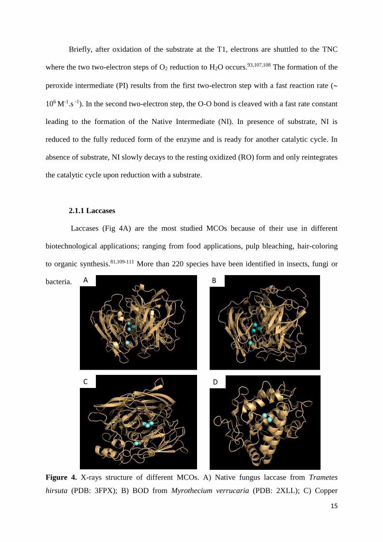
Laccases (Fig 4A) are the most studied MCOs because of their use in different
biotechnological applications; ranging from food applications, pulp bleaching, hair-coloring
to organic synthesis.81,109-111 More than 220 species have been identified in insects, fungi or
bacteria.
Figure 4. X-rays structure of different MCOs. A) Native fungus laccase from Trametes
hirsuta (PDB: 3FPX); B) BOD from Myrothecium verrucaria (PDB: 2XLL); C) Copper

16
efflux Oxidase CueO (PDB: 4NER); D) Tyrosinase from Bacillus megaterium (PDB : 5I38).
All Coppers are displayed in blue
Among them, fungal laccases have been the most studied.5,112-114 Laccases are known
to oxidize a variety of polyphenolic compounds concomitant with the reduction of O2. Due to
this latter property, Tarasevich et al. have pioneered the use of laccases for O2 reduction. In
1979, they reported the immobilization of monolayers of laccases on vitreous carbon
electrodes, generating O2 reduction currents of 175 A.cm-2 at pH 5.115 The
geometry/coordination of the T1 copper deserves attention since this is the entry point of
electrons coming from the electrode or from the redox mediator. It has been shown that its
redox potential was modulated by the nature of its coordination. A T1 with a weakly
coordinating Met in the axial position exhibits generally a lower redox potential than MCOs
where Met has been replaced by non-coordinating Phe or Leu.116 However, many exceptions
exist and the type of coordination cannot explain alone the difference in the observed
electrochemical potentials. Other factors such as electrostatic interactions and hydrogen bonds
must be taken into account, making therefore difficult to predict the redox potential of
MCOs.117 We list here some of the most commonly studied laccases in bioelectrochemistry.
Fungal laccases have been identified in basidiomycetes. Among them we use the abbreviation
Th-laccase to call without differentiation the two isoenzymes from Trametes hirsutus, also
known as Coriolus hirsutus118, and Tv-laccase to call the laccase from Trametes versicolor,
also known as Coriolus versicolor or Polyporous versicolor. Other laccases are extracted
from trees, among which Rv-laccase the laccase from the Japanese lacquer tree Rhus
vernicifera.

17
2.1.2 Bilirubin oxidases
BODs (Fig 4B) have been discovered in 1981 by Tanaka and Murao.119 Because of
their ability to oxidize bilirubin, they were classified as BODs and not laccases, even though
BODs can also oxidize a variety of phenolic compounds.53 BODs have been primarily used in
the medical field to elaborate bilirubin biosensors.53,58 Only 7 BODs have been
identified/characterized so far, among which BOD from Myrothecium verrucaria (Mv-BOD),
the most studied BOD, BOD from Trachyderma tsunodae120 (Tt-BOD), BOD from
Magnaporthae orizae121 (Mo-BOD) and BOD from Bacillus pumilus (Bp-BOD).101 Unlike Tt-
BOD or Mv-BOD, the two newly identified Mo-BOD and Bp-BOD display good thermal
stability, a high activity at pH 7 and high temperature, a low sensitivity towards NaCl and no
sensitivity to urate. Bilirubin is not a substrate that has been systematically tested with
laccases or other MCOs. Therefore, it is not unlikely that MCOs already identified and
characterized also display BOD-like activity. The use of Mv-BOD for the electrocatalytic O2
reduction at neutral pH has been reported for the first time in 2001 by Tsujimura et al.122 Ever
since, the number of studies relying on BODs has been flourishing for one main reason:
unlike laccases, BODs display a high activity and stability at neutral pH and in presence of
chloride. Two points deserve attention. The first concerns the denomination of the enzyme.
Reiss et al. recently identified a CotA enzyme from Bacillus pumilus123 which was classified
as a laccase while Durand et al. classified an enzyme sharing 98% identity as a BOD.101
BOD-like activity has also been observed in a CotA from Bacillus subtilis but the enzyme was
classified as a laccase.124 Reiss et al. have suggested that the denomination of BOD was
improper since these enzymes could oxidize a variety of substrates. Therefore, they suggested
to introduce the term of laccase-like MCOs (LMCOs).125 On the other hand, not all laccases
oxidize bilirubin. Therefore, the denomination of BOD would permit to differentiate between
those MCOs that oxidize bilirubin or those that do not. The question of classification remains

18
open. The second point deserving attention, which is linked to the first, is the core structural
difference between laccases and BODs. Detailed analysis of amino acid sequences does not
permit to differentiate BODs and laccases.126 Combining different analytical tools such as
molecular modeling or X-rays analysis would definitely help, but only two X-rays structures
of BODs are available so far, for Mo-BOD127 and Mv-BOD.128,129
2.1.3. Cuprous oxidase and tyrosinase
The two enzymes have been rarely used in O2 biocathodes. Cuprous oxidase, or
copper efflux oxidase (CueO) (Fig 4C) is involved in copper efflux in E.coli.130,131 CueO
possesses the same catalytic site as other MCOs but exhibits a strong activity towards cuprous
ion and, unlike other MCOs, no activity towards phenolic compounds. Tyrosinase, or
polyphenol oxidase (Fig 4D), only carries a couple of T3 copper atoms and is involved in the
ortho-hydroxylation of monophenols and two-electron oxidation of diphenols.95
In the following, we will not consider cerulosplasmin and ascorbate oxidase (EC
1.10.3.3) which have not yet proven their utility in O2 reduction.11 It has been shown that
direct electron transfer (DET) between ceruloplasmin and an electrode surface was feasible
under non turnover conditions, but in most cases electrocatalytic reduction of O2 was not
achieved.18,132-135 The only example of O2 reduction with ceruloplasmin on gold modified
with ferromagnetic particles encapsulated in carbon was reported in 2015 by Matysiak et
al.136 However, the current density is too low to envision, so far, application in EBFCs. The
DET of ascorbate oxidase has been reviewed by Shleev18 and also investigated by Atanassov
et al.137

19
2.2 Basic feature of enzymatic biofuel cells
Detailed thermodynamic and kinetic analyses of enzymatic biofuel cells (EBFCs)
have been provided in earlier reviews.19,22,24,61 Therefore, in the following, we only briefly
summarize key points and advantages of EBFCs.
Despite the fact that electrochemical reactions are catalyzed by enzymes at one or both
electrodes of the EBFCs, there is no conceptual difference between the latter and conventional
fuel cells (CFCs).9 The fuel is oxidized at the anode while the oxidant is reduced at the
cathode, and electrons are exchanged between the two electrodes in an external electrical
circuit. The driving force of the reactions is the difference in electrochemical potential
between the two electrodes. The transport of charges in the electrolyte solution (or polymer) is
ensured by ions. However, unlike CFCs, EBFCs operate at near-neutral pH and ambient
temperature. Enzymes also have advantages as electrocatalysts: specificity towards their
substrates, renewability, biodegradability, high turnover frequencies, and low intrinsic
overvoltages. Moreover, the great diversity of enzymes allows envisioning EBFCs working
with diverse fuels and oxidants. In addition, if the bioelectrodes are properly designed, no
membrane is needed in EBFCs to separate anodic and cathodic compartments, allowing
thereby miniaturization of the devices.
An EBFC is characterized by its current intensity, I, its voltage Ecell, and its power,
defined as the product of these latter:
P [W] = Ecell [V] x I [A] Eq. 1
The electrical current intensity I is the charge flux entering the anode or leaving the cathode.
In lots of studies the current density, defined as the ratio of current I by surface area A and
denoted j, is preferred. Similarly, the power is often expressed in terms of power density. This
makes actually hard to compare the efficiency of different devices because in some (rare)

20
cases the real electroactive surface area of the electrode is considered for the calculation,
whereas it is more common to use the projected geometric area.
This point deserves a particular attention. While the normalization of the current is
critical for comparing the results of the numerous publications, normalization is difficult.
Other difficulties for comparison arise from the fact that experimental conditions (pH,
temperature, scan rate, electrode rotation…) differ and in some studies, experimental details
(enzymes activities and purities, loading…) are not provided. Natural convection which is
often underestimated may also significantly affect current densities.138 Comparisons are even
more difficult when porous materials are used and particularly when detailed characterization
of the materials are not provided (BET, thickness, porosity, mass,..). In addition, the different
electrochemical techniques used to evaluate the power profile of EBFCs may also be
problematic in some cases. For example, using linear sweep voltammetry at a quite fast scan
rate (>5 mV.s-1) to evaluate the power profile of EBFCs based on highly porous electrode
materials may induce errors. Since porous materials usually have a high capacitance, it is
more than likely that the measured currents are composed by both catalytic and capacitive
current, which artificially enhances the biofuel cell performances. In order to evaluate the
actual power, the effective catalytic currents have to be determined using chronoamperometry
(for individual bioelectrodes) and under continuous discharge (for the biofuel cell setup) at
different imposed potentials, or using linear sweep voltammetry at very low scan rates so that
the steady-state can be reached. For all the above considerations, it would undoubtedly be
useful to develop a protocol for comparison of data, as was already suggested by Minteer and
others,139-141 but was not favorably received. The authors of this review believe that such
normalization is much needed, after answering the question of the best way to normalize
performances, taking into account the variety of electrode materials, EBFC designs and
enzymes used.

21
While in CFCs currents can be measured in A, they are mostly in the range of few
tenths of µA to few hundreds of µA (resp. few µA.cm-2 to few mA.cm-2) in EBFCs, or more
generally at enzymatic electrodes. Similarly, power (resp. power densities) are in the range of
kW to MW in CFCs whereas they are mostly between few µW and few mW in EBFCs. Some
important features of enzymatic electrodes and EBFCs are described in Figure 5.
Figure 5. Electrochemical characterization of an EBFC. A: Current/Voltage response of the
bioanode (blue curve) and of the biocathode (red curve). The purple arrows indicate the
current density and the cell voltage giving the maximum power density. B: Polarization curve
(red) and power curve (blue) of the complete EBFC. Adapted from [27]. Copyright 2008
American Chemical Society.
The potential adopted by the electrode at j = 0 is the open circuit potential (OCP). It is
close to the onset potential, the starting potential of catalysis. It must be underlined that the

22
definition of “onset potential” is quite fuzzy since it is difficult to define the exact point at
which catalysis actually “starts”. The difference between the OCP and the apparent standard
redox potential, the overvoltage (), characterizes the deviation to ideality of a catalyst and is
a significant measure of its efficiency. Enzymes and MCOs in particular, are potentially
powerful catalysts since for most of them electrochemical overvoltages are lower than for
conventional catalysts. The difference between the OCP of the biocathode and the bioanode
determines the maximal electrochemical driving force or open circuit voltage (OCV, also Ecell
for j = 0) of the EBFC. The OCV of biofuel cells lies below the thermodynamic limit of 1.23
V for H2/O2 EBFCs, but sometimes very close to it,142,143 and below the maximal OCV of
1.18 V for glucose/O2 EBFCs, where glucose is oxidized into gluconolactone at the anode.
EBFC performances can be characterized in terms of OCV, polarization curve and
power curve. These two curves show respectively the evolution of the cell voltage and of the
power density with the current density (Fig 5B). Particular data are the maximum power
density Pmax, and the maximum current density jmax which corresponds very often to the short-
circuit current (i.e. current density for Ecell = 0). As seen in Figure 5B, the polarization curve
can be divided into three regions. In Segment I, the steep decrease of the cell voltage
corresponds to kinetic losses, which are related to the slow rate of the electrode reaction due
to e.g. slow electron-transfer kinetics. After this first decrease of the cell voltage, the
polarization curve continues decreasing more linearly with a smaller slope over a wide range
of current density values. This decrease, which can be seen in Segment II, corresponds to the
ohmic voltage drop originating from the system’s intrinsic resistance as well as from the
resistance to the flow of ions within the electrolyte. Finally, (concentration polarization)
losses related to mass transport limitations result in the final decrease of the potential until it
reaches zero (Segment III). At that point, the current reaches its maximal value and the system
behaves as if the electrodes were in a short circuit. Each of the mentioned potential losses can
23
be described with corresponding overpotentials (Eq. 2). This allows calculating the cell
voltage as:
𝐸𝑐𝑒𝑙𝑙 = 𝐸𝑒𝑞 − 𝜂𝑎𝑐𝑡 − 𝜂𝑑𝑖𝑓𝑓 − 𝐼 ∑ 𝑅 𝐸𝑞. 2
where: 𝐸𝑐𝑒𝑙𝑙 and 𝐸𝑒𝑞 are respectively measured and theoretical (thermodynamic) voltages,
𝜂𝑎𝑐𝑡 and 𝜂𝑑𝑖𝑓𝑓 are respectively kinetic (activation) and mass transport based overpotentials, 𝐼
is the current that flows through the cell and ∑ 𝑅 is the sum of all resistances that are present
in the system. The challenge is to minimize all kinetic losses by optimizing each ingredient of
the biofuel cell; the electrode material, enzymes, and redox mediators if needed; and by
Efficiently integrating them together. It could be done for example by optimizing the cell
design and the immobilization of the enzymes.
2.3 Electronic connection of immobilized enzymes
One of the major challenges of using an enzyme as an electrocatalyst is to establish an
Efficient electronic connection between the enzyme and the electrode, particularly because of
the important size of the protein molecule, and the anisotropy of its electronic properties. A
poor electrical connection will induce consequent kinetic losses. Two mechanisms allow
electron transfer between an enzyme and an electrode. On one hand, in direct electron transfer
(DET), electrons tunnel directly between an enzyme and an electrode. This mechanism relies
on the ability of the enzyme to accept electrons not only from its natural substrate but also
from artificial electron donors like the electrode. On the other hand, in mediated electron
transfer (MET), electrons are shuttled via a redox molecule, called mediator, which exchanges
electrons with the enzyme and is reversibly oxidized or reduced at the electrode surface (Fig
6).

24
Figure 6. Two different mechanisms of electron exchange between an enzyme (here a
laccase) and an electrode. Left: Direct Electron Transfer (DET) and Right: Mediated Electron
Transfer (MET), here with a diffusive mediator.
DET can only occur if the enzyme active site or an electron relay is close enough to
the electrode surface. The rate of interfacial electron transfer kET, described by Marcus semi-
classical theory144,145 (Equation 3), depends on the driving force of the reaction (∆G°), the
nuclear reorganization energy (λ), and the electronic coupling (HDA) between donor D and
acceptor A.
𝑘𝐸𝑇 =1
ℎ
𝐻𝐷𝐴2
√4𝜋𝜆𝑅𝑇𝑒
−(∆𝐺°+𝜆)2
4𝜆𝑅𝑇 𝐸𝑞. 3
The simplest model to describe the electronic coupling in case of proteins is the
« square-barrier model », according to which 𝐻𝐷𝐴2 decreases exponentially with increasing
distance between donor and acceptor (rDA):
𝐻𝐷𝐴2 = (𝐻𝐷𝐴
0 )2𝑒−𝛽(𝑟𝐷𝐴−𝑟0) 𝐸𝑞. 4
where HDA0 is the electronic coupling at Van-der-Waals distance (r0). The tunnel parameter β
reflects the protein efficiency for electron transfer and depends on the protein structure.146,147
In DET, the catalytic wave is centered on a potential that is only affected by the
properties of the enzyme molecule. This is discussed further in section 3.4.5 and 3.4.7. The

25
distribution of orientations allowing DET results in distribution of electronic transfer rates148
reflected by the trailing edge in the catalytic cyclic voltammogram (Fig 7A).
Figure 7. Representative cyclic voltammograms of a MCO in DET (A) and MET (B). Curves
1 and 1’ are recorded in non-catalytic conditions in the absence of O2. Curve 1’ clearly shows
the redox process of the mediator. Curves 2 and 2’ are recorded in catalytic conditions under
O2. The dotted vertical bars indicate the apparent standard potentials of the enzyme (E°’enzyme)
and of the mediator (E°’mediator).
Therefore, a performant DET can only be obtained for a monolayer of enzymes
properly oriented. Experimentally, it has been verified for redox proteins including MCOs
immobilized on SAMs with different alkyl lengths that the efficiency of electron transfer
decreases exponentially with increasing numbers of methyl groups.149-151 In proteins,
electrons tunnel through a maximum distance of 14 Å.146
MET is possible if the affinity between the enzyme and the mediator is high enough, and
if the difference in their redox potentials induces a driving force for the electron transfer. Both
the oxidized and the reduced forms of the mediator must be stable chemically, and of course
none of them must inhibit the bioelectrochemical reaction. The mediator can be for example

26
another protein like cytochrome c,152 diffusive species like ABTS or transition metal
complexes, or a metal complex bound to a polymer, like osmium complexes.153 The
corresponding polymers are used in the form of a hydrogel, whose reticulated structure allows
permeation of solvent and diffusion of ions and non-charged species. Unlike most hydrogels,
osmium based redox polymers are also capable of conducting electrons via self-collision
between neighboring oxidized and reduced osmium redox moieties. The “diffusivity” of
electrons in the hydrogel depends on the length of the tether that links the osmium complex to
the polymer backbone, the characteristic electron-hoping distance between neighboring redox
sites, the self-exchange rate constant, and the concentration of redox sites.154 In MET
configuration, the catalytic wave is centered on the mediator potential (Fig 7B).
The two approaches have advantages and drawbacks. DET is reasonably simple and
cheap, and allows working with overvoltages close to zero thanks to intrinsic properties of
enzymes. However, it requires a unique orientation and only allows the simultaneous
connection of a maximum of one monolayer of enzymes. In contrast, MET allows to wire
electronically lots of enzymes regardless of their orientation and even in multilayers. The
hydrogel especially is a convenient matrix for enzyme immobilization that even protects
enzymes from inhibitors. However, electron-hopping in the redox polymer introduces
additional steps that can lower the catalytic efficiency of the bioelectrode. A drop of cell
voltage is also induced because catalysis is recorded at the potential of the mediator which
must be lower than that of the enzyme. Finally, in case of diffusive mediators, a cell
membrane is required to avoid cross reactions.
3. Mechanistic studies of MCOs
The catalytic mechanism of MCOs in homogeneous solution has been largely
investigated by different spectroscopic techniques, crystallography and density functional

27
theory (DFT) calculations and only partly elucidated.92,93,95 For example, the mechanisms of
MCO inhibition at high pH or high concentrations of halides, or the differences between
BODs and laccases are still unclear.
Figure 8. Comparison of different steps for electron transfer for enzymes in solution (A) and
immobilized (B) with Direct electron transfer (DET) or Mediated electron transfer (MET). A)
Step 1: Substrate oxidation at the T1; Step 2: IET; Step 3: O2 reduction. B) Step 1: Electron
transfer from the electrode to the T1; Step 2: IET; Step 3: O2 reduction; Step 4: O2 diffusion;
Step 5: hypothesized electron transfer from the electrode to the TNC; Step 6: Electron transfer
from the electrode to the redox mediator; Step 7: electron transfer within the redox mediator;
Step 8 electron transfer from the redox mediator to the T1. Adapted with permission from
[155] Copyright 2017 Elsevier
During homogeneous catalysis, oxidation of organic substrates occurs via a ping-pong
type mechanism. T1 is the primary center at which electrons from the reducing substrates are
accepted,92,93,95,155,156 and O2 is reduced at the TNC (Fig 8). One of the major questions,
4 H+ + O2
2 H2O
CA
THO
DE
DET
ENZYME IMMOBILIZED
T1
T3
T3
T2
1
2
5
3
CA
THO
DE
RedoxMediator
MET8
6
7
O2
4
3
2 H2O
Reduced substrate
Oxidized substrate
A. SOLUTION
T1
T3
T3
T2
2
1
B.

28
however, is whether the mechanism is the same for an enzyme immobilized at the electrode.
Intuitively, we may for example expect different rate limiting steps, depending whether the
MCOs are connected at the electrode in DET or in MET.
The influences of pH and chloride on the MCOs activity are of particular importance,
especially if MCO-based biocathodes are envisioned for BFCs operating in physiological
media. In this section, we first present commonly adopted opinion about how pH and halide
inhibitors impact on MCO mechanism in homogeneous solutions. Due to the general
similarity between mechanism in homogeneous solution and in electrochemistry in MET
connection, we simultaneously develop investigations realized in electrochemistry in MET.
The reader can find more information about mechanism in homogeneous solution in different
specialized reviews.93,157-161 The following part presents a comprehensive view of DET
connected immobilized enzymes. We also consider two less-known inhibitors, hydrogen
peroxide and methanol, without differentiating DET and MET, because a priori their effect is
similar in both modes.
3.1 Effect of pH on MCOs
The effect of pH on the MCO catalytic mechanism has been the subject of intense
research and has only recently been partly elucidated. The first studies were reported in
homogenous solution in 1997 by Xu.162 They demonstrated that both high- and low-potential
laccases from Trametes villosa, Rhizoctonia solani and Myceliopthora thermophila were
inhibited at neutral pH and above. It was hypothesized that laccases were inhibited upon
increasing the pH due to the binding of OH- to the T2 site, therefore interrupting
intramolecular electron transfer between the copper sites.157,158
Solomon et al. investigated in details how pH influences laccases reaction
mechanism.93 They particularly tried to decipher which step of the reaction cycle was
29
inhibited at high pHs. Since the rates of formation of the PI and NI states are pH-
independent163, these steps cannot be associated to the pH inhibition (Fig. 3). The remaining
possibilities are therefore the formation of the fully reduced form, either from NI or from the
resting oxidized form, or an alternative mechanism. More recently, by combining protein film
volammetry,164 computational modeling and numerical analysis, Blanford et al.165
investigated the pH dependence of O2 reduction of two high and two low-potential MCOs
immobilized in DET mode. They attributed the pH inhibition to another redox state of the
enzymes, unknown so far, called “X state”. This point is detailed further in section 3.4.8.
However, since there are no direct spectroscopic or crystallographic features of the “X state”,
it cannot be totally excluded that the inhibition effect may come from the NI state. If the
formation of NI has been studied extensively, this is not the case of the protons involved
during the reduction of NI.105
While the residual activity of laccases is almost nil at neutral pH, irrespective of the
redox mediator used, this is not so for BODs.53 Since the specific activity of BODs is
declining with the pH (when ABTS is used as a substrate), one can hypothesize that OH- also
inhibits BODs. Nevertheless, detailed kinetics studies on BODs still have to be performed.
Another question remains open: why are BODs less sensitive to pH than laccases?
Different strategies have been explored to prevent laccases inhibition at neutral pH, or
to get around it. For example, by switching from a fungal Tv-laccase to the small bacterial
laccase from Steptomyces coelicolor, which was reported to be highly active at pH 7,
Calabrese-Barton et al. reached 1.5 mA.cm-2 at 40°C and 900 rpm while they only reached
0.2 mA.cm-2 with the fungal one.166 The exact reason for the higher activity of Sc-laccase at
neutral pH still has to be identified. Enzyme engineering and molecular biology are also
powerful tools to evolve laccases.167 For example in 2009, Cusano et al.168 succeeded in
shifting the pH profile of chimeric laccases and in 2013, Alcalde et al. widened the pH profile

30
of a fungal laccase.169 The same authors later used directed molecular evolution to evolve a
basydiomycete laccase.170 After several rounds of evolution, they succeeded in getting a high
redox potential laccase with an optimum activity shifted towards neutral pH and more
resistant to chloride. Surprisingly, all mutations were located in the second coordination
sphere of the T1. All these engineered enzymes have yet to be tested in electrochemistry. Pita,
De Lacey et al.171 proposed a different approach. By locally acidifying the pH at pH 5 near a
modified electrode with Th-laccase, the authors succeeded in keeping the enzyme active while
the pH of the bulk solution was 6.5. To create an acidic local pH around the Th-laccase
electrode, they used magnetic particles modified with glucose oxidase.
3.2 Halides inhibition
The first detailed studies in homogeneous solution on the effect of halides (Cl-, F-, Br-)
on MCOs activity have been reported in 1996 by Xu using ABTS as a substrate.172 In the
study, the author tested 5 different laccases and Mv-BOD. He concluded that the inhibition
order was following the general trend F- > Cl- > Br- and depended on the accessibility of ions
to the TNC. The author also noticed that the magnitude of the inhibition was dependent on the
species of laccases, particularly because the latter influences the size of the channel leading to
the TNC. All MCOs were inhibited by F-, and Mv-BOD was 1000 times more tolerant to Cl-
compared to laccases. In 2001, Xu reviewed the effect of pH and anions on the inhibition on 5
laccases from different species.157 In 1998, Hirose et al.173 specifically studied the effect of
halides inhibition on bilirubin ditaurate oxidation by Tt-BOD. The inhibition order was SCN-
(thiocyanate) > F- >> Cl- > Br-, and the inhibition was non-competitive.
Mechanistically, the affinity between fluoride and the TNC is a general trait of MCOs
and has been the subject of numerous investigations particularly by combining EPR, X-ray
crystallography and modeling.158-161 In 2005, Quintanar et al.174 proposed a model in which

31
the binding of F- only occurs in presence of the fully oxidized TNC. It is explained by strong
electrostatic interactions between the negatively charged fluoride and the positively charged
fully oxidized TNC. On the contrary, the exact mechanism of chloride inhibition of MCOs in
homogeneous solution is not yet elucidated. It is not excluded that Cl- may interact with the
T1 or with the TNC but spectroscopic and crystallographic evidence is still missing.
Because of the envisioned applications of MCOs, the effect of halides on the catalytic
reduction of O2 by MCOs immobilized at electrodes has been studied. The current density of
the “wired” Mv-BOD and Tt-BOD electrodes with osmium redox polymer only declined
respectively by 6% and 3% in presence of 140 mM NaCl at pH 7.4.120,175 Moreover, even
though fluoride has a very high affinity with the MCOs, the presence of 100 mM F- did not
significantly affect the current density. It can be hypothesized that the electrostatic adduct
formed by the enzyme and the redox polymer acts as a shield against Cl- and to some extent F-
. A similar effect has been seen by Calvo et al. with a self-assembled layer of laccase and
osmium bound to poly(allylamine), when the concentration of Cl- in solution was below the
concentration of chloride bound to positively charged groups in the polycationic film.176
When the same redox polymer used to wire BODs was quaternized,177 the effect of fluoride
was much more pronounced, the current declining already by 40% in presence of 20 mM F-.
Quaternization introduces positive charges within the redox polymer and may therefore affect
the interaction between enzymes and polymer, exposing the TNC to fluoride. Although BODs
already share a relative tolerance to chloride, to prevent even more the inhibition by Cl-, a
different strategy consists in identifying new BODs with increased resistance to it. Durand et
al. have for example reported a new BOD from Bacillus pumilus which only loses 20% of its
activity towards SGZ oxidation in presence of 500 mM NaCl in a 100 mM citrate–phosphate
buffer pH 7.101 Once immobilized within an osmium redox polymer, this new enzyme showed
a high tolerance to chloride.

32
In contrast to BOD electrodes, the effect of Cl- on laccase-modified electrodes is
pronounced. The activity of an electrode modified with osmium polymers and Th-laccase
declined by 60% at pH 5 when the chloride concentration was raised from nil to 100 mM.178 It
has been hypothesized that Cl- was a competitive inhibitor towards redox mediators, blocking
their access to the T1 site.179 Even though this hypothesis still has to be clearly proven, strong
evidence has already been reported. For example, Bey et al. designed a new osmium redox
polymer with the objective to improve the interaction between the T1 and the osmium,
therefore restricting the access of anions to the catalytic site. It resulted in the formation of a
chloride-resistant cathode based on Trametes hirsuta.180 The authors also applied the same
strategy to shield CotA from Bacillus subtilis from chloride.181 With the same objective but a
different strategy, Leech et al. used a laccase from Melanocarpus albomyces, which displays
an optimal activity near neutral pH,182 co-immobilized with (polyvinylimidazole)10[Os(2,2’-
bipyridine)2 Cl ]+/2+. They could reach 3.8 mA cm-2 at 0.2 V in a physiological buffer solution
containing 100 mM NaCl,183 the highest current density reported for laccase at neutral pH,
and later used it in a glucose/O2 BFC.184 The inhibition effect of Cl- on laccase modified
electrodes is also observed in DET, to some extent. For example, when Cerrena maxima
laccase was immobilized on graphite particles, the current declined by 30% upon addition of
0.1 M NaCl.185 Salaj-Kosla et al. showed that the activity of nanoporous gold electrodes
modified with a laccase from Trametes hirsuta was strongly dependent on halides in a citrate-
phosphate buffer pH 4.186 Addition of fluoride completely inhibited the enzyme because, as
discussed above, F- blocks the electron transfer between the T1 and the TNC. The addition of
200 mM Cl- decreased the current by 50%. In contrast, when the same enzyme was
immobilized on graphite carbon electrodes through phenyl derivatives, De Lacey et al. did not
observe any inhibition by Cl-.187 Interestingly, the addition of ABTS as a diffusing redox

33
mediator made the modified electrodes sensitive again to Cl-. This observation tends to
confirm that Cl- may act as a competitive inhibitor towards redox mediators.
3.3 Model of mediated electron transfer
3.3.1 Model
Mathematical models predict and describe the observed steady-state electrochemical
currents and concentration profiles of enzymes, substrates and redox mediators. The models
consider mass and charge transport, enzymatic reactions and electrode reactions. Modeling
coupled reaction/diffusion problems leads to a set of non-linear differential equations that
cannot always be solved analytically. Therefore, either analytical solutions are derived under
certain hypothesis or simplifications (i.e describing limiting cases), or alternatively numerical
full solutions can be simulated. We give below a brief introduction of the models of an MCO
cathode based on mediated electron transfer (MET) mechanism. Further details and last
achievements can be found in a recent review.90
MET mechanism includes two substrates, O2 and the redox mediator. The mechanism
is described by a non-sequential, or “ping-pong” mechanism, where all steps of enzyme
reaction with O2 are included in a global oxygen reduction reaction (ORR) mechanism (Fig
9). The redox mediator transfers one electron to the electrode.

34
Figure 9. Schematic illustration of a MCO-based oxygen-reducing cathode. The processes
described in the mathematical model occur throughout the film from x = 0 (electrode surface)
to x = l (film thickness). Partition of oxygen between the film and solution is described by the
partition coEfficient KP,O2. The motion of charges is described by the apparent diffusion
coEfficient DMed,app of the redox mediator. Oxygen diffusion occurs within the film with a
diffusion coEfficient DO2,app. O2 is reduced by the reduced MCO to H2O, the oxidized MCO is
reduced by the mediator, and the mediator is re-reduced at the electrode surface. Adapted with
permission from [188]. Copyright 1995 Elsevier.
The reaction mechanism for MCO can therefore be described by the following
equations, derived from a general model of ping-pong mechanism188-191
𝑂2 + 4𝐻+ + 𝑀𝐶𝑂𝑟𝑒𝑑 → 𝑀𝐶𝑂𝑜𝑥 + 2𝐻2𝑂 𝐸𝑞. 5
4𝑀𝑒𝑑𝑟𝑒𝑑 + 𝑀𝐶𝑂𝑜𝑥 → 4𝑀𝑒𝑑𝑜𝑥 + 𝑀𝐶𝑂𝑟𝑒𝑑 𝐸𝑞. 6
and the mediator reduction at the electrode can be expressed as:
𝑀𝑒𝑑𝑜𝑥 + 𝑒− → 𝑀𝑒𝑑𝑟𝑒𝑑 𝐸𝑞. 7
Typically only the case where both the enzyme and the redox mediator are confined by
a membrane or a polymer in the reaction layer at the electrode surface was considered.188-194
In this case, the enzyme is immobile while the mediator is diffusing with an apparent
35
diffusion coEfficient DMed, app to/from the electrode surface where it is reduced. Alternatively,
in case of a linked redox mediator, charges are propagating by electron-hopping between the
neighboring redox sites across the hydrogel layer and the model is formally unchanged.
Oxygen is diffusing in the film with an apparent DO2,app. The bulk solution is supposed to be
ideally mixed (i.e. [O2]x=l = constant) and the convection in the catalytic layer can be
neglected. Assuming a one-dimensional diffusion, the following differential equations can
then be derived (for 0 < x < l):188,189,193
𝜕[𝑀𝑒𝑑𝑟𝑒𝑑]
𝜕𝑡= 𝐷𝑀𝑒𝑑,𝑎𝑝𝑝
𝜕2[𝑀𝑒𝑑𝑟𝑒𝑑]
𝜕𝑥2− 4𝑣𝐸𝑛𝑧 𝐸𝑞. 8
𝜕[𝑂2]
𝜕𝑡= 𝐷𝑂2,𝑎𝑝𝑝
𝜕2[𝑂2]
𝜕𝑥2− 𝑣𝐸𝑛𝑧 𝐸𝑞. 9
where 𝑣𝐸𝑛𝑧 is the rate of the enzymatic O2 reduction. In some studies, only the
substrate consumption rate was supposed to exhibit a Michaelis-Menten kinetics,188 while in
other cases the catalytic rate constants of the enzymatic reaction were supposed to be
independent on both O2 and mediator concentrations.192 The pH-dependency of enzymatic
activity was not taken into account.
It may reasonably be proposed that the model assuming a Michaelis-Menten kinetics
both for O2 and the redox mediator, first derived for a stoichiometric coEfficient for electrons,
can be extended to the case of MCOs for which 4 electrons are necessary to reduce 1
molecule of O2. In these conditions, 𝑣𝐸𝑛𝑧 at steady-state is therefore defined as:189,191,193
𝑣𝐸𝑛𝑧 =𝑘𝑐𝑎𝑡[𝑀𝐶𝑂𝑇𝑜𝑡]
1 +𝐾𝑀,𝑀𝑒𝑑
[𝑀𝑒𝑑𝑟𝑒𝑑]⁄ +𝐾𝑀,𝑂2
[𝑂2]⁄ 𝐸𝑞. 10
kcat is the effective rate constant and 𝐾𝑀,𝑂2 and 𝐾𝑀,𝑀𝑒𝑑 are the effective Michaelis-Menten
constants for the ping-pong mechanism. This allows introducing two additional kinetic

36
constants, 𝑘𝑂2, the maximal catalytic rate constant at saturating mediator concentration and
𝑘𝑀𝑒𝑑, the maximal catalytic rate constant at saturating O2 concentration. With the notations
used above, theseconstants are respectively defined as:
𝑘𝑂2=
𝑘𝑐𝑎𝑡
𝐾𝑀,𝑂2+ [𝑂2]
𝐸𝑞. 11
𝑘𝑀𝑒𝑑 =𝑘𝑐𝑎𝑡
𝐾𝑀,𝑀𝑒𝑑 + [𝑀𝑒𝑑𝑟𝑒𝑑] 𝐸𝑞. 12
The kinetics of equation 7 defines the boundary conditions of [Mred] at x = 0. It is reasonable
to consider that the electrochemical reaction is reversible and that its kinetics follows a
Butler-Volmer law. In most cases, diffusion is considered to be the only mass transport
mechanism. In some recent models however, an additional term has been introduced to take
into account the mass transport by migration (i.e. the influence of electric field),194or
convection.192
3.3.2 Mathematical modeling
Both analytical and numerical methods have been used to derive the equations
describing the behavior of an enzymatic electrode188-193,195,196 or more rarely of complete
EBFCs.90,194
3.3.2.1 Analytical solutions
In a first fundamental study Bartlett et al.188 built a case diagram based on complete
analytical solutions derived for a set of limiting cases. In their study, they only considered a
Michaelis-Menten kinetics for the substrate conversion (i.e. O2 reduction in our case). Figure
10 shows the 2D case diagram corresponding to the further assumption that diffusion/reaction
of redox mediators are well balanced with diffusion/reaction of substrate in the reaction film
37
(in our case it would correspond to 𝐷𝑂2,𝑎𝑝𝑝𝐾𝑀,𝑂2
𝑘𝑂2
= 𝐷𝑀𝑒𝑑,𝑎𝑝𝑝𝐾𝑀,𝑀𝑒𝑑
𝑘𝑀𝑒𝑑), while substrate
concentration is below the saturation condition in the film (KP,O2[O2] << KM,O2).
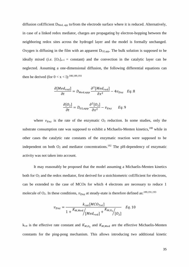
Figure 10. Two-dimensional case diagram showing the five limiting cases predicted
analytically. The concentration profiles surrounding the case diagram are from simulations at
the points marked () on the case diagram: case I, = 0.1, = 0.01; case II, = 10, = 0.001;
case III, = 10, = 1; case IV, = 10, = 1000; case V, = 0.l, = 100. The continuous
lines indicate dimensionless mediator concentration profiles ([Medred]/[MedTot], assuming that
the mediator is confined in the film); and the dotted lines the dimensionless substrate
concentration profiles ([O2]/KP,O2[O2]). Horizontal scale is from x = 0 (electrode) to x = 1
(bulk solution). Vertical scale is dimensionless concentration. Note the compressed vertical
scale for profiles I and V. Reprinted with permission from [188]. Copyright 1995 Elsevier.
The two dimensionless coordinates of the resulting case diagram are , which
describes the relative thicknesses of the film and diffusion layer; and , the relative
concentrations of enzyme in its reduced or oxidized forms. In the first case (I), the limiting
step is the enzyme-mediator reaction (i.e. mass and charge transport of substrates and redox

38
mediators within the film are faster than the enzymatic reaction). The second case (II)
corresponds to limitation by the mass and charge transport of the redox mediator. In case III,
the reaction kinetics are much faster than substrate and mediator diffusion, so that the
enzymatic reaction is limited by the substrate in one part of the film, while it is limited by the
mediator in the other part of the film. This situation results in the reaction occurring only in a
very thin region inside the film. The fourth case (IV) corresponds to limitation by the mass
and charge transport of the substrate. Finally, in case V the limiting step is the enzyme-
substrate reaction (i.e. mass and charge transport of substrate and redox mediators within the
film are faster than the reaction).
More recently, analytical solutions were derived using the homotopy perturbation
method (HPM) combined with the inversion conjuncture in the Laplace plane.192,195,196 While
limiting cases mostly describe steady-state electrochemical responses, this method allowed
describing current densities in chronoamperometry at different potentials both in the transient
and steady states. It was applied to an electrode based on MET of laccase in an osmium redox
hydrogel. The time to reach steady-state was determined, and proved to depend on the
potential of the redox mediator. Interestingly this approach could show how much the steady
state current densities were influenced by the enzyme characteristics (catalytic and Michaelis-
Menten constants), concentration of substrate in the bulk, and film thickness.192 In a further
study, the same approach showed its significance for the derivation of analytical expressions
of steady-state current densities and O2 and mediator concentrations, valid in the potential
step methods. Moreover, two graphical procedures were proposed for the estimation of
Michaelis-Menten constants. In both cases, the model was validated by fitting with
experimental data of oxygen-reducing laccase cathodes.192,196
3.3.2.2 Full numerical derivation

39
In the first study of Bartlett mentioned above, numerical simulations of the one
dimensional model were performed based on the relaxation method, and were in good
agreement with the analytical study.188 This 1D model was then applied to an enzymatic O2
reducing porous cathode.193 The composite electrode was modelled as cylindrical conducting
fibers around which the reaction layer film was coated. The influence of morphology (in
terms of film thickness, composite porosity and fiber diameter) was evaluated. The authors
also studied how to improve mass transport of the oxygen substrate, and predicted that the
maximum current densities could be close to 100 mA.cm-2 for a pure O2-breathing electrode,
and 60 mA.cm-2 for an air-breathing electrode.193 While this study mainly focused on thick
films, another proposed to consider high-surface area electrodes grafted with thin polymer
layers.189 In that case also it was proposed that currents around 100 mA.cm-2 could be
achieved with the porous electrode.
Another important contribution190 used modeling to determine the effect of ∆Eet
(difference between the T1 and the redox mediator potentials) on the bimolecular rate
constants of the enzymatic reaction; kcat/Km and kcat/KS towards mediator and oxygen
respectively (corresponding to kcat/KM,Med and kcat/KM,O2 with our notations). Using a finite
difference method, expressions of substrate and redox mediator concentrations were
determined, from which the expression of current density was derived. This expression was
used to extract kinetic parameters from experimental CVs recorded with T. hirsuta laccase
immobilized in redox hydrogels with osmium based redox mediators covering a wide range of
potentials.190 The bimolecular rate constants were found to be slightly lower in the film than
those determined in free solution.156 In addition, the performance of the cathode was found to
vary linearly with ∆Eet for ∆Eet < 300 mV, and to be independent on ∆Eet for ∆Eet > 300 mV
(Fig 11).190

40
Figure 11. Effect of mediator redox potential, E0, on laccase-mediator bimolecular rate
constant, kcat/Km, and laccase-oxygen rate constant, kcat/Ks. Also shown are redox potentials of
laccase and O2/H2O at pH 4. A linear free energy region is observed for high E0 (small ΔEet)
and constant mediation rate at lower E0 values. Reproduced with permission from [190].
Copyright 2008 American Chemical Society.
A model of a complete glucose/O2 EBFC composed of a cascade of enzymes and a
redox mediator at the anode, and an air-breathing cathode based on BOD with ferricyanide
has also been reported.194 This model was used to describe an EBFC formerly reported by
Sakai et al.197 where both enzymatic systems were immobilized on porous carbon-fiber
electrodes. The simulated data agree with the experimental results. Clues could be obtained
concerning the evolution of concentrations and currents over time at a given cell voltage, but
also concerning the evolution of several parameters in space. For example, pH was shown to
vary in the cell, being maximal at the anode. When a voltage was applied across the cell for a
long time, the overpotential at the carbon-fiber cathode was found to drop drastically with the
distance from the electron collector/carbon-fiber interface.194

41
3.4 Studies of the mechanism of direct electron transfer
In this part, we discuss the electrochemical behavior of enzymes at the electrode
interface in DET and explain general mechanistic clues provided by electrochemical studies.
In the DET mode, electrons could theoretically be transferred directly to the TNC, provided
that this latter is accessible and close enough to the electrode surface, thus bypassing the
internal electron transfer. Therefore, one of the major questions is whether the O2-reduction
mechanism remains unchanged for an enzyme directly connected at the electrode (DET
mode).
3.4.1 Redox potentials of the Cu sites
Redox potentials characterizing the different Cu sites of various MCOs have been
determined by redox titration in homogeneous solutions. T1 Cu, whose oxidation state can
only vary between +I and +II, is well characterized by a single redox transition at a
determined apparent standard potential E°’. Slightly different values for the same enzyme can
however be found in literature. MCOs can be classified according to the redox potential of
their T1 site in low (340-490 mV vs. NHE), middle (470-710 mV vs. NHE) and high (730-780
mV vs. NHE) redox potential MCOs.18,198 Tree laccases belong to the first group and fungal
laccases to the third one,18,199,200 while BODs are part of the 2nd with for example T1
potentials of 660 mV and 670 mV vs. NHE at pH 7 for Tt-BOD and Mv-BOD.201-204
Moreover, the T1 titration curve is indicative of a one electron process since the potential
varies by 49-90 mV per unit of log(Abs).118,202 The molecular origin of these potential
differences is discussed in section 2.1. It is more difficult to attribute redox potentials to the
TNC because this cluster can be characterized by several redox transitions. In very early
studies, the redox potentials of the T3 sites of R. vernicifera and T. versicolor laccases have
been determined at 480 and 780 mV vs. NHE resp.,199,200 while a redox potential of 390 mV

42
vs. NHE was attributed to the T2 Cu of the low-potential Rv-laccase.199,200 In this section,
unless stated otherwise, we quote redox potentials against the NHE reference electrode for the
sake of consistency with the literature.
Determination of the redox potential of T1 in MCOs has also been realized by non-
mediated spectroelectrochemistry following the vanishing of the blue band at 600 nm upon
application of reductive potentials and its re-apparition upon re-oxidation.118 Direct electron
transfer between a gold electrode and a high-potential laccase was also observed for the first
time with this technique, with Trametes hirsuta in a gold capillary.118 Non-mediated
spectroelectrochemistry furthermore confirmed the possibility to establish DET with the T1
Cu for several fungal laccases from different basidiomycetes (Trametes hirsuta, Trametes
ochracea, Cerrena maxima).205 Simultaneous detection of T1 and T3 Cu (at resp. 600 and 300
nm) also proved that enzymes could be fully reduced via DET from an electrode.205 Here
again, analysis of the titration curves gave information about the electron transfer mechanism.
In case of high potential Th-laccase and Mv-BOD, DET to the T1 was very slow, and the
curve showed a hysteresis and 2 noticeable electron transfer (ET) processes. This was
attributed to ET to the T1 (resp. at 860 mV for Th-laccase and 805 vs. NHE for Mv-BOD)
mediated by another redox site in the MCO (resp. at 290-370 and 460 mV vs. NHE for Th-
laccase and Mv-BOD).118,202 Since only ascorbate can fully reduce the laccase, implying that
the redox potential of the T2 might be considerably more negative than the T1, it was
proposed that the T2 redox potential for high-potential laccases was about 400 mV vs. NHE at
pH 6.5. Therefore, although in these cases electron transfer from the electrode to the T1 was
realized without the help of an external redox mediator, it was not, strictly speaking, “direct
electron transfer” to the T1. It proceeded via another enzymatic redox site of lower potential
and most likely the T2 Cu. 118
3.4.2 Non-catalytic faradaic processes under anaerobic conditions

43
In several cases, direct electron transfer (DET) between MCOs and electrodes has
been observed under anaerobic conditions and characterized by the appearance of reversible
or quasi reversible redox peaks in cyclic voltammetry (CV), linear sweep voltammetry (LSV)
or square wave voltammetry (SWV). For high-potential laccases, different redox peaks have
been observed corresponding to either a high or a low-potential process, or both. The
midpoint redox potentials (given by 𝐸𝑚 =𝐸𝑎+𝐸𝑐
2 where Ea is the potential of the anodic peak
and Ec is the potential of the cathodic peak) were usually observed in the 300-450 mV vs.
NHE range for the low-potential processes, and between 600 and 900 mV vs. NHE for the
high-potential processes respectively.
3.4.2.1 Strongly oxidizing laccases
3.4.2.1.1 Low redox potential
In 1999, a low redox quasi reversible process at Em = 411 mV vs. NHE was observed
at pH 6 for a recombinant truncated Tv-laccase immobilized on a thiol-modified gold
electrode.206 Numerous other examples of this low-potential redox process can be found in the
literature: a pair of peaks with a midpoint at Em = 337 mV vs. NHE at neutral pH was
recorded for Tv-laccaseat thiol modified monocrystalline gold (111) surfaces;207 a single quasi
reversible redox process at Em = 405 mV vs. NHE was observed for Th-laccase in a gold
capillary.118 Th-laccase adsorbed on a bare gold disc, and laccases from Cerrena maxima,
Trametes hirsuta and Trametes versicolor covalently bound on 4-aminothiophenol (AMT) on
gold exhibited two pairs of redox peaks. One of them had a low-potential resp. at Em = 486
mV vs. NHE for Th-laccase on bare gold, and approx. 400 mV for the three laccases on
aminothiophenol.205 T. versicolor and T. hirsuta laccases entrapped in a cationic polymer
matrix on a gold electrode also exhibited two pairs of peaks, with one in the low-potential

44
range (resp. Em = 394 ± 3 mV vs. NHE for Tv-laccase and Em = 408 ± 4 mV vs. NHE for Th-
laccase at pH 6.5).208
This low-potential process, mostly observed with a gold working electrode,18,118,205-208
can hardly be assigned to the T1. It was hypothesized that it could be related to the T2 redox
transition. To confirm this hypothesis, experiments were realized with T2-depleted
enzymes.118 While a 50% T2-depleted Th-laccase gave 50% peak current, no low-potential
redox peak was observed for the apo-enzyme. In addition, in presence of F-, whose affinity for
the TNC has been shown in homogeneous solution, the midpoint potential increased by about
35 mV. All these experiments tended to confirm that T2 potential is around 400 mV vs. NHE,
and that in all the above mentioned cases, at least a fraction of laccases were oriented with the
T2 Cu facing the surface.118 Experiments performed with a recombinant laccase bearing a
Cys-tag, inducing different orientations of the enzyme on gold via the S-Au bond, also
supported the attribution of the peak at lower potential to a TNC intermediate. When upon
immobilization the T1 was repelled far from the electrode surface, and the T2/T3 approached
close to it, a redox process at Em = 377 mV vs. NHE was observed and attributed to the T2.
On the contrary when T1 and T2/T3 were at the same distance from the electrode surface, a
more irreversible pair of redox peaks with Em = 470 mV vs. NHE was observed and attributed
to simultaneous effect of both T1 and TNC Cu clusters.209
3.4.2.1.2 High redox potential
For high-potential laccases, a high-potential redox process, more certainly
corresponding to the T1 has also been identified (Fig 12).

45
Figure 12. Voltammograms recorded with HOPG electrodes (1) without and (2) with T.
hirsuta laccase (30 mg.mL-1 laccase solution) entrapped under a dialysis membrane at the
electrode surface. (A) Cyclic voltammograms recorded in air-saturated 50 mM citrate-
phosphate buffer pH 3.5 containing 100 mM NaClO4; scan rate: 10 mV.s-1. (B) Osteryoung
square wave voltammograms recorded under anaerobic conditions in 100 mM phosphate
buffer pH 6.5; amplitude: 10 mV, frequency: 10 Hz. Reprinted with permission from [210].
Copyright 2005 Elsevier.
A reversible redox process on a graphite electrode at 645-700 mV vs. NHE at pH 5 has
been observed for Tv-laccase, but only in the presence of a promotor (2,6-dimethoxyphenol
(DMP) or 4,4’-bipyridine).211 Later, Tv-laccase adsorbed on edge-plane pyrolytic graphite
exhibited a reversible redox process at 790 mV vs. NHE.212 Similarly, redox peaks between
780 and 800 mV vs. NHE at pH 6.5 were recorded by SWV for T. hirsuta, T. ochracea, and
C. maxima laccases adsorbed on HOPG.210 The same enzymes adsorbed on gold electrodes
exhibited high redox processes (between 857 and 903 mV vs. NHE), which lowered to
approx. 800 mV when the enzymes were covalently bound on AMT.205 Similarly, the high
potential redox peak of laccases from T. versicolor and T. hirsuta entrapped in a cationic
polymer matrix on a gold electrode were respectively 784 ± 3 and 778 ± 4 mV vs. NHE, in
good agreement with the T1 potential. 208
3.4.2.2 Low-potential laccases

46
For low potential laccases, electrochemical studies described only one pair of redox
peaks coinciding with the T1. A non-catalytic process with semi reversible behavior was
observed for Rv-laccase on a gold electrode modified with an anionic polymeric film at 410
mV (pH 5.5) or 400 mV vs. NHE (pH 7),213 and at 410 mV vs. NHE at pH 7 for the same
laccase covalently bound on mercapto-propionic acid (MPA) on gold.214 This anaerobic
process did not change upon addition of F-,214 and T2-depleted enzyme still exhibited the
same redox process while the apo-protein was electrochemically silent.213 Melanocarpus
albomyces and Rv-laccases entrapped in a cationic polymer matrix on a gold electrode also
exhibited only one redox process at resp. 422 ± 4 and 449 ± 5 mV vs. NHE at pH 6.5, in good
agreement with T1 potential.208
3.4.2.3 Bilirubin oxidases
BODs behave approximately like laccases with respect to their electrochemical
response in absence of O2. For Mv-BOD adsorbed on SPG, a unique and very small quasi
reversible process at Em = 515 mV vs. NHE at pH 7.4 with a large peak separation was first
recorded.215 Later, two pairs of peaks were simultaneously identified at Em = 690 mV (resp.
700 mV) and Em = 390 mV (resp. 400 mV) vs. NHE at pH 7 for Tt-BOD (resp. Mv-BOD)
adsorbed on SPGE (Fig 13).203,204

47
Figure 13. Representative cyclic voltammograms of (A) Cerrena maxima and (B) Trametes
hirsuta laccases immobilized on (A) bare and (B) 4-aminothiophenol self-assembled
monolayer (SAM) modified gold disc electrodes (0.1 M phosphate buffer pH 5.0; scan rate:
10 mV.s-1; second scan). (A) CVs of Cerrena maxima laccase under aerobic and anaerobic
conditions, and in the absence or presence of sodium fluoride starting at 1040 mV vs. NHE.
(B) CVs of Trametes hirsuta laccase under aerobic or anaerobic conditions, and in the
absence or presence of sodium fluoride starting at 1040 mV vs. NHE. Reprinted with
permission from [205] Copyright 2006 from Elsevier.
Furthermore, a quasi-reversible redox process at Em = 360 mV vs. NHE at pH 7,
observed for Tt-BOD covalently bound on MPA on gold, was attributed to a resting form of
the enzyme because it was almost not influenced by F-.203 This led to the suggestion that an
endergonic tunneling from the T1 to T2/T3 could be involved in MCO mechanism.203 A non-
catalytic low-potential redox signal of Mv-BOD (360 mV vs. NHE at pH 7), attributed to a
resting form of the enzyme, also appeared when the enzyme was oriented with the TNC
facing the electrode surface.216 3 non-catalytic cathodic peaks (at resp. 693, 574 and 403
mV vs. NHE), and only one anodic (at the lowest potential) were visible on a CV at pH 6 for
Mv-BOD entrapped in a matrix of Ketjen black (KB), carbon nanotubes (CNTs) and chitosan.
The highest was assigned to the T1, consistently with previous studies, and it was proposed
that the 2 others might be attributed to different redox states of the TNC, containing or not
bridging oxygen species.217 It is noteworthy however that a low potential redox peak at 420
mV had previously been assigned to a surface group of the enzyme.218,219 The simultaneous
presence of two pairs of peaks suggested that for different fractions of BOD only the T1 or the
T2 was in contact with the electrode.208 The appearance of the low-potential redox process
was attributed to either the T2 or to a coincidental electrochemical response of the 4 Cu

48
ions,18 while the high potential peak was attributed to the T1. Since different redox processes
could be observed for the same enzyme immobilized at different materials, it was proposed
that different protein sites were communicating with the electrode, depending on the electrode
material.203
3.4.3 Two- versus four-electron reduction of dioxygen
The redox processes observed in anaerobic conditions, especially those at low
potential, could not always be correlated to a catalytic current in aerobic conditions. This
means that some enzymes were immobilized in a configuration that permitted direct electron
exchange with the electrode, without however enabling the catalytic mechanism. This remark
suggested that in these cases the enzymes were not connected via the proper redox
center.118,207 This hypothesis was confirmed in some cases by the addition of a redox mediator
that enabled MET.203,220
The onset of catalysis was observed in some rare cases in the low-potential range for
high- or middle-potential MCOs. Onset at 300 mV vs. NHE at pH 5 was for example
reported for the fungal Th-laccase on bare gold221 or AMT self-assembled monolayer (SAMs)
on gold.222 Similarly, oxygen reduction by Mv-BOD in solution started at 450 mV vs. NHE,
when a gold wire modified with a mixed mercapto-undecanol/mercapto-undecanoic acid
SAM was used as the working electrode.223 Only low catalytic currents were observed with
these systems, while non-catalytic signals were not detected.221-223
The influence of F-, which is known to inhibit the IET in homogeneous catalysis, has
been also studied. For example, Th-laccase covalently bound on 4-AMT-modified gold
electrode exhibited a first reduction current starting at 510 mV vs. NHE at pH 5, which was
followed by a second more steeper increase at 240 mV. F- inhibited only the high-potential (E

49
> 240 mV) reduction, while the current at lower potentials remained unchanged (Fig 13).
Similar observation was done for Cm-laccase on a bare gold disc: O2 reduction started at a
quite low potential, 320 mV vs. NHE, and was not influenced by F-. This led to the
supposition that in both cases no IET was occurring.205
Moreover, the catalysis starting at low potential has been correlated to the incomplete
O2 reduction to H2O2 in several cases. For example, when a fungal laccase was adsorbed on
bare gold electrodes, H2O2 production in the low potential range (E < 240 mV vs. NHE) was 3
times higher than for naked gold alone. It was lower both in presence or absence of enzyme at
higher potentials (240-440 mV vs. NHE).205 Similarly, for an engineered laccase adopting
different orientations on a gold electrode, oxygen reduction was observed either at 495 mV vs.
NHE or at 340 mV vs. NHE and assigned respectively to DET by the T1-TNC or by the TNC
alone. It was associated with H2O2 production of resp. 2.75 and 7.53 µM.209 It seems
therefore that DET from the electrode to the T2/T3 is possible but leads either to no catalysis
or in the best cases to the 2-e- reduction of O2 to H2O2. It clearly appears that the orientation of
the enzyme at the electrode influences both the electron transfer pathway and the electron
efficiency of O2 reduction.209
Contrary to the catalysis observed at low potential and assigned to the reduction of O2
to H2O2, no significant formation of hydrogen peroxide has been reported for catalysis
starting at the T1 potential. H2O2 evolution was for example estimated with a porphyrin-
coated electrode in a solution in which O2 had been reduced by a Tv-laccase electrode. Direct
ORR started at 0.84 V vs. NHE at pH 5.5 with the fungal Tv-laccase adsorbed on graphite.
The experiment confirmed that no more than 5% O2 could have been reduced to H2O2.211 A
similar experiment was performed with Mv-BOD attached to multi-wall carbon nanotubes
(MW-CNTs) on the disc of a rotating ring disc electrode (RRDE), for which the ORR onset
was 0.7 V vs. NHE at pH 7.5. During the CV, the platinum ring was polarized to measure

50
the reduction of H2O2. Zero hydrogen peroxide was detected on the disc, and according to the
mass and charge balance of the RRDE no more than 7.5% O2 in total could have been reduced
to H2O2.224 In both cases, Koutecky-Levich analysis of the rotating electrode matched a 4-
electron reduction to water and not H2O2.211,224
It is therefore of interest to determine the number of electrons exchanged by an
enzyme respectively in high- versus low-potential anaerobic redox processes. For a peak at
high potential, the ratio of anodic to cathodic currents were shown to vary from 1:1 (which
actually can be converted to 4:4 by normalizing the peak intensity ip by the scan rate v) at very
low scan rates (below 1 mV.s-1) to 4:1 at fast scan rates (above 200 mV.s-1).208,214 Further
analysis of the peak separation ∆Ep as a function of ∆log(f) in SWV indicated that 3.65
electrons were transferred for Tv- laccase and 3.85 for Th-laccase.208 These results support a 4
e- transfer to/from the T1 site, which would correspond to a possible full reduction/oxidation
of the enzyme via the T1, with the full reduction being much slower than the oxidation. For a
low-potential peak of high-potential laccases, the width of the peak (given by the difference
between the half-wave potential EHW and the peak potential EP) was consistent with a 1 e-
transfer.118 The anodic to cathodic Qa/Qc charge ratio increased from 1:1 to 3:1 with
increasing v; and SWV analysis gave numbers of electrons of 2.78 for Tv and 2.9 for Th-
laccase208 which suggests that full reduction of the enzyme cannot occur via the low-potential
intermediate whereas it is possible via the T1 Cu. For the unique redox peak of low-potential
Ma and Rv-laccases, Qa/Qc varied from 1:1 for v < 100 mV.s-1 to 4:1 for scan rates above 800
mV.s-1, and SWV gave n values close to 4 (n = 3.9 for Rv and 3.8 for Ma-laccases).208 The
general observation that the reduction peak is lower than the oxidation peak118,205 or that the
rate of oxidation is faster than the reduction in CV or SWV208,214 is consistent with the
hysteresis noticed in spectroelectrochemistry, which also shows that the oxidation process is

51
significantly faster than the reduction process.205 All this suggests that IET might be the rate-
limiting step.
3.4.4 Causes of the low catalytic efficiency and stability on gold electrodes
Different behaviors were observed for BODs and laccases on gold electrodes. On bare
gold, besides the H2O2 related mechanism described above, no catalysis at high potential or
no catalysis at all was recorded for Th-laccase,205,225 Cm-laccase,205 or small laccase from
Fusarium proliferatum.226,227 On the other hand, a clear DET catalysis was observed for Mv-
BOD on bare monocrystalline gold17,225 or platinum electrodes,17 with an onset above 0.7 V
vs. NHE at pH 5.8 consistent with the T1 redox potential. With this latter enzyme, the
observed electrocatalytic responses vanished however relatively quickly17 with for example
half-lives of resp. 15 minutes and 2.5 hours at pH 4 and 7.4, rather due to gradual deactivation
than to desorption of the enzyme.225
The efficiency and stability of catalysis at gold electrodes was investigated by
coupling electrochemistry to other techniques like quartz crystal microbalance (QCM),151,228
QCM with dissipation monitoring,229-231 surface plasmon resonance (SPR),220 ellipsometry,225
atomic force microscopy (AFM)225-227 or dual polarization interferometry (DPI).231 No
desorption of either BOD or laccase immobilized on bare or on thiol-modified gold electrodes
was measured by ellipsometry, SPR or QCM, confirming the irreversible character of the
adsorption of the enzymes on these surfaces.220,225,228,230,231 Therefore, the decrease in
cathodic current observed in numerous studies cannot be related to enzyme loss but most
probably to enzyme denaturation or structural rearrangement.220,228,229 The enhancement of
stability observed with amide bonding of MCOs on the electrode surface was more likely due

52
to a stiffening of the immobilized layer preventing conformational changes, rather than due to
a better binding of enzymes to the electrode.220,229
3.4.4.1 Denaturation by enzyme-metal interaction
For MCOs adsorbed on bare metal surfaces, a certain loss of activity towards
oxidation of organic substrates was demonstrated. DMP oxidation was 7.5 times less Efficient
for Fp-laccase adsorbed on bare gold than on HOPG.227 Enzymatic assay with 2,2'-azino-
bis(3-ethylbenzothiazoline-6-sulphonic acid) (ABTS) showed that Th-laccase was completely
deactivated on bare polycrystalline gold, while some activity was still recorded for Mv-
BOD.225 Denaturation of MCOs adsorbed on bare metal has already been suggested.232 Very
fast adsorption of Fp-laccase on bare gold was observed by atomic force microscopy (AFM),
and the formation of aggregates with a wide variety of sizes suggested an intense interaction
with bare gold.227 For Mv-BOD and Th-laccase on bare gold, the size of globular features (
20 nm) was considered normal, taking into account the tip dilation effect of the AFM.225
Similarly, the low heights of the molecules, ranging between 2.5 and 3 nm, was attributed to a
tip compression effect that makes soft biological molecules appear always smaller with
AFM,225,227 although it could also correspond to a flattening of enzymes at the surface.231 Mv-
BOD was also more stable on MPA-modified gold than on bare gold.225 BOD faster
deactivation on single crystal might be due to tighter holding.17 These observations can be
correlated with the faster denaturation caused by stronger electrostatic interactions observed
when MCOs are adsorbed on N-doped carbon nanotubes. (see section 4.1.2 for further details)
3.4.4.2 Applied electrode potential
A potential-induced structural change of the enzyme was evidenced by in situ STM
studies of Streptomyces coelicolor laccase adsorbed on butanethiol modified Au (111)

53
surface.233 This technique allowed at the same time the first single-molecule resolution in
aqueous buffer. Interestingly the oxidized enzyme (at 0.89 V vs. NHE) appeared transparent,
while bright spots were observed for the enzyme under turnover (at 0.44 V vs. NHE under air)
(Fig 14A).
Figure 14. Influence of applied electrode potential on enzyme. (A): structural change induced
by potential, evidenced by in situ STM of Streptomyces coelicolor laccase on a butanethiol-
modified monocrystalline gold under pure O2 atmosphere. A1: Initial image at 0.84 V vs.
NHE. Bias: 0.70 V. A2: Image at 0.44 V vs. NHE. Bias: 0.30 V. A3: Image after returning at
0.84 V vs. NHE. Bias: 0.70 V. Tunneling current in all cases: 0.035 nA. Scan area: 100 × 100
nm2. A4 and A5 are histograms showing the height distribution of the bright spots in A2 and
A3, respectively. Height normalization was done against the maximum height measured in
A2. Lines are best fits to a normal distribution. Adapted with permission from [233],
copyright 2012 American Chemical Society. (B): Comparison of the catalytic current loss
measured in cyclic voltammetry after the Mv-BOD electrode has been left at OCV (red
columns) or submitted to an applied potential E = +0.2 V vs. Ag/AgCl (black columns). The
error bars are represented in green. For better accuracy with respects to the original figure, the
timescale is not linear. Adapted with permission from [220]. Copyright 2016 American
Chemical Society.
Under O2, a stronger STM contrast was observed and the process was less reversible
than under air. This study therefore highlights a structural change of the protein, that enables
current tunneling through the enzyme only in its oxidized form.233 Other studies suggested

54
either a conformational change or reorientation under applied potential.220,229,234 For Mv-BOD
adsorbed or covalently bound to thiol-modified gold electrodes, currents decay did not
coincide with the mass decay228,229 but with the energy dissipation change,229,230 indicating a
non-desorptive degradation and most probably structural changes. Since no activity was
recovered upon addition of a diffusing redox mediator; the current loss could not be explained
by reorientation of some enzymes.220
The current was even less stable when the potential was applied continuously,
compared to punctual measurements (Fig 14B).220,229 At the same time, the dissipation losses
were much higher, meaning that the BOD layer stiffened under application of a potential, and
supporting a potential-induced enzyme denaturation. Moreover, under potential cycling, a
mass drop was also recorded, therefore suggesting protein dehydration.229 This could be an
important cause of activity loss, since different studies have shown that drying the BOD
electrode in a desiccator led to complete loss of the enzymatic activity, which was attributed
to denaturation of the enzyme by losing water.235,236
To rationalize the direct influence of potential on the active site, the behavior of the
fraction of enzymes that were not directly connected at the electrode was investigated in MET
upon addition of a redox mediator. A more stable current was observed for Mv-BOD adsorbed
or covalently bound on NH2 terminated SAM on gold electrode in MET configuration, i.e.
when the enzyme was not directly submitted to potential cycling.220 However, on carboxylate
SAMs, the fraction of enzymes oriented in MET also lost its activity. These results suggest
that direct influence of the potential on the active site is not the only origin of denaturation.220
The electric field created at the electrode interface could also be deleterious for the enzyme
structure, but to the best of our knowledge, no direct proof of this hypothesis has been
reported. Since in the case discussed above the systems essentially differ by the chemistry of

55
the electrode surface, it also indicates that the electrode surface charges influence on the
enzyme stability.220 This effect is discussed further in section 4.1.4.
3.4.4.3 Enzyme concentration
Catalysis constants kcat much lower than values for homogeneous catalysis (250 s-1)
could be calculated for a surface coverage close to one monolayer of Mv-BOD adsorbed on
bare gold (ca 4 s-1)228 or on 3-MPA-modified flat polished gold surface of a QCM sensor (35
s-1).229 The decrease in activity with time was higher for immobilized enzymes than for
enzymes in solution.220 This suggested that BOD was poorly oriented at the electrode, and
that some denaturation occurred upon immobilization. Furthermore, at pH 4 more enzyme
was adsorbed but the measured current was even smaller than at pH 7.228 Similarly, a lower
efficiency was recorded for a monolayer of Mv-BOD on mercapto-hexanoic acid (MHA)-
modified gold (5 pmol.cm-2) than for a sub-monolayer (0.6 pmol.cm-2) with global kcat of
respectively 7 and 48 s-1.220 It was therefore likely that higher BOD concentrations were
detrimental, due to the increased interactions between neighboring BOD molecules. However,
the opposite effect has also been observed. The behaviors of Mv-BOD and Th-laccase upon
immobilization on bare gold electrodes were compared. Mv-BOD-always led to higher
surface coverages, and at the same time to the highest enzyme specific activity. It was
consequently proposed that steric effect or electrostatic interactions between neighboring
molecules, induced by higher enzyme coverage, could prevent the enzyme from flattening,
thereby lowering inactivation due to conformational changes.225,231
The influence of the initial Mv-BOD concentration on the adsorption was investigated
by DPI: intermediate concentrations (10-25 mg.mL-1) were shown to lead to the most
Efficient (in terms of kcat) and persistent (in terms of kcat stability) layers, with an optimized
kcat of 60 s-1.231 Adsorption was shown to occur in 2 stages: first an irreversible binding of a
protein layer attributed to the spreading of the molecules at the surface, and then the

56
formation of a less rigid layer of proteins. Higher concentrations of the enzyme solution
produced less rigid layers in the first phase, because faster adsorption prevented enzyme from
relaxing and flattening at the electrode surface. This correlated well with higher enzymatic
specific activity.231 However too dense layers also led to decreased efficiency.231
3.4.5 Catalytic mechanism on carbon electrodes
When BOD and laccase are immobilized on bare polished GC, catalytic currents have
not always been observed.235,237 Therefore unless stated otherwise, this paragraph is only
focused on studies of MCOs adsorbed on graphite electrodes. A well-defined catalytic wave
for O2 reduction was recorded for most enzymes (Fig 15). To determine the primary electron
acceptor in heterogeneous electron transfer (HET), characteristic potentials of the ORR under
aerobic conditions as well as their variation with pH have been first considered. This follows
the general assumption that catalysis is controlled by the potential of the redox center at which
electrons enter/leave the enzyme, and not by the potential of the actual catalytic center.238,239
Ecat, the potential corresponding to the maximum acceleration of the catalysis (i.e. E for
maximal di/dE) is a convenient tool to describe values of characteristic potentials in catalysis,
because it contains both thermodynamic information and clues about kinetics of the
transformation.165,240 However the steady-state potential of the electrode (OCP), onset
potential or half-wave (EHW) potential of the catalytic ORR wave are more widely described
in the literature.

57
Figure 15. (A) Cyclic voltammogram of BOD adsorbed on SPGE in the absence of O2. pH
7.0, 0.1 M phosphate buffer; scan rate: 100 mV.s−1; starting-scan potential: 1000 mV vs.
NHE. Curve 1: uncorrected for the background current; Curve 2: circles, background current
subtracted. The broken line curve represents the calculated theoretical voltammograms. (B)
Effect of the scan rate on the background-corrected voltammograms under O2. Curve 1:
10.mV s−1; Curve 2: 100 mV.s−1. Reprinted with permission from [203] Copyright 2008
Elsevier.
OCP for Th-laccase increased from 780 to 910 mV vs. NHE for pH decreasing from 6
to 3;210 and changed by 20-30 mV/pH unit between pH 4.5-6 for Th-, To-, Cf- and Cm-
laccases, and by 65, 33, 33, and 13 mV/pH resp. for the same laccases between pH 3 and
4.5.210 Approximate values of ORR onset were also reported: 840 mV and 490 mV vs.
NHE at pH 5.0 for Th- and Rv-laccases resp.,221 840 mV vs. NHE for Tv- laccase at pH
3.8,211 and values between 840 and 900 mV vs. NHE at pH 6.5 for Th-, To-, Cu-, Cm-, and Cf-
laccases.210 Similar values were recorded for both Mv- and Tt-BOD. The ORR onsets were
800 mV vs. NHE at pH 4 and 700 mV vs. NHE at pH 7.4 resp. for Mv-BOD,204,215,217,241
which coincided well with the high-potential quasi-reversible redox transformations observed
under anaerobic conditions, and 810 mV at pH 4, i.e. 120 mV above E°’T1, for Tt-BOD.204 All

58
these values of onset potential are slightly higher than the redox potential of the T1 Cu center,
indicating that the T1 might be the electrochemical control center of catalysis.
The onset pH-dependence was precisely 60 mV/pH unit in the first report for
laccase,242,243 which was consistent with a 1 proton-1 e- exchange, but slightly higher than 52
mV/pH unit reported later for Mv-BOD on SW-CNTs.244 Furthermore it was more than twice
higher than slopes of 20-25 mV/pH unit described for Mv and Tt-BODs on graphite.204 These
latter values were consistent with 23 mV/pH unit observed for the non-catalytic signal of both
BODs, and close to the pH dependence described for laccase T1 potential.200 This is a further
indication that T1 controls the potential of catalysis.
In the absence of diffusion limitation, it is also consistent to consider the half-wave
potential EHW, which coincides with the redox potential of the electrochemical control center
in catalysis, under kinetic limitation. For Mv-BOD, EHW was 670 mV vs. NHE at pH 7.4 in
one study,241 while a lower value of 600-605 mV vs. NHE at pH 7.4 can be found
elsewhere.215 This latter value shifted to 700 mV vs. NHE at pH 4.0, consistent with a change
of 30 mV/pH unit.215,217 Potentials lower than expected were ascribed to a slow HET. On the
other hand, EHW for Tt-BOD coincided well with Em of the high-potential redox process (690
mV).203 EHW was between 740 and 800 vs. NHE and changed by 5-30 mV/pH unit in the pH
range 3-5 for Cf- and Cu-laccases.210 An approximation of the standard apparent potential of
the redox center involved in catalysis, E°’, was determined for Mv-BOD by fitting the curves
with the model developed by Ikeda.235 E°’ was found quasi constant in the pH range 2-5.5 (
670 mV vs NHE) and a shift to less positive values with an average variation of 45 mV/pH
unit was recorded between pH 6 and 8. 245
For the different MCOs adsorbed on various graphite electrodes, reproducible values
were obtained for characteristic potentials of catalysis (OCP, onset, EHW and E°’) and all

59
correlated with values of the T1 Cu potential. Although one must be careful that CV curves
are not in all cases centered on the potential of the first electron relay when enzymes present
several redox centers,246 it is clear that the T1 Cu strongly influences the potential of catalytic
O2 reduction. Moreover, the catalytic potential correlates in numerous cases with a non-
catalytic signal centered on the T1 potential, indicating that this latter must be directly facing
the electrode surface. This strongly supports that DET at graphite electrodes under turnover
conditions occurs via the T1 Cu, similarly with the O2-reduction mechanism in homogeneous
catalysis. 18,198,202
This conclusion is furthermore supported by the electrochemical behavior of a mutant
of Mv-BOD, Met467Gln in which the axial methionine ligand of T1 Cu was replaced by a
glycine residue, and its comparison to that of the recombinant wild type BOD.247 The onset
potentials for O2 reduction by both enzymes were consistent with the previously-determined
T1 potentials, with a negative shift for the mutant at 230 mV vs. Ag/AgCl instead of 460 mV
vs. Ag/AgCl at pH 7.248
A further indication that a similar reaction mechanism is adopted is the consistency of
the enzyme activity with that observed in homogeneous solution. pH dependence of maximal
currents under kinetic control display a bell shape typical of reaction rate of MCOs vs.
pH.17,215,221,242 KM for O2 of resp. 0.11 mM and 0.27 mM at 500 and 50 mV vs. NHE at pH
7,204 and 0.7 mM at 342 mV vs. NHE at pH 5 were determined for graphite-adsorbed Mv-
BOD.17 These values are close to 0.2 mM at pH 7 measured in homogeneous system, which
suggests that Mv-BOD preserves its native state after adsorption at the carbon material.
Even though the same tendency has been reported in the literature concerning the
characteristic potentials at steady state and catalysis, the pH dependence of activity, KM
constants…, slight variations have been observed. They are probably due to different surface
60
states of the electrode, which result in varying affinities with MCO molecules (see section
4.1.4). However, no considerable deviation from homogeneous catalysis has been observed.
The same general conclusion can therefore be drawn for both laccases and BODs: since the
properties of heterogeneous electron transfer at graphite electrode are similar to those in
homogeneous reaction, the T1 copper must be the first electron acceptor of DET between
MCOs and carbon electrodes. This is furthermore consistent with T1 being situated not
further than 8 Å from the enzyme surface. Electrons accepted from the T1 are then transferred
to the TNC where the 4e--O2 reduction occurs.
Similarly, at modified gold electrodes, DET occurring at the T1 was also observed for
both high and low-potential laccases.233,249 Rv-laccase covalently bound on MPA-modified
gold showed an onset at 500 mV vs. NHE, and a half-wave potential EHW = 400 mV vs.
NHE at pH 7.214 For Rv- and Ma-laccases in a cationic polymer on gold, the onset was 450
mV vs. NHE at pH 6.5 and EHW was close to the non-catalytic process at 350 mV.208 Addition
of fluoride inhibited the catalysis both for BOD and laccase,17,214 while no change of the
anaerobic voltammogram was observed,214 which pointed to the inhibition of the internal
electron transfer. All these data showed that a similar mechanism accounts for homogeneous
and heterogeneous O2 reduction by MCOs, provided that DET to the T1 can be established.
The effect of surface modification on DET bioelectrocatalysis is discussed further in section
4.1.4
3.4.6 Possibility of endergonic electron-transfer
The general mechanism at carbon (or conveniently-modified gold) electrodes has been
described in previous paragraph. However, a more intriguing phenomenon has sometimes
been observed. Two consecutive catalytic waves in the cyclic voltammogram of O2 reduction
were detected in following cases. When Th- and Tv-laccases were immobilized within a

61
polymer on a gold electrode, an onset at 730 mV vs. NHE at pH 6.5 followed by a more
drastic increase at 620 mV was recorded.208 For both Mv- and Tt-BODs at graphite electrodes,
two consecutive reduction waves were observed in the absence of diffusion limitation (Fig
15).203,204 These processes coincided well with 2 quasi-reversible redox transformations at low
and high potential observed under anaerobic conditions (Fig 15).203,204,208
This curious phenomenon was particularly investigated for BOD electrodes.203,204
Several hypotheses had to be examined before considering that it was catalytically relevant.
First, a damaged and a native active fraction of enzyme could have been simultaneously
immobilized onto electrodes. Second, two different catalytically active BOD fractions could
coexist. The catalytic currents decreased over time but did not result in change of the
voltammogram shape, thus ruling out the first possibility. Moreover, since BODs had been
purified the second hypothesis was very unlikely.204 It was therefore proposed that a single
enzyme form could adopt 2 different orientations at the electrode interface. Several redox
centers of the oxidoreductase could therefore be addressed simultaneously.204,208 This
proposition is not contradictory with published BOD structures, in which both Cu clusters are
situated within tunneling distance from the enzyme surface.129,146
The low-potential wave was attributed to a catalytically active intermediate of the
TNC118,203,204 with a potential close to 400 mV vs. NHE. The redox transformation of this
intermediate was shown to be O2-independent and pH-dependent. As a consequence, it was
attributed to the redox transformation between the first and second partly reduced TNC
intermediates, i.e. to the proton-coupled redox transformation from the peroxide intermediate
to another state.95,250 This latter enzyme state would be comprised between peroxide and
native intermediates. It is noteworthy that, later, catalytic oxygen reduction by Mv-BOD
starting exclusively at low potential could be observed when the controlled immobilization of

62
the enzyme at CNTs specifically approached the TNC close to the electrode surface. In that
case, the low potential was attributed to a resting form of the enzyme.216
Despite the disputable opinion concerning the redox intermediate involved in the
above described catalytic mechanism, it is not unreasonable to consider that at least one IET
is uphill. Page et al.146 and other studies251 have indeed calculated for a series of redox
enzymes that endergonic ET can occur, as long as neighboring redox centers are not separated
by more than 14 Å. Taking into account this maximal distance, reorganization energy λ
calculated from quantum mechanical and molecular mechanics (QM/MM) evaluations, and
∆G° calculated from the measured potential difference between the two detectable sites, pH-
dependent internal electron transfer rate constants between T1 and the TNC low redox
intermediate were calculated. They ranged from 3 s-1 up to 138 s-1, in agreement with
experimental kcat values.204
3.4.7 Mathematical modeling of the catalytic current
Different DET models can account for the mechanism of the O2 reduction by MCOs
electronically connected to the electrode via the T1. Importantly, all models are derived
considering non-limiting O2 diffusion. In the first general model proposed by Tsujimura et
al.,235 the internal electron transfer (IET) and the different steps of O2 reduction at the TNC
are included in a general “catalytic” mechanism (Fig 16 A).

63
Figure 16. Schematic representation of DET mechanism for O2 reduction by MCOs. (A):
model proposed by Tsujimura et al. (adapted from [235]). kf and kb are the rate constants of
the reversible heterogeneous electron transfer , and kcat is the global catalytic constant for O2
reduction by the BOD including intramolecular electron transfer and all steps of O2 reduction
at the TNC. The second step is considered irreversible in the model. (B): refined model. The
heterogeneous electron transfer is considered reversible and characterized by the rates for
forward and reverse electron transfers k1 and k-1. Similarly, the internal electron transfer is
considered reversible and characterized by the rates of forward and reverse electron transfers
k2 and k-2. The only irreversible step is the O2 reduction at the TNC characterized by the rate
constant k3. Adapted with permission from [204,233]. Copyright 2012 Wiley-VCH Verlag
GmbH & Co. KGaA, and American Chemical Society.
O2 reduction by MCOs can therefore be decomposed in two parts, heterogeneous
electron transfer between enzyme and electrode, and catalysis. If the second step is considered
irreversible, two rate constants, k°, the standard surface rate constant, and kc, the catalytic rate
constant, are enough to describe the kinetics of O2 reduction. It is noteworthy that kc normally
follows a Michaelis-Menten dependence on O2 partial pressure. Moreover, only HET rates are
supposed to be dependent on the electrode potential, and this dependency can be described in
a first approximation by Butler-Volmer kinetics (equations 13 and 14).
𝑘𝑓 = 𝑘°𝑒−𝛼𝑛𝐹
𝑅𝑇(𝐸−𝐸°′) 𝐸𝑞. 13
𝑘𝑏 = 𝑘°𝑒(1−𝛼)𝑛𝐹𝑅𝑇
(𝐸−𝐸°′) 𝐸𝑞. 14
where kf and kb are forward and reverse interfacial electron-transfer rate constants, resp. for
the reduction and oxidation, and k° is the standard heterogeneous electron-transfer rate
constant at the apparent standard potential E°’ of catalysis. α is the charge transfer coEfficient
(usually considered equal to 0.5), and R is the universal gas constant. Missing parameters can

64
be determined by fitting the experimental CV curves with the expression of steady-state
current density given by:
𝑗 =𝑛𝐸𝐹𝑘𝑐𝛤𝑡
1+𝑘𝑐
𝑘𝑓⁄ +
𝑘𝑏𝑘𝑓
⁄ 𝐸𝑞. 15
where 𝑛𝐸 is the number of electrons exchanged (a single electron exchange at the T1 copper is
considered), F is the Faraday constant, and Γt is the total surface concentration of the enzyme
(in mol.cm-2). This model was also extended for enzymes diffusing in a reaction layer, just
replacing Γt by µ[E] in eq. 15, where [E] is the total concentration of enzyme in the reaction
layer, and µ is the thickness of this latter.252 µ is given by µ = √𝐷𝐸𝑘𝑐
⁄ , with DE being the
diffusion coEfficient of the enzyme in the layer.
This model considers a very fast internal electron transfer (IET), therefore the enzyme
behaves as if only one redox center able to take 2 different redox states (reduced or oxidized)
were present. Although this is a strong simplification, the resulting equation is able to
generate curves that closely fit to the experimental voltammetry data. However it is quite
disputable if E°’ can account for a real thermodynamic potential of one of the Cu centers.
A refined, yet still simplified, model is described in Figure 16B.204,233 The internal
electron transfers between T1 and TNC are separated from the catalytic O2 reduction at the
TNC and considered reversible, while all steps of O2 reduction at the TNC are still included in
a global catalytic mechanism considered irreversible. 5 kinetic rate constants are therefore
needed to account for the forward and reverse HET (k1 and k-1), forward and reverse IET (k2
and k-2) and irreversible catalysis (k3). Heterogeneous electron-transfer can still be described
by Butler-Volmer type equations (equations 13 and 14, with kf = k1 and kb = k2), while
catalysis is best described by Michaelis-Menten kinetics (equation 16).233,253

65
𝑘3 =𝑘3,𝑚𝑎𝑥[𝑂2]
𝐾𝑀 + [𝑂2] 𝐸𝑞. 16
In this approximation, IET is considered to occur as a bimolecular reaction of electron
exchange between T1 and TNC. If forward and reverse IET rates are considered equal (i.e. k2
= k-2), the total current can be expressed as:233
𝑗 =−𝑘1𝑘2𝑘3𝑛𝐸𝐹𝛤𝑡
𝑘1(𝑘2 + 𝑘3) + 𝑘−1(𝑘2 + 𝑘3) + 𝑘2𝑘3 𝐸𝑞. 17
It is interesting to note that by then considering fast IET, i.e. when catalysis and HET are the
limiting steps, equation 17 can be further simplified to give back equation 15, with kc = k3, kf
= k1 and kb = k2.233,253 On the contrary, when IET is the rate limiting case, i.e. when catalysis
is very fast (k3 >> k2), eq. 17 becomes :
𝑗 =𝑛𝐸𝐹𝑘2𝛤𝑡
1 +𝑘−1
𝑘1⁄ +
𝑘2𝑘1
⁄ 𝐸𝑞. 18
Importantly, as evidenced by the absence of k3 in the equation, no dependence of the catalytic
current on O2 concentration is expected in this latter case.233 This catalytic scheme is however
still incomplete as it does not consider all electron transfer steps and catalytic intermediates of
the general mechanism of O2 reduction by MCOs. Blanford et al. proposed a complete
general mechanism for O2 reduction by immobilized MCOs, based on that developed by
Solomon et al.92,93 for homogeneous conditions (Fig 17A, see sections 3.1 and 3.4.8 for more
details).

66
Figure 17. A: A mechanism for MCO-catalyzed O2 reduction based on that of Solomon and
co-workers, with additions from Blanford et al. shown in red. B: Evidence for the formation
of the Resting Oxidized (B1) and X state (B2) in response to a series of applied potentials.
Eapplied is the experimental potential used, typically 0.4 V vs. NHE at pH 4; when the electrode
is at OCP no potential is applied. B1 indicates how the fraction of enzymes in the RO state is
calculated. B2 is observed in some particular combinations of pH and time at OCP. Adapted
with permission from [165] Copyright 2012 Royal Society of Chemistry.
Due to the complexity of the mechanism, and since their purpose was to explain the
transient phenomena, they did not use a steady-state approximation and did not derive a
general expression of current density. They proved however that all kinetic parameters could
be experimentally elucidated. Gray et al. proposed an expression of the O2 reduction current
that takes all the catalytically-relevant intermediates into account (Fig 18).254

67
Figure 18. Mechanism for MCO-catalyzed O2 reduction used in the analytical model
developed by Agbo et al. The steps including HET are indicated by orange arrows, the
catalytically relevant intermediates and internal steps are indicated in blue, and the resting
form outside of the catalytic cycle is indicated in green. For the nth step, the forward rate
constant is indicated by kn and the reverse by k-n. Adapted with permission from [254].
Copyright 2014 American Chemical Society.
However, contrary to Blanford et al., they considered that the resting oxidized form
was formed irreversibly from the native intermediate, and did not consider the formerly
introduced X state. Moreover, as in the previous models, they considered reversible HETs.
Based on the assumption that the limiting step is O2 binding, they could derive an expression
of the cathodic current that contains 8 rate constants (Fig 18):
𝑗 =𝑛𝐸𝐹𝛤𝑡𝑘1
′ (𝑘2[𝑂2] − 𝑘−2𝑓)
𝑔(1 +𝑘1
𝑔 (1 + 𝑓 +𝑘3𝑓 + 𝑘−1
𝑘−3 + 𝑘1) 𝐸𝑞. 19
Where f and g are respectively given by:
𝑓 =𝑘2[𝑂2](𝑘−3 + 𝑘1)
(𝑘−3 + 𝑘1)(𝑘−2 + 𝑘3) − 𝑘3 + 𝑘−3 𝐸𝑞. 20
68
𝑔 = 𝑘−1 + 𝑘−1′ + 𝑘2[𝑂2] − 𝑘−2𝑓 − 𝑘1
𝑘3𝑓 + 𝑘−1
𝑘−3 + 𝑘1 𝐸𝑞. 21
The potential-dependent HET rate constants k1, k-1, k’1 and k’-1 can be described by Butler-
Volmer kinetics, where k°, the exchange rate at 0 overpotential, can be further expressed by
the semi-classical Marcus equation (equations 3 and 4 in part 2.3). This allowed to evaluate
both the electronic coupling and the mean distance between enzyme and electrode. This
theoretical approach was validated with experiments performed with Thermus termophilus
laccase.
3.4.8 Resting forms of MCOs
Electrochemical experiments did not only allow to determine all rate constants of O2
reduction on MCOs modified electrodes, but were also useful to evidence and study different
resting forms.127,165,255,256 The resting oxidized (RO) form, an inactive fully oxidized form of
MCOs, already characterized by spectroscopic, crystallographic and DFT techniques, is
known to re-enter the catalytic cycle only upon reduction. Similarly, activation of a fraction of
enzymes at reducing potentials was evidenced in electrochemistry both in CV or in
chronoamperometry (CA) (Fig 17B), but only for high-potential MCOs (Tv and Coriolopsis
gallica laccases, and Mv-BOD) and not for low potential (Bs-BOD or Rv-laccase).165 The
inactive state, formed outside the catalysis, was identified as the RO form. By simple
electrochemical experiments, the formation rate of this RO form was proved to increase with
decreasing pH. In addition, while the formation of RO was usually considered to be
reversible, the fraction of inactive form did not tend to 1, thus supporting an equilibrium
between RO and an active state (indicated by the red arrow for the reverse reaction added in
the catalytic cycle on Fig 17A). The existence of another inactive “X-state”, to the best of our
knowledge not described elsewhere, was also evidenced, based on the observation that under
certain conditions of pH and potential a fraction of enzymes seems to inactivate during

69
catalysis and to reactivate outside of catalysis.165 Different scenarios were envisioned to
elucidate the origin of the “X-state”: it could either be formed from the PI and then evolve to
give the NI; or be the result of reversible transformation of NI or PI. Experimental data
permitted to eliminate the first hypothesis since the PI to NI transition is pH-independent. The
second hypothesis was also ruled out by a modeling approach. The “X state” was therefore
assumed to be in a “dead-end” equilibrium with PI. (Fig 17)
Pronounced hysteresis with shift of O2-reduction onset towards a more negative
potential, characteristic of an inactivation, followed by reactivation at reductive potentials, has
also been observed for fungal BOD from Magnaporthe oryzae127,255 and in the presence of Cl-
for bacterial BODs from Bacillus subtilis and Bacillus pumilus (Fig 19).256 Complementary
UV-Vis and spectroscopic studies allowed to assign this feature to the “alternative” resting
form (AR), a partially reduced resting form of MCOs.127,255 Its formation and reactivation
were then fully characterized by electrochemistry, showing that its rate of formation increased
upon decreasing pH and increasing Cl- concentrations. AR formation did not require the
presence of O2 which is therefore not involved as bridging species in this form, and
sufficiently oxidizing potentials were needed to partially oxidize the Cu centers. Contrary to
the case of RO formation, all enzymes were converted to the AR form, supporting the
hypothesis that this transformation is irreversible. Enzymes in the AR form only reenter the
catalytic cycle upon full reduction.
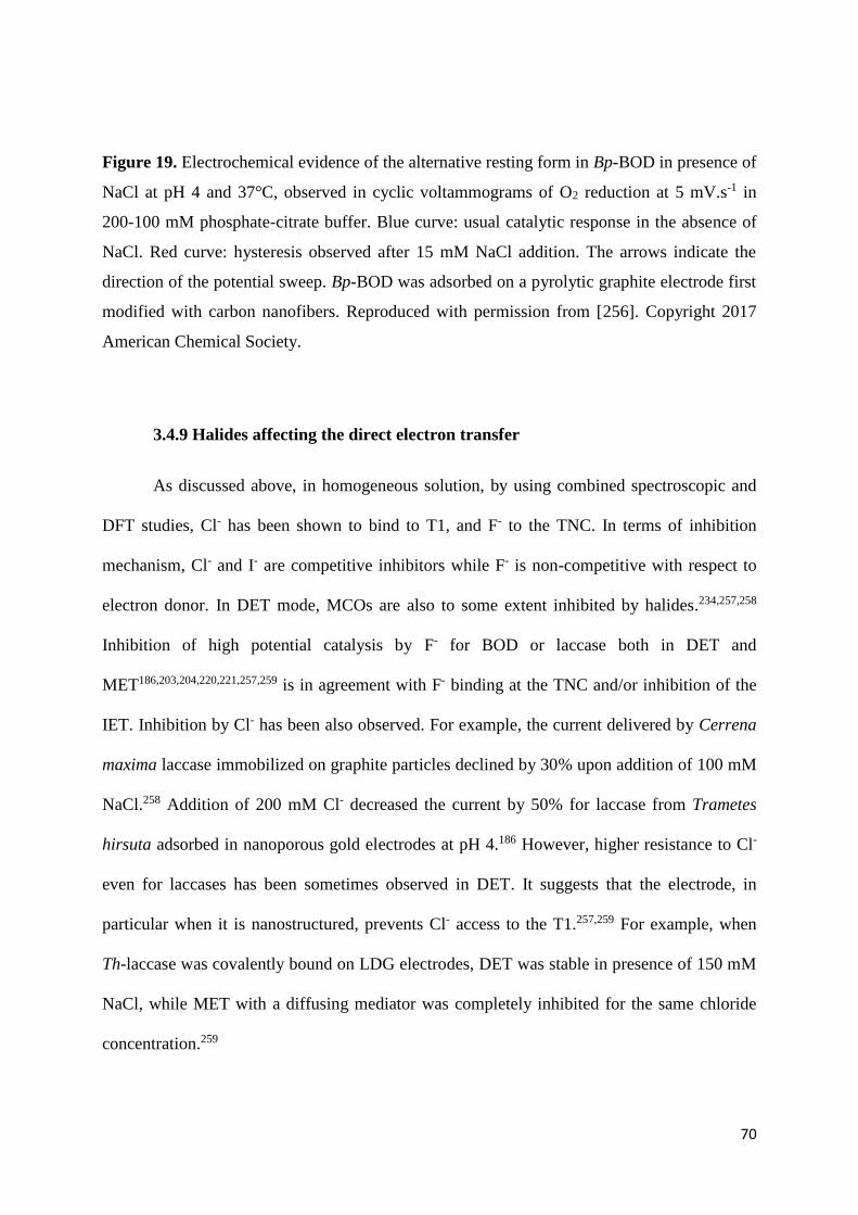
70
Figure 19. Electrochemical evidence of the alternative resting form in Bp-BOD in presence of
NaCl at pH 4 and 37°C, observed in cyclic voltammograms of O2 reduction at 5 mV.s-1 in
200-100 mM phosphate-citrate buffer. Blue curve: usual catalytic response in the absence of
NaCl. Red curve: hysteresis observed after 15 mM NaCl addition. The arrows indicate the
direction of the potential sweep. Bp-BOD was adsorbed on a pyrolytic graphite electrode first
modified with carbon nanofibers. Reproduced with permission from [256]. Copyright 2017
American Chemical Society.
3.4.9 Halides affecting the direct electron transfer
As discussed above, in homogeneous solution, by using combined spectroscopic and
DFT studies, Cl- has been shown to bind to T1, and F- to the TNC. In terms of inhibition
mechanism, Cl- and I- are competitive inhibitors while F- is non-competitive with respect to
electron donor. In DET mode, MCOs are also to some extent inhibited by halides.234,257,258
Inhibition of high potential catalysis by F- for BOD or laccase both in DET and
MET186,203,204,220,221,257,259 is in agreement with F- binding at the TNC and/or inhibition of the
IET. Inhibition by Cl- has been also observed. For example, the current delivered by Cerrena
maxima laccase immobilized on graphite particles declined by 30% upon addition of 100 mM
NaCl.258 Addition of 200 mM Cl- decreased the current by 50% for laccase from Trametes
hirsuta adsorbed in nanoporous gold electrodes at pH 4.186 However, higher resistance to Cl-
even for laccases has been sometimes observed in DET. It suggests that the electrode, in
particular when it is nanostructured, prevents Cl- access to the T1.257,259 For example, when
Th-laccase was covalently bound on LDG electrodes, DET was stable in presence of 150 mM
NaCl, while MET with a diffusing mediator was completely inhibited for the same chloride
concentration.259
71
Potential shifts similar to those described above, have also been observed in DET upon
addition of halides. EHW of Cerrena maxima laccase shifted 94 ± 3 mV in the negative
direction in presence of 150 mM Cl- at pH 5. This effect disappeared as soon as the electrode
was transferred in a chloride-free buffer.258 The EHW shift observed with Trametes laccase
immobilized on SW-CNTs increased with increasing Cl- concentration, being respectively 60
mV at 10 mM and 150 mV at 100 mM Cl-.257 For the same enzyme, similar potential shifts
were observed upon addition of I-.257 This potential shift can be explained by Cl- or I- binding
at the T1. In presence of F-, the low-potential anaerobic redox process observed for Mv-BOD
decreased, consistently with F- binding to TNC.204 More surprisingly, a negative shift of the
high potential catalysis has also been observed upon addition of F-.234,256,257 The slight 30 mV-
decrease in conjunction with 90% current loss was attributed to F- binding both at the T1 and
the TNC, thus shifting the T1 potential and blocking IET at the same time.257 A higher 100
mV-decrease for Didymocrea laccase at pH 4, with lesser-pronounced inhibition, was
attributed exclusively to F- binding to the TNC. In this case, surface-enhanced Raman
spectroscopy (SERS) studies of the enzyme orientation suggested that the T1 was relatively
far from electrode surface, so that DET was more likely occurring through the TNC.234
Interestingly this study is the first to propose a DET via TNC occurring at a higher potential
than T1.234 However, this is not a direct demonstration that DET to TNC is feasible and
additional experiments with T2-depleted MCOs would be required. A redox titration showed
that the T1 potential, ca. 100 mV lower than the half-wave potential correlated to TNC, was
little affected by F- addition. Exactly the same shift was observed for Bp-BOD, while the
primary electron acceptor in that case was supposed to be the T1 Cu.256
3.4.10 Rate limiting steps of O2 reduction
Different studies have shown that the rate limiting step during O2-reduction catalysis
by MCOs connected in DET mode varied depending on the experimental conditions and

72
applied potentials. The enzyme affinity for O2 was shown to depend on electrode potential,
which means that “apparent” Michaelis constants are measured at a given potential. This led
to the conclusion that the rate determining steps of the catalytic mechanism also depend on
the electrode potential.233 In the case where an endergonic IET was considered, the calculated
IET rates were influenced by pH, due to the different pH dependencies of the high and low-
potential intermediates. The thermodynamic driving force decreased at basic pH, so that IET
started to be the rate limiting step in alkaline solution.204 The same conclusion was drawn
from analysis of the O2-reduction CV shape in the absence of O2 diffusion limitation. At low
pH, the CV exhibited a trailing edge well described by the model introduced by Léger et al.,
whereas it was almost sigmoidal at high pH.17 This was attributed to fast IET and catalysis at
low pH. In this case, both current and turnover rate are determined by the rate of HET, which
increases at high overvoltage. On the contrary, at high pH, slow IET and catalysis are
determined by the turnover rate. T1 is therefore the electrochemical control center, and
defines the potential of electro-catalysis. The authors proposed that the process at high pH is
limited because the deprotonated carboxylate D105 close to TNC cannot supply the required
protons.17
3.4.11 Inhibition by H2O2
While studies describing the effect of pH and halides on MCOs activity are
widespread in the literature, the impact of hydrogen peroxide on laccases and bilirubin
oxidases has only more recently attracted attention. In the context of a glucose/O2 biofuel cell
where glucose oxidase is used at the anode, it is hypothesized that H2O2 generated during the
oxidative half reaction of glucose may affect BOD or laccase. The first paper addressing this
issue was reported in 2010 by Calvo et al.260 Their cathode was composed of a self-assembled
layer of laccase from Trametes versicolor or Trametes trogii and osmium complex bound to
poly(allylamine). They evidenced the inhibition of biocathodes by H2O2 during oxygen
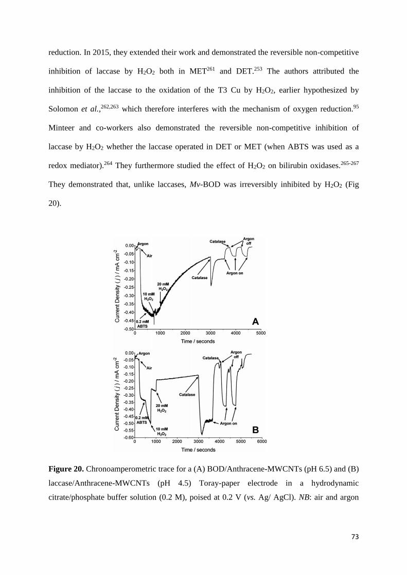
73
reduction. In 2015, they extended their work and demonstrated the reversible non-competitive
inhibition of laccase by H2O2 both in MET261 and DET.253 The authors attributed the
inhibition of the laccase to the oxidation of the T3 Cu by H2O2, earlier hypothesized by
Solomon et al.,262,263 which therefore interferes with the mechanism of oxygen reduction.95
Minteer and co-workers also demonstrated the reversible non-competitive inhibition of
laccase by H2O2 whether the laccase operated in DET or MET (when ABTS was used as a
redox mediator).264 They furthermore studied the effect of H2O2 on bilirubin oxidases.265-267
They demonstrated that, unlike laccases, Mv-BOD was irreversibly inhibited by H2O2 (Fig
20).
Figure 20. Chronoamperometric trace for a (A) BOD/Anthracene-MWCNTs (pH 6.5) and (B)
laccase/Anthracene-MWCNTs (pH 4.5) Toray-paper electrode in a hydrodynamic
citrate/phosphate buffer solution (0.2 M), poised at 0.2 V (vs. Ag/ AgCl). NB: air and argon

74
gasses were purged individually – air purging was terminated at t = 3600 seconds.
Reproduced with permission from [266]. Copyright 2014 The Royal Society of Chemistry.
However, the exact mechanism of inhibition still has to be understood. Fortunately, it
is less complicated to overcome the problem of H2O2 than the pH or Cl- effect. Different
strategies have been developed to protect the MCO cathode. To prevent the formation of the
inhibiting H2O2, O2-independant glucose-oxidizing enzymes such as glucose dehydrogenase
can be used. Alternatively, H2O2 can be degraded via the formation of bi-enzymatic systems
such as glucose oxidase/horseradish peroxidase, glucose oxidase/catalase or BOD/catalase. In
this latter case, H2O2 is dismutated to water by the second enzyme, thus protecting the MCO.
Although these studies on H2O2 inhibition are of interest from a mechanistic point of view,
they are not necessarily relevant for the field of biofuel cells since highly concentrated
solutions of H2O2 have been used (mM range and up to 20 mM). For example, a carbon fiber
coated with 35 wt% of an engineered glucose oxidase268 and an adequate redox polymer for a
total loading of 2000 µg.cm-², would generate 20 mM H2O2 after 4.2 months of continuous
operation in presence of 5 mM glucose in a saturated O2 buffer pH 7.2.
3.4.12 Effect of methanol
Palmore et al. envisioned the use of enzymatic electrodes in direct methanol fuel cells,
which typically operate with concentration of MeOH comprised between 1 and 5 M.269 The
authors showed that the activity of laccases either in solution or entrapped in polypyrrole film
was decreasing upon increasing MeOH concentrations.270 However, the decline was slower
for the immobilized enzymes. They attributed the loss of activity to the replacement of water
molecules by MeOH within the enzyme.271 In a series of three papers, Calabrese-Barton et al.
also investigated the impact of methanol on the oxygen reduction current delivered by an
electrode modified with an osmium polymer and Tv-laccase. When the bioelectrocatalysts

75
were immobilized on a carbon-fiber paper support incorporated in a membrane-electrode
assembly, the authors demonstrated the tolerance of laccase to methanol. A 6% increase in
current density at Ecell = 0.2 V was found for 10 M methanol as compared to 1 M methanol.272
They later demonstrated that in presence of 1 M methanol, the performance of a biocathode
based on Th-laccase on carbon cloth was only reduced by 4.5%. This decrease was explained
by a de-swelling of the redox polymer rather than by the enzyme deactivation.273 In 2011,274
they showed that the sensitivity of O2 reduction to methanol depended not only on the
concentration of MeOH, but also on the osmium polymer used and particularly on its redox
potential. The loss in current was nil below 2.5 M methanol, it was 30% between 2.5 and 5 M,
while 100% current was lost above 7.5 M. For a 7.5 M methanol concentration, the authors
showed that Tv-laccase inactivation was irreversible. Blanford et al.275 also studied the effect
of MeOH on Tv-laccase immobilized on pyrolitic graphite edge electrode modified by
reduction of diazonium salts. In presence of 2.7 M and 5 M methanol the current density was
reversibly decreased by respectively 10 and 20%, while at 9.7 M the current loss was
irreversible. Xiao et al. have reported a Mv-BOD based biocathode which could operate in
acetonitrile, acetone and various alcohols (vide infra).276 De Andrade et al.277,278 have
designed laccase-based EBFCs operating at pH 5 in presence of 100 mM methanol.
4. Enzymatic O2 reduction
4.1 Direct electron transfer
Catalysis of O2 reduction by laccase in DET was demonstrated for the first time almost
40 years ago. In this early study, Tv-laccase was immobilized on carbon black.243 As for
BOD, connection in DET was reported much later, in 2004, with Mv-BOD adsorbed on
different carbon electrodes.235 In the first studies of DET catalysis by MCOs, the main efforts
were directed towards elucidation of O2 reduction mechanism by enzymes directly connected

76
at the electrode (see section 3.4), and less importance was attributed to electrocatalytic
performances. In this section, we describe works performed to achieve Efficient enzyme
connection in DET, with the purpose to use MCOs as cathodic enzymes in EBFCs. Important
parameters are therefore high onset potential, high current densities and stabilities. To obtain
high onset potentials the enzyme must be connected to the electrode via the T1. Lots of efforts
have been devoted to reach large current densities at sufficiently high potentials. This
involves immobilization of high amounts of enzymes, and high apparent catalytic rates. We
consider first random orientation of the enzyme at the electrode, and describe current densities
reached at different planar electrodes. Since current densities remained limited in that
configuration, two main strategies have then been developed. On one hand, current
magnitudes could be increased by immobilizing higher number of catalysts at a given
geometric surface. Nano- or micro-structuration of the electrode surface has been envisioned
in this context, to multiply the real electroactive surface area of an electrode. We develop this
in part 4.1.2 and 4.1.3. On the other hand, the enzyme electronic connection had to be
optimized, which mostly relied on achieving functional orientation of the enzyme at the
electrode surface. We describe strategies developed to ensure optimal orientation of MCOs at
both planar and structured electrodes in part 4.14. Then, we discuss the stability issue in part
4.1.5.
Contrary to previous section describing the mechanistic study of MCOs, and still for
the sake of consistency with the literature, we will here quote the potentials versus the
Ag/AgCl reference electrode, unless stated otherwise. The potentials are therefore typically
200 mV lower than when quoted vs. the NHE reference electrode.
4.1.1 Planar electrodes
4.1.1.1 “Planar” Carbon electrodes

77
In the first studies, MCOs have been immobilized on various kinds of carbon
electrodes: glassy carbon (GC),235 various types of graphite (low density graphite (LDG),
spectroscopic graphite (SPG), pyrolytic graphite (PG), basal plane or edge plane highly
oriented pyrolytic graphite (HOPG)…),203,210,215,221,235 and screen printed carbon.279 Except
glassy carbon, carbon surfaces are however rarely strictly planar. While it is clear that DET
catalysis has been observed with different MCOs simply adsorbed on most of these materials,
it is difficult to compare their efficiency as enzyme support. Indeed, it is well-known that
carbon electrodes differ from one another as far as microstructure, roughness, and chemical
functions are concerned.
To give an idea of the magnitude of currents or current densities recorded with MCOs
adsorbed on carbon electrodes, we list below some values obtained at 0 V vs. Ag/AgCl. Tv-
laccase adsorbed on edge plane graphite gave currents of 40 µA. In that case, the maximum
rate at which O2 could be reduced, deduced from the intercept of a Koutecky Levich plot,
indicated a turnover rate of ca. 20 s-1.211 Rv-laccase was adsorbed on two carbon materials
(RWII and RW0), and the higher electrocatalytic current, 60 µA.cm-2 at neutral pH, was
observed with RWII. It was attributed to a higher porosity and the presence of more oxygen
functions. With laccases of different origins, T. ochracea, C. maxima, C. unicolor, C.
fulvocinerea, adsorbed on SPG or HOPG, currents were in the range 2-20 µA.210 It is
interesting to note that Tv-laccase adsorbed on HOPG was observed by STM, revealing that
the enzyme was not homogeneously distributed at the electrode surface and probably forming
aggregates.271
In the very first study of DET by a BOD, Mv-BOD was also adsorbed on different
electrode materials: GC, HOPG and PFC (plastic formed carbon). PFC and edge-plane HOPG
gave the best results (200 µA.cm-2 with HOPG at pH 7), even exceeding the highest current
densities obtained at that time with laccase. It was proposed that surface functional groups

78
and/or nanostructure governed the surface ET kinetics and adsorption property of BOD, but
no more details were provided.235 In the following reports, the current densities diverged. The
differences cannot be assigned merely to different operating conditions; they may be
explained by the fact that, as already mentioned, the electrode surface was not well
characterized. For example 25 µA.cm-2 at pH 7 were recorded with Mv-BOD adsorbed on
HOPG,245 and 30 µA.cm-2 for the same enzyme adsorbed on SPG.215 Tt-BOD adsorbed on
SPG led to 20 µA.cm-2 at pH 7203 and 0.5 mA.cm-2 under O2 at pH 4,204 i.e. the results
differed by more than one order of magnitude. Although it is not possible to identify the exact
origin of this difference, we can note that it is much higher than those observed in studies of
pH influence performed in homogeneous solution.
DET on carbon electrodes was also routinely used to characterize less widespread or
newly discovered MCOs.166,181,280,281 Interestingly, the proof of concept of some EBFCs was
established with enzymatic planar carbon cathodes. Tv-laccase adsorbed on graphite provided
the cathode of the first totally enzymatic H2/O2 BFC.282,283 Similarly, the first mediator- and
cofactor-free EBFC based on cellulose dehydrogenase (CDH) operating in neutral chloride-
containing buffers as well as in serum relied on Mv-BOD adsorbed on SPG electrode as a
cathode.241 A cathode based on Mv-BOD adsorbed on a LDG electrode was also combined to
a bi-enzymatic anode for the 6 e--oxidation of glucose in a membrane-less EBFC operating in
a buffer mimicking human physiological fluid.284 This is detailed further in section 6.
4.1.1.2 Other planar electrodes
DET was also achieved for different MCOs on bare or modified gold surfaces. Since
this is explored in details in section 3.4.4. and 4.1.4., we only remind here some current
densities obtained when MCOS were randomly immobilized.221 For most laccases, no DET

79
current was recorded when they were adsorbed on bare gold. Rv-laccase on MPA-modified
gold gave 84 µA.cm-2 at pH 7.214 With Mv-BOD, very different values ranging between 5 and
more than 100 µA.cm-2 were recorded on bare gold electrodes.17,225,228,231
Some less conventional materials have been proposed, like boron doped diamond
(BDD) or semiconductors. BDD has a wide working potential window, low background
current, high stability, tunable hydrophilicity and optical transparency. Direct O2 reduction by
Cerrena unicolor laccase was demonstrated on BDD285 with the enzyme either in solution or
immobilized in a liquid crystalline cubic phase at the electrode. However, DET currents were
very low (< 1 µA.cm-2) while much higher currents were achieved upon addition of a redox
mediator (25 µA.cm-2). This proved that the enzyme orientation was not optimal.285 Tv-
laccase was immobilized on p-type nanostructured silicon (111) as a model semiconductor.286
Under illumination with visible light, the material was theoretically able to supply electrons
for the enzyme activation. O2 reduction started about 200 mV more positive when laccase was
present, but still at a very low potential (-0.24 V vs. SCE). The plateau current was 30 µA.cm-
2.286
4.1.1.3 Strategies for increasing the enzyme loading
To overcome the limitation in current densities, and reach values usable in EBFC, the
surface concentration of enzymes on electrode surfaces had to be improved. A study by Ikeda
et al. showed that multiple layers of enzymes could be connected in DET configuration on a
planar electrode.252 Multilayers of Mv-BOD were entrapped electrostatically in poly-L-lysine
(PLL) on a PFC electrode. In O2 saturated buffer, the process was diffusion controlled. A
plateau at 845 µA.cm-2 was reached upon stirring at 1400 rpm, while only 300 µA.cm-2 were
recorded when BOD was simply adsorbed on this PFC electrode. Calculations according to
the model presented in section 3 indicated that the reaction layer was 0.2 µm-thick, i.e. much
smaller than the real thickness of the BOD-PLL film (50 µm). HET standard rate constant was
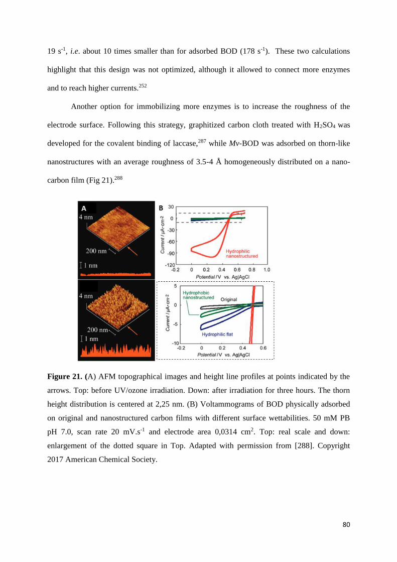
80
19 s-1, i.e. about 10 times smaller than for adsorbed BOD (178 s-1). These two calculations
highlight that this design was not optimized, although it allowed to connect more enzymes
and to reach higher currents.252
Another option for immobilizing more enzymes is to increase the roughness of the
electrode surface. Following this strategy, graphitized carbon cloth treated with H2SO4 was
developed for the covalent binding of laccase,287 while Mv-BOD was adsorbed on thorn-like
nanostructures with an average roughness of 3.5-4 Å homogeneously distributed on a nano-
carbon film (Fig 21).288
Figure 21. (A) AFM topographical images and height line profiles at points indicated by the
arrows. Top: before UV/ozone irradiation. Down: after irradiation for three hours. The thorn
height distribution is centered at 2,25 nm. (B) Voltammograms of BOD physically adsorbed
on original and nanostructured carbon films with different surface wettabilities. 50 mM PB
pH 7.0, scan rate 20 mV.s-1 and electrode area 0,0314 cm2. Top: real scale and down:
enlargement of the dotted square in Top. Adapted with permission from [288]. Copyright
2017 American Chemical Society.

81
In both cases, currents increased compared to the planar electrode: 0.27 mA.cm-2 were
reached at 0 V in the first case, supposedly due to higher number of electrochemically active
laccases287, and 102 µA.cm-2 in the second thanks to the nanostructure.288 The importance of
the roughness has also been highlighted with Mv-BOD immobilized on a planar electrode
together with platinum group metal-free catalysts. Current densities did not only increase
because of the inherent properties of the catalysts, but the contribution of the increased
roughness was also important, providing better results when pores within the ratio 3-5 nm
where formed. No significant increase occurred with larger pores (12-20 and 40-80 nm).289
Alternatively, a large variety of materials can be used to enlarge the electroactive
specific surface area (ESSA) of the electrode. Matrices based on nanomaterials with a high
surface/volume ratio have been developed. Typical nanomaterials are carbon nanotubes,
nanoparticles or nanofibers (resp. CNTs, CNPs or CNFs), and gold nanoparticles (AuNPs).
Various metallic nanoparticles (Fe, Pt...) have also been described. Ideally, it would be
beneficial to find materials with hierarchical porosity allowing Efficiently immobilizing
enzymes and preventing mass transport limitations. Therefore, highly porous 3D materials
like mesoporous gold or carbon with the adequate and tailored porosity have also been
engineered. In the following of this section we will focus on studies where the nano-
structuration of the electrode has been evaluated, first with carbon and then with gold-based
materials.
4.1.2 Carbon nanostructures
Carbon based materials are especially available, cheap, biocompatible, and can be
produced in a variety of structures. Moreover, they are chemically and electrochemically
inert.

82
4.1.2.1 Carbon nanotubes
Discovered in 1991, carbon nanotubes (CNTs)290 are rolled layers of sp2 carbon atoms.
Single-wall (SW) CNTs consist of one single graphene layer, while multi-wall (MW) CNTs
are composed of several layers. Their typical diameters range from 1 to few nm (SW-CNTs)
or 20-50 nm (MW-CNTs), and their lengths are in the range of few µm to few tens of µm.
Their nanometer-sized dimensions impart them a high aspect ratio. Moreover, they possess a
high electronic conductivity, and a huge mechanical strength.291 Their surface chemistry can
be divided into two types. Their walls, made of sp2 graphene carbon atoms, are hydrophobic
pristine, while the defects on the side walls and edge sites at the open ends are full of
hydrophilic carboxy groups. As for their electrochemical properties, it is widely accepted that
atoms at the ends are like edge planes of HOPG and favor electrochemical activation; and
atoms at the sidewalls are like basal plane of HOPG and show very slow electron transfer
kinetics.292,293 However, recent SECM studies have demonstrated that electrochemical
properties are more likely to be similar at the ends and at the walls.294
It is often stated that DET is favored by the appropriate dimensions of CNTs. It is true
that DET has been often reported on CNTs, whether it was a catalytic295-297 or a non-catalytic
signal,295-298 while the same enzyme on a planar electrode gave no DET. It must however be
noted that in the case where no DET was observed on the planar electrode, this latter was
most of the time made of bare gold or GC.295,296,298 The properties of these surfaces towards
DET have already been discussed in previous section. Therefore, the reasons why DET is
appearing are most of the time unclear. It could be due to an increase in ESSA, and thus an
increase in enzyme loadings, that makes visible a signal non detectable with fewer enzymes.
Alternatively, it could come from chemical functions inducing appropriate immobilization of
the enzyme, to the increase in roughness, or to the peculiar properties associated to nano-size
of CNTs. The very few fundamental studies will be discussed herein.
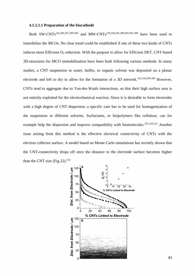
83
4.1.2.1.1 Preparation of the biocathode
Both SW-CNTs142,296,297,299-304 and MW-CNTs218,244,295,298,303,305-308 have been used to
immobilize the MCOs. No clear trend could be established if one of these two kinds of CNTs
induces more Efficient O2 reduction. With the purpose to allow for Efficient DET, CNT-based
3D-structures for MCO immobilization have been built following various methods. In many
studies, a CNT suspension in water, buffer, or organic solvent was deposited on a planar
electrode and left to dry to allow for the formation of a 3D network.142,244,299,309 However,
CNTs tend to aggregate due to Van-der-Waals interactions, so that their high surface area is
not entirely exploited for the electrochemical reaction. Since it is desirable to form electrodes
with a high degree of CNT dispersion, a specific care has to be used for homogenization of
the suspension in different solvents. Surfactants, or biopolymers like cellulose, can for
example help the dispersion and improve compatibility with biomolecules.295,310,311 Another
issue arising from this method is the effective electrical connectivity of CNTs with the
electron collector surface. A model based on Monte Carlo simulations has recently shown that
the CNT-connectivity drops off once the distance to the electrode surface becomes higher
than the CNT size (Fig 22).312

84
Figure 22. (A) Percentage of CNTs connected to the electrode in a 125-mm thick film for
various volumes. % CNT fill, ΦC: 0.25% (o); 1.6% (); 3% (∆); 4.5% (▲). (B) MATLAB
image displaying only those CNTs connected to the electrode, shown as black lines.
Reproduced with permission from [312]. Copyrigth 2016 Elsevier
Another possibility is to fabricate ordered arrays of CNTs grown by chemical vapor
deposition on the electrode, 257,313,314 or by electrophoretic deposition.313 Covalent binding of
CNTs on Au218 has also been performed. Free standing CNT-based electrodes have been
designed, either in the form of buckypaper (BP),315 fibers310 or 3D pellets.316-318 These
structures have ensured DET for different MCOs: Mv-BOD,142,296,302,303,305,308,311,319,320 Mo-
BOD,310 Th-laccase, Rv-laccase,320 Tv-laccase,295,299,321 Cerrena unicolor laccase,301,322
Agaricus bisporus laccase,313 or even tyrosinase.317 Similar protocols have been used for the
immobilization of MCO, whatever their origin, and their peculiar surface properties have been
little considered. This latter point will be discussed further in section 4.1.4.
CNTs were often treated with HNO3299,300,323 or H2SO4 before use, which eliminates
impurities but also introduces defects and oxygenated functions.142,313,320 Enzymes have been
directly adsorbed on the CNTs network,299,320 mixed with CNTs prior to the formation of the
network,297 covalently bound using EDC-NHS on CNT carboxy groups, 142,244 or cross-linked
with glutaraldehyde.295,296,323 Another possibility has taken benefit of the π-conjugated
systems of CNT walls to induce functionalization, using a bi-functional molecule that bears a
defined aromatic functionality to bind by π-π stacking to the CNT walls, and a functional
tether group for attachment of the enzyme.324,325 This method has been used for example to
introduce pyrene-butyric succinimidyl ester (PBSE) 306,307,321,326,327 or pyrene butyric acid.308
The impact of the chemical functionalization is discussed further in section 4.1.4. The strong
interaction between concavalin A immobilized on SW-CNTs and the glycosylated laccase has
been exploited.328 The laccase presented a higher activity towards oxidation of organic

85
substrates than the enzyme directly adsorbed, however no electrochemical catalysis was
proved in this case.
4.1.2.1.2 CNT properties affecting direct electron transfer to MCOs
Very few studies have been performed to establish a clear link between the different kinds
of CNTs, or the different protocols used to immobilize the MCOs, and the efficiency of the
DET. The electrocatalytic activity of cross-linked Tv-laccase329 and Mv-BOD309 on two types
of CNTs has been compared. The first kind had a uniform inner diameter (10-30 nm),
carboxylic surface groups, a higher graphitization degree, a better conductivity and a higher
electroactive surface area. The second kind had tapered inner tube diameter and mostly
quinone surface groups. Without much surprise both enzymes exhibited a nicer behavior on
the first kind. For Tv-laccase, a faster electron transfer, a clearer non catalytic signal, and 6
times higher current densities in catalysis (reaching 25 µA.cm-2) were observed.329 For Mv-
BOD, the non-catalytic signal was observed only on the first kind that led also to 5 times
higher current densities (90 µA.cm-2). However, currents did not reach a plateau for any of
these CNT-types, meaning that the interfacial electron transfer was a quite slow process.309 In
another report, the authors tried to compare electrophoretic deposition and chemical vapor
deposition (CVD) of SW-CNTs on porous silicon.313 Better DET for a covalently linked
laccase was obtained with electrophoretically-deposited CNTs, but these results have to be
considered carefully since porous silicon was covered with a conductive gold layer, while an
insulating layer was formed during CVD growth. Therefore, higher cathodic currents could be
assigned to a better conductivity of the electrode/nanomaterial interface rather than to better
inherent properties of the material. Highly crystalline SW-CNTs (grown by CVD on a gold
wire) were also compared to Toray carbon paper treated with UV-ozone.257 Adsorbed laccase

86
from Trametes sp. led to catalysis on both materials but kET constants were respectively 1200
and 800 s-1, reflecting higher degree of appropriate orientations on SW-CNTs. The coverage
of electrochemically active enzymes was also much higher on these latter (3.6.10-12 against
0.31.10-12 mol.cm-2), translating a higher affinity.
Recently, the effect of the length of MW-CNTs has also been evaluated330 for Mv-BOD in
a kinetically controlled regime. The intermediate length (1-4 µm) led to the highest current
densities (6.1 mA.cm-2 at 0.1 V) while performances decreased for longer (10 µm) and shorter
(<1 µm) CNTs sharing the same diameter (10-15 nm) (Fig 23). This quite complex behavior
was explained by considering determinant the ratio of hydrophobicity to density of carboxy
groups. Since the walls are mostly hydrophobic, while carboxy groups are mostly at the ends,
this ratio is driven quasi exclusively by the length of the CNTs.
Figure 23. Influence of the length of MW-CNTs on ORR by Mv BOD. LSVs in O2- saturated
100 mM phosphate buffer (pH 7) at 41 C, ω = 4000 rpm, and v = 5 mV.s-1. Mv BOD is
adsorbed at MW-CNTs of different lengths (L) and outer diameter (OD), or at CNPs. Black
solid line: L = 1-4 µm and OD = 10-15 nm ; black broken line: L = 1-4 µm and OD = 10-15
nm and a surfactant is added ; black dotted line : L < 1 µm and OD = 8-10 nm ; and gray solid
line : L = 10 µm and OD = 10-15 nm. The gray broken line represents CNPs. Adapted with
permission from [330]. Copyrigth 2016 Elsevier

87
Considering that substituting carbon atoms by heteroatoms in graphene sheet of CNTs
change the local electron density and should affect DET, qualitative effects of N-doping of
SW-CNTs on the DET reaction of laccases were also studied.331 A laccase from Trametes sp.
was adsorbed onto SW-CNTs and N-doped SW-CNTs grown by CVD on a gold electrode, for
N/C ratios of 0.1, 2.4 and 4.1%. Enzyme loading per BET surface and onset for DET did not
depend on doping, but DET currents decreased with increasing doping. It was proposed that
the induced positive charge led to increase electrostatic interactions with the slightly negative
patch surrounding the T1 Cu, therefore denaturating the laccase. This explanation was
furthermore confirmed by thermodynamic investigation showing that enthalpy change ∆H of
laccase binding becomes more negative with increasing doping. This observation can be
correlated with what happens on bare gold electrodes, where strong interactions between the
enzyme and the electrode leads to enzyme denaturation (see section 3.4.4).
The spatial homogeneity of the mediator-less bioelectrocatalysis in a CNT composite was
also evaluated by SECM. Cerrena unicolor laccase was encapsulated in a sol-gel silicate
matrix with SW-CNTs. ORR was recorded in the presence of the laccase at Eapplied = 0.3 V
and compared to a control experiment with the laccase at OCP. The activity was more evenly
distributed than in a silicate film without CNT where proteins were shown to aggregate. 332
4.1.2.1.3 Increasing the current densities through CNT networks
In evaluating the benefits of CNTs, performance improvements are reported most of the
time in terms of higher current densities. Importantly, very little change in onset potentials has
been reported. Although CNTs were shown to increase DET efficiency, currents were small
when MCOs were first immobilized in CNT-networks: 5 µA at the plateau with Mv-BOD
and Tv-laccase cross-linked with glutaraldehyde on MW-CNTs for the first demonstration;295
28 µA.cm-2 under air and 102 µA.cm-2 under O2 with Mv-BOD immobilized on SW-CNTs.300

88
Later the increase in current was mostly assigned to the higher electroactive surface area
rather than to a better electrical connection. We give below some examples of MCOs
immobilized in 3D networks constructed by drop casting of a CNT-solution on the electrode.
A 300-fold current increase was reported for Mv-BOD covalently linked on SW-CNTs
compared to bare PG under 100% O2 at 25°C, giving almost 1 mA.cm-2.142 Laccase embedded
in a bioceramic (Hydroxyapatite)-CNTs self-assembled nanocomposite led to around 100
µA.cm-2 under air with an OCP of 0.56 V vs. Ag/AgCl at pH 6, and the currents were
diffusion limited.323 When Mv-BOD was immobilized in a MW-CNT/cellulose network, 800
µA.cm-2 were reached under air at pH 5, and 60% were still recorded after 45 h at the constant
potential 0.2 V vs. Ag/AgCl.311 Cerrena unicolor laccase incorporated in SW-CNTs covered
with a transparent and highly viscous cubic phase of monoolein gave 200 µA.cm-2. This was
however not optimal since the addition of covalently bound ABTS raised the current to 400
µA.cm-2, indicating that some enzymes could not participate in DET currents.301 In another
study, when Mv-BOD was covalently attached at MW-CNTs, themselves covalently attached
on a gold electrode, currents increased up to 500 µA.cm-2 in an unstirred solution.218 Although
these results already represented a considerable increase compared to most bare carbon
electrodes, the current densities remained limited. Therefore, specific and oriented attachment
of the enzymes has been the main concern in most of the following studies (section 4.1.4.).
Some of the developed cathodes were successfully used in EBFCs, for example with
MW-CNTs oxidizing ascorbate at the anode,295 or with glucose dehydrogenase (GDH) on
SW-CNTs at the anode.296,299,300,302 The first Glucose/O2 EBFC based on SW-CNTs showed
OCV = 0.8 V and Pmax = 9.5 µW.cm-2 at Ecell = 0.52 V at pH 6.299 Other devices included a
H2/O2 EBFC with a hyperthermophilic hydrogenase at the anode leading to OCV = 1.1 V,
more than 300 µA.cm-2 at Ecell = 0.65 V, and less than 40% loss after 24 h at an applied Ecell =
0.65 V.142 A CNT-based cathode was also used as a proof of concept in a photosynthetic

89
electrochemical cell for the light-to-electricity conversion with water as the only input. Water
was oxidized by thylakoid membranes while the laccase reduced O2, regenerating water in the
system. The maximal generated power was 5 µW.cm-2.306 The hydroxyapathite-CNT cathode
combined with glucose oxidase (GOx) also gave an OCV of 0.58 V and Pmax = 15.8 µW.cm-2
at 0.28 V. 323 In a fructose/air one chamber EBFC, OCV = 663 mV and Pmax = 126 µW.cm-2 at
pH 5 were obtained. 90% of the initial power remain after 12 h, and 52% after 87 hours at
Ecell = 0.35 V.311
To obtain a hierarchical porosity, CNTs were also deposited in an already porous
material. 3D hierarchically structured composites were built by growing MW-CNTs by CVD
on CMF with diameters ranging from 6 to 9 µm,314,333,334 or by depositing CNTs on a porous
carbon fabric to form a gas diffusion layer (GDL),305 or by coating CNTs-Anthracene or
CNTs-COOH in a highly porous carbon felt.335,336 In the latter case, after deposition of CNTs-
COOH by LBL, ESSA increased 5 times, and cathodic currents reached almost 1.5 mA.cm-2
at 0 V. This allowed the development of a H2/O2 EBFC with an OCV of 1.12 V and net
currents of 765 µA at short circuit. Absolute Pmax was 410 ± 5 µW which represented a
successful scale-up compared to a previously reported device,142 indicating that the diffusion
limitation of O2 was overcome by the porosity of the electrode.335
4.1.2.1.4 Free-standing CNT-based 3D layers
Besides porous random networks, CNTs can be used as electrospun fibers310,337 or
buckypaper.319-321,326,327,338,339 BP is a flexible self-supporting membrane-type film made of
densely packed CNTs maintaining close contact by Van der Waals interactions. It is obtained
by filtration of CNTs dispersion. This nanoporous material has attractive properties such as
very high surface area, high porosity, chemical and electrochemical stability, high mechanical

90
strength and high conductivity (a value of 51.2 S.cm-1 has been reported326). Importantly for
the construction of potentially implantable biodevices, it has also a low toxicity both in vivo
and in vitro.340 To quote some performances: BP enabled an enhancement in current densities
for Th and Rv-laccases as well as Mv-BOD. Currents reached 120-220 µA.cm-2 compared to
12-35 µA.cm-2 for spectrographic graphite, leading to a mass-transport limited regime.320 An
EBFC based on BP had an OCV of 710 mV and Pmax > 100 µW.cm-2 319 and this device was
furthermore able to work in urine and saliva.338 BP was also compared to “conventional”
MW-CNTs electrodes where CNTs formed aggregates of 500 µm in diameter. Tv-laccase
was adsorbed on a 30 µm thick BP made of 0.9 mg.cm-2 MW-CNTs homogeneously
dispersed, and presenting a median pore diameter of 48 nm.315 Reporting the current to the
mass of material showed that properties of MW-CNTs were better exploited in BP: at 0.54
V vs. Ag/AgCl, the current was 471 ± 64 µA.mg-1, which represented a 68-fold increase. In
terms of current densities, they were an order of magnitude higher than previously reported:
487 ± 117µA.cm-2 against only 50 µA.cm-2. 315,326
Miyake et al. constructed an innovative free-standing CNT-based enzymatic film.341
After introducing Trametes sp. laccase in a forest made of 1 mm-long CNTs separated by 16
nm, further drying induced shrinkage to a close packed structure, whose total thickness was
20 µm (Fig 24). Current densities of 2.1 ± 0.3 mA.cm-2 were reached with stirring, and further
increased to 4 mA.cm-2 when 2 films were joined (Fig 24).

91
Figure 24. (A) Schematic diagram of laccase entrapment in a CNT forest by “liquid-induced
shrinkage”. The photograph shows a free-standing enzyme-CNT film that can be manipulated
with tweezers. (B) Corresponding CV of the so-formed single-piece electrode in N2 (black
curve) and O2-saturated stirred Mc Illvaine buffer pH 5 (red curve). The green curve shows
the currents obtained with a double-piece electrode, v=10 mV.s-1. The electrode films were
prepared using a 20 µm thick CNT film and 0.25 mg.mL-1 laccase solution. Adapted with
permission from [341]. Copyright 2011 American Chemical Society.
In a fructose/O2 EBFC, where fructose dehydrogenase was used at the anode, Pmax was
1.8 mW.cm-2 at 0.45 V in a stirred oxygenated fructose solution. More than 80% of Pmax was
maintained after 24 h of continuous operation at an external load of 35.3 kΩ. It is noteworthy
that this technique allowed both anodic and cathodic enzymes to be closely surrounded by
CNTs. In contrast with the commonly used surface-coating technique where the enzymes are
deposited on an electrode surface, in this case, enzymes are trapped within the CNT networks.
Such homogeneous entrapment was also achieved by trapping Magnaporthe oryzae BOD and
SW-CNTs in wet-spun fibers.310 Homogeneous distribution of enzymes in the resulting
material was confirmed by fluorescence imaging and energy dispersive X-Ray analysis
(EDX). Amounts of immobilized BOD were higher than when the enzyme had to diffuse in a
previously made CNT-fiber. The currents increased 3 times, and no further increase was
recorded upon addition of ABTS, indicating a proper orientation of the enzymes. In the
previous system, 50% increase in current was recorded after ABTS addition.
Cosnier et al. developed free standing pellets obtained by mechanical compression of
CNTs mixed with enzymes. They showed that a 3D porous network was formed with 10 nm
thick CNTs, and that pores inter CNTs were ranging between 50 and 100 nm.317 The CNT
matrix had a high surface area and high porosity. With 250 mg MW-CNTs and 50 mg
enzyme, the electroactive surface area was 52 cm2, the conductivity 3300 S.m-1, and the
percentage of void 43%.316 This technique allowed in particular to record the first DET-based

92
O2 reduction by a tyrosinase317, and to develop highly powerful cathodes and EBFCs.
Cathodes exhibited for example 5.8 mA.cm-2 at 0 V under O2 with the quantities mentioned
above,316 or a current plateau of 0.17 mA at pH 7 with a smaller electrode made of 35 mg
MW-CNTs and 15 mg enzyme.342 The cathode OCP was shown to increase from 740 to 820
mV vs. NHE with increasing enzyme amounts.316 The first glucose/O2 BFC using these
electrodes had an OCV = 0.95 V and Pmax = 1.3 mW.cm-2 at Ecell = 0.6 V in a pH 7 buffer
containing 50 mM glucose.316 Upon improvement of the bioanode, Pmax was 1.54 mW.cm-2 in
an air-saturated buffer pH 7 containing 50 mM glucose.342 Impressive stabilities were
recorded.316-318 An EBFC based on 35 mg MW-CNTs and 15 mg laccase at the cathode and
both GOx and catalase at the anode had an initial Pmax of 160 µW.cm-2 and OCV of 0.75 V at
pH 7, with a buffer containing 5 mmol.L-1 glucose under air. It was operated regularly during
one year with 1 hour constant-load discharges, and kept in a pH 5 buffer when not used. 22%
of the initial power was still recorded after 1 year.318 However, this corresponds to the
“apparent stability” described by Armstrong et al.27 With a high amount of enzymes, the
current is indeed limited by the substrate diffusion, therefore enzymes can be lost from the
electrode surface or inactivated without altering the current magnitude.
4.1.2.2 Graphene
Discovered in 2004, graphene is a honeycomb crystalline lattice of sp2 carbon atoms
with high in-plane electron conductivity, mechanical strength, and transparency.343 It can be
obtained via mechanical exfoliation of graphite, chemical synthesis like chemical vapor
deposition, or by chemical or electrochemical reduction of graphene oxide (GO).237,344-346 This
latter method restores partially the sp2 structure and electron conductivity of graphene, while
conserving oxygen functionalities. Despite its promising properties, graphene has not yet been

93
widely used in EBFCs.74,347 Graphene has been used as a building block for the construction
of 3D networks, either alone or in combination with CNTs or AuNPs.348-350 Unless otherwise
stated, in the cases described here ORR started at a potential close to T1 of the enzyme,
proving that currents were due to DET between the MCO and the electrode. Moreover, like in
most studies, the DET performances will be described in terms of current densities.
In earlier studies, it was not obvious that graphene could be of any interest for DET of
MCOs, and that DET was obtained at all. An ORR onset at potentials below 0 V vs. Ag/AgCl
was for example reported for Mv-BOD immobilized on graphene, while no control without
the enzyme was presented, suggesting that direct electroreduction of O2 on graphene was
more likely recorded.351 In another case, with a graphene-modified electrode, Tv-laccase in
solution reduced O2 at 0.25 V vs. Ag/AgCl. This potential was attributed to a redox process of
hydroxyle, which suggests a MET mechanism with hydroxyle acting as a redox mediator.345
A combination of GO and cobalt hydroxide with chitosan was described for a glucose/O2
BFC employing laccase at the cathode. An OCV of 0.6 V and a Pmax of 517 µW.cm-2 were
reported, but from the article it is not clear if a redox mediator has been used or not.352
In case of assessed DET mechanism, the current densities were very low at the
beginning of the use of graphene. Tv-laccase adsorbed on electroreduced GO gave currents
inferior to 5 µA.cm-2, probably due to aggregation of the enzymes.345 With BOD adsorbed on
graphene, reduction current 74 times higher than at GC were obtained, but they still only
reached 1.5 µA.353 BOD covalently attached to a block copolymer bearing carboxylate
moieties modified with graphene was employed in an EBFC using GOx at the anode. In an
oxygenated solution containing 5 mM glucose at pH 7.4, power and current densities reached
0.115 µW.cm-2 and 0.16 µA.cm-2, and 3.7 µW.cm-2 and 5.1 µA.cm-2, respectively without and
with graphene. Not many details about the electrodes were however given.354

94
However, DET currents could be increased by simply optimizing the electrode
preparation protocol. Different protocols were evaluated for Mv-BOD: 1/ layer-by-layer
deposition of electrochemically reduced GO and BOD; 2/ electrochemical reduction of BOD
mixed with GO flakes presenting a wide size distribution of 0,5-5 µm; 3/ the same protocol as
in 2 but with a narrower size distribution of 200-650 nm, and 4/ the same protocol as in 3 but
with an increased mixing duration. This led to current densities of respectively 2.6, 46 ± 4,
120 ± 19 and 280 ± 30 µA.cm-2, i.e. a more than 100-fold increase between the least and the
most Efficient parameters (Fig 25).346,355
Figure 25. Comparison of different protocols for Mv-BOD immobilization on GO flakes.
Small GO flakes are labelled GO-S2. (A): The LBL biocathodes with BOD adsorbed on
layers of GO (black), electrochemically reduced GO (red), GO-S2 (blue) and
electrochemically reduced (GO-S2) (green). (B): The biocathodes prepared by in-situ mixing
of BOD on the GCE with GO (red) and GO-S2 (blue) with their subsequent reduction. The
biocathode prepared by “ex situ” incubation of BOD with GO-S2 and consequent
electrochemical reduction is shown in black and control electrode with only electrochemically
reduced GO layer is shown in green. Adapted with permission from [355]. Copyrigth 2014
Elsevier
The total pore volume of a network of graphene nanoplatelets could also be increased
by adding in the graphene suspension NaH2PO4 that crystallized in the carbon thin film during

95
the drying process, thereby acting as a mold.356 The adsorbed Trametes sp. laccase gave
catalytic currents of 1.6 mA.cm-2 at pH 3, a 2-fold increase compared to the network without
Na2HPO4. This enhancement corresponded to an increase of enzyme coverage thanks to a
more appropriate porosity. Competitive ORR currents of respectively 2.1 mA.cm-2 at pH 4
and 0.4 mA.cm-2 at pH 7.4 were obtained with Mv-BOD and Th-laccase covalently bound on
graphene after simultaneous electrodeposition and electroreduction of GO. A rotation rate of
1500 rpm had to be used to overcome mass transport limitation.237 Finally, BOD adsorbed on
graphene and coated on a carbon fiber cloth rendered hydrophilic by UV-ozone treatment led
to a plateau current of 0.2 mA.cm-2. 357
Very recently, parameters influencing adsorption and electrocatalytic properties of
BOD integrated in an electrochemically reduced GO matrix were investigated.358 GO flakes
with different negative charge densities were studied. It was found that the current density (j),
the heterogeneous electron transfer rate (kET) and the coverage of electrochemically active
enzyme (Γa) depended on the negative surface charge density of graphene, and that j was
more influenced by Γa than kET (i.e. the electron transfer was fast enough not to be limiting).
The maximum current density was 597 ± 25 µA.cm-2.
Hierarchical 3D graphene-based structures have also been designed. A graphene-
laccase composite was electrodeposited and covalently bound on carbon micropillars (50 µm
in diameter, 200 µm in height) of a 3D array. The cathode was combined to a GOx-based
anode developed following the same procedure. The power density of this EBFC was 7 times
higher than that of the graphene-less EBFC. However, the stability was lower, which could be
related to enzyme denaturation within the graphene matrix.359 Tv-laccase was also
immobilized on carbon fiber electrodes decorated with graphene nano-sheets produced in situ
by an electrochemical procedure. This cathode was coupled to a PQQ-dependent GDH in a
96
glucose/O2 EBFC leading to jmax = 70 µA.cm-2 and Pmax = 5.5 µW.cm-2 at an external load of
6 kΩ.360
4.1.2.3 Other carbon-based 3-dimensional structures
4.1.2.3.1 Carbon nanoparticles
Widely used carbon nanoparticles (CNPs) offer a more or less cost-effective
alternative as well as an Efficient tool for production of high surface area matrices for enzyme
immobilization and improved bio-electro-catalysis. They are easily available using fast,
simple and cheap synthetic routes. Most are strongly adsorbent, have a high specific surface
area and a good biocompatibility, a high electrical conductivity and excellent chemical
stability. However, the general term “CNPs” covers a large variety of materials more or less
defined, with different sizes, microstructures, internal porosity, so that it is difficult to
precisely determine them. The described materials include carbon nano-dots, i.e. discrete
quasi spherical carbonaceous nanoparticles with average diameter of 50-60 nm;361 glassy
carbon double-shelled hollow spheres with a porous shell of about 480 nm,362 carbon
Vulcan,253,363 hydrophilic or hydrophobic CNPs,258,364 Black pearl 2000 carbon particles,365
carbon coal or carbon black,366,367 Ketjen Black (KB),219,367-371 carbon nanofibers made of
stacked graphene cones,372 mesoporous CNPs with diameters below 500 nm and 6.3 nm
pores,373 carbon aerogels with average pore size 22 nm,374,375 or carbon cryogel with average
radius of 20 nm.376
More generally, CNPs seem to promote DET but in many cases poor results were
obtained. The protocols mostly involved drop-casting of CNP-suspensions with Nafion® or
PVDF as a binder.361-363 When Mv-BOD was adsorbed or cross-linked with glutaraldehyde,
less than 5 and 10 µA.cm-2 were respectively obtained under air and O2, while no DET at all

97
was observed with a GC electrode. This corresponds likely to an increase of ESSA.361,362 A
biocomposite of Cerrena maxima laccase, glutaraldehyde and CNPs delivered 43 µA.cm-2,
while only 17 µA.cm-2 were obtained on graphite.258 For Mv-BOD adsorbed on Carbon
Vulcan 0.27 mA.cm-2 were obtained in O2 at 600 rpm without a mediator, and 1.6 mA.cm-2
upon ABTS addition. This underlines that despite the structuration of the electrode not all
enzymes were favorably connected for DET.363 Different polymers have also been introduced
for the dispersion of CNPs, like chitosan346 or polylactic acid (PLA),377 leading respectively to
83 ± 19 µA.cm-2 and 255 ± 42 µA.cm-2 and underlying the influence of the charge of the
polymer. Interestingly, the example of Trametes trogii laccase adsorbed on large surface area
graphite and Vulcan carbon electrodes showed that current densities were in the nA.cm-2
range when referred to the real electroactive surface area, while they reached 600 µA.cm-2
when calculated with the geometric surface.253 It is therefore questionable if DET was really
promoted or if a “zoom factor” was more likely observed.
4.1.2.3.2 Enzymatic BFCs based on carbon nanoparticles
Despite generally relatively low performances, the described mesoporous
carbonaceous cathodes were used for EBFC development. A glucose/air EBFC with GOx at
the anode had a 0.93 V OCV, and Pmax = 40.8 µW.cm-2 at Ecell = 0.41 V with a pH 7.2 air-
saturated buffer containing 4 mM glucose. Connected 24 h at an external load of 1 MΩ, the
EBFC lost 7.1% power density in 24 h and 37.4% in 7 days while the OCV remained
constant.361 A compartmentless lactate/O2 BFC had a 0.6 V OCV and Pmax = 3.13 µW.cm-2 at
Ecell = 0.4 V. Connected 24 h at 1 MΩ, in a pH 7 buffer containing 40 mM lactate and 20 mM
NAD+ under ambient air, the power losses were 6% in 24 h and 41% in 7 days.362 A

98
compartmentless fructose/O2 EBFC with FDH immobilized on KB at the anode gave a 790
mV OCV, jmax = 2.8 mA.cm-2 and Pmax = 850 µW.cm-2 at Ecell = 410 mV at pH 5, room
temperature, and in presence of 200 mM fructose under stirring. In a quiet solution,
performances were lower: jmax was 1.1 mA.cm-2, while Pmax was 390 µW.cm-2 at Ecell = 410
mV. 63% of power was lost after 12 h of continuous operation under stirring. But the
delivered power was already usable: 4 EBFCs connected in series lighted a red LED (1.8 V)
during 60 days.374 CNFs also allowed the development of a H2/O2 EBFC delivering Pmax=
1.2 ± 0.2 mW.cm-2 at Ecell = 0.75 V while the OCV was 1.1 V at pH 6 and 50°C. The
biocathode was quite stable, with 90, 70 and 50% current remaining after 30 minutes cycling
at 50, 60 and 70°C.372
4.1.2.3.3 Importance of porosity
What seems nevertheless important for entrapping an enzyme is the adequacy between
the size of the enzyme and the size of the meso-pores. The increased current densities
observed with pores of appropriate sizes were attributed not only to the ESSA enlargement
but also to a “curvature effect” promoting DET.371 Contrary to low current densities reported
previously, currents of 4 mA.cm-2 were obtained under stirring for Trametes sp laccase
adsorbed on carbon aerogels with an average pore size of 22 nm,374 reaching even 10 mA.cm-2
with a RDE at 8000 rpm.375 Stabilization in the meso-pores was observed with stable currents
recorded continuously during 10 days.375 Mv-BOD immobilized in the same carbon aerogel
gave 6 mA.cm-2 at 3000 rpm and pH 7375 while Bp-BOD adsorbed in a mesoporous matrix of
carbon cryogel with average radius of 20 nm gave 3 mA.cm-2 at -0.1 V vs. Ag/AgCl, 37°C
and pH 7.2.376 The same enzyme Bp-BOD adsorbed on carbon nanofibers made of stacked
graphene cones presenting a large volume of mesopores gave 4 mA.cm-2 under O2 at 60°C

99
and pH 4.372 Similarly, around 1 mA.cm-2 was obtained when Mv-BOD was adsorbed at KB
with mesopores in the range 2-100 nm, while only 3.5 µA.cm-2 were recorded at a planar
electrode.371 On the contrary, with too small pores of 6.3 nm, currents delivered with
immobilized BOD were only 20 µA.cm-2 in DET and twice more in MET, showing that both
enzyme immobilization and orientation were not optimized.373
A mathematical model of random orientation of enzymes on planar and mesoporous
electrodes was derived from Laviron’s model378 for the non-catalytic signal379 and from
Léger’s model148 for the catalytic signal.371 It estimated the possibility to establish DET
between enzyme and electrode. BOD was chosen as a model protein, among others. It was
assimilated to a sphere with a radius r = 3 nm, and a distance between enzyme center and
active site rAS = 2 nm (a unique redox center was considered), while electrode pores were
considered to be spherical with a radius Rp. Computation showed that no clear non-catalytic
redox process in the absence of substrate was expected with the BOD model on planar or
mesoporous electrode with Rp >> r, but a clear redox peak when Rp was close to r.379 Catalytic
currents on the contrary were predicted both for planar and mesoporous electrodes, but they
were much higher when pore and enzyme sizes were close to each other.371 Although the
model showed that an adequate porosity should promote DET, it did not take into account the
problems related to adsorption of enzymes in the pores, that may be sterically restricted in
case of small pores radii (typically adsorption is not expected for pores smaller or of the same
size than enzymes).
Besides the pores needed for enzyme immobilization, a network for Efficient transport
of substrate is also important. Mv-BOD was for example adsorbed in macro/mesoporous
carbon with different ratios of meso/macropores, obtained by carbonization of magnesium
oxide hard template (MgOC). This technique ensures the control of morphology and pore
size, and the material was composed of interconnected macropores (150 nm) with an

100
additional morphology of 40 nm mesopores. The highest current density was obtained with
the material containing 33% macropores, with which around 12 mA.cm-2 was reached in O2
saturated pH 5 buffer at 25°C and 10000 rpm, i.e. approx. twice more than when either only
meso or only macropores were present. Mesopores are important for enzyme immobilization,
and ensure better stabilization. For the materials containing only mesopores, only macropores
or the 33% mixture, respectively 80%, 66% and 75% of the current density was retained after
100 CV cycles. However, additional macropores are necessary to ensure O2 mass transport, as
demonstrated by the high increase in current densities recorded with increasing macropores
contents.380
4.1.2.3.4 Free-standing carbonaceous films
Free-standing carbonaceous materials have been developed, either with
micro/macroporous carbonaceous monoliths made from hard macrocellular silica
templating141 or cylinders of compacted nanomaterials: CNTs (see above), graphitized
mesoporous carbon143 or mixtures of graphite and MW-CNTs381. The first presented excellent
mechanical properties, and tunable size, geometry and porosity. Small pores of diameters
slightly bigger than enzymes were present for effective immobilization of enzymes, micro-
pores of 5.4 Å representing a total microporous area of 600 m2.g-1 were supposed to help the
enzyme attachment, and larger pores (> 10 µm) were introduced to allow mass transport of
fuels. In the second case, mixing different nanoparticles in various proportions influenced the
ratio of pores with diameters larger than 10 nm. With a ratio of 60/40 graphite/MW-CNTs,
these latter pores represented 0.42 cm3.g-1, i.e. 75% of the total pore volume. Currents
delivered with adsorbed Tt- and Mv-BOD were respectively 2.1 mA.cm-2 at 1000 rpm, 25°C
and pH 7.2, i.e. a 500-fold increase compared to GC,141 and 1.23 mA.cm-2 at 25°C and pH
6.381 Interestingly, catalytic currents increased linearly with electrode thicknesses and
volumetric current densities were constant, reflecting that the same intrinsic efficiency was

101
obtained. It is also interesting to note that when currents were normalized by the specific area,
values of 1.58 µA.cm-2 in mesoporous carbon and 2 µA.cm-2 at GC were calculated (resp. at
7500 and 1000 rpm).141 Similarly, with mixtures of graphite and MW-CNTs, currents were
divided by 2 when electrodes were cut in half. For higher thicknesses, a linear scale up of the
current was however not observed.381 Despite the tuned microstructure in carbon monoliths,
systems were still limited by random orientation of enzymes, as shown by the wide
distribution in ET interfacial rate constants reflected by the CV shape, and mass transport was
not fully optimized.141 In the second system, designed for H2/O2 fuel cells, intrinsic
limitations were induced because non-explosive gas mixtures were used to design a
membrane-less EBFC. A low level of O2 (4.6%) was therefore present. This, combined with a
poor affinity of Mv-BOD towards O2, explains why cathodic currents were much lower than
anodic currents where a hydrogenase was oxidizing H2. The electrodes were scaled to
compensate this cathodic limitation, and ultimately permitted to get an EBFC delivering 1.67
± 0.24 mW.cm-2 (relative to the anode area).143 Power densities could be further multiplied by
a simple stack design. For example, assembling in series two composed cells, themselves
made by parallel assembling of 4 individual cells, gave: OCV = 2.1 V and Pmax= 7.5 mW
(0.18 mW.cm-3) with the same gas mixture at pH 6 and 20°C.381 Enzyme stability inside the
pores was comparable to that in solution. The current kept a constant value of 0.7 mA.cm-2
during 18 hours at 0 V vs. Ag/AgCl and 25°C, while the current at a GC electrode was nil
after 2 h in the same conditions.141 In an EBFC configuration, 90% of the initial current
densities were retained after 1 day, and 54% after 1 week at an applied voltage of Ecell = 0.8
V. 76% were retained after 24 h when the voltage was Ecell = 0.55 V.381 This last point
underlines that estimated stability is not only an intrinsic characteristic of a system, but also
depends on the conditions chosen for the test (See section 4.1.5.).

102
4.1.2.3.5 Comparison of carbon nano-structures
To conclude, it is interesting to quote a study comparing the efficiency of different
carbon-based nanomaterials or porous 3D materials as DET promoters for MCOs. Catalysis
was described both in terms of volume and surface current densities for the various materials.
To calculate surface current densities, the real surface (BET) was taken into account. Tv-
laccase was adsorbed on graphite felt, CNFs, SW and MW-CNTs or porous carbon tubes, and
ORR was recorded at 0.45 V vs. Ag/AgCl. In terms of volumetric current densities, the least
Efficient was graphite felt with less than 10 µA.cm-3, and the most Efficient were MWCNTs
and CNFs with resp. 77 µA.cm-3 and 70 µA.cm-3. SW-CNTs and porous carbon tubes gave
intermediate results (resp. 40 µA.cm-3 and 37 µA.cm-3). The rankings were totally different
when surface current densities were compared. CNFs and graphite felt gave the highest
current densities (resp. 129 and 125 µA.cm-2), SW-CNTs gave the lowest (2 µA.cm-2), while
intermediate results were recorded with porous carbon tubes and MW-CNTs (resp. 14 and 10
µA.cm-2, i.e. 1 order of magnitude lower than the CNFs or graphite felt). When the current
was normalized by the surface, the results highlighted a possible limitation: CNTs may form
aggregates so that their surface is not entirely available for enzyme adsorption.382 This study
also demonstrated that nanomaterials have to be selected carefully depending on the
envisioned application.
4.1.3 Gold nanostructures
Besides widespread carbon, metallic 3D electrodes have also been designed for MCOs
immobilization and DET bioelectrocatalysis. The most described metal for this purpose is
gold, either as gold nanoparticles (AuNPs) for the nanostructuration of planar electrodes, or

103
by direct synthesis of porous gold materials. Gold has a good electronic conductivity, a high
chemical stability, and superior surface tunability by chemisorbed thiols, used to form self-
assembled monolayers (SAMs), or by other techniques like electrochemical reduction of
diazonium salts.383,384 AuNPs in particular present a high surface area, a controllable size and
are easily synthesized. Immobilization of already synthesized AuNPs, or direct synthesis of
AuNPs at the electrode surface allowed the development of a 3D network. The first method
involved deposition by drop casting,385-390 layer-by-layer,391,392 or covalent bonding.393,394 The
second method relied on electrochemical or chemical reduction of gold salts,395-397 or on de-
alloying of Au alloys.398 Porous gold electrodes could also be formed by covering with gold a
3D silicon material,399 by electrodepositing a porous gold film on a gold electrode,400 or by
bottom-up templating approach.401-403 Pores of different sizes, depending on the Au/Ag
ratio,398 or a nanoporous gold structure displaying 3D networks of interconnected pores with
typical pore diameters of ca. 30-40 nm, were also obtained via de-alloying of Au alloys.404
Calculation of the real electroactive surface area of 3D gold electrodes is then possible by
integrating the peak associated to the reduction of gold oxide, which corresponds to a
theoretical charge density of 390 ± 10 µC.cm-2.405
The use of other metallic nanoparticles has also been reported. Thioaniline modified
BOD and Pt nanoparticles (Ø 2 nm) were simultaneously electro polymerized on a gold
electrode. The presence of BOD increased the onset potential, and higher current densities (ca
90 µA.cm-2) were recorded, as a synergic effect of Pt and BOD.406 Nano-structuring the
electrode surface with nano-crystalline hematite semiconductor (αFe2O3) allowed DET
currents to be multiplied by 20 compared to a flat surface. However, the onset was 50 mV
vs. Ag/AgCl at pH 6.4, which is quite low for Mv- BOD.407 No DET was recorded for the
same enzyme with other metal oxides like TiO2 or WO3.407 Alloys were also used. For
example, a BFC was built with a cathode based on Mv-BOD encapsulated in a mesoporous

104
Pd-Pt aerogel. The electrode delivered a maximum current density of 200 µA.cm-2, with
combined contributions of the enzyme and the alloy to catalysis.408
DET was observed when Tv-laccase was immobilized at TiO2 nanoparticles, but the
catalysis was more Efficient when ABTS was added.409 For a laccase covalently bound on
ordered arrays of polyaniline-coated polycrystalline copper nanotubes (200 nm in diameter
and 30 µm in height) the plateau current density was 1.2 mA.cm-2 (which corresponded to an
absolute current of 220 µA).410 To the best of our knowledge, no DET current was observed
for a laccase covalently bound on 40-nm magnetite Fe3O4 nanoparticles,411 nor with platinum
nanoparticles.412
As already stated for carbonaceous nanostructures, there is a discussion whether
AuNPs increase DET efficiency by enhancing the electroactive area and thus by connecting
more enzyme molecules, or favor a better relative orientation of the enzyme towards the
conducting material. Actually one can imagine that when the size of the AuNP is comparable
or smaller than the enzyme, the nanoparticles should approach closer to the active site and
therefore drive a better electronic communication while allowing more freedom of
orientations. This must be carefully evaluated in the case of MCOs. There is sometimes a
clear evidence that DET appears on nanostructured gold, while not on planar gold (all other
parameters being kept constant),413,414 or that catalytic currents are enhanced. Nonetheless,
precise evaluation of electron transfer rate is rarely performed. Contradictory results can even
be found in the literature. We will first discuss this point in the following, then address the
increase in current densities via surface increase and finally discuss the actual usefulness and
limitations of gold nanostructured electrodes.
4.1.3.1 Effect of the size of AuNPs on DET

105
First studies that compared the influence of different sizes of AuNPs showed quite
contradictory results. 5 and 16-nm spherical AuNPs were used for oriented attachment of Th-
laccase (see section 4.1.5.). The recorded efficiency of DET was attributed not only to an
appropriate orientation but also to an adequate size of the AuNPs. Results were indeed better
when the particle dimensions were close to those of Th-Laccase (5 nm), leading to catalytic
O2 reduction in DET, and only 6% increase in currents after addition of the diffusing redox
mediator ABTS.393 Mv-BOD was also covalently bound to small nanoparticles (5 ± 3 nm),
giving 375 µA.cm-2 under pure O2, with only 5% increase upon ABTS addition.415 The
catalytic efficiency decreased when nanorods (average size 31 ± 6 nm) were used instead of
nanoparticles, supporting the assumption that when the nano-elements are bigger than the
immobilized MCO, the improvement effect in the electronic coupling does not take place.394
Higher values of βd0 (16.6) than on AuNPs (11.0), representing higher dispersion of
orientations, were also attributed to the presence of different crystalline facets of the
nanorods.394 Yet, on the other hand, another study suggested that better electronic coupling
was favored with bigger AuNPs. Diameters of 7, 15 and 70 nm, leading to the same electrode
roughness, were used as a support for Mv-BOD. The highest current density was obtained
with the biggest AuNPs, bringing the conclusion that the most important parameter is the size
of the spaces between the AuNPs where the enzyme must penetrate.416
Moreover, the presence of AuNPs is not enough by itself to allow for Efficient DET,
as shown by the following examples. Rv-laccase covalently bound on small nanoparticles (1.7
± 0.4 nm) encapsulated in polyamidoamine (PAMAM) dendrimers exhibited a non-catalytic
redox peak which increased with AuNPs compared to dendrimers only, but no O2 reduction
catalysis was recorded.417 In another study Tv-laccase was cross-linked with glutaraldehyde
on AuNPs forming uniform aggregates of 200-500 nm. While AuNPs increased the
conductivity of the ITO electrode, only MET catalysis could be observed.396 Polypyrrole

106
nanotubes were decorated with AuNPs whose sizes ranged between 40 and 1000 nm. When
attached on them, neither Tv-laccase nor Mv-BOD exhibited DET but only MET, probably
due to the high abundance of repulsive amino groups on polypyrrole.418
Calculations suggested also that even at AuNPs not all enzymes were in DET
configuration.413,414 The number of Th-laccases per AuNP (22 nm) was evaluated at 44 by
QCM-D coupled to electrochemistry.413,414 On the other hand, the maximum current density,
obtained for a monolayer of AuNPs, was 31.10-18 A at a single nanoparticle. The low value of
kcat (13 s-1) was explained by unfavorable orientation of some enzymes.413,414 Interestingly,
another laccase from Trichaptum abietinum (Ta-laccase) showed the same electronic coupling
with AuNPs, but a better catalytic activity was recorded.419 This latter result was attributed to
the smaller size of Ta-laccase reducing conformational changes at the AuNPs surface.
Shleev et al. proposed to rationalize the influence of the size of the nanoparticles by
taking into account not the geometric area but the real area of the electrode.386,387,419 The study
was performed with Mv-BOD immobilized on sub-monolayers of AuNPs with uniform sizes
significantly bigger than that of enzymes (20, 40, 60, 80 nm). imax clearly increased with
AuNPs size, but when the current was reported to the real area of the electrode, the same
value of 15 ± 3 µA.cm-2 was obtained for all particle sizes (Fig 26).

107
Figure 26. AuNPs: (A) SEM images of bare electrode (1) and electrode modified with 20
(2), 40 (3), 60 (4) and 80nm (5) AuNPs. (B) Experimental (points) vs. modelled (lines)
voltamogramms of electrodes modified with Mv-BOD in O2 saturated buffer. In 1 currents are
reported to the geometric area and in 2 they are reported to the real area. Black: bare gold;
Green: 20 nm AuNPs; Red: 40 nm; Purple: 60 nm and Blue 80 nm. Adapted with permission
from [386]. Copyright 2014 The Royal Society of Chemistry
kcat, app and kET, resp 15.0 ± 1.5s-1 and 10.4 ± 0.4s-1, were also not influenced by the
AuNPs size. These results demonstrated that kinetic constants were not influenced by the
addition of AuNPs nor by their size. The increase in DET on AuNPs was in general more
likely due to an increase in electroactive surface than to an accelerated heterogeneous electron
transfer or biocatalytic rate. However, as the authors stated it, these conclusions are not
necessarily true for all enzymes and for particles smaller than enzymes. Recently, Bilewicz’s
group designed a fructose/O2 EBFC with Cerrena unicolor laccase immobilized on
macroporous reticulated vitreous carbon (RVC) covered with a gold thin layer and decorated
with very small AuNPs (1.8 nm) at the cathode.420 The maximal current density, calculated
with the BET surface of bare RVC, was 301 ± 26 µA.cm-2, i.e. approx. 1.5 times more than

108
when bigger AuNPs (14 nm) were used instead. It is nevertheless hard to conclude on the
effect of the size of AuNPs, since the calculation does not take into account the developed
surface of AuNPs.
4.1.3.2 Increasing the current density by enlarging the electroactive surface
We list in this section some examples of increase in current density attributed to a
greater electroactive surface area. A 70-fold increase in surface area with AuNPs (Ø 50
nm) provided currents of 5.2 mA.cm-2 with Mv-BOD at 4000 rpm.390 Catalytic O2 reduction
with Th-laccase on 5-nm AuNPs reached 1.3 mA.cm-2 at 200 mV vs. Ag/AgCl,393 and 0.6
mA.cm-2 on gold nanorods at the same voltage.394 Mv-BOD directly adsorbed on AuNPs (Ø
20 nm) on a gold disk gave 110 µA.cm-2 (= 2.2 µA) in an air-saturated quiescent solution.385
When the 3D structure was built with 17-nm AuNPs on a gold wire (3 mm in length, 100 µm
in diameter), the ESSA increased around 100 times and Mv-BOD O2 reduction reached 1.5
mA.cm-2 in an O2-saturated buffer at pH 7.391,421-423 For a surface ratio of AuNPs (22 ± 3 nm)
to planar electrode of 40, Mv-BOD adsorbed on carboxylate-bearing SAMs gave 0.4 mA.cm-
2.392 In all the above mentioned studies, ESSA increases more than the current densities,
which means that the surface is not entirely exploited.
4.1.3.3 Hierarchical porosity
To obtain a hierarchical porosity, besides mixing AuNPs with other nanomaterials,424
different structures have been developed. A nano/microstructured porous silicon was covered
with a gold thin layer, further chemically modified for covalent binding of Trametes sp.
laccase. The structure presented micrometer-size channels ( 1-2 µm in diameter) and an

109
additional nano-morphology (50-300 nm). However, DET was limited and currents of 70
µA.cm-2 were only obtained in MET configuration.399 In another study, Mv-BOD236 and Th-
laccase186 were adsorbed in nano-porous gold electrodes of roughness factor 26. Currents of
0.3 mA.cm-2 and 2 µA.cm-2 were obtained with BOD and laccase respectively, which further
increased to 0.8 mA.cm-2 and 28 µA.cm-2 when the enzymes were covered with an epoxy
polymer. For both enzymes the current was nil on planar gold. Currents increased even further
with addition of an Os polymer, proving that not all enzymes were in DET configuration.186
Another article reports the use of porous gold consisting of large micropores connected with
nanopores, with a wide pore size distribution (10 nm-30 µm), at both electrodes of a
glucose/O2 EBFC. Pmax was 6 µW.cm-2 at Ecell = 0.2 V, but no details were given on the
electrochemistry of the cathode.400 Tv-laccase was also covalently bond in an orientation
favoring DET in a 3D macroporous sponge-like network based on gold “supraspheres”,
themselves made of 20-nm AuNPs. The current density reached 0.61 mA.cm-2 compared to
0.13 mA.cm-2 on a planar electrode, while the ESSA was multiplied by 45 (Fig 27).401

110
Figure 27. (A) SEM images of the building blocks, gold supraspheres. (B) top view of the
resulting porous gold surface. (C) Polarization curves of the biocathode for O2 reduction in 50
mM acetate buffer pH 5.5 under saturated N2, ambient air and saturated O2 conditions. Scan
rate 1 mV.s-1. Adapted with permission from [401]. Copyright 2014 American Chemical
Society.
Mv-BOD was also adsorbed on nanostructured gold electrodes obtained by
nanoimprint lithography, presenting cavities of 300 nm in lateral dimension and 100-200 nm
in depth. Currents reached 186 µA.cm-2 compared to 48 µA.cm-2 at the planar electrode, and
the increase was attributed to surface increase, the electrode roughness being multiplied from
1.06 to 1.7.425
Recently a more rational approach of the effect of the pore size has been proposed.
Mv-BOD was adsorbed under vacuum in nanostructured gold, consisting in a 3D network of
channels with evenly distributed pores, and obtained by de-alloying Ag/Au alloys.398,426 The
maximal currents of 65 µA.cm-2 were obtained with pores of 8 ± 2 nm (i.e close the enzyme
size), while increasing the pore size to 24, 42 and 62 nm decreased the current densities.398
4.1.3.4 Mass transport issues and stabilization in pores
In some cases, O2 mass transport limitation was also reported and clearly evidenced by
the shape of the cyclic voltammogram (a peak shape characteristic of diffusion-limitation was
observed in some cases), or by using a rotating electrode.385,390 In other cases, even with
elevated current densities, this limitation was absent.399 This can be related to the size of the
channels of the porous network: nanometer-sized in the case of mass transport limitation and
micrometer in its absence, but other parameters like the length of the diffusing distance for the
substrate, could also intervene. A stabilization effect has also been noticed. The stability was

111
enhanced compared to a flat gold electrode and attributed to the confinement of enzymes or
their retention in the mesopores.390,426,427 The confinement could not only prevent their
leaching but reduce their flattening at the electrode surface, thus reducing the activity loss.425
Some of the gold nanostructures described above have been implemented successfully in
EBFCs.385,388,390-392,400,421,426 This point is detailed later in section 6.
4.1.4 Tailored orientation of MCOs at the electrode interface
We have already discussed that increasing the amount of immobilized enzymes
allowed reaching higher catalytic currents. However, the magnitude of these latter was still
limited, and most of the time not all immobilized enzymes could take part in the catalysis.
Another requirement is therefore to ameliorate the enzyme electronic connection.
Whatever the material of the electrode, there is a huge interest in controlling and
optimizing the heterogeneous electron transfer at the enzyme/electrode interface. The major
parameter involved, the distance between the active site of the enzyme and the electrode
surface, has to be minimized. A first possibility, previously developed, is to use nanomaterials
with adequate porosity/dimensions, while random orientation is allowed. However, the
limitations encountered in terms of current densities and percentage of favorable connection
underline that this approach is rarely completely successful. On the other hand, particularly
when the dimensions of the electrode surface structure are big compared to the enzyme, this
distance mainly depends on the relative orientation of the protein towards the electrode
surface. Enormous efforts have been accomplished to understand the parameters that drive
this orientation, and to engineer adequately the chemistry of the electrode surface to obtain the
desired positioning of the enzyme molecule, both on planar and on structured electrodes. It is
easy to understand that rationalization and improvement of the orientation necessitate a

112
precise knowledge of the enzyme structure. Both the positioning of the active site in the
protein molecule, and the chemical properties of the enzyme surface are important
parameters. Several examples showed that different enzymes, even originated from the same
class and sharing very close amino-acid sequences, did not display the same behavior with
respect to their orientation at a given interface. Most chemical modifications intend to
approach the T1 Cu closer to the electrode surface, although there is still some controversy
about which Cu atom is the electron entry point during bioelectrocatalysis (see above). In the
following we classify the different developed strategies according to the nature of the main
interaction involved: hydrophobic interactions, supramolecular arrangement, electrostatic
interactions, and covalent bonding.
Surfaces can be chemically functionalized via different methodologies. To quote only
a few used here, monolayers can be formed by chemisorption of thiols (SAMs) on gold or
reduction of diazonium salts on carbon, gold or platinum.216,249,348,428 This theoretically allows
grafting of any desired chemical function. The reduction of diazonium salts is supposed to be
more stable than SAMs that can be easily oxidatively or reductively desorbed, but can lead to
more than one monolayer.429 It can then possibly increase the distance between enzyme and
the conductive surface, or even lead to the formation of insulating layers. Oxygen groups can
be created by oxidative chemical or plasma treatments430 or by UV/ozone treatment.431 A fast
and easy way to functionalize CNTs is to use the non-covalent π-π interactions with aromatic
molecules, which is stronger than simple electrostatic interactions or physical adsorption but
conserves electronic properties of CNTs contrary to covalent grafting.325,337,432-436 This even
allowed wrapping CNTs in DNA, enabled by the presence of π-electrons in stacked base pairs
of DNA.437
To evaluate the efficiency of the adopted strategy towards enzymes orientation, it is
possible to evaluate the increase in DET, which however reflects also the increase in enzyme

113
loadings. The comparison DET/MET, that is a reciprocal function of the amount of properly
oriented enzymes, is therefore more rigorous. Kinetic models that extract information from
voltammograms give useful and precise information. Several models have been developed by
Tsujimura et al.235, Léger et al.148, Hexter et al.239 The first one, already described in section
3.4.7, assumes that all electrocatalytically active enzymes have the same orientation.
Therefore, heterogeneous electron transfer can be described by a unique average rate constant,
k0.235 In the second model, it is assumed that enzymes adopt a distribution of slightly different
orientations.148,238,239 The distance between the electrode surface and the enzyme redox center
is evenly distributed between a minimal distance dmin, and a distance dmin+d0. The product βd0
reflects the scattering of orientations (β is the decay constant in the Marcus theory), and
determines the wave shape at high driving force where the slope of i vs. E is given by:
𝑑𝑖
𝑑𝐸=
𝑖𝑙𝑖𝑚𝛽𝑑0
∗𝐹
2𝑅𝑇 𝐸𝑞. 22
4.1.4.1 Laccases immobilized by supramolecular/hydrophobic interactions
This approach is quite simple and cheap since the enzyme is directly adsorbed on the
chemically modified electrode. The idea to orientate the laccase via a “lock and key” or
“plug-in-the socket” interaction with a substrate-like function at the electrode surface was first
introduced by the group of Armstrong.438 They considered that the T1 Cu in the structure of
Tv-laccase (PDB ID 1KYA) was surrounded by a hydrophobic and electron-rich region, the
“substrate-binding pocket”, where the oxidation of the substrate takes place. In their first
studies, a wide variety of polycyclic aromatic molecules275,438 was covalently grafted on a
“planar” PG electrodes. This led to more uniform and higher enzyme coverage of the
electrode, as evidenced by epifluorescence microscopy. Importantly, the wave shape and the

114
onset of O2 electro-reduction were not affected by the chemical functions, thus attesting no
mediation nor change of the enzymatic mechanism. This result also underlines that the grafted
chemical functions do not have to possess redox properties. However, the chemical
modification, especially with anthracene and chrysene moieties, proved to be very good both
for increasing the current density and improving the stability (Fig 28); leading to up to 4-fold
increase in the current density. These results were attributed to the presence of specific
hydrophobic and π-electron conductor recognition sites with the right length, angle and
rigidity.
Figure 28. Catalytic cyclic voltammogramms of a laccase at a modified or unmodified PG
electrode. Inserts show the grafted surface groups. (A) Pc-laccase at an electrode modified
with anthracene groups. The black curves are the catalytic waves immediately after spotting
on laccase solution and the red curves are the catalytic wave after cell solution was changed
for fresh buffer. (B) Tv-laccase at an electrode modified with chrysene groups. Adapted with
permission from [438] and [275]. Copyright 2007 and 2008, Royal Society of Chemistry.
In the following, most studies were performed with laccases from Pycnoporus
cinnabarinus,275,438 Trametes versicolor,275,435,439,440 Trametes hirsuta,438, Cerrena
unicolor,441,442 but also with other enzymes with considerably different structure like the low-
potential laccase from Streptomyces coelicolor.443

115
The use of very smooth planar surfaces like monolayers obtained by protecting the
electrode during the electrochemical grafting on polished glassy carbon electrodes444 or
closely packed SAMs on gold surface, 445,446 raised the question whether a good orientation is
only due to chemical modification or also to the roughness of the electrode. Very low DET
was observed in these cases, although the electrode surface was decorated with anthracene or
anthraquinone moieties. This result was explained by the fact that on a smooth surface, or on
a well-organized monolayer, the chemical functions are less accessible. On the contrary, when
a mixed (antracene-methanethiol/ethanethiol) SAM was formed on an electrochemically
roughened gold surface, the grafted moieties became accessible to the laccase and the current
doubled.445 A direct proof that isolated accessible molecules are needed for an Efficient
immobilization of laccase via supramolecular interaction was given by a SECM study of
mixed SAMs on monocrystalline gold (111).446 Mixed thiolated veratric acid (tVA) /
mercaptopropionic acid (MPA) SAMs were built with different water contents, leading either
to separated pure tVA or MPA phases or to a unique well-mixed phase. In the case of the pure
phase, neighboring tVA moieties were closely interacting, thus forming clusters bigger than
the enzyme hydrophobic substrate-binding pocket. This was directly linked to quasi-inexistent
DET for adsorbed Tv-laccase on this SAM. On the contrary, tVA molecules were widely
separated by MPA molecules in the well-mixed phase. Adsorbed Tv-laccase exhibited in this
case a clear cathodic wave with j = 0.58 µA.cm-2 at 0.4 V vs. Ag/AgCl.446
This approach was further extended to different electrode materials and nanostructured
architectures: carbon cloth,275 carbon nanotubes covalently modified with anthracene,
anthraquinone, naphthalene or related compounds at their ends or walls,424,439,441,442,447-450
CNTs covalently modified with bisphenyl groups,451 or CNTs non-covalently modified with
pyrene molecules bearing the desired chemical functions.440,452 Even on nanostructures, a
considerable increase in catalytic currents was observed.

116
A major problem with CNTs is that the terminal « substrate-like » moieties are rich in
aromatic electrons and can also bind to CNTs by π-stacking, thus reducing the amount of
available anchoring groups for the attachment of the enzyme. Better availability of anchoring
groups at the electrode surface can be induced by bi-functionality where at least one
functional linker is not bound to CNT walls due to steric hindrance,436,452 as it has been done
with pyrene-bis anthraquinone,452 pyrene-bis naphtoquinone,436 or by functionalization of
graphene oxide further immobilized by π-π interactions on MW-CNTs.348 Another possibility
is to use another hydrophobic moiety unable of π-stacking like adamantane.433 Interestingly,
although the binding energy of anthraquinone (evaluated by theoretical calculations) is almost
twice higher than that of adamantane, catalytic currents for the reduction of O2 were much
higher with adamantane, reaching 2.2 mA.cm-2. This result, combined with QCM-D
experiments showed that more enzyme was immobilized, supporting the idea that more
groups were accessible and allowed for enzyme binding and orientation.
The hydrophobic interactions between electrode and substrate-binding pocket was also
exploited for SW-CNTs modified with a steroid biosurfactant bearing a large, rigid and planar
hydrophobic moiety.453,454 This allowed very fast heterogeneous electron transfer rates
between T1 Cu site and SW-CNTs (3000 s-1). Hydrophobic polymers were also able to act as
a conductive wire approached to the T1 via π-π stacking.455,456
However, a possible limitation of this approach is the lack of specificity of the
hydrophobic interaction, since several hydrophobic regions co-exist at the surface of the
enzyme, and not only close to the T1. This was evidenced in several cases by different
experimental observations. Either a trailing edge was observed in the cyclic
voltammograms275 or currents were consistently enhanced upon addition of a soluble
mediator, whatever the surface modification,444 consistent with the assumption that a unique
molecule orientation was probably not achieved. In addition, the immobilization of Tv- and

117
Sc-laccases on SW-CNT modified with the molecular tether PBSE was investigated via
molecular dynamic. SW-CNTs (2.71 nm in diameter) were positioned near the hydrophobic
substrate binding pocket of the laccases (position 1) and close to 2 other regions where
adsorption was also likely (positions 2 and 3). In case of Tv-laccase, T1 Cu was situated resp.
at 0.5, 2.1 and 3.0 nm from the surface in the considered positions 1, 2 and 3. Calculations
demonstrated that adsorption was favored in region 1 over region 2 by 20-40 kcal.mol-1, but
by only 11-15 kcal.mol-1 in region 1 over region 3. For Sc-laccase, the distance between T1
Cu and the protein surface was resp. 0.7, 3.5 and 1.6 nm in the regions 1, 2 and 3. No
preferential adsorption was shown between regions 1 and 3. This modeling study is a further
indication that several positions of the enzyme at the electrode surface have approximately the
same probability.457
4.1.4.2 Electrostatic interactions
4.1.4.2.1 Laccases
Although in most studies laccase orientation has been performed via the
supramolecular orientation strategy, another approach relies on electrostatic interaction. This
approach was proposed for Cerrena unicolor laccase (pI = 3.95) immobilized on CNPs364 or
SW-CNTs458 bearing sulfonate functionalities in a silicate sol-gel matrix. This surface
chemistry induced better reproducibility and at least twice higher currents. However, the
increased performances observed in this case are more likely favored by the hydrophilicity of
the structured matrix, rather than due to better orientation on negative charges, since for the
same laccase a better orientation on positive charges was confirmed by PM-IRRAS
studies.428 Electrostatic interactions could also drive an enhanced immobilization for an
engineered enzyme, as demonstrated with an unusual homo-trimeric laccase bearing a His-
118
affinity tag at the C-ter close to the T1.459 In contrast, QCM studies performed with different
laccases showed that the nature of the charges did not influence the enzyme coverage.428,459
Studies performed with various laccases highlighted that electrode surface chemistry
has to be customized according to the specific structure of each enzyme. Ulstrup et al.
compared the influence of electrode surface charge for 3 different laccases: Coprineus
cinereus (Cc-laccase), Streptomyces coelicolor (Sc-laccase), Myceliophtora thermophila (Mt-
laccase), and Mv-BOD.233 The differences in electrocatalytic behavior were notable, and
could be easily correlated with the residues surrounding the T1 Cu atom. Mv-BOD and Cc-
laccase, who share a positive charge area around the T1 center, displayed the highest activity
on carboxylate-terminated SAMs. On the contrary, Sc-laccase showed the best activity on
alkyl-terminated thiols, consistently with a hydrophobic pocket around the T1 Cu. Mt-laccase,
the only one bearing a negative patch around the active site, was also the only one not to
display a catalytic current.233
4.1.4.2.2 Bilirubin oxidases
A considerably different behavior between most laccases and Mv-BOD was observed:
contrary to laccases, adsorbing Mv-BOD on molecules with a single aromatic ring or
presenting a hydrophobic end lowered catalytic currents.17,233 When a complete structure of
Mv-BOD was obtained,129 this could be explained by the presence of a narrow hydrophilic
substrate binding site close to T1, with basic amino acid residues positively charged at acidic
and neutral pH. The differences in terms of amino-acid sequence between bacterial BODs and
fungal BODs also resulted in different behaviors.434
Due to the difficulty to rationalize how DET with Mv-BOD could be obtained on
carbon surfaces, which generally bear lots of functionalities and have a porous structure,460
studies were performed on well-ordered and atomically flat single gold crystal 151 and then

119
extended to carbon or more complex architectures: KB,461 GO, ECR-sputtered CNFs431 and
CNTs.152,216,338,437,462,463 Here also DET was proved because the catalysis started at a potential
that could be attributed to the T1 whatever the chemical functions at the electrode surface,151
even when the surface was modified with redox active groups.152,338,458 Studies globally
showed the importance of the electrode charges,151,437,463 catalytic currents being low for
positive charges, and higher for negative charges (most studies were performed at neutral or
near-neutral pH). This proved that amino-acids near the T1 are more important than the global
charge of the BOD whose isoelectric point is 4.1. Especially the adequacy of carboxylic
functions17,151,220,432 has been highlighted. On carbon presenting carboxylate functions on
aromatic protrusion,17 kinetic analysis148 evidenced that the enzyme adopted a narrow cone of
orientations. This can be partly attributed to an increase of hydrophilicity, or a better
wettability of the electrode431,437,462 rather than to enhanced enzyme loadings. As a matter of
fact, similar enzyme loadings have been calculated for different electrode surface charges.216
However, the interactions favoring a functional immobilization could be more
complex than simply electrostatic. Indeed, some studies have suggested that for optimal
immobilization of Mv-BOD, the anchoring molecules need to possess both aromatic
properties and negative charges.432,461 This is consistent with previous studies where Mv-BOD
adsorbed on MW-CNTs modified with different compounds, all bearing carboxylic functions,
showed no special difference with unmodified CNTs, except when the compound was
Pyrroloquinoline quinone (PQQ). For this latter the catalytic currents almost doubled.152,319,338
Consistently also, Mv-BOD was perfectly oriented on MW-CNTs bearing carboxy-
naphthalene moieties. DET currents reached 4 ± 0.4 mA.cm-2 in a stirred oxygenated solution,
and no increase was observed upon ABTS addition.216 On the contrary, when Mv-BOD was
immobilized on CNTs bearing positively charged amino groups, O2 reduction was observed at
much lower potential (EHW = 0.19 V vs. Ag/AgCl at pH 7). This was attributed to DET to the

120
TNC.216 In that case, addition of ABTS led to high MET currents ( 2.5 mA.cm-2). This is
also consistent with DET at T1 requiring negative surface charges at the electrode.
Quite contradictory results were however recorded, with Mv-BOD showing a better
orientation than Tv-laccase on anthracene-modified MW-CNTs. Upon addition of ABTS only
11% increase in current densities were observed, while 59% increase was recorded for Tv-
laccase.266,267,464,465
Mv-BOD immobilized on PG modified with naphthyl-2-carboxylate moieties was
compared with platinum. The limiting current density, jlim, was 10 mA.cm-2 for the platinized
PG electrode, with an estimated platinum concentration of 2150 pmol.cm-2. For Mv-BOD, jlim
was 0.7 mA.cm-2, which represents a much better efficiency since the enzyme concentration
was supposed to be lower than 10 pmol.cm-2. In addition, the reaction overpotential for
oxygen reduction was much smaller at the Mv-BOD naphthyl-2-carboxylate modified PG
electrode than at Pt under all experimental conditions.17
Although electrostatic interactions have enabled Efficient immobilization of different
MCOs, the specificity of the electrode modification towards immobilization of each particular
enzyme is of crucial importance. A few examples underline it. The presence of COO- on the
electrode surface did not improve the bioelectrocatalytic activity for a CueO, whose T1 is
surrounded by acidic residues, while an improvement was recorded with positively charged
modification.461 Two different BODs, the bacterial Bp-and the fungal Mv-BODs, sharing only
36% sequence homology had also very different behaviors on CNTs with different surface
charges.434 The comparison of DET/MET currents, and modeling with Armstrong’s
model,148,239 showed that Mv- was correctly oriented on negative surfaces (as already
demonstrated), while Bp- was much better on positive. This result was consistent with their
respective positive and negative charges surrounding the T1 in the considered pH range. This

121
difference was also observed for naphtoate-CNTs, which were favorable for Mv-BOD but
detrimental for Bp-BOD.466
4.1.4.3 Orientation of bilirubin oxidase with substrates
The supramolecular arrangement involved in orientation of BOD with its substrate is
also consistent with the fact that both aromatic and negative functions are needed.129,461 The
onset of catalysis did not change467 but currents increased when Mv-BOD was adsorbed on
PG electrode or MW-CNTs modified with bilirubin.129,468,469 A shift in half wave potential
towards more oxidizing potentials was ascribed to a better control of the orientation when
BOD was adsorbed on Ketjen black modified with bilirubin or other bilirubin related
compounds.470 Both the coverage of electrochemically active enzyme (λΓ) and standard
surface electron-transfer rate constant (k°, determined according to Tsujimura’s model)
increased with bilirubin.470 This latter induces rearrangements in the binding pocket that favor
the interaction of BOD with the electrode, as was recently demonstrated by molecular
docking and DFT.471
The idea of binding the BOD through its substrate was extended to other compounds;
syringaldazine on CNTs,472 or CNTs with different porphyrins (by considering that bilirubin
is a decomposition product of protoporphyrin IX),432 leading to current densities up to 4.66
mA.cm-2. This furthermore showed that the aromatic moiety (here the porphyrin skeleton)
influences the enzyme coverage, while carboxylate groups help orientating the proteins.
4.1.4.4 Covalent attachment
Covalent binding has also been introduced to force the desired MCO orientation by
attaching a specific part of the enzyme, mostly a determined amino-acid residue or a

122
carbohydrate of the glycosylation shell, at the electrode surface. Linking is ideally performed
at a site that does not affect the catalytic activity of the immobilized enzyme, and is also
enhanced when the enzyme is linked via several points307 due to a more constraint orientation.
It is of course necessary to identify the appropriate site. In a first approach, this was often
done by trial and errors rather than by extensive modelling analysis. Even if a good position is
identified, this does not guarantee the success of the immobilization.
Different chemistries for the covalent binding have been compared in terms of
DET/MET ratios for Th-laccase on planar gold249, gold nanoparticles393, gold nanorods394 or
on a composite of carbon microfibers and carbon nanotubes (CNT/CMF).333 The Schiff’s base
or imino-bond formation involving glycosylated residues close to the T1 binding pocket led
consistently to a preferential orientation, contrary to amide bond formations involving
carboxylic functions located at the opposite of T1.222,249,333,394,473-475 Therefore, chemical
modification can be used to tune the electrode surface for the enzyme: for example, amino
aryl functions were introduced on graphene for amide bonding with oxidated sugar residues of
Th-laccase, and naphtoic acid was grafted to enable amide bond formation with Lys residues
of Mv-BOD.237 An Efficient DET cannot however always be attributed to optimal orientation,
as shown by the covalent anchoring of Mv-BOD on a CNT/CMF material. The best results
were obtained when a lysine of the enzyme was linked to the CNTs via a phenylbutanoic acid
or glutaraldehyde spacer, i.e. a very long chain (23 atoms in the case of glutaraldehyde).314 An
explanation is that the flexibility of the long chain allowed repositioning of the enzyme at the
CNT surface for an Efficient electron transfer.314
Covalent binding often leads to random immobilization because the involved residues
are in most cases distributed over the protein surface. For example, C. gallica laccase bears 7
reactive lysines that can be involved in a covalent bond, but none of them is situated close to
T1.476 In case of laccase covalently bond on NHS-SAMs, DET was evidenced but no

123
electrocatalytic activity was recorded, suggesting that the copper ion undergoing electron
transfer was not the primary electron acceptor in electrocatalysis.207 In another case, addition
of ABTS improved the current density by respectively 20% (laccase) and 24% (BOD)
indicating that not all molecules were correctly oriented for DET.237 In case of Mv-BOD,
reactive dye labelling was performed to identify lysine involved in covalent attachment: all of
these groups would position the metal center at least 2 nm from a planar electrode surface.229
4.1.4.5 Engineered enzymes
To be even more specific, the orientation of MCOs could be driven via supramolecular
chemistry as described above, and followed by covalent binding for example via carbodiimide
chemistry.337,476 But another solution was to artificially introduce amino acids at a desired
location via site-directed mutagenesis. This approach exploited the well-known ternary metal-
chelate complex formed between His-tagged proteins and nitrilo-triacetic acid, which is easily
grafted on a planar electrode.477 With His-tagged laccases,478,479 although the specific binding
of the protein was proved, this approach did not allow a DET probably because of a too long
distance between T1 and electrode, even in case of a proper orientation. But importantly the
activity towards mediators in solution was not affected neither by the tag nor by the
immobilization. With a His-Cys tag at the N- or C-terminus, a laccase from Trametes sp.
could also be directly attached to a gold surface. A DET communication could be established
with the electrode, but the quite low onset potential was attributed to either the TNC alone, or
a mixed T1-TNC signal, and the incomplete O2 reduction to H2O2 was recorded.209 An
unnatural amino acid, 4-azido-L-phenylalanine, was also genetically introduced at different
positions of Sc-laccase to enable specific attachment at the electrode surface via click-
chemistry. Surprisingly however, the best results in terms of O2 reduction were obtained with
positions that were supposed to keep the Cu sites away from the electrode surface.480 More

124
recently, a recombinant laccase from Trametes sp. was engineered to present a unique surface
lysine residue close to the T1 copper atom.481 A pyrene was grafted at this lysine enabling
immobilization on β-cyclodextrine decorated AuNPs (3 nm) on MW-CNTs. O2 reduction
currents reached 2.75 mA.cm-2, and this ten-fold increase compared to random adsorption was
attributed both to a better coverage, and to an excellent DET proved by a pure Nernstian
behavior (Fig 29).
Figure 29. Site-specific pyrene modified laccase immobilized either at β–cyclodextrin-
AuNPs/MW-CNTs (1) or directly via π-π stacking of the pyrene on MW-CNTs (2) and the
corresponding cyclic voltammogramms under O2. Reproduced with permission from [481].
Copyright 2016 American Chemical Society.
4.1.4.6 Other strategies
Some more anecdotic studies rely on oriented immobilization of the MCO under the
influence of an external magnetic field.482,483 Tv-laccase was immobilized on a GC electrode
modified with nanomagnets, made of carbon nanocapsules (Ø 30-70 nm) with an iron core.
O2 reduction currents were doubled under application of an external magnetic field 𝐵 ,482,483
reaching 5 mA.cm-2 when PPY and GA were added.483 𝐵 was supposed to influence on the

125
migration of paramagnetic O2, but also on the enzyme orientation via attraction of the
paramagnetic T1 Cu(II).
A very recent study proposed MCO orientation assisted by organic solvents.484 Tv-
laccase was mixed with SW-CNTs in the presence or absence of 20% organic solvent. Results
were particularly detailed in case of ethanol. When the mixtures were immobilized on an
electrode surface, currents were increased by 70% in presence of ethanol compared to
controls made in absence of ethanol, leading to 711 µA.cm-2 at 0.4 V vs. Ag/AgCl at pH 6 and
500 rpm. The enzyme structure was studied by circular dichroism (CD) and attenuated total
reflectance infrared (ATR-IR) spectroscopy in presence of CNTs and ethanol, either in
solution or dried at the electrode surface. Surprisingly, no drastic influence of ethanol on
enzyme denaturation/unfolding was observed. An orientation change was recorded instead,
with β sheets of the enzyme aligned perpendicularly to CNT sidewalls, indicating that the
substrate binding pocket was facing the sidewalls.484
4.1.5 Stability issues
Bioelectrochemical devices, either single electrodes, complete EBFCs or various
biosensors, generally share a relatively poor operational stability. Enzymes in solution are
typically active for a few hours to a few days485, and most enzymatic systems are rarely stable
for more than few hours under continuous operations.27 However, it is important to be aware
that the stability is not only an inherent property of the device, but that numerous other
parameters such as the experimental conditions, the method to evaluate the stability or the
storage conditions have a huge influence on it. The problems encountered in assessing, and
therefore comparing, the stability of various devices are comparable to those encountered for
the normalization of current, since in both cases there is no specific guidelines or established
protocol.

126
An interesting study published by Minteer and coworkers underlined for example the
impact of the measurement method. They compared galvanostatic and potentiostatic
measurements, and different experimental conditions for both techniques.486 For a laccase
from Trametes sp. immobilized on anthracene-modified MW-CNTs, at pH 4.5, 25°C and in
atmospheric conditions, galvanostatic measurement (at 30% of the electrode maximal current)
led to a 3% decrease over 24 hours, while 15% were lost in potentiostatic measurement (at
0.44 V vs. Ag/AgCl, i.e. 200 mV more negative that the onset potential) (Fig 30). Stability in
potentiostatic conditions also decreased with increasing overvoltage, 21 ± 6% being lost after
24 hours at 0.54 V vs. Ag/AgCl, and 42 ± 12 % at 0.24 V vs. Ag/AgCl. In galvanostatic
conditions, the stability also decreased with increasing applied currents, only 3.0 ± 0.9%
being lost after 24 hours at 10% of the electrode limiting current, and 100% at 90% of the
limiting current.486
Figure 30. Comparison of operational stability of the anthracene-MW-CNTs/laccase cathode
in potentiostatic (solid line) and galvanostatic (dashed line) mode. Potentiostatic: cathode was
poised at 0.4 V vs. SCE in 50 mM citrate buffer, pH 4.5 at 25°C for 24 h in atmospheric
conditions and the current was measured as a function of time. Galvanostatic: Under the same
conditions, 30% of the individual electrode current was passed through the electrode for 24 h,
and the voltage measured as a function of time. Reproduced with permission from [486].
Copyright 2014. The Electrochemical Society.

127
Many mechanisms could account for the relative instability of an enzymatic electrode:
lack of intrinsic stability of the enzyme itself;27 denaturation, change of conformation or
configuration, reorientation, and chemical inactivation upon immobilization;27,229,320 or just
enzyme leaching from the electrode surface due to a weak enzyme/electrode interaction.
Electrochemistry by itself does not permit to link the catalytic stability with the
structure/conformation of proteins, so combined approaches have been put forward. Efforts to
elucidate the nature of the damage on gold electrodes involved the coupling of
electrochemistry to other analytical techniques (QCM, PM-IRRAS, ellipsometry, AFM, SPR:
see section 3.4). The problem is more complex with carbon electrodes due to the lack of
Efficient analytical tools. Therefore, it is still unclear which factor is mainly responsible for
instability.
In this context, different techniques have been envisioned to improve the stability of
enzymatic electrodes. Most are directed towards retaining the enzyme molecule at the
electrode surface, or ensuring a stabilizing environment for the enzyme at the electrode.
4.1.5.1 Reducing damaging surface-interactions with a promoter
Promoters used to favor enzyme orientation have also shown to stabilize the
electroenzymatical O2 reduction current. When Pycnoporus cinnabarinus laccase was
adsorbed on a 2-anthracene modified PG electrode, 57% of the initial current was retained
after 8 weeks. On the contrary, on an unmodified electrode, more than 95% were lost after 4
weeks. The stability was measured by daily cycling in O2-saturated solution.438 The stability
of Tv-laccase on carbon cloth modified by reduction of anthracene-2-diazonium (A2D) was
measured continually over 2000 hours. Without A2D the current dropped from 75 to 10
µA.cm-2 in 72 hours; with A2D a rapid decrease was observed, followed by a stabilization at
75% of the initial current density (50 µA.cm-2).275 An electrode modified with Th-laccase

128
coated on monolayers of anthracene or anthraquinone exhibited still half of the initial current
after 30 days of storage in the buffer. However, no comparison was reported with the
promoter-free electrode.444 For Tv-laccase adsorbed on CNTs bearing bis-anthraquinone
moieties, 48% of the initial signal were retained after 20 days with one hour discharge every
day, while all the current was lost for the same experiment without the promoter.452 Similarly,
Tv-laccase was more stable when adsorbed at MW-CNTs bearing adamantane moieties than
immobilized on pristine MW-CNTs. The stability was evaluated over one month with a daily
1 h discharge at 0.24 V vs. Ag/AgCl. On bare MW-CNTs, currents were below 5% of their
initial value after 20 days, while 66% was conserved after one month on adamantane-MW-
CNTs.433
Similar observations were made when Mv-BOD was immobilized at an interface
covered with bilirubin or related compounds. Lifetime of the BOD-PG electrode assessed by
chronoamperometry or cyclic voltammetry was increased by 50% when the surface was
coated with bilirubin.129 An increase in the stability was also recorded when KB CNPs were
modified with bilirubin. With daily measurements, 60% of initial current densities were left
after 7 days at bilirubin-coated KB, while only 20% were left after 7 days (or 40% after 3
days) when Mv-BOD was adsorbed at bare KB (Fig 31).470 With phenylcarboxylate-modified
KB, 60% of initial current densities were retained after 4 days.461 In all these cases, the
stability increase could be due to a better retention of the enzyme at the electrode surface, or
to the promoter reducing conformational change or reorientation.

129
Figure 31. Long-term stability of the BOD adsorbed at a KB-GC electrode modified with
bilirubin (circles) and at an unmodified KB-GC electrode (triangles). The activity was
measured from the steady-state catalytic current in CA at 0 V vs. Ag/AgCl and 60 s after the
potential step in an O2-saturated phosphate buffer (pH 7) at ω= 4000 rpm. The current is
plotted as the relative value. Reproduced with permission from [470]. Copyright 2014.
Published by the PCCP Owner Societies
4.1.5.2. Covalent bonding
To prevent enzyme leaching from the electrode, covalent binding has also been
envisioned. An electrode based on BOD covalently bound at PQQ-modified MW-CNTs had a
half-life of about 20 days compared to 5 days for a MW-CNT electrode with only adsorbed
BOD.152 For Th-laccase covalently bound at PG bearing aminophenol moieties, 75% of the
initial current was retained after 10 days of continuous chronoamperometry at 0.2 V vs.
Ag/AgCl, with pure O2 bubbled through the electrolyte solution.259 Stability of the covalently
bound Th-laccase at 6-mercapto-1-hexanol modified gold electrode was evaluated by
continuous operation at Eapplied = 200 mV vs. Ag/AgCl, at ω = 500 rpm and under O2. 70% of
the initial current was left after 24h. Unfortunately, control experiments were not provided.249
When the same enzyme was covalently bound on AuNPs on a LDG electrode, 40% of the
catalytic current was maintained after 4 days of continuous operation in
chronoamperometry.393 When AuNPs were replaced by gold nanorods, the electrodes were
stable during a day and kept their stability during few days of storage at 4 °C. The stability
was not tested longer in continuous operation.394
4.1.5.3. Encapsulation
It has been reported that the lifetime of an enzyme in solution could be extended to ~
one year29 when enzymes were included in micellar polymers. Similarly, at electrodes, MCOs

130
have been embedded in sol-gel matrices, sometimes with conductive nanomaterials.364,458,463
A composite film formed of tetramethyl orthosilicate sol-gel (TMOS), CNPs and laccase was
deposited on an electrode and tested by chronoamperometry at 0.2 V vs. Ag/AgCl. The
dioxygen reduction current decreased by ca. 60% in the first hour, but only 17% were lost
over the next 30 h.364 Mv-BOD was adsorbed on SW-CNTs modified with pyrene sulfonic
acid in a TMOS sol-gel. The current density, measured in air-saturated buffer at 0.2 V vs.
Ag/AgCl, dropped from 20 µA.cm−2 to 16 µA.cm−2 in the first 12 hours, and was then
stable for another 60 hours.463 This represents an interesting stability, however the influence
of the different elements was not tested separately. An increased stability at medium
temperatures (37°C) was also recorded when Mv-BOD was embedded with CNTs in a TMOS
matrix, and attributed to the stabilizing environment of the sol-gel.487
Other matrices have also been proposed to stabilize the enzyme. Mv-BOD was for
example immobilized in a network of naphtylated MW-CNTs and AuNPs in a lipidic cubic
phase (monoolein). The stability was evaluated by realizing daily cyclic voltammograms.
70% of the initial current density was left after 9 days, whereas only 10% were retained in the
absence of the lipidic cubic phase (Fig 32).424 Mv-BOD in solution was stabilized by
polyammonium cations.

131
Figure 32. Long-term stability of Mv-BOD adsorbed on a composite of naphthalene-
MWCNTs and AuNPs on a GC electrode (empty circles) and Mv-BOD + AuNPs embedded
in the lipidic cubic phase on naphthalene-MWCNTs on a GC electrode (full circles). The
activity of the electrode in time is shown as the limiting catalytic current measured at 0.3 V in
oxygen-saturated phosphate buffer (pH 7). Reproduced with permission from [424].
Copyright 2017. Wiley-VCH Verlag GmbH & Co. KGaA.
This remained true for the immobilized enzyme, but so far it has indisputably been
shown only in MET.488 A biocathode based on Tv-laccase adsorbed at SW-CNTs was coated
with a hydrophobic room-temperature ionic liquid.489 This protected the laccase in a buffer
neutral pH and in the presence Cl-, probably by forming a weakly acidic layer at the electrode
surface. In cyclic voltammetry, the signal was stable over 50 cycles.489 Efficient O2 reduction
by Mv-BOD adsorbed on AuNPs (15 nm) has also been reported in non-polar ionic liquids
with small quantity of water, however a stabilization effect has not been described.490
Nafion® is often used as a binder or as a covering membrane for laccase electrodes.
To the best of our knowledge, indisputable evidence of its stabilization effect has however not
been reported so far. For example, a laccase physically adsorbed on carbon coal or carbon
black and covered by Nafion®366 was more stable than in solution, retaining 55% activity
after one month of storage in buffer. However, the relative influence of Nafion® and
nanoparticles has not been deciphered. A Trametes laccase was embedded with anthracene-
MW-CNTs in different polymer matrixes to compare their influence on stability. Three
matrixes were used: Nafion® modified with tetrabutylammonium ions (TBAB Nafion®),
hydrophobically modified linear polyethyleneimine presenting surfactant like properties (C8
LPEI), and vapor deposited TMOS sol-gel.486 We give here the results obtained in
potentiostatic mode, since it has been proved to be harsher than galvanostatic mode.486 The 3
different electrodes, TBAB Nafion®, C8 LPEI and TMOS, respectively lost 4.1 ± 0.4%, 9.6 ±
0.5% and 10 ± 2% of the initial current densities after 24 hours at Eapplied 0.44 V vs.

132
Ag/AgCl at pH 4.5 and 25 °C in atmospheric conditions. According to this study, Nafion®-
TBAB was therefore the most suitable matrix for laccase entrapment.486 Elsewhere, authors
from the same group demonstrated that Nafion® modified with ammonium cations provided a
suitable matrix for immobilization of enzymes,491 which could also explain the results
obtained here.
Stabilization was also noticed in some cases when the enzyme was confined in a 3D
network and closely surrounded by nanomaterials. A CNTs-enzyme composite induced by
liquid shrinkage allowed a laccase to be closely surrounded by CNTs. The so-formed cathode
was used in a fructose/O2 EBFC, which retained 80% of its initial power density after 24 h of
continuous operation at an external load of 35.3 kΩ.341 A one-year stability of an EBFC was
also reported with enzymatic electrodes made of compressed enzyme-CNTs pellets. After one
year of regular discharges, 22% of the initial power was still recorded.318 (see section 4.1.2.1)
4.1.5.4 Other strategies
Other strategies have also been proposed to extend the lifetime of a bioelectrochemical
device, without necessarily stabilizing the enzyme at the electrode. The lifetime of an
enzymatic cathode based on Tv-laccase and bucky paper was extended 2.5 fold by
resupplying fresh enzyme in the electrolyte.492 Untreated culture supernatants containing
laccase were also used instead of purified enzymes. Here also fresh supernatant was
periodically introduced in the catholyte. The potential was stable during 120 days under
continuous application of 50 µA.cm-2 (i.e. 40% of the limiting current) while the half-life of
the electrode was 10 days when no fresh enzymes were supplied.493
4.2. Mediated electron transfer
133
Direct electrical communication between MCOs and electrodes has been largely
exploited in the recent past, and nowadays 60% of the published papers concern DET-based
MCO cathodes, and 40% MET-based electrodes. Although DET efficiency has been greatly
enhanced over the last decade, MET still possesses numerous advantages, as described above.
An obvious asset of MET, beside its versatility, is that multilayers of enzymes can be
simultaneously connected at an electrode surface whatever their orientation. This might be an
explanation why between 1982 and 2004, most of the published papers on MCOs for O2
reduction were based on mediated electron transfer connection. This ability to electronically
connect multilayers of enzymes is a precious tool to enhance the current densities. This is the
major focus of this section. We will willingly not compare the stabilities obtained for such
MCOs electrode (except in some particular cases) because all the necessary information for a
fair evaluation are, most of the times, not provided.
MET connection is often optimized on planar rotating or non-rotating electrodes, made
of glassy carbon, platinum or gold, and then usually scaled to 3 dimensional electrodes to
enhance specific surface and enzyme loading. Diffusive redox mediators are commonly used
to probe enzymatic kinetics while immobilized redox mediators are rather used to elaborate
devices. For the latter, 80% are osmium based redox polymers, and 20% other redox
mediators including conducting polymers, proteins or organic/inorganic compounds. In this
section, we will first discuss MET on various types of bare electrodes with diffusive or
immobilized redox mediators. In a second time, we will describe the use of 3D materials in
MET based cathodes.
4.2.1 Bare electrodes
4.2.1.1 Diffusing redox mediators
Diffusing redox mediator are used to probe electrochemically the kinetics between
enzymes and redox mediator,201,494 to probe the effect of mutations on MCOs247,488,495 or to

134
evaluate the potentiality of an enzyme as a bioelectrocatalyst.496,497 For this purpose, the most
common diffusive redox mediator is 2,2'-azinobis(3-ethylbenzothiazolin-6-sulfonate)
(ABTS),216,363,498-502 first introduced in 2001 by Tsujimura et al.122 with Mv-BOD. In an air-
saturated pH 7.0 buffer containing 0.5 mM ABTS2- and 0.11 M of enzyme, they could reach
2 mA on a carbon felt electrode. ABTS is also used routinely as a control to verify the
efficiency of enzymes connected in DET.503-507 However, at neutral pH, ABTS has the
disadvantage to undergo gradual decomposition122,494,508 and other redox mediator should be
preferred. For example, Cavaco-Pualo et al.509 used 6 redox mediators (ABTS, promazine,
hydroquinone, syringaldazine, Ferrocyanide, hydroquinone) with redox potentials ranging
from +150 mV to +800 mV vs. Ag/AgCl to mediate the reduction of O2 by a glassy carbon
electrode modified with the fungal laccase T. villosa in a pH 5 buffer. Leech et al. were the
first to report the use of an osmium complex, [Os(2,2'-bipyridine)2(N-methylimidazole)Cl]+,
as a diffusing redox mediator for the reduction of O2 to water.135 They determined the
homogeneous second order rate constant for tyrosinase from Agaricus bisporus in a 0.1 M
phosphate buffer pH 7.4 (3×10⁴ M-1s-1) and for laccase from Trametes versicolor in a 0.1 M
acetate buffer pH 4.7 (3.3×10⁶ M-1s-1). Other cyano-metal complexes such as [W(CN)8]3-/4-,
[Os(CN)6]3-/4-, [Mo(CN)8]
3-/4- and Ru(bpy)32+ have also been used in this context.497,510 In
presence of 0.25 mM [W(CN)8]3-/4- in an O2 saturated pH 7 buffer at 4000 rpm, Tsujimura et
al. reached 17 mA.cm-2 at +0.25 V vs. Ag/AgCl with a carbon felt electrode modified with
Mv-BOD.511 Later Ikeda et al. used [Fe(CN)6]3-/4- as a redox mediator to study the effect of
poly[oxyethylene(dimethylimino)propyl-(dimethylimino)ethylene]495 and of polyammonium
cations as enzyme stabilizers for Mv-BOD.488
From an engineering point of view, the use of diffusive redox mediator is not the
“ideal” strategy when designing electrochemical devices. In the case of EBFCs, a membrane
separating anodic and cathodic compartments is required to avoid cross over of the redox

135
mediator. Diffusive redox mediators therefore complexify the system and introduce new
limitations, kinetic losses and additional costs.496,512 For example, Palmore et al. reported one
of the first membrane EBFC using ABTS and laccase from Pyricularia oryzae generating
Pmax = 42 W.cm-2 at Ecell = 0.61 V in a 0.2 M acetate pH 4 buffer. However, Nafion® had to
be used as a separating membrane.513
4.2.1.2 Immobilized redox mediators
4.2.1.2.1 Osmium based redox polymers
The use of osmium based redox polymers has been pioneered by Adam Heller in the
90’s.514-519 These hydrogels have been first used with glucose oxidase to make glucose
sensors.520,521 Due to its success, this system was then extended to numerous other enzymatic
devices.522-525 Os-based polymers constitute the only hydrated electron conducting system
which allows for rapid diffusion of ions, substrates (glucose, O2,…) and products.154 Such
systems permit to immobilize simultaneously enzymes and redox mediators on the electrode
surface. Phase separation is avoided by creating an electrostatic adduct between the
polycationic osmium redox polymer and enzymes, which are polyanionic at neutral pH (for
example, the pI of most MCOs lies between 3 and 6). In addition, this adduct can be further
cross-linked to enhance its stability. Because the enzymes are surrounded by the redox
hydrogel, they are electrically wired irrespective of their orientation. This allows the electrical
connection of multiple layers of enzymes in 3 dimensions. Conduction within the hydrogel
results from the collisions between neighboring oxidized and reduced osmium moieties
tethered to the polymer backbone (Fig 33A).514

136
Figure 33. (A) Schematic representation of electron conduction in an Os-based redox
polymer. Electron transfer in between reduced and oxidized osmium centers is based on a
self-collision phenomenon. Introducing a long tether increases electron “diffusivity” along the
polymer “wire” (Adapted from [526] with permission from the PCCP Owner Societies); (B)
Chemical structure of a tris(4,4’-dimethyl-2,2’-bipyridil)osmium complex tethered to a
partially quaternized polymer (poly(4-vinilpyridine)) backbone through an eight atoms long
link. Reprinted with permission from [527]. Copyright 2003 American Chemical Society
In addition, these hydrogels are particularly versatile. Their redox potential can be
tuned at will to be close to the redox potential of the enzymes, by choosing the adequate
ligands around the osmium atom. The concentration of osmium, the nature and composition
of the polymer backbone,528-532 but also the spacer that connect osmium complexes to the
backbone,527,533 can be designed depending on the enzymes and the future applications.
Furthermore, quaternization of the pyridine rings permits to introduce positive charges in the
osmium redox polymer, enhancing both the rate of electron transport and the strength of the
electrostatic complex formed between the polycationic redox polymer and the polyanionic
MCO.534,535 The electrochemical properties of the modified electrodes can be for example
optimized by tuning the ratio between enzymes, redox polymer and cross linker536, by
A B

137
choosing an adequate film thickness537 and/or by optimizing the temperature and drying
time.538
In 2004, Leech et al.539 reported the co-immobilisation of Tv-laccase with an osmium
based redox polymer (E°’ = +0.4 V vs. Ag/AgCl) on GC electrodes which reached a current
density of 240 A.cm-2 in pH 5.0 buffer, at +0.3 V vs. Ag/AgCl. At pH 7.5, the current was
only 7% of the catalytic current observed at pH 5. The authors attributed this loss in current to
the low activity of laccases at neutral pH, but also to a change in electron transport upon
hydrogel swelling.540 The same authors later optimized the cathodic redox polymer and
increased its redox potential by 90 mV. Their objective was to improve the OCP of an EBFC,
and to demonstrate it was possible to yield a significant power density in a 0.05 M phosphate
buffer pH 7.4 containing 0.1 M NaCl, even though laccases are sensitive to chloride and less
active at neutral pH.541 The cathodic current was increased by 20%, and a maximum power
density of 16 W.cm-2 was reached at a cell voltage of 0.25 V when a glucose oxidizing
anode was used. To improve the stability of their modified electrodes, they specifically
tailored Os redox polymers to three enzymes Mv-BOD, Ma-laccase and Th-laccase and
evaluated their performance and stability.542 They showed that the magnitude of the O2
reduction current densities depended on the enzyme selected, the thermodynamic driving
force induced by the difference between the enzyme and the polymer redox potentials, and
also on the chemical structure of the osmium complex. The same authors pre-treated graphite
electrode by reduction of diazonium salts, or gold electrodes with cysteamine, to introduce
anchoring amine groups543 to further improve the stability, and particularly improve the
interface between the osmium polymer and the electrode surface. After functionalization and
modification with osmium polymer and enzymes, the authors showed for example that a pre-
treated graphite electrode retained 90% of the original redox signal after 48 hours. By
replacing GC electrodes with their newly developed pretreated graphite electrodes, they

138
showed a four-fold increase of the power density of their EBFC.544 In 2003, Mano et al.
demonstrated that the overpotential of an enzymatic cathode was lower than platinum for the
reduction of oxygen at any current density (Fig 34).545
Figure 34. Dependence of the current density on the overvoltage for electrodes rotating at
500 rpm (top curve) 1000 rpm, 2000 rpm, and 4000 rpm (bottom curve) under 1 atm O2 at
37C. Scan rate. 5 mV.s-1 Left. Polished Pt in 0.5 M H2SO4. Right. “Wired”-BOD on polished
vitreous carbon in a pH 7.2 saline (0.15 M NaCl) phosphate (0.02 M) buffer. 32.3 wt% BOD,
60.2 wt% polymer, 7.5 wt% PEGDGE, at 0.17 mg.cm-2 loading. Reprinted with permission
from [545]. Copyright 2003 American Chemical Society.
For example, under 1 atm O2 pressure, 9.5 mA.cm-2 at an overpotential of -0.31 V was
reached on the enzymatic electrode and only 0.6 mA.cm-2 (16-fold lower) on a polished
platinum cathode in 0.5 M H2SO4. The electrode consisted in the cross-linked adduct of Mv-
BOD and a copolymer of polyacrylamide and poly(N-vinylimidazole) (PAA-PVI) complexed
with [Os (4,4’-dichloro-2,2’-bipyridine)2Cl]+/2+ whose redox potential is +340 mV vs.
Ag/AgCl. The modified electrode reached 5 mA cm-2 when poised at -0.18 V vs. the
reversible O2/H2O electrode potential in a phosphate buffer saline. With the same redox
polymer, the same authors reported the wiring of Bp-BOD which led to an Efficient
biocathode that could operate in physiological conditions with a high tolerance to chloride,101
in serum with less than 9% of current loss after 3.3 h of continuous experiments546 and at
-10
-6
-2
2
-0.4 -0.2 0
/ V
I/ m
A.c
m-2
-6
-4
-2
0
-0.8 -0.6 -0.4 -0.2
/ V
I /
mA
.cm
-2

139
temperatures up to 70 °C376,547 making it particularly suitable for the design of high-
temperatures H2/O2 biofuel cells in combination with a thermophilic hydrogenase.372,466 With
the high redox potential Mo-BOD, it was possible to reach 1.37 mA.cm-2 for the enzymatic O2
reduction at +0.2 V vs. Ag/AgCl under physiological conditions by designing a new Os redox
polymer with an increased density of catalytic sites.531 The onset for O2 reduction was +0.46
V vs. Ag/AgCl at pH 7. By combining Tt-BOD and PVI−Os(4,4-dimethyl-bpy)2Cl on carbon
rod electrodes, Scherson et al. reached 40 A.cm-2 at neutral pH. They further combined this
cathode with a bi-enzymatic anode composed of trehalase and glucose oxidase to build an
EBFC able to operate in a living insect.548,549
The wide variety of developed protocols is another proof of the versatility of osmium
polymers. Apart from the original method developed by Heller, different authors have
proposed alternative synthesis and immobilization strategies. Gao et al.550 developed a cross
linking technique taking advantage of the labile Cl-, which is substituted upon
electrodeposition by nitrogen-containing amino acid residues (histidine, lysine and arginine)
naturally present in enzyme. This new immobilization technique was used to successfully
wire bilirubin oxidases551,552 and laccases.553,554 Photoinitiated polymerization using
poly(ethyleneglycol)diacrylate and 2-hydroxy-2-methyl propiophenone has also been shown
to be an Efficient method to co-immobilize Mv-BOD and osmium based redox polymers.555
This new protocol permitted to reduce the preparation time of electrodes from 18 h to 1 h. The
possibility to modify electrodes by self-assembling alternating layers of polycationic redox
polymers and polyanionic enzymes has also been exploited. Calvo et al. designed a
biocathode made by self-assembling laccase from Trametes troggii and an osmium complex
bound to polly(allylamine).176 For three enzymes layers, the authors reached 80 A.cm-2 at
+0.3 V vs. Ag/AgCl in a citrate buffer. Leech et al. used the same approach to elaborate a

140
membrane-less EBFC with a cathode made by layer-by-layer self-assembling of Th-laccase
and osmium polymers.556
A new synthesis route for osmium based redox polymers has been elaborated by
Schuhmann et al. (Fig 35).557 Instead of using the classical “ligand exchange method”, they
proposed a covalent binding approach with an osmium complex already bearing the suitable
ligands.
Figure 35. (A) Synthesis route for redox polymers synthesized by the ligand exchange
approach; (B) Synthesis route for redox polymers synthesized by the covalent binding
approach. Adapted with permission from [557]. Copyright 2012 Elsevier
The authors successfully used this strategy to wire laccase from Trametes hirsuta. In
2008, Banta et al.558 proposed a particularly original strategy to immobilize a small laccase
from Streptomyces coecicolor and an osmium redox complex by creating a bioelectrocatalytic
A
B

141
supramolecular hydrogel (Fig 36). This hydrogel was composed of two building blocks. The
first one consisted in two -helical leucine zipper domains separated by a randomly coiled
domain rendered conductive by the addition of an osmium complex. The second building
block was composed of a -helical leucine zipper domain attached to a random coiled domain
genetically fused to Sc-laccase. Upon mixture, the helical domains assemble into tetrameric
coiled coils, thus forming an ordered and conductive supramolecular hydrogel. As seen in Fig
36B, after drop casting such hydrogel on a GC electrode, the authors could reach 15 A.cm-2
at +0.2 V vs. Ag/AgCl in an oxygenated 100 mM phosphate buffer pH 7 at 900 rpm and 25
°C.
Figure 36. Bioelectrocatalytic hydrogel. (A) Schematic diagram of the supramolecular
hydrogel. (B) Bioelectrocatalysis of a mixed hydrogel of 7.5 wt% OsHSH-1 and 2.5 wt% HS-
SLAC on a 3-mm glassy carbon rotating disk electrode. Buffered to pH 7.0 with 100 mM
phosphate buffer, 900 rpm, 25°C, 1 mM O2. Adapted with permission from [558]. Copyright
(2008) National Academy of Sciences, U.S.A.
More recently, Le Goff et al.559 reported the synthesis of original osmium complexes
bearing chelating N-heterocyclic and bipyridine ligands to wire Mv-BOD. These complexes
were easily grafted at the surface of both planar and nanostructured CNT electrodes, either by

142
electrogeneration of a redox polymer, or by supramolecular π-π interactions. The
immobilization of diazonium derivatives of an osmium bipyridine complex has also been
reported, but has not been used with an enzyme so far.560
4.2.1.2.2 Conducting polymers
Different examples of the use of conductive polymers and particularly polypyrole
(PPY) can be found in the literature. PPY is a versatile polymer which can be easily
functionalized to introduce functional groups for the covalent binding of enzymes.561
However, since PPY is a poor electronic conductor, redox mediators are preferably added to
ensure a better electronic conduction. The use of PPY with laccase was first described in 2007
by Palmore et al.270 Films of PPY doped with ABTS and laccase were grown on glassy
carbon electrodes. Under 1 atm O2, a current density of 3 mA.cm-2 was reached at +0.33 V in
a pH 5 buffer at 500 rpm. The same system has also been grafted by Yoo et al. on a gold
electrode.562 With the same idea, PPY was used by Mousty et al. to entrap laccase and
modified anionic clays with ABTS.563 Fransaer et al.564 reported the electrodeposition of a
solution containing PPY, a derivative of ruthenium as a redox mediator, 4-sulfonyldiphenol
and Tv-laccase, yielding 50 A.mm-2 at 0 V in a pH 7.4 phosphate buffer. The same author
also reported a biocathode made by adsorbing laccase and ABTS on MW-CNTs, further
stabilized by an electrodeposited film of PPY.565 In their later system, they also added catalase
to benefit from an extra boost at their EBFC by dismuting H2O2, which is naturally present in
bodily fluids.566 PPY has also been used by Tingry et al.567,568 In a series of papers, they
immobilized Mv-BOD or laccase and ABTS in a PPY film569 and for example used it as a
cathode in a concentric EBFC generating Pmax = 42 µW cm-2 at Ecell = 0.3 V.570 To improve
the stability of the biocathode, functional groups were also introduced on PPY to covalently
attach BOD.571 Redox mediators such as porphyrin, ferrocene, osmium, and ruthenium

143
complexes were also entrapped in electropolymerized PPY.572 Shimomura et al.573 used
different conductive polymers, polythiophene derivatives, but also had to dope them with
ABTS. Upon immobilization with Mv-BOD on a modified gold electrode, 0.4 mA.cm-2 was
reached at +0.2 V vs. Ag/AgCl in a 0.1 M pH 7 phosphate buffer. The use of conductive
polymers such as polyaniline has also been reported more recently. Shleev et al. elaborated a
composite electrode comprising Mv-BOD, polyaniline and CNTs.574 In a pH 7.4 phosphate
buffer complemented with active blood components, a current density of 0.48 mA.cm-2 was
reached at 37°C. This demonstrated the ability of such system to operate in a blood-like
medium.
4.2.1.2.3 Proteins as redox mediators
The use of Cytochrome c (Cyt c) as a redox mediator in homogeneous solution has
been reported by Sakurai in 1992.575 The authors studied the kinetic parameters of the electron
exchange between horse heart Cyt C and Rhus vernicifera laccase as a function of pH, ionic
strength, and temperature. Cyt C was subsequently used as a redox shuttle between
immobilized enzymes and gold electrode surfaces. This has been reviewed in deeper details
by Siveria et al.576 Of particular interest is the work of Lisdat et al.577 In 2007,223 they
demonstrated that Mv-BOD could accept electrons from Cyt C, whether this latter was used as
a diffusive redox mediator or electrostatically immobilized with the enzyme. It was then
possible to achieve a direct inter-protein electron transfer. In 2008,578 they built an electrode
by alternating layers of poly(aniline sulfonic acid) and a mixture of Mv-BOD and Cyt C, and
demonstrated the possibility to electrically connect multilayers of both proteins. The
electrochemical properties of such electrode depended on the number of deposited layers of
proteins. Later, they extended this concept to other enzymes such as laccases,579,580 and in

144
2011, used different forms of Cyt C, including mutant forms of human Cyt C, in their self-
assembled protein multilayers (Fig 37).581
Figure 37. Schematic illustration of the redox chain in Cyt C/BOD multilayer electrodes. Red
circles = Cyt C protein, blue shapes = BOD enzyme, arrows indicate electron transfer
pathways between Cyt C and BOD within the polyelectrolyte network and the four-electron
oxygen reduction process. Adapted with permission from [579]. Copyright 2009 The Royal
Society of Chemistry
Surprisingly, the authors considered their system as a DET. Even though
organic/inorganic redox mediators are not used, it may not be considered as a DET per se
since electrons are shuttled from one protein to another one, i.e. from Cyt C to BOD, and not
directly transferred between enzymes and electrode. With the same idea of using proteins as
redox mediators, a paper by Altamura et al. is of particular interest (Fig 38).582 The authors
designed an electroconductive bionanowire which resulted from the attachment of a prion
domain with a rubredoxin. The newly formed bionanowire was then used as a redox mediator
for the reduction of O2 by Tv-Laccase in a sodium phosphate buffer pH 5. The current density
obtained for such system was higher than those obtained with Cyt C. This technique may
provide a new strategy to elaborate enzymatic electrodes.

145
Figure 38. Chimeric Rubredoxin (Rd) as a redox mediator for the bioelectrocatalytic
reduction of oxygen by laccase. A) Graphical depiction of the electron transfer from the
electrode towards laccase through Rd-HET bionanowires. B) CV of electrocatalytic-mediated
oxygen reduction of Rd-HET/laccase in sodium phosphate buffer (pH 5) at 37 °C. Both the
redox couple of Chimeric-Rd/laccase in deoxygenated sodium phosphate buffer (dashed black
line) and the catalytic oxygen reduction of Rd-HET/laccase in the presence of oxygen (blue
line) are shown. Reproduced with permission from [582]. Copyright 2016, Nature Publishing
Group
4.2.2 Carbon electrodes
4.2.2.1 Nanomaterials
4.2.2.1.1 Carbon particles
Carbon particles of various shapes and sizes (graphite, graphene, carbon black, ketjen
black, carbon vulcan,..) have been used to make carbon electrodes.376 The common point of
these electrodes is the preparation method which comprises the mixture of the particles with a
binder to form a paste, further deposited onto electrode surfaces. It is a very easy way to
increase the specific surface, however without controlling or maximizing the porosity. For
example, Kondo et al. mixed Mv-BOD, ABTS and carbon paste to elaborate a biocathode,
further used in a starchy biomass EBFC delivering 99 W.cm-2 at 0.55 V.583 The second
O2
H2O
e- e-
ELECTR
ODE
-0.3 -0.2 -0.1 0.0 0.1-25
-20
-15
-10
-5
0
5
I / µ
A c
m-2
E / V vs Ag/AgCl
A B

146
common point of this preparation method is the frequent need to add a membrane to prevent
the leaching of the redox mediator. For example, Kokoh et al. used Carbon Vulcan XC72 to
elaborate an electrode further modified by adsorbing Mv-BOD and ABTS. Nafion® was
added on top of the modified layer, or in the mix of enzyme and ABTS, to prevent the
leaching of the bioelectrocatalyst.363,584 Nafion® was also used by Cosnier et al. in their
compressed electrode made of graphite, laccase and ABTS.585 Sol-gel586, poly-L-lysine497 or
lipid liquid crystalline cubic phases587 were also used to prevent the leaking of redox
mediators.
4.2.2.1.2 Carbon nanotubes
The general properties and advantages of CNTs have been presented in section
4.1.2.1. These outstanding nanomaterials have also been implemented in MET-based
electrodes. They were generally used to formulate carbon ink. Such carbon ink was developed
by Viikari et al.588 by mixing carbon nanotubes, ABTS and Trametes hirsuta laccase. After
coating on different printing substrates, they could reach current densities up to 39.1 µA.cm-2
at 0.25 V vs. Ag/AgCl on Whatman filter paper. Such methodology has also been reported
later by Leech et al.589 CNTs can also be incorporated to the enzyme solution prior to casting
on an electrode. For example, in 2013, Leech et al. used osmium redox polymers to elaborate
three different biocathodes with two laccases and Mv-BOD.590 To enhance the electroactive
surface of their electrodes, they added MW-CNTs in the mixture containing enzymes and
redox polymer. In presence of MW-CNTs, the current density was increased by a factor of 2.5
compared to the corresponding planar electrode without MW-CNTs. Such increase was
attributed to higher loading of enzymes enabled by the addition of carbon nanotubes. The
authors exploited this advantage to elaborate various EBFCs.591,592 In 2010, Sadowska et al.
described the first deposition of single-walled carbon nanotubes (SW-CNTs) covalently

147
modified with ABTS593 on glassy carbon electrodes, and further cover with a mixture of
laccase/Nafion®. Covalent linking of ABTS to SW-CNTs prevented its leaking from the
electrode surface and allowed a better interaction with laccase molecules. The presence of
Nafion® also prevented the leaching of the enzyme without impeding the diffusion of O2. The
combination of CNTs and graphene has also been reported by Chen et al.594 After growing 3D
graphene networks, the electrode was first modified with SW-CNTs, and then laccase was
covalently immobilized on the composite electrode. Poor electron transfer was observed on
the graphene modified electrode compared to the composite electrode. However, even in
presence of SW-CNTs, ABTS was needed to improve the electron transfer.
4.2.2.1.3 Carbon fibers
Even if carbon fibers usually do not offer a large volumetric area, their geometry
allows them to reduce mass transport limitations.595 Radial diffusion with electrode diameters
smaller than the diffusion length of the reactants allows for faster mass transport compared
with semi-infinite planar electrodes. In addition, the use of carbon fibers allows for
miniaturization of EBFCs. 7 m diameter-2 cm long commercial carbon fiber electrodes have
been used by Mano et al. in biocathodes and EBFCs with Mv-BOD and Tt-BOD,545,596-601 Tv-
laccase533 and Bp-BOD.602 Current densities 1mA.cm-2 were reached in PBS buffer. Home-
made carbon fibers were also designed by Tingry et al.603 by electrospinning polyacrylonitrile
followed by carbonization. The carbon fiber electrodes were then modified with PPY, ABTS
and laccase. 1 mA.cm-2 at +0.1 V vs. Ag/AgCl in a 0.1 M pH 3 phosphate buffer was reached.
4.2.3 3D materials
Although on planar electrodes MET permits to significantly enhance O2 current
densities compared to DET, the performances are still not sufficient to envision the

148
elaboration of high power EBFCs. In such a context, the most intuitive way to enhance the
power densities is to increase the specific surface of the electrode, to allow for higher enzyme
loading. It is especially crucial to develop a microstructure suitable both for enzyme
immobilization and mass transfer of substrates. Regarding that, different carbonaceous or
metallic matrices based on conductive nanomaterials, as well as free-standing 3D structures,
have been proposed for MET electrodes and will be discussed in the following. Of particular
interest is the recent paper from Eychmuller et al.62 and Zhao et al.91 reviewing the use of
porous nanostructures for enzymatic EBFCs.
4.2.3.1 Porous carbon electrodes
4.2.3.1.1 Carbon cloth
Carbon cloth is usually made with carbon fibers of different diameters, which can be
further functionalized with CNTs to enhance even more the specific surface.604 It is usually
fixed on a planar electrode with a conductive carbon paint, and further modified with polymer
and enzymes (Fig 39). Calabrese-Barton et al.605,606 were the first to report the use of a 375
m thick carbon cloth with a porosity of 78% and made with 10 m diameter carbon fiber.
Their electrode was composed of PVI-Os(tpy)(dme-bpy)2+/3+ (with 1/5th of the imidazoles
complexed with [Os(tpy)(dme-bpy)]2+/3+ (tpy = terpyridine; dme-bpy = 4,4’-dimethyl-2,2’-
bipyridine)]) and Th-laccase. They reached a current density of 5 mA.cm-2 at 0.57 V vs.
Ag/AgCl in a pH 5 chloride-free citrate buffer at 37.5ºC (Fig 39B).
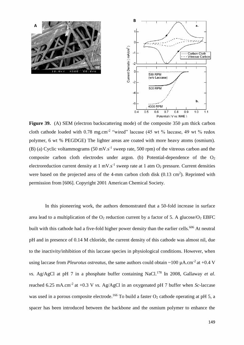
149
Figure 39. (A) SEM (electron backscattering mode) of the composite 350 m thick carbon
cloth cathode loaded with 0.78 mg.cm-2 “wired” laccase (45 wt % laccase, 49 wt % redox
polymer, 6 wt % PEGDGE) The lighter areas are coated with more heavy atoms (osmium).
(B) (a) Cyclic voltammograms (50 mV.s-1 sweep rate, 500 rpm) of the vitreous carbon and the
composite carbon cloth electrodes under argon. (b) Potential-dependence of the O2
electroreduction current density at 1 mV.s-1 sweep rate at 1 atm O2 pressure. Current densities
were based on the projected area of the 4-mm carbon cloth disk (0.13 cm2). Reprinted with
permission from [606]. Copyright 2001 American Chemical Society.
In this pioneering work, the authors demonstrated that a 50-fold increase in surface
area lead to a multiplication of the O2 reduction current by a factor of 5. A glucose/O2 EBFC
built with this cathode had a five-fold higher power density than the earlier cells.606 At neutral
pH and in presence of 0.14 M chloride, the current density of this cathode was almost nil, due
to the inactivity/inhibition of this laccase species in physiological conditions. However, when
using laccase from Pleurotus ostreatus, the same authors could obtain ~100 µA.cm-2 at +0.4 V
vs. Ag/AgCl at pH 7 in a phosphate buffer containing NaCl.178 In 2008, Gallaway et al.
reached 6.25 mA.cm-2 at +0.3 V vs. Ag/AgCl in an oxygenated pH 7 buffer when Sc-laccase
was used in a porous composite electrode.166 To build a faster O2 cathode operating at pH 5, a
spacer has been introduced between the backbone and the osmium polymer to enhance the
B A

150
rate of collisions between neighboring osmium complexes and therefore the apparent electron
diffusion from (6.2 0.8)×10-9 cm2.s-1 to (7.8 0.4) × 10-7 cm2.s-1.533,607 As seen in Fig 33B,
the presence of the tether permitted to significantly increase the current. 500 A.cm-2 was
reached at only +0.63 V vs. Ag/AgCl for the polymer with the tether while it was at +0.54 V
vs. Ag/AgCl for the polymer without spacer. A glucose/O2 biofuel cell built with this new
cathode operated at Ecell = 0.88 V while producing Pmax = 350 W.cm-2 in a stagnant pH 5
buffer at 37°C.533 At the cathode, a current density of 3 mA.cm-2 at +0.54 V vs. Ag/AgCl at
pH 5 was reached, which is at only -140 mV vs. the potential of the reversible O2/H2O
electrode. In 2002, Mano and Heller were the first to report the reduction of O2 under
physiological conditions (pH 7.4, 0.15 M NaCl, 37°C) with immobilized redox mediators.175
The electrode consisted in the crosslinked adduct of Mv-BOD and PAA-PVI-[Os (4,4’-
dichloro-2,2’-bipyridine)2Cl]+/2+ with a redox potential of +340 mV vs. Ag/AgCl. The
modified electrode reached 5 mA cm-2 when poised at +0.3 V vs. Ag/AgCl. The same year, to
upshift the operating voltage of the EBFC, a new cathode was elaborated with the same redox
polymer but with a new BOD from Trachyderma tsunodae.120 Cardoso et al. have also
reported the use of carbon cloth modified with polyamidoamine dendrimers for the
immobilization of Tv-laccase using ABTS entrapped into polypyrrole films.278
Minteer et al. used a different approach.510 Their carbon cloth electrode was first
coated with a suspension of Nafion® modified with quaternary ammonium salt and
containing bilirubin and Mv-BOD, and further soaked in 1 mM Ru(bpy)32+ to pre-concentrate
the redox mediator in the membrane. In addition to be ethanol tolerant, the advantage of this
cathode was its extended active lifetime provided by the treated porous Nafion®
membrane.491 The authors postulated that the modified structure of Nafion® with ammonium
salts could help stabilizing and increasing the enzyme activity, by providing a protective outer
shell and an ideal chemical environment resisting to a decrease in pH within the pore

151
structure. This cathode was used in an ethanol/oxygen biofuel cell which displayed an active
lifetime of 30 days and power densities of up to 0.46 mW.cm-2. With the same idea of
improving the active lifetime of Mv-BOD, Heller et al. adopted a different strategy.608 After
modification of their carbon cloth electrode with osmium polymer, crosslinker and BOD, they
coated their electrode with lyotropic liquid crystals. The authors demonstrated that such
coating permitted to prevent mechanical stripping of the bioelectrocatalyst upon shearing.
Moreover, it reduced the damage due to urate electro-oxidation without affecting the
reduction of O2. A similar strategy has been recently developed by Jian et al. with the same
system, but using a supramolecular coating made with glycosylated nucleoside fluorocarbon
amphiphile instead of liquid crystals.609 Carbon nanotubes were also controllably grown on
carbon cloth as reported by Schuhmann et al.610 With such electrode modified with osmium
polymer and Th-Laccase, the authors could increase 125 fold the power density of their EBFC
compared to an EBFC made with a bare carbon cloth electrode and identical
bioelectrocatalysts.
4.2.3.1.2 Buckypapers
Buckypaper (BP)-based nanostructured electrodes have been widely used due to their
simplicity of fabrication. BPs are usually obtained by classical filtration technique of a CNT
suspension. Redox mediators may be added during the filtration or a posteriori to
functionalize the electrode surface. However, with this filtration process, it is not possible to
control neither the porosity, nor the orientation of the nanotubes, and non-homogenous papers
are usually obtained.
Cosnier et al. developed free standing buckypaper electrodes by combining multi-
walled carbon nanotubes (MW-CNTs) and bis-pyrene modified with ABTS. The latter acted
both as a cross linker between the nanotubes and as a redox mediator for the Tt-laccase. The

152
authors could reach 1.6 mA at 0 V vs. Ag/AgCl in a 0.1 M pH 5 buffer with an operational
stability evaluated at two weeks.611 Hussein et al.612,613 also described the use of buckypapers.
Novel BPs fabricated from commercially available multi-walled carbon nanotubes (MW-
CNTs) were coated with Mv-BOD, ABTS and Nafion®.613 The authors demonstrated that
they could obtain a higher electrocatalytic activity of BOD for the ORR than at a
commercially available carbon black (Vulcan XC-72R).613 In their last report,614 BP was
modified with Mv-BOD and ABTS and further coated with Nafion®. When this electrode was
implemented in an EBFC with a glucose oxidizing anode, a power density of 26 W.cm-2 at
Ecell = 0.2 V was attained at 37°C in a quiescent O2-saturated physiological buffer containing
5 mM glucose.
4.2.3.1.3 Carbon-nanotube fibers
3D-structured carbon fiber is the ideal candidate to develop miniaturized EBFCs. It is
the ideal combination for achieving large surface area and fast mass transport while
maintaining the geometric surface as small as possible. In 2010, Gao et al. developed a new
porous cylindrical electrode combining radial transport and large specific surface area
allowing for fast mass transport. It was made of assembled and oriented nanotubes further
coated with osmium redox polymer and Mv-BOD.615 In a PBS buffer and under air, the
current density was 10 folds higher on the newly engineered porous microwire electrode
compared to a conventional modified carbon fiber. It also allowed smaller overpotentials for
O2 reduction. In addition, the mechanical stability was considerably enhanced due to the
presence of nanotubes. Such strengthening effect has also been reported for numerous
electrodes made with CNTs. For example, Kang et al.551,616 observed a similar effect on
glassy carbon electrode modified with oxidatively treated carbon nanotubes on top of which
was deposited an adduct of Mv-BOD and osmium polymers. Imbrication of BOD and CNTs
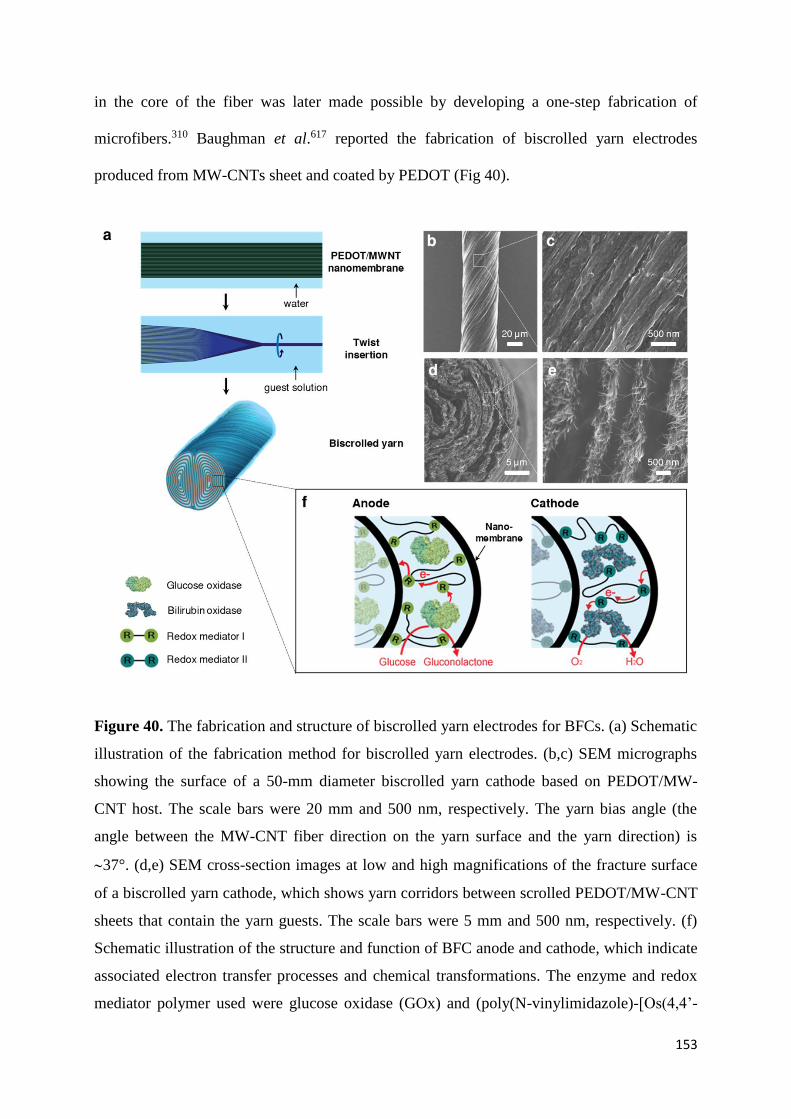
153
in the core of the fiber was later made possible by developing a one-step fabrication of
microfibers.310 Baughman et al.617 reported the fabrication of biscrolled yarn electrodes
produced from MW-CNTs sheet and coated by PEDOT (Fig 40).
Figure 40. The fabrication and structure of biscrolled yarn electrodes for BFCs. (a) Schematic
illustration of the fabrication method for biscrolled yarn electrodes. (b,c) SEM micrographs
showing the surface of a 50-mm diameter biscrolled yarn cathode based on PEDOT/MW-
CNT host. The scale bars were 20 mm and 500 nm, respectively. The yarn bias angle (the
angle between the MW-CNT fiber direction on the yarn surface and the yarn direction) is
37°. (d,e) SEM cross-section images at low and high magnifications of the fracture surface
of a biscrolled yarn cathode, which shows yarn corridors between scrolled PEDOT/MW-CNT
sheets that contain the yarn guests. The scale bars were 5 mm and 500 nm, respectively. (f)
Schematic illustration of the structure and function of BFC anode and cathode, which indicate
associated electron transfer processes and chemical transformations. The enzyme and redox
mediator polymer used were glucose oxidase (GOx) and (poly(N-vinylimidazole)-[Os(4,4’-

154
dimethoxy-2,2’-bipyridine)2Cl])+/2+ for the anode. The enzyme and redox mediator polymer
used were bilirubin oxidase (BOD) and (poly(acryl acid)-poly(N-vinylimidazole)-[Os(4,4’-
dichloro-2,2’-bipyridine)2Cl])+/2+ for the cathode. Reproduced with permission from [617].
Copyright 21014, Nature Publishing Group
After twisting to obtain a yarn, the electrodes were modified with osmium polymer
and Mv-BOD. The authors could reach 6 mA.cm-2 at +0.2 V vs. Ag/AgCl in an O2-saturated
PBS buffer at 37°C. The high current density resulted from the presence of the hydrophilic
PEDOT layer which permitted to enhance the connectivity, conductivity and wettability of
CNTs while providing electrolyte accessibility within the entire yarn. A different strategy has
been developed by Cosnier et al.337 The authors produced electrospun carbon nanofibers
containing CNTs. After annealing, the fibers were first functionalized via the reduction of 4-
carboxyphenyldiazonium to generate carboxy functions. Then, Tt-laccase was immobilized on
the modified fiber by amide coupling. To further enhance the amount of wired enzymes, a bis-
pyrene modified with ABTS was added on the fibers. Such modified fibers permitted to reach
100 A.cm-2 at +0.2 V vs. Ag/AgCl in a 0.1 M phosphate buffer pH5.
4.2.3.1.4 3D carbonaceous electrodes
In 2014, Tsujimura et al.618 reported the formation of Magnesium oxide-templated
mesoporous carbon (MgOC, mean pore diameter 38 nm). The advantage of magnesium oxide
templating is the possibility to tune at will the pore size. In 2015, they used this technique to
elaborate a cathode modified with Mv-BOD and ABTS.508 The adsorption of ABTS on the
electrode surface improved both the BOD loading and the electron transfer rate from the
electrode to the electro-active site of BOD. No significant leaching of the redox mediator was
observed. Willner et al.373 described the design of highly porous electrodes based on glassy
carbon first modified with mesoporous carbon particles (500 nm diameter, with pore

155
dimensions 6.3 nm) perfused with ABTS after drying. The modified electrodes were then
coated with Mv-BOD and Nafion®. According to the author, the advantage of this method
was the combination of the high surface area, the close proximity between the redox mediator
and the BOD, and the possibility to entrap the redox mediator within the matrix (Fig 41).
Figure 41. Schematic illustration of the modified cathode with BOD and ABTS2-. Adapted
with permission from [373]. Copyright (2013) American Chemical Society
With such design, a current density of 20 A in a 0.1 M pH 7 HEPES buffer was
reached. Wallace et al. reported nanostructured networks of carbon nanotubes grown by
chemical vapour deposition onto reticulated vitreous carbon (RVC). After modification with
Mv-BOD and osmium polymer, 175 A.cm-2 at +0.1 V vs. Ag/AgCl in a PBS buffer was
obtained with or without rotation.619 Additional examples can also be found in the review of
Bellino et al.73 who described in details the use of mesoporous materials for wired enzymes.
4.2.4 Gold electrodes
Magner et al.620 fabricated and characterized nanoporous gold substrates, with
different morphologies and roughnesses, which were attached to GC electrodes. After

156
modification with an osmium polymer and Mv-BOD, the authors reached 500 µA. cm-2 at
neutral pH in unstirred conditions. Interestingly, the authors also demonstrated that the
diffusion layer was exceeding the film thickness, resulting in the overlapping of the diffusion
zone between adjacent nanopores, and therefore yielding to similar currents than a planar
electrode. Leech et al. developed highly-ordered macroporous electrodes with 500 nm
diameter pores (Fig 42).621
Figure 42. SEM images, at different magnification of a macroporous gold scaffold obtained
upon electro-depositing though templates formed by vertical deposition of 500 nm diameter
polystyrene spheres to yield 2½ deposited layers of gold. Cyclic voltammograms at planar
(black), 1/2 sphere (red), 1½ sphere (green) and 2½ sphere (blue) gold layer electrodes
modified with [Os(2,2-bipyridine)2(PVI)10Cl]+ and Ma-laccase recorded in 0.05 M phosphate
buffer, pH 7.4, 0.15 M NaCl, 10 mM glucose. Scan rate 5 mVs-1 and current is normalised to
projected geometric area of underlying gold substrate (0.1964 cm2). Adapted with permission
from [621]. Copyright 2012 The Royal Society of Chemistry
For electrodes made with 2 and a half layers, they could reach 100 µA. cm-2 in a 0.05
M phosphate buffer pH 7.4 containing 0.1 M NaCl when the electrode was modified with Ma-
laccase and an osmium polymer.
4.2.5 Other enzymatic systems
If state-of-the art biocathodes mainly rely on the use of laccases or BODs, alternative
approaches have been developed to enzymatically reduce O2 to water. This includes the use of
bienzymatic systems, or of different enzymes such as copper efflux oxidase (CueO) or
tyrosinase (also called polyphenol oxidase, abbreviated PPO). Even if the reduction of oxygen
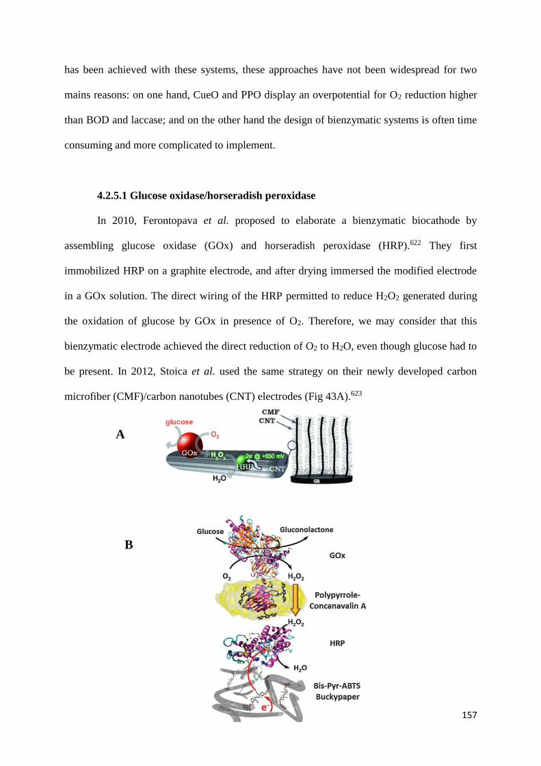
157
has been achieved with these systems, these approaches have not been widespread for two
mains reasons: on one hand, CueO and PPO display an overpotential for O2 reduction higher
than BOD and laccase; and on the other hand the design of bienzymatic systems is often time
consuming and more complicated to implement.
4.2.5.1 Glucose oxidase/horseradish peroxidase
In 2010, Ferontopava et al. proposed to elaborate a bienzymatic biocathode by
assembling glucose oxidase (GOx) and horseradish peroxidase (HRP).622 They first
immobilized HRP on a graphite electrode, and after drying immersed the modified electrode
in a GOx solution. The direct wiring of the HRP permitted to reduce H2O2 generated during
the oxidation of glucose by GOx in presence of O2. Therefore, we may consider that this
bienzymatic electrode achieved the direct reduction of O2 to H2O, even though glucose had to
be present. In 2012, Stoica et al. used the same strategy on their newly developed carbon
microfiber (CMF)/carbon nanotubes (CNT) electrodes (Fig 43A).623
B
A

158
Figure 43. (A) Representation of CMF/CNT composite carbon structure onto Graphite rod
and involved enzymatic/heterogeneous reactions responsible for reductive current at cathode.
Reproduced from [623] with permission from John Wiley and Sons. (B) Sketch of the
biocathode functioning based on bis-pyrene-ABTS BP after adsorption of HRP and
subsequent electropolymerization of pyrrole–concanavalin A for the immobilization of GOx.
Reproduced with permission from [624]. Copyright 2015 The Royal Society of Chemistry
After modification of the electrode with pyrenehexanoic acid, the modified electrode
was sequentially modified with HRP and GOx. In an oxygenated PBS buffer containing 5
mM glucose, the authors could reach a current density of 280 A. cm-2 at +0.3 V vs.
Ag/AgCl. The onset potential was 0.6 V vs. Ag/AgCl, close to the voltage that could be
obtained with a high potential laccase. They also demonstrated that in presence of 6 mM
glucose, the addition of 100 mM Cl- did not affect the current density. Cosnier et al.
elaborated two types of electrodes relying on the same concept. In the first, they used double-
walled carbon nanotubes modified with HRP and GOx.625 The authors reached 200 A.cm-2
at +0.3 V vs. Ag/AgCl in an oxygenated 5 mM glucose solution. For their second biocathode,
they developed a more complex system (Fig 43B).624 First, they coated buckypaper with
pyrene modified with ABTS, and then incubated it in a HRP solution. The modified
electrodes were functionalized with pyrrole-concanavalin A and incubated in a GOx solution.
Their freestanding electrode generated 11 mA.cm-2 at +0.1 V vs. Ag/AgCl during 1 hour of
continuous discharge in an oxygenated 5 mM glucose buffer, and was insensitive to Cl-.
Ramanivicius et al. 626 used graphite rod electrode modified with GOx and HRP, and
generated 150 A.cm-2 in an aerated 100 mM glucose solution at 25°C.
4.2.5.2 Cytochrome c and cytochrome oxidase
In 1999, Katz and Willner reported the first membrane-less glucose/O2 EBFC and
paved the way for the development of such systems. They reached 4 W for an external load

159
of 0.6 k.627 Their biocathode was composed of an assembly of Cyt c and cytochrome
oxidase (COx) (Fig 44).
Figure 44. Schematic configuration of a biofuel cell employing glucose and O2 as a fuel and
an oxidizer, respectively, and PQQ-FAD/GOx and Cyt C/COx-functionalized electrodes as
biocatalytic anode and cathode, respectively. Reproduced with permission from [627].
Copyright 1995 Elsevier
In 1982, it had already been shown that such protein-enzyme combination could
undergo the 4-electron reduction of O2 to water.628 To allow for Efficient DET, the authors
immobilized Cyt C to a maleamide modified gold electrode through a cysteine residue. Then,
the assembled monolayer of Cyt C interacted with COx to generate a multiprotein complex.
The resulting multilayers were then further cross-linked to provide stability. The authors used
this kind of biocathodes for a variety of biocatalytic systems.30,34,629-631 For example, in 2001,
they reported the first self-powered biosensors with such bienzymatic cathode.632 In 2003,633
they developed an EBFC with an electrochemically switchable power output. Their cathode
was composed of poly(acrylic acid) loaded with Cu2+ and further functionalized with Cyt c
and COx. Depending of the applied potential, i.e -0.5 V or +0.5 V vs. Ag/AgCl, the authors
could respectively tune the biocathode into a conductive or nonconductive state. In 2005,634
they also developed a magnetically controlled EBFC still based on this biocathode and
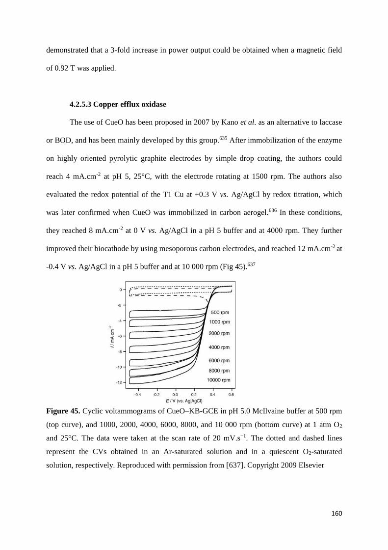
160
demonstrated that a 3-fold increase in power output could be obtained when a magnetic field
of 0.92 T was applied.
4.2.5.3 Copper efflux oxidase
The use of CueO has been proposed in 2007 by Kano et al. as an alternative to laccase
or BOD, and has been mainly developed by this group.635 After immobilization of the enzyme
on highly oriented pyrolytic graphite electrodes by simple drop coating, the authors could
reach 4 mA.cm-2 at pH 5, 25°C, with the electrode rotating at 1500 rpm. The authors also
evaluated the redox potential of the T1 Cu at +0.3 V vs. Ag/AgCl by redox titration, which
was later confirmed when CueO was immobilized in carbon aerogel.636 In these conditions,
they reached 8 mA.cm-2 at 0 V vs. Ag/AgCl in a pH 5 buffer and at 4000 rpm. They further
improved their biocathode by using mesoporous carbon electrodes, and reached 12 mA.cm-2 at
-0.4 V vs. Ag/AgCl in a pH 5 buffer and at 10 000 rpm (Fig 45).637
Figure 45. Cyclic voltammograms of CueO–KB-GCE in pH 5.0 McIlvaine buffer at 500 rpm
(top curve), and 1000, 2000, 4000, 6000, 8000, and 10 000 rpm (bottom curve) at 1 atm O2
and 25°C. The data were taken at the scan rate of 20 mV.s−1. The dotted and dashed lines
represent the CVs obtained in an Ar-saturated solution and in a quiescent O2-saturated
solution, respectively. Reproduced with permission from [637]. Copyright 2009 Elsevier

161
They also showed that the limiting current densities were pH independent in the 2-8
pH range. The exact kinetic mechanism of this enzyme, and particularly the role of the fifth
copper, has yet to be elucidated. While it has been shown that the fifth copper of the enzyme
is required to oxidize ABTS,638 its presence is not necessary to achieve DET with an electrode
surface. Kerzenmacher et al. have also reported the use of crude supernatants containing
CueO.639 The implementation of CueO in EBFCs using dissolved O2 has however not been
done so far.
4.2.5.4 Polyphenol oxidase
Because it was shown by Kang et al. that some BOD species were sensitive to
urate,640,641 which would prevent their use in blood, Cosnier et al. proposed to use instead the
urate-tolerant tyrosinase as a cathodic enzyme. In 2010, they designed an EBFC implanted in
rats with a biocathode composed of PPO, graphite and ubiquinone.642 PPO could reduce O2 to
water in complex media, and the implanted EBFC generated Pmax = 24.4 W.mL-1. Since a
dialysis bag was needed to prevent the leaching of ubiquinone, the authors replaced
ubiquinone with tetrathiafulvalene.643 An original electrode has also been developed by Wang
et al. A mixture of banana pulp, rich in PPO, and graphite particles, was used to elaborate a
biocathode. In presence of hydroquinone, as diffusing redox mediator, the authors were able
to reach 0.27 mA.cm- 2 in 0.1 M PBS pH 7.4.644
Non catalytic DET between PPO and graphite electrodes has been first reported in
1996 by Gorton et al., but the catalytic reduction of O2 to water was not observed.134 PPO has
also been immobilized on nano titanium dioxide particles,645 on silver electrodes,646 on single-
walled carbon nanotubes,647 or onto glassy carbon electrodes,648 but again catalytic
electroreduction of O2 was never achieved. The only success has been reported by Cosnier et
al. in 2012.649 The authors made an electrode by compressing MW-CNTs and PPO together.

162
With the resulting electrode, a current density of 0.55 mA.cm-2 was recorded at 0 V vs.
Ag/AgCl in a 0.2 M PBS buffer pH 7 at 25°C (Fig 46A). The authors concluded that they
succeeded in connecting the T3 copper to the electrode surface. Mechanistically, the authors
proposed a kinetic model previously discussed for tyrosinase.102,650 The oxy form might
undergo an O-O cleavage via the DET, or via the formation of an intermediate which is
further reduced at the electrode surface. After addition of 4 protons and 4 electrons, the
generated deoxy form further reacts with O2 which completes the catalytic cycle (Fig. 46B).
Figure 46. (A) Cyclic voltammograms recorded at MW-CNT/tyrosinase bioelectrodes in
argon-deaerated solution (dotted line) or oxygen saturated solution (straight line). The dashed
line corresponds to a MW-CNT electrode without tyrosinase in oxygen saturated solution (v =
0.2 mV.s−1, 0.2 M PBS, pH 7, 25 °C). Adapted with permission from ref [649]. Copyright
2011 Elsevier. (B) Proposed kinetic mechanism of O2 reduction.
5. MCOS based biocathodes in enzymatic biofuel cells
In this paragraph, we only give some examples of the implementation of MCO-based
biocathodes in EBFCs. The interested reader will find in Table 1 a series of reviews published
since 2004 that provide countless information on the history, applications and challenges of
EBFCs. Of particular interest are the most up-to-date reviews by Rasmussen et al.651 and
Shleev.89
4H+ 4e-2H2O
O2
PPO4e-
PPO
0.0 0.2 0.4-100
-50
0
50
I (µ
A)
E (V)
A B

163
5.1 Glucose/O2 EBFCs
5.1.1 Operated in buffer
As mentioned above, the first membrane-less glucose/O2 EBFC was reported in 1999
by Katz, Willner et al.627 This paper is important for the field as it was the first paper that
demonstrated that by properly choosing and/or designing bioelectrocatalysts, a membrane
separating anodic and cathodic compartments was not necessary. It also opened the way to
miniaturized systems. However, both power density and operating voltage were quite low at
that time. The next important step has been taken by Chen et al. two years later.652 Their
EBFCs consisted in two 7-m diameter, 2-cm long carbon fibers respectively coated with
glucose oxidase and an osmium redox polymer at the anode, and Tv-laccase and a second
osmium redox polymer at the cathode. At 37°C, in a pH 5 aqueous solution containing 15 mM
glucose, and under quiescent conditions, the EBFC reached Pmax = 137 W.cm-2 at Ecell = 0.4
V. Ever since, the numbers of MET and DET based glucose/O2 EBFCs have been growing
exponentially due to the envisioned applications in bodily fluids.19,22,24,25,30,51,61,89 Laccases
and BODs have been mainly used at the cathodic side in combination with glucose
oxidase,596,601,653,654 pyranose dehydrogenase,655 PQQ-dependent glucose
dehydrogenase,426,590,656-658 FAD-dependent glucose dehydrogenase,591,659 or cellobiose
dehydrogenase388,660-663 at the anode. It is worth mentioning that bionanodes based on enzyme
cascades have also been developed664-666 to improve the coulombic efficiency of EBFCs. One
of the highest power densities was reported by Sakai et al. in 2009.197 Their EBFC generated
a maximum power density of 1.45 mW.cm-2 at 0.3 V under quiescent conditions, with a
cathode based on Mv-BOD immobilized within a matrix composed of polylysine and
K3[Fe(CN)6].
The work of Xiao et al. deserves a particular attention because they reported the first
and only example of EBFC operating in organic solvents containing 5 mM glucose.276 Their

164
nanoporous gold EBFC was respectively modified with glucose oxidase and osmium redox
polymer on the anodic side, and bilirubin oxidase and osmium redox polymer on the cathodic
side. The EBFC could operate in acetonitrile, acetone and alcohols. In this latter case, the
power output of the cell decreased with increasing hydrophobicity of the alcohols; methanol
ethanol 1-propanol 1-butanol 1-pentanol. More importantly, even though the power
density was lower in organic solvents than in aqueous solvents, the anode and the cathode
recovered their initial activities when transferred in aqueous buffer after operation in organic
solutions. This inspiring work may lead to new applications of EBFCs, such as for example to
their use in bioelectrosyntheses.
5.1.2 In fruits/vegetables
As a proof of concept of possible implantation in media containing glucose and O2,
EBFCs have been inserted in different fruits, vegetables or juices containing glucose and O2
(Fig 47).667-669 The first has been reported by Mano et al. in 2002 by inserting a Glucose/O2
EBFC made with a Tt-BOD cathode within a grape (Fig 47A).598 The O2 mass transport
limited EBFC could generate Pmax = 2.4 W.mm-2 at Ecell = 0.52 V. Tsujimura et al.
assembled in 2007 four DET based EBFCs in two oranges and two apples to light a LED (Fig
47B).374 Miyake et al.670 also implanted an EBFC in a grape, thus generating Pmax = 111
W.cm-2 at Ecell = 0.25 V (Fig 47C). In 2010, Flexer et al.671 reported the insertion of an
EBFC in a cactus, generating Pmax = 8 W.cm-2 at Ecell = 0.4 V while being able to harvest
glucose and O2 produced during photosynthesis (Fig 47D).
A B C

165
Figure 47. Different examples of EBFCs implanted in fruits and cactus. (A) in a
grape598 (Reprinted with permission from ref [598] Copyright 2003 American Chemical
Society.); (B) in oranges and apples;374 Published by the PCCP Owner Societies; (C) in
grape670 (Reproduced with permission from [670] Copyright 2011 The Royal Society of
Chemistry); (D) in Cactus671 (Reprinted with permission from [671]. Copyright 2010
American Chemical Society.
5.1.3 In vertebrates and invertebrates
40 years after the first implantation of abiotic electrodes in a sheep and a dog,672,673
Cinquin, Cosnier et al. were the first to report an EBFC implanted in the abdominal cavity of
a rat in 2010 (Fig 48).642
Figure 48. EBFCs implanted in animals. In rats in 2010[642] and 2013[674,675]; in rabbit in
2011[670]; in cockroaches in 2012[548] and 2015[549]; in snail [676] and clams[327] in 2012 and
in lobsters in 2013 [677]. Adapted with permission from [678]. Copyright 2015, University of
Bordeaux.
Their cathode was made by entrapping PPO and quinone in a graphite matrix. Their
system produced Pmax = 24.4 W.cm-3 at Ecell = 0.13 V. In 2013, the same authors replaced
PPO by Tv-laccase entrapped in CNTs and reached Pmax = 193.5 W.cm-2, also upon
implantation in rats.674 In 2011, Miyake et al. 670 implanted an EBFC in a rabbit ear,
generating Pmax = 131 W.cm-2 at Ecell = 0.56 V. Note that in this particular case, an air-
breathing cathode made with Mv-BOD was used. These works were followed by a series of
2010 2011 2013 2015

166
implantations in various vertebrates and invertebrates which have been reviewed by Falk et
al.56 Because the dominant sugar in the body fluids of many insects is not glucose, the sugar
of the vertebrates, but trehalose, Rasmussen et al.548 designed a bi-enzymatic anode by
combining glucose oxidase and trehalase. Coupled with a cathode made with Tt-BOD, the
EBFC generated Pmax = 55 W.cm-2 at Ecell = 0.2 V when implanted in a cockroach. In 2015,
the same authors reported549 the wireless communication between a receiver and a cyborg
cockroach modified with the same type of EBFC. In a series of paper, Katz et al. have
implanted EBFCs in a snail (Pmax = 30 W.cm-2 at Ecell = 0.39 V),676 in clams (Pmax = 40
W.cm-2 at Ecell = 0.17V),327 in lobsters (Pmax = 640 W.cm-2 at Ecell = 0.28V)677 and in rats
(Pmax = 70 W.cm-2 at Ecell = 0.23V).675 Tv-laccase based cathodes were used in all those
EBFCs. Of interest is the experiment made by Southcott et al. in 2013.679 To mimic the
human blood circulating in a 8 m capillary at 1 mm.s-1, the authors elaborated a biofuel flow
cell. When a human serum solution with 6.4 mM glucose concentration was used, a current
density of 0.83 mAcm-2 was obtained with a laccase-based biocathode.
5.1.4 In serum and blood
Numerous examples of EBFCs operating in bovine or human serum can be found in
the literature. To the best of our knowledge, the first EBFC operating in human serum has
been reported by Gao et al. in 2007.680 With a cathode based on Mv-BOD immobilized on
carbon nanotubes, they obtained Pmax = 5 µW.cm-2 at Ecell = 0.4 V. After further optimization
of their electrodes with SW-CNTs, they reached Pmax = 35 µW.cm-2 at Ecell = 0.39 V.302 In
2010, Pmax = 3 µW.cm-2 at Ecell = 0.37 V was reported for the first EBFC operating in DET
with Mv-BOD coated on graphite electrodes.241 In 2014, Scherbahn et al.319 reached Pmax = 45
µW.cm-2 at Ecell = 0.39 V in an unmodified serum for an EBFC cathode made with Mv-BOD
immobilized on vertically aligned carbon nanotubes. With the same enzyme at the cathode,

167
Pmax = 57.5 µW.cm-2 at 37°C in human serum containing 5 mM glucose was obtained by
Milton et al.464 Ammam et al. reported Pmax = 160 µW.cm-2 at Ecell = 0.21 V on glassy carbon
electrodes564 and Pmax = 69 µW.cm-2 at Ecell = 0.151 V on multi-walled carbon nanotubes
when laccase was used.681 The EBFC developed by Ó Conghaile et al.591 generated Pmax = 37
µW.cm-2 at Ecell = 0.26 V with a Sc-laccase immobilized within an osmium redox polymer,
and Pmax = 60 µW.cm-2 at Ecell = 0.3 V in artificial plasma when Myceliophthora thermophila
laccase was used.682 The highest power density obtained so far for an EBFC operating in
human serum has been reported in 2015 by Kwon et al. using the device described in Figure
40. They first reached Pmax = 1.02 mW.cm-2 at Ecell = 0.40 V in 2014617 and then Pmax = 1,1
mW.cm-2 at Ecell = 0.50 V in 2015.683
Experiments in human blood have been rarer, and only 5 can be found in the literature.
In 2010, an EBFC made with a laccase entrapped within a matrix of Nafion® and poly(vinyl
pyrrolidone) generated Pmax = 5.56 µW.cm-2 at Ecell = 0.07 V.684 However, neither the
concentration of glucose nor the concentration of O2 were provided in this work. Pmax = 2.8
µW.cm-2 at Ecell = 0.47 V in presence of 4.6 mM glucose was reached by Wang et al.663 with
Mv-BOD immobilized in three-dimensional gold electrodes. More recently, Pmax = 73 µW.cm-
2 at Ecell = 0.3 V has been obtained by Ó Conghaile et al.685 in whole blood containing 5.4 mM
glucose. Their cathode was made with adsorbed Mv-BOD on a gold electrode modified with
gold nanoparticles. The highest power density in whole and undiluted human blood so far has
been reported by Cadet et al.602 It was Pmax = 129 µW.cm-2 at Ecell = 0.38 V with a glycaemia
of 8.2 mM. The cathode was made with a combination of Bp-BOD and an osmium redox
polymer (Fig 49A). The first ex vivo experiment in human blood under homeostatic conditions
has been reported in 2016 in a vein replica.686 The power generated by this EBFC made with a
Mv-BOD cathode was enough to power a flexible low-voltage display (Fig 49B).
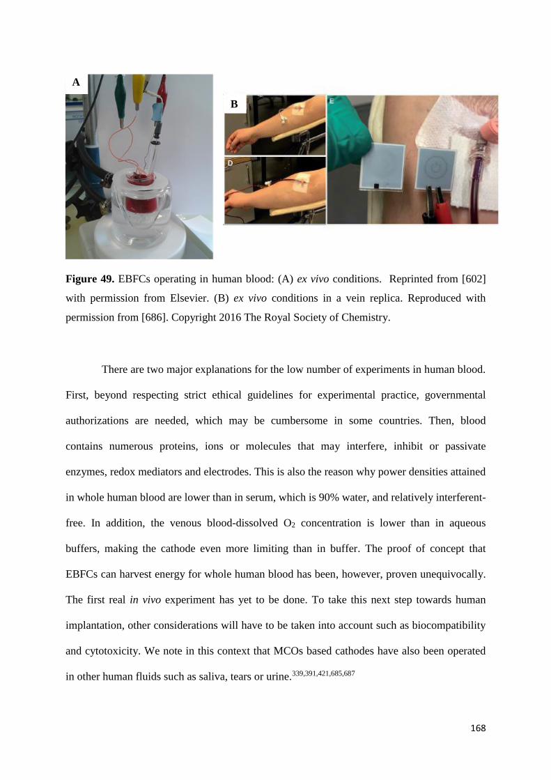
168
Figure 49. EBFCs operating in human blood: (A) ex vivo conditions. Reprinted from [602]
with permission from Elsevier. (B) ex vivo conditions in a vein replica. Reproduced with
permission from [686]. Copyright 2016 The Royal Society of Chemistry.
There are two major explanations for the low number of experiments in human blood.
First, beyond respecting strict ethical guidelines for experimental practice, governmental
authorizations are needed, which may be cumbersome in some countries. Then, blood
contains numerous proteins, ions or molecules that may interfere, inhibit or passivate
enzymes, redox mediators and electrodes. This is also the reason why power densities attained
in whole human blood are lower than in serum, which is 90% water, and relatively interferent-
free. In addition, the venous blood-dissolved O2 concentration is lower than in aqueous
buffers, making the cathode even more limiting than in buffer. The proof of concept that
EBFCs can harvest energy for whole human blood has been, however, proven unequivocally.
The first real in vivo experiment has yet to be done. To take this next step towards human
implantation, other considerations will have to be taken into account such as biocompatibility
and cytotoxicity. We note in this context that MCOs based cathodes have also been operated
in other human fluids such as saliva, tears or urine.339,391,421,685,687
A
B
169
5.2 H2/O2 EBFCs
H2/O2 EBFCs have been reviewed in details by Lojou et al. in 201463 and some more
recent developments have been described by Cosnier et al.75 Except of the first device
combining microbial H2 oxidation and enzymatic mediated O2 reduction by Mv-BOD and
ABTS496, these devices mainly rely on a hydrogenase (Hase) at the anode, and a MCO at the
cathode, both operating in DET conditions (unless otherwise specified below). Even if high
interests arise from the high OCV of H2/O2 EBFCs, and the high turnover frequencies and
ability to establish DET of Hases, their story is rather short compared to that of glucose/O2
EBFCs due to the problematic high sensitivity of most Hases towards O2. The discovery of
O2-tolerant Hases allowed recently envisioning H2/O2 enzymatic BFCs. The publication in
2005 of the proof of concept of this kind of devices, that displayed a maximum power density
of 5 μW cm−2 at a cell voltage above 0.8 V at pH 5,282,283 was followed by further
developments and fundamental studies. Particular care is indeed required due to the
specificities of Hase as an EBFC catalyst, and the inherent problems of the H2 fuel that,
combined with O2, leads to explosive mixtures under certain conditions.142,282,308 Power
densities have overcome 100 µW.cm-2 in 2012 thanks to the structuration of electrodes with
CNTs.142,308 The same kinds of performances were also obtained when the enzymes were
immobilized at AuNP-modified electrodes.392 H2/O2 EBFCs with power densities above 1
mW.cm-2 have been developed in the groups of Armstrong143 and Lojou.372 In particular, the
second was able to operate over a wide range of temperatures thanks to the combination of a
hyperthermophilic Hase and the thermostable Bp-BOD, both immobilized in DET
configuration at CNF-modified electrodes. In 2014 the first H2/O2 EBFC using an O2-
sensitive Hase was reported by Plumeré et al.688 No membrane separated the anodic and
cathodic compartments, and the Hase, operating via a MET mechanism, was protected from
O2 denaturation by a redox hydrogel.689 The ability of these EBFCs to operate small devices
170
was also recently demonstrated.335,381 A membrane-less H2/O2 EBFC using an air-breathing
Mv-BOD cathode was reported in 2015466, followed by the development of an EBFC using
gas-diffusion electrodes both at the anode and the cathode by Kano et al. in 2016467,690. In this
configuration, a maximal power density of 6.1 mW.cm-2 at Ecell = 0.72 V was reached at room
temperature and under quiescent conditions.467
Besides glucose or H2, other fuels have been studied in combination with MCO-based
biocathodes666: to name a few, methanol,277,278,691 ethanol,692-694 fructose,470,695 ascorbate,391 or
sulfite423 have also been reported in the literature. The interested reader will find additional
information in the reviews listed in Table 1.
5.3 Microfluidic biofuel cells
MCOs based electrodes have also been used to elaborate microfluidic EBFCs even if,
so far, their use for such applications is less widespread. This field has been reviewed by
Kjeang et al.,39 Sinton et al.32 and more recently by Sanchez et al.82 Microfluidic biofuel cells
are usually composed of two channels, of various designs and geometries, into which circulate
the respective anodic and cathodic substrates. Redox mediators and enzymes can also be
added in this stream if they are not immobilized. The lack of convective mixing at low
Reynolds numbers permits to eliminate the need for a membrane between the anode and
cathode, therefore minimizing ohmic losses. Protons diffuse through the liquid-liquid
interface between the two substrate streams. Despite the conceptual simplicity, numerous
difficulties can be encountered such as the cross-diffusional mixing of substrates at the
interface between the two streams. The fabrication of the microchannel and the
lengths/diameters of the electrodes require particular care. One of the first use of a MCO-
based biocathode in a microfluidic BFC has been reported by Palmore et al.696 The anolyte
consisted of 10 mM ABTS and the catholyte of 10 mM ABTS and 0.5 mg.mL-1 of laccase.
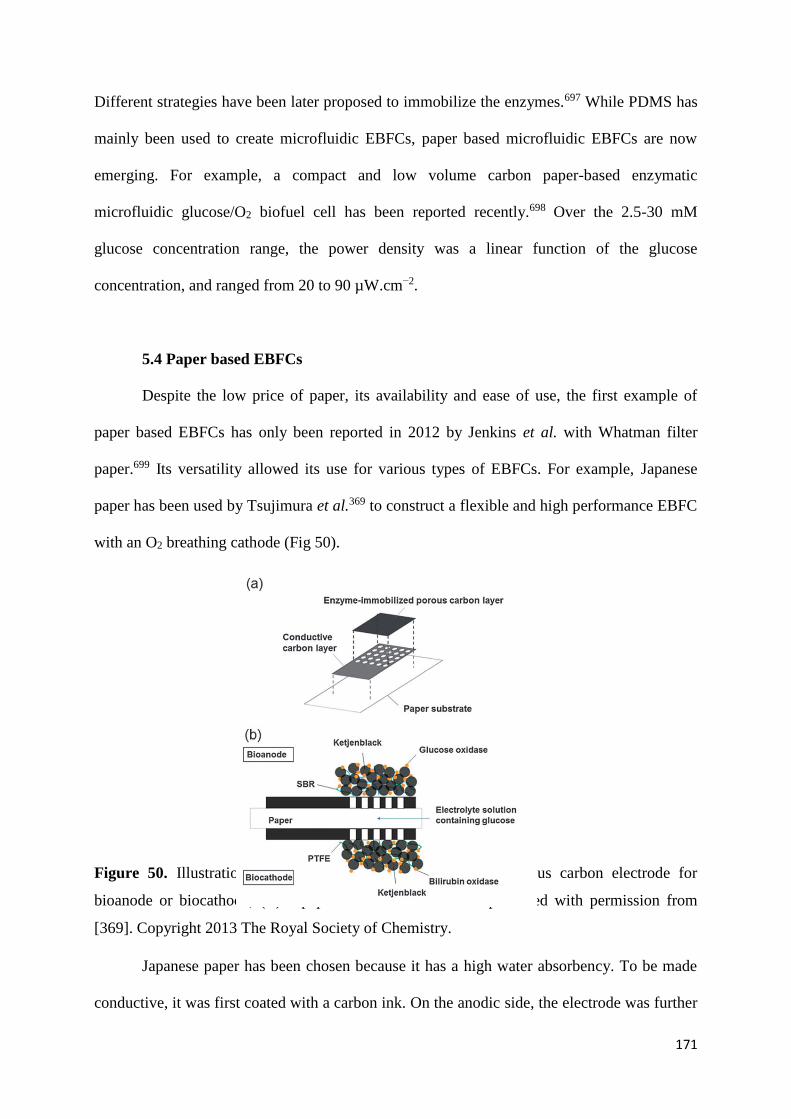
171
Different strategies have been later proposed to immobilize the enzymes.697 While PDMS has
mainly been used to create microfluidic EBFCs, paper based microfluidic EBFCs are now
emerging. For example, a compact and low volume carbon paper-based enzymatic
microfluidic glucose/O2 biofuel cell has been reported recently.698 Over the 2.5-30 mM
glucose concentration range, the power density was a linear function of the glucose
concentration, and ranged from 20 to 90 µW.cm−2.
5.4 Paper based EBFCs
Despite the low price of paper, its availability and ease of use, the first example of
paper based EBFCs has only been reported in 2012 by Jenkins et al. with Whatman filter
paper.699 Its versatility allowed its use for various types of EBFCs. For example, Japanese
paper has been used by Tsujimura et al.369 to construct a flexible and high performance EBFC
with an O2 breathing cathode (Fig 50).
Figure 50. Illustration of (A) a screen-printed paper-based porous carbon electrode for
bioanode or biocathode, (B) a paper-based biofuel cell. Reproduced with permission from
[369]. Copyright 2013 The Royal Society of Chemistry.
Japanese paper has been chosen because it has a high water absorbency. To be made
conductive, it was first coated with a carbon ink. On the anodic side, the electrode was further

172
modified with a mixture of Ketjen black, glucose oxidase and tetrathiafulvalene. At the
cathode, a mixture of KB and polytetrafluoroethylene was coated on the conductive paper
before Mv-BOD addition. The power density of the fabricated EBFC reached 0.12 mW.cm-2
at Ecell = 0.4 V in presence of 100 mM glucose in a 1 M phosphate buffer pH 7 at room
temperature, which was the highest power reported in 2013. Paper based electrodes have also
been used in microfludidic EBFCs,700 in self-powered systems,701 or to elaborate a self-
powered hybrid micro-supercapacitor integrated system.702 All used MCO-based cathodes. A
recent and excellent review by Desmet et al.76 summarizes the use of paper electrodes in
biofuel cells.
5.5 MCOs with air-breathing electrodes
Most of the EBFCs are limited by the cathode because of the low diffusion coEfficient
(< 5.10-5 cm-2.s-1) and concentration of dissolved O2 (1.2 mM maximum at neutral pH) in
aqueous solution. Increasing the low current densities and enhancing mass transport on the
cathodic side are key challenges in EBFCs. These limitations could be circumvented by using
an air-breathing cathode, where O2 would diffuse directly from the air to the cathode. In 2005,
Calabrese-Barton703 reported a particularly interesting numerical simulation of a MCO-based
cathode. The authors predicted that in an O2 saturated pH 5 buffer at 37°C, a maximum
current density of 9.2 mA.cm-2 could be attained with dissolved oxygen. By introducing an
air-breathing cathode, the model predicted that jmax = 60 mA.cm-2 in air or jmax = 100 mA.cm-2
in pure O2 could be reached. However, designing a gas-diffusion cathode is not trivial. The
gas-diffusion cathode usually consists of two layers: a gas-diffusion layer made of
hydrophobized carbon black704,705 or Ketjen black690,706,707, which has to be permeable to gas;
a hydrophilic catalytic layer made with carbon black708, carbon cloth709, CNTs710-712 or carbon
felt713 and containing the MCO. These first two layers may also be added on top of a third

173
layer providing for mechanical stability. A delicate balance between hydrophobicity and
hydrophilicity of the carbon/air and carbon/electrolyte interfaces has to be achieved (Fig 51).
Figure 51. Schematic of the three-phase interphase of a gas-diffusion electrode. Reproduced
with permission from [307] Copyright 2012 John Wiley and Sons.
It is challenging to obtain a tri-phase interface allowing simultaneously adequate
immobilization of the MCO, and performant diffusion of gas and electrolytes. Design of
experiments have been used to optimize such interface.714 DET and MET based air-breathing
electrodes have been reported with BODs,368,466,712,715 laccases708,709,716 and CueO.706 For
example, Atanassov et al. reported a DET-based laccase-modified gas-diffusion electrode
with a current density 0.5 mA.cm-2.307 20 mA.cm-2 have been reached for a DET-based
CueO biocathode.706 An interesting approach has been reported recently by Atanassov et al.717
who combined BOD and non-platinum based catalysts. jmax = 1.2 mA.cm-2 was reached in a
0.1 M phosphate buffer pH 7.5. MCO-based gas-diffusion electrode have been used in
H2/O2,466,467,690 methanol/O2
718 or in fructose/O2 biofuel cells.305,368
5.6 Biofuel cells for self-powered devices
Self-powered sensing devices do not need any external power source, and usually
consist of only two electrodes. One electrode acts as the sensing part and both electrodes

174
combined together provide the power for the sensing device. The power output is usually
proportional to the concentration of the analyzed substrate. The first self-powered system for
the measurement of glucose using an EBFC, made with a glucose oxidase anode and an O2-
reducing cathode combining Cyt C and COx, has been reported by Willner and Katz in
2001.632 Katz et al. have also pioneered the use of MCO-based cathodes to develop EBFCs
with switchable power output and their use in enzymatic logic systems.34,633,719-723 The
principle of such systems has been described in their first report in 2009.724 It is based on the
dependence on pH of swelling/deswelling of the redox hydrogel used to immobilize the
laccase (Fig 52).
Figure 52. The Biofuel Cell Composed of the pH-Switchable Logically Controlled
Biocatalytic Cathode and Glucose-Oxidizing Anode. Reprinted with permission from [724].
Copyright 2009 American Chemical Society.
The authors synthesized a polymer brush covalently functionalized with osmium
redox centers. For pH > 5.5, mediated electron transfer was prohibited because of the
shrinkage of the hydrogel; while for pH < 4.5, the fully swollen and hydrated hydrogel

175
allowed electron transfer. Two Boolean logic gates (AND/OR) were assembled using this
concept. MCO-based biocathodes have also been used for example for self-powered immune
sensors,725 self-powered biosensors for arsenite and arsenate,726 lactate biosensors,336
antiobitic residue determination,727,728glucose measurement with amperometric602 or optical
readout,729 neurochemical monitoring in rat brain,730 self-powered fluorescence switch
systems,731 or for wireless signal transmission.732 This field has been reviewed by Zhou et
al.43,47 and by Arechederra et al.45
5.7 Hybrid bio-devices
Hybrid biodevices exhibit dual function and are a combination of an enzymatic biofuel
cell and an electrochemical capacitor. Once charged by the internal EBFC, the device can be
discharged as a supercapacitor. The concept and denomination of “self-charging
electrochemical biocapacitor” have been first published in 2014 by Pankratov et al.733 Their
2-electrodes device is illustrated in Figure 53.
Figure 53. Schematic representation of a self-charging biological supercapacitor. The
electrochemical capacitor is built using a graphite foil modified with a polyaniline/CNTs
composite on one side (the capacitive side/face), and the enzymatic fuel cell is designed from
three-dimensional gold NPs based nanobiostructures on the other (the charging side/face).
Gold NPs: pink-orange spheres; thiol layer: black sticks. The structure of bilirubin oxidase
was taken from the know crystal structure (PDB: 2XLL) and cellobiose dehydrogenase was
176
rendered using the cytochrome and the FAD domains of the enzyme from Phanerochaete
chrysosporium (PDB: 1D7D and 1NAA, respectively). Protein globule: green ribbons; copper
ions: blue spheres; heme: red spheres; FAD: yellow spheres; carbohydrates: black sticks.
Reprinted with permission from [733] .Copyright 2014 Wiley-VCH Verlag GmbH & Co.
KGaA.
One side of both electrodes was made of graphite modified with CNTs and
polyaniline. The other face was modified with the enzyme (resp. cellobiose dehydrogenase or
Mv-BOD) immobilized on gold nanoparticles. In an aerated PBS pH 7.4 buffer containing 50
mM glucose, the device displayed excellent reproducibility upon 4 cycles of charge/discharge,
and provided a power density Pmax = 1.2 mW.cm-2 at Ecell = 0.38 V, without being optimized.
The authors reported also the same year a self-charging ascorbate/O2 electrochemical
capacitor.734 In 2014 also, Cosnier et al.735 published a similar concept using CNT matrixes
modified with Tv-laccase. The authors demonstrated that their hybrid system was stable for at
least 40 000 pulses of 2 mW. In 2016, the first self-charging Nernstian bio-supercapacitor
operating with an identical redox mediator at the anode and cathode was reported.736 In 2017,
Xiao et al.737 reported a hybrid device using Mv-BOD immobilized on de-alloyed nanoporous
gold. Good operational stability was observed for 50 charge/discharge cycles at j = 0.2 mA
cm−2. A power density of 608.8 μW.cm−2 was obtained when discharged at 2 mA cm−2, i.e.
468 higher than the power output of the BFC itself. The work of Bilewicz et al. using a
laccase-based cathode deserves also attention in this field.738-741 For example, they used a
biocathode covered with laccase adsorbed on naphthylated multiwalled carbon nanotubes to
elaborate an hybrid devices generating Pmax = 1.6 mW.cm-2 at Ecell = 0.76 V. Hybrid devices
may also rely on solar power, which allows simultaneous solar energy conversion and storage.
Nanostructured indium tin oxide electrodes were therefore used.742,743 This new promising

177
field, which may circumvent low power densities and substrates depletion arising in EBFCs,
has been reviewed in 2014 by Pankratov et al.67 and in 2016 by Sode et al.79
5.8. Wearable biofuel cells
Wearable electronics, and particularly wearable electrochemical sensors, have
attracted recently a lot of attention.744 Since they are likely to be in contact with the human
skin or human tears80,339, they should be flexible, light, and biocompatible. The detection of a
wide variety of substrates, ranging from glucose to uric acid has been realized. Even more
recently, the use of wearable biofuel cells has been envisioned as a power source for wearable
applications. Bandodkar has written two reviews on this subject.745,746 Surprisingly, Pt has
been preferred so far to MCOs as the cathodic catalyst. For example, an epidermal EBFC able
to harvest chemical energy from human perspiration and particularly from lactate,747 or the
fabrication of a wearable biofuel cell printed directly onto textile substrates have been
reported.748 In both cases, Pt was the catalyst of O2-reduction. In 2014 however, Mv-BOD has
been used by Ogawa et al.749 The authors developed an organic transdermal iontophoresis
patch with an integrated EBFC (Fig 54). The anodic electrode was made with fructose
dehydrogenase coated on a CNT-modified carbon strip, and an O2 diffusion cathode was
based on Mv-BOD. Once immobilized on the skin, the OCP was 0.75 V and the maximal
current density 300 A.cm-2.

178
Figure 54. (A) Schematic of the transdermal iontophoresis patch that is an assembly of
enzyme-modified CFs that serve as the electrodes, hydrogel films containing a biofuel and a
chemical to be delivered, a conducting polymer-based stretchable resistor, a
poly(dimethylsiloxane) (PDMS) frame, and O2 permeable medical tape. (B) Photograph of the
patch mounted on a human arm, and a schematic of the iontophoresis-assisted drug delivery
mechanism. The fructose/O2 biofuel cell generates a transdermal ionic current that is
accompanied by an osmotic flow from the anode to the cathode, thereby assisting the
electrophoretic movement of small molecules into the skin. Reprinted with permission from
[749] Copyright 2015 Wiley-VCH Verlag GmbH & Co. KGaA
6. Conclusions and Outlook
The major cause of inefficiency in common EBFCs, envisioned as energy-converting
devices for more than half a century, is the four-electron electroreduction of oxygen to water,
particularly because of the low concentration and slow diffusion of O2 in aqueous media.
Studies in the recent decades aimed to overcome this limitation. We have witnessed an
exponential growth in publications on optimizing MCO-based biocathodes. The different
strategies that have been put forward are listed in Table 1. Two major research axes have

179
developed: 1) design of increasingly sophisticated nanostructured porous electrodes, allowing
increased enzyme loadings without impeding the diffusion of O291 and 2) improvement of the
electrical connection between MCOs and the electrode surfaces, whether in MET or DET.750
The continuous increase in OCP, current densities or power densities reflect particularly well
the significant progress made along the two lines of research.
The studies have led to a fundamental milestone with the first implantation in a
vertebrate and to a second milestone with the first operation in human blood. The latter now
raises new questions and exciting challenges, such as immunogenicity, bioinertness and
cytotoxicity issues. Solutions have to be found to minimize the reaction with the foreign
body. It is likely that a protective/bioinert coating or membrane will be required to minimize
the foreign body reaction. It must not be, however, detrimental to O2 diffusion.
Because the enzymatic reduction of O2 is becoming increasingly Efficient, new
applications have emerged e.g. in wearable enzymatic biofuel cells and new promising field
such has the development of hybrid supercapacitors, which may circumvent low power
densities and substrates depletion arising in EBFCs.
In their 2004 pioneering review on EBFCs Calabrese-Barton and Atanassov19 said
that the development of successful biofuel cells is driven by demand arising from specific
applications. The low current densities prevented the use of EBFCs in most of applications.
However, high current densities are not a prerequisite in some, as a current of a few pA
suffices, when the voltage is high enough, to power ACSICs.751 For high power applications,
EBCs/supercapacitor systems are of particular interest due to their ability to deliver high
power densities within a very short burst.
Despite enormous advances and encouraging achievements, some limitations remain.
One of the most underappreciated problem is the stability. Relatively few studies are
dedicated to the study of stability of immobilized MCOs231, and even fewer deal with their

180
thermostability. Stability studies are often only corollaries of other optimizations. If one
envisions the use of EBFCs in medical or other long-term applications, this problem has to be
tackled. As discussed in this review, the causes of the poor stability of MCOs immobilized on
electrode surfaces are many and most are interrelated. Fundamental and mechanistic studies
are needed to resolve these issues. Because electrochemistry by itself does not permit to link
the catalytic stability with the structure/conformation of proteins, the electrochemical studies
must be combined with others. Enzyme engineering can be a valuable tool to improve the
stability/thermostability of MCOs in homogeneous solutions. However, solution phase
stability does not predict the stability of immobilized enzymes. This is particularly well
illustrated by their inhibition by halides. Inhibition by Cl− varies between the MCOs, and
differs for different MCOs immobilized on different electrodes, as well as for MET,
depending on the redox mediator used. Again the combination of different analytical
techniques in homogeneous and heterogeneous conditions is required to enhance our
understanding of the MCOs and further improve cathodes for EBFCs applications.
In addition to catalyzing reactions in biofuel cells, enzymes also inspire the design of
analogous inorganic and metal-organic catalysts.752-754 “Bio-inspired” catalysts aim to
reproduce the metal coordination observed in biological molecules to attain similar efficiency
in a less complex structure. Further development is expected also along this axis.
7. Acknowledgments
The authors thank Prof Adam Heller (The university of Texas-Austin-USA) for
critical reviewing/editing this MS. NM thanks his past PhD/Post Doctoral students and
collaborators as indicated in the references cited for their outstanding contributions to this
field. AP thanks her collaborators at the Laboratory of Bioenergetics and Protein Engineering,
and especially Drs. V. Fourmond, M. Ilbert, E. Lojou, I. Mazurenko and M. Roger for fruitful

181
discussions. NM also acknowledges support from the Région Aquitaine, the Agence
Nationale de la Recherche (12-BS08-0011-01 and 16-CE19-0001-02), the European project
BIOENERGY (FP7-PEOPLE-2013-ITN607793) and the Labex AMADEus (ANR-10-LABX-
0042-AMADEus) belonging to the program Initiative d’Excellence IdEx Bordeaux (ANR-10-
IDEX-003-02).
Biographies:
Nicolas Mano is senior researcher at the CNRS. He received his Ph.D. from the
University of Bordeaux (France) under the supervision of Prof Alexander Kuhn in 2001. He
then carried out postdoctoral studies at the University of Texas at Austin (USA) with Prof
Adam Heller, working towards the development of glucose sensors and miniature membrane-
less glucose/O2 biofuel cells. In 2006, he integrated the CNRS and created his team at the
Centre de Recherche Paul Pascal located in Pessac (France) working on different fields of
bioelectrochemistry with a special emphasis on biofuel cells. He also developed an activity in
enzymes engineering on various oxidases including bilirubin oxidases. His contribution to the
field of bioelectrochemistry has been recognized by numerous National and International
Prizes
Anne de Poulpiquet got interested in electroenzymatic O2 reduction during her PhD
dedicated to the development of enzymatic H2/O2 biofuel cells. She obtained her degree under
the direction of Dr. Elisabeth Lojou at Aix-Marseille University (France). After a postdoctoral
stay at the Institute of Molecular Sciences in Bordeaux (France), she joined the Bioenergetic
and Protein Engineering Laboratory (Marseille, France) as an assistant professor. Her research
interests now focus on coupling electrochemistry to optical techniques to study the electro-
enzymatic reactivity.

182
8. References
(1) Farquhar, J.; Bao, H. M.; Thiemens, M. Atmospheric Influence of Earth's Earliest
Sulfur Cycle. Science 2000, 289, 756-758.
(2) Festa, R. A.; Thiele, D. J. Copper: an Essential Metal in biology. Current Biology
2011, 21, R877-R883.
(3) Ridge, P. G.; Zhang, Y.; Gladyshev, V. N. Comparative Genomic Analyses of Copper
Transporters and Cuproproteomes Reveal Evolutionary Dynamics of Copper
Utilization and Its Link to Oxygen. Plos One 2008, 3.
(4) Muttkowski, R. A. Copper: Its Occurrence and Role in Insects and Other Animals.
Trans Am. Microsc. Soc. 1921, 40, 144-157.
(5) Solomon, E. I.; Heppner, D. E.; Johnston, E. M.; Ginsbach, J. W.; Cirera, J.; Qayyum,
M.; Kieber-Emmons, M. T.; Kjaergaard, C. H.; Hadt, R. G.; Tian, L. Copper Active
Sites in Biology. Chem. Rev. 2014, 114, 3659-3853.
(6) Liu, J.; Chakraborty, S.; Hosseinzadeh, P.; Yu, Y.; Tian, S. L.; Petrik, I.; Bhagi, A.;
Lu, Y. Metalloproteins Containing Cytochrome, Iron-Sulfur, or Copper Redox
Centers. Chem. Rev. 2014, 114, 4366-4469.
(7) Solomon, E. I.; Szilagyi, R. K.; George, S. D.; Basumallick, L. Electronic Structures
of Metal Sites in Proteins and Models: Contributions to Function in Blue Copper
Proteins. Chem. Rev. 2004, 104, 419-458.
(8) Yoshikawa, S.; Shimada, A. Reaction Mechanism of Cytochrome c Oxidase. Chem.
Rev. 2015, 115, 1936-1989.
(9) Debe, M. K. Electrocatalyst Approaches and Challenges for Automotive Fuel Cells.
Nature 2012, 486, 43-51.

183
(10) Tiwari, J. N.; Tiwari, R. N.; Singh, G.; Kim, K. S. Recent Progress in the
Development of Anode and Cathode Catalysts for Direct Methanol Fuel Cells. Nano
Energy 2013, 2, 553-578.
(11) Shleev, S.; Andoralov, V.; Pankratov, D.; Falk, M.; Aleksejeva, O.; Blum, Z. Oxygen
Electroreduction versus Bioelectroreduction: Direct Electron Transfer Approach.
Electroanalysis 2016, 28, 2270-2287.
(12) Kjaergaard, C. H.; Rossmeisl, J.; Norskov, J. K. Enzymatic versus Inorganic Oxygen
Reduction Catalysts: Comparison of the Energy Levels in a Free-Energy Scheme.
Inorg. Chem. 10, 49, 3567-3572.
(13) Zagal, J.; Bindra, P.; Yeager, E. A Mechanistic Study of O2 Reduction of Water
Soluble Phtalocyanines Adsorbed on Graphite Electrodes. J. Electrochem. Soc. 1980,
127, 1506-1517.
(14) Shi, C. N.; Anson, F. C. Catalytic Pathways for the Electroreduction of O2 by Iron
Tetrakis(4-N-methylpyridyl)porphyrin or Iron Tetraphenlyporphyrin Adsorbed on
Edge Plane Pyrolytic Graphite Electrodes. Inorg. Chem. 1990, 29, 4298-4305.
(15) Heller, A. Integrated Medical Feedback Systems for Drug Delivery. AIChE Journal
2005, 51, 1054-1066.
(16) Mano, N.; Soukharev, V.; Heller, A. A Laccase-Wiring Redox Hydrogel for Efficient
Catalysis of O2 Electroreduction. J. Phys. Chem. B 2006, 110, 11180-11187.
(17) dos Santos, L.; Climent, V.; Blanford, C. F.; Armstrong, F. A. Mechanistic Studies of
the 'Blue' Cu Enzyme, Bilirubin Oxidase, as a Highly Efficient Electrocatalyst for the
Oxygen Reduction Reaction. Phys. Chem. Chem. Phys. 2010, 12, 13962-13974.
(18) Shleev, S.; Tkac, J.; Christenson, A.; Ruzgas, T.; Yaropolov, A. I.; Whittaker, J. W.;
Gorton, L. Direct Electron Transfer between Copper-containing Proteins and
Electrodes. Biosens. Bioelectron. 2005, 20, 2517-2554.

184
(19) Barton, S. C.; Gallaway, J.; Atanassov, P. Enzymatic Biofuel Cells for Implantable
and Microscale Devices. Chem. Rev. 2004, 104, 4867-4886.
(20) Shukla, A. K.; Suresh, P.; Berchmans, S.; Rajendran, A. Biological Fuel Cells and
Their Applications. Curr. Sci. 2004, 87, 455-468.
(21) Kim, J.; Jia, H.; Wang, P. Challenges in Biocatalysis for Enzyme-Based Biofuel Cells.
Biotech. Adv. 2006, 24, 296-308.
(22) Bullen, R. A.; Arnot, T. C.; Lakeman, J. B.; Walsh, F. C. Biofuel Cells and Their
Development. Biosens. Bioelectron. 2006, 21, 2015-2045.
(23) Heller, A. Potentially Implantable Miniature Batteries. Anal. Bioanal. Chem. 2006,
385, 469-473.
(24) Davis, F.; Higson, S. P. Biofuel Cells - Recent Advances and Applications. Biosens.
Bioelectron. 2007, 22, 1224-1235.
(25) Minteer, S. D.; Liaw, B. Y.; Cooney, M. J. Enzyme-Based Biofuel Cells. Curr. Opin.
Biotechnol. 2007, 18, 228-234.
(26) Kerzenmacher, S.; Ducrée, J.; Zengerle, R.; von Stetten, F. Energy Harvesting by
Implantable Abiotically Catalyzed Glucose Fuel Cells. J. Power Sources 2008, 182, 1-
17.
(27) Cracknell, J. A.; Vincent, K. A.; Armstrong, F. A. Enzymes as Working or
Inspirational Electrocatalysts for Fuel Cells and Electrolysis. Chem. Rev. 2008, 108,
2439-2461.
(28) Cooney, M. J.; Svoboda, V.; Lau, C.; Martin, G.; Minteer, S. D. Enzyme Catalysed
Biofuel Cells. Energy Environ. Sci. 2008, 1, 320-337.
(29) Moehlenbrock, M. J.; Minteer, S. D. Extended Lifetime Biofuel Cells. Chem. Soc.
Rev. 2008, 37, 1188-1196.

185
(30) Willner, I.; Yan, Y.; Willner, B.; Tel-Vered, R. Integrated Enzyme-Based Biofuel
Cells - A Review. Fuel Cells 2009, 9, 7-24.
(31) Ramanavicius, A.; Ramanaviciene, A. Hemoproteins in Design of Biofuel Cells. Fuel
Cells 2009, 9, 25-36.
(32) Kjeang, E.; Djilali, N.; Sinton, D. Microfluidic Fuel Cells: A Review. J. Power
Sources 2009, 186, 353-369.
(33) Aquino Neto, S.; de Andrade, A. R. An Overview of Enzymatic Biofuel Cells.
Electrocatalysis 2010, 1, 87-94.
(34) Katz, E. Biofuel Cells with Switchable Power Output. Electroanalysis 2010, 22, 744-
756.
(35) Güven, G.; Prodanovic, R.; Schwaneberg, U. Protein Engineering – an Option for
Enzymatic Biofuel Cell Design. Electroanalysis 2010, 22, 765-775.
(36) Ivanov, I.; Vidaković-Koch, T.; Sundmacher, K. Recent Advances in Enzymatic Fuel
Cells: Experiments and Modeling. Energies 2010, 3, 803-846.
(37) Gellet, W.; Kesmez, M.; Scumacher, J.; Akers, N. L.; Minteer, S. Biofuel Cells for
Portable Power. Electroanalysis 2010, 22, 727-731.
(38) Hao Yu, E.; Scott, K. Enzymatic Biofuel Cells - Fabrication of Enzyme Electrodes.
Energies 2010, 3, 23-42.
(39) Lee, J. w.; Kjeang, E. A Perspective on Microfluidic Biofuel Cells. Biomicrofluidics
2010, 4, 041301.
(40) Brito, P.; Turner, A. P. F. Mediated Biocatalytic Electrodes and Enzyme Stabilisation
for Power Generation. Electroanalysis 2010, 22, 732-743.
(41) Caruana, D. J.; Howorka, S. Biosensors and Biofuel Cells with Engineered Proteins.
Mol. BioSyst. 2010, 6, 1-9.

186
(42) Rubenwolf, S.; Kerzenmacher, S.; Zengerle, R.; von Stetten, F. Strategies to Extend
the Lifetime of Bioelectrochemical Enzyme Electrodes for Biosensing and Biofuel
Cell Applications. Appl. Microbiol. Biotechnol. 2011, 89, 1315-1322.
(43) Zhou, M.; Dong, S. Bioelectrochemical Interface Engineering: toward the Fabrication
of Electrochemical Biosensors, Biofuel Cells and Self Powered Logic Biosensors. Acc.
Chem. Res. 2011, 44, 1232-1243.
(44) Opallo, M.; Bilewicz, R. Recent Developments of Nanostructured Electrodes for
Bioelectrocatalysis of Dioxygen Reduction. Adv. Phys. Chem. 2011,
doi:10.1155/2011/947637.
(45) Arechederra, R. L.; Minteer, S. D. Self-Powered Sensors. Anal. Bioanal. Chem. 2011,
400, 1605-1611.
(46) Osman, M. H.; Shah, A. A.; Walsh, F. C. Recent Progress and Continuing Challenges
in Bio-Fuel Cells. Part I : Enzymatic Cells. Biosens. Bioelectron. 2011, 26, 3087-
3102.
(47) Zhou, M.; Wang, J. Biofuel Cells for Self-Powered Electrochemical Biosensing and
Logic Biosensing: a Review. Electroanalysis 2012, 24, 197-209.
(48) Freguia, S.; Virdis, B.; Harnisch, F.; Keller, J. Bioelectrochemical Systems: Microbial
versus Enzymatic Catalysis. Electrochim. Acta 2012, 82, 165-174.
(49) Minteer, S. D.; Atanassov, P.; Luckarift, H. R.; Johnson, G. New Materials for
Biological Fuel Cells Materials Today 2012, 15, 166-173.
(50) Yang, X.-Y.; Tian, G.; Jiang, N.; Su, B.-L. Immobilization Technology: a Sustainable
Solution for Biofuel Cell Design. Energy Environ. Sci. 2012, 5, 5540-5563.
(51) Leech, D.; Kavanagh, P.; Schuhmann, W. Enzymatic Fuel Cells: Recent Progress
Electrochim. Acta. 2012, 84, 223-234.

187
(52) Holzinger, M.; Le Goff, A.; Cosnier, S. Carbon Nanotube/Enzyme Biofuel Cells.
Electrochim. Acta. 2012, 82, 179-190.
(53) Mano, N. Features and Applications of Bilirubin Oxidases. Appl. Microbiol.
Biotechnol. 2012, 96, 301-307.
(54) Lapinsonnière, L.; Picot, M.; Barrière, F. Enzymatic versus Microbial Bio-Catalyzed
Electrodes in Bio-Electrochemical Systems. ChemSusChem 2012, 5, 995-1005.
(55) Falk, M.; Blum, Z.; Shleev, S. Direct Electron Transfer Based Enzymatic Fuel Cells.
Electrochim. Acta 2012, 82, 191-202.
(56) Falk, M.; Narváez Villarrubia, C. W.; Babanova, S.; Atanassov, P.; Shleev, S. Biofuel
Cells for Biomedical Applications: Colonizing the Animal Kingdom. ChemPhysChem
2013, 14, 2045-2058.
(57) Liu, Y.; Du, Y.; Li, C. M. Direct Electrochemistry Based Biosensors and Biofuel Cells
Enabled with Nanostructured Materials. Electroanalysis 2013, 25, 815-823.
(58) Mano, N.; Edembe, L. Bilirubin Oxidases in Bioelectrochemistry: Features and Recent
Findings. Biosens. Bioelectron. 2013, 50, 478-485.
(59) Katz, E.; MacVittie, K. Implanted Biofuel Cells Operating in vivo – Methods,
Applications and Perspectives Energy Environ. Sci. 2013, 6, 2791-2803.
(60) Aquino Neto, S.; De Andrade, A. R. New Energy Sources: the Enzymatic Biofuel
Cell. J. Braz. Chem. Soc. 2013, 24, 1891-1912.
(61) Luz, R. A. S.; Pereira, A. R.; de Souza, J. C. P.; Sales, F. C. P. F.; Crespilho, F. N.
Enzyme Biofuel Cells: Thermodynamics, Kinetics and Challenges in Applicability.
ChemElectroChem 2014, 1, 1751-1777.
(62) Wen, D.; Eychmüller, A. Enzymatic Biofuel Cells on Porous Nanostructures. Small
2016, 12, 4649-4661.

188
(63) de Poulpiquet, A.; Ranava, D.; Monsalve, K.; Giudici-Orticoni, M.-T.; Lojou, E.
Biohydrogen for a New Generation of H2/O2 Biofuel Cells: a Sustainable Energy
Perspective. ChemElectroChem. 2014, 1, 1724-1750.
(64) de Poulpiquet, A.; Ciaccafava, A.; Lojou, E. New Trends in Enzyme Immobilization
at Nanostructured Interfaces for Efficient Electrocatalysis in Biofuel Cells.
Electrochim. Acta 2014, 126, 104-114.
(65) Nguyen, T. H.; Fraiwan, A.; Choi, S. Paper-Based Batteries: a Review. Biosens.
Bioelectron. 2014, 54, 640-649.
(66) Cosnier, S.; Holzinger, M.; Le Goff, A. Recent Advances in Carbon Nanotubes-Based
Enzymatic Fuel Cells. Front. Bioeng. Biotechnol. 2014, 2, 45.
(67) Pankratov, D.; Blum, Z.; Shleev, S. Hybrid Electric Power Biodevices.
ChemElectroChem 2014, 1, 1798-1807.
(68) Ammam, M. Electrochemical and Electrophoretic Deposition of Enzymes: Principles,
Differences and Application in Miniaturized Biosensor and Biofuel Cell Electrodes.
Biosens. Bioelectron. 2014, 58, 121-131.
(69) Filip, J.; Tkac, J. Is Graphene Worth Using in Biofuel Cells? Electrochim. Acta 2014,
136, 340-354.
(70) Vazquez-Duhalt , R.; Aguila, S. A.; Arrocha, A. A.; Ayala, M. QM/MM Molecular
Modeling and Marcus Theory in the Molecular Design of Electrodes for Enzymatic
Fuel Cells. ChemElectroChem 2014, 1, 496-513.
(71) Cosnier, S.; Le Goff, A.; Holzinger, M. Towards Glucose Biofuel Cells Implanted in
Human Body for Powering Artificial Organs: Review. Electrochem. Commun. 2014,
38, 19-23.
(72) Le Goff, A.; Holzinger, M.; Cosnier, S. Recent Progress in Oxygen-Reducing Laccase
Biocathodes for Enzymatic Biofuel Cells. Cel. Mol. Life Sci. 2015, 72, 941-952.

189
(73) Catalano, P. N.; Wolosiuk, A.; Soler-Illia, G. J. A. A.; Bellino, M. G. Wired Enzymes
in Mesoporous Materials: a Benchmark for Fabricating Biofuel Cells. Bioelectrochem.
2015, 106, Part A, 14-21.
(74) Karimi, A.; Othman, A.; Uzunoglu, A.; Stanciu, L.; Andreescu, S. Graphene Based
Enzymatic Bioelectrodes and Biofuel Cells. Nanoscale 2015, 7, 6909-6923.
(75) Cosnier, S.; J. Gross, A.; Le Goff, A.; Holzinger, M. Recent Advances on Enzymatic
Glucose/Oxygen and Hydrogen/Oxygen Biofuel Cells: Achievements and Limitations.
J. Power Sources 2016, 325, 252-263.
(76) Desmet, C.; Marquette, C. A.; Blum, L. J.; Doumèche, B. Paper Electrodes for
Bioelectrochemistry: Biosensors and Biofuel Cells. Biosens. Bioelectron. 2016, 76,
145-163.
(77) Babadi, A. A.; Bagheri, S.; Hamid, S. Bee A. Progress on Implantable Biofuel Cell:
Nano-Carbon Functionalization for Enzyme Immobilization Enhancement. Biosens.
Bioelectron. 2016, 79, 850-860.
(78) Bandodkar, A. J.; Wang, J. Wearable Biofuel Cells: a Review. Electroanalysis 2016,
28, 1188-1200.
(79) Sode, K.; Yamazaki, T.; Lee, I.; Hanashi, T.; Tsugawa, W. Biocapacitor: a Novel
Principle for Biosensors. Biosens. Bioelectron. 2016, 76, 20-28.
(80) Pankratov, D.; Gonzalez-Arribas, E.; Blum, Z.; Shleev, S. Tear Based Bioelectronics.
Electroanalysis 2016, 28, 1250-1266.
(81) Mate, D. M.; Alcalde, M. Laccase: A Multi-Purpose Biocatalyst At The Forefront Of
Biotechnology. Microbial Biotechnology 2016, DOI:10.1111/1751-7915.12422
(82) Safdar, M.; Janis, J.; Sanchez, S. Microfluidic Fuel Cells for Energy Generation. Lab
Chip 2016, 16, 2754-2758.

190
(83) Qiu, H.-J.; Guan, Y.; Luo, P.; Wang, Y. Recent Advance in Fabricating Monolithic 3D
Porous Graphene and Their Applications in Biosensing and Biofuel Cells. Biosens.
Bioelectron. 2017, 89, Part 1, 85-95.
(84) Bandodkar, A. J. Review—Wearable Biofuel Cells: Past, Present and Future. J.
Electrochem. Soc. 2017, 164, H3007-H3014.
(85) Xu, Q.; Zhang, F.; Xu, L.; Leung, P.; Yang, C.; Li, H. The Applications and Prospect
of Fuel Cells in Medical Field: a Review. Renew. Sust. Energy Rev. 2017, 67, 574-
580.
(86) Ghassemi, Z.; Slaughter, G. Biological Fuel Cells and Membranes. Membranes 2017,
7, 3-15
(87) Holade, Y.; Tingry, S.; Servat, K.; Napporn, T. W.; Cornu, D.; Kokoh, K. B.
Nanostructured Inorganic Materials at Work in Electrochemical Sensing and Biofuel
Cells. Catalysts 2017, 7. 31-73
(88) Kim, J.; Kumar, R.; BAndodkar, A. J.; Wang, J. Advanced Materials for Printed
Wearable Electrochemical Devices: A Review. Adv. Electron. Mat. 2017, 3, 1600260
(89) Shleev, S. Quo Vadis, Implanted Fuel Cell? ChemPlusChem 2017, 82, 522-539.
(90) Rajendran, L.; Kirthiga, M.; Laborda, E. Mathematical Modeling of Nonlinear
Reaction–Diffusion Processes in Enzymatic Biofuel Cells. Cur. Opinion Electrochem.
2017, 1, 121-132.
(91) Zhao, C.; Gai, P.; Song, R.; Chen, Y.; Zhang, J.; Zhu, J.-J. Nanostructured Material-
Based Biofuel Cells: Recent Advances and Future Prospects. Chem. Soc. Rev. 2017,
46, 1545-1564.
(92) Jones, S. M.; Solomon, E. I. Electron Transfer and Reaction Mechanism of Laccases.
Cell. Mol. Life. Sci. 2015, 72, 869-883.

191
(93) Solomon, E. I.; Augustine, A. J.; Yoon, J. O2 Reduction to H2O by the Multicopper
Oxidases. Dalton Trans. 2008, 3921-3932.
(94) Solomon, E. I.; Szilagyi, R. K.; DeBeer George, S.; Basumallick, L. Electronic
Structures of Metal Sites in Proteins and Models: Contributions to Function in Blue
Copper Proteins. Chem. Rev. 2004, 104, 419-458.
(95) Solomon, E. I.; Sundaram, U. M.; Machonkin, T. E. Multicopper Oxidases and
Oxygenases. Chem. Rev. 1996, 96, 2563-2605.
(96) Solomon, E. I.; Baldwin, M. J.; Lowery, M. D. Electronic Structures of Active Sites in
Copper Proteins: Contributions to Reactivity. Chem. Rev. 1992, 92, 521-542.
(97) Wherland, S.; Farver, O.; Pecht, I. Multicopper Oxidases: Intramolecular Electron
Transfer and O2 Reduction. J. Biol. Inorg. Chem. 2014, 19, 541-554.
(98) Komori, H.; Higuchi, Y. Structural Insights into the O2 Reduction Mechanism of
Multicopper Oxidase. J. Biochem. 2015, 158, 293-298.
(99) Quintanar, L.; Stoj, C.; Taylor, A. B.; Hart, P. J.; Kosman, D. J.; Solomon, E. I. Shall
We Dance? How a Multicopper Oxidase Chooses Its Electron Transfer Partner. Acc.
Chem. Res. 2007, 40, 445-452.
(100) Tanaka, N.; Murao, S. Difference Between Various Copper containing Enzymes
(Polyporus Laccase, MushroomTyrosinase And Cucumber Ascorbate Oxidase) And
Bilirubin Oxidase Agric. Biol. Chem. 1983, 47, 1627-1628.
(101) Durand, F.; Kjaergaard, C. H.; Suraniti, E.; Gounel, S.; Hadt, R. G.; Solomon, E. I.;
Mano, N. Bilirubin Oxidase from Bacillus pumilus: a Promising Enzyme for the
Elaboration of Efficient Cathodes in Biofuel Cells. Biosens. Bioelectron. 2012, 35,
140-146.

192
(102) Solomon, E. I.; Chen, P.; Metz, M.; Lee, S.-K.; Palmer, A. E. Oxygen Binding,
Activation, and Reduction to Water by Copper Proteins. Angew. Chem. Int. Ed. 2001,
40, 4570-4590.
(103) Li, J.; Farrokhnia, M.; Rulíšek, L.; Ryde, U. Catalytic Cycle of Multicopper Oxidases
Studied by Combined Quantum- and Molecular-Mechanical Free-Energy Perturbation
Methods. J. Phys. Chem. B 2015, 119, 8268-8284.
(104) Ryde, U.; Hsiao, Y.-W.; Rulíšek, L.; Solomon, E. I. Identification of the Peroxy
Adduct in Multi-Copper Oxidases by a Combination of Computational Chemistry and
Extended X-Ray Absorption Fine-Structure Measurements. J. Am. Chem. Soc. 2007,
129, 726-727.
(105) Lee, S.-K.; DeBeer George, s.; Antholine, W. E.; Hedman, B.; Hodgson, K. O.;
Solomon, E. I. Nature of the Intermediate Formed in the Reduction of O2 to H2O at the
Trinuclear Copper Cluster Active Site in Native Laccase. J. Am. Chem. Soc. 2002,
124, 6180-6193.
(106) Cole, J. L.; Clark, P. A.; Solomon, E. I. Spectroscopic and Chemical Studies of the
Laccase Trinuclear Copper Active Site: Geometric and Electronic Structure. J. Am.
Chem. Soc. 1990, 112, 9534-9548.
(107) Guckert, J. A.; Lowery, M. D.; Solomon, E. I. Electronic Structure of the Reduced
Blue Copper Active Site: Contributions to Reduction Potentials and Geometry. J. Am.
Chem. Soc. 1995, 117, 2817-2844.
(108) Lowery, M. D.; Guckert, J. A.; Gebhard, M. S.; Solomon, E. I. Active-Site Electronic
Structure Contributions to Electron-Transfer Pathways in Rubredoxin and
Plastocyanin: Direct versus Superexchange. J. Am. Chem. Soc. 1993, 115, 3012-3013.
(109) Riva, S. Laccases: Blue Enzyme for Green Chemistry. Trends Biotechnol. 2006, 24,
219-226.

193
(110) Couto, S. R.; Herrea, J. L. Industrial and Biotechnological Applications of Laccase : a
Review. Biotechnol. Adv. 2006, 24, 500-513.
(111) Mayer, A. M.; Staples, R. C. Laccase: New Functions for an Old Enzyme.
Phytochemistry 2002, 60, 551-565.
(112) Mate, D. M.; Alcalde, M. Laccase Engineering: from Rational Design to Directed
Evolution. Biotechnol. Adv. 2015, 33, 25-40.
(113) Claus, H. Laccases and their Occurrence in Prokaryotes. Arch. Microbiol. 2003, 179,
145-150.
(114) Baldrian, P. Fungal Laccases - Occurrence and Properties. FEMS Microbiol. Rev.
2006, 30, 215-242.
(115) Tarasevich, M. R.; Yaropolov, A. I.; Bogdanovskaya, V. A.; Varfolomeev, S. D.
Electrocatalysis of a Cathodic Oxygen Reduction by Laccase. Bioelectrochem.
Bioenerg. 1979, 6, 393-403.
(116) Palmer, A. E.; Randall, D. W.; Xu, F.; Solomon, E. I. Spectroscopic Studies and
Electronic Structure Description of the High Potential Type 1 Copper Site in Fungal
Laccase: Insight into the Effect of the Axial Ligand. J. Am. Chem. Soc. 1999, 121,
7138-7149.
(117) Marshall, N. M.; Garner, D. K.; Wilson, T. D.; Gao, Y. G.; Robinson, H.; Nilges, M.
J.; Lu, Y. Rationally Tuning the Reduction Potential of a Single Cupredoxin beyond
the Natural Range. Nature 2009, 462, 113-116.
(118) Shleev, S.; Christenson, A.; Serezhenkov, V.; Burbaev, D.; Yaropolov, A.; Gorton, L.;
Ruzgas, T. Electrochemical Redox Transformations of T1 and T2 Copper Sites in
Native Trametes hirsuta Laccase at Gold Electrode. Biochem. J. 2005, 385, 745-754.
(119) Murao, S.; Tanaka, N. A New Enzyme "Bilirubin Oxidase" Produced by Myrothecium
verrucaria MT-1. Agric. Biol. Chem. 1981, 45, 2383-2384.

194
(120) Mano, N.; Kim, H.-H.; Heller, A. On the Relationship between the Characteristics of
Bilirubin Oxidases and O2 Cathodes Based on Their "Wiring". J. Phys. Chem. B 2002,
106, 8842-8848.
(121) Durand, F.; Gounel, S.; Kjaergaard, C. H.; Solomon, E. I.; Mano, N. Bilirubin Oxidase
from Magnaporthe oryzae: an Attractive New Enzyme for Biotechnological
Applications. Appl. Microbiol. Biotechnol. 2012, 96, 1489-1498.
(122) Tsujimura, S.; Tatsumi, H.; Ogawa, J.; Shimizu, S.; Kano, K.; Ikeda, T.
Bioelectrocatalytic Reduction of Dioxygen to Water at Neutral pH Using Bilirubin
Oxidase as an Enzyme and 2,2'-Azinobis(3-ethylbenzothiazolin-6-silfonate) as an
Electron Transfer Mediator. J. Electroanal. Chem. 2001, 496, 69-75.
(123) Reiss, R.; Ihssen, J.; Thony-Meyer, L. Bacillus pumilus Laccase: a Heat Stable
Enzyme with a Wide Substrate Spectrum. BMC Biotechnol. 2011, 11, 9.
(124) Sakasegawa, S.-i.; Ishikawa, H.; Imamura, S.; Sakuraba, H.; Goda, S.; Ohshima, T.
Bilirubin Oxidase Activity of Bacillus subtilis CotA. Appl. Environ. Microbiol. 2006,
72, 972-975.
(125) Reiss, R.; Ihssen, J.; Thony-Meyer, L.; Richter, M.; Eichhorn, E.; Schilling, B.
Laccase versus Laccase-Like Multi-Copper Oxidase: a Comparative Study of Similar
Enzymes with Diverse Substrate Spectra. PLOS One 2013, 8, e65633.
(126) Durand, F.; Gounel, S.; Mano, N. Purification and Characterization of a New Laccase
from the Filamentus Fungus Podospora anserina. Prot. Exp. Purif. 2013, 88, 61-66.
(127) Kjaergaard, C. H.; Durand, F.; Tasca, F.; Qayyum, M. F.; Kauffmann, B.; Gounel, S.;
Suraniti, E.; Hodgson, K. O.; Hedman, B.; Mano, N.; E. I. Solomon, Spectroscopic
and Crystallographic Characterization of "Alternative Resting" and "Resting
Oxidized" Enzyme Forms of Bilirubin Oxidase: Implications for Activity and

195
Electrochemical Behavior of Multi Copper Oxidases. J. Am. Chem. Soc. 2012, 134,
5548-5551.
(128) Mizutani, K.; Toyoda, M.; Sagara, K.; Takahashi, N.; Sato, A.; Kamitaka, Y.;
Tsujimura, S.; Nakanishi, Y.; Sugiura, T.; Yamaguchi, S.; Kano, K.; Mikami, B., X-
Ray Analysis of Bilirubin Oxidase from Myrothecium verrucaria at 2.3 A Resolution
Using a Twinned Crystal. Acta Crystallogr. Sect. F Struct. Biol. Cryst. Commun.
2010, 66, 765-770.
(129) Cracknell, J. A.; McNamara, T. P.; Lowe, E. D.; Blanford, C. F. Bilirubin Oxidase
from Myrothecium verrucaria: X-Ray Determination of the Complete Crystal
Structure and a Rational Surface Modification for Enhanced Electrocatalytic O2
Reduction. Dalton Trans. 2011, 40, 6668-6675.
(130) Sakurai, T.; Kataoka, K. Basic and Applied Features of Multi-Copper Oxidases,
CueO, Bilirubin Oxidase, and Laccase. Chem. Rec. 2007, 7, 220-229.
(131) Kataoka, K.; Komori, H.; Ueki, Y.; Konno, Y.; Kamitaka, Y.; Kurose, S.; Tsujimura,
S.; Higuchi, Y.; Kano, K.; Seo, D.; Sakurai, T. Structure and Function of the
Engineered Multi-Copper Oxidase CueO from Escherichia coli—Deletion of the
Methionine-Rich Helical Region Covering the Substrate-Binding Site. J. Mol. Biol.
2007, 373, 141-152.
(132) Haberska, K.; Vaz-Domínguez, C.; De Lacey, A. L.; Dagys, M.; Reimann, C. T.;
Shleev, S. Direct Electron Transfer Reactions between Human Ceruloplasmin and
Electrodes. Bioelectrochem. 2009, 76, 34-41.
(133) Razumas, V. J.; Vidugiris, G. J. A.; Zapalskyté, A. A.; Gudavičius, A. V.; Kulys, J. J.
855 — Electrochemical Conversion of Lactoperoxidase, Ceruloplasmin and Alkaline
Phosphatase on Mercury Electrodes. Bioelectrochem. Bioenerg. 1986, 15, 407-415.

196
(134) Yaropolov, A. I.; Kharybin, A. N.; Emnéus, J.; Marko-Varga, G.; Gorton, L.
Electrochemical Properties of Some Copper-Containing Oxidases. Bioelectrochem.
Bioenerg. 1996, 40, 49-57.
(135) Daigle, F.; Trudeau, F.; Robinson, G.; Smyth, M. R.; Leech, D. Mediated Reagentless
Enzyme Inhibition Electrodes. Biosens. Bioelectron. 1998, 13, 417-425.
(136) Matysiak, E.; Botz, A. J. R.; Clausmeyer, J.; Wagner, B.; Schuhmann, W.; Stojek, Z.;
Nowicka, A. M. Assembling Paramagnetic Ceruloplasmin at Electrode Surfaces
Covered with Ferromagnetic Nanoparticles. Scanning Electrochemical Microscopy in
the Presence of a Magnetic Field. Langmuir 2015, 31, 8176-8183.
(137) Ivnitski, D. M.; Khripin, C.; Luckarift, H. R.; Johnson, G. R.; Atanassov, P. Surface
Characterization and Direct Bioelectrocatalysis of Multi-Copper Oxidases.
Electrochim. Acta 2010, 55, 7385-7393.
(138) Urban, J.; Zloczewska, A.; Stryczniewicz, W.; Jonsson-Niedziolka, M. Enzymatic
Oxygen Reduction under Quiescent Conditions — the Importance of Convection.
Electrochem. Commun. 2013, 34, 94-97.
(139) Svoboda, V.; Cooney, M.; Liaw, B. Y.; Minteer, S.; Piles, E.; Lehnert, D.; Calabrese
Barton, S.; Rincon, R.; Atanassov, P. Standardized Characterization of Electrocatalytic
Electrodes. Electroanalysis 2008, 20, 1099-1109.
(140) Moehlenbrock, M. J.; Arechederra, R. L.; Sjöholm, K. H.; Minteer, S. D. Analytical
Techniques for Characterizing Enzymatic Biofuel Cells. Anal. Chem. 2009, 81, 9538-
9545.
(141) Flexer, V.; Brun, N.; Courjean, O.; Backov, R.; Mano, N. Porous Mediator-Free
Enzyme Carbonaceous Electrodes Obtained through Integrative Chemistry for Biofuel
Cells. Energy Environ. Sci. 2011, 4, 2097-2106.

197
(142) Ciaccafava, A.; de Poulpiquet, A.; Techer, V.; Giudici-Orticoni, M. T.; Tingry, S.;
Innocent, C.; Lojou, E. An Innovative Powerful and Mediatorless H2/O2 Biofuel Cell
Based on an Outstanding Bioanode. Electrochem. Commun. 2012, 23, 25-28.
(143) Xu, L.; Armstrong, F. A. Optimizing the Power of Enzyme-Based Membrane-Less
Hydrogen Fuel Cells for Hydrogen-Rich H2-Air Mixtures. Energy Environ. Sci. 2013,
6, 2166-2171.
(144) Marcus, R. A. On the Theory of Oxidation-Reduction Reactions Involving Electron
Transfer. J. Chem. Phys. 1956, 24, 966-978.
(145) Marcus, R. A.; Sutin, N. Electron Transfers in Chemistry and Biology. Biochim.
Biophys. Acta 1985, 811, 265-322.
(146) Page, C. C.; Moser, C. C.; Chen, X. X.; Dutton, P. L. Natural Engineering Principles
of Electron Tunnelling in Biological Oxidation-Reduction. Nature 1999, 402, 47-52.
(147) Gray, H. B.; Winkler, J. R. Electron Tunneling through Proteins. Q Rev Biophys 2003,
36, 341-372.
(148) Leger, C.; Jones, A. K.; Albracht, S. P. J.; Armstrong, F. A. Effect of a Dispersion of
Interfacial Electron Transfer Rates on Steady State Catalytic Electron Transport in
NiFe -Hydrogenase and Other Enzymes. J. Phys. Chem. B 2002, 106, 13058-13063.
(149) Chi, Q. J.; Zhang, J. D.; Andersen, J. E. T.; Ulstrup, J. Ordered Assembly and
Controlled Electron Transfer of the Blue Copper Protein Azurin at Gold (111) Single-
Crystal Substrates. J. Phys. Chem. B 2001, 105, 4669-4679.
(150) Jeuken, L. J. C. Conformational Reorganisation in Interfacial Protein Electron
Transfer. Biochim. Biophys. Acta-Bioenergetics 2003, 1604, 67-76.
(151) Tominaga, M.; Ohtani, M.; Taniguchi, I. Gold Single-Crystal Electrode Surface
Modified with Self-Assembled Monolayers for Electron Tunneling with Bilirubin
Oxidase. Phys. Chem. Chem. Phys. 2008, 10, 6928-6934.

198
(152) Gobel, G.; Lisdat, F. Organic Interlayers for Oxygen Reducing Electrodes Based on
Bilirubin Oxidase and MWCNT Modified Gold. Electrochem. Commun. 2008, 10,
1691-1694.
(153) Ohara, T. J.; Rajagopalan, R.; Heller, A. Glucose Electrodes Based on Cross-Linked
Os(bpy)2 +/2+ Complexed Poly(L-vinyilimidazole) Films. Anal. Chem. 1993, 65, 3512-
3517.
(154) Heller, A. Electron-Conducting Redox Hydrogels: Design, Characteristics and
Synthesis. Curr. Opin. Chem. Biol. 2006, 10, 664-672.
(155) Stines-Chaumeil, C.; Roussarie, E.; Mano, N. The Nature of the Rate-Limiting Step of
Blue Multi-Copper Oxidases: Homogeneous Studies versus Heterogeneous. Biochimie
Open 2017, 4, 36-40.
(156) Xu, F.; Shin, W. S.; Brown, S. H.; Wahleithner, J. A.; Sundaram, U. M.; Solomon, E.
I. A Study of a Series of Recombinant Fungal Laccases and Bilirubin Oxidase that
Exhibit Significant Differences in Redox Potential, Substrate Specificity, and
Stability. Biochim. Biophys. Acta-Prot. Struct. Mol. Enzymol. 1996, 1292, 303-311.
(157) Xu, F. Dioxygen Reactivity of Laccase: Dependence on Laccase Source, pH, and
Anion Inhibition. Appl. Biochem. Biotechnol. 2001, 95, 125-133.
(158) Xu, F.; Berka, R. M.; Wahleithner, J. A.; Nelson, B. A.; Shuster, J. R.; Brown, S. H.;
Palmer, A. E.; Solomon, E. I. Site-Directed Mutations in Fungal Laccase: Effect on
Redox Potential, Activity and pH Profile. Biochem. J. 1998, 334, 63-70.
(159) Spira-Solomon, D. J.; Allendorf, M. D.; Solomon, E. I. Low-Temperature Magnetic
Circular Dichroism Studies of Native Laccase: Confirmation of a Trinuclear Copper
Active Site. J. Am. Chem. Soc. 1986, 108, 5318-5328.
199
(160) Cole, J. L.; Tan, G. O.; Yang, E. K.; Hodgson, K. O.; Solomon, E. I. Reactivity of the
Laccase Trinuclear Copper Active Site with Dioxygen: an X-Ray Absorption Edge
Study. J. Am. Chem. Soc. 1990, 112, 2243-2249.
(161) G. B. Koudelka; Ettinger, M. J. Fluoride Effects on the Activity of Rhus Laccase and
the Catalytic Mechanism under Steady-State Conditions. J. Biol. Chem 1988, 263,
3698-3705.
(162) Xu, F. Effects of Redox Potential and Hydroxide Inhibition on the pH Activity Profile
of Fungal Laccases. J. Biol. Chem 1997, 272, 924-928.
(163) Palmer, A. E.; Lee, S. K.; Solomon, E. I. Decay of the Peroxide Intermediate in
Laccase: Reductive Cleavage of the O−O Bond. J. Am. Chem. Soc. 2001, 123, 6591-
6599.
(164) Léger, C.; Bertrand, P. Direct Electrochemistry of Redox Enzymes as a Tool for
Mechanistic Studies. Chem. Rev. 2008, 108, 2379-2438.
(165) Cracknell, J. A.; Blanford, C. F. Developing the Mechanism of Dioxygen Reduction
Catalyzed by Multicopper Oxidases using Protein Film Electrochemistry. Chem. Sci.
2012, 3, 1567-1581.
(166) Gallaway, J.; Wheeldon, I.; Rincon, R.; Atanassov, P.; Banta, S.; Barton, S. C.
Oxygen-Reducing Enzyme Cathodes Produced from SLAC, a Small Laccase from
Streptomyces coelicolor. Biosens. Bioelectron. 2008, 23, 1229-1235.
(167) Guven, G.; Prodanovic, R.; Schwaneberg, U. Protein Engineering - An Option for
Enzymatic Biofuel Cell Design. Electroanalysis 2010, 22, 765-775.
(168) Cusano, A. M.; Mekmouche, Y.; Meglecz, E.; Tron, T. Plasticity of Laccase
Generated by Homeologous Recombination in Yeast. FEBS Journal 2009, 276, 5471-
5480.

200
(169) Torres-Salas, P.; Mate, D. M.; Ghazi, I.; Plou, F. J.; A.O., B.; Alcalde, M. Widening
the pH Activity Profile of a Fungal Laccase by Directed Evolution. ChemBioChem
2013, 14, 934-937.
(170) Mate, D. M.; Gonzalez-Perez, D.; Falk, M.; Kittl, R.; Pita, M.; De Lacey, A. L.;
Ludwig, R.; Shleev, S.; Alcalde, M. Blood Tolerant Laccase by Directed Evolution.
Chem. Biol. 2013, 20, 223-231.
(171) Clot, S.; Gutteriez-Sanchez, C.; Shleev, S.; De Lacey, A. L.; Pita, M. Laccase Cathode
Approaches to Physiological Conditions by Local pH Acidification. Electrochem.
Commun. 2012, 18, 37-40.
(172) Xu, F. Oxidation of Phenols, Anilines, and Benzenethiols by Fungal Laccases:
Correlation between Activity and Redox Potentials as well as Halide Inhibition.
Biochemistry 1996, 35, 7608-7614.
(173) Hirose, J.; Inoue, T.; Sakuragi, H.; Kikkawa, M.; Minakami, M.; Morikawa, T.;
Iwamoto, H.; Hiromi, K. Anions Binding to Bilirubin Oxidase from Trachyderma
tsunodae K-2593. Inorg. Chim. Acta 1998, 273, 204-212.
(174) Quintanar, L.; Yoon, J.; Aznar, C. P.; Palmer, A. E.; Andersson, K. K.; Britt, R. D.;
Solomon, E. I. Spectroscopic and Electronic Structure Studies of the Trinuclear Cu
Cluster Active Site of the Multicopper Oxidase Laccase: Nature of Its Coordination
Unsaturation. J. Am. Chem. Soc. 2005, 127, 13832-13845.
(175) Mano, N.; Kim, H. H.; Zhang, Y.; Heller, A. An Oxygen Cathode Operating in a
Physiological Solution. J. Am. Chem. Soc. 2002, 124, 6480-6486.
(176) Szamocki, R.; Flexer, V.; Levin, L.; Forchiasin, F.; Calvo, E. J. Oxygen Cathode
Based on a Layer-by-Layer Self-Assembled Laccase and Osmium Redox Mediator.
Electrochim. Acta 2009, 54, 1970-1977.

201
(177) Shin, H.; Cho, S.; Heller, A.; Kang, C. Stabilization of a Bilirubin Oxidase-Wiring
Redox Polymer by Quaternization and Characteristics of the Resulting O2 Cathode. J.
Electrochem. Soc. 2009, 156, F87-F92.
(178) Barton, S. C.; Pickard, M.; Vazquez-Duhalt, R.; Heller, A. Electroreduction of O2 to
Water at 0.6 V (SHE) at pH 7 on the ‘Wired’ Pleurotus ostreatus Laccase Cathode.
Biosens. Bioelectron. 2002, 17, 1071-1074.
(179) Enaud, E.; Trovaslet, M.; Naveau, F.; Decristoforo, A.; Bizet, S.; Vanhulle, S.;
Jolivalt, C. Laccase Chloride Inhibition Reduction by an Anthraquinone Susbtrate.
Enzyme Microb. Technol. 2011, 49, 517-525.
(180) Beyl, Y.; Guschin, D. A.; Shleev, S.; Schuhmann, W. A Chloride Resistant High
Potential Oxygen Reducing Biocathode Based on a Fungal Laccase Incorporated into
an Optimized Os-Complex Modified Redox Hydrogel. Electrochem. Commun. 2011,
13, 474-476.
(181) Beneyton, T.; Beyl, Y.; Guschin, D. A.; Griffiths, A. D.; Taly, V.; Schuhmann, W.
The Thermophilic CotA Laccase from Bacillus subtilis: Bioelectrocatalytic Evaluation
of O2 Reduction in the Direct and Mediated Electron Transfer Regime.
Electroanalysis 2011, 23, 1781-1789.
(182) Kiiskinen, L.-L.; Viikari, L.; Kruus, K. Purification and Characterisation of a Novel
Laccase from the Ascomycete Melanocarpus albomyces. Appl. Microbiol. Biotechnol.
2002, 59, 198-204.
(183) Kavanagh, P.; Jenkins, P.; Leech, D. Electroreduction of O2 at a Mediated
Melanocarpus albomyces Laccase Cathode in a Physiological Buffer. Electrochem.
Commun. 2008, 10, 970-972.

202
(184) Kavanagh, P.; Boland, S.; Jenkins, P.; Leech, D. Performance of a Glucose/O2
Enzymatic Biofuel Cell Containing a Mediated Melanocarpus albomyces Laccase
Cathode in a Physiological Buffer. Fuel Cells 2009, 9, 79-84.
(185) Jensen, U. B.; Vagin, M.; Koroleva, O.; Sutherland, D. S.; Besenbacher, F.;
Ferapontova, E. E. Activation of Laccase Bioelectrocatalysis of O2 Reduction to H2O
by Carbon Nanoparticles. J. Electroanal. Chem. 2012, 667, 11-18.
(186) Salaj-Kosla, U.; Poller, S.; Schuhmann, W.; Shleev, S.; Magner, E. Direct Electron
Transfer of Trametes hirsuta Laccase Adsorbed at Unmodified Nanoporous Gold
Electrodes. Bioelectrochem. 2013, 91, 15-20.
(187) Vaz-Dominguez, C.; Campuzano, S.; Rüdiger, O.; Pita, M.; Gorbacheva, M.; Shleev,
S.; FernAndez, V. M.; De Lacey, A. L. Laccase Electrode for Direct Electrocatalytic
Reduction of O2 to H2O with High Operational Stability and Resistance to Chloride
Inhibition. Biosens. Bioelectron. 2008, 24, 531-537.
(188) Bartlett, P. N.; Pratt, K. F. E. Theoretical Treatment of Diffusion and Kinetics in
Amperometric Immobilized Enzyme Electrodes. 1 Redox Mediator Entrapped within
the Film. J. Electroanal. Chem. 1995, 397, 61-78.
(189) Tamaki, T.; Ito, T.; Yamaguchi, T. Modelling of Reaction and Diffusion Processes in
a High-Surface-Area Biofuel Cell Electrode Made of Redox Polymer-Grafted Carbon.
Fuel Cells 2009, 9, 37-43.
(190) Gallaway, J. W.; Barton, S. A. C. Kinetics of Redox Polymer-Mediated Enzyme
Electrodes. J. Am. Chem. Soc. 2008, 130, 8527-8536.
(191) Flexer, V.; Pratt, K. F. E.; Garay, F.; Bartlett, P. N.; Calvo, E. J. Relaxation and
Simplex Mathematical Algorithms Applied to the Study of Steady-State
Electrochemical Responses of Immobilized Enzyme Biosensors: Comparison with
Experiments. J. Electroanal. Chem. 2008, 616, 87-98.

203
(192) Rasi, M.; Rajendran, L.; Sangaranarayanan, M. V. Enzyme-Catalyzed Oxygen
Reduction Reaction in Biofuel Cells: Analytical Expressions for Chronoamperometric
Current Densities. J. Electrochem. Soc. 2015, 162, H671-H680.
(193) Barton, S. C. Oxygen Transport in Composite Mediated Biocathodes. Electrochim.
Acta. 2005, 50, 2145-2153.
(194) Osman, M. H.; Shah, A. A.; Wills, R. G. A. Detailed Mathematical Model of an
Enzymatic Fuel Cell. J. Electrochem. Soc. 2013, 160, F806-F814.
(195) Saravanakumar, K.; Ganesan, S. P.; Rajendran, L. Theoretical Analysis of Reaction
and Diffusion Processes in a Biofuel Cell Electrode. Fuel Cells 2015, 15, 523-536.
(196) Saravanakumar, K.; Rajendran, L.; Sangaranarayanan, M. V. Current-Potential
Response and Concentration Profiles of Redox Polymer-Mediated Enzyme Catalysis
in Biofuel Cells - Estimation of Michaelis-Menten Constants. Chem. Phys. Lett. 2015,
621, 117-123.
(197) Sakai, H.; Nakagawa, T.; Tokita, Y.; Hatazawa, T.; Ikeda, T.; Tsujimura, S.; Kano, K.
A High-Power Glucose/Oxygen Biofuel Cell Operating under Quiescent Conditions.
Energy Environ. Sci. 2009, 2, 133-138.
(198) Christenson, A.; Dimcheva, N.; Ferapontova, E. E.; Gorton, L.; Ruzgas, T.; Stoica, L.;
Shleev, S.; Yaropolov, A. L.; Haltrich, D.; Thorneley, R. N. F.; Aust, S. D. Direct
Electron Transfer Between Ligninolytic Redox Enzymes and Electrodes.
Electroanalysis 2004, 16, 1074-1092.
(199) Reinhammar, B. R.; Vanngard, T. I. Electron Accepting Sites in Rhus vernicifera
Laccase as Studied by Anaerobic Oxidation-Reduction Titrations. Eur. J. Biochem.
1971, 18, 463-468.
(200) Reinhammar, B. R. Oxidation-Reduction Potentials of Electron Acceptors in Laccases
and Stellacyanin. Biochim. Biophys. Acta 1972, 275, 245-259.

204
(201) Tsujimura, S.; Kuriyama, A.; Fujieda, N.; Kano, K.; Ikeda, T. Mediated
Spectroelectrochemical Titration of Proteins for Redox Potential Measurements by a
Separator-Less One-Compartment Bulk Electrolysis Method. Anal. Biochem. 2005,
337, 325-331.
(202) Christenson, A.; Shleev, S.; Mano, N.; Heller, A.; Gorton, L. Redox Potentials of the
Blue Copper Sites of Bilirubin Oxidases. Biochim. Biophys. Acta-Bioenergetics 2006,
1757, 1634-1641.
(203) Ramirez, P.; Mano, N.; Andreu, R.; RuzGas, T.; Heller, A.; Gorton, L.; Shleev, S.
Direct Electron Transfer from Graphite and Functionalized Gold Electrodes to T1 and
T2/T3 Copper Centers of Bilirubin Oxidase. Biochim. Biophys. Acta-Bioenergetics
2008, 1777, 1364-1369.
(204) Shleev, S.; Andoralov, V.; Falk, M.; Reimann, C. T.; RuzGas, T.; Srnec, M.; Ryde,
U.; Rulisek, L. On the Possibility of Uphill Intramolecular Electron Transfer in
Multicopper Oxidases: Electrochemical and Quantum Chemical Study of Bilirubin
Oxidase. Electroanalysis 2012, 24, 1524-1540.
(205) Pita, M.; Shleev, S.; RuzGas, T.; FernAndez, V. M.; Yaropolov, A. I.; Gorton, L.
Direct Heterogeneous Electron Transfer Reactions of Fungal Laccases at Bare and
Thiol-Modified Gold Electrodes. Electrochem. Commun. 2006, 8, 747-753.
(206) Gelo-Pujic, M.; Kim, H. H.; Butlin, N. G.; Palmore, G. T. R. Electrochemical Studies
of a Truncated Laccase Produced in Pichia pastoris. Appl. Environ. Microbiol. 1999,
65, 5515-5521.
(207) Cabrita, J. F.; Abrantes, L. M.; Viana, A. S. N-Hydroxysuccinimide-Terminated Self-
Assembled Monolayers on Gold for Biomolecules Immobilisation. Electrochim. Acta.
2005, 50, 2117-2124.
205
(208) Frasconi, M.; Boer, H.; Koivula, A.; Mazzei, F. Electrochemical Evaluation of
Electron Transfer Kinetics of High and Low Redox Potential Laccases on Gold
Electrode Surface. Electrochim. Acta. 2010, 56, 817-827.
(209) Li, Y.; Zhang, J. W.; Huang, X. R.; Wang, T. H. Construction and Direct
Electrochemistry of Orientation-Controlled Laccase Electrode. Biochem. Biophys.
Research Commun. 2014, 446, 201-205.
(210) Shleev, S.; Jarosz-Wilkolazka, A.; Khalunina, A.; Morozova, O.; Yaropolov, A.;
Ruzgas, T.; Gorton, L. Direct Electron Transfer Reactions of Laccases from Different
Origins on Carbon Electrodes. Bioelectrochem. 2005, 67, 115-124.
(211) Lee, C. W.; Gray, H. B.; Anson, F. C.; Malmstrom, B. G. Catalysis of the Reduction
of Dioxygen at Graphite Electrodes Coated with Fungal Laccase. J. Electroanal.
Chem. 1984, 172, 289-300.
(212) Thuesen, M. H.; Farver, O.; Reinhammar, B.; Ulstrup, J. Cyclic Voltammetry and
Electrocatalysis of the Blue Copper Oxidase Polyporus versicolor Laccase. Acta
Chem. Scand. 1998, 52, 555-562.
(213) Santucci, R.; Ferri, T.; Morpurgo, L.; Savini, I.; Avigliano, L. Unmediated
Heterogeneous Electron Transfer Reaction of Ascorbate Oxidase and Laccase at a
Gold Electrode. Biochem. J. 1998, 332, 611-615.
(214) Johnson, D. L.; Thompson, J. L.; Brinkmann, S. M.; Schuller, K. A.; Martin, L. L.
Electrochemical Characterization of Purified Rhus vernicifera Laccase: Voltammetric
Evidence for a Sequential Four-Electron Transfer. Biochemistry 2003, 42, 10229-
10237.
(215) Shleev, S.; El Kasmi, A.; RuzGas, T.; Gorton, L. Direct Heterogeneous Electron
Transfer Reactions of Bilirubin Oxidase at a Spectrographic Graphite Electrode.
Electrochem. Commun. 2004, 6, 934-939.

206
(216) Lalaoui, N.; Holzinger, M.; Le Goff, A.; Cosnier, S. Diazonium Functionalisation of
Carbon Nanotubes for Specific Orientation of Multicopper Oxidases: Controlling
Electron Entry Points and Oxygen Diffusion to the Enzyme. Chem. Eur. J. 2016, 22,
10494-10500.
(217) Filip, J.; Tkac, J. The pH dependence of the cathodic peak potential of the Active Sites
in Bilirubin Oxidase. Bioelectrochem. 2014, 96, 14-20.
(218) Schubert, K.; Goebel, G.; Lisdat, F. Bilirubin Oxidase Bound to Multi-Walled Carbon
Nanotube-Modified Gold. Electrochim. Acta. 2009, 54, 3033-3038.
(219) Filip, J.; Sefcovicova, J.; Gemeiner, P.; Tkac, J. Electrochemistry of Bilirubin Oxidase
and Its Use in Preparation of a Low Cost Enzymatic Biofuel Cell Based on a
Renewable Composite Binder Chitosan. Electrochim. Acta. 2013, 87, 366-374.
(220) Gutierrez-Sanchez, C.; Ciaccafava, A.; Blanchard, P. Y.; Monsalve, K.; Giudici-
Orticoni, M. T.; Lecomte, S.; Lojou, E. Efficiency of Enzymatic O2 Reduction by
Myrothecium verrucaria Bilirubin Oxidase Probed by Surface Plasmon Resonance,
PMIRRAS, and Electrochemistry. ACS Catal. 2016, 6, 5482-5492.
(221) Yaropolov, A. I.; Kharybin, A. N.; Emneus, J.; MarkoVarga, G.; Gorton, L.
Electrochemical Properties of some Copper-Containing Oxidases. Bioelectrochem.
Bioenerg. 1996, 40, 49-57.
(222) Gupta, G.; Rajendran, V.; Atanassov, P. Bioelectrocatalysis of Oxygen Reduction
Reaction by Laccase on Gold Electrodes. Electroanalysis 2004, 16, 1182-1185.
(223) Dronov, R.; Kurth, D. G.; Scheller, F. W.; Lisdat, F. Direct and Cytochrome c
Mediated Electrochemistry of Bilirubin Oxidase on Gold. Electroanalysis 2007, 19,
1642-1646.
(224) Brocato, S.; Lau, C.; Atanassov, P. Mechanistic Study of Direct Electron Transfer in
Bilirubin Oxidase. Electrochim. Acta. 2012, 61, 44-49.

207
(225) Pankratov, D.; Sotres, J.; Barrantes, A.; Arnebrant, T.; Shleev, S. Interfacial Behavior
and Activity of Laccase and Bilirubin Oxidase on Bare Gold Surfaces. Langmuir
2014, 30, 2943-2951.
(226) Arzola, K. G.; Gimeno, Y.; Arevalo, M. C.; Falcon, M. A.; Creus, A. H.
Electrochemical and AFM characterization on Gold and Carbon Electrodes of a High
Redox Potential Laccase from Fusarium proliferatum. Bioelectrochem. 2010, 79, 17-
24.
(227) Arzola, K. G.; Orive, A. G.; Arevalo, M. C.; Vazquez, L.; Creus, A. H.; Falcon, M. A.
Adsorption of a Laccase from Fusarium proliferatum on Au(111) and HOPG
Electrodes: a Scanning Probe Microscopy and Electrochemical Approach. Int. J.
Electrochem. Sci. 2012, 7, 1011-1026.
(228) Kamitaka, Y.; Tsujimura, S.; Ikeda, T.; Kano, K. Electrochemical Quartz Crystal
Microbalance Study of Direct Bioelectrocatalytic Reduction of Bilirubin Oxidase.
Electrochemistry 2006, 74, 642-644.
(229) Singh, K.; McArdle, T.; Sullivan, P. R.; Blanford, C. F. Sources of Activity Loss in
the Fuel Cell Enzyme Bilirubin Oxidase. Energy Environ. Sci. 2013, 6, 2460-2464.
(230) Singh, K.; Blanford, C. F. Electrochemical Quartz Crystal Microbalance with
Dissipation Monitoring: a Technique to Optimize Enzyme Use in Bioelectrocatalysis.
ChemCatChem 2014, 6, 921-929.
(231) McArdle, T.; McNamara, T. P.; Fei, F.; Singh, K.; Blanford, C. F. Optimizing the
Mass-Specific Activity of Bilirubin Oxidase Adlayers through Combined
Electrochemical Quartz Crystal Microbalance and Dual Polarization Interferometry
Analyses. ACS Appl. Mater. Interfaces 2015, 7, 25270-25280.
208
(232) Shleev, S.; Pita, M.; Yaropolov, A. I.; RuzGas, T.; Gorton, L. Direct Heterogeneous
Electron Transfer Reactions of Trametes hirsuta Laccase at Bare and Thiol-Modified
Gold Electrodes. Electroanalysis 2006, 18, 1901-1908.
(233) Climent, V.; Zhang, J. D.; Friis, E. P.; Ostergaard, L. H.; Ulstrup, J. Voltammetry and
Single-Molecule in situ Scanning Tunneling Microscopy of Laccases and Bilirubin
Oxidase in Electrocatalytic Dioxygen Reduction on Au(111) Single-Crystal
Electrodes. J. Phys. Chem. C 2012, 116, 1232-1243.
(234) Dagys, M.; Laurynenas, A.; Ratautas, D.; Kulys, J.; Vidziunaite, R.; Talaikis, M.;
Niaura, G.; Marcinkeviciene, L.; Meskys, R.; Shleev, S. Oxygen Electroreduction
Catalysed by Laccase Wired to Gold Nanoparticles via the Trinuclear Copper Cluster.
Energy Environ. Sci. 2017, 10, 498-502.
(235) Tsujimura, S.; Nakagawa, T.; Kano, K.; Ikeda, T. Kinetic Study of Direct
Bioelectrocatalysis of Dioxygen Reduction with Bilirubin Oxidase at Carbon
Electrodes. Electrochemistry 2004, 72, 437-439.
(236) Salaj-Kosla, U.; Poller, S.; Beyl, Y.; Scanlon, M. D.; Beloshapkin, S.; Shleev, S.;
Schuhmann, W.; Magner, E. Direct Electron Transfer of Bilirubin Oxidase
(Myrothecium verrucaria) at an Unmodified Nanoporous Gold Biocathode.
Electrochem. Commun. 2012, 16, 92-95.
(237) Di Bari, C.; Goni-Urtiaga, A.; Pita, M.; Shleev, S.; Toscano, M. D.; Sainz, R.; De
Lacey, A. L. Fabrication of High Surface Area Graphene Electrodes with High
Performance towards Enzymatic Oxygen Reduction. Electrochim. Acta. 2016, 191,
500-509.
(238) Hexter, S. V.; Grey, F.; Happe, T.; Climent, V.; Armstrong, F. A. Electrocatalytic
Mechanism of Reversible Hydrogen Cycling by Enzymes and Distinctions between

209
the Major Classes of Hydrogenases. Proc. Natl. Acad. Sci. USA 2012, 109, 11516-
11521.
(239) Hexter, S. V.; Esterle, T. F.; Armstrong, F. A. A Unified Model for Surface
Electrocatalysis Based on Observations with Enzymes. Phys. Chem. Chem. Phys.
2014, 16, 11822-11833.
(240) Leger, C.; Bertrand, P. Direct Electrochemistry of Redox Enzymes as a Tool for
Mechanistic Studies. Chem. Rev. 2008, 108, 2379-2438.
(241) Coman, V.; Ludwig, R.; Harreither, W.; Haltrich, D.; Gorton, L.; Ruzgas, T.; Shleev,
S. A Direct Electron Transfer-Based Glucose/Oxygen Biofuel Cell Operating in
Human Serum. Fuel Cells 2010, 10, 9-16.
(242) Kuznetsov, A. M.; Bogdanovskaya, V. A.; Tarasevich, M. R.; Gavrilova, E. F. The
Mechanism of Cathode Reduction of Oxygen in a Carbon Carrier Laccase System.
FEBS Letters 1987, 215, 219-222.
(243) Tarasevich, M. R.; Yaropolov, A. I.; Bogdanovskaya, V. A.; Varfolomeev, S. D.
Electrocatalysis of a Cathodic Oxygen Reduction by Laccase. Bioelectrochem.
Bioenerg. 1979, 6, 393-403.
(244) Weigel, M. C.; Tritscher, E.; Lisdat, F. Direct Electrochemical Conversion of
Bilirubin Oxidase at Carbon Nanotube-Modified Glassy Carbon Electrodes.
Electrochem. Commun. 2007, 9, 689-693.
(245) Otsuka, K.; Sugihara, T.; Tsujino, Y.; Osakai, T.; Tamiya, E. Electrochemical
Consideration on the Optimum pH of Bilirubin Oxidase. Anal. Biochem. 2007, 370,
98-106.
(246) Fourmond, V.; Baffert, C.; Sybirna, K.; Lautier, T.; Abou Hamdan, A.; Dementin, S.;
Soucaille, P.; Meynial-Salles, I.; Bottin, H.; Léger, C. Steady-State Catalytic Wave-

210
Shapes for 2-Electron Reversible Electrocatalysts and Enzymes. J. Am. Chem. Soc.
2013, 135, 3926-3938.
(247) Kamitaka, Y.; Tsujimura, S.; Kataoka, K.; Sakurai, T.; Ikeda, T.; Kano, K. Effects of
Axial Ligand Mutation of the Type I Copper Site in Bilirubin Oxidase on Direct
Electron Transfer-Type Bioelectrocatalytic Reduction of Dioxygen. J. Electroanal.
Chem. 2007, 601, 119-124.
(248) Shimizu, A.; Sasaki, T.; Kwon, J. H.; Odaka, A.; Satoh, T.; Sakurai, N.; Sakurai, T.;
Yamaguchi, S.; Samejima, T. Site-Directed Mutagenesis of a Possible Type 1 Copper
Ligand of Bilirubin Oxidase; a Met467Gln Mutant Shows Stellacyanin-Like
Properties. J. Biochem. 1999, 125, 662-668.
(249) Pita, M.; Gutierrez-Sanchez, C.; Olea, D.; Velez, M.; Garcia-Diego, C.; Shleev, S.;
Fernandez, V. M.; De Lacey, A. L. High Redox Potential Cathode Based On Laccase
Covalently Attached to Gold Electrode. J. Phys. Chem. C 2011, 115, 13420-13428.
(250) Shin, W.; Sundaram, U. M.; Cole, J. L.; Zhang, H. H.; Hedman, B.; Hodgson, K. O.;
Solomon, E. I. Chemical and Spectroscopic Definition of the Peroxide-Level
Intermediate in the Multicopper Oxidases: Relevance to the Catalytic Mechanism of
Dioxygen Reduction to Water. J. Am. Chem. Soc. 1996, 118, 3202-3215.
(251) Jones, M. L.; Kurnikov, I. V.; Beratan, D. N. The Nature of Tunneling Pathway and
Average Packing Density Models for Protein-Mediated Electron Transfer. J. Phys.
Chem. A 2002, 106, 2002-2006.
(252) Tsujimura, S.; Kano, K.; Ikeda, T. Bilirubin Oxidase in Multiple Layers Catalyzes
Four-Electron Reduction of Dioxygen to Water Without Redox Mediators. J.
Electroanal. Chem. 2005, 576, 113-120.

211
(253) Adam, C.; Scodeller, P.; Grattieri, M.; Villalba, M.; Calvo, E. J. Revisiting Direct
Electron Transfer in Nanostructured Carbon Laccase Oxygen Cathodes.
Bioelectrochem. 2016, 109, 101-107.
(254) Agbo, P.; Heath, J. R.; Gray, H. B. Modeling Dioxygen Reduction at Multicopper
Oxidase Cathodes. J. Am. Chem. Soc. 2014, 136, 13882-13887.
(255) Kjaergaard, C. H.; Jones, S. M.; Gounel, S.; Mano, N.; Solomon, E. I. Two-Electron
Reduction versus One-Electron Oxidation of the Type 3 Pair in the Multicopper
Oxidases. J. Am. Chem. Soc. 2015, 137, 8783-8794.
(256) de Poulpiquet, A.; Kjaergaard, C.; Rouhana, J.; Mazurenko, I.; Infossi, P.; Gounel, S.;
Gadiou, R.; Giudici-Orticoni, M.-T.; Solomon, E.; Mano, N.; Lojou, E. Mechanism of
Chloride Inhibition of Bilirubin Oxidase from Bacillus pumilus and Its Dependence on
Potential and pH. ACS Cat. 2017, 7, 3916-3923.
(257) Tominaga, M.; Sasaki, A.; Togami, M. Bioelectrocatalytic Oxygen Reaction and
Chloride Inhibition Resistance of Laccase Immobilized on Single-Walled Carbon
Nanotube and Carbon Paper Electrodes. Electrochemistry 2016, 84, 315-318.
(258) Jensen, U. B.; Vagin, M.; Koroleva, O.; Sutherland, D. S.; Besenbacher, F.;
Ferapontova, E. E. Activation of Laccase Bioelectrocatalysis of O2 Reduction to H2O
by Carbon Nanoparticles. J. Electroanal. Chem. 2012, 667, 11-18.
(259) Vaz-Dominguez, C.; Campuzano, S.; Rudiger, O.; Pita, M.; Gorbacheva, M.; Shleev,
S.; Fernandez, V. M.; De Lacey, A. L. Laccase Electrode for Direct Electrocatalytic
Reduction of O2 to H2O with High-Operational Stability and Resistance to Chloride
Inhibition. Biosens. Bioelectron. 2008, 24, 531-537.
(260) Scodeller, P.; Carballo, R.; Szamocki, R.; Levin, L.; Forchiassin, F.; Calvo, E. J.
Layer-by-Layer Self-Assembled Osmium Polymer-Mediated Laccase Oxygen

212
Cathodes for Biofuel Cells: the Role of Hydrogen Peroxide. J. Am. Chem. Soc. 2010,
132, 11132-11140.
(261) Grattieri, M.; Scodeller, P.; Adam, C.; Calvo, E. J. Non-Competitive Reversible
Inhibition of Laccase by H2O2 in Osmium Mediated Layer-by-Layer Multilayer O2
Biocathodes. J. Electrochem. Soc. 2015, 162, G82-G86.
(262) Kau, L. S.; Spira-Solomon, D. J.; Penner-Hahn, J. E.; Hodgson, K. O.; Solomon, E. I.
X-Ray Absorption Edge Determination of the Oxidation State and Coordination
Number of Copper. Application to the Type 3 Site in Rhus vernicifera Laccase and Its
Reaction with Oxygen. J. Am. Chem. Soc. 1987, 109, 6433-6442.
(263) Lubien, C. D.; Winkler, M. E.; Thamann, T. J.; Scott, R. A.; Co, M. S.; Hodgson, K.
O.; Solomon, E. I. Chemical and Spectroscopic Properties of the Binuclear Copper
Active Site in Rhus Laccase: Direct Confirmation of a Reduced Binuclear Type 3
Copper Site in Type 2 Depleted Laccase and Intramolecular Coupling of the Type 3 to
the Type 1 and Type 2 Copper Sites. J. Am. Chem. Soc. 1981, 103, 7014-7016.
(264) Milton, R. D.; Minteer, S. D. Investigating the Reversible Inhibition Model of Laccase
by Hydrogen Peroxide for Bioelectrocatalytic Aplications. J. Electrochem. Soc. 2014,
161, H3011-H3014.
(265) Milton, R. D.; Giroud, F.; Thumser, A. E.; Minteer, S. D.; Slade, R. C. T. Hydrogen
Peroxide Produced by Glucose Oxidase Affects the Performance of Laccase Cathodes
in Glucose/Oxygen Fuel Cells: FAD-Dependent Glucose Dehydrogenase as a
Replacement. Phys. Chem. Chem. Phys. 2013, 15, 19371-19379.
(266) Milton, R. D.; Giroud, F.; Thumser, A. E.; Minteer, S. D.; Slade, R. C. T. Bilirubin
Oxidase Bioelectrocatalytic Cathodes: the Impact of Hydrogen Peroxide. Chem.
Commun. (Camb) 2014, 50, 94-96.

213
(267) Milton, R. D.; Giroud, F.; Thumser, A. E.; Minteer, S. D.; Slade, R. C. T. Glucose
Oxidase Progressively Lowers Bilirubin Oxidase Bioelectrocatalytic Cathode
Performance in Single-Compartment Glucose/Oxygen Biological Fuel Cells.
Electrochim. Acta 2014, 140, 59-64.
(268) Tremey, E.; Suraniti, E.; Courjean, O.; Gounel, S.; Stines-Chaumeil, C.; Louerat, F.;
Mano, N. Switching an O2 sensitive Glucose Oxidase Bioelectrode into an almost
Insensitive by Cofactor Redesign. Chem. Commun. 2014, 50, 5912-5914.
(269) Li, X.; Faghri, A. Review and advances of Direct Methanol Fuel Cells (DMFCs) Part
I: Design, Fabrication, and Testing with High Concentration Methanol Solutions. J.
Power Sources 2013, 226, 223-240.
(270) Fei, J.; Song, H.-K.; Palmore, G. T. R. A Biopolymer Composite that Catalyzes the
Reduction of Oxygen to Water. Chem. Mater. 2007, 19, 1565-1570.
(271) Bogdanovskaya, V. A.; Tarasevich, M. R.; Kuznetsova, L. N.; Reznik, M. F.;
Kasatkin, E. V. Peculiarities of Direct Bioelectrocatalysis by Laccase in Aqueous–
Nonaqueous Mixtures. Biosens. Bioelectron. 2002, 17, 945-951.
(272) Hudak, N. S.; Barton, S. C. Mediated Biocatalytic Cathode for Direct Methanol
Membrane-Electrode Assemblies. J. Electrochem. Soc. 2005, 152, A876-A881.
(273) Sun, Y.; Barton, S. C. Methanol Tolerance of a Mediated, Biocatalytic Oxygen
Cathode. J. Electroanal. Chem. 2006, 590, 57-65.
(274) Chakraborty, D.; Barton, S. C. Influence of Mediator Redox Potential on Fuel
Sensitivity of Mediated Laccase Oxygen Reduction Electrodes. J. Electrochem. Soc.
2011, 158, B440-B447.
(275) Blanford, C. F.; Foster, C. E.; Heath, R. S.; Armstrong, F. A. Efficient Electrocatalytic
Oxygen Reduction by the 'Blue' Copper Oxidase, Laccase, Directly Attached to
Chemically Modified Carbons. Farad. Disc. 2009, 140, 319-335.

214
(276) Xiao, X.; Magner, E. A Biofuel Cell in Non-Aqueous Solution. Chem. Commun.
(Camb) 2015, 51, 13478-13480.
(277) Aquino Neto, S.; Zimbardi, A. L. R. L.; Cardoso, F. P.; Crepaldi, L. B.; Minteer, S.
D.; Jorge, J. A.; Furriel, R. P. M.; De Andrade, A. R. Potential Application of Laccase
from Pycnoporus sanguineus in Methanol/O2 Biofuel Cells. J. Electroanal. Chem.
2016, 765, 2-7.
(278) Cardoso, F. P.; Aquino Neto, S.; Fenga, P. G.; Ciancaglini, P.; De Andrade, A. R.
Electrochemical Characterization of Methanol/O2 Biofuel Cell: Use of Laccase
Biocathode Immobilized with Polypyrrole Film and PAMAM Dendrimers.
Electrochim. Acta 2013, 90, 90-94.
(279) Ivnitski, D. M.; Khripin, C.; Luckarift, H. R.; Johnson, G. R.; Atanassov, P. Surface
Characterization and Direct Bioelectrocatalysis of Multicopper Oxidases. Electrochim.
Acta. 2010, 55, 7385-7393.
(280) Shleev, S.; Wang, Y.; Gorbacheva, M.; Christenson, A.; Haltrich, D.; Ludwig, R.;
Ruzgas, T.; Gorton, L. Direct Heterogeneous Electron Transfer Reactions of Bacillus
halodurans Bacterial Blue Multicopper Oxidase. Electroanalysis 2008, 20, 963-969.
(281) Saito, K.; Kurose, S.; Tsujino, Y.; Osakai, T.; Kataoka, K.; Sakurai, T.; Tamiya, E.
Electrochemical Characterization of a Unique, "Neutral" Laccase from Flammulina
velutipes. J. Biosci. Bioeng. 2013, 115, 159-167.
(282) Vincent, K. A.; Cracknell, J. A.; Clark, J. R.; Ludwig, M.; Lenz, O.; Friedrich, B.;
Armstrong, F. A. Electricity from Low-Level H2 in Still Air - an Ultimate Test for an
Oxygen Tolerant Hydrogenase. Chem. Commun. 2006, 28, 5033-5035.
(283) Vincent, K. A.; Cracknell, J. A.; Lenz, O.; Zebger, I.; Friedrich, B.; Armstrong, F. A.
Electrocatalytic Hydrogen Oxidation by an Enzyme at High Carbon Monoxide or
Oxygen Levels. Proc. Natl. Acad. Sci. USA 2005, 102, 16951-16954.

215
(284) Shao, M. L.; Zafar, M. N.; Falk, M.; Ludwig, R.; Sygmund, C.; Peterbauer, C. K.;
Guschin, D. A.; MacAodha, D.; Conghaile, P. O.; Leech, D.; Toscano, M.D.; Shleev,
S.; Schuhmann, W.; Gorton, L. Optimization of a Membraneless Glucose/Oxygen
Enzymatic Fuel Cell Based on a Bioanode with High Coulombic Efficiency and
Current Density. ChemPhysChem 2013, 14, 2260-2269.
(285) Stolarczyk, K.; Nazaruk, E.; Rogalski, J.; Bilewicz, R. Mediatorless Catalytic Oxygen
Reduction at Boron-Doped Diamond Electrodes. Electrochem. Commun. 2007, 9, 115-
118.
(286) Skorupska, K.; Lewerenz, H. J.; Berzal, P. U.; Rutkowska, I. A.; Kulesza, P. J. A
Semiconductor-Enzyme Photoelectrode for Oxygen Reduction by Direct Transfer of
Photogenerated Electrons to Laccase. J. Mat. Chem. 2012, 22, 15267-15274.
(287) Jensen, U. B.; Lorcher, S.; Vagin, M.; Chevallier, J.; Shipovskov, S.; Koroleva, O.;
Besenbacher, F.; Ferapontova, E. E. A 1.76 V Hybrid Zn-O2 Biofuel Cell with a
Fungal Laccase-Carbon Cloth Biocathode. Electrochim. Acta. 2012, 62, 218-226.
(288) Ueda, A.; Kato, D.; Kurita, R.; Kamata, T.; Inokuchi, H.; Umemura, S.; Hirono, S.;
Niwa, O. Efficient Direct Electron Transfer with Enzyme on a Nanostructured Carbon
Film Fabricated with a Maskless Top-Down UV/Ozone Process. J. Am. Chem. Soc.
2011, 133, 4840-4846.
(289) Atanassov, P.; Rojas-Carbonell, S.; Babanova, S.; Serov, A.; Artyushkova, K.;
Workman, M.; Santoro, C.; Mirabal, A.; Calabrese Barton, S. Integration of Platinum
Group Metal-Free Catalysts with Bilirubin Oxidase into a Hybrid Material for Oxygen
Reduction Reaction: Interplay of Chemistry and Morphology. ChemSusChem 2017,
DOI:10.1002/cssc.201601822 10.1002/cssc.201601822, n/a-n/a.
(290) Iijima, S. Helical Microtubules of Graphitic Carbon. Nature 1991, 354, 56-58.

216
(291) Yu, M. F.; Lourie, O.; Dyer, M. J.; Moloni, K.; Kelly, T. F.; Ruoff, R. S. Strength and
Breaking Mechanism of Multiwalled Carbon Nanotubes under Tensile Load. Science
2000, 287, 637-640.
(292) Britto, P. J.; Santhanam, K. S. V.; Ajayan, P. M. Carbon Nanotube Electrode for
Oxidation of Dopamine. Bioelectrochem. Bioenerg. 1996, 41, 121-125.
(293) Musameh, M.; Lawrence, N. S.; Wang, J. Electrochemical Activation of Carbon
Nanotubes. Electrochem. Commun. 2005, 7, 14-18.
(294) Guell, A. G.; Ebejer, N.; Snowden, M. E.; McKelvey, K.; Macpherson, J. V.; Unwin,
P. R. Quantitative Nanoscale Visualization of Heterogeneous Electron Transfer Rates
in 2D Carbon Nanotube Networks. Proc. Natl. Acad. Sci. USA 2012, 109, 11487-
11492.
(295) Zheng, W.; Li, Q. F.; Su, L.; Yan, Y. M.; Zhang, J.; Mao, L. Q. Direct
Electrochemistry of Multi-Copper Oxidases at Carbon Nanotubes Noncovalently
Functionalized with Cellulose Derivatives. Electroanalysis 2006, 18, 587-594.
(296) Yan, Y. M.; Yehezkeli, O.; Willner, I. Integrated, Electrically Contacted NAD(P)(+)-
Dependent Enzyme - Carbon Nanotube Electrodes for Biosensors and Biofuel Cell
Applications. Chem. Eur. J. 2007, 13, 10168-10175.
(297) Ivnitski, D.; Artyushkova, K.; Atanassov, P. Surface Characterization and Direct
Electrochemistry of Redox Copper Centers of Bilirubin Oxidase from Fungi
Myrothecium verrucaria. Bioelectrochem. 2008, 74, 101-110.
(298) Korani, A.; Salimi, A. Fabrication of High Performance Bioanode Based on Fruitful
Association of Dendrimer and Carbon Nanotube Used for Design O2/Glucose
Membrane-Less Biofuel Cell with Improved Bilirubine Oxidase Biocathode. Biosens.
Bioelectron. 2013, 50, 186-193.

217
(299) Yan, Y. M.; Zheng, W.; Su, L.; Mao, L. Q. Carbon-Nanotube-Based Glucose/O2
Biofuel Cells. Adv. Mater. 2006, 18, 2639-2643.
(300) Gao, F.; Yan, Y. M.; Su, L.; Wang, L.; Mao, L. Q. An Enzymatic Glucose/O2 Biofuel
Cell: Preparation, Characterization and Performance in Serum. Electrochem. Commun.
2007, 9, 989-996.
(301) Nazaruk, E.; Sadowska, K.; Madrak, K.; Biernat, J. F.; Rogalski, J.; Bilewicz, R.
Composite Bioelectrodes Based on Lipidic Cubic Phase with Carbon Nanotube
Network. Electroanalysis 2009, 21, 507-511.
(302) Li, X.; Zhang, L.; Su, L.; Ohsaka, T.; Mao, L. A Miniature Glucose/O2 Biofuel Cell
with a High Tolerance against Ascorbic Acid. Fuel Cells 2009, 9, 85-91.
(303) Ogawa, Y.; Takai, Y.; Kato, Y.; Kai, H.; Miyake, T.; Nishizawa, M. Stretchable
Biofuel Cell With Enzyme-Modified Conductive Textiles. Biosens. Bioelectron. 2015,
74, 947-952.
(304) Garcia, K. E.; Babanova, S.; Scheffler, W.; Hans, M.; Baker, D.; Atanassov, P.; Banta,
S. Designed Protein Aggregates Entrapping Carbon Nanotubes for Bioelectrochemical
Oxygen Reduction. Biotechnol. Bioeng. 2016, 113, 2321-2327.
(305) Miyake, T.; Haneda, K.; Yoshino, S.; Nishizawa, M. Flexible, Layered Biofuel Cells.
Biosens. Bioelectron. 2013, 40, 45-49.
(306) Calkins, J. O.; Umasankar, Y.; O'Neill, H.; Ramasamy, R. P. High Photo-
Electrochemical Activity of Thylakoid-Carbon Nanotube Composites for
Photosynthetic Energy Conversion. Energy Environ. Sci. 2013, 6, 1891-1900.
(307) Lau, C.; Adkins, E. R.; Ramasamy, R. P.; Luckarift, H. R.; Johnson, G. R.; Atanassov,
P. Design of Carbon Nanotube-Based Gas-Diffusion Cathode for O2 Reduction by
Multicopper Oxidases. Adv. Energy Mat. 2012, 2, 162-168.

218
(308) Krishnan, S.; Armstrong, F. A. Order-of-Magnitude Enhancement of an Enzymatic
Hydrogen-Air Fuel Cell Based on Pyrenyl Carbon Nanostructures. Chem. Sci. 2012, 3,
1015-1023.
(309) Zheng, W.; Zhao, H. Y.; Zhou, H. M.; Xu, X. X.; Ding, M. H.; Zheng, Y. F.
Electrochemistry of Bilirubin Oxidase at Carbon Nanotubes. J. Solid State
Electrochem. 2010, 14, 249-254.
(310) Mateo-Mateo, C.; Michardière, A.-S.; Gounel, S.; Ly, I.; Rouhana, J.; Poulin, P.;
Mano, N. Wet-Spun Bioelectronic Fibers of Imbricated Enzymes and Carbon
Nanotubes for Efficient Microelectrodes. ChemElectroChem 2015, 2, 1908-1912.
(311) Wu, X.; Zhao, F.; Varcoe, J. R.; Thumser, A. E.; Avignone-Rossa, C.; Slade, R. C. T.
A One-Compartment Fructose/Air Biological Fuel Cell Based on Direct Electron
Transfer. Biosens. Bioelectron. 2009, 25, 326-331.
(312) Reid, R. C.; Jones, S. R.; Hickey, D. P.; Minteer, S. D.; Gale, B. K. Modeling Carbon
Nanotube Connectivity and Surface Activity in a Contact Lens Biofuel Cell.
Electrochim. Acta. 2016, 203, 30-40.
(313) Wang, S. C.; Yang, F.; Silva, M.; Zarow, A.; Wang, Y. B.; Iqbal, Z. Membrane-Less
and Mediator-Free Enzymatic Biofuel Cell Using Carbon Nanotube/Porous Silicon
Electrodes. Electrochem. Commun. 2009, 11, 34-37.
(314) Vivekananthan, J.; Rincon, R. A.; Kuznetsov, V.; Poller, S.; Schuhmann, W. Biofuel-
Cell Cathodes Based on Bilirubin Oxidase Immobilized through Organic Linkers on
3D Hierarchically Structured Carbon Electrodes. ChemElectroChem 2014, 1, 1901-
1908.
(315) Hussein, L.; Rubenwolf, S.; von Stetten, F.; Urban, G.; Zengerle, R.; Krueger, M.;
Kerzenmacher, S. A Highly Efficient Buckypaper-Based Electrode Material for

219
Mediatorless Laccase-Catalyzed Dioxygen Reduction. Biosens. Bioelectron. 2011, 26,
4133-4138.
(316) Zebda, A.; Gondran, C.; Le Goff, A.; Holzinger, M.; Cinquin, P.; Cosnier, S.
Mediatorless High-Power Glucose Biofuel Cells Based on Compressed Carbon
Nanotube-Enzyme Electrodes. Nature Commun. 2011, 2.
(317) Reuillard, B.; Le Goff, A.; Agnes, C.; Zebda, A.; Holzinger, M.; Cosnier, S. Direct
Electron Transfer between Tyrosinase and Multi-Walled Carbon Nanotubes for
Bioelectrocatalytic Oxygen Reduction. Electrochem. Commun. 2012, 20, 19-22.
(318) Reuillard, B.; Abreu, C.; Lalaoui, N.; Le Goff, A.; Holzinger, M.; Ondel, O.; Buret, F.;
Cosnier, S. One-Year Stability for a Glucose/Oxygen Biofuel Cell Combined with pH
Reactivation of the Laccase/Carbon Nanotube Biocathode. Bioelectrochem. 2015, 106,
73-76.
(319) Scherbahn, V.; Putze, M. T.; Dietzel, B.; Heinlein, T.; Schneider, J. J.; Lisdat, F.
Biofuel Cells Based on Direct Enzyme-Electrode Contacts Using PQQ-Dependent
Glucose Dehydrogenase/Bilirubin Oxidase and Modified Carbon Nanotube Materials.
Biosens. Bioelectron. 2014, 61, 631-638.
(320) Pankratov, D. V.; Zeifman, Y. S.; Morozova, O. V.; Shumakovich, G. P.; Vasileva, I.
S.; Shleev, S.; Popov, V. O.; Yaropolov, A. I. A Comparative Study of Biocathodes
Based on Multiwall Carbon Nanotube Buckypapers Modified with Three Different
Multicopper Oxidases. Electroanalysis 2013, 25, 1143-1149.
(321) Halamkova, L.; Halamek, J.; Bocharova, V.; Szczupak, A.; Alfonta, L.; Katz, E.
Implanted Biofuel Cell Operating in a Living Snail. J. Am. Chem. Soc. 2012, 134,
5040-5043.

220
(322) Stolarczyk, K.; Nazaruk, E.; Rogalski, J.; Bilewicz, R. Nanostructured Carbon
Electrodes for Laccase-Catalyzed Oxygen Reduction without Added Mediators.
Electrochim. Acta. 2008, 53, 3983-3990.
(323) Zhao, H. Y.; Zhou, H. M.; Zhang, J. X.; Zheng, W.; Zheng, Y. F. Carbon Nanotube-
Hydroxyapatite Nanocomposite: a Novel Platform for Glucose/O2 Biofuel Cell.
Biosens. Bioelectron. 2009, 25, 463-468.
(324) Katz, E. Applications of Bifunctionnal Reagents for Immobilization of Proteins on a
Carbon Electrode Surface - Oriented Immobilization of Photosynthetic Reaction
Centers. J. Electroanal. Chem. 1994, 365, 157-164.
(325) Chen, R. J.; Zhang, Y. G.; Wang, D. W.; Dai, H. J. Noncovalent Sidewall
Functionalization of Single-Walled Carbon Nanotubes for Protein Immobilization. J.
Am. Chem. Soc. 2001, 123, 3838-3839.
(326) Strack, G.; Luckarift, H. R.; Nichols, R.; Cozart, K.; Katz, E.; Johnson, G. R.
Bioelectrocatalytic Generation of Directly Readable Code: Harnessing Cathodic
Current for Long-Term Information Relay. Chem. Commun. 2011, 47, 7662-7664.
(327) Szczupak, A.; Halamek, J.; Halamkova, L.; Bocharova, V.; Alfonta, L.; Katz, E.
Living Battery - Biofuel Cells Operating in vivo in Clams. Energy Environ. Sci. 2012,
5, 8891-8895.
(328) Li, Y.; Huang, X. R.; Qu, Y. B. A Strategy for Efficient Immobilization of Laccase
and Horseradish Peroxidase on Single-Walled Carbon Nanotubes. J. Chem. Technol.
Biotechnol. 2013, 88, 2227-2232.
(329) Zheng, W.; Zho, H. M.; Zheng, Y. F.; Wang, N. A Comparative Study on
Electrochemistry of Laccase at Two Kinds of Carbon Nanotubes and Its Application
for Biofuel Cell. Chem. Phys. Lett. 2008, 457, 381-385.

221
(330) So, K.; Onizuka, M.; Komukai, T.; Kitazumi, Y.; Shirai, O.; Kano, K. Significance of
the Length of Carbon Nanotubes on the Bioelectrocatalytic Activity of Bilirubin
Oxidase for Dioxygen Reduction. Electrochim. Acta. 2016, 192, 133-138.
(331) Tominaga, M.; Togami, M.; Tsushida, M.; Kawai, D. Effect of N-Doping of Single-
Walled Carbon Nanotubes on Bioelectrocatalysis of Laccase. Anal. Chem. 2014, 86,
5053-5060.
(332) Nogala, W.; Szot, K.; Burchardt, M.; Jonsson-Niedziolka, M.; Rogalski, J.; Wittstock,
G.; Opallo, M. Scanning Electrochemical Microscopy Activity Mapping of Electrodes
Modified with Laccase Encapsulated in Sol-Gel Processed Matrix. Bioelectrochem.
2010, 79, 101-107.
(333) Gutierrez-Sanchez, C.; Jia, W. Z.; Beyl, Y.; Pita, M.; Schuhmann, W.; De Lacey, A.
L.; Stoica, L. Enhanced Direct Electron Transfer Between Laccase and Hierarchical
Carbon Microfibers/Carbon Nanotubes Composite Electrodes. Comparison of Three
Enzyme Immobilization Methods. Electrochim. Acta. 2012, 82, 218-223.
(334) Sosna, M.; Stoica, L.; Wright, E.; Kilburn, J. D.; Schuhmann, W.; Bartlett, P. N. Mass
Transport Controlled Oxygen Reduction at Anthraquinone Modified 3D-CNT
Electrodes with Immobilized Trametes hirsuta Laccase. Phys. Chem. Chem. Phys.
2012, 14, 11882-11885.
(335) Monsalve, K.; Mazurenko, I.; Lalaoui, N.; Le Goff, A.; Holzinger, M.; Infossi, P.;
Nitsche, S.; Lojou, J. Y.; Giudici-Orticoni, M. T.; Cosnier, S.; Lojou, E. A H2/O2
Enzymatic Fuel Cell as a Sustainable Power for a Wireless Device. Electrochem.
Commun. 2015, 60, 216-220.
(336) Hickey, D. P.; Reid, R. C.; Milton, R. D.; Minteer, S. D. A Self-Powered
Amperometric Lactate Biosensor Based on Lactate Oxidase Immobilized in
Dimethylferrocene-modified LPEI. Biosens. Bioelectron. 2016, 77, 26-31.

222
(337) Bourourou, M.; Holzinger, M.; Elouarzaki, K.; Le Goff, A.; Bossard, F.; Rossignol,
C.; Djurado, E.; Martin, V.; Curtil, D.; Chaussy, D.; Maaref, A.; Cosnier, S. Laccase
Wiring on Free-Standing Electrospun Carbon Nanofibres Using a Mediator Plug.
Chem. Commun. 2015, 51, 14574-14577.
(338) Gobel, G.; Beltran, M. L.; Mundhenk, J.; Heinlein, T.; Schneider, J.; Lisdat, F.
Operation of a Carbon Nanotube-Based Glucose/Oxygen Biofuel Cell in Human Body
Liquids-Performance Factors and Characteristics. Electrochim. Acta. 2016, 218, 278-
284.
(339) Reid, R. C.; Minteer, S. D.; Gale, B. K. Contact Lens Biofuel Cell Tested in a
Synthetic Tear Solution. Biosens. Bioelectron. 2015, 68, 142-148.
(340) Bellucci, S.; Chiaretti, M.; Cucina, A.; Carru, G. A.; Chiaretti, A. I. Multiwalled
Carbon Nanotube Buckypaper: Toxicology and Biological Effects in vitro and in vivo.
Nanomedicine 2009, 4, 531-540.
(341) Miyake, T.; Yoshino, S.; Yamada, T.; Hata, K.; Nishizawa, M. Self-Regulating
Enzyme-Nanotube Ensemble Films and Their Application as Flexible Electrodes for
Biofuel Cells. J. Am. Chem. Soc. 2011, 133, 5129-5134.
(342) Reuillard, B.; Le Goff, A.; Agnes, C.; Holzinger, M.; Zebda, A.; Gondran, C.;
Elouarzaki, K.; Cosnier, S. High Power Enzymatic Biofuel Cell Based On
Naphthoquinone-Mediated Oxidation of Glucose by Glucose Oxidase in a Carbon
Nanotube 3D Matrix. Phys. Chem. Chem. Phys. 2013, 15, 4892-4896.
(343) Katsnelson, M. I.; Novoselov, K. S.; Geim, A. K. Chiral Tunnelling and the Klein
Paradox in Graphene. Nature Phys. 2006, 2, 620-625.
(344) Zhou, X. H.; Huang, X. R.; Liu, L. H.; Bai, X.; Shi, H. C. Direct Electron Transfer
Reaction of Laccase on a Glassy Carbon Electrode Modified with 1-Aminopyrene
Functionalized Reduced Graphene Oxide. RSC Adv. 2013, 3, 18036-18043.
223
(345) Swietlikowska, A.; Gniadek, M.; Palys, B. Electrodeposited Graphene Nano-Stacks
for Biosensor Applications. Surface Groups as Redox Mediators for Laccase.
Electrochim. Acta. 2013, 98, 75-81.
(346) Filip, J.; Sefcovicova, J.; Gemeiner, P.; Tkac, J. In Materials And Applications for
Sensors And Transducers Ii; Hristoforou, E.;Vlachos, D. S., Eds., 2013; Vol. 543.
(347) Filip, J.; Tkac, J. Is Graphene Worth Using in Biofuel Cells? Electrochim. Acta. 2014,
136, 340-354.
(348) Lalaoui, N.; Le Goff, A.; Holzinger, M.; Mermoux, M.; Cosnier, S. Wiring Laccase on
Covalently Modified Graphene: Carbon Nanotube Assemblies for the Direct Bio-
electrocatalytic Reduction of Oxygen. Chem. Eur. J. 2015, 21, 3198-3201.
(349) Campbell, A. S.; Jeong, Y. J.; Geier, S. M.; Koepsel, R. R.; Russell, A. J.; Islam, M. F.
Membrane/Mediator-Free Rechargeable Enzymatic Biofuel Cell Utilizing
Graphene/Single-Wall Carbon Nanotube Cogel Electrodes. ACS Appl. Mater.
Interfaces 2015, 7, 4056-4065.
(350) Campbell, A. S.; Jose, M. V.; Marx, S.; Cornelius, S.; Koepsel, R. R.; Islam, M. F.;
Russell, A. J. Improved Power Density of an Enzymatic Biofuel Cell with Fibrous
Supports of High Curvature. RSC Adv. 2016, 6, 10150-10158.
(351) Chen, J. Y.; Zhou, C.; Liu, H. Y.; Li, P.; Song, Y. H.; Xu, F. G. Signal Amplification
of Self-Potential Biosensor for Glucose Monitoring. Int. J. Electrochem. Sci. 2015, 10,
9142-9153.
(352) Lee, H. U.; Yoo, H. Y.; Lkhagvasuren, T.; Song, Y. S.; Park, C.; Kim, J.; Kim, S. W.
Enzymatic Fuel Cells Based on Electrodeposited Graphite Oxide/Cobalt
Hydroxide/Chitosan Composite-Enzyme Electrode. Biosens. Bioelectron. 2013, 42,
342-348.

224
(353) Shiba, S.; Inoue, J.; Kato, D.; Yoshioka, K.; Niwa, O. Graphene Modified Electrode
for the Direct Electron Transfer of Bilirubin Oxidase. Electrochemistry 2015, 83, 332-
334.
(354) Korkut, S.; Kilic, M. S.; Uzuncar, S.; Hazer, B. Novel Graphene-Modified
Poly(styrene-b-isoprene-b-styrene) Enzymatic Fuel Cell with Operation in Plant
Leaves. Anal. Lett. 2016, 49, 2322-2336.
(355) Filip, J.; Tkac, J. Effective Bioelectrocatalysis of Bilirubin Oxidase on
Electrochemically Reduced Graphene Oxide. Electrochem. Commun. 2014, 49, 70-74.
(356) Tominaga, M.; Noda, N.; Hashiguchi, T.; Mizuta, H.; Kawai, D.; Togami, M.
Improvement of Laccase Bioelectrocatalyst at a Phosphate Templating Graphene
Nanoplatelet Plate Electrode. Electrochem. Commun. 2015, 59, 32-35.
(357) Hoshi, K.; Muramatsu, K.; Sumi, H.; Nishioka, Y. Graphene-Coated Carbon Fiber
Cloth for Flexible Electrodes of Glucose Fuel Cells. Jpn. J. Appl. Phys. 2016, 55.
(358) Filip, J.; Andicsova-Eckstein, A.; Vikartovska, A.; Tkac, J. Immobilization of
Bilirubin Oxidase on Graphene Oxide Flakes with Different Negative Charge Density
for Oxygen Reduction. The Effect of GO Charge Density on Enzyme Coverage,
Electron Transfer Rate and Current Density. Biosens. Bioelectron. 2017, 89, 384-389.
(359) Song, Y.; Chen, C. H.; Wang, C. L. Graphene/Enzyme-Encrusted Three-Dimensional
Carbon Micropillar Arrays for Mediatorless Micro-Biofuel Cells. Nanoscale 2015, 7,
7084-7090.
(360) Koushanpour, A.; Guz, N.; Gamella, M.; Katz, E. Biofuel Cell Based on Carbon Fiber
Electrodes Functionalized with Graphene Nanosheets. ECS J Solid State Sci. Technol.
2016, 5, M3037-M3040.

225
(361) Zhao, M.; Gao, Y.; Sun, J. Y.; Gao, F. Mediatorless Glucose Biosensor and Direct
Electron Transfer Type Glucose/Air Biofuel Cell Enabled with Carbon Nanodots.
Anal. Chem. 2015, 87, 2615-2622.
(362) Gao, F.; Guo, X. Y.; Yin, J.; Zhao, D.; Li, M. G.; Wang, L. Electrocatalytic Activity
of Carbon Spheres towards NADH Oxidation at Low Overpotential and Its
Applications in Biosensors and Biofuel Cells. RSC Adv. 2011, 1, 1301-1309.
(363) Habrioux, A.; Napporn, T.; Servat, K.; Tingry, S.; Kokoh, K. B. Electrochemical
Characterization of Adsorbed Bilirubin Oxidase on Vulcan XC 72R for the
Biocathode Preparation in a Glucose/O2 Biofuel Cell. Electrochim. Acta. 2010, 55,
7701-7705.
(364) Szot, K.; Nogala, W.; Niedziolka-Jonsson, J.; Jonsson-Niedziolka, M.; Marken, F.;
Rogalski, J.; Kirchner, C. N.; Wittstock, G.; Opallo, M. Hydrophilic Carbon
Nanoparticle-Laccase Thin Film Electrode for Mediatorless Dioxygen Reduction
SECM Activity Mapping and Application in Zinc-Dioxygen Battery. Electrochim.
Acta. 2009, 54, 4620-4625.
(365) Wang, K. Q.; Tang, J.; Zhang, Z. M.; Gao, Y.; Chen, G. Laccase on Black Pearl 2000
Modified Glassy Carbon Electrode: Characterization of Direct Electron Transfer and
Biological Sensing Properties for Pyrocatechol. Electrochim. Acta. 2012, 70, 112-117.
(366) Shleev, S.; Shumakovich, G.; Morozova, O.; Yaropolov, A. Stable 'Floating' Air
Diffusion Biocathode Based on Direct Electron Transfer Reactions Between Carbon
Particles and High Redox Potential Laccase. Fuel Cells 2010, 10, 726-733.
(367) Santoro, C.; Babanova, S.; Erable, B.; Schuler, A.; Atanassov, P. Bilirubin Oxidase
Based Enzymatic Air-Breathing Cathode: Operation under Pristine and Contaminated
Conditions. Bioelectrochem. 2016, 108, 1-7.

226
(368) Haneda, K.; Yoshino, S.; Ofuji, T.; Miyake, T.; Nishizawa, M. Sheet-Shaped Biofuel
Cell Constructed from Enzyme-Modified Nanoengineered Carbon Fabric.
Electrochim. Acta 2012, 82, 175-178.
(369) ShitAnda, I.; Kato, S.; Hoshi, Y.; Itagaki, M.; Tsujimura, S. Flexible and High-
Performance Paper-Based Biofuel Cells Using Printed Porous Carbon Electrodes.
Chem. Commun. (Camb) 2013, 49, 11110-11112.
(370) Filip, J.; Tkac, J. Direct Electron Transfer of Bilirubin Oxidase on Graphene and
Carbon Black. J. Biotech. 2014, 185, S21-S22.
(371) Sugimoto, Y.; Kitazumi, Y.; Shirai, O.; Kano, K. Effects of Mesoporous Structures on
Direct Electron Transfer-Type Bioelectrocatalysis: Facts and Simulation on a Three-
Dimensional Model of Random Orientation of Enzymes. Electrochemistry 2017, 85,
82-87.
(372) de Poulpiquet, A.; Ciaccafava, A.; Gadiou, R.; Gounel, S.; Giudici-Orticoni, M. T.;
Mano, N.; Lojou, E. Design of a H2/O2 Biofuel Cell Based on Thermostable Enzymes.
Electrochem. Commun. 2014, 42, 72-74.
(373) Trifonov, A.; Herkendell, K.; Tel-Vered, R.; Yehezkeli, O.; Woerner, M.; Willner, I.
Enzyme-Capped Relay-Functionalized Mesoporous Carbon Nanoparticles: Effective
Bioelectrocatalytic Matrices for Sensing and Biofuel Cell Applications. ACS Nano
2013, 7, 11358-11368.
(374) Kamitaka, Y.; Tsujimura, S.; Setoyama, N.; Kajino, T.; Kano, K. Fructose/Dioxygen
Biofuel Based on Direct Electron Transfer-Type Bioelectrocatalysis. Phys. Chem.
Chem. Phys. 2007, 9, 1793-1801.
(375) Tsujimura, S.; Kamitaka, Y.; Kano, K. Diffusion-Controlled Oxygen Reduction on
Multicopper Oxidase-Adsorbed Carbon Aerogel Electrodes without Mediator. Fuel
Cells 2007, 7, 463-469.
227
(376) Tsujimura, S.; Suraniti, E.; Durand, F.; Mano, N. Oxygen Reduction Reactions of the
Thermostable Bilirubin Oxidase from Bacillus Pumilus on Mesoporous Carbon-
Cryogel Electrodes. Electrochim. Acta 2014, 117, 263-267.
(377) Filip, J.; Monosik, R.; Tkac, J. Poly(lactic acid)-Based Nanocomposite for
Construction of Efficient Bilirubin Oxidase-Based Biocathodes and Stable Biofuel
Cells. Int. J. Electrochem. Sci. 2014, 9, 2491-2506.
(378) Laviron, E. Voltammetric Methods for the Study of Adsorbed Species. Electroanal.
Chem. 1982, 12, 53-157.
(379) Sugimoto, Y.; Takeuchi, R.; Kitazumi, Y.; Shirai, O.; Kano, K. Significance of
Mesoporous Electrodes for Non Catalytic Faradaic Process of Randomly Oriented
Redox Proteins. J. Phys. Chem. C 2016, 120, 26270-26277.
(380) Funabashi, H.; Takeuchi, S.; Tsujimura, S. Hierarchical Meso/Macro-porous Carbon
Fabricated from Dual MgO Templates for Direct Electron Transfer Enzymatic
Electrodes. Sci. Rep. 2017, 7, 45147.
(381) Xu, L.; Armstrong, F. A. Pushing the Limits for Enzyme-based Membrane-Less
Hydrogen Fuel Cells - Achieving Useful Power and Stability. RSC Adv. 2015, 5,
3649-3656.
(382) Rubenwolf, S.; Strohmeier, O.; Kloke, A.; Kerzenmacher, S.; Zengerle, R.; von
Stetten, F. Carbon Electrodes for Direct Electron Transfer Type Laccase Cathodes
Investigated by Current Density-Cathode Potential Behavior. Biosens. Bioelectron.
2010, 26, 841-845.
(383) Lyskawa, J.; Belanger, D. Direct Modification of a Gold Electrode with Aminophenyl
Groups by Electrochemical Reduction of in situ Generated Aminophenyl
Monodiazonium Cations. Chem. Mater. 2006, 18, 4755-4763.

228
(384) Bernard, M. C.; Chausse, A.; Cabet-Deliry, E.; Chehimi, M. M.; Pinson, J.; Podvorica,
F.; Vautrin-Ul, C. Organic Layers Bonded to Industrial, Coinage, and Noble Metals
through Electrochemical Reduction of Aryldiazonium Salts. Chem. Mater. 2003, 15,
3450-3462.
(385) Wang, X. J.; Falk, M.; Ortiz, R.; Matsumura, H.; Bobacka, J.; Ludwig, R.; Bergelin,
M.; Gorton, L.; Shleev, S. Mediatorless Sugar/Oxygen Enzymatic Fuel Cells Based on
Gold Nanoparticle-Modified Electrodes. Biosens. Bioelectron. 2012, 31, 219-225.
(386) Pankratov, D.; Sundberg, R.; Suyatin, D. B.; Sotres, J.; Barrantes, A.; Ruzgas, T.;
Maximov, I.; Montelius, L.; Shleev, S. The Influence of Nanoparticles on Enzymatic
Bioelectrocatalysis. RSC Adv. 2014, 4, 38164-38168.
(387) Pankratov, D. V.; Zeifman, Y. S.; Dudareva, A. V.; Pankratova, G. K.; Khlupova, M.
E.; Parunova, Y. M.; Zajtsev, D. N.; Bashirova, N. F.; Popov, V. O.; Shleev, S. V.
Impact of Surface Modification with Gold Nanoparticles on the Bioelectrocatalytic
Parameters of Immobilized Bilirubin Oxidase. Acta Naturae 2014, 6, 102-106.
(388) Krikstolaityte, V.; Lamberg, P.; Toscano, M. D.; Silow, M.; Eicher-Lorka, O.;
Ramanavicius, A.; Niaura, G.; Abariute, L.; Ruzgas, T.; Shleev, S. Mediatorless
Carbohydrate/Oxygen Biofuel Cells with Improved Cellobiose Dehydrogenase Based
Bioanode. Fuel Cells 2014, 14, 792-800.
(389) Lamberg, P.; Shleev, S.; Ludwig, R.; Arnebrant, T.; Ruzgas, T. Performance of
Enzymatic Fuel Cell in Cell Culture. Biosens. Bioelectron. 2014, 55, 168-173.
(390) Murata, K.; Kajiya, K.; Nakamura, N.; Ohno, H. Direct Electrochemistry of Bilirubin
Oxidase on Three-Dimensional Gold Nanoparticle Electrodes and Its Application in a
Biofuel Cell. Energy Environ. Sci. 2009, 2, 1280-1285.

229
(391) Falk, M.; Andoralov, V.; Silow, M.; Toscano, M. D.; Shleev, S. Miniature Biofuel
Cell as a Potential Power Source for Glucose-Sensing Contact Lenses. Anal. Chem.
2013, 85, 6342-6348.
(392) Monsalve, K.; Roger, M.; Gutierrez-Sanchez, C.; Ilbert, M.; Nitsche, S.; Byrne-
Kodjabachian, D.; Marchi, V.; Lojou, E. Hydrogen Bioelectrooxidation on Gold
Nanoparticle-Based Electrodes Modified by Aquifex aeolicus Hydrogenase:
Application to Hydrogen/Oxygen Enzymatic Biofuel Cells. Bioelectrochem. 2015,
106, 47-55.
(393) Gutierrez-Sanchez, C.; Pita, M.; Vaz-Dominguez, C.; Shleev, S.; De Lacey, A. L.
Gold Nanoparticles as Electronic Bridges for Laccase-Based Biocathodes. J. Am.
Chem. Soc. 2012, 134, 17212-17220.
(394) Di Bari, C.; Shleev, S.; De Lacey, A. L.; Pita, M. Laccase-Modified Gold Nanorods
for Electrocatalytic Reduction of Oxygen. Bioelectrochem. 2016, 107, 30-36.
(395) Le, T. X. H.; Bechelany, M.; Engel, A. B.; Cretin, M.; Tingry, S. Gold Particles
Growth on Carbon Felt for Efficient Micropower Generation in a Hybrid Biofuel Cell.
Electrochim. Acta. 2016, 219, 121-129.
(396) Diaconu, M.; Chira, A.; Radu, L. Modulating Indium Doped Tin Oxide Electrode
Properties for Laccase Electron Transfer Enhancement. Thin Solid Films 2014, 565,
84-88.
(397) Gao, C. M.; Zhang, L. N.; Wang, Y. H.; Yu, J. H.; Song, X. R. Visible-Light Driven
Biofuel Cell Based on Hierarchically Branched Titanium Dioxide Nanorods
Photoanode for Tumor Marker Detection. Biosens. Bioelectron. 2016, 83, 327-333.
(398) Siepenkoetter, T.; Salaj-Kosla, U.; Xiao, X. X.; Belochapkine, S.; Magner, E.
Nanoporous Gold Electrodes with Tuneable Pore Sizes for Bioelectrochemical
Applications. Electroanalysis 2016, 28, 2415-2423.

230
(399) Ressine, A.; Vaz-Dominguez, C.; Fernandez, V. M.; De Lacey, A. L.; Laurell, T.;
Ruzgas, T.; Shleev, S. Bioelectrochemical Studies of Azurin and Laccase Confined in
Three-Dimensional Chips Based on Gold-modified Nano-/Microstructured Silicon.
Biosens. Bioelectron. 2010, 25, 1001-1007.
(400) du Toit, H.; Di Lorenzo, M. Glucose Oxidase Directly Immobilized onto Highly
Porous Gold Electrodes for Sensing and Fuel Cell Applications. Electrochim. Acta.
2014, 138, 86-92.
(401) Hou, C. T.; Yang, D. P.; Liang, B.; Liu, A. H. Enhanced Performance of a Glucose/O2
Biofuel Cell Assembled with Laccase-Covalently Immobilized Three-Dimensional
Macroporous Gold Film-Based Biocathode and Bacterial Surface Displayed Glucose
Dehydrogenase-Based Bioanode. Anal. Chem. 2014, 86, 6057-6063.
(402) Karajic, A.; Reculusa, S.; Heim, M.; Garrigue, P.; Ravaine, S.; Mano, N.; Kuhn, A.
Bottom-up Generation of Miniaturized Coaxial Double Electrodes with Tunable
Porosity. Adv. Mat. Int. 2015, 2, 1500192
(403) Karajic, A.; Reculusa, S.; Ravaine, S.; Mano, N.; Kuhn, A. Miniaturized
Electrochemical Device from Assembled Cylindrical Macroporous Gold Electrodes.
ChemElectroChem 2016, 3, 2031-2035.
(404) Xiao, X.; Si, P.; Magner, E. An Overview of Dealloyed Nanoporous Gold in
Bioelectrochemistry. Bioelectrochem. 2016, 109, 117-126.
(405) Trasatti, S.; Petrii, O. A. Real Surface-Area Measurements in Electrochemistry. Pure
Appl. Chem. 1991, 63, 711-734.
(406) Yan, Y.-M.; Baravik, I.; Tel-Vered, R.; Willner, I. An Ethanol/O2 Biofuel Cell Based
on an Electropolymerized Bilirubin Oxidase/Pt Nanoparticle Bioelectrocatalytic O2-
Reduction Cathode. Adv. Mater. 2009, 21, 4275-4279.

231
(407) Nakamura, R.; Kamiya, K.; Hashimoto, K. Direct Electron Transfer Conduits
Constructed at the Interface between Multicopper Oxidase and Nanocrystalline
Semiconductive Fe Oxides. Chem. Phys. Lett. 2010, 498, 307-311.
(408) Wen, D.; Liu, W.; Herrmann, A. K.; Eychmuller, A. A Membraneless Glucose/O2
Biofuel Cell Based on Pd Aerogels. Chem. Eur. J. 2014, 20, 4380-4385.
(409) Zhou, Y.; Umasankar, Y.; Ramasamy, R. P. Laccase-TiO2 Nanoconjugates as
Catalysts for Oxygen Reduction Reaction in Biocathodes. J. Electrochem. Soc. 2015,
162, H911-H917.
(410) Kashyap, D.; Yadav, R. S.; Gohil, S.; Venkateswaran, P. S.; Pandey, J. K.; Kim, G.
M.; Kim, Y. H.; Dwivedi, P. K.; Sharma, A.; Ayyub, P.; Goel, S. Fabrication of
Vertically Aligned Copper Nanotubes as a Novel Electrode for Enzymatic Biofuel
Cells. Electrochim. Acta. 2015, 167, 213-218.
(411) Almeida, I.; Henriques, F.; Carvalho, M. D.; Viana, A. S. Carbon Disulfide Mediated
Self-Assembly of Laccase and Iron Oxide Nanoparticles on Gold Surfaces for
Biosensing Applications. J. Colloid Interface Sci. 2017, 485, 242-250.
(412) Li, Y.; Chen, S. M.; Chen, W. C.; Li, Y. S.; Ali, M. A.; AlHemaid, F. M. A. Platinum
Nanoparticles (PtNPs) - Laccase Assisted Biocathode Reduction of Oxygen for
Biofuel Cells. Int. J. Electrochem. Sci. 2011, 6, 6398-6409.
(413) Dagys, M.; Haberska, K.; Shleev, S.; Arnebrant, T.; Kulys, J.; Ruzgas, T. Laccase-
Gold Nanoparticle Assisted Bioelectrocatalytic Reduction of Oxygen. Electrochem.
Commun. 2010, 12, 933-935.
(414) Krikstolaityte, V.; Barrantes, A.; Ramanavicius, A.; Arnebrant, T.; Shleev, S.; Ruzgas,
T. Bioelectrocatalytic Reduction of Oxygen at Gold Nanoparticles Modified with
Laccase. Bioelectrochem. 2014, 95, 1-6.

232
(415) Gutierrez-Sanchez, C.; Pita, M.; Toscano, M. D.; De Lacey, A. L. Bilirubin Oxidase-
Based Nanobiocathode Working in Serum-Mimic Buffer for Implantable Biofuel Cell.
Electroanalysis 2013, 25, 1359-1362.
(416) Suzuki, M.; Murata, K.; Nakamura, N.; Ohno, H. The Effect of Particle Size on the
Direct Electron Transfer Reactions of Metalloproteins Using Au Nanoparticle-
Modified Electrodes. Electrochemistry 2012, 80, 337-339.
(417) Rahman, M. A.; Noh, H. B.; Shim, Y. B. Direct Electrochemistry of Laccase
Immobilized on Au Nanoparticles Encapsulated-Dendrimer Bonded Conducting
Polymer: Application for a Catechin Sensor. Anal. Chem. 2008, 80, 8020-8027.
(418) Ilcikova, M.; Filip, J.; Mrlik, M.; Plachy, T.; Tkac, J.; Kasak, P. Polypyrrole
Nanotubes Decorated with Gold Particles Applied for Construction of Enzymatic
Bioanodes and Biocathodes. Int. J. Electrochem. Sci. 2015, 10, 6558-6571.
(419) Dagys, M.; Lamberg, P.; Shleev, S.; Niaura, G.; Bachmatova, I.; Marcinkeviciene, L.;
Meskys, R.; Kulys, J.; Arnebrant, T.; Ruzgas, T. Comparison of Bioelectrocatalysis at
Trichaptum abietinum and Trametes hirsuta Laccase Modified Electrodes.
Electrochim. Acta. 2014, 130, 141-147.
(420) Kizling, M.; Dzwonek, M.; Olszewski, B.; Bącal, P.; Tymecki, Ł.; Więckowska, A.;
Stolarczyk, K.; Bilewicz, R. Reticulated Vitreous Carbon as a Scaffold for Enzymatic
Fuel Cell Designing. Biosens. Bioelectron. 2017, 95, 1-7.
(421) Falk, M.; Andoralov, V.; Blum, Z.; Sotres, J.; Suyatin, D. B.; Ruzgas, T.; Arnebrant,
T.; Shleev, S. Biofuel Cell as a Power Source for Electronic Contact Lenses. Biosens.
Bioelectron. 2012, 37, 38-45.
(422) Falk, M.; Pankratov, D.; Lindh, L.; Arnebrant, T.; Shleev, S. Miniature Direct
Electron Transfer Based Enzymatic Fuel Cell Operating in Human Sweat and Saliva.
Fuel Cells 2014, 14, 1050-1056.

233
(423) Zeng, T.; Pankratov, D.; Falk, M.; Leimkuhler, S.; Shleev, S.; Wollenberger, U.
Miniature Direct Electron Transfer Based Sulphite/Oxygen Enzymatic Fuel Cells.
Biosens. Bioelectron. 2015, 66, 39-42.
(424) Grippo, V.; Pawłowska, J.; Biernat, J. F.; Bilewicz, R. Synergic Effect of
Naphthylated Carbon Nanotubes and Gold Nanoparticles on Catalytic Performance of
Hybrid Films Containing Bilirubin Oxidase for the Dioxygen Reduction.
Electroanalysis 2017, 29, 103-109.
(425) Pankratov, D.; Sundberg, R.; Sotres, J.; Suyatin, D. B.; Maximov, I.; Shleev, S.;
Montelius, L. Scalable, High Performance, Enzymatic Cathodes Based on
Nanoimprint Lithography. Beilstein J. Nanotech. 2015, 6, 1377-1384.
(426) Siepenkoetter, T.; Salaj-Kosla, U.; Xiao, X.; Conghaile, P. Ó.; Pita, M.; Ludwig, R.;
Magner, E. Immobilization of Redox Enzymes on Nanoporous Gold Electrodes:
Applications in Biofuel Cells. ChemPlusChem 2016, 82, 553-560
(427) Coman, V.; Vaz-Dominguez, C.; Ludwig, R.; Herreither, W.; Haltrich, D.; De Lacey,
A. L.; RuzGas, T.; Gorton, L.; Shleev, S. A Membrane-, Mediator-, Cofactor-Less
Glucose/Oxygen Biofuel Cell. Phys. Chem. Chem. Phys. 2008, 10, 6093-6096.
(428) Olejnik, P.; Palys, B.; Kowalczyk, A.; Nowicka, A. M. Orientation of Laccase on
Charged Surfaces. Mediatorless Oxygen Reduction on Amino- and Carboxyl-Ended
Ethylphenyl Groups. J. Phys. Chem. C 2012, 116, 25911-25918.
(429) Doppelt, P.; Hallais, G.; Pinson, J.; Podvorica, F.; Verneyre, S. Surface Modification
of Conducting Substrates. Existence of Azo Bonds in the Structure of Organic Layers
Obtained from Diazonium Salts. Chem. Mater. 2007, 19, 4570-4575.
(430) Ardhaoui, M.; Zheng, M. H.; Pulpytel, J.; Dowling, D.; Jolivalt, C.; Khonsari, F. A.
Plasma Functionalized Carbon Electrode for Laccase-Catalyzed Oxygen Reduction by
Direct Electron Transfer. Bioelectrochem. 2013, 91, 52-61.

234
(431) Xue, Q.; Kato, D.; Kamata, T.; Guo, Q.; You, T.; Niwa, O. Improved Direct
Electrochemistry for Proteins Adsorbed on a UV/Ozone-Treated Carbon Nanofiber
Electrode. Anal. Sci. 2013, 29, 611-618.
(432) Lalaoui, N.; Le Goff, A.; Holzinger, M.; Cosnier, S. Fully Oriented Bilirubin Oxidase
on Porphyrin-Functionalized Carbon Nanotube Electrodes for Electrocatalytic Oxygen
Reduction. Chem. Eur. J. 2015, 21, 16868-16873.
(433) Lalaoui, N.; David, R.; Jamet, H.; Holzinger, M.; Le Goff, A.; Cosnier, S. Hosting
Adamantane in the Substrate Pocket of Laccase: Direct Bioelectrocatalytic Reduction
of O2 on Functionalized Carbon Nanotubes. ACS Catal. 2016, 6, 4259-4264.
(434) Mazurenko, I.; Monsalve, K.; Rouhana, J.; Parent, P.; Laffon, C.; Le Goff, A.;
Szunerits, S.; Boukherroub, R.; Giudici-Orticoni, M. T.; Mano, N.; Lojou E. How The
Intricate Interactions between Carbon Nanotubes and two Bilirubin Oxidases Control
Direct and Mediated O2 Reduction. ACS Appl. Mater. Interfaces 2016, 8, 23074-
23085.
(435) Giroud, F.; Sawada, K.; Taya, M.; Cosnier, S. 5,5-Dithiobis(2-nitrobenzoic acid)
Pyrene Derivative-Carbon Nanotube Electrodes for NADH Electrooxidation and
Oriented Immobilization of Multicopper Oxidases for the Development of Glucose/O2
Biofuel Cells. Biosens. Bioelectron. 2017, 87, 957-963.
(436) Giroud, F.; Milton, R. D.; Tan, B. X.; Minteer, S. D. Simplifying Enzymatic Biofuel
Cells: Immobilized Naphthoquinone as a Biocathodic Orientational Moiety and
Bioanodic Electron Mediator. ACS Catal. 2015, 5, 1240-1244.
(437) Korani, A.; Salimi, A. High Performance Glucose/O2 Compartment-Less Biofuel Cell
Using DNA/CNTs as Platform for Immobilizing Bilirubin Oxidase as Novel
Biocathode and Integrated NH2-CNTs/Dendrimer/Glucose Dehydrogenase/Nile Blue
as Bioanode. Electrochim. Acta. 2015, 185, 90-100.

235
(438) Blanford, C. F.; Heath, R. S.; Armstrong, F. A. A Stable Electrode for High-Potential,
Electrocatalytic O2 Reduction Based on Rational Attachment of a Blue Copper
Oxidase to a Graphite Surface. Chem. Commun. 2007, 17, 1710-1712.
(439) Meredith, M. T.; Minson, M.; Hickey, D.; Artyushkova, K.; Glatzhofer, D. T.;
Minteer, S. D. Anthracene-modified Multi-Walled Carbon Nanotubes as Direct
Electron Transfer Scaffolds for Enzymatic Oxygen Reduction. ACS Catal. 2011, 1,
1683-1690.
(440) Giroud, F.; Minteer, S. D. Anthracene-Modified Pyrenes Immobilized on Carbon
Nanotubes for Direct Electroreduction of O2 by Laccase. Electrochem. Commun.
2013, 34, 157-160.
(441) Stolarczyk, K.; Lyp, D.; Zelechowska, K.; Biernat, J. F.; Rogalski, J.; Bilewicz, R.
Arylated Carbon Nanotubes for Biobatteries and Biofuel Cells. Electrochim. Acta.
2012, 79, 74-81.
(442) Stolarczyk, K.; Sepelowska, M.; Lyp, D.; Zelechowska, K.; Biernat, J. F.; Rogalski, J.;
Farmer, K. D.; Roberts, K. N.; Bilewicz, R. Hybrid Biobattery Based on Arylated
Carbon Nanotubes and Laccase. Bioelectrochem. 2012, 87, 154-163.
(443) Lorcher, S.; Lopes, P.; Kartashov, A.; Ferapontova, E. E. Direct Bio-electrocatalysis
of O2 Reduction by Streptomyces coelicolor Laccase Orientated at Promoter-Modified
Graphite Electrodes. ChemPhysChem 2013, 14, 2112-2124.
(444) Sosna, M.; Chretien, J. M.; Kilburn, J. D.; Bartlett, P. N. Monolayer Anthracene and
Anthraquinone Modified Electrodes as Platforms for Trametes hirsuta Laccase
Immobilisation. Phys. Chem. Chem. Phys. 2010, 12, 10018-10026.
(445) Thorum, M. S.; Anderson, C. A.; Hatch, J. J.; Campbell, A. S.; Marshall, N. M.;
Zimmerman, S. C.; Lu, Y.; Gewirth, A. A. Direct, Electrocatalytic Oxygen Reduction

236
by Laccase on Anthracene-2-Methanethiol-Modified Gold. J. Phys. Chem. Lett. 2010,
1, 2251-2254.
(446) Traunsteiner, C.; Sek, S.; Huber, V.; Valero-Vidal, C.; Kunze-Liebhauser, J. Laccase
Immobilized on a Mixed Thiol Monolayer on Au(111) - Structure-Dependent Activity
towards Oxygen Reduction. Electrochim. Acta. 2016, 213, 761-770.
(447) Karaskiewicz, M.; Nazaruk, E.; Zelechowska, K.; Biernat, J. F.; Rogalski, J.;
Bilewicz, R. Fully Enzymatic Mediatorless Fuel Cell with Efficient Naphthylated
Carbon Nanotube-Laccase Composite Cathodes. Electrochem. Commun. 2012, 20,
124-127.
(448) Nazaruk, E.; Karaskiewicz, M.; Zelechowska, K.; Biernat, J. F.; Rogalski, J.;
Bilewicz, R. Powerful Connection of Laccase and Carbon Nanotubes Material for
Mediator-Free Electron Transport on the Enzymatic Cathode of the Biobattery.
Electrochem. Commun. 2012, 14, 67-70.
(449) Neto, S. A.; Zimbardi, A.; Cardoso, F. P.; Crepaldi, L. B.; Minteer, S. D.; Jorge, J. A.;
Furriel, R. P. M.; De Andrade, A. R. Potential Application of Laccase from
Pycnoporus sanguineus in Methanol/O2 Biofuel Cells. J. Electroanal. Chem. 2016,
765, 2-7.
(450) Kizling, M.; Stolarczyk, K.; Kiat, J. S. S.; Tammela, P.; Wang, Z. H.; Nyholm, L.;
Bilewicz, R. Pseudocapacitive Polypyrrole-Nanocellulose Composite for Sugar-Air
Enzymatic Fuel Cells. Electrochem. Commun. 2015, 50, 55-59.
(451) Stolarczyk, K.; Kizling, M.; Majdecka, D.; Zelechowska, K.; Biernat, J. F.; Rogalski,
J.; Bilewicz, R. Biobatteries and Biofuel Cells with Biphenylated Carbon Nanotubes.
J. Power Sources 2014, 249, 263-269.
(452) Bourourou, M.; Elouarzaki, K.; Lalaoui, N.; Agnes, C.; Le Goff, A.; Holzinger, M.;
Maaref, A.; Cosnier, S. Supramolecular Immobilization of Laccase on Carbon

237
Nanotube Electrodes Functionalized with (Methylpyrenylaminomethyl)anthraquinone
for Direct Electron Reduction of Oxygen. Chem. Eur. J. 2013, 19, 9371-9375.
(453) Tominaga, M.; Sasaki, A.; Togami, M. Laccase Bioelectrocatalyst at a Steroid-Type
Biosurfactant-Modified Carbon Nanotube Interface. Anal. Chem. 2015, 87, 5417-
5421.
(454) Tominaga, M.; Sasaki, A.; Tsushida, M.; Togami, M. Biosurfactant Functionalized
Single-Walled Carbon Nanotubes to Promote Laccase Bioelectrocatalysis. New J.
Chem. 2017, 41, 231-236.
(455) Wang, X.; Latonen, R. M.; Sjooberg-Eerola, P.; Eriksson, J. E.; Bobacka, J.; Boer, H.;
Bergelin, M. Direct Electron Transfer of Trametes hirsuta Laccase in a Dual-Layer
Architecture of Poly(3,4-ethylenedioxythiophene) Films. J. Phys. Chem. C 2011, 115,
5919-5929.
(456) Kuwahara, T.; Asano, T.; Kondo, M.; Shimomura, M. Bioelectrocatalytic O2
Reduction with a Laccase-bearing Poly(3-methylthiophene) Film Based on Direct
Electron Transfer from the Polymer to Laccase. Bioelectrochem. 2013, 91, 28-31.
(457) Trohalaki, S.; Pachter, R.; Luckarift, H. R.; Johnson, G. R. Immobilization of the
Laccases from Trametes versicolor and Streptomyces coelicolor on Single-Wall
Carbon Nanotube Electrodes: a Molecular Dynamics Study. Fuel Cells 2012, 12, 656-
664.
(458) Jonsson-Niedziolka, M.; Szot, K.; Rogalski, J.; Opallo, M. Pyrene Sulfonate
Functionalised Single-Walled Carbon Nanotubes for Mediatorless Dioxygen
Bioelectrocatalysis. Electrochem. Commun. 2009, 11, 1042-1044.
(459) Tsujimura, S.; Asahi, M.; Goda-Tsutsumi, M.; Shirai, O.; Kano, K.; Miyazaki, K.
Direct Electron Transfer to a Metagenome-Derived Laccase Fused to Affinity Tags
Near the Electroactive Copper Site. Phys. Chem. Chem. Phys. 2013, 15, 20585-20589.
238
(460) Blanford, C. F.; Armstrong, F. A. The Pyrolytic Graphite Surface as an Enzyme
Substrate: Microscopic and Spectroscopic Studies. J. Solid State Electrochem. 2006,
10, 826-832.
(461) Xia, H. Q.; Kitazumi, Y.; Shirai, O.; Kano, K. Enhanced Direct Electron Transfer-
Type Bioelectrocatalysis of Bilirubin Oxidase on Negatively Charged Aromatic
Compound-Modified Carbon Electrode. J. Electroanal. Chem. 2016, 763, 104-109.
(462) Navaee, A.; Salimi, A.; Jafari, F. Electrochemical Pretreatment of Amino-Carbon
Nanotubes on Graphene Support as a Novel Platform for Bilirubin Oxidase with
Improved Bioelectrocatalytic Activity towards Oxygen Reduction. Chem. Eur. J.
2015, 21, 4949-4953.
(463) Jonsson-Niedziolka, M.; Kaminska, A.; Opallo, M. Pyrene-Functionalised Single-
Walled Carbon Nanotubes for Mediatorless Dioxygen Bioelectrocatalysis.
Electrochim. Acta. 2010, 55, 8744-8750.
(464) Milton, R. D.; Lim, K.; Hickey, D. P.; Minteer, S. D. Employing FAD-Dependent
Glucose Dehydrogenase within a Glucose/Oxygen Enzymatic Fuel Cell Operating in
Human Serum. Bioelectrochem. 2015, 106, Part A, 56-63.
(465) Holade, Y.; Yuan, M.; Milton, R. D.; Hickey, D. P.; Sugawara, A.; Peterbauer, C. K.;
Haltrich, D.; Minteer, S. D. Rational Combination of Promiscuous Enzymes Yields a
Versatile Enzymatic Fuel Cell with Improved Coulombic Efficiency. J. Electrochem.
Soc. 2017, 164, H3073-H3082.
(466) Lalaoui, N.; de Poulpiquet, A.; Haddad, R.; Le Goff, A.; Holzinger, M.; Gounel, S.;
Mermoux, M.; Infossi, P.; Mano, N.; Lojou, E.; Cosnier S. A Membraneless Air-
Breathing Hydrogen Biofuel Cell Based on Direct Wiring of Thermostable Enzymes
on Carbon Nanotube Electrodes. Chem. Commun. 2015, 51, 7447-7450.

239
(467) So, K.; Kitazumi, Y.; Shirai, O.; Nishikawa, K.; Higuchi, Y.; Kano, K. Direct Electron
Transfer-Type Dual Gas Diffusion H2/O2 Biofuel Cells. J. Mater. Chem. A 2016, 4,
8742-8749.
(468) Lopez, R. J.; Babanova, S.; Ulyanova, Y.; Singhal, S.; Atanassov, P. Improved
Interfacial Electron Transfer in Modified Bilirubin Oxidase Biocathodes.
ChemElectroChem 2014, 1, 241-248.
(469) So, K.; Kitazumi, Y.; Shirai, O.; Kano, K. Analysis of Factors Governing Direct
Electron Transfer-Type Bioelectrocatalysis of Bilirubin Oxidase at Modified
Electrodes. J. Electroanal. Chem. 2016, 783, 316-323.
(470) So, K.; Kawai, S.; Hamano, Y.; Kitazumi, Y.; Shirai, O.; Hibi, M.; Ogawa, J.; Kano,
K. Improvement of a Direct Electron Transfer-Type Fructose/Dioxygen Biofuel Cell
with a Substrate-Modified Biocathode. Phys. Chem. Chem. Phys. 2014, 16, 4823-
4829.
(471) Matanovic, I.; Babanova, S.; Chavez, M. S.; Atanassov, P. Protein-Support
Interactions for Rationally Designed Bilirubin Oxidase Based Cathode: a
Computational Study. J. Phys. Chem. B 2016, 120, 3634-3641.
(472) Ulyanova, Y.; Babanova, S.; Pinchon, E.; Matanovic, I.; Singhal, S.; Atanassov, P.
Effect of Enzymatic Orientation through the Use of Syringaldazine Molecules on
Multiple Multicopper Oxidase Enzymes. Phys. Chem. Chem. Phys. 2014, 16, 13367-
13375.
(473) Zheng, M. H.; Griveau, S.; Dupont-Gillain, C.; Genet, M. J.; Jolivalt, C. Oxidation of
Laccase for Improved Cathode Biofuel Cell Performances. Bioelectrochem. 2015, 106,
77-87.
(474) Polyakov, K. M.; Fedorova, T. V.; Stepanova, E. V.; Cherkashin, E. A.; Kurzeev, S.
A.; Strokopytov, B. V.; Lamzin, V. S.; Koroleva, O. V. Structure of Native Laccase

240
from Trametes hirsuta at 1.8 Angstrom Resolution. Acta Crystallogr. Sect. D-Biol.
Crystallogr. 2009, 65, 611-617.
(475) Piontek, K.; Antorini, M.; Choinowski, T. Crystal Structure of a Laccase from the
Fungus Trametes versicolor at 1.90-Angstrom Resolution Containing a Full
Complement of Coppers. J. Biol. Chem. 2002, 277, 37663-37669.
(476) Martinez-Ortiz, J.; Flores, R.; Vazquez-Duhalt, R. Molecular Design of Laccase
Cathode for Direct Electron Transfer in a Biofuel Cell. Biosens. Bioelectron. 2011, 26,
2626-2631.
(477) Hochuli, E.; Dobeli, H.; Schacher, A. New Metal Chelate Adsorbent Selective for
Proteins and Peptides Containing Neighboring Histidine-Residues. J. Chrom. 1987,
411, 177-184.
(478) Balland, V.; Hureau, C.; Cusano, A. M.; Liu, Y.; Tron, T.; Limoges, B. Oriented
Immobilization of a Fully Active Monolayer of Histidine-Tagged Recombinant
Laccase on Modified Gold Electrodes. Chem. Eur. J. 2008, 14, 7186-7192.
(479) Sosna, M.; Boer, H.; Bartlett, P. N. A His-Tagged Melanocarpus albomyces Laccase
and Its Electrochemistry Upon Immobilisation on NTA-Modified Electrodes and in
Conducting Polymer Films. ChemPhysChem 2013, 14, 2225-2231.
(480) Guan, D. L.; Kurra, Y.; Liu, W. S.; Chen, Z. L. A Click Chemistry Approach to Site-
Specific Immobilization of a Small Laccase Enables Efficient Direct Electron Transfer
in a Biocathode. Chem. Commun. 2015, 51, 2522-2525.
(481) Lalaoui, N.; Rousselot-Pailley, P.; Robert, V.; Mekmouche, Y.; Villalonga, R.;
Holzinger, M.; Cosnier, S.; Tron, T.; Le Goff, A. Direct Electron Transfer between a
Site-Specific Pyrene-Modified Laccase and Carbon Nanotube/Gold Nanoparticle
Supramolecular Assemblies for Bioelectrocatalytic Dioxygen Reduction. ACS Catal.
2016, 6, 1894-1900.

241
(482) Nowicka, A. M.; Kowalczyk, A.; Donten, M. L.; Donten, M.; Bystrzejewski, M.;
Stojek, Z. Carbon-Encapsulated Iron Nanoparticles as Ferromagnetic Matrix for
Oxygen Reduction in Absence and Presence of Immobilized Laccase. Electrochim.
Acta. 2014, 126, 115-121.
(483) Matysiak, E.; Nowicka, A. M.; Wagner, B.; Donten, M. Space-Oriented
Immobilization of Fully Active Laccase on PPy-Ferromagnetic Nanoparticles
Composite Layer. Electrochim. Acta. 2016, 191, 586-593.
(484) Wu, F.; Su, L.; Yu, P.; Mao, L. Q. Role of Organic Solvents in Immobilizing Fungus
Laccase on Single Walled Carbon Nanotubes or Improved Current Response in Direct
Bioelectrocatalysis. J. Am. Chem. Soc. 2017, 139, 1565-1574.
(485) Yang, H.; Liu, L.; Li, J.; Chen, J.; Du, G. Rational Design to Improve Protein
Thermostability: Recent Advances and Prospects. Chem. Bio. Eng. Rev. 2015, 2, 87-
94.
(486) Shrier, A.; Giroud, F.; Rasmussen, M.; Minteer, S. D. Operational Stability Assays for
Bioelectrodes for Biofuel Cells: Effect of Immobilization Matrix on Laccase
Biocathode Stability. J. Electrochem. Soc. 2014, 161, H244-H248.
(487) Lim, J.; Cirigliano, N.; Wang, J.; Dunn, B. Direct Electron Transfer in Nanostructured
Sol-Gel Electrodes Containing Bilirubin Oxidase. Phys. Chem. Chem. Phys. 2007, 9,
1809-1814.
(488) Katano, H.; Tatsumi, H.; Hibi, T.; Ikeda, T.; Tsukatani, T. Application of
Polyammonium Cations to Enzyme-Immobilized Electrode: Application as Enzyme
Stabilizer for Bilirubin Oxidase. Anal. Sci. 2008, 24, 1421-1424.
(489) Qian, Q.; Su, L.; Yu, P.; Cheng, H.; Lin, Y.; Jin, X.; Mao, L. Ionic Liquid-Assisted
Preparation of Laccase-Based BioCathodes with Improved Biocompatibility. J. Phys.
Chem. B 2012, 116, 5185-5191.

242
(490) Kuwahara, J.; Ikari, R.; Murata, K.; Nakamura, N.; Ohno, H. Electrocatalytic
Reduction of Oxygen by Bilirubin Oxidase in Hydrophobic Ionic Liquids Containing
a Small Quantity of Water. Catal. Today 2013, 200, 49-53.
(491) Moore, C. M.; Akers, N. L.; Minteer, S. Improving the Environment for Immobilized
Dehydrogenase Enzymes by Modifying Nafion with Tetraalkylammonium Bromides.
Biomacromol. 2004, 5, 1241.
(492) Rubenwolf, S.; Sane, S.; Hussein, L.; Kestel, J.; von Stetten, F.; Urban, G.; Krueger,
M.; Zengerle, R.; Kerzenmacher, S. Prolongation of Electrode Lifetime in Biofuel
Cells by Periodic Enzyme Renewal. App. Microbiol. Biotech. 2012, 96, 841-849.
(493) Sane, S.; Jolivalt, C.; Mittler, G.; Nielsen, P. J.; Rubenwolf, S.; Zengerle, R.;
Kerzenmacher, S. Overcoming Bottlenecks of Enzymatic Biofuel Cell Cathodes:
Crude Fungal Culture Supernatant Can Help to Extend Lifetime and Reduce Cost.
Chemsuschem 2013, 6, 1209-1215.
(494) Xu, F.; Shin, W.; Brown, S. H.; Walhleithner, J. A.; Sundaram, U. M.; Solomon, E. I.
A Study of a Series of Recombinant Fungal Laccases and Bilirubin Oxidase that
Exhibit Significant Differences in Redox Potential, Substrate Specificity and Stability.
Biochim. Biophys. Acta 1996, 1292, 303-311.
(495) Ikeda, T.; Uematsu, K.; Ma, H.; Katano, H.; Hibi, T. Measurements of Reversible and
Irreversible Inactivation Processes of a Redox Enzyme, Bilirubin Oxidase, by
Electrochemical Methods Based on Bioelectrocatalysis. Anal. Sci. 2009, 25, 1283-
1288.
(496) Tsujimura, S.; Fujita, M.; Tatsumi, H.; kano, K.; Ikeda, T. Bioelectrocatalysis-based
Dihydrogen/Dioxygen Fuel Cell Operating at PhysioLogical pH. Phys. Chem. Chem.
Phys. 2001, 3, 1331-1335.

243
(497) Nakagawa, T.; Tsujimura, S.; kano, K.; Ikeda, T. Bilirubin Oxidase and [Fe(CN6)]3-/4-
Modified Electrodes Allowing Diffusion Controlled Reduction of O2 to Water at pH 7.
Chem. Lett. 2003, 32, 54-55.
(498) Li, D.-A.; Okajima, T.; Mao, L.; Ohsaka, T. Bioelectrocatalytic Oxygen Reduction
Reaction By Bilirubin Oxidase Adsorbed on Glassy Carbon And Edge-Plane Pyrolytic
Graphite Electrodes: Effect Of Redox Mediators Int. J. Electrochem. Sci., 2014, 9,
1390-1398.
(499) Fokina, O.; Eipper, J.; Kerzenmacher, S.; Fischer, R. Selective Natural Induction of
Laccases in Pleurotus sajor-caju, Suitable for Application at a Biofuel Cell Cathode at
Neutral pH. Biores.Tech. 2016, 218, 455-462.
(500) Guttierez-Sanchez, C.; Pita, M.; Toscano, M. D.; De Lacey, A. L. Bilirubin Oxidase-
Based Nano-Bio-Cathode Working in Serum-Mimic Buffer for Implantable Biofuel
Cell. Electroanalysis 2013, 25, 1359-1362.
(501) Chen, Y.; Gai, P.; Zhang, J.; Zhu, J.-J. Design of an Enzymatic Biofuel Cell with
Large Power Output. J. Mater. Chem. A 2015, 3, 11511-11516.
(502) Beneyton , T.; El Harrah, A.; Griffiths, A. D.; Hellwig, P.; Taly, V. Immobilization of
CotA, an Extremophilic Laccase from Bacillus subtilis, on Glassy Carbon Electrodes
for Biofuel Cell Applications. Electrochem. Commun. 2011, 13, 24-27.
(503) Mazurenko, I.; Monsalve, K.; Rouhana, J.; Parent, P.; Laffon, C.; Goff, A. L.;
Szunerits, S.; Boukherroub, R.; Giudici-Orticoni, M.-T.; Mano, N.; Lojou E. How the
Intricate Interactions between Carbon Nanotubes and Two Bilirubin Oxidases Control
Direct and Mediated O2 Reduction. ACS Appl. Mat. Interfaces 2016, 8, 23074-23085.
(504) Zloczewska, A.; Jonsson-Niedziolka, M.; Rogalski, J.; Opallo, M. Vertically Aligned
Carbon Nanotube Film Electrodes for Bioelectrocatalytic Dioxygen Reduction.
Electrochim. Acta. 2011, 56, 3947-3953.

244
(505) Szot, K.; Lynch, R. P.; Lesniewski, A.; Majewska, E.; Sirieix-Plenet, J.; Gaillon, L.;
Opallo, M. The Effect of Linker of Electrodes Prepared from Sol–Gel Ionic Liquid
Precursor and Carbon Nanoparticles on Dioxygen Electroreduction
Bioelectrocatalysis. Electrochim. Acta 2011, 56, 10306-10312.
(506) Engel, A. B.; Holade, Y.; Cornu, D.; Servat, K.; Napporn, T. W.; Kokoh, K. B.;
Tingry, S. Optimization of Chitosan Film-Templated Biocathode for Enzymatic
Oxygen Reduction in Glucose Hybrid Biofuel Cell. J. Electrochem. Soc. 2017, 164,
G29-G35.
(507) Quan, D.; Kim, Y.; Shin, W. Characterization of an Amperometric Laccase Electrode
Covalently Immobilized on Platinum Surface. J. Electroanal. Chem. 2004, 561, 181-
189.
(508) Tsujimura, S.; Murata, K. Electrochemical Oxygen Reduction Catalyzed by Bilirubin
Oxidase with the Aid of 2,2′-Azinobis(3-ethylbenzothiazolin-6-sulfonate) on a MgO-
Template Carbon Electrode. Electrochim. Acta 2015, 180, 555-559.
(509) Fernández-Sánchez, C.; Tzanov, T.; Gübitz, G. M.; Cavaco-Paulo, A. Voltammetric
Monitoring of Laccase-catalysed Mediated Reactions. Bioelectrochem. 2002, 58, 149-
156.
(510) Topcagic, S.; Minteer, S. Development of a Membraneless Ethanol/Oxygen Biofuel
Cell. Electrochim. Acta. 2005, 52, 2168-2172.
(511) Tsujimura, S.; Kawaharada, M.; Nakagawa, T.; Kano, K.; Ikeda, T. Mediated
Bioelectrocatalytic O2 Reduction to Water at Highly Positive Electrode Potentials near
Neutral pH. Electrochem. Commun. 2003, 5, 138-141.
(512) Liu, Y.; Wang, M.; Zhao, F.; LIu, B.; Dong, S. A Low-Cost Biofuel Cell with pH-
Dependent Power Output Based on Porous Carbon as Matrix. Chem. Eur. J. 2005, 11,
4970-4974.

245
(513) Palmore, G. T. R.; Kim, H.-H. Electro-Enzymatic Reduction of Dioxygen to Water in
the Cathode Compartment of a Biofuel Cell. J. Electroanal. Chem. 1999, 464, 110-
117.
(514) Heller, A. Electrical Wiring of Redox Enzymes. Acc. Chem. Res. 1990, 23, 128-134.
(515) Degani, Y.; Heller, A. Direct Electrochemical Communication between Chemically
Modified Enzymes and Metal Electrodes. 2. Methods for Bonding Electron-Transfer
Relays to Glucose Oxidase and D-Amino-Acid Oxidase. J. Am. Chem. Soc. 1988, 110,
2615-2620.
(516) Degani, Y.; Heller, A. Electrical Communication between Redox Centers of Glucose
Oxidase and Electrodes via Electrostatically and Covantly Bound Redox Polymers. J.
Am. Chem. Soc. 1989, 111, 2357-2358.
(517) Gregg, B. A.; Heller, A. Redox Polymer Films Containing Enzymes. 2. Glucose
Oxidase Containing Enzymes Electrodes. J. Phys. Chem. 1991, 95, 5976-5980.
(518) Gregg, B. A.; Heller, A. Redox Polymer Films Containing Enzymes. 1. A Redox-
Conducting Epoxy Cement : Synthesis, Characterisation, and Electrocatalytic
Oxidation of Hydroquinone. J. Phys. Chem. 1991, 95, 5970-5975.
(519) Heller, A. Electrical Connection of Enzyme Redox Centers to Electrodes. J. Phys.
Chem. B 1992, 96, 3579-3587.
(520) Heller, A.; Feldman, B. Electrochemistry in Diabetes Management. Acc. Chem. Res.
2009, 43, 963-973.
(521) Schmidtke, D. W.; Freeland, A. C.; Heller, A.; Bonnecaze, R. T. Measurement and
Modeling of the Transient Difference between Blood and Subcutaneous Glucose
Concentrations in the Rat after Injection of Insulin. Proc. Natl. Acad. Sci. USA 1998,
95, 294-299.

246
(522) Kenausis, G.; Chen, Q.; Heller, A. Electrochemical Glucose and Lactate Sensors
Based on "Wired' Thermostable Soyeban Peroxidase Operating Continuously and
Stability at 37°C. Anal. Chem. 1997, 69, 1054-1060.
(523) Csoregi, E.; Schmidtke, D. W.; Heller, A. Design and Optimization of a Selective
Subcutaneously Implantable Glucose Electrode Based on "Wired' Glucose Oxidase.
Anal. Chem. 1995, 34, 1240-1244.
(524) Katakis, I.; Heller, A. L-alpha-Glycerophosphate and L-Lactate Electrodes Based on
the Electrochemical "Wiring" of Oxidases. Anal. Chem. 1992, 64, 1008-1013.
(525) Schumann, W.; Ohara, T. J.; H-L., S.; Heller, A. Electron Transfer between Glucose
Oxidase and Electrodes via Redox Mediators Bound with Flexible Chains to the
Enzyme Surface. J. Am. Chem. Soc. 1991, 113, 1394-1397.
(526) Heller, A. Miniature Biofuel Cells. Phys. Chem. Chem. Phys. 2004, 6, 209-216.
(527) Mao, F.; Mano, N.; Heller, A. Long Tethers Binding Redox Centers to Polymer
Backbones Enhance Electron Transport in Enzyme "Wiring" Hydrogels. J. Am. Chem.
Soc. 2003, 125, 4951-4957.
(528) Mano, N.; Mao, F.; Heller, A. On the Parameters Affecting the Characteristics of the
"Wired" Glucose Oxidase. J. Electroanal. Chem. 2005, 574, 347-357.
(529) Forster, R. J.; Walsh, D. A.; Mano, N.; Mao, F.; Heller, A. Modulating the Redox
Properties of an Osmium-Containing Metallopolymer through the Supporting
Electrolyte and Cross-Linking. Langmuir 2004, 20, 862-868.
(530) Gallaway, J. W.; Calabrese Barton, S. A. Effect of Redox Polymer Synthesis on the
Performance of a Mediated Laccase Oxygen Cathode. J. Electroanal. Chem. 2009,
626, 149-155.

247
(531) Cadet, M.; Brilland, S.; Gounel, S.; Louerat, F.; Mano, N. Designing a Highly
Efficient O2 Cathode Based on Bilirubin Oxidase from Magnaporthe oryzae.
ChemPhysChem 2013, 14, 2097-2100.
(532) Ackermann, Y.; Guschin, D. A.; Eckhard, K.; Shleev, S.; Schuhmann, W. Design of a
Bioelectrocatalytic Electrode Interface for Oxygen Reduction in Biofuel Cells Based
on a Specifically Adapted Os-Complex Containing Redox Polymer with Entrapped
Trametes hirsuta Laccase. Electrochem. Commun. 2010, 12, 640-643.
(533) Soukharev, V.; Mano, N.; Heller, A. A Four-Electron O2 Electroreduction Biocatalyst
Superior to Platinum and a Biofuel Cell Operating at 0.88 V. J. Am. Chem. Soc. 2004,
126, 8368-8369.
(534) Shin, H.; Chi, S.; Heller, A.; Kang, C. Stabilization of a Bilirubin Oxidase-Wiring
Redox Polymer by Quaternization and Characteristics of the Resulting O2 Cathode
Physical and Analytical Electrochemistry J. Electrochem. Soc. 2009, 156, F87-F92.
(535) Rajagopalan, R.; Aoki, A.; Heller, A. Effect of Quaternization of the Glucose Oxidase
"Wiring" Redox Polymer on the Maximum Densities of Glucose Electrode. J. Phys.
Chem. 1996, 100, 3719-3727.
(536) Fernandez, J. L.; Mano, N.; Heller, A.; Bard, A. J. Optimization of "Wired" Enzyme
O2-Electroreduction Catalyst Compositions by Scanning Electrochemichal
Microscopy. Angew. Chem. Int. Ed . 2004, 43, 6355-6357.
(537) Chakraborty, D.; McClellan, E.; Hasselbeck, R.; Barton, S. C. Characterization of
Enzyme-Redox Hydrogel Thin-Film Electrodes for Improved Utilization. J.
Electrochem. Soc. 2014, 161, H3076-H3082.
(538) Suraniti, E.; AbIntou, M.; Durand, F.; Mano, N. Heat and Drying Time Modulate the
O2 Reduction Current of Modified Glassy Carbon Electrodes with Bilirubin Oxidases.
Bioelectrochem. 2012, 88, 65-69.

248
(539) Barrière, F.; Ferry, Y.; RocheFort, D.; Leech, D. Targetting Redox Polymers as
Mediators for Laccase Oxygen Reduction in a Membraneless Biofuel Cell.
Electrochem. Commun. 2004, 6, 237-241.
(540) Forster, R. J.; Vos, J. G. Synthesis, Characterization, and Properties of a Series of
Osmium- and Ruthenium-Containing Metallopolymers. Macromolecules 1990, 23,
4372.
(541) Barrière, F.; Kavanagh, P.; Leech, D. A Laccase-Glucose Oxidase Biofuel Cell
Prototype Operating in a Physiological Buffer. Electrochim. Acta 2006, 51, 5187-
5192.
(542) Jenkins, P. A.; Boland, S.; Kavanagh, P.; Leech, D. Evaluation of Performance and
Stability of Biocatalytic Redox Films Constructed with Different Copper Oxygenases
and Osmium-Based Redox Polymers. Bioelectrochem. 2009, 76, 162-168.
(543) Boland, S.; Jenkins, P.; Kavanagh, P.; Leech, D. Biocatalytic Fuel Cells: a
Comparison of Surface Pre-Treatments for Anchoring Biocatalytic Redox Films on
Electrode Surfaces. J. Electroanal. Chem. 2009, 626, 111-115.
(544) Rengaraj, S.; Kavanagh, P.; Leech, D. A Comparison of Redox Polymer and Enzyme
Co-Immobilization on Carbon Electrodes to Provide Membraneless Glucose/O2
Enzymatic Fuel Cells with Improved Power Output and Stability. Biosens.
Bioelectron. 2011, 30, 294-299.
(545) Mano, N.; Fernandez, J. L.; Kim, Y.; Shin, W.; Bard, A. J.; Heller, A. Oxygen Is
Electroreduced to Water on a "Wired" Enzyme Electrode at a Lesser Overpotential
than on Platinum. J. Am. Chem. Soc. 2003, 125, 15290-15291.
(546) Edembe, L.; Gounel, S.; Cadet, M.; Durand, F.; Mano, N. A Highly Efficient O2
Cathode Based on Bilirubin Oxidase from Bacillus pumilus Operating in Serum
Electrochem. Commun. 2012, 23, 80-82.
249
(547) Suraniti, E.; Tsujimura, S.; Durand, F.; Mano, N. Thermophilic Biocathode with
Bilirubin Oxidase from Bacillus pumilus. Electrochem. Commun. 2013, 26, 41-44.
(548) Rasmussen, M.; Ritzmann, R. E.; Lee, I.; Pollack, A. J.; Scherson, D. An Implantable
Biofuel Cell for a Live Insect. J. Am. Chem. Soc. 2012, 134, 1458-1460.
(549) Schwefel, J.; Ritzmann, R. E.; Lee, I. N.; Pollack, A.; Weeman, W.; Garverick, S.;
Willis, M.; Rasmussen, M.; Scherson, D. Wireless Communication By an
Autonomous Self-Powered Cyborg Insect. J. Electrochem. Soc. 2015, 161, H3113-
H3116.
(550) Gao, Z.; Binyamin, G.; Kim, H.-H.; Barton, S. C.; Zhang, Z.; Heller, A.
Electrodeposition of Redox Polymers and Co-Electrodeposition of Enzymes by
Coordinative Crosslinking. Angew. Chem. Int. Ed. 2002, 41, 810-813.
(551) Song, J.; Shin, H.; Kang, C. A Carbon Nanotube Layered Electrode for the
Construction of the Wired Bilirubin Oxidase Oxygen Cathode Electroanalysis 2011,
23, 2941-2948.
(552) Shin, H.; Kang, C. Co-Electrodeposition of Bilirubin Oxidase with Redox Polymer
through Ligand Substitution for Use as an Oxygen Reduction Cathode. Bull. Korean
Chem. Soc. 2010, 31, 3118-3122.
(553) Hudak, N. S.; Gallaway, J. W.; Barton, S. C. Formation of Mediated Biocatalytic
Cathodes by Electrodeposition of a Redox Polymer and Laccase. J. Electroanal.
Chem. 2009, 629, 57-62.
(554) Shen, W.; Deng, H.; Teo, A. K. L.; Gao, Z. An Electrodeposited Redox Polymer-
Laccase Composite Film for Highly Efficient Four-Electron Oxygen Reduction. J.
Power Sources 2013, 226, 27-32.
250
(555) Suraniti, E.; Studer, V.; Sojic, N.; Mano, N. Fast and Easy Enzyme Immobilization by
Photoinitiated Polymerization for Efficient Bioelectrochemichal Devices. Anal. Chem.
2011, 83, 2824-2828.
(556) Rengaraj, S.; Mani, V.; Kavanagh, P.; Rusling, J.; Leech, D. A Membrane-Less
Enzymatic Fuel Cell with Layer-by-Layer Assembly of Redox Polymer and Enzyme
Over Graphite Electrodes. Chem Commun (Camb) 2011, 47, 11861-11863.
(557) Pöller, S.; Beyl, Y.; VivekananThan, J.; Guschin, D. A.; Schuhmann, W. A New
Synthesis Route for Os-Complex Modified Redox Polymers for Potential Biofuel Cell
Applications. Bioelectrochem. 2012, 87, 178-184.
(558) Wheeldon, I. R.; Gallaway, J. W.; Barton, S. C.; Banta, S. Bioelectrocatalytic
Hydrogels from Electron-Conducting Metallopolypeptides Coassembled with
Bifunctional Enzymatic Building Blocks. Proc. Natl. Acad. Sci. USA 2008, 105,
15275-15280.
(559) Lalaoui, N.; Reuillard, B.; Philouze, C.; Holzinger, M.; Cosnier, S.; Le Goff, A.
Osmium(II) Complexes Bearing Chelating N-heterocyclic Carbene and Pyrene-
Modified Ligands: Surface Electrochemistry and Electron Transfer Mediation of
Oxygen Reduction by Multicopper Enzymes. Organometallics 2016, 35, 2987-2992.
(560) Garrett, D. J.; Jenkins, P.; Polson, M. I. J.; Leech, D.; Baronian, K. H. R.; Downard,
A. J. Diazonium Salt Derivatives of Osmium Bipyridine Complexes: Electrochemical
Grafting and Characterisation of Modified Surfaces. Electrochim. Acta 2011, 56,
2213-2220.
(561) Cosnier, S. Biomolecule Immobilization on Electrode Surfaces by Entrapment or
Attachment to Electrochemichally Polymerized Films. A Review. Biosens.
Bioelectron. 1999, 14, 443-456.
251
(562) Kim, J.; Yoo, K.-H. Glucose Oxidase Nanotube-Based Enzymatic Biofuel Cells with
Improved Laccase Biocathodes. Phys. Chem. Chem. Phys. 2013, 15, 3510-3517.
(563) Zebda, A.; Tingry, S.; Innocent, C.; Cosnier, S.; Forano, C.; Mousty, C. Hybrid
Layered Double Hydroxides-Polypyrrole Composites for Construction of Glucose/O2
Biofuel Cell. Electrochim. Acta 2011, 56, 10378-10384.
(564) Ammam, M.; Fransaer, J. Micro-Biofuel Cell Powered by Glucose/O2 Based on
Electro-Deposition of Enzyme, Conducting Polymer and Redox Mediators:
Preparation, Characterization and Performance in Human Serum. Biosens.
Bioelectron. 2010, 25, 1474-1480.
(565) Ammam, M.; Fransaer, J. Glucose/O2 Biofuel Cell Based On Enzymes, Redox
Mediators And Multiple Walled Carbon Nanotubes Deposited By AC-electrophoresis
then Stabilized By Electropolymerized Polypyrrole. Biotechnol. Bioeng. 2012, 109,
1601-1609.
(566) Ammam, M.; Fransaer, J. Combination of Laccase and Catalase in Construction of
H2O2–O2 Based Biocathode for Applications in Glucose Biofuel Cells. Biosens.
Bioelectron. 2013, 39, 274-281.
(567) Servat, K.; Tingry, S.; Brunel, L.; Querelle, S.; Crétin, M.; Innocent, C.; Jolivalt, C.;
Rolland, M. Modification of Porous Carbon Tubes with Enzymes: Application for
Biofuel Cells J. Appl. Electrochem. 2007, 37, 121-127.
(568) Merle, G.; Brunel, L.; Tingry, S.; Cretin, M.; Rolland, M.; Servat, K.; Jolivalt, C.;
Innocent, C.; Seta, P. Electrode Biomaterials Based on Immobilized Laccase.
Application for Enzymatic Reduction of Dioxygen. Mater. Sci. Eng. C 2008, 28, 932-
938.

252
(569) Brunel, L.; Denele, J.; Servat, K.; Kokoh, K. B.; Jolivalt, C.; Innocent, C.; Cretin, M.;
Rolland, M.; Tingry, S. Oxygen Transport through Laccase Biocathodes for a
Membraneless Glucose/O2 Biofuel Cell. Electrochem. Commun. 2007, 9, 331-336.
(570) Habrioux, A.; Merle, G.; Servat, K.; Kokoh, K. B.; Innocent, C.; Cretin, M.; Tingry, S.
Concentric Glucose/O2 Biofuel Cell. J. Electroanal. Chem. 2008, 622, 97-102.
(571) Merle, G.; Habrioux, A.; Servat, K.; Rolland, M.; Innocent, C.; Kokoh, K. B.; Tingry,
S. Long-Term Activity of Covalent Grafted Biocatalysts during Intermittent Use of a
Glucose/O2 Biofuel Cell. Electrochim. Acta. 2009, 54, 2998-3003.
(572) Cardoso, F. P.; Aquino Neto, S.; Crepaldi, L. B.; Nikolaou, S.; Barros, V. P.; De
Andrade, A. R. Biocathodes for Enzymatic Biofuel Cells Using Laccase and Different
Redox Mediators Entrapped in Polypyrrole Matrix. J. Electrochem. Soc. 2014, 161,
F445-F450.
(573) Kuwahara, T.; Homma, H.; Kondo, M.; Shimomura, M. Fabrication of Enzyme
Electrodes with a Polythiophene Derivative and Application of Them to a Glucose
Fuel Cell. Synt. Met. 2009, 159, 1859-1864.
(574) Yu, M.; Parunova, S.; Bushnev, O.; Gonzalez-Arriba, E.; Falkman, P.; Lipkin, A. V.;
Popov, V. O.; Shleev, S. V.; Pankratov, D. V. Potentially Implantable Biocathode with
the Function of Charge Accumulation Based on Nanocomposite of Polyaniline/Carbon
Nanotubes. Russ. J. Electrochem. 2016, 52, 1166-1171.
(575) Sakurai, T. Kinetics of Electron Transfer between Cytochrome c and Laccase.
Biochemistry 1992, 31, 9844-9847.
(576) Silveira, C. M.; Gabriela Almeida, M. Small Electron-Transfer Proteins as Mediators
in Enzymatic Electrochemichal Biosensors. Anal. Bioanal. Chem. 2013, 405, 3619-
3635.

253
(577) Feifel, S. C.; Kapp, A.; Ludwig, R.; Lisdat, F. Nanobiomolecular Multiprotein
Clusters on Electrodes for the Formation of a Switchable Cascadic Reaction Scheme.
Angew. Chem. Int. Ed. 2014, 53, 5676-5679.
(578) Dronov, R.; Kurth, D. G.; Mohwald, H.; Scheller, F. W.; Lisdat, F. Communication in
a Protein Stack: Electron Transfer between Cytochrome c and Bilirubin Oxidase
within a Polyelectrolyte Multilayer. Angew. Chem. Int. Ed. 2008, 47, 3000-3003.
(579) Lisdat, F.; Dronov, R.; Mohwald, H.; Scheller, F. W.; Kurth, D. G. Self-Assembly Of
Electro-Active Protein Architectures On Electrodes For The Construction Of
Biomimetic Signal Chains. Chem. Commun. (Camb) 2009, 0, 274-283.
(580) Balkenhohl, T.; Adelt, S.; Dronov, R.; Lisdat, F. Oxygen-Reducing Electrodes Based
on Layer-by-Layer Assemblies of Cytochrome c and Laccase. Electrochem. Commun.
2008, 10, 914-917.
(581) Wegerich, F.; Turano, P.; Allegrozzi, M.; Mohwald, H.; Lisdat, F. Electroactive
Multilayer Assemblies of Bilirubin Oxidase and Human Cytochrome c Mutants:
Insight in Formation and Kinetic Behavior. Langmuir 2011, 27, 4202-4211.
(582) Altamura, L.; Horvath, C.; Rengaraj, S.; Rongier, A.; Elouarzaki, K.; Gondran, C.;
Maçon, A. L. B.; Vendrely, C.; Bouchiat, V.; Fontecave, M.et al. A Synthetic Redox
Biofilm Made From Metalloprotein–Prion Domain Chimera Nanowires. Nat. Chem.
2016, 9, 157-163
(583) Yamamoto, K.; Matsumoto, T.; Shimada, S.; Tanaka, T.; Kondo, A. Starchy Biomass-
Powered Enzymatic Biofuel Cell Based on Amylases and Glucose Oxidase Multi-
Immobilized Bioanode. N. Biotechnol. 2013, 30, 531-535.
(584) Habrioux, A.; Servat, K.; Tingry, S.; Kokoh, K. B. Enhancement of the Performances
of a Single Concentric Glucose/O2 Biofuel Cell by Combination of Bilirubin

254
Oxidase/Nafion Cathode and Au–Pt Anode. Electrochem. Commun. 2009, 11, 111-
113.
(585) Cosnier, S.; Shan, D.; Ding, S.-H. An Easy Compartmentless Biofuel Cell
Construction Based on the Physical Co-Inclusion of Enzyme and Mediator Redox
within Pressed Graphite Discs. Electrochem. Commun. 2010, 12, 266-269.
(586) Liu, C.; Alwarappan, S.; Chen, Z.; Kong, X.; Li, C.-Z. Membraneless Enzymatic
Biofuel Cells Based on Graphene Nanosheets. Biosens. Bioelectron. 2010, 25, 1829-
1833.
(587) Nazaruk, E.; Smoliński, S.; Swatko-Ossor, M.; Ginalska, G.; Fiedurek, J.; Rogalski, J.;
Bilewicz, R. Enzymatic Biofuel Cell Based on Electrodes Modified with Lipid Liquid-
Crystalline Cubic Phases. J. Power Sources 2008, 183, 533-538.
(588) Smolander, M.; Boer, H.; Valkiainen, M.; Roozeman, R.; Bergelin, M.; Eriksson, J.-
E.; Zhang, X.-C.; Koivula, A.; Viikari, L. Development of a Printable Laccase-based
Biocathode for Fuel Cell Applications. Enz. Microbial. Tech. 2008, 43, 93-102.
(589) Jenkins, P.; Tuurala, S.; Vaari, A.; Valkiainen, M.; Smolander, M.; Leech, D. A
Comparison of Glucose Oxidase and Aldose Dehydrogenase as Mediated Anodes in
Printed Glucose/Oxygen Enzymatic Fuel Cells Using ABTS/Laccase Cathodes.
Bioelectrochem. 2012, 87, 172-177.
(590) MacAodha, D.; Conghaile, P. Ó.; Egan, B.; Kavanagh, P.; Leech, D. Membraneless
Glucose/Oxygen Enzymatic Fuel Cells Using Redox Hydrogel Films Containing
Carbon Nanotubes. ChemPhysChem 2013, 14, 2302-2307.
(591) Ó Conghaile, P.; MacAodha, D.; Egan, B.; Kavanagh, P.; Leech, D. Tethering
Osmium Complexes within Enzyme Films on Electrodes to Provide a Fully Enzymatic
Membraneless Glucose/Oxygen Fuel Cell. J. Electrochem. Soc. 2013, 160, G3165-
G3170.

255
(592) Osadebe, I.; Conghaile, P. Ó.; Kavanagh, P.; Leech, D. Glucose Oxidation by Osmium
Redox Polymer Mediated Enzyme Electrodes Operating at Low Potential and in
Oxygen, for Application to Enzymatic Fuel Cells. Electrochim. Acta 2015, 182, 320-
326.
(593) Sadowska, K.; Stolarczyk, K.; Biernat, J. F.; Roberts, K. P.; Rogalski, J.; Bilewicz, R.
Derivatization of Single-Walled Carbon Nanotubes with Redox Mediator for
Biocatalytic Oxygen Electrodes. Bioelectrochem. 2010, 80, 73-80.
(594) Prasad, K. P.; Chen, Y.; Chen, P. Three-Dimensional Graphene-Carbon Nanotube
Hybrid for High-Performance Enzymatic Biofuel Cells. ACS Appl. Mat. Interfaces
2014, 6, 3387-3393.
(595) Bard, A. J.; Faulkner, L. R. Electrochemichal Methods. Fundamentals and
Applications; 2nd Ed.; John Wiley and Sons: New-York, 2001.
(596) Mano, N.; Mao, F.; Heller, A. A Miniature Membraneless Biofuel Cell Operating at
+0.60 V under Physiological Conditions. ChemBioChem 2004, 5, 1703-1705.
(597) Mano, N.; Mao, F.; Shin, W.; Chen, T.; Heller, A. A Miniature Biofuel Cell Operating
at 0.78 V. Chem Commun (Camb) 2003, 518-519.
(598) Mano, N.; Mao, F.; Heller, A. Characteristics of a Miniature Compartmentless
Glucose-O2 Biofuel Cell and Its Operation in a Living Plant. J. Am. Chem. Soc. 2003,
125, 6588-6594.
(599) Mano, N.; Heller, A. A Miniature Membraneless Biofuel Cell Operating at 0.36 V
under Physiological Conditions. J. Electrochem. Soc. 2003, 150, A1136-A1138.
(600) Kim, H.-H.; Mano, N.; Zhang, Y.; Heller, A. A Miniature Membraneless Biofuel Cell
Operating under Physiological Conditions at 0.5 V. J. Electrochem. Soc. 2003, 150,
A209-A213.

256
(601) Mano, N.; Mao, F.; Heller, A. A Miniature Biofuel Cell Operating in a Physiological
Buffer. J. Am. Chem. Soc. 2002, 124, 12962-12963.
(602) Cadet, M.; Gounel, S.; Stines-Chaumeil, C.; Brilland, X.; Rouhana, J.; Louerat, F.;
Mano, N. An Enzymatic Glucose/O2 Biofuel Cell Operating in Human Blood. Biosens.
Bioelectron. 2016, 83, 60-67.
(603) Che, A.-F.; Germain, V.; Cretin, M.; Cornu, D.; Innocent, C.; Tingry, S. Fabrication of
Free-Standing Electrospun Carbon Nanofibers as Efficient Electrode Materials for
Bioelectrocatalysis. New J. Chem. 2011, 35, 2848-2853.
(604) Barton, S. C.; Sun, Y. C., B.; White, S.; Hone, J. Mediated Enzyme Electrodes with
Combined Micro- and Nanoscale Supports. Electrochem. Sol. State Lett. 2007, 10,
B96-B100.
(605) Barton, S. C.; Kim, H.-H.; Binyamin, G.; Zhang, Y.; Heller, A. Electroreduction of O2
to Water on the "Wired" Laccase Cathode. J. Phys. Chem. B 2001, 105, 11917-11921.
(606) Barton, S. C.; Kim, H.-H.; Binyamin, G.; Zhang, Y.; Heller, A. The "Wired' Laccase
Cathode : High Current Density Electroreduction of O2 to Water at + 0.7V (NHE) at
pH 5. J. Am. Chem. Soc. 2001, 123, 5802-5803.
(607) Mano, N.; Soukharev, V.; Heller, A. A Laccase-Wiring Redox Hydrogel for Efficient
Catalysis of O2 Electroreduction. J. Phys. Chem. B 2006, 110, 11180-11187.
(608) Rowinski, P.; Kang, C.; Shin, C.; Heller, A. Mechanical and Chemical Protection of a
Wired Enzyme Oxygen Cathode by a Cubic Phase Lyotropic Liquid Crystal. Anal.
Chem. 2007, 79, 1173-1180.
(609) Jain, D.; Karajic, A.; Murawska, M.; Goudeau, B.; Bichon, S.; Gounel, S.; Mano, N.;
Kuhn, A.; Barthélémy, P. Low-Molecular-Weight Hydrogels as New Supramolecular
Materials for Bioelectrochemichal Interfaces. ACS Appl. Mat. Interfaces 2017, 9,
1093-1098.
257
(610) Haddad, R.; Xia, W.; Guschin, D. A.; Pöller, S.; Shao, M.; VivekananThan, J.;
Muhler, M.; Schuhmann, W. Carbon Cloth/Carbon Nanotube Electrodes for Biofuel
Cells Development. Electroanalysis 2013, 25, 59-67.
(611) Bourourou, M.; Elouarzaki, K.; Holzinger, M.; Agnes, C.; Le Goff, A.; Reverdy-
Bruas, N.; Chaussy, D.; Party, M.; Maaref, A.; Cosnier, S. Freestanding Redox
Buckypaper Electrodes from Multi-Wall Carbon Nanotubes for Bioelectrocatalytic
Oxygen Reduction via Mediated Electron Transfer. Chem. Sci. 2014, 5, 2885-2888.
(612) Hussein, L.; Feng, Y. J.; Alonso-Vante, N.; Urban, G.; Krüger, M. Functionalized
Carbon Nanotube Supported Electrocatalysts and Buckypaper-Based Biocathodes for
Glucose Fuel Cell Applications. Electrochim. Acta 2011, 56, 7659-7665.
(613) Hussein, L.; Urban, G.; Kruger, M. Fabrication and Characterization of Buckypaper-
Based Nanostructured Electrodes as a Novel Material for Biofuel Cell Applications.
Phys. Chem. Chem. Phys. 2011, 13, 5831-5839.
(614) Bunte, C.; Hussein, L.; Urban, G. A. Performance of Non-Compartmentalized
Enzymatic Biofuel Cell Based on Buckypaper Cathode and Ferrocene-Containing
Redox Polymer Anode. J. Power Sources 2014, 247, 579-586.
(615) Gao, F.; Viry, L.; Maugey, M.; Poulin, P.; Mano, N. Engineering Hybrid Nanotube
Wires for High-Power Biofuel Cells. Nat. Commun. 2010, 1, 10.1038/ncomms1000.
(616) Shin, H.; Kang, C. Enhanced Performance of the Wired Bilirubin Oxidase Oxygen
Cathode with Incorporation of Carboxylated Single-Walled Carbon Nanotubes.
Electrochim. Acta 2014, 115, 599-606.
(617) Kwon, C. H.; Lee, S.-H.; Choi, Y.-B.; Lee, J. A.; Kim, S. H.; Kim, H.-H.; Spinks, G.
M.; Wallace, G. G.; Lima, M. D.; Kozlov, M. E.; Baughmann R.H.; Kim S.J. High-
Power Biofuel Cell Textiles from Woven Biscrolled Carbon Nanotube Yarns. Nat.
Commun. 2014, 5, 3928.

258
(618) Tsujimura, S.; Murata, K.; Akatsuka, W. Exceptionally High Glucose Current on a
Hierarchically Structured Porous Carbon Electrode with “Wired” Flavin Adenine
Dinucleotide-Dependent Glucose Dehydrogenase. J. Am. Chem. Soc. 2014, 136,
14432-14437.
(619) Little, S. J.; Ralph, S. F.; Mano, N.; Chen, J.; Wallace, G. G. A Novel Enzymatic
Bioelectrode System Combining a Redox Hydrogel with a Carbon NanoWeb. Chem
Commun (Camb) 2011, 47, 8886-8888.
(620) Scanlon, M. D.; Salaj-Kosla, U.; Belochapkine, S.; MacAodha, D.; Leech, D.; Ding,
Y.; Magner, E. Characterization of Nanoporous Gold Electrodes for
Bioelectrochemical Applications. Langmuir 2012, 28, 2251-2261.
(621) Boland, S.; Leech, D. A Glucose/Oxygen Enzymatic Fuel Cell Based on Redox
Polymer and Enzyme Immobilisation at Highly-Ordered Macroporous Gold
Electrodes. Analyst 2012, 137, 113-117.
(622) Gomez, C.; Shipovskov, S.; Ferapontova, E. E. Peroxidase Biocathodes for a Biofuel
Cell Development J. Renew. Sust. Energy. 2010, 2, 013103.
(623) Jia, W.; Jin, C.; Xia, W.; Muhler, M.; Schuhmann, W.; Stoica, L. Glucose
Oxidase/Horseradish Peroxidase Coimmobilized at a CNT-Modified Graphite
Electrode: towards Potentially Implantable Biocathodes. Chem. Eur. J. 2012, 18,
2783-2786.
(624) Elouarzaki, K.; Bourourou, M.; Holzinger, M.; Le Goff, A.; Marks, R. S.; Cosnier, S.
Freestanding HRP-GOx Redox Buckypaper as an Oxygen-Reducing Biocathode for
Biofuel Cell Applications. Energy Environ. Sci. 2015, 8, 2069-2074.
(625) Agnès, C.; Reuillard, B.; Le Goff, A.; Holzinger, M.; Cosnier, S. A Double-Walled
Carbon Nanotube-Based Glucose/H2O2 Biofuel Cell Operating under Physiological
Conditions. Electrochem. Commun. 2013, 34, 105-108.
259
(626) Krikstolaityte, V.; Oztekin, Y.; Kuliesius, J.; Ramanaviciene, A.; Yazicigil, Z.; Ersoz,
M.; Okumus, A.; Kausaite-Minkstimiene, A.; Kilic, Z.; Solak, A. O.; Makaraviciute
A.; Ramanavicius A. Biofuel Cell Based on Anode and Cathode Modified by Glucose
Oxidase. Electroanalysis 2013, 25, 2677-2683.
(627) Katz, E.; Willner, I.; Kotlyar, A. B. A Non-Compartmentalized Glucose O2 Biofuel
Cell by Bioengineered Electrode Surfaces. J. Electroanal. Chem. 1999, 479, 64-68.
(628) Hill, H. A. O.; Walton, N. J. Investigation of Some Intermolecular Electron Transfer
Reactions of Cytochrome c by Electrochemichal Methods. J. Am. Chem. Soc. 1982,
104, 6515-6519.
(629) Pardo-Yissar, V.; Katz, E.; Willner, I.; Kotlyar, A. B.; Sanders, C.; Lill, H.
Biomaterial Engineered Electrodes for Bioelectronics. Farad. Disc. 2000, 116, 119-
134.
(630) Zayats, M.; Willner, B.; Willner, I. Design of Amperometric Biosensors and Biofuel
Cells by the Reconstitution of Electrically Contacted Enzyme Electrodes.
Electroanalysis 2008, 20, 583-601.
(631) Willner, I.; Katz, E. Integration of Layered Redox Proteins and Conductive Supports
for Bioelectronic Applications. Angew. Chem. Int. Ed. 2000, 39, 1180-1218.
(632) Katz, E.; Buckmann, A. F.; Willner, I. Self-Powered Enzyme-Based Biosensors. J.
Am. Chem. Soc. 2001, 123, 10752-10753.
(633) Katz, E.; Willner, I. A Biofuel Cell with Electrochemichally Switchable and Tunable
Power Output. J. Am. Chem. Soc. 2003, 125, 6803-6813.
(634) Katz, E.; Lioubashevski, O.; Willner, I. Magnetic Field Effects on Bioelectrocatalytic
Reactions of Surface-Confined Enzyme Systems: Enhanced Performance of Biofuel
Cells. J. Am. Chem. Soc. 2005, 127, 3979-3988.

260
(635) Miura, Y.; Tsujimura, S.; Kamitaka, Y.; Kurose, S.; Kataoka, K.; Sakurai, T.; Kano,
K. Bioelectrocatalytic Reduction of O2 Catalyzed by CueO from Escherichia coli
Adsorbed on a Highly Oriented Pyrolytic Graphite Electrode. Chem. Lett. 2007, 36,
132-133.
(636) Miura, Y.; Tsujimura, S.; Kurose, S.; Kamitaka, Y.; Kataoka, K.; Sakurai, T.; Kano,
K. Direct Electrochemistry of CueO and Its Mutants at Residues to and Near Type I
Cu for Oxygen-Reducing Biocathode. Fuel Cells 2009, 9, 70-78.
(637) Tsujimura, S.; Miura, Y.; Kano, K. CueO-Immobilized Porous Carbon Electrode
Exhibiting Improved Performance of Electrochemichal Reduction of Dioxygen to
Water. Electrochim. Acta 2008, 53, 5716-5720.
(638) Roberts, S. A.; Weichsel, A.; Grass, G.; Thakali, K.; Hazzard, J. T.; Tollin, G.;
Rensing, C.; MontFort, W. R. Crystal Structure and Electron Transfer Kinetics of
CueO, a Multicopper Oxidase Required for Copper Homeostasis in Escherichia coli.
Proc. Natl. Acad. Sci. USA 2002, 99, 2766-2771.
(639) Sané, S.; Richter, K.; Rubenwolf, S.; Matschke, N. J.; Jolivalt, C.; Madzak, C.;
Zengerle, R.; Gescher, J.; Kerzenmacher, S. Using Planktonic Microorganisms to
Supply the Unpurified Multi-Copper Oxidases Laccase and Copper Efflux Oxidases at
a Biofuel Cell Cathode. Biores.Tech. 2014, 158, 231-238.
(640) Kang, C.; Shin, H.; Heller, A. On the Stability of the "Wired" Bilirubin Oxidase
Oxygen Cathode in Serum. Bioelectrochem. 2006, 68, 22-26.
(641) Kang, C.; Cho, S.; Shin, H.; Heller, A. An Oxidizable Anion Excluding Polymer
Overlayer for Oxygen Electrodes. Electroanalysis 2009, 24, 2709-2712.
(642) Cinquin, P.; Gondran, C.; Giroud, F.; Mazabrard, S.; Pellissier, A.; Boucher, F.;
Alcaraz, J. P.; Gorgy, K.; Lenouvel, F.; Mathe, S.; Porcu, P.; Cosnier, S. A Glucose
Biofuel Cell Implanted in Rats. PLOS One 2010, 5, e10476.
261
(643) Giroud, F.; Gondran, C.; Gorgy, K.; Vivier, V.; Cosnier, S. An Enzymatic Biofuel
Cell Based on Electrically Wired Polyphenol Oxidase and Glucose Oxidase Operating
under Physiological Conditions. Electrochim. Acta 2012, 85, 278-282.
(644) Gu, Y.; Sattayasamitsathit, S.; Jia, W.; Kaufmann, K.; Wang, C.; Wang, J. High-
Power Low-Cost Tissue-Based Biofuel Cell. Electroanalysis 2013, 25, 838-844.
(645) Zhou, H.; Liu, L.; Yin, K.; Liu, S.; Li, G. Electrochemical Investigation on the
Catalytic Ability of Tyrosinase with the Effect of Nano Titanium Dioxide.
Electrochem. Commun. 2006, 8, 1168-1172.
(646) Ye, B.; Zhou, X. Direct Electrochemichal Redox of Tyrosinase at Silver Electrodes.
Talanta 1997, 44, 831-836.
(647) Mohammadi, A.; Moghaddam, A.; Dinarvand, R.; Rezaei-Zarchi, S. Direct Electron
Transfer of Polyphenol Oxidase on Carbon Nanotube Surfaces: Application in
Biosensing Int. J. Electrochem. Sci. 2009, 4, 895-905.
(648) Faridnouri, H.; Ghourchian, H.; Hashemnia, S. Direct Electron Transfer Enhancement
of Covalently Bound Tyrosinase to Glassy Carbon via Woodward's Reagent K.
Bioelectrochem. 2011, 8, 1-9.
(649) Reuillard, B.; Le Goff, A.; Agnès, C.; Zebda, A.; Holzinger, M.; Cosnier, S. Direct
Electron Transfer between Tyrosinase and Multi-Walled Carbon Nanotubes for
Bioelectrocatalytic Oxygen Reduction. Electrochem. Commun. 2012, 20, 19-22.
(650) Halfen, J. A.; Mahapatra, S.; Wilkinson, E. C.; Kaderli, S.; Young, V. G.; Que, L.;
Zuberbühler, A. D.; Tolman, W. B. Reversible Cleavage and Formation of the
Dioxygen O-O Bond within a Dicopper Complex. Science 1996, 271, 1397-1400.
(651) Rasmussen, M.; Abdellaoui, S.; Minteer, S. D. Enzymatic Biofuel Cells: 30 Years of
Critical Advancements. Biosens. Bioelectron. 2016, 76, 91-102.

262
(652) Chen, T.; Barton, S. C.; Binyamin, G.; Gao, Z.; Zhang, Y.; Kim, H.-H.; Heller, A. A
Miniature Biofuel Cell. J. Am. Chem. Soc. 2001, 123, 8630-8631.
(653) Mano, N. A 280 microW cm-2 Biofuel Cell Operating At Low Glucose Concentration.
Chem Commun (Camb) 2008, 0, 2221-2223
(654) Gao, F.; Courjean, O.; Mano, N. An Improved Glucose/O2 Membrane-Less Biofuel
Cell through Glucose Oxidase Purification. Biosens. Bioelectron. 2009, 25, 356-361.
(655) Zafar, M. N.; Tasca, F.; Boland, S.; Kujawa, M.; Patel, I.; Peterbauer, C. K.; Leech,
D.; Gorton, L. Wiring of Pyranose Dehydrogenase with Osmium Polymers of
Different Redox Potentials. Bioelectrochem. 2010, 80, 38-42.
(656) Pinyou, P.; Ruff, A.; Pöller, S.; Ma, S.; Ludwig, R.; Schuhmann, W. Design of an Os
Complex-Modified Hydrogel with Optimized Redox Potential for Biosensors and
Biofuel Cells. Chem. Eur. J. 2016, 22, 5319-5326.
(657) Saleh, F. S.; Mao, L.; Ohsaka, T. A Promising Dehydrogenase-Based Bioanode for a
Glucose Biosensor and Glucose/O2 Biofuel Cell. Analyst 2012, 137, 2233-2238.
(658) Scherbahn, V.; Putze, M. T.; Dietzel, B.; Heinlein, T.; Schneider, J. J.; Lisdat, F.
Biofuel Cells Based on Direct Enzyme/Electrode Contacts Using PQQ-dependent
Glucose Dehydrogenase/Bilirubin Oxidase and Modified Carbon Nanotube Materials.
Biosens. Bioelectron. 2014, 61, 631-638.
(659) Fapyane, D.; Lee, Y.; Lim, C. Y.; Ahn, J.-H.; Kim, S.-W.; Chang, I. S. Immobilisation
of Flavin-Adenine-Dinucleotide-Dependent Glucose Dehydrogenase α Subunit in
Free-Standing Graphitised Carbon Nanofiber Paper Using a Bifunctional Cross-Linker
for an Enzymatic Biofuel Cell. ChemElectroChem. 2014, 1, 1844-1848.
(660) Ludwig, R.; Harreither, W.; Tasca, F.; Gorton, L. Cellobiose Dehydrogenase: a
Versatile Catalyst for Electrochemichal Applications. ChemPhysChem. 2010, 11,
2674-2697.

263
(661) Tasca, F.; Gorton, L.; Harreither, W.; Haltrich, D.; Ludwig, R.; Noll, G. Highly
Efficient and Versatile Anodes for Biofuel Cells Based on Cellobiose Dehydrogenase
from Myriococcum thermophilum. J. Phys. Chem. C 2008, 112, 13668-13673.
(662) Stoica, L.; Dimcheva, N.; Ackermann, Y.; Karnicka, K.; Guschin, D. A.; Kulesza, P.
J.; Rogalski, J.; Haltrich, D.; Ludwig, R.; Gorton, L.; Schuhmann, W. Membrane-Less
Biofuel Cell Based on Cellobiose Dehydrogenase (Anode)/Laccase (Cathode) Wired
via Specific Os-Redox Polymers. Fuel Cells 2009, 9, 53-62.
(663) Wang, X.; Falk, M.; Ortiz, R.; Matsumura, H.; Bobacka, J.; Ludwig, R.; Bergelin, M.;
Gorton, L.; Shleev, S. Mediatorless Sugar/Oxygen Enzymatic Fuel Cells Based on
Gold Nanoparticle-Modified Electrodes. Biosens. Bioelectron. 2012, 31, 219-225.
(664) Tasca, F.; Gorton, L.; Kujawa, M.; Patel, I.; Harreither, W.; Peterbauer, C. K.;
Ludwig, R.; Noll, G. Increasing the Coulombic Efficiency of Glucose Biofuel Cell
Anodes by Combination of Redox Enzymes. Biosens. Bioelectron. 2010, 25, 1710-
1716.
(665) Moehlenbrock, M. J.; Toby, T. K.; Waheed, A.; Minteer, S. D. Metabolon Catalyzed
Pyruvate/Air Biofuel Cell. J. Am. Chem. Soc. 2010, 132, 6288-6289.
(666) Sokic-Lazic, D.; Arechederra, R. L.; Treu, B. L.; Minteer, S. D. Oxidation of Biofuels:
Fuel Diversity and Effectiveness of Fuel Oxidation through Multiple Enzyme
Cascades. Electroanalysis 2010, 22, 757-764.
(667) Liu, Y.; Dong, S. A Biofuel Cell Harvesting Energy from Glucose-Air and Fruit Juice-
Air. Biosens. Bioelectron. 2007, 23, 593-597.
(668) MacVittie, K.; Conlon, T.; Katz, E. A Wireless Transmission System Powered by an
Enzyme Biofuel Cell Implanted in an Orange. Bioelectrochem. 2015, 106, Part A, 28-
33.

264
(669) Wen, D.; Xu, X.; Dong, S. A Single-Walled Carbon Nanohorn-Based Miniature
Glucose/Air Biofuel Cell for Harvesting Energy from Soft Drinks. Energy Environ.
Sci. 2011, 4, 1358-1363.
(670) Miyake, T.; Haneda, K.; Nagai, N.; Yatagawa, Y.; Onami, H.; Yoshino, S.; Abe, T.;
Nishizawa, M. Enzymatic Biofuel Cells Designed for Direct Power Generation from
Biofluids in Living Organisms. Energy Environ. Sci. 2011, 4, 5008-5012.
(671) Flexer, V.; Mano, N. From Dynamic Measurements of Photosynthesis in a Living
Plant to Sunlight Transformation into Electricity. Anal. Chem. 2010, 82, 1444-1449.
(672) Drake, R. F. A Tissue Implantable Fuel Cell Power Supply. Trans - ASAIO 1970, 16,
199-205.
(673) Giner, J.; Holleck, G.; Malachesky, P. A. Eine implantierbare Brennstoffzelle zum
Betrieb eines mechanischen Herzens. Berichte der Bunsengesellschaft für
Physikalische Chemie 1973, 77, 782.
(674) Zebda, A.; Cosnier, S.; Alcaraz, J.-P.; Holzinger, M.; Le Goff, A.; Gondran, C.;
Boucher, F.; Giroud, F.; Gorgy, K.; Lamraoui, H.; Cinquin P. Single Glucose Biofuel
Cells Implanted in Rats Power Electronic Devices. Sci. Rep. 2013, 3, 1-5.
(675) Castorena-Gonzalez, J. A.; Foote, C.; MacVittie, K.; Halamek, J.; Halamkova, L.;
Martinez-Lemus, L. A.; Katz, E. Biofuel Cell Operating in vivo in Rat. Electroanalysis
2013, 25, 1579-1584.
(676) Halamkova, L.; Halamek, J.; Bocharova, V.; Szczupak, A.; Alfonta, L.; Katz, E.
Implanted Biofuel Cell Operating in a Living Snail. J. Am. Chem. Soc. 2012, 134,
5040-5043.
(677) MacVittie, K.; Halamek, J.; Halamkova, L.; Southcott, M.; Jemison, W. D.; Lobel, R.;
Katz, E. From "Cyborg" Lobsters to a Pacemaker Powered by Implantable Biofuel
Cells. Energy Environ. Sci. 2013, 6, 81-86.

265
(678) Cadet, M. Vers la conception d'une biopile enzymatique Glucose/oxygène efficace en
milieu biologique; The University of Bordeaux, 2015.
(679) Southcott, M.; MacVittie, K.; Halamek, J.; Halamkova, L.; Jemison, W. D.; Lobel, R.;
Katz, E. A Pacemaker Powered by an Implantable Biofuel Cell Operating under
Conditions Mimicking the Human Blood Circulatory System - Battery Not Included.
Phys. Chem. Chem. Phys. 2013, 15, 6278-6283.
(680) Gao, F.; Yan, Y.; Su, L.; Wang, L.; Mao, L. An Enzymatic Glucose/O2 Biofuel Cell:
Preparation, Characterization and Performance in Serum. Electrochem. Commun.
2007, 9, 989-996.
(681) Ammam, M.; Fransaer, J. Glucose/O2 Biofuel Cell Based on Enzymes, Redox
Mediators, and Multiple-Walled Carbon Nanotubes Deposited by AC-Electrophoresis
then Stabilized by Electropolymerized Polypyrrole. Biotech. Bioeng. 2012, 109, 1601-
1609.
(682) MacAodha, D.; O Conghaile, P.; Egan, B.; Kavanagh, P.; Leech, D. Membraneless
Glucose/Oxygen Enzymatic Fuel Cells Using Redox Hydrogel Films Containing
Carbon Nanotubes. ChemPhysChem 2013, 14, 2302-2307.
(683) Kwon, C. H.; Lee, J. A.; Choi, Y.-B.; Kim, H.-H.; Spinks, G. M.; Lima, M. D.;
Baughman, R. H.; Kim, S. J. Stability of Carbon Nanotube Yarn Biofuel Cell in
Human Body Fluid. J. Power Sources 2015, 286, 103-108.
(684) Pan, C.; Fang, Y.; Wu, H.; Ahmad, M.; Luo, Z.; Li, Q.; Xie, J.; Yan, X.; Wu, L.;
Wang, Z. L.et al. Generating Electricity from Biofluid with a Nanowire-Based Biofuel
Cell for Self-Powered Nanodevices. Adv. Mater. 2010, 22, 5388-5392.
(685) Ó Conghaile, P.; Falk, M.; MacAodha, D.; Yakovleva, M. E.; Gonaus, C.; Peterbauer,
C. K.; Gorton, L.; Shleev, S.; Leech, D. Fully Enzymatic Membraneless
Glucose|Oxygen Fuel Cell that Provides 0.275 mA cm-2 in 5 mM Glucose, Operates in

266
Human Physiological Solutions, and Powers Transmission of Sensing Data. Anal.
Chem. 2016, 88, 2156-2163.
(686) Pankratov, D.; Ohlsson, L.; Gudmundsson, P.; Halak, S.; Ljunggren, L.; Blum, Z.;
Shleev, S. Ex vivo Electric Power Generation in Human Blood Using an Enzymatic
Fuel Cell in a Vein Replica. RSC Adv. 2016, 6, 70215-70220.
(687) Göbel, G.; Beltran, M. L.; Mundhenk, J.; Heinlein, T.; Schneider, J.; Lisdat, F.
Operation of a Carbon Nanotube-Based Glucose/Oxygen Biofuel Cell in Human Body
Liquids — Performance Factors and Characteristics. Electrochim. Acta 2016, 218,
278-284.
(688) Plumeré, N.; Rüdiger, O.; Oughli, A. A.; Williams, R.; VivekananThan, J.; Pöller, S.;
Schuhmann, W.; Lubitz, W. A Redox Hydrogel Protects Hydrogenase from High-
Potential Deactivation and Oxygen Damage. Nat. Chem. 2014, 6, 822-827.
(689) Fourmond, V.; Stapf, S.; Li, H.; Buesen, D.; Birrell, J.; Rüdiger, O.; Lubitz, W.;
Schuhmann, W.; Plumeré, N.; Léger, C. Mechanism of Protection of Catalysts
Supported in Redox Hydrogel Films. J. Am. Chem. Soc. 2015, 137, 5494-5505.
(690) Xia, H.-q.; So, K.; Kitazumi, Y.; Shirai, O.; Nishikawa, K.; Higuchi, Y.; Kano, K.
Dual Gas-Diffusion Membrane- and Mediatorless Dihydrogen/Air-Breathing Biofuel
Cell Operating at Room Temperature. J. Power Sources 2016, 335, 105-112.
(691) Palmore, G. T.; Bertschy, H.; Bergens, S. H.; Whitesides, G. M. A
Methanol/Dioxygen Biofuel Cell that Uses NAD+-dependent Dehydrogenases as
Catalysts : Application of an Electro-Enzymatic Method to Regenerate Nicotinamide
Adenine Dinucleotide at Low Overpotentials. J. Electroanal. Chem. 1998, 443, 155-
161.
(692) Aquino Neto, S.; Milton, R. D.; Hickey, D. P.; De Andrade, A. R.; Minteer, S. D.
Membraneless Enzymatic Ethanol/O2 Fuel Cell: Transitioning from an Air-Breathing
267
Pt-Based Cathode to a Bilirubin Oxidase-Based Biocathode. J. Power Sources 2016,
324, 208-214.
(693) Ramanavicius, A.; Kausaite, A.; Ramanaviciene, A. Enzymatic Biofuel Cell Based on
Anode and Cathode Powered by Ethanol. Biosens. Bioelectron. 2008, 24, 767-772.
(694) Moore, C. M.; Minteer, S. D.; Martin, R. S. Microchip-Based Ethanol/Oxygen Biofuel
Cell. Lab Chip 2005, 5, 218-225.
(695) Giroud, F.; Hickey, D. P.; Schmidtke, D. W.; Glatzhofer, D. T.; Minteer, S. D. A
Monosaccharide-Based Coin-Cell Biobattery. ChemElectroChem. 2014, 1, 1880-1885.
(696) Lim, K. G.; Palmore, G. T. R. Microfluidic Biofuel Cells: the Influence of Electrode
Diffusion Layer on Performance. Biosens. Bioelectron. 2007, 22, 941-947.
(697) Kjeang, E.; Sinton, D.; Harrington, D. A. Strategic Enzyme Patterning for
Microfluidic Biofuel Cells. J. Power Sources 2006, 158, 1-12.
(698) González-Guerrero, M. J.; del Campo, F. J.; Esquivel, J. P.; Leech, D.; Sabaté, N.
Paper-Based Microfluidic Biofuel Cell Operating under Glucose Concentrations
within Physiological Range. Biosens. Bioelectron. 2017, 90, 475-480.
(699) Jenkins, P.; Tuurala, S.; Vaari, A.; Valkiainen, M.; Smolander, M.; Leech, D. A
Mediated Glucose/Oxygen Enzymatic Fuel Cell Based on Printed Carbon Inks
Containing Aldose Dehydrogenase and Laccase as Anode and Cathode. Enz.
Microbial Tech. 2012, 50, 181-187.
(700) González-Guerrero, M. J.; del Campo, F. J.; Esquivel, J. P.; Giroud, F.; Minteer, S. D.;
Sabaté, N. Paper-Based Enzymatic Microfluidic Fuel Cell: from a Two-Stream Flow
Device to a Single-Stream Lateral Flow Strip. J. Power Sources 2016, 326, 410-416.
(701) Fischer, C.; Fraiwan, A.; Choi, S. A 3D Paper-Based Enzymatic Fuel Cell for Self-
Powered, Low-Cost Glucose Monitoring. Biosens. Bioelectron. 2016, 79, 193-197.

268
(702) Narvaez Villarrubia, C. W.; Soavi, F.; Santoro, C.; Arbizzani, C.; Serov, A.; Rojas-
Carbonell, S.; Gupta, G.; Atanassov, P. Self-Feeding Paper-Based Biofuel Cell/Self-
Powered Hybrid μ-Supercapacitor Integrated System. Biosens. Bioelectron. 2016, 86,
459-465.
(703) Calabrese Barton, S. Oxygen Transport in Composite Mediated Biocathodes.
Electrochim. Acta 2005, 50, 2145-2153.
(704) Gupta, G.; Lau, C.; Rajendran, V.; Colon, F.; Branch, B.; Ivnitski, D.; Atanassov, P.
Direct Electron Transfer Catalyzed by Bilirubin Oxidase for Air Breathing Gas-
Diffusion Electrodes. Electrochem. Commun. 2011, 13, 247-249.
(705) Gupta, A.; Lau, C.; Branch, B.; Rajendran, A.; Ivnitski, D.; Atanassov, P. Direct Bio-
Electrocatalysis by Multicopper Oxidases: Gas-Diffusion Laccase-Catalyzed Cathodes
for Biofuel Cells. Electrochim. Acta 2011, 56, 10767-10771.
(706) Kontani, R.; Tsujimura, S.; Kano, K. Air Diffusion Biocathode with CueO as
Electrocatalyst Adsorbed on Carbon Particle-Modified Electrodes. Bioelectrochem.
2009, 76, 10-13.
(707) Yamagiwa, K.; Ikeda, Y.; Yasueda, K.; Handa, Y.; Yabuuchi, N.; Komaba, S.
Improvement of Electrochemichal Performance of Bilirubin Oxidase Modified Gas
Diffusion Biocathode by Hydrophilic Binder. J. Electrochem. Soc. 2015, 162, F1425-
F1430.
(708) Gellett, W.; Schumacher, J.; Kesmez, M.; Le, D.; Minteer, S. D. High Current Density
Air-Breathing Laccase Biocathode. J. Electrochem. Soc. 2010, 157, B557-B562.
(709) Hudak, N. S.; Gallaway, J. W.; Calabrese Barton, S. Mediated Biocatalytic Cathodes
Operating on Gas-Phase Air and Oxygen in Fuel Cells. J. Electrochem. Soc. 2009,
156, B9-B15.

269
(710) Zloczewska, A.; Jonsson-Niedziolka, M. Efficient Air-Breathing Biocathodes for
Zinc/Oxygen Batteries. J. Power Sources 2013, 228, 104-111.
(711) So, K.; Onizuka, M.; Komukai, T.; Kitazumi, Y.; Shirai, O.; Kano, K.
Binder/Surfactant-Free Biocathode with Bilirubin Oxidase for Gas-Diffusion-Type
System. Electrochem. Commun. 2016, 66, 58-61.
(712) Omar Garcia, S.; Narvaez Villarrubia, C.; Falase, A.; Atanassov, P. Gas-Diffusion
Cathodes Integrating Carbon Nanotube-Modified Toray Paper and Bilirubin Oxidase.
J. Electrochem. Soc. 2014, 161, H523-H528.
(713) Borole, A. P.; LaBarge, S.; Spott, B. A. Three-Dimensional, Gas Phase Fuel Cell with
a Laccase Biocathode. J. Power Sources 2009, 188, 421-426.
(714) Babanova, S.; Artyushkova, K.; Ulyanova, Y.; Singhal, S.; Atanassov, P. Design of
Experiments and Principal Component Analysis as Approaches for Enhancing
Performance of Gas-Diffusional Air-Breathing Bilirubin Oxidase Cathode. J. Power
Sources 2014, 247, 1055.
(715) Ciniciato, G. P. M. K.; Lau, C.; Cochrane, A.; Sibbett, S. S.; Gonzalez, E. R.;
Atanassov, P. Development of Paper-Based Electrodes: from Air-Breathing to
Paintable Enzymatic Cathodes. Electrochim. Acta 2012, 82, 208-213.
(716) Rincon, R. A.; Lau, C.; Luckarift, H. R.; Garcia, K. E.; Adkins, E.; Johnson, G. R.;
Atanassov, P. Enzymatic Fuel Cells: Integrating Flow-through Anode and Air-
Breathing Cathode into a Membrane-Less Biofuel Cell Design. Biosens. Bioelectron.
2011, 27, 132-136.
(717) Rojas-Carbonell, S.; Babanova, S.; Serov, A.; Ulyanova, Y.; Singhal, S.; Atanassov,
P. Hybrid Electrocatalysts for Oxygen Reduction Reaction: Integrating Enzymatic and
Non-Platinum Group Metal Catalysis. Electrochim. Acta 2016, 190, 504-510.

270
(718) Das, M.; Barbora, L.; Das, P.; Goswami, P. Biofuel Cell for Generating Power from
Methanol Substrate using Alcohol Oxidase Coanode and Air-Breathed Laccase
Biocathode. Biosens. Bioelectron. 2014, 59, 184-191.
(719) Tam, T. K.; Pita, M.; Ornatska, M.; Katz, E. Biofuel Cell Controlled by Enzyme Logic
Network - Approaching Physiologically Regulated Devices. Bioelectrochem. 2009,
76, 4-9.
(720) Katz, E.; Pita, M. Biofuel Cells Controlled by Logically Processed Biochemical
Signals: towards Physiologically Regulated Bioelectronic Devices. Chem. Eur. J.
2009, 15, 12554-12564.
(721) Tam, T. K.; Strack, G.; Pita, M.; Katz, E. Biofuel Cell Logically Controlled by
Antigen-Antibody Recognition: towards Immune-Regulated Bioelectronic Devices. J.
Am. Chem. Soc. 2009, 131, 11670-11671.
(722) Halamek, J.; Tam, T. K.; Strack, G.; Bocharova, V.; Pita, M.; Katz, E. Self-Powered
Biomolecular Keypad Lock Security System Based on a Biofuel Cell. Chem Commun
(Camb) 2010, 46, 2405-2407.
(723) Katz, E.; Fernández, V. M.; Pita, M. Switchable Bioelectrocatalysis Controlled by pH
Changes. Electroanalysis 2015, 27, 2063-2073.
(724) Amir, L.; Tam, T. K.; Pita, M.; Meijler, M. M.; Alfonta, L.; Katz, E. Biofuel Cell
Controlled by Enzyme Logic Systems. J. Am. Chem. Soc. 2009, 131, 826-832.
(725) Cheng, J.; Han, Y.; Deng, L.; Guo, S. Carbon Nanotube–Bilirubin Oxidase
Bioconjugate as a New Biofuel Cell Label for Self-Powered Immunosensor. Anal.
Chem. 2014, 86, 11782-11788.
(726) Wang, T.; Milton, R. D.; Abdellaoui, S.; Hickey, D. P.; Minteer, S. D. Laccase
Inhibition by Arsenite/Arsenate: Determination of Inhibition Mechanism and

271
Preliminary Application to a Self-Powered Biosensor. Anal. Chem. 2016, 88, 3243-
3248.
(727) Conzuelo, F.; VivekananThan, J.; Pöller, S.; Pingarrón, J. M.; Schuhmann, W.
Immunologically Controlled Biofuel Cell as a Self-Powered Biosensor for Antibiotic
Residue Determination. ChemElectroChem. 2014, 1, 1854-1858.
(728) Panpan, G.; Chengcheng, G.; Ting, H.; Feng, L. Ultrasensitive Self-Powered
Aptasensor Based on Enzyme Biofuel Cell and DNA Bioconjugate: a Facile and
Powerful Tool for Antibiotic Residue Detection. Anal. Chem. 2017, 89, 2163-2169.
(729) Pinyou, P.; Conzuelo, F.; Sliozberg, K.; VivekananThan, J.; Contin, A.; Pöller, S.;
Plumeré, N.; Schuhmann, W. Coupling of an Enzymatic Biofuel Cell to an
Electrochemichal Cell for Self-Powered Glucose Sensing with Optical Readout.
Bioelectrochem. 2015, 106, Part A, 22-27.
(730) Cheng, H.; Yu, P.; Lu, X.; Lin, Y.; Ohsaka, T.; Mao, L. Biofuel Cell-Based Self-
Powered Biogenerators for Online Continuous Monitoring of Neurochemicals in Rat
Brain. Analyst 2013, 138, 179-185.
(731) Bai, L.; Jin, L.; Han, L.; Dong, S. Self-Powered Fluorescence Controlled Switch
Systems Based on Biofuel Cells. Energy Environ. Sci. 2013, 6, 3015-3021.
(732) Falk, M.; Alcalde, M.; Bartlett, P. N.; De Lacey, A. L.; Gorton, L.; Gutierrez-Sanchez,
C.; Haddad, R.; Kilburn, J.; Leech, D.; Ludwig, R.et al. Self-Powered Wireless
Carbohydrate/Oxygen Sensitive Biodevice Based on Radio Signal Transmission.
PLOS ONE 2014, 9, e109104.
(733) Pankratov, D.; Blum, Z.; Suyatin, D. B.; Popov, V. O.; Shleev, S. Self-Charging
Electrochemichal Biocapacitor. ChemElectroChem 2014, 1, 343-346.

272
(734) Pankratov, D.; Falkman, P.; Blum, Z.; Shleev, S. A Hybrid Electric Power Device for
Simultaneous Generation and Storage of Electric Energy. Energy Environ. Sci. 2014,
7, 989-993.
(735) Agnes, C.; Holzinger, M.; Le Goff, A.; Reuillard, B.; Elouarzaki, K.; Tingry, S.;
Cosnier, S. Supercapacitor/Biofuel Cell Hybrids Based on Wired Enzymes on Carbon
Nanotube Matrices: Autonomous Reloading after High Power Pulses in Neutral
Buffered Glucose Solutions. Energy Environ. Sci. 2014, 7, 1884-1888.
(736) Pankratov, D.; Conzuelo, F.; Pinyou, P.; Alsaoub, S.; Schuhmann, W.; Shleev, S. A
Nernstian Biosupercapacitor. Angew. Chem. Int. Ed. 2016, 55, 15434-15438.
(737) Xiao, X.; Conghaile, P. Ó.; Leech, D.; Ludwig, R.; Magner, E. A Symmetric
Supercapacitor/Biofuel Cell Hybrid Device Based on Enzyme-modified Nanoporous
Gold: an Autonomous Pulse Generator. Biosens. Bioelectron. 2017, 90, 96-102.
(738) Kizling, M.; Stolarczyk, K.; Tammela, P.; Wang, Z.; Nyholm, L.; Golimowski, J.;
Bilewicz, R. Bioelectrodes Based on Pseudocapacitive Cellulose/Polypyrrole
Composite Improve Performance of Biofuel Cell. Bioelectrochem. 2016, 112, 184-
190.
(739) Kizling, M.; Stolarczyk, K.; Kiat, J. S. S.; Tammela, P.; Wang, Z.; Nyholm, L.;
Bilewicz, R. Pseudocapacitive Polypyrrole–Nanocellulose Composite for Sugar-Air
Enzymatic Fuel Cells. Electrochem. Commun. 2015, 50, 55-59.
(740) Kizling, M.; Draminska, S.; Stolarczyk, K.; Tammela, P.; Wang, Z.; Nyholm, L.;
Bilewicz, R. Biosupercapacitors for Powering Oxygen Sensing Devices.
Bioelectrochem. 2015, 106, Part A, 34-40.
(741) Stolarczyk, K.; Kizling, M.; Majdecka, D.; Żelechowska, K.; Biernat, J. F.; Rogalski,
J.; Bilewicz, R. Biobatteries and Biofuel Cells with Biphenylated Carbon Nanotubes.
J. Power Sources 2014, 249, 263-269.

273
(742) González-ArribAs, E.; Aleksejeva, O.; Bobrowski, T.; Toscano, M. D.; Gorton, L.;
Schuhmann, W.; Shleev, S. Solar Biosupercapacitor. Electrochem. Commun. 2017, 74,
9-13.
(743) Pankratova, G.; Pankratov, D.; HAsan, K.; Åkerlund, H.-E.; Albertsson, P.-Å.; Leech,
D.; Shleev, S.; Gorton, L. Supercapacitive Photo-Bioanodes and Biosolar Cells: a
Novel Approach for Solar Energy Harnessing. Adv. Energy Mat. 2017,
DOI:10.1002/aenm.201602285 10.1002/aenm.201602285, 1602285-n/a.
(744) Steinberg, M. D.; Kassal, P.; Steinberg, I. M. System Architectures in Wearable
Electrochemichal Sensors. Electroanalysis 2016, 28, 1149-1169.
(745) Bandodkar, A. J.; Wang, J. Non-Invasive Wearable Electrochemichal Sensors: a
Review. Trends Biotech. 2014, 32, 363-371.
(746) Bandodkar, A. J.; Molinnus, D.; Mirza, O.; Guinovart, T.; Windmiller, J. R.; Valdès-
Ramirez, G.; Andrade, F. J.; Schöning, M. J.; Wang, J. Epidermal Tattoo
Potentiometric Sodium Sensors with Wireless Signal Transduction for Continuous
Non-Invasive Sweat Monitoring. Biosens. Bioelectron. 2014, 54, 603-609.
(747) Jia, W.; Valdès-Ramirez, G.; Bandodkar, A. J.; Windmiller, J. R.; Wang, J. Epidermal
Biofuel Cells: Energy Harvesting from Human Perspiration. Angew. Chem. Int. Ed.
2013, 52, 7233-7236.
(748) Jia, W. Z.; Wang, X.; Imani, S.; Bandodkar, A. J.; Ramirez, J.; Mercier, P. P.; Wang,
J. Wearable Textile Biofuel Cells for Powering Electronics. J. Mater. Chem. A 2014,
2, 18184-18189.
(749) Ogawa, Y.; Kato, K.; Miyake, T.; Nagamine, K.; Ofuji, T.; Yoshino, S.; Nishizawa,
M. Organic Transdermal Iontophoresis Patch with Built-In Biofuel Cell. Adv. Health.
Mat. 2015, 4, 506-510.

274
(750) Laftsoglou, T.; Jeuken, L. J. C. Supramolecular Electrode Assemblies for
Bioelectrochemistry. Chem. Commun. (Camb) 2017, 53, 3801-3809.
(751) Suraniti, E.; Mark, A.; Roche, J.; Richter, H.; Kuhn, A.; Mano, N.; Fischer, P. An
Integrated Enzymatic Micro Biofuel Cell for Powering Logic Operations in
Application Specific Integrated Circuits. Lab Chip 2017, 17, 1761–1768.
(752) Gentil, S.; Lalaoui, N.; Dutta, A.; Nedellec, Y.; Cosnier, S.; Shaw, W. J.; Artero, V.;
Le Goff, A. Carbon Nanotube-Supported Bio-Inspired Nickel Catalyst and Its
Integration in Hybrid Hydrogen/Air Fuel Cells. Angew. Chem. Int. Ed. 2017, 56,
1845-1849.
(753) Collman, J. P.; Devaraj, N. K.; Decréau, R. A.; Yang, Y.; Yan, Y.-L.; Ebina, W.;
Eberspacher, T. A.; Chidsey, C. E. D. A Cytochrome c Oxidase Model Catalyzes
Oxygen to Water Reduction under Rate-Limiting Electron Flux. Science 2007, 315,
1565-1568.
(754) Chenevier, P.; Mugherli, L.; Darbe, S.; Darchy, L.; DiManno, S.; Tran, P. D.;
Valentino, F.; Iannello, M.; Volbeda, A.; Cavazza, C.; Artero, V. Hydrogenase
Enzymes: Application in Biofuel Cells and Inspiration for the Design of Noble-Metal
Free Catalysts for H2 Oxidation. C. R. Chim. 2013, 16, 491-505.


![Synthesis and Structure of trans-[O2(Im)4Tc]Cl•2H,O, trans-[O2(1-meIm1)4Tc]Cl•3H2O and Related Compounds](https://static.fdokumen.com/doc/165x107/63398ee975188717130476f6/synthesis-and-structure-of-trans-o2im4tccl2ho-trans-o21-meim14tccl3h2o.jpg)