Enta
14
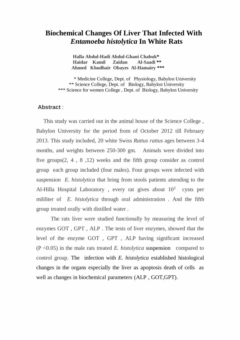
Biochemical Changes Of Liver That Infected With Entamoeba histolytica In White Rats Halla Abdul-Hadi Abdul-Ghani Chabuk* Haidar Kamil Zaidan Al-Saadi ** Ahmed Khudhair Obayes Al-Hamairy *** * Medicine College, Dept. of Physiology, Babylon University ** Science College, Dept. of Biology, Babylon University *** Science for women College , Dept. of Biology, Babylon University Abstract : This study was carried out in the animal house of the Science College , Babylon University for the period from of October 2012 till February 2013. This study included, 20 white Swiss Rattus rattus ages between 3-4 months, and weights between 250-300 gm. Animals were divided into five groups(2, 4 , 8 ,12) weeks and the fifth group consider as control group each group included (four males). Four groups were infected with suspension E. histolytica that bring from stools patients attending to the Al-Hilla Hospital Laboratory , every rat gives about 10 3 cysts per mililiter of E. histolytica through oral administration . And the fifth group treated orally with distilled water . The rats liver were studied functionally by measuring the level of enzymes GOT , GPT , ALP . The tests of liver enzymes, showed that the level of the enzyme GOT , GPT , ALP having significant increased (P <0.05) in the male rats treated E. histolytica suspension compared to control group. The infection with E. histolytica established histological changes in the organs especially the liver as apoptosis death of cells as well as changes in biochemical parameters (ALP , GOT,GPT).
Transcript of Enta
Biochemical Changes Of Liver That Infected With
Entamoeba histolytica In White Rats
Halla Abdul-Hadi Abdul-Ghani Chabuk*
Haidar Kamil Zaidan Al-Saadi **
Ahmed Khudhair Obayes Al-Hamairy ***
* Medicine College, Dept. of Physiology, Babylon University
** Science College, Dept. of Biology, Babylon University
*** Science for women College , Dept. of Biology, Babylon University
Abstract :
This study was carried out in the animal house of the Science College ,
Babylon University for the period from of October 2012 till February
2013. This study included, 20 white Swiss Rattus rattus ages between 3-4
months, and weights between 250-300 gm. Animals were divided into
five groups(2, 4 , 8 ,12) weeks and the fifth group consider as control
group each group included (four males). Four groups were infected with
suspension E. histolytica that bring from stools patients attending to the
Al-Hilla Hospital Laboratory , every rat gives about 103 cysts per
mililiter of E. histolytica through oral administration . And the fifth
group treated orally with distilled water .
The rats liver were studied functionally by measuring the level of
enzymes GOT , GPT , ALP . The tests of liver enzymes, showed that the
level of the enzyme GOT , GPT , ALP having significant increased
(P <0.05) in the male rats treated E. histolytica suspension compared to
control group. The infection with E. histolytica established histological
changes in the organs especially the liver as apoptosis death of cells as
well as changes in biochemical parameters (ALP , GOT,GPT).
Introduction:
Amoebiasis caused by Entamoeba histolytica and is the second
cause of global morbidity and mortality due to parasitic diseases in
humans, It causes more than 100,000 deaths each year and is responsible
for 50 million cases of diarrhea each year (Huston, 2004 ; WHO, 2009).
The parasite is endemic in most tropical and subtropical areas of the
world, infected persons display a wide range of disease severity,
reflecting the contribution of the patient’s immune and nutritional status
(Eichinger , 2009) .
E. histolytica inhabits the large intestine, It is acquired when
infective cysts are ingested through contaminated food or water.
Excystation releases trophozoites into the terminal ileum and from there
parasites migrate to the colon where they colonize
(Mortimer & Chadee,2010 ). Trophozoites remain in the lumen as
commensals, multiplying via binary fission and satisfying their energetic
needs by ingesting resident microflora and nutrients from the host. Some
parasites undergo encystment in the descending colon, resulting in
passage of mature infective cysts in the stool and perpetuation of the life
cycle through fecal-oral spread. In 90% of cases amoebic infections are
asymptomatic and self-limiting (Haque et al., 2003). But approximately
in 1% of the cases, trophozoites penetrate the intestinal mucosa and
spread to other organs, producing extra-intestinal amoebiasis, among
which amoebic liver abscess (ALA) is the most common
(Bernal Redondo, 2001).
E. histolytica induces apoptosis, both, in human cells, and during
the development of ALA in hamsters and mice (Boettner et al., 2008). It
has also been found that the death of hepatocytes and immune cells
during amoebic invasion is not only due to the cytolysis activity of the
trophozoites, but also because of an apoptotic process
(Pelosof et al., 2006).
Abscesses located just below the diaphragm can lead to pleural
pain or referred right shoulder pain. Liver alkaline phosphatase levels
(ALP) and alanine aminotransferase levels (GOT,GPT) are elevated in
acute liver abscess, which may, however, reverse over time. Males are ten
times more likely to present with liver abscess than females and middle-
age and young adults more than children (Eichinger,2009)
Aminotransferase levels are sensitive indicators of liver-cell injury and
are helpful in recognizing hepatocellular diseases such as liver abscess,
levels of this enzyme are accordingly more specific indicators of liver
injury. Both enzymes are released into the blood in increasing amounts
when the liver cell membrane is damaged. Necrosis of liver cells is
required for the release of the aminotransferases ( Dufour , 2001) .
The aim of this study to determine the level of liver enzymes
(alkaline phosphatase levels (ALP) and alanine aminotransferase levels
(GOT,GPT) and its relationship with E. histolytica infection in rats .
Materials and Methods : Isolats of E. histolytica from some patients in Babylon Hospital
in Hilla city, sample collection from the period lies between October
2012 till February 2013 , stool patients selected for this study were
usually suffering from diarrhea, vomiting , dysentery . abdominal pain
and stools were contain blood , mucus. Then keep in sterile plastic
container and this stool samples were examined in advanced parasitology
lab. of Science College for women by direct smear methods. A drop of
Lugols̕ iodine was added and mixed with small piece of feces and
examined under compound microscope to diagnosed the cysts and
trophozoite of E. histolytica , added to this stool normal saline
proportion 1:1 and take one milliliter of suspension that contain about
103 cysts were infected of 20 males rats ( divided into five groups 2
weeks, 4 weeks, 8 weeks,12 weeks and the fifth group consider as control
group each group included (four males) orally administration , their
weight range between 250 - 300 gm. Then anesthesia animals using
chloroform dose and open the abdominal cavity until the sternum using
scissors medical and draw blood directly from the heart in a way (Heart
puncture) using a syringe medical sterile capacity of 5 ml and put the
blood in the jell tube free of material proof coagulation containing
material gelatinous help to increase serum formed after centrifugation ,
left the samples for 30 minutes at room temperature and then put inside
the centrifuge centrifuge at 3000 rpm / 10 minutes for the purpose of
separation of serum, were taken serum to measure the level of liver
enzymes GOT, GPT, ALP.
Result:
Result reveals a significant differences (p< 0.05) in liver enzymes
(GOT, GPT, ALP) comparison with control group. The GOT enzyme
tests increased in all infected animals (all groups) when comparison with
control group. in spite of the first group (2 weeks) , non significant
differences (p> 0.05) when comparison with second group (4 weeks) and
third group (8 weeks) as well as fourth group (12 weeks) . But the first
and second groups when comparison with third and fourth groups reveals
significant differences (p<0.05) as shown in Table (1).
The result of GPT enzyme showed increased significantly in the
second, third, fourth groups when comparison with first group. as well as
comparison with fourth groups with others .
The ALP enzyme reveals high significant differences in the second,
third, fourth when comparison with control and first groups, in spite of
the differences not reach to significantly among all groups as well as the
first group not significant when compared with control group .
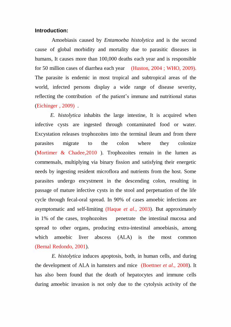
Table 1 : effect infected males rats with E. histolytica according to
different time periods on mean levels liver enzymes GOT,GPT , ALP
(I.U/L )
MEAN
ALP
(I.U/L )
SE
MEAN
)GPT ALT)
(I.U/L ) SE
MEAN
GOT (AST)
(I.U/L ) SE
ENZYMES
GROUPS
a 440.50
±
15.87
a 60.15
±
3.13
a 58.48
±
2.88
Control
a 448.25
±
19.18
a 57.45
±
2.82
b 69.33
±
3.29
Group 1 (2 weeks)
b 538.25
±
20.45
b 74.10
±
3.73
b 77.00
±
2.42
Group 2
(4 weeks)
b 550.0
±
18.17
c 90.05
±
4.50
c 91.50
±
4.92
Grroup 3 (8 weeks)
b 566.50
±
19.75
d 104.00
±
4.02
c 98.25
±
3.94
Group 4
) 12 week)
Mean ± SE
different index on significant character (p<0.05)
Number animals in all group
n = 4
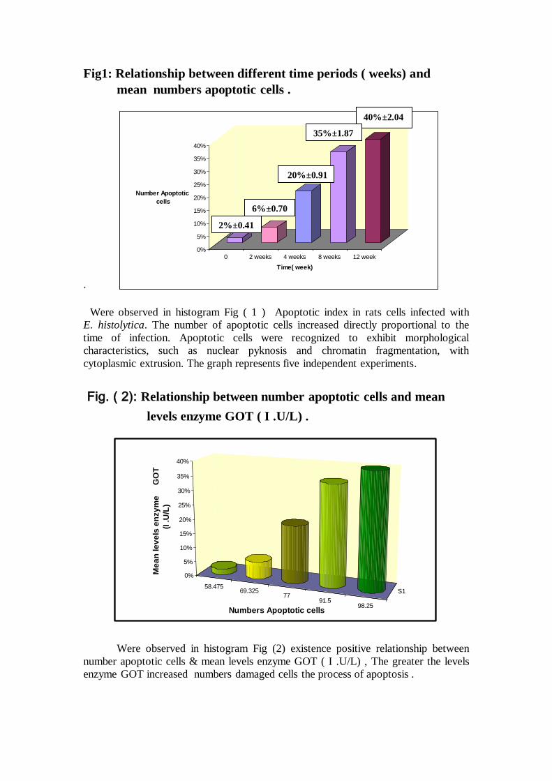
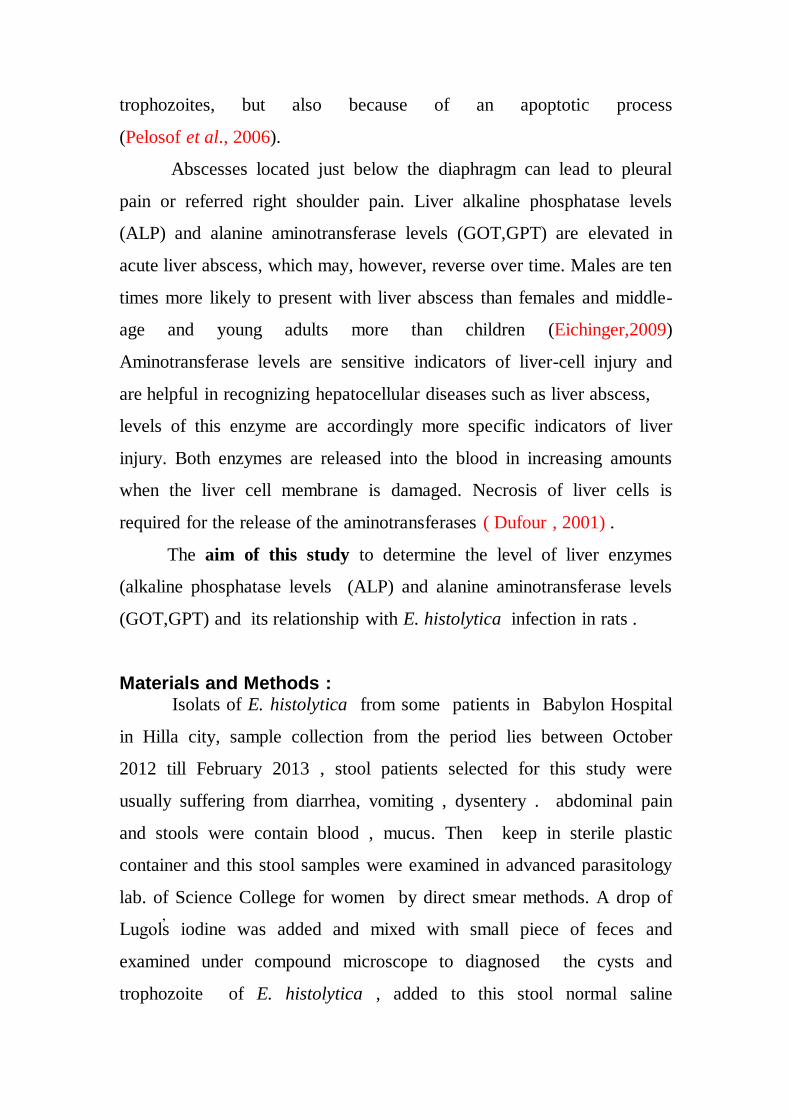
Fig1: Relationship between different time periods ( weeks) and
mean numbers apoptotic cells .
. Were observed in histogram Fig ( 1 ) Apoptotic index in rats cells infected with
E. histolytica. The number of apoptotic cells increased directly proportional to the
time of infection. Apoptotic cells were recognized to exhibit morphological
characteristics, such as nuclear pyknosis and chromatin fragmentation, with
cytoplasmic extrusion. The graph represents five independent experiments.
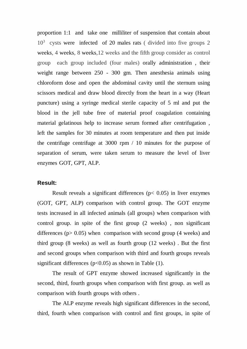
Fig. ( 2): Relationship between number apoptotic cells and mean
levels enzyme GOT ( I .U/L) .
Were observed in histogram Fig (2) existence positive relationship between
number apoptotic cells & mean levels enzyme GOT ( I .U/L) , The greater the levels
enzyme GOT increased numbers damaged cells the process of apoptosis .
0%
5%
10%
15%
20%
25%
30%
35%
40%
Number Apoptotic
cells
0 2 weeks 4 weeks 8 weeks 12 week
Time( week)
6%±0.70
2%±0.41
20%±0.91
40%±2.04
35%±1.87
58.47569.325
7791.5
98.25
S1
0%
5%
10%
15%
20%
25%
30%
35%
40%
Me
an
le
ve
ls e
nzy
me
G
OT
(I
.U
/L)
Numbers Apoptotic cells
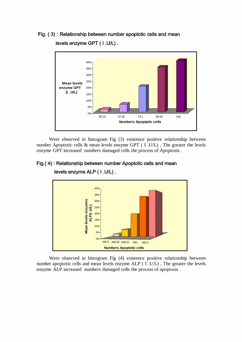
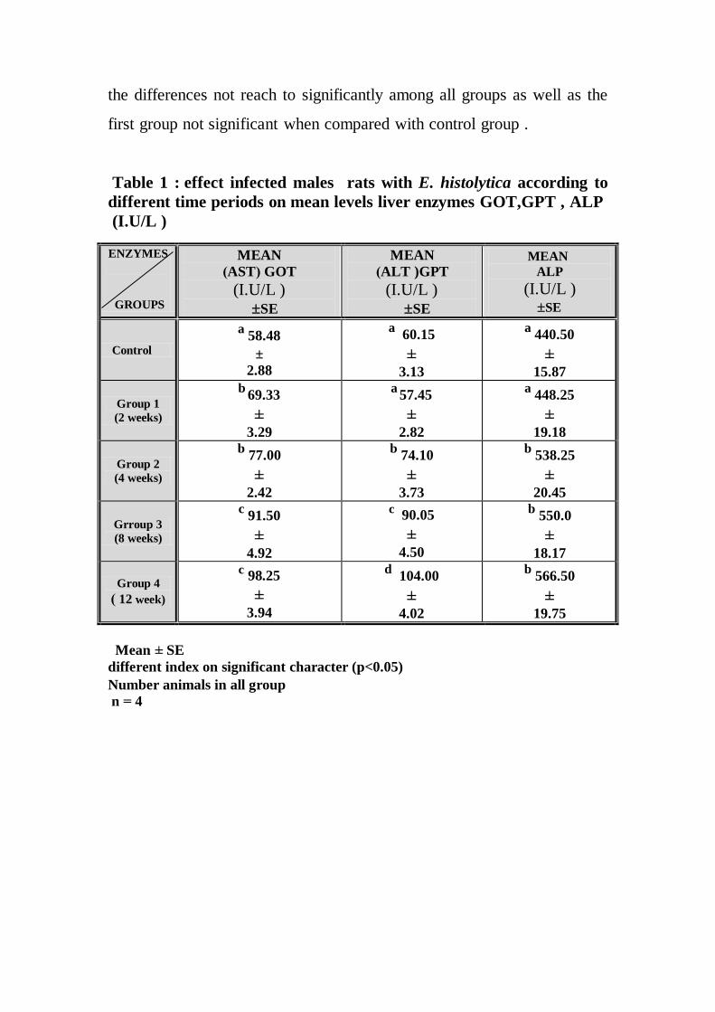
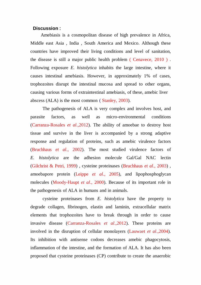
Fig. ( 3) : Relationship between number apoptotic cells and mean
levels enzyme GPT ( I .U/L) .
Were observed in histogram Fig (3) existence positive relationship between
number Apoptotic cells & mean levels enzyme GPT ( I .U/L) , The greater the levels
enzyme GPT increased numbers damaged cells the process of Apoptosis .
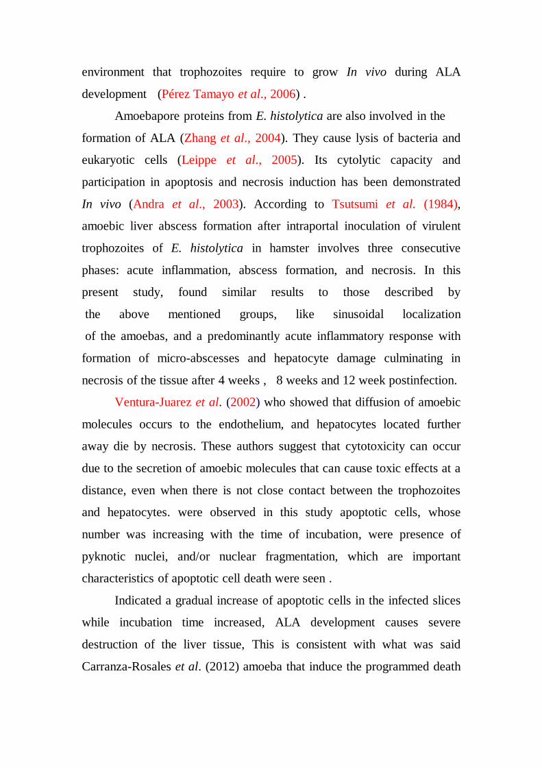
Fig.( 4) : Relationship between number Apoptotic cells and mean
levels enzyme ALP ( I .U/L) .
Were observed in histogram Fig (4) existence positive relationship between
number apoptotic cells and mean levels enzyme ALP ( I .U/L) , The greater the levels
enzyme ALP increased numbers damaged cells the process of apoptosis .
0%
5%
10%
15%
20%
25%
30%
35%
40%
Mean levels
enzyme GPT
(I . U/L)
60.15 57.45 74.1 90.05 104
Numbers Apoptptic cells
440.5 448.25 538.25 550 566.5
0%
5%
10%
15%
20%
25%
30%
35%
40%
Me
an
le
ve
ls e
nzy
me
s
AL
P(I
.U
/L)
Numbers Apoptotic cells
Discussion :
Amebiasis is a cosmopolitan disease of high prevalence in Africa,
Middle east Asia , India , South America and Mexico. Although these
countries have improved their living conditions and level of sanitation,
the disease is still a major public health problem ( Cenavece, 2010 ) .
Following exposure E. histolytica inhabits the large intestine, where it
causes intestinal amebiasis. However, in approximately 1% of cases,
trophozoites disrupt the intestinal mucosa and spread to other organs,
causing various forms of extraintestinal amebiasis, of these, amebic liver
abscess (ALA) is the most common ( Stanley, 2003).
The pathogenesis of ALA is very complex and involves host, and
parasite factors, as well as micro-environmental conditions
(Carranza-Rosales et al.,2012). The ability of amoebae to destroy host
tissue and survive in the liver is accompanied by a strong adaptive
response and regulation of proteins, such as amebic virulence factors
(Bruchhaus et al., 2002). The most studied virulence factors of
E. histolytica are the adhesion molecule Gal/Gal NAC lectin
(Gilchrist & Petri, 1999) , cysteine proteinases (Bruchhaus et al., 2003) ,
amoebapore protein (Leippe et al., 2005), and lipophosphoglycan
molecules (Moody-Haupt et al., 2000). Because of its important role in
the pathogenesis of ALA in humans and in animals.
cysteine proteinases from E. histolytica have the property to
degrade collagen, fibrinogen, elastin and laminin, extracellular matrix
elements that trophozoites have to break through in order to cause
invasive disease (Carranza-Rosales et al.,2012). These proteins are
involved in the disruption of cellular monolayers (Lauwaet et al.,2004).
Its inhibition with antisense codons decreases amebic phagocytosis,
inflammation of the intestine, and the formation of ALA. It has also been
proposed that cysteine proteinases (CP) contribute to create the anaerobic
environment that trophozoites require to grow In vivo during ALA
development (Pérez Tamayo et al., 2006) .
Amoebapore proteins from E. histolytica are also involved in the
formation of ALA (Zhang et al., 2004). They cause lysis of bacteria and
eukaryotic cells (Leippe et al., 2005). Its cytolytic capacity and
participation in apoptosis and necrosis induction has been demonstrated
In vivo (Andra et al., 2003). According to Tsutsumi et al. (1984),
amoebic liver abscess formation after intraportal inoculation of virulent
trophozoites of E. histolytica in hamster involves three consecutive
phases: acute inflammation, abscess formation, and necrosis. In this
present study, found similar results to those described by
the above mentioned groups, like sinusoidal localization
of the amoebas, and a predominantly acute inflammatory response with
formation of micro-abscesses and hepatocyte damage culminating in
necrosis of the tissue after 4 weeks , 8 weeks and 12 week postinfection.
Ventura-Juarez et al. (2002) who showed that diffusion of amoebic
molecules occurs to the endothelium, and hepatocytes located further
away die by necrosis. These authors suggest that cytotoxicity can occur
due to the secretion of amoebic molecules that can cause toxic effects at a
distance, even when there is not close contact between the trophozoites
and hepatocytes. were observed in this study apoptotic cells, whose
number was increasing with the time of incubation, were presence of
pyknotic nuclei, and/or nuclear fragmentation, which are important
characteristics of apoptotic cell death were seen .
Indicated a gradual increase of apoptotic cells in the infected slices
while incubation time increased, ALA development causes severe
destruction of the liver tissue, This is consistent with what was said
Carranza-Rosales et al. (2012) amoeba that induce the programmed death
of hepatic cells and noted that it increased in number the progress of
infected time.
In Table (1) Were observed levels liver enzymes increased directly
proportional to the time of incubation , It was also noted high levels of
these enzymes in the serum increased numbers damaged cells the
process of Apoptosis ,Probably due to these enzymes mostly reside
within the cells of the liver. But when the liver is injured for any reason,
these enzymes are spilled into the blood stream . These enzymes are
normally predominantly contained within liver cells and to a lesser degree
in the muscle cells. If the liver is injured or damaged, the liver cells spill
these enzymes into the blood, raising the aspartate aminotransferase
(AST) and alanine aminotransferase (ALT) enzyme blood levels and
signaling liver disease , While ALP is a substance found in the bile ducts
of the liver , intestine and the bone. Damage or obstruction of the bile
ducts may result in elevated levels of ALP. These tests can provide a
host of information on a range of disease processes . (Dufour , 2001)
This is consistent with what he found Al-Kubaissi (2002) to note a
high in the level of concentration of the enzyme ALP reached 90 % of the
cases with a high level of enzyme GOT, GPT in the serum of patients
infected with dysentery,
This result matched the findings of the (Pluta & Pluta , 2008 ) as
the very high levels of liver enzymes in the serum of patients infected
with the parasite , as well as demonstrated Al- Ghanimi ( 2013 ) for an
increase in the levels of liver enzymes in mice infected with parasite
Giardia lamblia .
But this is not consistent with what has been recorded Fernandes
et al. (2009) that the tests of liver function in patients with dysentery level
was normal , except for the enzyme ALP to get noticed high in the level .
The conclusion of these study that the E. histolytica established
histological changes in the organs especially the liver as apoptosis death
of cells as well as changes in biochemical parameters (ALP , GOT,GPT).
References
Haque, R.; Huston, C.D.; Hughes, Houpt, E. and Petri, W.A.
(2003). Current concepts: amebiasis. The New England Journal of
Medicine., 348 : 1565–1573 .
Cenavece, ( 2010). Centro Nacional de Vigilancia Epidemiologica
Control de Enfermedades, Secretaria de Salud, México . 1-6
Stanley, S.L.( 2003). Amoebiasis. Lancet., 36: 481–489.
Bruchhaus, I., Roeder, T., Lotter, H., Schwerdtfeger, M. and
Tannich, E.( 2002). Differential gene expression in Entamoeba
histolytica isolated from amoebic liver abscess. Molecular Microbiology.,
44 :1063–1072.
Bruchhaus, I.; Loftus, B.J.; Hall, N. and Tannich, E. ( 2003). The
intestinal protozoan parasite Entamoeba histolytica contains 20 cysteine
protease genes, of which only a small subset is expressed during in vitro
cultivation. Eukaryotic Cell ., 2: 501–509.
Leippe, M.; Bruhn, H.; Hecht, O. and Grotzinger, J. ( 2005) . Ancient
weapons: the three dimensional structure of amoebapore A. Trends in
Parasitology,, 21: 5–7.
Gilchrist, C. and Petri, W.A.( 1999). Virulence factors of Entamoeba
histolytica. Current Opinion in Microbiology., 2: 433–437.
Lauwaet, T. ; Oliveira, M.J. ; Callewaert, B. ; De Bruyne, G. ;
Mareel, M. and Leroy, A.( 2004). Proteinase inhibitors TPCK and
TLCK prevent Entamoeba histolytica disturbance of tight junctions and
microvilli in enteric cell layer in vitrol. International Journal of
Parasitology., 34: 785–794.
Moody-Haupt, S., Patterson, J.H., Mirelman, D. and McConville,
M.J. (2000). The major surface antigens of Entamoeba histolytica
trophozoites are GPI-anchored proteophosphoglycans. Journal of
Molecular Biology., 297: 4-8
Zhang, X. ; Zhang, Z. ; Alexander, D. ; Bracha, R. ; Mirelman, D.
and Stanley, S.L. ( 2004). Expression of amoebapores is required for full
expression of Entamoeba histolytica virulence in amebic liver abscess but
is not necessary for theinduction of inflammation or tissue damage in
amebic colitis. Infection andImmunity., 72: 678–683.
Pérez -Tamayo, R. ; Montfort, I. ; Olivos - Garcia, A. ; Ramos, E. ;
Nequiz, M. and Tello, E.( 2006). Amibiasis hepatica. Revista de
Gastroenterologia de México., 71: 47–72.
WHO (World Health Organization), 2009. State of the art of vaccine
research and development. Initiative for Vaccine Research.
Andra, J.; Herbst, R. and Leippe, M.( 2003). Amoebapore archaic
effector peptides of protozoan origin are discharged into phagosomes and
kill bacteria by permeabilizing their membranes. Developmental &
Comparative Immunology ., 27: 291–304.
Boettner, D.R. ; Huston, C.D. ; Linford, A.S. ; Buss, S.N., Houpt, E.,
Sherman, N.E. and Petri, W.A.( 2008). Entamoeba histolytica
phagocytosis of human erythrocytes involves PATMK, a member of the
transmembrane kinase family. PloS Pathogens., 4: 122–133.
Pelosof, L.C. ; Davis, P.H. ; Zhang, Z. ; Zhang, X. and Stanley, S.L.,
(2006). Co-ordinate but disproportionate activation of apoptotic,
regenerative and inflammatory pathways characterizes the liver response
to acute amebic infection. Cellular Microbiology, 8: 508–522.
Ventura-Juarez, J. ; Campos-RodrIguez, R. and Tsutsumi, V.( 2002).
Early interactions of Entamoeba histolytica trophozoites with
parenchymal and inflammatory cells in the hamster liver: an
immunocytochemical study. Canadian Journal of Microbiology., 48: 123–
131.
Carranza-Rosales, P.; Santiago-Mauricio, M.G.; Guzman-Delgado,
N.; Vargas- Villarreal, J.; Lozano-Garza, G.; Viveros-Valdez, E. ;
Ortiz-Lopez,R.; Moran-Martinez, J. and Gandolfi, A.J. (2012).
Induction of virulence factors, apoptosis, and cytokines in precision-cut
hamster liver slices infected with Entamoeba histolytica Experimental
Parasitology .,132 : 424–433
Huston, C.D., (2004). Parasite and hosts contributions to the
pathogenesis of amebic colitis. Trends in Parasitology., 20, 23–26.
Bernal - Redondo, R.M. (2001). Entamoebiosis-amibiasis intestinal.
Entamoeba histolytica,Entamoeba dispar. Boletin Médico del Hospital
Infantil de México .,58: 217–219
Eichinger , D.J. (2009). Amebiasis. In : Satoskar , A.R. ; Simon, G.L. ;
Hotez, P. and Tsuji, M. (Eds.) Medical parasitology Landes Bioscience ,
USA., 171-182.
Mortimer, L. and Chadee, K. ( 2010) . The immunopathogenesis of
Entamoeba histolytica , Experimental Parasitology., 126 : 366–380 .
Dufour, D. (2001). Evaluation of liver function and injury in clinical
(Henry J editor). W. B. Saunders Company. 264 pp.
Tsutsumi, V., Mena-Lopez, R., Anaya-Velazquez, F. and Martinez-
Palomo, A.,( 1984). Cellular bases of experimental amebic liver abscess
formation. American Journal of Pathology., 117: 81–91. .
Al-Kubaissi , Abdul – Wahab Badawi Hussien (2002) .
Immunological and Epidemiological Study of Patients Infected with
Entamoeba histolytica . Ph.D. thesis, College of Science ,
AL – Mustansiriya University ., 125 pp.
Al - Ghanimi, Fatima Yusuf Ktan (2013). Study the opposite effect for
some plant extracts on the parasite Giardia lamblia in mice infected
laboratory. Master Thesis, College of Science, University of
Al-Muthanna., 130 pp.
Pluta , H. and Pluta , J.N. (2008) . Hepatic abscess : current approach
to patients with pyogenic or amebic abscess . Gastroenterologia polska .,
15(5) : 343-346
Fernandes , H.; Souza , C.R. ; Swethadri , G.K. and Naik ,C.R.(2009).
Ameboma of the colon with amebic liver abscess mimicking metastatic
colon cancer . Indian Journal of Pathology and Microbiology.,
52 (2) : 228-230 .
كيموحيوية الكباد الجرذان المصابة باميبا الزحار ال التغيرات
Entamoeba histolytica
هالة عبدالهادي عبدالغني جابك*
أ.د.حيدر كامل زيدان السعدي**
*** م.د.احمد خضير عبيس الحميريأ.
لطب /فرع الفسيولوجي /جامعة بابلكلية ا *
** كلية العلوم /قسم علوم الحياة /جامعة بابل
***كلية العلوم للبنات /قسم علوم الحياة /جامعة بابل
: الخالصة
اجريت الدراسة في البيت الحيواني التابع الى كلية العلوم/جامعة بابل
جرذا سويسريا ( 21) الدراسة , شملت2102ولغاية شباط 2102للمدة من تشرين االول
غم .قسمت الحيوانات 211-251شهر وتراوحن اوزانها بين 4-2تراوحت اعمارها بين
اعتبرت كمجموعة سيطرة ( اسبوعا والمجموعة الخامسة 02,،,2,4, 2الى خمسة مجاميع )
.مجموعة تحتوي على اربعة جرذانوكل

ن لمختبر معلق اميبا الزحار اذ جلبت من عينات من المرضى المراجعياربع مجاميع تم اصابتها ب
0111مستشفى الحلة الجراحي التعليمي , وجرع كل جرذ من كل مجموعة فمويا بما يعادل
كيس من اميبا الزحار اما المجموعة الخامسة )مجموعة السيطرة( فجرعت بالماء المقطر.
زحار وظيفيا من خالل قياس مستويات انزيماتدرست اكباد الجرذان المصابة باميبا ال
(GOT,GPT,ALP) وتبين من خالل النتائج ان مستويات هذه االنزيمات ازداد بزيادة معنوية
كذلك ادت االصابة الى مع اكباد الجرذان المصابة مقارنة بمجموعة السيطرة غير المصابة .
وت المبرمج لخالياه وكذلك الى تغيرات في بعض تغيرات نسجية في الكبد من خالل ظاهرة الم
المعايير الكيموحيوية .