Protective KIR–HLA interactions for HCV infection in intravenous drug users
Upload
independentCategory
view
3download
0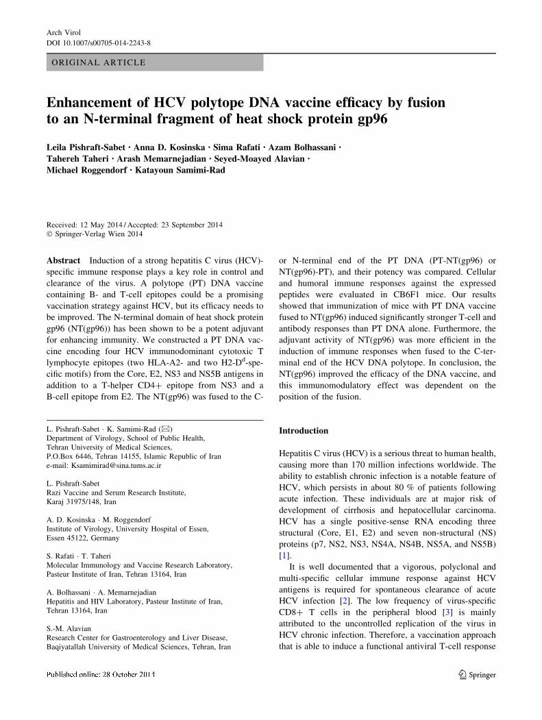
ORIGINAL ARTICLE
Enhancement of HCV polytope DNA vaccine efficacy by fusion
to an N-terminal fragment of heat shock protein gp96
Leila Pishraft-Sabet • Anna D. Kosinska • Sima Rafati • Azam Bolhassani •
Tahereh Taheri • Arash Memarnejadian • Seyed-Moayed Alavian •
Michael Roggendorf • Katayoun Samimi-Rad
Received: 12 May 2014 / Accepted: 23 September 2014
� Springer-Verlag Wien 2014
Abstract Induction of a strong hepatitis C virus (HCV)-
specific immune response plays a key role in control and
clearance of the virus. A polytope (PT) DNA vaccine
containing B- and T-cell epitopes could be a promising
vaccination strategy against HCV, but its efficacy needs to
be improved. The N-terminal domain of heat shock protein
gp96 (NT(gp96)) has been shown to be a potent adjuvant
for enhancing immunity. We constructed a PT DNA vac-
cine encoding four HCV immunodominant cytotoxic T
lymphocyte epitopes (two HLA-A2- and two H2-Dd-spe-
cific motifs) from the Core, E2, NS3 and NS5B antigens in
addition to a T-helper CD4? epitope from NS3 and a
B-cell epitope from E2. The NT(gp96) was fused to the C-
or N-terminal end of the PT DNA (PT-NT(gp96) or
NT(gp96)-PT), and their potency was compared. Cellular
and humoral immune responses against the expressed
peptides were evaluated in CB6F1 mice. Our results
showed that immunization of mice with PT DNA vaccine
fused to NT(gp96) induced significantly stronger T-cell and
antibody responses than PT DNA alone. Furthermore, the
adjuvant activity of NT(gp96) was more efficient in the
induction of immune responses when fused to the C-ter-
minal end of the HCV DNA polytope. In conclusion, the
NT(gp96) improved the efficacy of the DNA vaccine, and
this immunomodulatory effect was dependent on the
position of the fusion.
Introduction
Hepatitis C virus (HCV) is a serious threat to human health,
causing more than 170 million infections worldwide. The
ability to establish chronic infection is a notable feature of
HCV, which persists in about 80 % of patients following
acute infection. These individuals are at major risk of
development of cirrhosis and hepatocellular carcinoma.
HCV has a single positive-sense RNA encoding three
structural (Core, E1, E2) and seven non-structural (NS)
proteins (p7, NS2, NS3, NS4A, NS4B, NS5A, and NS5B)
[1].
It is well documented that a vigorous, polyclonal and
multi-specific cellular immune response against HCV
antigens is required for spontaneous clearance of acute
HCV infection [2]. The low frequency of virus-specific
CD8? T cells in the peripheral blood [3] is mainly
attributed to the uncontrolled replication of the virus in
HCV chronic infection. Therefore, a vaccination approach
that is able to induce a functional antiviral T-cell response
L. Pishraft-Sabet � K. Samimi-Rad (&)
Department of Virology, School of Public Health,
Tehran University of Medical Sciences,
P.O.Box 6446, Tehran 14155, Islamic Republic of Iran
e-mail: [email protected]
L. Pishraft-Sabet
Razi Vaccine and Serum Research Institute,
Karaj 31975/148, Iran
A. D. Kosinska � M. Roggendorf
Institute of Virology, University Hospital of Essen,
Essen 45122, Germany
S. Rafati � T. Taheri
Molecular Immunology and Vaccine Research Laboratory,
Pasteur Institute of Iran, Tehran 13164, Iran
A. Bolhassani � A. Memarnejadian
Hepatitis and HIV Laboratory, Pasteur Institute of Iran,
Tehran 13164, Iran
S.-M. Alavian
Research Center for Gastroenterology and Liver Disease,
Baqiyatallah University of Medical Sciences, Tehran, Iran
123
Arch Virol
DOI 10.1007/s00705-014-2243-8
may be a promising strategy to overcome persistent
infection. Production of cross-neutralizing antibodies
against envelope proteins is also necessary to prevent the
attachment and entry of circulating virus into the hepato-
cytes [4].
The potency of multi-epitope (polytope) vaccines has
been examined previously in treatment of various tumors
and infectious diseases [5]. Recently, polytope DNA vac-
cines containing epitopes derived from structural and NS
proteins of HCV were tested in vivo [6, 7]. However, the
efficacy of these vaccines was limited due to the lack of
tertiary structure and their instability during polytope
expression [7]. Appropriate carriers and adjuvants could
improve the immunogenicity of polytope DNA vaccines.
Heat shock proteins (HSPs) have been demonstrated to
act as potent adjuvants in the immunotherapy of malignant
tumors and infectious diseases. gp96, the major chaperon in
the lumen of the endoplasmic reticulum involved in cross-
presentation of peptides to MHC class I and class II mole-
cules, activates specific CD8? and CD4? T-cells [8, 9].
Moreover, gp96 can activate dendritic cells and macro-
phages through induction of proinflamatory cytokines via
interaction with a subset of Toll-like receptors (TLRs) [10].
Previous experiments showed that gp96 or its N-terminal
domain had an adjuvant function in tumor- [11] and HBV-
specific CTL and humoral immune responses [12].
In the present study, we constructed a polytope (PT)
DNA vaccine encoding both structural and non-structural
antigens of HCV. To investigate the influence of fusing the
N-terminal domain of gp96 (NT(gp96)) to the PT on the
efficacy of the PT DNA vaccine, we also constructed two
fusion DNAs with NT(gp96) fused either to the C- or the
N-terminal end of the polytope sequence. The immuno-
genicity of the polytope DNA vaccines fused to NT(gp96)
was evaluated in CB6F1 mice. The impact of vaccination
on the percentage of regulatory CD4? T-cells (Tregs) was
also examined in this study.
Materials and methods
Laboratory animals
Ten-week-old CB6F1 female mice (genotype H-2d/b) were
purchased from Harlan Winkelmann Laboratories (Bor-
chen, Germany) and handled according to the guidelines of
the animal facility at the University Hospital, Essen. All
animal experiments were conducted in accordance with the
Guide for the Care and Use of Laboratory Animals and
were approved by the local Animal Care and Use Com-
mittee (Animal Care Center, University of Duisburg-Essen,
Essen, Germany, and the district government of Dussel-
dorf, Germany).
Cells and peptides
HeLa cells (HeLa, ATCC) were cultured in monolayers in
Dulbecco’s modified Eagle’s medium (Gibco, Germany).
Murine splenocytes and hepatocytes were cultured in
RPMI medium (Gibco). Cell culture media were supple-
mented with 10 % heat-inactivated fetal bovine serum
(FBS; Biochrom AG, Germany) and 10 U of penicillin-
streptomycin (PAA Laboratories, Austria) per ml. Cell
lines were maintained in a humidified 5 % CO2 atmosphere
at 37 �C.
For evaluation of the immune responses, peptides of
Core132-142 (DLMGYIPLVGA), E2405-414 (SGPSQKIQLV)
and E2412-426 (QLINTNGSWHINSTA) with 90 % purity
were synthesized (EMC Microcollections, Germany). All
lyophilized peptides were dissolved in DMSO (Sigma-
Aldrich, Germany) at concentration of 10 lg/ll and stored
at -20 �C.
Preparation of NT(gp96)-PT and PT-NT(gp96)
HCV PT DNA, cloned in pBlueScript II SK (?), was
synthesized by Biomatik (Biomatik Corporation, Canada).
The PT construct was designed to encode the selected HCV
epitopes: H2-Dd-restricted epitopes of Core132-142(DLMGYIPLVGA) [13, 14] and E2405-414 (SGPSQK
IQLV) [15], HLA-A2-restricted epitopes of NS31073-1081(CINGVCWTV) [16] and NS5B2727-2735 (GLQDCTMLV)
[16], T-helper (Th) CD4 epitope from NS31248-1262 (GY-
KVLVLNPSVAATL) [17] and a neutralizing B-cell epi-
tope from E2412-426 (QLINTNGSWHINSTA) [18].
To prepare the plasmid pcDNA-PT, the PT sequence
was subcloned into the HindIII and BamHI restriction sites
of the plasmid pcDNA3.1(-) (Invitrogen, Germany).
To generate NT (gp96)-PT, the region of nucleotides 1
to 1014 of gp96 (designed as NT(gp96)) was first amplified
from pQE30-NT(gp96) [19] using the forward primer 50-
ATTGGATCCACCATGGAAGATGACGTTGAA - 30
and the reverse primer 50- GGCGAGCTCGGTACCTT
TGTAGAAGGCTTT- 30 and then inserted into the BglII
and SacI restriction sites of pEGFP-N3 (Invitrogen).
The BamHI and SacI restriction sites in the forward and
reverse primers are shown in bold, and the Kozak sequence
is underlined. The PT sequence was then cloned into
unique HindIII and BamHI cloning sites of pEGFP-NT
(gp96). pcDNA3.1-NT (gp96)-PT was prepared by sub-
cloning NT (gp96)-PT into the unique NheI and BamHI
cloning sites of pcDNA3.1(-).
To prepare the PT-NT(gp96) fusion construct, the PT
sequence was first amplified using primers designed to
generate NheI restriction site at its 50 end and cloned into
NheI and BamHI multiple cloning sites of pcDNA3.1(-).
In the same manner, BamHI and KpnI restriction enzymes
L. Pishraft-Sabet et al.
123
were used for cloning of NT(gp96). Finally, PT-NT(gp96)
was subcloned into the NheI and KpnI sites of the pEGFP-
N3 expression vector.
The accuracy of the constructs was confirmed by DNA
sequencing (Eurofins MWG-Operon, Germany). pcDNA-
NT(gp96)-PT, pcDNA-PT-NT(gp96), and pcDNA-PT
were purified using an EndoFree Plasmid Giga Kit (QIA-
GEN, Germany). A schematic diagram of the constructs is
presented in Fig. 1.
In vitro expression of PT-NT (gp96) and NT (gp96)-PT
constructs in HeLa cells
HeLa cells were transfected separately with 1 lg of pEG-
FP-PT-NT(gp96) or pEGFP-NT(gp96)-PT plasmids using
Lipofectamine 2000 reagent (Invitrogen) according to the
manufacturer’s instructions. A Lipofectamine/plasmid
complex was prepared with 5 lL of Lipofectamine.
Expression of the fusion proteins was confirmed by
observation of the EGFP signal under a fluorescence
microscope at 24 h post-transfection. In addition, the cells
were collected at 24 h post-transfection, washed with 1x
PBS, and analyzed by flow cytometry to determine the
proportion of GFP positive cells (FL1 channel). Untrans-
fected cells were used as a control.
Cytotoxicity studies
The cytotoxicity of PT, PT-NT(gp96) and NT(gp96)-PT to
HeLa cells was measured using a 3-(4,5-dimethylthiazol-2-
yl)-2,5-diphenyl tetrazolium bromide (MTT) dye reduction
assay [20]. Cells were seeded in a 96-well plate at a density
of 105 cells/well in 100 lL DMEM supplemented with
10 % fetal bovine serum (FBS) and incubated overnight at
37 �C in a 5 % CO2 atmosphere. Cells were treated with
pcDNA-PT, pcDNA- PT-NT(gp96) and pcDNA-
NT(gp96)-PT in concentrations ranging from 6.25 to
100 lg/100 ll in DMEM (supplemented with 2% FBS) for
24, 48 and 72 h. Then, 20 ll of MTT dye solution (5 mg/
ml in PBS) was added to each well and plates were incu-
bated for 4 h at 37 �C. The supernatant of each well was
removed, and formazan crystals were dissolved in 200 ll
of dimethylsulfoxide (DMSO). The absorbance values
were measured using a microplate reader at 570 nm. The
percentage of cells that were viable was determined by
comparing the optical density of plasmid-treated cells to
that of normal cells (control).
Mice immunization
Mice were pretreated by intramuscular injection of 100 ll
of cardiotoxin (10 lM in phosphate-buffered saline [PBS];
Latoxan, France) into tibialis anterior muscle of both hind
limbs one week before the plasmid immunization. Mice
were then injected intramuscularly with 100 lg of plas-
mids in PBS at 2-week intervals as described previously
[21], and two weeks after the last immunization, they were
sacrificed. Naıve untreated mice were used as controls.
Isolation of mouse splenic and hepatic lymphocytes
Preparation of a single-cell suspension of murine spleno-
cytes was performed by homogenization according to a
procedure described previously [22]. Hepatic lymphocytes
were also isolated from the liver using previously published
methods [22]. Briefly, livers were perfused with prewarmed
PBS (to flush blood from the hepatic vasculature) and were
forced through a 70-mm nylon cell strainer (BD Falcon,
USA). After washing, cell pellets were resuspended for
30 min at 37 �C in prewarmed enzyme solution containing
collagenase type II and DNase type I (Sigma-Aldrich) in
HBSS supplemented with FBS. Cells were then layered on
40 % Percoll solution (Sigma-Aldrich) in RPMI supple-
mented with penicillin-streptomycin for density separation,
and then centrifuged at 2000 rpm for 10 minutes. Cells were
washed and suspended in Buffer EL (QIAGEN) to lyse red
blood cells. Cell yields and viability were determined by
trypan blue exclusion microscopy.
Intracellular cytokine staining and flow cytometry
analysis
Up to 1 9 106 lymphocytes isolated from mice were see-
ded per well in 96-well plates in 200 ll of RPMI 1640 and
stimulated of HCV peptides (E2405-414 [SGPSQKIQLV] or
Core132-142 [DLMGYIPLVGA]) at a concentration of 2 lg/
Fig. 1 Schematic illustration of two polytope fusion constructs containing six different epitopes of HCV in different blocks
Adjuvant activity of NT(gp96) on HCV polytope vaccine
123
ml and incubated for 6 days (in the presence of 10 U of
recombinant murine IL-2 [Roche, UK] per ml) or 6 h at
37 �C in a 5 % CO2 atmosphere. Cells with no peptide
served as negative controls.
Prior to intracellular cytokine staining, the cells were
cultured for 5–6 h in the presence of anti-CD28 (1 mg/mL)
(clone 37.51; BD Pharmingen, Germany) and 5 mg/ml of
brefeldin A (Sigma-Aldrich). Cells were then stained with
anti-CD8 (clone 56.6-7; eBioscience, Germany), and anti-
CD4 (clone L3T4; BD Pharmingen) antibodies, perme-
abilized and fixed with perm/wash reagents (eBioscience),
stained with anti-IFNc (clone XMG1.2; BD Pharmingen),
anti-TNFa (clone MP6-XT22; eBioscience,), and anti-IL-2
(clone JES6-5H4, eBioscience) at 4 �C for 30 minutes, and
analyzed immediately on a Gallios flow cytometer (Becton
Coulter). A minimum of 100,000 events were acquired per
sample. Subsequent analysis was performed using FlowJo
software (Tree Star, Inc, Ashland, Ore). Dead cells were
excluded from analyses using Fixable Viability Dye (FVD)
(eBioscience) [23].
For Treg cell analysis, splenocytes from treated mice
were stained with anti-CD4, anti-CD25 (clone PC 61.5,
eBiosceience), and anti-Foxp3 antibodies (clone FJK-16 s;
eBiosceience). Then, Treg cells were counted relative to
total CD4? cells by flow cytometry.
Determination of IgG isotypes
Two weeks after the last immunization, mice were bled via
retro-orbital puncture. Specific total IgG and its subclasses
(IgG1 and IgG2a) were measured in the sera of the mice by
ELISA as described previously [19]. Briefly, an ELISA
plate was coated with 100 ll of E2412-426 peptide (10 lg/
ml) in 0.5 M carbonate/bicarbonate buffer, pH 9.6. The
plate was rinsed with PBS containing 0.05 % Tween 20
(PBS-T) and incubated with blocking buffer (5 % skim
milk in PBS) for 2 h at 37 �C. After washing with PBS-T,
diluted sera (1:50) in PBS buffer containing 1 % BSA and
0.05 % Tween-20 were added, and the plate was incubated
at 37 �C for 2 h, followed by washing and incubation at
37 �C for 2 h with horseradish peroxidase (HRP)-conju-
gated goat anti-mouse IgG, IgG1, or IgG2a (1:4000;
Southern Biotechnology Association, USA). Detection was
done with Tetramethylbenzidine (TMB) substrate and the
absorbance was measured at 450 nm.
Statistical analysis
GraphPad Prism version 5 software (GraphPad Software,
Inc., La Jolla, Calif) was used for plotting graphs and
statistical analysis. Statistical differences were evaluated
using Student’s t-test and Mann-Whitney analysis. A P-
value\0.05 was considered significant.
Results
Monitoring of PT-NT(gp96) and NT(gp96)-PT
expression in HeLa cells
We first designed and constructed a PT DNA vaccine that
encoded HCV immunodominant CTL epitopes (HLA-A2
and H2-Dd restricted) from Core, NS3 and NS5B, a Th
CD4? epitope from NS3 and a B-cell epitope from E2. In
the next step, NT(gp96) was fused to the 50 or 30 terminus
of the PT. To test whether these two fused constructs could
be expressed properly in vitro, they were cloned into
pEGFP-N3 reporter plasmid at the N-terminus of enhanced
green fluorescent protein (EGFP). The clones that were
obtained were named pEGFP-NT(gp96)-PT and pEGFP-
PT-NT(gp96). HeLa cells were transfected with pEGFP-
NT(gp96)-PT, pEGFP-PT-NT(gp96), pEGFP-N3 (as a
positive control) and pcDNA3.1 (as a negative control).
Then, expression was evaluated by detection of GFP
fluorescence using fluorescence microscopy at 24 h after
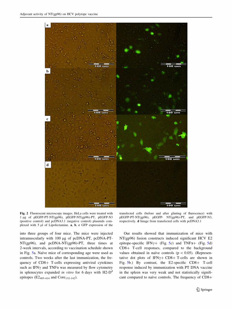
transfection. As shown in Fig. 2a and b, PT-NT(gp96) and
NT(gp96)-PT were efficiently expressed in HeLa cells, and
the antigen expression levels of these fusion DNAs were
almost the same. GFP expression of the positive control
was also confirmed (Fig. 2c). In the negative control,
fluorescence emission was not observed (Fig. 2d). The
experiments were performed twice, and similar results
were obtained.
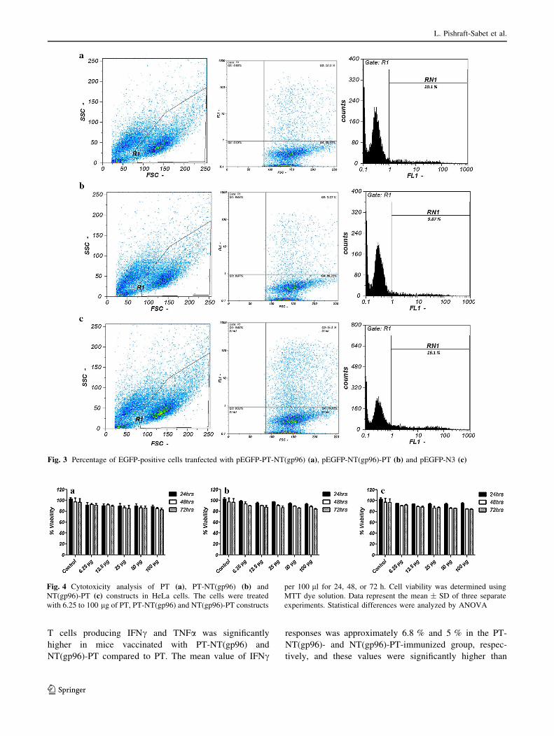
Quantification data obtained by flow cytometry showed
that the expression of GFP in pEGFP-PT-NT(gp96)
(Fig. 3a), pEGFP-NT(gp96)-PT (Fig. 3b) and pEGFP-N3
(Fig. 3c) was 10.1 %, 9.87 % and 16.1 %, respectively.
Cytotoxicity studies
The cytotoxicity of the DNA constructs was assessed in
HeLa cells by MTT assay. More than 80 % of cells were
observed to be viable at all time points and all concentra-
tions of PT, PT-NT(gp96) and NT(gp96)-PT constructs
(Fig. 4a, b, and c). No significant differences were found in
the viability of cells after exposure to DNA constructs.
Vaccination with HCV polytope DNA fused
to NT(gp96) enhanced HCV-specific CD8? T-cell-
mediated immune responses
We evaluated the immunogenic effects of the NT(gp96)
fusion in vivo by immunization of CB6F1 mice. In the first
step, we compared the effector functions of HCV epitope-
specific CD8? T cells in the spleens of mice immunized
with the polytope and the two NT(gp96)/PT fusion DNA
vaccines (pcDNA-PT-NT(gp96) and pcDNA-NT(gp96)-
PT). Twelve female CB6F1 mice were randomly divided
L. Pishraft-Sabet et al.
123
into three groups of four mice. The mice were injected
intramuscularly with 100 lg of pcDNA-PT, pcDNA-PT-
NT(gp96), and pcDNA-NT(gp96)-PT, three times at
2-week intervals, according to vaccination schedule shown
in Fig. 5a. Naıve mice of corresponding age were used as
controls. Two weeks after the last immunization, the fre-
quency of CD8? T-cells expressing antiviral cytokines
such as IFNc and TNFa was measured by flow cytometry
in splenocytes expanded in vitro for 6 days with H2-Dd
epitopes (E2405-414 and Core132-142).
Our results showed that immunization of mice with
NT(gp96) fusion constructs induced significant HCV E2
epitope-specific IFNc? (Fig. 5c) and TNFa? (Fig. 5d)
CD8? T-cell responses, compared to the background
values obtained in naıve controls (p\ 0.05). (Represen-
tative dot plots of IFNc? CD8? T-cells are shown in
Fig. 5b.) By contrast, the E2-specific CD8? T-cell
response induced by immunization with PT DNA vaccine
in the spleen was very weak and not statistically signifi-
cant compared to naıve controls. The frequency of CD8?
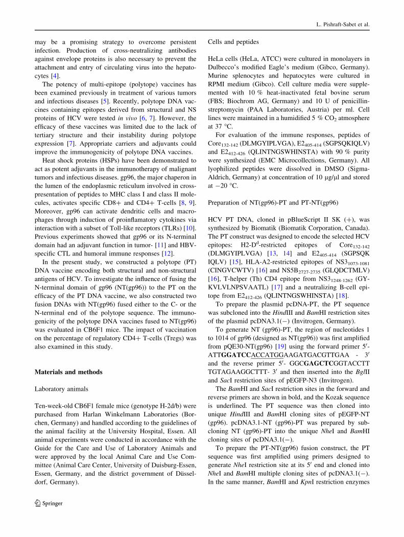
Fig. 2 Fluorescent microscopy images. HeLa cells were treated with
1 lg of pEGFP-PT-NT(gp96), pEGFP-NT(gp96)-PT, pEGFP-N3
(positive control) and pcDNA3.1 (negative control) plasmids com-
plexed with 5 ll of Lipofectamine. a, b, c GFP expression of the
transfected cells (before and after glinting of flurescence) with
pEGFP-PT-NT(gp96), pEGFP- NT(gp96)-PT, and pEGFP-N3,
respectively. d Image from transfected cells with pcDNA3.1
Adjuvant activity of NT(gp96) on HCV polytope vaccine
123
T cells producing IFNc and TNFa was significantly
higher in mice vaccinated with PT-NT(gp96) and
NT(gp96)-PT compared to PT. The mean value of IFNc
responses was approximately 6.8 % and 5 % in the PT-
NT(gp96)- and NT(gp96)-PT-immunized group, respec-
tively, and these values were significantly higher than
Fig. 3 Percentage of EGFP-positive cells tranfected with pEGFP-PT-NT(gp96) (a), pEGFP-NT(gp96)-PT (b) and pEGFP-N3 (c)
Fig. 4 Cytotoxicity analysis of PT (a), PT-NT(gp96) (b) and
NT(gp96)-PT (c) constructs in HeLa cells. The cells were treated
with 6.25 to 100 lg of PT, PT-NT(gp96) and NT(gp96)-PT constructs
per 100 ll for 24, 48, or 72 h. Cell viability was determined using
MTT dye solution. Data represent the mean ± SD of three separate
experiments. Statistical differences were analyzed by ANOVA
L. Pishraft-Sabet et al.
123
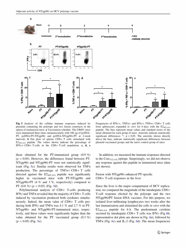
those obtained for the PT-immunized group (0.9 %)
(p\ 0.05). However, the differences found between PT-
NT(gp96) and NT(gp96)-PT were not statistically signif-
icant (Fig. 5c). Similar results were observed for TNFa
production. The percentage of TNFa? CD8? T cells
directed against the E2405-414 peptide was significantly
higher in vaccinated mice with PT-NT(gp96) and
NT(gp96)-PT (4 % and 3 %, respectively) compared to
PT (0.8 %) (p\ 0.05) (Fig. 5d).
Polyfunctional analysis of CD8? T-cells producing
IFNc and TNFa revealed that the majority of CD8? T cells
induced by vaccination produced both cytokines simulta-
neously. Indeed, the mean value of CD8? T cells pro-
ducing both IFNc and TNFa was 4.1 % and 2.7 % in PT-
NT(gp96)- and NT(gp96)-PT-immunized mice, respec-
tively, and these values were significantly higher than the
value obtained for the PT vaccinated group (0.3 %)
(p\ 0.05) (Fig. 5e).
In addition, we measured the immune responses directed
to the Core132-142 epitope. Surprisingly, we did not observe
any response against this peptide in immunized mice (data
not shown).
Fusion with NT(gp96) enhanced PT-specific
CD8? T-cell responses in the liver
Since the liver is the major compartment of HCV replica-
tion, we compared the magnitude of the intrahepatic CD8?
T-cell response elicited by the polytope and the two
NT(gp96)/PT fusion DNA vaccines. For this purpose, we
isolated liver-infiltrating lymphocytes two weeks after the
last immunization and stimulated the cells in vitro with the
E2405-414 peptide for 6 h. The predominant cytokine
secreted by intrahepatic CD8? T cells was IFNc (Fig 6b;
representative dot plots are shown in Fig. 6a), followed by
TNFa (Fig. 6c) and IL-2 (Fig. 6d). The mean frequencies
Fig. 5 Analysis of the cellular immune responses induced by
plasmids containing the polytope and two fusion constructs in the
spleen of immunized mice. a Vaccination schedule. The CB6F1 mice
were immunized three times intramuscularly with 100 lg of pcDNA-
PT, pcDNA-PT-NT(gp96) and pcDNA-NT(gp96)-PT at 2-week
intervals. b Dot plots of splenic CD8? T cells stimulated with
E2405-414 peptide. The values shown indicate the percentage of
IFNc? CD8? T-cells in the CD8? T-cell population. c, d, e
Frequencies of IFNc?, TNFa? and IFNc? TNFa? CD8? T cells
from splenocytes expanded in vitro for 6 days with the E2405-414peptide. The bars represent mean values and standard errors of the
mean obtained for each group of mice. Asterisks indicate statistically
significant differences: *, p\ 0.05. The asterisks shown directly
above the bars, indicate statistically significant differences between
plasmid-vaccinated groups and the naıve control group of mice
Adjuvant activity of NT(gp96) on HCV polytope vaccine
123
of IFNc? or TNFa? CD8? T cells in three immunized
groups were significantly higher than the corresponding
values obtained for naıve mice. The results showed that
immunization with PT-NT(gp96) and NT(gp96)-PT
induced a stronger immune response than PT. The per-
centages of IFNc? and TNFa? CD8? T cells detected in
the liver of mice immunized with PT-NT(gp96) (4.2 % and
3.6 %, respectively) and NT(gp96)-PT (3 % and 2.3 %,
respectively) were significantly higher than the mean per-
centages detected in the PT-immunized group (1.6 % and
1.2 %, respectively) (p\ 0.05). It should be noted that the
level of IL-2 production in all three groups of mice was
low, and no statistically significant differences between the
groups were observed.
Fig. 6 Analysis of the cellular immune responses induced by
plasmids containing the polytope and two fusion constructs in the
liver of immunized mice. The hepatocytes of the vaccinated CB6F1
mice were stimulated for 6 hours with E2405-414 peptide. a Dot plots
of intrahepatic CD8? T cells stimulated with E2405-414 peptide. The
values shown indicate the percentage of IFNc? CD8? T cells in the
CD8? T-cell population. b, c, d Frequencies of IFNc?, TNFa?, and
IL-2? CD8? T cells from hepatocytes stimulated in vitro for 6 h
with the epitope E2405-414. The bars represent the mean values and
standard errors of the mean obtained for each group of mice. Asterisks
indicate statistically significant differences: *, p\ 0.05; **,
p\ 0.005. The asterisks shown directly above the bars indicate
statistically significant differences between plasmid-vaccinated
groups and the naıve control group of mice
Fig. 7 Humoral immune responses induced by plasmid containing
the polytope and two fusion constructs. Specific IgG (a), IgG1 (b) and
IgG2a (c) antibodies against E2412-426 peptide were detected in
immunized mice. The bars represent the mean values and standard
errors of the mean obtained for each group of mice. Asterisks indicate
statistically significant differences: *, p\ 0.05; **, p\ 0.005; ***,
p\ 0.0005. The asterisks shown directly above the bars indicate
statistically significant differences between plasmid-vaccinated
groups and the naıve control group of mice
L. Pishraft-Sabet et al.
123
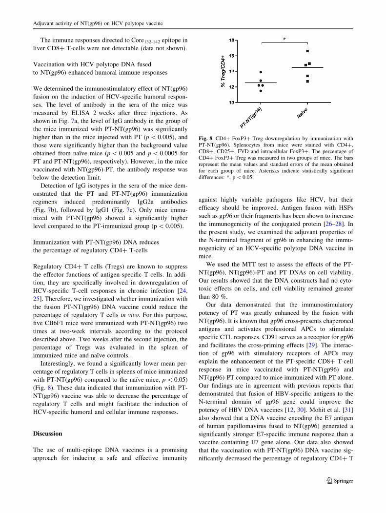
The immune responses directed to Core132-142 epitope in
liver CD8? T-cells were not detectable (data not shown).
Vaccination with HCV polytope DNA fused
to NT(gp96) enhanced humoral immune responses
We determined the immunostimulatory effect of NT(gp96)
fusion on the induction of HCV-specific humoral respon-
ses. The level of antibody in the sera of the mice was
measured by ELISA 2 weeks after three injections. As
shown in Fig. 7a, the level of IgG antibody in the group of
the mice immunized with PT-NT(gp96) was significantly
higher than in the mice injected with PT (p\ 0.005), and
those were significantly higher than the background value
obtained from naıve mice (p\ 0.005 and p\ 0.0005 for
PT and PT-NT(gp96), respectively). However, in the mice
vaccinated with NT(gp96)-PT, the antibody response was
below the detection limit.
Detection of IgG isotypes in the sera of the mice dem-
onstrated that the PT and PT-NT(gp96) immunization
regimens induced predominantly IgG2a antibodies
(Fig. 7b), followed by IgG1 (Fig. 7c). Only mice immu-
nized with PT-NT(gp96) showed a significantly higher
level compared to the PT-immunized group (p\ 0.005).
Immunization with PT-NT(gp96) DNA reduces
the percentage of regulatory CD4? T-cells
Regulatory CD4? T cells (Tregs) are known to suppress
the effector functions of antigen-specific T cells. In addi-
tion, they are specifically involved in downregulation of
HCV-specific T-cell responses in chronic infection [24,
25]. Therefore, we investigated whether immunization with
the fusion PT-NT(gp96) DNA vaccine could reduce the
percentage of regulatory T cells in vivo. For this purpose,
five CB6F1 mice were immunized with PT-NT(gp96) two
times at two-week intervals according to the protocol
described above. Two weeks after the second injection, the
percentage of Tregs was evaluated in the spleen of
immunized mice and naıve controls.
Interestingly, we found a significantly lower mean per-
centage of regulatory T cells in spleens of mice immunized
with PT-NT(gp96) compared to the naıve mice, p\ 0.05)
(Fig. 8). These data indicated that immunization with PT-
NT(gp96) vaccine was able to decrease the percentage of
regulatory T cells and might facilitate the induction of
HCV-specific humoral and cellular immune responses.
Discussion
The use of multi-epitope DNA vaccines is a promising
approach for inducing a safe and effective immunity
against highly variable pathogens like HCV, but their
efficacy should be improved. Antigen fusion with HSPs
such as gp96 or their fragments has been shown to increase
the immunogenicity of the conjugated protein [26–28]. In
the present study, we examined the adjuvant properties of
the N-terminal fragment of gp96 in enhancing the immu-
nogenicity of an HCV-specific polytope DNA vaccine in
mice.
We used the MTT test to assess the effects of the PT-
NT(gp96), NT(gp96)-PT and PT DNAs on cell viability.
Our results showed that the DNA constructs had no cyto-
toxic effects on cells, and cell viability remained greater
than 80 %.
Our data demonstrated that the immunostimulatory
potency of PT was greatly enhanced by the fusion with
NT(gp96). It is known that gp96 cross-presents chaperoned
antigens and activates professional APCs to stimulate
specific CTL responses. CD91 serves as a receptor for gp96
and facilitates the cross-priming effects [29]. The interac-
tion of gp96 with stimulatory receptors of APCs may
explain the enhancement of the PT-specific CD8? T-cell
response in mice vaccinated with PT-NT(gp96) and
NT(gp96)-PT compared to mice immunized with PT alone.
Our findings are in agreement with previous reports that
demonstrated that fusion of HBV-specific antigens to the
N-terminal domain of gp96 gene could improve the
potency of HBV DNA vaccines [12, 30]. Mohit et al. [31]
also showed that a DNA vaccine encoding the E7 antigen
of human papillomavirus fused to NT(gp96) generated a
significantly stronger E7-specific immune response than a
vaccine containing E7 gene alone. Our data also showed
that the vaccination with PT-NT(gp96) DNA vaccine sig-
nificantly decreased the percentage of regulatory CD4? T
Fig. 8 CD4? FoxP3? Treg downregulation by immunization with
PT-NT(gp96). Splenocytes from mice were stained with CD4?,
CD8?, CD25?, FVD and intracellular FoxP3?. The percentage of
CD4? FoxP3? Treg was measured in two groups of mice. The bars
represent the mean values and standard errors of the mean obtained
for each group of mice. Asterisks indicate statistically significant
differences: *, p\ 0.05
Adjuvant activity of NT(gp96) on HCV polytope vaccine
123
cells. This effect could also contribute to further
enhancement of PT-specific immune responses. This is in
keeping with a study by Wang et al. [32], who suggested
that NT(gp96) led to a decrease in the Treg population in
the vaccinated group.
Previous experiments showed that the resolution of
acute hepatitis C in a chimpanzee model was related to an
early intrahepatic CTL response directed against HCV
proteins [33]. Interestingly, our results revealed that fusion
of NT(gp96) to PT DNA vaccine also enhanced intrahe-
patic CTL responses. Moreover, these responses were
multifunctional. We found an increase in the frequency of
IFNc/TNFa double producers in mice vaccinated with two
fusion DNAs compared to the PT-immunized group. These
are encouraging data for HCV vaccine design, as it has
been reported that induction of multifunctional effector T
cells, which produce multiple cytokines simultaneously,
are critical for immune defense against pathogens [34] and
vaccine development [35, 36].
It has been suggested that an effective vaccine for HCV
should prime anti-envelope neutralizing antibodies (nAbs)
in addition to inducing a broad cellular immune response
[37]. This kind of vaccine could increase the chance of
clearance of the virus after exposure. It has been shown
that the E2412-426 epitope is conserved between HCV
genotypes and can induce the production of nAbs [18]. In
this study, we observed significant improvement of the
humoral immune responses against the E2 epitope through
the NT(gp96) fusion. Our findings are consistent with the
data obtained by Li et al. [28], which showed that
NT(gp96) greatly enhanced the humoral immune response
induced by HBsAg. Moreover, Chen et al. [38] provided
evidence for involvement of NT(gp96) in antigen-specific
humoral immune responses elicited by B-cell epitopes of
porcine reproductive and respiratory syndrome virus in
swine. In this study, we showed that IgG2a was the dom-
inant IgG isotype produced in the immunized mice. Our
experiment revealed that immunization with PT-NT(gp96)
DNA was able to produce a significantly higher amount of
IgG2a antibody compared to PT DNA. These results
showed that our vaccines preferentially primed Th1-type
immune responses.
In addition, our findings indicated that position of the
NT(gp96) fusion could affect the quantity of the induced
PT-specific cellular and humoral immune responses. We
showed that fusion of NT(gp96) to the C-terminus of the
PT (PT-NT(gp96)) resulted in enhancement of the function
of CTL responses. However, this difference was not sta-
tistically significant. The results are consistent with the
findings that a C-terminal fusion of gp96 (CT(gp96)) to
Her2 (Her2-CT(gp96)) could enhance the efficacy of a
Her2 DNA vaccine [39]. In addition, Yan et al. [30] found
that the fusion of NT(gp96) to the 50 end of the HBV S
gene negatively affected CTL immune responses. More-
over, in vitro expression of PT-NT(gp96) and NT(gp96)-
PT in HeLa cells indicated that the two constructs had
similar expression. Therefore, it seems that the enhanced
immune responses observed for PT-NT(gp96) were not a
consequence of increased expression.
Strikingly, in this study N-terminal and C-terminal
fusion had different effects on the humoral immune
response. HCV-specific antibody production was nearly
abolished by the NT(gp96)-PT fusion, whereas an
increased antibody response to PT-NT(gp96) was
observed. We suggest that N-terminal fusion of NT(gp96)
to PT results in a conformational change or steric hin-
drance, which reduces the efficacy of the PT DNA vaccine.
Moreover, the position of the fusion may affect the pattern
of ubiquitination of our fusion constructs [40].
One of the H2-Dd epitopes in our PT DNA vaccine was
derived from HCV Core protein (Core132-142). Even though
in silico prediction tools were used to design the PT to
provide proper proteosomal processing, we did not detect a
CTL immune response against the Core epitope. This could
be due to (1) higher binding affinity of E2 compared to
Core epitope, leading to an immunodominant effect of E2
on the Core epitope, or (2) a failure in appropriate trim-
ming of the PT in the endoplasmic reticulum.
In conclusion, our data demonstrate that NT(gp96) is
able to enhance cellular and humoral immune responses.
More-potent adjuvant activities of the N-terminal domain
of gp96 were shown when the adjuvant was fused down-
stream of the HCV DNA PT, thus demonstrating the
importance of the position of the fusion in the DNA vac-
cine. Further studies are needed to determine whether
differences in conformation and ubiquitination patterns due
to differences in the position of the adjuvant in the poly-
peptide led to differences in the immune response. More-
over, preclinical studies will be done to analyze the CTL
immune responses against HLA-A2-restricted epitopes
incorporated in PT in HLA-A2 transgenic mice. These
future studies may provide useful data to motivate further
evaluation of our PT DNA vaccine candidate in clinical
settings.
Acknowledgments This study was funded and supported by Grant
No. 13270 from Tehran University of Medical Sciences (TUMS) and
Grant No. 90-803 from Baqiyatallah University of Medical Science.
Conflict of interest The authors declare that they have no conflict
of interest.
References
1. Roohvand F, Kossari N (2011) Advances in hepatitis C virus
vaccines, part one: advances in basic knowledge for hepatitis C
virus vaccine design. Expert Opin Ther Pat 21:1811–1830
L. Pishraft-Sabet et al.
123
2. Roohvand F, Kossari N (2012) Advances in hepatitis C virus
vaccines, part two: advances in hepatitis C virus vaccine for-
mulations and modalities. Expert Opin Ther Pat 22:391–415
3. Cerny A, Chisari FV (1999) Pathogenesis of chronic hepatitis C:
immunological features of hepatic injury and viral persistence.
Hepatology 30:595–601
4. Pestka JM, Zeisel MB, Blaser E, Schurmann P, Bartosch B,
Cosset FL, Patel AH, Meisel H, Baumert J, Viazov S, Rispeter K,
Blum HE, Roggendorf M, Baumert TF (2007) Rapid induction of
virus-neutralizing antibodies and viral clearance in a single-
source outbreak of hepatitis C. Proc Natl Acad Sci USA 104:
6025–6030
5. Suhrbier A (2002) Polytope vaccines for the codelivery of mul-
tiple CD8 T-cell epitopes. Expert Rev Vaccines 1:207–213
6. Memarnejadian A, Roohvand F (2010) Fusion of HBsAg and
prime/boosting augment Th1 and CTL responses to HCV poly-
tope DNA vaccine. Cell Immunol 261:93–98
7. Martin P, Simon B, Lone YC, Chatel L, Barry R, Inchauspe G,
Fournillier A (2008) A vector-based minigene vaccine approach
results in strong induction of T-cell responses specific of hepatitis
C virus. Vaccine 26:2471–2481
8. Doody AD, Kovalchin JT, Mihalyo MA, Hagymasi AT, Drake
CG, Adler AJ (2004) Glycoprotein 96 can chaperone both MHC
class I- and class II-restricted epitopes for in vivo presentation,
but selectively primes CD8? T cell effector function. J Immunol
172:6087–6092
9. Javid B, MacAry PA, Lehner PJ (2007) Structure and function:
heat shock proteins and adaptive immunity. J Immunol 179:
2035–2040
10. Yang Y, Liu B, Dai J, Srivastava PK, Zammit DJ, Lefrancois L,
Li Z (2007) Heat shock protein gp96 is a master chaperone for
toll-like receptors and is important in the innate function of
macrophages. Immunity 26:215–226
11. Baker-LePain JC, Sarzotti M, Fields TA, Li CY, Nicchitta CV
(2002) GRP94 (gp96) and GRP94N-terminal geldanamycin
binding domain elicit tissue nonrestricted tumor suppression.
J Exp Med 196:1447–1459
12. Li H, Zhou M, Han J, Zhu X, Dong T, Gao GF, Tien P (2005)
Generation of murine CTL by a hepatitis B virus-specific peptide
and evaluation of the adjuvant effect of heat shock protein gly-
coprotein 96 and its terminal fragments. J Immunol 174:195–204
13. Lohr HF, Schmitz D, Arenz M, Weyer S, Gerken G, Meyer zum
Buschenfelde KH (1999) The viral clearance in interferon-treated
chronic hepatitis C is associated with increased cytotoxic T cell
frequencies. World J Hepatol 31:407–415
14. Shirai M, Okada H, Nishioka M, Akatsuka T, Wychowski C,
Houghten R, Pendleton CD, Feinstone SM, Berzofsky JA (1994)
An epitope in hepatitis C virus core region recognized by cyto-
toxic T cells in mice and humans. J Virol 68:3334–3342
15. Park SH, Yang SH, Lee CG, Youn JW, Chang J, Sung YC (2003)
Efficient induction of T helper 1 CD4? T-cell responses to hep-
atitis C virus core and E2 by a DNA prime-adenovirus boost.
Vaccine 21:4555–4564
16. Chang KM, Thimme R, Melpolder JJ, Oldach D, Pemberton J,
Moorhead-Loudis J, McHutchison JG, Alter HJ, Chisari FV
(2001) Differential CD4(?) and CD8(?) T-cell responsiveness in
hepatitis C virus infection. Hepatology 33:267–276
17. Zhu F, Eckels DD (2002) Functionally distinct helper T-cell
epitopes of HCV and their role in modulation of NS3-specific,
CD8?/tetramer positive CTL. Hum Immunol 63:710–718
18. Zhang P, Zhong L, Struble EB, Watanabe H, Kachko A, Mihalik
K, Virata-Theimer ML, Alter HJ, Feinstone S, Major M (2009)
Depletion of interfering antibodies in chronic hepatitis C patients
and vaccinated chimpanzees reveals broad cross-genotype neu-
tralizing activity. Proc Natl Acad Sci USA 106:7537–7541
19. Bolhassani A, Zahedifard F, Taslimi Y, Taghikhani M, Naha-
vandian B, Rafati S (2009) Antibody detection against HPV16 E7
& GP96 fragments as biomarkers in cervical cancer patients.
Indian J Med Res 130:533–541
20. Tavakoli-Yaraki M, Karami-Tehrani F, Salimi V, Sirati-Sabet M
(2013) Induction of apoptosis by trichostatin A in human breast
cancer cell lines: involvement of 15-Lox-1. Tumour Biol 34:
241–249
21. Lu M, Isogawa M, Xu Y, Hilken G (2005) Immunization with the
gene expressing woodchuck hepatitis virus nucleocapsid protein
fused to cytotoxic-T-lymphocyte-associated antigen 4 leads to
enhanced specific immune responses in mice and woodchucks.
J Virol 79:6368–6376
22. Kosinska AD, Zhang E, Johrden L, Liu J, Seiz PL, Zhang X, Ma
Z, Kemper T, Fiedler M, Glebe D, Wildner O, Dittmer U, Lu M,
Roggendorf M (2013) Combination of DNA prime adenovirus
boost immunization with entecavir elicits sustained control of
chronic hepatitis B in the woodchuck model. PLoS Pathog
9:e100339. doi:10.1371/journal.ppat.1003391
23. Zelinskyy G, Dietze K, Sparwasser T, Dittmer U (2009) Regu-
latory T cells suppress antiviral immune responses and increase
viral loads during acute infection with a lymphotropic retrovirus.
PLoS Pathog 5:e1000406. doi:10.1371/journal.ppat.1000406
24. Boettler T, Spangenberg HC, Neumann-Haefelin C, Panther E,
Urbani S, Ferrari C, Blum HE, von Weizsacker F, Thimme R
(2005) T cells with a CD4? CD25? regulatory phenotype sup-
press in vitro proliferation of virus-specific CD8? T cells during
chronic hepatitis C virus infection. J Virol 79:7860–7867
25. Rushbrook SM, Ward SM, Unitt E, Vowler SL, Lucas M,
Klenerman P, Alexander GJ (2005) Regulatory T cells suppress
in vitro proliferation of virus-specific CD8? T cells during per-
sistent hepatitis C virus infection. J Virol 79:7852–7859
26. Bolhassani A, Zahedifard F, Taghikhani M, Rafati S (2008)
Enhanced immunogenicity of HPV16 E7 accompanied by Gp96
as an adjuvant in two vaccination strategies. Vaccine 26:
3362–3370
27. Daemi A, Bolhassani A, Rafati S, Zahedifard F, Hosseinzadeh S,
Doustdari F (2012) Different domains of glycoprotein 96 influ-
ence HPV16 E7 DNA vaccine potency via electroporation
mediated delivery in tumor mice model. Immunol Lett 148:
117–125
28. Li HT, Yan JB, Li J, Zhou MH, Zhu XD, Zhang YX, Tien P
(2005) Enhancement of humoral immune responses to HBsAg by
heat shock protein gp96 and its N-terminal fragment in mice.
World J Gastroenterol 11:2858–2863
29. Basu S, Binder RJ, Ramalingam T, Srivastava PK (2001) CD91 is
a common receptor for heat shock proteins gp96, hsp90, hsp70,
and calreticulin. Immunity 14:303–313
30. Yan J, Liu X, Wang Y, Jiang X, Liu H, Wang M, Zhu X, Wu M,
Tien P (2007) Enhancing the potency of HBV DNA vaccines
using fusion genes of HBV-specific antigens and the N-terminal
fragment of gp96. J Gene Med 9:107–121
31. Mohit E, Bolhassani A, Zahedifard F, Taslimi Y, Rafati S (2011)
The contribution of NT-gp96 as an adjuvant for increasing
HPV16 E7-specific immunity in C57BL/6 mouse model. Scand J
Immunol 75:27–37
32. Wang S, Qiu L, Liu G, Li Y, Zhang X, Jin W, Gao GF, Kong X,
Meng S (2011) Heat shock protein gp96 enhances humoral and T
cell responses, decreases Treg frequency and potentiates the anti-
HBV activity in BALB/c and transgenic mice. Vaccine 29:
6342–6351
33. Cooper S, Erickson AL, Adams EJ, Kansopon J, Weiner AJ,
Chien DY, Houghton M, Parham P, Walker CM (1999) Analysis
of a successful immune response against hepatitis C virus.
Immunity 10:439–449
Adjuvant activity of NT(gp96) on HCV polytope vaccine
123
34. Seder RA, Darrah PA, Roederer M (2008) T-cell quality in
memory and protection: implications for vaccine design. Nat Rev
Immunol 8:247–258
35. Ahmed R, Akondy RS (2011) Insights into human CD8(?) T-cell
memory using the yellow fever and smallpox vaccines. Immunol
Cell Biol 89:340–345
36. Li S, Roberts S, Plebanski M, Gouillou M, Spelman T, Latour P,
Jackson D, Brown L, Sparrow RL, Prince HM, Hart D, Loveland
BE, Gowans EJ (2012) Induction of multi-functional T cells in a
phase I clinical trial of dendritic cell immunotherapy in hepatitis
C virus infected individuals. PLoS One 7:e39368. doi:10.1371/
journal.pone.0039368
37. Houghton M, Abrignani S (2005) Prospects for a vaccine against
the hepatitis C virus. Nature 436:961–966
38. Chen C, Li J, Bi Y, Yang L, Meng S, Zhou Y, Jia X, Sun L, Liu
W (2013) Synthetic B- and T-cell epitope peptides of porcine
reproductive and respiratory syndrome virus with Gp96 as adju-
vant induced humoral and cell-mediated immunity. Vaccine
31:1838–1847
39. Pakravan N, Hashemi SM, Hassan ZM (2011) Adjuvant activity
of GP96 C-terminal domain towards Her2/neu DNA vaccine is
fusion direction-dependent. Cell Stress Chaperones 16:41–48
40. Hoeller D, Dikic I (2009) Targeting the ubiquitin system in
cancer therapy. Nature 458:438–444
L. Pishraft-Sabet et al.
123
Copyright © 2022 FDOKUMEN