High-molecular weight hyaluronan reduced renal PKC activation in genetically diabetic mice
ARTICLE
Enhancement of glucagon secretion in mouse and humanpancreatic alpha cells by protein kinase C (PKC) involvesintracellular trafficking of PKCα and PKCδ
Y. Z. De Marinis & E. Zhang & S. Amisten & J. Taneera &
E. Renström & P. Rorsman & L. Eliasson
Received: 28 August 2009 /Accepted: 19 November 2009 /Published online: 18 December 2009# Springer-Verlag 2009
AbstractAims/hypothesis Protein kinase C (PKC) regulates exocy-tosis in various secretory cells. Here we studied intracellulartranslocation of the PKC isoenzymes PKCα and PKCδ, andinvestigated how activation of PKC influences glucagonsecretion in mouse and human pancreatic alpha cells.Methods Glucagon release from intact islets was measuredin static incubations, and the amounts released weredetermined by RIA. Exocytosis was monitored as increasesin membrane capacitance using the patch-clamp technique.The expression of genes encoding PKC isoforms wasanalysed by real-time PCR. Intracellular PKC distributionwas assessed by confocal microscopy.Results The PKC activator phorbol 12-myristate 13-acetate(PMA) stimulated glucagon secretion from mouse andhuman islets about fivefold (p<0.01). This stimulationwas abolished by the PKC inhibitor bisindolylmaleimide(BIM). Whereas PMA potentiated exocytosis more thanthreefold (p<0.001), BIM inhibited alpha cell exocytosis by60% (p<0.05). In mouse islets, the PKC isoenzymes,PKCα and PKCβ1, were highly abundant, while in humanislets PKCη, PKCε and PKCζ were the dominant variants.PMA stimulation of human alpha cells correlated with the
translocation of PKCα and PKCδ from the cytosol to thecell periphery. In the mouse alpha cells, PKCδ wassimilarly affected by PMA, whereas PKCα was alreadypresent at the cell membrane in the absence of PMA. Thisassociation of PKCα in alpha cells was principallydependent on Ca2+ influx through the L-type Ca2+ channel.Conclusions/interpretation PKC activation augments glu-cagon secretion in mouse and human alpha cells. Thiseffect involves translocation of PKCα and PKCδ to theplasma membrane, culminating in increased Ca2+-depen-dent exocytosis. In addition, we demonstrated that PKCαtranslocation and exocytosis exhibit differential Ca2+
channel dependence.
Keywords BIM . Ca2+ . Glucagon . Exocytosis . Pancreaticalpha cell . PKC . PKCα . PKCδ . PMA . Translocation
AbbreviationsBIM BisindolylmaleimideMunc-18 Mammalian homologue of Unc-18PKC Protein kinase CPMA Phorbol 12-myristate 13-acetateRRP Readily releasable pool
Introduction
Glucagon is produced by the pancreatic alpha cells andreleased in response to decreased blood glucose. It is themost important hyperglycaemic hormone in animals andman [1, 2]. Glucagon increases plasma glucose bypromoting hepatic glycogenolysis. In type 2 diabetes, thenormal balance between glucagon and insulin is disturbed[3]. Collectively, these defects culminate in increased
Y. Z. De Marinis (*) : E. Zhang : J. Taneera : E. Renström :L. EliassonLund University Diabetes Centre, Department of ClinicalSciences, Clinical Research Centre, Lund University,CRC 91-11, UMAS entrance 72,SE-20502 Malmö, Swedene-mail: [email protected]
S. Amisten : P. RorsmanOxford Centre for Diabetes, Endocrinology & Metabolism,University of Oxford, Churchill Hospital,Oxford, UK
Diabetologia (2010) 53:717–729DOI 10.1007/s00125-009-1635-x

hepatic glucose production [4]. Because of its importantrole in hyperglycaemia in type 2 diabetes, glucagon hasbecome a very attractive target for diabetes research.
Glucagon secretion is regulated through paracrine,metabolic and hormonal/neuronal mechanisms [5]. Thelatter includes adrenaline (epinephrine) [6, 7], glucagon-like peptide 1 [8], somatostatin [9] and glucagon itself [10].Like the neighbouring beta and delta cells [11], alpha cellsare electrically excitable and fire action potentials at lowglucose concentrations or when glucagon secretion isstimulated by amino acids like arginine [12]. These actionpotentials result from the opening of voltage-gated K+, Na+
and Ca2+ channels [13]. We have previously demonstratedthat glucagon secretion can be regulated by a KATP channel-dependent mechanism in rodent and human alpha cells[14]. Electrical activity is possible in a narrow window oflow KATP channel activity, resulting in partial depolarisation,which triggers opening of voltage-gated Na+ and Ca2+
channels. Increasing glucose levels have been postulated tomediate the closure of remaining active KATP channels,thereby leading to stronger membrane depolarisation. This inturn causes voltage-dependent inactivation of the membraneconductance involved in action potential firing, culminatingin inhibition of glucagon secretion and alpha cell exocytosis.In rodent alpha cells, exocytosis is Ca2+-dependent [13] andbasal glucagon secretion principally depends on influxthrough N-type Ca2+ channels. In addition, alpha cells areequipped with L-type Ca2+ channels, which are important forglucagon secretion occurring under conditions associatedwith elevation of intracellular cyclic AMP (like in thepresence of adrenaline) [6].
Glucagon secretion is also modulated by protein kinaseC (PKC) [15, 16]. Genetic screening has unveiled tendistinct PKC isotype genes [17]. The PKC superfamily hasbeen subdivided on the basis of properties: conventional/classical PKCs (α, βI, βII and γ), novel PKCs (η, ε, δ andθ) and atypical PKCs (ι/λ and ζ). Activation of PKC byexogenous diacylglycerol [18, 19] and phorbol estersaugments insulin [20] and glucagon secretion in rat [15].Conversely, pharmacological inhibitors of PKC suppressglucose-induced insulin secretion [21, 22].
Several PKC isoenzymes have been shown to beimplicated in control of exocytosis [23–26], includingPKCα [27] and PKCδ [28], but the effect of PKC onexocytosis in pancreatic alpha cells remains to be estab-lished. In rat beta cells, the activation of several PKCisoforms leads to their translocation from the cytosol to thecell membrane [29]. Conventional PKCs are activated bybinding to Ca2+ and diacylglycerol, whereas activation ofnovel PKCs is only sensitive to diacylglycerol [17]. Thismay explain why PKCα, but not PKCδ becomes trans-located to the plasma membrane in beta cells in response tothe glucose-induced elevation of [Ca2+]i [27, 29]. Once at
the plasma membrane, PKC probably acts by phosphory-lating downstream target proteins. One potential PKCsubstrate is mammalian homologue of Unc-18 (Munc-18).In many cell types, Munc-18 is crucial for the docking andpriming of secretory granules, as well as for actual fusionwith the plasma membrane [30]. It has been demonstratedthat Munc-18 is phosphorylated by PKC in response tophorbol ester treatment [31, 32].
Here we studied PKC-dependent exocytosis and gluca-gon secretion in mouse and human alpha cells. We alsoinvestigated islet level expression of genes encoding PKCisoenzymes aswell as the redistribution of the two isoenzymes,PKCα and PKCδ, in response to phorbol 12-myristate13-acetate (PMA) and Ca2+ influx.
Methods
Tissues and cell culture Islets from NMRI mice wereisolated by collagenase digestion. All experimental proce-dures performed on mice were approved by the Lund-MalmöEthics Committee. Islets were incubated in Ca2+-free buffer toobtain a suspension of individual cells. Two days prior toexperiments, these cells were plated on plastic Petri dishes(for patch-clamp recording) or glass cover slips (for confocalmicroscopy) and maintained in tissue culture medium for6 h. The culture medium consisted of RPMI 1640supplemented with 10% (vol./vol.) fetal calf serum,100 IU/ml penicillin and 10 μg/ml streptomycin. Humanislets were from non-diabetic individuals (BMI 17.6–29.0 kg/m2, aged 26–73 years) and provided by the Nordicnetwork for clinical islets transplantation (O. Korsgren,Uppsala University, Sweden). The human islets werecultured at 37°C (5% CO2) for 1 to 9 days prior to theexperiments in CMRL 1066 (ICN Biomedicals, CostaMesa, CA, USA) supplemented with 10 mmol/l HEPES,2 mmol/l L-glutamine, 50 μg/ml gentamicin, 0.25 μg/mlfungizone (Gibco, BRL, Gaithersburg, MD, USA), 20 μg/mlciprofloxacin (Bayer Healthcare, Leverkusen, Germany)and 10 mmol/l nicotinamide. All procedures wereapproved by the ethical committees at Uppsala and LundUniversities.
In vitro pancreatic islet glucagon release Freshly isolatedislets were pre-incubated for 30 min at 37°C in a KRBbuffer (pH 7.4) consisting of (mmol/l) 120 NaCl, 25NaHCO3, 4.7 KCl, 1.2 MgSO4, 2.5 CaCl2, 1.2 KH2PO4,1 glucose, 10 HEPES at pH 7.4 and 1 mg/ml BSA. Themedium was gassed with 95% O2 and 5% CO2 to obtainconstant pH and oxygenation. Groups of twelve islets wereincubated for 60 min at 37°C in 1 ml KRB buffersupplemented as indicated. PMA and bisindolylmaleimide(BIM) were purchased from Calbiochem (San Diego, CA,
718 Diabetologia (2010) 53:717–729
USA). Immediately after incubation, an aliquot of themedium was removed to determine glucagon content aspreviously described [14].
Capacitance measurement Experiments were carried outon mouse alpha cells as detailed elsewhere [10]. Whole-cell currents and exocytosis were recorded using an EPC-9patch-clamp amplifier and Pulse software (version 8.50)(both Heka Electronics, Lambrecht, Germany). Mousealpha cells were identified by their small size and Na+-current inactivation properties [13, 14]. The extracellularmedium contained (mmol/l) 118 NaCl, 20 tetraethylam-monium chloride, 5.6 KCl, 2.6 CaCl2, 1.2 MgCl2, 5HEPES (pH 7.4 with NaOH) and 5 glucose. Theelectrophysiological measurements were conducted usingthe perforated patch technique and pipette solution con-taining (mmol/l) 76 Cs2SO4, 10 NaCl, 10 KCl, 1 MgCl2and 5 HEPES (pH 7.35 with CsOH), and 60 µg/mlamphotericin B. Exocytosis was monitored as changes inmembrane capacitance using the software-based lock-infunction of the Pulse software. Experiments were con-ducted at 32 to 34°C.
Relative quantitative real-time PCR Human and mousepancreatic islets were dissolved in TRIzol (Invitrogen,Carlsbad, CA, USA) and stored at −80°C. Total isletRNA was extracted according to a modified TRIzolprotocol and reverse-transcribed into cDNA using TaqManReverse transcription (Applied Biosystems, Foster City,CA, USA) as described elsewhere [33]. Relative quantita-tive real-time PCR of human and mouse PKC genes wasperformed using a kit and primer assays (QuantiFast SYBRGreen PCR and QuantiTect; Qiagen, Venlo, the Nether-lands) according to the manufacturer’s instructions. Theexpression of genes encoding pancreatic islet PKC wascalculated relative to the housekeeping gene GAPDH usingthe ∆∆Ct method [34].
Immunohistochemistry Mouse and human islets weredissociated into single cells, plated on glass cover slipsand cultured overnight. The culture media were replacedby the same KRB buffer as in the secretion assay during30 min of pre-incubation. The cells were then incubatedfor 1 h in KRB buffer (1 mmol/l glucose) supplementedwith either 1 μmol/l PMA, 400 μmol/l diazoxide or2 μmol/l isradipine (all from Sigma-Aldrich, Stockholm,Sweden). After incubation, the cells were fixed with 4%(wt/vol.) paraformaldehyde in Ca2+-free PBS for 1 h andpermeabilised with 5% (vol./vol.) Triton X-100 overnight.Unspecific binding was blocked with 5% (vol./vol.)donkey serum (Jackson Immunoresearch Laboratories,Avondale, PA, USA) before incubating with differentprimary antibodies overnight. The primary antibodies
were rabbit anti-PKCα (1:250; Abcam, Cambridge, UK),rabbit anti-PKCδ (1:50; Santa Cruz Biotechnology, SantaCruz, CA, US), rabbit anti-PKCε (1:250; Abcam, Cam-bridge, MA, USA) and guinea pig anti-glucagon (1:500;Jackson ImmunoResearch Laboratories). After washingwith PBS, the cells were exposed for 2 h to the followingsecondary antibodies: DyLight 488 AffiniPure DonkeyAnti-rabbit IgG (1:100) and anti-guinea pig Cy 5 (1:50;both from Jackson ImmunoResearch Laboratories). Thelabelled cells were viewed using a confocal microscope(LSM510; Zeiss, Jena, Germany) using the 488 nm (Cy2)and 633 nm (Cy 5) lasers for excitation. Unspecificbinding of the secondary antibodies was excluded bycontrol experiments performed in the absence of theprimary antibodies. The localisation of PKCα and PKCδwas quantified by the ratio between the mean fluorescentintensity in an area within 0.5 µm from the plasmamembrane (I1) and the cytosolic region (I2), where I1 ¼s1� s2ð Þ= a1� a2ð Þ and I2 ¼ s2� s3ð Þ= a2� a3ð Þ. Heres1 is the total fluorescence intensity in the cell, s2 isfluorescence in the cytosol and nuclear area, and s3 is thefluorescence in the nuclear area; a1 is the total area of thecell, a2 is the area of cytosol and nuclear section, and a3 isthe area of nucleus.
Data analysis The increase in cell membrane capacitancewas measured once a steady-state level had been attained.All data are quoted as mean values ± SEM of the indicatednumber of experiments (n). Statistical significances wereevaluated using Student’s t test or two-way ANOVA.
Results
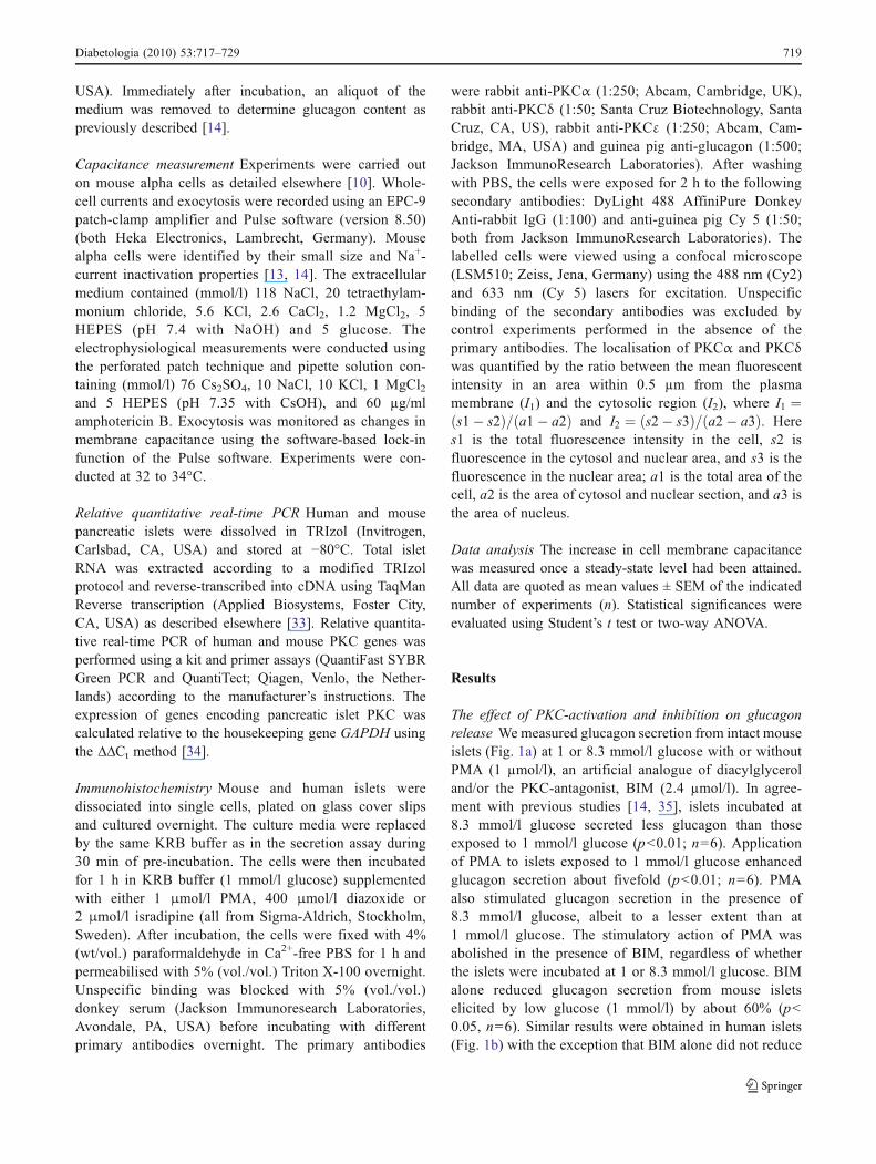
The effect of PKC-activation and inhibition on glucagonrelease Wemeasured glucagon secretion from intact mouseislets (Fig. 1a) at 1 or 8.3 mmol/l glucose with or withoutPMA (1 µmol/l), an artificial analogue of diacylglyceroland/or the PKC-antagonist, BIM (2.4 µmol/l). In agree-ment with previous studies [14, 35], islets incubated at8.3 mmol/l glucose secreted less glucagon than thoseexposed to 1 mmol/l glucose (p<0.01; n=6). Applicationof PMA to islets exposed to 1 mmol/l glucose enhancedglucagon secretion about fivefold (p<0.01; n=6). PMAalso stimulated glucagon secretion in the presence of8.3 mmol/l glucose, albeit to a lesser extent than at1 mmol/l glucose. The stimulatory action of PMA wasabolished in the presence of BIM, regardless of whetherthe islets were incubated at 1 or 8.3 mmol/l glucose. BIMalone reduced glucagon secretion from mouse isletselicited by low glucose (1 mmol/l) by about 60% (p<0.05, n=6). Similar results were obtained in human islets(Fig. 1b) with the exception that BIM alone did not reduce
Diabetologia (2010) 53:717–729 719
glucagon secretion at 1 mmol/l glucose. Collectively,these data implicate PKC in the modulation of glucagonsecretion from mouse and human alpha cells.
Treatment with PMA stimulates pancreatic alpha cellexocytosis High-resolution capacitance perforated patchmeasurements were applied to single mouse alpha cells to
investigate the effect of PKC activation on exocytosiselicited by trains of five 500-ms depolarisations from −70to 0 mV (Fig. 2). PMA (10 nmol/l) augmented theexocytotic responses regardless of the magnitude of theresponses under basal condition (Fig. 2a, b). The totalcapacitance increase evoked by the trains averaged 40±15fF (n=12) under control condition and 185±25 fF (p<0.001) (Fig. 2c) 6 min after addition of PMA. Exocytosiselicited by this stimulation paradigm reflects fusion ofgranules belonging to the readily releasable pool (RRP) andits replenishment from a reserve pool [13]. An upperestimate of RRP can be obtained from the sum of thecapacitance increase elicited by the first two depolarisationsof the train [36], whereas the response to the followingthree pulses was used to estimate exocytosis due toreplenishment of RRP. The RRP was thus estimated to be48±30 fF (n=12) and 113±26 fF (p<0.001) in the absenceand presence of PMA, respectively. This was paralleled bya sixfold increase in membrane capacitance from 12±6 fF(n=12) to 75±14 fF (p<0.001) in the response reflectingrefilling of RRP.
To rule out the possibility that PMA stimulates alpha cellexocytosis by increased Ca2+ channel activity, we measuredwhole cell Ca2+ currents in response to depolarisations fromthe holding membrane potential −70 mV to membranepotentials ranging from −60 to +50 mV. However, no effectof PMA on the peak current amplitude was observed(Fig. 2d).
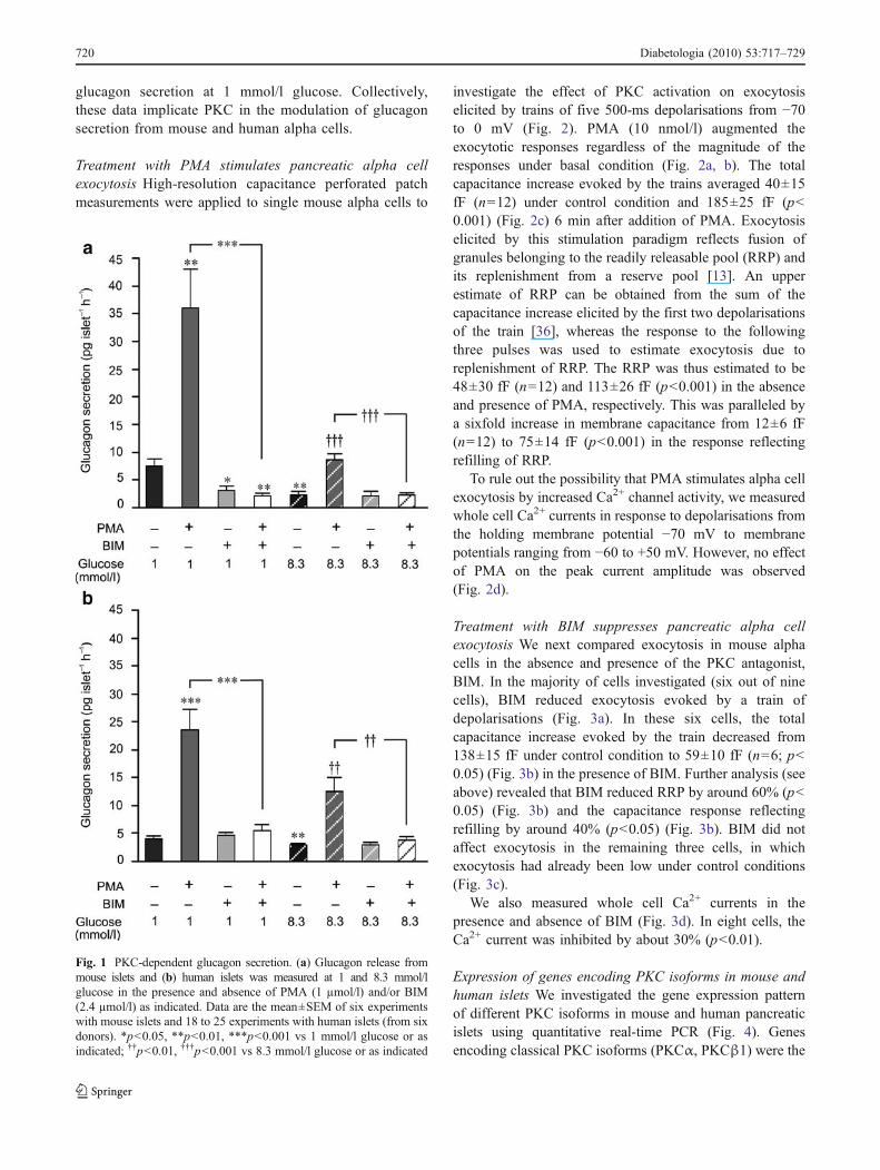
Treatment with BIM suppresses pancreatic alpha cellexocytosis We next compared exocytosis in mouse alphacells in the absence and presence of the PKC antagonist,BIM. In the majority of cells investigated (six out of ninecells), BIM reduced exocytosis evoked by a train ofdepolarisations (Fig. 3a). In these six cells, the totalcapacitance increase evoked by the train decreased from138±15 fF under control condition to 59±10 fF (n=6; p<0.05) (Fig. 3b) in the presence of BIM. Further analysis (seeabove) revealed that BIM reduced RRP by around 60% (p<0.05) (Fig. 3b) and the capacitance response reflectingrefilling by around 40% (p<0.05) (Fig. 3b). BIM did notaffect exocytosis in the remaining three cells, in whichexocytosis had already been low under control conditions(Fig. 3c).
We also measured whole cell Ca2+ currents in thepresence and absence of BIM (Fig. 3d). In eight cells, theCa2+ current was inhibited by about 30% (p<0.01).
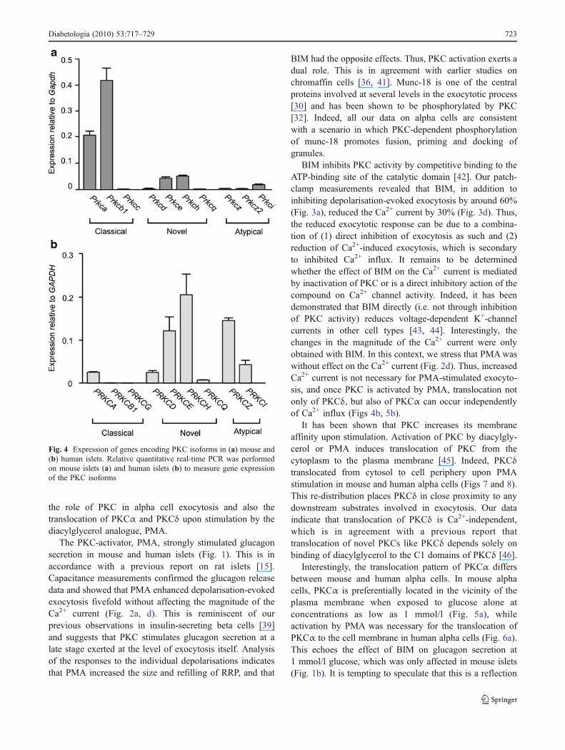
Expression of genes encoding PKC isoforms in mouse andhuman islets We investigated the gene expression patternof different PKC isoforms in mouse and human pancreaticislets using quantitative real-time PCR (Fig. 4). Genesencoding classical PKC isoforms (PKCα, PKCβ1) were the
Fig. 1 PKC-dependent glucagon secretion. (a) Glucagon release frommouse islets and (b) human islets was measured at 1 and 8.3 mmol/lglucose in the presence and absence of PMA (1 µmol/l) and/or BIM(2.4 µmol/l) as indicated. Data are the mean±SEM of six experimentswith mouse islets and 18 to 25 experiments with human islets (from sixdonors). *p<0.05, **p<0.01, ***p<0.001 vs 1 mmol/l glucose or asindicated; ††p<0.01, †††p<0.001 vs 8.3 mmol/l glucose or as indicated
720 Diabetologia (2010) 53:717–729

most highly expressed in mouse, whereas those encodingthe novel (PKCδ, PKCε, PKCη, PKCθ) and atypical PKC(PKCι, PKCζ) isoforms were more abundant in humanislets.
Due to the extremely low expression of PKCβ1(alsoknown as PRKCB or PRKCB1) in human islets, we selectedPKCα for further analysis of activation and translocation ofclassical PKC isoforms in alpha cells. Among the novel PKCisoforms, PKCδ and PKCε were highly abundant in bothspecies and have been described as being involved inexocytosis [28, 37]. We opted for PKCδ in our further study,since PKCε staining showed very low levels by confocalimmunocytochemistry in mouse alpha cells (data not shown).
PMA- and Ca2+-dependent translocation of PKCα inmouse and human alpha cells The distribution of PKCαwas analysed by confocal microscopy on single mouse andhuman alpha cells exposed to 1 mmol/l glucose (Figs 5and 6), i.e. conditions associated with a high rate ofglucagon secretion (Fig. 1). The identity of the alpha cellswas established by immunoreactivity for glucagon. Thetranslocation of PKC isoforms was quantified by calculat-ing the ratio of the fluorescent intensity in the vicinity ofthe plasma membrane (within a distance of 0.5 μm to the
plasma membrane, I1) to that in the inner cytosolic area (I2)as illustrated in Fig. 5c (details, see Methods).
In the absence of PMA, PKCα was located in thevicinity of the plasma membrane in about 90% of themouse alpha cells (Fig. 5a, d), while only a few cellsdisplayed cytosolic localisation (data not shown). Thispattern did not change following stimulation with PMA(Fig. 5a, d).
Previous studies have shown that PKCα is Ca2+-sensitive [38]. To determine whether the association ofPKCα with the plasma membrane observed in the presenceof 1 mmol/l glucose alone is Ca2+-dependent, we treatedmouse pancreatic cells with 400 μmol/l of the KATP-channel opener diazoxide or 2 μmol/l of the L-type Ca2+
channel inhibitor, isradipine, in the absence and presence of1 μmol/l PMA. Confocal microscopy showed that exposureto diazoxide caused a re-distribution of PKCα from the cellmembrane to the cytosol and decreased the ratio (I1/I2) by50% (p<0.001) (Fig. 5b, d). In the presence of isradipine,the reduction of the ratio was 45% compared with control(p<0.001) (Fig. 5b, d). Thus, the re-distribution to thecytosol by diazoxide can almost entirely be attributed toinhibition of Ca2+ entry via L-type Ca2+ channels. Theisradipine-induced redistribution of PKCα was counter-
Fig. 2 PMA-dependent stimulation of exocytosis in single mousealpha cells. a Capacitance increase elicited by a train of five 500 msdepolarisations from −70 to 0 mV before (black trace) and 6 min after(grey trace) inclusion of 10 nmol/l PMA in the extracellular solution.b Capacitance increase determined as above (a) for an alpha cell witha larger response in the absence of PMA. c Histogram summarisingthe average increase in cell membrane capacitance (ΔCm) evoked by
all five depolarisations of the train, the first two depolarisations (RRP)and the following three depolarisations (refilling from reserve pool) inthe presence and absence of PMA. d Peak Ca2+ current amplitude (I)against membrane potential (V) recorded before (white symbols) andafter the addition of 10 nmol/l PMA in the extracellular solution(black symbols). Data are the mean±SEM of 12 paired experiments.***p<0.001 vs control
Diabetologia (2010) 53:717–729 721
acted when the cells were simultaneously exposed to PMA(Fig. 5b, d). The latter experiment demonstrates that thetranslocation of PKCα to the plasma membrane can betriggered independently, either by increased Ca2+ influxthrough L-type Ca2+ channels or by activation of PKC byPMA.
In contrast to the situation in mouse, PKCα in humanalpha cells principally located to the cytosol in the absenceof PMA (Fig. 6a, c). PMA treatment translocated PKCαfrom the cytosol towards the cell periphery and the I1/I2ratio increased around twofold (p<0.001). The distributionof PKCα in human alpha cells was not affected bydiazoxide or isradipine (Fig. 6b, c), and PMA remainedcapable of triggering the translocation of PKCα in thepresence of isradipine (Fig. 6b, c).
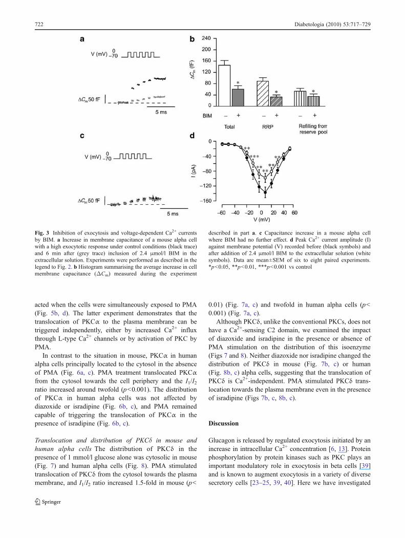
Translocation and distribution of PKCδ in mouse andhuman alpha cells The distribution of PKCδ in thepresence of 1 mmol/l glucose alone was cytosolic in mouse(Fig. 7) and human alpha cells (Fig. 8). PMA stimulatedtranslocation of PKCδ from the cytosol towards the plasmamembrane, and I1/I2 ratio increased 1.5-fold in mouse (p<
0.01) (Fig. 7a, c) and twofold in human alpha cells (p<0.001) (Fig. 7a, c).
Although PKCδ, unlike the conventional PKCs, does nothave a Ca2+-sensing C2 domain, we examined the impactof diazoxide and isradipine in the presence or absence ofPMA stimulation on the distribution of this isoenzyme(Figs 7 and 8). Neither diazoxide nor isradipine changed thedistribution of PKCδ in mouse (Fig. 7b, c) or human(Fig. 8b, c) alpha cells, suggesting that the translocation ofPKCδ is Ca2+-independent. PMA stimulated PKCδ trans-location towards the plasma membrane even in the presenceof isradipine (Figs 7b, c, 8b, c).
Discussion
Glucagon is released by regulated exocytosis initiated by anincrease in intracellular Ca2+ concentration [6, 13]. Proteinphosphorylation by protein kinases such as PKC plays animportant modulatory role in exocytosis in beta cells [39]and is known to augment exocytosis in a variety of diversesecretory cells [23–25, 39, 40]. Here we have investigated
Fig. 3 Inhibition of exocytosis and voltage-dependent Ca2+ currentsby BIM. a Increase in membrane capacitance of a mouse alpha cellwith a high exocytotic response under control conditions (black trace)and 6 min after (grey trace) inclusion of 2.4 μmol/l BIM in theextracellular solution. Experiments were performed as described in thelegend to Fig. 2. b Histogram summarising the average increase in cellmembrane capacitance (ΔCm) measured during the experiment
described in part a. c Capacitance increase in a mouse alpha cellwhere BIM had no further effect. d Peak Ca2+ current amplitude (I)against membrane potential (V) recorded before (black symbols) andafter addition of 2.4 μmol/l BIM to the extracellular solution (whitesymbols). Data are mean±SEM of six to eight paired experiments.*p<0.05, **p<0.01, ***p<0.001 vs control
722 Diabetologia (2010) 53:717–729
the role of PKC in alpha cell exocytosis and also thetranslocation of PKCα and PKCδ upon stimulation by thediacylglycerol analogue, PMA.
The PKC-activator, PMA, strongly stimulated glucagonsecretion in mouse and human islets (Fig. 1). This is inaccordance with a previous report on rat islets [15].Capacitance measurements confirmed the glucagon releasedata and showed that PMA enhanced depolarisation-evokedexocytosis fivefold without affecting the magnitude of theCa2+ current (Fig. 2a, d). This is reminiscent of ourprevious observations in insulin-secreting beta cells [39]and suggests that PKC stimulates glucagon secretion at alate stage exerted at the level of exocytosis itself. Analysisof the responses to the individual depolarisations indicatesthat PMA increased the size and refilling of RRP, and that
BIM had the opposite effects. Thus, PKC activation exerts adual role. This is in agreement with earlier studies onchromaffin cells [36, 41]. Munc-18 is one of the centralproteins involved at several levels in the exocytotic process[30] and has been shown to be phosphorylated by PKC[32]. Indeed, all our data on alpha cells are consistentwith a scenario in which PKC-dependent phosphorylationof munc-18 promotes fusion, priming and docking ofgranules.
BIM inhibits PKC activity by competitive binding to theATP-binding site of the catalytic domain [42]. Our patch-clamp measurements revealed that BIM, in addition toinhibiting depolarisation-evoked exocytosis by around 60%(Fig. 3a), reduced the Ca2+ current by 30% (Fig. 3d). Thus,the reduced exocytotic response can be due to a combina-tion of (1) direct inhibition of exocytosis as such and (2)reduction of Ca2+-induced exocytosis, which is secondaryto inhibited Ca2+ influx. It remains to be determinedwhether the effect of BIM on the Ca2+ current is mediatedby inactivation of PKC or is a direct inhibitory action of thecompound on Ca2+ channel activity. Indeed, it has beendemonstrated that BIM directly (i.e. not through inhibitionof PKC activity) reduces voltage-dependent K+-channelcurrents in other cell types [43, 44]. Interestingly, thechanges in the magnitude of the Ca2+ current were onlyobtained with BIM. In this context, we stress that PMAwaswithout effect on the Ca2+ current (Fig. 2d). Thus, increasedCa2+ current is not necessary for PMA-stimulated exocyto-sis, and once PKC is activated by PMA, translocation notonly of PKCδ, but also of PKCα can occur independentlyof Ca2+ influx (Figs 4b, 5b).
It has been shown that PKC increases its membraneaffinity upon stimulation. Activation of PKC by diacylgly-cerol or PMA induces translocation of PKC from thecytoplasm to the plasma membrane [45]. Indeed, PKCδtranslocated from cytosol to cell periphery upon PMAstimulation in mouse and human alpha cells (Figs 7 and 8).This re-distribution places PKCδ in close proximity to anydownstream substrates involved in exocytosis. Our dataindicate that translocation of PKCδ is Ca2+-independent,which is in agreement with a previous report thattranslocation of novel PKCs like PKCδ depends solely onbinding of diacylglycerol to the C1 domains of PKCδ [46].
Interestingly, the translocation pattern of PKCα differsbetween mouse and human alpha cells. In mouse alphacells, PKCα is preferentially located in the vicinity of theplasma membrane when exposed to glucose alone atconcentrations as low as 1 mmol/l (Fig. 5a), whileactivation by PMA was necessary for the translocation ofPKCα to the cell membrane in human alpha cells (Fig. 6a).This echoes the effect of BIM on glucagon secretion at1 mmol/l glucose, which was only affected in mouse islets(Fig. 1b). It is tempting to speculate that this is a reflection
Fig. 4 Expression of genes encoding PKC isoforms in (a) mouse and(b) human islets. Relative quantitative real-time PCR was performedon mouse islets (a) and human islets (b) to measure gene expressionof the PKC isoforms
Diabetologia (2010) 53:717–729 723
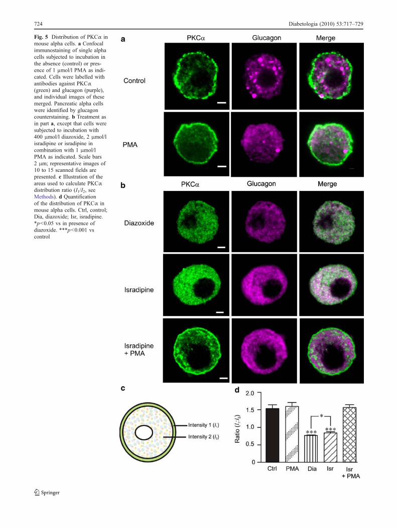
Fig. 5 Distribution of PKCα inmouse alpha cells. a Confocalimmunostaining of single alphacells subjected to incubation inthe absence (control) or pres-ence of 1 µmol/l PMA as indi-cated. Cells were labelled withantibodies against PKCα(green) and glucagon (purple),and individual images of thesemerged. Pancreatic alpha cellswere identified by glucagoncounterstaining. b Treatment asin part a, except that cells weresubjected to incubation with400 μmol/l diazoxide, 2 μmol/lisradipine or isradipine incombination with 1 μmol/lPMA as indicated. Scale bars2 μm; representative images of10 to 15 scanned fields arepresented. c Illustration of theareas used to calculate PKCαdistribution ratio (I1/I2, seeMethods). d Quantificationof the distribution of PKCα inmouse alpha cells. Ctrl, control;Dia, diazoxide; Isr, isradipine.*p<0.05 vs in presence ofdiazoxide. ***p<0.001 vscontrol
724 Diabetologia (2010) 53:717–729
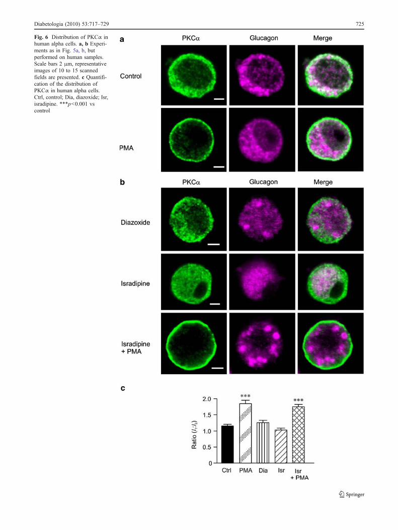
Fig. 6 Distribution of PKCα inhuman alpha cells. a, b Experi-ments as in Fig. 5a, b, butperformed on human samples.Scale bars 2 μm, representativeimages of 10 to 15 scannedfields are presented. c Quantifi-cation of the distribution ofPKCα in human alpha cells.Ctrl, control; Dia, diazoxide; Isr,isradipine. ***p<0.001 vscontrol
Diabetologia (2010) 53:717–729 725
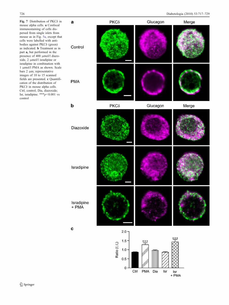
Fig. 7 Distribution of PKCδ inmouse alpha cells. a Confocalimmunostaining of cells dis-persed from single islets frommouse as in Fig. 5a, except thatcells were labelled with anti-bodies against PKCδ (green)as indicated. b Treatment as inpart a, but performed in thepresence of 400 μmol/l diazo-xide, 2 μmol/l isradipine orisradipine in combination with1 μmol/l PMA as shown. Scalebars 2 μm; representativeimages of 10 to 15 scannedfields are presented. c Quantifi-cation of the distribution ofPKCδ in mouse alpha cells.Ctrl, control; Dia, diazoxide;Isr, isradipine. ***p<0.001 vscontrol
726 Diabetologia (2010) 53:717–729

Fig. 8 Distribution of PKCδ inhuman alpha cells. a, b As inFig 7a, b, except that experi-ments were performed onhuman alpha cells. Scale bars2 μm, representative images of10 to 15 scanned fields arepresented. c Quantification ofthe distribution of PKCδ inhuman alpha cells. Ctrl, control;Dia, diazoxide; Isr, isradipine.***p<0.001 vs control
Diabetologia (2010) 53:717–729 727

of PKCα being tonically activated in mouse alpha cells andthereby susceptible to inhibition by BIM. PKCα, whichbelongs to the classical PKCs, is activated by increasedintracellular Ca2+ and diacylglycerol [46]. In beta cells,glucose has been reported to produce a Ca2+-dependenttranslocation of classical PKCs to the plasma membrane[29]. In mouse alpha cells, we attribute the near-plasmamembrane localisation of PKCα in the absence of PMA tothe high [Ca2+]i resulting from Ca2+ influx through Ca2+
channels, which are activated during the action potentialsgenerated at low glucose. This scenario is suggested by theredistribution of PKCα to the cytosolic region in cellsexposed to diazoxide or isradipine (Fig. 5b). We demon-strated that Ca2+ influx through the L-type Ca2+ channel isthe major source (90%) responsible for the Ca2+-dependenttranslocation of PKCα, while only 10% of the translocationis due to Ca2+ influx through other channels. We can onlyspeculate about why this does not occur in human alphacells, but it may be pertinent that distribution of PKCα is lessperipheral in human than in mouse alpha cells (Figs 5 and 6).It is possible that in human alpha cells, the intracellular Ca2+
concentration is lower than in mouse alpha cells [47, 48],resulting in translocation of PKCα to the plasma membranein a less Ca2+-dependent manner. This explanation wouldalso account for the greater sensitivity to PMA.
Glucagon secretion and exocytosis in the pancreaticalpha cell triggered by low glucose alone is dependent oninflux through the N-type Ca2+ channel [14]. AlthoughL-type Ca2+ channels are not essential for exocytosis inalpha cells in the absence of high concentrations of cyclicAMP, the data presented here indicate that these channelsmediate the Ca2+ influx that induces the Ca2+-dependenttranslocation of PKCα to the plasma membrane. Thus,L- and N-type Ca2+ channels fulfil different functions in thealpha cell (PKC translocation and exocytosis, respectively).We finally point out that elevation of intracellular cyclicAMP causes an interesting switch in Ca2+ channel depen-dence and that in the presence of forskolin or adrenaline,L-type Ca2+ channels account for most of the Ca2+ entryinvolved in exocytosis [6]. Further studies are required toelucidate the interaction between cyclic AMP/PKA- andPKC-mediated effects on exocytosis, and how they dependon Ca2+ influx and PKC translocation. Clearly, the control ofalpha cell exocytosis is very complex, making it easy toenvisage disturbances capable of causing defective regula-tion of glucagon secretion, which is a hallmark of type 2diabetes [49].
Acknowledgements We thank B.-M. Nilsson (Lund UniversityDiabetes Centre) for expert technical assistance. This work wassupported by the Swedish Research Council, EU-Biosim, the Knutand Alice Wallenberg foundation, the Crafoord foundation, theThurings foundation, the Novo Nordisk Foundation and the AlbertPåhlssons Foundation. P. Rorsman is supported by the Welcome Trust
and the Medical Research Council. L. Eliasson and E. Renström areSwedish Research Council senior researchers.
Duality of interest The authors declare that there is no duality ofinterest associated with this manuscript.
References
1. Lins PE, Wajngot A, Adamson U, Vranic M, Efendic S (1983)Minimal increases in glucagon levels enhance glucose productionin man with partial hypoinsulinemia. Diabetes 32:633–636
2. Myers SR, Diamond MP, Adkins-Marshall BA, Williams PE,Stinsen R, Cherrington AD (1991) Effects of small changes inglucagon on glucose production during a euglycemic, hyper-insulinemic clamp. Metabolism 40:66–71
3. Cryer PE (2004) Diverse causes of hypoglycemia-associatedautonomic failure in diabetes. N Engl J Med 350:2272–2279
4. Shah P, Vella A, Basu A, Basu R, Schwenk WF, Rizza RA (2000)Lack of suppression of glucagon contributes to postprandialhyperglycemia in subjects with type 2 diabetes mellitus. J ClinEndocrinol Metab 85:4053–4059
5. Gromada J, Franklin I, Wollheim CB (2007) Alpha-cells of theendocrine pancreas: 35 years of research but the enigma remains.Endocr Rev 28:84–116
6. Gromada J, Bokvist K, Ding WG et al (1997) Adrenalinestimulates glucagon secretion in pancreatic A-cells by increasingthe Ca2+ current and the number of granules close to the L-typeCa2+ channels. J Gen Physiol 110:217–228
7. Vieira E, Liu YJ, Gylfe E (2004) Involvement of alpha1 and beta-adrenoceptors in adrenaline stimulation of the glucagon-secretingmouse alpha-cell. Naunyn Schmiedebergs Arch Pharmacol 369:179–183
8. Dunning BE, Foley JE, Ahren B (2005) Alpha cell function inhealth and disease: influence of glucagon-like peptide-1. Diabe-tologia 48:1700–1713
9. Gromada J, Hoy M, Buschard K, Salehi A, Rorsman P (2001)Somatostatin inhibits exocytosis in rat pancreatic alpha-cells by G(i2)-dependent activation of calcineurin and depriming of secre-tory granules. J Physiol 535:519–532
10. Ma X, Zhang Y, Gromada J et al (2005) Glucagon stimulatesexocytosis in mouse and rat pancreatic alpha-cells by binding toglucagon receptors. Mol Endocrinol 19:198–212
11. Gopel S, Zhang Q, Eliasson L et al (2004) Capacitance measure-ments of exocytosis in mouse pancreatic alpha-, beta- and delta-cells within intact islets of Langerhans. J Physiol 556:711–726
12. Rorsman P, Hellman B (1988) Voltage-activated currents inguinea pig pancreatic alpha 2 cells. Evidence for Ca2+-dependentaction potentials. J Gen Physiol 91:223–242
13. Barg S, Galvanovskis J, Gopel SO, Rorsman P, Eliasson L (2000)Tight coupling between electrical activity and exocytosis in mouseglucagon-secreting alpha-cells. Diabetes 49:1500–1510
14. MacDonald PE, De Marinis YZ, Ramracheya R et al (2007) A KATP channel-dependent pathway within alpha cells regulatesglucagon release from both rodent and human islets of Langer-hans. PLoS Biol 5:e143
15. Hii CS, Stutchfield J, Howell SL (1986) Enhancement ofglucagon secretion from isolated rat islets of Langerhans byphorbol 12-myristate 13-acetate. Biochem J 233:287–289
16. Bjaaland T, Hii CS, Jones PM, Howell SL (1988) Role of proteinkinase C in arginine-induced glucagon secretion from isolated ratislets of Langerhans. J Mol Endocrinol 1:105–110
17. Shirai Y, Saito N (2002) Activation mechanisms of proteinkinase C: maturation, catalytic activation, and targeting. J Biochem132:663–668
728 Diabetologia (2010) 53:717–729
18. Wolf BA, Easom RA, Hughes JH, McDaniel ML, Turk J (1989)Secretagogue-induced diacylglycerol accumulation in isolatedpancreatic islets. Mass spectrometric characterization of the fattyacyl content indicates multiple mechanisms of generation.Biochemistry 28:4291–4301
19. Malaisse WJ, Dunlop ME, Mathias PC, Malaisse-Lagae F, SenerA (1985) Stimulation of protein kinase C and insulin release by1-oleoyl-2-acetyl-glycerol. Eur J Biochem 149:23–27
20. Zawalich W, Brown C, Rasmussen H (1983) Insulin secretion:combined effects of phorbol ester and A23187. Biochem BiophysRes Commun 117:448–455
21. Stutchfield J, Jones PM, Howell SL (1986) The effects ofpolymyxin B, a protein kinase C inhibitor, on insulin secretionfrom intact and permeabilized islets of Langerhans. BiochemBiophys Res Commun 136:1001–1006
22. Easom RA, Hughes JH, Landt M, Wolf BA, Turk J, McDanielML (1989) Comparison of effects of phorbol esters and glucoseon protein kinase C activation and insulin secretion in pancreaticislets. Biochem J 264:27–33
23. Hille B, Billiard J, Babcock DF, Nguyen T, Koh DS (1999)Stimulation of exocytosis without a calcium signal. J Physiol 520(Pt 1):23–31
24. Turner KM, Burgoyne RD, Morgan A (1999) Protein phosphor-ylation and the regulation of synaptic membrane traffic. TrendsNeurosci 22:459–464
25. Leenders AG, Sheng ZH (2005) Modulation of neurotransmitterrelease by the second messenger-activated protein kinases:implications for presynaptic plasticity. Pharmacol Ther 105:69–84
26. Hoy M, Berggren PO, Gromada J (2003) Involvement of proteinkinase C-epsilon in inositol hexakisphosphate-induced exocytosisin mouse pancreatic beta-cells. J Biol Chem 278:35168–35171
27. Ganesan S, Calle R, Zawalich K et al (1992) Immunocytochemicallocalization of alpha-protein kinase C in rat pancreatic beta-cellsduring glucose-induced insulin secretion. J Cell Biol 119:313–324
28. Uchida T, Iwashita N, Ohara-Imaizumi M et al (2007) Proteinkinase Cdelta plays a non-redundant role in insulin secretion inpancreatic beta cells. J Biol Chem 282:2707–2716
29. Yedovitzky M, Mochly-Rosen D, Johnson JA et al (1997)Translocation inhibitors define specificity of protein kinase Cisoenzymes in pancreatic beta-cells. J Biol Chem 272:1417–1420
30. Toonen RF, Verhage M (2007) Munc18-1 in secretion: lonely Muncjoins SNARE team and takes control. Trends Neurosci 30:564–572
31. Craig TJ, Evans GJ, Morgan A (2003) Physiological regulation ofMunc18/nSec1 phosphorylation on serine-313. J Neurochem 86:1450–1457
32. Barclay JW, Craig TJ, Fisher RJ et al (2003) Phosphorylation ofMunc18 by protein kinase C regulates the kinetics of exocytosis.J Biol Chem 278:10538–10545
33. Amisten S, Braun OO, Bengtsson A, Erlinge D (2008) Geneexpression profiling for the identification of G-protein coupledreceptors in human platelets. Thromb Res 122:47–57
34. Pfaffl MW (2001) A new mathematical model for relativequantification in real-time RT-PCR. Nucleic Acids Res 29:e45
35. Salehi A, Vieira E, Gylfe E (2006) Paradoxical stimulation ofglucagon secretion by high glucose concentrations. Diabetes55:2318–2323
36. Gillis KD, Mossner R, Neher E (1996) Protein kinase C enhancesexocytosis from chromaffin cells by increasing the size of thereadily releasable pool of secretory granules. Neuron 16:1209–1220
37. Mendez CF, Leibiger IB, Leibiger B et al (2003) Rapidassociation of protein kinase C-epsilon with insulin granules isessential for insulin exocytosis. J Biol Chem 278:44753–44757
38. Nishizuka Y (1984) The role of protein kinase C in cell surfacesignal transduction and tumour promotion. Nature 308:693–698
39. Ammala C, Eliasson L, Bokvist K et al (1994) Activation ofprotein kinases and inhibition of protein phosphatases play acentral role in the regulation of exocytosis in mouse pancreaticbeta cells. Proc Natl Acad Sci USA 91:4343–4347
40. Lindau M, Gomperts BD (1991) Techniques and concepts inexocytosis: focus on mast cells. Biochim Biophys Acta 1071:429–471
41. Nili U, de Wit H, Gulyas-Kovacs A et al (2006) Munc18-1phosphorylation by protein kinase C potentiates vesicle poolreplenishment in bovine chromaffin cells. Neuroscience 143:487–500
42. Toullec D, Pianetti P, Coste H et al (1991) The bisindolylmalei-mide GF 109203X is a potent and selective inhibitor of proteinkinase C. J Biol Chem 266:15771–15781
43. Park WS, Son YK, Ko EA et al (2005) The protein kinase Cinhibitor, bisindolylmaleimide (I), inhibits voltage-dependentK+ channels in coronary arterial smooth muscle cells. Life Sci77:512–527
44. Kim A, Bae YM, Kim J et al (2004) Direct block bybisindolylmaleimide of the voltage-dependent K+ currents of ratmesenteric arterial smooth muscle. Eur J Pharmacol 483:117–126
45. Kraft AS, Anderson WB (1983) Phorbol esters increase theamount of Ca2+, phospholipid-dependent protein kinase associatedwith plasma membrane. Nature 301:621–623
46. Mellor H, Parker PJ (1998) The extended protein kinase Csuperfamily. Biochem J 332(Pt 2):281–292
47. Quesada I, Todorova MG, Alonso-Magdalena P et al (2006)Glucose induces opposite intracellular Ca2+ concentration oscilla-tory patterns in identified alpha- and beta-cells within intacthuman islets of Langerhans. Diabetes 55:2463–2469
48. Nadal A, Quesada I, Soria B (1999) Homologous and heterolo-gous asynchronicity between identified alpha-, beta- and delta-cells within intact islets of Langerhans in the mouse. J Physiol 517(Pt 1):85–93
49. Ali S, Drucker DJ (2009) Benefits and limitations of reducingglucagon action for the treatment of type 2 diabetes. Am J PhysiolEndocrinol Metab 296:E415–E421
Diabetologia (2010) 53:717–729 729
Copyright © 2022 FDOKUMEN