mGlu5-GABAB interplay in animal models of positive, negative and cognitive symptoms of schizophrenia
Upload
independentCategory
view
2download
0
Coordinated action of NSF and PKC regulatesGABAB receptor signaling efficacy
Stephanie M Pontier1, Nicolas Lahaie1,Rachel Ginham2, Fannie St-Gelais3,Helene Bonin1, David J Bell4, Helen Flynn4,Louis-Eric Trudeau3, Jeffrey McIlhinney2,Julia H White4 and Michel Bouvier1,*1Departement de Biochimie and Groupe de Recherche Universitaire surle Medicament, Institut de recherche en immunologie et Cancerologie,Universite de Montreal, Montreal, Qc, Canada, 2Medical ResearchCouncil Anatomical Neuropharmacology Unit, Oxford, UK,3Departement de Pharmacologie, Faculte de medecine, Universite deMontreal, Montreal, Qc, Canada and 4Pathway Discovery, Genomics andProteomic Sciences, GlaxoSmithKline Medicines Research Centre,Stevenage, UK
The obligatory heterodimerization of the GABAB receptor
(GBR) raises fundamental questions about molecular me-
chanisms controlling its signaling efficacy. Here, we show
that NEM sensitive fusion (NSF) protein interacts directly
with the GBR heterodimer both in rat brain synaptosomes
and in CHO cells, forming a ternary complex that can be
regulated by agonist stimulation. Inhibition of NSF bind-
ing with a peptide derived from GBR2 (TAT-Pep-27) did not
affect basal signaling activity but almost completely abol-
ished agonist-promoted GBR desensitization in both CHO
cells and hippocampal slices. Taken with the role of PKC
in the desensitization process, our observation that TAT-
Pep-27 prevented both agonist-promoted recruitment of
PKC and receptor phosphorylation suggests that NSF is a
priming factor required for GBR desensitization. Given
that GBR desensitization does not involve receptor inter-
nalization, the NSF/PKC coordinated action revealed here-
in suggests that NSF can regulate GPCR signalling efficacy
independently of its role in membrane trafficking. The
functional interaction between three bona fide regulators
of neurotransmitter release, such as GBR, NSF and PKC,
could shed new light on the modulation of presynaptic
GBR action.
The EMBO Journal (2006) 25, 2698–2709. doi:10.1038/
sj.emboj.7601157; Published online 25 May 2006
Subject Categories: signal transduction
Keywords: desensitization; GABAB; heterodimer; NSF; PKC
Introduction
Ionotropic and metabotropic receptors mediate the action of
the inhibitory neurotransmitter g-amino-butyric acid (GABA)
in the central nervous system. The metabotropic GABAB
receptor (GBR) consists of an obligatory heterodimer be-
tween two seven transmembrane domain (7TM) receptors,
GBR1 and GBR2 (Jones et al, 1998; Kaupmann et al, 1998;
White et al, 1998; Kuner et al, 1999). In addition to playing a
role in ER export (Couve et al, 1998; Margeta-Mitrovic et al,
2000), GBR1/GBR2 heterodimerization is required for the
formation of a functional receptor. Indeed, whereas only
GBR1 can bind GABA, GBR2 appears to engage the hetero-
trimeric G protein for downstream signaling (Galvez et al,
2001; Margeta-Mitrovic et al, 2001; Robbins et al, 2001). Such
transactivation across two distinct 7TM receptors raises
fundamental questions about the molecular mechanisms
controlling their signaling efficacy.
Among the mechanisms controlling 7TM receptor activity,
agonist-promoted desensitization is one of the best character-
ized at the molecular level. As for most receptors, sustained
stimulation of GBR can lead to functional desensitization
(Couve et al, 2002; Gonzalez-Maeso et al, 2003; Perroy
et al, 2003; Tosetti et al, 2004). However, b-arrestin recruit-
ment to the receptor and the ensuing endocytosis of the
complex that are classically associated to desensitization do
not appear to contribute to the regulation of GBR responsive-
ness (Perroy et al, 2003; Fairfax et al, 2004). We recently
reported that a phosphorylation-independent mechanism in-
volving the G protein receptor kinase 4 (GRK4) can regulate
GBR activity in the cerebellum (Perroy et al, 2003). However,
the restricted expression pattern of GRK4, mainly found in
testes and cerebellum (Virlon et al, 1998; Sallese et al, 2000),
suggests that other mechanisms may modulate GBR signaling
efficacy in other tissues. There is in fact multiple evidences
that different mechanisms may be at work since distinct
desensitization and phosphorylation profiles were reported
for different cellular systems (Couve et al, 2002; Gonzalez-
Maeso et al, 2003; Tosetti et al, 2004). These discrepancies
between different systems are most likely due to the relative
expression levels of different protein partners that can influ-
ence receptor signaling efficacy. Among other factors, PKC
activation has previously been shown to regulate GBR activ-
ity (Dutar and Nicoll, 1988; Thompson and Gahwiler, 1992),
even if its direct role in agonist-promoted desensitization has
not yet been documented.
In an effort to identify new proteins that could regulate
GBR function, we performed a yeast two-hybrid screen using
the GBR2 carboxyl tail (c-tail) as bait that revealed the
N-ethylmaleimide (NEM) sensitive fusion (NSF) protein as
a potential interacting partner. NSF belongs to the ‘ATPase
Associated to various cellular Activities’ (AAA) family and is
classically devoted to the regulation of protein complexes
supporting membrane fusion and trafficking events
(Whiteheart and Matveeva, 2004). In this context, it pro-
motes the disruption of soluble NSF-associated protein re-
ceptor (SNARE) coiled-coil interactions. Such NSF uncoiling
activity is of particular interest when considering its interac-
tion with GBR as receptor’s c-tails are engaged in a coiled-coilReceived: 7 December 2005; accepted: 27 April 2006; publishedonline: 25 May 2006
*Corresponding author. Departement de Biochimie and Groupe deRecherche Universitaire sur le Medicament, Institut de recherche enimmunologie et Cancerologie, Universite de Montreal, Montreal, Qc,Canada H3C 3J7. Tel.: þ 1 514 343 6319; Fax: þ 1 514 343 2210;E-mail: [email protected]
The EMBO Journal (2006) 25, 2698–2709 | & 2006 European Molecular Biology Organization | All Rights Reserved 0261-4189/06
www.embojournal.org
The EMBO Journal VOL 25 | NO 12 | 2006 &2006 European Molecular Biology Organization
EMBO
THE
EMBOJOURNAL
THE
EMBOJOURNAL
2698
interaction within the GBR1/GBR2 heterodimer (White et al,
1998; Kammerer et al, 1999).
In addition to the established role of NSF in controlling the
assembly/disassembly of SNARE complexes, its direct inter-
action with cell surface receptors such as the AMPA receptor
(Nishimune et al, 1998; Osten et al, 1998), the b2-adrenergic
(b2AR) receptors (Cong et al, 2001), the dopaminergic recep-
tors (Heydorn et al, 2004; Zou et al, 2005), and the adreno-
medulin receptor (Bomberger et al, 2005) has been reported.
In the three cases where the functional consequences were
characterized, NSF binding was proposed to modulate the
postendocytic sorting of these receptors (Noel et al, 1999;
Cong et al, 2001; Hanley et al, 2002; Lee et al, 2004;
Bomberger et al, 2005). Given that GBR does not undergo
rapid agonist-promoted internalization in any of the systems
tested (Perroy et al, 2003; Fairfax et al, 2004), we sought to
investigate further the interaction between NSF and GBR and
its potential role in regulating receptor function.
Results
NSF interacts directly with GBR subunits
A yeast two-hybrid screen (YTH) was performed with both
GBR1 and GBR2 full-length c-tails (860I-961K and 741I-941L,
respectively) against a human brain cDNA library. In addition
to complementary GBR subunit (White et al, 2002) and
transcription factor ATFx/CREB2 (White et al, 2000), we
found NSF as a potential binding partner of GBR2
(Figure 1). Using shorter segments of the c-tail, the interact-
ing region was narrowed down to the distal part of the GBR2
coiled-coil domain and more specifically to a 27 amino-acid
peptide encompassing residues 799–825 (Pep27). As pre-
viously observed for the interaction between GluR2 and
NSF (Nishimune et al, 1998), the integrity of the full-length
ATPase appeared essential for its association with receptor
c-tail. Indeed, none of the truncation mutants of NSF tested
could bind to the receptor c-tail (data not shown).
To further characterize the interaction between NSF and
GBR2 c-tail (GBR2ct), we carried out in vitro binding assays
using the receptor c-tail fused to a glutathione-S-transferase
(GST) protein and purified His6-tagged NSF. GST pull-down
performed with increasing amount of NSF in the presence of
non-hydrolysable ATPgS (to inhibit ATPase activity) demon-
strated a direct binding of NSF to GBR2ct (Figure 2A). This
binding tends to saturate at B50 nM and reach half-satura-
tion at 15 nM (Figure 2A, right panel), indicating a relatively
high affinity of NSF for the receptor. The nucleotide depen-
dence of NSF interaction was then determined (Figure 2B) by
evaluating the impact of ATP and ATPgS under different salt
conditions. Conditions favoring NSFATPase activity (ATP and
Mg2þ ) disrupted the association, whereas its inhibition
(ATPgSþMg2þ , ATPþEDTA or ATPþMg2þ þEDTA) fa-
vored the interaction with GBR2ct (Figure 2B). Such influ-
ence of the nucleotide state of NSF is reminiscent of other
relevant interactions involving NSF (Sollner et al, 1993; Osten
et al, 1998; Hanley et al, 2002). Consistent with YTH data
presented above, the Pep27 region of the coiled-coil domain
of GBR2 was sufficient to sustain NSF binding as shown by
the efficient pull-down of NSF by a GST–Pep27 fusion protein
(Figure 2C). Interestingly, Pep27 was as efficient as a pre-
viously described 10 amino-acid peptide corresponding to the
GluR2 NSF binding site (Pep2m) (Nishimune et al, 1998;
Osten et al, 1998; Song et al, 1998). In contrast to the results
obtained in YTH experiments, NSF binding was not found to
be restricted to GBR2ct and GBR1ct (854I-961K) also inter-
acted selectively with NSF (Figure 2D).
Such partial discrepancy between YTH and GST pull-
down experiments is not unusual. Nevertheless, to clarify
whether the GBR1 could truly interact with NSF and to
confirm that interaction between GBR2 and NSF can occur
in living cells, immunoprecipitations were performed in
CHO cells. Immunoprecipitation of GBR1 or GBR2 from
cell expressing each of the receptor individually led to the
co-sedimentation of NSF (Figure 3A and B). In the reverse
configuration, NSF isolation revealed that it binds to two
molecular species of GBR2 (Figure 3B, right panel), corre-
sponding to ER-localized core-glycosylated precursor
(B100 kDa) and fully processed (B120 kDa) forms of the
receptor (Supplementary data and Supplementary Figure
S1). These results suggest that NSF binds to distinct GBR2
species along the maturation path from the ER to the
plasma membrane. In the case of GBR1, which cannot
reach the cell surface and is retained in the ER when
expressed alone (Couve et al, 1998), co-immunoprecipita-
tion with NSF implies that interaction between these two
molecules occurs in the ER. Taken together, these data may
indicate that NSF could be involved in GBR transport to the
cell surface, as has been previously suggested for other
membrane proteins (Noel et al, 1999; Cong et al, 2001;
Shi et al, 2001; Hanley et al, 2002).
In agreement with the influence of NSF nucleotide binding
state in GST-pull-down experiments, the functionality of its
Gal4 DNA binding hybridt
Gal4 activation hybrid
β-Galac tivity
NSF
NSFNSFNSF
NSFNSF
NSF
NSF
–+–+–––+
741 941Coiled-coil
Coiled-coil
GBR2
N-terminal TM 1–7 c-term
854 961
c-termTM 1–7N-terminal
GBR1a/b
GBR1ct 860–961GBR2ct 741–941
GBR2ct 741–785GBR2ct 780–827GBR2ct 827–873GBR2ct 870–910
GBR2ct 908–941GBR2ct 799–825
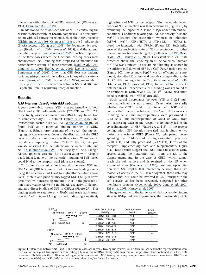
Figure 1 Interaction between NSF and GBR c-termini assessed in yeast two-hybrid system. GBR c-termini (see schematic representation) wereused as bait in a yeast two-hybrid screen testing a human brain cDNA library. NSF was one of the positive clones obtained with the GBR2c-terminus. To delineate the GBR2 minimal region of interaction with NSF, two-hybrid assay was performed between the indicated GBR2 c-tailmutants (see table) and NSF. b-Gal activity is determined (þ /�) for each construct.
PKC and NSF regulates GBR signaling efficacySM Pontier et al
&2006 European Molecular Biology Organization The EMBO Journal VOL 25 | NO 12 | 2006 2699

ATPase was found to be essential for its interaction with both
GBR1 and GBR2 as a treatment with the alkylating agent,
N-ethylmaleimide, inhibited its co-immunoprecipitation with
these receptors (data not shown).
The active form of GBR associates with NSF
To determine if, in addition to its ability to interact with each
receptor subtype individually, NSF can also bind to the
functional GBR1/GBR2 heterodimer, we compared the
amount of NSF co-immunoprecipitated with GBR1 between
cells expressing and cells not expressing GBR2. Equivalent
amount of NSF were recovered in both conditions (Figure 3C,
lanes 5 and 6), suggesting that GBR1/GBR2 heterodimeriza-
tion does not interfere with the NSF/GBR1 interaction. To
confirm the existence of an NSF/GBR1/GBR2 ternary com-
plex, we took advantage of the fact that GBR1 can be
trafficked to the cell surface only in association with GBR2
(White et al, 1998). Hence, co-sedimentation of NSF follow-
ing cell surface immunoprecipitation of GBR1 would indicate
that at least part of the GBR interacting with NSF correspond
to the GBR1/GBR2 heterodimer. As shown in Figure 3C (lanes
7 and 8), both NSF and GBR2 were co-precipitated with cell
surface GBR1, suggesting the binding of the active GBR
population to NSF. The occurrence of such an interaction
between the functional heterodimer and NSF was also con-
firmed in vivo. Indeed, mass spectrometry analysis of the
protein complex that co-sedimented with GBR1 following its
immunoprecipitation from rat brain synaptosomes revealed
the presence of NSF (Supplementary data and Supplementary
Table 1).
To confirm the specificity of interaction between NSF and
GBR, we assessed the ability of the Pep27 peptide to inhibit
the association of NSF to GBR2 in cells co-expressing GBR1
and GBR2. For this purpose, we inserted an HIV TATsequence
(that allows the diffusion of the hybrid protein through the
plasma membrane (Becker-Hapak et al, 2001)) attached to an
HA epitope within the GST-Pep27 protein in order to generate
a GST-TAT-HA-Pep27 (TAT-Pep27) fusion protein. The ability
of this peptide to penetrate plasma membrane was confirmed
by immunofluorescence using an anti-HA antibody (data not
shown). As expected for a specific interaction, treatment
performed with the TAT-Pep27 abolished NSF/receptor asso-
ciation (Figure 3D), whereas a treatment with a random
sequence peptide (TAT-RSP) had no effect. Interestingly,
TAT-Pep2m also impaired NSF co-precipitation with the
receptor, indicating that GluR2- and GBR2-derived peptides
share a unique binding site on NSF.
Classically, NSF is described as a cytoplasmic protein
(Morgan and Burgoyne, 2004). To further document a possi-
ble interaction between GBR and NSF at cellular surface, their
co-localization was assessed using confocal immunofluores-
cence microscopy. For this purpose, cells expressing GBR1
and GBR2 (Figure 4A) were first labeled using antibodies
directed against the N-terminal epitopes displayed by each of
the receptor (myc for GBR1 (panel a) and HA for GBR2 (panel
b)). Following extensive washing, cells were then permeabi-
lized and NSF labeled with a specific antibody (panel c). As
can be seen in the overlay panels e and f, both GBR1 and
GBR2 (that were as well co-localizing; panel d, signal in
turquoise) are detected in close apposition with NSF at the
plasma membrane, as illustrated by co-localization signals in
magenta (panel e) and yellow (panel f), respectively. Similar
results were obtained using green fluorescent protein-tagged-
NSF (avoiding the need for permeabilization), indicating that
A
B GST-GBR2ct
IB: NSF
C
IB: NSF
GST
RSP Pep27
D GST
– GBR1ctGBR2ct
IB: NSF
NSF (nM)
GST
GST-GBR2ct
IB: NSF
0 25 50 75 1000
10
20
30
40
Spe
cific
NS
F b
indi
ng(a
.u.)
EDTA
Mg2+
ATP
ATPγS
60 kDa
60 kDa
60 kDa60 kDa
60 kDa
GST 1 2 3 4 5 6
0 10 25 50 100 200 400
NSF (nM)
Pep2m
– –
–
– –
–
–
–
–
– –
–
––
+ + + +
++ + ++
+ + +
+
+
Figure 2 Influence of the ATPase state of NSF on its direct interaction with GBR c-termini. For all experiments, purified His6-NSF wasincubated with GSTor GST-hybrid proteins and bound NSF was revealed by Western blot analysis. In (A) GST pull-down assay was performedwith increasing amount of His6-NSF and GSTor GST-GBR2ct in the presence of ATPgS and MgCl2. The graphic representation of NSF binding tothe c-terminus of GBR2 is shown on the right. (B) The binding of 50 nM NSF to GSTor GST-GBR2ct was assessed under the indicated nucleotideand ionic conditions. (C) GST-Pep27, GST-Pep2m or GST-RSP were incubated with 50 nM NSF in the presence of ATP, MgCl2 and EDTA.(D) GST-GBR1ct or GST-GBR2ct were incubated with 50 nM NSF in the presence of ATP, MgCl2 and EDTA. In all cases, results are representativeof two to three independent experiments.
PKC and NSF regulates GBR signaling efficacySM Pontier et al
The EMBO Journal VOL 25 | NO 12 | 2006 &2006 European Molecular Biology Organization2700
the co-localization between NSF and both receptors did not
result from a permeabilization artifact (data not shown).
Moreover, this co-localization did not result from a massive
redistribution of NSF upon expression of the GBR as the
extent of co-localization between NSF and GFP-GRK5, a
protein constitutively associated to the plasma membrane
(Thiyagarajan et al, 2004), was not noticeably affected by
GBR expression (Supplementary Figure S2). This indicates
that NSF/GBR interaction may occur in specialized mem-
brane domains.
To confirm that co-localization of GBR and NSF can occur
in native tissue, we examined the subcellular localization
of GBR2 and NSF in primary cultures of rat cortical neurons.
Co-localized immunoreactivity for GBR2 and NSF was
observed in both neuronal cell bodies (Figure 4B (a–f)) and
dendritic extensions (Figure 4B (a–i), where they display a
punctuated labeling pattern (white arrows). This is reminis-
cent of the co-localization between NSF and GluR2 AMPA
receptor previously observed at the synaptic junctions of
hippocampal neurons (Song et al, 1998).
GBR activation destabilize the NSF/heterodimer
complex
As NSF was found to bind to the heterodimer, we wondered
if GBR activation could modulate this association. As
shown in Figure 5A, stimulation with GABA promoted the
disruption of the ternary complex, as indicated by the time-
dependent decrease in the amount of NSF co-immunopreci-
pitated with GBR1. This effect was mimicked by baclofen, a
selective GBR agonist (Figure 5B). Pharmacological selectiv-
ity of GABA action is further supported by the observation
that the level of co-immunoprecipitated NSF with GBR2
expressed alone (the subunit that does not bind agonists)
was insensitive to agonist pre-incubation (Figure 5C). In
contrast, agonist effect was recovered when GBR2 was
expressed with GBR1 (the subunit harboring the GABA/
baclofen binding site) (Figure 5C). Interestingly, similar
results were obtained if GBR2 was co-expressed with
GBR1a or GBR1b, two common splice variants of GBR1. In
addition to confirming that NSF binds the GBR1/GBR2
heterodimer at cellular surface (where GABA acts), these
IP: Myc
IB: Myc
IB: NSF
Extract IP: GBR2 IP: NSF
IB: GBR2
IB: NSF
Extract
Mock GBR1 Mock GBR2 Mock GBR2 Mock GBR2Mock GBR1
Extract
IP: Myc
IB: Myc
IB: GBR2
IB: NSF
Total Surface
1 2 3 4 5 6 7 8
HA-GBR2:
Myc-GBR1:
TAT HAGST
KRM KVAKNPQKRM KVAKNPQ
GIPGST RAAASGIPGST RAAAS
Pep27
Pep2m
RSP
TAT HA
TAT HA
RM KITELDKDLEEVT M QLQDT PEKTT YRM KITELDKDLEEVT M QLQDT PEKTT Y
IB: NSF
IP: GBR2
CHO GBR1/GBR2CHO
1 2 3 4 5
Pep27:
RSP:
Pep2m:
60 kDa
110 kDa
80 kDa 110 kDa
110 kDa
110 kDa
80 kDa
60 kDa
60 kDa
–
– –
–
– – –
–+ +
+
+ +
+ +
+
–
–
– –
–
–
–
– –
– –
–
+
+
+
A B
C
D
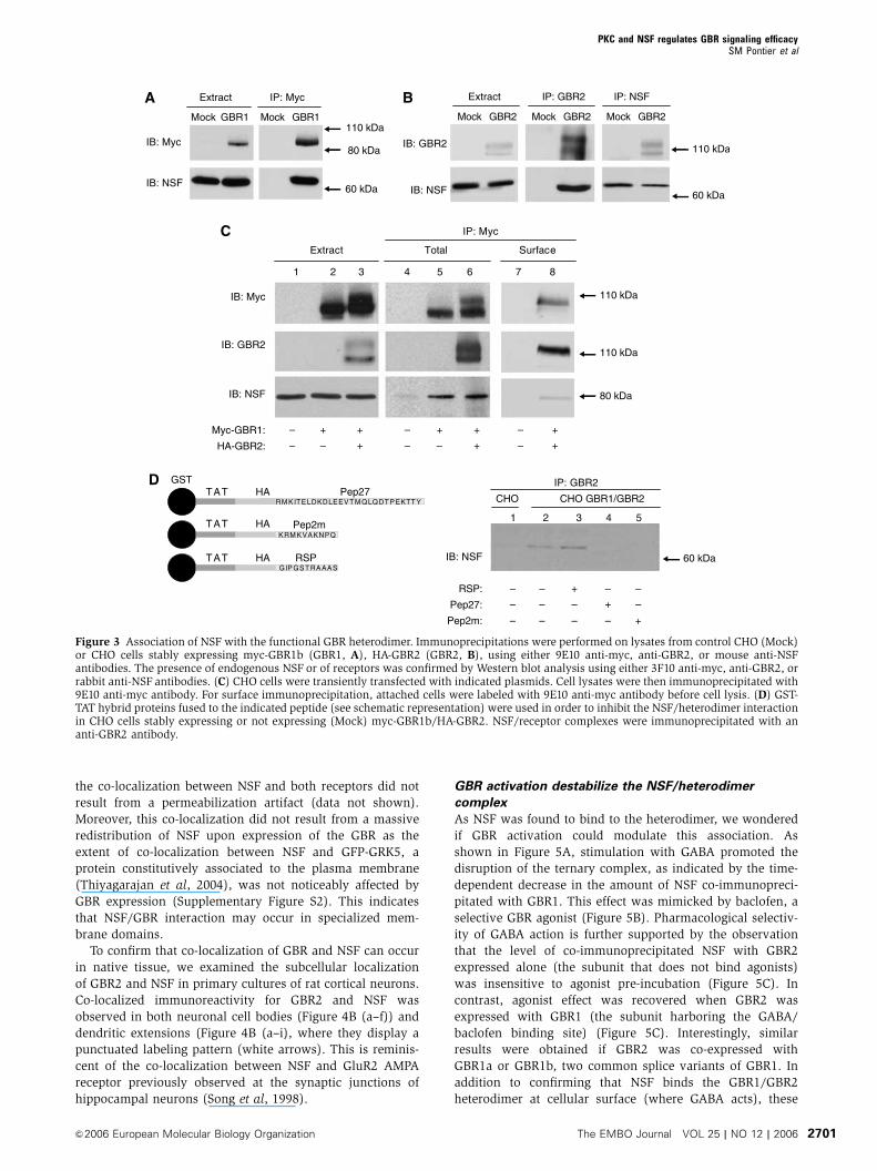
Figure 3 Association of NSF with the functional GBR heterodimer. Immunoprecipitations were performed on lysates from control CHO (Mock)or CHO cells stably expressing myc-GBR1b (GBR1, A), HA-GBR2 (GBR2, B), using either 9E10 anti-myc, anti-GBR2, or mouse anti-NSFantibodies. The presence of endogenous NSF or of receptors was confirmed by Western blot analysis using either 3F10 anti-myc, anti-GBR2, orrabbit anti-NSF antibodies. (C) CHO cells were transiently transfected with indicated plasmids. Cell lysates were then immunoprecipitated with9E10 anti-myc antibody. For surface immunoprecipitation, attached cells were labeled with 9E10 anti-myc antibody before cell lysis. (D) GST-TAT hybrid proteins fused to the indicated peptide (see schematic representation) were used in order to inhibit the NSF/heterodimer interactionin CHO cells stably expressing or not expressing (Mock) myc-GBR1b/HA-GBR2. NSF/receptor complexes were immunoprecipitated with ananti-GBR2 antibody.
PKC and NSF regulates GBR signaling efficacySM Pontier et al
&2006 European Molecular Biology Organization The EMBO Journal VOL 25 | NO 12 | 2006 2701

results laid the foundation to explore the potential functional
implications of this interaction.
Preventing NSF binding preserves the GBR/G protein
coupling following chronic stimulation
The observation that NSF is released from GBR complex
following stimulation leads us to test whether NSF could be
implicated in the regulation of the GABA-mediated G protein
activation of the receptor. Given the ability of native GBR to
interact with NSF in rat brain, quantification of the receptor-
promoted GDP/GTP exchange was assessed by GTPg[35S]
binding assays in both CHO cells and hippocampal slices (a
tissue known to endogenously express functional GBR
(Lopez-Bendito et al, 2004)). Treatment of hippocampal slices
with TAT-Pep27 (to impair the GBR/NSF interaction) or the
corresponding TAT-RSP control protein did not alter the
ability of the receptor to promote GTPgS35 binding. Indeed,
the peptides were without effect on either baclofen maximal
efficacy or EC50 to stimulate GTPgS35 binding (Table I).
Previous studies demonstrated that GBR G protein coupling
activity wanes over time, following sustained agonist stimu-
lation in CHO cells, cerebellar granule, or rat hippocampal
cells (Couve et al, 2002; Gonzalez-Maeso et al, 2003; Perroy
et al, 2003; Tosetti et al, 2004). Consistent with these findings,
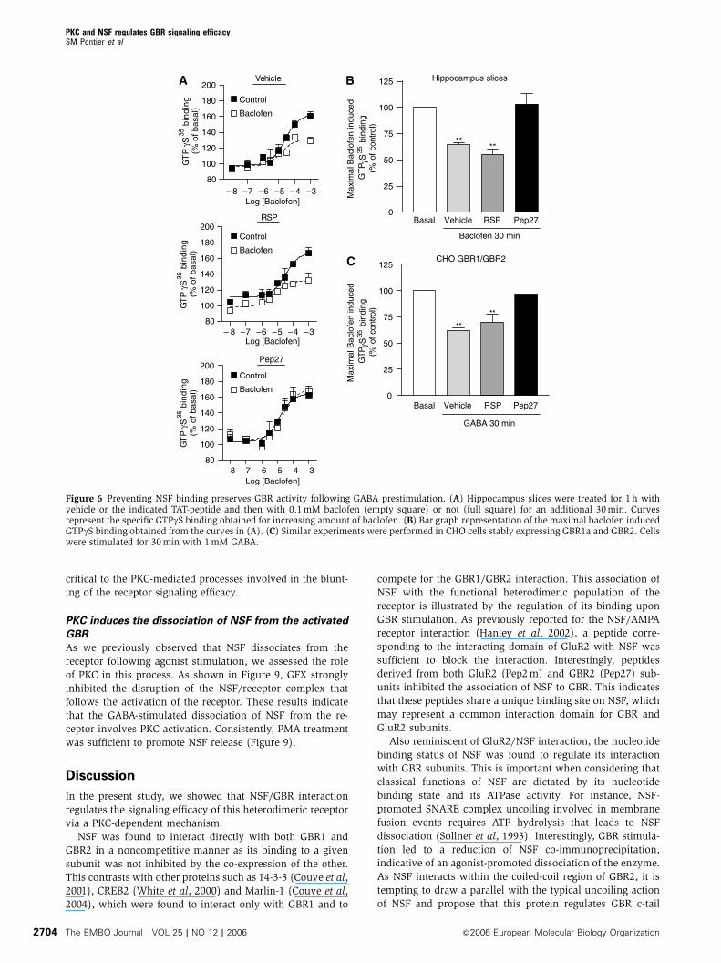
we observed that a 30-min prestimulation with baclofen led
to a 3672% reduction of the maximal baclofen-stimulated
GTPgS binding (Figure 6A and B) in hippocampal slices.
Treatment with TAT-Pep27 completely blocked this baclo-
fen-promoted desensitization (�379%), whereas TAT-RSP
was without effect (4575%; Figure 6A and B). These results,
which indicate a role of NSF in the agonist-promoted desen-
sitization of GBR in hippocampus, were recapitulated in CHO
cells (Figure 6C) where the 3978% desensitization promoted
by a 30 min pretreatment with GABA was also blocked by
TAT-Pep27 (874%). As NSF was previously implicated in the
membrane sorting of GluR2 and b2AR (Cong et al, 2001; Shi
et al, 2001; Hanley et al, 2002), we assessed whether receptor
prestimulation with GABA affected cell surface expression of
GBR1 or GBR2 in these cells. As recently reported in hippo-
campal neurons (Fairfax et al, 2004), ELISA analysis revealed
that the cell surface receptor density was stable in the
presence of its ligand (Supplementary Figure S2). NSF, how-
ever, has previously been shown to modulate both forward
trafficking and endocytosis of membrane proteins (Cong et al,
2001; Shi et al, 2001; Hanley et al, 2002; Lee et al, 2004).
Therefore, as NSF was found to interact with GBR immature
species, one cannot formally exclude the possibility that NSF
affects both endocytosis and insertion of de novo synthesized
receptors such that the steady-state concentration of GBR at
the cell surface remained unaffected. This is however un-
likely given the lack of surface labeled GBR internalization
following a 30 min agonist stimulation in CHO cells (data not
shown); a behavior also observed in HEK293 cells (Perroy
et al, 2003), COS cells, and hippocampal neurons (Fairfax
et al, 2004). It follows that, in the absence of agonist-
promoted internalization, insertion of de novo synthesized
receptors should lead to an increase in the steady-state
receptor level detected by ELISA. As this was not the case,
the above results suggest that the role that NSF could play in
GBR forward trafficking does probably not impact on the
short-term events contributing to rapid desensitization.
The agonist-promoted desensitization of GBR
is a PKC-dependent mechanism
Given the proposed role of PKC in the regulation of GBR
signaling efficacy in rat hippocampus (Dutar and Nicoll,
1988; Thompson and Gahwiler, 1992; Tosetti et al, 2004),
we investigated the contribution of this kinase in the NSF-
mediated desensitization of the receptor in CHO cells. First,
NSFMyc-GBR1 HA-GBR2
CHO Myc-GBR1/HA-GBR2
Overlay
Myc-GBR1+ HA-GBR2
Myc-GBR1+ NSF
HA-GBR2+ NSF
A
a b c
a b c
d e f
g h i
d e f
GBR2 NSF OverlayB
Figure 4 NSF co-localizes with both GBR subunits at the plasmamembrane. (A) CHO cells stably expressing myc-GBR1b/HA-GBR2were used. Myc-GBR1b, GBR2 and NSF were respectively labeledfor immunofluorescence experiments with the secondary antibodiescoupled to Alexa633 (Blue) (a), Oregon green (b) and Texas red (c)fluorophore respectively. GBR1 and GBR2 were surface labeledbefore cell fixation and permeabilization was then carried out inorder to mark NSF. Overlay panels (d, e and f) correspond to thesuperposition of the a-b, a-c and b-c images, respectively. (B) GBR2(green, a, d and g) and NSF (red, b, e and h) co-localizations (c, fand i) were determined in primary cultures of cortical neurons.Arrows indicate synaptic structures where both proteins are co-localized. White bars represent the scale of the image (20mm).
PKC and NSF regulates GBR signaling efficacySM Pontier et al
The EMBO Journal VOL 25 | NO 12 | 2006 &2006 European Molecular Biology Organization2702

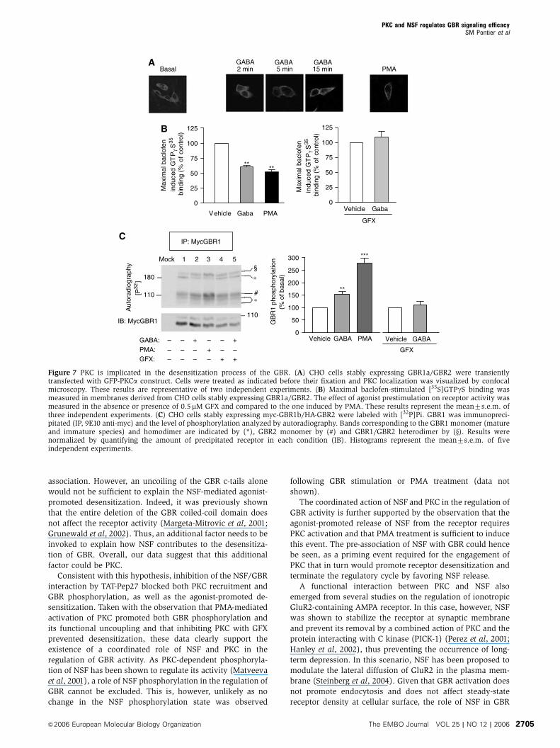
we studied the ability of GABA to promote PKC plasma
membrane translocation in cells expressing a GFP-tagged
PKCa (GFP-PKC) construct. In agreement with what was
previously observed in hippocampal neurons (Tremblay
et al, 1995), treatment with GABA induced membrane re-
cruitment of GFP-PKC (Figure 7A), reaching its maximum at
5 min. This translocation was comparable to the one pro-
moted by phorbol 12-myristate 13-acetate (PMA), a molecule
directly activating PKC (Blumberg, 1991). Inhibiting PKC
activation with GFX completely blocked the GABA-promoted
attenuation of baclofen-stimulated GTPgS binding (Figure 7B,
right panel), suggesting a role for this protein in the desensi-
tization process. Consistent with such a role, direct PKC
activation with PMA reduced the maximal baclofen response
by 4873%, thus mimicking the desensitizing effects of GABA
(4073%; Figure 7B). Also, consistent with a role for PKC is
the observation that the GABA-promoted phosphorylation
of GBR1 (15479%; Figure 7C) and GBR2 (data not shown)
was blocked by GFX whereas PMA strongly stimulated it
(278719%). Interestingly, PKC recruitment occurred conco-
mitantly with the release of NSF from the receptor (50% of
NSF being released from GBR following 5 min of stimulation,
a time at which the recruitment of PKC is maximal; Figure 5),
thus suggesting that these two events act in a coordinated
manner to promote agonist-mediated desensitization of GBR.
NSF primes GBR for its PKC-dependent desensitization
To further explore the PKC/NSF link in the agonist-promoted
desensitization process, we assessed the influence of the
NSF/GBR interaction blocking peptide, TAT-Pep27, on the
GABA-promoted recruitment of PKC and the ensuing phos-
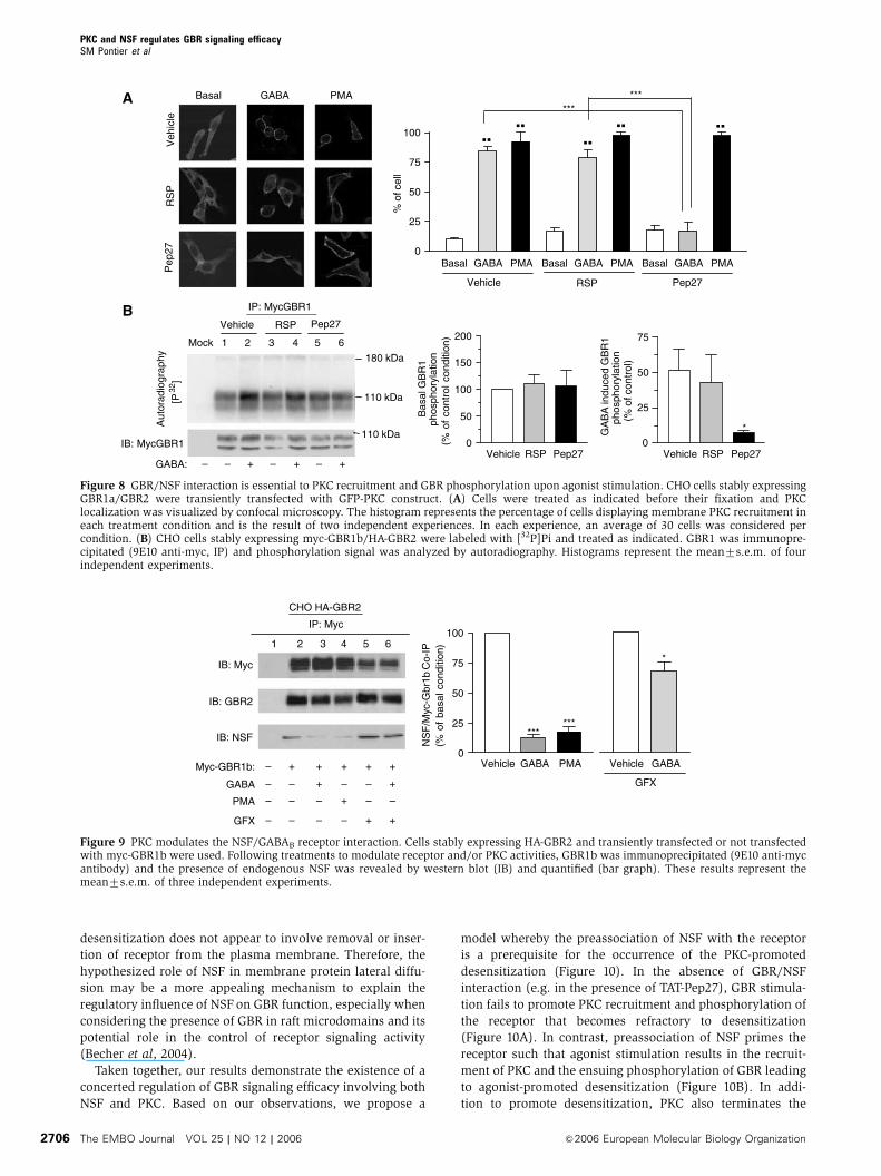
phorylation of GBR. As observed in Figure 8A, pretreatment
with TAT-Pep27 completely abolished the GABA-induced re-
cruitment of GFP-PKC to the plasma membrane. Consistent
with these results, TAT-Pep27 also prevented the agonist-
dependent increase in GBR phosphorylation (Figure 8B).
Overall, these results indicate that NSF/GBR interaction is
A
B C
GABA 1 mM (min) Extract
0 60 0 5 15 60
IB: GBR2
IB: NSF
IP: Myc
Myc-GBR1b:
CHO GBR2
– + ++ ++– –
IP: GBR2
IB: GBR1
IB: GBR2
IB: NSF
GBR2 GBR1a/2 GBR1b/2–
CHO
+– –GABA +– +–
110b kDa
110 kDa
80 kDa
IB: NSF
IP: Myc
Myc-GBR1b:
CHO GBR2
BaclofenGABA
– +– –+ –– –
+ + +–
80 kDa
110 kDa
110 kDa
80 kDa
IB: Myc
∗
0 5 15 30 60GABA 1 mM (min)
100
75
50
25
0NS
F/M
yc-G
br1b
CoI
P
Figure 5 GABA stimulation disrupts the NSF/GBR complex. (A) CHO cells stably expressing HA-GBR2 were transfected or not transfected withmyc-GBR1b and were treated with GABA for the indicated times. Following immunoprecipitation with 9E10 anti-myc, the amount ofprecipitated NSF was revealed by Western blot and quantified (bar graph). These results represent the means7s.e.m. of three to fiveexperiments performed independently. (B) Cells were treated either with 1 mM GABA or with 0.1 mM baclofen for 30 min. In (C) control CHOcells (�) or CHO cells stably expressing HA-GBR2 alone (GBR2), GBR1a/GBR2 or myc-GBR1b/HA-GBR2 (GBR1b/GBR2) were used and treatedor not treated with GABA 1 mM for 30 min. GBR2 was immunoprecipitated and the presence of endogenous NSF and receptors revealed byWestern blot.
Table I Functional characterization of native hippocampal GBR following treatment with TAT-peptides
Condition Basal GTPgS35 binding (% of control) Maximal GTPgS35 binding (% of control) EC50 (M) n R2
Vehicle 100 10077 2.48 10�5 3 0.909RSP 10578 85,576 2.03 10�5 3 0.909Pep27 103718 9679 1.3 10�5 3 0.857
Rat hippocampal slices were treated for 1 h with the indicated TAT-peptide and baclofen-stimulated [35S]GTPgS binding was measured inmembranes derived from these slices expressing native GBR. The results were expressed in percentage of the vehicle condition for basal andmaximal GTPgS binding. EC50 was derived from dose–response curves. These results are the mean7s.e.m. of three independent experimentsperformed in triplicate.
PKC and NSF regulates GBR signaling efficacySM Pontier et al
&2006 European Molecular Biology Organization The EMBO Journal VOL 25 | NO 12 | 2006 2703
critical to the PKC-mediated processes involved in the blunt-
ing of the receptor signaling efficacy.
PKC induces the dissociation of NSF from the activated
GBR
As we previously observed that NSF dissociates from the
receptor following agonist stimulation, we assessed the role
of PKC in this process. As shown in Figure 9, GFX strongly
inhibited the disruption of the NSF/receptor complex that
follows the activation of the receptor. These results indicate
that the GABA-stimulated dissociation of NSF from the re-
ceptor involves PKC activation. Consistently, PMA treatment
was sufficient to promote NSF release (Figure 9).
Discussion
In the present study, we showed that NSF/GBR interaction
regulates the signaling efficacy of this heterodimeric receptor
via a PKC-dependent mechanism.
NSF was found to interact directly with both GBR1 and
GBR2 in a noncompetitive manner as its binding to a given
subunit was not inhibited by the co-expression of the other.
This contrasts with other proteins such as 14-3-3 (Couve et al,
2001), CREB2 (White et al, 2000) and Marlin-1 (Couve et al,
2004), which were found to interact only with GBR1 and to
compete for the GBR1/GBR2 interaction. This association of
NSF with the functional heterodimeric population of the
receptor is illustrated by the regulation of its binding upon
GBR stimulation. As previously reported for the NSF/AMPA
receptor interaction (Hanley et al, 2002), a peptide corre-
sponding to the interacting domain of GluR2 with NSF was
sufficient to block the interaction. Interestingly, peptides
derived from both GluR2 (Pep2 m) and GBR2 (Pep27) sub-
units inhibited the association of NSF to GBR. This indicates
that these peptides share a unique binding site on NSF, which
may represent a common interaction domain for GBR and
GluR2 subunits.
Also reminiscent of GluR2/NSF interaction, the nucleotide
binding status of NSF was found to regulate its interaction
with GBR subunits. This is important when considering that
classical functions of NSF are dictated by its nucleotide
binding state and its ATPase activity. For instance, NSF-
promoted SNARE complex uncoiling involved in membrane
fusion events requires ATP hydrolysis that leads to NSF
dissociation (Sollner et al, 1993). Interestingly, GBR stimula-
tion led to a reduction of NSF co-immunoprecipitation,
indicative of an agonist-promoted dissociation of the enzyme.
As NSF interacts within the coiled-coil region of GBR2, it is
tempting to draw a parallel with the typical uncoiling action
of NSF and propose that this protein regulates GBR c-tail
80
100
120
140
160
180
200
– 8 –7 –6 –5 –4 –3
– 8 –7 –6 –5 –4 –3
– 8 –7 –6 –5 –4 –3
80
100
120
140
160
180
200
Log [Baclofen]
Log [Baclofen]
Log [Baclofen]
GT
Pγ S
35 b
ind
ing
(% o
f bas
al)
GT
Pγ S
35 b
ind
ing
(% o
f bas
al)
80
100
120
140
160
180
200
GT
Pγ S
35 b
ind
ing
(% o
f bas
al)
Vehicle
RSP
Control
Baclofen
A
Basal Vehicle RSP Pep270
25
50
75
100
125
**
**
Basal Vehicle RSP Pep270
25
50
75
100
125
****
Max
imal
Bac
lofe
n in
duce
dG
TPγS
35 b
indi
ng(%
of
cont
rol)
Baclofen 30 min
B
C
Hippocampus slices
CHO GBR1/GBR2
GABA 30 min
Control
Baclofen
Control
Baclofen
Pep27M
axim
al B
aclo
fen
indu
ced
GTP
γS35
bin
ding
(% o
f co
ntro
l)
Figure 6 Preventing NSF binding preserves GBR activity following GABA prestimulation. (A) Hippocampus slices were treated for 1 h withvehicle or the indicated TAT-peptide and then with 0.1 mM baclofen (empty square) or not (full square) for an additional 30 min. Curvesrepresent the specific GTPgS binding obtained for increasing amount of baclofen. (B) Bar graph representation of the maximal baclofen inducedGTPgS binding obtained from the curves in (A). (C) Similar experiments were performed in CHO cells stably expressing GBR1a and GBR2. Cellswere stimulated for 30 min with 1 mM GABA.
PKC and NSF regulates GBR signaling efficacySM Pontier et al
The EMBO Journal VOL 25 | NO 12 | 2006 &2006 European Molecular Biology Organization2704
association. However, an uncoiling of the GBR c-tails alone
would not be sufficient to explain the NSF-mediated agonist-
promoted desensitization. Indeed, it was previously shown
that the entire deletion of the GBR coiled-coil domain does
not affect the receptor activity (Margeta-Mitrovic et al, 2001;
Grunewald et al, 2002). Thus, an additional factor needs to be
invoked to explain how NSF contributes to the desensitiza-
tion of GBR. Overall, our data suggest that this additional
factor could be PKC.
Consistent with this hypothesis, inhibition of the NSF/GBR
interaction by TAT-Pep27 blocked both PKC recruitment and
GBR phosphorylation, as well as the agonist-promoted de-
sensitization. Taken with the observation that PMA-mediated
activation of PKC promoted both GBR phosphorylation and
its functional uncoupling and that inhibiting PKC with GFX
prevented desensitization, these data clearly support the
existence of a coordinated role of NSF and PKC in the
regulation of GBR activity. As PKC-dependent phosphoryla-
tion of NSF has been shown to regulate its activity (Matveeva
et al, 2001), a role of NSF phosphorylation in the regulation of
GBR cannot be excluded. This is, however, unlikely as no
change in the NSF phosphorylation state was observed
following GBR stimulation or PMA treatment (data not
shown).
The coordinated action of NSF and PKC in the regulation of
GBR activity is further supported by the observation that the
agonist-promoted release of NSF from the receptor requires
PKC activation and that PMA treatment is sufficient to induce
this event. The pre-association of NSF with GBR could hence
be seen, as a priming event required for the engagement of
PKC that in turn would promote receptor desensitization and
terminate the regulatory cycle by favoring NSF release.
A functional interaction between PKC and NSF also
emerged from several studies on the regulation of ionotropic
GluR2-containing AMPA receptor. In this case, however, NSF
was shown to stabilize the receptor at synaptic membrane
and prevent its removal by a combined action of PKC and the
protein interacting with C kinase (PICK-1) (Perez et al, 2001;
Hanley et al, 2002), thus preventing the occurrence of long-
term depression. In this scenario, NSF has been proposed to
modulate the lateral diffusion of GluR2 in the plasma mem-
brane (Steinberg et al, 2004). Given that GBR activation does
not promote endocytosis and does not affect steady-state
receptor density at cellular surface, the role of NSF in GBR
B
A
C
BasalGABA2 min
GABA5 min
GABA15 min PMA
GFXVehicle Gaba PMA
0
25
50
75
100
125
****
Vehicle Gaba0
25
50
75
100
125
Max
imal
bac
lofe
nin
duce
d G
TP
γS
35
bind
ing
(% o
f co
ntro
l)
Max
imal
bac
lofe
nin
duce
d G
TP
γS
35
bind
ing
(% o
f co
ntro
l)
Vehicle GABA PMA0
50
100
150
200
250
300
**
***
GB
R1
phos
phor
ylat
ion
(% o
f bas
al)180
IB: MycGBR1
Aut
orad
iogr
aphy
[P]
32
IP: MycGBR1
110
110
Mock 1 2 3 4 5
GABA: – + – – +
PMA: – – + – –GFX: –
–
–
– – – + +
§
∗
∗#
GFX
Vehicle GABA
Figure 7 PKC is implicated in the desensitization process of the GBR. (A) CHO cells stably expressing GBR1a/GBR2 were transientlytransfected with GFP-PKCa construct. Cells were treated as indicated before their fixation and PKC localization was visualized by confocalmicroscopy. These results are representative of two independent experiments. (B) Maximal baclofen-stimulated [35S]GTPgS binding wasmeasured in membranes derived from CHO cells stably expressing GBR1a/GBR2. The effect of agonist prestimulation on receptor activity wasmeasured in the absence or presence of 0.5mM GFX and compared to the one induced by PMA. These results represent the mean7s.e.m. ofthree independent experiments. (C) CHO cells stably expressing myc-GBR1b/HA-GBR2 were labeled with [32P]Pi. GBR1 was immunopreci-pitated (IP, 9E10 anti-myc) and the level of phosphorylation analyzed by autoradiography. Bands corresponding to the GBR1 monomer (matureand immature species) and homodimer are indicated by (*), GBR2 monomer by (#) and GBR1/GBR2 heterodimer by (y). Results werenormalized by quantifying the amount of precipitated receptor in each condition (IB). Histograms represent the mean7s.e.m. of fiveindependent experiments.
PKC and NSF regulates GBR signaling efficacySM Pontier et al
&2006 European Molecular Biology Organization The EMBO Journal VOL 25 | NO 12 | 2006 2705
desensitization does not appear to involve removal or inser-
tion of receptor from the plasma membrane. Therefore, the
hypothesized role of NSF in membrane protein lateral diffu-
sion may be a more appealing mechanism to explain the
regulatory influence of NSF on GBR function, especially when
considering the presence of GBR in raft microdomains and its
potential role in the control of receptor signaling activity
(Becher et al, 2004).
Taken together, our results demonstrate the existence of a
concerted regulation of GBR signaling efficacy involving both
NSF and PKC. Based on our observations, we propose a
model whereby the preassociation of NSF with the receptor
is a prerequisite for the occurrence of the PKC-promoted
desensitization (Figure 10). In the absence of GBR/NSF
interaction (e.g. in the presence of TAT-Pep27), GBR stimula-
tion fails to promote PKC recruitment and phosphorylation of
the receptor that becomes refractory to desensitization
(Figure 10A). In contrast, preassociation of NSF primes the
receptor such that agonist stimulation results in the recruit-
ment of PKC and the ensuing phosphorylation of GBR leading
to agonist-promoted desensitization (Figure 10B). In addi-
tion to promote desensitization, PKC also terminates the
A Basal GABA PMA
Veh
icle
RS
PP
ep27
B
180 kDa
110 kDa
110 kDa
Mock 1 2 3 4 5 6
Vehicle RSP Pep27
GABA: – – + – + – +
IB: MycGBR1
Aut
orad
iogr
aphy
[P32
]
IP: MycGBR1
Vehicle RSP Pep270
50
100
150
200
Bas
al G
BR
1ph
osph
oryl
atio
n(%
of
cont
rol c
ondi
tion)
Vehicle RSP Pep270
25
50
75
*GA
BA
indu
ced
GB
R1
phos
phor
ylat
ion
(% o
f con
trol
)
Basal GABA PMA Basal GABA PMA Basal GABA PMA0
25
50
75
100
% o
f cel
l
******
Vehicle RSP Pep27
Figure 8 GBR/NSF interaction is essential to PKC recruitment and GBR phosphorylation upon agonist stimulation. CHO cells stably expressingGBR1a/GBR2 were transiently transfected with GFP-PKC construct. (A) Cells were treated as indicated before their fixation and PKClocalization was visualized by confocal microscopy. The histogram represents the percentage of cells displaying membrane PKC recruitment ineach treatment condition and is the result of two independent experiences. In each experience, an average of 30 cells was considered percondition. (B) CHO cells stably expressing myc-GBR1b/HA-GBR2 were labeled with [32P]Pi and treated as indicated. GBR1 was immunopre-cipitated (9E10 anti-myc, IP) and phosphorylation signal was analyzed by autoradiography. Histograms represent the mean7s.e.m. of fourindependent experiments.
Vehicle GABA PMA0
25
50
75
100
******
NS
F/M
yc-G
br1b
Co-
IP(%
of
basa
l co
nditi
on)
IP: Myc
1 2 3 4 5 6
IB: Myc
IB: GBR2
IB: NSF
CHO HA-GBR2
Myc-GBR1b: –
–
+ + +
– +– +
+ +
– + –
–
–– –
– – + +– –
GABA
PMA
GFX
GFX
Vehicle GABA
*
Figure 9 PKC modulates the NSF/GABAB receptor interaction. Cells stably expressing HA-GBR2 and transiently transfected or not transfectedwith myc-GBR1b were used. Following treatments to modulate receptor and/or PKC activities, GBR1b was immunoprecipitated (9E10 anti-mycantibody) and the presence of endogenous NSF was revealed by western blot (IB) and quantified (bar graph). These results represent themean7s.e.m. of three independent experiments.
PKC and NSF regulates GBR signaling efficacySM Pontier et al
The EMBO Journal VOL 25 | NO 12 | 2006 &2006 European Molecular Biology Organization2706

regulatory process by favoring NSF dissociation from the
heterodimer (Figure 10C). Although the precise mechanism
by which NSF/GBR association primes the desensitization
process cannot be firmly established, two nonmutually ex-
clusive mechanisms can be imagined: (1) through its uncoil-
ing activity, NSF could unmask phosphorylation sites that are
implicated in desensitization; (2) the presence of NSF could
favor the engagement of a signaling pathway required for
PKC activation. Also remaining to be established is whether
NSF dissociation occurs before or after the completion of the
PKC-dependent desensitization and/or contributes to the
regulation of signaling efficacy.
The role of NSF and PKC in the desensitization of GBR
should also be placed in the context of other mechanisms that
have been shown to contribute to this process. For instance, a
phosphorylation-independent contribution of GRK4 (Perroy
et al, 2003) as well as a negative influence of PKA-mediated
phosphorylation of GBR (Couve et al, 2002) on its desensiti-
zation have been reported. The relative contribution of these
various mechanisms most likely depends on the relative
expression levels of the different regulatory proteins.
Noticeably, no GRK4 expression could be detected in CHO
cells, suggesting that the NSF/PKC may represent the domi-
nant mechanism in these cells. This process also appear to be
relevant for GBR desensitization in at least some native
tissues as the NSF blocking peptide TAT-Pep27 abolished
agonist-promoted desensitization in hippocampal slices.
Interestingly, the GBR desensitization previously observed
in rat hippocampal slices was shown to concern pre-synaptic
GBR that regulates neurotransmitter release (Tosetti et al,
2004). Taken with the role of both NSF and PKC in the
regulation of synaptic exocytosis (Lin and Scheller, 2000;
Barclay et al, 2003), our demonstration of their implication
in GBR desensitization raises the intriguing possibility of the
existence of an integrated process controlling the GBR-
mediated inhibition of neurotransmitter release.
Materials and methods
A detailed description of the different materials and plasmids usedin this manuscript may be found in Supplementary data.
YTHThis protocol has already been described (White et al, 2000), buta detailed description can be found in Supplementary data.
In vitro protein interaction assayProtocols to purify the different proteins and describing the assayare detailed in the Supplementary methods appended to thismanuscript.
Cell culture and rat hippocampal slice preparationThe protocols are described in detail in the Supplementary datasection.
Cell treatmentsTreatments were performed at 371C on CHO cells or at RT forhippocampal slices. Prestimulations of cells with 1 mM GABA or0.1 mM baclofen were performed for 30 min when not specified orfor the indicated time. To demonstrate the role of PKC, cells wereincubated with vehicle or 0.5mM GFX for 30 min before theprestimulation with GABA or with 1 mM PMA for 10 min. To inhibit
Figure 10 Coordinated action of NSF and PKC regulates GABAB receptor signaling efficacy. (a) In the presence of TAT-Pep27, NSF dissociatesfrom GBR and agonist-stimulation fails to promote desensitization. (b) Preassociation of NSF, however, primes the receptor such that agoniststimulation results in the recruitment of PKC and the ensuing phosphorylation of GBR leading to agonist-promoted desensitization. (c) PKCterminates the regulatory process by favoring NSF dissociation from the heterodimer.
PKC and NSF regulates GBR signaling efficacySM Pontier et al
&2006 European Molecular Biology Organization The EMBO Journal VOL 25 | NO 12 | 2006 2707

NSF/GBR interaction, cells were incubated with TAT-Pep27 400 nMor TAT-Pep2 m or RSP 800 nM, for 1 h before any treatment.
ImmunoprecipitationThe protocol is described in detail in the Supplementary datasection.
Whole phosphorylation assayThis was performed previously described (Perroy et al, 2003), butsee details in Supplementary methods.
[35S]GTPcS binding assayThis protocol has been performed as previously described (Perroyet al, 2003) and details can be found in Supplementary materials.
ImmunofluorescenceThe protocol is described in detailed in the Supplementary datasection.
Mathematical and statistical analysisFor GTPgS binding, dose–response curve experiments wereanalyzed by nonlinear regression using Prism program (GraphPadsoftware, San Diego, CA) (Figure 6A). For other GTPgS bindingstudies, basal GTPgS binding obtained without stimulation was
subtracted to the maximal GTPgS binding obtained in the presenceof 0.1 mM baclofen. Every condition was expressed in thepercentage of the corresponding control condition. The statisticalsignificance of results obtained in GTPgS binding, co-immunopre-cipitation, or PKC recruitment experiments was determined usinga one-way ANOVA analysis followed by a Bonferroni’s multiplecomparison test. Statistical significances between the controlcondition and the condition of interest are represented as follows:* when Po0.05, ** when Po0.01 and *** when Po0.001.
Supplementary dataSupplementary data are available at The EMBO Journal Online.
Acknowledgements
We are grateful to Monique Lagace for her constant fruitful discus-sion and her careful reading of the manuscript. We are also gratefulto Billy Breton for the kind gift of the GFP-GRK5 plasmid. This workwas supported by a CIHR/Rx&D Grant and sponsored byGlaxoSmithKline. NL holds a CIHR studentship. MB is the recipientof the Canada Research Chair in Signal Transduction and MolecularPharmacology.
References
Barclay JW, Craig TJ, Fisher RJ, Ciufo LF, Evans GJ, Morgan A,Burgoyne RD (2003) Phosphorylation of Munc18 by proteinkinase C regulates the kinetics of exocytosis. J Biol Chem 278:10538–10545
Becher A, Green A, Ige AO, Wise A, White JH, McIlhinney RA(2004) Ectopically expressed gamma-aminobutyric acid receptorB is functionally down-regulated in isolated lipid raft-enrichedmembranes. Biochem Biophys Res Commun 321: 981–987
Becker-Hapak M, McAllister SS, Dowdy SF (2001) TAT-mediatedprotein transduction into mammalian cells. Methods 24: 247–256
Blumberg PM (1991) Complexities of the protein kinase C pathway.Mol Carcinog 4: 339–344
Bomberger JM, Parameswaran N, Hall CS, Aiyar N, Spielman WS(2005) Novel function for receptor activity-modifying proteins(RAMPs) in post-endocytic receptor trafficking. J Biol Chem 280:9297–9307
Cong M, Perry SJ, Hu LA, Hanson PI, Claing A, Lefkowitz RJ (2001)Binding of the beta2 adrenergic receptor to N-ethylmaleimide-sensitive factor regulates receptor recycling. J Biol Chem 276:45145–45152
Couve A, Filippov AK, Connolly CN, Bettler B, Brown DA, Moss SJ(1998) Intracellular retention of recombinant GABAB receptors.J Biol Chem 273: 26361–26367
Couve A, Kittler JT, Uren JM, Calver AR, Pangalos MN, Walsh FS,Moss SJ (2001) Association of GABA(B) receptors and membersof the 14-3-3 family of signaling proteins. Mol Cell Neurosci 17:317–328
Couve A, Restituito S, Brandon JM, Charles KJ, Bawagan H,Freeman KB, Pangalos MN, Calver AR, Moss SJ (2004) Marlin-1, a novel RNA-binding protein associates with GABA receptors.J Biol Chem 279: 13934–13943
Couve A, Thomas P, Calver AR, Hirst WD, Pangalos MN, Walsh FS,Smart TG, Moss SJ (2002) Cyclic AMP-dependent protein kinasephosphorylation facilitates GABA(B) receptor-effector coupling.Nat Neurosci 5: 415–424
Dutar P, Nicoll RA (1988) Pre- and postsynaptic GABAB receptors inthe hippocampus have different pharmacological properties.Neuron 1: 585–591
Fairfax BP, Pitcher JA, Scott MG, Calver AR, Pangalos MN, Moss SJ,Couve A (2004) Phosphorylation and chronic agonist treatmentatypically modulate GABAB receptor cell surface stability. J BiolChem 279: 12565–12573
Galvez T, Duthey B, Kniazeff J, Blahos J, Rovelli G, Bettler B,Prezeau L, Pin JP (2001) Allosteric interactions between GB1and GB2 subunits are required for optimal GABA(B) receptorfunction. EMBO J 20: 2152–2159
Gonzalez-Maeso J, Wise A, Green A, Koenig JA (2003) Agonist-induced desensitization and endocytosis of heterodimeric GABABreceptors in CHO-K1 cells. Eur J Pharmacol 481: 15–23
Grunewald S, Schupp BJ, Ikeda SR, Kuner R, Steigerwald F, KornauHC, Kohr G (2002) Importance of the gamma-aminobutyricacid(B) receptor C-termini for G-protein coupling. MolPharmacol 61: 1070–1080
Hanley JG, Khatri L, Hanson PI, Ziff EB (2002) NSF ATPase andalpha-/beta-SNAPs disassemble the AMPA receptor-PICK1 com-plex. Neuron 34: 53–67
Heydorn A, Sondergaard BP, Hadrup N, Holst B, Haft CR, SchwartzTW (2004) Distinct in vitro interaction pattern of dopaminereceptor subtypes with adaptor proteins involved in post-endo-cytotic receptor targeting. FEBS Lett 556: 276–280
Jones KA, Borowsky B, Tamm JA, Craig DA, Durkin MM, Dai M, YaoWJ, Johnson M, Gunwaldsen C, Huang LY, Tang C, Shen Q, SalonJA, Morse K, Laz T, Smith KE, Nagarathnam D, Noble SA,Branchek TA, Gerald C (1998) GABA(B) receptors function asa heteromeric assembly of the subunits GABA(B)R1 andGABA(B)R2. Nature 396: 674–679
Kammerer RA, Frank S, Schulthess T, Landwehr R, Lustig A, Engel J(1999) Heterodimerization of a functional GABAB receptor ismediated by parallel coiled-coil alpha-helices. Biochemistry 38:13263–13269
Kaupmann K, Malitschek B, Schuler V, Heid J, Froestl W, Beck P,Mosbacher J, Bischoff S, Kulik A, Shigemoto R, Karschin A,Bettler B (1998) GABA(B)-receptor subtypes assemble into func-tional heteromeric complexes. Nature 396: 683–687
Kuner R, Kohr G, Grunewald S, Eisenhardt G, Bach A, Kornau HC(1999) Role of heteromer formation in GABAB receptor function.Science 283: 74–77
Lee SH, Simonetta A, Sheng M (2004) Subunit rules governing thesorting of internalized AMPA receptors in hippocampal neurons.Neuron 43: 221–236
Lin RC, Scheller RH (2000) Mechanisms of synaptic vesicleexocytosis. Annu Rev Cell Dev Biol 16: 19–49
Lopez-Bendito G, Shigemoto R, Kulik A, Vida I, Fairen A, Lujan R(2004) Distribution of metabotropic GABA receptor subunitsGABAB1a/b and GABAB2 in the rat hippocampus during prenataland postnatal development. Hippocampus 14: 836–848
Margeta-Mitrovic M, Jan YN, Jan LY (2000) A trafficking checkpointcontrols GABA(B) receptor heterodimerization. Neuron 27:97–106
Margeta-Mitrovic M, Jan YN, Jan LY (2001) Function of GB1 andGB2 subunits in G protein coupling of GABA(B) receptors. ProcNatl Acad Sci USA 98: 14649–14654
Matveeva EA, Whiteheart SW, Vanaman TC, Slevin JT (2001)Phosphorylation of the N-ethylmaleimide-sensitive factor is asso-ciated with depolarization-dependent neurotransmitter releasefrom synaptosomes. J Biol Chem 276: 12174–12181
Morgan A, Burgoyne RD (2004) Membrane traffic: controllingmembrane fusion by modifying NSF. Curr Biol 14: R968–R970
PKC and NSF regulates GBR signaling efficacySM Pontier et al
The EMBO Journal VOL 25 | NO 12 | 2006 &2006 European Molecular Biology Organization2708

Nishimune A, Isaac JT, Molnar E, Noel J, Nash SR, Tagaya M,Collingridge GL, Nakanishi S, Henley JM (1998) NSF binding toGluR2 regulates synaptic transmission. Neuron 21: 87–97
Noel J, Ralph GS, Pickard L, Williams J, Molnar E, Uney JB,Collingridge GL, Henley JM (1999) Surface expression of AMPAreceptors in hippocampal neurons is regulated by an NSF-depen-dent mechanism. Neuron 23: 365–376
Osten P, Srivastava S, Inman GJ, Vilim FS, Khatri L, Lee LM, StatesBA, Einheber S, Milner TA, Hanson PI, Ziff EB (1998) The AMPAreceptor GluR2 C terminus can mediate a reversible, ATP-depen-dent interaction with NSF and a. Neuron 21: 99–110
Perez JL, Khatri L, Chang C, Srivastava S, Osten P, Ziff EB (2001)PICK1 targets activated protein kinase Calpha to AMPA receptorclusters in spines of hippocampal neurons and reduces surfacelevels of the AMPA-type glutamate receptor subunit 2. J Neurosci21: 5417–5428
Perroy J, Adam L, Qanbar R, Chenier S, Bouvier M (2003)Phosphorylation-independent desensitization of GABA(B)receptor by GRK4. EMBO J 22: 3816–3824
Robbins MJ, Calver AR, Filippov AK, Hirst WD, Russell RB, WoodMD, Nasir S, Couve A, Brown DA, Moss SJ, Pangalos MN (2001)GABA(B2) is essential for g-protein coupling of the GABA(B)receptor heterodimer. J Neurosci 21: 8043–8052
Sallese M, Salvatore L, D’Urbano E, Sala G, Storto M, Launey T,Nicoletti F, Knopfel T, De Blasi A (2000) The G-protein-coupledreceptor kinase GRK4 mediates homologous desensitization ofmetabotropic glutamate receptor 1. FASEB J 14: 2569–2580
Shi S, Hayashi Y, Esteban JA, Malinow R (2001) Subunit-specificrules governing AMPA receptor trafficking to synapses in hippo-campal pyramidal neurons. Cell 105: 331–343
Sollner T, Bennett MK, Whiteheart SW, Scheller RH, Rothman JE(1993) A protein assembly-disassembly pathway in vitro thatmay correspond to sequential steps of synaptic vesicle docking,activation, and fusion. Cell 75: 409–418
Song I, Kamboj S, Xia J, Dong H, Liao D, Huganir RL (1998)Interaction of the N-ethylmaleimide-sensitive factor with AMPAreceptors. Neuron 21: 393–400
Steinberg JP, Huganir RL, Linden DJ (2004) N-ethylmaleimide-sensitive factor is required for the synaptic incorporation and
removal of AMPA receptors during cerebellar long-term depres-sion. Proc Natl Acad Sci USA 101: 18212–18216
Thiyagarajan MM, Stracquatanio RP, Pronin AN, Evanko DS,Benovic JL, Wedegaertner PB (2004) A predicted amphipathichelix mediates plasma membrane localization of GRK5. J BiolChem 279: 17989–17995
Thompson SM, Gahwiler BH (1992) Comparison of the actions ofbaclofen at pre- and postsynaptic receptors in the rat hippocam-pus in vitro. J Physiol 451: 329–345
Tosetti P, Bakels R, Colin-Le Brun I, Ferrand N, Gaiarsa JL, CaillardO (2004) Acute desensitization of presynaptic GABAB-mediatedinhibition and induction of epileptiform discharges in the neona-tal rat hippocampus. Eur J Neurosci 19: 3227–3234
Tremblay E, Ben Ari Y, Roisin MP (1995) Different GABAB-mediatedeffects on protein kinase C activity and immunoreactivity inneonatal and adult rat hippocampal slices. J Neurochem 65:863–870
Virlon B, Firsov D, Cheval L, Reiter E, Troispoux C, Guillou F,Elalouf JM (1998) Rat G protein-coupled receptor kinase GRK4:identification, functional expression, and differential tissue dis-tribution of two splice variants. Endocrinology 139: 2784–2795
White JH, McIllhinney RA, Wise A, Ciruela F, Chan WY, Emson PC,Billinton A, Marshall FH (2000) The GABAB receptor interactsdirectly with the related transcription factors CREB2 andATFx. Proc Natl Acad Sci U S A 97: 13967–13972
White JH, Wise A, Main MJ, Green A, Fraser NJ, Disney GH, BarnesAA, Emson P, Foord SM, Marshall FH (1998) Heterodimerizationis required for the formation of a functional GABA(B) receptor.Nature 396: 679–682
White JH, Wise A, Marshall FH (2002) Heterodimerization ofgamma-aminobutyric acid B receptor subunits as revealed bythe yeast two-hybrid system. Methods 27: 301–310
Whiteheart SW, Matveeva EA (2004) Multiple binding proteinssuggest diverse functions for the N-ethylmaleimide sensitivefactor. J Struct Biol 146: 32–43
Zou S, Li L, Pei L, Vukusic B, Van Tol HH, Lee FJ, Wan Q, Liu F(2005) Protein-protein coupling/uncoupling enables dopamineD2 receptor regulation of AMPA receptor-mediated excitotoxicity.J Neurosci 25: 4385–4395
PKC and NSF regulates GBR signaling efficacySM Pontier et al
&2006 European Molecular Biology Organization The EMBO Journal VOL 25 | NO 12 | 2006 2709
Copyright © 2022 FDOKUMEN