Lactose intolerance: diagnosis, genetic, and clinical factors

Enhanced Interleukin-1 Activity Contributes to ExerciseIntolerance in Patients with Systolic Heart FailureBenjamin W. Van Tassell1,2*, Ross A. Arena3, Stefano Toldo2,4, Eleonora Mezzaroma1,2, Tania Azam5,
Ignacio M. Seropian1,2, Keyur Shah4, Justin Canada6, Norbert F. Voelkel2, Charles A. Dinarello5, Antonio
Abbate2,4
1 School of Pharmacy, Virginia Commonwealth University, Richmond, Virginia, United States of America, 2 Victoria Johnson Center, Virginia Commonwealth University,
Richmond, Virginia, United States of America, 3 Physical Therapy Program, University of New Mexico, Albuquerque, New Mexico, United States of America, 4 Virginia
Commonwealth University Pauley Heart Center, Virginia Commonwealth University, Richmond, Virginia, United States of America, 5 Department of Internal Medicine,
University of Colorado, Boulder, Colorado, United States of America, 6 Department of Physical Therapy, Virginia Commonwealth University, Richmond, Virginia, United
States of America
Abstract
Background: Heart failure (HF) is a complex clinical syndrome characterized by impaired cardiac function and poor exercisetolerance. Enhanced inflammation is associated with worsening outcomes in HF patients and may play a direct role indisease progression. Interleukin-1b (IL-1b) is a pro-inflammatory cytokine that becomes chronically elevated in HF andexerts putative negative inotropic effects.
Methods and Results: We developed a model of IL-1b-induced left ventricular (LV) dysfunction in healthy mice thatexhibited a 32% reduction in LV fractional shortening (P,0.001) and a 76% reduction in isoproterenol response (P,0.01) at4 hours following a single dose of IL-1b 3 mcg/kg. This phenotype was reproducible in mice injected with plasma from HFpatients and fully preventable by pretreatment with IL-1 receptor antagonist (anakinra). This led to the design and conductof a pilot clinical to test the effect of anakinra on cardiopulmonary exercise performance in patients with HF and evidence ofelevated inflammatory signaling (n = 7). The median peak oxygen consumption (VO2) improved from 12.3 [10.0, 15.2] to 15.1[13.7, 19.3] mL?kg–1?min–1 (P = 0.016 vs. baseline) and median ventilator efficiency (VE/VCO2 slope) improved from 28.1[22.8, 31.7] to 24.9 [22.9, 28.3] (P = 0.031 vs. baseline).
Conclusions: These findings suggest that IL-1b activity contributes to poor exercise tolerance in patients with systolic HFand identifies IL-1b blockade as a novel strategy for pharmacologic intervention.
Trial Registration: ClinicalTrials.gov NCT01300650
Citation: Van Tassell BW, Arena RA, Toldo S, Mezzaroma E, Azam T, et al. (2012) Enhanced Interleukin-1 Activity Contributes to Exercise Intolerance in Patientswith Systolic Heart Failure. PLoS ONE 7(3): e33438. doi:10.1371/journal.pone.0033438
Editor: Partha Mukhopadhyay, National Institutes of Health, United States of America
Received December 2, 2011; Accepted February 9, 2012; Published March 16, 2012
Copyright: � 2012 Van Tassell et al. This is an open-access article distributed under the terms of the Creative Commons Attribution License, which permitsunrestricted use, distribution, and reproduction in any medium, provided the original author and source are credited.
Funding: This project was supported by award number UL1RR031990 from the National Center for Research Resources [BVT]. This project also received supportfrom an American Heart Association Scientist Development Grant [AA]. The content is solely the responsibility of the authors and does not necessarily representthe official views of the National Center for Research Resources or the National Institutes of Health. The funders had no role in study design, data collection andanalysis, decision to publish, or preparation of the manuscript.
Competing Interests: AA is a PLoS ONE Editorial Board Member. However, his position does not alter the authors’ adherence to all the PLoS ONE policies onsharing data and materials.
* E-mail: [email protected]
Introduction
Heart failure (HF) is a complex clinical syndrome characterized by
dyspnea, fatigue, and poor exercise tolerance. [1–2] While contem-
porary HF treatments have slowed disease progression and improved
survival, the overall incidences of HF morbidity and mortality
continue to rise, suggesting that the current treatment paradigm still
misses one or more key pathophysiologic mechanisms. [1–2]
Among HF patients, a significant correlation exists between
declining functional class and increasing levels of inflammatory
cytokines. [3–4] Interleukin-1b (IL–1b) is the prototypal inflamma-
tory cytokine that acts as an acute phase reactant following tissue
injury (i.e. ischemia) and becomes persistently elevated in patients
with chronic HF. [5–7] Early investigations in septic cardiomyop-
athy identified IL-1b as a soluble ‘depressant factor’ in the sera of
these patients, producing a concentration-dependent depression of
myocyte contractility in vitro [7] Further studies identify a pathologic
role for IL-1b in ventricular remodeling, systolic dysfunction, and
cardiomyocyte death in both ischemic and non-ischemic models of
HF. [7–12] IL-1b has also been shown to affect b-adrenergic
receptor responsiveness in vitro, which may be a key determinant of
exercise capacity in HF. [13–14] Nevertheless, the precise
contribution of IL-1b to human HF has not been well established.
We therefore designed a bench-to-bedside approach to
determine the contribution of IL-1b in the development of cardiac
dysfunction and ultimately guide the use of IL-1b blockade in a
pilot study of patients with HF. We first conducted a dose ranging
study with IL-1b in healthy mice to induce LV dysfunction at rest
PLoS ONE | www.plosone.org 1 March 2012 | Volume 7 | Issue 3 | e33438

and impaired responsiveness to b-adrenergic receptor (b-AR)
stimulation. We then observed that mice injected with plasma
from HF patients exhibited a similar phenotype of impaired
systolic dysfunction that was completely prevented by pre-
treatment with recombinant IL-1 receptor antagonist (IL-1Ra).
These observations led to the current pilot study in human patients
with HF in which 2 weeks treatment with anakinra improved
cardiopulmonary exercise performance as measured by peak
oxygen consumption, ventilatory efficiency, and exercise time.
Methods
The protocol for this trial is available as supporting information;
see Protocol S1.
Mouse model of IL-1b-induced systolic dysfunctionAnimals. Adult outbreed Institute of Cancer Research (ICR)
male mice were purchased from Harlan Sprague-Dawley
(Indianapolis, IN). Mice were 30 – 35 g at the time of the study.
All protocols involving animals were reviewed and approved by
the Animal Care and Use Committee of Virginia Commonwealth
University. Each group included between 6–10 mice.
Dose response. Mice received intraperitoneal (IP) injection
with recombinant human IL-1b (Cell Sciences, Canton, MA) with
transthoracic echocardiography to measure left ventricular (LV)
fractional shortening at baseline, 4 hours, and 24 hours. IL-1b was
administered at doses of 1 ng, 10 ng, 100 ng, and 1000 ng
(0.03 mcg/kg, 0.3 mcg/kg, 3 mcg/kg, and 30 mcg/kg) diluted in
0.2 mL of normal saline (NaCl 0.9%).
Echocardiography. Mice underwent transthoracic echo-
cardiography under light anesthesia (pentobarbital 30–50 mg/kg
IP). The chest was shaved and the mice were placed supine on a
heating pad. Doppler echocardiography was performed with the
Vevo770 imaging system (VisualSonics Inc, Toronto, Ontario,
Canada) and a 30-MHz probe. The transducer was positioned on
the left anterior side of the chest. The heart was first imaged in the
2-dimensional mode in the short-axis view of the left ventricle. Then,
M-mode images were obtained at the level of the papillary muscles
below the mitral valve tip according to the American Society of
Echocardiography recommendations. [15] The M-mode cursor was
positioned perpendicular to the anterior and posterior wall to measure
the left ventricular (LV) end-diastolic diameter (LVEDD) and end-
systolic diameter (LVESD). LV fractional shortening (LVFS) was
calculated as follows: LVFS = (LVEDD–LVESD)/LVEDDx100. LV
ejection fraction (LVEF) was calculated with the Teichholz formula.
[15] Transmitral and left ventricle outflow tract pulsed Doppler flow
spectra were obtained from the apical view. Measurement of the
outflow tract flow was performed and isovolumetric contraction (ICT)
and relaxation (IRT) times and ejection time (ET) were measured. LV
outflow tract (LVOT) flow velocity–time integral (AoVTI) was also
determined, with LVOT measured as the cross-sectional area in the
parasternal long-axis view. These data were used to calculate the Tei
index (Tei index = ICT+IRT/ET). In humans, a higher Tei index is
associated with both systolic and diastolic dysfunction and worse
outcomes. [15] Velocity of circumferential shortening was calculated
as FS/ET, with ET divided by the square root of the preceding RR
interval to correct for heart rate. [16] All echocardiograms were
performed and interpreted jointly by 2 investigators (BVT and AA)
throughout all experiments.
Contractile reserve. Mice received a single IP injection of a
b adrenergic receptor agonist (isoproterenol, 0.01 mcg/kg;
SigmaAldrich, St. Louis, MO) to calculate contractile reserve.
Contractile reserve was defined as the maximum percent increase
in LVFS within 5 minutes after isoproterenol.
Effects of plasma from patients with HF injected intohealthy mice
We collected blood from four groups of patients based upon HF
symptoms and elevation of high sensitivity C-reactive protein
(hsCRP, a surrogate marker of IL-1b activity): healthy controls
(CTRL), ambulatory HF patients with low CRP (HF L-CRP),
ambulatory HF with high CRP (HF H-CRP), and patients
hospitalized for acute decompensated HF (ADHF). For inclusion
in this stage of the investigation, healthy controls were defined as
patients with no history of cardiac disease. Ambulatory HF
patients were defined as patients with a documented history of HF
with LV ejection fraction (LVEF) ,40% but without any recent
hospitalizations (previous 12 months) or recent changes in medi-
cations (previous 3 months). Patients with acute decompensated
HF were eligible if they had been admitted with a confirmed
diagnosis of acute decompensated HF within the previous 24
hours. Plasma samples from each group were injected into healthy
mice who underwent transthoracic echocardiography to measure
LV function at baseline and 4 hours. To neutralize the effects of
IL-1b on LV function, separate groups of mice received injections
of the same human plasma following pre-treatment with
recombinant human IL-1 receptor antagonist (anakinra, KineretH,
Biovitrum) 100 mg/kg IP administered 30 minutes prior to
injection of human plasma. Upon completion of echocardiography
at 4 hours, all groups of mice received an injection of isoproterenol
0.01 mcg/kg IP to evaluate contractile reserve.
Pilot study of IL-1b blockade in patients with stable HFStudy design. We conducted an open-label, single-arm, pilot-
study to evaluate the safety and feasibility of IL-1b blockade to
improve aerobic exercise performance in ambulatory patients with
HF and high hsCRP. The study was registered on ClinicalTrials.gov
(NCT01300650) and received an exemption for investigational new
drug use from the Food and Drug Administration according
to current federal regulations (Code of Federal Regulations,
312.2[b]). All human subjects research was conducted at Virginia
Commonwealth University. The study design and protocol received
approval from the VCU Institutional Review Board and all patients
provided written informed consent. Patients presenting to the VCU
Cardiology Clinic were screened for potential enrollment. Following
enrollment, patients completed a HF symptom questionnaire (Duke
Activity Status Index [DASI], see Appendix) [17] and underwent
baseline cardiopulmonary exercise testing (CPX). Patients then
received a 14-day supply of anakinra (100 mg subcutaneous
injection daily) accompanied by detailed instructions on drug
administration, dosing, and storage. Patients were then scheduled
for a repeat questionnaire and CPX upon completion of the
anakinra regimen.
Entry criteria. The inclusion criteria were age .18 years, a
diagnosis of HF, documented LVEF ,40%, and hsCRP .2 mg/L.
The exclusion criteria were recent changes (previous 3 months) in
HF maintenance medications (beta-blockers, angiotensin convert-
ing enzyme [ACE] inhibitors, aldosterone antagonists, vasodilators,
digoxin, diuretics); hospitalization for worsening HF or acute
decompensated HF within the previous 12 months; anticipated
need for cardiac resynchronization therapy (CRT) or automated-
implantable cardioverter defibrillator (AICD); angina or electro-
cardiograph (ECG) changes that limit maximum exertion during
CPX or baseline ECG changes that limit the ability to detect
ischemia (i.e. left bundle-branch block); recent (,14 days) use of
anti-inflammatory drugs (not including NSAIDs), chronic infla-
mmatory disorder (including but not limited to rheumatoid arthritis,
systemic lupus erythematosus), malignancy, active infection, or any
comorbidity limiting survival or ability to complete the study; severe
IL-1 Activity in Systolic Heart Failure
PLoS ONE | www.plosone.org 2 March 2012 | Volume 7 | Issue 3 | e33438

kidney dysfunction (eGFR ,30 mL/min); coagulopathy (INR
.1.5), thrombocytopenia (,50,000/mm3), or leukopenia
(absolute neutrophil count ,1,500/mm3); pregnancy (female
patients were required to take a urine pregnancy test); latex or
rubber allergy; or inability to give informed consent.
Cardiopulmonary Exercise Testing. A physician-
supervised maximal CPX was administered using a metabolic
cart that is interfaced with a treadmill (Vmax Encore, Viasys,
Yorba Linda, CA). A conservative ramping treadmill protocol was
used as described previously. [18] Prior to each test, the oxygen
and carbon dioxide sensors were calibrated using gases of known
oxygen, nitrogen, and carbon dioxide concentrations and the flow
sensor was also calibrated using a 3-Liter syringe. Subjects were
briefed regarding the protocol and were requested to exercise to
fatigue. 12-lead ECG monitoring were conducted at baseline,
throughout the test and into recovery. Blood pressure was
measured every two minutes using an automated exercise-
compatible device (Tango, SunTech Medical). In this technique,
expired gases were sampled using a mouthpiece-mounted sensor,
and analyzed to continuously measure oxygen consumption
(VO2), carbon dioxide production (VCO2) and minute
ventilation (VE). The highest 10-second average value for VO2
during the last 30 seconds of exercise defined the peak value (peak
VO2 in mL?kg–1?min–1). Ten second averaged VE and VCO2
data, from the initiation of exercise to peak, were input into
spreadsheet software (Microsoft Excel, Microsoft Corp., Seattle,
WA) to calculate the VE/VCO2 slope via least squares linear
regression (y = mx+b, m = slope). The oxygen uptake efficiency
slope (OUES) was determined using least-squares linear regression
analysis (oxygen consumption = a log10VE+b, with VO2 and VE
expressed in liters per minute) using spreadsheet software
(Microsoft Excel, Microsoft, Seattle, Washington). [19] All
exercise data were also used to calculate the OUES. American
Heart Association/American College of Cardiology guidelines for
exercise testing contraindications and termination criteria were
followed. [20]
Heart Failure Symptom Questionnaire. The Duke
Activity Status Index (DASI) questionnaire is a 12-question, yes/
no, instrument that allows for the calculation of perceived
functional capacity. [17] Each question describes a different
physical activity and asks the subjects if they feel they can perform
the task. The questions are weighted according to their degree of
physical exertion. The weighted values from the ‘‘yes’’ responses
are summed to produce a VO2 score (see Appendix). [17]
Blood collection. Blood was collected from patients at
screening and after anakinra treatment (immediately prior to
final CPX) for analysis of hsCRP, brain natriuretic peptide (BNP),
complete blood count, and inflammatory cytokines.
Cytokine assay. Freshly obtained blood was centrifuged at
20006g for 15 minutes followed by plasma separation and storage
at –80uC. Samples were analyzed with the MOSAIC Cytokine
Panel 1 (R and D Systems, Minneapolis, MN) using a QuanSys
Imager (Logan, UT).
Statistical Analysis. Values derived from the experimental
animal studies are presented as mean and standard error of mean.
The differences between mouse treatment groups were compared
using the Student’s T-test for unpaired data when comparing 2
groups or using the ANOVA when comparing 3 or more groups.
Figure 1. Model of IL-1-induced systolic dysfunction in healthy mice. Healthy, adult, mice underwent baseline echocardiography followedby a single intraperitoneal injection of recombinant human IL-1b (0, 0.03, 0.3, 3, or 30 mcg/kg) and subsequent echocardiography at 4 hours. (A) Alldoses of IL-1b $0.3 mcg/kg produced a significant 28 – 32% reduction in left ventricular fractional shortening (LVFS) at 4 hours. Panels B (baseline)and C (4 hours) show representative echocardiographic images at 4 hours of mice injected with 3 mcg/kg IL-1b. Panels D – F show additionalmeasures of LV function at 4 hours: (D) LV ejection fraction (LVEF); (E) FS/myocardial performance index (MPI) or FS-Tei index; (F) velocity ofcircumferential fiber shortening (Vcfc) corrected for heart rate. In all experiments, pre-treatment with anakinra 100 mg/kg 30 minutes prior to IL-1bwas sufficient to prevent changes in LV function. (G) Isoproterenol 0.01 mcg/kg induced a reproducible 46% increase in LVFS that was significantlyblunted by pre-treatment with IL-1b 3 mcg/kg. *P,0.01 versus control/baseline; {P,0.01 versus IL-1b.doi:10.1371/journal.pone.0033438.g001
IL-1 Activity in Systolic Heart Failure
PLoS ONE | www.plosone.org 3 March 2012 | Volume 7 | Issue 3 | e33438

When comparing changes compared to baseline between 2
experimental groups we used the ANOVA for repeated measures
assessing the time_x_group interaction. Data derived from the pilot
clinical trial are reported as the median and interquartile range for
potential deviation from Gaussian distribution. The differences
between baseline and final measurements computed using the
Wilcoxon signed-rank test for continuous variables or Fisher’s exact
test for discrete variables. Unadjusted p values are reported
throughout, with statistical significance set at the 2-tailed 0.05
level. The analyses were completed using the Statistical Package for
Social Sciences, version 11.0.1, software (SPSS, Chicago, Illinois).
Results
IL-1b i nduces systolic function and impaired contractilereserve
To the hypothesis that IL-1b induces systolic dysfunction, we
injected healthy mice with increasing doses of IL-1b and measured
changes in cardiac function by transthoracic echocardiography. At
4 hours after injection, IL-1b produced significant reductions in
LVFS at all doses $0.3 mcg/kg (Figure 1A-1C). We further
characterized IL-1b 3 mcg/kg as a standard dose in all subsequent
experiments as this dose was 10X the minimum dose required to
significantly impair contractile function and appeared to give the
greatest numerical reduction in LVFS. In addition to changes in
contractile indices such as the ratio of fractional shortening/
myocardial performance index (FS/MPI) and circumferential
shortening–both of which are less sensitive to changes in preload
[21,22]–IL-1b 3 mcg/kg significantly reduced LV stroke volume
from 4162 mL to 2963 mL (–27%, P,0.001) and increased heart
rate from 345622 beats/min to 432630 beats/min (+25%,
P = 0.004). Estimated cardiac output remained unchanged. No
effects were noted with IL-1b doses less than 0.3 mcg/kg. The
effects of IL-1b 3 mcg/kg were reproduced by IP administration
of IL-1a 3 mcg/kg (data not shown) and prevented by pre-
administration of anakinra 100 mg/kg (Figure 1D-1F), suggesting
conserved signaling through the IL-1 type 1 membrane receptor
(IL-1R1).
We then evaluated the effect of IL-1b on contractile reserve in
response to a single injection of isoproterenol (0.01 mcg/kg IP).
This dose of isoproterenol elicited a 4665% increase in LVFS in
healthy (untreated) mice within 5 minutes of injection, that was
significantly blunted when isoproterenol was administered 4 hours
after IL-1b administration (1166% increase, P,0.01 versus
untreated mice), revealing a phenotype of impaired response to
b-adrenergic receptor agonist (Figure 1G).
Plasma from HF patients with elevated hsCRP induced LVsystolic dysfunction and impaired contractile reserve inhealthy mice
To test whether the plasma from HF patients could reproduce
the IL-1b phenotype of cardiac dysfunction, we injected healthy
mice with plasma collected blood from four different patient
groups: acute decompensated HF (ADHF), stable chronic HF with
high hsCRP (HF-HCRP), stable chronic HF with low hsCRP (HF-
LCRP), and healthy control (CTRL) patients with no cardiovas-
Figure 2. Plasma high sensitivity C-reactive protein (hsCRP).Blood samples were collected from patients from acute decompensatedHF (ADHF), HF with high hsCRP (HF-HCRP), HF with low hsCRP (HF-LCRP), and healthy control patients (CTRL) without any cardiovasculardisease. High hsCRP was defined as.2 mg/L. *P,0.05 versus CTRL.doi:10.1371/journal.pone.0033438.g002
Figure 3. Effects of plasma from HF patients on LV function inhealthy mice. Plasma from HF patients (0.2 mL) was injected intohealthy mice, follwed by echocardiographic assessment of LV functionat 4 hours. Plasma from patients with ADHF induced a 22% reduction inLV fractional shortening (LVFS) in the mouse that was preventable bypretreating the mice with anakinra 100 mg/kg given 30 minutes priorto plasma injection. Plasma from patients with ADHF and HF high CRPreduced contractile reserve in the mouse as measured by LVFSresponse to isoproterenol. Pre-treating the mice with anakinra100 mg/kg given 30 minutes prior to plasma injection prevented thereduction in contractile reserve. *P,0.001 versus CTRL, {P,0.05 versusADHF, {P,0.05 versus HF high CRP.doi:10.1371/journal.pone.0033438.g003
IL-1 Activity in Systolic Heart Failure
PLoS ONE | www.plosone.org 4 March 2012 | Volume 7 | Issue 3 | e33438

cular disease. As expected, patients with ADHF and HF-HCRP
exhibited higher inflammatory activity as evidenced by elevated
hsCRP compared to HF-LCRP and CTRL patients (P,0.05,
Figure 2). Of the four patient groups tested, only plasma from
ADHF patients induced any significant reduction in resting
cardiac function (Figure 3A). However, this dysfunction was
completely prevented by pre-treatment with anakinra (P,0.001 vs
ADHF), indicating a critical role for IL-1b signaling in the
dysfunction (Figure 3, uppler panel). Plasma from patients with
HF-HCRP, HF-LCRP, and CTRL patients showed no significant
reduction in resting cardiac function.
To test whether HF plasma would also influence contractile
reserve, we then injected each mouse with a single dose of
isoproterenol (Figure 3, lower panel). Plasma from both patient
groups with high inflammatory burden (ADHF and HF-HCRP)
blunted the response to isoproterenol, whereas mice injected
with plasma from patients without high inflammatory burden
(HF-LCRP and CTRL) remained fully sensitive to isoproterenol
injection. The blunted response to isoproterenol was completely
eliminated by pre-treatment with anakinra, indicating a critical
role for IL-1b signaling in the impaired contractile reserve induced
by plasma from ADHF and HF-HCRP patients. Among mice
injected with plasma form patients with stable chronic HF, there
was a significant difference in contractile reserve based upon
hsCRP status (HF-HCRP vs HF-LCRP, P = 0.042) that also
disappeared with anakinra pre-treatment.
Pilot studyEnrollment. Patient screening commenced on February 15,
2011. 19 patients met initial entry criteria and underwent
laboratory screening for elevation of hsCRP. A total of 11
patients met full entry criteria and were enrolled in the study. Two
patients withdrew consent and another patient was revealed to
have unstable HF symptoms prior to baseline exercise testing,
leaving 8 patients who underwent baseline CPX and received
anakinra injections (Figure 4). Seven patients completed both
study visits and 1 patient experienced systemic flu-like symptoms
and withdrew from the study after 8 days of treatment. The
remaining 7 patients were included in the analysis. Baseline
patient characteristics are displayed in Table 1.
IL-1 blockade reduced inflammatory cytokines andbiomarkers in patients with HF
Two weeks treatment with anakinra reduced median plasma
hsCRP by 84% (5.7 mg/L to 0.9 mg/L, P = 0.016). Absolute
neutrophil count underwent a significant reduction, but no
patients developed clinically significant neutropenia (ANC
,1.86109 cells/L, Table 2). A subset of patients (n = 3) provided
additional plasma for analysis of inflammatory cytokines. While
the limited sample size prohibited statistical analysis, median IL-1band IL-6 concentrations were reduced by 90.0% (12.6 pg/mL to
1.3 pg/mL) and 90% (9.9 pg/mL to 1.0 pg/mL), respectively,
while TNFa concentrations appeared unchanged (30.3 pg/mL to
32.1 pg/mL).
IL-1 blockade improved CPX performance in patientswith HF
All 7 patients experienced improvement in peak VO2 and 6 out
of 7 patients experienced improvement in the VE/VCO2 slope
following 2 weeks treatment with anakinra (Figure 5). All 7
patients experienced improvements in secondary endpoints of
exercise time and oxygen utilization efficiency score. There were
trends toward improvement in both DASI score (increased score)
and BNP (reduced concentration), although these changes failed to
reach statistical significance. The median peak VO2 improved
from 12.3 [10.0, 15.2] to 15.1 [13.7, 19.3] mL?kg-1?min-1 (P = 0.016
Figure 4. Pilot clinical study. Patients with a diagnosis of stable HF (no hospitalizations within 12 months, no medication changeswithin 3 months) and documented LV dysfunction (LV ejection fraction ,40%) underwent screening for hsCRP .2 mg/L to qualifyfor a pilot clinical study. Enrolled patients received anakinra 100 mg subcutaneously every day for 2 weeks and the co-primary outcomes werechange in peak oxygen consumption (VO2) and the slope of minute ventilation over carbon dioxide production (VE/VCO2). Secondary outcomesincluded inflammatory biomarkers and the Duke Activity Status Index (DASI), a patient-reported survey of HF symptoms.doi:10.1371/journal.pone.0033438.g004
IL-1 Activity in Systolic Heart Failure
PLoS ONE | www.plosone.org 5 March 2012 | Volume 7 | Issue 3 | e33438

vs. baseline), representing a relative improvement of 23%,
2.8 mL?kg–1?min–1, or nearly 1 metabolic equivalent (Table 3).
The median VE/VCO2 slope improved from 28.1 [22.8, 31.7] to
24.9 [22.9, 28.3] (P = 0.031 vs. baseline), representing a relative
improvement of 12% or 3.2 slope units. No significant changes
occurred in resting heart rate (HR), maximum HR, resting blood
pressure, maximum blood pressure, or heart rate recovery between
baseline and 2 weeks CPX. Despite significant improvements in
exercise parameters and significant reductions in hsCRP, the
correlation between hsCRP and peak VO2 did not achieve
statistical significance (Figure 6).
Effects of plasma from HF patients treated with anakinrafor 2 weeks
Similar to the effects of pre-treating mice with anakinra, plasma
from patients with HF–HCRP who were treated anakinra for
2 weeks no longer impaired contractile reserve when injected into
healthy mice (Figure 7).
Discussion
The role of inflammation in human HF has not been well
established. Multiple pre-clinical investigations have demonstrated
plausible mechanisms of pathologic inflammatory mediators, yet
there are still no effective clinical treatments that target
inflammation in HF.[5–12] Herein we describe that plasma from
decompensated HF patients was sufficient to reproduce a
phenotype of HF in healthy mice (impaired systolic function at
rest and impaired contractile reserve) and that plasma from
patients with stable HF and a high inflammatory burden was
sufficient to reproduce a phenotype that was absent of symptoms
at rest, but was still characterized by impaired contractile reserve.
In both cases, the murine phenotypes mirrored the clinical
phenotypes and were fully prevented with IL-1b blockade.
Moreover, this same IL-1b blocking strategy translated to
significant improvements in CPX performance over a 2-week
period in a pilot study of HF patients.
Our results are consistent with previous pre-clinical studies in
murine models of both ischemic and non-ischemic cardiomyop-
athy that demonstrate significant reductions in LV remodeling and
improved cardiac function following IL-1 blockade. [8–12] Two
small, randomized, double-blind trials have reported significant
cardiovascular benefits following IL-1b blockade. Ikonomidis et al.
reported improved vascular function (flow-mediated forearm
vasodilation), coronary flow, and LV function (ratio of mitral
annulus systolic/diastolic velocities) at 3 hours following random-
ization to a single anakinra injection versus placebo in patients
with rheumatoid arthritis. [23] Within the same publication,
investigators reported similar benefits in a separate population of
rheumatoid arthritis patients following open-label assignment to
30 days treatment with anakinra versus prednisolone. In a recently
completed study conducted in patients presenting with ST-
elevation myocardial infarction, our group observed more
favorable LV remodeling (smaller LV end systolic volume index)
at 90 days following randomization to 14 days treatment with
anakinra versus placebo. [24]
Compared to previous approaches to block inflammatory
signaling in HF patients, IL-1b blockade represents a novel and
finely targeted approach devoid of off-target effects. Prior attempts
to inhibit inflammation in HF using corticosteroids or non-
steroidal anti-inflammatory drugs (NSAIDs) have shown disap-
pointing results. [25–27] Although both classes of drugs are
powerful anti-inflammatory agents, both are substantially different
from the IL-1b blockers and are inevitably non-specific in their
Table 1. Patient characteristics in pilot clinical study.
Baseline (n = 7)
Demographics
Age, y 48 [44, 56]
Race, AA, n (%) 7 (100)
Male, n (%) 3 (43)
Patient Characteristics
Height (m) 1.65 [1.59, 1.75]
Weight (kg) 100 [82, 117]
BMI (kg/m2) 35.4 [25.9, 42.0]
LV ejection fraction (%) 30 [23,35]
NYHA function class 2 [2,2]
Estimated GFR (mL/kg/1.73m2) 62 [57, 86]
CHF, n (%) 7 (100)
- Ischemic 4 (57)
- Non-ischemic (hypertensive) 3 (43)
Hypertension, n (%) 5 (71)
Diabetes, n (%) 3 (43)
CKD, n (%) 2 (29)
COPD/Asthma, n (%) 1 (14)
Medications
b-AR blocker, n (%) 7 (100)
ACEI/ARB, n (%) 7 (100)
Furosemide, n (%) 6 (86)
Aspirin, n (%) 6 (86)
Statin, n (%) 5 (71)
Spironolactone, n (%) 5 (71)
Nitrates, n (%) 2 (29)
Hydralazine, n (%) 1 (14)
Abbreviations: AA (African American); ACEI (angiotensin converting enzymeinhibitor); ARB (angiotensin receptor blocker); b-AR (b-adrenergic receptor); BMI(body mass index); CAD (coronary heart disease); CHF (chronic heart failure);CKD (chronic kidney disease); COPD (chronic obstructive pulmonary disease); MI(myocardial infarction); NYHA (New York Heart Association).doi:10.1371/journal.pone.0033438.t001
Table 2. Laboratory values in pilot clinical study.
Baseline (n = 7) Final (n = 7) P
hsCRP, mg/L 5.69 [3.62, 9.48] 0.94 [0.66, 1.77] 0.016
BNP, pg/mL 22 [16, 559] 37 [15, 312] 0.30
Platelets6109/L 316 [156, 322] 229 [173, 266] 0.52
Hemoglobin, g/dL 13.7 [12.8, 13.9] 13.1 [12.5, 13.6] 0.73
WBC6109/L 7.3 [6.7, 8.3] 5.4 [4.8, 7.1] 0.022
Neutrophils6109/L 4.4 [3.9, 5.3] 2.8 [2.0, 3.7] 0.016
Lymphocytes6109/L 2.4 [1.8, 2.6] 2.2 [1.6, 2.6] 0.90
Monocytes6109/L 0.4 [0.4, 0.5] 0.4 [0.3, 0.5] 0.17
Eosinophils6109/L 0.2 [0.2, 0.3] 0.2 [0.2, 0.3] 0.61
Basophils6109/L 0.0 [0.0, 0.05] 0.0 [0.0, 0.0] 0.85
Abbreviations: BNP (brain natriuretic peptide); hsCRP (high sensitivity C-reactiveprotein); WBC (white blood cell).doi:10.1371/journal.pone.0033438.t002
IL-1 Activity in Systolic Heart Failure
PLoS ONE | www.plosone.org 6 March 2012 | Volume 7 | Issue 3 | e33438

effects as they simultaneously block multiple pathways, some of
which may be protective. For example, both corticosteroids and
NSAIDs promote fluid retention and hypertension that lead to
worsening HF. Corticosteroids also induce hyperglycemia and
activate the aldosterone receptor in the heart, which is directly
linked to adverse remodeling and HF. Moreover, NSAIDs
eliminate potential beneficial effects of locally produced prosta-
glandins and bradykinins. In contrast, IL-1b blockade is a selective
mechanism that does not promote fluid retention, hypertension,
hyperglycemia or any other significant metabolic alterations. No
direct effects of IL-1b blockade on hemodynamic parameters,
cardiac function, platelet function or coagulation have been
reported in healthy volunteers. Therefore, IL-1b blockade may
represent a safer approach.
As expected, IL-1b blockade in HF patients led to a significant
reduction in hsCRP. The observation of potential reductions in
IL-1b and IL-6 plasma concentrations without any observable
change in TNFa concentration may be meaningful for two distinct
reasons. First, the reduction of IL-1b plasma concentration in the
presence of IL-1b blockade would be indicative of an underlying
auto-inflammatory process in HF. In most physiological systems,
administration of a receptor antagonist promotes agonist up-
regulation through interruption of negative feedback loops.
Conversely, in auto-inflammatory disorders, initial IL-1b activity
primes the cellular apparatus for accelerated IL-1b production
leading to a vicious cycle of inflammatory activity. [28–29] While
HF has not, as of yet, been identified as an auto-inflammatory
disorder, our observations would be consistent with this explana-
Figure 5. Cardiopulmonary exercise results. Two weeks treatment with anakinra improved multiple measures of cardipulmonary exerciseperformance. Individual panels show paired data for (A) peak oxygen consumption (peak VO2), (B) the slope of minute ventilation over carbondioxide production (VE/VCO2), (C) exercise time, (D) oxygen uptake efficiency slope (OUES), (E) the Duke Activity Status Index (DASI), and (F) brainnatriuretid peptide (BNP).doi:10.1371/journal.pone.0033438.g005
IL-1 Activity in Systolic Heart Failure
PLoS ONE | www.plosone.org 7 March 2012 | Volume 7 | Issue 3 | e33438

tion. Second, the lack of TNFa effects may differentiate the effects
of IL-1b blockade from the disappointments with TNFa blockers
(i.e. etanercept and infliximab) in previous HF studies. [30]
Although the 2 classes of anti-cytokine therapy are occasionally
considered interchangeable for the treatment of rheumatic
diseases, they block distinct signaling pathways that are mostly
not convergent and have led to significant differences in efficacy,
safety, and tolerability. Moreover, etanercept may exert partial
agonist activity through a cytokine ‘‘reservoir’’ effect whereby
cytokine binding proteins with prolonged elimination half-lives
exhibit nontraditional dose–response curves that describe a
maximum effective concentration. In addition, anti-TNF-aantibodies such as infliximab, may induce complement-mediated
damage to cardiomyocytes expressing membrane-bound TNF-a.
There is an urgent need to identify novel treatment strategies for
patients with HF. Despite significant advances in primary and
secondary prevention strategies, HF remains a leading cause of
hospitalization among US patients. [31] Poor aerobic exercise
capacity and ventilatory inefficiency are common findings among
HF patients and impose significant detriments to quality of life.
[32] Moreover, quantifiable measures of exercise capacity such as
peak VO2 and the VE/VCO2 slope represent strong independent
predictors of HF mortality and hospitalization. [33] In fact, these
aforementioned CPX variables have consistently proven to be
amongst the strongest predictors of adverse events in this chronic
disease population. [34] Given the prognostic implications of
improvement in CPX variables, the results of the present study are
particularly compelling. We further emphasize that these benefits
occurred in addition to standard medical therapy including
angiotensin converting enzyme inhibitors, b-AR blocker, diuretics,
and aldosterone antagonists.
From a mechanistic standpoint, numerous studies have linked both
a lower peak VO2 and higher VE/VCO2 slope to diminished cardiac
function in patients with HF. [35] Therefore, the improvements in
these CPX variables observed in the current investigation may be
linked to IL-1b blockade-induced improvements in cardiac function.
In this context, the degree of CPX improvements induced by IL-1bblockade treatment may be used to gauge therapeutic efficacy, if
supported by future investigations.
There are numerous limitations to our current investigation. First,
we were not able to elucidate the signaling mechanism of IL-1-
induced systolic dysfunction. We observed a delayed, reversible LV
systolic dysfunction coupled with a blunted response to isoproter-
Figure 6. Correlation between peak VO2 and hsCRP. Two weeks treatment with anakinra produced independent benefits on both peak VO2(increase) and hsCRP (decrease), however, the correlation between the two did not achieve statistical significance. White circles represent baselinevalues and dark circles represent follow-up values. The left panel also displays the linear Spearman’s correlation and 75% confidence interval. Theright panel shows the change in median values for baseline and follow-up measurements.doi:10.1371/journal.pone.0033438.g006
Table 3. Cardiopulmonary exercise parameters in pilotclinical study.
Baseline (n = 7) Final (n = 7) P
Peak VO2 12.3 [10.6, 13.8] 15.1 [14.0, 17.5] 0.016
Slope VEVCO2 28.1 [24.2, 30.6] 24.9 [23.75, 28] 0.031
OUES 2.18 [1.58, 2.45] 2.24 [1.92, 2.52] 0.022
Exercise time (min) 4.51 [2.64, 4.8] 7.43 [5.6, 7.9] 0.016
RestHR (min–1) 75 [72, 80] 71 [68, 75] 0.11
MaxHR (min–1) 108 [83, 114] 102 [93, 126] 0.06
RestSBP (mmHg) 112 [102, 122] 113 [112, 120] 0.56
MaxSBP (mmHg) 125 [114, 155] 146 [123, 167] 0.09
RestDBP (mmHg) 76 [62, 80] 76 [70, 84] 0.36
MaxDBP (mmHg) 75 [65, 91] 84 [75, 92] 0.25
Abbreviations: DBP (diastolic blood pressure); OUES (oxygen utilizationefficiency score); Peak VO2 (peak oxygen consumption); SBP (systolic bloodpressure); VE/VCO2 (ventilatory efficiency).doi:10.1371/journal.pone.0033438.t003
IL-1 Activity in Systolic Heart Failure
PLoS ONE | www.plosone.org 8 March 2012 | Volume 7 | Issue 3 | e33438

enol. These findings may be explained by multiple potential
mechanisms of b-adrenergic receptor dysregulation that have been
published in cellular models of IL-1b signaling. [13–14] Second, we
recognize that IL-1b is not the only circulating factor in human
plasma with the potential to alter cardiac function. Instead of
quantifying all of the numerous signaling molecules present in HF
plasma, we focused our experiments on the physiologic effects of HF
plasma that were susceptible to IL-1b blockade. While this approach
does not reveal the comprehensive network of signaling molecules
that may contribute to the phenotype, it does identify IL-1b as a
critical mediator of the cardiac dysfunction. Third, our clinical study
was not designed as a definitive evaluation of IL-1b blockade on
cardiopulmonary exercise. Instead, we designed a pilot study to test
the safety and feasibility of anakinra in a stable, ambulatory, HF
population. The lack of randomization in the study limits the
interpretation of study results to imply a definitive cause-and-effect
relationship between anakinra treatment and changes in cardiopul-
monary exercise capacity. We therefore report these findings as
proof-of-concept for an association between anakinra treatment and
the significant improvements observed in these patients. Fourth, HF
patients are notoriously unstable and often progress through
multiple periods of improvement or deterioration in physical
symptoms. Any changes observed in non-randomized studies are
therefore subject to increased scrutiny to demonstrate the ‘‘stability’’
of the HF population and the reliability of the baseline measure-
ments. We therefore selected a population of ambulatory HF
patients free from recent hospitalizations or medications changes
and a relatively short study window to minimize the potential of
rapid deteriorations or improvements in cardiopulmonary exercise
performance over the course the study. We also relied on the use of
objective, quantifiable measures of CPX performance (peak VO2
and the VE/VCO2 slope) that have been the most rigorously
validated in previous studies of HF patients. [33] Further studies will
soon be underway to confirm these findings and evaluate the
persistence of CPX benefits with prolonged IL-1b blockade in a
randomized, double-blind clinical trials accompanied by cardiac
imaging to evaluate direct changes in cardiac dimension and
function. Patients will receive treatment for up to 12 weeks with IL-
1b blockade, followed by a prolonged follow-up (i.e. washout
period).
In summary we report the presence of a circulating factor in the
plasma of human HF patients that is sufficient to induce cardiac
dysfunction in healthy mice and susceptible to IL-1b blockade.
Furthermore, we report the safety and feasibility of IL-1b blockade
over the course of 2 weeks in stable HF patients with elevated
hsCRP. Treatment with anakinra was associated with a significant
reduction in hsCRP and significant improvements in aerobic
capacity, ventilatory efficiency, and total exercise time. These
findings suggest that IL-1b activity contributes to exercise intoler-
ance in patients with systolic HF and identifies IL-1b blockade as a
potential novel strategy for pharmacologic intervention.
Supporting Information
Protocol S1 Trial Protocol.
(PDF)
Appendix S1 The Duke Activity Status Index (DASI)questionnaire.
(DOCX)
Acknowledgments
We would like to thank Sydney Whitaker, BS, Gihun Kim, BS, Che Min
Chang, BS, and Bryan Yumori, BS, George Mueller, DO, Rosemary
Rengel, MD, and Michael Lucas Gambill, DO, and for their diligent
assistance in these studies. We also thank Howard A. Rockman, MD, for
his careful review of the data.
Author Contributions
Conceived and designed the experiments: BVT RAA ST EM IMS NFV
CAD AA. Performed the experiments: BVT RAA ST EM TA IMS KS JC
NFV CAD AA. Analyzed the data: BVT RAA TA NFV CAD AA.
Contributed reagents/materials/analysis tools: BVT RAA ST EM TA
IMS KS JC NFV CAD AA. Wrote the paper: BVT RAA NFV CAD AA.
Figure 7. Effects of plasma from HF patients on LV function inhealthy mice. Plasma from HF patients with high CRP was obtainedbefore and after 2 weeks treatment with anakinra. After treatment withanakinra, plasma from HF patients with high CRP did not induce leftventricular dysfunction in mice as measured by (A) LV fractionalshortening (LVFS) and (B) contractile reserve. Treatment with anakinra(in patients) neutralized the effects of HF plasma (in mice) similar towhat was observed when pre-treatment with anakinra (injected intomice) neutralized the cardiac dysfunction observed in Figure 3. *P,0.05versus Baseline.doi:10.1371/journal.pone.0033438.g007
IL-1 Activity in Systolic Heart Failure
PLoS ONE | www.plosone.org 9 March 2012 | Volume 7 | Issue 3 | e33438
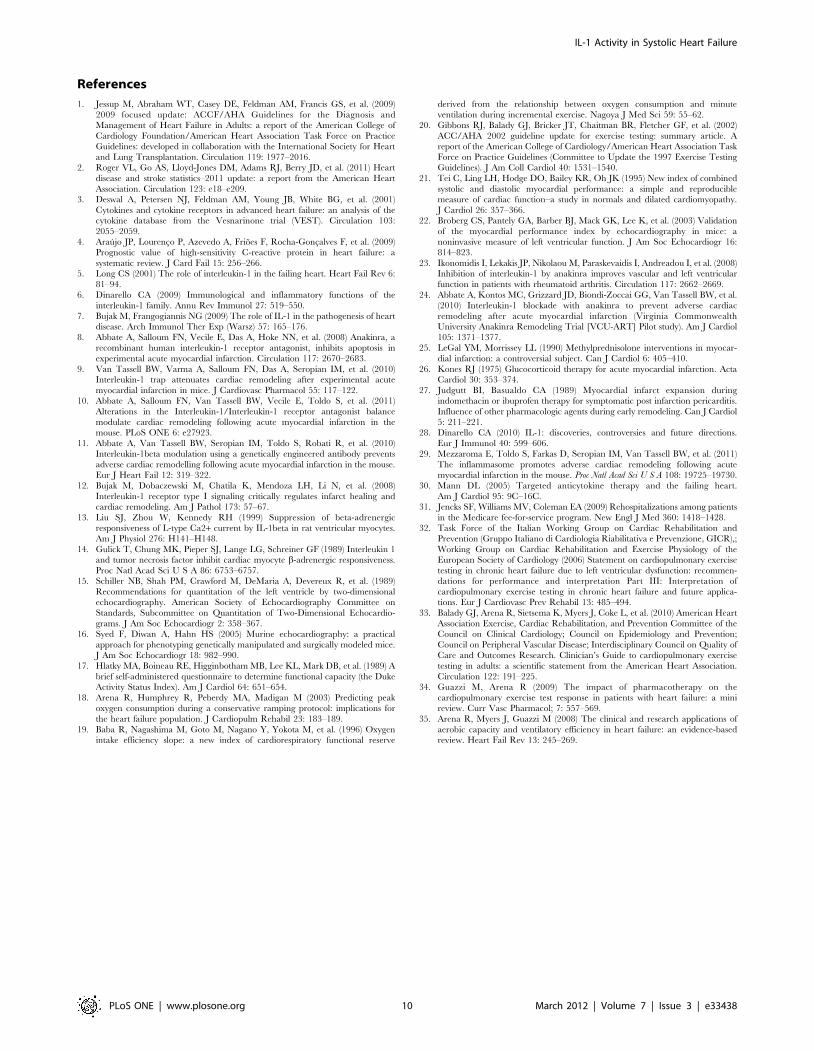
References
1. Jessup M, Abraham WT, Casey DE, Feldman AM, Francis GS, et al. (2009)
2009 focused update: ACCF/AHA Guidelines for the Diagnosis andManagement of Heart Failure in Adults: a report of the American College of
Cardiology Foundation/American Heart Association Task Force on PracticeGuidelines: developed in collaboration with the International Society for Heart
and Lung Transplantation. Circulation 119: 1977–2016.2. Roger VL, Go AS, Lloyd-Jones DM, Adams RJ, Berry JD, et al. (2011) Heart
disease and stroke statistics–2011 update: a report from the American Heart
Association. Circulation 123: e18–e209.3. Deswal A, Petersen NJ, Feldman AM, Young JB, White BG, et al. (2001)
Cytokines and cytokine receptors in advanced heart failure: an analysis of thecytokine database from the Vesnarinone trial (VEST). Circulation 103:
2055–2059.
4. Araujo JP, Lourenco P, Azevedo A, Frioes F, Rocha-Goncalves F, et al. (2009)Prognostic value of high-sensitivity C-reactive protein in heart failure: a
systematic review. J Card Fail 15: 256–266.5. Long CS (2001) The role of interleukin-1 in the failing heart. Heart Fail Rev 6:
81–94.
6. Dinarello CA (2009) Immunological and inflammatory functions of theinterleukin-1 family. Annu Rev Immunol 27: 519–550.
7. Bujak M, Frangogiannis NG (2009) The role of IL-1 in the pathogenesis of heartdisease. Arch Immunol Ther Exp (Warsz) 57: 165–176.
8. Abbate A, Salloum FN, Vecile E, Das A, Hoke NN, et al. (2008) Anakinra, arecombinant human interleukin-1 receptor antagonist, inhibits apoptosis in
experimental acute myocardial infarction. Circulation 117: 2670–2683.
9. Van Tassell BW, Varma A, Salloum FN, Das A, Seropian IM, et al. (2010)Interleukin-1 trap attenuates cardiac remodeling after experimental acute
myocardial infarction in mice. J Cardiovasc Pharmacol 55: 117–122.10. Abbate A, Salloum FN, Van Tassell BW, Vecile E, Toldo S, et al. (2011)
Alterations in the Interleukin-1/Interleukin-1 receptor antagonist balance
modulate cardiac remodeling following acute myocardial infarction in themouse. PLoS ONE 6: e27923.
11. Abbate A, Van Tassell BW, Seropian IM, Toldo S, Robati R, et al. (2010)Interleukin-1beta modulation using a genetically engineered antibody prevents
adverse cardiac remodelling following acute myocardial infarction in the mouse.Eur J Heart Fail 12: 319–322.
12. Bujak M, Dobaczewski M, Chatila K, Mendoza LH, Li N, et al. (2008)
Interleukin-1 receptor type I signaling critically regulates infarct healing andcardiac remodeling. Am J Pathol 173: 57–67.
13. Liu SJ, Zhou W, Kennedy RH (1999) Suppression of beta-adrenergicresponsiveness of L-type Ca2+ current by IL-1beta in rat ventricular myocytes.
Am J Physiol 276: H141–H148.
14. Gulick T, Chung MK, Pieper SJ, Lange LG, Schreiner GF (1989) Interleukin 1and tumor necrosis factor inhibit cardiac myocyte b-adrenergic responsiveness.
Proc Natl Acad Sci U S A 86: 6753–6757.15. Schiller NB, Shah PM, Crawford M, DeMaria A, Devereux R, et al. (1989)
Recommendations for quantitation of the left ventricle by two-dimensionalechocardiography. American Society of Echocardiography Committee on
Standards, Subcommittee on Quantitation of Two-Dimensional Echocardio-
grams. J Am Soc Echocardiogr 2: 358–367.16. Syed F, Diwan A, Hahn HS (2005) Murine echocardiography: a practical
approach for phenotyping genetically manipulated and surgically modeled mice.J Am Soc Echocardiogr 18: 982–990.
17. Hlatky MA, Boineau RE, Higginbotham MB, Lee KL, Mark DB, et al. (1989) A
brief self-administered questionnaire to determine functional capacity (the DukeActivity Status Index). Am J Cardiol 64: 651–654.
18. Arena R, Humphrey R, Peberdy MA, Madigan M (2003) Predicting peakoxygen consumption during a conservative ramping protocol: implications for
the heart failure population. J Cardiopulm Rehabil 23: 183–189.
19. Baba R, Nagashima M, Goto M, Nagano Y, Yokota M, et al. (1996) Oxygenintake efficiency slope: a new index of cardiorespiratory functional reserve
derived from the relationship between oxygen consumption and minute
ventilation during incremental exercise. Nagoya J Med Sci 59: 55–62.20. Gibbons RJ, Balady GJ, Bricker JT, Chaitman BR, Fletcher GF, et al. (2002)
ACC/AHA 2002 guideline update for exercise testing: summary article. Areport of the American College of Cardiology/American Heart Association Task
Force on Practice Guidelines (Committee to Update the 1997 Exercise TestingGuidelines). J Am Coll Cardiol 40: 1531–1540.
21. Tei C, Ling LH, Hodge DO, Bailey KR, Oh JK (1995) New index of combined
systolic and diastolic myocardial performance: a simple and reproduciblemeasure of cardiac function–a study in normals and dilated cardiomyopathy.
J Cardiol 26: 357–366.22. Broberg CS, Pantely GA, Barber BJ, Mack GK, Lee K, et al. (2003) Validation
of the myocardial performance index by echocardiography in mice: a
noninvasive measure of left ventricular function. J Am Soc Echocardiogr 16:814–823.
23. Ikonomidis I, Lekakis JP, Nikolaou M, Paraskevaidis I, Andreadou I, et al. (2008)Inhibition of interleukin-1 by anakinra improves vascular and left ventricular
function in patients with rheumatoid arthritis. Circulation 117: 2662–2669.
24. Abbate A, Kontos MC, Grizzard JD, Biondi-Zoccai GG, Van Tassell BW, et al.(2010) Interleukin-1 blockade with anakinra to prevent adverse cardiac
remodeling after acute myocardial infarction (Virginia CommonwealthUniversity Anakinra Remodeling Trial [VCU-ART] Pilot study). Am J Cardiol
105: 1371–1377.25. LeGal YM, Morrissey LL (1990) Methylprednisolone interventions in myocar-
dial infarction: a controversial subject. Can J Cardiol 6: 405–410.
26. Kones RJ (1975) Glucocorticoid therapy for acute myocardial infarction. ActaCardiol 30: 353–374.
27. Judgutt BI, Basualdo CA (1989) Myocardial infarct expansion duringindomethacin or ibuprofen therapy for symptomatic post infarction pericarditis.
Influence of other pharmacologic agents during early remodeling. Can J Cardiol
5: 211–221.28. Dinarello CA (2010) IL-1: discoveries, controversies and future directions.
Eur J Immunol 40: 599–606.29. Mezzaroma E, Toldo S, Farkas D, Seropian IM, Van Tassell BW, et al. (2011)
The inflammasome promotes adverse cardiac remodeling following acutemyocardial infarction in the mouse. Proc Natl Acad Sci U S A 108: 19725–19730.
30. Mann DL (2005) Targeted anticytokine therapy and the failing heart.
Am J Cardiol 95: 9C–16C.31. Jencks SF, Williams MV, Coleman EA (2009) Rehospitalizations among patients
in the Medicare fee-for-service program. New Engl J Med 360: 1418–1428.32. Task Force of the Italian Working Group on Cardiac Rehabilitation and
Prevention (Gruppo Italiano di Cardiologia Riabilitativa e Prevenzione, GICR),;
Working Group on Cardiac Rehabilitation and Exercise Physiology of theEuropean Society of Cardiology (2006) Statement on cardiopulmonary exercise
testing in chronic heart failure due to left ventricular dysfunction: recommen-dations for performance and interpretation Part III: Interpretation of
cardiopulmonary exercise testing in chronic heart failure and future applica-tions. Eur J Cardiovasc Prev Rehabil 13: 485–494.
33. Balady GJ, Arena R, Sietsema K, Myers J, Coke L, et al. (2010) American Heart
Association Exercise, Cardiac Rehabilitation, and Prevention Committee of theCouncil on Clinical Cardiology; Council on Epidemiology and Prevention;
Council on Peripheral Vascular Disease; Interdisciplinary Council on Quality ofCare and Outcomes Research. Clinician’s Guide to cardiopulmonary exercise
testing in adults: a scientific statement from the American Heart Association.
Circulation 122: 191–225.34. Guazzi M, Arena R (2009) The impact of pharmacotherapy on the
cardiopulmonary exercise test response in patients with heart failure: a minireview. Curr Vasc Pharmacol; 7: 557–569.
35. Arena R, Myers J, Guazzi M (2008) The clinical and research applications of
aerobic capacity and ventilatory efficiency in heart failure: an evidence-basedreview. Heart Fail Rev 13: 245–269.
IL-1 Activity in Systolic Heart Failure
PLoS ONE | www.plosone.org 10 March 2012 | Volume 7 | Issue 3 | e33438
Copyright © 2022 FDOKUMEN