Engineered Chimeric Enzymes as Tools for Drug Discovery: Generating Reliable Bacterial Screens for...
15
Engineered Chimeric Enzymes as Tools for Drug Discovery: Generating Reliable Bacterial Screens for the Detection, Discovery, and Assessment of Estrogen Receptor Modulators Georgios Skretas,* ,²,‡ Aggeliki K. Meligova, § Carolina Villalonga-Barber, | Dimitra J. Mitsiou, § Michael N. Alexis, §,⊥ Maria Micha-Screttas, | Barry R. Steele, | Constantinos G. Screttas, | and David W. Wood* ,²,¶ Contribution from the Departments of Chemical Engineering and Molecular Biology, Princeton UniVersity, Princeton, New Jersey 08544, and the Biomedical Applications Unit and the Institutes of Biological Research and Biotechnology and Organic and Pharmaceutical Chemistry, National Hellenic Research Foundation, 48 Vassileos Constantinou AVenue, 11635 Athens, Greece Received October 30, 2006; E-mail: [email protected]; [email protected] Abstract: Engineered protein-based sensors of ligand binding have emerged as attractive tools for the discovery of therapeutic compounds through simple screening systems. We have previously shown that engineered chimeric enzymes, which combine the ligand-binding domains of nuclear hormone receptors with a highly sensitive thymidylate synthase reporter, yield simple sensors that report the presence of hormone-like compounds through changes in bacterial growth. This work describes an optimized estrogen sensor in Escherichia coli with extraordinary reliability in identifying diverse estrogenic compounds and in differentiating between their agonistic/antagonistic pharmacological effects. The ability of this system to assist the discovery of new estrogen-mimicking compounds was validated by screening a small compound library, which led to the identification of two structurally novel estrogen receptor modulators and the accurate prediction of their agonistic/antagonistic biocharacter in human cells. Strong evidence is presented here that the ability of our sensor to detect ligand binding and recognize pharmacologically critical properties arises from allosteric communication between the artificially combined protein domains, where different ligand-induced conformational changes in the receptor are transmitted to the catalytic domain and translated to distinct levels of enzymic efficiency. To the best of our knowledge, this is one of the first examples of an engineered enzyme with the ability to sense multiple receptor conformations and to be either activated or inactivated depending on the nature of the bound effector molecule. Because the proposed mechanism of ligand dependence is not specific to nuclear hormone receptors, we anticipate that our protein engineering strategy will be applicable to the construction of simple sensors for different classes of (therapeutic) binding proteins. Introduction The recent development of chimeric protein-based sensors for ligand binding may greatly simplify the detection of potentially valuable compounds with specific affinity for particular protein targets. These biosensors are typically con- structed by fusing a target ligand-binding domain (LBD) to an easily assayed reporter protein. Properly designed fusions allow ligand-induced conformational changes in the target LBD to be transmitted to the reporter and allosterically modulate its properties. By selecting a suitable signaling protein, these systems can be tuned to report binding through detectable changes in phenotype. A recent example of a successful design of this type is a set of chimeric fusions of the maltose-binding protein (MBP) with -lactamase. 1,2 Escherichia coli (E. coli) cells expressing these fusions are rescued on antibiotic-contain- ing media only in the presence of ligands that bind to and change the structure of the MBP domain. Another example is based on fusions of calmodulin with circularly permuted variants of the green fluorescent protein. 3 In this case, the efficiency of chromophore formation depends on calcium binding, thus allowing the intracellular concentration of calcium ions to be conveniently reflected by cell fluorescence. Drug discovery often relies on the identification of com- pounds with the ability to specifically bind to and modulate ² Department of Chemical Engineering, Princeton University. ‡ Present address: Department of Chemical Engineering and Institute for Cellular and Molecular Biology, The University of Texas at Austin, 2500 Speedway, Austin, Texas 78712. § Institute of Biological Research and Biotechnology, National Hellenic Research Foundation. | Institute of Organic and Pharmaceutical Chemistry, National Hellenic Research Foundation. ⊥ Biomedical Applications Unit, National Hellenic Research Foundation. ¶ Department of Molecular Biology, Princeton University. (1) Guntas, G.; Mitchell, S. F.; Ostermeier, M. Chem. Biol. 2004, 11, 1483-7. (2) Guntas, G.; Mansell, T. J.; Kim, J. R.; Ostermeier, M. Proc. Natl. Acad. Sci. U.S.A. 2005, 102, 11224-9. (3) Baird, G. S.; Zacharias, D. A.; Tsien, R. Y. Proc. Natl. Acad. Sci. U.S.A. 1999, 96, 11241-6. Published on Web 06/15/2007 10.1021/ja067754j CCC: $37.00 © 2007 American Chemical Society J. AM. CHEM. SOC. 2007, 129, 8443-8457 9 8443
-
Upload
independent -
Category
Documents
-
view
0 -
download
0
Transcript of Engineered Chimeric Enzymes as Tools for Drug Discovery: Generating Reliable Bacterial Screens for...

Engineered Chimeric Enzymes as Tools for Drug Discovery:Generating Reliable Bacterial Screens for the Detection,
Discovery, and Assessment of Estrogen Receptor ModulatorsGeorgios Skretas,*,†,‡ Aggeliki K. Meligova,§ Carolina Villalonga-Barber,|
Dimitra J. Mitsiou,§ Michael N. Alexis,§,⊥ Maria Micha-Screttas,| Barry R. Steele,|
Constantinos G. Screttas,| and David W. Wood*,†,¶
Contribution from the Departments of Chemical Engineering and Molecular Biology,Princeton UniVersity, Princeton, New Jersey 08544, and the Biomedical Applications Unit and
the Institutes of Biological Research and Biotechnology and Organic and PharmaceuticalChemistry, National Hellenic Research Foundation, 48 Vassileos Constantinou AVenue,
11635 Athens, Greece
Received October 30, 2006; E-mail: [email protected]; [email protected]
Abstract: Engineered protein-based sensors of ligand binding have emerged as attractive tools for thediscovery of therapeutic compounds through simple screening systems. We have previously shown thatengineered chimeric enzymes, which combine the ligand-binding domains of nuclear hormone receptorswith a highly sensitive thymidylate synthase reporter, yield simple sensors that report the presence ofhormone-like compounds through changes in bacterial growth. This work describes an optimized estrogensensor in Escherichia coli with extraordinary reliability in identifying diverse estrogenic compounds and indifferentiating between their agonistic/antagonistic pharmacological effects. The ability of this system toassist the discovery of new estrogen-mimicking compounds was validated by screening a small compoundlibrary, which led to the identification of two structurally novel estrogen receptor modulators and the accurateprediction of their agonistic/antagonistic biocharacter in human cells. Strong evidence is presented herethat the ability of our sensor to detect ligand binding and recognize pharmacologically critical propertiesarises from allosteric communication between the artificially combined protein domains, where differentligand-induced conformational changes in the receptor are transmitted to the catalytic domain and translatedto distinct levels of enzymic efficiency. To the best of our knowledge, this is one of the first examples of anengineered enzyme with the ability to sense multiple receptor conformations and to be either activated orinactivated depending on the nature of the bound effector molecule. Because the proposed mechanism ofligand dependence is not specific to nuclear hormone receptors, we anticipate that our protein engineeringstrategy will be applicable to the construction of simple sensors for different classes of (therapeutic) bindingproteins.
Introduction
The recent development of chimeric protein-based sensorsfor ligand binding may greatly simplify the detection ofpotentially valuable compounds with specific affinity forparticular protein targets. These biosensors are typically con-structed by fusing a target ligand-binding domain (LBD) to aneasily assayed reporter protein. Properly designed fusions allowligand-induced conformational changes in the target LBD tobe transmitted to the reporter and allosterically modulate itsproperties. By selecting a suitable signaling protein, these
systems can be tuned to report binding through detectablechanges in phenotype. A recent example of a successful designof this type is a set of chimeric fusions of the maltose-bindingprotein (MBP) withâ-lactamase.1,2 Escherichia coli(E. coli)cells expressing these fusions are rescued on antibiotic-contain-ing media only in the presence of ligands that bind to and changethe structure of the MBP domain. Another example is based onfusions of calmodulin with circularly permuted variants of thegreen fluorescent protein.3 In this case, the efficiency ofchromophore formation depends on calcium binding, thusallowing the intracellular concentration of calcium ions to beconveniently reflected by cell fluorescence.
Drug discovery often relies on the identification of com-pounds with the ability to specifically bind to and modulate
† Department of Chemical Engineering, Princeton University.‡ Present address: Department of Chemical Engineering and Institute
for Cellular and Molecular Biology, The University of Texas at Austin,2500 Speedway, Austin, Texas 78712.
§ Institute of Biological Research and Biotechnology, National HellenicResearch Foundation.
| Institute of Organic and Pharmaceutical Chemistry, National HellenicResearch Foundation.
⊥ Biomedical Applications Unit, National Hellenic Research Foundation.¶ Department of Molecular Biology, Princeton University.
(1) Guntas, G.; Mitchell, S. F.; Ostermeier, M.Chem. Biol.2004, 11, 1483-7.(2) Guntas, G.; Mansell, T. J.; Kim, J. R.; Ostermeier, M.Proc. Natl. Acad.
Sci. U.S.A.2005, 102, 11224-9.(3) Baird, G. S.; Zacharias, D. A.; Tsien, R. Y.Proc. Natl. Acad. Sci. U.S.A.
1999, 96, 11241-6.
Published on Web 06/15/2007
10.1021/ja067754j CCC: $37.00 © 2007 American Chemical Society J. AM. CHEM. SOC. 2007 , 129, 8443-8457 9 8443

the function of a particular protein target. Simple biosensorsthat enable the reliable detection of protein-ligand interactionsamong large libraries of test compounds in a facile, high-throughput fashion could make an important contribution to thedrug discovery process.4 Attractive targets for this strategy arethe nuclear hormone receptors (NHRs). They are the largestgroup of metazoan transcription factors and are involved in vitalfunctions of the cell, such as development, differentiation,homeostasis, reproduction, and metabolism.5,6 Hence, deregula-tion of their function is closely related to a number ofpathological conditions, including carcinogenesis, infertility,obesity, inflammations, cardiovascular diseases, and osteoporo-sis.5,6 Accordingly, NHRs comprise one of the largest classesof protein drug targets, with a pharmaceutical significancecomparable to that of G protein-coupled receptors, kinases, ionchannels, and other membrane transporters. Currently, about 4%of all marketed therapeutics interfere with the activity of theseproteins.7
NHRs are modular proteins consisting of a DNA-bindingdomain and a C-terminal LBD, with some of these receptorsalso containing an N-terminal transactivation domain.5,6 Ap-proximately half of these transcription factors are activated bythe binding of lipophilic small-molecule hormones or synthetic/natural hormone agonists. For example, signaling of the estrogenreceptor (ER), the best characterized member of this superfam-ily, is typically initiated by the endogenous estrogen 17â-estradiol (E2). This steroidal hormone binds to the two ERsubtypes (ERR and ERâ) and induces a conformational changethat allows ER homo- and heterodimers to form.8 These canthen recruit different coactivators and form complexes capableof regulating the expression of specific genes, e.g., by bindingto regulatory regions of DNA termed estrogen response elements(EREs). Gene expression via ER can be activated by twotranscription activation functions (AFs), one mediated by theN-terminal transctivation domain (AF-1) and one by theC-terminal region of the LBD (AF-2).5,6
Therapeutically valuable compounds with the ability toantagonize the effects of estrogen, such as 4-hydroxytamoxifen(the active metabolite of tamoxifen) and raloxifene, bind to ERwith high affinity but cause a conformational change that isdistinct from that induced by estrogen agonists.5,6 In this case,ER can still dimerize, but is unable to recruit coactivators, andinstead interacts weakly with corepressors. However, althoughantagonist binding results in transcriptional silencing throughAF-2, the antagonist-bound ER complex may retain some abilityto induce gene expression via AF-1.5 Therefore, a number ofknown estrogen analogues, including tamoxifen and raloxifene,exhibit tissue-specific estrogenic or antiestrogenic responses andare therefore termed selective ER modulators (SERMs). SERMssometimes also act as mixed agonists/antagonists in the samecell and via the same ER subtype depending on the absence/presence of E2, respectively.9,10The specific response in a given
tissue depends on a number of parameters, including thepromoter of the estrogen target gene and the coactivator/corepressor complement of the cell.5,8 However, the conforma-tion that the binding domain adopts upon binding of a particularligand is probably the most decisive factor determining thepharmacological responses.11
We have shown in previous work that engineered chimericfusions of NHR LBDs with the very sensitive reporter enzymethymidylate synthase (TS) yield simple sensors that report thepresence of hormone-like compounds by changes in bacterialgrowth.12 Our present work describes a dramatically advancedE. coli-based estrogen sensor with extraordinary reliability indetecting a wide variety of estrogenic compounds and inrecognizing important aspects of their pharmacological profiles.By using this sensor, we were able to rapidly screen a smallchemical library and identify structurally novel ER modulators,while predicting their agonistic/antagonistic biocharacter inhuman cell assays. Further, we provide strong evidence thatour engineered chimeric sensor discriminates between agonisticand antagonistic effects by functioning as an allosteric enzyme,which presumably recognizes different ligand-induced confor-mational changes that occur in the LBD and translates theminto distinct levels of TS activity.
Results
Construction of a Second-Generation Estrogen-RegulatedChimeric Enzyme. In previous work, we constructed twosimple bacterial hormone-sensing prototypes by fusing the LBDof either the human ERR or the human thyroid hormone receptorâ, in combination with a solubilization (MBP) and a stabilizationdomain (modified intein splicing domain), to the N terminusof the bacteriophage T4 TS enzyme.12 In these constructs, thestabilization domain comprised the first 96 and the last 41 aminoacids of a splicing-deficient variant of theMycobacteriumtuberculosisRecA (Mtu RecA) intein. These chimeric sensorproteins provided hormone-dependent TS activity, which waseasily monitored by growth phenotype in TS-knockoutE. colicells. Subsequently, we showed that a second estrogen-sensingbacterial system can be constructed on the basis of a fusion ofthe human ERâ LBD with a more stable intein splicing domaincomprising the first 110 and the last 58 residues of the MtuRecA intein (plasmid pMIT::ERâ*; Figure 1A).13,14This workdemonstrated that the ERR- and ERâ-based sensors allow thefacile detection of subtype-selective ER ligands in the contextof positive TS selection.14 In the present work, we investigatedthe ability of the ERâ-based system to be used as an improvedsensor for general estrogenicity, using both the positive andnegative tunable selections provided by the TS reporter system(Figure 1B).15
As it had been observed with our ERR-based sensor,12 TS-knockout E. coli cells transfected with pMIT::ERâ* andincubated in liquid thymine-free medium (-THY) at 34°C were
(4) Ohlstein, E. H.; Ruffolo, R. R., Jr.; Elliott, J. D.Annu. ReV. Pharmacol.Toxicol.2000, 40, 177-91.
(5) Nettles, K. W.; Greene, G. L.Annu. ReV. Physiol.2005, 67, 309-33.(6) Bourguet, W.; Germain, P.; Gronemeyer, H.Trends Pharmacol. Sci.2000,
21, 381-8.(7) Hopkins, A. L.; Groom, C. R.Nat. ReV. Drug DiscoVery 2002, 1, 727-
30.(8) Koehler, K. F.; Helguero, L. A.; Haldosen, L. A.; Warner, M.; Gustafsson,
J. A. Endocr. ReV. 2005, 26, 465-78.(9) Barkhem, T.; Carlsson, B.; Nilsson, Y.; Enmark, E.; Gustafsson, J.; Nilsson,
S. Mol. Pharmacol.1998, 54, 105-12.
(10) Dayan, G.; Lupien, M.; Auger, A.; Anghel, S. I.; Rocha, W.; Croisetiere,S.; Katzenellenbogen, J. A.; Mader, S.Mol. Pharmacol.2006, 70, 579-88.
(11) Wijayaratne, A. L.; Nagel, S. C.; Paige, L. A.; Christensen, D. J.; Norris,J. D.; Fowlkes, D. M.; McDonnell, D. P.Endocrinology1999, 140, 5828-40.
(12) Skretas, G.; Wood, D. W.J. Mol. Biol. 2005, 349, 464-74.(13) Derbyshire, V.; Wood, D. W.; Wu, W.; Dansereau, J. T.; Dalgaard, J. Z.;
Belfort, M. Proc. Natl. Acad. Sci. U.S.A.1997, 94, 11466-71.(14) Skretas, G.; Wood, D. W.Appl. EnViron. Microbiol. 2005, 71, 8995-7.(15) Belfort, M.; Pedersen-Lane, J.J. Bacteriol.1984, 160, 371-8.
A R T I C L E S Skretas et al.
8444 J. AM. CHEM. SOC. 9 VOL. 129, NO. 27, 2007

able to grow only in the presence of estrogen.14 More specif-ically, cells incubated in the presence of estrogen yieldedsaturated cultures after 15-20 h of incubation but were unableto grow beyond an OD600 ) 0.2-0.4 in the absence of estrogen,even after 36 h of incubation (see below). The rapid initialbackground growth in the absence of estrogen occurs presum-ably due to small amounts of thymine and thymidine that aretransferred to the selective medium with the inoculum. However,after incubation under suitable conditions of positive TSselection, the presence/absence of estrogen results in an apparentgrowth/no growth readout. As expected, in a nonselective
thymine-rich medium (+THY), healthy growth was observedirrespective of the presence of estrogen. Interestingly, with thepMIT::ERâ* sensor, the impact of estrogen binding on TSactivity could also be observed when negative selection wasperformed: the addition of E2 to cells incubated in a thymine-rich medium supplemented with trimethoprim (TTM) resultedin an inhibitory effect on cell growth (Figure 1C).
Previously reported control experiments utilizing auxotrophicE. coli strains indicate that estrogenic compounds (includingE2) do not have a general impact on TS activity or bacterialgrowth.12 This work included additional control experiments
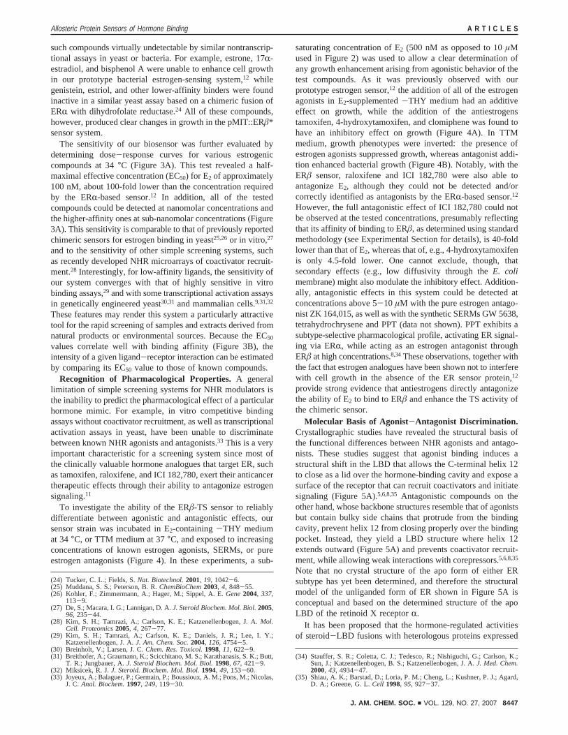
Figure 1. Design and associated growth phenotypes of the ERâ-based estrogen-sensing system. (A) Chimeric protein fusion used to couple estrogen bindingvia ERâ with the catalytic activity of thymidylate synthase. Ptac*, artificial tac promoter required for hormone-dependent phenotypes;12 MBP, maltose-binding protein; N-Mtu, the first 110 residues of theMycobacterium tuberculosisRecA intein (Mtu RecA intein) carrying a splicing-inactivating Cys1Alamutation; C-Mtu, the last 58 residues of the Mtu RecA intein; ERâ, residues Arg254 to Lys504 of the human ERâ (encompasses the entire ligand-bindingdomain of the receptor); TS, bacteriophage T4 thymidylate synthase. (B) Schematic representation of the growth phenotypes associated with the TS geneticselection system. In a thymine-rich medium (+THY medium), cells do not require active TS for growth (nonselective). In the absence of thymine (-THY),cells are subjected to selection for high TS activity (positive selection). When both thymine and the dihydrofolate reductase inhibitor trimethoprim are addedto the medium (TTM), selection against TS activity takes place (negative selection). The stringency of both modes of selection is tunable, in the caseof thenegative selection by varying the trimethoprim concentration, while positive selection becomes more stringent at elevated temperatures. (C) Effect of theaddition of 10µM estrogen (orange curves) on the growth rates of bacterial cells harboring the pMIT::ERâ* plasmid in liquid -THY medium at 34°C(circles), and in liquid+THY (squares) and TTM media (triangles) at 37°C. OD600: Optical density at 600 nm.
Allosteric Protein Sensors of Hormone Binding A R T I C L E S
J. AM. CHEM. SOC. 9 VOL. 129, NO. 27, 2007 8445

with estrogen and thyroid-hormone sensing strains showing thatgrowth enhancement in this system is only observed when anappropriate ligand is provided for a given sensor strain. Finally,the current observation that growth is enhanced in-THYmedium in the presence of estrogen, but inhibited by estrogenin TTM medium, provides very strong evidence that ligand
binding directly modulates TS activity in the chimeric sensorprotein, and not through a more general growth effect. Thus,the effect of estrogen on TS activity and the resulting growthphenotypes is highly specific and acts through the ERâ LBDin our chimeric sensor protein.
Reliable Detection of Estrogenic Compounds.To evaluatethe ligand-sensing performance of the ERâ-based sensor, cellsharboring pMIT::ERâ* were incubated in liquid-THY mediumat 34°C in the presence of a small library of synthetic estrogenanalogues. As observed previously (Figure 1C), these cells wereunable to grow in the absence of ligand, but the addition of allthe compounds that are known to possess estrogenic propertiesresulted in the induction of growth (Figure 2A). The controlcompounds 3,3′,5-triiodo-L-thyronine (T3) and progesterone, thecognate ligands of the thyroid hormone and progesteronereceptors, respectively, were unable to enhance growth.
We then exposed our sensor cells to a second librarycomprising estrogenic compounds originating from plants(phytoestrogens). These natural hormone analogues exemplifythe dietary contribution to estrogen signaling and includecompounds with considerable ERâ binding selectivity.16 Anumber of these are thought to be promising natural productsfor use in hormone replacement therapy,17,18and in many casestheir intake is associated with cardioprotective and anti-inflammatory properties, as well as a reduced risk for endocrine-related cancer.19 As with the synthetic estrogen library, everyphytoestrogen tested was able to enhance cell growth and couldbe easily detected (Figure 2B).
The ERâ-based sensor was also able to detect the binding ofseveral steroids with low affinity for ER. These were selectedfrom the metabolic precursors or metabolites of testosterone,and although some of them are classified as androgens, theyare able to bind to ER weakly and induce estrogenic and otherresponses both in males and females.20,21 Growth phenotypesin the presence of these compounds indicated that approximatelyhalf of the tested steroids could be detected, although some ofthem required longer incubation times (Figure 2C). The ligandsthat were unable to induce a growth response are known toexhibit extremely low binding affinities for ER (e.g., testoster-one).20 One of the detected steroids, the dihydrotestosteronemetabolite 3â-androstanediol, has been proposed to function asthe principal ligand of ERâ in the prostate,22,23 where it mayinhibit the migration of prostate cancer cells.21
Notably, a number of the detected compounds, such asbisphenol A and biochanin A, exhibit very low binding affinitiesfor ER (1000- to 10000-fold lower than that of E2).16 This makes
(16) Kuiper, G. G.; Lemmen, J. G.; Carlsson, B.; Corton, J. C.; Safe, S. H.; vander Saag, P. T.; van der Burg, B.; Gustafsson, J. A.Endocrinology1998,139, 4252-63.
(17) Fokialakis, N.; Lambrinidis, G.; Mitsiou, D. J.; Aligiannis, N.; Mitakou,S.; Skaltsounis, A. L.; Pratsinis, H.; Mikros, E.; Alexis, M. N.Chem. Biol.2004, 11, 397-406.
(18) Halabalaki, M.; Alexi, X.; Aligiannis, N.; Lambrinidis, G.; Pratsinis, H.;Florentin, I.; Mitakou, S.; Mikros, E.; Skaltsounis, A. L.; Alexis, M. N.Planta Med.2006, 72, 488-93.
(19) Adlercreutz, H.EnViron. Health Perspect.1995, 103 (Suppl 7), 103-12.(20) Kuiper, G. G.; Carlsson, B.; Grandien, K.; Enmark, E.; Haggblad, J.;
Nilsson, S.; Gustafsson, J. A.Endocrinology1997, 138, 863-70.(21) Guerini, V.; Sau, D.; Scaccianoce, E.; Rusmini, P.; Ciana, P.; Maggi, A.;
Martini, P. G.; Katzenellenbogen, B. S.; Martini, L.; Motta, M.; Poletti, A.Cancer Res.2005, 65 5445-53.
(22) Weihua, Z.; Makela, S.; Andersson, L. C.; Salmi, S.; Saji, S.; Webster, J.I.; Jensen, E. V.; Nilsson, S.; Warner, M.; Gustafsson, J. A.Proc. Natl.Acad. Sci. U.S.A.2001, 98, 6330-5.
(23) Weihua, Z.; Lathe, R.; Warner, M.; Gustafsson, J. A.Proc. Natl. Acad.Sci. U.S.A.2002, 99, 13589-94.
Figure 2. Reliable detection of estrogenic compounds: (A) syntheticestrogen analogues; (B) phytoestrogens; (C) testosterone and some of itsmetabolic precursors and metabolites. The relative binding affinity valuesof testosterone, androstenediol, dehydroepiandrosterone, 3R-androstanediol,3â-androstanediol, epiandrosterone, and androsterone for ERâ are<0.01,16
13.84 ( 1.93, 0.090( 0.011, 0.288( 0.112, 2.608( 0.440, 0.013(0.001, and 0.003( 0.002, respectively. 17â-Estradiol is arbitrarily set to100. TS-knockout bacterial cells transfected with pMIT::ERâ* were grownin liquid -THY medium at 34°C in the presence of 10µM ligands forapproximately 15 h in A and B and for 22 (gray) and 35 h (black) in C.Experiments were carried out in triplicate, and the error bars represent onestandard deviation from the mean value.
A R T I C L E S Skretas et al.
8446 J. AM. CHEM. SOC. 9 VOL. 129, NO. 27, 2007
such compounds virtually undetectable by similar nontranscrip-tional assays in yeast or bacteria. For example, estrone, 17R-estradiol, and bisphenol A were unable to enhance cell growthin our prototype bacterial estrogen-sensing system,12 whilegenistein, estriol, and other lower-affinity binders were foundinactive in a similar yeast assay based on a chimeric fusion ofERR with dihydrofolate reductase.24 All of these compounds,however, produced clear changes in growth in the pMIT::ERâ*sensor system.
The sensitivity of our biosensor was further evaluated bydetermining dose-response curves for various estrogeniccompounds at 34°C (Figure 3A). This test revealed a half-maximal effective concentration (EC50) for E2 of approximately100 nM, about 100-fold lower than the concentration requiredby the ERR-based sensor.12 In addition, all of the testedcompounds could be detected at nanomolar concentrations andthe higher-affinity ones at sub-nanomolar concentrations (Figure3A). This sensitivity is comparable to that of previously reportedchimeric sensors for estrogen binding in yeast25,26or in vitro,27
and to the sensitivity of other simple screening systems, suchas recently developed NHR microarrays of coactivator recruit-ment.28 Interestingly, for low-affinity ligands, the sensitivity ofour system converges with that of highly sensitive in vitrobinding assays,29 and with some transcriptional activation assaysin genetically engineered yeast30,31 and mammalian cells.9,31,32
These features may render this system a particularly attractivetool for the rapid screening of samples and extracts derived fromnatural products or environmental sources. Because the EC50
values correlate well with binding affinity (Figure 3B), theintensity of a given ligand-receptor interaction can be estimatedby comparing its EC50 value to those of known compounds.
Recognition of Pharmacological Properties.A generallimitation of simple screening systems for NHR modulators isthe inability to predict the pharmacological effect of a particularhormone mimic. For example, in vitro competitive bindingassays without coactivator recruitment, as well as transcriptionalactivation assays in yeast, have been unable to discriminatebetween known NHR agonists and antagonists.33 This is a veryimportant characteristic for a screening system since most ofthe clinically valuable hormone analogues that target ER, suchas tamoxifen, raloxifene, and ICI 182,780, exert their anticancertherapeutic effects through their ability to antagonize estrogensignaling.11
To investigate the ability of the ERâ-TS sensor to reliablydifferentiate between agonistic and antagonistic effects, oursensor strain was incubated in E2-containing-THY mediumat 34°C, or TTM medium at 37°C, and exposed to increasingconcentrations of known estrogen agonists, SERMs, or pureestrogen antagonists (Figure 4). In these experiments, a sub-
saturating concentration of E2 (500 nM as opposed to 10µMused in Figure 2) was used to allow a clear determination ofany growth enhancement arising from agonistic behavior of thetest compounds. As it was previously observed with ourprototype estrogen sensor,12 the addition of all of the estrogenagonists in E2-supplemented-THY medium had an additiveeffect on growth, while the addition of the antiestrogenstamoxifen, 4-hydroxytamoxifen, and clomiphene was found tohave an inhibitory effect on growth (Figure 4A). In TTMmedium, growth phenotypes were inverted: the presence ofestrogen agonists suppressed growth, whereas antagonist addi-tion enhanced bacterial growth (Figure 4B). Notably, with theERâ sensor, raloxifene and ICI 182,780 were also able toantagonize E2, although they could not be detected and/orcorrectly identified as antagonists by the ERR-based sensor.12
However, the full antagonistic effect of ICI 182,780 could notbe observed at the tested concentrations, presumably reflectingthat its affinity of binding to ERâ, as determined using standardmethodology (see Experimental Section for details), is 40-foldlower than that of E2, whereas that of, e.g., 4-hydroxytamoxifenis only 4.5-fold lower. One cannot exclude, though, thatsecondary effects (e.g., low diffusivity through theE. colimembrane) might also modulate the inhibitory effect. Addition-ally, antagonistic effects in this system could be detected atconcentrations above 5-10 µM with the pure estrogen antago-nist ZK 164,015, as well as with the synthetic SERMs GW 5638,tetrahydrochrysene and PPT (data not shown). PPT exhibits asubtype-selective pharmacological profile, activating ER signal-ing via ERR, while acting as an estrogen antagonist throughERâ at high concentrations.8,34These observations, together withthe fact that estrogen analogues have been shown not to interferewith cell growth in the absence of the ER sensor protein,12
provide strong evidence that antiestrogens directly antagonizethe ability of E2 to bind to ERâ and enhance the TS activity ofthe chimeric sensor.
Molecular Basis of Agonist-Antagonist Discrimination.Crystallographic studies have revealed the structural basis ofthe functional differences between NHR agonists and antago-nists. These studies suggest that agonist binding induces astructural shift in the LBD that allows the C-terminal helix 12to close as a lid over the hormone-binding cavity and expose asurface of the receptor that can recruit coactivators and initiatesignaling (Figure 5A).5,6,8,35 Antagonistic compounds on theother hand, whose backbone structures resemble that of agonistsbut contain bulky side chains that protrude from the bindingcavity, prevent helix 12 from closing properly over the bindingpocket. Instead, they yield a LBD structure where helix 12extends outward (Figure 5A) and prevents coactivator recruit-ment, while allowing weak interactions with corepressors.5,6,8,35
Note that no crystal structure of the apo form of either ERsubtype has yet been determined, and therefore the structuralmodel of the unliganded form of ER shown in Figure 5A isconceptual and based on the determined structure of the apoLBD of the retinoid X receptorR.
It has been proposed that the hormone-regulated activitiesof steroid-LBD fusions with heterologous proteins expressed
(24) Tucker, C. L.; Fields, S.Nat. Biotechnol.2001, 19, 1042-6.(25) Muddana, S. S.; Peterson, B. R.ChemBioChem2003, 4, 848-55.(26) Kohler, F.; Zimmermann, A.; Hager, M.; Sippel, A. E.Gene2004, 337,
113-9.(27) De, S.; Macara, I. G.; Lannigan, D. A.J. Steroid Biochem. Mol. Biol.2005,
96, 235-44.(28) Kim, S. H.; Tamrazi, A.; Carlson, K. E.; Katzenellenbogen, J. A.Mol.
Cell. Proteomics2005, 4, 267-77.(29) Kim, S. H.; Tamrazi, A.; Carlson, K. E.; Daniels, J. R.; Lee, I. Y.;
Katzenellenbogen, J. A.J. Am. Chem. Soc.2004, 126, 4754-5.(30) Breinholt, V.; Larsen, J. C.Chem. Res. Toxicol.1998, 11, 622-9.(31) Breithofer, A.; Graumann, K.; Scicchitano, M. S.; Karathanasis, S. K.; Butt,
T. R.; Jungbauer, A.J. Steroid Biochem. Mol. Biol.1998, 67, 421-9.(32) Miksicek, R. J.J. Steroid. Biochem. Mol. Biol.1994, 49, 153-60.(33) Joyeux, A.; Balaguer, P.; Germain, P.; Boussioux, A. M.; Pons, M.; Nicolas,
J. C.Anal. Biochem.1997, 249, 119-30.
(34) Stauffer, S. R.; Coletta, C. J.; Tedesco, R.; Nishiguchi, G.; Carlson, K.;Sun, J.; Katzenellenbogen, B. S.; Katzenellenbogen, J. A.J. Med. Chem.2000, 43, 4934-47.
(35) Shiau, A. K.; Barstad, D.; Loria, P. M.; Cheng, L.; Kushner, P. J.; Agard,D. A.; Greene, G. L.Cell 1998, 95, 927-37.
Allosteric Protein Sensors of Hormone Binding A R T I C L E S
J. AM. CHEM. SOC. 9 VOL. 129, NO. 27, 2007 8447

Figure 3. Sensitivity of the ERâ-TS sensor. (A) Dose-response curves of endogenous (17â-estradiol14), synthetic (estriol, diethylstilbestrol, dienestrol,DPN14), and plant-derived (genistein,14 kaempferol, apigenin) estrogens. Cells carrying pMIT::ERâ* were grown in liquid-THY medium at 34°C for15-20 h. Experiments were carried out in triplicate and the error bars represent one standard deviation from the mean value. (B) Correlation between theEC50 values of the tested estrogen analogues determined from the generated curves in A, and the in vitro measurements of their relative binding affinitiesfor ERâ as reported by Kuiper et al.16,20 and Kim et al.28
A R T I C L E S Skretas et al.
8448 J. AM. CHEM. SOC. 9 VOL. 129, NO. 27, 2007

in eukaryotic hosts occur due to interactions with the molecularchaperone Hsp90.36 In these chimeric fusions, chaperone bindingto the partially unfolded LBD sterically blocks the activity ofthe fusion partner. Hormone binding induces folding of the LBDand dissociation of Hsp90, resulting in activation of the reporterprotein. Estrogen-dependent activity of our ERâ-TS sensor couldsimilarly be occurring due to ligand-induced dissociation ofbacterial Hsp90 homologues such as HtpG, or otherE. colimolecular chaperones.37 An alternative mechanism for theobserved phenotypes is that binding of estrogen analogues tothe ERâ domain affects the overall thermodynamic or proteolyticstability of the ERâ-TS chimera. Such an effect would resultin hormone-dependent amounts of the TS reporter in theE. colicytoplasm, leading to apparent ligand-regulated TS activity. Webelieve that these mechanisms cannot account for all of thehormone-dependent activities of our NHR-TS fusions expressed
in E. coli, however, as a number of our previously and presentlydescribed observations are not consistent with the mechanisticmodels mentioned above. Instead, we have proposed that theconformational changes that occur in the LBD upon ligandbinding are communicated through the intein to the TS domainto allosterically modulate its catalytic efficiency.12 This com-munication is thought to take place intramolecularly in a stablyfolded sensor protein, and to be independent of interactions withother proteins acting in trans.
We investigated this hypothesis by exposing our sensor strainto a number of known estrogen agonists and antagonists in-THY medium in the absence of E2. As observed with theestrogen analogues in Figure 2, agonist addition resulted inenhancement of TS activity and induction of cell growth (Figure5B). On the contrary, antagonist addition did not increase TSactivity, although these compounds are known to bind to ERwith high affinity.20 The fact that antiestrogens can antagonizeE2-enhanced cell growth in our system even at relatively low
(36) Mattioni, T.; Louvion, J. F.; Picard, D.Methods Cell Biol.1994, 43A, 335-52.
(37) Buchner, J.Trends Biochem. Sci.1999, 24, 136-41.
Figure 4. Recognition of the pharmacological properties of estrogen analogues. (A) Bacterial cells carrying the plasmid pMIT::ERâ* were exposed toincreasing concentrations of known estrogen agonists (black) and antagonists (orange) in liquid-THY medium containing 500 nM E2 at 34°C for approximately15 h. The upper blue line represents the level of cell growth in the presence only of 500 nM E2, while the lower line the measured level of growth in theabsence of E2. (B) Cells transfected with pMIT::ERâ* and grown in E2-enriched (500 nM) liquid-THY at 34 °C or TTM media at 37°C were exposedto a 5µM concentration of estrogen analogues (tamoxifen was added at a 2µM concentration) for approximately 17 h. Experiments were carried out intriplicate, and the error bars in B represent one standard deviation from the mean value. E2 ) 17â-estradiol.
Allosteric Protein Sensors of Hormone Binding A R T I C L E S
J. AM. CHEM. SOC. 9 VOL. 129, NO. 27, 2007 8449

ratios of antagonist/agonist concentrations (Figure 4) is anindication that the receptor domain retains the ability to bindthese compounds tightly in the bacterial cytoplasm. Westernblot analysis further revealed that the addition of estrogenagonists, antagonists, or combinations of these types of com-pounds does not affect the expression levels of the ERâ-TSsensor protein (Figure 5C). Taken together, these results implythat agonist and antagonist binding induce receptor conforma-tions with distinct fingerprints on the enzymic efficiency of ourchimeric sensor. The fingerprint corresponding to the antagonist-bound form of the receptor results in lower activity of the
catalytic TS domain, thus allowing the ERâ-TS sensor todiscriminate between agonistic and antagonistic effects.
As judged by growth phenotypes in-THY medium, theenzymic efficiency of the antagonist-bound forms of the ERâ-TS fusion closely resemble that of the apo form (Figure 5B).Although the apo and antagonist-bound forms of ER are similarin several aspects,28,29 they are clearly distinguishable usingconformation-sensitive fluorophores38 or other sensitive sensorsof receptor conformations.27,39,40To investigate the potential for
(38) Tamrazi, A.; Carlson, K. E.; Katzenellenbogen, J. A.Mol. Endocrinol.2003,17, 2593-602.
Figure 5. Molecular basis of agonist-antagonist discrimination. (A) Differential positioning adopted by the C-terminal helix 12 of NHRs (colored red) inthe presence of pharmacologically distinct classes of hormone analogues. Crystal structures of the ligand-binding domains of the human retinoid X receptorR in the absence of ligand (gray)58 and of the human ERR in the presence of the full agonist diethylstilbestrol (green) and the partial estrogen antagonist4-hydroxytamoxifen (yellow).35 (B) Cells harboring pMIT::ERâ* were grown in liquid-THY medium at 34°C for 15 h (gray) and in liquid TTM mediumat 37°C for 16 h (green) in the presence of estrogen agonists and antagonsts. Ligands were added at a 4µM concentration, apart from tamoxifen which wasadded at 2µM. For the experiments in TTM media, raloxifene and ZK 164,015 were supplied at 40µM and ICI 182,780 at a 30µM concentration.Experiments were carried out in triplicate, and the error bars represent one standard deviation from the mean value. (C). Expression levels of the ERâ-TSsensor protein as revealed by Western blot analysis using an anti-MBP antibody. Estrogen analogues were added to a 5µM concentration. The raloxifene+ 17â-estradiol sample contained 0.5µM 17â-estradiol and 5µM raloxifene. GroEL expression probed with an anti-GroEL antibody as described inExperimental Section was used as a marker for equal loading.
A R T I C L E S Skretas et al.
8450 J. AM. CHEM. SOC. 9 VOL. 129, NO. 27, 2007

detecting conformational differences between the apo andantagonist-bound forms of the ERâ domain in our system,ligand-dependent phenotypes were additionally evaluated underconditions of negative TS selection. As expected, in an estrogen-free TTM medium, cells could grow healthily, while agonistaddition had an inhibitory effect on growth (Figure 5B). Mostinterestingly, the addition of the antiestrogens raloxifene, ICI182,780, and ZK 164,015 at saturating or near-saturatingconcentrations resulted in significant growth enhancement. Thisimplies that the antagonist-bound form of the receptor inducesa state of lower TS activity than the apo form of the receptor.Tamoxifen, 4-hydroxytamoxifen, and clomiphene could not befully evaluated for this effect, as they exhibited general toxicityeffects in the 10-20 µM range.
The findings presented here provide further supportagainsta mechanism where hormone-dependent effects occur due tosimple ligand-dependent receptor stability or hormone-regulatedchaperone dissociation. The antagonist-inducedinactiVation ofthe ERâ-TS chimera observed in this work strongly supports amechanistic model based on intramolecular effects in a well-folded sensor protein. Although pure estrogen antagonists suchas ICI 182,780 antagonize ER signaling by an additionalmechanism that involves destabilization of the ER LBD andreduction of the receptor levels in the cell,11,41,42most anties-trogens do not possess an inherent ability to destabilize ER.Indeed, some of the studied estrogen antagonists are known toeither not interfere (e.g., raloxifene) or to even increase ERstability (e.g., 4-hydroxytamoxifen).11,42 In the context of theERâ-TS fusion inE. coli, all of these antiestrogens were foundnot to interfere with the expression levels of our sensor protein.Furthermore, if the catalytic activity of the ERâ-TS fusion wasregulated by interactions with molecular chaperones, antagonistbinding would result (at least in some cases) in LBD foldingand chaperone dissociation, leading to enhancement of TSactivity. A consequence of this mechanistic model, wherebiologically relevant ligand effects are recognized on the basisof the conformations adopted by the LBD of the ERâ-TS fusion,is that SERMs and pure estrogen antagonists are simplyclassified as “antagonists” by our system. This has beenobserved both with this work (Figures 4 and 5) and in ourprevious work.12
Discovery of Novel Estrogen Receptor Modulators.Toinvestigate the ability of the constructed biosensor to assist dis-covery of new ER modulators, a library of 20 structurally novelcompounds was synthesized and screened for hormone-mimick-ing behavior in our system. We chose to synthesize mainly (E)-stilbenoids (Figure 6A) bearing hydroxy groups at ring positionsidentical (compounds4a, 4c) or similar (compound4b) to thoseof resveratrol, a plant-derived stilbenoid reportedly exhibitingmixed ER agonist/antagonist properties.43 Studies on structure-activity relationships for ER binding suggest that the O-Odistance between the two distant OH groups should be in the
range of 9.7-12.3 Å to mimic the hydrogen bonding ability ofthe 3- and 17â-OH groups of E2.44 For the new stilbenoids wehave estimated the O-O distances between pairs of distant OHgroups in molecular mechanics (MM3) optimized geometriesof these compounds to lie in the range of 9.3-12.5 Å. Severalof the new stilbenoids were also endowed with bulky aliphatic(e.g.,tert-butyl) substituents in the hope that they would behaveas ERâ antagonists by a mechanism similar to that describedfor tetrahydrochrysene, an ERR agonist acting as an antagonistthrough ERâ (see Discussion).45
Incubation of our sensor strain in-THY medium with thenew compounds revealed that most of them, such as the
(39) Paige, L. A.; Christensen, D. J.; Gron, H.; Norris, J. D.; Gottlin, E. B.;Padilla, K. M.; Chang, C. Y.; Ballas, L. M.; Hamilton, P. T.; McDonnell,D. P.; Fowlkes, D. M.Proc. Natl. Acad. Sci. U.S.A.1999, 96, 3999-4004.
(40) Koide, A.; Abbatiello, S.; Rothgery, L.; Koide, S.Proc. Natl. Acad. Sci.U.S.A.2002, 99, 1253-8.
(41) Pike, A. C.; Brzozowski, A. M.; Walton, J.; Hubbard, R. E.; Thorsell, A.G.; Li, Y. L.; Gustafsson, J. A.; Carlquist, M.Structure2001, 9, 145-53.
(42) Wu, Y. L.; Yang, X.; Ren, Z.; McDonnell, D. P.; Norris, J. D.; Willson, T.M.; Greene, G. L.Mol. Cell 2005, 18, 413-24.
(43) Bhat, K. P.; Lantvit, D.; Christov, K.; Mehta, R. G.; Moon, R. C.; Pezzuto,J. M. Cancer Res.2001, 61, 7456-63.
(44) Fang, H.; Tong, W.; Shi, L. M.; Blair, R.; Perkins, R.; Branham, W.; Hass,B. S.; Xie, Q.; Dial, S. L.; Moland, C. L.; Sheehan, D. M.Chem. Res.Toxicol.2001, 14, 280-94.
(45) Shiau, A. K.; Barstad, D.; Radek, J. T.; Meyers, M. J.; Nettles, K. W.;Katzenellenbogen, B. S.; Katzenellenbogen, J. A.; Agard, D. A.; Greene,G. L. Nat. Struct. Biol.2002, 9, 359-64.
Figure 6. Discovery of novel compounds with the ability to bind to ERâand differentially mediate ER signaling. (A) Chemical structures of someof the studied compounds. (B) Cells transfected with pMIT::ERâ* andgrown in liquid -THY medium at 34°C in the presence of 5µM testligands for 22 (gray) and 32 h (black). (C) Effects of the addition of a 5µM concentration of some of the synthesized compounds on the growthphenotypes of cells carrying pMIT::ERâ* and incubated in liquid-THYmedium containing 500 nM E2 at 34°C for 18 h. Experiments were carriedout in triplicate, and the error bars represent one standard deviation fromthe mean value.
Allosteric Protein Sensors of Hormone Binding A R T I C L E S
J. AM. CHEM. SOC. 9 VOL. 129, NO. 27, 2007 8451
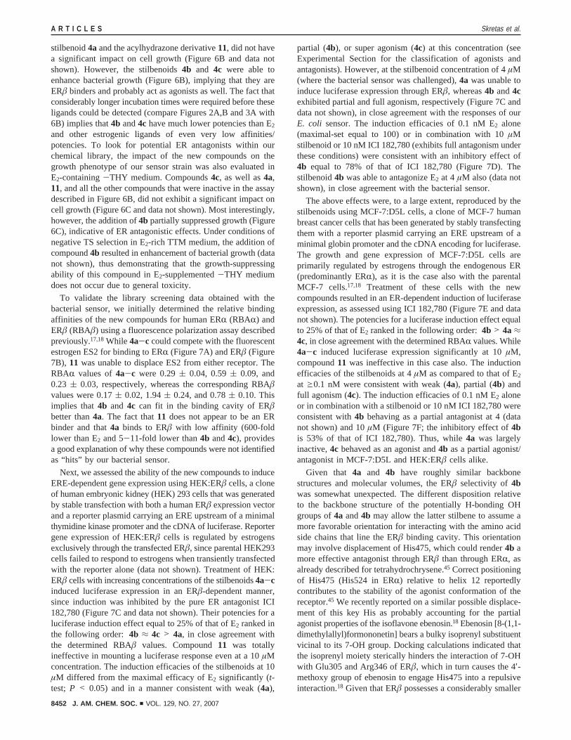
stilbenoid4aand the acylhydrazone derivative11, did not havea significant impact on cell growth (Figure 6B and data notshown). However, the stilbenoids4b and 4c were able toenhance bacterial growth (Figure 6B), implying that they areERâ binders and probably act as agonists as well. The fact thatconsiderably longer incubation times were required before theseligands could be detected (compare Figures 2A,B and 3A with6B) implies that4b and4c have much lower potencies than E2
and other estrogenic ligands of even very low affinities/potencies. To look for potential ER antagonists within ourchemical library, the impact of the new compounds on thegrowth phenotype of our sensor strain was also evaluated inE2-containing-THY medium. Compounds4c, as well as4a,11, and all the other compounds that were inactive in the assaydescribed in Figure 6B, did not exhibit a significant impact oncell growth (Figure 6C and data not shown). Most interestingly,however, the addition of4b partially suppressed growth (Figure6C), indicative of ER antagonistic effects. Under conditions ofnegative TS selection in E2-rich TTM medium, the addition ofcompound4b resulted in enhancement of bacterial growth (datanot shown), thus demonstrating that the growth-suppressingability of this compound in E2-supplemented-THY mediumdoes not occur due to general toxicity.
To validate the library screening data obtained with thebacterial sensor, we initially determined the relative bindingaffinities of the new compounds for human ERR (RBAR) andERâ (RBAâ) using a fluorescence polarization assay describedpreviously.17,18While 4a-c could compete with the fluorescentestrogen ES2 for binding to ERR (Figure 7A) and ERâ (Figure7B), 11 was unable to displace ES2 from either receptor. TheRBAR values of4a-c were 0.29( 0.04, 0.59( 0.09, and0.23 ( 0.03, respectively, whereas the corresponding RBAâvalues were 0.17( 0.02, 1.94( 0.24, and 0.78( 0.10. Thisimplies that4b and 4c can fit in the binding cavity of ERâbetter than4a. The fact that11 does not appear to be an ERbinder and that4a binds to ERâ with low affinity (600-foldlower than E2 and 5-11-fold lower than4b and4c), providesa good explanation of why these compounds were not identifiedas “hits” by our bacterial sensor.
Next, we assessed the ability of the new compounds to induceERE-dependent gene expression using HEK:ERâ cells, a cloneof human embryonic kidney (HEK) 293 cells that was generatedby stable transfection with both a human ERâ expression vectorand a reporter plasmid carrying an ERE upstream of a minimalthymidine kinase promoter and the cDNA of luciferase. Reportergene expression of HEK:ERâ cells is regulated by estrogensexclusively through the transfected ERâ, since parental HEK293cells failed to respond to estrogens when transiently transfectedwith the reporter alone (data not shown). Treatment of HEK:ERâ cells with increasing concentrations of the stilbenoids4a-cinduced luciferase expression in an ERâ-dependent manner,since induction was inhibited by the pure ER antagonist ICI182,780 (Figure 7C and data not shown). Their potencies for aluciferase induction effect equal to 25% of that of E2 ranked inthe following order: 4b ≈ 4c > 4a, in close agreement withthe determined RBAâ values. Compound11 was totallyineffective in mounting a luciferase response even at a 10µMconcentration. The induction efficacies of the stilbenoids at 10µM differed from the maximal efficacy of E2 significantly (t-test; P < 0.05) and in a manner consistent with weak (4a),
partial (4b), or super agonism (4c) at this concentration (seeExperimental Section for the classification of agonists andantagonists). However, at the stilbenoid concentration of 4µM(where the bacterial sensor was challenged),4a was unable toinduce luciferase expression through ERâ, whereas4b and4cexhibited partial and full agonism, respectively (Figure 7C anddata not shown), in close agreement with the responses of ourE. coli sensor. The induction efficacies of 0.1 nM E2 alone(maximal-set equal to 100) or in combination with 10µMstilbenoid or 10 nM ICI 182,780 (exhibits full antagonism underthese conditions) were consistent with an inhibitory effect of4b equal to 78% of that of ICI 182,780 (Figure 7D). Thestilbenoid4b was able to antagonize E2 at 4µM also (data notshown), in close agreement with the bacterial sensor.
The above effects were, to a large extent, reproduced by thestilbenoids using MCF-7:D5L cells, a clone of MCF-7 humanbreast cancer cells that has been generated by stably transfectingthem with a reporter plasmid carrying an ERE upstream of aminimal globin promoter and the cDNA encoding for luciferase.The growth and gene expression of MCF-7:D5L cells areprimarily regulated by estrogens through the endogenous ER(predominantly ERR), as it is the case also with the parentalMCF-7 cells.17,18 Treatment of these cells with the newcompounds resulted in an ER-dependent induction of luciferaseexpression, as assessed using ICI 182,780 (Figure 7E and datanot shown). The potencies for a luciferase induction effect equalto 25% of that of E2 ranked in the following order:4b > 4a≈4c, in close agreement with the determined RBAR values. While4a-c induced luciferase expression significantly at 10µM,compound11 was ineffective in this case also. The inductionefficacies of the stilbenoids at 4µM as compared to that of E2at g0.1 nM were consistent with weak (4a), partial (4b) andfull agonism (4c). The induction efficacies of 0.1 nM E2 aloneor in combination with a stilbenoid or 10 nM ICI 182,780 wereconsistent with4b behaving as a partial antagonist at 4 (datanot shown) and 10µM (Figure 7F; the inhibitory effect of4bis 53% of that of ICI 182,780). Thus, while4a was largelyinactive,4c behaved as an agonist and4b as a partial agonist/antagonist in MCF-7:D5L and HEK:ERâ cells alike.
Given that 4a and 4b have roughly similar backbonestructures and molecular volumes, the ERâ selectivity of 4bwas somewhat unexpected. The different disposition relativeto the backbone structure of the potentially H-bonding OHgroups of4a and4b may allow the latter stilbene to assume amore favorable orientation for interacting with the amino acidside chains that line the ERâ binding cavity. This orientationmay involve displacement of His475, which could render4b amore effective antagonist through ERâ than through ERR, asalready described for tetrahydrochrysene.45 Correct positioningof His475 (His524 in ERR) relative to helix 12 reportedlycontributes to the stability of the agonist conformation of thereceptor.45 We recently reported on a similar possible displace-ment of this key His as probably accounting for the partialagonist properties of the isoflavone ebenosin.18 Ebenosin [8-(1,1-dimethylallyl)formononetin] bears a bulky isoprenyl substituentvicinal to its 7-OH group. Docking calculations indicated thatthe isoprenyl moiety sterically hinders the interaction of 7-OHwith Glu305 and Arg346 of ERâ, which in turn causes the 4′-methoxy group of ebenosin to engage His475 into a repulsiveinteraction.18 Given that ERâ possesses a considerably smaller
A R T I C L E S Skretas et al.
8452 J. AM. CHEM. SOC. 9 VOL. 129, NO. 27, 2007

binding cavity than ERR,46 the finding that4b is a less effectiveantagonist through ERR than through ERâ probably suggeststhat the repulsive interaction is weakened as the binding cavitybecomes spacious enough to accommodate the bulkytert-butylsubstituent of this stilbene.
The observation that4b and 4c enhanced growth of ourbacterial sensor strain in-THY medium (Figure 6B) is inaccordance with their partial and full agonist behavior, respec-tively, in E2-deprived mammalian cells (Figure 7C,E). Mostinterestingly, while4c failed to exhibit a significant impact on
bacterial growth in E2-containing-THY medium,4b suppressedgrowth under these conditions (Figure 6C), in full accordancewith the finding that only the latter stilbenoid exhibitedantagonist behavior in E2-supplemented mammalian cells(Figure 7D,F). In spite of the low potencies and the pharma-cological effects exhibited by the stilbenoids4b and4c, whichmost probably do not constitute them as potential hormone
(46) Pike, A. C.; Brzozowski, A. M.; Hubbard, R. E.; Bonn, T.; Thorsell, A.G.; Engstrom, O.; Ljunggren, J.; Gustafsson, J. A.; Carlquist, M.EMBO J.1999, 18, 4608-18.
Figure 7. Demonstration of the ability of the bacterial sensor to reliably report on the signaling properties of compounds capable of binding to ER. (A)Dose-response curves of the displacement of the fluorescent estrogen ES2 from ERR by serial 1/3.3 dilutions of E2 or the new compounds4a-c and11.(B) Dose-response curves of the displacement of the fluorescent estrogen ES2 from ERâ as in A. (C) Dose-response curves of the induction of luciferaseexpression in HEK:ERâ cells by serial 1/10 dilutions of E2 and the new compounds. (D) Luciferase response of HEK:ERâ cells exposed to E2 (0.1 nM) inthe absence or presence (+) of 4a, 4b, or 4c (10 µM), or the pure estrogen antagonist ICI 182,780 (10 nM). (E) Dose-response curves of the induction ofluciferase expression in MCF-7:D5L cells by serial 1/10 dilutions of E2 and the new compounds. (F) Luciferase response of MCF-7:D5L cells exposed to0.1 nM E2 (set equal to 100) in the absence or presence (+) of 4a, 4b, or 4c (10 µM), or ICI 182,780 (10 nM). Dose-response experiments were carriedout in triplicate, and the data are mean( SEM of the three measurements. Data in the histograms are mean( SEM of three independent experiments carriedout in triplicate; ICI) ICI 182,780.
Allosteric Protein Sensors of Hormone Binding A R T I C L E S
J. AM. CHEM. SOC. 9 VOL. 129, NO. 27, 2007 8453

therapeutics, these results suggest that ourE. coli system is areliable sensor for the detection of ERâ modulators and that itcan be used as a sensor of general estrogenicity as well. Ourfindings convincingly support the applicability of this simplesystem for screening large compound libraries in search ofpotentially useful ER modulators.
Discussion
This work describes a dramatically enhanced bacterial sensorfor hormone binding that is appropriate for practical screeningapplications of potentially therapeutic compounds. An engi-neered fusion comprising the LBD of the human ERâ and thevery sensitive reporter enzyme TS yielded an enzyme chimerawith the ability to report ligand-induced conformational shiftsof the receptor domain through changes in TS activity. Ligand-dependent enzymic activity could be readily detected byE. coligrowth phenotypes in selective media. Due to the high sensitivityand reversibility of the TS genetic selection, this system is ableto evaluate a wide repertoire of ligand-dependent effects.Demonstrated applications include the accurate detection of alarge variety of known estrogen analogues, the discovery ofstructurally novel ones, and the ability to reliably report onimportant aspects of the pharmacological profile of a particularhormone mimic.
The binding of steroidal or nonsteroidal and of natural orsynthetic estrogen agonists to the receptor domain enhanced thecatalytic activity of the ERâ-TS chimera and promoted growthof our bacterial sensor strain, thus enabling rapid detection ofagonist binding. We observed that the ligand-sensing capabilityof the ERâ-based fusion was dramatically enhanced comparedto our prototype ERR-containing sensor and allowed the faciledetection of nearlyeVery estrogenic compound of the chemicallibrary examined, including binders with very low affinities foreither ER subtype. In addition, the sensitivity of this second-generation sensor for E2 was also increased 100-fold comparedto the prototype system.12 We attribute these critical improve-ments to the inclusion of a more stable intein splicing domainand to the somewhat narrower binding pocket of ERâ comparedto that of ERR,46 which may be allowing for more fastidiousligand-receptor recognition. It is known that the “single-hormone receptors” such as ER, the thyroid hormone, and theglucocorticoid receptors possess smaller ligand-binding pocketsthan the “multiple-hormone receptors”, e.g. the peroxisomeproliferator-activated receptor.47 This feature helps single-hormone receptors bind their cognate ligands with higher affinityand specificity and increases their sensitivity. We believe thatin a nonnative environment such as the cytoplasm ofE. coli,where the folding of these protein domains (natively found onlyin animals) is problematic, small variations in the stability ofthe chimeric fusion and in the molecular recognition efficiencycan have a profound effect on the performance of the sys-tem.
Despite the large improvement in sensitivity, the potenciesof E2 and other high-affinity ER binders in our sensor (asmeasured by EC50) remain 2-3 orders of magnitude lower thanthat of transcriptional activation assays in yeast30,31 and mam-malian cells.9,17,28,31,32This is potentially due to differences intransport and stability of the ligands inE. coli cultures, as wellas differences in binding affinity for the ligands to the artificial
sensor protein. However, it is also likely that this arises fromthe fact that a single tight binding event in our system leads tothe activation of a single molecule of reporter protein, while intranscriptional activation systems a single transcriptional activa-tion event (one mRNA molecule) can yield multiple copies ofactive reporter protein. Most interestingly, though, as the affinityof a particular hormone analogue for ER is decreased, thesensitivity of our system converges with that of the eukaryoticsystems, and in some cases even surpasses it.30,31 We attributethe latter, somewhat unanticipated, behavior to the nontran-scriptional nature of this biosensor, where weak binding ispresumably sufficient to slightly enhance the activity of the TSreporter proteins expressed in a given cell, even though mostare unbound at any given moment. In a transcriptional system,however, weak binding may not be sufficient to elicit the morecomplex transcriptional initiation of the reporter gene, thuscausing these compounds to be much more difficult to detectwith these systems. Although the potencies of high-affinity“drug-like” estrogenic compounds in the bacterial screen do notquantitatively match those of mammalian transactivation assays,this system nonetheless holds great potential as a cheap, fast,and facile first-line detection system. Candidate compoundsidentified by this system, as with candidates identified by anyinitial screen, would be subjected to more sensitive andpharmacologically relevant mammalian assays and eventualanimal studies before adoption as potential therapeutics. How-ever, reliability in detecting all active compounds from an initiallibrary is the primary desired characteristic of any screeningsystem, and this system has demonstrated this capability forboth known and unreported estrogenic compounds with highlyvarying activities. Further, the ability of this system to detectlow-potency compounds is a great advantage, as many of thesecompounds are becoming toxicologically relevant, especiallywhen metabolically stable and capable of accumulating in thebody. In particular, persistent weakly active pollutant andenvironmental estrogens have been shown to have potentialhealth impacts on human and animal populations.48 Thus, thereis a great need for highly sensitive systems to rapidly andcheaply evaluate thousands of commodity chemicals for po-tential estrogenic/antiestrogenic effects. Our system is particu-larly good at detecting low-affinity compounds of endocrinedisrupting potential (unpublished results; see also Figure 2A,e.g., for bisphenol A).
The presence of estrogen antagonists did not enhance thecatalytic activity of the ERâ-TS sensor, although these com-pounds are able to diffuse through the bacterial membrane andbind tightly to the recognition domain in theE. coli cytoplasm.Instead, antagonist binding was found to have an inactivatingeffect on the enzymic activity of the ERâ-TS fusion and wasreadily detectable under certain conditions (Figure 5B; TTM).This engineered enzyme can thus occupy three distinct statesof catalytic efficiency: in the presence of an agonist it adoptsa state of higher activity, in the absence of any ligand it providesintermediate activity, and in the presence of antagonists itexhibits lower TS efficiency. On the basis of this property,important features of the complicated phenomena that determinethe pharmacological activity of a particular hormone analoguecan be recognized by mere observation of bacterial growth.
(47) Nagy, L.; Schwabe, J. W.Trends Biochem. Sci.2004, 29, 317-24.(48) Falconer, I. R.; Chapman, H. F.; Moore, M. R.; Ranmuthugala, G.EnViron.
Toxicol.2006, 21, 181-91.
A R T I C L E S Skretas et al.
8454 J. AM. CHEM. SOC. 9 VOL. 129, NO. 27, 2007

Nontranscriptional hormone-binding sensors based on arti-ficial chimeric enzymes have great potential for the constructionof assays with the ability to recognize pharmacologically criticalproperties. Previously constructed fusions of ERR with Flprecombinase expressed in yeast49 or mammalian cells,50 and ofthe glucocorticoid receptor with dihydrofolate reductase in yeastand mammalian cells51 have been found capable of differentiat-ing agonists from antagonists. These systems were the first todemonstrate that transcriptional processes may not be requiredfor such purposes. The ability of our chimeric sensor to unravelbiologically relevant activity in a prokaryotic environment, andissue a report by way of a trivial phenotypic assay, offersremarkable simplicity, speed, and potential for high-throughputapplications. Some nontranscriptional in vitro screens, such asNHR microarrays of coactivator recruitment28 and FRET-basedsensors of receptor conformations,27 can make these kinds ofdistinctions and are sufficiently simple for high-throughputscreening formulations, but they lack the genetic tractability andpotential for evolutionary approaches provided by bacterialselections.
We used our bacterial sensor to rapidly screen a small libraryof structurally novel compounds for potential estrogen-mimick-ing behavior and identified two new ERâ ligands (4b and4c).Compound4c was characterized as an estrogen agonist, while4b as an antagonist by our bacterial assay. The reliability ofthis sensor for the screening and detection of novel ERmodulators was verified by confirming these results in ERâ-and ERR-mediated transcriptional activation assays in humancells. The stilbenoid4b contains atert-butyl substituent that isconsiderably less bulky than the helix 12-interfering moietiesof typical estrogen antagonists (e.g., tamoxifen and raloxifene).Nevertheless,4b was able to fully antagonize estrogen signalingvia ERâ, and to a lesser extent through ERR. This was revealedby the full and partial antagonism exercised by 10µM 4b onE2 induction of luciferase expression in HEK:ERâ and MCF-7:D5L cells, respectively. We believe that these antagonisticeffects occur due to interference with the positioning of the helix11 residue His475 of ERâ (and possibly to a lesser extent withHis525 of ERR), which could displace helix 12 indirectly by amechanism already described for tetrahydrochrysene.45 Tet-rahydrochrysene is perhaps the only known example of an“indirect” ERâ antagonist,8 while no such antagonistic com-pounds have been reported for ERR yet.
Our group and others have argued previously that thehormone-regulated effects observed in (at least some) chimericNHR fusion proteins reflect a mechanism involving intramo-lecular effects, where interactions with other proteins, such asmolecular chaperones,36 are not involved.12,49-51 Specifically,we have proposed that the hormone-regulated catalytic activityof NHR-TS fusions occur due to the large conformationalchanges that occur at the receptor domain upon ligand binding.12
These structural shifts, which primarily reflect the highlyflexible, hormone-dependent repositioning of the C-terminalhelix 12, but also involve modest shifts of theR-helices thatconfound the LBD, are presumably transduced allostericallythrough the intein hinge to the catalytic domain. The perturbed
structure of the overall fusion most likely interferes with theability of the intein to block homodimerization of the TS domain(required for its activity),52 thus altering its enzymic efficiency.Evidence presented here provides further support for this model,where known structural differences in binding modes of thereceptor are sensed by the catalytic domain, allowing for boththe detection of hormone-like binding and the facile differentia-tion between agonistic and antagonistic effects.
The findings presented here strongly suggest that the ERâ-TS fusion functions as a new type of enzymic sensor formolecular conformations of ER. To the best of our knowledge,this is the first example of an engineered allosteric enzyme withthe ability to be either activated or inactivated, depending onthe pharmacological nature of a bound effector molecule. Limand co-workers have engineered three-domain proteins with theability to be activated by one ligand and inactivated by another,but these fusions do not exhibit catalytic activities themselves.53
Other previously reported examples of chimeric enzyme switcheshave been able to sense a ligand-free “open” conformation anda ligand-bound “closed” form of the recognition domain andtransduce a binary output signal (“on-off” or “higher-lower”activity of a reporter protein).54 A particularly interesting caseis the engineered MBP-â-lactamase fusion RG13, whichrecognizes two different closed conformations of MBP.1 In thiscase, maltose andâ-cyclodextrin binding to the MBP pocketare thought to induce different closure angles of the recognitiondomain that result in different levels ofâ-lactamase activation.The chimeric enzyme described here can sense and report onat least three distinct receptor domain conformations: an apoform, an agonist- and an antagonist-bound form, while thebiocharacter of the effector molecule determines whether theactivity of the reporter will be positively or negatively regulated.These results demonstrate further the potential of engineeredchimeric proteins for sensing complex ligand-dependent func-tions and for the construction of regulatable enzymes withadvanced ligand-switching behaviors.
On the basis of our proposed model for ligand sensing, weanticipate that the adopted protein engineering strategy willallow the construction of simple biosensors for a variety ofligand-binding target proteins, including those from othertherapeutically relevant classes. Large conformational changesupon ligand binding may not be required for the constructionof such chimeric sensors, as communication between artificiallycombined protein domains can be additionally enhanced throughdirected evolution techniques.55 The highly sensitive and select-able TS phenotype makes this reporter ideal for these applica-tions, while the genetic tractability ofE. coli suggests additionalcombinatorial possibilities in the development of new drugcandidates.
Conclusion
An optimized chimeric enzyme comprising the ERâ LBD, astable intein splicing domain, and the TS reporter enzyme,allowed the construction of a dramatically enhanced estrogensensor with the ability to report the presence of hormone-like
(49) Nichols, M.; Rientjes, J. M.; Stewart, A. F.EMBO J.1998, 17, 765-73.(50) Logie, C.; Nichols, M.; Myles, K.; Funder, J. W.; Stewart, A. F.Mol.
Endocrinol.1998, 12, 1120-32.(51) Israel, D. I.; Kaufman, R. J.Proc. Natl. Acad. Sci. U.S.A.1993, 90, 4290-
4.
(52) Wood, D. W.; Wu, W.; Belfort, G.; Derbyshire, V.; Belfort, M.Nat.Biotechnol.1999, 17, 889-92.
(53) Dueber, J. E.; Yeh, B. J.; Chak, K.; Lim, W. A.Science2003, 3011904-8.
(54) Ostermeier, M.Protein Eng. Des. Sel.2005, 18, 359-64.(55) Doi, N.; Yanagawa, H.FEBS Lett.1999, 453, 305-7.
Allosteric Protein Sensors of Hormone Binding A R T I C L E S
J. AM. CHEM. SOC. 9 VOL. 129, NO. 27, 2007 8455

compounds through changes in bacterial growth. This systemis able to detect a wide repertoire of natural and syntheticestrogen analogues, and differentiate between their agonistic/antagonistic biocharacter in a very reliable manner. We usedthis simple sensor to screen a small library of structurally novelcompounds and identified two new ER modulators, for whichwe were able to accurately predict their ability to function asestrogen agonists or antagonists in human cells.
Strong evidence was presented that our sensor proteinfunctions as an allosteric enzyme, which senses pharmacologi-cally relevant ligand-induced conformational changes in thereceptor domain and translates them into distinct levels ofenzymic efficiency. Due to the high sensitivity and the reversibleselection capability of the TS genetic system, we found thatour chimeric enzyme exhibits different catalytic efficienciesdepending on the nature of the bound effector molecule: in thepresence of an estrogen agonist it adopts a state of higheractivity, in the absence of any ligand it provides intermediateactivity, and in the presence of antagonists it exhibits lower TSefficiency. To our knowledge, this is the first example of anengineered chimeric enzyme that recognizes more than a ligand-bound and an unbound form of the recognition domain anddemonstrates the potential of artificial protein chimeras forsensing complex ligand-dependent functions. Because theproposed mechanism of ligand dependence of our sensor is notspecific to NHRs, we anticipate that the herein adopted proteinengineering strategy will enable the construction of similarsensors for different classes of (therapeutic) ligand-bindingproteins.
Experimental Section
Reagents. The estrogen analogues 17R-estradiol, 17â-estradiol,diethylstilbestrol, hexestrol, dienestrol, estriol, estrone, tamoxifen,4-hydroxytamoxifen, raloxifene, tetrahydrochrysene ((R,R)-cis-dieth-yltetrahydro-2,8-chrysenediol), genistein, daidzein, kaempferol, cou-mestrol, phloretin, apigenin, naringenin, zearalenone,â-zearalanol,biochanin A, and the thyroid hormone 3,3′,5-triiodo-L-thyronine werepurchased from Sigma. Clomiphene, progesterone, and bisphenol A(4,4′-isopropylidenediphenol) were obtained from ICN Biomedicals,while ICI 182,780, DPN, PPT, and ZK 164,015 were obtained fromTocris Cookson. The steroids testosterone, androstenediol (5-androsten-3â, 17â-diol), dehydroepiandrosterone (5-androsten-3â-ol-17-one), 3R-androstanediol (5R-androstan-3R,17â-diol), 3â-androstanediol (5R-androstan-3â,17â-diol), epiandrosterone (5R-androstan-3â-ol-17-one),and androsterone (5R-androstan-3R-ol-17-one) were purchased fromSteraloids. GW 5638 was synthesized according to previously describedprocedures.56 All hormone analogues were dissolved in ethanol to form10 mM stock solutions, apart from progesterone and PPT which wereprepared as 1 mg/mL and 5 mM solutions in ethanol, respectively.Daidzein was prepared as a 50 mM solution in 1:1 (v/v) ethanol:dimethylsulfoxide (DMSO), apigenin as a 5 mMsolution in 3:1 (v/v)ethanol:DMSO, androstenediol as a 5 mMsolution in 6:1 (v/v) ethanol:DMSO and tetrahydrochrysene as a 5 mMsolution in DMSO.
Plasmids.The construction of the plasmid pMIT::ERâ* has beenreported in previous work.14 The expression vector pCDNA3.1-hERâencoding hERâ was constructed by subcloning the BamHI fragmentfrom the plasmid pSG5-hERâ into the BamHI site of pCDNA3.1/myc-HisB. Plasmid pCDNA3.1/myc-HisB containing the neomycin resistance
gene is from Invitrogen. Plasmids pERE-tk-Luc, pSG5-hERR, andpSG5-hERâ have been described in previous work.57
Bacterial and Human Cell Lines. E. coli XL1-Blue cells (Strat-agene) were used for plasmid constructions and theE. coli strainD1210∆thyA::KanR [F-∆(gpt-proA)62 leuB6 supE44 ara-14 galK2lacY1 ∆(mcrC-mrr) rpsL20 (Strr) xyl-5 mtl-1 recA13 lacIq] was usedfor the determination of growth phenotypes.13
MCF-7:D5L cells were maintained as already described.17,18 HEK-293 cells (ATCC) were maintained in Dulbecco’s MEM (DMEM)medium (Sigma) supplemented with 10% fetal bovine serum (FBS).HEK:ERâ cells are HEK-293 cells stably transfected with the calciumphosphate co-precipitation method using 5µg of pCDNA3.1-hERâexpression plasmid together with 20µg of pERE-tk-Luc reporterplasmid. 18 h after transfection cells were washed with phosphate-buffered saline (PBS), fed with fresh medium, and 24 h later re-fedwith medium containing 0.8 mg/mL geneticin. Cells were fed with freshgeneticin-containing medium every 2-3 days, and colonies wereisolated 3 weeks later and tested for luciferase activity in the presenceor the absence of 1 nM E2. MCF-7:D5L and HEK:ERâ cells werecultured and subcultured as recommended by the supplier (ATCC) forthe respective parental cells.
Bacterial Growth Phenotypes.Cells derived from three individualbacterial colonies were grown for approximately 12 h in ampicillin-containing LB medium and supplemented with 50µg/mL thymine.These cultures were used with a 1:200 dilution to inoculate 5 mL ofdefined selective media15 with 200µg/mL ampicillin, and the specifiedconcentrations of each of the hormone analogues in triplicate. Thymine-rich media contained at least 50µg/mL thymine, and, in TTM media,trimethoprim was added to a 10µg/mL concentration. In all bacterialgrowth experiments the concentration of organic solvents was keptbelow 0.2%. Levels ofE. coli growth were measured as OD600 on aGENESYS 2 spectrophotometer.
Western Blotting. E. coli D1210 ∆thyA transfected with pMIT::ERâ* were grown in thymine-supplemented LB medium at 37°C toearly stationary phase (OD600 ∼ 1.0) in the presence of the specifiedconcentrations of estrogen analogues. Cells from 1 mL of culture wereharvested by centrifugation and resuspended in 100µL of lysis buffer(300 mM NaCl, 50 mM NaH2PO4, 10 mM imidazole, pH 8.0).Following boiling lysis, the total protein content of 2.5µL of cell extract(corresponding to an equal number of cells as judged by OD600
measurements) were analyzed by sodium dodecyl sulfate polyacryla-mide gel electrophoresis (SDS-PAGE) and transferred to poly(vi-nylidene fluoride) (PVDF) membranes. Membranes were blocked with5% nonfat dried milk in Tris-buffered saline containing 0.1% Tween-20 (TBST) at 4°C overnight. After washing with TBST, membraneswere sequentially incubated with various dilutions of the followingantibodies in TBST+ 0.5% milk: mouse anti-maltose binding protein(Sigma), rabbit anti-GroEL (Sigma), peroxidase-conjugated anti-rabbitIgG (Sigma), and peroxidase-conjugated anti-mouse IgG (Biorad) atroom temperature for approximately 1 h. After washing with TBSTagain, the probed proteins were visualized on X-ray film withSuperSignal West Pico chemiluminescent substrate (Pierce).
Induction of Luciferase Expression.Stilbenoid induction of ERE-dependent luciferase gene expression was assessed using HEK:ERâas well as MCF-7:D5L cells as previously described.17,18Briefly, cellscultured and subcultured as reported above, were plated in flat-bottomed96-well microplates at a density of 10 000 cells/well in phenol-red-free DMEM supplemented with 5% dextran coated charcoal (DCC)-treated FBS (DCC-FBS),17,18and 72 h later the cells were treated withthe test compounds for 16 h. Following treatment, luciferase activitywas assayed using the commercial Steady-Glo Luciferase Assay System(Promega). The number of viable cells was also determined using
(56) Willson, T. M.; Henke, B. R.; Momtahen, T. M.; Charifson, P. S.; Batchelor,K. W.; Lubahn, D. B.; Moore, L. B.; Oliver, B. B.; Sauls, H. R.;Triantafillou, J. A.; Wolfe, S. G.; Baer, P. G.J. Med. Chem.1994, 37,1550-2.
(57) Cowley, S. M.; Parker, M. G.J. Steroid Biochem. Mol. Biol.1999, 69,165-75.
(58) Bourguet, W.; Ruff, M.; Chambon, P.; Gronemeyer, H.; Moras, D.Nature1995, 375, 377-82.
A R T I C L E S Skretas et al.
8456 J. AM. CHEM. SOC. 9 VOL. 129, NO. 27, 2007
similarly treated parallel microcultures for measuring the conversionof MTT [3-(4,5-dimethylthiazol-2-yl)-2,5-diphenyltetrazolium bromide](Sigma) to colored formazan17,18 as a means to normalize luciferaseactivity values. Full agonist (E2 at g0.1 nM) and nonagonist (vehicleonly) controls served to classify ER modulators as super, full, partial,weak and marginal agonists depending on whether their luciferaseinduction efficacy was significantly>100, 76-100, 26-75, 10-25,and 1-10% of the efficacy of E2. Similarly, full suppression of theluciferase induction effect of 0.1 nM E2 by ICI 182, 780 (atg10 nM)and nonsuppression (vehicle only) controls served to classify ERmodulators as full, partial, weak, and marginal antagonists dependingon whether their suppression of the effect of E2 was 76-100, 26-75,10-25, and 1-10% of the efficacy of ICI 182, 780. The significanceof the difference in luciferase activity between control and stilbenoid-treated cells was determined using Student’st-test.
Binding to Isolated Recombinant Human ERr and ERâ. TheRBA values were assessed as previously described.17,18 Briefly, theconcentrations of the stilbenoids that inhibited binding of 1 nM ES2(a fluorescein-labeled estrogen from Invitrogen) to the isolated humanERR or ERâ (Invitrogen) by 50% (IC50), was assessed using a Beacon2000 Fluorescence Polarization Reader (Invitrogen). The RBAR andRBAâ values (mean( SEM of at least three independent experiments)were calculated by [(IC50 estradiol/IC50 derivative)× 100]. IC50 valuesof E2 for ERR and ERâ were 3.59( 0.12 and 4.13( 0.44, respectively.The RBAR and RBAâ values of E2 were set equal to 100.
Statistics. Data were analyzed using the SPSS 10.0 statisticalpackage for Windows and compared using an independent samplest-testfor comparison of the means of at least three independent experimentseach involving triplicate or more measurements. Differences wereconsidered statistically significant for values ofP < 0.05.
Acknowledgment. The authors are grateful to George Geor-giou for facilitating the Western blotting experiments. Wewould also like to thank Navin Varadarajan for critical readingand comments on the manuscript, as well as A. James Linkand Eva-Maria Strauch for useful discussions. This work wassupported by the National Science Foundation CAREER AwardBES-0348220 (to G.S. and D.W.W.) and by the HellenicSecretariat for Research and Technology Grants EPAN 3.3.1(to C.V.-B., M.M.-S., B.R.S., and C.G.S.) and EUREKA E!3060(to A.M., D.J.M., and M.N.A.).
Supporting Information Available: Detailed experimentalprocedures for the synthesis of compounds4a-c and11 andanalytical data for the final compounds. This material is availablefree of charge via the Internet at http://pubs.acs.org.
JA067754J
Allosteric Protein Sensors of Hormone Binding A R T I C L E S
J. AM. CHEM. SOC. 9 VOL. 129, NO. 27, 2007 8457