Methods of Endotoxin Removal from Biological Preparations: a Review
Upload
independentCategory
view
0download
0
Endotoxemia and Endotoxin Shock: Disease, Diagnosis and Therapy

Contributions to Nephrology
Vol. 167
Series Editor
Claudio Ronco Vicenza

Endotoxemia and Endotoxin ShockDisease, Diagnosis and Therapy
Volume Editors
Claudio Ronco Vicenza
Pasquale Piccinni Vicenza
Mitchell H. Rosner Charlottesville, Va.
22 figures, 4 in color, and 10 tables, 2010
Basel · Freiburg · Paris · London · New York · Bangalore ·
Bangkok · Shanghai · Singapore · Tokyo · Sydney

Bibliographic Indices. This publication is listed in bibliographic services, including Current Contents® and Index
Medicus.
Disclaimer. The statements, opinions and data contained in this publication are solely those of the individual
authors and contributors and not of the publisher and the editor(s). The appearance of advertisements in the book
is not a warranty, endorsement, or approval of the products or services advertised or of their effectiveness, quality
or safety. The publisher and the editor(s) disclaim responsibility for any injury to persons or property resulting from
any ideas, methods, instructions or products referred to in the content or advertisements.
Drug Dosage. The authors and the publisher have exerted every effort to ensure that drug selection and dosage set
forth in this text are in accord with current recommendations and practice at the time of publication. However, in
view of ongoing research, changes in government regulations, and the constant flow of information relating to
drug therapy and drug reactions, the reader is urged to check the package insert for each drug for any change in
indications and dosage and for added warnings and precautions. This is particularly important when the
recommended agent is a new and/or infrequently employed drug.
All rights reserved. No part of this publication may be translated into other languages, reproduced or utilized in any
form or by any means electronic or mechanical, including photocopying, recording, microcopying, or by any
information storage and retrieval system, without permission in writing from the publisher.
© Copyright 2010 by S. Karger AG, P.O. Box, CH–4009 Basel (Switzerland)
www.karger.com
Printed in Switzerland on acid-free and non-aging paper (ISO 9706) by Reinhardt Druck, Basel
ISSN 0302–5144
ISBN 978–3–8055–9484–4
e-ISBN 978–3–8055–9485–1
Library of Congress Cataloging-in-Publication Data
Endotoxemia and endotoxin shock : disease, diagnosis, and therapy / volume
editors, Claudio Ronco, Pasquale Piccinni, Mitchell H. Rosner.
p. ; cm. -- (Contributions to nephrology, ISSN 0302-5144 ; v. 167)
Includes bibliographical references and indexes.
ISBN 978-3-8055-9484-4 (hard cover : alk. paper) -- ISBN 978-3-8055-9485-1
(e-ISBN)
1. Endotoxemia. 2. Septic shock. I. Ronco, C. (Claudio), 1951- II.
Piccinni, Pasquale. III. Rosner, Mitchell H. IV. Series: Contributions to
nephrology, v. 167. 0302-5144 ;
[DNLM: 1. Endotoxemia--therapy. 2. Endotoxemia--diagnosis. 3.
Hemoperfusion--methods. 4. Polymyxin B--therapeutic use. 5. Shock,
Septic--diagnosis. 6. Shock, Septic--therapy. W1 CO778UN v.167 2010 / WC
240 E557 2010]
RC182.S4E527 2010
616.9�4407--dc22
2010017258
Claudio RoncoDepartment of NephrologyDialysis & TransplantationInternational Renal Research InstituteSan Bortolo HospitalIT-36100 Vicenza (Italy)
Mitchell H. RosnerDivision of NephrologyUniversity of Virginia Health SystemCharlottesville, VA 22908
Pasquale PiccinniDepartment of NephrologyDialysis & TransplantationInternational Renal Research InstituteSt. Bortolo HospitalIT-36100 Vicenza (Italy)

V
Contents
VII Preface Ronco, C.; Piccinni, P. (Vicenza); Rosner, M.H. (Charlottesville, Va.)
Endotoxemia: Pathophysiological Background
1 Endotoxin in the Pathogenesis of Sepsis Marshall, J.C. (Toronto, Ont.)
14 Endotoxins and Other Sepsis Triggers Opal, S.M. (Pawtucket, R.I.)
Extracorporeal Endotoxin Removal: Theory and Technology
25 Rationale of Extracorporeal Removal of Endotoxin in Sepsis: Theory,
Timing and Technique Ronco, C.; Piccinni, P. (Vicenza); Kellum, J. (Pittsburgh, Pa.)
35 Extracorporeal Removal of Endotoxin: The Polymyxin B-Immobilized
Fiber Cartridge Tani, T. (Otsu City); Shoji, H. (Tokyo); Guadagni, G. (Milan); Perego, A.
(Monselice)
45 Mechanisms of Polymyxin B Endotoxin Removal from Extracorporeal
Blood Flow: Molecular Interactions Vesentini, S.; Soncini, M.; Fiore, G.B.; Redaelli, A. (Milan)
55 Mechanisms of Polymyxin B Endotoxin Removal from Extracorporeal
Blood Flow: Hydrodynamics of Sorption Fiore, G.B.; Soncini, M.; Vesentini, S.; Redaelli, A. (Milan)
Endotoxin Removal in Septic Shock in Clinical Settings
65 Endotoxin Removal by Polymyxin B Immobilized Cartridge Inactivates
Circulating Proapoptotic Factors Martin, E.L.; Ranieri, V.M. (Turin)

VI Contents
77 Polymyxin-B Hemoperfusion and Endotoxin Removal: Lessons from a
Review of the Literature Cruz, D.N.; de Cal, M.; Piccinni, P.; Ronco, C. (Vicenza)
83 PMX Endotoxin Removal in the Clinical Practice: Results from the
EUPHAS Trial Antonelli, M. (Rome); Fumagalli, R. (Monza); Cruz, D.N. (Vicenza); Brienza, N.
(Bari); Giunta, F. (Pisa) on behalf of the EUPHAS Study Group
91 Early Management of Endotoxemia Using the Endotoxin Activity
Assay and Polymyxin B-Based Hemoperfusion Novelli, G.; Ferretti, G.; Ruberto, F.; Morabito, V.; Pugliese, F. (Rome)
102 Endotoxin Activity Level and Septic Shock: A Possible Role for Specific
Anti-Endotoxin Therapy? Monti, G.; Bottiroli, M.; Pizzilli, G.; Minnini, M.; Terzi, V.; Vecchi, I.; Gesu, G.;
Brioschi, P.; Vesconi, S.; Casella, G. (Milan)
Endotoxin Removal: Building the Evidence
111 Endotoxin Removal: How Far from the Evidence? From EUPHAS to
EUPHRATES Rachoin, J.-S. (Camden, N.J.); Foster, D. (Toronto, Ont.); Dellinger, R.P.
(Camden, N.J.)
119 Endotoxin Removal: How Far from the Evidence? The EUPHAS 2
Project Martin, E.L. (Turin); Cruz, D.N. (Vicenza); Monti, G.; Casella, G.; Vesconi, S. (Milan);
Ranieri, V.M. (Turin); Ronco, C. (Vicenza); Antonelli, M. (Rome)
126 Author Index
127 Subject Index
Retraction
“Acute Heart Failure Treatment: Traditional and New Drugs” by Gheorghiade M, Palazzuoli A, Ronco C.
Contrib Nephrol, 2010;165;112-128.
This chapter of a previous volume of Contributions to Nephrology has been retracted at the authors’ request.
A miscommunication between the corresponding author and the co-authors resulted in the publishing of an
unfinished article.

Preface
Several signs and symptoms in sepsis are due to the presence of endotoxin in
the circulation. Both in animal and human models there is an evident immuno-
logical response to the bacterial invasion of the host and the consequent release
of endotoxin into the bloodstream. The presence of endotoxin in the circula-
tion leads to altered cardiovascular function, lung dysfunction and acute kidney
injury, often characterizing a clinical picture of sepsis and septic shock. This
humoral nature of the syndrome makes it logical to try to remove the circulat-
ing endotoxin as much as possible in order to mitigate its biological and clinical
effects at the cellular, tissue and organ levels. This can be achieved today with
a very specific hemoperfusion process utilizing cartridges with immobilized
polymyxin B in an extracorporeal circuit. This approach seems to provide for a
significant removal of endotoxin with a significant reduction of its circulating
levels.
The basic mechanisms, rationale and the clinical results of this new thera-
peutic approach are summarized in the present volume. The contributors of this
book represent a group of outstanding investigators whose studies have helped
expand the scientific knowledge about this field. The clinical effects reported
in several chapters demonstrate a mitigation of the septic cascade in the early
phases, with amelioration of the prognosis and outcome in septic patients
treated with this specific form of hemoperfusion. Recent clinical trials seem to
confirm the expectations showing a reduction of mortality in patients with early
signs of abdominal sepsis due to recent surgery. This opens new avenues for
specific interventions in sepsis and, once more, represents important material
for a book in the Contributions to Nephrology series.
We would like to thank the authors and all the contributors for the enormous
effort and the quality of their scientific chapters. We also would like to thank all
who made this publication possible and especially Karger for the outstanding
editorial assistance.
VII

VIII Preface
We feel this book will be a milestone in the field of extracorporeal therapies
in sepsis and will be a companion for both basic scientists and clinical profes-
sionals for their continuous educational improvement.
Claudio Ronco, Vicenza
Pasquale Piccinni, Vicenza
Mitchell H. Rosner, Charlottesville, Va.

Endotoxemia: Pathophysiological Background
Ronco C, Piccinni P, Rosner MH (eds): Endotoxemia and Endotoxin Shock: Disease, Diagnosis and
Therapy. Contrib Nephrol. Basel, Karger, 2010, vol 167, pp 1–13
Endotoxin in the Pathogenesis of Sepsis
John C. Marshall
Department of Surgery, University of Toronto, and the Li Ka Shing Knowledge Institute, St. Michael’s
Hospital, Toronto, Ont., Canada
AbstractThe word ‘sepsis’ is a descriptive term that denotes the clinical syndrome resulting from
the activation of an innate host response to infection. Sepsis is a useful concept that
underlines the fact that the morbidity of serious infection arises through the response of
the host, rather than through intrinsic cytopathic effects of the microorganism. However,
it has proven inadequate as a means to delineate a population of patients who might
benefit from therapies that modulate this response. The syndrome is variable in its clinical
expression, and not specific for infection as a cause. Emerging insights into the biology of
the innate host immune response reveal that the cellular response can be evoked by a
variety of stimuli – including both microbial products and host-derived molecules that
are normally intracellular – that signal danger to the host. The disconnect between con-
cept and disease that has hampered the conduct of clinical trials is nicely exemplified in
the host response to endotoxin. Endotoxemia occurs in many patients with sepsis, but
also in many clinical settings that are noninfectious in nature. Moreover, the biologic
behavior of endotoxin resembles that of a hormone more than that of a toxin, suggesting
that low level endotoxemia may, under some circumstances, be beneficial. Future studies
of antiendotoxin strategies in acute illness are more likely to succeed if they recruit
patients with endotoxemia, and titrate therapy to an optimal level.
Copyright © 2010 S. Karger AG, Basel
Sepsis is defined as the systemic host response to invasive infection [1]. This
seemingly simple definition belies a much more complex biologic reality, for
host-microbial interactions are evolutionarily ancient, intimate and fundamen-
tally symbiotic rather than pathologic. To the clinician, however, sepsis lacks
this nuance: it is a potentially devastating clinical disorder that poses enormous
therapeutic challenges. Within the developed world, sepsis is the leading cause
of death for patients admitted to an intensive care unit (ICU), affecting close
to one million North Americans annually, and is responsible for the deaths of

2 Marshall
more than 200,000 of these patients [2]. Viewed in a global context, sepsis is
the process that underlies the leading causes of death in the developing world,
e.g. malaria, pneumonia, parasitic diseases, tuberculosis and infantile diarrhea.
Therefore, it can legitimately be seen as the leading cause of preventable mor-
bidity and mortality in the world today.
The clinical syndrome of sepsis embodies a large number of overlapping infec-
tious triggers and host responses, and while consensus definitions emphasize the
role of infection with viable microorganisms as the sine qua non for the diag-
nosis, an identical biologic response with identical clinical sequelae can be trig-
gered by noninfectious causes. Indeed an evolving understanding of the intimate
and symbiotic interactions of the eukaryotic host and the prokaryotic microbial
world render conventional concepts of infection increasingly inadequate.
This brief review addresses the evolution of the concept of sepsis, contem-
porary understanding about host-microbial interactions and the biologic pro-
cesses that are responsible for the clinical syndrome, and the specific role that
endotoxin plays as a prototypical trigger.
Sepsis: A Conceptual History
The word ‘sepsis’ is of Greek origin and is originally attributed to Hippocrates
(460–370 BCE) (fig. 1) [3]. Hippocrates held that living organisms die in one of
Fig. 1. Hippocrates (460–370 BCE), the
Greek physician, first used the word sepsis.

Endotoxin in the Pathogenesis of Sepsis 3
two fundamental ways. Sepsis was the process of death that produced ill health,
and was exemplified by putrefaction, rot and a foul smell. Pepsis, on the other
hand, was tissue breakdown that was life-giving and occurred when food was
digested or when grapes were fermented to produce wine. His ideas antedated
the articulation of the germ theory of disease by more than two thousand years,
and so spoke to the consequences of tissue breakdown rather than its causes.
Work in the 19th century by Semmelweiss, Pasteur and Lister estab-
lished that infection could be transmitted from one patient to another, and
arose through the proliferation of microscopic organisms within the host.
Conversely, their studies showed that transmission of infection could be pre-
vented by adherence to principles of asepsis. Articulation of the germ theory of
disease led to profound changes in patterns of human illness (fig. 2). Infection
could be prevented through measures such as sterilization or pasteurization
of milk, or through vaccination with killed or attenuated organisms; it could
be successfully treated using an expanding repertoire of antimicrobial agents
that could selectively kill the invading organism. Because the most dramatic
and important examples of the earlier concept of sepsis were scourges such
as the plague, smallpox and a host of other infectious diseases, it was a logical
conceptual step to consider sepsis as the clinical manifestation of infection,
and the two words came to be used synonymously. As recently as the 1970s,
medical dictionaries defined sepsis as ‘the presence of pus-forming organisms
in the bloodstream’.
19000
200
400
600
800
1,000 40 states have
health
departments
Influenza pandemic
Last human-to-human
transmission of plague
First use
of penicillin
Salk vaccine
introducedPassage of
vaccination assistance act
First continuous
municipal use
of chlorine in water
in the United States§
Rat
e
1920 1940 1960
Year
1980 2000
Fig. 2. Incidence of death from infection over the 20th century. Rates dropped precipi-
tously during the first half of the century, primarily as a result of advances in public health
measures. The advent of antibiotics and intensive care units had a much more modest
impact on mortality at the population level. [6].

4 Marshall
However, within decades of the identification of bacteria as the transmis-
sible agents of infectious disease, it began to become apparent that the clinical
sequelae of infection resulted from processes more complicated than the simple
uncontrolled proliferation of microorganisms within the host. The German
microbiologist, Richard Pfeiffer (fig. 3), during studies of the pathogen Vibrio
cholerae, found that even killed vibrios could evoke illness in laboratory animals.
He hypothesized that the culprit was a toxin in the bacterial cell wall. Because
it was endogenous to the microorganism and toxic to the mammalian host, he
termed this factor ‘endotoxin’.
The development of antibiotics in the early twentieth century provided fur-
ther support for the notion that sepsis was a bacterial phenomenon that could
be cured by killing the microorganism. Several emerging lines of evidence sug-
gested that things were not so simple.
First, it is apparent that even in the pre-antibiotic era, the majority of patients
with serious infections, such as pneumonia, survived their illness [4]. Conversely,
Fig. 3. Richard Pfeiffer (standing), the German microbiologist credited with the discovery
of endotoxin picture here with Robert Koch (seated).

Endotoxin in the Pathogenesis of Sepsis 5
the widespread introduction of antibiotics into hospitals did not reduce either the
rates or mortality of infection, but simply altered the predominant infecting organ-
isms [5]. Population data on the lethality of infection over the 20th century con-
firm that the largest reduction in mortality occurred in the first half of the century,
prior to the introduction of antibiotics or ICUs, and was more credibly linked to
the widespread adoption of improved public health measures [6] (fig. 2).
Second, advances in understanding the biology of the response to infection
revealed that many of the cardinal features of infection, e.g. fever, leukocytosis
and the characteristic hemodynamic derangements, are mediated not through
the direct effects of toxins from the microorganism, but rather indirectly through
the activity of factors synthesized and released by the host [7, 8]. Clinical studies
showed that the clinical features of infection with Gram-positive and Gram-
negative organisms were indistinguishable [9] and similar to those evoked by
isolated viral infection [10], or even the infusion of sterile stress hormones [11]
or isolated proinflammatory cytokines [12] into healthy volunteers.
Finally, epidemiologic studies revealed that prognosis in critically ill patients
is influenced more by the intensity of the septic response than by factors related
to the site or bacteriology of invasive infection [13]. We addressed this question
in a cohort study of 211 critically ill patients who remained in an ICU for at least
2 days. The presence of infection was diagnosed using exclusively microbiologic
criteria that identified the presence of a microorganism in normally sterile tis-
sues, without reference to the response evoked in the host. The magnitude of the
clinical response was quantified using a sepsis score that measured increasing
severity in five separate domains: temperature, white cell count, alterations in
consciousness, increased cardiac output and insulin resistance. While both the
development of infection and the expression of a systemic septic response (mea-
sured using the sepsis score) correlated with an increased risk of ICU mortality,
in patients with documented infection (fig. 4) and in those with a significant
septic response (table 1), only the severity of the response predicted ultimate
ICU mortality.
Contemporary terminology reflects this awareness that the stimulus –
infection – and the response it evokes must be differentiated. Thus, infection
is defined as the invasion of normally sterile tissues by microorganisms. The
response to that event may be a local response or a disseminated systemic
response. In the latter case it is termed ‘sepsis’; therefore, sepsis is the systemic
host inflammatory response to infection. This response is an adaptive one that
can aid the host in clearing the infection; however, it can also have maladap-
tive consequences. ‘Severe sepsis’ defines the development of organ dysfunction
in association with a septic response, while ‘septic shock’ denotes accompany-
ing cardiovascular derangements that impair tissue perfusion. Finally, since the
response is not necessarily specific for infection, the concept of the Systemic
Inflammatory Response Syndrome (SIRS) was articulated to describe the clini-
cal syndrome independent of its cause [1].

6 Marshall
Consensus terminology has provided a mechanism for clarifying sepsis as a
concept; it has not, however, proven particularly useful in characterizing sepsis
as a disease [14]. The delineation of a disease implies not only an understanding
of pathologic mechanism, but also the definition of a population of patients who
might benefit from specific therapies to modify that process.
Ma
xim
um
se
psi
s sc
ore
0
2
4
6
8
10
Primary ICU-acquired
*
*
*
*
Mode of acquisition
Fig. 4. For patients admitted with infection, and those who developed infection while in
the ICU, maximal sepsis scores were significantly higher in nonsurvivors (dark bars) than
in survivors (open bars); * p < 0.01 [13].
Table 1. Determinants of adverse outcome in patients with clinical sepsis (sepsis score
≥7) [13]
Type of Infection Survivors Nonsurvivors p
Primary, % 38.1 46.7 NS
ICU-acquired, % 76.2 60 NS
None, % 14.3 13.2 NS
Pneumonia, % 47.6 53.3 NS
Peritonitis, % 33.3 40 NS
Bacteremia, % 42.9 20 NS
Sepsis score 7.7±0.2 8.9±0.4 <0.01

Endotoxin in the Pathogenesis of Sepsis 7
How Do Microorganisms Evoke a Response in the Host?
Multicellular organisms such as human beings live in intimate proximity with a
complex microbial world. Our mucosal surfaces are colonized by an extraordi-
narily diverse group of bacteria – current estimates are that the normal flora of
the healthy human comprises somewhere between 500 and 1,000 unique spe-
cies [15]. Microbial cells outnumber host cells by a factor of 10 to 1 [16] and
microbial genes outnumber human genes by 100 to 1 [17]. Yet the consequences
of this interaction are not only benign, but necessary for normal physiologic
development, and one of the more remarkable aspects of the immune system is
its capacity not to respond to the indigenous flora.
Yet tissue invasion, or infection, poses a threat to the organism, and a complex
response has evolved to counter this threat. That response is based on the capac-
ity of the host to recognize molecular patterns that are foreign to the normal
cellular environment and that signal danger [18]. The prototypical mechanism
through which danger is recognized involves a family of receptors – encoded in
the germ-line – known as Toll-like receptors (TLRs). Distinct TLRs bind and
are activated by characteristic molecular patterns [19]. TLR2, for example, binds
products from the cell wall of Gram-positive organisms, including lipoteichoic
acid and peptidoglycan, while TLR5 is activated by the protein flagellin which
is found in bacterial flagellae. TLR9 binds the CpG motifs that are characteristic
of bacterial DNA, while TLR3, TLR7 and TLR8 recognize viral nucleic acids.
TLR4 is activated by endotoxin from the cell wall of Gram-negative bacteria, as
well as by a number of endogenous ligands including oxidized phospholipids
[20], elastase [21] and HMGB1 [22].
The consequences of the interaction of a ligand with a TLR are complex, but
worthy of consideration as they provide valuable insights into how a response
is effected, and therefore how it might be modified therapeutically. Endotoxin
or lipopolysaccharide – the focus of this volume – is a prototypical TLR ago-
nist, inducing cellular activation following its engagement with TLR4 (fig. 5). Its
interactions with elements of the innate host immune system are more reminis-
cent of those of a hormone than of a toxin [23].
Endotoxin that has been absorbed into the circulation through the gut or
the lung, or shed during the course of an invasive Gram-negative infection,
is transported in a complex with a dedicated carrier protein, lipopolysaccha-
ride-binding protein. The lipopolysaccharide-binding protein:endotoxin com-
plex is then capable of being transferred to TLR4, although full activation of
the receptor requires the membrane receptor CD14 and an accessory protein,
MD2. Engagement of the TLR4 complex results in the recruitment of several
adapter proteins, including MyD88, TIRAP and IRAK, creating a signaling
complex that, in turn, leads to activation of downstream signaling through
the mitogen-activated protein (MAP) kinase and phospahtidylinositol-3 (PI3)
kinase pathways, and through the activation of the transcription factor, NF-κB.

8 Marshall
The consequence is the transcription of members of a family of early proinflam-
matory genes, including interleukin-1 (IL-1) and tumor necrosis factor (TNF),
and their release from the cell. IL-1 and TNF then act on target cells through
their own specific receptors, evoking further cellular responses that shape the
phenotype of sepsis by, for example, activating coagulation through increased
expression of tissue factor or inducing vasodilatation through upregulation of
synthesis of nitric oxide catalyzed by inducible nitric oxide synthase. The com-
plexity of the response is underlined by the observation that more than 3,700
genes are either induced or inhibited by exposure to endotoxin in vivo [24].
The biology of Toll-like receptor activation recapitulates observations from
clinical studies that the inflammatory response of sepsis is not specific to infec-
tion, but a response that can be activated by nonmicrobial endogenous ligands
that are abnormally present in an extracellular location. Thus, the endotoxin
receptor TLR4 can also be activated by the nuclear protein HMGB1, released
from injured cells, or by oxidized phospholipids in the membranes of cells.
Indeed the differentiation between infectious triggers is somewhat arbitrary,
given the long and intimate interaction between the host and microbial worlds
that has occurred over evolutionary history. Mitochondria, for example, owe
their evolutionary roots to protobacteria that parasitized primitive unicellular
TLR4 CD14
Endotoxin
IRAKMyD88
TRAF2
NF�B
MAP kinases: Erk,
p38, PI3 kinase
Proinflammatory genes:
IL-1, TNF
PAF, nitric oxide,
coagulation
Tissue ischemia,
cell necrosis, apoptosis
MD2
Fig. 5. A schematic representation of the interaction of endotoxin with host cells. Endotoxin
activates cells through a dedicated receptor complex, evoking a complex transcriptional
response leading to the differential expression of more than 3,700 genes (see text for details).

Endotoxin in the Pathogenesis of Sepsis 9
organisms more than a billion years ago [25]. This event set the stage for cellu-
lar differentiation and the evolution of multicellular organisms. However mito-
chondria have retained some of the characteristic biochemical features of their
ancient origins, and mitochondrial DNA, rich in the CpG motifs found in bac-
terial DNA, can activate neutrophils by binding to TLR9 [26], providing yet
another mechanism through which injured tissue can evoke a response indistin-
guishable from that resulting from bacterial infection.
At both the clinical and the biochemical level, the distinction between infec-
tious and noninfectious causes of a systemic inflammatory response is arbitrary
and inadequate. Attempts to modulate this response therapeutically have proven
unsatisfactory, in no small part because sepsis as a concept does not readily
define an appropriate patient population for therapeutic intervention. Nowhere
is this more apparent than in approaches to treat clinical sepsis by targeting the
activity of endotoxin.
Endotoxin and Sepsis: What Is the Connection?
Circulating endotoxin appears to be present in most patients who meet classi-
cal clinical criteria for sepsis [27, 28], although several authors have suggested
otherwise [29, 30]. This discrepancy reflects, in part, the well-recognized limita-
tions of the Limulus amebocyte lysate assay for endotoxin in protein-containing
specimens, but also an important clinical reality: endotoxin is present in some,
but not all patients with sepsis, as well as in many patients with acute life-threat-
ening illnesses that would not meet the criteria for sepsis (fig. 6). Moreover, it
does not necessarily follow that the simple presence of circulating endotoxin
constitutes an adequate rationale for its elimination.
Endotoxemia has been demonstrated in a variety of clinical settings, including
following cardiopulmonary bypass [31], in patients with congestive heart failure
[32], in chronic renal failure [33], in cirrhosis [34] and in patients with a ruptured
Sepsis
Endotoxemia
Patients who
might benefit
from
antiendotoxin
therapy
Fig. 6. The relationship between sepsis
and endotoxemia. A majority of patients
meeting clinical criteria for sepsis can be
shown to have elevated levels of circulating
endotoxin; however, these elevated levels
are also seen in a large number of acutely ill
patients who do not meet sepsis criteria.
Within this population of endotoxemic
patients, it is likely that only a subgroup, as
yet unidentifiable, will benefit from reduc-
tion of the total endotoxin burden.

10 Marshall
abdominal aortic aneurysm [35]. While endotoxemia is prominent in critically ill
patients with sepsis, it is also detectable in more than half of all ICU patients on
the day of ICU admission, even though the majority of these patients do not meet
sepsis criteria [28]. In fact, endotoxemia occurs during strenuous exercise [36],
in smokers [37] and following ingestion of a high-fat diet [38]. It is sufficiently
ubiquitous during states of physiologic stress that it might readily be considered
part of an adaptive response, and not simply an undesirable external insult.
If endotoxemia is a purely pathologic state, then antiendotoxin therapies
would be expected to show reproducible evidence of benefit when they are
employed in disease processes such as sepsis in which endotoxemia is common.
Early studies supported this hypothesis. Ziegler et al. [39], for example, showed
that neutralizing endotoxin with an antiserum resulted in improved survival
for patients with Gram-negative infections, and particularly for those in shock.
A monoclonal antibody directed against endotoxin from a mutant strain of
Escherichia coli showed similar promise of efficacy in a multicenter study of 543
patients [40], an effect, however, that was not replicated in a subsequent larger
study [41]. Similarly the extracorporeal removal of endotoxin using a polymyxin
B column has shown evidence of efficacy in pooled data from a number of small
trials that recruited an heterogeneous population of patients [42], as well as in a
study of patients with severe intra-abdominal infections [43].
There is, however, evidence that neutralization of endotoxemia may not
always be beneficial. The lack of efficacy apparent in a number of recent stud-
ies of a variety of approaches to neutralize endotoxin in patients with sepsis
[44, 45] may be explained by shortcomings of the intervention, suboptimal dos-
ing or a low prevalence of endotoxemia in the target population. It may also
reflect the possibility that endotoxemia is an adaptive and beneficial response in
some patients with life-threatening infection. Animal studies, for example, show
that a genetic inability to respond to endotoxin is associated with an impaired
response to infection with Candida [46], and patients with a polymorphism in
the TLR4 gene show enhanced susceptibility to Candida [47]. Intriguingly, neu-
tralization of endotoxin using a monoclonal antibody was associated with an
increased risk of death for patients with Gram-positive infection [41].
Elevated levels of circulating endotoxin can cause a syndrome that bears most
of the features of clinical sepsis [48], and the acute administration of a large
amount can result in organ dysfunction [49]. But endotoxemia, rather than sep-
sis, is the specific therapeutic target, and the unanswered challenge remains to
determine in which patients with endotoxemia will intervention be beneficial.
Conclusions
Elevated circulating levels of bacterial endotoxin are a prominent feature of clin-
ical sepsis, and plausibly linked to the pathogenesis of the resulting morbidity.

Endotoxin in the Pathogenesis of Sepsis 11
1 Bone RC, Balk RA, Cerra FB, et al:
Definitions for sepsis and organ failure and
guidelines for the use of innovative therapies
in sepsis. The ACCP/SCCM Consensus
Conference Committee. American College
of Chest Physicians/Society of Critical Care
Medicine. Chest 1992;101:1644–1655.
2 Angus DC, Linde-Zwirble WT, Lidicker
J, Clermont G, Carcillo J, Pinsky MR:
Epidemiology of severe sepsis in the United
States: analysis of incidence, outcome, and
associated costs of care. Crit Care Med 2001;
29:1303–1310.
3 Majno G: The ancient riddle of sigma psi iota
sigma (sepsis). J Inf Dis 1991;163:937–945.
4 Capps JA, Coleman GH: Influence of alcohol
on prognosis of pneumonia in Cook County
Hospital. JAMA 1923;80:750–752.
5 Rogers DE: The changing pattern of life-
threatening microbial disease. N Engl J Med
1959;261:677–683.
6 Achievements in public health, 1900–1999.
MMWR 1999;48:621–629.
7 Atkins E, Wood WB Jr: Studies on the
pathogenesis of fever. II. Identification of an
endogenous pyrogen in the blood stream fol-
lowing the injection of typhoid vaccine. J Exp
Med 1955;102:499–516.
8 Michalek SM, Moore RN, McGhee JR,
Rosenstreich DL, Mergenhagen SE: The
primary role of lymphoreticular cells in the
mediation of host responses to bacterial
endotoxin. J Infect Dis 1980;141:55–63.
9 Wiles JB, Cerra FB, Siegel JH, Border JR: The
systemic septic response: does the organism
matter? Crit Care Med 1980;8:55–60.
10 Deutschman CS, Konstantinides FN, Tsai
M, Simmons RL, Cerra FB: Physiology and
metabolism in isolated viral septicemia.
Further evidence of an organism indepen-
dent host dependent response. Arch Surg
1987;122:21–25.
11 Watters JM, Bessey PQ, Dinarello CA, Wolff
SM, Wilmore DW: Both inflammatory
and endocrine mediators stimulate host
responses to sepsis. Arch Surg 1986;121:179–
190.
12 Michie HR, Spriggs DR, Manogue KR, et
al: Tumor necrosis factor and endotoxin
induce similar metabolic responses in human
beings. Surgery 1988;104:280–286.
13 Marshall JC, Sweeney D: Microbial infection
and the septic response in critical surgical
illness. Sepsis, not infection, determines out-
come. Arch Surg 1990;125:17–23.
14 Marshall JC: Rethinking sepsis: from con-
cepts to syndromes to diseases. Sepsis 1999;
3:5–10.
15 Guarner F, Malagelada JR: Gut flora in health
and disease. Lancet 2003;361:512–519.
16 Savage DC: Microbial ecology of the gastro-
intestinal tract. Annu Rev Med 1977;31:107–
133.
17 Gill SR, Pop M, Deboy RT, et al:
Metagenomic analysis of the human distal
gut microbiome. Science 2006;312:1355–
1359.
18 Matzinger P: The danger model: a renewed
sense of self. Science 2002;296:301–305.
19 Miyake K: Innate immune sensing of patho-
gens and danger signals by cell surface Toll-
like receptors. Semin Immunol 2007;19:3–10.
20 Imai Y, Kuba K, Neely GG, et al:
Identification of oxidative stress and Toll-like
receptor 4 signaling as a key pathway of acute
lung injury. Cell 2008;133:235–249.
From a therapeutic perspective, however, the neutralization of endotoxin can
only be beneficial in patients in whom levels are excessive. Our challenge for the
future is to redirect our thinking to evaluate antiendotoxin therapies in patients
with endotoxemia, rather than in patients with the ill-defined syndrome of sep-
sis, and then to determine in which of this cohort might endotoxin neutraliza-
tion be beneficial, as opposed to potentially harmful.
References

12 Marshall
21 Devaney JM, Greene CM, Taggart CC,
Carroll TP, O’neill SJ, McElvaney NG:
Neutrophil elastase up-regulates interleu-
kin-8 via toll-like receptor 4. FEBS Lett 2003;
544:129–132.
22 Tsung A, Sahai R, Tanaka H, Nakao A,
Fink MP, Lotze MT, et al: The nuclear fac-
tor HMGB1 mediates hepatic injury after
murine liver ischemia-reperfusion. J Exp
Med 2005;201:1135–1143.
23 Marshall JC: Lipopolysaccharide: an endo-
toxin or an exogenous hormone? Clin Infect
Dis 2005;41(Suppl 7):S470–S480.
24 Calvano SE, Xiao W, Richards DR, et al: A
network-based analysis of systemic inflam-
mation in humans. Nature 2005;437:1032–
1037.
25 Dyall SD, Brown MT, Johnson PJ: Ancient
invasions: from endosymbionts to organelles.
Science 2004;304:253–257.
26 Zhang Q, Raoof M, Chen Y, et al: Circulating
mitochondrial DAMPs cause inflammatory
responses to injury. Nature 2010;464:104–
107.
27 Opal SM, Scannon PJ, Vincent JL, et al:
Relationship between plasma levels of
lipopolysaccharide (LPS) and LPS-binding
protein in patients with severe sepsis and
septic shock. J Infect Dis 1999;180:1584–
1589.
28 Marshall JC, Foster D, Vincent JL, et al:
Diagnostic and prognostic implications of
endotoxemia in critical illness: results of the
MEDIC study. J Infect Dis 2004;190:527–534.
29 Danner RL, Elin RJ, Hosseini JM, Wesley
RA, Reilly JM, Parrillo JE: Endotoxemia in
human septic shock. Chest 1991;99:169–175.
30 Bates DW, Parsonnet J, Ketchum PA, et al:
Limulus amebocyte lysate assay for detec-
tion of endotoxin in patients with sepsis
syndrome. AMCC Sepsis Project Working
Group. Clin Infect Dis 1998;27:582–591.
31 Riddington DW, Venkatesh B, Boivin CM,
et al: Intestinal permeability, gastric intra-
mucosal pH, and systemic endotoxemia
in patients undergoing cardiopulmonary
bypass. JAMA 1996;275:1007–1012.
32 Niebauer J, Volk HD, Kemp M, et al:
Endotoxin and immune activation in chronic
heart failure: a prospective cohort study.
Lancet 1999;353:1838–1842.
33 Goncalves S, Pecoits-Filho R, Perreto S, et al:
Associations between renal function, volume
status and endotoxaemia in chronic kidney
disease patients. Nephrol Dial Transplant
2006;21:2788–2794.
34 Lin CY, Tsai IF, Ho YP, et al: Endotoxemia
contributes to the immune paralysis in
patients with cirrhosis. J Hepatol 2007;46:
816–826.
35 Roumen RMH, Frieling JTM, van Tits
HWHJ, et al: Endotoxemia after major vas-
cular operations. J Vasc Surg 1993;18:853–
857.
36 Jeukendrup AE, Vet-Joop K, Sturk A, et
al: Relationship between gastro-intestinal
complaints and endotoxaemia, cytokine
release and the acute-phase reaction during
and after a long-distance triathlon in highly
trained men. Clin Sci (Lond) 2000;98:47–
55.
37 Wiedermann CJ, Kiechl S, Dunzendorfer
S, et al: Association of endotoxemia with
carotid atherosclerosis and cardiovascu-
lar disease: prospective results from the
Bruneck study. J Am Coll Cardiol 1999;34:
1975–1981.
38 Erridge C, Attina T, Spickett CM, Webb DJ:
A high-fat meal induces low-grade endo-
toxemia: evidence of a novel mechanism of
postprandial inflammation. Am J Clin Nutr
2007;86:1286–1292.
39 Ziegler EJ, McCutchan JA, Fierer J, et al:
Treatment of Gram-negative bacteremia and
shock with human antiserum to a mutant
Escherichia coli. N Engl J Med 1982;307:
1225–1230.
40 Ziegler EJ, Fisher CJ, Sprung CL, et al:
Treatment of Gram-negative bacteremia and
septic shock with HA-1A human monoclonal
antibody against endotoxin. N Engl J Med
1991;324:429–436.
41 McCloskey RV, Straube RC, Sanders C,
Smith SM, Smith CR, the CHESS Trial
Study Group: Treatment of septic shock with
human monoclonal antibody HA-1A. A ran-
domized double-blind, placebo-controlled
trial. Ann Intern Med 1994;121:1–5.
42 Cruz DN, Perazella MA, Bellomo R, et al:
Effectiveness of polymyxin B-immobilized
fiber column in sepsis: a systematic review.
Crit Care 2007;11:R47.

Endotoxin in the Pathogenesis of Sepsis 13
43 Cruz DN, Antonelli M, Fumagalli R, et al:
Early use of polymyxin B hemoperfusion in
abdominal septic shock: the EUPHAS ran-
domized controlled trial. JAMA 2009;301:
2445–2452.
44 Angus DC, Birmingham MC, Balk RA, et
al: E5 murine monoclonal antiendotoxin
antibody in Gram-negative sepsis: a random-
ized controlled trial. E5 Study Investigators.
JAMA 2000;283:1723–1730.
45 Dellinger RP, Tomayko JF, Angus DC, et al:
Efficacy and safety of a phospholipid emulsion
(GR270773) in Gram-negative severe sepsis:
results of a phase II multicenter, randomized,
placebo-controlled, dose-finding clinical trial.
Crit Care Med 2009;37:2929–2938.
46 Netea MG, Van Der Graaf CA, Vonk AG,
Verscheueren I, van der Meer JW, Kullberg
BJ: The role of Toll-like receptor (TLR) 2 and
TLR4 in the host defense against disseminated
candidiasis. J Infect Dis 2002;185: 1483–1489.
47 Van Der Graaf CA, Netea MG, Morré SA, et
al: Toll-like receptor 4 Asp299Gly/Thr399Ile
polymorphisms are a risk factor for Candida
bloodstream infection. Eur Cytokine Netw
2006;17:29–34.
48 Lowry SF: Human endotoxemia: a model for
mechanistic insight and therapeutic target-
ing. Shock 2007;24(Suppl 1):94–100.
49 Taveira Da Silva AM, Kaulach HC, Chuidian
FS, Lambert DR, Stuffredini AF, Danner
RL: Brief report: shock and multiple organ
dysfunction after self administration of
salmonella endotoxin. N Engl J Med 1993;
328:1457–1460.
John C. Marshall, MD
St. Michael’s Hospital, 4th Floor Bond Wing, Rm. 4–007
30 Bond Street
Toronto, ON M5B 1W8 (Canada)
Tel. +1 416 864 5225, Fax +1 416 864 5141, E-Mail [email protected]

Endotoxemia: Pathophysiological Background
Ronco C, Piccinni P, Rosner MH (eds): Endotoxemia and Endotoxin Shock: Disease, Diagnosis and
Therapy. Contrib Nephrol. Basel, Karger, 2010, vol 167, pp 14–24
Endotoxins and Other Sepsis Triggers
Steven M. Opal
Infectious Disease Division, Memorial Hospital of Rhode Island, Pawtucket, R.I., USA
AbstractEndotoxin, or more accurately termed bacterial lipopolysaccharide (LPS), is recognized as
the most potent microbial mediator implicated in the pathogenesis of sepsis and septic
shock. Yet despite its discovery well over a century ago, the fundamental role of circulat-
ing endotoxin in the blood of most patients with septic shock remains enigmatic and a
subject of considerable controversy. LPS is the most prominent ‘alarm molecule’ sensed
by the host’s early warning system of innate immunity presaging the threat of invasion of
the internal milieu by Gram-negative bacterial pathogens. In small doses within a local-
ized tissue space, LPS signaling is advantageous to the host in orchestrating an appropri-
ate antimicrobial defense and bacterial clearance mechanisms. Conversely, the sudden
release of large quantities of LPS into the bloodstream is clearly deleterious to the host,
initiating the release of a dysregulated and potentially lethal array of inflammatory medi-
ators and procoagulant factors in the systemic circulation. The massive host response to
this single bacterial pattern recognition molecule is sufficient to generate diffuse endothe-
lial injury, tissue hypoperfusion, disseminated intravascular coagulation and refractory
shock. Numerous attempts to block endotoxin activity in clinical trials with septic patients
have met with inconsistent and largely negative results. Yet the groundbreaking discover-
ies within the past decade into the precise molecular basis for LPS-mediated cellular acti-
vation and tissue injury has rekindled optimism that a new generation of therapies that
specifically disrupt LPS signaling might succeed. Other microbial mediators found in
Gram-positive bacterial and viral and fungal pathogens are now appreciated to activate
many of the same host defense networks induced by LPS. This information is providing
novel interventions in the continuing effots to improve the care of septic patients.
Copyright © 2010 S. Karger AG, Basel
Sepsis and the multiorgan failure that frequently accompanies severe infection
remains a leading cause of mortality in the intensive care unit [1]. It is estimated
that about 650,000–750,000 patients develop sepsis annually in the United
States with similar incidences in Europe and around the world [2]. Nearly half

Endotoxins and Other Sepsis Triggers 15
of septic patients develop severe sepsis and septic shock. The mortality for sep-
tic shock remains approximately 30–45%, despite advances in supportive care
and numerous efforts to improve patient outcome [1–3].
The microbiology of sepsis has significantly evolved over the past 25 years.
The principal microbial pathogens in the 1970s were enteric Gram-negative
bacilli and Pseudomonas aeruginosa. In the late 1980s, a transition to predomi-
nantly Gram-positive bacterial pathogens was observed [3]. The rapid trans-
mission and acquisition of antibiotic resistance genes among Gram-positive
bacteria, and their propensity to adhere and persist on vascular catheter sur-
faces and other implantable medical devices have contributed to the increasing
incidence of Gram-positive pathogens as a cause of sepsis. Opportunistic fungal
pathogens are also increasing in frequency as a cause of sepsis [3].
Remarkably, Gram-negative bacterial pathogens now appear to be staging a
comeback as the predominant causative microorganisms of ICU infections in
recent surveys [4].
Endotoxin, Microbial Mediators and the Recognition of Sepsis
The consensus working definitions for such clinical terms as sepsis, septic
shock, systemic inflammatory response syndrome and multiple organ dysfunc-
tion syndrome have been recently updated by the surviving sepsis campaign [2].
These definitions take into account the myriad of infectious agents and micro-
bial mediators implicated in the pathogenesis of sepsis. Actual bloodstream
infection by these pathogens at the time sepsis is recognized by the clinician is
documented in only about one third of patients, but the evidence of generalized
inflammation and procoagulant activity is almost invariably present. The sys-
temic inflammatory response in human sepsis is primarily initiated by micro-
bial-derived, highly conserved, macromolecules that feature surface patterns
not found in human tissues. The most potent of all the pathogen-associated
molecular pattern (PAMP) molecules is bacterial lipopolysaccharide (LPS), also
known as endotoxin. A large number of other PAMPs are expressed on Gram-
positive bacteria, fungi, parasites and viral pathogens. These molecules serve
as ligands for the pattern recognition receptors expressed on immune effector
cells known as the Toll-like receptors (TLRs) [5, 6]. A summary of the major
pathogen-derived mediators of sepsis and their respective Toll-like receptors
(TLRs) is found in table 1.
The TLR family is the most important, but not the only PAMP recognition
receptor complex, within the human innate immune system. TLRs are type 1
transmembrane receptors for the detection of LPS and many other microbial
mediators, such as peptidoglycan, lipopeptides, flagellins, microbial nucleic
acids, multiple fungal cell wall components, viral proteins and lipoteichoic acid.
Ten TLRs have been identified by human genome searches thus far [5].

16 Opal
Table 1. PAMPs and DAMPs (danger-associated molecular patterns) and their primary
pattern recognition receptors in humans
Origin TLR
Bacterial PAMPs
LPS-MD2 Gram-negative bacteria TLR4
Lipoteichoic acid Gram-positive bacteria TLR2a
Peptidoglycan Gram-pos./neg. bacteria TLR2
Triacyl lipopeptides Gram-pos./neg. bacteria TLR1/TLR2
Diacyl lipopeptides Mycoplasma spp. TLR2/TLR6
Porins, OMPs Neisseria spp. TLR2
Flagellin motile Gram-pos./neg. bacteria TLR5
CpG DNA bacteria, some DNA viruses TLR9
Viral PAMPs
dsRNA double-stranded RNA virus TLR3
ssRNA single-stranded RNA virus TLR7/8
Fungal PAMPs
Zymosan Saccharomyces cerevisiae TLR2/TLR6
Phospholipomannan Candida albicans TLR2
Mannan Candida albicans TLR4
O-linked mannosyl residues Candida albicans TLR4
β-glucans Candida albicans TLR2/dectin-1
DAMPs
S100a proteins host RAGE
Heat shock proteins host TLR4
Fibrinogen, fibronectin host TLR4
Hyaluronan host TLR4
Biglycans host TLR4
HMGB1 host TLR4, TLR2
OMP = Outer membrane protein; CpG = cytosine-phosphate-guanine motifs; RAGE =
receptor for advanced glycation endproducts; HMGB1 = high mobility group box-1.a For detection of LTA from some pathogens TLR6 functions as a coreceptor for TLR2.

Endotoxins and Other Sepsis Triggers 17
Microbial Virulence and the Causative Microorganisms of Sepsis
It is important to recognize that most microorganisms lack the requisite capac-
ity to successfully invade humans. Most encounters between microbes and the
human immune system results in rapid inhibition and microbial clearance by
our innate and adaptive immune systems. Only a select few microbial patho-
gens possess a highly organized and sophisticated set of virulence properties
needed to evade host defenses, invade tissues and detect stress signals within the
host. Pathogens also process a series of delivery systems capable of distributing
toxins to their cellular targets [7, 8]. These microorganisms have mechanisms
for packaging and exchanging favorable gene arrays (e.g. antibiotic resistance
genes, virulence genes, pathogenicity islands, repair genes and mutational con-
trol genes). This network of virulence genes, known as the ‘virulome’, work in
concert to cause disease if left unchecked by the host’s antimicrobial defense
mechanisms [8].
The Role of Bacterial Endotoxin
Bacterial LPS is an intrinsic component of the outer membrane of Gram-
negative bacteria and is essential for the viability of enteric bacteria. LPS makes
up about 75% of the entire outer membrane of enteric bacteria and up to 4 mil-
lion LPS molecules are found in each bacterial cell wall [8]. The unique potency
of endotoxin is illustrated by the recent isolation of an endotoxin-deficient strain
of Neisseria meningitidis, which is at least 100-fold less potent as an inducer of
cytokine production than wild-type bacteria [9]. LPS functions as an alarm
molecule, alerting the host at the earliest stage to the possibility of an invasive
Gram-negative bacterial infection [10]. LPS release in the circulation provokes a
vigorous systemic inflammatory response. It is the host response to LPS, rather
than the intrinsic properties of endotoxin itself, that accounts for the poten-
tially lethal consequences attributable to LPS. By comparative analysis, humans,
chimpanzees and horses are particularly susceptible to the immunostimulant
capacity of LPS, whereas mice and rats are relatively resistant to LPS-mediated
toxicity.
LPS is a biphosphorylated, polar macromolecule that usually contains three
distinct components: (1) a highly conserved, hydrophobic sequence of fatty
acids within its lipid A structure; (2) a less highly conserved, core glycolipid,
segment containing some unusual heptose and hexose moieties; and (3) hydro-
philic elements expressed on its repeating polysaccharide along its outer surface
components [11]. LPS spontaneously forms microaggregates (mini-micelles) in
aqueous solutions with its hydrophobic lipid section in the center of the micelle
and the hydrophilic polysaccharide components displayed on the outside surface
of micelles. In biologic fluids, such as human plasma, LPS rapidly interacts with

18 Opal
a variety of serum or membrane-bound lipophilic proteins. Very little LPS cir-
culates freely in the plasma as virtually all LPS molecules are rapidly complexed
with circulating proteins and lipoproteins. Three receptors for LPS have been
recognized in human cells: (1) soluble or membrane-bound CD14-MD2-TLR4
molecules, (2) CD11/CD18 molecules β2 integrins) and (3) scavenger receptors
for lipid molecules. Soluble and membrane-bound CD14 greatly potentiate the
host response to small quantities of LPS and other microbial mediators [11].
In human plasma and other body fluids, LPS trafficking is greatly facilitated
by a hepatically derived, acute-phase plasma protein known as LPS-binding pro-
tein (LBP). LBP performs as a shuttle molecule picking up polymeric LPS aggre-
gates and transferring LPS monomers to CD14. LPS competes with another
neutrophil-derived LPS-binding molecule known as bactericidal/permeabil-
ity-increasing protein (BPI). Despite BPI’s 45% primary amino acid sequence
homology with LBP, BPI specifically antagonizes the actions with respect to LPS
handling. LBP assists in the delivery of LPS to immune effector cells while BPI
inhibits LPS delivery to CD14. The relative concentrations of these two LPS-
binding proteins primarily determine the net effect of LPS release [12].
CD14 is a glycosyl phosphatidylinositol-linked protein found primarily on
the cell surfaces of myeloid cells. It lacks a transmembrane domain and an intra-
cellular domain and, therefore, is incapable of transducing the LPS signal across
cell membranes to activate target cells. After docking to membrane-bound
CD14, LPS is then delivered to an essential extracellular adaptor protein known
as MD2 (myeloid differentiation factor-2) [10, 13]. The molecular details of LPS
binding into the hydrophobic pocket of MD2 are now known in precise ultra-
structural detail following the successful crystallization and atomic locations of
LPS-MD2 and TLR4 by the work of Park et al. [14]. Hexa-acylated lipid A with
precisely aligned 12 or 14 carbon-linked fatty acids fit tightly into the MD2 pro-
tein. LPS structures that usually have long carbon-linked fatty acids (C16-C18)
or short fatty acids (C8–10) do not fit well into the MD2 pocket and are poor
activators of the MD2-TLR4 complex. Likewise, tetra-acylated lipid structures
(e.g. eritoran or lipid 4a) occupy the center of MD2, but do not possess the cor-
rect surface features to activate TLR4. They act as antagonists to LPS signaling
rather than agonists.
The R2-β hydroxyl myristic acid of lipid A is surface exposed and its hydro-
phobic end aligns into a lipophilic groove along the inner surface of the ectodo-
main of TLR4 at the interface between the C-terminal domain of TLR4 and the
leucine rich repeat loops 15–17 [14]. Once the LPS-MD2 complex is presented
to the extracellular domain of TLR4, a large dimeric structure of two LPS-MD2-
TLR4 molecule complexes joins together to bring the transmembrane and
intracellular domain of the two TLR molecules in close proximity to each other
(often as aggregates on lipid rafts along the cell surface). This series of events
then engages the necessary adapter molecules with the TIR (Toll interleukin-1
receptor) domain of TLR4, triggering intracellular signaling. The end result of

Endotoxins and Other Sepsis Triggers 19
these signal transduction pathways is to activate LPS-responsive gene programs
within the nucleus of target cells.
Once TLR4 binds to its LPS ligand, two possible pathways of cellular activa-
tion can occur through either the MyD88 (myeloid differentiation factor 88)
or the TRIF (Toll-like receptor domain adaptor inducing interferon-β) pathway
[6, 10]. A series of signaling events occur with sequential activation of specific
tyrosine and threonine/serine kinases. This signaling cascade ultimately leads to
phosphorylation, ubiquitylation and degradation of inhibitory κB (IκB) along
with other transcriptional activators. IκB degradation releases nuclear factor
κB (NFκB) nuclear membrane-binding sites to bind to and translocate into the
nucleus. Clotting elements, complement, other acute phase proteins, cytokines,
chemokines and nitric oxide synthase genes have NFκB-binding sites at their
promoter regions. The outpouring of inflammatory cytokines and other inflam-
matory mediators after LPS exposure contributes to generalized inflammation,
procoagulant activity, tissue injury and septic shock [15, 16].
The Inflammation-Coagulation Networks
Activation of coagulation and generation of a consumptive coagulopathy and
diffuse microthrombi are well-recognized complications of severe sepsis (fig. 1).
Studies of endotoxin challenge and TNF challenge in normal human volunteers
indicate that the extrinsic pathway (tissue factor pathway) is the predominant
mechanism by which the coagulation system is activated in human sepsis [17–
19]. The contact factors in the intrinsic pathway are also activated and amplify
clotting and vasodilation through the generation of bradykinin. Activation of
intravascular coagulation results in microthrombi and may contribute to the
multiorgan failure that occurs in septic patients. Depletion of coagulation fac-
tors and activation of plasmin, antithrombin III and protein C may subsequently
lead to a hemorrhagic diathesis as the final manifestation of disseminated intra-
vascular coagulation [18].
The highly interlinked relationship between the coagulation and the innate
immune response within the microcirculation has led to numerous attempts to
improve the prognosis of sepsis by controlling coagulation [2, 8, 20]. Thrombin
and other clotting factors can directly stimulate cytokine and chemokine syn-
thesis in the microcirculation by activating endothelial cells, neutrophils and
monocytes via the protease-activated receptors (PAR) [21]. PAR-1 is activated
by thrombin and factor X. PAR linkage to its serine protease ligands on endothe-
lial surfaces will increase P-selectin and adhesion molecule expression promot-
ing leukocyte-endothelial cell attachment. This interaction between white cells
and the endothelium is advantageous in localized infection for directing phago-
cytic cells to the site of injury. However, this same system is disadvantageous
in generalized inflammation and coagulation in sepsis as diffuse white cells

20 Opal
binding to endothelial surfaces damage vascular tissues leading to microcircu-
latory failure [17].
Endotoxin Tolerance (Reprogramming) and Sepsis-Induced Immune
Suppression
The phenomenon of endotoxin tolerance (or reprogramming) has been well
characterized in experimental models of sepsis and probably also occurs in
human sepsis. Endotoxin tolerance is the desensitization to LPS-induced lethal-
ity after a priming (small) dose of LPS before an otherwise lethal large dose of
LPS. This reprogramming event primarily occurs at the transcriptional level,
with downregulation of genes encoding for proinflammatory cytokines and
other acute phase proteins. The initial desensitizing dose of endotoxin induces
endogenous corticosteroids and anti-inflammatory cytokines such as interleu-
kin-10, decreases cell surface expression of TLRs and major histocompatibil-
ity antigens, alters nuclear translocation of signal transduction molecules, and
decreases the stability of messenger RNA (mRNA) for cytokine genes [20].
1
t-PA
Plasminogen
Plasmin
TF:F VIIa
FXF Xa
TF expression
F VII
+F VIIIa
PT Thrombin
Fibrinogen Fibrin
+F Va
F XIaF IXa
Clot+F XIII
CD14
MD2
TLR4
LPS
IL-6 TNF
Amplification
loop
32
Myeloid cells
PAR1-4
IL-8, MCF,
PAF, IL-6
4
Acute phase
response, C’, PAF,
NOS, cytokines,
chemokines
Fig. 1. The endotoxin-coagulation network and interacting signal circuits. (1) LPS recog-
nition by innate immune effector cells initiates acute phase response; (2) interleukin (IL-6)-
mediated tissue factor (TF) expression; (3) tumor necrosis factor (TNF)-α-mediated
activation of the fibrinolytic system; (4) tissue factor: factor VIIa, FXa and thrombin activa-
tion of the PARs (protease activated receptors) 1–4 [TLR; F-factor; t-PA (tissue type plasmi-
nogen activator)]. Modified with permission from Opal [26].

Endotoxins and Other Sepsis Triggers 21
Experimental and clinical evidence indicates that endotoxin reprogramming
can be accompanied by a more generalized immunosuppressive state. In addi-
tion to these alterations in transcriptional programs, specific subsets of lympho-
cytes, dendritic cells and epithelial cells undergo apoptosis at an alarmingly high
rate in septic patients [8, 20]. Some degree of immune refractoriness is rather
commonplace in human sepsis. The widely held view that sepsis is a monopolar
hyperinflammatory state is overly simplistic and based upon acute endotoxin
models in the animal research laboratory. It is now evident that many patients
experience a systemic hypoinflammatory state, often in later phases of septic
shock. Repeated insults by microbial mediators result in less profound physi-
ologic alterations than those observed in the initial phases of severe sepsis [8,
20, 22].
Recognition of Other Microbial Mediators by Pattern Recognition
Receptors
TLR4 is the primary LPS receptor, whereas TLR2 and its heterodimeric signal-
ing partners, either TLR1 or TLR6, recognize an array of other microbial media-
tors that serve as PAMPs for fungal, viral, parasitic and Gram-positive bacterial
pathogens [10, 13, 22, 23]. Similar to its anchoring role for LPS, CD14 initially
binds to bacterial peptidoglycan, lipoteichoic acid and lipopeptides from Gram-
positive bacteria and delivers these microbial ligands to TLR2 for intracellu-
lar signaling. TLR5 recognizes bacterial flagellin from either Gram-negative or
Gram-positive bacteria that possess motility by the action of flagella [5, 15]. The
transcriptional profiles and host response patterns to Gram-positive bacteria
(lacking LPS) and Gram-negative bacteria significantly differ, indicating that
the signaling networks from Gram-positive bacteria are dissimilar from bacte-
ria that possess endotoxin in their outer membrane [24–27].
Specific genetic elements found in bacterial and some viral pathogens are
recognized by specialized TLR nucleic acid receptors found within the endo-
somal compartment inside cells. TLR9 recognizes unmethylated CpG motifs
found in bacterial DNA [28], while single-stranded RNA and double-stranded
RNA found in viral pathogens are recognized by TLR7/8 and TLR3, respectively
[6, 10, 13]. The natural ligand for TLR10 has yet to be identified, but it appears
to associate with TLR2 and may form a heterodimeric structure to recognize
some microbial ligands [15].
Even if pathogens escape detection and successfully invade the intracellular
space, their presence can still be detected by another pattern recognition recep-
tor family known as the NOD-LRR (nucleotide oligomerization domain-leucine
rich repeat) proteins. These proteins recognize specific components of Gram-
negative and Gram-positive bacterial peptidoglycan, activating acute response
genetic programs to eliminate the invader [15, 22]. A separate intracellular

22 Opal
recognition system, the RLHs (retinoic acid-inducible gene-1-like helicases)
detect intracellular viral genomes and alert the innate immune system to the
presence of viral pathogens [22].
Other pattern-recognition molecules include alternative complement com-
ponents, mannose-binding lectin and CD14 [22, 29]. The innate immune sys-
tem is by nature an early response and nonspecific antimicrobial defense system.
Innate immune function lacks the precision of the adaptive immune system
(e.g. B cells and T cells), but the rapid response capability with phagocytosis
and clearance of pathogens more than compensates for this lack of precision.
The innate immune system, a critical survival mechanism, and its cellular com-
ponents (neutrophils, monocytes, macrophages and natural killer cells) play a
central role in the pathogenesis of septic shock [20].
Other Microbial Toxins in Sepsis
Another important microbial mediator in the pathogenesis of septic shock is
bacterial superantigen. Superantigens comprise a diverse group of protein-based
exotoxins, from streptococci, staphylococci and other pathogens that all share
the capacity to bind to specific sites on major histocompatibility class II mol-
ecules on antigen-presenting cells, and activate large numbers of CD4+ T cells,
bypassing the usual mechanism of antigen processing and presentation [30].
Whereas a conventional peptide antigen stimulates only about one in 105
circulating lymphocytes that can recognize each unique structural epitope, a
superantigen (e.g. toxic shock syndrome toxin-1 from Staphylococcus aureus,
which binds to the Vβ2 region of T cells) can stimulate up to 10–20% of the
entire circulating lymphocyte population. This results in excessive activation of
both lymphocytes and macrophages, which in turn, leads to the uncontrolled
synthesis and release of inflammatory cytokines. Polymicrobial infections with
pathogens that release both bacterial superantigens and endotoxin may be par-
ticularly injurious to the host. LPS sensitivity is upregulated by superantigens
that prime the immune system to subsequent LPS exposure.
Conclusions
The innate immune system is primed to recognize a set of highly conserved
molecular patterns that alter the host to foreign invaders. This recognition
system is protective against the myriad of minor injuries and infections we all
experience over a lifetime. This same alarm system can be potentially lethal if
activated in an unregulated and generalized systemic reaction that character-
izes the pathophysiology of sepsis. Extracorporeal removal of microbial media-
tors such as endotoxin, superantigens or other pathogen-derived mediators is

Endotoxins and Other Sepsis Triggers 23
1 Angus DC, Liinde-Zwirble WT, Lidicker
J, Clermont G, Carcillo J, Pinsky MR:
Epidemiology of severe sepsis in the United
States: analysis of incidence, outcome, and
associated costs of care. Crit Care Med
2001;29:1303–1310.
2 Dellinger RP, Levy MM, Carlet JM, Bion J,
Parker MM, Jaeschke R, Reinhart K, Angus
DC, Brun-Buisson C, Beale R, Calandra T,
Dhainaut JF, Gerlach H, Harvey M, Marini JJ,
Marshall J, Ranieri M, Ramsay G, Sevransky
J, Thompson T, Townsend S, Vender JS,
Zimmerman JL, Vincent JL, International
Surviving Sepsis Campaign Guidelines
Committee, American Association of
Critical-Care Nurses, American College
of Chest Physicians, American College of
Emergency Physicians, Canadian Critical
Care Society, European Society of Clinical
Microbiology and Infectious Diseases,
European Society of Intensive Care
Medicine, European Respiratory Society,
International Sepsis Forum, Japanese
Association for Acute Medicine, Japanese
Society of Intensive Care Medicine, Society
of Critical Care Medicine, Society of Hospital
Medicine, Surgical Infection Society, World
Federation of Societies of Intensive and
Critical Care Medicine: Surviving Sepsis
Campaign: international guidelines for man-
agement of severe sepsis and septic shock:
2008. Crit Care Med 2008;36:296–327.
3 Martin GS, Mannino DM, Eaton S, Moss M:
The epidemiology of sepsis in the United
States from 1979 through 2000. N Engl J Med
2003;348:1546–1554.
4 Opal SM, Calandra T: Antibiotic usage and
resistance. gaining or losing ground on infec-
tions in critically ill patients? JAMA 2009;
302:2367–2368.
5 Akira S, Takeda K: 2004. Toll-like receptor
signaling. Nat Rev Immunol 2004;4:499.
6 Takeda K: Evolution and integration of
innate immune recognition systems: the Toll-
like receptors. J Endotoxin Res 2005;11:51.
7 Merrell DS, Falkow S: Frontal and stealth
attack strategies in microbial pathogenesis.
Nature 2004;403:250–256.
8 van der Poll T, Opal SM: Host-pathogen
interactions in sepsis. Lancet Infect Dis
2008;8:32–43.
9 Pridmore AC, Wyllie DH, Abdillahi F,
Steeghs L, van der Ley P, Dower SK, Read
RC: A lipopolysaccharide-deficient mutant
of Neisseria meningitidis elicits attenuated
cytokine release by human macrophages and
signals via toll-like receptor (TLR)2 but not
via TLR4/MD2. J Infect Dis 2001;183:89–96.
10 Beutler B: Inferences, questions and possi-
bilities in Toll-like receptor signaling. Nature
2004;430:257–263.
11 Opal SM, Scannon P, Vincent J-L, Carroll S,
White M, Palardy JE, Parejo N, Pribble JP,
Lemke J: Relationship between plasma levels
of lipopolysaccharide (LPS) and LPS binding
protein in severe sepsis and septic shock. J
Infect Dis 1999;180:1584–1589.
12 Opal SM, Marra MN, McKelligan B, Fisher
CJ, Palardy JE, Scott R: Relative concentra-
tions of endogenous endotoxin binding
proteins in infected body fluids. Lancet
1994;344:429–431.
13 Akira S, Uematsu S, Takeuchi O: Pathogen
recognition and innate immunity. Cell
2006;124:783–801.
14 Park BS, Song DH, Kim HM, Choi BS, Lee
H, Lee JO: The structural basis of lipopoly-
saccharide recognition by the TLR4-MD-2
complex. Nature 2009;458:1191–1196.
15 Cristofaro P, Opal SM: Role of Toll-like
receptors in infection and immunity: clinical
implications. Drugs 2006;66:15–29.
a logical therapeutic intervention in septic patients. The tools to successfully
remove these injurious mediators are now available. Clinical trials will be
needed to determine when to employ these blood purification technologies in
caring for septic patients.
References

24 Opal
16 Reitsma PH, Branger J, Van Den Blink
B, Weijer S, van der Poll T, Meijers JC:
Procoagulant protein levels are differentially
increased during human endotoxemia. J
Thromb Haemost 2003;1:1019–1023.
17 Levi M, Opal SM: Coagulation abnor-
malities in critically ill patients. Crit Care
2006;10:222–228.
18 Opal SM, Esmon C: Functional relationships
between coagulation and the innate immune
response and their respective roles in the
pathogenesis of sepsis. Crit Care 2003;7:
23–38.
19 Riewald M, Petrovan RJ, Donner A, Mueller
BM, Ruf W: Activation of endothelial cell
protease activated receptor 1 by the protein C
pathway. Science 2002;296:1880–1882.
20 Hotchkiss RS, Karl IE: The pathophysiol-
ogy and treatment of sepsis. N Engl J Med
2003;348:138–150.
21 Coughlin SR: Thrombin signaling and
protease-activated receptors. Nature
2000;407:258–264.
22 Cinel I, Opal SM: Molecular biology of
inflammation and sepsis: a primer. Crit Care
Med 2009;37:291–304.
23 Levitz SM: Interactions of Toll-like receptors
with fungi. Microbes Infect 2004;6:1351–
1355.
24 Jenner RG, Young RA: Insights into host
responses against pathogens from transcrip-
tional profiling. Nature Rev 2005;3:281–294.
25 Opal SM, Cohen J: Clinical Gram-positive
sepsis: does it fundamentally differ from
Gram-negative bacterial sepsis. Crit Care
Med 1999;27:1608–1616.
26 Opal SM: The host response to endotoxin,
anti-LPS strategies and the management
of severe sepsis. Int J Med Microbiol
2007;297:365–377.
27 Yu SL, Chen HW, Yang PC, Peck K, Tsai
MH, Chen JJW, Lin FY: Differential gene
expression in Gram-negative and Gram-
positive sepsis. Am J Respir Crit Care Med
2004;169:1135–1143.
28 Dalpke A, Frank J, Peter M, Heeg K:
Activation of Toll-like receptor 9 by DNA
from different bacterial species. Infect
Immun 2006;74:940–946.
29 Hoffmann JA, Kafatos KC, Janeway CA,
Ezekowitz RAB: Phylogenetic perspectives
in innate immunity. Science 1999;284:1313–
1317.
30 Sriskandan S, Ferguson M, Elliot V, Faulkner
L, Cohen J: Human intravenous immuno-
globulin for experimental streptococcal toxic
shock: bacterial clearance and modulation
of inflammation. J Antimicrob Chemother
2006;58:117–124.
Steven M. Opal, MD, Professor of Medicine
Infectious Disease Division, Memorial Hospital of R.I.
111 Brewster Street
Pawtucket, RI 02860 (USA)
Tel. +1 401 729 2545, Fax +1 401 729 2795, E-Mail [email protected]

Extracorporeal Endotoxin Removal: Theory and Technology
Ronco C, Piccinni P, Rosner MH (eds): Endotoxemia and Endotoxin Shock: Disease, Diagnosis and
Therapy. Contrib Nephrol. Basel, Karger, 2010, vol 167, pp 25–34
Rationale of Extracorporeal Removal of Endotoxin in Sepsis: Theory, Timing and Technique
Claudio Roncoa,b � Pasquale Piccinnia,b � John Kellumc
aDepartment of Nephrology, Dialysis and Transplantation, San Bortolo Hospital, and bInternational
Renal Research Institute (IRRIV), Vicenza, Italy; cDepartment of Critical Care Medicine, University of
Pittsburgh, Pittsburgh, Pa., USA
AbstractSeveral signs and symptoms in sepsis are due to the presence of endotoxin in the circula-
tion. Both in animal and human models, there is an evident immunological response to
the endotoxin insult. Furthermore, altered cardiovascular function, lung dysfunction and
acute kidney injury are common in sepsis and endotoxemia. In these circumstances it
would be extremely important to identify patients with sepsis in the early phases and to
characterize the humoral alterations involved with it, including the identification and
quantification of circulating endotoxin. Once this is obtained, it seems logical to try to
remove as much of the circulating endotoxin as possible in order to mitigate the clinical
effects of this condition. This can be achieved today with a very specific hemoperfusion
process utilizing cartridges with immobilized polymixin B in an extracorporeal circuit. This
approach seems to provide a significant removal of endotoxin with a significant reduc-
tion of its circulating levels. The clinical consequences of this approach can be summa-
rized in a mitigation of the septic cascade in the early phases, with improvement of
outcome. Recent clinical results seem to confirm these expectations showing a reduction
of mortality in patients with early signs of abdominal sepsis due to recent surgery. This
opens a new avenue for intervention in sepsis. Copyright © 2010 S. Karger AG, Basel
Endotoxemia in Animal and Human Sepsis Models
Recent investigations have greatly advanced our understanding of the human
biology of severe inflammation and infection [1]. It is now appreciated that
innate immune responses to pathogens occur largely via immunocyte recog-
nition of molecular motifs and the subsequent activation of numerous kinase

26 Ronco · Piccinni · Kellum
pathways. It is also evident that temporally overlapping pro- and anti-inflam-
matory signals are generated during such responses. Unfortunately, variability
of the clinical phenotype frequently precludes real-time interpretation of such
influences and limits opportunities for effective intervention.
The initial human response to infectious challenge from the elective admin-
istration of endotoxin, a lipopolysaccharide (LPS) which is a major component
of the Gram-negative bacteria outer membrane, has been elucidated in several
studies. Within 1 h after the intravenous administration of endotoxin (LPS),
subjects may variably experience symptoms including chills, headache, back
pain, myalgias, nausea and photophobia. The expression of discomfort from
these symptoms varies between subjects, although virtually all subjects report
attenuation of symptoms within 4–6 h.
The most reproducible features include increases in core temperature
(1–4°C) and heart rate. These manifestations of systemic inflammation gener-
ally decrease within 6–8 h [2, 3]. The appearance of cytokines follows a charac-
teristic pattern, beginning with tumor necrosis factor (TNF) activity, which is
found to be increased in the circulation within 45–60 min after LPS injection.
Endotoxemia elicits dynamic and reproducible changes in the circulating leuko-
cyte population as well as the function of such cells. The circulating leukocyte
count begins to decline within 15–30 min after LPS challenge [4] at a time before
the onset of clinical symptoms or mediator appearance. This is noteworthy for a
marked monocytopenia that reverts toward normal over the ensuing 6–8 h.
A progressive decline in total lymphocyte count also begins shortly after LPS
challenge and continues over the ensuing 9–12 h. The differential of the leuko-
cyte count is influenced largely by the circulating polymorphonuclear leukocyte
population that, after an early decline, rises to levels above basal within 4–6 h
and returns to normal within 24 h. Endothelial cells can be included among the
immune cell populations exhibiting activation after in vivo LPS challenge. As
evidenced by surrogate markers, such as soluble E-selectin, robust endothelial
cell responses can be observed within 2 h of LPS challenge and persist for over
6 h afterward.
Cardiovascular Response in Sepsis
Marked abnormalities in cardiovascular function accompany septic shock, and
bacterial endotoxin is believed to be one of the principal mediators of these
abnormalities [5]. As empirically demonstrated, the administration of endo-
toxin to normal subjects causes a depression of left ventricular function that is
independent of changes in left ventricular volume or vascular resistance. The
changes in function are similar to those observed in septic shock and suggest
that endotoxin is a major mediator of the cardiovascular dysfunction in this
condition.

Extracorporeal Removal of Endotoxin in Sepsis 27
Septic shock is characterized by myocardial dysfunction, vasoplegia and
microvascular thromboses leading to multiple organ dysfunction and death.
The marked cardiac depression witnessed in human clinical sepsis has been
simulated in numerous experimental systems. The administration of LPS to
human volunteers results in a septic-like syndrome accompanied by decreased
ventricular ejection fractions, biventricular dilatation and altered cardiac index
[6]. LPS may exert its effects by directly acting on cells, but also via downstream
mediators including cytokines, adhesion molecules, nitric oxide and reactive
oxygen species. Kumar et al. [7] demonstrated that LPS-induced myocardial
dysfunction is mediated by TNF and interleukin-1b (IL-1b), although other
downstream mediators have been implicated. Others [8] have documented an
increase of myocardial TNF after LPS stimulation, partly synthesized locally by
cardiac myocytes themselves. Local myocardial TNF levels may be an impor-
tant factor in the progression of myocardial dysfunction because TNF both sup-
presses myocardial contractility and induces cardiac myocyte apoptosis [9].
Pulmonary Response in Sepsis
The lung is particularly susceptible to acute injury in shock. This injury may
progress to life-threatening adult respiratory distress syndrome [10]. The ability
of endotoxin to induce the release of IL-1 and TNF by both circulating mono-
cytes and the liver suggests that endotoxin may also stimulate local pulmonary
production of these inflammatory mediators by alveolar macrophages either
directly or through secondary mediators. Following their release, IL-1 and TNF
could act on alveolar capillaries in a manner similar to their described action
on systemic vessel endothelium, thereby promoting increased alveolar perme-
ability and secondary fluid accumulation. In this regard, the persistence of lung
dysfunction after the systemic changes of septic shock have subsided suggests
that local factors are indeed important in shock lung.
Renal Response in Sepsis and Sepsis-Related Acute Kidney Injury
The kidney is a target organ in sepsis with significant alterations in tissue and
function. Acute kidney injury related to sepsis has been closely linked to renal
cell apoptosis [11]. In Gram-negative sepsis, LPS can directly cause apoptosis
of tubular cells through Fas-mediated and caspase-mediated pathways, and
increased plasma levels of soluble Fas has been described in septic patients [12].
Additionally, experimental models of sepsis have shown that increased caspase
activation is associated with the presence of acute renal failure [13].
Apoptosis, an energy-dependent process whereby cells carry out pro-
grammed death, contributes to the pathogenesis of acute renal failure. Recently,
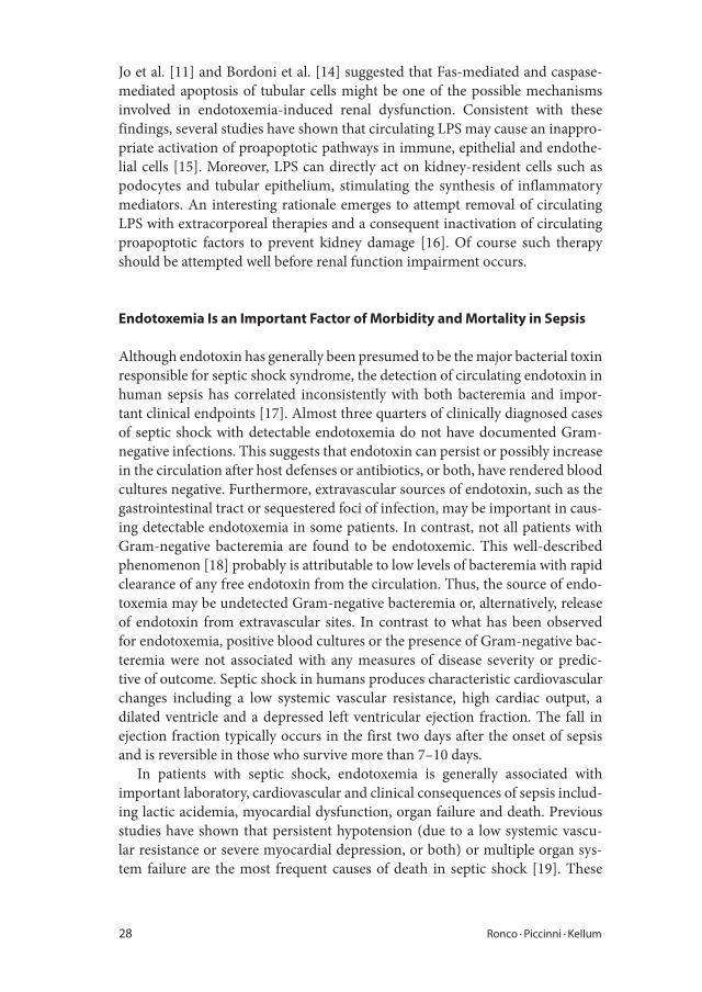
28 Ronco · Piccinni · Kellum
Jo et al. [11] and Bordoni et al. [14] suggested that Fas-mediated and caspase-
mediated apoptosis of tubular cells might be one of the possible mechanisms
involved in endotoxemia-induced renal dysfunction. Consistent with these
findings, several studies have shown that circulating LPS may cause an inappro-
priate activation of proapoptotic pathways in immune, epithelial and endothe-
lial cells [15]. Moreover, LPS can directly act on kidney-resident cells such as
podocytes and tubular epithelium, stimulating the synthesis of inflammatory
mediators. An interesting rationale emerges to attempt removal of circulating
LPS with extracorporeal therapies and a consequent inactivation of circulating
proapoptotic factors to prevent kidney damage [16]. Of course such therapy
should be attempted well before renal function impairment occurs.
Endotoxemia Is an Important Factor of Morbidity and Mortality in Sepsis
Although endotoxin has generally been presumed to be the major bacterial toxin
responsible for septic shock syndrome, the detection of circulating endotoxin in
human sepsis has correlated inconsistently with both bacteremia and impor-
tant clinical endpoints [17]. Almost three quarters of clinically diagnosed cases
of septic shock with detectable endotoxemia do not have documented Gram-
negative infections. This suggests that endotoxin can persist or possibly increase
in the circulation after host defenses or antibiotics, or both, have rendered blood
cultures negative. Furthermore, extravascular sources of endotoxin, such as the
gastrointestinal tract or sequestered foci of infection, may be important in caus-
ing detectable endotoxemia in some patients. In contrast, not all patients with
Gram-negative bacteremia are found to be endotoxemic. This well-described
phenomenon [18] probably is attributable to low levels of bacteremia with rapid
clearance of any free endotoxin from the circulation. Thus, the source of endo-
toxemia may be undetected Gram-negative bacteremia or, alternatively, release
of endotoxin from extravascular sites. In contrast to what has been observed
for endotoxemia, positive blood cultures or the presence of Gram-negative bac-
teremia were not associated with any measures of disease severity or predic-
tive of outcome. Septic shock in humans produces characteristic cardiovascular
changes including a low systemic vascular resistance, high cardiac output, a
dilated ventricle and a depressed left ventricular ejection fraction. The fall in
ejection fraction typically occurs in the first two days after the onset of sepsis
and is reversible in those who survive more than 7–10 days.
In patients with septic shock, endotoxemia is generally associated with
important laboratory, cardiovascular and clinical consequences of sepsis includ-
ing lactic acidemia, myocardial dysfunction, organ failure and death. Previous
studies have shown that persistent hypotension (due to a low systemic vascu-
lar resistance or severe myocardial depression, or both) or multiple organ sys-
tem failure are the most frequent causes of death in septic shock [19]. These

Extracorporeal Removal of Endotoxin in Sepsis 29
associations provide strong evidence that endotoxin is an important mediator of
the high morbidity and mortality of septic shock.
Plasma Levels of LPS and LPS-Binding Protein
Plasma levels of LPS and LPS-binding protein (LBP) represent an important
finding in sepsis and septic shock. In a study from Opal et al. [20], where all
patients met consensus definitions of severe sepsis, 80% were found to be in
septic shock. The average APACHE II score was 26 ± 13.6, and the 28-day all-
cause mortality rate for the entire study group (n = 253) was 32.4%. Severe sep-
sis patients with elevated endotoxin levels had significantly greater mortality
rates than those patients without measurable endotoxin. In some studies the
28-day all-cause mortality was 35% in endotoxemic patients and only 22% in
nonendotoxemic patients. The greater quartiles of plasma endotoxin levels were
associated with shorter survival times (p < 0.05). Detectable levels of endotoxin
in the plasma were more common in patients with shock than in patients with-
out shock. No correlation was observed between the endotoxin levels and LBP
levels found in the systemic circulation of these patients.
There was also no significant association between the type of microorgan-
ism that caused the septic process and either plasma endotoxin or LBP levels. In
general, patients with documented Gram-negative bacterial sepsis had similar
levels of endotoxin and LBP as did patients with Gram-positive bacterial sepsis
or fungal sepsis. This study shows that endotoxin is frequently found in the sys-
temic circulation in the presence of sepsis, regardless of the infecting microor-
ganism. Endotoxemia may have originated from unrecognized Gram-negative
infections in some patients or from enteric bacteria within the gastrointestinal
tract [21]. Regional hypoperfusion and mucosal ischemia are thought to pro-
mote the translocation of endotoxin from the intestinal lumen to the systemic
circulation [21, 22].
Infusion of endotoxin in animals and in healthy human hosts activates a
signaling cascade analogous to sepsis, which, at high levels, results in organ
dysfunction. However, the degree of organ dysfunction does not show a direct
dose-response relationship with the amount of LPS infused [23]. Genetic back-
ground and sex have been shown to influence in vivo human responses to
endotoxin challenge [24]. In clinical testing, levels of endotoxin on admission
to intensive care units (ICUs) have been shown to be predictive of the develop-
ment of organ dysfunction in patients with sepsis [25]. Endotoxemia is com-
mon in critically ill patients and levels of endotoxin have been reported to vary
over time [22, 26].
Endotoxin levels in the normal host are tightly regulated through a highly
conserved series of mechanisms for endotoxin binding, signaling and clearance.
Transient endotoxemia has been found in many healthy host states, including

30 Ronco · Piccinni · Kellum
marathon runners, Olympic athletes and smokers [27, 28]. However, in disease,
the mechanisms that regulate endotoxin may vary as the inflammatory response
becomes uncoupled from the inciting injury. The breakdown of these regulatory
mechanisms along with ongoing translocation of gut-derived endotoxin may
contribute to the fluctuating levels observed in several patients. Other potential
contributors to fluctuating levels of endotoxin can also include an uncontrolled
or recurrent source of Gram-negative sepsis.
The concept of linking recurrent exposure to endotoxin to the overwhelm-
ing, uncontrolled systemic inflammatory response associated with multiple
organ failure in critical illness is attractive. If ongoing fluctuations in endotoxin
levels play a role not only as a marker of ongoing injury, but also as a mediator, it
may be possible to measure and modulate this process. A broader understand-
ing of the kinetics of changing levels of endotoxin over time in critical illness
may improve therapeutic timing and targeting of specific antiendotoxin thera-
pies [29].
Diagnostic and Prognostic Implications of Endotoxemia in Critical Illness
Although endotoxin is ubiquitous, it has been notoriously difficult to mea-
sure reliably in human illness. The most commonly used diagnostic test – the
chromogenic limulus amebocyte lysate assay – has been widely used to detect
endotoxin contamination of drugs and fluids; however, its utility in biological
samples has been limited [30] due to circulating inhibitors of the coagulation
reaction. Moreover, other microbial products, notably from fungi, can activate
the limulus reaction. Therefore, the assay is not specific for endotoxin. Gram-
negative infection is one of the many infectious causes. Although microbiologi-
cally proven infection is relatively uncommon in cohort studies, endotoxemia is
generally present on the day of ICU admission in the majority of patients.
In a cohort study, Marshall et al. [25], by means of the new Endotoxin Activity
(EA) Assay (approved by the US Food and Drug Administration) found EA lev-
els ≤0.40 in 367 patients (42.8% of the population), between 0.40 and 0.60 in
228 patients (26.6%), and ≥0.60 in 262 patients (30.6%); thus, the majority of
patients of the study had elevated endotoxin levels at the ICU admission. Criteria
for severe sepsis were present at the time of admission in 74 patients (8.6% of the
study cohort). ICU mortality for this population was higher than that of patients
who did not have sepsis (32.4 vs. 11.5%; p < 0.001). EA levels were significantly
higher for patients who met criteria for severe sepsis (0.57 ± 0.26 vs. 0.46 ± 0.26
units; p < 0.001), and the risk of severe sepsis increased with increasing incre-
ments of EA levels. Patients with endotoxemia had increased ICU and hospital
mortality. The ICU length of stay was modestly longer for patients with EA lev-
els ≥0.60 at the time of ICU admission (6.8 ± 12.2 vs. 4.9 ± 10.7 days; p < 0.05).
However, patients who had intermediate or high levels of endotoxin on the day

Extracorporeal Removal of Endotoxin in Sepsis 31
of admission were clearly a sicker population, as reflected by higher admission
APACHE II scores and a greater prevalence of severe sepsis. Moreover, patients
with the highest levels of circulating endotoxin had a significantly increased risk
of dying while in the ICU. Thus, the presence of endotoxemia identifies a high-
risk subpopulation of critically ill patients.
Whether this increased mortality risk might be reduced by specific measures
to neutralize endotoxin is unknown, but the hypothesis is an attractive one. Not
only have the results of animal studies supported a pathogenic role for increased
concentrations of endotoxin, but previous analyses of studies designed to neu-
tralize endotoxin in human sepsis have suggested that the greatest potential for
benefit occurs in those patients in whom endotoxemia is present, whereas inter-
vention may actually harm those with infection caused by non-Gram-negative
organisms [31].
A Targeted Extracorporeal Therapy for Endotoxemia
Since its discovery, whatever the source, endotoxemia is associated with increased
organ dysfunction and risk of death in critically ill patients [25]. Furthermore,
blood levels of endotoxin have been reported to vary over time in patients, sug-
gesting subsequent waves of exposure either from infection or from intestinal
translocation. However, despite new methods of detecting endotoxin, treatment
is quite limited. Bacterial infection is treated with antibiotics, but there is no
effective method of restoring gut barrier function. Furthermore, many antibiot-
ics result in endotoxin release as bacteria are killed.
Antiendotoxin therapies, like antiendotoxin antibody (HA-1A or E5), have
been disappointing, and have failed to demonstrate benefit in patients with con-
firmed Gram-negative sepsis. Polymyxin B is a well-known antibiotic that has
high affinity for endotoxin and is able to neutralize it, although it is associated
with neurotoxicity and nephrotoxicity, which precludes its systemic use. Since
1994, polymyxin B has been bound and immobilized to polystyrene fibers.
When it is used for hemoperfusion treatment, it can effectively bind endotoxin
both in vitro and in vivo. This therapy has been available in Japan since 1994
and thousands of patients have been treated. Unfortunately, despite widespread
use in Japan, no large randomized trials have established the efficacy of poly-
myxin B hemoperfusion.
Several small studies have been conducted, however, and most have stud-
ied similar patients. In a recent systematic review, Cruz et al. [32] identified a
total of 28 publications, including randomized controlled trials, of polymyxin B
hemoperfusion for treatment of sepsis and septic shock. Polymyxin B hemop-
erfusion was associated with a significantly lower mortality compared to con-
ventional therapy (relative risk: 0.53, 95% CI: 0.43–0.65). Secondary endpoints
such as mean arterial pressure increase, vasopressor decrease and mean partial

32 Ronco · Piccinni · Kellum
1 Lowry SF: Human endotoxemia: a model for
mechanistic insight and therapeutic target-
ing. Shock 2005;24(Suppl 1):94–100.
2 Suffredini AF, Shelhamer JH, Neumann RD,
Brenner M, Baltaro RJ, Parillo JE: Pulmonary
and oxygen transport effects of intravenously
administered endotoxin in normal humans.
Am Rev Respir Dis 1992;145:1398–1403.
3 Smith PD, Suffredini AF, Allen JB, Wahl LM,
Parillo JE, Wahl SM: Endotoxin administra-
tion to humans primes alveolar macrophages
for increased production of inflammatory
mediators. J Clin Immunol 1994;14:141–148.
4 Richardson RP, Rhyne CD, Fong Y, Hesse DG,
Tracey KJ, Marano MA, Lowry SF, Antonacci
AC, Calvano SE: Peripheral blood leukocyte
kinetics following in vivo lipopolysaccharide
(LPS) administration to normal human
subjects: influence of elicited hormones and
cytokines. Ann Surg 1989;210:239–245.
5 Suffredini AF, Fromm RE, Parker MM,
Brenner M, Kovacs JA, Wesley RA, Parrillo
JE: the cardiovascular response of normal
humans to the administration of endotoxin.
N Engl J Med 1989;321:280–287.
6 Aras O, Shet A, Bach RR, Hysjulien JL,
Slungaard A, Hebbel RP, Escol G, Jilma B,
Key NS: Induction of microparticle- and
cell-associated intravascular tissue factor
in human endotoxemia. Blood 2004;103:
4545–4553.
7 Kumar A, Thota V, Dee L, Olson J, Uretz
E, Parillo JE: Tumor necrosis factor-alpha
and interleukin 1-beta are responsible for
in vitro myocardial cell depression induced
by human septic shock serum. J Exp Med
1996;183:949–958.
pressure of oxygen in arterial blood/forced inspiratory pressure of oxygen ratio
increase were also highly significant. Nevertheless, polymyxin B hemoperfu-
sion appears to have favorable effects on survival and hemodynamics, and the
authors argue for the need for further rigorous study of this therapy.
More recently, the same authors reported the results of a multicenter ran-
domized controlled trial on a population of patients with septic shock arising
from abdominal infection [33]. The authors reported for the first time prospec-
tively a superior outcome in patients treated with two sessions of hemoperfu-
sion with polymyxin B cartridges compared to patients treated with the current
standard of care. This study opens new avenues for the evaluation of this thera-
peutic approach in other selected populations with endotoxin-mediated septic
shock. Indeed, given the poor overall outcomes associated with endotoxemia,
polymyxin B hemoperfusion would seem to be a welcome intervention, par-
ticularly now that better methods for detection of endotoxemia have become
available. Thus, the time is right to confirm the encouraging results recently
published in the literature with adequately powered trials and clinical registries
combining diagnostic and therapeutic interventions on endotoxin–mediated
syndromes.
The theory is in favor of the use of extracorporeal therapies where other
therapeutic approaches have failed. The rationale is fully consistent with the
experimental findings in animal and human models. The technology is simple
and safe and ready to be utilized on a larger scale. All the ingredients for a pos-
sible success are included in this new recipe.
References

Extracorporeal Removal of Endotoxin in Sepsis 33
8 Giroir BP, Johnson JH, Brown T, Allen GL,
Beutler B: The tissue distribution of tumor
necrosis factor biosynthesis during endotox-
emia. J Clin Invest 1992;90:693–698.
9 Meldrum DR: Tumor necrosis factor in the
heart. Am J Physiol 1998;274:R577–R595.
10 Smith PD, Suffredini AF, Allen JB, Wahl LM,
Parillo JE, Wahl SM: Endotoxin administra-
tion to humans primes alveolar macrophages
for increased production of inflammatory
mediators. J Clin Immunol 1994;14:141–148.
11 Jo SK, Cha DR, Cho WY, Kim HK, Chang
KH, Yun SY, Won NH: Inflammatory
cytokines and lipopolysaccharide induce
Fas-mediated apoptosis in renal tubular cells.
Nephron 2002;91:406–415.
12 Guo R, Wang Y, Minto AW, Quigg RJ,
Cunningham PN: Acute renal failure in
endotoxemia is dependent on caspase activa-
tion. J Am Soc Nephrol 2004;15:3093–3102.
13 Wesche-Soldato DE, Swan RZ, Chung CS,
Ayala A: The apoptotic pathway as a thera-
peutic target in sepsis. Curr Drug Targets
2007;8:493–500.
14 Bordoni V, Bolgan I, Brendolan A, Crepaldi
C, Gastaldon F, D’Intini V, Pilotto L,
Inguaggiato P, Bonello M, Galloni E, Everard
P, Bellomo R, Ronco C: Caspase-3 and -8
activation and cytokine removal with a novel
cellulose triacetate superpermeable mem-
brane in an in vitro sepsis model. Int J Artif
Organs 2003;26:897–905.
15 Camussi G, Mariano F, Biancone L, De
Martino A, Bussolati B, Montrucchio G,
Tobias PS: Lipopolysaccharide binding
protein and CD14 modulate the synthe-
sis of platelet-activating factor by human
monocytes and mesangial and endothelial
cells stimulated with lipopolysaccharide. J
Immunol 1995;155:316–324.
16 Cantaluppi V, Assenzio B, Pasero D,
Romanazzi GM, Pacitti A, Lanfranco G,
Puntorieri V, Martin EL, Mascia L, Monti G,
Casella G, Segoloni GP, Camussi G, Ranieri
VM: Polymyxin-B hemoperfusion inactivates
circulating proapoptotic factors. Intensive
Care Med 2008;34:1638–1645.
17 Stumacher RJ, Kovnat MJ, McCabe WR:
Limitations of the usefulness of the limulus
assay for endotoxin. N Engl J Med 1973;283:
1261–1264.
18 Jorgensen JH: Clinical applications of the
limulus amebocyte lysate test; in Proctor
RA (ed): Clinical Aspects of Endotoxin
Shock: Handbook of Endotoxin. Amsterdam,
Elsevier, 1986, vol 4, pp 127–160.
19 Herring WB, Herion JC, Walker RI, Palmer
JG: Distribution and clearance of circulating
endotoxin. J Clin Invest 1963;43:79–87.
20 Opal SM, Scannon OJ, Vincent JL, White M,
Carroll SF, Palardy JE, Parejo NA, Pribble
JP, Lemke JH: Relationship between plasma
levels of LPS and LPS-binding protein in
patients with severe sepsis and septic shock. J
Infect Dis 1999;180:1584–1589.
21 Hurley JC: Endotoxemia: methods of detec-
tion and clinical correlates. Clin Microbiol
Rev 1995;8:268–292.
22 Danner RL, Elin RJ, Hosseini JM, Wesley RA,
Reilly JM, Parillo JE: Endotoxemia in human
septic shock. Chest 1991;99:169–175.
23 Stephens RC, O’Malley CM, Frumento RJ, et
al: Low-dose endotoxin elicits variability in
the inflammatory response in healthy volun-
teers. J Endotoxin Res 2005;11:207–212.
24 Coyle SM, Calvano SE, Lowry SF: Gender
influences in vivo human responses to endo-
toxin. Shock 2006;26:538–543.
25 Marshall JC, Foster D, Vincent JL, et al:
Diagnostic and prognostic implications of
endotoxemia in critical illness: results of the
MEDIC study. J Infect Dis 2004;190:527–534.
26 Klein DJ, Derzko A, Foster D, Seely AJE,
Brunet F, Romaschin AD, Marshall JC: Daily
variation in endotoxin levels is associated
with increased organ failure in critically ill
patients. Shock 2007;28:524–529.
27 Brock-Utne JG, Gaffin SL, Wells MT, et al:
Endotoxaemia in exhausted runners after
a long-distance race. S Afr Med J 1988;73:
533–536.
28 Hasday JD, Bascom R, Costa JJ, et al:
Bacterial endotoxin is an active component
of cigarette smoke. Chest 1999;115:829–835.
29 Opal SM, Gluck T: Endotoxin as a drug tar-
get. Crit Care Med 2003;31(Suppl 1):57–64.
30 Cohen J: The detection and interpretation
of endotoxemia. Intensive Care Med 2000;
26(Suppl 1):S51–S56.

34 Ronco · Piccinni · Kellum
31 Ziegler EJ, Fisher CJ, Sprung CL, et al:
Treatment of Gram-negative bacteremia and
septic shock with HA-1A human monoclonal
antibody against endotoxin. A randomized,
double-blind, placebo-controlled trial. The
HA-1A Sepsis Study Group. N Engl J Med
1991;324:429–436.
32 Cruz DN, Perazella MA, Bellomo R, de
Cal M, Polanco N, Corradi V, Lentini P,
Nalesso F, Ueno T, Ranieri VM, Ronco C:
Effectiveness of polymyxin B-immobilized
fiber column in sepsis: a systematic review.
Critical Care 2007;11:R47.
33 Cruz DN, Antonelli M, Fumagalli R, et al:
Early use of polymyxin B hemoperfusion in
abdominal septic shock: the EUPHAS ran-
domized controlled trial. JAMA 2009;301:
2445–2452.
Claudio Ronco, MD
Department of Nephrology, Dialysis and Transplantation, San Bortolo Hospital
Viale Rodolfi 37
IT–36100 Vicenza (Italy)
Tel. +39 0 444993869, Fax +39 0 444993949, E-Mail [email protected]

Extracorporeal Endotoxin Removal: Theory and Technology
Ronco C, Piccinni P, Rosner MH (eds): Endotoxemia and Endotoxin Shock: Disease, Diagnosis and
Therapy. Contrib Nephrol. Basel, Karger, 2010, vol 167, pp 35–44
Extracorporeal Removal of Endotoxin: The Polymyxin B-Immobilized Fiber Cartridge
Tohru Tania � Hisataka Shojib � Gualtiero Guadagnic �
Angelo Peregod
aDepartment of Surgery, Shiga University of Medical Science, Otsu City, bDivision of Emergency and
Critical Care Medicine, Toray Medical Co., Ltd., Tokyo, Japan; cESTOR S.p.A., Milan, dU.O.C Nefrologia e
Dialisi, Ospedale di Monselice, Monselice, Italy
AbstractEndotoxin, which consists of lipopolysaccharide (LPS), is an outer membrane component
of the Gram-negative bacterial cell wall. Endotoxin in the blood stream from an infectious
focus or through translocation from the gut plays an important role in the pathogenesis
of severe sepsis and septic shock. It binds to monocytes and macrophages, activating
them to trigger the production of a variety of mediators. These mediators injure endothe-
lial cells and induce microcirculatory dysfunction. This leads to subsequent organ dys-
function and multiorgan failure. The neutralization or elimination of endotoxin in the
blood is an enticing approach for treating severe sepsis and septic shock. Selective adsor-
bent therapy targeting blood endotoxin has been clinically applied for more than 15
years, mainly in Japan and more recently in Italy and other countries. ToraymyxinTM (PMX;
Toray, Tokyo, Japan) is a selective blood endotoxin removal cartridge. PMX is composed of
polymyxin B (PL-B) covalently bonded to polystyrene-derivative fibers. It is well known
that PL-B binds endotoxin and has bactericidal activity. PL-B has a strong affinity to endo-
toxin and is able to bind the lipid A portion of endotoxin through ionic and hydrophobic
interactions. Intravenous injection of PL-B has significant nephrotoxic and neurotoxic
effects. However, covalently immobilized PL-B on the adsorbents of PMX do not leak out
into the blood stream, thus allowing the clinical application without the known toxic
effects of PL-B. Within each cartridge, an adsorbent structure made of PL-B-fixed fabrics is
included. Blood flow direction is well controlled by adopting a radial flow system. PMX
treatment occurs by hemoperfusion at a blood flow rate of about 80–120 ml/min. Heparin
is preferably used as an anticoagulant. In Japan, PMX has been clinically used since 1994
under the national health insurance system. It is estimated that over 80,000 patients have
received PMX treatment in Japan. Not only has PMX been clinically used safely in Japan,
but also in other countries. Copyright © 2010 S. Karger AG, Basel

36 Tani · Shoji · Guadagni · Perego
Bacterial lipopolysaccharide (LPS or endotoxin) is one of the most powerful
known molecules of bacterial signaling. Animals and humans have sensitive
mechanisms for recognizing the presence of LPS in their tissues. Monocytes,
macrophages and neutrophils, expressing specific receptors, the transductors
CD14 and Toll-like receptor-4, are particularly sensitive to the presence of LPS.
They respond by producing inflammatory mediators which enhance and diver-
sify the LPS signal, triggering the host defenses which isolate and destroy the
invading bacteria.
For reasons not entirely clear, the response to the LPS can be excessive and
may lead to severe sepsis, septic shock and even death.
In the mid-1970s, polymyxin B (PL-B) was discovered to be protective against
endotoxin-induced hemodynamic shock but, at the same time, was demonstrated
to be extremely toxic for the kidney and central nervous system. Based on this,
several antiendotoxin strategies have been proposed (e.g. monoclonal antibod-
ies, antiendotoxin vaccines, inhibition of endotoxin synthesis); however, those
failed in demonstrating reproducible outcomes in septic subjects. In 1983 Toray
Industries Inc. developed a blood endotoxin removal cartridge, ToraymyxinTM
(PMX), which could be clinically applied by direct hemoperfusion. PMX consists
of a polystyrene-based, fibrous adsorbent in which the antibiotic PL-B is cova-
lently immobilized as a ligand to adsorb endotoxin. PMX was approved for Japan
in 1994, CE-marked in 1998 and has been commercially available in Europe since
2002. PMX is the only device currently available on the market for the removal
of endotoxins from blood. The working principle is based on the antiendotoxin
potential of PL-B. Since PL-B is known to have dramatic side effects for the organ-
ism when used systemically, this molecule is linked to an inert material in the
PMX cartridge, with a covalent bond to prevent its release into the bloodstream.
Endotoxins and Polymyxin B
Endotoxin invasion in the organism is followed by an immediate reaction of
monocytes which bind it through the antigenic portion and start the inflamma-
tory reaction. The monocyte activation is followed by the intervention of lipo-
proteins which also bind free endotoxin through the lipid A portion and try
to steal LPS from monocytes in the global attempt to fight the increase of the
inflammatory state [1].
If the endotoxin level is high, the monocyte response exceeds the lipoprotein-
mediated LPS clearance, creating the conditions in which sepsis can originate.
Therefore, in order to remove endotoxin from whole blood, a strong binding
force is needed, as LPS which has already bonded to monocytes or lipoprotein
complexes has to be removed.
PL-B is an antibiotic agent that has strong bactericidal activity for Gram-
negative bacilli, and also has a strong affinity with endotoxin. PL-B plays a

The Polymyxin B-Immobilized Fiber Cartridge 37
double role towards endotoxin: binding and neutralization. The neutralization
is the most important action and depends on the interaction between the hydro-
phobic residues of PL-B and the fatty acid chains of LPS. The results of this
chemical interaction is a unimolecular compound (fig. 1).
How to Design the PL-B-Immobilized Fiber and Cartridge
PL-B cannot be intravenously injected due to its nephrotoxicity and neurotoxic-
ity. Therefore, it was covalently bonded to an insoluble substrate as a ligand and
used as a selective adsorbent for an endotoxin. Polystyrene- and polypropylene-
conjugated fibers, with island-sea type-conjugated fibers and polypropylene
(island component) to provide reinforcement to the fibers, were utilized as a
substrate fiber. A bundle of the conjugated fibers were knitted into a fabric and
the polystyrene component of the fibers was chemically modified to introduce
α-chloroacetamide methyl groups as functional groups to fix PL-B. PL-B has 5
primary amino groups derived from α,γ-diaminobutyric acid in the molecular
structure. PL-B was covalently immobilized on the surface of the fibers through
the chemical reaction between the primary amino groups of PL-B and an active
chlorine atom of the functional groups (fig. 2) [2].
Interaction between PL-B and endotoxin is based on ionic and hydrophobic
forces. Ionic forces are through negatively charged phosphate groups in the lipid
A portion of endotoxin and positively charged α,γ-diaminobutyric acid residue
in PL-B. As the primary amino groups are also utilized for the immobilization
leu
D-phe
dab
dab
dab
dab
dab fatty acid
PL-B
dab
thr
thr
Lipid PL-B
+
Lipid/PL-B (Lipid/PL-B)*
k1
k–1
k2
k–2
Fig. 1. Interaction between the hydrophobic residues of the PL-B and the acid chains of
LPS. The hydrophobic residue is indicated by arrows.

38 Tani · Shoji · Guadagni · Perego
reaction, optimization of PL-B immobilization so as not to reduce the interac-
tion forces was needed. It was found that the higher the number of primary
amino groups in the fixed PL-B, the more the endotoxin removal capacity of
PL-B-immobilized fiber was increased. PL-B was fixed so that there were 3–4
active primary amino groups in the fixed PL-B molecule.
An adsorbent compartment was built up with a knitted fabric made of PL-B-
immobilized fiber (fig. 3). The PL-B-fixed knitted fabric was wrapped around a
central pipe which had many side holes. By embedding this compartment into
a case, the PMX cartridge was produced. Adopting this structural compart-
ment, the homogeneous blood flow within the cartridge could be obtained.
Blood enters the cartridge from the inlet and moves up within the central pipe.
After going up the pipe, the blood goes out of the side holes in the central pipe
crossing the layer of the fabric in touch with the adsorbent. By the develop-
ment of a radial flow system within the cartridge, a homogeneous blood flow
distribution could be achieved, contributing to the efficient utilization of the
adsorbent.
Safety Evaluation
A healthy dog safety study was conducted to evaluate biocompatibility and
safety of the PMX cartridge. Study subjects underwent a 2-hour extracorporeal
Conjugated fiber
Polystyrene/polypropylene (9:1)
Polypropylene
Fig. 2. The illustration on the left illustrates the schema of the cross-section of island-sea
type-conjugated fiber filament. The island component is made of polypropylene polymer
and the sea component is made of a mixed polymer of polystyrene and polypropylene
(9:1). The picture on the right shows an electron micrograph of the cross-section of a fiber
filament.

The Polymyxin B-Immobilized Fiber Cartridge 39
treatment with a PMX cartridge. They were monitored for mean arterial pres-
sure, hematocrit, platelet count, leukocyte count, serum proteins and transami-
nases for up to 3 h after treatment. All measurements remained or returned to
acceptable ranges during and after treatment, and no study subjects died. No
unfavorable (adverse) events were reported in this study.
Performances of PL-B-Immobilized Fiber Cartridge
In order to asses PMX performance in LPS removal, we have to introduce
two concepts. The first is the ‘endotoxin burden’, which is the quantity of
circulating endotoxins in a patient affected by (endotoxemic) septic shock.
Based on the results from the MEDIC trial [3], high endotoxin activity level
Fig. 3. An adsorbent compartment of a Toraymyxin cartridge.

40 Tani · Shoji · Guadagni · Perego
can be considered to be an endotoxin activity assay (EAA) ≥0.6; Monti et
al. [4] showed EAA >0.9 in patients with septic shock. In turn, consider-
ing Escherichia coli-derived LPS, the corresponding LPS concentration is 7.0
ng/ml or 70 EU/ml (EU = endotoxin units); therefore, under the condition
mentioned, the endotoxin burden of a 70-kg male is: 70 × 5,000 (total blood
volume 5 l) = 350,000 EU.
The second concept is the ‘device adsorbtion capability’ (DAC), which
looks at how many EUs a single PMX is able to remove from blood. The
DAC of PMX is not reported officially. It was, however, possible to calculate
it in a quite precise way from some previously published in vitro studies. It
could also be estimated on the basis of the experience gained in the last years
from the in vivo treatments monitored by EAA. The following table 1 shows
the calculated values of the DAC of PMX (type: PMX-20R) in three different
situations: (1) in vitro using solutions of hemoglobin, (2) in vitro with bovine
blood (2 liters of recirculating blood) and (3) on a hypothetical septic patient
with an infection from E. coli and an initial EAA value of 0.85 (which corre-
sponds to an LPS concentration of 43 EU/ml for the E. coli). After a treatment
with PMX, the hypothetical patient would have an EAA of 0.6 (which cor-
responds, to an LPS concentration of 15 EU/ml for E. coli). This hypothetical
patient is absolutely realistic and consistent with data from different research-
ers (table 1) [4, 5].
How It Works
PMX is designed so that whole blood might be circulated during a hemoperfu-
sion procedure. A large surface area could be obtained using fibrous material
which has a small diameter. Furthermore, fibers have a porous surface structure
resulting from the chemical reaction used to introduce a functional group. This
induces an enlargement of the surface area of the adsorbents. On the other hand,
pressure loss between the inlet and outlet of the cartridge remains low due to the
use of a fibrous material, a knitted fabric as an adsorbent structure and the con-
trolled homogeneous blood flow within a cartridge enabling stable whole blood
hemoperfusion. The endotoxin DAC of PMX was evaluated in vitro comparing
the circulation of 1.5 liters of calf serum solution containing purified E. coli LPS
at the concentration of 10 ng/ml through the PMX cartridge at a flow rate of 100
ml/min and through a carrier fiber cartridge without PL-B. After 2 h of circula-
tion, LPS concentration changes of the two solutions were evaluated. Only PMX
could decrease the level of LPS.
Recently, Nishibori et al. [6] investigated the cellular components in the
PMX column after hemoperfusion in septic patients. It is well known that dif-
ferent populations of leukocytes are activated during septic shock and change
their adhesive phenotype. They therefore hypothesized that some populations

The Polymyxin B-Immobilized Fiber Cartridge 41
of leukocytes might be adsorbed in the PMX column and removed from the
blood circulation after the treatment. They demonstrated that PMX also specifi-
cally bound monocytes from the peripheral blood leukocytes of septic patients
by means of an analysis of bound cells using immunocytochemical and electron
microscopic techniques.
The specific removal of monocytes from septic patients may produce ben-
eficial effects by reducing the interaction between monocytes and functionally
associated cells, including vascular endothelial cells. The mechanism by which
monocytes are specifically adsorbed on the surface of PL-B-immobilized fiber is
still not clear, with the main hypotheses being physical characteristics of fibrous
material of PL-B-immobilized fiber or some interactions mediated by endo-
toxin removal. This second appealing hypothesis (still to be demonstrated) is
based on the assumption that binding sites of endotoxin and PL-B are different
from those of endotoxin and monocytes. Therefore, PMX could remove mono-
cytes by removing the LPS-monocyte complex. Anyhow, it is very important to
remove endotoxin as a trigger of inflammatory reaction in the body. The fact
that PMX can remove some activated immune cells is very interesting from the
standpoint of its possible immunomodulatory effect for the treatment of severe
sepsis and septic shock.
Table 1. Endotoxin adsorption capability device with in vitro and in vivo conditions
Toraymyxin LPS
adsorption
Operating conditions Initial endotoxin
concentration
Adsorption
capacity device
In vitro data
(Tani et al. [9], 1992),
static model
Hb solution – PMX
fiber only
solution of hemoglobin
+2 g of PMX fibers; 1 h
incubating time
686 EU/ml 1,404,480 EU
In vitro data
(Sakai et al. [10], 1993),
perfusion model
bovine blood
in vitro perfusion –
bovine blood: 2 liters;
perfusion time: 2 h
400 EU/ml 640,000 EU
Septic shock patients
with E. coli infection
and EAA monitoring
hypothetical patient
with E. coli infection;
EAA = 0.9 before,
ed EAA = 0.6 after
70 EU/ml 275,000 EU
Disregarding the first in vitro test (very far from the clinical setting), 1 PMX device is able to trap an aver-
age of around 300,000 EU in a standard session. This value is truly interesting because it is of the same
order of magnitude of the ‘endotoxin burden’.

42 Tani · Shoji · Guadagni · Perego
Advantages of Uses of Adsorbent Technology to Remove Endotoxin
A possible mechanism for blood purification techniques is to remove inflam-
matory mediators from the bloodstream which are involved in the pathogenesis
and/or pathophysiology of severe sepsis and septic shock. There are multiple
mediators that play an important role in the systemic inflammatory process
such as endotoxin and exotoxin from exogenous sources or endogenous media-
tors such as cytokines, chemokines and various forms of lipids. Their molecular
weight range is widely distributed, which is important when considering a 10- to
20-kd cutoff for a hollow fiber filter. Therefore, there is a need to choose appro-
priate modalities for selective mediator removal. The physicochemical aspect
of mass transfer is the principle based on diffusion, filtration and adsorption.
Renal replacement therapy, such as hemodialysis, hemofiltration and hemodia-
filtration, uses the principles of diffusion and filtration, while the adsorption
process is not affected by the cutoff point of hollow fiber. As a result, targeting
molecular species becomes possible.
The basic molecular structure of endotoxin consists of a polysaccharide por-
tion which includes an O-specific side chain, an inner and outer core region, and
lipid A moiety which is an active center of LPS. Endotoxemia can occur in Gram-
negative sepsis, or even Gram-positive sepsis, through bacterial or endotoxic
translocation from the gut. The molecular aspect of endotoxin in blood or plasma
is not clear. However, blood endotoxin is not always an LPS molecule itself, but
can be in a variety of forms, such as membrane fragments, blebs and vesicles, in
combination with bacterial phospolipids. In blood circulation, endotoxin may be
bound by a large number of plasma components. In addition, LPS forms micelles
in the blood. Therefore, the molecular weight is so big that it is difficult to remove
through hemodialysis and the hemofiltration technique. Plasma exchange is one
of the options for eliminating blood endotoxin. However, a lot of expensive fresh
frozen plasma is not efficient and it carries some risk of infection as well.
Conclusion
Endotoxin has been discussed for a long time as the therapeutic target for the
treatment of sepsis. However, reagents such as antiendotoxin monoclonal anti-
body and BPI (bactericidal/permeability increasing protein), which neutral-
izes endotoxin, did not improve the outcome of sepsis. This raised the question
whether endotoxin is a real target for the development of novel therapeutics.
Recently, favorable results of E5564 have been reported [7]. However, treat-
ment with a phospholipid emulsion did not reduce 28-day all-cause mortality
or reduce the onset of new organ failure in patients with suspected or confirmed
Gram-negative severe sepsis [8]. It seems that endotoxin as a therapeutic target
has mixed success and is still controversial.

The Polymyxin B-Immobilized Fiber Cartridge 43
1 Bhor VM, Thomas CJ, Surolia N, Surolia
A: Polymyxin B: an ode to an old antidote
for endotoxic shock. Mol Biosyst 2005;1:
213–222.
2 Shoji H: Extracorporeal endotoxin removal
for the treatment of sepsis: endotoxin
adsorption cartridge (Toraymyxin). Ther
Aher Dial 2003;7:108–114.
3 Marshall JC, Foster D, Vincent JL, Cook
DJ, Dellinger RP, Opal S, Abraham E, Brett
S, Smith T, Mehta S, Derzko A, Romaschin
A: Diagnostics and prognostic implications
of endotoxemia in critically illness: results
of the MEDIC study. J Infect Dis 2004;190:
527–534.
4 Monti G, Terzi V, Mininni M, Colombo
S, Vesconi S, Casella G: Polymyxin B
hemoperfusion in high endotoxin activity
level septic shock patients. Critical Care
2008;12(Suppl 2):P458, DOI: 10.1186/
cc6679.
5 Novelli G, Rossi M, Poli L, Ferretti G,
Ruberto F, Spoletini G, Levi Sandri GB,
Mennini G, Morabito V, Berloco PB (Roma):
Valutazione precoce dell’endotossinemia
in pazienti affetti da sepsi nel periodo post
operatorio mediante lo spectral’s EAA (TM)
endotoxin activity assay XXXIII Congresso
nazionale SITO, Milano 13–15 Dicembre
2009.
The reason why some antiendotoxin reagents do not work well is not always
clear. It may be related to the low efficiency of the reagent itself in neutralizing
endotoxin. In the past a major question was to identify the more effective antien-
dotoxin strategy: endotoxin neutralization with reagents in the body or endotoxin
extracorporeal removal from the blood. It seems possible to say after 15 years of
clinical application that extracorporeal endotoxin removal by polymyxin B hemo-
perfusion is rational for the treatment of severe sepsis and septic shock. In addi-
tion, it is rational to use adsorbent technology to remove endotoxin from the blood
as endotoxin is estimated to exist in the blood as a large molecule or aggregate.
It is very important to design an adsorbent which can exert a strong affinity
with endotoxin in the blood. It may be possible to design an adsorbent using a
different kind of ligand. However, PL-B is the most potent molecule that has a
strong affinity with endotoxin and could construct a selective adsorbent which
could work in the blood binding LPS even if linked to a multicomponent struc-
ture. PMX has been safely applied clinically for the treatment of severe sepsis
and septic shock since 1994 in Japan and since 2002 in Italy, and is becoming
more widely used year by year.
Another important therapeutic point of view is in relation to diagnostics. It
is questionable whether blood endotoxin has been correctly measured in the
previous antiendotoxin studies. Blood endotoxin measurement is still contro-
versial. However, the newly developed EAA, which is an FDA-approved novel
assay for endotoxin, is commercially available. This could enable endotoxin-
targeted treatment by selecting an adequate patient population. PMX combined
with EAA therapy is one of the more promising approaches for treating severe
sepsis septic shock.
References

44 Tani · Shoji · Guadagni · Perego
6 Nishibori M, Takahashi HK, Katayama H,
Mori S, Saito S, Iwagaki H, Tanaka N, Morita
K, Ohtsuka A: Specific removal of monocytes
from peripheral blood of septic patients by
polymyxin B-immobilized filter column.
Acta Med Okayama 2009;63:65–69.
7 Tidswell M, Tilllis W, LaRosa SP, Lynn M,
Wittek AE, Kao R, Wheeler J, Gogate J, Opal
SM, the Eritoran Sepsis Study Group: Phase 2
trial of Eritoran tetrasodium (E5564), a Toll-
like receptor 4 antagonist, in patients with
severe sepsis. Crit Care Med 2010;38:72–83.
8 Dellinger RP, Tomakyo JF, Angus DC, Opal
S, Cupo MA, McDermont S, Ducher A,
Calandra T, Cohen J, the Lipid Infusion
and Patient Outcomes in Sepsis (LIPOS)
Investigators: Efficacy and safety of a phos-
pholipid emulsion (GR270773) in Gram-
negative severe sepsis: results of a phase II
multicenter, randomized, placebo-controlled,
dose-finding clinical trial. Crit Care Med
2009;37:2929–2938.
9 Tani T, Chang TMS, Kodama M, Tsuchiya M:
Endotoxin removed from hemoglobin solu-
tion using polymyxin B immobilized fiber
followed by a new turbidometric endotoxin
assay. Biomat Art Cells & Immob Biotech
1992;20:457–462.
10 Sakay Y, Shoji H, Kobayashi T, Terada R,
Sugaya H, Murakami M, Moriyama K,
Minaga M, Kunimoto T, Takeyama T: New
extracorporeal blood purification devices
for critical care medicine under develop-
ment. Therapeutic Plasmapheresis 1993;12:
837–842.
Tohru Tani, MD, PhD
Department of Surgery, Shiga University of Medical Science
Tsukiwa-cho, Seta
Otsu City, Shiga, 520-2121 (Japan)
Tel. +81 077 548 2237, Fax +81 077 548 2240, E-Mail [email protected]

Extracorporeal Endotoxin Removal: Theory and Technology
Ronco C, Piccinni P, Rosner MH (eds): Endotoxemia and Endotoxin Shock: Disease, Diagnosis and
Therapy. Contrib Nephrol. Basel, Karger, 2010, vol 167, pp 45–54
Mechanisms of Polymyxin B Endotoxin Removal from Extracorporeal Blood Flow: Molecular Interactions
S. Vesentini � M. Soncini � G.B. Fiore � A. Redaelli
Department of Bioengineering, Politecnico di Milano, Milan, Italy
AbstractThe outer leaflet of Gram-negative bacteria membrane contains a great amount of
lipopolysaccharides, also known as endotoxins, which play a central role in the pathogen-
esis of septic shock. It has been demonstrated that the polymyxin B (PMB) molecule has
both antibacterial and antiendotoxin capabilities; in fact, it is able to compromise the bac-
terial outer membrane and bind lipopolysaccharides, thereby neutralizing its toxic effects.
Extracorporeal hemoperfusion treatments based on cartridges containing PMB-
immobilized fibers (Toraymyxin PMX-F; Toray Industries, Tokyo, Japan) are used to remove
endotoxins circulating in the blood flow. In this study, we focused on the characterization
of the interactions occurring in the formation of the PMB-endotoxin complex at the
molecular level. In particular, the molecular mechanics approach was used to evaluate the
interaction energy and eventually the interaction force between the two molecules. PMB
was faced with five molecular portions of lipopolysaccharides differing in their structure.
The interaction energy occurring for each molecular complex was calculated at different
intermolecular distances and the binding forces were estimated by fitting interaction
energy data. Results show that the short-range interactions between PMB and endotox-
ins are mediated mainly by hydrophobic forces, while in the long term, the complex for-
mation is driven by ionic forces only. Maximum binding forces calculated via molecular
mechanics for the PMB-endotoxin complex are in the range of 1.39–3.79 nN. Understanding
the interaction mechanism of the single molecular complex is useful both in order to fig-
ure out the molecular features of such interaction and to perform higher scale level analy-
sis, where such nanoscale detail is impractical but could be used to account for molecular
behavior at a coarse level of discretization. Copyright © 2010 S. Karger AG, Basel
Sepsis is a generalized infection of an organism with presence of bacteria in
the blood flow (bacteremia). Recently, in parallel to clinical and research

46 Vesentini · Soncini · Fiore · Redaelli
developments concerning antibiotic treatments, antiendotoxinic therapies have
been explored in order to define new approaches able to act directly on endo-
toxins, which are the main constituent of the outer leaflet of the Gram-negative
bacteria external wall.
An interesting antiendotoxin strategy is represented by the use of an extracor-
poreal device (Toraymyxin PMX-F; Toray Industries, Tokyo, Japan), wherein the
polymyxin B (PMB) antibiotic is grafted on sorbent material and is able to pro-
vide selective removal of circulating endotoxins. PMB is a cationic amphiphilic
cyclic decapeptide able to compromise the bacterial membrane (antibacterial
effect) and bind the residual endotoxins (antiendotoxin effect) [1]. The use of
an extracorporeal cartridge avoids the negative effects produced by circulating
PMB molecules, which have been demonstrated to be nephrotoxic and neuro-
toxic when released at the local and systemic level. In fact, in the Toraymyxin
device, PMB molecules are covalently grafted on the fiber surface (a knitted tis-
sue of polypropylene and α-chloroacetamide-methylpolystyrene) packed inside
the cartridge.
The highly amphiphilic character of PMB is due to the presence of hydro-
phobic groups alternating with positively charged hydrophilic diaminobutyric
acid (Dab) residues [2]. Such a structure allows PMB molecules to bind to a
specific region of the lipopolysaccharides (LPS), the lipid-A region, and to neu-
tralize LPS toxic effects by acting on the spatial organization of the LPS fatty
acid chains.
LPS consist of a hydrophilic polysaccharide domain bound to a hydrophobic
lipid tail (lipid A), which is the LPS portion embedded in the bacterial outer
membrane (fig. 1). The molecular structure of the lipid A is constituted by a
diphosphorylated D-glucosamine disaccharide backbone acylated by up to seven
asymmetric fatty acid chains. The polysaccharide region is made by a sequence of
oligosaccharide units (O-antigen) and several rare sugars (inner and outer core).
The O-antigen structure provides serotype specificity and polar properties to the
overall LPS structure, while lipid A is responsible for most of the pathological
effects. The O-antigen structure varies markedly among different bacterial spe-
cies, while the inner and outer core portions show slight interbacterial variability,
and the lipid-A portion is the most conserved structure in LPS [3–5].
The expression of endotoxic activity is regulated by a specific equilibrium
between hydrophilic (diphosphorylated disaccharide backbone and ionization
state of phosphate groups) and hydrophobic (number, length and position of
fatty acid chains) regions, which drive the molecular mechanisms underlying
the adverse biochemical effects induced by LPS binding with specific immune
system receptors of healthy cells in vivo. The active state conformation is pro-
moted by the 2-keto-3-deoxy-manno-octonoic acid dimer (Kdo) of the inner
core directly connected to the lipid-A saccharide backbone [6, 7]. In the Kdo-
Kdo-lipid-A complex, named ReLPS (fig. 1a), the Kdo’s negative charges play
a regulatory effect on the ionization state of the disaccharide backbone, which

Molecular Interactions in Endotoxin Removal 47
HO
HO
HO
HO
HO
HO
HO HO
OH
OHOH
OH
OH
OH
OH
OH
OH
OH
OH
OH
OH
OH
OH
OH
OH
OH
OH
OH
OH
OH
OH
HO
HO
HO
HOHO
HO
HO
HO
HOHO
HO
HO
HO
HO
OH
HO
HO
HO
HO
OH
P
O
OO
OO
O
O
O
O
O
O
O
O
O O O
1O
OO O
O
OO
NH NH
P
OHOH
HO
O
O
O
NH
O
O
NH
OH
OH OHHO
HO
HO
HO
HOHO
HO
HO
HOHO
O O
O
O
O
O
P
OHOH
OH
HOHO
P
O
OH
HO HO
HOHOHO
HOHO
ONH NH
O1’ 1
24
4’
44 32
1’ 12
O
O
OO
14
14
14 14
MP
LAP
MS
ReLPSa
b
c
d
e
S-LPS
14
12
OO
OHO
HO
O
O O
O
O O OO
OHOH
P
O
OH OH
OH
OHO
HO
HO
HO
O
O
O
OH
OHOH
OO
OH
O
O
O
O
8’8
5
7
63
24
Kdo(II) Kdo(I)
GlcN(II)
14 14 1414
14
1414
1414
1414 12
GlcN(I)
1
7’
5’
5’
6’
6’
4’
4’
4’
6’
5’1’ 4
6 5
3
2
3’
HO OO
O
P
OHOH
O
O
HO
HO
HO
NH
O
46 5 12
3
2’
3’
3’
2’
2’
1’
1’ 46
5
3
21
CH3
H3C
H3N
O
O
O
O
O
O
OO
O
O
OO
ONH
O
O
OO
OO
OO
O
O
OO
OO
O
O
O
H
O
O
O
O
O
O
O
O
O
OO
O
O
O
O
NH
NH
HO
HO
HO
O
O
O
14 14 12 14
14 14
O
O
O
O
O
OO O
P
O
O
OP
O O
O
O
O
O
O
P
OO
O
P
O
O
P
Fig. 1. Molecular structure of LPS. a The minimum structure required for accomplishing
the endotoxin activity of LPS (ReLPS), which consists of lipid A (consisting of a D-glu-
cosamine disaccharide – GlcN(I) and GlcN(II) – with 2 phosphate groups attached to posi-
tions 1 and 4’, and by 6 fatty acid chains linked to the saccharide backbone) and the
inner-core region (composed by the α-(2, 4)-dimer of 2-keto-3deoxy-manno-octonoic
acid, named the Kdo-Kdo group). b Complete structure of the LPS molecule (S-LPS)
obtained adding the O-antigen and the outer core (marked with dotted contour) to the
inner core and the lipid-A structures of the ReLPS. c Structure of ReLPS monophosphate
(MP), obtained removing the phosphate group at the position 4 from the ReLPS structure
(the removed phosphate group is marked in red in panel a). d Precursor of lipid A (LAP),
derived from ReLPS model without fatty acid side secondary chains (the removed fatty
acid chains are marked in blue in panel a). e ReLPS monosaccharide (MS), without one of
the sugars of its saccharide backbone [GlcN (II)] and related fatty acid chains, and the Kdo-
Kdo group (the removed structures are marked by means of a black contour in panel a).
Numbers close to the fatty acid chains indicate the number of carbon atoms.

48 Vesentini · Soncini · Fiore · Redaelli
influences the fatty acid chain configuration. The amphiphilic character of PMB
structure makes PMB able to selectively bind the lipid-A region of LPS, thus
neutralizing their adverse effects on cells.
The present paper is a summary of a more extended study [8] that investi-
gated the interaction forces generated at the molecular level within the LPS-PMB
complexes and clarified the mechanisms of complex formation. In particular,
different molecular models of LPS structures were constructed and the interac-
tion forces between LPS and PBM were estimated at different distances using
the molecular mechanics approach.
Methods
The molecular model of PMB together with five different LPS structures were developed
and evaluated. The molecular analyses were carried out by means of the computational
chemistry commercial package Hyperchem® 6.01 (Hypercube, Canada) using a force
field specific for biological molecules (Amber 3) and simulating an implicit water-like
environment by means of a dielectric constant equal to 78.
The PMB Model
The active structure of PMB was modeled on the basis of the atomic coordinates by
Pristovsek and Kidric [2]. In the active state, the PMB molecule is in the typical confor-
mation bound to the lipid-A region of endotoxins. The Gasteiger and Marsili empiric
method [9] was used to determine the atomic charges for the PMB model. A +0.5e total
charge (e is the electron charge) was assigned to the amine group (NH2) of each Dab
residue, equally shared between the two hydrogen atoms. The molecular structure was
not optimized in a water-like environment, with the aim of preserving the bound PBM
active structure [2]. In this way, the highly amphiphilic conformation of the PMB model
was maintained with the hydrophobic residues lying on one side and the hydrophilic
residues lying on the opposite side. The total length of the molecule was 2.6 nm.
The LPS Models
Five different LPS structures were considered to be faced with PMB (fig. 1). The com-
plete endotoxin structure (S-LPS) is shown in figure 1b and the minimum structure
required for accomplishing the endotoxin activity of LPS, which is the Kdo-Kdo-lipid-A
complex (ReLPS), is shown in figure. 1a. The ReLPS and S-LPS models were obtained
according to the atomic models proposed by Kastowsky et al. [6]. In addition, in order to
evaluate the effects of ionic and hydrophobic interactions driving the molecular complex
formation, three other structures were obtained by removing some functional groups
from the ReLPS structure. In particular, the monophosphate precursor (MP) was con-
sidered to reduce the ionic interaction by eliminating the phosphate group in position 4
(fig. 1c), the lipid-A precursor (LAP) was obtained from ReLPS to reduce the hydropho-
bic interaction by removing the secondary fatty acid chains (fig. 1d), and the monosac-
charide (MS) was modeled to verify the effects of the decrease of both ionic and
hydrophobic interactions caused by the lack of an entire sugar from its saccharide back-
bone and the corresponding phosphate group and fatty acid chains (fig. 1e).

Molecular Interactions in Endotoxin Removal 49
The Gasteiger and Marsili method [9] based on partial equalization of the orbital
electronegativity scheme was applied to calculate the atomic charges. The ionization
state of the ReLPS molecular model was assigned according to Din et al. [10]. Negative
charges are located on the two phosphate groups of lipid A (–1e for each phosphate
group) and on the two carboxylate groups of Kdo (–0.5e charge was assigned for the two
oxygens of each carboxylate group). The S-LPS ionization state was set according to
atomic charges used for the Kdo-Kdo-lipid-A portion in the ReLPS model; the third
Kdo residue was set accordingly to the other two Kdo groups.
Energetic optimization was performed for both S-LPS and ReLPS models using the
steepest descent followed by the Polak-Ribiere minimization algorithm in order to
obtain the initial structure corresponding to the minimum potential energy of the
molecular system. The combination of these two minimization algorithms allows one to
obtain an adequate optimization procedure saving computational costs.
The MP, LAP and MS models were derived from the optimized ReLPS model,
maintaining the ionization state; the precursor models were optimized following the
same minimization scheme used for large complexes.
LPS-PMB Interaction Force Calculation
The five endotoxin models (S-LPS, ReLPS, MP, LAP, MS) were faced with PMB molecu-
lar model in order to obtain five different molecular complexes (CS-LPS, CReLPS, CMP, CLAP,
CMS). Each LPS-PMB molecular complex was energetically minimized to obtain a stable
coupling between PMB and LPS.
The optimized complexes were then used as initial configurations in order to estimate
the interactions at different intermolecular distances. Intermolecular distance was
imposed to the molecular system moving the LPS molecule with respect to the PMB
position and energy optimization was performed in two steps to achieve the minimum
energy configuration. Specifically, in the first step the PMB structure is fixed and LPS is
free to move; in the second step both molecules can move. The single reaction coordinate
rPL was imposed by acting on the LPS center of mass (CML) and maintaining the position
of the PMB center of mass (CMP) fixed; the LPS was moved along the line connecting
CML and CMP, applying step distance increments of 0.05 nm. Specifically, rPL represents
the intermolecular distance calculated as:
(1)
(2)
where xk, yk, zk are the atomic spatial coordinates and mk is the atomic weight of each kth
atom.
At each applied intermolecular distance, the interaction energy (E�PL) was calculated
by subtracting the potential energy of the PMB (EP) and LPS (EL) to the potential energy
of the overall molecular system (ETOT):
(3)

50 Vesentini · Soncini · Fiore · Redaelli
The interaction energy (EPL) between the two molecules as a function of the intermo-
lecular distance (rPL) was obtained interpolating E�PL versus rPL values by means of a
Lennard-Jones (L-J) potential:
(4)
where ε and σ are the L-J parameters determined through a best-fit algorithm. In par-
ticular, ε represents the equilibrium energy (ε = EPLmin) and σ the equilibrium length
(σ21/6 = rPLmin).
The binding force is calculated as the first order derivative of EPL with respect to the
reaction coordinate rPL:
(5)
Results and Discussion
Following the interaction calculation procedure, the interaction energy was
evaluated for all complexes in order to quantify the L-J interaction energy curve
characterizing each PMB-LPS complex and the binding forces were derived
from L-J-interpolated curves as a function of the intermolecular distance.
For the LPS-PMB complexes, the major contribution to the interaction energy
is due to the hydrophobic effects, being the van der Waals energy term equal to
96% of the total interaction energy. The only exception is represented by the CMS
complex where the van der Waals contribution is 44% of the total energy due to
the marked reduction of the number of fatty chains in this LPS precursor.
The range of maximum force values calculated for each complex is reported
in table 1 (column 1). Large complexes show higher binding forces than small
complexes (table 1, column 1). This difference is definitely due to the suppres-
sion of functional groups in the small LPS precursors, and is particularly marked
for CLAP and CMS, but negligible for CMP. The observed behavior is likely due to
the number of fatty acid chains within LPS models [11]. In fact, in the S-LPS,
ReLPS and MP models, the lipid-A portion consists of 6 fatty acid chains, while
in the LAP and MS it has 4 and 2 fatty acid chains, respectively. The highest
binding interactions are measured for those complexes containing the complete
structure of lipid A (CS-LPS and CReLPS) according to Din et al. [10]. The maxi-
mum binding force calculated for CS-LPS and CReLPS complexes ranges between
1.94 and 3.79 nN. Lower binding forces ( range 1.39–1.85 nN) are observed for
the other three complexes (CMP, CLAP and CMS).
In order to elucidate the binding mechanisms occurring between PMB and
LPS structures, figure 2 shows a sequence of snapshots at different interaction
distances for the ReLPS-PMB complex. For each simulated configuration, the

Molecular Interactions in Endotoxin Removal 51
Table 1. Characterization of the LPS-PMB complexes: binding force and L-J curve parameters
LPS-PMB complex Fmax nN ε kJ/mol σ nm
CS-LPS 2.69±1.10 274.0±33.0 0.63±0.13
CReLPS 1.13±1.04 196.0±35.0 0.58±0.16
CMP 1.28±0.57 222.5±10.7 0.41±0.14
CLAP 0.98±0.41 156.3±21.8 0.72±0.21
CMS 0.96±0.84 132.9±31.7 0.46±0.10
Fmax = Maximum binding force; ε = equilibrium energy; σ = equilibrium length.
0–400
–200
0
200
400
0.5 1.0
rPL (nm)
E’PL
(kJ
/mo
l)
1.5 2.0 2.5 3.0
A B C
DE F G H
Fm
ax
= 1
.91
nN
r PL =
0.4
5 n
m
Fm
ax
= 2
.17
nN
r PL =
0.5
3 n
m
Fm
ax
= 1
.88
nN
r PL =
0.5
6 n
m
Fm
ax
= 0
.52
nN
r PL =
0.7
1 n
m
Fm
ax
= 1
7 p
N
r PL =
1.1
7 n
m
Fm
ax
= 3
.2 p
N
r PL =
1.3
5 n
m
Fm
ax
= 1
.1 p
N
r PL =
1.7
3 n
m
Fm
ax
= 0
.5 p
N
r PL =
1.9
1 n
m
A B C D E F G H
Fig. 2. L-J interaction energy curve obtained for the molecular complex CReLPS (upper
graph) and related sequence of configurations explored at different intermolecular dis-
tance (lower panel). Short-range configurations: snapshot A, B and C are characterized by
low intermolecular distances and high binding forces (both ionic and hydrophobic inter-
actions are involved at this stage of the complex formation). Long-range configurations:
snapshots D–H are characterized by smaller binding forces (hydrophobic effects drive the
complex formation).

52 Vesentini · Soncini · Fiore · Redaelli
intermolecular distance and the maximum binding force calculated using the
L-J parameters obtained by fitting the L-J energy curve (σ = 0.74 nm and ε = 231
kJ/mol for CReLPS) are also reported. In figure 2, the short-range (panels A, B and
C) and the long-range (panels D–H) conformations are shown, representing
different configurations of the complex at increasing intermolecular distances.
In the short-range interaction, the binding force is about 2 nN and both ionic
and hydrophobic effects are involved in the binding mechanisms. In this phase
the fatty acid chains are responsible for the stabilization of the complex. In long
range interaction, the binding force decreases by orders of magnitude when the
distance between molecules is progressively increased. In this second phase, the
ionic forces become prevalent.
The results obtained via molecular mechanics simulations for the bind-
ing force generated in the LPS-PMB complexes are slightly higher than, but of
the same order of magnitude as, the binding force values obtained by means
of atomic force microscopy for different biological complexes: (1) the antigen-
antibody complex (0.1–0.05 nN; [12]), (2) the protein complex involved in
cell adhesion (0.4 nN; [13]) and (3) the complex formed by the receptor pro-
tein of Escherichia coli bacterium with its proteic substrate (0.4–0.9 nN; [14]).
Comparing the results obtained in this work for the LPS-PMB complexes with
data from the literature about other common biological complexes, we can
hypothesize that both large (S-LPS, ReLPS) and small precursor structures (MP,
LAP and MS) are able to establish very stable complexes with PMB, proving the
antiendotoxic ability of PMB molecules.
Concluding Remarks
The effectiveness of antiendotoxin treatments is based on the stable and specific
interaction occurring between endotoxin and PMB molecules at the molecular
level. With the aim of characterizing the stability of PMB-LPS binding inter-
action, a molecular model of PMB was interfaced with five molecular models
of LPS differing in their structure, and molecular mechanics simulations were
performed at different intermolecular distances in order to calculate the inter-
action energies of the complex. Binding forces were calculated by fitting inter-
action energy data with a L-J energy function. Calculated maximum binding
forces for different PMB-LPS complexes are in a range between 1.39 and 3.79
nN (maximum values in table 1), which is comparable with data obtained for
other complexes of biological relevance and proves that PMB is able to provide
stable binding with large LPS structures and their precursors. The interaction
mechanism is driven by ionic and hydrophobic forces at the short range, while
ionic forces dominate the complex formation in the long-range interactions.
The characterization of LPB-PMB complexes at the molecular level can
be used to set up dynamic simulations at a higher-scale level, applying a

Molecular Interactions in Endotoxin Removal 53
1 Morrison DC, Jacobs DM: Binding of poly-
myxin B to the lipid A portion of bacterial
lipopolysaccharides. Immunochemistry
1976;13:813–818.
2 Pristovsek P, Kidric J: Solution structure of
polymyxins B and E and effect of binding to
lipopolysaccharide: an NMR and molecular
modeling study. J Med Chem 1999;42:4604–
4613.
3 Brade H, Brade L, Rietschel ET: Structure-
activity relationships of bacterial lipopolysac-
charides (endotoxins). Current and future
aspects. Zentralbl Bakteriol Mikrobiol Hyg
[A] 1988;268:151–179.
4 Rietschel ET, Brade L, Schade U, Seydel U,
Zäringer U, Kusumoto S, Brade H: Bacterial
endotoxins: properties and structure of
biologically active domains; in Schrinner E,
Richmond MH, Seibert G, Schwarz U (eds):
Surface Structures of Microorganisms and
Their Interactions with the Mammalian
Host. Weinheim, VCH, 1993, pp 1–41.
5 Zahringer U, Lindner B, Rietschel ET:
Molecular structure of lipid A, the endotoxic
center of bacterial lipopolysaccharides. Adv
Carbohydr Chem Biochem 1994;50:211–276.
6 Kastowsky M, Obst S, Bradaczek H:
Molecular dynamics simulations of six dif-
ferent fully hydrated monomeric conformers
of Escherichia coli re-lipopolysaccharide in
the presence and absence of Ca2+. Biophys J
1997;72:1031–1046.
7 Wang Y, Hollingsworth RI: An NMR spec-
troscopy and molecular mechanics study
of the molecular basis for the supramo-
lecular structure of lipopolysaccharides.
Biochemistry 1996;35:5647–5654.
8 Vesentini S, Soncini M, Zaupa A, Silvestri V,
Fiore GB, Redaelli A: Multi-scale analysis of
the toraymyxin adsorption cartridge. Part I:
molecular interaction of polymyxin B with
endotoxins. Int J Artif Organs 2006;29:239–
250.
9 Gasteiger J, Marsili M: Iterative partial
equalization of orbital electronegativity – a
rapid access to atomic charges. Tetrahedron
1980;36:3219–3222.
10 Din ZZ, Mukerjee P, Kastowsky M,
Takayama K: Effect of pH on solubility and
ionic state of lipopolysaccharide obtained
from the deep rough mutant of Escherichia
coli. Biochemistry 1993;32:4579–4586.
11 Yin N, Marshall RL, Matheson S, Savage
PB: Synthesis of lipid A derivatives and
their interactions with polymyxin B and
polymyxin B nonapeptide. J Am Chem Soc
2003;125:2426–2435.
multiscale approach in order to evaluate the efficacy of a device for endotoxin
removal. In particular, at the microscale level, the fluid-dynamic field within
the cartridge can be taken into account. Hence, the competition between the
drag forces exerted on LPS by the fluid flow and the binding forces exerted
by grafted PMB molecules can be investigated [see the chapter by Fiore et al.,
pp. 55–64].
Acknowledgments
The authors thank Dr. P. Pristovsek of the National Institute of Chemistry, Ljubljana,
Slovenia, for providing the atomic coordinates of PMB molecule and Dr. M.J. Kastowsky
of the Max Planck Institute, Jena, Germany, for providing the atomic coordinates of
S-LPS and ReLPS lipopolysaccharides.
References

54 Vesentini · Soncini · Fiore · Redaelli
12 Dammer U, Hegner M, Anselmetti D,
Wagner P, Dreier M, Huber W, Guntherodt
HJ: Specific antigen/antibody interactions
measured by force microscopy. Biophys J
1996;70:2437–2441.
13 Dammer U, Popescu O, Wagner P,
Anselmetti D, Güntherodt HJ, Misevic NC:
Binding strength between cell adhesion pro-
teoglycans measured by atomic force micros-
copy. Science 1995;267:1173–1175.
14 Vinckier A, Gervasoni P, Zaugg F, Ziegler
U, Lindner P, Groscurth P, Pluckthun A,
Semenza G: Atomic force microscopy detects
changes in the interaction forces between
GroEL and substrate proteins. Biophys J
1998;74:3256–3263.
Simone Vesentini
Department of Bioengineering, Politecnico di Milano
Piazza Leonardo da Vinci, 32
IT–20132 Milano (Italy)
Tel. +39 02 23993375, Fax +39 02 23993360, E-Mail [email protected]

Extracorporeal Endotoxin Removal: Theory and Technology
Ronco C, Piccinni P, Rosner MH (eds): Endotoxemia and Endotoxin Shock: Disease, Diagnosis and
Therapy. Contrib Nephrol. Basel, Karger, 2010, vol 167, pp 55–64
Mechanisms of Polymyxin B Endotoxin Removal from Extracorporeal Blood Flow: Hydrodynamics of Sorption
G.B. Fiore � M. Soncini � S. Vesentini � A. Redaelli
Department of Bioengineering, Politecnico di Milano, Milan, Italy
AbstractThe removal of blood endotoxins with the Toraymyxin extracorporeal sorption device
exploits the capability of immobilized polymyxin B (PMB) to bind endotoxins stably with a
high specificity. Although adsorption is a molecular-scale mechanism, it involves hydro-
dynamic phenomena in the whole range from the macroscopic down to the supramo-
lecular scales. In this paper we summarize our experience with a computational, multiscale
investigation of this device’s hydrodynamic functionality. 3D computational fluid dynam-
ics models were developed for the upper-scale studies. The flow behavior in the sorbent
material was either modeled as a homogeneous Darcy’s flow (macroscale study), or
described as the flow through realistic geometrical models of its knitted fibers (mesoscale
study). In the microscale study, simplified 2D models were used to track the motion of
modeled endotoxin particles subjected to the competition of flow drag and molecular
attraction by the fiber-grafted PMB. The results at each scale level supplied worst-case
input data for the subsequent study. The macroscale results supplied the peak velocity of
the flow field that develops in the sorbent. This was used in the mesoscale analysis, yield-
ing a realistic range for the shear stresses in the fluid next to the fiber surface. With wall
shear stresses in this range, endotoxin particle tracking was studied both in the vicinity of
a single immobilized PMB molecule, and in the presence of a layer of PMB molecules
evenly distributed at the fiber surface. Results showed that the capability to seize endo-
toxin molecules extends at least at a distance of 10–20 nm from the surface, which is one
order of magnitude greater than the stable intermolecular bond characteristic distance.
We conclude that a multiscale approach has the power to provide a comprehensive
understanding, shedding light both upon the physics involved at each scale level and the
mutual interactions of phenomena occurring at different scales.
Copyright © 2010 S. Karger AG, Basel

56 Fiore · Soncini · Vesentini · Redaelli
Sepsis has been reported to be the first cause of death in noncoronary ICUs [1],
and the 11th cause of overall mortality [2] in the United States. Aged and/or
immunodepressed critical-conditions patients are the subjects for whom sep-
sis represents a threatening cause of mortality in the ICU [3, 4]. In an attempt
to act on the primary trigger of the inflammatory process (i.e. the endotoxins)
[5], a specific protein, polymyxin B (PMB), was discovered in the 1970s and
found to be effective against endotoxin-induced hemodynamic shock [6–8],
but extremely toxic for the kidney and central nervous system [9–11]. However,
exploiting the binding capability of immobilized PMB with respect to endo-
toxins by extracorporeal means was proposed as a way to avoid the drawbacks
related to systemic toxicity. PMB molecules covalently grafted to an inert fiber
are capable of acting as adsorption agents to seize endotoxins from an extracor-
poreal blood stream lapping the fiber surface.
Adsorption is a molecular-scale mechanism; however, its accomplishment
involves hydrodynamic phenomena that take place in the whole scale range
from the macroscopic level down to the supramolecular scale. Indeed, endo-
toxin removal efficiency must rely on conveying a blood stream through a
small adsorption cartridge so as to ensure an extended and efficient interac-
tion of the blood stream with the active fiber surface. This is obtained with
a careful hydrodynamic design of the blood device. In 1994, Toray Medical
Co. (Tokyo, Japan) presented the Toraymyxin extracorporeal device for endo-
toxin removal on the Japanese market. It was CE marked in 1998 and has
been available in Europe since 2002. The patient’s venous blood enters this
cartridge through a cylindrical blood distributor. It flows radially through a
fibrous material (which is rolled up the central distributor), then into a col-
lection chamber and finally to the device outlet. The fibrous region contains
immobilized PMB, which is meant to reduce the blood endotoxin content.
The effectiveness of the treatment with the Toraymyxin device was demon-
strated clinically [12–15], although questions have been raised about its core
functionality [16–18].
This paper summarizes our experience with a multiscale fluid-dynamic
analysis of the Toraymyxin cartridge [19]. We analyzed the functional mecha-
nisms at three different scales. At the macroscale, computational fluid dynamics
(CFD) was used to characterize the whole device in terms of its overall hydro-
dynamic variables. At the mesoscale, the effect of the knitted fiber structure
was analyzed and CFD simulations were run with input fluid velocities equal to
the worst-case data calculated at the macroscale level. At the microscale level,
the capturing efficacy of immobilized PMB was investigated by balancing the
forces exerted on endotoxin molecules by the fluid drag and molecular inter-
actions, the latter being studied in detail in a parallel work dealing with the
endotoxin/PMB molecular mechanics [see the chapter by Vesentini et al., pp.
45–54].

Hydrodynamics of Endotoxin Adsorption Cartridge 57
Materials and Methods
Macroscale Study: Global Hydrodynamics within the Device
Our CFD study of the whole device started from building a 3D geometrical model of the
blood flow path (fig. 1a) from technical drawings provided by the manufacturer. Thanks
to symmetries, only one fourth of the real shape was modeled, and meshed with 1,560,000
tetrahedral cells.
Macroscale CFD simulations were run with Fluent software (Ansys Inc.) under the
following general assumptions: laminar flows, Newtonian, homogeneous and incom-
pressible fluids.
Collection region Outlet
conduit8.0·10–4
4.0
0.0
0.0
Inlet conduita
c d e
b
Sorbent region
Central distributor
Fig. 1. a 3D sketch of the macroscale geometrical model of the Toraymyxin filter: one
fourth of the overall geometry is modeled thanks to the device’s symmetry. b Macroscale
study: map of the fluid velocity in the sorbent region (m/s). Peak velocity in the sorbent
takes place immediately beyond the impact surface to the sorbent itself. c–e Mesoscale
study: WSS maps at the fiber walls (Pa). WSS is quite homogeneous for cluster A (c); clus-
ters B and C (d and e, respectively) display uneven WSS patterns.

58 Fiore · Soncini · Vesentini · Redaelli
The device was modeled to work at the standard conditions for the Toraymyxin
cartridge, i.e. with a 100-ml/min inlet blood flow rate at 37°C. The sorbent material was
hydraulically modeled as a permeable porous medium behaving according to Darcy’s
law: �p = – �—�
v, where �p is pressure gradient, v is the so-called ‘apparent’ fluid velocity
vector, μ is fluid viscosity and α is the porous medium’s permeability. Two values for α
were considered (5.90·1011 and 7.05·1011 m–2) based on the manufacturer’s characterization
data. Blood density was set at ρ = 1,060 kg m–3 and viscosity values were varied in the
range μ = (2.7–4.0)·10–3 kg m–1 s–1 [20].
Mesoscale Study: Dynamics of Flow through the Fibers
When modeling the hydrodynamic phenomena at characteristic dimensions in the sub-
millimetric scale, it is no longer possible to ignore the real geometrical features of the
sorbent material, which is made up with knitted fibers. A single-layer model was used in
the present study as a building block to define multiple-layer clusters which represent
the way how subsequent fiber layers randomly superimpose during manufacturing.
Each cluster comprised 4 knitted layers in our model, with cluster A built with the fibers
of different layers perfectly aligned, and clusters B and C built with differently translated
fiber layers. For each cluster, the fluid volume (obtained by subtraction of the cluster
volume from a solid volume) was meshed with approximately 230,000 tetrahedral cells.
The general assumptions for the mesoscale study were the same as for the macroscale
study, with blood density ρ = 1,060 kg m–3 and viscosity μ = 4·10–3 kg m–1 s–1. CFD
simulations were run with the inlet velocity set equal to the largest value obtained in the
macroscale simulations in the sorbent region, and results were post-processed estimating
the wall shear stress (WSS) range of values to be used in the subsequent microscale study.
Microscale Study: Competition between Dragging and Binding of Endotoxin Molecules
Submicrometric characteristic dimensions were considered in the microscale study. At
this scale, it is possible to describe the motion of an endotoxin molecule as a result of the
forces acting on it, namely the drag force due to fluid flow and the molecular interaction
forces, as calculated with molecular mechanics methods. The scale of the analyzed phe-
nomena is small enough to let one isolate the hydrodynamics of the sole plasma sublayer
adjacent to the fiber surface. Therefore, the problem was reduced to a 2D Cartesian
problem, with the z-axis lying on the fiber surface and oriented as the direction of the
adjacent plasma flow, and the positive y-coordinates measuring the distance from the
surface. In the fluid, a shear flow of plasma was considered, with velocity parallel to the
z-axis and proportional to the y-coordinate with a slope depending on the shear rate.
This velocity profile was assumed to be undisturbed in the presence of moving particles.
Density ρ = 1,035 kg m–3 and viscosity μ = 1.6·10–3 kg m–1 s–1 were used for plasma.
Simulations were run with five different WSS values in the interesting range identified
by the mesoscale results.
The suspended endotoxin molecules were represented as spherical particles,
characterized by radii calculated on the basis of the respective van der Waals molecular
volumes, and assumed to obey the Stokes’ law when dragged by the flow:
Fd = 6 π μ req (v – vp)
where Fd is the drag force vector and v – vp is the difference between the fluid velocity
vector and the particle velocity vector. The complete endotoxin structure (S-LPS, equiv-

Hydrodynamics of Endotoxin Adsorption Cartridge 59
alent radius req = 1.56 nm) and the minimum structure displaying an endotoxin activity
(ReLPS, req = 1.07 nm) were considered [21].
Grafted PMB molecules were placed on the fiber wall with their binding site facing
outwards at y = 2.6 nm, in consideration of the overall molecule length. The dynamics of
endotoxin particles was studied both in the vicinity of a single immobilized PMB
molecule and in the presence of a layer of PMB molecules evenly distributed at the fiber
wall. In the latter case, based on data supplied by the manufacturer, an average surface
concentration of 0.25 molecules/nm2 was considered, which implied (in our 2D domain)
an average linear concentration of 1 molecule every 2 nm.
The molecular interaction force was applied to the endotoxin particle’s center of
mass, pointing towards the PMB’s binding site. Its modulus was expressed as a function
of the intermolecular distance r as a Lennard-Jones force (positive values are
attractions):
where σ and ε are the parameters of the corresponding expression for the interaction
energy (σ = 0.63 nm and ε = 274 kJ/mol for the S-LPS-to-PMB interaction and σ =
0.58 nm and ε = 196 kJ/mol for the ReLPS-to-PMB interaction, chosen based on the
smallest peak attraction forces; see our companion chapter).
Results
Macroscale Study
Simulation results show that the velocity distribution is quite uniform within
the sorbent region: an example velocity map is displayed in figure 1b. Even if
higher velocities (peak value: 8 mm/s) take place in the distributor region, par-
ticularly where the flow changes in direction, velocity values rapidly dampen as
the fluid moves away from the distributor towards the sorbent. In the sorbent
region, peak velocity spots are located immediately below the impact surface
(range 0.354–0.356 mm/s). This value was found to be nearly insensitive either
to permeability α and fluid viscosity μ, or to the axial position.
Mesoscale Study
The WSS patterns calculated at the fibers’ surfaces for the three different cluster
models are shown in figures 1c–e. Cluster A, owing to the perfect alignment of
the different fiber layers, displayed quite a homogenous WSS pattern. Uneven
WSS patterns were found for clusters B and C.
The calculated WSS frequency distribution was largely spread on low values:
the main parameters of the distributions for clusters A, B and C (the median,
the 90th percentile and the upper tail maximum) are summarized in table 1.
The worse WSS distributions were found for clusters B and C, which were used
as reference WSS cases for the microscale model.

60 Fiore · Soncini · Vesentini · Redaelli
Microscale Study
In the presence of a single PMB molecule grafted at the fiber wall, the fate of an
endotoxin particle depends both upon its initial distance from the fiber wall and
upon WSS. At each WSS (range: 0.06–5 Pa), there is a threshold value yT, such that
particles starting their motion at y <yT are captured by the PMB molecule; particles
starting at y >yT are dragged away by the flow. In figure 2a the threshold distances
for the S-LPS and ReLPS molecules are plotted versus the applied WSS values.
The y-WSS plane is thus parted into two regions: a binding region (below the line)
identifying the conditions for the endotoxin to be captured and bound to the PMB,
and a nonbinding region identifying the conditions for the drag force to prevail.
In turn, in the presence of an entire surface layer of PMB molecules and in
the absence of any other disturbing agent, the destination of an endotoxin par-
ticle is definitely to be attracted towards the PMB layer (fig. 2b). However, even
a small increase of the particle’s initial distance from the fiber wall implies a
large magnification of the length that the particle has to travel next to the fiber’s
surface before being captured.
Discussion
Endotoxin adsorption in the Toraymyxin device relies on the capability of
immobilized PMB to bind endotoxins with a high specificity and with a stable
bond. The formation of this bond is governed by hydrophobic interactions in the
short range (distances up to 1 nm) and by electrostatic interactions for farther
distances, which yields extremely smaller forces. Conveying the endotoxin-rich
plasma to come in contact with the fiber surface is therefore a crucial point for
attaining an efficient endotoxin removal. In this work, we focused on blood and
plasma hydrodynamic phenomena: starting from the analysis of global hydro-
dynamics, then zooming in down to the submillimetric and submicrometric
scales. The transfer of data through scale levels was performed pessimistically
(i.e. in favor of safety), so as to get a general view of the worst-case functionality
Table 1. Relevant values of the WSS distributions obtained in the mesoscale study
Cluster ID Wall elements
number
Median WSS
value (Pa)
90th percentile
WSS value (Pa)
Maximum WSS
value (Pa)
A 25,066 0.0657 0.364 0.824
B 21,423 0.140 1.04 4.91
C 16,756 0.154 0.935 2.89

Hydrodynamics of Endotoxin Adsorption Cartridge 61
of the device. With these presuppositions, according to our results, the grafted
PMB is able to capture endotoxin molecules from a plasma layer extending tens
of nanometers from the fiber surface. Although it is a small fraction of the thick-
ness of the blood plasma sublayer, it is still far beyond the short-range interval
that is characteristic of the ultimate stable intermolecular bond.
In our multiscale modeling framework, the primary goal of the macroscale
study was to yield the peak velocity values in the sorbent region, which served as
a worst-case input for the subsequent mesoscale model. For most of the sorbent
0.010
2
4
6
8
10
Th
resh
old
dis
tan
ce y
T (
nm
)
12
14
16
18
20
0.1 1
WSS (Pa)
Nonbinding
10
002468
101214161820
5 10 15
z (nm)
20 25 30
ReLPS
S-LPS
0 500 1,000 1,500 2,000 2,5000
5
10
15
20
25
30
y (n
m)
z (nm)b
a
y (n
m)
Binding
002468
101214161820
5 10 15 20 25 30
z (nm)
y (n
m)
Fig. 2. Microscale study. a Relationship between the threshold distance and the applied
WSS for the S-LPS and ReLPS molecules in the presence of a single PMB molecule at the
fiber surface. Each line splits the plane in 2 regions: the binding region, and the nonbind-
ing region. WSS values are plotted with a logarithmic scale. Insets depict the particle tra-
jectories typical of the binding or nonbinding behavior. b The trajectories of endotoxin
particles in the presence of an even distribution of PMB molecules at the fiber surface.
Shown is the case of ReLPS particles starting their motion at a distance in the range 20–30
nm from the surface (step 2 nm) with WSS equal to 5 Pa.

62 Fiore · Soncini · Vesentini · Redaelli
volume, however, fluid velocities were far below the peak values, which were reg-
istered only immediately below the impact surface. The uniformity of the veloc-
ity field shows that the available sorbent volume is well exploited by the blood
flow, confirming previous experimental observations [22]. Next, the study at the
mesoscale level involved building realistic 3D CFD models of the flow around
the fibers that constitute the sorbent material, so as to obtain a pessimistic range
for the shear stresses in the fluid adjacent to the fiber walls, to be passed to the
lower-scale analysis. The subsequent microscale study was performed choosing
an observation scale at which the fluid could still be considered as a continuum,
whereas endotoxin molecules could be modeled as suspended particles. This
allowed us to study the local endotoxin adsorption phenomenon as a competi-
tion of the attraction exerted by the fiber-grafted PMB molecules versus the
drag exerted by flow, the latter driving molecules to move together with the fluid
stream (and hence, ultimately, out of the device). Our analysis was quite simpli-
fied with respect to reality. We disregarded the steric complexity of molecules,
except for gross molecule dimensions (the equivalent radii of endotoxin mole-
cules, the length of surface-grafted PMB molecules). Drag forces were expressed
through the classical Stokes’ law, despite the closeness of its limit of validity (the
considered endotoxin molecules are 1 order of magnitude larger than water
molecules). However, it is also thanks to simplicity that the developed model
has the power of catching the essential characters of the phenomenon.
The potential for seizing endotoxins by the surface-grafted PMB molecules
was investigated by tracking the trajectories of endotoxin molecules traveling
with the flow at different distances above the surface. It is surprising to observe
that with WSS values spanning 2 orders of magnitude (0.06–5 Pa), a single PMB
molecule may capture endotoxin molecules traveling as far as 10–20 nm from
the surface, which is a distant range for the molecular interaction. This, per se,
is an indication of the way how hydrodynamics affect the adsorption poten-
tial. It has to be considered that at these distances the molecular attraction force
is 8–10 orders of magnitude lower than the maximum attraction force. When
an entire surface field of PMB molecules work synergistically, the seizing effect
may even extend beyond the 20-nm distance, although for this to occur, the
endotoxin molecule must travel as long as micrometers next to the fiber surface,
which increases the probability that a disturbing effect (not considered here)
will interfere with the molecular attraction.
Concluding Remarks
The whole multiscale framework depicted allows one to take a possible picture of
the overall mechanisms influencing the antiendotoxinic effect of the Toraymyxin
device. Close to the surface of fibers, even at relatively high WSS levels, an endo-
toxin-free layer of fluid is likely to form almost immediately. Above a certain

Hydrodynamics of Endotoxin Adsorption Cartridge 63
1 Martin GS, Mannino DM, Eaton S, Moss M:
The epidemiology of sepsis in the United
States from 1979 through 2000. N Engl J Med
2003;348:1546–1554.
2 Murphy SL: Deaths: final data for 1998. Natl
Vital Stat Rep 2000;48:1–105.
3 Friedman G, Silva E, Vincent JL: Has the
mortality of septic shock changed with time?
Crit Care Med 1998;26:2078–2086.
4 Balk RA: Pathogenesis and management
of multiple organ dysfunction or failure in
severe sepsis and septic shock. Crit Care Clin
2000;16:337–352, vii.
5 Opal SM, Gluck T: Endotoxin as a drug tar-
get. Crit Care Med 2003;31:S57–S64.
6 Palmer JD, Rifkind D: Neutralization of
the hemodynamic effects of endotoxin by
polymyxin B. Surg Gynecol Obstet 1974;138:
755–759.
distance (tens of nanometers), the endotoxin concentration would not be affected
significantly. As a consequence of this, a concentration gradient would arise, pro-
moting the diffusion of endotoxins towards the fiber wall. Hence, as far as the
plasma sublayer of blood is considered, diffusion must be the major limiting fac-
tor for adsorption. This (together with the problem of sorbent saturation, which
was not considered in this work), forces one to compensate with a huge active
fiber area, which in the Toraymyxin device exceeds 500 m2 by design. However,
zooming out to a higher scale, convective effects would start playing a role in
the transport of endotoxins from the bulk of the fluid towards the fiber surface.
Therefore, the presence of a highly sheared local blood flow is appropriate because
of the microconvective effects that may be generated by rotating erythrocytes or
erythrocyte clusters in shear flow [23]. This may be obtained by enhancing the
tortuosity of the local blood path through the fibers, even if it is recommended
that the flow through the sorbent volume, taken as a whole, be free of any fluid
dynamic irregularity, such as stagnation regions or preferential paths.
We investigated the mechanisms that underlie extracorporeal endotoxin
removal by immobilized PMB through a multiscale computational study, rang-
ing from the nanoscale, where the endotoxin/PMB molecular interactions take
place, to the macroscale, i.e. the scale at which the treatment of the whole blood
stream withdrawn from the patient is involved. Even if it is unavoidable to apply
modeling simplifications at each observation scale, a multiscale approach has
the power to provide a comprehensive picture, shedding light upon both the
physics involved at each scale level and the mutual interactions of phenomena
taking place at different levels.
Acknowledgements
The authors are grateful to Dr. Gualtiero Guadagni (Estor SpA, Milan, Italy) and Dr.
Hisata Shoji (Toray Medical Inc., Tokyo, Japan) for supplying materials, technical draw-
ings and design details.
References

64 Fiore · Soncini · Vesentini · Redaelli
7 From AH, Fong JS, Good RA: Polymyxin B
sulfate modification of bacterial endotoxin:
effects on the development of endotoxin shock
in dogs. Infect Immun 1979;23:660–664.
8 Baldwin G, Alpert G, Caputo GL, Baskin M,
Parsonnet J, Gillis ZA, Thompson C, Siber
GR, Fleisher GR: Effect of polymyxin B on
experimental shock from meningococcal
and Escherichia coli endotoxins. J Infect Dis
1991;164:542–549.
9 Cooperstock MS: Inactivation of endo-
toxin by polymyxin B. Antimicrob Agents
Chemother 1974;6:422–425.
10 Danner RL, Joiner KA, Rubin M, Patterson
WH, Johnson N, Ayers KM, Parrillo JE:
Purification, toxicity, and antiendotoxin activ-
ity of polymyxin B nonapeptide. Antimicrob
Agents Chemother 1989;33: 1428–1434.
11 Wheeler AP: Bacterial peritonitis: innova-
tive experimental treatment. Crit Care Med
1999;27:1055–1056.
12 Suzuki H, Nemoto H, Nakamoto H,
Okada H, Sugahara S, Kanno Y, Moriwaki
K: Continuous hemodiafiltration with
polymyxin-B immobilized fiber is effective
in patients with sepsis syndrome and acute
renal failure. Ther Apher 2002;6:234–240.
13 Shoji H: Extracorporeal endotoxin removal
for the treatement of sepsis: endotoxin
adsorbtion cartridge (Toraymyxin). Ther
Apher Dial 2003;7:108–114.
14 Kushi H, Miki T, Okamaoto K, Nakahara J,
Saito T, Tanjoh K: Early hemoperfusion with
an immobilized polymyxin B fiber column
eliminates humoral mediators and improves
pulmonary oxygenation. Crit Care 2005;9:
R653–R661.
15 Cruz DN, Antonelli M, Fumagalli R, et al:
Early use of polymyxin B hemoperfusion
in abdominal septic shock. The EUPHAS
randomized control trial. JAMA 2009;301:
2445–2452.
16 Nakamura T, Matsuda T, Suzuki Y, Shoji H,
Koide H: Polymyxin B-immobilized fiber
hemoperfusion in patients with sepsis. Dial
Transplant 2003;32:602–607.
17 Nemoto H, Nakamoto H, Okada H, Sugahara
S, Moriwaki K, Arai M, Kanno Y, Suzuki H:
Newly developed immobilized polymyxin B
fibers improve the survival of patients with
sepsis. Blood Purif 2001;19:361–369.
18 Vincent JL, Laterre PF, Cohen J, Burchardi
H, Bruining H, Lerma FA, Wittebole X,
De Backer D, Brett S, Marzo D, Nakamura
H, John S: A pilot-controlled study of a
polymyxin B-immobilized hemoperfusion
cartridge in patients with severe sepsis sec-
ondary to intra-abdominal infection. Shock
2005;23:400–405.
19 Fiore B, Soncini M, Vesentini S, Penati A,
Visconti G, Redaelli A: Multiscale analysis of
the Toraymyxin adsorption cartridge. Part
II: computational fluid-dynamic study. Int J
Artif Organs 2006;29:251–260.
20 Wells R: Syndromes of hyperviscosity. N Engl
J Med 1970;283:183–186.
21 Vesentini S, Soncini M, Zaupa A, Silvestri
V, Fiore GB, Redaelli A: Multi-scale analysis
of the Toraymyxin adsorption cartridge.
Part I: molecular interaction of polymyxin B
with endotoxins. Int J Artif Organs 2006;29:
239–250.
22 Ronco C, Brendolan A, Scabardi M, Ronco
F, Nakamura H: Blood flow distribution in a
polymyxin coated fibrous bed for endotoxin
removal. Effect of a new blood path design.
Int J Artif Organs 2001;24:167–172.
23 Wang NL, Keller KH: Augmented transport
of extracellular solutes in concentrated eryth-
rocyte suspensions in Couette flow. J Colloid
Interface Sci 1985;103:210–225.
Ing. Gianfranco B. Fiore
Department of Bioengineering, Politecnico di Milano
Piazza Leonardo da Vinci, 32
IT–20133 Milano (Italy)
Tel. +39 02 23993337, Fax +39 02 23993360, E-Mail [email protected]

Endotoxin Removal in Septic Shock in Clinical Settings
Ronco C, Piccinni P, Rosner MH (eds): Endotoxemia and Endotoxin Shock: Disease, Diagnosis and
Therapy. Contrib Nephrol. Basel, Karger, 2010, vol 167, pp 65–76
Endotoxin Removal by Polymyxin B Immobilized Cartridge Inactivates Circulating Proapoptotic Factors
Erica L. Martin � V. Marco Ranieri
Department of Anesthesiology and Critical Care, University of Turin, Ospedale S. Giovanni
Battista-Molinette, Turin, Italy
AbstractBackground/Aims: Severe sepsis and septic shock continue to be major clinical chal-
lenges due to high associated mortality. Lipopolysaccharide (LPS) is a component of the
cell membrane of Gram-negative bacteria, and is believed to initiate septic-induced
signaling, inflammation and organ damage, including acute renal failure. Polymyxin B
(PMX-B) hemoperfusion of septic patients can improve survival and decreasing organ
dysfunction by removing circulating LPS. Unfortunately, some clinicians have been slow
to adopt this novel therapy due to the lack of understanding of the cellular mechanisms
involved in this treatment. Apoptosis, or programmed cell death, is known to contribute
to acute renal failure and overall organ dysfunction during sepsis, and can be activated by
LPS-initiated signaling pathways. Therefore, the protective renal effects associated with
PMX-B hemoperfusion of septic patients may result from alterations in cellular apoptosis.
This chapter will review recent data regarding the role of apoptosis prevention in the
mechanism leading to the improved outcome and decreased acute renal failure associ-
ated with PMX-B hemoperfusion during sepsis. Methods: Blood was collected, upon
inclusion and following 72 h, from conventionally treated patients and patients receiving
two PMX-B hemoperfusion treatments. Plasma was subsequently used to stimulate renal
tubule cells or glomerular podocytes to assess their ability to induce apoptosis. Results:
All plasma collected upon inclusions, as well as plasma from conventionally treated
patients at 72 h, significantly increased apoptosis, while plasma collected from patients
following PMX-B treatment induced significantly less apoptosis than time 0 or conven-
tionally treated controls. This decreased proapoptotic signal resulted from decreased
extrinsic and intrinsic apoptotic signaling determined by decreased caspase activity, Fas
expression and Bax/Bcl-2 balance. Conclusion: The protective effects of extracorporeal
therapy with PMX-B on the development of acute renal failure result, in part, through its
ability to reduce the systemic proapoptotic activity of septic patients on renal cells.
Copyright © 2010 S. Karger AG, Basel

66 Martin · Ranieri
Severe sepsis and septic shock, involving wide-spread systemic inflammation
and infection, are serious medical conditions that continue to plague inten-
sive care units [1]. Furthermore, despite intensive research and progress in our
understanding of the mechanisms involved in the pathology of sepsis, its associ-
ated mortality remains at 40–60%, often resulting from the progression of sepsis
to multiple organ failure [1]. One of the organs prone to septic-induced failure
is the kidney, cumulating in acute renal failure (ARF) in 23% of patients with
severe sepsis and 51% of patients with septic shock [2]. In addition, the presence
of ARF significantly increases the probability of mortality to 70% [2].
Lipopolysaccharide (LPS), a component of the Gram-negative bacterial cell
wall, is believed to be one of the driving mechanic forces behind the induction
of ARF during sepsis [3]. LPS induces many of its effects through the activa-
tion of the Toll-like receptor 4 signaling cascade, involving mitogen-activated
protein kinases and NFκB, which in turn augment the inflammatory response
and induce cellular damage and organ dysfunction [4]. Removal of circulating
LPS by hemoperfusion through an immobilized polymyxin B (PMX-B) column
has been shown to improve septic outcome and reduce the incidence of septic-
induced ARF [5–7]; however, some physicians are hesitant to initiate the wide-
spread application of this novel therapy without a better understanding of the
cellular mechanisms leading to its beneficial effects.
Apoptosis
The process of apoptosis involves the active elimination of cells through the ini-
tiation of programmed cell death [8]. This process is normally triggered when
cells are damaged beyond repair and can be elicited by the cell itself, its sur-
rounding tissue or via the immune system [8]. The process of apoptosis is char-
acterized by specific changes in cellular shape and organization, including cell
blebbing, loss of membrane asymmetry and attachment, cell shrinkage, nuclear
fragmentation, chromatin condensation, chromosomal DNA fragmentation,
and the presence of apoptotic bodies [9].
Ultimately, this process ends in the disposal of all cellular debris through
phagocytosis in order to prevent an inflammatory reaction or organ damage [9].
Apoptotic Pathways
Apoptosis occurs primarily through three different signaling pathways: the
extrinsic (death receptor) pathway, the intrinsic (mitochondrial) pathway and
the endoplasmic reticulum (stress-induced) pathway [8].
Death receptors of the extrinsic pathway belong to the tumor necrosis fac-
tor (TNF) superfamily of membrane receptors [8]. While several death receptors

Endotoxin Removal Decreases Apoptosis 67
have been described, the Fas antigen and TNF receptor 1 (TNF-R1) are the most
dominant and best described activators of this pathway [8, 10]. They are activated
by Fas ligand (FasL) and TNF-α, respectively, which induces trimerization to cre-
ate the death-induced signal complex, and recruit the adaptor proteins FADD
(for Fas) or TRADD (for TNF-R1) [8, 10]. This complex can then bind the acti-
vated death domains of procaspase-8, resulting in its cleavage and activation [8].
Caspase-8, in turn, cleaves caspases-3, -6 and -7, which cleaves DNA leading to
apoptosis [8]. Additionally, caspase-8 can activate the proapoptotic molecule BID,
which induces cross-talk and apoptosis through the intrinsic pathways [11].
The intrinsic apoptotic pathway can be triggered by cellular or mitochondrial
stress, including changes in growth factors, cytokines, steroids, reactive oxygen
species, nitric oxide and heat shock proteins [8]. These signals induce the trans-
location of proapoptotic members of the Bcl-2 family (i.e. BID, Bax) from the
cytosol to the mitochondrial membrane, where they reduce the mitochondrial
membrane potential. Under normal conditions, antiapoptotic Bcl-2 family mem-
bers (Bcl-2, Bcl-xL) block the release of cytochrome c from the mitochondria [8].
However, when the balance of Bcl-2 family members shifts from antiapoptotic
to proapoptotic, this induces the release of large amounts of destructive cyto-
chrome c from the mitochondria [8]. Upon its release, cytochrome c combines
with adenosine triphosphate and the enzyme Apaf to active caspase-9 to form an
apoptosome, which then cleaves and activates caspases-3, -6 and -7, which simi-
larly to the extrinsic pathway, cleave DNA resulting in cell death [8].
The endoplasmic reticulum stress-induced pathway of apoptosis is the least
understood and can be induced by many cellular stresses, such as hypoxia, oxida-
tive injury, infection and calcium disturbances [12]. Each of these disturbances
leads to the accumulation of unfolded proteins within the endoplasmic reticu-
lum, causing the dissociation of the glucose-regulated protein (78 kDa) from
its three endoplasmic reticulum stress receptors (PERK, ATF6, IRE1), thereby
initiating the unfolded protein response [12]. This dissociation of the glucose-
regulated protein (78 kDa) activates downstream signaling pathways that can
either (1) induce apoptosis directly through the activation of caspase-12, or
its possible human homologue caspase-4; (2) induce activation of the intrinsic
mitochondrial pathway; or (3) initiate caspase-independent cell death [12].
Apoptotic Effects on Tissue/Organ Function
The presence of significant apoptosis can effect the activity and overall function
of the affected tissue or organ, either by decreasing the critical number of viable
cells or inhibiting communication between cells [9, 13]. During sepsis, apopto-
sis occurs in different cell types, each inducing various forms of dysfunction [9].
For example, immune suppression has been shown to result from apoptosis in
lymphocytes, the thymus and/or spleen; vascular permeability can be caused by

68 Martin · Ranieri
endothelial apoptosis; cardiac depression has been shown to be caspase-depen-
dent; and acute lung injury is correlated to the amount of Fas/FasL-induced
apoptosis in alveolar lung cells [14].
Septic-induced ARF has also been specifically associated with the induc-
tion of apoptotic mechanisms in various renal cell types, including tubule cells,
glomerular endothelial cells and podocytes [15–18]. Renal tubule cells exposed
to plasma from septic patients display increased apoptosis and disturbed perme-
ability, inflammation, adhesion and tubular formation [17, 19]. In addition, this
proapoptotic signal correlates with the severity of proteinuria in patients [17].
Similarly, renal endothelial cells exposed to plasma from patients with ARF show
increased Fas expression and apoptosis, which can be blocked by either overex-
pression of the antiapoptotic mediator Bcl-2, or by inhibition of caspases [20].
Furthermore, a separate study found that both LPS and TNF-α induce apoptosis
in glomerular endothelial cells, which are essential for the regulation of glomeru-
lar ultrafiltration, and that this response is caspase-dependent [18]. Podocytes, or
visceral epithelial cells, are involved in glomerular filtration and are also stimu-
lated to undergo apoptosis when exposed to plasma from septic patients [19].
LPS Activation of Apoptosis
The mechanisms through which LPS induces apoptosis is not entirely clear; how-
ever, several lines of evidence indicate that the extrinsic and intrinsic pathways
play a dominant role in this process. Several studies have convincingly shown
that sepsis-induced renal apoptosis is dependent on the activity of caspases,
either through the use of caspase inhibitors or caspase-deficient mice [16, 21,
22]. Additionally, Guo et al. [21] demonstrated that caspase inhibition decreased
renal inflammation, despite the fact that the process of apoptosis is believed to be
noninflammatory, suggesting that the blockade of caspase-dependent apoptosis
may also decrease nonapoptotic renal injury. The extrinsic pathway is believed
to initiate the process of apoptosis by LPS since both LPS and TNF, produced by
LPS signaling, upregulate the mRNA of Fas and FasL in renal tubule cells [23].
Therefore, LPS activates both the Fas and TNF-R1 death receptors, thereby trig-
gering the extrinsic pathway of apoptosis. In addition, the inflammatory response
induced by LPS produces a vast array of cytokines capable of stimulating the
intrinsic pathway through altering the balance of the Bcl-2 family [19].
PMX-B Hemoperfusion and Apoptosis
Since apoptosis has been shown to play an important role in the development
of septic-induced ARF, and LPS, a key mediator in the pathogenesis of sepsis, is
known to initiate apoptosis in various renal cells, it was hypothesized that PMX-B

Endotoxin Removal Decreases Apoptosis 69
hemoperfusion therapy, which removes circulating LPS in septic patients, may
reduce ARF through the reduction of proapoptotic signaling [19]. In order to
test this hypothesis, plasma from septic patients who received either conven-
tional treatment alone or conventional treatment with two PMX-B hemoperfu-
sion sessions were tested on renal tubular cells and glomerular podocytes.
Patients were eligible to participate if they had a confirmed or suspected
Gram-negative infection, at least three indications of systemic inflammatory
response syndrome, at least one organ dysfunction, and could be randomized
within 24 h of matching study criteria [19]. Once enrolled with informed con-
sent, plasma samples were taken at time 0. Patients randomized to PMX-B treat-
ment then received one hemoperfusion session upon inclusion and a second
treatment at 24 h. For both patient groups, plasma was also collected at 2, 26
and 72 h following study inclusion. Additionally throughout the 72-hour study
period, demographic and clinical data were recorded.
At enrollment, the patient groups were not significantly different in any way;
however, in agreement with other PMX-B hemoperfusion studies, patients in the
PMX-B treatment group showed a significant decrease in their SOFA scores over
72 h, while conventionally treated patients showed no improvement in SOFA
scores [19]. Furthermore, a similar pattern was observed for RIFLE scores, with
the PMX group displaying significant improvement, which was not observed in
the conventionally treated group. In addition, the need for renal replacement ther-
apy was significantly greater in the conventionally treated patients versus PMX-B
hemoperfused patients [19]. Together, these data indicate that the patient plasma
collected in this study reflects other historical studies, showing protection against
the development of septic-induced ARF following PMX-B hemoperfusion.
Renal Cytotoxicity
Cellular viability and cytotoxicity was measured using an XTT-based assay,
showing that, in both glomerular podocytes and tubular cells, plasma from
PMX-B-treated patients induces significantly less cytotoxicity compared to
pretreatment or conventionally treated control patients [19]. This effect was
observed immediately following both PMX-B hemoperfusion sessions and 72 h
following enrollment, indicating that the protection following PMX-B therapy
remains constant over time. While this measurement gives a general assessment
of cellular cytotoxicity, it does not specify the type of cellular death.
TUNEL Apoptotic Assessment
In order to determine the specific role of apoptosis, renal tubular cells exposed
to PMX-B or conventionally treated plasma were assessed using a terminal

70 Martin · Ranieri
deoxynucleotidyl transferase dUTP nick end labeling (TUNEL) assay (fig. 1).
Plasma from both groups of patients at time 0 induced significant tubular cell
apoptosis compared to healthy controls [19]. Interestingly, while plasma from
72 h of conventionally treated patients continued to induce a similar level of
apoptosis to that of time 0, plasma taken at 72 h from PMX-B-treated sep-
tic patients produced significantly less apoptosis than time 0 or convention-
ally treated patients, thereby indicating that reduced apoptosis contributes
to the cellular protection observed following PMX-B hemoperfusion [19].
Furthermore, when PMX-B was added to the plasma culture at a dose estab-
lished to inhibit LPS biological activity without causing tubular cell death, there
was an apoptotic effect for both patient groups at time 0, and of conventionally
treated patients at time 72, while PMX-B addition had no effect on the PMX-B
hemoperfused group at 72 h [19]. This indicates that the protective effect
Vehicle
Healthy
LPS
CONV T
0
CONV T
72
PMX T
0
PMX T
720
10
20
30
40
50
Ap
op
toti
c ce
lls/fi
eld
(n
)
60
70
** *
†
*
a
Vehicle
Healthy
LPS0
10
20
30
40
50
Ap
op
toti
c ce
lls/fi
eld
(n
)
60
CONV T
72
*
PMX T
0
*
PMX T
72
*
CONV T
0
*
b
Control siRNATNF-R1 siRNA
Fig. 1. a Tubular apoptosis (TUNEL) induced by conventional (CONV) or PMX plasmas. All
PMX and CONV plasmas induced a significant increase of tubular apoptosis (* p < 0.05).
Incubation with PMX T72 plasma resulted in a significant decrease of apoptosis compared
to PMX T0 plasma († p < 0.05). b Evaluation of tubular apoptosis (TUNEL) using short inter-
fering RNA (siRNA) for TNF-R1. Compared to control siRNA, a significant decrease of tubu-
lar apoptosis was observed in siRNA TNFR1 tubular cells incubated with CONV and PMX
plasma (* p < 0.05).

Endotoxin Removal Decreases Apoptosis 71
observed in the plasma from PMX-B patients at 72 h results from decreased
LPS biological activity.
Caspase Activity
Most pathways of apoptosis result in the activation of caspases, which cut
DNA and ultimately induce cell death. As expected, plasma from time 0 of
both conventional and PMX-B groups induced increase caspase-3, -8 and -9
activity, indicating that the renal apoptosis caused by septic plasma occurs
through caspase-dependent pathways [19]. Furthermore, in agreement with
the TUNEL results, plasma taken at 72 h from conventionally treated patients
induced similar levels of caspase activity to time 0, whereas plasma from
PMX-B treated patients induced significantly less activity of all three caspases
as compared to time 0 or conventionally treated controls [19]. Although cas-
pase-3 is common to both the extrinsic and intrinsic pathways, the fact that
PMX-B treatment reduced the activity of both caspase-8, which is activated by
the death receptors, and caspase-9, which is activated by mitochondrial cyto-
chrome c, implies that both the extrinsic and intrinsic pathways are involved
in this mechanism.
Extrinsic Pathway
Since caspase-8 activity was altered by PMX-B hemoperfusion therapy of sep-
tic patients, it is likely that this treatment protects against apoptosis through
an alteration of the extrinsic pathway. As described above in this chapter, cas-
pase-8 is cleaved and activated by the apoptotic death receptors TNF-R1 and
Fas [8]. Our work has additionally shown that while both TNF-R1 and Fas
signaling are likely involved, the protection observed through PMX-B hemop-
erfusion is likely dominated by the Fas pathway. This conclusion is based on
the observation that while blockade of TNF-R1 signaling, through the use of
short interfering RNA (siRNA) targeting this receptor, significantly reduced
the proapoptotic effect of septic plasma taken at time 0, it also significantly
reduced the apoptosis observed in response to plasma from PMX-B treated
patients [19]. These results indicate that while PMX-B therapy reduces TNF-
R1-induced apoptosis, it does not completely block this signaling pathway. In
contrast, the high levels of Fas expression of tubular cells following exposure to
septic plasma was drastically reduced when plasma was taken from PMX-B-
treated patients [19], thereby suggesting a larger role for the involvement of Fas
signaling in the mechanisms leading to the protection resulting from PMX-B
therapy.

72 Martin · Ranieri
Intrinsic Pathway
Although LPS is more clearly linked with the activation of the extrinsic apop-
totic pathway, the observed alteration of caspase-9 activity in response to
PMX-B hemoperfusion of septic patients suggests an additional role of the
intrinsic pathway [8]. Since the activation of apoptosis via the intrinsic path-
way is dependent on the balance between the proapoptotic and antiapoptotic
members of the Bcl family, our study reports the ratio of Bax, a proapoptotic
protein, to Bcl-2, an antiapoptotic protein [19]. This analysis shows that while
plasma from conventionally treated septic patients induced a higher Bax/Bcl-2
ratio over the 72-hour study period, plasma from patients who received PMX-B
hemoperfusion had a significantly lower Bax/Bcl-2 ratio [19], indicating a role
for intrinsic apoptotic signaling in the mechanisms causing PMX-B-induced
improved cellular viability.
Additional Nonapoptotic Mechanisms
In addition to altering apoptosis, improved outcome resulting from PMX-B
hemoperfusion during sepsis may result from other cellular mechanisms,
including impaired cellular orientation, improved reabsorption capacity, and
reduced inflammation and permeability [19, 24–26].
To obtain proper tubular formation, renal cells must obtain proper apical-
basal orientation. When renal tubule cells were stimulated with septic plasma,
cells lost this orientation and, thus, were unable to obtain tubular formation;
however, when stimulated with plasma from septic PMX-B treated patients,
renal cells were able to adhere to matrix and restore tubular structures [19].
An important function of tubule renal cells is the reabsorption of glomeru-
lar-filtered low molecular weight proteins and molecules, preventing their loss
Fig. 2. a ELISA evaluation of caspase-3, -8 and -9 activity on tubular cells cultured with
conventional (CONV) or PMX plasma. PMX and CONV plasmas induced increased activity
of all caspases (*p < 0.05 vs. healthy plasma). A significant decrease of all caspase activities
was found with PMX T72 plasma compared to PMX T0 († p < 0.05 PMX T72 vs. PMX T0);
however, caspase-3 and -9 activities remained significantly higher than healthy plasma
(* p < 0.05 PMX T72 vs. healthy plasma). b Representative images of FACS and immuno-
fluorescence (insets) analysis of Fas (CD95) expression on tubular cells. PMX T0 and CONV
T0 and T72 plasmas all induced a marked up-regulation of Fas, which was significantly
reduced in the presence of PMX T72 plasma. Magnification: 400×. c Representative
Western blot analysis of the mitochondrial proteins Bax and Bcl-2 in tubular cells, and
related densitometric analysis expressed as Bax/Bcl-2 ratio. PMX T0, CONV T0 and CONV
T72 plasmas induced a marked upregulation of the Bax/Bcl2 ratio that was reduced in the
presence of PMX T72 plasma. (lane 1: vehicle; lane 2: healthy; lane 3: PMX T0; lane 4: PMX
T72; lane 5: CONV T0; lane 6: CONV T72).

Endotoxin Removal Decreases Apoptosis 73
0
1
2
3
4
5
6
7C
asp
ase
act
ivit
y
(fo
ld in
cre
ase
)
8
Healthy CONV T0
**
*
CONV T72
**
*
PMX T0
*
*
*
PMX T72
†
†
*
†
*
a
Caspase-3
Caspase-8
Caspase-9
1000
Co
un
ts
128
101 102
FL1-H
Vehicle
103 104 1000
128
101 102
FL1-H
CONV T0
103 104 1000
128
101 102
FL1-H
PMX T0
103 104
1000
Co
un
ts
128
101 102
FL1-H
Healthy
103 104 1000
128
101 102
FL1-H
CONV T72
103 104 1000
128
101 102
FL1-H
PMX T72
103 104
b
1 2 3 4 5 60.00
0.50
1.00
1.50
2.50
2.00
3.00
Ba
x/B
cl-2
3.50
4.00
c
Bax
Bcl-2
�-Actin

74 Martin · Ranieri
in the urine. Megalin is one of the transport receptors found on the luminal
surface of renal proximal tubules [25]. Our study found that exposure of renal
tubule cells to septic plasma significantly reduced megalin expression, while
exposure to plasma from PMX-B-treated patients displayed megalin levels simi-
lar to healthy or vehicle-treated controls [19], suggesting a complete blockade of
this reabsorption impairment.
Generally, renal cellular injury is often characterized by the presence of
inflammation and increased cellular permeability [24, 26], which through the
altered expression of CD40, ICAM-1, B7–1 and nephrin, as well as differences
in resistance and albumin permeability, was observed in both renal tubule cells
and glomerular podocytes upon exposure to plasma from septic patients [19].
In contrast, renal cells exposed to PMX-B-treated plasma induced significantly
less inflammation and permeability [19], suggesting that these mechanisms may
contribute to the improved outcome associated with PMX-B hemoperfusion.
Supportive Evidence in Animals
In support of these findings, a recent animal study performed using the cecal liga-
tion and perforation model in Sprague-Dawley rats has confirmed the conclusion
that PMX-B hemoperfusion can reduce the proapoptotic effect on kidneys [27].
In this study, rats underwent cecal ligation and perforation to induce sepsis, then
after 24 h received a 1-hour PMX-B hemoperfusion treatment [27]. At 1 h after
the external circulation, animals were sacrificed and assessed for apoptosis in the
lung, liver and kidneys by TUNEL assay [27]. The results showed that rats who
received the PMX-B treatment had significantly less apoptosis in renal tubule
cells compared to nontreated controls [27]. While their results did not show any
significant alteration in lung or liver apoptosis due to the PMX-B hemoperfusion
[27], this may result from the timing at which apoptosis was analyzed or the fact
that PMX-B therapy was not given until 24 h after cecal ligation and perforation.
Therefore, while this study clearly demonstrates a role of renal apoptosis, it can-
not exclude the possibility of the contribution of apoptosis in other organs.
Overall Conclusions
In summary, apoptosis plays an important role in multiple organ failure and,
in particular, ARF during systemic sepsis. Furthermore, circulating LPS dur-
ing sepsis can initiate apoptotic programmed cell death, thereby suggesting that
removal of LPS through PMX-B hemoperfusion may improve renal function
and overall patient outcome through decreasing the systemic proapoptotic sig-
nal. Recent experimental studies and results from human renal tubular cells
and glomerular podocytes confirm the role of apoptosis as part of the cellular

Endotoxin Removal Decreases Apoptosis 75
1 Dremsizov TT, Kellum JA, Angus DC:
Incidence and definition of sepsis and associ-
ated organ dysfunction. Int J Artif Organs
2004;27:352–359.
2 Schrier RW, Wang W: Acute renal failure and
sepsis. N Engl J Med 2004;351:159–169.
3 Cunningham PN, Wang Y, Guo R, He G,
Quigg RJ: Role of Toll-like receptor 4 in
endotoxininduced acute renal failure. J
Immunol 2004;172:2629–2635.
4 Salomao R, Martins PS, Brunialti MK,
Fernandes ML, Martos LS, Mendes ME,
Gomes NE, Rigato O: TLR signaling
pathway in patients with sepsis. Shock
2008;30(Suppl 1):73–77.
5 Cruz DN, Antonelli M, Fumagalli R, Foltran
F, Brienza N, Donati A, Malcangi V, Petrini
F, Volta G, Bobbio Pallavicini FM, Rottoli F,
Giunta F, Ronco C: Early use of polymyxin B
hemoperfusion in abdominal septic shock:
the EUPHAS randomized controlled trial.
JAMA 2009;301:2445–2452.
6 Cruz DN, Perazella MA, Bellomo R, de
Cal M, Polanco N, Corradi V, Lentini P,
Nalesso F, Ueno T, Ranieri VM, Ronco C:
Effectiveness of polymyxin B-immobilized
fiber column in sepsis: a systematic review.
Crit Care 2007;11:R47.
7 Nakamura T, Kawagoe Y, Matsuda T, Ueda
Y, Koide H: Effects of polymyxin B immo-
bilized fiber on urinary N-acetyl-beta-
glucosaminidase in patients with severe
sepsis. ASAIO J 2004;50:563–567.
8 Wesche-Soldato DE, Swan RZ, Chung CS,
Ayala A: The apoptotic pathway as a thera-
peutic target in sepsis. Curr Drug Targets
2007;8:493–500.
9 Hotchkiss RS, Swanson PE, Freeman BD,
Tinsley KW, Cobb JP, Matuschak GM,
Buchman TG, Karl IE: Apoptotic cell death
in patients with sepsis, shock, and multiple
organ dysfunction. Crit Care Med 1999;27:
1230–1251.
10 Sheikh MS, Huang Y: Death receptor activa-
tion complexes: it takes two to activate TNF
receptor 1. Cell Cycle 2003;2:550–552.
11 Strasser A, Jost PJ, Nagata S: The many roles
of FAS receptor signaling in the immune sys-
tem. Immunity 2009;30:180–192.
12 Kim I, Xu W, Reed JC: Cell death and endo-
plasmic reticulum stress: disease relevance
and therapeutic opportunities. Nat Rev Drug
Discov 2008;7:1013–1030.
13 Hotchkiss RS, Nicholson DW: Apoptosis and
caspases regulate death and inflammation in
sepsis. Nat Rev Immunol 2006;6:813–822.
14 Papathanassoglou ED, Moynihan JA,
Ackerman MH: Does programmed cell death
(apoptosis) play a role in the development
of multiple organ dysfunction in critically ill
patients? a review and a theoretical frame-
work. Crit Care Med 2000;28:537–549.
15 Taguchi T, Uchida H, Kiyokawa N, Mori
T, Sato N, Horie H, Takeda T, Fujimoto J:
Verotoxins induce apoptosis in human renal
tubular epithelium derived cells. Kidney Int
1998;53:1681–1688.
16 Ueda N, Kaushal GP, Shah SV: Apoptotic
mechanisms in acute renal failure. Am J Med
2000;108:403–415.
17 Mariano F, Cantaluppi V, Stella M,
Romanazzi GM, Assenzio B, Cairo M,
Biancone L, Triolo G, Ranieri VM, Camussi
G: Circulating plasma factors induce tubular
and glomerular alterations in septic burns
patients. Crit Care 2008;12:R42.
18 Messmer UK, Briner VA, Pfeilschifter J:
Tumor necrosis factor-alpha and lipopolysac-
charide induce apoptotic cell death in bovine
glomerular endothelial cells. Kidney Int
1999;55:2322–2337.
mechanisms leading to a decrease in ARF following PMX-B treatment. Through
further understanding of these mechanisms involved in PMX-B hemoperfusion
protection, we aim to improve its clinical acceptance and thereby increase its
application to patients at risk.
References

76 Martin · Ranieri
19 Cantaluppi V, Assenzio B, Pasero D,
Romanazzi GM, Pacitti A, Lanfranco G,
Puntorieri V, Martin EL, Mascia L, Monti G,
Casella G, Segoloni GP, Camussi G, Ranieri
VM: Polymyxin-B hemoperfusion inactivates
circulating proapoptotic factors. Intensive
Care Med 2008;34:1638–1645.
20 Mitra D, Kim J, MacLow C, Karsan A,
Laurence J: Role of caspases 1 and 3 and Bcl-2-
related molecules in endothelial cell apoptosis
associated with thrombotic microangiopa-
thies. Am J Hematol 1998;59: 279–287.
21 Guo R, Wang Y, Minto AW, Quigg RJ,
Cunningham PN: Acute renal failure in
endotoxemia is dependent on caspase activa-
tion. J Am Soc Nephrol 2004;15:3093–3102.
22 Wang W, Faubel S, Ljubanovic D, Mitra A,
Falk SA, Kim J, Tao Y, Soloviev A, Reznikov
LL, Dinarello CA, Schrier RW, Edelstein CL:
Endotoxemic acute renal failure is attenuated
in caspase-1-deficient mice. Am J Physiol
Renal Physiol 2005;288:F997–F1004.
23 Ortiz-Arduan A, Danoff TM, Kalluri R,
Gonzalez-Cuadrado S, Karp SL, Elkon K,
Egido J, Neilson EG: Regulation of Fas and
Fas ligand expression in cultured murine
renal cells and in the kidney during endotox-
emia. Am J Physiol 1996;271:F1193–F1201.
24 Reiser J, von Gersdorff G, Loos M, Oh
J, Asanuma K, Giardino L, Rastaldi MP,
Calvaresi N, Watanabe H, Schwarz K, Faul
C, Kretzler M, Davidson A, Sugimoto H,
Kalluri R, Sharpe AH, Kreidberg JA, Mundel
P: Induction of B7–1 in podocytes is associ-
ated with nephrotic syndrome. J Clin Invest
2004;113:1390–1397.
25 Christensen EI, Birn H: Megalin and cubi-
lin: synergistic endocytic receptors in renal
proximal tubule. Am J Physiol Renal Physiol
2001;280:F562–F573.
26 De Gaudio AR, Adembri C, Grechi S, Novelli
GP: Microalbuminuria as an early index of
impairment of glomerular permeability in
postoperative septic patients. Intensive Care
Med 2000;26:1364–1368.
27 Ito M, Kase H, Shimoyama O, Takahashi T:
Effects of polymyxin B-immobilized fiber
using a rat cecal ligation and perforation
model. ASAIO J 2009;55:246–250.
Erica L. Martin
Dipartimento di Anestesiologia e Rianimazione, Università di
Torino, Ospedale S. Giovanni Battista-Molinette
Corso A.M. Dogliotti 14, IT–10126 Torino (Italy)
Tel. +39 011 633 4005, Fax +39 011 696 0448, E-Mail [email protected]

Section TitleEndotoxin Removal in Septic Shock in Clinical Settings
Ronco C, Piccinni P, Rosner MH (eds): Endotoxemia and Endotoxin Shock: Disease, Diagnosis and
Therapy. Contrib Nephrol. Basel, Karger, 2010, vol 167, pp 77–82
Polymyxin-B Hemoperfusion and Endotoxin Removal: Lessons from a Review of the Literature
Dinna N. Cruza,c � Massimo de Cala,c �
Pasquale Piccinnib � Claudio Roncoa,c
Departments of aNephrology, Dialysis and Transplantation and bAnesthesiology and Intensive Care
Medicine, San Bortolo Hospital, and cInternational Renal Research Institute (IRRIV), Vicenza, Italy
AbstractSepsis involves a complex interaction between bacterial toxins and the host immune sys-
tem. Endotoxin, a component of the outer membrane of Gram-negative bacteria, is
involved in the pathogenesis of sepsis producing proinflammatory cytokines and activat-
ing the complement system, and is thus an ideal potential therapeutic target. Direct
hemoperfusion using polymyxin B-immobilized fiber column (PMX-F) has been shown to
bind and neutralize endotoxin in both in vitro and in vivo studies. Therefore, this extracor-
poreal therapy with PMX-F can potentially interrupt the biological cascade of sepsis. A
systematic review of the published literature found positive effects of PMX-F on blood
pressure and dopamine/dobutamine use, the PaO2/FiO2 ratio, endotoxin removal, and
mortality. It should be noted, however, that many of the analyzed studies were of subop-
timal quality, which may then exaggerate the magnitude of these effects. Since this meta-
analysis, other studies have been published including a multicenter randomized
controlled trial on abdominal septic shock. In this study, PMX-F, when added to conven-
tional therapy, significantly improved hemodynamics and organ dysfunction, and reduced
28-day mortality in this targeted population. There is clear biological rationale for endo-
toxin removal in the clinical management of severe sepsis and septic shock. The current
literature seems to provide some support for this premise, and provides the basis for fur-
ther rigorous study. Copyright © 2010 S. Karger AG, Basel
Sepsis is characterized by an overwhelming systemic inflammatory response
and subsequent immune dysfunction. It is a major cause of death in intensive
care units, with an estimated incidence in the United States of 750,000 cases per
year and a mortality rate of 25–80% [1]. Sepsis involves a complex interaction

78 Cruz · de Cal · Piccinni · Ronco
between bacterial toxins and the host immune system. Endotoxin, a component
of the outer membrane of Gram-negative bacteria, is involved in the pathogen-
esis of sepsis producing proinflammatory cytokines, including TNF-α and IL-1,
which play roles as mediators in inflammatory responses and organ injuries [2,
3]. Moreover, endotoxin activates complements and coagulation factors, and is
an ideal potential therapeutic target to treat septic shock [2]. However, lack of
demonstrable clinical benefit with antiendotoxin or anticytokine therapy has
shifted interest to extracorporeal therapies to reduce circulating levels of septic
mediators.
Polymyxin B (PMX) is a cationic cyclic polypeptide antibiotic which binds
with high affinity to endotoxin, neutralizing its effects. However, it has signifi-
cant nephrotoxic and neurotoxic effects, and these toxicities preclude its sys-
temic use. This subsequently led to the development of an adsorptive cartridge
in which PMX is covalently bound to polystyrene fibers (PMX-F) [4]. This
design capitalizes on the endotoxin-neutralizing effects of PMX while minimiz-
ing systemic toxicity. Direct hemoperfusion through this PMX-F column has
been shown to bind and neutralize endotoxin in both in vitro and in vivo stud-
ies [4]. Therefore, the extracorporeal therapy by PMX-F can reduce circulating
endotoxin levels and potentially interrupt the biological cascade of sepsis. The
PMX-F column has been in clinical use in Japan since 1994 for patients with
endotoxemia or suspected Gram-negative infection who fulfill the conditions of
systemic inflammatory response syndrome (SIRS) and have septic shock requir-
ing vasoactive agents. Since 1994, more than 70,000 patients are said to have
received this treatment [Toray Industries, unpubl. data].
A Systematic Review of the Literature
Several studies have demonstrated efficient removal of endotoxin with PMX-F
therapy as well as suppression of TNF-α production. However, despite the
well-documented capacity to lower blood endotoxin levels, the impact of this
therapy on clinical endpoints remains unclear. In early studies, PMX-F therapy
appears to effectively reduce endotoxin levels and have some positive effects on
blood pressure, use of vasoactive agents, gas exchange and short-term mortality.
However, many studies were small and underpowered, and therefore inconclu-
sive. In 2007, we performed a systematic review on the effectiveness of PMX-F
in sepsis to summarize the available clinical literature [5, 6]. This meta-analy-
sis covered 9 randomized controlled trials and 19 observational studies which
included more than 1,400 patients treated in 7 countries. The results of this
analysis and some of the included studies are briefly summarized below.
In 9 randomized controlled trials and 10 observational studies, levels of circu-
lating endotoxin decreased by 33–80% from baseline levels when patients were
treated with PMX-F [6]. In addition to its capacity to lower endotoxin levels,

Polymyxin-B Hemoperfusion and Endotoxin Removal 79
reduced levels of other mediators such as IL-6 [7–9], IL-10 [8, 9], IL-18 [10],
TNF-α [9, 11], metalloproteinase-9 [12], plasminogen activator inhibitor-1 [9,
11, 13, 14], neutrophil elastase [13, 14], platelet factor-4 [15], β-thromboglobulin
[15], soluble P-selectin [15] and endogenous cannabinoids, such as anandamide
[16], have also been reported after PMX-F therapy.
The effect of PMX-F therapy on pulmonary function was ascertained in a
pooled analysis of 7 studies [6]. Overall, the PaO2/FiO2 ratio increased by 32
units (95% CI: 23–41 units) after PMX-F, suggesting an improvement in pul-
monary function. This finding was also seen in later studies. PMX-F therapy
improved the PaO2/FiO2 ratio in patients with acute lung injury or acute respi-
ratory distress syndrome caused by sepsis [17], and this appeared to be related
to reduction in the blood neutrophil elastase and IL-8 levels [13].
In terms of hemodynamics, the mean arterial pressure (MAP) increased
by 26% (range: 14–42%) after PMX-F [6]. The mean weighted difference was
19 mm Hg (95% CI: 15–22 mm Hg). The degree to which the MAP improved
depended on the severity of hypotension of the patients. Studies which enrolled
patients with lower baseline blood pressure demonstrated a bigger increase in
MAP after PMX-F therapy [6]. Furthermore, some studies also demonstrated a
decrease in the dose of dopamine or dobutamine after PMX-F [7, 18–20].
Pooled mortality rates were 61.5% in the conventional therapy group and
33.5% in the PMX-F group [6]. In the pooled estimate, PMX appeared to sig-
nificantly reduce mortality compared with conventional medical therapy (RR:
0.53; 95% CI: 0.43–0.65). The results were similar in both randomized con-
trolled trials (RR: 0.50; 95% CI: 0.37–0.68) and non-randomized controlled tri-
als (RR: 0.55; 95% CI: 0.38–0.81). However, it should be noted that very few of
the included studies were planned or powered to specifically assess mortality.
Putting these data into perspective, this systematic review of the published
literature found positive effects of PMX-F on blood pressure and dopamine/
dobutamine use, the PaO2/FiO2 ratio, endotoxin removal, and mortality [6]. It
should be noted that many of the analyzed studies were of suboptimal quality,
which may have exaggerated the magnitude of these effects.
After the Systematic Review
Following the publication of the meta-analysis, additional studies were pub-
lished on the use of PMX-F hemoperfusion. A number of these studies confirm
the hemodynamic effects described in the meta-analysis, demonstrating either
improvement in blood pressure, reduction in vasopressor dose, or both [21–23].
Four of these are briefly discussed below.
In 2007, Cantaluppi et al. [21] investigated the potential of PMX-F hemop-
erfusion for prevention or attenuation of acute kidney injury related to sepsis.
Sixteen patients with Gram-negative sepsis were randomized to receive either

80 Cruz · de Cal · Piccinni · Ronco
standard care alone (Surviving Sepsis Campaign Guidelines) or standard care
plus PMX-F. In this small study, cell viability, apoptosis, polarity, morphogen-
esis and epithelial integrity were evaluated in cultured renal tubular cells and
glomerular podocytes incubated with plasma from patients of both groups.
They demonstrated that PMX-F therapy reduces the proapoptotic activity of
septic plasma on cultured renal tubular cells, via modulation of Fas upregu-
lation, caspase activity and the Bax/Bc12 ratio. Furthermore, loss of plasma-
induced polarity and permeability of cell cultures was abrogated with the
plasma of patients treated with PMX-F. Proteinuria and urine tubular enzymes
were also significantly reduced after PMX-F, but not after standard care alone.
These intriguing results pave the way for further study on the role of endotoxin
in septic acute kidney injury.
Using immunocytochemical and electron microscopic techniques, Nishibori
et al. [24] recently demonstrated the specific removal of activated monocytes
from peripheral blood of septic patients by the PMX-F column. PMX-F was
able to bind not only free plasmatic lipopolysaccaride, but also monocytes
immunoreactive to both CD14 and CD68, cells responsible for the activation
of the signaling of Toll-like receptor-4 and the subsequent release of inflamma-
tion mediators. They speculated that the removal of such activated monocytes
from septic patients may produce beneficial effects by reducing the interac-
tion between monocytes and functionally associated cells, including vascular
endothelial cells.
The EUPHAS Study (Early Use of Polymyxin B Hemoperfusion in Abdominal
Septic Shock) was also recently published [22] and discussed in further detail
by Antonelli et al. [see pp. 83–90]. This study was unique in that it enrolled a
targeted patient population which was likely to have high endotoxin levels and
in whom definitive source control by surgery was possible. Sixty-four surgical
patients with severe abdominal sepsis or septic shock were randomized to either
conventional therapy or conventional therapy plus two sessions of PMX-F
hemoperfusion. Aside from hemodynamic improvement, PMX-F also appeared
to improve organ dysfunction [Sequential Organ Failure Assessment (SOFA)
scores] and reduce 28-day mortality (unadjusted HR: 0.43, 95% CI: 0.20–0.94;
adjusted HR: 0.36, 95% CI, 0.16–0.80). The authors advocated further multi-
center studies to confirm these encouraging findings.
Transplant patients represent an interesting study population, due to their
altered immune response and high risk for infections, particularly during the
first months after transplantation. The incidence of septic shock in solid organ
transplanted patients is reported to be 14%, with a 54% mortality rate [25].
Ruberto et al. [23] investigated the clinical effects of DHP-PMX in solid organ
transplant patients who developed severe sepsis or septic shock. Fifteen patients
who underwent kidney or liver transplantation and subsequently developed
Gram-negative severe sepsis or septic shock were treated by 3 sessions of PMX-
F. Similar to the findings in the systematic review, MAP increased from baseline

Polymyxin-B Hemoperfusion and Endotoxin Removal 81
1 Angus D, Wax R: Epidemiology of sepsis: an
update. Crit Care Med 2001;29:S109–S116.
2 Manocha S, Feinstein D, Kumar A, Kumar
A: Novel therapies for sepsis: antiendotoxin
therapies. Expert Opin Investig Drugs
2002;11:1795–1812.
3 Kim JH, Kim SJ, Lee IS, Lee MS, Uematsu S,
Akira S, Oh KI: Bacterial endotoxin induces
the release of high mobility group box 1 via
the IFN-β signaling pathway. J Immunol
2009;182:2458–2466.
4 Shoji H: Extracorporeal endotoxin removal
for the treatment of sepsis: endotoxin
adsorption cartridge (Toraymyxin) Ther
Apher Dial 2003;7:108–114.
5 Cruz DN, Bellomo R, Ronco C: Clinical
effects of polymyxin B-immobilized fiber
column in septic patients. Contrib Nephrol
2007;156:444–451.
6 Cruz DN, Perazella MA, Bellomo R, de
Cal M, Polanco N, Corradi V, Lentini P,
Nalesso F, Ueno T, Ranieri VM, Ronco C:
Effectiveness of polymyxin B-immobilized
fiber column in sepsis: a systematic review.
Critical Care 2007;11:R47.
7 Suzuki H, Nemoto H, Nakamoto H, Okada
H, Sugahara S, Kanno Y, Moriwaki K:
Continuous hemodiafiltration with poly-
myxin B immobilized fiber is effective in
patients with sepsis syndrome and acute
renal failure. Ther Apher 2002;6:234–240.
8 Ono S, Tsujinomoto H, Matsumoto A, Ikuta
S, Kinoshita M, Michizuki H: Modulation of
human leukocyte antigen-DR on monocytes
and CD16 on granulocytes in patients with
polymyxin B immobilized fiber. Am J Surg
2004;188:150–156.
9 Tani T, Hanasawa K, Kodama M, Imaizumi
H, Yonekawa M, Saito M, Ikeda T, Yagi Y,
Takayama K, Amano I: Correlation between
plasma endotoxin, plasma cytokines, and
plasminogen activator inhibitor-1 in septic
patients. World J Surg 2001;25:660–668.
10 Nakamura T, Ebihara I, Shoji H, Ushiyama
C, Suzuki S, Koide H: Treatment with poly-
myxin B-immobilized fiber reduces platelet
activation in septic shock patients: decrease
in plasma levels of soluble P-selectin, platelet
factor-4 and betathromboglobulin. Inflamm
Res 1999;48:171–175.
11 Ikeda T, Ikeda K, Nagura M, Taniuchi H,
Matsushita M, Kiuchi S, Kuroki Y, Suzuki
K, Matsuno N: Clinical evaluation of PMX-
DHP for hypercytokinemia caused by septic
multiple organ failure. Ther Apher Dial
2004;8:293–298.
to the third treatment (from 63 ± 5 to 83 ± 4 mm Hg), while the dosage of dobu-
tamine (from 7.5 ± 3 to 3 ± 2 μg/kg/min) and noradrenaline (from 1.3 ± 0.45
to 0.05 ± 0.02 μg/kg/min) were reduced. The PaO2/FiO2 ratio also increased
(from 234 ± 38.47 to 290 ± 107.48 mm Hg). The authors concluded that the use
of PMX-F in association with conventional therapy may be an important aid in
solid organ transplant patients with sepsis.
Summary
There is clear biological rationale for endotoxin removal in the clinical manage-
ment of severe sepsis and septic shock. The current literature seems to provide
support for this premise, and provides the basis for further rigorous study. Future
trials are planned using assays of endotoxin activity to select and enroll patients.
References

82 Cruz · de Cal · Piccinni · Ronco
12 Nakamura T, Kawagoe Y, Matsuda T, Shoji
H, Ueda Y, Tamura N, Ebihara I, Koide H:
Effect of polymyxin B-immobilized fiber on
blood metalloproteinase-9 and tissue inhibi-
tor of metalloproteinase-1 levels in acute
respiratory distress syndrome patients. Blood
Purif 2004;22:256–260.
13 Kushi H, Miki T, Okamoto K, Nakahara J,
Saito T, Tanjoh K: Early haemoperfusion
with an immobilized polymyxin B fiber
column eliminates humoral mediators and
improves pulmonary oxygenation. Critical
Care 2005;9:R653–R661.
14 Kushi H, Nakahara J, Miki T, Okamoto K,
Saito T, Tanjo K: Hemoperfusion with an
immobilized polymyxin B fiber column
inhibits activation of vascular endothelial
cells. Ther Apher Dial 2005;9:303–307.
15 Nemoto H, Nakamoto H, Okada H, Sugahara
S, Moriwaki K, Arai M, Kanno Y, Suzuki H:
Newly developed polymyxin B-immobilized
fibers improve the survival of patients with
sepsis. Blood Purif 2001;19:361–369.
16 Wang Y, Liu Y, Sarker KP, Nakashima M,
Serizawa T, Kishida A, Akashi M, Nakata M,
Kitajima I, Maruyama I: Polymyxin B binds
to anandamide and inhibits its cytotoxic
effect. FEBS Lett 2000;470:151–155.
17 Suyama H, Kawasaki Y, Morikawa S, Kaneko
K, Yamanoue T: Hiroshima: Early induc-
tion of PMX-DHP improves oxygenation in
severe sepsis patients with acute lung injury.
J Med Sci 2008;57:79–84.
18 Tojimbara T, Sato S, Nakajima I, Fuchinoue
S, Akiba T, Teraoka S: Polymyxin
B-immobilized fiber hemoperfusion after
emergency surgery in patients with chronic
renal failure. Ther Apher Dial 2004;8:286–
292.
19 Uriu K, Osajima A, Kamochi M, Watanabe
H, Aibara K, Kaizu K: Endotoxin removal by
direct hemoperfusion with an adsorbent col-
umn using polymyxin B-immobilized fiber
ameliorates systemic circulatory disturbance
in patients with septic shock. Am J Kidney
Dis 2002;39:937–947.
20 Kojika M, Sato N, Yaegashi Y, Suzuki
Y, Suzuki K, Nakae H, Sigeatu Endo S:
Endotoxin adsorption therapy for septic
shock using polymyxin B-immobilized
fibers (PMX): evaluation by high-sensitivity
endotoxin assay and measurement of the
cytokine production capacity. Ther Apher
Dial 2006;10:12–18.
21 Cantaluppi V, Assenzio B, Pasero D, et al:
Polymyxin-B hemoperfusion inactivates cir-
culating proapoptotic factors. Intensive Care
Med 2008;34:1638–1645.
22 Cruz DN, Antonelli M, Fumagalli R, et al:
Early use of polymyxin B hemoperfusion in
abdominal septic shock: the EUPHAS ran-
domized controlled trial. JAMA 2009;301:
2445–2452.
23 Ruberto F, Pugliese F, D’Alio A, et al: Clinical
effects of direct hemoperfusion using a
polymyxin-B immobilized column in solid
organ transplanted patients with signs of
severe sepsis and septic shock. A pilot study.
Int J Artif Organs 2007;30:915–922.
24 Nishibori M, Takahashi HK, Katayama H,
et al: Specific removal of monocytes from
peripheral blood of septic patients by poly-
myxin B-immobilized filter column. Acta
Med Okayama 2009;63:65–69.
25 Candel FJ, Grima E, Matesanz M, et al:
Bacteremia and septic shock after solid-
organ transplantation. Transplant Proc
2005;37:4097–4099.
Dinna N. Cruz, MD, MPH
Department of Nephrology, Dialysis and Transplantation, San Bortolo Hospital
Viale Rodolfi 37
IT–36100 Vicenza (Italy)
Tel. +39 0 444993869, Fax +39 0 444993949, E-Mail [email protected]

Endotoxin Removal in Septic Shock in Clinical Settings
Ronco C, Piccinni P, Rosner MH (eds): Endotoxemia and Endotoxin Shock: Disease, Diagnosis and
Therapy. Contrib Nephrol. Basel, Karger, 2010, vol 167, pp 83–90
PMX Endotoxin Removal in the Clinical Practice: Results from the EUPHAS Trial
Massimo Antonellia � Roberto Fumagallib �
Dinna N. Cruzc,d � Nicola Brienzae � Francesco Giuntaf
on behalf of the EUPHAS Study GroupaDepartment of Intensive Care and Anesthesiology, Catholic University of Sacred Heart, Rome, bDepartment of Anesthesiology and Intensive Care I, Milano Bicocca University, St Gerardo Hospital,
Monza, cDepartment of Nephrology, Dialysis & Transplantation, San Bortolo Hospital, Vicenza, dInternational Renal Research Institute (IRRIV), Vicenza, eDepartment of Emergency and Organ
Transplantation, Anesthesia and Intensive Care Unit, University of Bari, Bari, fDepartment of Surgery,
University of Pisa, Pisa, Italy
AbstractPolymyxin B fiber column is a medical device designed to reduce blood endotoxin levels in
sepsis. Gram-negative-induced abdominal sepsis is likely to be associated with high circu-
lating endotoxin. In June 2009, the EUPHAS study (Early Use of Polymyxin B Hemoperfusion
in Abdominal Sepsis) was published in JAMA. Sixty-four patients who underwent emer-
gency surgery for intra-abdominal infection between December 2004 and December 2007
were enrolled with severe sepsis or septic shock. Intervention patients were randomized to
either conventional therapy (n = 30) or conventional therapy plus two sessions of polymyxin
B hemoperfusion (n = 34). The main outcome measures were change in mean arterial pres-
sure (MAP) and vasopressor requirement, and secondary outcomes were the PaO2/FiO2
(fraction of inspired oxygen) ratio, change in organ dysfunction measured using sequential
organ failure assessment (SOFA) scores, and 28-day mortality. At 72 h, MAP increased (76 to
84 mm Hg; p = 0.001) and the vasopressor requirement decreased (inotropic score: 29.9 to
6.8; p = 0.001) in the polymyxin B group, but not in the conventional therapy group (MAP: 74
to 77 mm Hg; p = 0.37; inotropic score: 28.6 to 22.4; p = 0.14). The PaO2/FiO2 ratio increased
slightly (235 to 264; p = 0.049) in the polymyxin B group, but not in the conventional therapy
The Euphas Study Group are Dinna N. Cruz MD, MPH, Massimo Antonelli MD,
Roberto Fumagalli MD, Francesca Foltran MD, Nicola Brienza MD, PhD, Abele Donati
MD, Vincenzo Malcangi MD, Flavia Petrini MD, Giada Volta MD, Franco M. Bobbio
Pallavicini MD, Federica Rottoli MD, Francesco Giunta MD, Claudio Ronco MD.

84 Antonelli · Fumagalli · Cruz · Brienza · Giunta
group (217 to 228; p = 0.79). SOFA scores improved in the polymyxin B group, but not in the
conventional therapy group (change in SOFA: –3.4 vs. –0.1; p = 0.001), and 28-day mortality
was 32% (11/34 patients) in the polymyxin B group and 53% (16/30 patients) in the conven-
tional therapy group (unadjusted HR: 0.43, 95% CI: 0.20–0.94; adjusted HR: 0.36, 95% CI:
0.16–0.80). The study demonstrated how polymyxin B hemoperfusion added to conven-
tional therapy significantly improved hemodynamics and organ dysfunction and reduced
28-day mortality in a targeted population with severe sepsis and/or septic shock from intra-
abdominal Gram-negative infections. Copyright © 2010 S. Karger AG, Basel
Endotoxin, one of the principal components on the outer membrane of Gram-
negative bacteria, has a key role in the pathogenesis of sepsis and septic shock.
High levels of endotoxin activity are associated with worse clinical outcomes
[1]. However, the effectiveness of endotoxin-targeted therapy is still controver-
sial [2, 3]. Since the 1970s, polymyxin B (PMX) was discovered to be protective
against endotoxin-induced hemodynamic shock, but at the same time was dem-
onstrated to be extremely toxic for the kidney and central nervous system. Since
all new anti-endotoxin drugs have failed in demonstrating favorable outcomes
in septic shock patients, Toray Industries immobilized PMX to polystyrene
fibers in a medical device for hemoperfusion treatment, which can effectively
bind endotoxin both in vitro and in vivo, and could potentially interrupt the
biological cascade of sepsis [4]. In a recent systematic review by Cruz et al. [5],
direct hemoperfusion with the PMX filters appeared to have favorable effects
on mean arterial pressure, vasopressor use, the PaO2/FiO2 ratio and mortality.
However, results were highly questionable due to heterogeneity and method-
ological limitations of previously available studies.
Septic shock of intra-abdominal origin is often due to Gram-negative or
mixed pathogens, and is likely associated with high endotoxin levels. Therefore,
it represents a condition in which endotoxin-targeted therapy may be of par-
ticular benefit [3]. The EUPHAS study, a randomized controlled trial in a tar-
geted population of patients with septic shock due to intra-abdominal infection
after emergency abdominal surgery, was recently published in JAMA on June
17, 2009 [6]. The hypothesis of this study was that PMX hemoperfusion would
be associated with improvement of hemodynamic and oxygenation, mitigation
of organ dysfunction, and improved survival compared to conventional therapy
alone.
Methods
The EUPHAS trial was a prospective multicenter randomized controlled trial conducted
in 10 Italian tertiary care intensive care units (ICUs). The patients with severe sepsis/
septic shock who had emergency surgery for intra-abdominal infection were random-
ized to either conventional therapy or conventional therapy plus two sessions of PMX-

PMX Endotoxin Removal in the Clinical Practice 85
hemoperfusion. One of the primary outcomes was the improvement in mean arterial
pressure (MAP) and vasopressor requirement 72 h after PMX therapy. As indexes of
hemodynamic status, the inotropic score (an indicator of both catecholamine and dop-
amine use) and the vasopressor dependency index (VDI) (an expression of the dose-
response relationship between vasopressors and MAP) were used. Outcomes included
PaO2/FiO2 increase and improvement in organ dysfunction with reductions of the
sequential organ failure assessment (SOFA) score at 72 h and in 28-day mortality.
Multivariate analysis of mortality end points was done using Cox proportional hazards
regression, adjusting for the SOFA score at baseline.
Results
Sixty-four patients (34 in the PMX group and 30 in the conventional therapy
group) were enrolled in the trial. There were no significant differences between
the two groups at baseline. Both Gram-positive and -negative microorganisms
were isolated, and 7 patients had positive cultures from more than one site.
Multiple microorganisms were isolated in 34% of the patients.
Physiologic End Points
At 72 h, MAP significantly increased and the inotropic score decreased in the
PMX group, but not in the conventional therapy group (table 1). The VDI
decreased significantly in the PMX group, but not in the conventional group.
There was a borderline significant improvement in PaO2/FiO2 in the PMX
group, but remained unchanged in the conventional group.
Table 1. Physiologic end points at baseline (T0) and at 72 h (T72)
PMX p CONV p
T0 T72 T0 T72
Patients, n 34 34 30 27a
MAP, mm Hg 76 (72–80) 84 (80–88) 0.001 74 (70–78) 77 (72–82) 0.37
Inotropic score 29.9 (20.4–39.4) 6.8 (2.9–10.7) <0.001 28.6 (16.6–40.7) 22.4 (9.3–35.5) 0.14
VDI (mm Hg–1) 4.3 (2.7–5.9) 0.9 (0.3–1.5) <0.001 4.1 (2.3–6.0) 3.3 (1.3–5.3) 0.26
PaO2/FiO2 235 (206–265) 264 (236–292) 0.049 217 (188–247) 228 (199–258) 0.79
Delta renal sofa –0.3 (–0.7 to 0.1) 0.6 (0.1–1.1)*
Values are expressed as means (95% CI) * p = 0.01 between groups. a 3 patients died before 72 h.

86 Antonelli · Fumagalli · Cruz · Brienza · Giunta
The delta SOFA scores showed significant improvement of organ failure in
the PMX group (fig. 1). At 72 h, the PMX group had a greater reduction of
the total SOFA score and of the cardiovascular and renal components of the
SOFA when compared to the conventional group. The two groups were similar
in terms of delta respiratory SOFA score.
Mortality End Points
The 28-day mortality was 32% (11/34) in the PMX group and 53% (16/30) in the
conventional therapy group (fig. 2). Adjusted for SOFA score, the PMX group
–4
–3
–2
–1
0
1
PMXConv
p < 0.001 p = 0.036
p = 0.011
p = 0.97
SOFA
Cardiovascular
SOFA
Renal SOFARespiratory
SOFA
De
lta
SO
FA
sco
re
Fig. 1. Change in delta SOFA scores at 72 h; negative values for delta SOFA scores indicate
improvement in organ function, while positive values indicate worsening.
0
10
20
30
40
50
60
70
28-day Hospital
%
PMXConv
Fig. 2. Crude mortality in the PMX and conventional therapy groups.

PMX Endotoxin Removal in the Clinical Practice 87
had a significant reduction in 28-day mortality (adjusted HR: 0.36, 95% CI:
0.16–0.80, p = 0.012). Hospital mortality was 67% (20/30) in the conventional
therapy group, as compared with 41% (14/34) in the PMX group. Adjusted for
SOFA score, the PMX group had a significant reduction in hospital mortality
rate (adjusted HR: 0.43, 95% CI: 0.21–0.90, p = 0.026).
Discussion
This was the first study focused on a highly selected population of patients with
severe sepsis and septic shock due to intra-abdominal sepsis, admitted to the
ICU. It strongly reinforces results of previous studies on heterogeneous critical
care patients, summarized in the meta-analysis by Cruz et al. [5].
The PMX group showed an adjusted HR of 0.36 and 0.43 for 28-day and all-
cause hospital mortality, respectively, when compared with the control group.
The delta SOFA scores were significantly better in the PMX group (fig. 1), indi-
cating improvement in overall organ function [7, 8].
The analysis of the delta SOFA showed that its cardiovascular and renal com-
ponents were mainly responsible for the organ failure improvement recorded
after PMX application. The increase of blood pressure and the reduction in
vasopressor doses have also been demonstrated by other groups [9, 10]. In the
present study, the dose of vasoactive agents (as indicated by the inotropic score)
was also reduced, with a significant increase of MAP 72 h after PMX treatment
(table 1).
Accordingly, the VDI decreased significantly in the PMX group, but not in
the conventional one. Those findings match the results of a previous European
pilot study where Vincent et al. [11] showed that patients treated with PMX had
significant increases in the cardiac index, left ventricular stroke work index and
oxygen delivery index compared with the controls.
The renal component of the SOFA score at 72 h was better in the PMX group
(fig. 1), indicating the improvement in the degree of renal organ dysfunction in
this group. The proportion of patients treated with renal replacement therapy
was similar between the two groups. Earlier studies have demonstrated positive
renal effects of PMX therapy [12, 13].
The EUPHAS trial focused on a very homogenous and ill patient popula-
tion, likely to have high endotoxin levels and in whom definitive source con-
trol was possible. Unlike the European pilot study [11], elective abdominal
surgery cases were excluded from the EUPHAS trial. Moreover, PMX hemop-
erfusion sessions were performed twice, rather than once, since the use of
two sessions was more consistent with the large Japanese experience with this
device.
Caution should be exercised in extrapolating our results to sepsis in a medical
population. It is worthwhile to note that while PMX therapy reduces endotoxin

88 Antonelli · Fumagalli · Cruz · Brienza · Giunta
levels, and is thus capable of modulating the cascade of events in sepsis, it does
not directly address the primary event of sepsis, i.e. the infection. Therefore,
PMX cannot afford a definitive cure, but instead could potentially serve as an
adjunct to timely and appropriate antibiotic and other medical therapy in severe
sepsis.
Larger multicenter studies are needed to confirm these encouraging findings
in other patient populations and explore the utility of newer assays for endo-
toxin activity, both for patient selection and to determine the optimal number of
hemoperfusion sessions for individual patients.
The EUPHAS trial presents a series of limitations. First, its highly targeted
patient population, although considered one its strengths, contributed to slow
patient accrual: only 64 patients were randomized between December 2004
and December 2007. Second, due to the nature of the study intervention, it was
not feasible to blind treating physicians to the patient’s allocation group, even
though the data analysts were blinded. Third, the trial was stopped early based
on the results of the interim analysis, following accepted standards for stopping
[14–16]. Despite the fact that the sample size was relatively modest, we believe
that our results were noteworthy and were in alignment with the results of the
meta-analysis on a varied population [5].
Usually small studies tend to overestimate the true magnitude of a clinical
effect, but in our trial the benefit with PMX therapy gave a strong biological
‘signal’. We feel that the 20% relative reduction in 28-day mortality, as indicated
by the higher value of the 95% CI, would be considered clinically relevant in
this highly fatal condition. We also acknowledge the inability of the trial to pro-
vide definitive answers on the effects of dose duration and number of required
PMX treatments. Lastly, as mentioned above, we evaluated the effect of PMX in
a surgical population with intra-abdominal sepsis, in whom definitive surgical
removal of the septic focus was possible.
In summary, this preliminary randomized controlled trial demonstrated that
PMX therapy, when added to conventional medical therapy, was effective in
improving 28-day and hospital survival, blood pressure, vasopressor require-
ment, and degree of organ failure in a targeted population of severe sepsis/septic
shock due to intra-abdominal infections.
The adverse events associated with PMX therapy were minimal and similar
to those which would be encountered for any extracorporeal therapy in the ICU.
Our findings are in agreement with those of other studies in diverse popula-
tions, which were summarized in a recent meta-analysis. It is fair to remem-
ber that more than one worldwide sepsis expert debated the EUPHAS study
[17–20], clearly underlining the study limitations. The author’s reply basically
accepted the criticism, but stressed the concept that EUPHAS studied a popula-
tion selected to maximize the signal-noise ratio [21]. As authors, we strongly
believe that the EUPHAS trial added a small, but important piece of knowledge
to the complex picture of the treatment of sepsis. Our inspiring principle was

PMX Endotoxin Removal in the Clinical Practice 89
1 Marshall JC, Foster D, Vincent JL, et al:
Diagnostic and prognostic implications of
endotoxemia in critical illness: results of the
MEDIC study. J Infect Dis 2004;190:
527–534.
2 Opal SM, Glück T: Endotoxin as a drug
target. Crit Care Med 2003;31(Suppl 1):
S57–S64.
3 Kellum JA: A targeted extracorporeal therapy
for endotoxemia: the time has come. Crit
Care 2007;11:137.
4 Shoji H, Tani T, Hanasawa K, Kodama M:
Extracorporeal endotoxin removal by poly-
myxin B immobilized fiber cartridge:
designing and antiendotoxin efficacy in the
clinical application. Ther Apher 1998;2:
3–12.
5 Cruz DN, Perazella MA, Bellomo R, de
Cal M, Polanco N, Corradi V, Lentini P,
Nalesso F, Ueno T, Ranieri VM, Ronco C:
Effectiveness of polymyxin B-immobilized
fiber column in sepsis: a systematic review.
Crit Care 2007;11:R47.
6 Cruz DN, Antonelli M, Fumagalli R, et al:
Early use of polymyxin B hemoperfusion
in abdominal septic shock: the EUPHAS
randomized controlled trial. JAMA
2009;301:2445–2452.
7 Vincent JL, Moreno R, Takala J, et al:
The SOFA (Sepsis-related Organ Failure
Assessment) score to describe organ dysfunc-
tion/failure: on behalf of the Working Group
on Sepsis-Related Problems of the European
Society of Intensive Care Medicine. Intensive
Care Med 1996;22:707–710.
8 Jones AE, Trzeciak S, Kline JA: The
Sequential Organ Failure Assessment score
for predicting outcome in patients with
severe sepsis and evidence of hypoperfu-
sion at the time of emergency department
presentation. Crit Care Med 2009;37:1649–
1654.
9 Perego AF, Morabito S, Graziani G,
Casella GP, Parodi O: Polymyxin-B direct
hemo perfusion (PMX-DHP) in gram
negative sepsis (in Italian). G Ital Nefrol
2006;23(Suppl 36):S94–S102.
10 Ruberto F, Pugliese F, D’Alio A, et al: Clinical
effects of direct hemoperfusion using a
polymyxin-B immobilized column in solid
organ transplanted patients with signs of
severe sepsis and septic shock: a pilot study.
Int J Artif Organs 2007;30:915–922.
11 Vincent JL, Laterre PF, Cohen J, et al: A
pilot controlled study of a polymyxin
B-immobilized hemoperfusion cartridge
in patients with severe sepsis second-
ary to intra-abdominal infection. Shock
2005;23:400–405.
12 Cantaluppi V, Assenzio B, Pasero D, et al:
Polymyxin- B hemoperfusion inactivates cir-
culating proapoptotic factors. Intensive Care
Med 2008;34:1638–1645.
13 Nakamura T, Kawagoe Y, Matsuda T,
Ueda Y, Koide H: Effects of polymyxin
B-immobilized fiber on urinary N-acetyl-
B-glucosaminidase in patients with severe
sepsis. ASAIO J 2004;50:563–567.
14 Esteban A, Frutos-Vivar F, Ferguson ND,
et al: Noninvasive positive-pressure
ventilation for respiratory failure after
extubation. N Engl J Med 2004;350:2452–
2460.
15 Lau WY, Leung TW, Ho SK, et al: Adjuvant
intraarterial iodine-131-labelled lipiodol
for resectable hepatocellular carcinoma:
a prospective randomised trial. Lancet
1999;353:797–801.
16 Pocock SJ: Clinical Trials: A Practical
Approach. Chichester, Wiley, 1983.
17 Kellum JA, Uchino S: International
differences in the treatment of sepsis:
are they justified? JAMA 2009;301:2496–
2497.
18 Vincent JL: Polymyxin B hemoperfusion and
mortality in abdominal septic shock. JAMA
2009;302:1968.
the concept expressed by Hippocrates 400 years B.C.: ‘cure the patient and not
the disease’.
References

90 Antonelli · Fumagalli · Cruz · Brienza · Giunta
19 Amaral AC: Polymyxin B hemoperfusion
and mortality in abdominal septic shock.
JAMA 2009;302:1968–1969.
20 Kida Y: Polymyxin B hemoperfusion and
mortality in abdominal septic shock. JAMA
2009;302:1969.
21 Antonelli M, Giunta F, Ronco C: Polymyxin
B hemoperfusion and mortality in
abdominal septic shock — reply. JAMA
2009;302:1969–1970.
Massimo Antonelli, MD
Department of Intensive Care and Anesthesiology, Catholic University of Sacred Heart
Largo A. Gemelli, 8
IT–00168 Rome (Italy)
Tel. +39 0 630154386, Fax +39 0 63013450, E-Mail [email protected]

Endotoxin Removal in Septic Shock in Clinical Settings
Ronco C, Piccinni P, Rosner MH (eds): Endotoxemia and Endotoxin Shock: Disease, Diagnosis and
Therapy. Contrib Nephrol. Basel, Karger, 2010, vol 167, pp 91–101
Early Management of Endotoxemia Using the Endotoxin Activity Assay and Polymyxin B-Based Hemoperfusion
G. Novellia � G. Ferrettib � F. Rubertoc � V. Morabitoa �
F. Pugliesec
aDipartimento ‘P. Stefanini’ Chirurgia Generale e Trapianti d’Organo, bDipartimento di Malattie
Infettive e Tropicali, cDipartimento di Scienza Anestesiologiche, Medicina Critica e del Dolore,
La Sapienza Università di Roma, Rome, Italy
AbstractBackground: We evaluated the ability of the endotoxin activity (EA) assay to determine
the need for early intervention for endotoxemia using polymyxin B-based hemoperfusion
(PMX-DHP) on septic patients. Methods: Twenty-four patients were enrolled. Eleven
patients had a high EA level (≥0.6) and were treated with PMX-DHP every 24 h until the EA
level was low (<0.4). The remaining 13 patients had EA levels <0.60 and received standard
therapy only. Results: Two PMX-DHP treatments were performed on 4 patients, three
treatments on 6 patients and four treatments on 1 patient. After the therapy, mean arte-
rial pressure increased (69.45 to 84.09 mm Hg; p < 0.01), heart rate decreased (111.73 to
77.91 beats/min; p < 0.01), white blood cell count decreased (18,380 to 9,550 cells/mm3; p
< 0.01), the PMN (polymorphonuclear) percentage decreased (88.45 to 67.82%; p < 0.01)
and PaO2/FiO2 increased (275 to 308.09; p < 0.01). All 24 patients survived to the 28-day
follow-up. Conclusion: The EA assay can identify patients eligible for PMX-DHP treatment
and aids its therapeutic dosing. Copyright © 2010 S. Karger AG, Basel
The pathogenesis of sepsis is driven by a systemic inflammatory response syn-
drome (SIRS) of the host that involves hemodynamic, respiratory, metabolic and
immunologic alterations. The prevalence of SIRS in surgical and ICU patients
is very high. One third of these SIRS cases evolve to sepsis, and of these sep-
sis cases, half evolve to severe sepsis that could be followed by shock in about
25% of cases [1]. The prognosis of these critical patients is related to comorbidi-
ties, as well as the severity of the inflammatory response and its development in

92 Novelli · Ferretti · Ruberto · Morabito · Pugliese
shock and organ dysfunction or failures. The interval of progression from SIRS
to sepsis, severe sepsis and septic shock seems to be inversely correlated to the
number of SIRS criteria met by patients [2]. On the other hand, as the severity
of the illness shows a graded increase from SIRS to sepsis, severe sepsis and
septic shock, the 28-day mortality increases from 10 to 60%. The presence or
absence of infections does not influence outcome, although the source of infec-
tion does [1].
The speed and appropriateness of diagnosis and therapy administered in
the initial hours after the syndrome develops has been shown to influence out-
come. Appropriate fluid management and early administration of antibiotics are
mandatory during the first hours after a diagnosis of sepsis. Initial antibiotic
therapy is often reassessed on the basis of microbiological cultures and clini-
cal data. Moreover, bacterial toxins also play a key role in the onset of SIRS
due to their capability to increase capillary permeability and activate neutro-
phils which induce tissue damage. Microbial components, such as endotoxin
[lipopolysaccharide (LPS)], peptidoglycan, peptidoglycan-associated lipopro-
tein, lipoteichoic acid and other membrane proteins have been reported to acti-
vate inflammatory cascades during microbial infections via Toll-like receptors
located on the surface of immune cells, i.e. macrophages, dendritic cells or neu-
trophils [3].
Several studies have reported the effects of endotoxin as a key determinant
in the outcome of patients with sepsis; particularly those patients with endotox-
emia at ICU admission showed a higher severity of illness and a lower survival
than patients without endotoxin detected [4–6].
The primary end point of our study was the early diagnosis of critical endo-
toxemia in postsurgical abdominal patients showing two or more SIRS criteria
in order to identify a subpopulation highly exposed to the progression of the
sepsis cascade.
Recently, a new assay was developed that can rapidly detect endotoxin activ-
ity (EA) in whole blood. Studies using this assay report EA levels to be signifi-
cantly correlated with the severity of illness of ICU patients and permits the
determination of risk for developing severe sepsis and septic shock. The EA
has a predetermined high cutoff level of 0.60 EA units, which is associated with
increased risk for adverse outcomes [7].
The relevance of endotoxemia has been confirmed by many studies from
Japan and in a recently published Italian multicenter randomized controlled
trial (EUPHAS). Those studies evaluated the effectiveness of polymyxin B
(PMX)-based hemoperfusion (PMX-DHP) in severe sepsis and septic shock.
The treatment was generally administered to patients with suspected Gram-
negative infections or endotoxemia (suspected on the basis of an intra-abdom-
inal source). Published data has shown that efficient endotoxin removal has
a significant effect on hemodynamics, oxygenation, renal function and sur-
vival [8–11]. Thus, clinical experience supports the rationale that endotoxin

Management of Endotoxemia 93
removal prevents the progression of sepsis to septic shock, multiorgan failure
and death.
Therefore, as suggested by recent studies, the use of specific diagnostic tools
could identify patients with clinically relevant endotoxemia early and provide
useful information to optimize targeted therapeutic interventions.
The secondary end point of our investigation was to evaluate the ability of
the EA assay to profitably suggest the therapeutic dosing of selective endotoxin
removal on high-risk patients, thus preventing progression of the biological cas-
cade of sepsis.
Materials and Methods
Patient Selection
From April 2008 to April 2009, patients diagnosed for postsurgical sepsis were enrolled
in this study. Inclusion criteria were the following findings within the previous 24 h: two
or more signs of SIRS such as fever or hypothermia (body temperature >38 or <36°C,
respectively), tachycardia (>90 beats/min), tachypnea (>20 breaths/min), an arterial car-
bon dioxide tension <32 mm Hg, a white blood cell count >12.0 × 10³/l or <4.0 × 10³/l or
more than 10% immature neutrophils. The degree of organ dysfunction was also evalu-
ated [12, 13]. All the patients received a resuscitation therapy, including fluid challenge
and antibiotic therapy.
Endotoxin Activity Assay
EA in whole blood was measured as described by Romaschin et al. [14] using the EAA
chemiluminescent assay (Spectral Diagnostics Inc., Toronto, Ont., Canada). EA can be
classified as low for EA (<0.4), intermediate for EA (0.4≤ EA <0.6) and high for EA
(≥0.6), according to the manufacturer’s guidelines.
EA was measured within 24 h of the onset of sepsis symptoms (T0). If EA ≥0.6 was
detected (T0 � T1A), patients were then assigned to receive endotoxin removal therapy
(PMX-DHP). If the EA level was <0.60, a follow-up EA level was performed at 24 h; if
that result remained below the high cutoff, the patient was assigned to receive standard
therapy alone.
PMX-DHP Treatment
PMX hemoperfusion was performed using a column of polystyrene fibers to which
PMX was covalently bonded [15–17]. Vascular access was obtained with use of dou-
ble-lumen venous catheters. Each treatment was carried out for 2 h at a flow rate of
100 ml/min.
Patients were treated with PMX-DHP every 24 h if the EA level remained elevated
and the patients were exhibiting signs of SIRS. However, PMX-DHP was stopped when
an EA level <0.4 was measured. EA levels were measured 1 h after each hemoperfusion
in order to evaluate the post-treatment effect, and then again just prior to the start of the
next PMX-DHP treatment.
Patients received anticoagulation therapy according to their coagulation profile
(platelet count, PTT) at the discretion of the treating physician.

94 Novelli · Ferretti · Ruberto · Morabito · Pugliese
Clinical Parameters
Clinical data were reported at T0 (enrolment – T1A), T1B (after the 1st treatment), T2A
(before the 2nd treatment), T2B (after the 2nd treatment), T3A (before the 3rd treat-
ment), T3B (after the 3rd treatment), T4A (before the 4th treatment) and T4B (after the
4th treatment).
The following clinical parameters were reported at each time point: body temperature,
mean arterial pressure (mm Hg), heart rate (beats/min), the PaO2/FiO2 ratio, white
blood cell count (cells/mm3), percentage of neutrophils in the white blood cell count and
urine output (ml/day). The comparison of clinical parameters was carried out between
the values measured at enrolment (assignment to treatment group) and after the last
PMX-DHP session. Episodes of infections were diagnosed by microbiological data and
was observed for the period of enrolment up to 72 h.
Statistical Analysis
Data are presented as means (SD) for normally distributed variables or medians (range)
for non-normally distributed variables. All data underwent the Wilcoxon signed-ranked
test for paired data; statistical significance was obtained for p < 0.01.
Results
Twenty-four postsurgical patients showing clinical signs of sepsis were evalu-
ated by the EA assay (Table 1). The endotoxin removal therapy by PMX-DHP
was carried out if a high EA (≥0.60 EA units) was found.
Fourteen patients (58%) showed a low or intermediate EA level at the first
examination [median 0.32 and range (0.16–0.53)]. These levels did not signifi-
cantly change after 24 h except for 1 patient. This patient showed an EA of 0.62
after 24 h and was then shifted to the treatment group. Microbiological findings
of these patients in the low and intermediate EA level group showed the pres-
ence of Gram-positive infections in 7 of 13 patients, 4 infections of mycetes and
2 fungal infections.
Table 1. Baseline characteristics of patients enrolled in the EA determination
Age 57.8 (range 40–70)
Sex, M/F 10/14
Cause of surgery Gynecological neoplasias (3)
Major abdominal surgery (10)
Liver transplantation (7)
Kidney transplantation (3)
Lung transplantation (1)

Management of Endotoxemia 95
Ten patients (42%) showed a positive EA level at the first examination
[median 0.74 (0.62–1.25)]. A total of 11 patients were included in the PMX-
DHP group. A summary of the clinical parameters of these patients is reported
in table 2. None of the patients were in septic shock and ventilatory support was
provided by means of noninvasive techniques.
Two PMX-DHP treatments were performed on 4 patients [median EA = 0.64
(0.62–0.73)], three treatments on 6 patients [median EA = 0.85 (0.74–0.95)] and
four treatments on 1 patient (EA = 1.25). The latter patient’s EA level was at the
upper limit of detection for the EA assay. No adverse events were observed dur-
ing the 28 treatments performed.
The changes in EA levels measured during each cycle of treatments are pre-
sented in figure 1. At the end of the endotoxin removal therapy, the median EA
level was 0.29 [range (0.22–0.38)]. Each PMX-DHP session showed a reduction
of EA levels of 20–40%. In the patient with a baseline EA of >1.0, the levels were
reduced by >20%.
Following the last PMX-DHP treatment, when the EA levels were <0.4, a sta-
tistically significant improvement in the hemodynamic parameters was observed.
Table 2. Characteristics of the treated patients
Cause of surgery Gynecological neoplasias (2)
Major abdominal surgery (4)
Liver transplantation (3)
Kidney transplantation (2)
Body temperature, °C 39.3±0.3
Mean arterial pressure, mm Hg 69.45±1.86
Heart rate, beats/min 111.73±13.08
PaO2/FiO2 275±12.04
White blood cells, cells/mm3 18,380±2510
Neutrophils, % 88.45±3.72
Platelets, cells/mm3 208.45±48.47
INR 0.79±0.19
Reported infections Pseudomonas aeruginosa (2)
Klebsiella pneumoniae (1)
Enterobacter (2)
Escherichia coli (4)
Klebsiella oxytoca (1)
Proteus mirabilis (1)

96 Novelli · Ferretti · Ruberto · Morabito · Pugliese
T1A0
0.2
EA
ass
ay
T1B T2A T2B T3A T3B T4A T4B
0.4
0.6
0.8
1.0
1.2
1.4Patient 1
Patient 2
Patient 3
Patient 4
Patient 5
Patient 6
Patient 7
Patient 8
Patient 9
Patient 10
Patient 11
Fig. 1. EA levels of all the patients during the endotoxin removal therapy. Letters A and B
identify EA levels prior to and 1 h before each treatment.
0
–5
–10
–15
–20
–25
–30
–35
–40
–45
T1 T2 T3 T4
Me
dia
n E
A d
ecr
ea
se (
%)
1 patient6 patients4 patients
Median EA value at the beginning of the treatments
0.64
0.85
1.25
0.49
0.67
0.86
0.49
0.65 0.52
Fig. 2. Percentage reduction of EA levels after each treatment. The grey scales identify
patients treated by 2, 3 or 4 hemoperfusions. The median EA values at the beginning of
treatments is also reported.

Management of Endotoxemia 97
Pre- and post-treatment values of mean arterial pressure and heart rate were
observed to have changed from 69.45 ± 1.86 to 84.09 ± 3.75 mm Hg (p < 0.01)
and from 111.73 ± 13.08 to 77.91 ± 6.59 beats/min (p < 0.01), respectively.
The white blood cell count and neutrophil percentage in the white blood cell
count respectively changed from 18,380 ± 2,510 cells/mm3 and 88.45 ± 3.72%
to 9,550 ± 1,050 cells/mm3 and 67.82 ± 8.36% after the completion of therapy
(p < 0.01). The PaO2/FiO2 ratio increased after the PMX-DHP therapy from the
initial value of 275 ± 12.04 to 308.09 ± 8.32 (p < 0.01).
Satisfactory values of daily urine output were reached at the end of the ther-
apy to a mean daily output of >1,200 ml, except in 1 patient where the daily
output was 600 ml, which increased to average 1,000 ml/day after 3 days.
The evaluation of hepatic parameters of the three patients which had under-
gone a liver transplantation showed an increase in bilirubin (7 ± 2.1 mg/dl),
GOT (950 ± 31 mg/dl), GPT (789 ± 55.4 mg/dl) and alkaline phosphatase (356
± 42.5). All these parameters reached normal values within 5 days after the end
of the therapy.
Microbiological findings showed the presence of Gram-negative infections
in 9 of 11 patients within 72 h from enrolment. The remaining two patients had
negative hemocultures.
All 24 patients survived to the 28-day follow-up.
Discussion
The presence of endotoxemia in patients with severe sepsis and septic shock is
well documented in several published studies. Opal et al. [6] in 1999 reported a
large series of 253 septic patients where the incidence of endotoxemia was 80%.
Patients with marked endotoxemia had a significantly greater 28-day mortal-
ity than patients with low values of endotoxemia. However, the natural history
of endotoxemia in sepsis is not fully elucidated as there are conflicting reports
of the effects of endotoxemia on patients with different ages, comorbidities or
degrees of organ dysfunction. This may, in part, be due to variability of the inter-
action of circulating endotoxin with the host immune system than the absolute
level of endotoxin in the bloodstream.
A newly developed method for endotoxin measurement, the EA, incorpo-
rates the individual variation associated with the respiratory burst activity of host
neutrophils in the presence of an LPS-antibody complex [14, 18]. Marshall et al.
[7] in 2004 showed a significant correlation among EA levels and the worsening
of clinical parameters, such as the APACHE II score, SOFA score, PaO2/FiO2
ratio and white blood cell count. The same study reported a threefold increase
of the risk of severe sepsis in patients with high EA levels (EA >0.6) compared to
patients with low EA levels (EA <0.4). Moreover, patients with endotoxemia had
increased ICU and hospital mortality.

98 Novelli · Ferretti · Ruberto · Morabito · Pugliese
Recently, Valenza et al. [19] evaluated the prevalence of endotoxemia in
102 patients admitted to the ICU after elective surgery. Despite the low level
of physiologic derangements at admission (mean APACHE score 8.3), 17% of
their patients showed high EA levels. The overall length of stay in the ICU was
longer (5.2 days) for patients with high EA levels than patients showing low and
intermediate EA levels (1.9 and 1.8 days, respectively). Moreover, patients who
underwent abdominal or thoracic procedures exhibited higher EA levels at ICU
admission and had a significantly longer ICU length of stay than for those with
other surgical procedures.
For patients with septic shock, EA levels have been shown to stratify patients
with increasing severity of illness [20]. Although patient numbers were small,
septic shock patients with low EA levels had 0% mortality compared to those
with intermediate levels (17% mortality) and patients with high EA levels (37.5%
mortality).
Taken together, these data suggest that the presence of endotoxin worsens
the prognosis of ICU patients and the risk for an adverse outcome is associated
with endotoxemia, resulting from or contributing to severe sepsis and septic
shock.
In our study, we evaluated the levels of EA in 24 postsurgical patients with
sepsis. A high level of EA (>0.6) was found in 42% of these patients at an early
stage, i.e. within 24 h of the onset of clinical signs of sepsis. Thus, we hypoth-
esized that an early intervention targeted against endotoxin may improve their
predicted sepsis outcome.
PMX-based hemoperfusion has demonstrated the ability to selectively
remove endotoxin from the bloodstream and favorably impact patients’ out-
come. This has been extensively described in Japanese and European litera-
ture [9].
Recently, a randomized controlled trial was carried out in a population
of patients with septic shock who underwent emergency surgery for intra-
abdominal infection. This study showed a greater reduction of the total SOFA
score at 72 h in the PMX hemoperfusion group compared to the conventional
therapy group (–3.4 vs. –0.1). Cruz et al. [8] also reported a reduction in
28-day mortality in the PMX-DHP-treated group (32 vs. 53% in the conven-
tional group).
Monti et al. [21] carried out a retrospective analysis among patients with
septic shock with EA >0.6. Although the study included only small numbers
of patients, those assigned to endotoxin removal by PMX-DHP treatment had
a significantly shorter length of stay in ICU (21.5 days) compared to patients
treated with conventional therapy alone (53.6 days). The difference in mortality
of the two groups was clinically relevant but not statistically significant (45% in
the conventional therapy group vs. 16% in the PMX-DHP group).
In the current study, postsurgical patients with signs of sepsis and endo-
toxemia (based on EA values >0.6) were treated with PMX-DHP, specifically

Management of Endotoxemia 99
targeting EA levels with a therapeutic target to lower levels to <0.4. The ‘dos-
ing schedule’ (number of PMX-DHP treatments) was determined by measuring
the EA levels at 24 h after each treatment and by considering the clinical sta-
tus of the patient. EA levels were lowered after each treatment. The percentage
decrease was higher (approx. 40%) for treatments starting from EA <0.6 than
those starting from EA >0.6 (approx. 20%).
This may be explained by characteristics of the assay method and by char-
acteristics of the PMX-DHP method. There is a nonlinear LPS dose response
curve for the EA assay [22], and the amount of LPS that can be removed by each
PMX-DHP is fixed and determined by the number of available bonding sites of
PMX within each cartridge [15].
The antiendotoxin PMX-DHP intervention produced an immediate and
significant improvement in hemodynamics and a significant lowering of the
inflammatory state was observed as demonstrated by the decrease in white
blood cell count and neutrophil percentage. The effect of PMX-DHP on the
inflammatory state has been reported by many authors in terms of cytokine
production and endothelial cell activation [23–25]. Most recently, Nishibori et
al. [26] demonstrated that PMX-DHP selectively removes monocytes from the
circulating blood, which may provide a beneficial effect due to a reduced inter-
action between monocytes and vascular endothelial cells.
In this study, all the patients with elevated endotoxin levels were treated with
PMX-DHP and survived to the 28-day follow-up, despite the high risk for bad
outcome associated with high EA levels.
This study provides interesting insight into the early management of endo-
toxemia in sepsis: first, the EA assay showed the ability to identify patients eli-
gible for a targeted therapy against endotoxin; second, the EA assay efficiently
measured the effect of the PMX-DHP treatment, thus aiding the therapeutic
dosing of this treatment; and third, endotoxemia was also detected in cases
without culture evidence of a Gram-negative infection, thereby suggesting the
role of translocation from the gastrointestinal tract as a source of endotoxin.
Further studies are needed with a greater number of patients who have more
severe disease, and populations at high risk for endotoxemia, such as trauma
and pulmonary infections, also need to be selected. Moreover, the EA method
may be used to clarify the role of endotoxin translocation in the onset of septic
states.
Acknowledgement
We would like to thank Dr. M. Bufi, Dr. A. Morelli and Dr. G. Tritapepe from La Sapienza
Università di Roma for allowing us to study some of their patients.

100 Novelli · Ferretti · Ruberto · Morabito · Pugliese
1 Brun-Buisson C: The epidemiology of the
systemic inflammatory response. Intensive
Care Med 2000;26:S64–S74.
2 Rangel-Frausto MS, Pittet D, Costigan M,
Hwang T, Davis CS, Wenzel RP: The natu-
ral history of the systemic inflammatory
response syndrome (SIRS). A prospective
study. JAMA 1995;273:117–123.
3 Shimizu T, Endo Y, Tsuchihashi H, Akabori
H, Yamamoto H, Tani T: Endotoxin aphere-
sis for sepsis. Transfus Apher Sci 2006;35:
271–282.
4 Danner RL, Elin RJ, Hosseini JM, Wesley RA,
Reilly JM, Parillo JE: Endotoxemia in human
septic shock. Chest 1991;99:169–175.
5 Hurley JC: Endotoxemia: methods of detec-
tion and clinical correlates. Clin Microbiol
Rev 1995;8:268–292.
6 Opal SM, Scannon PJ, Vincent JL, White M,
Carroll SF, Palardy JE, Parejo NA, Pribble
JP, Lemke JH: Relationship between plasma
levels of lipopolysaccharide (LPS) and LPS-
binding protein in patients with severe sepsis
and septic shock. J Infect Dis 1999;180:
1584–1589.
7 Marshall J, Foster D, Vincent J, et al:
Diagnostic and prognostic implications of
endotoxemia in critical illness: results of the
MEDIC study. J Infect Dis 2004;190:527–534.
8 Cruz DN, Antonelli M, Fumagalli R, Foltran
F, Brienza N, Donati A, Malcangi V, Petrini
F, Volta G, Bobbio Pallavicini FM, Rottoli F,
Giunta F, Ronco C: Early use of polymyxin B
hemoperfusion in abdominal septic shock:
the EUPHAS randomized controlled trial.
JAMA 2009;301:2445–2452.
9 Cruz DN, Perazella MA, Bellomo R, de
Cal M, Polanco N, Corradi V, Lentini P,
Nalesso F, Ueno T, Ranieri VM, Ronco C:
Effectiveness of polymyxin B-immobilized
fiber column in sepsis: a systematic review.
Crit Care 2007;11:R47.
10 Cantaluppi V, Assenzio B, Pasero D,
Romanazzi GM, Pacitti A, Lanfranco G,
Puntorieri V, Martin EL, Mascia L, Monti G,
Casella G, Segoloni GP, Camussi G, Ranieri
VM: Polymyxin-B hemoperfusion inactivates
circulating proapoptotic factors. Intensive
Care Med 2008;34:1638–1645.
11 Ruberto F, Pugliese F, D’Alio A, Martelli S,
Bruno K, Marcellino V, Summonti D, Celli
P, Perrella S, Cappannoli A, Pietropaoli
C, Tosi A, Diana B, Novelli G, Rossi M,
Ginanni-Corradini S, Ferretti G, Berloco
PB, Pietropaoli P: Clinical effects of direct
hemoperfusion using a polymyxin-B immo-
bilized column in solid organ transplanted
patients with signs of severe sepsis and sep-
tic shock. A pilot study. Int J Artif Organs
2007;30:915–922.
12 Sands KE, Bates DW, Lanken PN, et al:
Epidemiology of sepsis syndrome in 8 aca-
demic medical centers. JAMA 1997;278:
234–240.
13 Bone RC, Fisher CJ, Clemmer TP, et al:
Sepsis syndrome: a valid clinical entity. Crit
Care Med 1989;17:389–393.
14 Romaschin AD, Harris DM, Ribeiro MB, et
al: A rapid assay of endotoxin in whole blood
using autologous neutrophil-dependent
chemiluminescence. J Immunol Methods
1998;212:169–185.
15 Shoji H: Extracorporeal endotoxin removal
for the treatment of sepsis: endotoxin
adsorption cartridge (Toraymyxin). Ther
Apher Dial 2003,7:108–114.
16 Vesentini S, Soncini M, Zaupa A, Silvestri V,
Fiore GB, Redaelli A: Multi-scale analysis of
the Toraymyxin adsorption cartridge. Part
I: molecular interaction of Polymyxin-B
with endotoxins. Int J Artif Organs 2006;29:
239–250.
17 Fiore GB, Soncini M, Vesentini S, Penati A,
Visconti G, Redaelli A: Multi-scale analysis
of the Toraymyxin adsorption cartridge. Part
II: computational fluid-dynamic study. Int J
Artif Organs 2006;29:251–260.
18 Marshall JC, Walker PM, Foster DM, Harris
D, Ribeiro M, Paice J, Romaschin AD,
Derzko AN: Measurement of endotoxin
activity in critically ill patients using whole
blood neutrophil dependent chemilumines-
cence. Critical Care 2002;6:342–348.
19 Valenza F, Fagnani L, Coppola S, Froio S,
Sacconi F, Tedesco C, Maffioletti M, Pizzocri
MM, Salice V, Ranzi ML, Marenghi C,
Gattinoni L: Prevalence of endotoxemia after
surgery and its association with ICU length
of stay. Critical Care 2009,13:R102.
References

Management of Endotoxemia 101
20 Monti G, Colombo S, Mininni M, Terzi
V, Ortisi GM, Vesconi S, Casella G: Why
measure endotoxin in septic shock patients?
Critical Care 2008;12:S74–S75.
21 Monti G, Terzi V, Mininni M, Colombo S,
Vesconi S, Casella G: Polymyxin B hemoper-
fusion in high endotoxin activity level septic
shock patients. Critical Care 2008;12:P458.
22 Foster D, Derzko A, Romaschin A: A novel
method for the rapid detection of human
endotoxaemia. Clin Lab Int 2004;4:10–12.
23 Kushi H, Miki T, Nakahara J, Okamoto K,
Saito T, Tanjoh K: Hemoperfusion with
immobilized polymyxin-B column reduces
the blood level of neutrophil elastase. Blood
Purif 2006;24:212–217.
24 Nakamura T, Matsuda T, Suzuki Y, Shoji H,
Koide H: Polymyxin B-immobilized fiber
hemoperfusion in patients with sepsis. Dial
Transplant 2003;32:602–607.
25 Tani T, Hanasawa K, Kodama M, et al:
Correlation between plasma endotoxin,
plasma cytokines, and plasminogen activator
inhibitor-1 activities in septic patients. World
J Surg 2001;25:660–668.
26 Nishibori M, Takahashi HK, Katayama H,
Mori S, Saito S, Iwagaki H, Tanaka N, Morita
K, Ohtsuka A: Specific removal of monocytes
from peripheral blood of septic patients by
polymyxin B-immobilized filter column.
Acta Medica Okayama 2009;63:65–69.
Prof. Gilnardo Novelli
Dipartimento ‘P.Stefanini’ Chirurgia Generale e Trapianti d’Organo
Università di Roma ‘La Sapienza’, Viale del Policlinico, 155
IT–00161 Rome (Italy)
Tel./Fax +39 0649970401, E-Mail [email protected]

Endotoxin Removal in Septic Shock in Clinical Settings
Ronco C, Piccinni P, Rosner MH (eds): Endotoxemia and Endotoxin Shock: Disease, Diagnosis and
Therapy. Contrib Nephrol. Basel, Karger, 2010, vol 167, pp 102–110
Endotoxin Activity Level and Septic Shock: A Possible Role for Specific Anti-Endotoxin Therapy?
Gianpaola Montia � Maurizio Bottirolia � Giacinto Pizzillia �
Maria Minninia � Valeria Terzia � Irene Vecchia � Giovanni Gesub �
Paolo Brioschia � Sergio Vesconia � Giampaolo Casellaa
Departments of aAnesthesiology and Intensive Care Medicine, and bMicrobiology, Niguarda
Hospital, Milan, Italy
AbstractEndotoxin activity (EA) plays an essential role in sepsis syndrome pathogenesis. There
has been considerable interest in measuring and removing EA to predict and improve
the morbidity and mortality of patients with sepsis. We performed a prospective study
to assess the prevalence of EA in critically ill patients and its association with organ dys-
function and outcome, as well as in septic shock. EA (EAATM) was measured within 24 h
from onset of refractory septic shock in an intensive care unit. Our study demonstrated
that EA level is independent from the type or the source of infection, but reflects the
severity of illness in critically ill septic shock patients. Extracorporeal EA removal
(PMX-HP) was assessed following our ICU clinical practice. PMX-HP seems to have better
outcome, but further studies are required to verify this hypothesis.
Copyright © 2010 S. Karger AG, Basel
Endotoxin or lipopolysaccharide (LPS), an intrinsic component of the Gram-
negative outer membrane, is one of the most lethal actors in sepsis syndrome
pathogenesis [1]. Although it plays an essential role in the cell wall physiology
of the bacterium, endotoxin acts as a potent pattern recognition molecule by
alerting, but mostly activating, the innate immune system response of the host
at the earliest stages of bacterial invasion [2]. All of these peculiarities underline
the ‘dual’ nature of endotoxin. It is both an ‘alarm molecule’, warning the host
of bacterial invasion within his internal milieu, and a ‘trigger molecule’ of the

Endotoxin Activity Level and Septic Shock 103
pro- and anti-inflammatory cascade in the attempt of orchestrating an efficient
antimicrobial defense/elimination. For reasons not entirely clear, in some cases,
a ‘deregulated or exaggerated’ host response to LPS or the release of large quan-
tities of LPS may induce and maintain a sepsis cascade that, if left unchecked,
may culminate in multiple organ failure and lethal septic shock [3].
While this ‘unique’ molecule is a sepsis biomarker, at the same time it could
potentially be useful as a diagnostic or a monitoring ‘tool’, as well as a pivotal
mediator in sepsis cascade as an attractive target for therapeutic intervention
[4, 5].
Why Measure Endotoxin? Endotoxin as a Sepsis Biomarker
A new and reliable endotoxin activity assay (EAATM), previously described
in details, has been recently validated in clinical practice [6]. Briefly EAATM
is based upon the degree of priming of the circulating neutrophil population
by endotoxin exposure. At bed side, patient whole blood is incubated with an
anti-LPS antibody and then stimulated with opsonized zymosan. The resulting
respiratory burst activity of patient white blood cells induced in the presence of
endotoxin is detected by chemiluminescence, which is in turn used to quantify
the amount of endotoxin activity (EAATM) [7]. EA levels are expressed as units
on a scale ranging from 0 to 1: low (EA <0.4 units), intermediate (0.4 < EA <0.6
units) and high (EA >0.6 units).
In a recent trial, Marshall et al. [8] reported that endotoxemia is common
in a heterogeneous population of critically-ill patients on the day of admission
in ICU: more than 50% of all patients have intermediate or high levels of EA as
compared to healthy volunteers. However, only 4% of this global population had
a documented Gram-negative infection according to CDC criteria. Moreover,
prevalence of Gram-negative infections in the subgroups with intermediate and
high levels of EA was not significantly higher, but rather 4.8 and 6.9%, respec-
tively. This discrepancy suggests that (1) EA is not a specific test in detecting
Gram-negative infection and, consequently, directing empiric antibiotic therapy
before culture tests, and that (2) endotoxemia may derive from sources other
than an exogenous Gram-negative infection [9]. The detection of endotoxemia
may in fact indicate translocation of viable Gram-negative bacteria or endo-
toxin from the gastrointestinaI tract in the setting of gut barrier dysfunction as
observed in hypoperfusion states [10]. In agreement with this, the same study
demonstrated that the prevalence of shock was twice as common in critically-ill
patients with intermediate and high levels of EA.
There has been considerable interest in the possibility of measuring EA
alone or in combination with other markers to predict the outcome of patients
with sepsis. Data from the literature states that endotoxemia (or better Limulus
lysate amoebocyte-positivity) is found in perhaps 20–40% of septic patients

104 Monti · Bottiroli · Pizzilli · Minnini · Terzi · Vecchi · Gesu · Brioschi · Vesconi · Casella
[9, 11]. Moreover, Casey et al. [12] previously demonstrated that critically ill
septic patients with a high LPS-cytokine score had a significantly increased
risk of dying compared with patients who had a lower LPS-cytokine score.
Unfortunately these results are related to endotoxin measurements by Limulus
lysate amoebocyte assay, whose limitations (lack of precision, accuracy and
specificity) need to be kept in mind [9, 13].
In the more recent MEDIC study, which was based on EA chemilumines-
cence assay, there was a significant association between the EA level of criti-
cally ill patients and illness severity, as reflected in higher admission APACHE
II and SOFA scores in high level EA patients on the day of ICU admission. This
subgroup also presented a greater degree of organ dysfunction as reflected by a
higher incidence of shock and hypoxemia [8]. Additionally in the same study,
increasing levels of EA at the time of admission correlated with increasing ICU
mortality. Unfortunately, this study enrolled only a small percentage of patients
in sepsis and focused its attention on a heterogeneous population of critically-ill
patients, leaving open the question of a possible link between EA and prognosis
in sepsis.
On this basis we performed a prospective study to assess the prevalence of
endotoxemia in critically ill septic shock patients and its association with out-
come. Secondary objectives were to evaluate the relationship between EA and
organ dysfunction and prognostic parameters in septic shock. A total of 80
critically ill septic shock patients admitted to our eight-bed general ICU from
January 2007 to December 2009 were recruited for this study. EA measurements,
assessed by EAATM, were performed in all ICU patients within 24 h of septic
shock onset according to the Surviving Sepsis Campaign (SSC) criteria. All sep-
tic shock patients were mechanically ventilated and in vasopressor support, 25%
of them were in CRRT (continuous renal replacement therapy) need for anuric
renal failure. All patients were treated with standard therapy according to SSC.
When stratified according to EA levels, more than 80% of septic shock
patients had intermediate (EA >0.4) or high EA levels (>0.6), confirming the
key role of endotoxin as a key trigger of the sepsis cascade. Low EA levels (<0.4)
were registered in only 17% of this population. There was no significant differ-
ence in the Gram-positive/Gram-negative documented infection ratio between
these two groups (data not shown).
More interestingly, our results seem to provide evidence of a good correla-
tion between EA levels and severity of illness in the sepsis context. In fact, ‘high
EA level septic shock patients’ were in need of a significantly higher norepi-
nephrine + epinephrine (NOR+EPI) dose as compared to intermediate and low
EA groups (* p < 0.05; table 1). Moreover, in our study increasing levels of EA in
septic shock patients were associated with increased hospital mortality, even if
not statistically different (14, 20 and 32%, respectively, in low, intermediate and
high EA septic shock patients). The mortality rate of the high EA group took
into account only septic shock patients treated with standard therapy according

Endotoxin Activity Level and Septic Shock 105
to SSC (n = 28) and excluded patients treated with antiendotoxin therapy (n =
13), as shown in the following section.
In summary, endotoxemia per se is not able to identify septic shock patients
with documented Gram-negative infections. In our study, as previously demon-
strated, the presence of high concentrations of circulating endotoxin is appar-
ently independent of the causative microorganism responsible for sepsis (data
not shown) [14].
Our results, even if preliminary, support the view that high levels of EA in
septic shock patients are correlated with severity of illness, and in particular
with hemodynamic dysfunction. In this setting, endotoxemia may be hypotheti-
cally seen as the result of splancnic hypoperfusion and may eventually signal
the need for improved resuscitation. The role of other possible mechanisms in
endotoxemia pathogenesis should be further investigated.
Endotoxemia seems to identify ‘a high-risk population’ even in septic shock
patients, as demonstrated in the trend of increasing mortality associated with
increasing EA levels in this population. This observation suggests the hypothesis
that the presence of endotoxemia may be able to identify a population of septic
shock patients who could probably benefit from specific antiendotoxin therapy.
Endotoxin as a Sepsis Mediator: Target for Antiendotoxin Therapy?
Bacterial endotoxin is one of the most powerful known molecules of bacterial
signaling. In human and experimental models, LPS exposure induces a vigor-
ous systemic inflammatory response contributing to generalized inflammation,
procoagulant and proapoptotic activity, tissue injury, and septic shock [15].
Table 1. EA levels and severity of illness in septic shock patients (* p < 0.05)
EAATM < 0.4 0.4 <EAATM < 0.6 EAATM > 0.6
Patients 14 (17%) 25 (31%) 41 (52%)
EAATM level, units 0.28 ± 0.05 0.49 ± 0.06 0.78 ± 0.14
MAP, mm Hg 79.8 ± 16 79.7 ± 10.2 80.3 ± 13.0
CI 3.99 ± 1.02 3.61 ± 1.01 3.72 ± 1.54
SVRI 1540 ± 343 1686 ± 703 1704 ± 790
NEP+EPI μg/kg/min 0.32 ± 0.23 0.34 ± 0.29 0.67 ± 0.56*
Lactates, mmol/l 4.52 ± 5.71 3.27 ± 2.64 6.15 ± 4.76
SOFA score, points 9.42 ± 4.68 9.68 ± 3.56 11.31 ± 3.79

106 Monti · Bottiroli · Pizzilli · Minnini · Terzi · Vecchi · Gesu · Brioschi · Vesconi · Casella
To better understand the response to endotoxin and the rationale of ‘anti-
LPS therapy’, some considerations should be kept in mind. First, it is the ‘highly
regulated host response’ to LPS, rather than the intrinsic properties of LPS itself,
which is responsible for the potentially lethal consequence attributed to this
mediator [3, 16]. Second, there is a beneficial homeostatic state between LPS
(gastrointestinal flora) and innate immunity. LPS is, in fact, involved in protec-
tive immunity supporting the ability to withstand an acute infection challenge.
In this view, a ‘complete LPS neutralization’ may be either beneficial or poten-
tially harmful and useless.
The concept of very early removal of the trigger agent of the sepsis cas-
cade in order to attenuate the excessive activation of innate immune responses
and release of host-derived pro/anti-inflammatory mediators was viewed as a
reasonable and perhaps optimal therapeutic approach to sepsis. In this con-
text, endotoxemia has been the target of previous clinical trials evaluating the
potential benefit of neutralizing or binding endotoxin, aiming at improving the
clinical prognosis of patients with presumed Gram-negative infections [17, 18].
Unfortunately, these attempts, which have included the antiendotoxin antibod-
ies HA-1A and E5mAb, have so far failed to show a benefit in this setting [19].
In our opinion, among the numerous approaches for combating endotoxin
shock, polymyxin B hemoperfusion (PMX-HP) is at the moment the most
attractive. Polymyxin B, an antibiotic with a high affinity for endotoxin, has been
bound and immobilized to polystyrene fibers in a medical device for hemoper-
fusion. As demonstrated by in vitro and in vivo studies, PMX-HP can effectively
bind and neutralize endotoxin, reducing the ‘lethal’ plasmatic endotoxin level
[20]. This effect is responsible for the interruption of the sepsis cascade loop, as
reflected by the reduced plasma proapoptotic activity in septic shock patients
treated with PMX-HP [21].
In a systematic review, Cruz et al. [22] reported that PMX-HP appeared
to have a favorable effect on organ dysfunction and mortality in septic shock
(mostly Gram-negative). More recently the EUPHASE randomized controlled
trial has demonstrated that PMX-HP added to conventional therapy induces a
significant improvement in hemodynamic and organ dysfunction with a signifi-
cant reduction in 28-day mortality in septic shock patients from intra-abdomi-
nal Gram-negative infections [23].
Unfortunately, until now no clinical evidence has paired EA in septic shock
and specific antiendotoxin therapy (PMX-HP). Considering the higher mortal-
ity rate and severity of illness observed in high EA level septic shock patients
compared to intermediate and low EA groups, we focused our attention on this
group which may ‘potentially and hypothetically’ benefit from anti-endotoxin
therapy. We performed a retrospective analysis on the clinical profile and out-
come of two treatment strategies (conventional vs. conventional plus PMX-HP)
only in ‘high EA level septic shock patients’ admitted to our ICU from January
2007 to December 2009.

Endotoxin Activity Level and Septic Shock 107
According to our ICU procedures, PMX-HP use has always been restricted
to refractory septic shock plus hemodynamic instability (i.e. rapidly increasing
dose of vasopressor/inotropes more than 50% from the starting dose in 6 h),
three organ failure and/or mechanical ventilation, and full treatment according
to SSC. As a result, PMX-HP has been applied as ‘adjunctive’ therapy to conven-
tional treatment following the decision of the attending physician (based on the
criteria mentioned above) and independently from EA level.
We collected data on clinical profile, evolution and outcome from 41 septic
shock patients who were stratified to the high EA septic shock group. Twenty-
eight of them had been treated with ‘conventional’ treatment, while thirteen of
them were treated with conventional treatment plus PMX-HP, according to our
procedures. In the latter group, PMX-HP was performed within 24 h from sep-
tic shock diagnosis and in two sessions with an interval of 24 h.
Clinical data was collected at baseline (T0) and at 48 h (T2). There was no sig-
nificant difference in the Gram-positive/Gram-negative documented infection
ratio between the two groups (data not shown). Considering our ICU procedures,
the PMX-HP group obviously resulted in a worse clinical manifestation of shock
as reflected by a higher need for vasopressors, higher serum lactate and higher
SOFA score at baseline (table 2). At T2, despite a higher severity of illness, the
PMX-HP treatment group showed a significant and fast reduction in vasopressor
requirements (p < 0.01; fig. 1), which was not observed in the conventional group.
Additionally at T2, a significant improvement of organ dysfunction, reflected by
Table 2. Baseline characteristics of high EA septic shock patients according to strategy of
treatment: ‘PMX-HP’ and ‘conventional’ groups
Conventional PMX-HP p
Patients, n 28 13
EAATM level, units 0.73 ± 0.10 0.89 ± 0.18 0.001
Age, years 58.3 ± 14 59.9 ± 13.7 0.76
MAP, mm Hg 80.7 ± 9.6 79.69 ± 18.7 0.81
SOFA score, points 10.32 ± 4.08 13.23 ± 2.17* 0.02
NEP+EPI μg/kg/min 0.45 ± 0.48 1.15 ± 0.51* 0.001
Lactates, mmol/l 4.42 ± 4.70 7.13 ± 4.50 0.09
PaO2/FiO2 255 ± 105 237 ± 118 0.64
CRRT, % 28 23 0.72
ICU length of stay 20.62 ± 18.11 41.1 ± 54.2 0.09

108 Monti · Bottiroli · Pizzilli · Minnini · Terzi · Vecchi · Gesu · Brioschi · Vesconi · Casella
a fall in blood lactate (from 7.13 ± 4.50 to 4.53 ± 4.14 mmol/l, p = 0.001) and
SOFA score (13.23 ± 2.17 to 10.97 ± 3.63 points, p < 0.05; fig. 2) was observed in
the PMX-HP treatment group and not in the conventional group (NS).
The clinical improvement in the PMX-HP group was associated with a reduc-
tion of EA level (data not shown; n = 7, due to retrospective nature of the study).
The incidence of CRRT at renal dose was similar in both groups, as steroid ther-
apy and APC. No complication related to PMX-HP was recorded. The crude
mortality was 15 and 32%, respectively, in the PMX-HP and conventional groups
(p = 0.614, χ2 test). Bearing in mind the limitations of retrospective analysis, a
small sample and unbalanced treatments, these clinical results seem to support
the extracorporeal removal of endotoxin in refractory septic shock with multiple
organ failure. We intentionally did not stress the strong difference in mortality,
which could be interesting, but misleading, for any comment in this context.
In summary, our findings are in agreement with data from the literature.
PMX-HP added to conventional therapy significantly improved hemodynamic
and organ dysfunction and seemed to reduce hospital mortality in a ‘targeted’
0
μg
/kg
/min
T0
1.15
0.45
T2
0.28
0.33
0.200.400.600.801.001.201.401.601.80
NEP PMX-HPNEP Standard
Fig. 1. Evolution of NEP+EPI dose PMX-HP and conventional group of high EA septic shock
patients at 48 h. A significant reduction was registered only in the PMX-HP group (p < 0.001).
0
Po
ints
T0
13.23
10.32
T2
10.97
10.22
4.00
8.00
12.00
16.00
20.00
SOFA PMX-HPSOFA Standard
Fig. 2. Evolution of the SOFA score in the PMX-HP and conventional groups of high EA
septic shock patients at 48 h. A significant reduction was registered only in the PMX-HP
group (p < 0.05).

Endotoxin Activity Level and Septic Shock 109
1 Ulevitch RJ, Tobias PS: Recognition of
Gram-negative bacteria and endotoxin by
innate immune system. Curr Opin Immunol
1999;11:19–22.
2 Rietschel ET, Brade H, Holst O, et al:
Bacterial endotoxin: chemical constitution,
biological recognition, host response and
immunological detoxification. Curr Top
Microbiol Immunol 1996;216:39–81.
3 Opal SM: The host response to endotoxin,
anti-LPS strategies and the management of
severe sepsis. Int J Med Microbiol 2007;297:
365–377.
4 Balk RA: Endotoxemia in critically ill
patients: why a reliable test could be benefi-
cial. Crit Care 2002;6:289–290.
septic shock population. In what was different from the previous studies, this
retrospective analysis put the attention on a possible link between EA level and a
specific antiendotoxin therapy, PMX-HP. If high EA is, as demonstrated, able to
identify a ‘high risk septic shock population’, we can speculate on the possibility
of a targeted intervention aiming at endotoxin removal to treat these patients.
With this in mind, our results comparing (independently from the type of infec-
tion) two strategies of treatment in ‘high EA level septic shock patients’ (con-
ventional vs. conventional + PMX-HP) seem to provide evidence of a positive
effect of antiendotoxin therapy in terms of fast and significant improvement of
organ dysfunction in a severe septic shock population. To our knowledge, this
is the first PMX-HP experience deliberately confined to ‘extreme septic shock’
(baseline NEP + EPI need = 1.15 ± 0.51 μg/kg/min) with multiple organ fail-
ure. Larger and prospective randomized studies are indicated to confirm these
encouraging findings in a less severe population. The encouraging data related
to EA level reduction post-PMX-HP treatment need to be validated by an analy-
sis of the EA level kinetic in all high EA septic shock patients.
Conclusions
In accordance with data from the literature, our study demonstrated that EA
level is independent from the type or the source of infection, but reflects the
severity of illness in critically ill septic shock patients. EA level may be used as
a marker to stratify a ‘high risk septic shock population’ or inversely to lay out
a favorable evolution of sepsis. The identification of a ‘high-risk’ septic shock
population by high EA level can open two original scenarios:
• Could the diagnosis of septic shock turn into ‘with or without high EAATM
level’ in the future? Furthermore, should endotoxin testing be part of
routine clinical assessment in septic shock patients?
• Is there any place for endotoxin removal in septic shock with high EA level?
Would it be dependent or independent from the type of infection?
Further study is required before we can accept either of these suggestions.
References

110 Monti · Bottiroli · Pizzilli · Minnini · Terzi · Vecchi · Gesu · Brioschi · Vesconi · Casella
5 Klein DJ, Derzko A, Seely A, Foster D,
Marshall J: Marker or mediator? Changes in
endotoxin activity as a predictor of adverse
outcomes in critical illness. Crit Care
2005;9(Suppl 1):161–165.
6 Marshall JC, Walker PM, Foster DM, et al:
Measurement of endotoxin activity in criti-
cally ill patients using whole blood neutro-
phil dependent chemiluminescence. Crit
Care 2002;6:342–348.
7 Romaskin AD, Harris DM, Ribeiro MB, et
al: Rapid assay of endotoxin in whole blood
using autologous neutrophil-dependent
chemiluminescence. J Immunol Methods
1998;212:169–185.
8 Marshall JC, Forster D, Vincent JL, et al:
Diagnostic and prognostic implications of
endotoxemia in critical illness: results of the
MEDIC study. J Infect Dis 2004;190:527–534.
9 Cohen J: The detection and interpreta-
tion of endotoxemia. Intensive Care Med
2000;26(Suppl 1):s51–s56.
10 Fink MP, Aranow JS: Gut barrier dysfunc-
tion and sepsis; in Fein A(ed): Sepsis and
Multiorgan Failure. Baltimore, William &
Wilkins, 1997, pp 383–407.
11 Guidet B, Barakett V, Vassal T, Petit JC,
Offenstadt G: Endotoxemia and bacteremia
in patients with sepsis syndrome in the inten-
sive care unit. Chest 1994;106:1194–1201.
12 Casey LC, Balk RA, Bone RC: Plasma
cytokine and endotoxin levels correlate with
survival in patients with the sepsis syndrome.
Ann Intern Med 1993;119:771–778.
13 Cohen J, McConnell JS: Observations on the
measurement and evaluation of endotoxemia
by a quantitative Limulus lysate microassay. J
Infect Dis 1984;150:916–924.
14 Opal SM, Scannon PJ, Vincent JL, et al:
Relationship between plasma levels of
lipopolysaccharide (LPS) and LPS-binding
protein in patients with severe sepsis and sep-
tic shock. J Infect Dis 1999;180:1584–1589.
15 Zhao B, Bowden RAS, Stavchansky SA,
Bowmann PD: Human endothelial cell
response to Gram-negative LPS assessed
with cDNA microarrays. Am J Physiol Cell
Physiol 2001;281:C1587–C1595.
16 van der Poll T, Opal SM: Host-pathogen
interactions in sepsis. Lancet Infect Dis 2008;
8:32–43.
17 Opal SM, Glück T: Endotoxin as a drug tar-
get. Crit Care Med 2003;31(Suppl):57–64.
18 Kellum JA: A targeted extracorporeal therapy
for endotoxemia: the time has come. Crit
Care 2007;11:137.
19 McCloskey RV, Straube RC, Sanders C, Smith
SM, Smith CR: Treatment of septic shock
with human monoclonal antibody HA-1A.
A randomized, double-blind, placebo-con-
trolled trial. CHESS Trial Study Group. Ann
Intern Med 1994;121:1–5.
20 Shoji H: Extracorporeal endotoxin removal
for the treatment of sepsis: endotoxin
adsorption cartridge (Toraymyxin). Ther
Apher Dial 2003;7:108–114.
21 Cantaluppi V, Assenzio B, Pasero D, et al:
Polymyxin B-hemoperfusion inactivates cir-
culating proapoptotic factors. Intensive Care
Med 2008;34:1638–1645.
22 Cruz DN, Perazella MA, Bellomo R, et al:
Effectiveness of polymyxin B-immobilized
fiber column in sepsis: a systematic rewiew.
Crit Care 2007;11:R47.
23 Cruz DN, Antonelli M, Fumagalli R, et al:
Early use of polymyxin B-hemoperfusion in
abdominal septic shock. JAMA 2009;301:
2445–2452.
Gianpaola Monti, MD
Intensive Care Unit, Niguarda Hospital
Piazza Ospedale Maggiore 3
IT–20100 Milano (Italy)
E-Mail [email protected]

Endotoxin Removal: Building the Evidence
Ronco C, Piccinni P, Rosner MH (eds): Endotoxemia and Endotoxin Shock: Disease, Diagnosis and
Therapy. Contrib Nephrol. Basel, Karger, 2010, vol 167, pp 111–118
Endotoxin Removal: How Far from the Evidence? From EUPHAS to EUPHRATES
Jean-Sebastien Rachoina � Debra Fosterb � R. Phillip Dellingera
aRobert Wood Johnson Medical School, University of Medicine and Dentistry of New Jersey,
Department of Medicine, Division of Critical Care Medicine, Cooper University Hospital, Camden,
N.J., USA; bSpectral Diagnostics Inc., Toronto, Ont., Canada
AbstractThere is a large amount of support for the safety of polymyxin-B (PMX-B) hemoperfusion in
the treatment of septic shock from Japan and Europe. There is also support for potential
efficacy, although randomized controlled trials are few and conflicting. PMX-B hemoperfu-
sion represents a promising new treatment that could significantly improve survival.
Previous clinical trials of PMX-B have been criticized for methodological issues, such as the
absence of blinding, the use of surrogate outcomes and lack of longer term mortality out-
comes. The variability in the number of treatment cartridges used, the selection of subjects
based on likelihood of endotoxin presence without endotoxin measurement, and small
sample sizes in mainly single-center trials have also been cited. The newly designed
EUPHRATES trial (Evaluating Use of Polymyxin Hemoperfusion in a Randomized Controlled
Trial of Adults treated for Endotoxemia and Septic Shock) addresses many of the method-
ological issues and represents a significant opportunity to test for clinical efficacy of endo-
toxin removal in the critically ill septic patient. Copyright © 2010 S. Karger AG, Basel
With more than 750,000 annual cases in the United States and 200,000 asso-
ciated deaths, sepsis represents a major public health concern [1]. Although
mortality attributable to septic shock has decreased over the last two decades,
it remains the most common cause of death in the intensive care unit [2]. The
U.S.-based Surviving Sepsis Campaign (SSC) has made substantial progress
in improving the process of care in patients with sepsis through evidence-
based guidelines and, more importantly, as an associated formal performance
improvement program (SSC sepsis bundles). Recently published data indicate
that implementation of the SSC PI program was associated with a significant
decrease in mortality [3]. Nevertheless, mortality remains unacceptably high.
The average cost to treat a patient with severe sepsis is estimated to be at least

112 Rachoin · Foster · Dellinger
USD 50,000 with an annualized total cost of USD 17 billion in the United States
[1].Therefore, new effective treatments targeting patients most likely to respond
has important potential to improve clinical outcomes and reduce healthcare
costs.
Treating Sepsis: Importance of Targeting Endotoxin
Endotoxin, a lipopolysaccharide from the cell wall of Gram-negative bacteria
that stimulates immune response by binding to the Toll-like receptor 4, has been
well characterized as one of the most potent triggers for the sepsis inflammatory
cascade [4].
Healthy humans given intravenous endotoxin consistently experience a sys-
temic inflammatory response that can result in myocardial depression, lower
blood pressure, and if sustained, multiple organ failure and death [5, 6]. In criti-
cally ill patients, elevated levels of endotoxin are known to be associated with
increased risk of death [7, 8].
Up to 80% of patients with severe sepsis have elevated levels of endotoxin,
while at least 30% have very high levels [7]. Elevated levels of endotoxin can
occur in the setting of a culture-documented Gram-negative infection, as well
as in the setting of a Gram-positive infection, fungal infection or in other cases
of septic shock where no microbiologic source is identified [7, 9, 10]. In such
patients, the source of endotoxin is thought to be related to the movement of
endotoxin across the gut mucosal barrier in the setting of shock, hypoxemia and
gut hypoperfusion [4, 11].
Targeting endotoxemia with a directed therapy in those patients known to
have high levels is an attractive therapeutic approach to decrease mortality in this
patient population in the United States [12]. A rapid turn around assay for mea-
suring endotoxin is available for use in the U.S. [7]. In Japan, a polymyxin B (PMX-
B) embedded hemoperfusion cannister is available for removal of endotoxin. The
potential to marry these two technologies to test the effect of endotoxin removal
in patients with septic shock and known high levels of endotoxin exists.
The Use of PMX-B in Japan
PMX-B is a potent antibiotic that binds the lipid A portion of endotoxin [13].
Intravenous use is associated with risk of neurotoxicity and nephrotoxicity [14].
These adverse effects do not occur when PMX-B is immobilized and acts as a
ligand in an extracorporeal adsorbent hemoperfusion column, where it binds to
endotoxin and removes it from the bloodstream [15].
Since the first published report of PMX-B clinical use in 1994 [16], there have
been over 50 articles published reporting PMX-B use in over 1,400 critically ill

Endotoxin Removal 113
patients. It has been approved for use in Japan since 1993 and in Europe since
1998. More than 70,000 patients have been treated with PMX-B in Japan and
Italy over the last 15 years.
PMX-B has been shown to decrease levels of endotoxin in patients with sep-
tic shock. In addition, endotoxin removal with PMX-B has been shown to mod-
ulate the inflammatory response by decreasing levels of cytokines such as TNF
and IL-6 in human studies [17].
In 2003, Nakamura et al. [18] published the result of the largest study to date
conducted on PMX-B. In this open-labeled controlled study, they enrolled 314
patients with severe sepsis. 206 patients met the criteria for use of PMX-B based
on a culture-confirmed infection or elevated endotoxin levels and the failure
of one organ. The PMX group received two hemoperfusion treatments in 24 h.
Mortality at 28 days for the PMX-treated group was 32%, in standard care it was
67% (p < 0.01). Following PMX-B use, survivors had significant improvements
in blood pressure, heart rate, body temperature and the PO2/FiO2 ratio (p <
0.05). In addition, survivors treated with PMX-B had significant reductions in
the levels of endotoxin as well as inflammatory mediators: IL-6, TNF, endothe-
lin-1, soluble IL-2 receptors, and PF-4 (p < 0.05 for all individual mediators).
In a single center trial in Japan, Nemoto et al. [19] reported on 98 patients
randomized to PMX-B (n = 54) or conventional therapy (n = 44). The over-
all 28-day survival rate for PMX-B-treated patients was 41% compared to 11%
in the conventional therapy group (p < 0.002). Mean APACHE II scores were
not significantly different between the groups; however, a subgroup analysis of
mortality based on an APACHE II cutoff of 30 points showed that for patients
with APACHE II scores >30, PMX-B treatment did not significantly improve
survival (7 vs. 0%; p = 0.59). However, in this trial, PMX-B use significantly
reduced endotoxin levels in all patients from 38.6 ± 5.7 to 21.4 ± 2.0 pg/ml (p
= 0.006) and PMX-B significantly improved mean blood pressure for APACHE
II score groups <30. There was a trend toward improved outcomes in those
patients with an APACHE II score <30 versus those with >30, suggesting that
patients with severely advanced disease may not benefit. This study is limited by
small numbers and numerous confounders.
Cruz et al. [17] conducted a systematic review of trials that studied the effects
of PMX-B hemoperfusion in patients with sepsis. The meta-analysis included
28 publications from 1998 to 2006 with a pooled sample size of 1,425 patients,
978 of whom received PMX-B and 447 standard medical care alone. The major
finding was that PMX-B therapy was associated with significantly lower hospi-
tal mortality (61.5% in the standard medical care group vs. 33.5% in the PMX-B
group, relative risk: 0.53, 95% CI: 0.43–0.65, p < 0.001). This dramatic reduc-
tion in mortality was accompanied by significant hemodynamic improvement
such as an increase in mean arterial pressure of 19 mm Hg (95% CI: 15–22, p
< 0.001) and a decrease in vasopressor dose by 1.8 μg/kg/min (95% CI: 0.4–3.3,
p = 0.01) after PMX use. In addition, the mean PO2/FiO2 ratio increased by 32

114 Rachoin · Foster · Dellinger
units (95% CI: 23–41, p < 0.001). The pooled estimate showed that endotoxin
levels decreased by 21.2 pg/ml (95% CI: 17.5–24.9) after PMX-B, representing
a decrease of 33–80% from pre-PMX levels. The conclusion reached from this
review of the published literature is that PMX-B appears to have favorable effects
on critical physiological, as well as mortality, outcomes.
The European Experience
European Pilot Study of PMX in Sepsis with Abdominal Infection
A European multicenter randomized trial for PMX-B use in intra-abdominal
sepsis demonstrated an improvement in physiologic measurements, but did
not show a mortality benefit [20]. Seventeen (out of a total of 36) postsurgical
patients with intra-abdominal sepsis were randomized to PMX-B. In this study,
the use of the PMX-B was limited to a single cartridge only. Endotoxin levels
were not used as an inclusion criterion. This trial did not show a significant
reduction in endotoxin levels using the Limulus amoebocyte lysate assay. Not all
patients had elevated levels of endotoxin at baseline and endotoxin levels were
highly variable between the groups at all time points.
For patients who received the PMX cartridge, there was an increase in mean
arterial pressure of 6 ± 13.6 mm Hg from baseline to day 2 (p = 0.006). Mortality
was 28% (5/18) in control patients, and 29% (5/17) in the PMX-treated group.
The author’s conclusions were that the PMX-B was safe, with no serious
adverse events noted, and its use is associated with improved hemodynamic
status and cardiac function. A limitation of this study may have been that one
cartridge hemoperfusion run was used, which may have been inadequate to opti-
mize endotoxin removal. By not prescreening for endotoxin levels, a number of
patients who were treated with PMX-B did not have high endotoxin levels.
The Early Use of Polymyxin B Hemoperfusion in Abdominal Sepsis – EUPHAS
Trial
A recently published multicenter randomized clinical trial (ClinicalTrials.gov
with identifier: NCT00629382) showed that PMX-B, when added to conventional
medical therapy for 64 patients with severe sepsis and septic shock from intra-
abdominal infections, resulted in a clinically significant reduction in 28-day mor-
tality [32% PMX group versus 53% in the conventional group, (unadjusted HR:
0.42, 95% CI: 0.20–0.94; adjusted HR: 0.36, 95% CI: 0.16–0.80; p = 0.012)] [21].
In addition, at 72 h post-treatment, the patients treated with PMX-B had a sta-
tistically significant improvement in a composite organ failure score [SOFA delta
–3.4 (95% CI: –4.4 to –2.4)] compared to patients receiving conventional therapy
alone [–0.1 (95% CI: –1.7 to 1.5); p < 0.001]. Patients treated with PMX-B had a
clinically and statistically significant increase in mean arterial pressure (76–84
mm Hg, p = 0.001) and the vasopressor requirement was significantly decreased.

Endotoxin Removal 115
This group of patients was chosen as the most likely to have high endotoxin
levels based on an intra-abdominal source of sepsis. However no endotoxin lev-
els were measured.
The initial sample size proposed for the study was 120 patients (60 treat-
ment/60 control).
The study enrolled 64 patients between December 2004 and December 2007
in 10 hospitals in Italy, but was stopped early based on results from one planned
interim analysis after 30 patients had been enrolled in each group and followed
to hospital discharge. The president of the host hospital’s ethics committee for
the lead investigative site declared it would be unethical to continue the study
based on the potential benefit for a group of patients that have a high hospital
mortality risk. There has been criticism of this decision.
The 28-day mortality rate was 32% (11/34) for the PMX-B group and 53% in
the conventional group (16/30). Following adjustment for the SOFA score, the
PMX group had a significant reduction in 28-day mortality (adjusted HR: 0.36,
95% CI: 0.16–0.80, p = 0.012). The mortality benefit continued beyond 28 days
through to hospital discharge.
This was the largest study conducted outside Japan for PMX-B use in patients
with septic shock. For patients who received PMX, there was a 21% absolute
risk reduction for hospital mortality (relative risk reduction: 39.6%).These find-
ings were consistent with other studies in diverse populations as summarized by
Cruz et al. [17] in the meta-analysis mentioned above.
Lessons Learned from Previous Trials and Future Directions
The EUPHRATES trial (Evaluating the Use of Polymyxin B Hemoperfusion in
a Randomized Controlled Trial of Adults Treated for Endotoxemia and Septic
Shock) was designed to test the safety and efficacy of PMX-B in a robust ran-
domized controlled trial in order to address previous criticisms of the published
literature that supported the clinical utility of PMX-B hemoperfusion in septic
shock.
One major methodological criticism of previous trials has been lack of blind-
ing. As all studies were open-label, there was the risk of introducing a bias
which could have ‘artificially’ prolonged survival in treated patients. Blinding in
the past has been a challenge due to the ethical problems of using a sham con-
trol. However, the EUPHRATES trial will attempt to blind the study for those in
charge of management decisions using an innovative procedure to blind asses-
sors without the use of a sham control. Another important criticism of EUPHAS
and other previous trials of PMX-B is the short-term end point for mortality or
other surrogate end points used. In more than 50 published studies, only two
had a mortality end point greater than 30 days [22–23] (60 days). Most, in fact,
used surrogate end points, including hemodynamic improvement and organ

116 Rachoin · Foster · Dellinger
failure improvement. EUPHRATES, although powered for 28-day mortality as
the primary outcome, will also track mortality for up to 1 year.
There has been inconsistency in previous trials over the number of columns
used to treat patients. While the bulk of preclinical data and data from Japanese
studies supports two treatments, the European pilot study from Vincent et al.
[20] only used one. The EUPHRATES protocol will use two treatments per
patient as was done in EUPHAS.
One of the clinical challenges preventing the verification of successful anti-
endotoxin strategies has been ensuring that subjects enrolled in trials targeting
neutralization or removal of endotoxin have endotoxemia [24]. No endotoxin
measurements were included in any of the previous studies, either as inclusion
or monitoring criteria, as the assay was not widely available at the time the tri-
als were conducted. EUPHAS and the European pilot study were designed to
enrich the study population with patients most likely to be endotoxemic by
enrolling patients with intra-abdominal postsurgical sepsis and a high suspi-
cion for Gram-negative infections. However, best clinical efforts did not result
in a homogenous population with respect to endotoxemia and as shown in the
European pilot study, where mortality benefit may have been hampered by
including patients without endotoxemia.
In 2003, the FDA cleared the Endotoxin Activity Assay (EAATM), K021885.
Prior to this, there was no FDA-approved method of measuring endotoxin in
patients. This assay can now be used to identify high levels of endotoxin in
patients with sepsis. A large multicenter observational study of endotoxemia
in critical illness [7] used the EAATM and reported that high levels of endotoxin
were found in patients with confirmed infections from Gram-negative or Gram-
positive organisms, as well as culture-negative patients, the latter presumably
due to get translocation of endotoxin.
As PMX-B is being tested to improve outcomes by removing endotoxin from
the bloodstream, patients with no or low endotoxin levels will not be enrolled in
the EUPHRATES study. In order to ensure the appropriate selection of patients
for the EUPHRATES trial, a two-step eligibility process is planned. All patients
that meet the prerequired criteria for septic shock will be screened for endotox-
emia using the EAATM. Only patients with high endotoxin levels will be eligible
for randomization.
Finally to overcome the criticism of trials that enrolled smaller numbers of
patients in single centers, the EUPHRATES trial will be the largest multicentered
trial for an extracorporeal sepsis therapy that has been conducted.
Conclusion
PMX-B hemoperfusion is a promising treatment for patients with septic shock
and documented high levels of endotoxin. Previous clinical trials support the

Endotoxin Removal 117
1 Martin GS, Mannino DM, Eaton S, Moss M:
The epidemiology of sepsis in the United
States from 1979 through 2000. N Engl J Med
2003;348:1546–1554.
2 Angus DC, Linde-Zwirble WT, Lidicker
J, Clermont G, Carcillo J, Pinsky MR:
Epidemiology of severe sepsis in the United
States: analysis of incidence, outcome, and
associated costs of care. Crit Care Med
2001;29:1303–1310.
3 Castellanos-Ortega A, Suberviola B, Garcia-
Astudillo LA, et al: Impact of the surviving
sepsis campaign protocols on hospital length
of stay and mortality in septic shock patients:
results of a 3-year follow-up quasi-exper-
imental study. Crit Care Med 2010, E-pub
ahead of print.
4 Cinel I, Dellinger RP: Advances in pathogen-
esis and management of sepsis. Curr Opin
Infect Dis 2007;20:345–352.
5 Taveira da Silva AM, Kaulbach HC, Chuidian
FS, Lambert DR, Suffredini AF, Danner
RL: Brief report: shock and multiple-organ
dysfunction after self-administration
of Salmonella endotoxin. N Engl J Med
1993;328:1457–1460.
6 Suffredini AF, Fromm RE, Parker MM, et
al: The cardiovascular response of normal
humans to the administration of endotoxin.
N Engl J Med 1989;321:280–287.
7 Marshall JC, Foster D, Vincent JL, et al:
Diagnostic and prognostic implications of
endotoxemia in critical illness: results of the
MEDIC study. J Infect Dis 2004;190:527–
534.
8 Casey LC, Balk RA, Bone RC: Plasma
cytokine and endotoxin levels correlate with
survival in patients with the sepsis syndrome.
Ann Intern Med 1993;119:771–778.
9 Opal SM, Gluck T: Endotoxin as a drug
target. Crit Care Med 2003;31(Suppl 1):
S57–S64.
10 Danner RL, Elin RJ, Hosseini JM, Wesley RA,
Reilly JM, Parillo JE: Endotoxemia in human
septic shock. Chest 1991;99:169–175.
11 Clark JA, Coopersmith CM: Intestinal cross-
talk: a new paradigm for understanding the
gut as the ‘motor’ of critical illness. Shock
2007;28:384–393.
12 Kellum JA: A targeted extracorporeal therapy
for endotoxemia: the time has come. Crit
Care 2007;11:137.
13 Bhor VM, Thomas CJ, Surolia N, Surolia
A: Polymyxin B: an ode to an old antidote
for endotoxic shock. Mol Biosyst 2005;1:
213–222.
14 Danner RL, Joiner KA, Rubin M, et al:
Purification, toxicity, and antiendotoxin
activity of polymyxin B nonapeptide.
Antimicrob Agents Chemother 1989;33:
1428–1434.
15 Shoji H: Extracorporeal endotoxin removal
for the treatment of sepsis: endotoxin
adsorption cartridge (Toraymyxin). Ther
Apher Dial 2003;7:108–114.
16 Aoki H, Kodama M, Tani T, Hanasawa
K: Treatment of sepsis by extracorpo-
real elimination of endotoxin using
polymyxin B-immobilized fiber. Am J Surg
1994;167:412–417.
17 Cruz DN, Perazella MA, Bellomo R, et al:
Effectiveness of polymyxin B-immobilized
fiber column in sepsis: a systematic review.
Crit Care. 2007;11:R47.
18 Nakamura T, Matsuda T, Suzuki Y, Shoji H,
Koide H: Polymyxin B-immobilized fiber
hemoperfusion in patients with sepsis. Dial
Transplant 2003;32:602–607.
19 Nemoto H, Nakamoto H, Okada H, et al:
Newly developed immobilized polymyxin B
fibers improve the survival of patients with
sepsis. Blood Purif 2001;19:361–368, discus-
sion 368–369.
potential for significant clinical benefit, but were hampered by issues such as the
absence of blinding, surrogate and short-term outcomes, the variable number of
treatments used, and the lack of endotoxin measurement prior to randomiza-
tion. The EUPHRATES trial will address those specific issues and is expected to
begin enrolling patients in the second or third quarter of 2010.
References

118 Rachoin · Foster · Dellinger
20 Vincent JL, Laterre PF, Cohen J, et al: A
pilot-controlled study of a polymyxin
B-immobilized hemoperfusion cartridge in
patients with severe sepsis secondary to intra-
abdominal infection. Shock 2005;23:400–405.
21 Cruz DN, Antonelli M, Fumagalli R, et al:
Early use of polymyxin B hemoperfusion in
abdominal septic shock: the EUPHAS ran-
domized controlled trial. JAMA 2009;301:
2445–2452.
22 Nakamura T, Ushiyama C, Suzuki Y, et
al: Combination therapy with polymyxin
B-immobilized fibre haemoperfusion and
teicoplanin for sepsis due to methicillin-
resistant Staphylococcus aureus. J Hosp Infect
2003;53:58–63.
23 Nakamura T, Ushiyama C, Suzuki Y,
et al: Hemoperfusion with polymyxin
B-immobilized fiber in septic patients with
methicillin-resistant Staphylococcus aureus-
associated glomerulonephritis. Nephron Clin
Pract 2003;94:c33–c39.
24 Marshall JC: Sepsis: rethinking the approach
to clinical research. J Leukoc Biol 2008;83:
471–482.
R. Phillip Dellinger, MD
Head, Division of Critical Care Medicine, Cooper University Hospital
One Cooper Plaza, 393 Dorrance
Camden, NJ 08103 (USA)
Tel. +1 856 342 2567, Fax +1 856 968 8306, E-Mail [email protected]

Endotoxin Removal: Building the Evidence
Ronco C, Piccinni P, Rosner MH (eds): Endotoxemia and Endotoxin Shock: Disease, Diagnosis and
Therapy. Contrib Nephrol. Basel, Karger, 2010, vol 167, pp 119–125
Endotoxin Removal: How Far from the Evidence? The EUPHAS 2 Project
Erica L. Martina � Dinna N. Cruzb,c � Gianpaola Montid �
Gianpaolo Casellad � Sergio Vesconid � V. Marco Ranieria �
Claudio Roncob,c � Massimo Antonellie
aDepartment of Anesthesiology and Critical Care, University of Turin, Ospedale S. Giovanni
Battista-Molinette, Turin, bDepartment of Nephrology, Dialysis and Transplantation, San Bortolo
Hospital, cInternational Renal Research Institute (IRRIV), Vicenza, dDepartment of Intensive Care
‘G. Bozza’, Ospedale Niguarda Ca’ Granda, Milan, and eDepartment of Intensive Care and
Anesthesiology, Catholic University of Sacred Heart, Rome, Italy
AbstractSince 1994, a polystyrene fiber cartridge used for extracorporeal hemoperfusion, to which
polymyxin B is bound and immobilized, has been used in septic patients in order to
absorb and remove circulating lipopolysaccharide, thereby neutralizing the effects of this
endotoxin. This therapy gradually gained acceptance as the amount of evidence increased
from initial small clinical studies to a carefully conducted systematic review, and ulti-
mately to the multicentered randomized clinical trial conducted in Italy, entitled the
EUPHAS Study (Early Use of Polymyxin B Hemoperfusion in Abdominal Septic Shock).
While the conclusions of this initial randomized controlled trial were in agreement with
previous studies, it possessed some important limitations, including a slow accrual rate,
enrolling only 64 patients between 2004 and 2007, inability to blind treating physicians,
and a premature study termination based on the results of the scheduled interim analy-
sis. These limitations resulted in a modest patient sample size, which may have overesti-
mated the true magnitude of the clinical effect. Apart from Japan, Italy is the current
primary user of polymyxin B-hemoperfusion in the treatment of sepsis, with about 600
cartridges being used per year. However, no structured collection of data has been
attempted, resulting in the an opportunity to understand the effects of polymyxin
B-hemoperfusion on a large, diverse sample size. In response, Italian investigators and
users of this treatment have designed a new prospective multicentered, collaborative
data collection study, entitled EUPHAS 2. The aim of the EUPHAS 2 project is to collect a
large database regarding polymyxin B-hemoperfusion treatments in order to better eval-
uate the efficacy and biological significance of endotoxin removal in clinical practice.
Additionally, this study aims to verify the reproducibility of the data currently available in

120 Martin · Cruz · Monti · Casella · Vesconi · Ranieri · Ronco · Antonelli
the literature, evaluate the patient population chosen for treatment and identify subpop-
ulations of patients who may benefit from this treatment more than others.
Copyright © 2010 S. Karger AG, Basel
Endotoxemia is defined as the presence of endotoxins, or toxic structural com-
ponents of certain bacteria, in the blood, which can result in organ dysfunc-
tion, shock and death. Endotoxemia can be caused directly by a Gram-negative
infection or indirectly through impaired intestinal epithelial barrier func-
tion, and remains extremely common in critically ill and injured patients [1].
Lipopolysaccharide (LPS) is the most common endotoxin, which in healthy
subjects is scarcely detectable in the blood [2]. During sepsis, however, it can
increase as much as 1,000-fold, even in the absence of an identified Gram-
negative infection [3]. Furthermore, these high levels of LPS are associated with
worsened clinical outcome [4].
Polymyxin B is an antibiotic used to restrain Gram-negative infections
through its ability to bind with high affinity to the LPS of the bacterial wall,
thereby increasing its permeability and inducing bacterial death. Due to it abil-
ity to strongly bind to LPS, polymyxin B was originally proposed as a treatment
to remove circulating endotoxin; however, it was quickly discovered that the
systemic use of polymyxin B was highly neurotoxic and nephrotoxic. Therefore,
pharmaceutical companies have attempted in recent years to design new mol-
ecules that can mimic the affinity of polymyxin B to LPS, while avoiding harm-
ful side effects. Unfortunately, many of these antiendotoxin therapies, including
monoclonal antibodies or LPS-neutralizing proteins, have failed to demonstrate
a significant biological benefit in clinical trials [5].
The first successful exploitation of the unique properties of polymyxin B was
by Toray Medical Ltd., a Japanese company leader in the extracorporeal dialysis
filters. Toray Medical bound and immobilized polymyxin B to a polystyrene
fiber cartridge used for extracorporeal hemoperfusion, during which the fixed
polymyxin B absorbs and removes circulating LPS, thereby neutralizing the
endotoxin. This treatment has been used in Japan since 1994 and has gradu-
ally gained confidence throughout Europe and North America. This acceptance
has grown as the amount of evidence increases, which began with several small
clinical studies and has led to an important systematic review, published in
2007 [6].
The EUPHAS Study
These efforts led to a multicenter randomized clinical trial, entitled the EUPHAS
study (Early Use of Polymyxin B Hemoperfusion in Abdominal Septic Shock),
which was published in June 2009 in JAMA (the Journal of the American

Endotoxin Removal Decreases Apoptosis 121
Medical Association) [7]. This study was conducted in 10 Italian intensive care
units (ICUs) and is the largest multicenter randomized controlled trial using
direct hemoperfusion with polymyxin B on patients with abdominal septic
shock. Cruz et al. [7] focused their inclusion criteria to study a very homoge-
nous and severely ill patient population, who were likely to have high endotoxin
levels from a definitive surgical source.
The main results showed that mean arterial pressure increased (76 to 84 mm
Hg; p = 0.001) while vasopressor requirement decreased (inotropic score, 29.9
to 6.8; p = 0.001) over the 72-hour study period in the polymyxin B group, but
not in the conventional therapy group (mean arterial pressure: 74 to 77 mm Hg;
p = 0.37, inotropic score: 28.6 to 22.4; p = 0.14). The PaO2/FiO2 ratio slightly
increased (235 to 264; p = 0.049) in the polymyxin B group, but not in the
conventional therapy group (217 to 228; p = 0.79). Additionally, SOFA scores
improved in the polymyxin group, but not in the conventional therapy group
(change in SOFA: –3.4 vs. –0.1; p = 0.001). The 28-day mortality rate was found
to be 32% (11/34 patients) in the PMX group compared to 53% (16/30 patients)
in the conventional therapy group (unadjusted HR: 0.43, 95% CI: 0.20–0.94;
adjusted HR: 0.36, 95% CI: 0.16–0.80).
In conclusion, the EUPHAS study results are in agreement with those shown
in the systematic review of Cruz et al. [6] and further confirm an effect of rapid
improvement in overall organ function, particularly within the cardiovascular
system. Nonetheless, the study had some important limitations (as described by
the authors), which must be taken into consideration. First of all, although it is
also one of the strengths of this study, the highly targeted patient population led
to a slow accrual, enrolling only 64 patients between 2004 and 2007. Secondly,
due to the nature of intervention, it was not possible to blind treating physicians
to the allocation group of the patients, although data analysts remained blinded
to experimental groups. Finally, this study was stopped prematurely based on
the results of the scheduled interim analysis, which met the accepted standard
criteria for early termination. These limitations resulted in a modest patient
sample size, which may overestimate the true magnitude of the clinical effect.
Moreover, in his editorial, John Kellum [8] stated that the EUPHAS study was
not designed as a definitive trial with a centered patient-point, but instead was
to determine whether polymyxin B hemoperfusion would result in improved
mean arterial pressure and less requirements for vasopressors in patients with
septic shock presumed to be from abdominal infection.
The EUPHAS 2 Study
Since its arrival on the market in 1994, when the Japanese health insurance sys-
tem first approved polymyxin B direct hemoperfusion (PMX-DHP), approxi-
mately 75,000 cartridges have been used. Combining both clinical trials and the

122 Martin · Cruz · Monti · Casella · Vesconi · Ranieri · Ronco · Antonelli
various surveillance systems of medical devices, no noteworthy side effects or
adverse events have ever been reported. Nevertheless, the international scien-
tific community remains skeptical regarding the effectiveness of extracorporeal
endotoxin removal therapy in clinical practice.
Apart from Japan, Italy is the primary country currently using polymyxin B
hemoperfusion in the treatment of sepsis, with about 600 cartridges used per
year (data from Estor Spa and Toray Medical Ltd.). However, to date, no struc-
tured collection of data has been attempted, resulting in a lost opportunity to
understand the effects of PMX-DHP on a large diverse sample size. In response,
the most established Italian investigators and users of polymyxin B hemoperfu-
sion have decided to design a new prospective multicenter, open, collaborative
data collection study, entitled EUPHAS 2.
EUPHAS 2 will be coordinated by a scientific steering committee (SSC)
composed of four members: two researchers of the original EUPHAS trial and
two other experienced PMX-DHP users who were not involved in the previous
EUPHAS trial.
The project will be a collaborative web database, where every registered user
will be allowed to introduce and analyze data of patients treated in their center.
A statistical overview of the whole database will be automatically available and
updated daily on the web. Data from the entire database will only be available to
the SSC; however, the data will be presented to all registered users annually at an
organized EUPHAS 2 user meeting. This meeting will provide the opportunity
to decide how to use and disseminate the collected PMX-DHP data.
The aim of the EUPHAS 2 project is to collect a large quantity of data regard-
ing PMX-DHP treatments in order to better evaluate the efficacy and biological
significance of endotoxin removal in clinical practice. Additionally, this study
aims to verify the reproducibility of the data currently available in literature,
evaluate the patient population chosen for treatment and identify subpopula-
tions of patients who may benefit from this treatment more than others.
In order to address these objectives, all patients with severe sepsis or septic
shock of any origin, dysfunction of one or more organs, and high levels of endo-
toxin activity, as determined using the Endotoxin Activity Assay (EAA™) with
an EAA™ value of greater than 0.6 [4, 9]. This will allow for the analysis of pos-
sible correlations between the type/location of initiating infection and/or time
of treatment to the effectiveness of PMX-DHP therapy.
The EUPHAS 2 Study will be characterized by two phases. Phase 1 will be
a retrospective collection of data from severe sepsis or septic shock patients
treated with polymyxin B hemoperfusion in Italian ICUs within the last 3 years.
The aim of this phase is to obtain historical data for at least 250 patients. This
preliminary data collection is estimated to require 6 months to complete, after
which the SSC will analyze the collected data in order to define the criteria
conditions which will be utilized in the subsequent prospective data collection
(phase 2).

Endotoxin Removal Decreases Apoptosis 123
Phase 2 will be a prospective, multicenter, open, web database and data
collection of all patients treated with PMX-DHP in participating Italian and
European ICUs. All patients will be completely managed according to standard
protocols of each ICU, which includes fluid infusion, vasopressor administra-
tion, mechanical ventilation, antimicrobial chemotherapy and support of renal
function, such as artificial CRRT and/or hemodialysis. Therefore, the inclu-
sion of patients in the database will not, under any circumstances, impose any
change on patient management, and each patient will receive a follow-up for 29
days (from day 0 to day 28) following entry into the study.
The database structure will be created to easily collect data using a specific
case report form that will include:
• Inclusion data: demographics, date of diagnosis of septic shock and
endotoxin activity value, results of biological cultures, underlying diseases,
main treatments and concomitant treatments with other medical devices,
and severity scores of the patient.
• Pretreatment, post-treatment and follow-up data: vital signs, vasoactive
pharmaceuticals, diuresis, SOFA score, hemodynamic variables, hemogas
analysis, adverse events, concomitant care (MV,CRRT, antibiotics), ICU
length of stay, and outcome.
Since in Italy alone, over 600 polymyxin-B based cartridges are used each
year, with patients normally receiving two PMX-DHP treatments, we have
modestly estimated that the database could collect one third of these treatments,
indicating a sample size of approximately 100 patients annually.
Database Statistical Analysis
Some information will be continuously processed and available in ‘real-time’
for all users of the database. This will include the number of registered centers,
the number of patients traced and statistics on the type of patients enrolled by
severity, age, gender and diagnosis. Individual centers will maintain ownership
and management of all data entered by that unit, i.e. each center can visualize,
analyze or modify their personal data at any time.
The collected raw data will be analyzed each year by the SSC in order to
monitor the use of PMX-DHP therapy in clinical practice, with particular refer-
ence to the variation of effectiveness of the treatment with respect to:
• Time of intervention
• Patient severity at the time of the fi rst PMX-DHP treatment
• Primitive pathology
• Assessment of endotoxin activity with EAATM.
The efficacy of the treatment will be assessed using the same criteria as the
original EUPHAS study, namely:
• A 20% decrease of the vasopressor dependency index at 72 h

124 Martin · Cruz · Monti · Casella · Vesconi · Ranieri · Ronco · Antonelli
1 Kellum JA: A targeted extracorporeal therapy
for endotoxemia: the time has come. Crit
Care 2007;11:137.
2 Klein DJ, Derzko A, Seeley A, et al: Marker
or mediator? Changes in endotoxin activity
as a predictor of adverse outcomes in critical
illness. Crit Care 2005;9(Suppl 1):161.
3 Opal SM, Scannon PJ, Vincent JL, et al:
Relationship between plasma levels of
lipopolysaccharide (LPS) and LPS-binding
protein in patients with severe sepsis and
septic shock. J Infect Dis 1999;180:1584–
1589.
4 Marshall JC, Foster D, Vincent JL, et al:
Diagnostic and prognostic implications of
endotoxemia in critical illness: results of the
MEDIC study. J Infect Dis 2004;190:527–534.
• A reduction of 3.5 points of delta SOFA score at 72 h
• Renal function: signifi cant improvement of urine output and/or frequency
of CRRT
• Respiratory function: signifi cant improvement in oxygen metabolism, blood
gases and/or mechanical ventilation-free days
• Improvement in the ICU length of stay and/or ICU-free days (indicating the
actual need for days of intensive care)
• Patient prognosis.
This analyzed data will be presented to all project participants during the
annual user group meeting, and upon agreement by the SSC, will be submitted
for publication.
The EUPHAS 2 project web-site and database will be created in both Italian
and English, allowing the collection of data not only from Italian users but also
from users of various countries as PMX-DHP use expands across Europe. New
sites will be added to the study upon approval of the SSC.
Conclusions
Although polymyxin B hemoperfusion treatment of septic patients has shown
early therapeutic promise, larger multicenter studies are still required to con-
firm the encouraging results of the EUPHAS study in other patient populations
[6]. The new EUPHAS 2 study, involving prospective data collection, will pro-
vide more evidence regarding the use of DHP-PMX since it will gather a large
quantity of data from many different centers and from a wide population range
of septic shock patients, involving any patient with high endotoxin activity. It is
therefore our aim that the EUPHAS 2 study will be able to provide a definitive
statement regarding the use of polymyxin B hemoperfusion for the treatment
of endotoxic septic shock, provide support for the FDA randomized controlled
trial proposed to start in the USA, and ultimately determine the role of PMX-
DHP in septic therapy.
References

Endotoxin Removal Decreases Apoptosis 125
5 Manocha S, Feinstein D, Kumar A: Novel
therapies for sepsis: antiendotoxin therapies.
Expert Opin Investig Drugs 2002,11:1795–
1812.
6 Cruz DN, Perazella MA, Bellomo R, et al:
Effectiveness of polymyxin B-immobilized
fiber column in sepsis: a systematic review.
Crit Care 2007;11:R47.
7 Cruz DN, Antonelli M, Fumagalli R, et al:
Early use of polymyxin B hemoperfusion in
abdominal septic shock: the EUPHAS ran-
domized controlled trial. JAMA 2009;301:
2445–2452.
8 Kellum JA, Uchino S: International differ-
ences in the treatment of sepsis: are they
justified? JAMA 2009;301:2496–2497.
9 Valenza F, Fagnani L, Coppola S, et al:
Prevalence of endotoxemia after surgery and
its association with ICU length of stay. Crit
Care 2009;13:R102.
Erica L. Martin
Dipartimento di Anestesiologia e Rianimazione
Università di Torino, Ospedale S. Giovanni Battista-Molinette
Corso A.M. Dogliotti 14
IT–10126 Torino (Italy)
Tel. +39 011 633 4005, Fax +39 011 696 0448, E-Mail [email protected]

Antonelli, M. 83, 119
Bottiroli, M. 102
Brienza, N. 83
Brioschi, P. 102
Casella, G. 102, 119
Cruz, D.N. 77, 83, 119
de Cal, M. 77
Dellinger, R.P. 111
Ferretti, G. 91
Fiore, G.B. 45, 55
Foster, D. 111
Fumagalli, R. 83
Gesu, G. 102
Giunta, F. 83
Guadagni, G. 35
Kellum, J. 25
Marshall, J.C. 1
Martin, E.L. 65, 119
Minnini, M. 102
Monti, G. 102, 119
Morabito, V. 91
Novelli, G. 91
Opal, S.M. 14
Perego, A. 35
Piccinni, P. VII, 25, 77
Pizzilli, G. 102
Pugliese, F. 91
Rachoin, J.-S. 111
Ranieri, V.M. 65, 119
Redaelli, A. 45, 55
Ronco, C. VII, 25, 77, 119
Rosner, M.H. VII
Ruberto, F. 91
Shoji, H. 35
Soncini, M. 45, 55
Tani, T. 35
Terzi, V. 102
Vecchi, I. 102
Vesconi, S. 102, 119
Vesentini, S. 45, 55
Author Index
126

Apoptosis
lipopolysaccharide induction 68
pathways 66, 67
polymyxin B hemoperfusion effects
animal studies 74
caspase assays 71, 72
extrinsic pathway 71
intrinsic pathway 73
overview 68, 69
renal cytotoxicity 69
TUNEL assay 69–71
tissue/organ function effects 67, 68
Bactericidal/permeability-increasing
protein
function 18
sepsis trials 42
Caspase, polymyxin B hemoperfusion
effects 71, 72
CD14, pattern recognition 22
Computational fluid dynamics,
Toraymyxin fluid-dynamic analysis of
sorption 56, 57, 59, 61, 62
Economic impact, sepsis 111, 112
Endotoxemia
associated conditions 9, 10
diagnostic and prognostic
implications 30, 31
early management 92–99
extracorporeal removal, see
Polymyxin B, Toraymyxin
sepsis role
mortality 28, 29
overview 9, 10, 25, 26, 104
Endotoxin, see Lipopolysaccharide
Endotoxin activity assay
early management of
endotoxemia 92–99
FDA approval 116
overview 30, 103
sepsis severity correlation 104–107,
109
EUPHAS study
adverse effects 88
end points
mortality 86, 87
physiologic end points 85, 86
limitations 88
overview 80, 84, 85, 106, 114, 115,
120, 121
EUPHAS 2 study
database statistical analysis
123, 124
overview 122, 123
EUPHRATES trial 115, 116
Fluid-dynamic analysis, see Toraymyxin
Hippocrates 2, 3, 89
Kidney
polymyxin B hemoperfusion effects
on cytotoxicity 69, 74, 75
sepsis response 27, 28, 66
Subject Index
127

128 Subject Index
Lipopolysaccharide, see also
Endotoxemia
antibody neutralization trials 31, 42
apoptosis induction, see Apoptosis
binding proteins 18
coagulation activation 19, 20
extracorporeal removal, see
Polymyxin B, Toraymyxin
plasma levels 29, 30
receptors 18
species susceptibility 17
structure 17, 46, 47
targeting rationale in sepsis 112
tolerance and sepsis-induced immune
suppression 20, 21
Toll-like receptor interactions 7, 8,
18, 19, 66
Lipopolysaccharide-binding protein
functional overview 18
plasma levels 29, 30
Lung
polymyxin B hemoperfusion effects
on pulmonary function 79
sepsis response 27
Mannose-binding lectin, pattern
recognition 22
Mean arterial pressure, polymyxin B
hemoperfusion effects 79, 121
MEDIC study 104
Mortality
EUPHAS study end point 86, 87
polymyxin B hemoperfusion
effects 79
septic shock 15, 28, 29, 77, 111
Organ transplantation, polymyxin B
hemoperfusion and outcomes 80,
81, 97
Pfeiffer, Richard 4
Polymyxin B
hemoperfusion studies in sepsis, see
also EUPHAS study, EUPHAS 2
study, Toraymyxin
apoptosis effects 68–73
cell polarity 73
early management of
endotoxemia 92–99
endotoxin activity response and
severity of disease 106–109
literature review 77–81
overview 31, 32
immobilized fiber cartridge, see
Toraymyxin
lipopolysaccharide interactions
molecular dynamics
simulations 48–52
overview 36, 37, 46
structure 46
Renal failure, see Kidney
Retinoic acid-inducible gene-1-like
helicases, pattern recognition 22
Sepsis
cardiovascular response 26, 27
definition 1
endotoxemia role 9, 10, 104
endotoxin tolerance and
sepsis-induced immune
suppression 20, 21
history of study 2–6
inflammation 91, 92
pathogens 15
pulmonary response 27
renal response 27, 28
superantigens 22
terminology origins 5, 6, 15
Sequential organ failure assessment,
EUPHAS study 85–87
Superantigen, sepsis role 22
Surviving Sepsis Campaign 111
Systemic inflammatory response
syndrome, sepsis pathogenesis 91, 92
Toll-like receptors
activation 8, 9
endotoxin interactions 7, 8, 18, 19, 66
signaling 7, 8
types and ligands 7, 15, 16, 21
Toraymyxin
advantages 42, 43
apoptosis inhibition, see Apoptosis

Subject Index 129
design 37, 38, 46
development 36, 56, 78
device adsorption capability 40
early management of
endotoxemia 92–99
endotoxin activity response and
severity of disease 106–109
endotoxin burden reduction 39, 40
European experience 114, 115, 122
fluid-dynamic analysis of sorption
computational fluid dynamics 56,
57, 59, 61, 62
mesoscale analysis 58, 60
microscale analysis 56,
58–60
overview 55, 56
Japan trials and use 112–114
literature review 77–81
mechanism of action 40, 41
safety evaluation 38, 39
Vasopressor dependency index, EUPHAS
study end point 85
Virulence genes, sepsis 17
Copyright © 2022 FDOKUMEN