Clinical relevance of the severe abnormalities of the T cell compartment in septic shock patients
8
Open Access Available online http://ccforum.com/content/13/1/R26 Page 1 of 8 (page number not for citation purposes) Vol 13 No 1 Research Clinical relevance of the severe abnormalities of the T cell compartment in septic shock patients Jorge Monserrat 1 *, Raul de Pablo 2 *, Eduardo Reyes 1 , David Díaz 1 , Hugo Barcenilla 1 , Manuel R Zapata 1 , Antonio De la Hera 1 , Alfredo Prieto 1 and Melchor Álvarez-Mon 1,3 1 Laboratory of Immune System Diseases and Oncology, National Biotechnology Center – Department of Medicine (CNB-CSIC) Associated Unit, University of Alcalá, Alcalá de Henares, 28871, Madrid, Spain 2 Intensive Care Unit, Hospital Universitario Príncipe de Asturias, Alcalá de Henares, 28871, Madrid, Spain 3 Immune System Diseases and Oncology Service, Hospital Universitario Príncipe de Asturias, Alcalá de Henares, 28871, Madrid, Spain * Contributed equally Corresponding author: Jorge Monserrat, [email protected] Received: 14 Nov 2008 Revisions requested: 23 Dec 2008 Revisions received: 20 Jan 2009 Accepted: 25 Feb 2009 Published: 25 Feb 2009 Critical Care 2009, 13:R26 (doi:10.1186/cc7731) This article is online at: http://ccforum.com/content/13/1/R26 © 2009 Monserrat et al.; licensee BioMed Central Ltd. This is an open access article distributed under the terms of the Creative Commons Attribution License (http://creativecommons.org/licenses/by/2.0 ), which permits unrestricted use, distribution, and reproduction in any medium, provided the original work is properly cited. Abstract Introduction Given the pivotal role of T lymphocytes in the immune system, patients with septic shock may show T cell abnormalities. We have characterised the T cell compartment in septic shock and assess its clinical implications. Methods T lymphocytes from the peripheral blood of 52 patients with septic shock and 36 healthy control subjects were analysed on admission to the intensive care unit, baseline, and 3, 7, 14 and 28 days later. T cell phenotypes (CD3+CD4+/ CD3+CD8+, CD45RA+/CD45RO+, CD62L+/CD28+) were assessed by quantitative flow cytometry. Results CD3+, CD3+CD4+ and CD3+CD8+ lymphocyte counts were significantly lower in patients with septic shock than control subjects. In surviving patients, CD3+CD4+ lymphocytes had normalised after 14 days, yet CD3+CD8+ numbers were still low. Non effector CD45RA+CD45RO- subsets of CD3+CD4+ and CD3+CD8+ were persistently low during patient follow up. CD3+CD8+CD28+ and CD3+CD8+CD62L+ were reduced in patients versus controls and survivors versus nonsurvivors in the first three days. A prediction receptor operative curve revealed that for the CD3+CD8+CD28+ subset, a cutoff of 136 cells/ml showed 70% sensitivity and 100% specificity for predicting death and the area under the curve was 0.84 at admission. Corresponding values for CD3+CD8+CD62L+ were 141 cells/ml, 60% sensitivity, 100% specificity and an area under the curve of 0.75. Conclusions A severe redistribution of T lymphocyte subsets is found in septic shock patients. A different kinetic pattern of T cell subset involvement is observed in surviving and nonsurviving patients, with lower numbers of circulating CD3+CD8+CD28+ and CD3+CD8+CD62L+ associated with a better disease outcome. Introduction Sepsis has been defined as the systemic inflammatory response syndrome that occurs in response to bacterial infec- tion [1]. Although the pathogenesis of sepsis is complex, cur- rent evidence points to a direct triggering effect of bacteria and, in greater measure, to an abnormal systemic immune response [2-4]. The T cell compartment plays a pivotal role in regulating the effector stage of the immune response. CD3+CD4+ T cells are mainly involved in the regulation of the immune response, and CD3+CD8+ T cells are critical in the cytotoxic response [5]. Several molecules, mainly the T cell receptor/CD3 com- plex and other co-receptors including CD28, contribute to the activation of T lymphocytes. The expression patterns of other molecules, such as the CD45 isoforms RA and RO, vary dur- APACHE: Acute Physiology and Chronic Health Evaluation; APC: allophycocyanin; FBS: fetal bovine serum; FITC: fluorescein isothiocyanate; ICU: intensive care unit; MODS: multiple organ dysfunction syndrome; PBMC: peripheral blood mononuclear cells; PE: phycoerythrin; PerCP: peridinin chlorophyll protein; PE-cy5.5: phycoerythrin-cyanine 5.5; ROC: receptor operative curve; SOFA: Sequential Organ Failure Assessment.
Transcript of Clinical relevance of the severe abnormalities of the T cell compartment in septic shock patients

Available online http://ccforum.com/content/13/1/R26
Open AccessVol 13 No 1ResearchClinical relevance of the severe abnormalities of the T cell compartment in septic shock patientsJorge Monserrat1*, Raul de Pablo2*, Eduardo Reyes1, David Díaz1, Hugo Barcenilla1, Manuel R Zapata1, Antonio De la Hera1, Alfredo Prieto1 and Melchor Álvarez-Mon1,3
1Laboratory of Immune System Diseases and Oncology, National Biotechnology Center – Department of Medicine (CNB-CSIC) Associated Unit, University of Alcalá, Alcalá de Henares, 28871, Madrid, Spain2Intensive Care Unit, Hospital Universitario Príncipe de Asturias, Alcalá de Henares, 28871, Madrid, Spain3Immune System Diseases and Oncology Service, Hospital Universitario Príncipe de Asturias, Alcalá de Henares, 28871, Madrid, Spain* Contributed equally
Corresponding author: Jorge Monserrat, [email protected]
Received: 14 Nov 2008 Revisions requested: 23 Dec 2008 Revisions received: 20 Jan 2009 Accepted: 25 Feb 2009 Published: 25 Feb 2009
Critical Care 2009, 13:R26 (doi:10.1186/cc7731)This article is online at: http://ccforum.com/content/13/1/R26© 2009 Monserrat et al.; licensee BioMed Central Ltd. This is an open access article distributed under the terms of the Creative Commons Attribution License (http://creativecommons.org/licenses/by/2.0), which permits unrestricted use, distribution, and reproduction in any medium, provided the original work is properly cited.
Abstract
Introduction Given the pivotal role of T lymphocytes in theimmune system, patients with septic shock may show T cellabnormalities. We have characterised the T cell compartment inseptic shock and assess its clinical implications.
Methods T lymphocytes from the peripheral blood of 52patients with septic shock and 36 healthy control subjects wereanalysed on admission to the intensive care unit, baseline, and3, 7, 14 and 28 days later. T cell phenotypes (CD3+CD4+/CD3+CD8+, CD45RA+/CD45RO+, CD62L+/CD28+) wereassessed by quantitative flow cytometry.
Results CD3+, CD3+CD4+ and CD3+CD8+ lymphocytecounts were significantly lower in patients with septic shockthan control subjects. In surviving patients, CD3+CD4+lymphocytes had normalised after 14 days, yet CD3+CD8+numbers were still low. Non effector CD45RA+CD45RO-subsets of CD3+CD4+ and CD3+CD8+ were persistently lowduring patient follow up. CD3+CD8+CD28+ and
CD3+CD8+CD62L+ were reduced in patients versus controlsand survivors versus nonsurvivors in the first three days. Aprediction receptor operative curve revealed that for theCD3+CD8+CD28+ subset, a cutoff of 136 cells/ml showed70% sensitivity and 100% specificity for predicting death andthe area under the curve was 0.84 at admission. Correspondingvalues for CD3+CD8+CD62L+ were 141 cells/ml, 60%sensitivity, 100% specificity and an area under the curve of0.75.
Conclusions A severe redistribution of T lymphocyte subsets isfound in septic shock patients. A different kinetic pattern of Tcell subset involvement is observed in surviving andnonsurviving patients, with lower numbers of circulatingCD3+CD8+CD28+ and CD3+CD8+CD62L+ associated witha better disease outcome.
IntroductionSepsis has been defined as the systemic inflammatoryresponse syndrome that occurs in response to bacterial infec-tion [1]. Although the pathogenesis of sepsis is complex, cur-rent evidence points to a direct triggering effect of bacteriaand, in greater measure, to an abnormal systemic immuneresponse [2-4].
The T cell compartment plays a pivotal role in regulating theeffector stage of the immune response. CD3+CD4+ T cellsare mainly involved in the regulation of the immune response,and CD3+CD8+ T cells are critical in the cytotoxic response[5]. Several molecules, mainly the T cell receptor/CD3 com-plex and other co-receptors including CD28, contribute to theactivation of T lymphocytes. The expression patterns of othermolecules, such as the CD45 isoforms RA and RO, vary dur-
Page 1 of 8(page number not for citation purposes)
APACHE: Acute Physiology and Chronic Health Evaluation; APC: allophycocyanin; FBS: fetal bovine serum; FITC: fluorescein isothiocyanate; ICU: intensive care unit; MODS: multiple organ dysfunction syndrome; PBMC: peripheral blood mononuclear cells; PE: phycoerythrin; PerCP: peridinin chlorophyll protein; PE-cy5.5: phycoerythrin-cyanine 5.5; ROC: receptor operative curve; SOFA: Sequential Organ Failure Assessment.

Critical Care Vol 13 No 1 Monserrat et al.
ing the different T cell activation effector stages [6]. ActivatedT lymphocytes show several profiles (proinflammatory or anti-inflammatory) of cytokine production [7]. Other molecules,such as CD62L, participate in the immune response by regu-lating the tissue distribution of the T lymphocytes.
A role for T lymphocytes in severe systemic bacterial infectionshas been described in several studies [13-16] , and the find-ings of other investigations have supported the notion that Tlymphocytes are involved in the pathogenesis of septic shock[8-12]. In this translational study, we have further character-ised the abnormalities of the T cell compartment in septicshock and explore its clinical significance. During the first 28days of follow up circulating T lymphocytes of 52 patients withseptic shock admitted to the intensive care unit (ICU) wereanalysed and 36 healthy subjects were analysed in parallel.We determined the counts and distributions of the main T cellsubsets, as well as their stage of activation (CD3, CD4, CD8,CD28, 45RA, 45RO and 62L antigens).
Materials and methodsPatientsFifty-two consecutive patients admitted to the ICU of the Uni-versity Hospital Príncipe de Asturias, Madrid, Spain, with sep-tic shock, diagnosed according to the criteria of the AmericanCollege of Chest Physicians/Society of Critical Care Medicine[13], were enrolled in the study. A further requirement was thedemonstration of an infectious aetiology through microbiologi-cal (Gram stain and/or culture) and/or radiological techniques,or direct observation of the infection focus. The study protocoldid not call for a standardised approach to critical care. Exclu-sion criteria were: anything causing primary or acquired immu-nodeficiency, previous immunosuppressive orimmunomodulation treatment, cancer, or autoimmune or aller-gic disease. The study was conducted according to the guide-lines of the 1975 Declaration of Helsinki, after obtaining theHospital Universitario Príncipe de Asturias Ethics Committeeapproval. Written informed consent was obtained from eachsubject included in the study or surrogate legal representa-tives.
Thirty-six age-matched and sex-matched healthy blood donorswere studied in parallel with the patients (0 and 28 days of thefollow up). They were studied to control the adequacy of thecytometric techniques as well as to characterise the normalrange of the T lymphocyte compartment parameters analysed.
Study designBlood was collected from the patients at baseline (ICU admis-sion) and at 3, 7, 14 and 28 days of follow up, and at baselineand at 28 days in healthy controls. White blood cell differentialcounts were conducted in a COULTER® LH instrument (Beck-man-Coulter Inc, Fullerton, CA, USA).
Cell separation and in vitro culturePeripheral blood mononuclear cells (PBMC) were obtainedfrom heparinised venous blood by Ficoll-Hypaque (Lympho-prep Nyegaard, Oslo, Norway) density gradient centrifugation.Cells were resuspended (1 × 10⁶ cells/ml) in RPMI-1640(Biowhittaker Inc, Walkersville, MD, USA) supplemented with10% heat-inactivated FBS (Cangera International, Ontario,Canada), 25 mM Hepes (Biochrom KG, Berlin, Germany) and1% penicillin streptomycin (Difco Lab, Detroit, MI, USA).
Surface immunofluorescence and T cell numbersT cells were phenotypically analysed in PBMC by four-colourflow cytometry in a FACScalibur cytometer using CellQuest-3.3 software (Becton-Dickinson, San Jose, CA, USA). PBMCwere incubated with combinations of fluorescein isothiocy-anate (FITC), phycoerythrin (PE), phycoerythrin-cyanine 5.5(PE-cy5.5), peridinin chlorophyll protein (PerCP) and allophy-cocyanin (APC)-labelled monoclonal antibodies. The mono-clonal antibodies were CD3-PerCP, CD3-FITC, CD45RAFITC, CD56-PE, CD28-PE, CD62L-PE (Becton-Dickinson,San Jose, CA, USA), CD19-PE-CY5, CD8-APC, CD45RO PE(Caltag Laboratories, San Francisco, CA, USA).
Absolute number lymphocytes calculationThe absolute numbers of T lymphocyte subsets were calcu-lated according to standard flow cytometry criteria for lym-phocyte subset identification and the lymphocytes obtained inconventional haemogram. First, we calculated the percentageof cells expressing CD3 in the total lymphocytes gate definedby forward and side scatter in PBMC. The absolute number ofcirculatory T lymphocytes was calculated by the percentage ofCD3+ cells in peripheral blood lymphocytes multiplied by thetotal number of lymphocytes per microlitre measured by aCoulter®. Next, we obtained the absolute number of CD4+and CD8+ T lymphocytes by multiplying the total number of Tlymphocytes previously calculated by the percentage of posi-tive cells for each one of both antigens in CD3+ T cells. Wesimultaneously stained PBMC with CD3, CD4 and CD8 anti-bodies to obtain this data. Finally, we calculated the absolutenumber of the CD3+CD4+ and CD3+CD8+ T cells subsetsdefined by the expression of CD45 isoformsCD45RA+CD45RO-, CD45RO+CD45RA-,CD45RA+CD45RO+ and the expression of the antigensCD28+ and CD62L+. To calculate these numbers, we multi-plied the percentage obtained of each subset in the parents'CD3+CD4+ or CD3+CD8+ populations by the absolutecount of CD3+CD4+ and CD3+CD8+ T cells, respectively.All absolute numbers are expressed as cells/ml.
Statistical analysisAnalyses were performed using SPSS-11.0 software (SPSS,Chicago, IL, USA). Most variables did not fulfill the normalityhypothesis, so the Mann Whitney U-test for non-parametricdata was used to analyse differences between the groups, andanalysis of variance followed by Wilcoxon Signed Ranks tests
Page 2 of 8(page number not for citation purposes)

Available online http://ccforum.com/content/13/1/R26
were used for within group analyses. The level of significancewas set at P < 0.05.
ResultsPatientsThere were ninety-two ICU admissions with a diagnosis ofseptic shock during the study period. Forty patients wereexcluded from the study: three patients were HIV-positive, 18patients were taking corticosteroids on admission, 12 were onor had received chemotherapy, five patients suffered neopla-sia, one had rheumatoid arthritis and one had anaphylacticshock in response to antibiotic therapy for sepsis. Mortalitywas 34.6%. The mean (± standard error) Acute Physiologyand Chronic Health Evaluation (APACHE) II [14] score was25.3 ± 1.5, the multiple organ dysfunction syndrome (MODS)score [15] was 7.78 ± 0.56 and Sequential Organ FailureAssessment (SOFA) score [16] was 9.09 ± 0.59 at admissionto the ICU. All the patients who died did so before day 14. Thecharacteristics of the study patients are summarised in Table1.
Surviving and nonsurviving patients with septic shock show different patterns of circulating T cell subsetsCounts and distributions of the main circulating T lymphocytesubsets were systematically examined in 52 patients with sep-tic shock at admission to the ICU and at 3, 7, 14 and 28 daysof follow up in the ICU. Furthermore, patients were classifiedas survivors or nonsurvivors according to their clinical out-come of sepsis during the four weeks of follow up. Thirty-sixhealthy blood donors who were age-matched and sex-matched (60 ± 3.4 years, 25 men and 11 women) were stud-ied in parallel with the patients as controls of the adequacy ofthe cytometric technique procedure as well as for characteri-sation of the normal range of the T lymphocyte compartmentparameters analysed.
Both surviving and nonsurviving patients showed significantlylower absolute CD3+ T lymphocyte numbers (surviving 740 ±152 cells/μl, non surviving 746 ± 144 cells/μl respectively)than controls (1394 ± 36 cells/μl) on admission and duringthe first 14 days of follow up (surviving 7 and 14 days 575 ±148 cells/μl and 1044 ± 150 cells/μl, respectively, non surviv-ing 7 days 713 ± 70 cells/μl). In survivors, CD3+ counts had
Table 1
Clinical characteristics of the patients with septic shock at intensive care unit admission
Outcome
Controls(n = 52)
Survivors(n = 34)
Nonsurvivors(n = 18)
p value(Survivor vs nonsurvivor)
Age (years) 62.0 ± 3.4 61.0 ± 3.3 64.5 ± 4.2 0.57
Sex, n (%)* 0.11
Male 36 (69.3) 21 (61.8) 15 (83.3)
Female 16 (30.7) 13 (38.2) 3 (16.6)
Lymphocyte(cells/μl)
2095 ± 93 1048 ± 192 1235 ± 178 0.61
Scores at admission in ICU
APACHE II 21.64 ± 1.38 29.25 ± 1.50 0.001
MODS 6.65 ± 0.54 9.57 ± 0.99 0.014
δ-MODS 2.25 ± 0.38 3.54 ± 0.73 0.169
SOFA 7.96 ± 0.67 10.64 ± 0.89 0.021
δ-SOFA 1.70 ± 0.47 2.90 ± 0.96 0.34
Pathogen types, n (%)* 0.517
Gram-positive cocci 8 (23.5) 3 (16.7)
Gram-negative bacilli 6 (17.7) 4 (22.2)
Polymicrobial disease 3 (8.8) 4 (22.2)
Unknown, non-detectable 17 (50.0) 7 (38.9)
Data are number of patients (%) or mean ± standard error of the mean.* P values were determined by bivariate statistical analysis.APACHE = Acute Physiology and Chronic Health Evaluation; ICU = intensive care unit; MODS = multiple organ dysfunction syndrome; SOFA = Sequential Organ Failure Assessment.
Page 3 of 8(page number not for citation purposes)

Critical Care Vol 13 No 1 Monserrat et al.
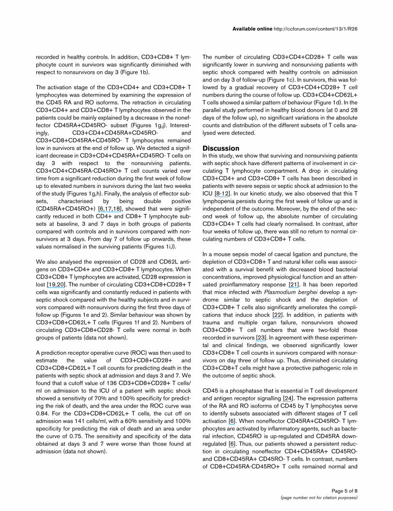
significantly returned to normal by day 28 (1191 ± 172 cells/μl). The CD3+CD4+ T cell subset was also reduced in surviv-ing and nonsurviving patients with respect to healthy controlsat baseline and during the first week of follow up. However,this dramatically decreased CD3+CD4+ T lymphocyte count
had normalised in survivors by day 14 (Figure 1a). On admis-sion, CD3+CD8+ T lymphocytes were also lower in survivorsand nonsurvivors versus controls. In survivors, this T cell sub-set showed a further drop on day 3 of follow up, followed by agradual recovery, although numbers failed to reach the counts
Figure 1
Kinetics of peripheral blood counts of T lymphocyte subsets in patients with septic shock during their stay in the intensive care unitKinetics of peripheral blood counts of T lymphocyte subsets in patients with septic shock during their stay in the intensive care unit. Data presented as surviving patients (black tirangles) and nonsurviving patients (white triangles). The dotted line represents the mean value recorded in the healthy controls. All values are expressed as the mean number of cells per microlitre ± standard error of the mean. * P < 0.05 for survivors or nonsurvivors versus healthy controls; † P < 0.05 for survivors versus nonsurvivors; ‡ P < 0.05 for each follow up time versus baseline or admission to the intensive care unit.
Page 4 of 8(page number not for citation purposes)
Available online http://ccforum.com/content/13/1/R26
recorded in healthy controls. In addition, CD3+CD8+ T lym-phocyte count in survivors was significantly diminished withrespect to nonsurvivors on day 3 (Figure 1b).
The activation stage of the CD3+CD4+ and CD3+CD8+ Tlymphocytes was determined by examining the expression ofthe CD45 RA and RO isoforms. The retraction in circulatingCD3+CD4+ and CD3+CD8+ T lymphocytes observed in thepatients could be mainly explained by a decrease in the nonef-fector CD45RA+CD45RO- subset (Figures 1g,j). Interest-ingly, CD3+CD4+CD45RA+CD45RO- andCD3+CD8+CD45RA+CD45RO- T lymphocytes remainedlow in survivors at the end of follow up. We detected a signif-icant decrease in CD3+CD4+CD45RA+CD45RO- T cells onday 3 with respect to the nonsurviving patients.CD3+CD4+CD45RA-CD45RO+ T cell counts varied overtime from a significant reduction during the first week of followup to elevated numbers in survivors during the last two weeksof the study (Figures 1g,h). Finally, the analysis of effector sub-sets, characterised by being double positive(CD45RA+CD45RO+) [6,17,18], showed that were signifi-cantly reduced in both CD4+ and CD8+ T lymphocyte sub-sets at baseline, 3 and 7 days in both groups of patientscompared with controls and in survivors compared with non-survivors at 3 days. From day 7 of follow up onwards, thesevalues normalised in the surviving patients (Figures 1i,l).
We also analysed the expression of CD28 and CD62L anti-gens on CD3+CD4+ and CD3+CD8+ T lymphocytes. WhenCD3+CD8+ T lymphocytes are activated, CD28 expression islost [19,20]. The number of circulating CD3+CD8+CD28+ Tcells was significantly and constantly reduced in patients withseptic shock compared with the healthy subjects and in survi-vors compared with nonsurvivors during the first three days offollow up (Figures 1e and 2). Similar behaviour was shown byCD3+CD8+CD62L+ T cells (Figures 1f and 2). Numbers ofcirculating CD3+CD8+CD28- T cells were normal in bothgroups of patients (data not shown).
A prediction receptor operative curve (ROC) was then used toestimate the value of CD3+CD8+CD28+ andCD3+CD8+CD62L+ T cell counts for predicting death in thepatients with septic shock at admission and days 3 and 7. Wefound that a cutoff value of 136 CD3+CD8+CD28+ T cells/ml on admission to the ICU of a patient with septic shockshowed a sensitivity of 70% and 100% specificity for predict-ing the risk of death, and the area under the ROC curve was0.84. For the CD3+CD8+CD62L+ T cells, the cut off onadmission was 141 cells/ml, with a 60% sensitivity and 100%specificity for predicting the risk of death and an area underthe curve of 0.75. The sensitivity and specificity of the dataobtained at days 3 and 7 were worse than those found atadmission (data not shown).
The number of circulating CD3+CD4+CD28+ T cells wassignificantly lower in surviving and nonsurviving patients withseptic shock compared with healthy controls on admissionand on day 3 of follow-up (Figure 1c). In survivors, this was fol-lowed by a gradual recovery of CD3+CD4+CD28+ T cellnumbers during the course of follow up. CD3+CD4+CD62L+T cells showed a similar pattern of behaviour (Figure 1d). In theparallel study performed in healthy blood donors (at 0 and 28days of the follow up), no significant variations in the absolutecounts and distribution of the different subsets of T cells ana-lysed were detected.
DiscussionIn this study, we show that surviving and nonsurviving patientswith septic shock have different patterns of involvement in cir-culating T lymphocyte compartment. A drop in circulatingCD3+CD4+ and CD3+CD8+ T cells has been described inpatients with severe sepsis or septic shock at admission to theICU [8-12]. In our kinetic study, we also observed that this Tlymphopenia persists during the first week of follow up and isindependent of the outcome. Moreover, by the end of the sec-ond week of follow up, the absolute number of circulatingCD3+CD4+ T cells had clearly normalised. In contrast, afterfour weeks of follow up, there was still no return to normal cir-culating numbers of CD3+CD8+ T cells.
In a mouse sepsis model of caecal ligation and puncture, thedepletion of CD3+CD8+ T and natural killer cells was associ-ated with a survival benefit with decreased blood bacterialconcentrations, improved physiological function and an atten-uated proinflammatory response [21]. It has been reportedthat mice infected with Plasmodium berghei develop a syn-drome similar to septic shock and the depletion ofCD3+CD8+ T cells also significantly ameliorates the compli-cations that induce shock [22]. In addition, in patients withtrauma and multiple organ failure, nonsurvivors showedCD3+CD8+ T cell numbers that were two-fold thoserecorded in survivors [23]. In agreement with these experimen-tal and clinical findings, we observed significantly lowerCD3+CD8+ T cell counts in survivors compared with nonsur-vivors on day three of follow up. Thus, diminished circulatingCD3+CD8+T cells might have a protective pathogenic role inthe outcome of septic shock.
CD45 is a phosphatase that is essential in T cell developmentand antigen receptor signalling [24]. The expression patternsof the RA and RO isoforms of CD45 by T lymphocytes serveto identify subsets associated with different stages of T cellactivation [6]. When noneffector CD45RA+CD45RO- T lym-phocytes are activated by inflammatory agents, such as bacte-rial infection, CD45RO is up-regulated and CD45RA down-regulated [6]. Thus, our patients showed a persistent reduc-tion in circulating noneffector CD4+CD45RA+ CD45RO-and CD8+CD45RA+ CD45RO- T cells. In contrast, numbersof CD8+CD45RA-CD45RO+ T cells remained normal and
Page 5 of 8(page number not for citation purposes)

Critical Care Vol 13 No 1 Monserrat et al.
CD4+CD45RA-CD45RO+ T cell counts initially fell yet hadreturned to normal by the second week of follow up in surviv-ing patients. An increased percentage of CD3+ cells express-ing CD45RO has been reported in patients with sepsis [25].Our findings could be the consequence of abnormal polyclo-nal activation of circulating T lymphocytes. It has been pro-posed that the switch of T cells from a CD45RA+CD45RO-to a CD45RA-CD45RO+ phenotype may have a functionaleffect in halting the sustained immune response in an effort to
avoid tissue injury [6]. Thus, it is possible to suggest that theexpansion of CD4+CD45RA-CD45RO+ T lymphocytesobserved here during the follow up of surviving patients mightbe considered a compensatory anti-inflammatory mechanismthat develops in these patients with septic shock.
CD28 is a costimulatory molecule that plays a key role in reg-ulating the activation and survival of T lymphocytes [26]. Acti-vation of CD3+CD8+ T lymphocytes has been related to the
Figure 2
Flow cytometry data analysis of the CD28 and CD62L surface expression in CD3+CD8+ T lymphocytes from peripheral blood of septic shock patientsFlow cytometry data analysis of the CD28 and CD62L surface expression in CD3+CD8+ T lymphocytes from peripheral blood of septic shock patients. Panels show CD28 and CD62L expression by CD3+CD8+ gated (Region = R) lymphocytes from peripheral blood of a representative (a) healthy control, (b) nonsurvivor and (c) survivor septic shock patient at the moment of admission to the intensive care unit.
Page 6 of 8(page number not for citation purposes)

Available online http://ccforum.com/content/13/1/R26
loss of CD28 expression [19,20]. In effect, it has beenreported that patients with severe sepsis showed a significantreduction in T lymphocyte CD28 expression [27]. Our dataindicate diminished circulating CD3+CD8+CD28+ T cellnumbers in patients with septic shock with respect to healthysubjects on admission to the ICU and at least during the first28 days of follow up. Interestingly, a reducedCD3+CD8+CD28+ T cell count during the first three days ofadmission to the ICU was of prognostic value for predictingthe survival of a patient. Conversely, an elevated number of cir-culating CD3+CD8+CD28+ T cells was associated with aworse prognosis for the patient. Recently, it has been reportedthat the stimulation of CD28 by a monoclonal antibody inhealthy volunteers is followed by severe multiple cytokine-release syndrome [28]. Our results support a relevant role forCD3+CD8+CD28+ T cells in the pathogenesis of septicshock. Future studies should address the potential clinical rel-evance of this cell variable.
The migration of circulating T lymphocytes to peripheral lymphnodes depends on the expression of the CD62L homingreceptor [29]. We found here that the down-regulation of L-selectin expression on CD3+CD8+ cells in patients with sep-tic shock was associated with a better outcome. Hence, therapid migration of CD8+ T cells to peripheral lymph nodes maybe a mechanism contributing to patient survival.
Our T lymphocyte phenotype data show a time difference, orshift, in the recirculation of T lymphocytes between patientswho survive septic shock and those who do not. Takentogether and analysing the phenotype of the circulating T cellsaccording to the activation criteria (CD45RA+ andCD45RO+) related to CD28 (activation and co-stimulation)and CD62L (activation and migration) expression point to aslower migration of naive and effector cells in nonsurvivingpatients. This different T lymphocyte kinetics would mean adelayed tissue response that could determine the failure of theimmune system and the fatal prognosis of the patient. In par-ticular, the delay in the disappearance of CD45RA+,CD45RA+CD45RO+, CD28+, CD62L+, T CD4+ and CD8+lymphocytes observed between days 3 and 7 of follow up inthe nonsurvivors appears to be crucial to the final outcome.Accordingly, in surviving patients, effector cells would migratemore rapidly to tissues and this would in turn trigger the quickaction of the immune system in combating the infection andthus determine the survival of the patient. It is known that cel-lular immune responses play a critical role in the defenseagainst viral infections and strong T-cell responses have beenreported in patients who clear infection [30]. If the immuneresponse is late or less efficient against microorganism viralepitopes, the outcome of the disease worsens [30-33].
Not surprisingly, the survivor group had lower APACHE II,MODS and SOFA scores than nonsurvivors. An increasingAPACHE II score reflects an increasing severity of illness and
escalating risk of hospital death for multidiagnostic ICU patientgroups. However, an APACHE II score cannot be directlyequated with a specific risk of lower mortality than the samescore for a patients with septic shock [34]. In this group ofpatients, we found that a cut-off value of 136 cells/ml forCD3+CD8+CD28+ T cells and 141 cells/ml forCD3+CD8+CD62L+ T cells on ICU admission showed highspecificity for predicting the risk of death. However, it is knownthat the positive predictive value for APACHE II for the valida-tion study population was only 69.6% and the negative predic-tive value was 87.9% [35]. Moreover, SOFA, MODS andAPACHE II scores require at least 24 hours of monitoring tobe performed and lymphocyte phenotyping can be performedin a short time (approximately 2 hours). It is not possible toreplace clinical score in septic shock patients by immunologi-cal markers. However, these analytical parameters may help tomake clinical decisions in these patients and to establish newpotential therapeutic targets. Future studies will need to studyin more depth the mechanisms involved in the severe abnor-mality found on the T cell compartment in patients with septicshock.
ConclusionsSeptic shock patients show a severe redistribution of circulat-ing T lymphocyte subsets. We found that CD62L and CD28expression on circulating T cells at ICU admission are goodmarkers to predict the outcome of shock septic patients. Tlymphocyte phenotype data show a time difference in the recir-culation of T cells between survivors and nonsurvivors thatmight provoke a delayed tissue response of the immune sys-tem.
Competing interestsThe authors declare that they have no competing interests.
Authors' contributionsJM and RP are joint authors and contributed equally to thismanuscript. AP, MAM, JM and RP contributed to the design ofthe study and drafted the manuscript. JM, DD and HBobtained the data. JM, RP, MÁ, AH, MRZ and ER participatedin data analysis and interpretation of the results.
Key messages
• Septic shock patients show a severe redistribution of circulating T lymphocyte subsets.
• CD62L and CD28 expression on circulating T cells at ICU admission are good markers for predicting the out-come of shock septic patients.
• T lymphocyte phenotype data show in nonsurviving patients a slower migration of naive and effector cells that might provoke a delayed immune response.
Page 7 of 8(page number not for citation purposes)

Critical Care Vol 13 No 1 Monserrat et al.
AcknowledgementsThe authors would like to thank all the medical doctors and nurses of the ICU of the Hospital Universitario Principe de Asturias for their careful and generous collaboration while doing this work. This study was sup-ported by grants S-BIO-0189/2006 MITIC/TIMEDIC from Comunidad de Madrid, Fondo de Investigaciones Sanitarias, CIBERehd and by a research prize awarded by the Fundación Lilly.
References1. Hotchkiss RS, Karl IE: The pathophysiology and treatment of
sepsis. N Engl J Med 2003, 348:138-150.2. Lederer JA, Rodrick ML, Mannick JA: The effects of injury on the
adaptive immune response. Shock 1999, 11:153-159.3. Oberholzer A, Oberholzer C, Moldawer LL: Sepsis syndromes:
understanding the role of innate and acquired immunity.Shock 2001, 16:83-96.
4. Shelley O, Murphy T, Paterson H, Mannick JA, Lederer JA: Inter-action between the innate and adaptive immune systems isrequired to survive sepsis and control inflammation afterinjury. Shock 2003, 20:123-129.
5. Ochoa JB, Makarenkova V: T lymphocytes. Crit Care Med 2005,33:S510-S513.
6. Hermiston ML, Xu Z, Weiss A: CD45: a critical regulator of sign-aling thresholds in immune cells. Annu Rev Immunol 2003,21:107-137.
7. Curfs JH, Meis JF, Hoogkamp-Korstanje JA: A primer oncytokines: sources, receptors, effects, and inducers. ClinMicrobiol Rev 1997, 10:742-780.
8. Holub M, Kluckova Z, Beneda B, Hobstova J, Huzicka I, Prazak J,Lobovska A: Changes in lymphocyte subpopulations andCD3+/DR+ expression in sepsis. Clin Microbiol Infect 2000,6:657-660.
9. Holub M, Kluckova Z, Helcl M, Prihodov J, Rokyta R, Beran O:Lymphocyte subset numbers depend on the bacterial origin ofsepsis. Clin Microbiol Infect 2003, 9:202-211.
10. Kabisch S, Gemar K, Krumholz W, Salomon F, Pralle H: [Lym-phocyte subpopulations in patients at risk of sepsis in a surgi-cal intensive care unit]. Anaesthesist 1990, 39:439-444.
11. Lin RY, Astiz ME, Saxon JC, Rackow EC: Altered leukocyteimmunophenotypes in septic shock. Studies of HLA-DR,CD11b, CD14, and IL-2R expression. Chest 1993,104:847-853.
12. Nishijima MK, Takezawa J, Hosotsubo KK, Takahashi H, ShimadaY, Yoshiya I: Serial changes in cellular immunity of septicpatients with multiple organ-system failure. Crit Care Med1986, 14:87-91.
13. Bone RC, Balk RA, Cerra FB, Dellinger RP, Fein AM, Knaus WA,Schein RM, Sibbald WJ: Definitions for sepsis and organ failureand guidelines for the use of innovative therapies in sepsis.The ACCP/SCCM Consensus Conference Committee. Ameri-can College of Chest Physicians/Society of Critical Care Med-icine. Chest 1992, 101:1644-1655.
14. Knaus WA, Draper EA, Wagner DP, Zimmerman JE: APACHE II: aseverity of disease classification system. Crit Care Med 1985,13:818-829.
15. Marshall JC, Cook DJ, Christou NV, Bernard GR, Sprung CL, Sib-bald WJ: Multiple organ dysfunction score: a reliable descrip-tor of a complex clinical outcome. Crit Care Med 1995,23:1638-1652.
16. Vincent JL, de Mendonca A, Cantraine F, Moreno R, Takala J, SuterPM, Sprung CL, Colardyn F, Blecher S: Use of the SOFA scoreto assess the incidence of organ dysfunction/failure in inten-sive care units: results of a multicenter, prospective study.Working group on "sepsis-related problems" of the EuropeanSociety of Intensive Care Medicine. Crit Care Med 1998,26:1793-1800.
17. Najera O, Gonzalez C, Toledo G, Lopez L, Cortes E, Betancourt M,Ortiz R: CD45RA and CD45RO isoforms in infected malnour-ished and infected well-nourished children. Clin Exp Immunol2001, 126:461-465.
18. Summers KL, O'Donnell JL, Hart DN: Co-expression of theCD45RA and CD45RO antigens on T lymphocytes in chronicarthritis. Clin Exp Immunol 1994, 97:39-44.
19. Fiorentini S, Licenziati S, Alessandri G, Castelli F, Caligaris S, Bon-afede M, Grassi M, Garrafa E, Balsari A, Turano A, Caruso A:CD11b expression identifies CD8+CD28+ T lymphocytes withphenotype and function of both naive/memory and effectorcells. J Immunol 2001, 166:900-907.
20. Labalette M, Leteurtre E, Thumerelle C, Grutzmacher C, TourvieilleB, Dessaint JP: Peripheral human CD8(+)CD28(+)T lym-phocytes give rise to CD28(-)progeny, but IL-4 prevents loss ofCD28 expression. Int Immunol 1999, 11:1327-1336.
21. Sherwood ER, Enoh VT, Murphey ED, Lin CY: Mice depleted ofCD8+ T and NK cells are resistant to injury caused by cecalligation and puncture. Lab Invest 2004, 84:1655-1665.
22. Chang WL, Jones SP, Lefer DJ, Welbourne T, Sun G, Yin L, SuzukiH, Huang J, Granger DN, Heyde HC van der: CD8(+)-T-celldepletion ameliorates circulatory shock in Plasmodiumberghei-infected mice. Infect Immun 2001, 69:7341-7348.
23. Menges T, Engel J, Welters I, Wagner RM, Little S, Ruwoldt R,Wollbrueck M, Hempelmann G: Changes in blood lymphocytepopulations after multiple trauma: association with posttrau-matic complications. Crit Care Med 1999, 27:733-740.
24. Mustelin T, Coggeshall KM, Altman A: Rapid activation of the T-cell tyrosine protein kinase pp56lck by the CD45 phosphotyro-sine phosphatase. Proc Natl Acad Sci USA 1989,86:6302-6306.
25. Roth G, Moser B, Krenn C, Brunner M, Haisjackl M, Almer G, Ger-litz S, Wolner E, Boltz-Nitulescu G, Ankersmit HJ: Susceptibilityto programmed cell death in T-lymphocytes from septicpatients: a mechanism for lymphopenia and Th2 predomi-nance. Biochem Biophys Res Commun 2003, 308:840-846.
26. Sansom DM, Walker LS: The role of CD28 and cytotoxic T-lym-phocyte antigen-4 (CTLA-4) in regulatory T-cell biology. Immu-nol Rev 2006, 212:131-148.
27. Manjuck J, Saha DC, Astiz M, Eales LJ, Rackow EC: Decreasedresponse to recall antigens is associated with depressed cos-timulatory receptor expression in septic critically ill patients. JLab Clin Med 2000, 135:153-160.
28. Suntharalingam G, Perry MR, Ward S, Brett SJ, Castello-Cortes A,Brunner MD, Panoskaltsis N: Cytokine storm in a phase 1 trial ofthe anti-CD28 monoclonal antibody TGN1412. N Engl J Med2006, 355:1018-1028.
29. Tang ML, Steeber DA, Zhang XQ, Tedder TF: Intrinsic differ-ences in L-selectin expression levels affect T and B lym-phocyte subset-specific recirculation pathways. J Immunol1998, 160:5113-5121.
30. Sarobe P, Lasarte JJ, Garcia N, Civeira MP, Borras-Cuesta F, Pri-eto J: Characterization of T-cell responses against immunodo-minant epitopes from hepatitis C virus E2 and NS4a proteins.J Viral Hepat 2006, 13:47-55.
31. Lim B, Sutherland RM, Zhan Y, Deliyannis G, Brown LE, Lew AM:Targeting CD45RB alters T cell migration and delays viralclearance. Int Immunol 2006, 18:291-300.
32. Heller T, Rehermann B: Acute hepatitis C: a multifaceted dis-ease. Semin Liver Dis 2005, 25:7-17.
33. Orland JR, Wright TL, Cooper S: Acute hepatitis C. Hepatology2001, 33:321-327.
34. Wagner DP, Knaus WA, Draper EA: Physiologic abnormalitiesand outcome from acute disease. Evidence for a predictablerelationship. Arch Intern Med 1986, 146:1389-1396.
35. Parrila Joseph E, Bone Roger C: Critical Care Medicine: Princi-ples of Diagnosis and Management. St Louis, Mosby; 1995.
Page 8 of 8(page number not for citation purposes)
http://www.ncbi.nlm.nih.gov/entrez/query.fcgi?cmd=Retrieve&db=PubMed&dopt=Abstract&list_uids=9336671
http://www.ncbi.nlm.nih.gov/entrez/query.fcgi?cmd=Retrieve&db=PubMed&dopt=Abstract&list_uids=9336671
http://www.ncbi.nlm.nih.gov/entrez/query.fcgi?cmd=Retrieve&db=PubMed&dopt=Abstract&list_uids=2240565
http://www.ncbi.nlm.nih.gov/entrez/query.fcgi?cmd=Retrieve&db=PubMed&dopt=Abstract&list_uids=2240565
http://www.ncbi.nlm.nih.gov/entrez/query.fcgi?cmd=Retrieve&db=PubMed&dopt=Abstract&list_uids=2240565
http://www.ncbi.nlm.nih.gov/entrez/query.fcgi?cmd=Retrieve&db=PubMed&dopt=Abstract&list_uids=7689946
http://www.ncbi.nlm.nih.gov/entrez/query.fcgi?cmd=Retrieve&db=PubMed&dopt=Abstract&list_uids=7689946
http://www.ncbi.nlm.nih.gov/entrez/query.fcgi?cmd=Retrieve&db=PubMed&dopt=Abstract&list_uids=7689946
http://www.ncbi.nlm.nih.gov/entrez/query.fcgi?cmd=Retrieve&db=PubMed&dopt=Abstract&list_uids=3943329
http://www.ncbi.nlm.nih.gov/entrez/query.fcgi?cmd=Retrieve&db=PubMed&dopt=Abstract&list_uids=3943329
http://www.ncbi.nlm.nih.gov/entrez/query.fcgi?cmd=Retrieve&db=PubMed&dopt=Abstract&list_uids=1303622
http://www.ncbi.nlm.nih.gov/entrez/query.fcgi?cmd=Retrieve&db=PubMed&dopt=Abstract&list_uids=1303622
http://www.ncbi.nlm.nih.gov/entrez/query.fcgi?cmd=Retrieve&db=PubMed&dopt=Abstract&list_uids=1303622
http://www.ncbi.nlm.nih.gov/entrez/query.fcgi?cmd=Retrieve&db=PubMed&dopt=Abstract&list_uids=3928249
http://www.ncbi.nlm.nih.gov/entrez/query.fcgi?cmd=Retrieve&db=PubMed&dopt=Abstract&list_uids=3928249
http://www.ncbi.nlm.nih.gov/entrez/query.fcgi?cmd=Retrieve&db=PubMed&dopt=Abstract&list_uids=7587228
http://www.ncbi.nlm.nih.gov/entrez/query.fcgi?cmd=Retrieve&db=PubMed&dopt=Abstract&list_uids=7587228
http://www.ncbi.nlm.nih.gov/entrez/query.fcgi?cmd=Retrieve&db=PubMed&dopt=Abstract&list_uids=9824069
http://www.ncbi.nlm.nih.gov/entrez/query.fcgi?cmd=Retrieve&db=PubMed&dopt=Abstract&list_uids=9824069
http://www.ncbi.nlm.nih.gov/entrez/query.fcgi?cmd=Retrieve&db=PubMed&dopt=Abstract&list_uids=9824069
http://www.ncbi.nlm.nih.gov/entrez/query.fcgi?cmd=Retrieve&db=PubMed&dopt=Abstract&list_uids=8033417
http://www.ncbi.nlm.nih.gov/entrez/query.fcgi?cmd=Retrieve&db=PubMed&dopt=Abstract&list_uids=8033417
http://www.ncbi.nlm.nih.gov/entrez/query.fcgi?cmd=Retrieve&db=PubMed&dopt=Abstract&list_uids=8033417
http://www.ncbi.nlm.nih.gov/entrez/query.fcgi?cmd=Retrieve&db=PubMed&dopt=Abstract&list_uids=2548204
http://www.ncbi.nlm.nih.gov/entrez/query.fcgi?cmd=Retrieve&db=PubMed&dopt=Abstract&list_uids=2548204
http://www.ncbi.nlm.nih.gov/entrez/query.fcgi?cmd=Retrieve&db=PubMed&dopt=Abstract&list_uids=2548204
http://www.ncbi.nlm.nih.gov/entrez/query.fcgi?cmd=Retrieve&db=PubMed&dopt=Abstract&list_uids=9590263
http://www.ncbi.nlm.nih.gov/entrez/query.fcgi?cmd=Retrieve&db=PubMed&dopt=Abstract&list_uids=9590263
http://www.ncbi.nlm.nih.gov/entrez/query.fcgi?cmd=Retrieve&db=PubMed&dopt=Abstract&list_uids=9590263
http://www.ncbi.nlm.nih.gov/entrez/query.fcgi?cmd=Retrieve&db=PubMed&dopt=Abstract&list_uids=3718136
http://www.ncbi.nlm.nih.gov/entrez/query.fcgi?cmd=Retrieve&db=PubMed&dopt=Abstract&list_uids=3718136
http://www.ncbi.nlm.nih.gov/entrez/query.fcgi?cmd=Retrieve&db=PubMed&dopt=Abstract&list_uids=3718136
http://www.ncbi.nlm.nih.gov/entrez/query.fcgi?cmd=Retrieve&db=PubMed&dopt=Abstract&list_uids=8584766