8th WCACS, World Congress of the Abdominal Compartment ...
98
Volume XLIX Supplement 1/2017 p-ISSN 1642–5758 e-ISSN 1731–2531 Oral and poster abstracts 8 th WCACS, World Congress of the Abdominal Compartment Society www.wcacs2017.org June 15–17, 2017 Banff Centre, Banff, Alberta, Canada The meeting is organized by the Abdominal Compartment Society aka the World Society of the Abdominal Compartment Syndrome www.wsacs.org The journal is indexed in Medline, Scopus, Embase, EBSCO, ESCI (Emerging Sources Citation Index), CAS, CrossRef, Urlich’s Periodical Directory, Index Copernicus (103,75), Google Scholar, EMCare, Medical Journals Links as well as in the databases of the Polish Ministry of Science and Higher Education (14) and Polish Medical Bibliography.
-
Upload
khangminh22 -
Category
Documents
-
view
5 -
download
0
Transcript of 8th WCACS, World Congress of the Abdominal Compartment ...
Volume XLIXSupplement 1/2017
p-ISSN 1642–5758e-ISSN 1731–2531
Oral and poster abstracts
8th WCACS, World Congress of the Abdominal Compartment Societywww.wcacs2017.org
June 15–17, 2017Banff Centre, Banff, Alberta, Canada
The meeting is organized by the Abdominal Compartment Societyaka the World Society of the Abdominal Compartment Syndrome www.wsacs.org
The journal is indexed in Medline, Scopus, Embase, EBSCO, ESCI (Emerging Sources Citation Index), CAS, CrossRef, Urlich’s Periodical Directory, Index Copernicus (103,75), Google Scholar, EMCare, Medical Journals Links as well as in the databases of the Polish Ministry of Science and Higher Education (14) and Polish Medical Bibliography.

Volume XLIX; Supplement 1/2017
Opinions presented in the articles not necessarily represent the opinions of the Editors
Anesthesiology Intensive Therapy (p-ISSN 1642–5758, e-ISSN 1731-2531) is published five times a year by VM Media sp. z o.o. VM Group sp.k., Grupa Via Medicaul. Świętokrzyska 73, 80–180 Gdańsk, Polandtel.: +48 58 320 94 94, faks: +48 58 320 94 60http://www.viamedica.pl, wap.viamedica.pl
Editorial Address:Prof. Radosław Owczuk MD, PhDKlinika Anestezjologii i Intensywnej TerapiiGdańskiego Uniwersytetu Medycznegoul. Smoluchowskiego 17, 80–214 Gdańsk, Polandphone: +48 58 349 32 81, +48 58 349 32 80, fax: +48 58 349 32 90e-mail: [email protected], www.ait.viamedica.pl
Price per no: 10 EUR (electronical no 7 EUR)The subscription rate in 2016: — paper subscription: 50 EUR (for institutions 100 EUR)— paper subscritption with electronical version: 55 EUR (for institutions 110 EUR)— electronical subscription: 20 EUR (for institutions 40 EUR)
Payment should be made to: VM Media Sp. z o.o. VM Group Sp. K., Grupa Via Medica, Fortis Bank Polska SA oddz. Gdańsk PL15 1600 1303 0004 1007 1035 9021; SWIFT: PPABPLPK SWIFT: PPABPLPK. Single issues, subscriptions orders and requests for sample copies should be send to e-mail: [email protected] orders option available at: www.dp.viamedica.pl
Advertising: For details on media opportunities within this journal please contact the advertising sales department, ul. Świętokrzyska 73, 80–180 Gdańsk, Poland, phone: +48 58 320 94 94; e-mail: [email protected]
The Editors accept no responsibility for the advertisement contents. All rights reserved, including translation into foreign languages. No part of this periodical, either text or illustration, may be used in any form whatsoever. It is particularly forbidden for any part of this material to be copied or translated into a mechanical or electronic language and also to be recorded in whatever form, stored in any kind of retrieval system or transmitted, whether in anelectronic or mechanical form or with the aid of photocopying, microfilm, recording, scanning or in any other form, without the prior written permis-sion of the publisher. The rights of the publisher are protected by national copyright laws and by international conventions, and their violation will be punishable by penal sanctions.
Alan R. Aitkenhead (Nottingham) Janusz Andres (Kraków)Mois Bahar (Istanbul)Martina Bellini (Paderno Dugnano)Wiliam Blunnie (Dublin)Romuald Bohatyrewicz (Szczecin)Leon Drobnik (Poznań)Andreas Franczak (Wien)Wojciech Gaszyński (Łodź)Zeev Goldik (Haifa)Robert G. Hahn (Sodertalje)Stefan De Hert (Ghent)Andreas Hoeft (Bonn)Markus W. Hollmann (Amsterdam)
Scientific board:Andrzej Nestorowicz (Lublin) — Head
Przemysław Jałowiecki (Katowice)Bogdan Kamiński (Warszawa)Zbigniew Kościelniak-Nielsen (Copenhagen)Krzysztof Kusza (Bydgoszcz)Andrzej Kübler (Wrocław)Philipp B. Lirk (Amsterdam)Manu Malbrain (Antwerpen)Ewa Mayzner-Zawadzka (Warszawa)Hanna Misiołek (Zabrze)Olav F. Munter Sellevold (Trondheim)Mahdi Najafi (Tehran)Helen Oudemans-van Straaten (Amsterdam)Andrzej Piotrowski (Łódź)Kathleen Puntillo (San Francisco)
Narinder Rawal (Örebro)Zbigniew Rybicki (Warszawa)Philippe Scherpereel (Lille)Armin Schubert (Cleveland)Nanette M. Schwann (Philadelphia)Andrzej Siemiątkowski (Białystok)Maria Siemionow (Cleveland)Elżbieta Sokół-Kobielska (Warszawa)Janina Suchorzewska (Gdańsk)Tadeusz Szreter (Warszawa)Jan de Waele (Ghent)Rod Westhorpe (Melbourne)Jerzy Wordliczek (Kraków)Maria Wujtewicz (Gdańsk)André van Zundert (Brisbane)
editor-in-chief:Radosław Owczuk (Gdańsk)
theMe editorS:
Indexed in base of The Ministry of Science and Higher Eductation (14 pts), Web of Science™ Core Collection, Emerging Sources Citation Index (ESCI), Medline (PubMed), Elsevier, Index Copernicus (103.75 pts), Polish Medical Bibliography. The journal was financially supported by Polish Ministry of Science and Higher Educations under the "Index Plus" programme (years 2012–2014). Articles published in ”Anaesthesiology Intensive Therapy” are free of charge
Copyright © 2017 Via Medica
David Ferson (Huston) — anaesthesiology, perioperative medicineAnna Fijałkowska (Lublin) — intensive therapyZbigniew Karwacki (Gdańsk) — neuroanaesthesiology, basic sciences
Marc J. Popovich (Cleveland) — critical care medicineMarcin Wąsowicz (Toronto) — cardiac and thoracic anaesthesiologyMagdalena A. Wujtewicz (Gdańsk) — intensive therapy, resuscitation
StatiSticaL editor:Kamil Chwojnicki (Gdańsk)
Managing editor:Kamila Recław (Gdańsk)
LangUage editor:Paul McNamara
Legal note: http://czasopisma.viamedica.pl/ait/about/legalNote

Contents
Oral presentationsS003. A survey for risk factors and outcome of abdominal compartment syndrome in critically ill patients in Imam Hussein Hospital 1Mohsen Sadeghi
S006. Incidence and outcomes of intra-abdominal hypertension and abdominal compartment syndrome in critically ill patients with sepsis managed with refined resuscitation strategies: a prospective observational study 1Jimmy Xiao, Paul McBeth, Derek J. Roberts, Chad G. Ball, Rohan Lall, John Kortbeek, Andrew W. Kirkpatrick
S007. Melanocortin-4 receptor agonists alleviate intestinal dysfunction in secondary intra-abdominal hypertension rat model 2Dong Liu, Hong-Guang Zhang, Ming-Tao Chang, Yang Li, Lian-Yang Zhang
S009. Carbon monoxide and hydrogen sulphide as possible therapeutics for abdominal compartment syndrome: a rat model 2Patrick Murphy, Aurelia Bihari, Neil Parry, Ian Ball, Ken Leslie, Kelly Vogt, Abdel-Rahman Lawendy
S010. Intra abdominal hypertension is more common than previously thought: a prospective study in a mixed medical-surgical intensive care unit 3Patrick Murphy, Aurelia Bihari, Neil Parry, Ian Ball, Ken Leslie, Kelly Vogt, Abdel-Rahman Lawendy
S011. Combined abdominal and incisional NPWT a potential technique to optimise complete closure following an open abdomen 3Michael Sugrue, Mary Connolly, Jamall Abdulaal
S012. Successful closure of catastrophic abdomen utilizing novel technique combining a mechanical closure system with biologic xenograft that accelerates wound healing 4Yana Puckett, Michelle Estrada, Catherine A. Ronaghan
S014. Mortality predictors in patients with acute pancreatitis 5Maja Stojanovic, Petar Svorcan, Predrag Stevanovic, Aleksandar Karamarkovic, Nebojsa Ladjevic, Radmilo Jankovic
S015. Who and when should we measure the intra-abdominal pressure? TBSA-independent analysis of risk factors for intra-abdominal compartment syndrome (TIRIFIC) in major burns 6Dorothee Boehm, Christoph Hirche, Christina Schröder, Denise Arras, Johannes Horter, Ulrich Kneser
S018. Abdominal wall integrity after open abdomen: long-term results of vacuum- -assisted wound closure and mesh-mediated fascial traction (VAWCM) 6Arnulf Gregor Willms, Robert Schwab, Christoph Guesgen
S019. A new, non-invasive, device for delayed primary fascial closure of the ‘open abdomen’: a randomized prospective clinical trial 7Joao Rezende-Neto, Carlos Semprun, Ghassan Al-Kefeiri
S020. Primary, complete fascial closure of the open abdomen and prevention of the ‘homeless bowel’ using a non-invasive device: a prospective, randomized, clinical trial 8Joao Rezende-Neto, Ghassan Al Kefeiri, Carlos Semprun, Sandro Rizoli, Ori Rotstein
S022. Effects of open abdomen on liver and renal dysfunction induced by intra- -abdominal infections and intra-abdominal hypertension 8Jianan Ren, Lei Wu, Ranran Li, Tianyu Lu, Gefei Wang
S023. Measurement of fat pressures during open colorectal surgery 9Heidi Paine, Vimal Hariharan
S027. Analysis of intra-abdominal pressure and its correlation with the outcome in patients undergoing emergency laparotomy 10Babitha Nagaraju, Hemanth Ghalige, Vinay H. D., Abhijit Bhoyate, Th Sudhir Chandra Singh, Birkumar Sharma, Moirangthem G. S.
S030. Changes in intra-abdominal pressure affect brain-heart interaction in traumatic brain injury a pilot study 10Wojciech Dabrowski, Jaroslaw Wosko, Hanna Brzozowska, Radoslaw Rola, Tomasz Trojanowski, Mateusz Bialy, Ziemowit Rzecki, Todd T Schlegel, Andrzej Jaroszynski
S031. Proceedings of resources for optimal care of acute care and emergency surgery consensus summit donegal ireland and abdominal compartment syndrome (ACS) KPIs 11Michael Sugrue, Marja Boermeester
S032. The impact of a canister-free single-use NPWT system on surgical site complications. A meta-analysis 12Vicki Strugala, Robin Martin
S034. Urinary bladder tumour affect result of intra-abdominal measurement an effect of tumour localization 13Pawel Plaza, Krzysztof Bar, Ziemowit Rzecki, Wojciech Dabrowski
S035. Mirna 125b and 596: promising biomarker candidates for the early detection of the transition from intra-abdominal hypertension (IAH) to abdominal compartment syndrome (ACS) in children 14Torsten Kaussen, Timo Schumacher, Martin Boehne, Alexander von Gise, Florian Schmidt, Thomas Jack, Michael Sasse, Thomas Thum, Philipp Beerbaum
S036. Intestinal failure in patients with acute necrotizing pancreatitis complicated by intraabdominal hypertension 15Oleksandr Rotar, Ihor Khomiak, Mikhail Nazarchuk, Grigory Petrovsky, Vasyl Rotar, Michael Fishbach
S039. A new device to prevent fascial retraction in the open abdomen 16Christian Krieglstein, Frank Beyer, Alexandra Maul,
S040. Benchtop validation of a novel device capable of automatic continuous IAP monitoring 17Devyani Nanduri, Brian Bechtel, Teresa Nguyen, Nahbee Jong, Daniel Burnett
S042. The effect of decompressive laparotomy on abdominal compartment syndrome: a systematic review and meta-analysis 17Lana Van Damme, Jan De Waele
S049. Management of the open abdomen following damage control surgery in trauma 18Parker Hu, Rindi Uhlich, Frank Gleason, Jeffrey Kerby, Patrick Bosarge
S051. Open abdomen therapy with vacuum and mesh-mediated fascial traction after aortic repair an international multi-centre study 19Stefan Acosta, Arne Seternes, Maarit Venermo, Leena Vikatmaa, Karl Sorelius, Anders Wanhainen, Mats Svensson, Khatereh Djavani, Martin Björck

S052. Early impact of abdominal compartment syndrome on liver, kidney, and lung damage in a rodent model 20Ricardo Lima, Pedro L. Silva, Vera L. Capelozzi, Mariana G. Oliveirta, Maria Cristina E. Santana, Fernanda F. Cruz, Paolo Pelos, Alberto Schanaider, Manu L.N.G. Malbrain, Patricia R.M. Rocco
S057. Effect of positive expiratory pressure in respirator mechanics, hemodynamics and intra-abdominal pressure in high-risk patients for IAH And ACS 20Natalia Andrade, Gabrielle Souza, Melissa Sibinelli, Juliana Bernardi, Bruno Pereira,
S061. Promising results for the endoscopic component separation technique: a tool to decrease the rate of wound morbidity in repair of large incisional hernias 21Aude Vanlander, Luis Abrue de Carvalho, Frederik Berrevoet
S062. Children and adolescents after liver transplant (LTX) are highly endangered to develop intra-abdominal hypertension (IAH) and abdominal compartment syndrome (ACS) 21Torsten Kaussen, Annika Artmann, Bernd Mitzlaff, Alexander von Gise, Florian Schmidt, Martin Boehne, Thomas Jack, Harald Koeditz, Michael Sasse, Philipp Beerbaum
S063. Femoral vein pressure measurement is also not an alternative IAP measurement method for children and adolescents 23Torsten Kaussen, Miriam Gutting, Bernd Mitzlaff, Alexander von Gise, Martin Boehne, Thomas Jack, Kathrin Seidemann, Harald Koeditz, Michael Sasse, Philipp Beerbaum
S064. Temporary abdominal closure assisted with negative pressure therapy vs primary abdominal wall closure in the severe abdominal sepsis: analysis of our results 26Antonio J Gonzalez Sanchez, Jos Manuel Aranda Narvaez, Aberto Titos-Garc a, Isaac Cabrera-Serna, Cristina Rodriguez Silva, Maria Perezreyes, Julio Santoyo-Santoyo
S067. Comparative study between continuous indirect intrabdominal pressure technique and direct technique. Preliminary result 26Francisco Pracca, Pablo Bousa, Corina Puppo, Alberto Biestro
S069. Intra-abdominal hypertension (IAH), microcirculation and hemodynamics in children after cardiopulmonary bypass assisted surgery 27Torsten Kaussen, Viviane Stamme, Alexander von Gise, Guntram Schwoerer, Martin Boehne, Thomas Jack, Christoph Happel, Harald Bertram, Michael Sasse, Philipp Beerbaum
S070. Could fluid intake lead to increased intra-abdominal pressure in septic shock treatment? A pilot study 28Alcir Escocia Dorigatti, Bruno Monteiro Pereira, Jennifer Leme dos Santos, Fernanda Dias Teramoto, Marcos Aurelio Boes, Roberta Nazario Aoki, Cesar Vanderlei Carmona, Thiago Martins Santos, Gustavo Pereira Fraga
S073. Relation between body anthropomorphy and baseline intraabdominal pressure measurements in critically ill patients: a datastories-based prediction model 29Manu L.N.G. Malbrain, Brecht De Tavernier, Tine Opsomer, Pieter-Jan Van Gaal, Valerie Van Biervliet
S075. Effects of extracorporeal life support on intra-abdominal pressure and acute kidney injury 31Curtis Bashkiharatee, Disha Kriplani, Rita Sheth, Danilyn Angeles, Farrukh Mirza, Donald Moores, Douglas Deming, Arlin Blood, Janeth Chiaka Ejike
S076. How can we manage abdominal pressure for severe acute pancreatitis with abdominal compartment syndrome 32Kosuke Sekiya, Koji Morishita, Tomo Oka, Yasuhiro Otomo
S078. Intra-abdominal hypertension and abdominal compartment syndrome in high risk patients admitted to the ICU: a prospective, observational study 32Marije Smit, Bart Koopman, Matijs van Meurs, Jan Zijlstra
S083. Relationship between splanchnic and renal oxygenation in pediatric patients on extracorporeal membrane oxygenation (ECMO) who develop acute kidney injury (AKI) 33Disha Kriplani, Salem Dehom, Jonathan Specht, Rita Sheth, Farrukh Mirza, Donald Moores, Douglas Deming, Curtis Bashkiharatee, Arlin Blood, Janeth Chiaka Ejike
S084. Predictors of primary fascial closure in trauma and acute care surgery patients with open abdominal wounds: a systematic review 33Derek J. Roberts, Andrew W. Kirkpatrick, Annika Reintam Blaser, Jan De Waele
S085. Intra-abdominal pressure: a clinical prognosis and outcome parameter around the closure of congenital abdominal wall hernias? 34Torsten Kaussen, Buppha Wanchaame, Martin Boehne, Michael Sasse, Philipp Beerbaum, Christoph Zoeller, Joachim Kuebler, Benno Ure, Carolin Boehne, Bettina Bohnhorst
Poster presentationsP004. Study of IAH prevalence and cognitive level of experienced medical staff in ICU 37Lian-Yang Zhang, Hua- yu Zhang, Dong Liu, Hao Tang, Shi-Jin Sun, Shan-mu, Wen-qun Yang, Dong-po Jiang
P005. Open components separation and underlay repair using biological mesh for the treatment of planned ventral hernia after open abdomen surgery 37Lian-Yang Zhang, Pei-Yuan Li, Dong Liu, Shi-Jin Sun
P008. Novel method for delayed primary closure and incisional hernia prevention in open abdomen 38Rafael Villalobos, Carmen Mias, Cristina Gas, Alfredo Escartin, Victor Palacios, Jordi Escoll, Jorge Juan Olsina
P016. Listeria monocytogenes inhibits Th17 responses through TLR2 signaling in intra-abdominal illness 38Song Liu, Xiuwen Wu,Gefei Wang, Jianan Ren
P017. Nucleic acid recognition by STING signaling controls effector T cells via mucosal CD103+ DCs in gut 39Song Liu, Xiuwen Wu,Gefei Wang, Jianan Ren
P021. Multivariate analysis derived biomediator panel to predict survival in critically ill patients 40Michelle S. Malig, Craig N. Jenne, Derek J. Roberts, Chad G. Ball, Zhengwen Xiao, Andrew W. Kirkpatrick
P025. Using the intensive care unit for bedside dressing changes for open abdomen is time- and staff-efficient 40Arne Seternes, Sigurd Fasting, Pål Klepstad, Skule Mo, Torbjørn Dahl, Martin Björck, Arne Wibe
P026. Ability of commercial NPWT systems to manage fluid in an experimental open abdomen study 41Raymond Dunn, Heather Tessier, Vicki Hammon, Iain Webster
P033. Validation of a continuous intra-gastral monitoring-technique for measuring intraabdominal pressure (IAP) in critically ill pediatric patients 41Torsten Kaussen, Miriam Gutting, Alexander von Gise, Martin Boehne, Florian Schmidt, Thomas Jack, Michael Sasse, Dietmar Bäthi, Philipp Beerbaum
P037. The optimal timing of exchange of the VAC (Vacuum assisted abdominal closure method) 43Tadao Kubota, Kentaro Yoshikawa, Ken Mizokami, Takashi Sakamoto, Jiro Kimura, Shota Fukai
P038. Open abdomen handling: mediated closure with silo against vacuum assisted therapy. Cohortes study 43David Arango, Juliana Maria Orda, Bruno Pereira
P041. The anterior rectus abdominis sheath and external oblique aponeurosis turnover flap method in patients requiring open abdominal management 44Koji Morishita, Junichi Aiboshi, Kosuke Sekiya, Tomo Oka, Yasuhiro Otomo
P043. Intra-abdominal hypertension during mission beyone earth orbit: implications regarding intra-peritoneal disease and therapy 45Andrew W. Kirkpatrick, Tim Broderick, Jessica L. McKee, Doug. R. Hamilton, Chad G. Ball, Paul B. McBeth
P046. The role of C-reactive protein measurement after traumatic injury can you quantify systemic inflammation? A systematic review 45Fatma Al Hinai, Aziza Al Rawahi, Christopher Dion, Andrew W. Kirkpatrick
P047. C-reactive protein (CRP) analysis in critically ill patients with open abdomen negative pressure therapy 46Fatma Al Hinai, Aziza Al Rawahi, Christopher Dion, Andrew W. Kirkpatrick
P053. Can intra-abdominal pressure measurement in blunt abdominal trauma patients predict hollow viscus perforation in resource crunch environment: experience from a middle income country 46Nawal Kishore Jha, Dipendra Kumar Sinha, Sanjay Kumar Yadav
P054. Preoperative predictors of intra-abdominal hypertension and potential open abdomen management: a retrospective analysis of pre-operative clinical, biochemical and radiological findings with outcomes 46Rajashekar Mohan, Anantharaju GS, Likhith Rai
P055. Interactive simulation with the human worn simulator significantly improves national surgery exam scores 47Anthony LaPorta, Joseph LaPorta, Tanner McClure, David Ross, Nancy Simon
P056. Abdominal compartment syndrome: special considerations after liver transplant surgery 48Marije Smit, Marieke de Boer, Matijs van Meurs, Jan Zijlstra
P058. Sir William Heneage Ogilvie: master surgeon, war surgeon, innovator ahead of his time 48Andrew W. Kirkpatrick, Ian B. Anderson
P059. Abdominal compartment syndrome: an unrecognized complication of anabolic steroids 49Ian B. Anderson, Andrew W Kirkpatrick
P060. Where did all our vacuum wound devices go? The limitations of working in obscure wars 49Ian B. Anderson, Paul Duffy, Robert Mulloy, Andrew W. Kirkpatrick
P065. ESPEDacs-surveillance study on epidemiology, diagnostics and therapy of abdominal compartment syndrom in critical ill children in Germany: preliminary data 50Torsten Kaussen, Anke Zellmer, Harald Käditzz, Kathrin Seidemann, Ruediger von Kries, Michael Sasse, Philipp Beerbaum
P066. Chimney VAC for entero-atmospheric fistuals. Early experience with a novel VAC system for open abdomen 52Arne Seternes, Lars Cato Rekstad, Knut Magne Augestad, Ola Rokke, Hans H Wasmuth
P068. Polycompartment syndrome: from theory to reality 52Zsolt Bodnar, Edit Tidrenczel
P071. Does sepsis influence intra-abdominal pressure in critical patients? A pilot study 53Alcir Escocia Dorigatti, Bruno Monteiro Pereira, Jennifer Leme dos Santos, Fernanda Dias Teramoto, Marcos Aurelio Boes, Roberta Nazario Aoki, Cesar Vanderlei Carmona, Thiago Martins Santos, Gustavo Pereira Fraga
P072. Experience of upper gastrointestinal massive bleeding patients requiring open abdominal management 53Yuzuru Mochida, Koji Morishita, Kosuke Sekiya, Yasuhiro Otomo
P074. Vacuum assisted closure treatment for abdominal compartment syndrome in a preterm diagnosed with necrotizing enterocolitis 54Alon Yulevich, Michael Weiss, Vered Sheffer-Fleisher, Tatyana Arzumanov, Yechiel Sweed
P077. Anatomical repere-based abdominal bell design to reduce intra-abdominal hypertension with ABDOPRE 55Silvia Diaz, Maria Jose Gonzalez Franco, Luciana Urruty, Florencia Peirano, Francisco Pracca, Alberto Biestroand Franco Simini
P079. Surviving sepsis, multi organ failure, open abdomen, enterocutanous fistula and intestinal failure 56Maria Gaard, G. Carlson, N. Meidell, K. Sunde
P081. Are anthropomorphic parameters able to predict the presence of intra-abdominal hypertension: a datastories-based prediction model 56Manu L.N.G. Malbrain, Brecht De Tavernier, Tine Opsomer, Pieter-Jan Van Gaal, Valerie Van Biervliet
P081. Are anthropomorphic parameters able to predict the presence of intra- -abdominal hypertension: a datastories-based prediction model 56Manu L.N.G. Malbrain, Brecht De Tavernier, Tine Opsomer, Pieter-Jan Van Gaal, Valerie Van Biervliet
P082. Early, definitive repair of traumatic lumbar hernia with a titanium, twinfix, bone-anchored suture technique 58Thomas Clements, Derek J. Roberts, Ryan Martin, Chad G. Ball, Andrew W. Kirkpatrick, Ruphus Rajakumar, Rohan Lall
Invited abstractsI001. Introduction to the International Fluid Academy and the 4 phases, 4 D’s and 4 questions in relation to fluid management 59Manu L.N.G. Malbrain
I002. Introduction to DAMPS and PAMPS and why they matter in critical intra-abdominal injury/illness 60Carl J. Hauser
I003. Mild to moderate intra-abdominal hypertension: does it matter? 60Annika Reintam Blaser
I004. Epidemiology of IAH and the ACS in acute general surgery 61Rob Wise
I005. Under appreciated champions: nursing awareness and advocacy 62Rosemary K. Lee
I006. Implications of IAH on renal failure in the critically ill 62Bart L. De Keulenaer
I007. Co-morbidities among those requiring AWR: when is “no” a hard-line? 63Ari Leppäniemi
I008. Abdominal wall reconstruction: managing the skin envelope 64Duncan Nickerson
I009. Global impacts of sepsis upon human health 65Carl J. Hauser
I010. Advances and challenges in modulating the inflammatory response within the peritoneal cavity: negative pressure therapy 65Andrew W. Kirkpatrick
I011. Advances and challenges in modulating the inflammatory response within the peritoneal cavity: extracorporeal therapies 66Jan J. De Waele
I012. Intra-abdominal hypertension and the human microbiome 66Braedon McDonald
I013. Occult abdominal compartment syndrome 67Inneke De Laet
I014. Systemic mediators and a local problem: interactions between systemic inflammation and the abdominal compartment 68Bruno M. Pereira
I015. The polycompartment syndrome and organ-organ interactions 68Janeth Chiaka Ejike
I016. The closed or open abdomen after laparotomy (cool) FOR source control of severe complicated intra-abdominal sepsis study 70Andrew W. Kirkpatrick
I017. When to leave the abdomen open after general surgery in 2016 71Michael Sugrue
I018. Implications of IAH on feeding and weaning the critically ill 71Inneke De Laet
I019. Fluid overload and deresuscitation: what, why, when and how? 72Manu L.N.G. Malbrain
I020. Pharmacologic management of IAP 73Rob Wise
I021. Positioning to manage IAP 74Bart de Keulenaer
I022. Comprehensive approaches to the problem 75Ari Leppaniemi
I023. Management of enteroatmospheric fistulas: from initial conservative treatment to definitive surgery 76Daniel Wainstein
I024. Diuresis and renal replacement therapy to manage IAP 77Jan de Waele
I025. Gut mucosal injury from IAH 78Patrick Murphy
I026. Mechanical open abdomen systems 78Sam Minor
I027. Introduction to the Concept of “GIPS” and “AIDS 79Manu L.N.G. Malbrain
I028. Guidelines on open abdomen from the European Hernia Society 80Frederik Berrevoet
I029. IAP and its potential role in diseases of pregnancy 80Diane Sawchuck
I030. Overview of advanced surgical techniques to avoid IAH/ACS in AWR 81Frederik Berrevoet
I031. Maternal positioning and it’s potential role in intra-abdominal pressure 81Bernd Wittmann, Diane Sawchuck
I032. Surgical simulation of the abdominal cavity: the abdominal component of the cut-suit 82Anthony LaPorta
I033. Posterior component release and laparoscopic approaches to decompress for IAH/ACS 83Ari Leppaniemi
I034. Abdominal simulators in training for far-forward damage control surgery in extreme environments 83Andrew W. Kirkpatrick
I035. Biomesh selection during hernia repair 84Bruno Pereira
I036. Long-term absorbable meshes 85Frederik Berrevoet
I037. The role of social media and FOAM in critical care education 85Manu L.N.G. Malbrain

1
ABSTRACTS
Anaesthesiology Intensive Therapy2017, Supplement 1
ISSN 0209–1712www.ait.viamedica.pl
S003. A survey for risk factors and outcome of abdominal compartment syndrome in critically ill patients in Imam Hussein Hospital
Mohsen SadeghiSBMU Shahid Beheshti University of Medicine Internal Medicine, Tehran, Iran
Background: Increased intra-abdominal pressure is devided into two categories:Intra-abdominal hypertension (IAH)and abdominal compartment syndrom (ACS). In case of IAH, abdominal perfusion pressure decreases and impaires performances of intra and extra abdominal organs, such as, renal failure,shock and impaired oxygenation. In many articles, intra-abdominal pressure acts as a prognostic factor in mortality.Objectives: The aim of this study was to investigate risk factors and outcome of abdominal compartment syndrome critically ill patients Methods: A total of 125 patients in ICU were enrolled into study. Their abdominal pressure was measuresd by standard intra-vesical method. Related factors such as age, sex, GCS, APACHE-II, shock, SIRS, length of hospital stay, cause of hospitalization, BMI, type and amount of fluids and blood products were recorded.Association and relationship of these data with intra-abdominal pressure presented with descriptive and analytic methods.Results: Of 125 patients,73 (58.4%) were men and 52 (41.6%) were women. Mean age of patients was 55.1 (SD 18.3) years old. 89 patients (71.2%) had normal intra-abdominal pressu-re (~6 mm Hg), while 31 patients (24.8%) had IAH and 5 pa-tients (4%) had ACS. The incresed intra-abdominal pressure was significantly associated with shock, SIRS, APACHE-II, central venous oxygen saturation and GCS (P < 0.05). The mortality rate was significantly high in patients wth higher intra-abdominal pressure (P < 0.05).Conclusions: These results suggest that intra-abdominal pressure measurment is necessary in all ICU patients in a routine manner to have a prognostic data for performance appropriate intervention.Acknowledgements: The authors wish to thank all person-nels working in Intensive Care Unit of Tehran Imam hossein hospital.
Oral presentations
Key words: abdominal compartment syndrome, critically ill patients, intra-abdominal hypertention, intra-abdominal pressure
S006. Incidence and outcomes of intra- -abdominal hypertension and abdominal compartment syndrome in critically ill patients with sepsis managed with refined resuscitation strategies: a prospective observational study
Jimmy Xiao, Paul McBeth, Derek J. Roberts, Chad G. Ball, Rohan Lall, John Kortbeek, Andrew W. KirkpatrickFoothills Medical Centre, University of Calgary and Alberta Health Services, Calgary, Alberta, Canada
Background: Intra-abdominal hypertension (IAH) and ab-dominal compartment syndrome (ACS) have been reported to be common in critically ill sepsis patients receiving vigo-rous crystalloid resuscitation, associating with high morbi-dity and mortality. Current refined resuscitation strategies involve far less crystalloid fluids and higher proportions of blood products, as well as timely surgical intervention to prevent and treat IAH/ACS, which may have changed the landscapes of IAH or ACS and improved outcomes in that critically ill population.Objectives: We sought to prospectively examine the inci-dence and outcomes of IAH or ACS in intensive care unit (ICU) septic patients managed with refined resuscitation strategies. Methods: Over a four-year period starting in September 2012 ongoing, adult patients with sepsis or septic shock admitted ICU for a stay of 24 hours or greater were eligible for inclusion. Eligible patients were screened from all ICU patients based on their clinical diagnosis and the current definitions of sepsis and septic shock. Patients with bladder ruptures were excluded. Monitoring intra-abdominal pres-sure (IAP) involved intermittent measurement of intravesical pressure after instilling 25 mL saline into the bladder when patient was positioned supine.Results: In total, 367 patients were eligible, only 108 (29%) of those patients were monitored with IAP. Among the 108 pa-tients, 23 were medical (e.g., pneumosepsis) and 85 were
2
Anaesthesiol Intensive Ther 2017, Supplement 1
trauma, burns, or intra-abdominal sepsis patients. The mean age was 55.8 years, median SOFA (sepsis-related or-gan failure assessment) score was 12.5, 106 (98%) patients received antibiotics and 84 (77.8%) required vasopressors, median first 24-hour crystalloid fluid balance was 8284 mL, 80 patients (74%) had IAH (two or more IAPs ‰ 12 mm Hg) and 18 developed ACS (GIII–IV IAH plus new organ dysfunc-tion). ICU-mortality (54.6%) was significantly related to age, SOFA score, acute kidney injuries, and vasopressor require-ment, but not to IAH or ACS. Interestingly, IAH was signifi-cantly related to SOFA score and ICU length of stay. ACS was detected significantly higher in severe burn patients (4 out of 7) than in non-burn patients (P = 0.01). In patients without burns, incidence of ACS was lower in those with abdominal sepsis managed with early laparotomy (n = 47), compared with patients (n = 54) who were not suitable to surgery.Conclusions: IAH appears still to be a common scenario in ICU septic patients requiring intensive resuscitation, which is significantly higher in patients with higher SOFA score and longer ICU stay. ACS incidence is high in severe burns patients, yet less common in other patients. Abdominal sepsis patients managed with timely surgical intervention have lower ACS rate compared with other patients. Although ICU-mortality was significantly related to SOFA score, acute kidney injuries and septic shock, however, both IAH and ACS were not predictive of ICU-mortality in this cohort.Acknowledgements: This study has been supported by the departments of Surgery and Critical Care Medicine, Foothills Me-dical Center, University of Calgary and Alberta Health Services.Key words: intra-abdominal hypertension, abdominal com-partment syndrome, sepsis, septic shock
S007. Melanocortin-4 receptor agonists alleviate intestinal dysfunction in secondary intra-abdominal hypertension rat model
Dong Liu1, Hong-Guang Zhang2, Ming-Tao Chang2, Yang Li2, Lian-Yang Zhang2
1Trauma Center, State Key Laboratory of Trauma, Bur Institute of Surgery Research, Daping Hospital Third Military Medical Uni-versity, ChongQing, China2Institute of Surgery Research, Daping Hospital, Third Military Medical University, ChongQing, China
Background: Intra-abdominal hypertension (IAH) is a po-tentially life-threatening disease. Then, melanocortin 4 re-ceptor activation exhibits life-saving properties.Objectives: The aim of the present study is to examine whether treatment with the melanocortin MC4 receptor agonist RO27-3225 ameliorates intestinal injury in IAH rats. Methods: A total of 72 male Sprague-Dawley rats were randomized into six groups. Group 1 was the sham group.
Group 2 received RO27-3225 (180 µg/kg ip) as the sham+RO group. Group 3 was the IAH group. IAH was induced by a blood draw (mean arterial pressure: 30 mm Hg for 90 min) followed by shed blood/Ringers solution reinfusion. The intra-abdominal pressure (IAP) was increased to 20 mm Hg by injecting air into the peritoneal cavity. Group 4 was the RO group and was administered with RO27-3225 5 min after drawing blood. Groups 5 and 6 were the Chl group and HS024 group and were pretreated with the nicotinic acetylcholine receptor antagonist chlorisondamine (Chl) or selective melanocortin 4 receptor antagonist (HS024), respectively, 2 min before RO27-3225 was administered.Results: RO27-3225 restored mean arterial pressure (MAP), reduced tumor-necrosis factor-a ± (TNF-a ± ) and interleu-kin-1b (IL-1b) mRNA expression increased by IAH, alleviated the histologic damage and improved the superoxide dismu-tase (SOD) activity in the intestine. Compared with the IAH group, the levels of intestinal fatty acid-binding protein, intestinal edema and intestinal permeability were lower in the RO group. Furthermore, RO27-3225 treatment increased the expression of Rho-associated coiled-coil-containing protein kinase 1 (ROCK1) and phosphorylated myosin light chain (MLC). Chlorisondamine and HS024 abrogated the protective effects of RO27-3225.Conclusions: These data indicate that melanocortin 4 re-ceptor agonist counteracting the intestinal inflammatory response ameliorates intestinal injury in experimental se-condary IAH by MC4 receptor-triggered activation of the cholinergic anti-inflammatory. It may be a promising me-thod for treating IAH in the future.Acknowledgements: New Techniques of Trauma Care (2012BAI11B01) and Twelfth-FiveYear Military Vehicle Traffic Safety and Prevention (BWS12J033).Key words: intra-abdominal hypertension, ischemia/re-perfusion, melanocortin receptors, nicotinic acetylcholine receptors
S009. Carbon monoxide and hydrogen sulphide as possible therapeutics for abdominal compartment syndrome: a rat model
Patrick Murphy, Aurelia Bihari, Neil Parry, Ian Ball, Ken Leslie, Kelly Vogt, Abdel-Rahman LawendyUniversity of Western Ontario Resideny General Surgery 800 Com-missioners Rd W Canada
Background: Abdominal compartment syndrome (ACS) results in significant organ, inflammatory, and metabolic derangement and is associated with considerable mortality. Carbon monoxide (CO) and hydrogen sulphide (H2S)-rele-
3
asing molecules (CORM-3 and GYY4137, respectively) have been shown to be potent anti-oxidant and anti-inflamma-tory agents at the tissue and systemic level.Objectives: Determine the effect of CO and H2S on the ischemia-reperfusion injury of ACS in a rat model. Methods: ACS was maintained for two hours in 19 rats using an abdominal plaster cast and intra-peritoneal CO2 insuffla-tion at 20 mm Hg. Three experimental groups underwent ACS: inactivated CORM-3, active CORM-3 and GYY4137 while one arm underwent no ACS to serve as a sham. Sinusoidal perfusion, inflammatory response and cell death were qu-antified in exteriorized livers. Respiratory, liver and renal dysfunction was assessed biochemically.Results: Hepatocellular death and the number of activated leukocytes within post-sinusoidal venules were significantly increased in rats undergoing ACS (16-fold increase in cell death, 17-fold increase in leukocyte activation, P < 0.05). Administration of CORM-3 or GYY4137 resulted in a signifi-cant decrease of both parameters (P = 0.0300 and P = 0.0086, respectively). ACS resulted in an increase in serum markers of renal and liver injury; CORM-3 or GYY4137 were able to partially restore the levels to those seen in sham ani-mals. Myeloperoxidase levels were significantly elevated in ACS group in lung, liver and small intestine (P = 0.0002, P = 0.0132 and 0.0813, respectively). CORM-3 treatment, but not GYY4137, was able to completely block the response (65 ± 11 U mL-1 and 92 ± 18 U mL-1, respectively versus 110 ± 10 U mL-1 in the ACS group, lung tissue).Conclusions: The organ dysfunction associated with ACS is severe and we have demonstrated the effect of two molecu-les, CO and H2S, on tempering the ACS-associated metabolic and organ derangement. CORM-3 demonstrated a greater effect than GYY4137, and was able to restore most of the measured parameters to levels comparable to sham. The data indicates that CORM-3 and/or GYY4137 may have po-tential as a therapeutic agent for ACS.Acknowledgements: None. Key words: abdominal compartment syndrome, CORM-3, H2S, animal
S010. Intra-abdominal hypertension is more common than previously thought: a prospective study in a mixed medical- -surgical intensive care unit
Patrick Murphy, Aurelia Bihari, Neil Parry, Ian Ball, Ken Leslie, Kelly Vogt, Abdel-Rahman LawendyUniversity of Western Ontario Resideny General Surgery 800 Com-missioners Rd W Canada
Background: Intra-abdominal hypertension (IAH) is an un-der recognized phenomenon in critically ill patients. The
true incidence has not been adequately determined by well powered, prospective studies which adhere to modern consensus definitions.Objectives: To determine the incidence of IAH, prospecti-vely, in a mixed-medical student ICU using modern WSACS guidelines Methods: A prospective observational study of consecuti-ve ICU patients admitted to a mixed medical-surgical ICU. Intra-abdominal pressures were measured twice daily using the modified Kron technique and were continued until di-scharge, death or removal of the indwelling catheter. IAH was defined according to published guidelines as a susta-ined intra-abdominal pressure > 12 mm Hg. Multivariable analysis was used to identify risk factors associated with IAH and ICU mortality.Results: 286 patients met our inclusion criteria. Thirty per-cent of patients had IAH on admission and a further 15% developed IAH during their ICU stay. The incidence of abdo-minal compartment syndrome (ACS) was 3.0%. Obesity, sep-sis, mechanical ventilation and 24-hour fluid balance (> 3 L) were all independent predictors for IAH. IAH occurred in 28% of non-ventilated patients. Admission type (medical vs. surgical vs. trauma) was not a significant predictor of IAH. ICU mortality was 20% and was significantly higher for patients with IAH (30%) compared to patients without IAH (11%). IAH of any grade was an independent predictor of mortality (OR 2.8; 95% CI 1.2–6.2).Conclusions: IAH is common, with an incidence of 45% in both surgical and non-surgical patients in the intensive care setting and in this study, was found to be independen-tly associated with mortality. Despite prior reports to the contrary, IAH develops in non-ventilated patients and in patients who do not have IAH on admission. Intra-abdomi-nal pressure monitoring is inexpensive, provides valuable clinical information, and should be routinely performed in the ICU. Future work should evaluate the impact of early intervention for patients with IAH.Acknowledgements: None. Key words: IAH, incidence, epidemiology
S011. Combined abdominal and incisional NPWT a potential technique to optimise complete closure following an open abdomen
Michael Sugrue, Mary Connolly, Jamall AbdulaalDepartment of Surgery Letterkenny University Donegal Clinical Research Academy Letterkenny Ireland
Background: The Open Abdomen (OA) a cornerstone in managing critical surgical patients and Abdominal Com-partment Syndrome (ACS), poses many challenges. Primary

4
Anaesthesiol Intensive Ther 2017, Supplement 1
fascial and skin closure is achieved in only 65% currently. New strategies in complete closure are required.Objectives: The study assessed feasibility of a new closure approach, combining abdominal and incisional negative pressure wound therapy (cNPWT). Methods: An ethically approved study of outcomes in consecuti-ve patients undergoing OA was undertaken in 2015–2016 using combined NPWT with either REANASYS/AbThera and PICO/Pro-vena to achieve both primary fascial and skin closure under a single surgeonâs care at a University Hospital. Indications for OA, demographics, Mannheim peritonitis index, Apache IV, SOFA and pPossum score were calculated. IAP was measured 8 hourly. Patients were following for 6 months. Fascia was closed with continuous 1/0 nylon followed by a layered closure with subcuticular suture. Data is expressed as mean and range.Results: 5 consecutive patients, 3 females, mean age 67.4 (38–85), years were studied. OA was performed for purulent peritonitis with haemodynamic instability in 2 and mesenteric ischaemia related small bowel resection in 3. Mean Mannheim peritonitis score was 20.4 (10–33), mean predicted mortality 31.2%, mean Apache IV of 61.4 (14 –101), SOFA 4.4 and mean predicted mortality as per pPossum of 58.4%. Observed mortality was 20% (1/5). Tertiary ACS was not seen. The fascia was closed in all pa-tients. Two had mesh-mediated traction. One patient had treatment withdrawn at 30 days due to inability to wean. All 5 cases had primary skin closure. One superficial infection occurred on day 21 which required no antimicrobial treat-ment. There were no early incisional herniaConclusions: In this small study cNPWT was effective in achieving complete closure with minimal wound related complications. It supports the concept of combined abdo-minal and incisional NPWT potentially affording the com-bined proven benefits of both techniques.Acknowledgements: Donegal Clinical Research Academy.Key words: ACS, abdominal wall, closure of abdomen, IAP
S012. Successful closure of catastrophic abdomen utilizing novel technique combining a mechanical closure system with biologic xenograft that accelerates wound healing
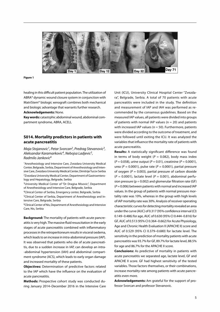
Yana Puckett, Michelle Estrada, Catherine A. RonaghanTexas Tech University Health Sciences Center, Lubbock, United States
Background: Closure of catastrophic open abdominal wo-unds presents a challenge to the surgeon. We present a case series of a technique utilizing a combination of mechanical abdominal closure device in conjunction with biologic xe-nograft in closing complex open abdominal wounds.
Objectives: To demonstrate an alternative option for de-finitive fascial closure and accelerated wound healing of catastrophic open abdominal wounds utilizing novel tech-nique combining a mechanical closure system with biologic xenograft. Methods: Twelve patients underwent closures of open abdominal wounds utilizing technique of combining me-chanical wound closure system with biologic xenograft. ABRA® dynamic wound closure system was placed ini-tially and adjusted daily until fascial closure was achieved. MatriStem® urinary porcine bladder matrix was then placed in the wound above closed fascia. Information was ab-stracted on age of patient, body mass index (BMI), incision length, myofascial gap size before and after ABRA place-ment, visceral extrusion size, number of ABRA adjsutments, and total time to fascial closure. Results were recorded utilizing measurements and photographs pre and post closure. Paired t-test was utilized to compare myofascial gap reduction before and after application of ABRA device. Means and standard deviation (SD) were used to report averages.Results: The average age of patient was 48.1 (SD 10.0) years. Mean BMI of patient was 40.5 (SD 9.8). Caucasians comprised 75% of the populations, Hispanics 25%. Ostomy was present in 25% of patients. Average length of midline incision was 26.6 cm (SD 10.1). The abdomen was open for an average of 8.0 (SD 9.0) days prior to application of ABRA device. Average visceral extrusion prior to application was 6.8 cm (SD 1.6). Average reduction of myofascial gap after initial placement of ABRA device was 10.2 cm (SD 4.8) 95% CI (6.5–13.9, P < 0.0001). Average number of ABRA adjustments made was 1.8 (SD 1.0). Delayed primary fascial closure was achieved an average of 9.4 (SD 4.2) days. Delayed primary fascial closure was achieved in 100% of patients. An overall reduction in wound area was achieved in 100% of patients (Table 1, Fig. 1).Conclusions: In conclusion, this technique offers another option for definitive fascial closure and accelerated wound
Table 1
Mean Sd
Age 48.11 10.03
BMI 40.45 9.83
Visceral extrusion (cm) 6.80 1.57
Incision length (cm) 26.55 10.13
Days to closure of fascia 9.36 4.18
Adjustments of ABRA prior to closure (days) 1.82 .98
Days abdomen open before ABRA application
8 9
Myofascial gap before ABRA application 18 6
Myofascial gap after ABRA application 9.06 2.04
5
Figure 1
healing in this difficult patient population. The utilization of ABRA® dynamic wound closure system in conjunction with MatriStem® biologic xenograft combines both mechanical and biologic advantage that warrants further research.Acknowledgements: None. Key words: catastrphic abdominal wound, abdominal com-partment syndrome, ABRA, ACELL
S014. Mortality predictors in patients with acute pancreatitis
Maja Stojanovic1, Petar Svorcan2, Predrag Stevanovic3, Aleksandar Karamarkovic4, Nebojsa Ladjevic5, Radmilo Jankovic6
1Anesthesiology and Intensive Care, Zvezdara University Medical Center, Belgrade, Serbia, Department of Anesthesiology and Inten-sive Care, Zvezdara University Medical Center, Dimitrije Tucov Serbia2Zvezdara University Medical Center, Department of Gastroentero-logy and Hepatology, Belgrade, Serbia3University Medical Center of “Dr Dragisa Misovic”, Department of Anesthesiology and Intensive Care, Belgrade, Serbia4Clinical Center of Serbia, Emergency center, Belgrade, Serbia5Clinical Center of Serbia, Department of Anesthesiology and In-tensive Care, Belgrade, Serbia6Clinical Center of Nis, Department of Anesthesiology and Intensive Care, Nis, Serbia
Background: The mortality of patients with acute pancre-atitis is very high. The massive fluid resuscitation in the early stages of acute pancreatitis combined with inflammatory processes in the retroperitoneum results in visceral oedema, which leads to an increase in intra-abdominal pressure (IAP). It was observed that patients who die of acute pancreati-tis, due to a sudden increase in IAP, can develop an intra- -abdominal hypertension (IAH) and abdominal compart-ment syndrome (ACS), which leads to early organ damage and increased mortality of these patients.Objectives: Determination of predictive factors related to the IAP which have the influence on the evaluation of acute pancreatitis. Methods: Prospective cohort study was conducted du-ring January 2014–December 2016 in the Intensive Care
Unit (ICU), University Clinical Hospital Center “Zvezda-ra”, Belgrade, Serbia. A total of 70 patients with acute pancreatitis were included in the study. The definition and measurement of IAP and IAH was performed as re-commended by the consensus guidelines. Based on the measured IAP values, all patients were divided into groups of patients with normal IAP values (n = 20) and patients with increased IAP values (n = 50). Furthermore, patients were divided according to the outcome of treatment, and were followed until exiting the ICU. It was analyzed the variables that influence the mortality rate of patients with acute pancreatitis.Results: A statistically significant difference was found in terms of body weight (P = 0.082), body mass index (P = 0.058), urine output (P = 0.01), creatinine (P = 0.0001), urea (P = 0.0001), pulse rate (P = 0.0001), partial pressure of oxygen (P = 0.003), partial pressure of carbon dioxide (P = 0.0001), lactate level (P = 0.001), abdominal perfu-sion pressure (p = 0.002) and glomerular filtration rate (GF) (P = 0.006) between patients with normal and increased IAP values. In the group of patients with normal pressure mor-tality rate was 10%, whereas in the group with high levels of IAP mortality rate was 30%. Analysis of receiver operating characteristic curves for detecting mortality revealed an area under the curve (AUC) of 0.317 (95% confidence interval (CI) 0.149–0.486) for age, AUC of 0.630 (95% CI 0.444–0.816) for GF, AUC of 0.513 (95% CI 0.364–0.662) for Acute Physiology, Age and Chronic Health Evaluation II (APACHE II) score and AUC of 0.529 (95% CI 0.379–0.680) for lactate level. The sensitivity in the prediction of mortality patients with acute pancreatitis was 93.7% for GF, 89.7% for lactate level, 88.5% for age and 86.7% for the APACHE II score.Conclusions: As predictive of mortality in patients with acute pancreatitis we separated age, lactate level, GF and APACHE II score. GF had highest sensitivity of the tested variables. Those factors themselves, or their combinations, increase mortality rate among patients with acute pancre-atitis even more.Acknowledgements: Am grateful for the support of pro-fessor Svorcan and professor Stevanovic.
6
Anaesthesiol Intensive Ther 2017, Supplement 1
Key words: intra-abdominal pressure, intra-abdominal hypertension, abdominal compartment syndrome, acute pancreatitis
S015. Who and when should we measure the intra-abdominal pressure? TBSA- -independent analysis of risk factors for intra-abdominal compartment syndrome (TIRIFIC) in major burns
Dorothee Boehm, Christoph Hirche, Christina Schröder, Denise Arras, Johannes Horter, Ulrich KneserBG Trauma Centre Ludwigshafen/Rhine, Hand, Plastic and Recon-structive Surgery, Burn Centre, Ludwigshafen, Germany
Background: Since the first consensus on clinical practi-ce in 2007, detailed guidelines exist for the management of intra-abdominal hypertension (IAH) and compartment (ACS). But still it remains unclear which patients are at risk for an IAH and need higher surveillance. Especially in burn patients, high resuscitation volumes and low urinary output are commonly seen over a prolonged period of time. Objectives: More detailed information about existing risk factors is necessary to estimate the individual risk of burn patients in the acute phase and throughout their stay on the ICU independently from the extend of burn. Therefore, a multi centre study with matched-pair analysis was con-ducted in four german burn centres.Methods: 37 burn patients with a mean total burn surface area (TBSA) of 50% (range 10–92%) with intra-abdominal compartment syndrome were matched with 37 patients for equal TBSA (mean 48,7%, range 11–92%) and age without ACS. Both groups were screened for risk factors previously described in the literature such as resuscitation volume, cristalloid/colloid-ratio, mean fluid administration per day, mechanical ventilation, catecholamines and prokinetic mo-tility agents. The groups were analyzed with a two-tailed Mann-Whitney-U test (P < 0,05).Results: Though measurement of intra-abdominal pressure is recommended in the acute resuscitation phase 3 days after burn, the mean time point for development of an ACS was 9.4 days after burn with a range between the first and 55th day. The mortality in the ACS-group was 83.8% versus 35.1% in the control group despite similar TBSA, ABSI-score, age and time of mechanical ventilation. In our study, mecha-nical ventilation with high peak inspiratory pressure and ma-ximum PEEP levels showed the most significant difference. Regarding resuscitation volume, the balance in the first 24 hours showed a significant difference whereas the over--all volume for resuscitation showed no difference between the ACS- and control group.
Conclusions: Though resuscitation volume is seen to be the most important risk factor for intra-abdominal com-partment syndrome, the over-all volume seems not to in-fluence the risk of an ACS, after matching for TBSA. Instead, the volume balance in the first 24 hours is a significant risk factor. Furthermore, ventilation pressures were significantly higher and seemed to be an important risk factor as well as a valuable indicator of a developing ACS. On the basis of this retrospective matched-pair analysis, an ACS-score for burn patient is beeing developed. Thus, the individual risk for an ACS in a burn patient can be estimated daily as low, medium or high risk and therefore surveillance can be focused on burn patients in the high risk group. This novel score will be evaluated in a prospective multicenter trial.Acknowledgements: Burn Centres Berlin, Halle, Bochum, LudwigshafenKey words: major burns, risk factors, ACS, ACS-score
S018. Abdominal wall integrity after open abdomen: long-term results of vacuum- -assisted wound closure and mesh-mediated fascial traction (VAWCM)
Arnulf Gregor Willms, Robert Schwab, Christoph Guesgen Department of General, Visceral and Thoracic Surgery, German Armed Forces Central Hospital of Koblenz, Koblenz, Germany
Background: The open abdomen has become a standard technique in the management of critically ill patients under-going surgery for severe intra-abdominal conditions. Nega-tive pressure and mesh-mediated fascial traction are com-monly used and achieve low fistula rates and high fascial closure rates. In this study, long-term results of a standar-dised treatment approach are presented.Objectives: Negative pressure and mesh-mediated fascial traction are commonly used and achieve low fistula rates and high fascial closure rates. In this study, long-term results of a standardised treatment approach are presented. Methods: Fifty-five patients who underwent OA manage-ment for different indications at our institution from 2006 to 2013 were enrolled. All patients were treated under a stan-dardised algorithm that uses a combination of vacuum-as-sisted wound closure and mesh-mediated fascial traction. Structured follow-up assessments were offered to patients and included a medical history, a clinical examination and abdominal ultrasonography. The data obtained were stati-stically analysed.Results: The fascial closure rate was 74% in an intention-to--treat analysis and 89% in a per-protocol analysis. The fistula rate was 1.8%. Thirty-four patients attended follow-up. The
7
median follow-up was 46 months (range: 12–88 month). Incisional hernias developed in 35%. Patients with hernias needed more operative procedures (10.3 versus 3.4, P = 0.03) than patients without hernia formation. A Patient Observer Scar Assessment Scale (POSAS) of 31.1 was calculated. Pa-tients with symptomatic hernias (NAS of 2“10) had a signi-ficantly lower mean POSAS score (P = 0.04).Conclusions: Vacuum-assisted wound closure and mesh--mediated fascial traction (VAWCM) seems to result in low complication rates and high fascial closure rates. Abdominal wall reconstruction, which is a challenging and complex pro-cedure and causes considerable patient discomfort, can thus be avoided in the majority of cases. Available results are based on studies involving only a small number of cases. Mul-ti-centre studies and registry-based data are therefore ne-eded to validate these findings. Vacuum-assisted wound closure and mesh-mediated fascial traction (VAWCM) seems to result in low complication rates and high fascial closure rates. Abdominal wall reconstruction, which is a challenging and complex procedure and causes considerable patientAcknowledgements: None. Key words: open abdomen, outcome, incisional hernia, peritonitis
S019. A new, non-invasive, device for delayed primary fascial closure of the ‘open abdomen’: a randomized prospective clinical trial
Joao Rezende-Neto, Carlos Semprun, Ghassan Al-KefeiriSt. Michael’s Hospital, University of Toronto Surgery, Toronto, Canada
Background: The ‘open abdomen’ (OA) is an operative strate-gy widely used in trauma and acute care surgery. However, it carries an inherent risk of complications. The inability to close the fascia and loss of abdominal domain constitute major problems. The goals of temporary abdominal closure devices are to prevent fascial retraction and preserve fascial integrity. Objectives: We set forth to test a new device, externally applied to the abdominal wall, at the bedside, to provide gradual midline traction and facilitate fascial closure.Methods: A prospective single center randomized control-led trial assessing the efficacy of a device for progressive closure of the open abdomen used in conjunction with negative pressure wound therapy (Device Group), versus negative pressure alone (Control Group). The study com-menced in June 2015 and is ongoing.Results: 20 patients were enrolled, 10 patients were rando-mized in each group. The mean age of the patients in the control group was 58.6 ± 10.8 years and 55.8 ± 11.6 years in the device group (P > 0.05). There was no statistically
significant differences in the body mass index (BMI) be-tween the control and the device groups; respectively 26.5 ± 3.2 and 28.2 ± 2.5. The was no statistically signifi-cant differences in the APACHE II score between the groups 22.3 ± 2.6 vs. 20 ± 2.8; respectively control vs. device gro-ups. Lactate levels up to 72 h were not statistically significant different between the groups; control (2.2 ± 0.4 mmol L-1), device (1.9 ± 1.4 mmol L-1). The device stayed on the abdo-mens for 3.8 ± 1.2 days. The initial widths of the abdominal wall defect measured at the widest point were 13.1 ± 0.8 cm and 14.8 ± 0.6 cm (P > 0.05); respectively control vs. de-vice. The initial areas of the abdominal wall defects were 257.9 ± 26.2 cm2 and 287.3 ± 38.2 cm2; respectively control vs. device. There was no statistically significant difference in the width of the abdominal wall defect at the time of closure compared to the initial width in the control gro-up; respectively 14.1 ± 2.1 cm vs. 13.1 ± 0.8 cm (P > 0.05). Whereas, in the device group there was a significant re-duction in the width of the abdominal wall defect at the time of closure compared to the initial width; respectively 11.4 ± 1.2 vs. 14.8 ± 1.2 (P < 0.05). The difference in the area of the abdominal wall defect at the time of closure, compared to baseline, did not differ significantly in both groups. However, the area increased in the control group and decreased in the device group, compared to baseline; respectively (288.4 ± 46 cm2 vs. 236.1 ± 62.4 cm2). Eight pa-tients in the device group underwent primary fascial closure. Only one patient in the device group required mesh. Five patients in the control group underwent primary closure. The remaining 5 patients in that group required mesh and or component separation. There were no complications related to the device, no abdominal compartment syndrome. The skin surface had no signs of injury in any of the patients in which the device was applied. Two patients in the device group underwent an ostomy, and one in the control group.Conclusions: 20 patients were enrolled, 10 patients were randomized in each group. The mean age of the patients in the control group was 58.6 ± 10.8 years and 55.8 ± 11.6 years in the device group (P > 0.05). There was no statistically significant differences in the body mass index (BMI) be-tween the control and the device groups; respectively 26.5 ± 3.2 and 28.2 ± 2.5. The was no statistically signifi-cant differences in the APACHE II score between the groups 22.3 ± 2.6 vs. 20 ± 2.8; respectively control vs. device gro-ups. Lactate levels up to 72 h were not statistically significant different between the groups; control (2.2 ± 0.4 mmol L-1), device (1.9 ± 1.4 mmol L-1). The device stayed on the abdo-mens for 3.8 ± 1.2 days. The initial widths of the abdominal wall defect measured at the widest point were 13.1 ± 0.8 cm and 14.8 ± 0.6 cm (P > 0.05); respectively control vs. de-vice. The initial areas of the abdominal wall defects were 257.9 ± 26.2 cm2 and 287.3 ± 38.2 cm2; respectively control
8
Anaesthesiol Intensive Ther 2017, Supplement 1
vs. device. There was no statistically significant difference in the width of the abdominal wall defect at the time of closure compared to the initial width in the control gro-up; respectively 14.1 ± 2.1 cm vs. 13.1 ± 0.8 cm (P > 0.05). Whereas, in the device group there was a significant re-duction in the width of the abdominal wall defect at the time of closure compared to the initial width; respectively 11.4 ± 1.2 vs. 14.8 ± 1.2 (P < 0.05). The difference in the area of the abdominal wall defect at the time of closure, compared to baseline, did not differ significantly in both groups. However, the area increased in the control group and decreased in the device group, compared to baseline; respectively (288.4 ± 46 cm2 vs. 236.1 ± 62.4 cm2). Eight pa-tients in the device group underwent primary fascial closure. Only one patient in the device group required mesh. Five patients in the control group underwent primary closure. The remaining 5 patients in that group required mesh and or component separation. There were no complications related to the device, no abdominal compartment syndrome. The skin surface had no signs of injury in any of the patients in which the device was applied. Two patients in the device group underwent an ostomy, and one in the control group.Acknowledgements: None. Key words: open abdomen, delayed primary fascial closure
S020. Primary, complete fascial closure of the open abdomen and prevention of the ‘homeless bowel’ using a non-invasive device: a prospective, randomized, clinical trial
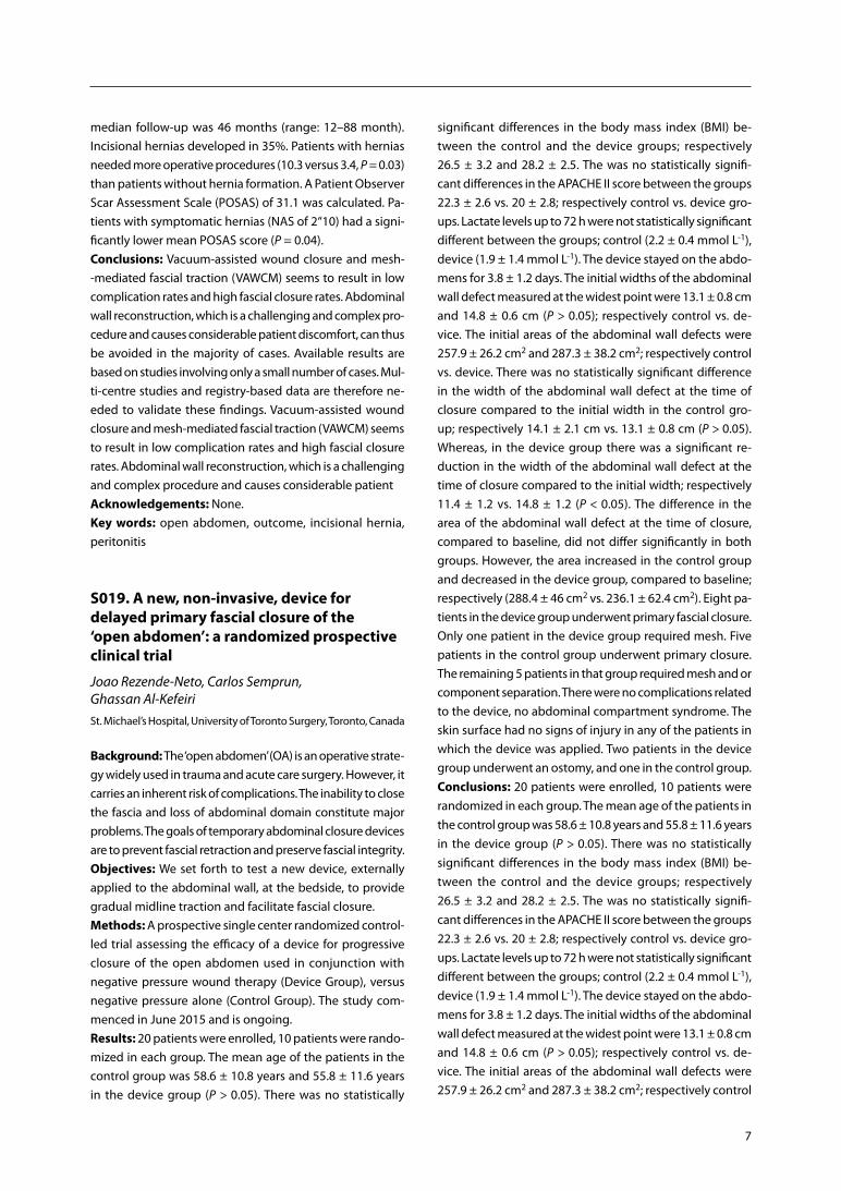
Joao Rezende-Neto, Ghassan Al Kefeiri, Carlos Semprun, Sandro Rizoli, Ori RotsteinTrauma And Acute Care Surgery, University of Toronto/St. Micha-el’s Hospital Surgery, Toronto, Canada
Background: Inability to perform fascial closure in the open abdomen results in loss of abdominal domain and ‘home-less bowel’. Temporary closure devices designed to prevent fascial retraction often compromise fascial integrity.Objectives: To test a new device applied externally to the abdominal wall, without sutures, to gradually produce mi-dline traction of the entire abdominal wall and facilitate pri-mary fascial closure in patients with open abdomen (Fig. 1). Methods: The device used in conjunction with vacuum-as-sisted closure (VAC) was compared to VAC alone in patients with open abdomens.Results: N = 20 patients, 10 in VAC group and 10 in the device group. No significant differences in BMI, APACHE II score, and lactate levels. Baseline width of the fascial defects at the widest point were 12.3 ± 0.7 cm and 14.9 ± 0.9 cm
and defect area were 251 ± 20.7 cm2 and 315 ± 37 cm2; re-spectively VAC vs. new device (P > 0.05). At 4 ± 1 days there was greater than 65% reduction in the maximum width and area of the fascial defects; respectively 14.9 ± 0. vs. 9.8 ± 1.6 cm and 315 ± 37 vs. 218 ± 48 cm2 (P < 0.05) with the new device. In contrast, the VAC group had greater than 5% increase in maximum width and area of the fascial defect compared to baseline. Primary fascial closure by direct suture of the fascial edges was achieved in 8 out of 10 patients in the new device group. In contrast, only 5 patients underwent fascial closure by primary suture in the VAC alone group. Moreover, 5 patients in that group required mesh and/or component separation pro-cedure. There were no complications related to the new device.Conclusions: The new device facilitated primary fascial closure of the open abdomen and effectively prevented lateralization of the muscles and loss of domain by encom-passing the entire abdominal wall.Acknowledgements: None. Key words: homeless bowel, primary closure, complete closure
S022. Effects of open abdomen on liver and renal dysfunction induced by intra- -abdominal infections and intra-abdominal hypertension
Jianan Ren, Lei Wu, Ranran Li, Tianyu Lu, Gefei WangJinling Hospital, Department Of General Surgery, Nanjing, China
Background: Intra-abdominal infection (IAI) combined with intra-abdominal hypertension (IAH) is a common and serious complication in critically ill patients for which there is no well--defined treatment strategy. Open abdomen (OA) therapy has been widely used in a variety of situations potentially benefi-cial to patients suffering intra-abdominal sepsis, abdominal compartment syndrome, trauma and abdominal wall defect.
Open abdomen
Dynamic sensors
Abdominal plates
Binder
2.79 Ncm–2 force
Resultant forceDue to abdominal Closure device
Outward forcesFrom abdominal muscles
Figure 1

9
Objectives: We explored the effect of OA on liver and renal dysfunction induced by IAI along with IAH. Methods: For porcine model of IAI, all animals were con-ducted CLP to induce the severe model of polymicrobial sepsis. Then the IAH model was established using the nitro-gen pneumoperitoneum procedure and the target intra-ab-dominal pressure (IAP) was 30 mm Hg. The desired IAP was maintained until the OA treatment was provided which was either at 6 hours post IAH (6 h OA group) or 24 hours post IAH (24 h OA group). The Control group was the IAI + IAH animals without open abdomen. The injuries of liver and kidney were first evaluated by histological scores. We then analysed the levels of alanine transaminase (ALT), aspartate aminotransferase (AST), creatinine (CR), and urea nitrogen (BUN). The expression of TNF-a ± and IL-6, superoxide dis-mutase (SOD) activity and malondialdehyde (MDA) level were also measured to examine inflammatory responses and antioxidant activity. The mRNA levels of cysteine-aspartic proteases-3 (Caspase-3) and terminal deoxynucleotidyl transferase dUTP nick end labeling (TUNEL) staining were examined to evaluate apoptosis.Results: We found that the liver injury was significantly improved in the 6 h OA group compared with the Control group (P < 0.05); the histological scores of renal injury was significantly lower in the 6 h OA group and 24 h OA group than the Control group (P < 0.05). The levels of liver enzymes such as ALT and AST had a significant decrease in the 6h OA group compared with the Control group (P < 0.05). No differ-ences were found between the 24 h OA group and Control group (P > 0.05). The indications of renal function (BUN and CR) were remarkably reduced in both the 6 h OA group and 24 h OA group compared with the Control group. The tissue levels of TNF-a ± and IL-6 in the kidney were significantly lower in the 6 h OA group than the Control group (P < 0.05), but only IL-6 level in the liver were reduced in the 6 h OA group compared with the Control group (P < 0.05). With respect of antioxidant activity, the expressions of SOD and MDA in the liver tissue were significantly reduced in the 6 h OA group compared with the Control group (P < 0.05); no differences were found in the kidney tissues (P > 0.05). The mRNA level of caspase-3 in the liver and kidney were remarkably decreased in the 6 h OA group compared with the Control group (P < 0.05), no differences were found between the 24 h OA group and Control group (P > 0.05). The same trends were found as for the apoptosis index from TUNEL staining of the liver and kidney.Conclusions: The early OA therapy could alleviate the his-tological injuries, down-regulated inflammatory responses and antioxidant activity, and attenuated the apoptosis of liver and kidney induced by IAI and IAH. The study may provide clues to guide the rational therapeutic treatment of organ failure for patients with peritonitis and IAH.
Acknowledgements: This study was supported by the Key Project of the Twelfth Five-Year Plan (BNJ13J002).Key words: open abdomen, organ dysfunction, intra- -abdominal hypertension, intra-abdominal infection
S023. Measurement of fat pressures during open colorectal surgery
Heidi Paine, Vimal HariharanRoyal London Hospital, Barts Health NHS Trust, Department of General Surgery, London, United Kingdom
Background: Several patient-specific risk factors have been implicated in the development of post-operative wound dehiscence in abdominal surgery. These include hypoalbu-minaemia, obesity, uraemia, and hypertension [1]. Less clear however is any contribution from intra-operative factors in ab-dominal wound dehiscence. Whilst several studies have found type of incision and method of closure to be not significant [1, 2] none have considered any potential role of fat necrosis secondary to trauma from intra-operative tissue retraction. Objectives: This study aimed to examine changes in pres-sure in the subcutaneous fat at the site of open abdominal surgery before, during, and after tissue retraction. Consider-ation of the impact of fat pressures in wound complications fell outside the scope of this study but provides a basis for further work. Methods: Fourteen consecutive patients undergoing open colorectal resections were included in this single-centre, prospective study. Fat pressures at the site of surgery were measured pre and post incision, during retraction, and fol-lowing closure; these were recorded alongside mean arterial pressures. Data analysis was performed using the paired Student t-test. Results: Of the fourteen patients, seven underwent midline incisions and seven transverse incisions. There was no dif-ference in mean fat pressures between those undergoing midline versus transverse incisions at any measurement point during the study; subsequent analysis therefore con-sidered patients with both incision type together. Fat pressures measured during retraction (mean = 40 mm Hg) were significantly higher than those measured at the time of incision (mean = 9 mm Hg) [P ≤ 0.05]. Similarly, fat pressures during retraction were significantly greater than those measured following closure (mean = 7 mm Hg) [P ≤ 0.05]. At all time points of measurement, there was no significant difference in mean arterial pressure. Conclusions: To our knowledge, this study is the first to pro-vide evidence of increases in fat pressure during retraction of tissues in open abdominal surgery. Further work should look to examine any relationship between fat pressures during retraction and post-operative wound complication rates.
10
Anaesthesiol Intensive Ther 2017, Supplement 1
Acknowledgements: None.Key words: fat pressure, open colorectal resection, wound dehiscence
References: 1. Riou J-PA, Cohen JR, Johnson H Jr. Factors Influencing Wound Dehiscence.
Am J Surg. 1992; 163: 324–330.2. Makela JT, Kiviniemi H, Juvonen T, Laitinen S. Factors Influencing wound
dehiscence after midline laparotomy. Am J Surg. 1995; 170: 387–390.
S027. Analysis of intra-abdominal pressure and its correlation with the outcome in patients undergoing emergency laparotomy
Babitha Nagaraju1, Hemanth Ghalige1, Vinay H. D.2, Abhijit Bhoyate3, Th Sudhir Chandra Singh3, Birkumar Sharma3, Moirangthem G. S.3
1General Surgery Bangalore, India2General Surgery, Hassan, India3Rims, General Surgery, Imphal, India
Background: Acute abdomen is the commonest surgical emergency encountered in day to day practice. An elevated intra-abdominal pressure (IAP) can be a symptom of an acu-te abdominal process or can be the cause of this process. But, most of the available studies on intra-abdominal hyperten-sion (IAH) and abdominal compartment syndrome (ACS) are on trauma patients or critically ill ICU patients. Only a few studies have been conducted so far about analyzing IAP in acute abdomen cases undergoing emergency laparotomy.Objectives: To study IAP of patients presenting with acute abdomen and to assess IAP as, one of the predictors of outcome in patients undergoing emergency laparotomy. Methods: This was a prospective observational study con-ducted in Department of Surgery, Regional Institute of Me-dical Sciences, Imphal, INDIA during a period from October 2013 to September 2015. The study comprised of 160 patients. IAP was measured pre-operatively and post-operatively at 0,6,24,72 hours by indirect intra-vesical technique using 16 Fr Foleyâs catheter and a manometer. All the values were recorded including duration of hospital stay, newly developed organ system dysfunction, wound dehiscence and mortality. Data was analyzed using SPSS Version 21Results: The mean (SD) IAPs before and after laparotomies were 14.21 (2.7) mm Hg and 6.6 (4.06) mm Hg respectively. The incidence of IAH in our study was 75% at admission. The overall mortality was seven (4.4%) but for ACS alone it was 100%. It was found that pre-operative intra-abdominal pressure at and above 14.07 mm Hg is a predictor of post operative outcomes with sensitivity of 65.1% and specificity of 73.2%.
Conclusions: IAH was significantly associated with wound dehiscence, post-operative duration of hospital stay, car-diovascular dysfunction, renal and respiratory dysfunction (P < 0.0001) at 6 hours and this positive association was also maintained at 24 (P = 0.002) and 72 hours (P = 0.003). Hence, IAH had a detrimental effect on various organ systems.Acknowledgements: None. Key words: acute abdomen, abdominal compartment syn-drome, intra-abdominal pressure, wound dehiscence
S030. Changes in intra-abdominal pressure affect brain-heart interaction in traumatic brain injury a pilot study
Wojciech Dabrowski1, Jaroslaw Wosko1, Hanna Brzozowska1, Radoslaw Rola2, Tomasz Trojanowski2, Mateusz Bialy1, Ziemowit Rzecki1, Todd T Schlegel3, Andrzej Jaroszynski4
1Department of Anaesthesiology Intensive Therapy, Medical University of Lublin, Lublin, Poland2Department of Neurosurgery and Paediatric Neurosurgery, Medical University of Lublin, Lublin, Poland3Department of Molecular Medicine and Surgery, Karolinska Institutet, Stockholm, Sweden and Nicollier-Schlegel SARI, Trelex, Switzerland4Department of Nephrology, Institute of Medical Science, Jan Kochanowski University, Kielce, Poland
Background: Traumatic brain injury (TBI) affects cardiac function and may induce life-threatening cardiac arrhyth-mias and sudden cardiac death (SCD). This well-established relationship is known as âœbrain-heart interaction. On the other hand, some studies have presented associations be-tween intra-abdominal pressure (IAP), cerebral circulation and intra-cranial pressure (ICP) [1, 2]. Recently, we docu-mented that increased IAP widens spatial QRS-T angle and prolongs QTc interval, both strong predictors of cardiac arrhythmia and SCD [3]. Based on these findings, we hy-pothesized that increased IAP may also affect brain-heart interactions.Objectives: The aim of this study was to investigate any effect of TBI on spatial QRS-T angle and QTc interval in re-lationship to IAP. Methods: Adult multitrauma patients with TBI and Glas-gow Coma Scale GCS < 8 were studied. Patients with tho-racic trauma were excluded. Electrocardiography (ECG) and derived vectorcardiography (VCG) were performed for spatial QRS-T angle and QTc interval calculations. IAP was measured in the urinary bladder (Kron method). Obse-rvations were performed just after the admission into ICU (baseline) and 24, 48, 72 and 96 hours after the admission into ICU.
11
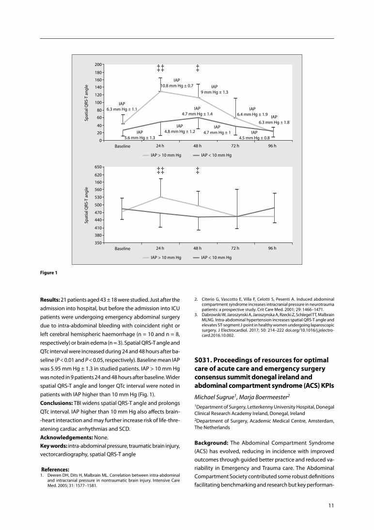
Figure 1
Results: 21 patients aged 43 ± 18 were studied. Just after the admission into hospital, but before the admission into ICU patients were undergoing emergency abdominal surgery due to intra-abdominal bleeding with coincident right or left cerebral hemispheric haemorrhage (n = 10 and n = 8, respectively) or brain edema (n = 3). Spatial QRS-T angle and QTc interval were increased during 24 and 48 hours after ba-seline (P < 0.01 and P < 0.05, respectively). Baseline mean IAP was 5.95 mm Hg ± 1.3 in studied patients. IAP > 10 mm Hg was noted in 9 patients 24 and 48 hours after baseline. Wider spatial QRS-T angle and longer QTc interval were noted in patients with IAP higher than 10 mm Hg (Fig. 1).Conclusions: TBI widens spatial QRS-T angle and prolongs QTc interval. IAP higher than 10 mm Hg also affects brain--heart interaction and may further increase risk of life-thre-atening cardiac arrhythmias and SCD.Acknowledgements: None.Key words: intra-abdominal pressure, traumatic brain injury, vectorcardiography, spatial QRS-T angle
References:1. Deeren DH, Dits H, Malbrain ML. Correlation between intra-abdominal
and intracranial pressure in nontraumatic brain injury. Intensive Care Med. 2005; 31: 1577–1581.
2. Citerio G, Vascotto E, Villa F, Celotti S, Pesenti A. Induced abdominal compartment syndrome increases intracranial pressure in neurotrauma patients: a prospective study. Crit Care Med. 2001; 29: 1466–1471.
3. Dabrowski W, Jaroszynski A, Jaroszynska A, Rzecki Z, Schlegel TT, Malbrain MLNG. Intra-abdominal hypertension increases spatial QRS-T angle and elevates ST-segment J-point in healthy women undergoing laparoscopic surgery. J Electrocardiol. 2017; 50: 214–222 doi.org/10.1016/j.jelectro-card.2016.10.002.
S031. Proceedings of resources for optimal care of acute care and emergency surgery consensus summit donegal ireland and abdominal compartment syndrome (ACS) KPIs
Michael Sugrue1, Marja Boermeester2
1Department of Surgery, Letterkenny University Hospital, Donegal Clinical Research Academy Ireland, Donegal, Ireland 2Department of Surgery, Academic Medical Centre, Amsterdam, The Netherlands
Background: The Abdominal Compartment Syndrome (ACS) has evolved, reducing in incidence with improved outcomes through guided better practice and reduced va-riability in Emergency and Trauma care. The Abdominal Compartment Society contributed some robust definitions facilitating benchmarking and research but key performan-
12
Anaesthesiol Intensive Ther 2017, Supplement 1
ce indicators (KPIs) for ACS have not been reported. To de-velop performance concepts the World Society of Emer-gency Surgery and Abdominal Compartment Society held a Summit in July 2016 in Ireland to enhance performance and quality outcomes in Emergency Surgery [1]. Part of the Summit was to explore and develop key performance indicators in key emergency surgery conditions. Objectives: The aim of paper is to report the development of KPIs for ACS.Methods: The KPIs had to be objective, easily measur-able, relating to a clinical need, be potentially reportable at specific periods and be reproducible. Five KPIs were to be developed.Results: Five KPIs were developed relating to ACS are shown below: 1) > 80% of ICU Surgical patients have an IAP measurement within 24 hours, 2) Patients with Grade 3 or 4 intra-abdominal hypertension have evidence of ACS pre-ventative strategy, 3) > 80% with an open abdomen (OA) had a clear closure plan in their index OA operation notes, 4) > 90% of patients with an OA should not develop a fis-tula, 5) > 80% with an OA are treated with NPWT from the index operationConclusions: The paper successfully described KPIs relating to ACS with an intent, rather than being definitive, to be provocative and encourage broader engagement in concept of performance improvement. These 5 KPIs will in time may need to be modified and will need validated.Acknowledgements: None. Key words: abdominal compartment, KPI, outcomes, per-formance
Reference: 1. Resources for Optimal Care of Emergency Surgery. Performance and qual-
ity outcome consensus summit. Donegal 2016 ISBN 973-0-9926109-9-9.
S032. The impact of a canister-free single-use NPWT system on surgical site complications. A meta-analysis
Vicki Strugala1, Robin Martin2
1Advanced Wound Management Smith & Nephew Scientific & Medical Affairs Hull, UK2Smith & Nephew, Scientific & Medical Affairs, Hull, UK
Background: Two recent meta-analyses have shown the be-nefit of negative pressure wound therapy (NPWT) to reduce the risk of surgical site complications (SSC) and specifically surgical site infections (SSI) when applied prophylactically to closed surgical incisions [1, 2]. The reports included a diverse mix of traditional NPWT and single-use NPWT devices and varying levels of negative pressure.
The PICO single-use NPWT system (Smith & Nephew) is a canister-free device that deals with exudate by evapora-tion and absorption using a unique dressing technology. It delivers — 80 mm Hg negative pressure (nominal).Objectives: Here we report the first meta-analysis on SSC oc-currence with a specific NPWT device to reduce heterogeneity. The influence of PICO on the rate of SSI, dehiscence and length of stay (LOS) in any surgical indication has been analysed. Methods: Review of a comprehensive database of 64 papers & 88 posters/abstracts identified 16 clinical papers (18 clini-cal studies) in which PICO was compared to standard care and SSI, dehiscence or LOS was reported. No valid reported studies were excluded.Extracted data was analysed by RevMan v5.3 and risk ratio (RR) (Mantel-Haenszel) or mean difference (Inverse Variance) were calculated with fixed effects model. Statistical signifi-cance determined by Chi2 test (P < 0.05).Results: 17 clinical studies (2343 patients) assessed SSI (% occurrence). Overall rate of SSI was 5.2% (58/1123) with PICO treatment and 12.1% (148/1220) in the standard care group; RR 0.43 [95%CI 0.33–0.58, P < 0.0001]. PICO reduced the rate of SSI compared to standard care in a wide variety of surgical patients by 57%. A sub-group analysis according to surgery type (orthopaedic, abdominal, colorectal, C-section) also showed a significant benefit of PICO to reduce SSI over standard care irrespective of surgery.Six clinical studies (1291 patients) assessed dehiscence (% occurrence). Overall rate of dehiscence was 12.8% (78/611) with PICO treatment and 17.4% (118/680) in the standard care group; RR 0.71 [95%CI 0.54–0.92 P < 0.01]. PICO reduced the rate of dehiscence compared to standard care in a wide variety of surgical patients by 29%. Eight clinical studies (725 patients) assessed LOS and these were subdivided as laparotomy surgery and other surgery. The mean reduction in LOS due to PICO treatment was 6.4 days in laparotomy surgery, 0.1 days in other surgery and 0.8 days with all combined (P < 0.0001).Conclusions: This is first comprehensive meta-analysis that has assessed the impact of a specific brand of NPWT on surgical site complications. There was a clear and significant reduction in surgical site infections, wound dehiscence and length of stay by application of the PICO single use NPWT system prophylactically on a closed surgical incision when compared to standard care. This data further supports the WHO recommendations that suggest the use of prophylactic NPWT on primarily closed surgical incisions in high-risk wounds for the purpose of preventing SSI [3].Acknowledgements: None.Key words: NPWT, meta-analysis, SSI, dehisence
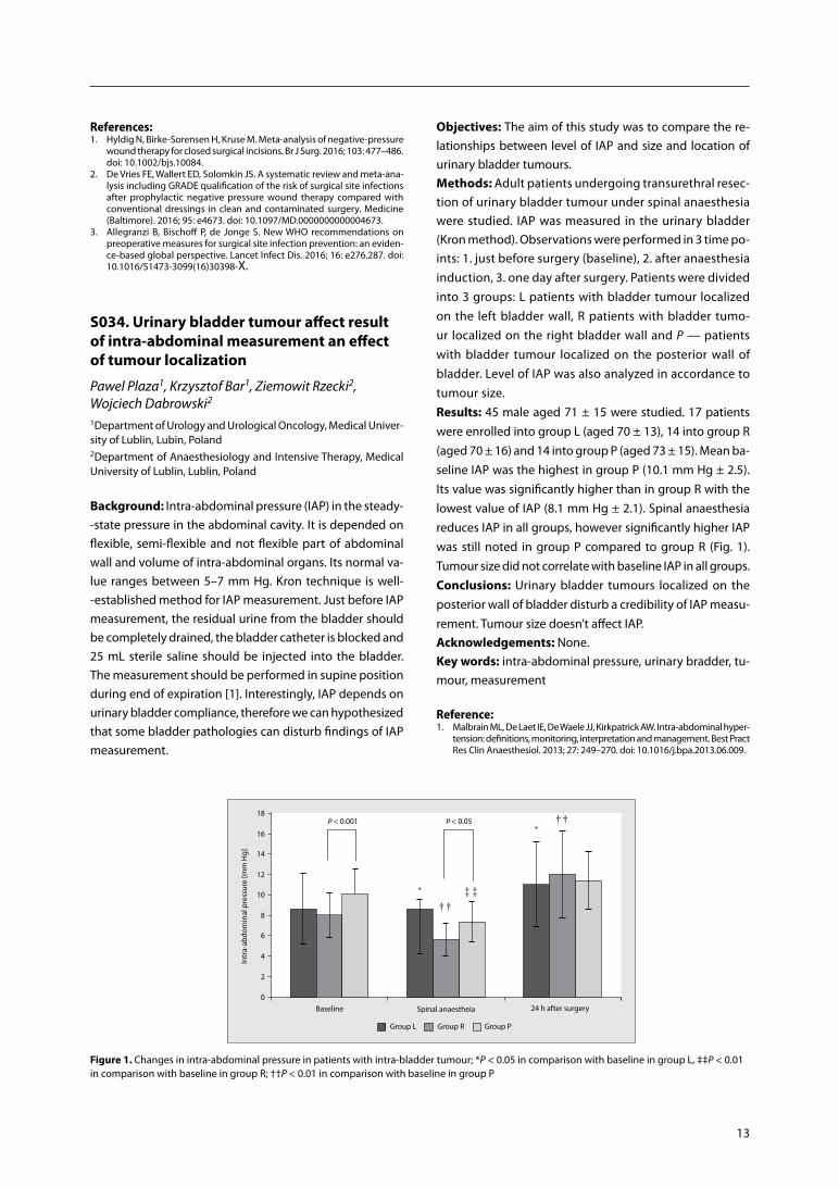
13
References:1. Hyldig N, Birke-Sorensen H, Kruse M. Meta-analysis of negative-pressure
wound therapy for closed surgical incisions. Br J Surg. 2016; 103: 477–486. doi: 10.1002/bjs.10084.
2. De Vries FE, Wallert ED, Solomkin JS. A systematic review and meta-ana-lysis including GRADE qualification of the risk of surgical site infections after prophylactic negative pressure wound therapy compared with conventional dressings in clean and contaminated surgery. Medicine (Baltimore). 2016; 95: e4673. doi: 10.1097/MD.0000000000004673.
3. Allegranzi B, Bischoff P, de Jonge S. New WHO recommendations on preoperative measures for surgical site infection prevention: an eviden-ce-based global perspective. Lancet Infect Dis. 2016; 16: e276.287. doi: 10.1016/S1473-3099(16)30398-X.
S034. Urinary bladder tumour affect result of intra-abdominal measurement an effect of tumour localization
Pawel Plaza1, Krzysztof Bar1, Ziemowit Rzecki2, Wojciech Dabrowski2
1Department of Urology and Urological Oncology, Medical Univer-sity of Lublin, Lubin, Poland 2Department of Anaesthesiology and Intensive Therapy, Medical University of Lublin, Lublin, Poland
Background: Intra-abdominal pressure (IAP) in the steady--state pressure in the abdominal cavity. It is depended on flexible, semi-flexible and not flexible part of abdominal wall and volume of intra-abdominal organs. Its normal va-lue ranges between 5–7 mm Hg. Kron technique is well--established method for IAP measurement. Just before IAP measurement, the residual urine from the bladder should be completely drained, the bladder catheter is blocked and 25 mL sterile saline should be injected into the bladder. The measurement should be performed in supine position during end of expiration [1]. Interestingly, IAP depends on urinary bladder compliance, therefore we can hypothesized that some bladder pathologies can disturb findings of IAP measurement.
Objectives: The aim of this study was to compare the re-lationships between level of IAP and size and location of urinary bladder tumours. Methods: Adult patients undergoing transurethral resec-tion of urinary bladder tumour under spinal anaesthesia were studied. IAP was measured in the urinary bladder (Kron method). Observations were performed in 3 time po-ints: 1. just before surgery (baseline), 2. after anaesthesia induction, 3. one day after surgery. Patients were divided into 3 groups: L patients with bladder tumour localized on the left bladder wall, R patients with bladder tumo-ur localized on the right bladder wall and P — patients with bladder tumour localized on the posterior wall of bladder. Level of IAP was also analyzed in accordance to tumour size.Results: 45 male aged 71 ± 15 were studied. 17 patients were enrolled into group L (aged 70 ± 13), 14 into group R (aged 70 ± 16) and 14 into group P (aged 73 ± 15). Mean ba-seline IAP was the highest in group P (10.1 mm Hg ± 2.5). Its value was significantly higher than in group R with the lowest value of IAP (8.1 mm Hg ± 2.1). Spinal anaesthesia reduces IAP in all groups, however significantly higher IAP was still noted in group P compared to group R (Fig. 1). Tumour size did not correlate with baseline IAP in all groups.Conclusions: Urinary bladder tumours localized on the posterior wall of bladder disturb a credibility of IAP measu-rement. Tumour size doesn’t affect IAP.Acknowledgements: None.Key words: intra-abdominal pressure, urinary bradder, tu-mour, measurement
Reference:1. Malbrain ML, De Laet IE, De Waele JJ, Kirkpatrick AW. Intra-abdominal hyper-
tension: definitions, monitoring, interpretation and management. Best Pract Res Clin Anaesthesiol. 2013; 27: 249–270. doi: 10.1016/j.bpa.2013.06.009.
Figure 1. Changes in intra-abdominal pressure in patients with intra-bladder tumour; *P < 0.05 in comparison with baseline in group L, ‡‡P < 0.01 in comparison with baseline in group R; ††P < 0.01 in comparison with baseline in group P
14
Anaesthesiol Intensive Ther 2017, Supplement 1
S035. Mirna 125b and 596: promising biomarker candidates for the early detection of the transition from intra- -abdominal hypertension (IAH) to abdominal compartment syndrome (ACS) in children
Torsten Kaussen1, Timo Schumacher1, Martin Boehne1, Alexander von Gise1, Florian Schmidt2, Thomas Jack1, Michael Sasse1, Thomas Thum1, Philipp Beerbaum1
1Hannover Medical School, Department of Pediatric Cardiology and Intensive Care Medicine, Hannover, Germany2Charite University Medicine, Pediatric Intensive Care Unit, Berlin, Germany
Background: In the case of a developing ACS, the duration until recognition and decompression is prognostic and de-cisive for survival. For this reason, various research groups (so far in vain) were looking for a biomarker that could be able to detect the transition from an IAH to an ACS before the clinical onset of an organ failure. According to recent results, various miRNAs seem to possess the potential to reflect inflammation as well as ischemia and/or reperfusion damage within few minutes in some tissue-specific fashion.Objectives: The aim of our study was therefore to test and identify specific miRNAs as candidate biomarkers for the early detection of a beginning ACS in children. Methods: This prospective observational study was ap-proved by the local ethics committee and registered inter-nationally (WHO-ICTRP). After obtaining written consent, all children aged 0–18 years were enrolled, who were admitted to the PICU of Hannover Medical School. IAD measure-ments were performed via nasogastric tube (Spiegelberg® Monitor) or via urinary catheter (Krons modified technique). Depending on the level of intra-abdominal pressure (IAP)
and organ dysfunction (OD) the patients were divided into five study groups (2 control groups (Neurosurgical Children (= NSH), No IAH [<10 mm Hg]), mild IAH [10–12 mm Hg], severe IAH [> 13 mm Hg] and ACS [ = IAH + OD]).EDTA blood samples were taken once a day and resulting plasma aliquots were frozen at —80°C. Sample processing and miRNA extraction/amplification were carried out to the manufacturer’s instructions (Qiagen).In the course of an initial screening, pooled samples from control and ACS groups were tested for the presence of about 800 defined miRNAs. Of these, the 8 miRNAs with the strongest differences between control and ACS pools were quantified in all individual samples. Group results were averaged and checked for significant differences.Results: Patient demographics are shown in Table 1. Most frequently detectable miRNAs in pooled samples were miR125b, 133a, 148b, 191#, 218, 326, 342, 433, 596 and 1271. Only miRNA 125b and 596 concentrations were sig-nificantly increased in ACS group samples when compared with samples of all other groups. PCR results and statistics are presented in the Figure 1 and Table 2.Conclusions: In our setting, miRNA 125b and 596 might have the potential to be considered biomarker for the de-tection of an ACS in children. All other screened miRNAs showed no pathophysiological associations. In future stud-ies, it must be checked: 1) by which pathophysiological pathway that miRNA-release can be explained, 2) whether these two miRNAs do have any tissue specificity and, 3) with which latency the miRNAs increase (compared to conven-tional inflammation parameters)Acknowledgements: None. Key words: IAH, ACS, intra abdominal hypertension, ab-dominal compartment syndrome
Table 1. Patient study grop demographic as mean ± standard deviation
total controls (none-iah +
nSh)
all iah (mild + severe)
acS P-value
Number [n] 88 44 25 19
Female [%] 45 44 24 55
Age [months] 67 1 65.3 67.7 1 61.4 60.8 1 62.0 j 73.0 4 79.8 j &P = 0.033
Weight [kg] 22.3 ± 19.7 21.3 ± 17.5 22.7 ± 22.3 25.0 ± 22.3 n.s.
LOS [days] 12.8 235.1 4.7 ± 9.9* 11.9 ± 12.3 35.0 ± 69.8* *P – 0.004
PRISM III (at admission) 11.0 + 6.1 8.7 ± 4.3** 11.3 ± 5.8&& 17.0 ± 7.1**&& **P < 0.001 &&P = 0.009
PRISM III (at discharge) 3.113.4 2.913.7 2.913.1 4.01 n.s.
deceased 5 0 0 5
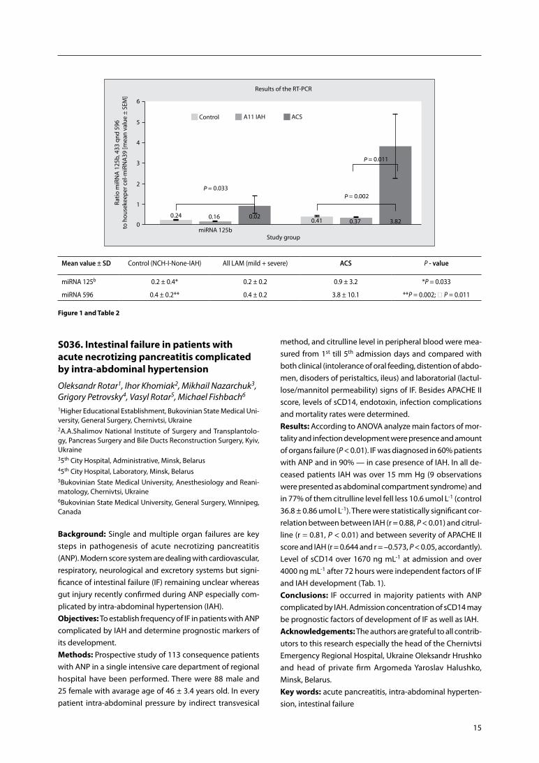
15
Figure 1 and Table 2
Mean value ± Sd Control (NCH-I-None-IAH) All LAM (mild + severe) acS P - value
miRNA 125b 0.2 ± 0.4* 0.2 ± 0.2 0.9 ± 3.2 *P = 0.033
miRNA 596 0.4 ± 0.2** 0.4 ± 0.2 3.8 ± 10.1 **P = 0.002; P = 0.011
S036. Intestinal failure in patients with acute necrotizing pancreatitis complicated by intra-abdominal hypertension
Oleksandr Rotar1, Ihor Khomiak2, Mikhail Nazarchuk3, Grigory Petrovsky4, Vasyl Rotar5, Michael Fishbach6
1Higher Educational Establishment, Bukovinian State Medical Uni-versity, General Surgery, Chernivtsi, Ukraine2A.A.Shalimov National Institute of Surgery and Transplantolo-gy, Pancreas Surgery and Bile Ducts Reconstruction Surgery, Kyiv, Ukraine35th City Hospital, Administrative, Minsk, Belarus45th City Hospital, Laboratory, Minsk, Belarus5Bukovinian State Medical University, Anesthesiology and Reani-matology, Chernivtsi, Ukraine6Bukovinian State Medical University, General Surgery, Winnipeg, Canada
Background: Single and multiple organ failures are key steps in pathogenesis of acute necrotizing pancreatitis (ANP). Modern score system are dealing with cardiovascular, respiratory, neurological and excretory systems but signi-ficance of intestinal failure (IF) remaining unclear whereas gut injury recently confirmed during ANP especially com-plicated by intra-abdominal hypertension (IAH).Objectives: To establish frequency of IF in patients with ANP complicated by IAH and determine prognostic markers of its development. Methods: Prospective study of 113 consequence patients with ANP in a single intensive care department of regional hospital have been performed. There were 88 male and 25 female with avarage age of 46 ± 3.4 years old. In every patient intra-abdominal pressure by indirect transvesical
method, and citrulline level in peripheral blood were mea-sured from 1st till 5th admission days and compared with both clinical (intolerance of oral feeding, distention of abdo-men, disoders of peristaltics, ileus) and laboratorial (lactul-lose/mannitol permeability) signs of IF. Besides APACHE II score, levels of sCD14, endotoxin, infection complications and mortality rates were determined.Results: According to ANOVA analyze main factors of mor-tality and infection development were presence and amount of organs failure (P < 0.01). IF was diagnosed in 60% patients with ANP and in 90% — in case presence of IAH. In all de-ceased patients IAH was over 15 mm Hg (9 observations were presented as abdominal compartment syndrome) and in 77% of them citrulline level fell less 10.6 umol L-1 (control 36.8 ± 0.86 umol L-1). There were statistically significant cor-relation between between IAH (r = 0.88, P < 0.01) and citrul-line (r = 0.81, P < 0.01) and between severity of APACHE II score and IAH (r = 0.644 and r = –0.573, P < 0.05, accordantly). Level of sCD14 over 1670 ng mL-1 at admission and over 4000 ng mL-1 after 72 hours were independent factors of IF and IAH development (Tab. 1).Conclusions: IF occurred in majority patients with ANP complicated by IAH. Admission concentration of sCD14 may be prognostic factors of development of IF as well as IAH.Acknowledgements: The authors are grateful to all contrib-utors to this research especially the head of the Chernivtsi Emergency Regional Hospital, Ukraine Oleksandr Hrushko and head of private firm Argomeda Yaroslav Halushko, Minsk, Belarus.Key words: acute pancreatitis, intra-abdominal hyperten-sion, intestinal failure

16
Anaesthesiol Intensive Ther 2017, Supplement 1
Table 1. Influence of intra-abdominal hypertension and citrulline concentration on intestinal failure, infection and mortality rate in patients with acute necrotizing pancreatitis (m ± SD)
Measurements control group, n–10
aPache ii score
i (0–9) n–30
ii (10–14)
n–20
iii (15–19)
n–20
iV (20–24)
n–17
V (25–29)
n–16
Vi (> 30) n–10
IAH 12–15 mm Hg 0 6 (20) 5 (25) 4 (20) 8 (45) 2 (12) 0
15–25 mm Hg 0 0 7 (35) 10 (50) 7 (41) 11 (63) 6 (60)
> 30 mm Hg 0 0 0 0 2 (17,5) 4 (25) 4 (40)
Citrulline, μmol L–1 36,8 ± 0,86
23,2 ± 0,19*
18,81 ± 0,48*
13,63 ± 0,86*
11,03 ± 0,19*
10,54 ± 0,86*
9,58 ± 0,39*
Lactulose/mannitol ratio 0,014 ± 0,005
0,022 ± 0,0031*
0,035 ± 0,003*
0,041 ± 0,0027*
0,066 ± 0,0038*
0,071 ± 0,0031*
0,077 ± 0,0036*
Paralitic ileus, n (%) 0 2 (10) 4 (20) 10 (50)* 17 (100)* 16 (100)* 10 (100)*
Infection, n (%) 0 4 (13) 5 (25)* 7 (35)* 8 (47)* 7 (44)* 8 (90)*
Mortality, n (%) 0 0 0 4 (25)* 8 (47)* 9 (56)* 10 (100)*
*P < 0,05 in comparasion with сontrol-operated group
S039. A new device to prevent fascial retraction in the open abdomen
Christian Krieglstein1, Frank Beyer2, Alexandra Maul3,1Head of Department St. Elisabeth Hospital Surgery Cologne Ger-many2Marien Hospital, Traumasurgery, Bergisch Gladbach, Germany3University Hospital, Center for Experimental Medicine, Cologne, Germany
Background: Fascial retraction remains an unsolved and major problem treating the open abdomen. All available methods are aiming at a secondary stepwise closure after the intestinal swelling has diminished. Fascial retraction often impedes a direct closure.Objectives: We developed a new device that aimes to prevent fascial retraction. This device was now tested for the first time in a porcine animal model of open ab-domen. Its effect on preventing fascial retraction was investigated. Methods: The prospective trial took place at the Center for Experimental Medicine at the University of Cologne, Germany. The trial was approved by the local governmental authority. Operative treatment was carried out under standardized conditions. 12 pigs were divided in two groups. Both groups received a median laparotomy of 30 cm centered between symphysis and xiphoid under general anaesthesia. Group one was treated with the aid of the new device. With this de-vice a ventrally directed traction was applied to both sides of the abdominal wall for 48 hours. A constant traction of 40 N was applied with the device bearing on thorax and anterior pelvic ring (Figs 1). Group two received a median laparotomy without further surgical procedures.
Drag forces for closing the abdominal fascia were mea-sured immediately after laparotomy as well as after 24 and 48 hours. In vitally critical situations, extra-measurements were performed. Measurements were performed in the laparotomy`s center as well as 7.5 cm cranially and cau-dally. Besides, distances between both fascial edges were measured at predefined traction forces. Craniocaudal fascial length as well as abdominal circumference were taken. Vital parameters and ventilation pressure were recorded. Post mortem all fascial tissues and weight bearing tissues were histologically examined. Results: Over the entire follow-up all pigs demonstrated an increase in abdominal circumference, which was mainly due to generalized edema. In both groups, drag forces for closing the abdomen increased. Group one demonstrated a significantly lesser increase in drag forces over the entire follow-up. Distances between the fascial edges yielded simi-lar results. The placement of the device on chest and pelvis didnt influence vital parameters and ventilation pressure.Conclusions: Characteristics of fascial retraction and ten-sion were observed and the new device improves the treat-ment of the open abdomen. It offers an innovative treatment option to counteract fascial retraction. The device facilitates closing a laparotomy after a longer period without any allo-plastic materials or abdominal wall reconstructions. Hereby it should reduce ICU stays and treatment costs. This trial shows the feasibility of treating open abdomen using our innovative retractor. A human trial is planned. Acknowledgements: None. Key words: open abdomen, new device, fascial retraction, abdominal wall
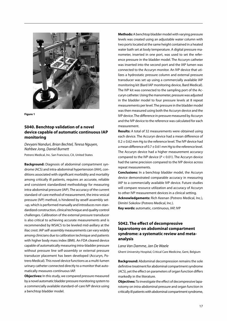
17
S040. Benchtop validation of a novel device capable of automatic continuous IAP monitoring
Devyani Nanduri, Brian Bechtel, Teresa Nguyen, Nahbee Jong, Daniel BurnettPotrero Medical, Inc. San Francisco, CA, United States
Background: Diagnosis of abdominal compartment syn-drome (ACS) and intra-abdominal hypertension (IAH), con-ditions associated with significant morbidity and mortality among critically ill patients, requires an accurate, reliable and consistent standardized methodology for measuring intra-abdominal pressure (IAP). The accuracy of the current standard-of-care method of measurement, the intra-vesical pressure (IVP) method, is hindered by œself-assembly set--up, which is performed manually and introduces non-stan-dardized construction, clinical technique and quality control challenges. Calibration of the external pressure transducer is also critical to achieving accurate measurements and is recommended by WSACS to be leveled mid-axillary at the iliac crest. IAP self-assembly measurements can vary widely among clinicians due to calibration technique and patients with higher body mass index (BMI). An FDA cleared device capable of automatically measuring intra-bladder pressure without pressure line self-assembly or external pressure transducer placement has been developed (Accuryn, Po-trero Medical). This novel device functions as a multi-lumen urinary catheter connected directly to a monitor that auto-matically measures continuous IAP.Objectives: In this study, we compared pressure measured by a novel automatic bladder pressure monitoring system to a commercially available standard-of-care IVP device using a benchtop bladder model.
Methods: A benchtop bladder model with varying pressure levels was created using an adjustable water column with two ports located at the same height contained in a heated water bath set at body temperature. A digital pressure ma-nometer, inserted in one port, was used to set the refer-ence pressure in the bladder model. The Accuryn catheter was inserted into the second port and the IAP lumen was connected to the Accuryn monitor. An IVP device that uti-lizes a hydrostatic pressure column and external pressure transducer was set up using a commercially available IAP monitoring kit (Bard IAP monitoring device, Bard Medical). The IVP kit was connected to the sampling port of the Ac-curyn catheter. Using the manometer, pressure was adjusted in the bladder model to four pressure levels at 8 repeat measurements per level. The pressure in the bladder model was then measured using both the Accuryn device and the IVP device. The difference in pressure measured by Accuryn and the IVP device to the reference was calculated for each measurement.Results: A total of 32 measurements were obtained using each device. The Accuryn device had a mean difference of 0.2 ± 0.62 mm Hg to the reference level. The IVP device had a mean difference of 0.7 ± 0.61 mm Hg to the reference level. The Accuryn device had a higher measurement accuracy compared to the IVP device (P < 0.01). The Accuryn device had the same precision compared to the IVP device across repeat measurements.Conclusions: In a benchtop bladder model, the Accuryn device demonstrated comparable accuracy in measuring IAP to a commercially available IVP device. Future studies will compare resource utilization and accuracy of Accuryn to other IVP measurement devices in a clinical setting.Acknowledgements: Rich Keenan (Potrero Medical, Inc.), Dimitri Sokolov (Potrero Medical, Inc.).Key words: automatic, continuous, IAP, IVP
S042. The effect of decompressive laparotomy on abdominal compartment syndrome: a systematic review and meta-analysis
Lana Van Damme, Jan De WaeleGhent University Hospital, Critical Care Medicine, Gent, Belgium
Background: Abdominal decompression remains the sole definitive treatment for abdominal compartment syndrome (ACS), yet the effect on parameters of organ function differs markedly in the literature.Objectives: To investigate the effect of decompressive lapa-rotomy on intra-abdominal pressure and organ function in critically ill patients with abdominal compartment syndrome,
Figure 1
18
Anaesthesiol Intensive Ther 2017, Supplement 1
specifically focusing on hemodynamic, respiratory and kidney function. Methods: We performed a systematic review and meta- -analysis of the literature (1966–February 2016). Articles reporting data on intra-abdominal pressure (IAP) and he-modynamic (mean arterial pressures (MAP), central ve-nous pressure (CVP), cardiac index (CI), heart rate (HR), systemic vascular resistance (SVR) and/or pulmonary capillary wedge pressure (PCWP)), respiratory (positive end-expiratory pressure (PEEP), peak inspiratory pressure (PIP) and/or P/F-ratio) and/or urinary output (UO) follow-ing decompressive laparotomy were identified. Outcomes were analyzed in both adults and children; in adults we report separately on studies that used the WSACS defini-tion for ACS, and those reported on Grade 4 ACS. For the statistical analysis, the CMA (comprehensive me-ta-analysis) software package was used. The outcome pa-rameters retrieved from the studies (described as mean, standard deviation and sample size) were used to calculate the standardized mean difference of each parameter as well as the P-value. Hedges g was used to examine the standardized mean difference; the following cut-offs were considered: 0.2–0.5: a small effect size, 0.5–0.8: a medium effect size and > 0.8: a large effect size.Results: A total of 16 articles, published between 1997 and 2016, were included, describing 296 patients and 302 epi-sodes of ACS; three of these included children only (18 years or younger). Half of the studies were prospective stud-ies. Of the 296 patients that were included in this analysis, 49% had primary ACS. The baseline mean IAP ranged from 21 mm Hg to 43 mm Hg and decreased with an average of 18 mm Hg following decompression. In the articles defining ACS according to the WSACS guidelines, there was an average decrease in: HR (5 bpm); PCWP (5 mm Hg); CVP (5 mm Hg); SVR (345 dyn·s cm5); MAP (11 mm Hg); PEEP (1 mm Hg); PIP (10 mm Hg) and a mean increase in: CI (0.6 L min-1m-²); P/F-ratio (75 mm Hg); UO (39 mL h-1). The articles that reported on grade IV ACS, reported a mean decrease in: HR (20 bpm); PCWP (6 mm Hg); CVP (4 mm Hg); SVR (343 dyn·s cm5); MAP (0 mm Hg); PEEP (1 mm Hg); PIP (10 mm Hg) and a mean increase in: CI (0.7 L min-1m-²); P/F-ratio (64 mm Hg); UO (125 mL h-1). In children, there was an increase in: HR (4 bpm); BP (18 mm Hg); P/F-ratio (324 mm Hg); UO (3 mL kg-1h-1) and a decrease in: CVP (9 mm Hg); PEEP (2 mm Hg); PIP (10 mm Hg). Overall, the effect size was large for the change in IAP, and change in respiratory parameters and urinary output, but only small for changes in hemodynamic parameters. Mortality following decompressive laparotomy ranged from 22 to 71%; mean overall mortality rate was 50%. The main cause of death was multiple organ failure.
Conclusions: Decompressive laparotomy not only drasti-cally lowered IAP, but had beneficial effects on hemody-namic, respiratory and renal parameters as well. However, the effect on hemodynamic parameters was less substan-tial compared to the effect on respiratory parameters and urinary output. Reported mortality after decompressive laparotomy remains high.Acknowledgements: None. Key words: ACS, decompressive laparotomy
S049. Management of the open abdomen following damage control surgery in trauma
Parker Hu, Rindi Uhlich, Frank Gleason, Jeffrey Kerby, Patrick BosargeUniversity of Alabama at Birmingham, Surgery, Birmingham, United States
Background: The introduction of damage control surgery has revolutionized the field of trauma surgery. Delay in definitive operative repair allows for resuscitation and reversal of coagulopathy. Temporary abdominal closure may be achieved by a variety of techniques, with wide reported ranges of success and timing in fascial closureObjectives: We sought to assess the effect of initial tempo-rary abdominal closure on the ability to gain fascial closure and the length of time to attain closure. Methods: A retrospective analysis of all trauma patients admitted to an ACS verified level one trauma center from 2011-2016 was performed. Those undergoing damage control laparotomy at the time of admission were in-cluded for analysis. Those with traumatic hernia pre-cluding the possibility of fascial closure were excluded. The medical record was used to identify demographic and clinical data, including admission lactate and base deficit, initial transfusion requirement, length of me-chanical ventilation, hospital length of stay (LOS), ICU LOS, complication rate, and mortality. Information on open abdominal management, including number and timing of operations, injury patterns, closure technique, and definitive fascial closure were identified. Analysis included one way ANOVA, Kaplan-Meier, and multivariate logistic regression.Results: 239 trauma patients met criteria for inclusion following damage control laparotomy. 34% of patients required massive transfusion (82/239). Skin only (SO), Ab-thera VAC system (AV), Bogota bag (BB), or standard VAC system (VAC) closure techniques were used in the initial surgery. Overall mean ISS 25.7 ± 13.8 was similar between all groups. Patients receiving SO closure had significantly
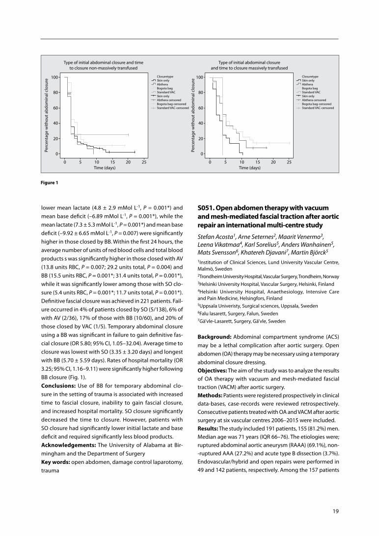
19
Figure 1
lower mean lactate (4.8 ± 2.9 mMol L-1, P = 0.001*) and mean base deficit (–6.89 mMol L-1, P = 0.001*), while the mean lactate (7.3 ± 5.3 mMol L-1, P = 0.001*) and mean base deficit (–9.92 ± 6.65 mMol L-1, P = 0.007) were significantly higher in those closed by BB. Within the first 24 hours, the average number of units of red blood cells and total blood products s was significantly higher in those closed with AV (13.8 units RBC, P = 0.007; 29.2 units total, P = 0.004) and BB (15.5 units RBC, P = 0.001*; 31.4 units total, P = 0.001*), while it was significantly lower among those with SO clo-sure (5.4 units RBC, P = 0.001*; 11.7 units total, P = 0.001*). Definitive fascial closure was achieved in 221 patients. Fail-ure occurred in 4% of patients closed by SO (5/138), 6% of with AV (2/36), 17% of those with BB (10/60), and 20% of those closed by VAC (1/5). Temporary abdominal closure using a BB was significant in failure to gain definitive fas-cial closure (OR 5.80; 95% CI, 1.05–32.04). Average time to closure was lowest with SO (3.35 ± 3.20 days) and longest with BB (5.70 ± 5.59 days). Rates of hospital mortality (OR 3.25; 95% CI, 1.16–9.11) were significantly higher following BB closure (Fig. 1).Conclusions: Use of BB for temporary abdominal clo-sure in the setting of trauma is associated with increased time to fascial closure, inability to gain fascial closure, and increased hospital mortality. SO closure significantly decreased the time to closure. However, patients with SO closure had significantly lower initial lactate and base deficit and required significantly less blood products.Acknowledgements: The University of Alabama at Bir-mingham and the Department of SurgeryKey words: open abdomen, damage control laparotomy, trauma
S051. Open abdomen therapy with vacuum and mesh-mediated fascial traction after aortic repair an international multi-centre study
Stefan Acosta1, Arne Seternes2, Maarit Venermo3, Leena Vikatmaa4, Karl Sorelius5, Anders Wanhainen5, Mats Svensson6, Khatereh Djavani7, Martin Björck5
1Institution of Clinical Sciences, Lund University Vascular Centre, Malmö, Sweden2Trondheim University Hospital, Vascular Surgery, Trondheim, Norway3Helsinki University Hospital, Vascular Surgery, Helsinki, Finland4Helsinki University Hospital, Anaethesiology, Intensive Care and Pain Medicine, Helsingfors, Finland5Uppsala Univeristy, Surgical sciences, Uppsala, Sweden6Falu lasarett, Surgery, Falun, Sweden7Gä’vle-Lasarett, Surgery, Gä’vle, Sweden
Background: Abdominal compartment syndrome (ACS) may be a lethal complication after aortic surgery. Open abdomen (OA) therapy may be necessary using a temporary abdominal closure dressing.Objectives: The aim of the study was to analyze the results of OA therapy with vacuum and mesh-mediated fascial traction (VACM) after aortic surgery. Methods: Patients were registered prospectively in clinical data-bases, case-records were reviewed retrospectively. Consecutive patients treated with OA and VACM after aortic surgery at six vascular centres 2006–2015 were included.Results: The study included 191 patients, 155 (81.2%) men. Median age was 71 years (IQR 66–76). The etiologies were; ruptured abdominal aortic aneurysm (RAAA) (69.1%), non--ruptured AAA (27.2%) and acute type B dissection (3.7%). Endovascular/hybrid and open repairs were performed in 49 and 142 patients, respectively. Among the 157 patients
20
Anaesthesiol Intensive Ther 2017, Supplement 1
who were alive at termination of OA therapy, the dura-tion of OA was 11 days (IQR 7–16). The primary delayed fascial closure rate was 91.8% (145/157), eleven patients underwent abdominal wall reconstruction, and one was left with a giant ventral hernia. Patients with OA initiated at relaparotomy (n = 88), compared to those left with OA at the primary operation (N = 103) had; more severe OA status (P = 0.006), more intestinal ischaemia (P = 0.002), longer duration of OA (P = 0.007), lower primary delayed fascial closure rate (P = 0.003), more renal replacement therapy (RRT, P < 0.001), longer intensive care unit stay (P = 0.005) and higher in-hospital mortality (P = 0.012). Nine patients developed entero-atmosperic fistulae, their mortality was 89%. Seven developed graft infections, all were diagnosed within 6 months, with a mortality of 28.6% at one year. In--hospital mortality was 39.3%. Intestinal ischaemia (OR 3.71, 95% CI 1.55–8.91]), RRT (OR 3.62 [95% CI 1.72–7.65]) and age (OR 1.12, [95% CI 1.06–1.12]), and were independent factors associated with in-hospital mortalityConclusions: Vacuum and mesh-mediated fascial traction was associated with a high primary delayed fascial closure rate after often prolonged OA therapy following aortic sur-gery. Patient outcomes were better when OA was initiated at the primary operation, compared to at relaparotomy.Acknowledgements: None. Key words: aortic aneurysm, abdominal compartment syn-drome, open abdomen, vacuum-assisted wound closure
S052. Early impact of abdominal compartment syndrome on liver, kidney, and lung damage in a rodent model
Ricardo Lima1, 2, Pedro L. Silva1, Vera L. Capelozzi3, Mariana G. Oliveirta1, Maria Cristina E. Santana2, Fernanda F. Cruz1, Paolo Pelos4, Alberto Schanaider2, Manu L.N.G. Malbrain5, Patricia R.M. Rocco1
1Laboratory of Pulmonary Investigation, Carlos Chagas Filho In-stitute of Biophysics, Federal University of Rio de Janeiro, Rio de Janeiro, Brazil2Post-Graduate Program, Department of Surgery, Faculty of Me-dicine, Federal University of Rio de Janeiro, Rio de Janeiro, Brazil3Department of Pathology, School of Medicine, University of São Paulo, São Paulo, Brazil4Department of Surgical Sciences and Integrated Diagnostics, IRCCS AOU San Martino-IST, University of Genoa, Genoa, Italy5Department of Intensive Care and High Care Burn Unit, Ziekenhuis Netwerk Antwerpen, ZNA Stuivenberg, Antwerp, Belgium
Background: Abdominal compartment syndrome (ACS) sometimes occurs in critically ill patients following damage control surgery.Objectives: The purpose of the present study was to deve-lop a model of ACS and to evaluate its pathologic impact on liver, kidney, and lung morphology.
Methods: Twenty Wistar rats (weight 300–350 g) were ran-domly divided into four groups: 1) intra-abdominal hyper-tension (IAH): laparotomy was performed and the abdomen packed with cotton until an intra-abdominal pressure (IAP) of 15 mm Hg was reached; 2) hypovolemia (HYPO): blood was withdrawn until a mean arterial pressure ~60 mm Hg was reached; 3) IAH + HYPO (to resemble clinical ACS); and 4) sham surgery. After 3 hours of protective mechanical ventilation, animals were euthanized and the liver, kidney, and lungs removed to examine the degree of tissue damage.Results: IAH resulted in: oedema and neutrophil infiltration in the kidney; necrosis, congestion, and microsteatosis in the liver; and alveolar collapse, haemorrhage, interstitial oede-ma, and neutrophil infiltration in the lungs. Furthermore, IAH was associated with greater cell apoptosis in the kidney, liver, and lungs compared to sham surgery. HYPO led to oedema and neutrophil infiltration in kidney. The combination of IAH and HYPO resulted in all aforementioned changes in lung, kidney, and liver tissue, as well as exacerbation of the inflammatory process in the kidney and liver and kidney cell necrosis and apoptosis.Conclusions: Intra-abdominal hypertension by itself is as-sociated with kidney, liver, and lung damage; when combi-ned with hypovolemia, it leads to further impairment and organ damage.Acknowledgements: We express our gratitude to Mr. Andre Benedito da Silva for animal care, Mrs. Ana Lucia Neves da Silva for her help with microscopy, Mrs. Moira Elizabeth Schottler and Mr. Filippe Vasconcellos for their assistance in editing the manuscriptKey words: hypovolemia, intra-abdominal hypertension, apoptosis, necrosis
S057. Effect of positive expiratory pressure in respirator mechanics, hemodynamics and intra-abdominal pressure in high-risk patients for IAH And ACS
Natalia Andrade1, Gabrielle Souza1, Melissa Sibinelli1, Juliana Bernardi1, Bruno Pereira2,1University of Campinas, Physiotherapy, Campinas, Brasil2University of Campinas, Trauma/ Acute Care Surgery & Surgical Critical Care, Campinas, Brasil
Background: Increased intra-abdominal pressure (IAP) is responsible for decreased of blood flow in the abdominal compartment, resulting in compression of the lung pa-renchyma, which reduces chest wall compliance and tidal volume,Objectives: To evaluate the influence of PEEP on IAP, on mechanic ventilation and on hemodynamics of patients who are at high risk for intra-abdominal hypertension (IAH) and abdominal compartmental syndrome (ACS),
21
Methods: 16 adult TICU patients were enrolled when at risk for IAH, hemodynamically stable, sedated and mechanically ventilated. Exclusion criteria were those who presented early removal of sedation, intra-cranial hypertension, hemodyna-mic instability, peritoneostomy and impossibility of bladder catheterization. Data from static (Cest) and dynamic (Cdyn) pulmonary compliance, pulmonary resistance (Raw), inspi-ratory peak pressure (Ppico), plateau pressure (Pplat), tidal volume (VT), minute volume, mean arterial pressure, heart rate, peripheral arterial saturation and IPA were evaluated. All patients followed the parameters in MV: assisted con-trolled by volume (A/C–V), VT of 6 mL kg-1 of body weight, respiratory rate of 12 breaths per minute, flow rate of 60 L min-1 and inspiratory pause of 1 second. The measurements were repeated with PEEP of 5, 10 and 15 cm H2O. IAP me-asurement was performed by nursing using the technique proposed by Kron with a three-way Folley catheter.Results: Fifteen male and one female patients with mean age of 42.93 ± 19.09 years, score Acute Physiologic Chronic He-alth Evaluation II (APACHE II) with mean of 13.62 ± 6.08 and score Sequential Organ Failure Assessment (SOFA) with an average of 6.93 ± 4.05. It was observed a significant incre-ase of Ppico in the comparison between 5 cm H2O and 10 cm H2O, between 5 cm H2O and 15 cm H2O and between 10 cm H2O and 15 cm H2O with P < 0.0001, in all comparisons, as well as Pplato with P < 0.0001 in the three comparisons. IAP showed an increase in the comparisons between 5 cm H2O and 10 cm H2O and 5 cm H2O and 15 cm H2O, both with P < 0.012, and in the comparison between 10 cm H2O and 15 cm H2O with P < 0.0455. VE presented reduction in the comparisons between 5 cm H2O and 10 cm H2O and 5 cm H2O and 15cmH2O with P < 0.006 and P < 0.001 respectively. On the other hand, Cdyn showed an increase in the com-parison between 5 cm H2O and 15 cm H2O with P < 0.01.Conclusions: An increase in IAP was observed concomitan-tly with an increase in PEEP and with an increase in peak and plateau pressures, Cdyn and VE reduction.Acknowledgements: UTC team that participated in this research.Key words: positive expiratory pressure, respirator mecha-nics, hemodynamics, IAH, ACS
S061. Promising results for the endoscopic component separation technique: a tool to decrease the rate of wound morbidity in repair of large incisional hernias
Aude Vanlander, Luis Abrue de Carvalho, Frederik BerrevoetDepartment of General and Hepatobiliary Surgery and Liver Trans-plantation, University Hospital Ghent, Belgium
Background: The technique of Component Separation is a useful tool in a surgeons arsenal for tension-free realignment
of the linea alba in order to obtain primary fascial closure after open abdomen or to cover up a mesh used as reinforcement of the abdominal wall after repair of large incisional hernias.The substantial subcutaneous dissection combined with the transsection of the perforants from the epigastric vessels to access the lateral border of the rectus muscle are cause for a high incidence of wound morbidity, which causes a pro-longed hospital stay and increased operative cost.Objectives: To determine the potential effectiveness of the endoscopic component separation technique as reductive technique for wound morbidity in large ventral hernias. Methods: A single centre, retrospective review of 8 patients planned for Rives-Stoppa repair in whom a total of 14 endo-scopic component separations were performed since intro-duction of the technique mid-2016. Demographic data, ope-rative time, and postoperative complications were recordedResults: 8 patients (5 male, 3 female), mean age of 62 y (48–75 y). Endoscopic release was performed unilaterally in two patients, and bilateral in the other 6 patients. Mean operating room time was 168 min (115–270 min) and correlated with the hernia size and additional adhaesiolysis in 2 patients. The mean length of stay was 5 days (3–7 d). We observed 1 mi-dline seroma and 1 flank seroma in 2 patients. Both were punctured because patients were symptomatic. No other complications, wound dehiscence or hematomas were regi-stered at the site of the endoscopic release in the first month postoperatively. Current follow-up of 9 months shows no recurrent hernia or long term wound morbidity.Conclusions: Endoscopic components separation is a non--time consuming, easy to learn technique that proves to be effective in reducing wound morbidity when used in large ventral hernias.Further studies are required to determine the exact benefits for endoscopic components separation hernia repair versus conventional hernia repair.Acknowledgements: None. Key words: endoscopic component separation, incisional hernias
S062. Children and adolescents after liver transplant (LTX) are highly endangered to develop intra-abdominal hypertension (IAH) and abdominal compartment syndrome (ACS)
Torsten Kaussen1, Annika Artmann1, Bernd Mitzlaff1, Alexander von Gise1, Florian Schmidt2, Martin Boehne1, Thomas Jack1, Harald Koeditz1, Michael Sasse1, Philipp Beerbaum1
1Hannover Medical School, Department of Pediatric Cardiology and Intensive Care Medicine, Hannover, Germany2University Childrens Hospital Charité Berlin, Pediatric Intensive Care Unit, Berlin, Germany
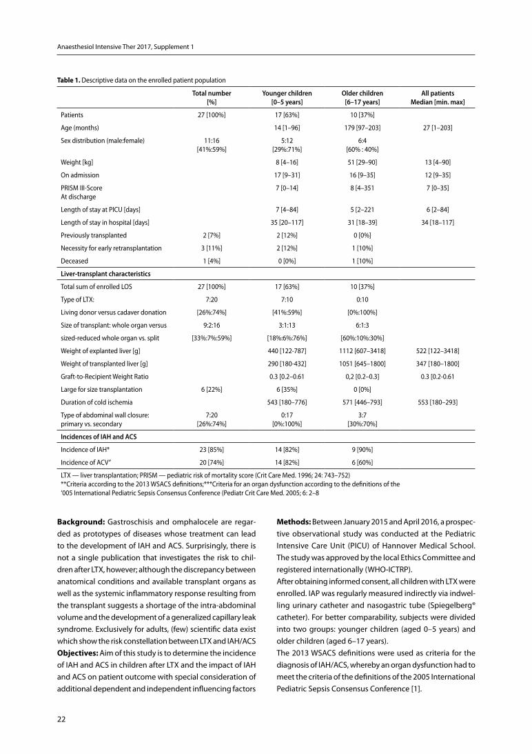
22
Anaesthesiol Intensive Ther 2017, Supplement 1
Table 1. Descriptive data on the enrolled patient population
total number[%]
Younger children [0–5 years]
older children [6–17 years]
all patients Median [min. max]
Patients 27 [100%] 17 [63%] 10 [37%]
Age (months) 14 [1–96] 179 [97–203] 27 [1–203]
Sex distribution (male:female) 11:16[41%:59%]
5:12[29%:71%]
6:4[60% : 40%]
Weight [kg] 8 [4–16] 51 [29–90] 13 [4–90]
On admission 17 [9–31] 16 [9–35] 12 [9–35]
PRISM III-ScoreAt discharge
7 [0–14] 8 [4–351 7 [0–35]
Length of stay at PICU [days] 7 [4–84] 5 [2–221 6 [2–84]
Length of stay in hospital [days] 35 [20–117] 31 [18–39] 34 [18–117]
Previously transplanted 2 [7%] 2 [12%] 0 [0%]
Necessity for early retransplantation 3 [11%] 2 [12%] 1 [10%]
Deceased 1 [4%] 0 [0%] 1 [10%]
Liver-transplant characteristics
Total sum of enrolled LOS 27 [100%] 17 [63%] 10 [37%]
Type of LTX: 7:20 7:10 0:10
Living donor versus cadaver donation [26%:74%] [41%:59%] [0%:100%]
Size of transplant: whole organ versus 9:2:16 3:1:13 6:1:3
sized-reduced whole organ vs. split [33%:7%:59%] [18%:6%:76%] [60%:10%:30%]
Weight of explanted liver [g] 440 [122-787] 1112 [607–3418] 522 [122–3418]
Weight of transplanted liver [g] 290 [180-432] 1051 [645–1800] 347 [180–1800]
Graft-to-Recipient Weight Ratio 0.3 [0.2–0.61 0,2 [0.2–0.3] 0.3 [0.2-0.61
Large for size transplantation 6 [22%] 6 [35%] 0 [0%]
Duration of cold ischemia 543 [180–776] 571 [446–793] 553 [180–293]
Type of abdominal wall closure:primary vs. secondary
7:20[26%:74%]
0:17[0%:100%]
3:7[30%:70%]
incidences of iah and acS
Incidence of IAH* 23 [85%] 14 [82%] 9 [90%]
Incidence of ACV” 20 [74%] 14 [82%] 6 [60%]
LTX — liver transplantation; PRISM — pediatric risk of mortality score (Crit Care Med. 1996; 24: 743–752)**Criteria according to the 2013 WSACS definitions; ***Criteria for an organ dysfunction according to the definitions of the‘005 International Pediatric Sepsis Consensus Conference (Pediatr Crit Care Med. 2005; 6: 2–8
Background: Gastroschisis and omphalocele are regar-ded as prototypes of diseases whose treatment can lead to the development of IAH and ACS. Surprisingly, there is not a single publication that investigates the risk to chil-dren after LTX, however; although the discrepancy between anatomical conditions and available transplant organs as well as the systemic inflammatory response resulting from the transplant suggests a shortage of the intra-abdominal volume and the development of a generalized capillary leak syndrome. Exclusively for adults, (few) scientific data exist which show the risk constellation between LTX and IAH/ACSObjectives: Aim of this study is to determine the incidence of IAH and ACS in children after LTX and the impact of IAH and ACS on patient outcome with special consideration of additional dependent and independent influencing factors
Methods: Between January 2015 and April 2016, a prospec-tive observational study was conducted at the Pediatric Intensive Care Unit (PICU) of Hannover Medical School. The study was approved by the local Ethics Committee and registered internationally (WHO-ICTRP). After obtaining informed consent, all children with LTX were enrolled. IAP was regularly measured indirectly via indwel-ling urinary catheter and nasogastric tube (Spiegelberg® catheter). For better comparability, subjects were divided into two groups: younger children (aged 0–5 years) and older children (aged 6–17 years).The 2013 WSACS definitions were used as criteria for the diagnosis of IAH/ACS, whereby an organ dysfunction had to meet the criteria of the definitions of the 2005 International Pediatric Sepsis Consensus Conference [1].
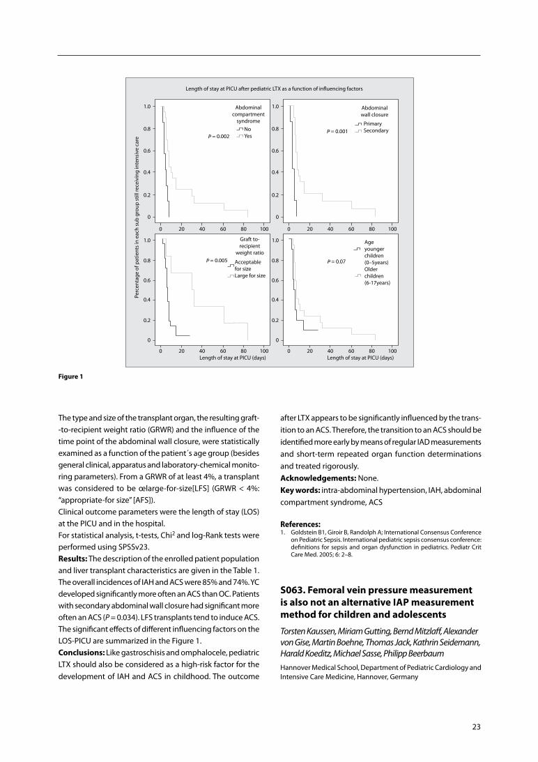
23
The type and size of the transplant organ, the resulting graft--to-recipient weight ratio (GRWR) and the influence of the time point of the abdominal wall closure, were statistically examined as a function of the patient´s age group (besides general clinical, apparatus and laboratory-chemical monito-ring parameters). From a GRWR of at least 4%, a transplant was considered to be œlarge-for-size[LFS] (GRWR < 4%: “appropriate-for size” [AFS]).Clinical outcome parameters were the length of stay (LOS) at the PICU and in the hospital.For statistical analysis, t-tests, Chi2 and log-Rank tests were performed using SPSSv23.Results: The description of the enrolled patient population and liver transplant characteristics are given in the Table 1. The overall incidences of IAH and ACS were 85% and 74%. YC developed significantly more often an ACS than OC. Patients with secondary abdominal wall closure had significant more often an ACS (P = 0.034). LFS transplants tend to induce ACS. The significant effects of different influencing factors on the LOS-PICU are summarized in the Figure 1.Conclusions: Like gastroschisis and omphalocele, pediatric LTX should also be considered as a high-risk factor for the development of IAH and ACS in childhood. The outcome
after LTX appears to be significantly influenced by the trans-ition to an ACS. Therefore, the transition to an ACS should be identified more early by means of regular IAD measurements and short-term repeated organ function determinations and treated rigorously.Acknowledgements: None. Key words: intra-abdominal hypertension, IAH, abdominal compartment syndrome, ACS
References: 1. Goldstein B1, Giroir B, Randolph A; International Consensus Conference
on Pediatric Sepsis. International pediatric sepsis consensus conference: definitions for sepsis and organ dysfunction in pediatrics. Pediatr Crit Care Med. 2005; 6: 2–8.
S063. Femoral vein pressure measurement is also not an alternative IAP measurement method for children and adolescents
Torsten Kaussen, Miriam Gutting, Bernd Mitzlaff, Alexander von Gise, Martin Boehne, Thomas Jack, Kathrin Seidemann, Harald Koeditz, Michael Sasse, Philipp BeerbaumHannover Medical School, Department of Pediatric Cardiology and Intensive Care Medicine, Hannover, Germany
Figure 1
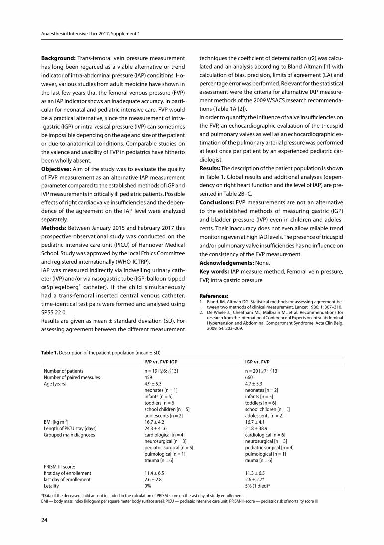
24
Anaesthesiol Intensive Ther 2017, Supplement 1
Table 1. Description of the patient population (mean ± SD)
iVP vs. fVP igP igP vs. fVP
Number of patientsNumber of paired measures Age [years]
BMI [kg m-2]Length of PICU stay [days] Grouped main diagnoses
PRISM-III-score:first day of enrollementlast day of enrollement Letality
n = 19 [♀6; ♂13]4594.9 ± 5.3neonates [n = 1]infants [n = 5]toddlers [n = 6]school children [n = 5] adolescents [n = 2]16.7 ± 4.224.3 ± 41.6 cardiological [n = 4] neurosurgical [n = 3] pediatric surgical [n = 5] pulmological [n = 1] trauma [n = 6]
11.4 ± 6.5 2.6 ± 2.8 0%
n = 20 [♀7; ♂13]6604.7 ± 5.3neonates [n = 2]infants [n = 5]toddlers [n = 6]school children [n = 5] adolescents [n = 2] 16.7 ± 4.121.8 ± 38.9 cardiological [n = 6] neurosurgical [n = 3] pediatric surgical [n = 4] pulmological [n = 1] rauma [n = 6]
11.3 ± 6.5 2.6 ± 2.7* 5% (1 died)*
*Data of the deceased child are not included in the calculation of PRISM score on the last day of study enrollement. BMI — body mass index [kilogram per square meter body surface area]; PICU — pediatric intensive care unit; PRISM-III-score — pediatric risk of mortality score III
Background: Trans-femoral vein pressure measurement has long been regarded as a viable alternative or trend indicator of intra-abdominal pressure (IAP) conditions. Ho-wever, various studies from adult medicine have shown in the last few years that the femoral venous pressure (FVP) as an IAP indicator shows an inadequate accuracy. In parti-cular for neonatal and pediatric intensive care, FVP would be a practical alternative, since the measurement of intra--gastric (IGP) or intra-vesical pressure (IVP) can sometimes be impossible depending on the age and size of the patient or due to anatomical conditions. Comparable studies on the valence and usability of FVP in pediatrics have hitherto been wholly absent.Objectives: Aim of the study was to evaluate the quality of FVP measurement as an alternative IAP measurement parameter compared to the established methods of IGP and IVP measurements in critically ill pediatric patients. Possible effects of right cardiac valve insufficiencies and the depen-dence of the agreement on the IAP level were analyzed separately. Methods: Between January 2015 and February 2017 this prospective observational study was conducted on the pediatric intensive care unit (PICU) of Hannover Medical School. Study was approved by the local Ethics Committee and registered internationally (WHO-ICTRP).IAP was measured indirectly via indwelling urinary cath-eter (IVP) and/or via nasogastric tube (IGP; balloon-tipped œSpiegelberg® catheter). If the child simultaneously had a trans-femoral inserted central venous catheter, time-identical test pairs were formed and analysed using SPSS 22.0.Results are given as mean ± standard deviation (SD). For assessing agreement between the different measurement
techniques the coefficient of determination (r2) was calcu-lated and an analysis according to Bland Altman [1] with calculation of bias, precision, limits of agreement (LA) and percentage error was performed. Relevant for the statistical assessment were the criteria for alternative IAP measure-ment methods of the 2009 WSACS research recommenda-tions (Table 1A [2]).In order to quantify the influence of valve insufficiencies on the FVP, an echocardiographic evaluation of the tricuspid and pulmonary valves as well as an echocardiographic es-timation of the pulmonary arterial pressure was performed at least once per patient by an experienced pediatric car-diologist.Results: The description of the patient population is shown in Table 1. Global results and additional analyses (depen-dency on right heart function and the level of IAP) are pre-sented in Table 2B–C.Conclusions: FVP measurements are not an alternative to the established methods of measuring gastric (IGP) and bladder pressure (IVP) even in children and adoles-cents. Their inaccuracy does not even allow reliable trend monitoring even at high IAD levels. The presence of tricuspid and/or pulmonary valve insufficiencies has no influence on the consistency of the FVP measurement.Acknowledgements: None. Key words: IAP measure method, Femoral vein pressure, FVP, intra gastric pressure
References:1. Bland JM, Altman DG. Statistical methods for assessing agreement be-
tween two methods of clinical measurement. Lancet 1986; 1: 307–310.2. De Waele JJ, Cheatham ML, Malbrain ML et al. Recommendations for
research from the International Conference of Experts on Intra-abdominal Hypertension and Abdominal Compartment Syndrome. Acta Clin Belg. 2009; 64: 203–209.
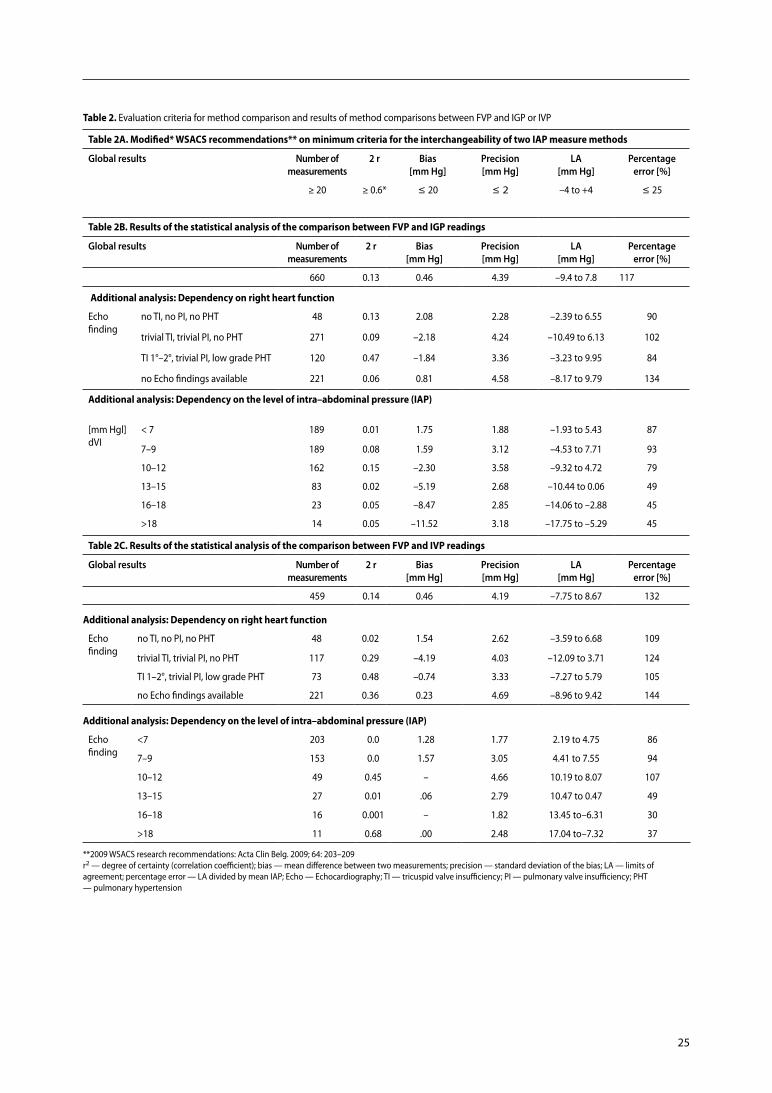
25
Additional analysis: Dependency on right heart function
Echo finding
no TI, no PI, no PHT 48 0.13 2.08 2.28 –2.39 to 6.55 90
trivial TI, trivial PI, no PHT 271 0.09 –2.18 4.24 –10.49 to 6.13 102
TI 1°–2°, trivial PI, low grade PHT 120 0.47 –1.84 3.36 –3.23 to 9.95 84
no Echo findings available 221 0.06 0.81 4.58 –8.17 to 9.79 134
Additional analysis: Dependency on the level of intra–abdominal pressure (IAP)
Additional analysis: Dependency on right heart function
Echo finding
no TI, no PI, no PHT 48 0.02 1.54 2.62 –3.59 to 6.68 109
trivial TI, trivial PI, no PHT 117 0.29 –4.19 4.03 –12.09 to 3.71 124
TI 1–2°, trivial PI, low grade PHT 73 0.48 –0.74 3.33 –7.27 to 5.79 105
no Echo findings available 221 0.36 0.23 4.69 –8.96 to 9.42 144
Table 2C. Results of the statistical analysis of the comparison between FVP and IVP readings
global results number of measurements
2 r bias [mm hg]
Precision [mm hg]
La [mm hg]
Percentage error [%]
459 0.14 0.46 4.19 –7.75 to 8.67 132
[mm Hgl] dVI
< 7 189 0.01 1.75 1.88 –1.93 to 5.43 87
7–9 189 0.08 1.59 3.12 –4.53 to 7.71 93
10–12 162 0.15 –2.30 3.58 –9.32 to 4.72 79
13–15 83 0.02 –5.19 2.68 –10.44 to 0.06 49
16–18 23 0.05 –8.47 2.85 –14.06 to –2.88 45
>18 14 0.05 –11.52 3.18 –17.75 to –5.29 45
Table 2B. Results of the statistical analysis of the comparison between FVP and IGP readings
global results number of measurements
2 r bias [mm hg]
Precision [mm hg]
La [mm hg]
Percentage error [%]
660 0.13 0.46 4.39 –9.4 to 7.8 117
Table 2A. Modified* WSACS recommendations** on minimum criteria for the interchangeability of two IAP measure methods
global results number of measurements
2 r bias [mm hg]
Precision [mm hg]
La [mm hg]
Percentage error [%]
≥ 20 ≥ 0.6* £ 20 £ 2 –4 to +4 £ 25
Additional analysis: Dependency on the level of intra–abdominal pressure (IAP)
Echo finding
<7 203 0.0 1.28 1.77 2.19 to 4.75 86
7–9 153 0.0 1.57 3.05 4.41 to 7.55 94
10–12 49 0.45 – 4.66 10.19 to 8.07 107
13–15 27 0.01 .06 2.79 10.47 to 0.47 49
16–18 16 0.001 – 1.82 13.45 to–6.31 30
>18 11 0.68 .00 2.48 17.04 to–7.32 37
**2009 WSACS research recommendations: Acta Clin Belg. 2009; 64: 203–209 r2 — degree of certainty (correlation coefficient); bias — mean difference between two measurements; precision — standard deviation of the bias; LA — limits of agreement; percentage error — LA divided by mean IAP; Echo — Echocardiography; TI — tricuspid valve insufficiency; PI — pulmonary valve insufficiency; PHT — pulmonary hypertension
Table 2. Evaluation criteria for method comparison and results of method comparisons between FVP and IGP or IVP
26
Anaesthesiol Intensive Ther 2017, Supplement 1
S064. Temporary abdominal closure assisted with negative pressure therapy vs primary abdominal wall closure in the severe abdominal sepsis: analysis of our results
Antonio J Gonzalez Sanchez1, Jos Manuel Aranda Narvaez2, Aberto Titos-Garc a2, Isaac Cabrera-Serna2, Cristina Rodriguez Silva3, Maria Perezreyes2, Julio Santoyo-Santoyo2
1General and Digestive Surgery, Acute Care and Trau Hospital Regional de Málaga General, Digestive and Transplantation Surgery, Málaga, Spain2Hospital Regional de Málaga, General, Digestive and Transplantion Surgery, Malaga, Spain3Hospital Comarcal de la Axarqu a, General and Digestive Surgery, Málaga, Spain
Background: Severe abdominal sepsis (SAS) is one of the most important causes of mortality associated with Acute Care Sur-gery. To decrease it, new technical aspects like the application of temporary abdominal closure have been developed. Altho-ugh this technique is not widely accepted, the assistance of negative pressure therapy (ABThera, KCI) could be determinantObjectives: To analyze the differences between patients with SAS, treated after surgery with temporary abdominal closure assisted with negative pressure therapy (TACNPT) or with primary abdominal closure (PAC). Primary endpoint: In-hospital mortality. Secondary endpoints: Intensive Care Unit (ICU) mortality, ICU stay, days of mechanical ventilation, prolonged mechanical ventilation (> 7 days), hemofiltration, vasoactives, nosocomial infections, pneumonies and adult respiratory distress syndrome (ARDS) rate. Methods: Retrospective cohort study using the prospective database TACNPT of the Trauma and Acute Care Surgery Unit of a tertiary hospital and the retrospective records of the ICU. Period of study: january 2009–january 2015. Statistical analysis with software SPSS, with Student-t or Chi2 test (or non-parametric equivalent) depending on analyzed varia-bles, accepting P < 0.05 as a statistically significant result.Results: 93 patients with SAS were included, 31 with TACNPT and 62 with PAC. Both groups were homogeneous in age, sex, Manheimm peritonitis index, Charlson, SOFA and APA-CHE II indexes. Hospital mortality was lower but not stati-scally significant in TACNPT patients (32% vs 37.1%, n.s.). ICU mortality was significantly lower in TACNPT patients (12.9% vs 32%, P < 0.05), although ICU stay was longer (22.6 vs 15.5 days, n.s.). Fewer days of mechanical ventilation were required for TACNPT patients (8 vs 10.4, n.s.), and necessity of prolongad mechanical ventilation was inferior (35.5% vs 54.8%, n.s.). In addition, they had less adult respiratory distress syndrome (22.6% vs 19.4%, n.s.) but they had more pneumonies (17.7% vs 12.9%, n.s.). Global nosocomial infec-tion rate was significantly inferior in TACNPT patients (35.5% vs 58.1%, P < 0.05), as was the necessity of vasoactives (71% vs 82.3%, n.s.) and hemofiltration (12.9% vs 21%, n.s.).
Conclusions: The use of TACNPT could improve mortality and decrease postoperative morbidity when compared with PAC in patients with SAS.Acknowledgements: None. Key words: severe abdominal sepsis, open abdomen, tem-porary abdominal closure, negative pressure therapy
S067. Comparative study between continuous indirect intrabdominal pressure technique and direct technique. Preliminary result
Francisco Pracca, Pablo Bousa, Corina Puppo, Alberto BiestroCTI, Hospital de Clinicas, Facultad de Medicina, Universidad de la Republica, Montevideo, Uruguay
Background: Intra-abdominal pressure (IAP) is another ele-ment on the abdominal monitoring. The intensive care unit proposed to compare IAP measured with indirect continuous technique vs IAP measured with direct continuous technique.Objectives: To compare the indirect continuous IAPsv measu-res with direct measures through the intra-abdominal catheter. Methods: Prospective design. Threee male patients have been studied. We have taken a total of 111 measures. Indi-rect continuous technique measures IAP by a blader cathe-ter connected to a monitor through transducer. Direct tech-nique with intra-abdominal catheter in Douglas. Catheter is introduced during surgery with direct vision, exteriorized through another suprapubic incision, connected to trans-ducer with reading on the monitor.Statistics: It has been used correlation coefficient of Pearson and non parametric coefficient of Spearman. Bland and Altman test, it has been considered aceptable if the average difference between reading was less than 1 mm Hg.Results: Average age 37.3 (16.67 years). Simultaneous me-asure with indirect continuous technique and intra-abdo-minal catheter. The association between the results of the measure of IAPsv with blader catheter technique and with peritoneal catheter tecnique valued through correlation has been significative to 0,01 level, bilaterally with a corre-lation coefficient Pearson (P) of 0,963 and a non parametric coefficient of Spearman of 0,841. The graphic comparison of Bland “Altman was used between the measures of the IAP by catheter blader and by peritoneal catheter, not founding any significative difference between both measures, with a difference of measure of –0,009 (CI –0,168 to 0,150) with estandar deviation of 0,84741 and a test of differences on the Pitman variance of: r = 0,085; n = 111; P = 0,377.Conclusions: To measure IAP on an indirect continuos way through vesical catheter is safe, trustable requiring setting to zero every three hours.Acknowledgements: None.
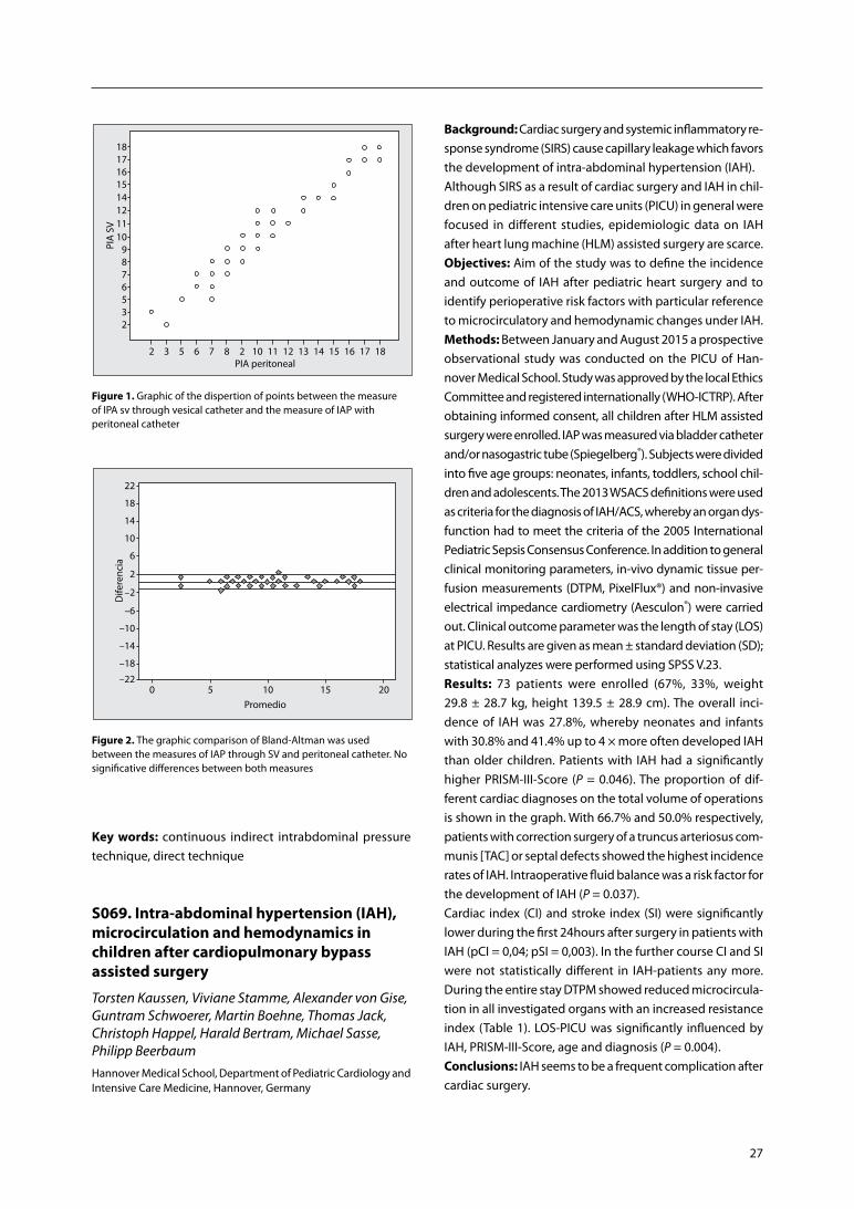
27
Background: Cardiac surgery and systemic inflammatory re-sponse syndrome (SIRS) cause capillary leakage which favors the development of intra-abdominal hypertension (IAH).Although SIRS as a result of cardiac surgery and IAH in chil-dren on pediatric intensive care units (PICU) in general were focused in different studies, epidemiologic data on IAH after heart lung machine (HLM) assisted surgery are scarce.Objectives: Aim of the study was to define the incidence and outcome of IAH after pediatric heart surgery and to identify perioperative risk factors with particular reference to microcirculatory and hemodynamic changes under IAH. Methods: Between January and August 2015 a prospective observational study was conducted on the PICU of Han-nover Medical School. Study was approved by the local Ethics Committee and registered internationally (WHO-ICTRP). After obtaining informed consent, all children after HLM assisted surgery were enrolled. IAP was measured via bladder catheter and/or nasogastric tube (Spiegelberg®). Subjects were divided into five age groups: neonates, infants, toddlers, school chil-dren and adolescents. The 2013 WSACS definitions were used as criteria for the diagnosis of IAH/ACS, whereby an organ dys-function had to meet the criteria of the 2005 International Pediatric Sepsis Consensus Conference. In addition to general clinical monitoring parameters, in-vivo dynamic tissue per-fusion measurements (DTPM, PixelFlux®) and non-invasive electrical impedance cardiometry (Aesculon®) were carried out. Clinical outcome parameter was the length of stay (LOS) at PICU. Results are given as mean ± standard deviation (SD); statistical analyzes were performed using SPSS V.23.Results: 73 patients were enrolled (67%, 33%, weight 29.8 ± 28.7 kg, height 139.5 ± 28.9 cm). The overall inci-dence of IAH was 27.8%, whereby neonates and infants with 30.8% and 41.4% up to 4 × more often developed IAH than older children. Patients with IAH had a significantly higher PRISM-III-Score (P = 0.046). The proportion of dif-ferent cardiac diagnoses on the total volume of operations is shown in the graph. With 66.7% and 50.0% respectively, patients with correction surgery of a truncus arteriosus com-munis [TAC] or septal defects showed the highest incidence rates of IAH. Intraoperative fluid balance was a risk factor for the development of IAH (P = 0.037).Cardiac index (CI) and stroke index (SI) were significantly lower during the first 24hours after surgery in patients with IAH (pCI = 0,04; pSI = 0,003). In the further course CI and SI were not statistically different in IAH-patients any more. During the entire stay DTPM showed reduced microcircula-tion in all investigated organs with an increased resistance index (Table 1). LOS-PICU was significantly influenced by IAH, PRISM-III-Score, age and diagnosis (P = 0.004).Conclusions: IAH seems to be a frequent complication after cardiac surgery.
Figure 1. Graphic of the dispertion of points between the measure of IPA sv through vesical catheter and the measure of IAP with peritoneal catheter
Figure 2. The graphic comparison of Bland-Altman was used between the measures of IAP through SV and peritoneal catheter. No significative differences between both measures
Key words: continuous indirect intrabdominal pressure technique, direct technique
S069. Intra-abdominal hypertension (IAH), microcirculation and hemodynamics in children after cardiopulmonary bypass assisted surgery
Torsten Kaussen, Viviane Stamme, Alexander von Gise, Guntram Schwoerer, Martin Boehne, Thomas Jack, Christoph Happel, Harald Bertram, Michael Sasse, Philipp BeerbaumHannover Medical School, Department of Pediatric Cardiology and Intensive Care Medicine, Hannover, Germany
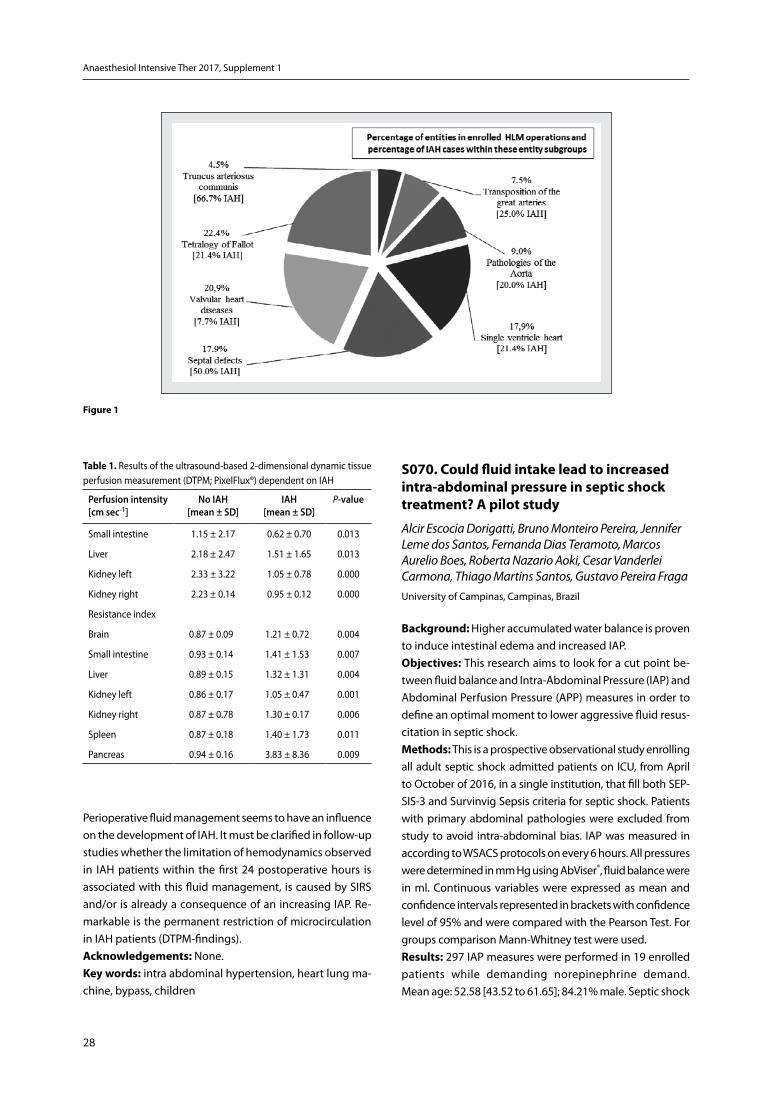
28
Anaesthesiol Intensive Ther 2017, Supplement 1
Figure 1
Table 1. Results of the ultrasound-based 2-dimensional dynamic tissue perfusion measurement (DTPM; PixelFlux®) dependent on IAH
Perfusion intensity [cm sec-1]
no iah[mean ± Sd]
iah [mean ± Sd]
P-value
Small intestine 1.15 ± 2.17 0.62 ± 0.70 0.013
Liver 2.18 ± 2.47 1.51 ± 1.65 0.013
Kidney left 2.33 ± 3.22 1.05 ± 0.78 0.000
Kidney right 2.23 ± 0.14 0.95 ± 0.12 0.000
Resistance index
Brain 0.87 ± 0.09 1.21 ± 0.72 0.004
Small intestine 0.93 ± 0.14 1.41 ± 1.53 0.007
Liver 0.89 ± 0.15 1.32 ± 1.31 0.004
Kidney left 0.86 ± 0.17 1.05 ± 0.47 0.001
Kidney right 0.87 ± 0.78 1.30 ± 0.17 0.006
Spleen 0.87 ± 0.18 1.40 ± 1.73 0.011
Pancreas 0.94 ± 0.16 3.83 ± 8.36 0.009
Perioperative fluid management seems to have an influence on the development of IAH. It must be clarified in follow-up studies whether the limitation of hemodynamics observed in IAH patients within the first 24 postoperative hours is associated with this fluid management, is caused by SIRS and/or is already a consequence of an increasing IAP. Re-markable is the permanent restriction of microcirculation in IAH patients (DTPM-findings).Acknowledgements: None. Key words: intra abdominal hypertension, heart lung ma-chine, bypass, children
S070. Could fluid intake lead to increased intra-abdominal pressure in septic shock treatment? A pilot study
Alcir Escocia Dorigatti, Bruno Monteiro Pereira, Jennifer Leme dos Santos, Fernanda Dias Teramoto, Marcos Aurelio Boes, Roberta Nazario Aoki, Cesar Vanderlei Carmona, Thiago Martins Santos, Gustavo Pereira FragaUniversity of Campinas, Campinas, Brazil
Background: Higher accumulated water balance is proven to induce intestinal edema and increased IAP.Objectives: This research aims to look for a cut point be-tween fluid balance and Intra-Abdominal Pressure (IAP) and Abdominal Perfusion Pressure (APP) measures in order to define an optimal moment to lower aggressive fluid resus-citation in septic shock. Methods: This is a prospective observational study enrolling all adult septic shock admitted patients on ICU, from April to October of 2016, in a single institution, that fill both SEP-SIS-3 and Survinvig Sepsis criteria for septic shock. Patients with primary abdominal pathologies were excluded from study to avoid intra-abdominal bias. IAP was measured in according to WSACS protocols on every 6 hours. All pressures were determined in mm Hg using AbViser®, fluid balance were in ml. Continuous variables were expressed as mean and confidence intervals represented in brackets with confidence level of 95% and were compared with the Pearson Test. For groups comparison Mann-Whitney test were used.Results: 297 IAP measures were performed in 19 enrolled patients while demanding norepinephrine demand. Mean age: 52.58 [43.52 to 61.65]; 84.21% male. Septic shock
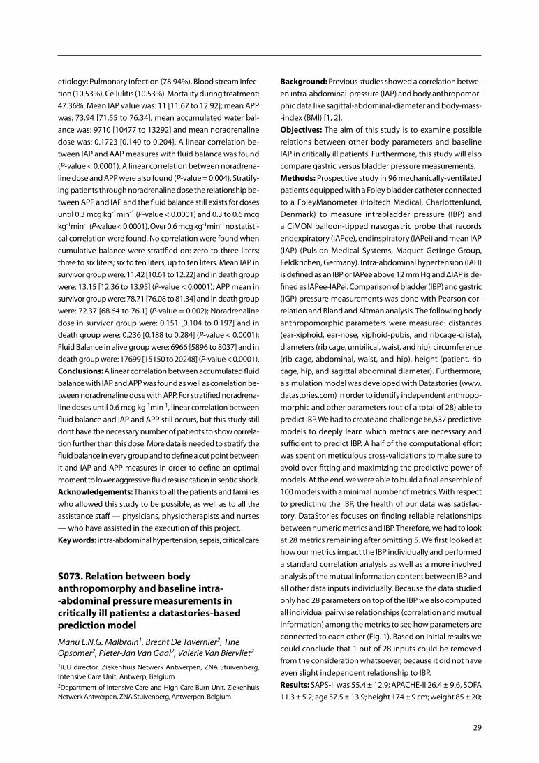
29
etiology: Pulmonary infection (78.94%), Blood stream infec-tion (10.53%), Cellulitis (10.53%). Mortality during treatment: 47.36%. Mean IAP value was: 11 [11.67 to 12.92]; mean APP was: 73.94 [71.55 to 76.34]; mean accumulated water bal-ance was: 9710 [10477 to 13292] and mean noradrenaline dose was: 0.1723 [0.140 to 0.204]. A linear correlation be-tween IAP and AAP measures with fluid balance was found (P-value < 0.0001). A linear correlation between noradrena-line dose and APP were also found (P-value = 0.004). Stratify-ing patients through noradrenaline dose the relationship be-tween APP and IAP and the fluid balance still exists for doses until 0.3 mcg kg-1min-1 (P-value < 0.0001) and 0.3 to 0.6 mcg
kg-1min-1 (P-value < 0.0001). Over 0.6 mcg kg-1min-1 no statisti-cal correlation were found. No correlation were found when cumulative balance were stratified on: zero to three liters; three to six liters; six to ten liters, up to ten liters. Mean IAP in survivor group were: 11.42 [10.61 to 12.22] and in death group were: 13.15 [12.36 to 13.95] (P-value < 0.0001); APP mean in survivor group were: 78.71 [76.08 to 81.34] and in death group were: 72.37 [68.64 to 76.1] (P-value = 0.002); Noradrenaline dose in survivor group were: 0.151 [0.104 to 0.197] and in death group were: 0.236 [0.188 to 0.284] (P-value < 0.0001); Fluid Balance in alive group were: 6966 [5896 to 8037] and in death group were: 17699 [15150 to 20248] (P-value < 0.0001).Conclusions: A linear correlation between accumulated fluid balance with IAP and APP was found as well as correlation be-tween noradrenaline dose with APP. For stratified noradrena-line doses until 0.6 mcg kg-1min-1, linear correlation between fluid balance and IAP and APP still occurs, but this study still dont have the necessary number of patients to show correla-tion further than this dose. More data is needed to stratify the fluid balance in every group and to define a cut point between it and IAP and APP measures in order to define an optimal moment to lower aggressive fluid resuscitation in septic shock.Acknowledgements: Thanks to all the patients and families who allowed this study to be possible, as well as to all the assistance staff — physicians, physiotherapists and nurses — who have assisted in the execution of this project.Key words: intra-abdominal hypertension, sepsis, critical care
S073. Relation between body anthropomorphy and baseline intra- -abdominal pressure measurements in critically ill patients: a datastories-based prediction model
Manu L.N.G. Malbrain1, Brecht De Tavernier2, Tine Opsomer2, Pieter-Jan Van Gaal2, Valerie Van Biervliet2
1ICU director, Ziekenhuis Netwerk Antwerpen, ZNA Stuivenberg, Intensive Care Unit, Antwerp, Belgium2Department of Intensive Care and High Care Burn Unit, Ziekenhuis Netwerk Antwerpen, ZNA Stuivenberg, Antwerpen, Belgium
Background: Previous studies showed a correlation betwe-en intra-abdominal-pressure (IAP) and body anthropomor-phic data like sagittal-abdominal-diameter and body-mass--index (BMI) [1, 2].Objectives: The aim of this study is to examine possible relations between other body parameters and baseline IAP in critically ill patients. Furthermore, this study will also compare gastric versus bladder pressure measurements. Methods: Prospective study in 96 mechanically-ventilated patients equipped with a Foley bladder catheter connected to a FoleyManometer (Holtech Medical, Charlottenlund, Denmark) to measure intrabladder pressure (IBP) and a CiMON balloon-tipped nasogastric probe that records endexpiratory (IAPee), endinspiratory (IAPei) and mean IAP (IAP) (Pulsion Medical Systems, Maquet Getinge Group, Feldkrichen, Germany). Intra-abdominal hypertension (IAH) is defined as an IBP or IAPee above 12 mm Hg and ΔIAP is de-fined as IAPee-IAPei. Comparison of bladder (IBP) and gastric (IGP) pressure measurements was done with Pearson cor-relation and Bland and Altman analysis. The following body anthropomorphic parameters were measured: distances (ear-xiphoid, ear-nose, xiphoid-pubis, and ribcage-crista), diameters (rib cage, umbilical, waist, and hip), circumference (rib cage, abdominal, waist, and hip), height (patient, rib cage, hip, and sagittal abdominal diameter). Furthermore, a simulation model was developed with Datastories (www.datastories.com) in order to identify independent anthropo-morphic and other parameters (out of a total of 28) able to predict IBP. We had to create and challenge 66,537 predictive models to deeply learn which metrics are necessary and sufficient to predict IBP. A half of the computational effort was spent on meticulous cross-validations to make sure to avoid over-fitting and maximizing the predictive power of models. At the end, we were able to build a final ensemble of 100 models with a minimal number of metrics. With respect to predicting the IBP, the health of our data was satisfac-tory. DataStories focuses on finding reliable relationships between numeric metrics and IBP. Therefore, we had to look at 28 metrics remaining after omitting 5. We first looked at how our metrics impact the IBP individually and performed a standard correlation analysis as well as a more involved analysis of the mutual information content between IBP and all other data inputs individually. Because the data studied only had 28 parameters on top of the IBP we also computed all individual pairwise relationships (correlation and mutual information) among the metrics to see how parameters are connected to each other (Fig. 1). Based on initial results we could conclude that 1 out of 28 inputs could be removed from the consideration whatsoever, because it did not have even slight independent relationship to IBP.Results: SAPS-II was 55.4 ± 12.9; APACHE-II 26.4 ± 9.6, SOFA 11.3 ± 5.2; age 57.5 ± 13.9; height 174 ± 9 cm; weight 85 ± 20;
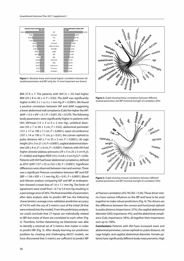
30
Anaesthesiol Intensive Ther 2017, Supplement 1
BMI 27.9 ± 7. The patients with IAH (n = 55) had higher BMI (29 ± 8 vs 26 ± 4, P = 0.02). The ΔIAP was significantly higher in IAH: 5 ± 1 vs 3 ± 1 mm Hg (P < 0.0001). We found a positive correlation between IAP and ΔIAP, suggesting a lower abdominal wall compliance (Cab) the higher the IAP: ΔIAP = 0.3 × IAP + 0.1 (P < 0.001, R2 = 0.579). The following body parameters were significantly higher in patients with IAH: IAPmean (15 ± 3 vs 9 ± 2 mm Hg), umbilical diam-eter (43 ± 7 vs 40 ± 5 cm, P = 0.02), abdominal perimeter (121 ± 17 vs 108 ± 11 cm, P < 0.0001), waist circumference (107 ± 14 vs 100 ± 11 cm, p = 0.01), the convex xiphoid to pubis distance (40 ± 7 vs 35 ± 5 cm, P < 0.0001), rib cage height (24 ± 3 vs 21 ± 4, P = 0.0001), sagittal abdominal diam-eter (28 ± 4 vs 21 ± 4 cm, P < 0.0001). Patients with IAH had higher alveolar plateau pressures (29 ± 5 vs 25 ± 5 cm H2O, P = 0.0002) and higher PEEP (10 ± 3 vs 8 ± 3 cm H2O, P = 0.04). Patients with IAH had lower abdominal compliance, defined as ΔTV/ ΔIAP (137 ± 55 vs 222 ± 85, P < 0.0001). Significant differences were observed between men and women. There was a significant Pearson correlation between IBP and IGP (IBP = 1.04 × IGP + 1.1 mm Hg, R2 = 0.91, P < 0.0001). Bland and Altman analysis comparing IGP and IBP at endexpira-tion showed a mean bias of 1.6 ± 1.1 mm Hg. The limits of agreement were small from –0.7 to 3.9 mm Hg resulting in a percentage error of 26%. The final ensemble of parameters after data analysis able to predict IBP has the following characteristics: average cross-validation prediction accuracy of 74.5% with the use of 3 metrics (out of the initial 28 that were entered into the model). From this preliminary analysis, we could conclude that 27 inputs are individually related to IBP, but many of them are correlated to each other (Fig. 2). Therefore, further datamining via Datastories was able to identify a minimal set of 3 metrics that matter in order to predict IBP (Fig. 3). After deeply learning our prediction problem by creating and challenging 66537 models, we have discovered that 3 metrics are sufficient to predict IBP
Figure 1. Absolute linear and mutual logistic correlation between 28 studied parameters and IBP (only the 15 most important are shown)
Figure 2. Graph showing linear correlations between different studied parameters and IBP (minimal strength of correlation 20%)
Figure 3. Graph showing mutual correlations between different studied parameters and IBP (minimal strength of correlation 50%)
at Pearson correlation of 0.745 (R2 = 0.56). These driver met-rics have various influence on the IBP and have to be used together to make robust predictions (Fig. 4). The drivers are the difference between the convex and horizontal xiphoid to pubis distance (importance: 37%), the sagittal abdominal diameter (SAD, importance: 4%), and the abdominal compli-ance (Cab, importance: 58%), all together their importance sum up to 100%. Conclusions: Patients with IAH have increased waist and abdominal perimeter, convex xiphoid-to-pubis distance, rib cage height, and sagittal abdominal diameter. Female pa-tients have significantly different body measurements. High
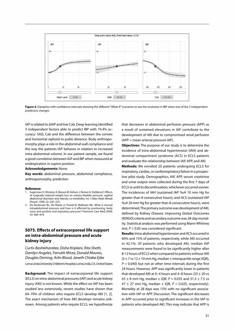
31
Figure 4. Dataplots with confidence intervals showing the different “What-If” scenarios to see the evolution in IBP when one of the 3 independent predictors changes
IAP is related to ΔIAP and low Cab. Deep learning identified 3 independent factors able to predict IBP with 74.4% ac-curacy: SAD, Cab and the difference between the convex and horizontal xiphoid to pubis distance. Body anthropo-morphy plays a role in the abdominal wall compliance and the way the patients IAP behaves in relation to increased intra-abdominal volume. In our patient sample, we found a good correlation between IGP and IBP when measured at endexpiration in supine position.Acknowledgements: None.Key words: abdominal pressure, abdominal compliance, anthropomorphy, prediction
Reference:1. Sugerman H, Windsor A, Bessos M, Kellum J, Reines H, DeMaria E. Effects
of surgically induced weight loss on urinary bladder pressure, sagittal abdominal diameter and obesity co-morbidity. Int J Obes Relat Metab Disord. 1998; 22: 230–235.
2. De Keulenaer BL, De Waele JJ, Powell B, Malbrain ML. What is normal intraabdominal pressure and how is it affected by positioning, body mass and positive end-expiratory pressure? Intensive Care Med 2009; 35: 969–976
S075. Effects of extracorporeal life support on intra-abdominal pressure and acute kidney injury
Curtis Bashkiharatee, Disha Kriplani, Rita Sheth, Danilyn Angeles, Farrukh Mirza, Donald Moores, Douglas Deming, Arlin Blood, Janeth Chiaka EjikeLoma Linda University Children’s Hospital, Loma Linda, CA, United States
Background: The impact of extracorporeal life support (ECLS) on intra-abdominal pressures (IAP) and acute kidney injury (AKI) is not known. While the effect on IAP has been studied less extensively, recent studies have shown that 64–70% of children who require ECLS develop AKI [1, 2]. The exact mechanism of how AKI develops remains unk-nown. Among patients who require ECLS, we hypothesize
that decreases in abdominal perfusion pressure (APP) as a result of sustained elevations in IAP contribute to the development of AKI due to compromised renal perfusion (APP = mean arterial pressure IAP).Objectives: The purpose of our study is to determine the incidence of intra-abdominal hypertension (IAH) and ab-dominal compartment syndrome (ACS) in ECLS patients and evaluate the relationship between IAP, APP, and AKI. Methods: We enrolled 20 patients undergoing ECLS for respiratory, cardiac, or cardiorespiratory failure in a prospec-tive pilot study. Demographics, IAP, APP, serum creatinine and urine output were collected during the first 7 days of ECLS or until its discontinuation, whichever occurred sooner. The incidences of IAH (sustained IAP ‰¥ 10 mm Hg for greater than 8 consecutive hours), and ACS (sustained IAP ‰¥ 20 mm Hg for greater than 8 consecutive hours), were determined. The primary outcome was development of AKI, defined by Kidney Disease: Improving Global Outcomes (KDIGO) criteria and secondary outcome was 28-day mortal-ity. Statistical analysis was performed using Mann-Whitney test, P < 0.05 was considered significant.Results: Intra-abdominal hypertension and ACS occurred in 60% and 15% of patients, respectively, while AKI occurred in 42.1%. Of patients who developed AKI, median IAP measurements were found to be significantly higher after 8-12 hours of ECLS when compared to patients without AKI (5 ± 7 vs 12 ± 10 mm Hg, median ± interquartile range (IQR), P = 0.040) but not at other time intervals during the first 24 hours. However, APP was significantly lower in patients that developed AKI at 0–4 hours and 4–8 hours (33 ± 20 vs 43 ± 8 mm Hg, median ± IQR, P = 0.035 and 31.5 ± 7.5 vs 47 ± 27 mm Hg, median ± IQR, P = 0.025, respectively). Mortality at 28 days was 15% with no significant associa-tion with IAP or APP. Discussion: The significant decreases in APP occurred prior to significant increases in the IAP in patients who developed AKI. This may indicate that APP is
32
Anaesthesiol Intensive Ther 2017, Supplement 1
an earlier sign than actual measured IAP. It could also reflect the 4 hour intervals used in measuring IAP or be due to the small number of patients enrolled at this time.Conclusions: Sixty percent of ECLS patients developed IAH but only 15% met criteria for ACS. There is a positive associa-tion between the occurrence of AKI and both increased IAP and decreased APP. Since the incidence of IAH and ACS in ECLS has never been reported, it remains to be determined if these findings are clinically significant. If decreased APP con-tributes to AKI then goal directed management of patients targeted at maintaining physiologic APP could reduce the rate of AKI in ECLS patients. Additional patients are being enrolled to validate these findings.Acknowledgements: None.Key words: extracorporeal life support, abdominal compart-ment syndrome, intra-abdominal hypertension, abdominal perfusion pressure
References:1. Zwiers AJ, de Wildt SN, Hop WC et al. Acute kidney injury is a frequent
complication in critically ill neonates receiving extracorporeal membrane oxygenation: a 14-year cohort study. Crit Care 2013; 17: R151.
2. Gadepalli SK, Selewski DT, Drongowski RA, Mychaliska GB. Acute kidney injury in congenital diaphragmatic hernia requiring extracorporeal life support: an insidious problem. J Pediatr Surg 2011; 46: 630–635.
S076. How can we manage abdominal pressure for severe acute pancreatitis with abdominal compartment syndrome
Kosuke Sekiya, Koji Morishita, Tomo Oka, Yasuhiro OtomoTokyo Medical and Dental University Hospital of Medicine, Trauma and Acute Critical Care Medical Center, Tokyo, Japan
Background: In recent years, the treatment of acute pancre-atitis is improved. In Japan, survival rate of acute pancreatitis is relatively high. On the other hand, the mortality of severe acute pancreatitis with abdominal compartment syndrome (ACS) is still high. Little information is available regarding the strategy for severe acute pancreatitis with ACS.Objectives: To determine the clinical outcome of severe acute pancreatitis and evaluate the relevance between ab-dominal pressure and treatment. Methods: We retrospectively reviewed records of all adult severe acute pancreatitis patients who had observed abdo-minal pressure from 2009 to 2016.Results: A total of 41 patients were enrolled as severe acute pancreatitis during the period. Twelve patients had suspected ACS and evaluated abdominal pressure. The ove-rall mortality was 14% (n = 6). the mortality rate of severe acute pancreatitis with ACS group were significantly high than without ACS group (41.6% vs. 3.4%, P < 0.01). The overall average value of abdominal pressure was 17.2 mm
Hg. All patients requireed ventilator. Significant difference of abdominal pressure were observed between survival and death group (12.1 mm Hg vs. 24.5 mm Hg). Four patients had undergone surgery.The mean value of abdominal pressure in surgical intervention group was 23.9 mm Hg (13–45 mm Hg) and only one patient survived.Conclusions: In our study the outcome of severe acute pancreatitis with ACS was not good. Surgical intervention will not improve outcome, so how can we prevent ACS will thought to be important.Acknowledgements: None. Key words: abdominal compartment syndrome, acute pan-creatitis, surgery, abdominal pressure
S078. Intra-abdominal hypertension and abdominal compartment syndrome in high risk patients admitted to the ICU: a prospective, observational study
Marije Smit, Bart Koopman, Matijs van Meurs, Jan ZijlstraDepartment of Critical Care, University Medical Center Groningen, University of Groningen, Groningen, Netherlands
Background: Intra-abdominal hypertension (IAH) and ab-dominal compartment syndrome (ACS) have increasingly been associated with morbidity and mortality in critically ill patients. Guidelines recommend monitoring of intra--abdominal pressure (IAP) in high risk patients in order to detect these complications at an early stage.Objectives: To investigate incidence of IAH and ACS in high risk patients admitted to the Intensive Care Unit (ICU). Methods: In this single-center, prospective, observational cohort study consecutive adult patients admitted to the ICU with known risk factors for IAH were enrolled. Inclu-sion criteria were pancreatitis, trauma, abdominal aorta surgery (elective and acute), liver transplant and abdominal surgery in general (elective and acute). Exclusion criteria were age < 18 years and contraindication for urine catheter placement. IAP was measured after admission to the ICU and subsequently every 4 hours for seven days or until discharge from the ICU, whichever came first. If the patient showed no signs of clinical improvement, in the opinion of the attending physician, the study continued for longer than seven days until clear improvement. If IAP was > 20 mm Hg at any time, the IAP measurement was repeated after 1 hour. If ACS was diagnosed, it was left up to the discretion of the attending physician whether or not an intervention (medical, radiological or surgical) was performed. Results: Enrollment took place from March 3, 2014 until March 31, 2016. 503 patients were included. 61% of the patients were male and mean age was 62.2 years (SD 12.85).
33
There were 340 admissions after elective surgery (67.7%), 116 after acute surgery (23.1%) and 47 medical admissions (9.3%). Mean length of stay in the ICU was 3.1 days (SD 7.1). Mean IAP at admission was 11.1 mm Hg (SD 5.5). IAH occurred in 166 patients (33.0%). ACS occurred in 18 patients (3.6%). Management of ACS was conservative in 12 patients (66.7%). Mortality in the ICU after ACS was 16.7%. Conclusions: In this prospective study of high risk patients, the incidence of IAH was 33%. The incidence of ACS was 3.6%. ICU mortality was 16.7% in ACS patients.Acknowledgements: We acknowledge Willem Dieperink and Maureen Werner for their help with data processing and management.Key words: intra-abdominal hypertension, abdominal com-partment syndrome
S083. Relationship between splanchnic and renal oxygenation in pediatric patients on extracorporeal membrane oxygenation (ECMO) who develop acute kidney injury (AKI)
Disha Kriplani, Salem Dehom, Jonathan Specht, Rita Sheth, Farrukh Mirza, Donald Moores, Douglas Deming, Curtis Bashkiharatee, Arlin Blood, Janeth Chiaka EjikeLoma Linda University Children’s Hospital, Loma Linda, CA, United States
Background: About 64% of patients on ECMO develop acute kidney injury (AKI)1. The effect of ECMO and intra-ab-dominal pressure (IAP) on splanchnic and renal oxygenation while on ECMO is not known and may contribute to AKI. Near infra-red spectroscopy (NIRS) is a non-invasive tissue oxygenation monitoring technology which measures ave-rage oxyhemoglobin concentration reflecting ~70% venous and 30% arterial blood.Objectives: To study the changes in splanchnic and renal oxygenation in ECMO patients in the first 24 hours. We also studied the relationship of splanchnic, renal oxygenation and elevated IAP to AKI. We propose that elevated IAP may contribute to AKI during ECMO by impairing abdominal perfusion pressure and tissue oxygenation. Methods: This is a single center, observational, prospective pilot study of pediatric patients requiring ECMO. Demogra-phics, IAP, abdominal perfusion pressure (APP = mean arte-rial pressure-IAP), intra-abdominal hypertension (sustained IAP ‰¥10 mm Hg for ‰¥ 12 hours), serum creatinine (Cr), and urine output were collected for the first 7 days of ECMO or until discontinuation, whichever was earlier. Renal and splanchnic tissue oxygen saturation (HbO2) were continu-ously measured using NIRS. The impaired tissue oxygenation
was defined as either hypoxia (NIRS measurement < 65) or hyperoxia (NIRS > 85). AKI was defined using KDIGO (Kidney Disease: Improving Global Outcomes) definition.Results: The preliminary data presented here is based on the interim analysis of the first 20 out of 40 patients for the pilot phase. The median age at start of ECMO was 4.5 days (IQR 2–18 days). IAH, ACS, and AKI developed in 45%, 0%, and 40% of patients, respectively. In univariate analysis, patients with AKI had higher IAPs (10.6 ± 0.4 vs 7.5 ± 0.2 mm Hg, mean ± SEM, P = 0.008), reduced APP at 4-hour (median; IQR, 43; 8 vs 33; 20) and 8-hour (median; IQR, 47; 27 vs 31.5; 7.5); and high IAP (median; IQR, 5; 7 vs 12; 10) at 12-hour after initiating ECMO (all P < 0.05). NIRS data was available for 17 patients. Percent time of hypoxia reduced after ECMO within 1 hour in both renal (27.06% vs 11.29%) and splanch-nic (12.78% vs 11.55%) HbO2. Percent time of hyperoxia increased after ECMO in both renal (15.84% vs 30.56) and splanchnic (12.52% vs 37.49%) HbO2. Comparing AKI to non-AKI group, both renal and splanchnic HbO2 were less often in hypoxic range (0% vs 5.95%; 0% vs 2.3%) and more often in hyperoxic range (50.3% vs 2.05%; 36.05% vs 71.7%). Comparing IAH to non-IAH group, both renal and splanchnic HbO2 were less often in hyperoxic range (2.05% vs 61.9%; 36.05% vs 71.7%) and had slightly less hypoxia (0.15% vs 0.3%; 1.25% vs 0.2%). Mean splanchnic (88.1% vs 80.3%, P = 0.06) and renal (82.33% vs 77.46%, P = 0.33) SpO2 were higher in AKI patients.Conclusions: In our interim analysis, we found AKI patients had less hypoxia and more hyperoxia which may support the role of reperfusion injury and oxidative stress in the pathophysiology of AKI in ECMO patients. Trend of reduced hyperoxia in IAH patients may reject our hypothesis that elevated IAP leads to AKI via reducing tissue oxygenation.Acknowledgements: None. Key words: near infra-red spectroscopy, ECMO, children, intra-abdominal hypertension
S084. Predictors of primary fascial closure in trauma and acute care surgery patients with open abdominal wounds: a systematic review
Derek J. Roberts1, Andrew W. Kirkpatrick2, Annika Reintam Blaser3, Jan De Waele4
1Department of Surgery, Foothills Medical Centre, University of Calgary, Calgary, Alberta Canada2Departments of Surgery, Critical Care Medicine and Trauma Servi-ces, Foothills Medical Centre, Calgary, Alberta, Canada3University of Tartu, Department of Anesthesiology and Intensive Care, Tartu, Estonia4Ghent University, Department of Critical Care Medicine, Ghent, Belgium
34
Anaesthesiol Intensive Ther 2017, Supplement 1
Background: Primary fascial closure (fascia-to-fascia closure of the open abdomen during the index hospitalization) in adults with open abdominal wounds has been reported to be associated with improved patient-important outcomes.Objectives: We conducted a systematic review to identify predictors of early primary fascial closure in trauma and acute surgery patients to guide development of an evi-dence-informed open abdominal management pathway. Methods: We searched electronic databases (MEDLINE, EM-BASE, the Cochrane Library, and Web of Science) (1950-Sep-tember 1, 2016) and reference lists of included articles for observational studies reporting predictors of primary fa-scial closure in adult trauma and acute care surgery pa-tients. Identified predictors were summarized with narrative synthesis methods and combined to provide summary esti-mates using DerSimonian and Laird random-effects models.Results: We included nine studies enrolling 2,344 adult trau-ma and acute care surgery patients in the systematic review. The studies evaluated 18 potential independent predictors of early primary fascial closure, which could be combined into seven themes. These included the injury severity of the patient, degree of patient physiologic derangement upon presentation, presence of intra-abdominal hypertension in the intensive care unit (ICU), delayed time to first take-back or requirement for multiple take-backs, administration of a large volume of crystalloid fluids, development of an en-teric fistula or intra-abdominal sepsis/infectious compli-cations, and miscellaneous. Independent predictors with evidence suggesting a decreased odds of primary fascial closure included an increased injury severity score [pooled odds ratio (OR) per unit increase = 1.02; 95% confidence interval (CI) = 0.97–1.08; n = 2 studies] and international normalized ratio (OR = 0.18; 95% CI = 0.034–0.98; n = 1 study) upon presentation; a higher intra-abdominal pressure (IAP) in the ICU (OR per increase in IAP = 0.85; 95% CI = 0.76–0.95; n = 1 study); time to take-back > 48 hours (OR = 0.53; 95% CI = 0.29–0.98; n = 1 study) or an increased number of take-backs (OR per take-back = 0.18; 95% CI = 0.11–0.29; n = 1 study); administration of a large volume of crystalloid fluids (pooled OR = 0.51; 95% CI = 0.28–0.94; n = 2 stu-dies); and development of an enteric fistula (OR = 0.16; 95% CI = 0.030–0.81; n = 1 study) or intra-abdominal abscess (OR = 0.38; 95% CI = 0.18–0.84; n = 1 study).Conclusions: Our findings suggest that primary fascial clo-sure rates in trauma and acute care surgery patients may be improved by returning to the operating room earlier than 48 hours after the index laparotomy, limiting use of crystalloid fluids perioperatively, and preventing and/or treating intra-abdominal hypertension, enteric fistuale, and intra-abdominal collections postoperatively. These findings should be confirmed by randomized trials before they are used to inform practice.
Acknowledgements: WSACS, The Abdominal Compart-ment Society Clinical Trials Working GroupKey words: damage control laparotomy, open abdomen, open abdominal management, predictors
S085. Intra-abdominal pressure: a clinical prognosis and outcome parameter around the closure of congenital abdominal wall hernias?
Torsten Kaussen1, Buppha Wanchaame1, Martin Boehne1, Michael Sasse1, Philipp Beerbaum1, Christoph Zoeller2, Joachim Kuebler2, Benno Ure2, Carolin Boehne3, Bettina Bohnhorst3
1Hannover Medical School, Department of Pediatric Cardiology and Intensive Care Medicine, Hannover, Germany2Hannover Medical School, Department of Pediatric Surgery, Han-nover, Germany3Hannover Medical School, Department of Pediatric Pneumology and Neonatology, Hannover, Germany
Background: Gastroschisis and omphalocele are considered as prototypes of diseases whose surgical correction may lead to an increase in intra-abdominal pressure (IAP). The advantages and disadvantages of primary and secondary abdominal wall closures (AWC) are discussed controversially. In terms of intra-abdominal hypertension (IAH), not only the AWC per se is to be considered as risky; also the systemic inflammatory response syndrome accompanying the opera-tions can lead to tissue and secondary IAP increases by fluid extravasation within the scope of a capillary leak syndrome. Nevertheless, intra- and postoperative IAP measurements are still not a common standard monitoring method during or after abdominal wall closure. Surprisingly, there was not yet a single published study dealing with the collection of epidemiological data on IAP under the influence of conge-nital abdominal wall defects (cAWD) and their correction.Objectives: Aim of this study is a systematic analysis of pe-ripartal, intra- and postoperative conditions and risk factors of newborns with cAWD with special consideration of IAP levels and their prognostic value. Methods: The study was approved by the local Ethics Committee. Retrospectively, the medical records of all newborns with an cAWD were analyzed, which were tre-ated and operated between 1998 and 2015 at Hannover Medical School. Prepartal, peripartal, operative and inten-sive medical findings were screened and integrated into a specially developed database. To diagnose IAH the WSACS definitions of 2013 were decisive. Results are presented as mean + standard deviation. Statistical analysis was carried out using SPSS v23.Results: 107 children were included into this retrospective analysis. The descriptive statistics on patient, surgical and
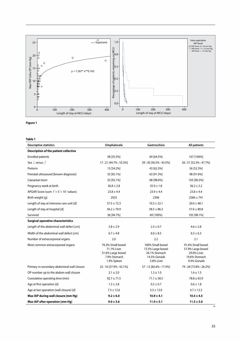
35
Figure 1
Table 1
descriptive statistics omphalocele gastroschisis all patients
Description of the patient collective
Enrolled patients 38 [35.5%] 69 [64.5%] 107 [100%]
Sex ♀ versus ♂ 17 : 21 [44.7% : 55.3%] 39 : 30 [56.5% : 43.5%] 56 : 51 [52.3% : 47.7%]
Preterm 13 [34.2%] 43 [62.3%] 56 [52.3%]
Prenatal ultrasound [known diagnosis] 35 [92.1%] 63 [91.3%] 98 [91.6%]
Caesarean born 35 [92.1%] 68 [98.6%] 103 [96.3%]
Pregnancy week at birth 36.8 ± 2.8 35.9 ± 1.8 36.2 ± 2.2
APGAR-Score [sum: 1´+ 5´+ 10´-values] 23.8 ± 4.4 23.9 ± 4.4 23.8 ± 4.4
Birth weight [g] 2925 2396 2584 ± 741
Length of stay at intensive care unit [d] 37.5 ± 72.3 10.3 ± 22.1 20.0 ± 48.1
Length of stay at hospital [d] 56.2 ± 70.9 58.5 ± 86.2 57.6 ± 80.8
Survived 36 [94.7%] 69 [100%] 105 [98.1%]
Surgical-operative characteristics
Length of the abdominal wall defect [cm] 5.8 ± 2.9 2.5 ± 0.7 4.6 ± 2.8
Width of the abdominal wall defect [cm] 6.7 ± 4.8 6.0 ± 8.5 6.5 ± 6.3
Number of extracorporeal organs 2.0 2.2 2.1
Most common extracorporeal organs 76.3% Small bowel71.1% Liver
31.6% Large bowel 7.9% Stomach 1.9% Spleen
100% Small bowel72.5% Large bowel
26.1% Stomach14.5% Gonads
5.8% Liver
91.6% Small bowel57.9% Large bowel
29.0% Liver19.6% Stomach 9.4% Gonads
Primary vs secondary abdominal wall closure 22 : 16 [57.9% : 42.1%] 57 : 12 [82.6% : 17.4%] 79 : 28 [73.8% : 26.2%]
OP-number up to the abdom wall closure 2.1 ± 2.0 1.3 ± 1.0 1.6 ± 1.5
Cumulative operating time [min] 92.7 ± 71.5 71.1 ± 58.5 78.8 ± 63.9
Age at first operation [d] 1.3 ± 2.8 0.2 ± 0.7 0.6 ± 1.8
Age at last operation (wall closure) [d] 7.3 ± 12.6 3.3 ± 12.0 4.7 ± 12.3
Max IAP during wall closure [mm Hg] 9.2 ± 6.0 10.9 ± 4.1 10.5 ± 4.5
Max IAP after operation [mm Hg] 9.0 ± 3.6 11.9 ± 5.1 11.5 ± 5.0Æ
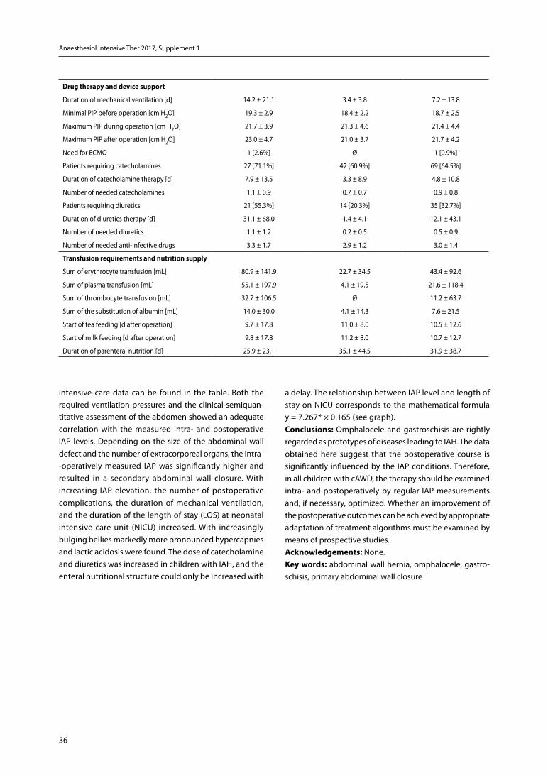
36
Anaesthesiol Intensive Ther 2017, Supplement 1
Drug therapy and device support
Duration of mechanical ventilation [d] 14.2 ± 21.1 3.4 ± 3.8 7.2 ± 13.8
Minimal PIP before operation [cm H2O] 19.3 ± 2.9 18.4 ± 2.2 18.7 ± 2.5
Maximum PIP during operation [cm H2O] 21.7 ± 3.9 21.3 ± 4.6 21.4 ± 4.4
Maximum PIP after operation [cm H2O] 23.0 ± 4.7 21.0 ± 3.7 21.7 ± 4.2
Need for ECMO 1 [2.6%] Ø 1 [0.9%]
Patients requiring catecholamines 27 [71.1%] 42 [60.9%] 69 [64.5%]
Duration of catecholamine therapy [d] 7.9 ± 13.5 3.3 ± 8.9 4.8 ± 10.8
Number of needed catecholamines 1.1 ± 0.9 0.7 ± 0.7 0.9 ± 0.8
Patients requiring diuretics 21 [55.3%] 14 [20.3%] 35 [32.7%]
Duration of diuretics therapy [d] 31.1 ± 68.0 1.4 ± 4.1 12.1 ± 43.1
Number of needed diuretics 1.1 ± 1.2 0.2 ± 0.5 0.5 ± 0.9
Number of needed anti-infective drugs 3.3 ± 1.7 2.9 ± 1.2 3.0 ± 1.4
Transfusion requirements and nutrition supply
Sum of erythrocyte transfusion [mL] 80.9 ± 141.9 22.7 ± 34.5 43.4 ± 92.6
Sum of plasma transfusion [mL] 55.1 ± 197.9 4.1 ± 19.5 21.6 ± 118.4
Sum of thrombocyte transfusion [mL] 32.7 ± 106.5 Ø 11.2 ± 63.7
Sum of the substitution of albumin [mL] 14.0 ± 30.0 4.1 ± 14.3 7.6 ± 21.5
Start of tea feeding [d after operation] 9.7 ± 17.8 11.0 ± 8.0 10.5 ± 12.6
Start of milk feeding [d after operation] 9.8 ± 17.8 11.2 ± 8.0 10.7 ± 12.7
Duration of parenteral nutrition [d] 25.9 ± 23.1 35.1 ± 44.5 31.9 ± 38.7
intensive-care data can be found in the table. Both the required ventilation pressures and the clinical-semiquan-titative assessment of the abdomen showed an adequate correlation with the measured intra- and postoperative IAP levels. Depending on the size of the abdominal wall defect and the number of extracorporeal organs, the intra--operatively measured IAP was significantly higher and resulted in a secondary abdominal wall closure. With increasing IAP elevation, the number of postoperative complications, the duration of mechanical ventilation, and the duration of the length of stay (LOS) at neonatal intensive care unit (NICU) increased. With increasingly bulging bellies markedly more pronounced hypercapnies and lactic acidosis were found. The dose of catecholamine and diuretics was increased in children with IAH, and the enteral nutritional structure could only be increased with
a delay. The relationship between IAP level and length of stay on NICU corresponds to the mathematical formula y = 7.267* × 0.165 (see graph).Conclusions: Omphalocele and gastroschisis are rightly regarded as prototypes of diseases leading to IAH. The data obtained here suggest that the postoperative course is significantly influenced by the IAP conditions. Therefore, in all children with cAWD, the therapy should be examined intra- and postoperatively by regular IAP measurements and, if necessary, optimized. Whether an improvement of the postoperative outcomes can be achieved by appropriate adaptation of treatment algorithms must be examined by means of prospective studies.Acknowledgements: None. Key words: abdominal wall hernia, omphalocele, gastro-schisis, primary abdominal wall closure
37
P004. Study of IAH prevalence and cognitive level of experienced medical staff in ICU
Lian-Yang Zhang, Hua- yu Zhang, Dong Liu, Hao Tang, Shi-Jin Sun, Shan-mu, Wen-qun Yang, Dong-po JiangTrauma Center, State Key Laboratory of Trauma, Burns and Com-bined Injury, Institute of Surgery Research, Daping Hospital, Third Military Medical University, Chongqing, China
Background: Intra-abdominal hypertension (IAH) will cause adverse effects to organ functions and prognosis. The im-portant role of guidelines published by the World Society of Abdominal Compartment Syndrome (WSACS) in promoting diagnosis and treatment efficiency has long been advocated by experts.Objectives: To explore the knowledge among intensive care unit (ICU) clinical staff of the guidelines published by the World Society of the Abdominal Compartment Syndrome (WSACS) in 2013 and its relationship to the prevalence of intra-abdominal hypertension (IAH). Methods: A one-day cross-sectional survey was conducted in 4 trauma/emergency/general ICUs of southwestern China. Epidemiological information about the included patients was collected, variables which may cause IAH were defined by reference to the guidelines, and intra-vesical pressure (IVP) was measured. The level of cognizance of ICU clinical staff about the WSACS guidelines was surveyed by question-naire (100 points).Results: Forty-one patients were selected from 4 ICUs. Fif-teen (36.59%) had IVP 12 mm Hg. A total of 37 (response rate 80.43%) clinical staff participated in the survey. The average score of the four hospitals was 30.68 ± 14.72, 33.00 ± 11.73, 27.75 ± 26.35, and 31.43 ± 15.09, respectively. All staff be-lieved that the IAH prevalence in their departments was no more than 20.00%. Significant negative correlation was observed between the awareness and IAH prevalence of each center (r = –0.975, P = 0.025).Conclusions: The prevalence of IAH among the surveyed population are 36.59%. Clinical staff have poor cognition of the 2013 WSACS guidelines. Strengthening their guideline training and enhancing their awareness of the guidelines is necessary to improve the efficiency of diagnosis and treat-ment of IAH.Acknowledgements: The authors declare support by grants from New Techniques of Trauma Care (2012BAI11B01)
Poster presentations
Key words: intra-abdominal pressure, intra-vesical pressure, in-tra-abdominal hypertension, abdominal compartment syndrome
P005. Open components separation and underlay repair using biological mesh for the treatment of planned ventral hernia after open abdomen surgery
Lian-Yang Zhang, Pei-Yuan Li, Dong Liu, Shi-Jin Sun Trauma Center, State Key Laboratory of Trauma, Burns and Com-bined Injury, Institute of Surgery Research, Daping Hospital, Third Military Medical University, Chongqing, China
Background: There are no standard surgical methods for planned ventral hernia (PVH).Objectives: To discuss the possibility of using ‘open com-ponents separation + underlay repair using biological mesh’ as a preferred surgical method for PVH. Methods: We retrospectively analyzed the clinical data of PVH patients who received open components separation and underlay repair using biological mesh.Results: Five PVH patients underwent this surgical method from June 2014 to April 2015. All patients were males with an average age of 43.6 ± 17.3 years, body mass index (BMI) of 23.1 ± 2.3 kg m-2, and skin graft width of 15–17 cm. Four patients received a sliding myofascial flap of bilateral musculus rectus abdominis + under-lay repair with biological mesh (Biodesign®, Cook Medical, Inc., Bloomington, IN, USA), and 1 patient received a sliding myofascial flap of unilateral musculus rectus abdominis + underlay repair with biological mesh. There was one case of incisional infection and 1 case of abdominal sinus formation 2 weeks after discharge. Both patients were cured using vacuum sealing draining (VSD) for 4 to 5 months without removal of the biological meshes. Five patients were followed up in an outpatient clinic after discharge, and the follow-up time ranged from 3 to 12 months. Abdominal incisions healed well, and abdominal computed tomography (CT) indicated no hernia recurrence in all patients.Conclusions: The ‘open components separation + underlay repair using biological mesh’ method is a safe and feasible treatment method for PVH with good short-term outcomes.Acknowledgements: The authors declare support by grants from New Techniques of Trauma Care (2012BAI11B01)Key words: hernia, reconstructive surgical procedures, surgical mesh.
38
Anaesthesiol Intensive Ther 2017, Supplement 1
P008. Novel method for delayed primary closure and incisional hernia prevention in open abdomen
Rafael Villalobos1, Carmen Mias2, Cristina Gas2, Alfredo Escartin2, Victor Palacios2, Jordi Escoll2, Jorge Juan Olsina2
1Arnau de Vilanova University Hospital, Chief of the Abdominal Wall Surgery Unit, Surgery Department, Lleida, Spain2Arnau de Vilanova University, Surgery Department, Lleida, Spain
Background: Open abdomen management has become a therapeutic option for not only cases of trauma but also for non-trauma patients, especially for those with severe sepsis. It is nonetheless associated with high rates of mor-bidity and mortality. Various techniques of temporary abdo-minal closure have been developed in order to improve the management and to achieve delayed primary closure with minimal complications. Despite these procedures incisional hernia rate is still very high being greater than 30%. Curren-tly no techniques has been described to solve this problem. In this context we describe a novel method called COmbined and MOdified Definitive Abdominal closure (COMODA) for obtaining a delayed open abdomen primary closure with a lower risk for subsequently developing an incisional her-nia. This method protect all time injury of the fascial edges observed by conventional techniques and reinforce the abdominal wall during its closure. Objectives: To achieve a high abdominal wall closure and a low incisional hernia rateMethods: Clinical trial conducted between march 2012 and december 2014 in which a negative pressure wound therapy (NPWT) system is combined with a condensed polytetra-fluoroethylene (cPTFE) mesh fixed 5 cm from the fascial edges. Demographic variables, comorbidities, intra-abdomi-nal pressure measurements, number of surgeries, time until definitive closure, early and late complications were studied. This method was used in non-trauma patients, mainly with sepsis, with the wound being examined every 3–4 days or on demand, with a constant medial traction applied away from the fascial edges. When primary closure was achieved, the remaining cPTFE mesh served as intraperitoneal rein-forcement. Trial registration: ISRCTN72678033Results: A total of 10 male patients with a mean age of 68.8 (43–87) years were included, 6 of whom (60%) had sepsis, 2 (20%) had eviscerations and 2 (20%) a ruptured ab-dominal aortic aneurysm. The primary closure rate was 80% in the intention-to-treat analysis and 100% per protocol. The mean number of procedures per patient until definiti-ve closure was 5.7 (± 1.3). Primary closure was obtained in 20.8 (10–32) days and mean hospital stay was 36.3 (18–52) days. One patient (14.2%) developed an enterocutaneous fistula after primary closure and was treated conservatively;
none of the patients presented wound infections. No pa-tients developed incisional hernias during a mean follow up of 35.9 (24–49) months.Conclusions: The COMODA method allows for a high rate of delayed primary closure in open abdomen patients, is safe and decreases the risk for development of incisional hernias. Nevertheless, a multi-center study is needed to draw firm conclusions.Acknowledgements: To Smith & Nephew company to supply the suction devices for the negative pressure wound therapyKey words: open abdomen, negative pressure wound the-rapy, temporary abdominal closure, incisional hernia
P016. Listeria monocytogenes inhibits Th17 responses through TLR2 signaling in intra- -abdominal illness
Song Liu1, Xiuwen Wu, Gefei Wang, Jianan Ren2
1Department of General Surgery, Nanjing Drum Tower Hospital, Nanjing University, Nanjing China2Department of General Surgery, Jinling Hospital, Nanjing Univer-sity, Nanjing, China
Background: Intestinal pathogens shape mucosal immune responses and determine clinical outcome in patients with intra-abdominal illnesses. Listeria monocytogenes can cause severe infection in human, named listeriosis. Immunological mechanism of host defenses against Listeria monocytoge-nes has been elusive.Objectives: This study is dedicated to investigate immuno-logical role of Th17 responses regulated by Listeria mono-cytogenes in intra-abdominal illnesses. Methods: We observed the localization of Listeria mono-cytogenes in small intestine lamina propria and mesenteric lymph nodes using GFP-labeled Listeria monocytogenes. We infected IL-17A-GFP mice with Listeria monocytogenes for 3 days, and analyzed the Th17 responses in intestine using flow cytometry. We employed TLR2-/- mice, and isolated CD11c+CD103+ dendritic cells (DCs) and CD3+CD4+ T cells from small intestine lamina propria after Listeria monocyto-genes infection, we then measured Ifnb1 mRNA expression in CD103+ DCs and IL-17A production by CD4+ T cells after co--stimulation by anti-CD3 and anti-CD28 antibodies for 3 days.Results: Listeria monocytogenes was detected in the small intestinal lamina propria by confocal microscopy three days after infection and thus was able to invade the small in-testinal lamina propria (Fig. 1a, b). We found that during Listeria monocytogenes infection, Th17 cells significantly decreased in the lamina propria of small intestine while Th17 cell numbers in MLNs remained similar to those found in healthy mice (Fig. 1c). In support of a direct modulation of mucosal DCs function by Listeria monocytogenes, we found
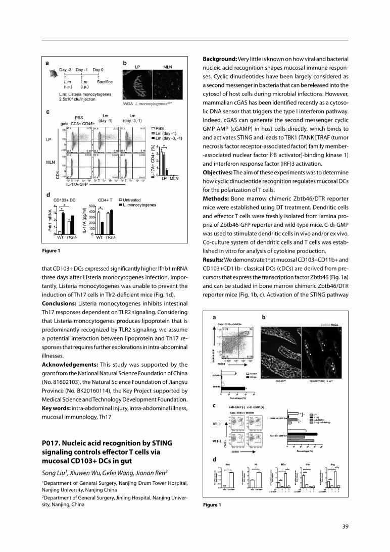
39
Figure 1
Figure 1
that CD103+ DCs expressed significantly higher Ifnb1 mRNA three days after Listeria monocytogenes infection. Impor-tantly, Listeria monocytogenes was unable to prevent the induction of Th17 cells in Tlr2-deficient mice (Fig. 1d).Conclusions: Listeria monocytogenes inhibits intestinal Th17 responses dependent on TLR2 signaling. Considering that Listeria monocytogenes produces lipoprotein that is predominantly recognized by TLR2 signaling, we assume a potential interaction between lipoprotein and Th17 re-sponses that requires further explorations in intra-abdominal illnesses.Acknowledgements: This study was supported by the grant from the National Natural Science Foundation of China (No. 81602103), the Natural Science Foundation of Jiangsu Province (No. BK20160114), the Key Project supported by Medical Science and Technology Development Foundation.Key words: intra-abdominal injury, intra-abdominal illness, mucosal immunology, Th17
P017. Nucleic acid recognition by STING signaling controls effector T cells via mucosal CD103+ DCs in gut
Song Liu1, Xiuwen Wu, Gefei Wang, Jianan Ren2
1Department of General Surgery, Nanjing Drum Tower Hospital, Nanjing University, Nanjing China2Department of General Surgery, Jinling Hospital, Nanjing Univer-sity, Nanjing, China
Background: Very little is known on how viral and bacterial nucleic acid recognition shapes mucosal immune respon-ses. Cyclic dinucleotides have been largely considered as a second messenger in bacteria that can be released into the cytosol of host cells during microbial infections. However, mammalian cGAS has been identified recently as a cytoso-lic DNA sensor that triggers the type I interferon pathway. Indeed, cGAS can generate the second messenger cyclic GMP-AMP (cGAMP) in host cells directly, which binds to and activates STING and leads to TBK1 (TANK [TRAF (tumor necrosis factor receptor-associated factor) family member--associated nuclear factor κB activator]-binding kinase 1) and interferon response factor (IRF)3 activation.Objectives: The aim of these experiments was to determine how cyclic dinucleotide recognition regulates mucosal DCs for the polarization of T cells. Methods: Bone marrow chimeric Zbtb46/DTR reporter mice were established using DT treatment. Dendritic cells and effector T cells were freshly isolated from lamina pro-pria of Zbtb46-GFP reporter and wild-type mice. C-di-GMP was used to stimulate dendritic cells in vivo and/or ex vivo. Co-culture system of dendritic cells and T cells was estab-lished in vitro for analysis of cytokine production.Results: We demonstrate that mucosal CD103+CD11b+ and CD103+CD11b- classical DCs (cDCs) are derived from pre-cursors that express the transcription factor Zbtb46 (Fig. 1a) and can be studied in bone marrow chimeric Zbtb46/DTR reporter mice (Fig. 1b, c). Activation of the STING pathway
40
Anaesthesiol Intensive Ther 2017, Supplement 1
by c-di-GMP significantly up-regulated Il17a and Ifng but inhibited Il10 gene expression by mucosal effector CD4+ T cells (Fig. 1d). The polarization of T cells upon STING de-pendent recognition of cyclic dinucleotides was mediated by mucosal CD103+ cDCs, since T cells lacked Il17a, Ifng or Il10 expression after depletion of Zbtb46+ cDCs (Fig. 1d).Conclusions: Foreign nucleic acid recognition pathways in mucosal Zbtb46+ DCs, which include distinct CD103+ DC subsets, can induce both IFN-γ and IL-17 expressing T helper cells in the mucosal immune system in response to STING mediated recognition of cyclic dinucleotides.Acknowledgements: This study was supported by the grant from the National Natural Science Foundation of China (No. 81602103), the Natural Science Foundation of Jiangsu Province (No. BK20160114), the Key Project supported by Medical Science and Technology Development Foundation.Key words: gut inflammation, intra-abdominal infection, intra-abdominal immune, mucosal immunology.
P021. Multivariate analysis derived biomediator panel to predict survival in critically ill patients
Michelle S. Malig, Craig N. Jenne, Derek J. Roberts, Chad G. Ball, Zhengwen Xiao, Andrew W. KirkpatrickDepartment of Microbiology, Immunology and Infectious Disease, Departments of General Surgery, Critical Care Medicine, and the Regional Trauma Program, Foothills Medical Centre, University of Calgary, Alberta Canada
Background: To date, no single biomarker of systemic in-flammation clearly predicts either outcomes or monitors therapy responses. However, biomediator panels may pro-vide a more comprehensive model of patient variation, and may reveal novel underlying relationships between mediators. Selection of open abdomen (OA) management of critically ill patients practically designates a critically ill population with markedly increased complexity and po-tentially catastrophic complications. The Peritoneal-VAC randomized-controlled human trial (PVAC) examining active negative pressure peritoneal therapy, provided a rich repo-sitory of biomediator samples in such an OA population, although no individual biomediator assessed was predictive of survival.Objectives: To create and test a novel biomediator panel using multivariate techniques in an attempt to examine predictive values in the provided OA cohort. Methods: Partial least squares regression with discrimi-nant analysis (PLS-DA) was used to develop a biomediator panel modelled at baseline, 24, and 48 hours after OA man-agement using the PVAC plasma samples. Eight cytokines and acute phase proteins (APPs) were used to generate an eight-component biomediator panel to predict overall
patient outcome. Components for the panel were selected based on the common literature reported sepsis/critical illness mediators, which included: TNF-Î ± , IL-1β, IL-4, IL-6, IL-7, IL-8, IL-10, and fibrinogen.Results: The generated models had R2 values ranging from 0.213–0.541, whereas Q2 ranged from 0.007-0.13. The bio-mediator panel modelled at 24 hours was most predictive for patient outcome (Q2 = 0.13), and accounted for over 50% of the observed experimental variance.Conclusions: The proposed biomediator panel was more predictive of patient outcome when modelled at certain time points than others; however, overall the panel was a poor predictor of patient outcome. Thus, further research is required to determine more informative biomediators and the most appropriate time frame in which the biomediator storm may best predict outcome in the critically ill/injured.Acknowledgements: The authors would like to acknowl-edge Caroline Leger and Mandy Tse of the Snyder Transla-tional Laboratory in Critical Care Medicine for their assis-tance with the study development and laboratory analysis of the original PVAC data. Key words: inflammation, injury, biomediator, multivariate analysis
P025. Using the intensive care unit for bedside dressing changes for open abdomen is time- and staff-efficient
Arne Seternes1, 5, Sigurd Fasting2, 5, Pål Klepstad2, 5, Skule Mo2, Torbjørn Dahl1, 5, Martin Björck4, Arne Wibe3, 5
1Department of Vascular Surgery, St. Olavs Hospital, Trondheim University Hospital, Trondheim, Norway 2Department of Anesthesiology and Intensive Care Medicine, St. Olavs Hospital, Trondheim University Hospital, Trondheim, Norway3Department of Gastrointestinal Surgery, St. Olavs Hospital, Trondheim University Hospital, Trondheim, Norway4Department of Surgical Science, Section of Vascular Surgery,
Uppsala University, Uppsala, Sweden5Norwegian University of Science and Technology (NTNU)
Background: Patients with an open abdomen (OA) treated with temporary abdominal closure (TAC) need multiple surgical procedures throughout the hospital stay with re-peated changes of the Vacuum Assisted Closure device (VAC changes).Objectives: The aim of this study was to examine if VAC changes in OA patients performed in the intensive care unit (ICU) save time, personnel and costs compared to VAC changes in the operating room (OR). Secondary aims were to evaluate if this practice influence on the frequency of blood stream infections (BSI) and survival. Methods: Retrospective study of 98 patients with OA and repeated VAC changes treated in the ICU from October

41
2006 to June 2014. Data were obtained from, clinical pa-tients records and the OR, ICU, anesthesia and microbiology databases.Results: 443 VAC changes were performed of which 165 (37%) were performed in the ICU. The mean duration for the surgical team performing a VAC change in the ICU was 63 (60–66) minutes and in the OR 98 (95–102) minutes (P < 0.001). The mean duration of work for the anesthesia team in the OR was 116 minutes, while this team was not used in the ICU. Forty-two patients had all the VAC-changes done in the OR (VAC-OR), 22 in the ICU (VAC-ICU) and 34 in both OR and ICU (VAC-OR/ICU). BSI was diagnosed in 8 (19%) of the VAC-OR patients, 7 (32%) of the VAC-ICU and 8 (24%) of the VAC-OR/ICU (P = 0.509). Thirty-five patients (83%) sur-vived 30 days in the VAC-OR group, 17 in the VAC-ICU group (77%) and 28 (82%) in the VAC-OR/ICU group (P = 0.844).Conclusions: VAC change for OA in the ICU saved time for the surgical team and the anesthesia team compared to using the OR, and reduced personnel costs. Importantly, the use of ICU for OA dressing change seemed to be as safe as using the OR.Acknowledgements: None. Key words: abdominal compartment syndrome, dressing changes, Intensive care, Health economy
P026. Ability of commercial NPWT systems to manage fluid in an experimental open abdomen study
Raymond Dunn1, Heather Tessier1, Vicki Hammon2, Iain Webster2
1University of Massachussettes Memorial Medical Centre, Boston, USA2Smith and Nephew, research and Development, Hull, UK
Background: The use of Negative Pressure Wound Therapy (NPWT) to provide Temporary Abdominal Closure (TAC) fol-lowing Open Abdomen (OA) procedures is associated with increased primary fascial closure compared with alternative methods of TAC for example Bogota bag [1]. Removal of abdominal fluid is one important benefit of applying NPWT following OA.Objectives: Different specialized NPWT systems are com-mercially available to provide TAC in patients with OA but no information is available to date which directly compares fluid removal capabilities of different NPWT systems. Methods: A porcine model was used to explore the fluid removal capabilities of two different commercially ava-ilable OA NPWT systems (A and B)*. A midline excisio-nal laparotomy was performed in 8 anaesthetized adult pigs. Abdominal NPWT systems were applied in a rando-mized order. 800 mL of albumin solution was introduced into the abdominal cavity via a previously inserted drain.
NPWT was applied at either –120 mm Hg or –125 mm Hg (for A and B* respectively) according to available settings on devices. The rate of fluid removal was monitored by recording the weight of fluid collected in device canisters throughout 60 minutes of therapy. At the end of this time, dressings were removed, and the volume of residual free fluid remaining in the abdomen was recorded and the test repeated with the other NPWT system.Results: For both NPWT systems, free abdominal fluid was found to be rapidly evacuated in the first few minutes follo-wing initiation of NPWT, followed by a plateau. Fluid removal was not significantly different between systems A and B* tested at 15 minutes (674 g and 716 g respectively; P = 0.194) and 60 minutes (755 g vs 773 g, P = 0.554) following the on-set of NPWT. This equates to a removal fraction of 94.4% and 96.6% respectively of the 800 mL volume of introduced free fluid. Following dressing removal at the end of 60 minutes, there was no significant difference between residual fluid (18.4 g ± 13.7 vs 10.0g ± 7.8; P = 0.208).Conclusions: Two NPWT devices specifically designed for use in the OA are both capable of managing large volumes of fluid in a relatively short period of time. No significant difference was observed between systems which achie-ved similar fluid removal volumes and left similar residual volumes in the porcine paracolic gutters over the clinically relevant timescale measured in this study. *A = RENASYSTM AB used with RENASYS EZ-max, Smith & Nephew, Hull, UK; B = ABThera TM used with infoVACTM pump, Acelity, Texas, USAAcknowledgements: None. Key words: NPWT, fluid handling, animal model
Reference: 1. Bruhin A, Ferreira F, Chariker M, Smith J, Runkel N. Systematic review
and evidence based recommendations for the use of negative pressure wound therapy in the open abdomen. Int J Surg. 2014; 12 (10): 1105–1114. doi: 10.1016/j.ijsu.2014.08.396.
P033. Validation of a continuous intra- -gastral monitoring-technique for measuring intra-abdominal pressure (IAP) in critically ill pediatric patients
Torsten Kaussen1, Miriam Gutting1, Alexander von Gise1, Martin Boehne1, Florian Schmidt2, Thomas Jack1, Michael Sasse1, Dietmar Bäthi1, Philipp Beerbaum1
1Department of Pediatric Cardiology and Intensive Care Medicine, Hannover Medical School, Germany2Pediatric Intensive Care Unit, Charite University Hospital, Berlin, Germany
Background: Intra-abdominal hypertension (IAH) and ab-dominal compartment syndrome (ACS) are predictors of
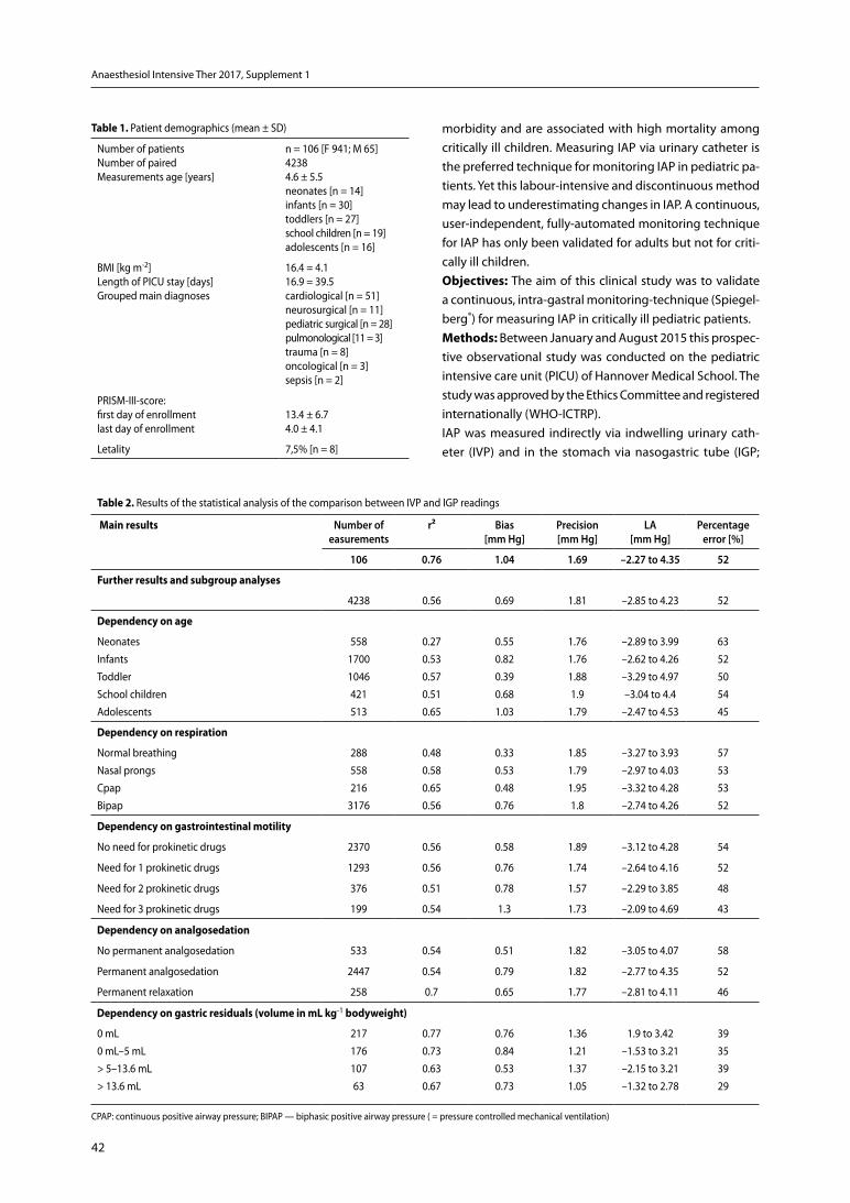
42
Anaesthesiol Intensive Ther 2017, Supplement 1
morbidity and are associated with high mortality among critically ill children. Measuring IAP via urinary catheter is the preferred technique for monitoring IAP in pediatric pa-tients. Yet this labour-intensive and discontinuous method may lead to underestimating changes in IAP. A continuous, user-independent, fully-automated monitoring technique for IAP has only been validated for adults but not for criti-cally ill children.Objectives: The aim of this clinical study was to validate a continuous, intra-gastral monitoring-technique (Spiegel-berg®) for measuring IAP in critically ill pediatric patients. Methods: Between January and August 2015 this prospec-tive observational study was conducted on the pediatric intensive care unit (PICU) of Hannover Medical School. The study was approved by the Ethics Committee and registered internationally (WHO-ICTRP).IAP was measured indirectly via indwelling urinary cath-eter (IVP) and in the stomach via nasogastric tube (IGP;
Table 2. Results of the statistical analysis of the comparison between IVP and IGP readings
Main results
number of easurements
r² bias [mm hg]
Precision [mm hg]
La [mm hg]
Percentage error [%]
106 0.76 1.04 1.69 –2.27 to 4.35 52
Further results and subgroup analyses
4238 0.56 0.69 1.81 –2.85 to 4.23 52
Dependency on age
Neonates Infants Toddler School children Adolescents
558 1700 1046 421 513
0.27 0.53 0.57 0.51 0.65
0.55 0.82 0.39 0.68 1.03
1.76 1.76 1.88 1.9
1.79
–2.89 to 3.99 –2.62 to 4.26 –3.29 to 4.97 –3.04 to 4.4
–2.47 to 4.53
63 52 50 54 45
Dependency on respiration
Normal breathing Nasal prongs Cpap Bipap
288 558 216
3176
0.48 0.58 0.65 0.56
0.33 0.53 0.48 0.76
1.85 1.79 1.95 1.8
–3.27 to 3.93 –2.97 to 4.03 –3.32 to 4.28 –2.74 to 4.26
57 53 53 52
Dependency on gastrointestinal motility
No need for prokinetic drugs 2370 0.56 0.58 1.89 –3.12 to 4.28 54
Need for 1 prokinetic drugs 1293 0.56 0.76 1.74 –2.64 to 4.16 52
Need for 2 prokinetic drugs 376 0.51 0.78 1.57 –2.29 to 3.85 48
Need for 3 prokinetic drugs 199 0.54 1.3 1.73 –2.09 to 4.69 43
Dependency on analgosedation
No permanent analgosedation 533 0.54 0.51 1.82 –3.05 to 4.07 58
Permanent analgosedation 2447 0.54 0.79 1.82 –2.77 to 4.35 52
Permanent relaxation 258 0.7 0.65 1.77 –2.81 to 4.11 46
Dependency on gastric residuals (volume in mL kg-1 bodyweight)
0 mL 0 mL–5 mL > 5–13.6 mL > 13.6 mL
217 176 107 63
0.77 0.73 0.63 0.67
0.76 0.84 0.53 0.73
1.36 1.21 1.37 1.05
1.9 to 3.42 –1.53 to 3.21 –2.15 to 3.21 –1.32 to 2.78
39 35 39 29
CPAP: continuous positive airway pressure; BIPAP — biphasic positive airway pressure ( = pressure controlled mechanical ventilation)
Table 1. Patient demographics (mean ± SD)
Number of patientsNumber of paired Measurements age [years]
n = 106 [F 941; M 65]42384.6 ± 5.5neonates [n = 14]infants [n = 30]toddlers [n = 27]school children [n = 19] adolescents [n = 16]
BMI [kg m-2]Length of PICU stay [days] Grouped main diagnoses
16.4 = 4.116.9 = 39.5cardiological [n = 51] neurosurgical [n = 11] pediatric surgical [n = 28]pulmonological [11 = 3] trauma [n = 8] oncological [n = 3]sepsis [n = 2]
PRISM-III-score:first day of enrollmentlast day of enrollment
13.4 ± 6.74.0 ± 4.1
Letality 7,5% [n = 8]
43
balloon-tipped Spiegelberg® catheter). One time-identical test pair per subject was taken into account for the main ex-amination. In the subgroup analyses, however, all test pairs were included. Results are presented as mean ± standard deviation (SD) and were analysed using SPSS V.22.0. For assessing agreement between the different measurement techniques the coefficient of determination (r2) was cal-culated and an analysis according to Bland Altman with calculation of bias, precision, limits of agreement (LA) and percentage error was performed.In order to detect factors that might influence the accuracy of measurements the following subgroups were divided and separately analysed: age, ventilation, gastrointestinal motility, analgosedation, level of IAP and volume of gastric residuals.Results: Patient demographics are shown in Table 1. Main results and further analyses of subgroups are presented in Table 2.Conclusions: The coefficient of determination (0.76) and bias between IVP and IGP (1 ± 1.69 mm Hg) indicate good agree-ment. WSACS research recommendations for the assessment of alternative IAP measurement methods have been fulfilled. The analysis of subgroups showed that the accuracy of the measurements seems to be influenced by age, ventilation, gastrointestinal motility, analgosedation and volume of gastric residual. In the case of a corresponding constellation, the measured value must therefore be critically interrogated.IGP measurement via balloon-tipped nasogastric tube is a practical, simple, less labour-intensive and continuous method for IAP monitoring. Taking into account the statistic results as well as advantages of IGP measurements, this technique is a valid method for monitoring IAP in critically ill children and can be assessed as at least equivalent to IVP monitoring.Acknowledgements: None. Key words: IAP measurement, ballon-tipped catheter, pe-diatric, intra abdominal hypertension
P037. The optimal timing of exchange of the VAC (Vacuum assisted abdominal closure method)
Tadao Kubota, Kentaro Yoshikawa, Ken Mizokami, Takashi Sakamoto, Jiro Kimura, Shota FukaiTokyo Bay Medical Center Department of General Surgery, Urayasu, Japan
Background: Vacuum assisted temporally abdominal closu-re method (VAC) has been developed in the field of trauma surgery. In the next step, it has been used for the treatment of abdominal compartment syndrome secondary to severe acute pancreatitis. And now its applied widely non-trauma
case such as lower gastrointestinal perforation, intestinal necrosis due to mesenteric ischemia and ruptured abdomi-nal aortic aneurysm. It has been more familiar for general surgeons, so the opportunity to manage of VAC has been increasing for us. When a VAC therapy is continue, exchan-ging the devices is mandatory every few days. However its optimal timing has been still unclear. Usually, its depend on surgeon’s decision.Objectives: The purpose of this research is to review of our re-sent case of VAC and to find a optimal timing to exchange a VAC. Methods: The documents of all patients who was admitted our ICU and applied VAC was reviewed retrospectively from Apri. 2014 to May. 2016. The patients age, sex, diagnosis, category (trauma/pancreatitis/other acute care surgery), severity (score of APACHE II), character of the ascitic fluid, bleeding tendency, timing of exchange, time of exchange, duration of ICU admission, mortality, etc. All complications related to VAC system itself were extracted. To sorted them by the timing of exchange, the least trouble one would be revealed.Results: 34 patients and 47 times of VAC were applied. The shortest duration of one time VAC was one day, and longest duration was five days. The mean was two days. The median was two too. There were 5 complications in 34 cases (15%). Two cases were leak of ascitic fluid from the edge of VAC that bothered the ICU team most. In both cases, the patient had bleeding tendency. There were two case of peritonitis. The other was eviceration of the peritoneal viscera which need unexpected exchange of the drape. In that case, the surgeon who handled the VAC was not familiar the system. There was no relation between the timing of exchange and complications.Conclusions: According to this study, the optimal timing of exchange of the VAC is still unclear. It was safe and feasible to continue a VAC one to five days at least in our cases. Most pro-blematic complication was leak of ascitic fluid or blood from the edge of a drape, which need frequent treatment and increase an extra work for ICU stuff. To secure the VAC system is essential. We know the results have a limitation. The detail and way of VAC is different for each facility, so a rule or protocol should be different in each facility.Acknowledgements: None. Key words: VAC, timing of exchange
P038. Open abdomen handling: mediated closure with silo against vacuum assisted therapy. Cohortes study
David Arango1, Juliana Maria Orda1, Bruno Pereira2
1Hospital Universitario Clinica San Rafael, Cundinamarca, Bogota D.C, Colombia2Universidad de Campinas, Sao paulo, Campinas, Brasil
44
Anaesthesiol Intensive Ther 2017, Supplement 1
Background: The management of the open abdomen for ab-dominal emergencies emerged, like many therapeutic options of medicine, of the necessity and during the war and had some changes for the stage management of the defects in the abdo-minal wall of the children (omphalocele and gastroschisis). It was Ogilvie who described one of the first open abdomen maneu-vers during World War II; Used wet compresses with Vaseline that fixed to the aponeurosis with catgut. Gross, a pediatric surgeon, was one of the first to recognize the consequences of intra-abdominal hypertension: “There is a continuous battle, often brutal, when it comes to introducing intestinal loops into a cavity that is very small, increasing pressure in The abdominal cavity leads to serious and fatal complications”. The benefits of open abdominal management include the prevention and treatment of abdominal compartment syndrome, temporary closures in damage control surgery, stabilization of traumatic abdominal wall defects, prevention and treatment of fasciitis and pancreatitis Necrotizing and washing in cases of perito-nitis. Currently the best type of temporary closure is not reco-gnized, defined as the intermediate closure of the abdominal wall between the time it is decided to start therapy with open abdomen until it closes in a first time, same hospitalization, the abdominal cavity. This is why it is of major importance to define what is the best type of temporary closure that should be offered to patients who are operated with this type of surgical technique.Objectives: Compare the primary closure rate achieved by each of the temporary closure techniques to be studied, Bogota bag vs VAC therapy. Describe the demographic characteristics of patients with open abdomen of the san Rafael clinical university hospital during the period between January 2012 and December 2016. De-scribe the indication for open abdomen of the population of the san Rafael clinical university Hospital during the period between January 2012 and December 2016. Describe the complications associated with the use of the techniques of temporary closure of the open abdomen of the population of the san Rafael clinical university hospital during the time between the years January 2012–December 2016. Determining factors influence the prima-ry closure rate: Comorbidity, intraoperative shock, perioperative transfusion, peritonitis, days until the second and third washMethods: Randomized search in clinical records of HEON Clinical History and daily lists of surgery service, San Rafael Clinical University Hospital, Bogota, Colombia, during the pe-riod January 2012–December 2016. Construction data base and matrix for statistical analysis. Use of exploratory data ana-lysis to describe the sample. Test: CHI square, Fischer’s Exact Test, Central limit, Grubbs test, Test F, T test, Software used RV3.2.2 October 2016, Real Stadistics V4.12.1 October 2016.Results: Demographic characteristics have a similar distri-bution in age and sex in the sample, as well as in each of the bag management and VAC therapy groups. Abdominal sepsis is the main diagnosis in HUCSR requiring open ab-domen management.
Conclusions: With this study it is impossible to identify whath of the two techniques have the best primary closure rate, it’s clear this study is the basis for making possible an aleatory prosspective study who clarify the principal objective. The best conclusion of this cohort stady it is that the patientes who receive VAC theraphy has more infection rate by multiresistant bacteria, in comparison with the ones with silo technique.Acknowledgements: None.Key words: open abdomen, primary closure, vaccum as-sisted closure
P041. The anterior rectus abdominis sheath and external oblique aponeurosis turnover flap method in patients requiring open abdominal management
Koji Morishita, Junichi Aiboshi, Kosuke Sekiya, Tomo Oka, Yasuhiro OtomoAcute Care Surgery Tokyo Medical and Dental University Hospital Shock Trauma and Emergency Medical Center, 1-3-16-805 Bunkyo--ku, Tokyo, Japan
Background: Open abdomen is commonly used during damage control laparotomy and consists of the temporary coverage of the abdomen for protection of the vicera and reduction of intra-abdominal pressure. Several techniques have been reported for abdominal wall reconstruction in the early and delay management of patients with open abdomen.Objectives: We present a new technique using turnover flap of the anterior rectus abdominis sheath and external oblique aponeurosis for early facial closure in acute care surgery patients requiring open abdominal management (OAM).Methods: The procedure is started by separating the skin and underlying adipose tissue form the external oblique. The external oblique aponeurosis is made longitudinally incision. Anterior rectus abdominal sheath and external oblique aponeurosis turn over flap technique created by dissecting from lateral to medial, free it from the rectus mu-scle.The fascial flap is then reflected medially and sutured.Results: The technique was used in 2 intestinal perfora-tion patients, undergoing surgery between January and March 2016. The time from initial laparotomy to turnover flap closure was 9 days (mean). No abdominal wall hernia, enterocutaneous fistula or abdominal abscess occurred in any of our patients during follow-up.Conclusions: Fascial closure suing the anterior rectus ab-dominal sheath and external oblique aponeurosis may be considered as an alternative technique in the early mana-gement of acute care surgery patients with OAM.Acknowledgements: None.Key words: open abdominal managment, turnover flap, reconstruction
45
P043. Intra-abdominal hypertension during mission beyone earth orbit: implications regarding intra-peritoneal disease and therapy
Andrew W. Kirkpatrick1, Tim Broderick2, Jessica L. McKee1, Doug. R. Hamilton1, Chad G. Ball1, Paul B. McBeth1
1Regional Trauma Services, University of Calgary, Calgary, Alberta, Canada 2Wright State University, Ohio, United States of America
Background: With the assembly of the International Spa-ce Station long duration space flight beyond earth’s orbit (BEO) is being contemplated. Acute surgical diseases or injury may be catastrophic and surgery would be required, as timely return to earth is not an option. Laparoscopic surgical (LS) approaches have been contemplated as me-ans of protecting the cabin environment, reducing patient morbidity, and are feasible in the microgravity environment of parabolic flight, which provides alternating periods of weightlessness (0 g) and hypergravity (2 g). The physio-logic tolerances to intra-peritoneal manipulations, both diagnostic and therapeutic, of an injured or septic space acclimatized astronaut have not been considered in this treatment paradigm.Methods: Results of original surgical interventions perfor-med on anesthetized 50 kg swine in parabolic flight were corroborated with the pertinent literature from the Space Life Sciences Field. Results: Prolonged weightlessness affects every organ sys-tem, inducing reductions in the circulating blood volume (10–23%), red cell mass, cardiac output, alterations in vascular tone and neuroendocrine function among many others. Al-though studies of renal markers have been mixed, intrinsic renal function appears unimpaired. In relation to the supine 1g “sick” posture, weightlessness increases lung volumes, although still reduced compared to 1 g erect “healthy”. LS is no more difficult in 0 g compared to in 1 g, with strict atten-tion to restraint of the operators, subjects, and equipment. Specific physiologic changes that might facilitate LS invo-lved a spontaneous increase in the anterior domain of the peritoneal cavity and splaying of the hollow viscid on their elastic mesenteries, presumably related to the unloading of gravitational forces. The adverse cardiovascular responses (pre-syncope or syncope) that occur in response to gravi-tational loading or negative lower body negative pressure raise concerns that raised intra-peritoneal pressures or even standard insufflation pressures for laparoscopy would not be tolerated in even the “healthy” long duration astronaut, even without considering the probability of further decompensa-tions related to hemorrhage or sepsis. However, initial studies in parabolic flight demonstrate improved thoracoabdominal compliance with IAH, such that less lung compression is indu-
ced and lung volumes defended by weightlessness. Thus the utility of interventions using reduced insufflation pressures are attractive but remain to be studied in space.Conclusions: The injured or ill astronaut’s ability to tolerate increased abdominal pressure is unknown, but is presumed to incur a greater risk of the abdominal compartment syn-drome. Although LS is technically feasibly in space, further study of the physiology of intra-peritoneal physiology and pathophysiology in space is required. Acknowledgements: None.Key words: weightlessness, IAH
P046. The role of C-reactive protein measurement after traumatic injury can you quantify systemic inflammation? A systematic review
Fatma Al Hinai1, Aziza Al Rawahi1, Christopher Dion2, Andrew W. Kirkpatrick3
1University of Calgary, General Surgery Calgary, Alberta Canada2University of Calgary, Critical Care, Calgary, Canada3University of Calgary, General Surgery and Trauma, Calgary, Canada
Background: Several studies have observed that C-reactive protein (CRP) levels are associated with the injury Seve-rity Score (ISS), morbidity and mortality in patents with traumatic injuries. Other studies have not observed these findings. We sought in this review to evaluate the value of CRP measurement in trauma patents.Objectives: We sought in this review to evaluate the value of CRP measurement in trauma patents.Methods: The PRISMA checklist was followed and two inve-stigators extracted data from included studies in duplicate. Studies included were published from 1985 until 2013, and CRP levels were obtained from adult trauma patents admit-ted within 24 hours. The outcomes of SIRS, MODS, Sepsis and mortality were evaluated, in addition to the serum CRP correlation with ISS score. Results: We identified 9340 citations, of which 16 studies met the inclusion criteria. A total of 1349 patients were inc-luded. Despite the heterogeneity, elevated CRP was found to be associated with the incidence of MODS, Sepsis and mor-tality in 6 studies. No evidence supported the correlation with SIRS. Furthermore, 11 studies supported the validation of serum CRP as a significant predictor of the ISS score. Conclusions: In the trauma setting, levels of serum CRP identify patients at high risk of deterioration or complica-tion, and may suggest more careful monitoring. The role of CRP measurement as a prognostic marker requires further study. Acknowledgements: None. Key words: CRP, ISS, trauma, morbidity
46
Anaesthesiol Intensive Ther 2017, Supplement 1
P047. C-reactive protein (CRP) analysis in critically ill patients with open abdomen negative pressure therapy
Fatma Al Hinai1, Aziza Al Rawahi1, Christopher Dion2, Andrew W. Kirkpatrick3
1University of Calgary, General Surgery Calgary, Alberta Canada2University of Calgary, Critical Care, Calgary, Canada3University of Calgary, General Surgery and Trauma, Calgary, Canada
Background: Open Abdomen Negative Pressure Thera-py (OANPT) is a known technique in the trauma setting, a randomized controlled trial showed that the survival rate was higher in patients treated with this technique. It was suggested that it improves mortality by reducing the sys-tematic inflammatory response produced by inflammatory mediators. However, the underlying mechanism was not well understood.Objectives: The aim of this study is to evaluate the role of CRP in patients with OANPTMethods: A single center randomized controlled trial was conducted at the Foothills Medical Center over 2 years. A to-tal of 45 patients who required damage control laparotomies were randomized into two Temporary Abdominal Closure (TAC) categories; ABThera or Barkers vacuum pack. The le-vels of serum and peritoneal levels of CRP were compared between the two groups.Results: Among those allocated to the ABthera, levels of serum CRP was significantly lower at 24 hours compared to baseline, however in barkers vaccum pack the reduction was not significant. Both showed no significant reduction in the 48 hours. The serum CRP was significantly higher when compared to the peritoneal level at baseline in ABThera group, conversely barkers group showed no differences at baseline level of CRP compared to the peritoneal level. In both groups the serum CRP remained higher compared to the peritoneal level in the 24 and 48 hours. Conclusions: CRP level can be related to the survival rate in patients with AONPT, its role as an inflammatory mediator can be a helpful tool in managing trauma patients. Acknowledgements: None.Key words: CRP, ISS, trauma, morbidity
P053. Can intra-abdominal pressure measurement in blunt abdominal trauma patients predict hollow viscus perforation in resource crunch environment: experience from a middle income country
Nawal Kishore Jha, Dipendra Kumar Sinha, Sanjay Kumar YadavDepartment of Endocrine Surgery, Lucknow, India
Background: Blunt abdominal trauma (BTA) is still challen-ging to treating surgeons and radiologists. Hollow viscus injury (HVI) due to blunt abdominal trauma is a major cause of morbidity and mortality, specially in low and middle income countries, where diagnosis is delayed due to lack of resources.Objectives: We wanted to study if measuring IAP, which is very simple and easy, can predict probability of hollow viscus injury after blunt abdominal trauma. Methods: This is a retrospective study of BTA patients ad-mitted at Rajendra Institute Of Medical Sciences, Ranchi, over a period of four and half years (January 2009 and July 2014). IAP was measured by the intravesical route in 73 pa-tients [Group I] with HVI due to BTA, prior to laparotomy and 56 patients [Group II] with BTA without HVI at our institute, Data were retrieved from patients files and analyzed using SPSS computer software version 16.0.Results: Average age of presentation was 29 years (Standard deviation 14.02). Most common site of injury was ileum (46.2%) followed by jejunum (44.5%). There were 5 gastric perforations, 2 each of duodenal, colonic, sigmoidal and rectal injuries. Free gas under diaphragm on x-ray erect abdomen was seen in 79.45% of cases while pre-operative CT Abdomen was done in 8.2% of cases. IAP was raised in 23.21% patients without HVI group and 52% with HVI group. It was not statistically significant but it can raise the high index of suspiscion.Conclusions: Although early recognition of intestinal in-juries from blunt abdominal trauma may be difficult in all cases, it is very important due to its tremendous life threate-ning potential. Raised IAP and X Ray erect abdomen can very useful in making diagnosis in low and middle income coun-tries where CT abdomen may not be available for all patients.Acknowledgements: None. Key words: hollow viscus injury, blunt abdominal trauma, intra-abdominal pressure
P054. Preoperative predictors of intra- -abdominal hypertension and potential open abdomen management: a retrospective analysis of pre-operative clinical, biochemical and radiological findings with outcomes
Rajashekar Mohan1, Anantharaju GS1, Likhith Rai1
SDM College of Medical Sciences & Hospital, Department of Surgery, Dharwad, India
Background: Decision on open abdomen management is usually an intra-operative decision, based on operative findings and the need for re-intervention. We retrospec-tively analyze pre-operative clinical findings which could
47
help in predicting need for open abdomen management and hence in counselling the patient and obtaining pre--operative consent.Objectives: To analyze pre-operative clinical findings which could be useful as predictors for open abdomen management. Methods: We retrospectively analyzed patients who un-derwent open abdomen management in the last 72 mon-ths. The parameters analyzed were heart rate, respiratory rate, blood pressure, clinical or radiological evidence of elevated diaphragm, abdominal distension, arterial blood gas analysis, elevated renal parameters, and radiological evidence of renal or vascular compression. We analyzed these parameters independently for their value in predicting need for open abdomen management.Results: During the last 72 months, 16 patients underwent open abdomen management. In all the 16 patients, there was persistent tachycardia and tachypnea. There was hy-potension on presentation in 12 of the 16 patients, clinical and radiological evidence of elevated diaphragm in all the patients. Abdominal distention was present in all 16 pa-tients. Arterial blood gas analysis suggestive of metabolic acidosis was present in all the 16 patients. Biochemical evi-dence of Acute Kidney Injury was present in 13 patients, with radiological evidence of renal compression in 7 patients, inferior vena cava compression in 12 patients. Conclusions: None of the parameters in isolation was an in-dependent predictor for open abdomen management. Ta-chycardia, tachypnea, with clinical evidence of elevated diaphragm in patients with abdominal distension were the most important preoperative predictors for open abdomen management. Presence of hypotension, metabolic acidosis, evidence of acute kidney injury and radiological evidence of organ or vascular compression could not be, in isolation or in combination, considered as pre-operative predictors for open abdomen management.Acknowledgements: None. Key words: pre-operative determinants, intra-abdominal hypertension, open abdomen management
P055. Interactive simulation with the human worn simulator significantly improves national surgery exam scores
Anthony LaPorta, Joseph LaPorta, Tanner McClure, David Ross, Nancy SimonRocky Vista University, Parker CO USA
Background: Throughout the 2015–2016 academic year, Rocky Vista University College of Osteopathic Medicine (RVUCOM) conducted a required one-week surgical simu-
lation course, Fundamentals of Surgery, as the first week of students™ 8-week surgical clerkship. This course was adapted from RVUCOM™s elective Intensive Surgical Skills Course and other surgical simulation courses identified in the literature. The objectives of this course were to teach sur-gical skills and clinical knowledge, aid students in adjusting to the stress of a surgical clerkship and improve students™ confidence and abilities during the clerkship.Objectives: This course was designed to close the gap between textbook learning and clinical practice.Students act out clinical problems while other students interact as an emergency room student or surgical student.The ma-jor objective being that the student starts with a clinical problem with all the ramifications of the complex interac-tions of multiple illnesses and differential diagnosis. He or she then also have to perform the procedures during the simulation. Methods: Assessment of this course included a daily stan-dardized stress survey, student feedback surveys at the end of the course and at the end of the surgical clerkship, pre and post-testing of surgical knowledge and instrument identification, and comparison of NBME Surgery Subject Assessment percentile scores with prior classes. The course requires the student to learn presentation skills, technical skills,and all students must present an assigned topic in a Grand Rounds manner.Results: NBME Surgery Subject Assessment percentile scores improved significantly (P < 0.01) from a mean percentile rank of 36.94 (Classes of 2014–2016) to a mean percentile rank of 43.82. Surgical knowledge, assessed by a board-style test of general surgical topics, improved significantly (P < 0.001) from a pre-test mean score of 47.02% to a post-test mean sco-re of 55.14%. Identification of 46 commonly-used surgical instruments improved significantly (P < 0.001) from a pre-test mean of 13.81% to a post-test mean of 95.28%. Daily stress surveys indicated decreased stress, or accli-mation to stress, on day 5 compared to day 1 (P < 0.01). Students™ confidence level increased significantly on day 5 compared to day 1 (P < 0.01). Student feedback surveys demonstrated the perception that surgical skills and cli-nical skills had been learned during the course and that the course improved students™ performance during the surgical clerkship. Conclusions: The RVU Fundamentals of Surgery simulation course significantly improved student performance acade-mically and helped students acclimate to stress, increase their confidence level, and improve surgical skills needed during third year surgery clerkship.Acknowledgements: Deidre McGee for her endless work.Key words: surgical simulation, simulation
48
Anaesthesiol Intensive Ther 2017, Supplement 1
P056. Abdominal compartment syndrome: special considerations after liver transplant surgery
Marije Smit1, Marieke de Boer2, Matijs van Meurs1, Jan Zijlstra1
1Department of Critical Care, University Medical Center Groningen, University of Groningen, Groningen, Netherlands2Department of Hepatobiliary Surgery and Liver Transplantation, University Medical Center Groningen, University of Groningen, Groningen, Netherlands
Background: Liver transplant surgery may be complicated by bleeding problems of surgical origin, and enhanced by coagulopathy. Massive fluid resuscitation may be required and post-operative ascites production is common. These are some of the factors that increase the risk of intra-abdominal hypertension (IAH) and abdominal compartment syndrome (ACS). However, the literature on ACS after orthotopic liver transplantation (OLT) is sparse.Objectives: To present cases of ACS after OLT.Methods: In a prospective, observational unit-wide study of IAH and ACS, patients undergoing OLT were also inclu-ded. The patients were treated in the intensive care unit (ICU) of a tertiary hospital with a liver transplant program. Intra-abdominal pressure (IAP) was measured transvesi-cally.Results: 40 patients were included in this study after OLT. 3 (7.5%) patients developed ACS. Treatment of ACS was
tailored to the individual patient and differed in each case.
Case 1 was a 56 year old patient treated for decompensated
alcoholic liver cirrhosis who underwent an OLT. Abdomi-
nal packing was used to control massive intraoperative
bleeding. High doses of noradrenalin and massive fluid
resuscitation due to hemodynamic instability were required.
ACS developed postoperatively with acute renal failure and
highest IAP of 28 mm Hg. Dialysis was started. Relaparotomy
was performed after stabilization three days later with re-
moval of the abdominal packing and primary closure of the
abdomen. Renal function was regained within one month
of transplantation. ICU length of stay (LOS) was 18 days
hospital LOS was 95 days.
Case 2 was a 29 year old patient who underwent 2 liver
transplants in the past for primary sclerosing cholangitis. The
third liver transplantation was complicated by massive blo-
od loss and coagulopathy. High doses of noradrenalin and
fluid resuscitation due to hemodynamic instability were
required. ACS developed postoperatively which was mana-
ged in a conservative manner. Renal function was preserved.
Highest IAP was 31 mm Hg. After 3 days a relaparotomy was performed and 3 liters of blood were removed from the abdomen. Primary closure of the abdomen took place. ICU LOS was 4 days and hospital LOS was 22 days.
Case 3 was a 63 year old patient who was transplanted for end-stage liver disease due to auto-immune hepatitis. ACS due to postoperative bleeding developed with highest IAP of 31 mm Hg. The patient was hemodynamically unstable and developed acute renal failure. Dialysis was started in the ICU. Decompression laparotomy was performed with removal of intra-abdominal blood and treatment of a small arterial hemorrhage at the bile duct anastomosis. The abdomen was subsequently closed. A second relaparotomy was performed 2 weeks later due to abdominal sepsis. Renal function impro-ved and dialysis was stopped after approximately 6 weeks. ICU LOS and hospital LOS were 24 and 74 days respectively.Conclusions: ACS occurred in 7.5% of patients after OLT. Numbers are small but massive blood loss was a major risk factor. Renal failure is probably multi factorial, IAH being one factor. Management was individually tailored and ti-mely relaparotomy was performed in all cases. All patients were discharged from the hospital and renal function was regained 1 month to 6 weeks after transplantation.Acknowledgements: We acknowledge Bart Koopman and Maureen Werner for their help with data management.Key words: abdominal compartment syndrome, intra-ab-dominal pressure, liver transplant
P058. Sir William Heneage Ogilvie: master surgeon, war surgeon, innovator ahead of his time
Andrew W. Kirkpatrick, Ian B. AndersonDepartment of Surgery and Trauma Services, Foothills Medical Centre, University of Calgary, Calgary, Alberta Canada
Background: While principally remembered for the syn-drome of functional colonic inertia that bears his name, Sir William Heneage Ogilvie was a master surgeon, educator, mentor, and firm taskmaster in an age of rapid evolution of visceral surgery, wars, and limited clinical support. As an edu-cator, he rose to prominent positions just before World War II in the Royal College of Surgeons, the Court of Examiners and many other institutes and organizations.Objectives: Sir W. H. Ogilvie, while principally memorialized by the syndrome that bears his name, he was in fact a trauma war surgeon and great innovator that pursued damage con-trol, the system approach to trauma, and foresaw the concept of the abdominal compartment syndrome by sixty years.Methods: A retrospective review of the life and career of this remarkable surgeon and leader.Results: Sir W. H. Ogilvie, while principally memorialized by the syndrome that bears his name, he was in fact a trauma war surgeon and great innovator that pursued damage control, the system approach to trauma, and foresaw the concept of the abdominal compartment syndrome by sixty
49
years. World War II gave him the opportunity to greatly influence surgical doctrine and train an entire generation of war surgeons in a new mobile war in far-flung austere the-aters of operations — appointed as consulting surgeon (as Major General Ogilvie) in the British Mediterranean Theater and afterwards in the Far East. These achievements included the Tobruk splint, obligatory diversion in colon injuries, and the dangers of early evacuation of post op abdominal cases. He described the limitations of war surgery: unusual injuries, lack of continuity of care, inadequate documenta-tion, and unusual or unconventional surgery that apply to this day. He placed great emphasis on the management of surgical abdominal wounds, the prevention of hernias, and the dangers of too great a tension in closure. He describes a temporary dodge of suturing a piece of canvas to the muscle and fascia when primary closure would be under too much tension. While he did not describe the physiologic results of tension he foresaw the treatment of abdominal compartment syndrome by over sixty years.Conclusions: Sir W. H. Ogilvie was a master surgeon, inno-vator, educator, administrator, and leader who would be welcome in the 21st century trauma system.Acknowledgements: None. Key words: abdominal compartment syndrome, war surge-ry, damage control surgery, abdominal trauma
P059. Abdominal compartment syndrome: an unrecognized complication of anabolic steroids
Ian B. Anderson, Andrew W KirkpatrickDepartment of Surgery and Trauma Services, Foothills Medical Centre, University of Calgary, Calgary, Alberta Canada
Background: Anabolic steroids are a frequent drug abused by many classes of athletes, especially body builders. Many side effects and complications have been ascribed to miss use of these drugs covering almost every organ system and many are a result of extreme enlargement of muscle groups.Objectives: Present a case history and describe and previously unrecognized complication of miss use of anabolic steroids.Methods: Case History: A young male construction wor-ker was admitted with a T6 “ T7 displaced burst fracture with complete paraplegia but without signs of neurogenic shock. Computerized Tomographic scanning (CT) of this head, neck, chest, abdomen, and pelvis demonstrated the fracture and substantial mediastinal hematoma but no other injuries. The abdominal viscera specifically were completely normal. Twenty-four hours after surgical fixation of the spine injuries, he developed a shock state and continued to dete-riorate with irreversible shock and death. Post mortem fin-dings included diffuse intestinal ischemia and frank necrosis.
Results: The initial assessment on admission failed to reveal a several year history of anabolic steroid use, which was reported part way through his hospital course. Re-assessment of the cross-sectional abdominal imaging of the abdomen revealed extremely enlarged muscles of the abdominal wall, paraspinal, and retroperitoneal muscles and a very small coelomic cavity relative to the patient™s size.Conclusions: The cause of the intestinal necrosis and septic death are felt to be related to a prolonged surgical procedure in the prone position and the severe restriction on the coelo-mic cavity by grossly hypertrophied abdominal muscles. If so, this is the first case of abdominal compartment syndrome related to anabolic steroid use reported.Acknowledgements: None. Key words: anabolic steroids, complications, abdominal compartment syndrome, abdominal sepsis
P060. Where did all our vacuum wound devices go? The limitations of working in obscure wars
Ian B. Anderson1, Paul Duffy2, Robert Mulloy1, Andrew W. Kirkpatrick1
1Department of Surgery and Trauma Services, Foothills Medical Centre, University of Calgary, Calgary, Alberta Canada2Department of Surgery, South Health Campus, Calgary, Canada
Background: The War on Terror took Canadian surgeons to work at the Role 3 Medical Unit on the Kandahar Airfield Afghanistan (KAF). This war, while not presented as such in the Canadian press, was a hot war with large-scale battles and daily skirmishes and bombings. The result was the bu-siest surgical unit since the Korean War (for Canada at least) and a rich field to gain experience and knowledge from our selves, British and especially American allies. The varied in-jury mechanisms included multiple fragments, blast, burns (often in the same patient), guns shot wounds (GSW), burns, blunt injuries including crush, all with the potential for large soft tissue coverage.Objectives: Review and compare the treatment of massive soft tissue and abdominal injuries in KAF compared with coalition casualties and Afghan nationals. Methods: A review of a database of casualties treated at KAF from blast and penetrating trauma.Results: Vacuum wound devices have become standard of care in the management of large, complex, infected, grossly contaminated wounds and can be used in a wide range of wound mechanism including amputations, reconstituted vascular injuries, burns, complex abdominal injuries, and many other damage control situations. While traditional International Committee of the Red Cross teaching was Get
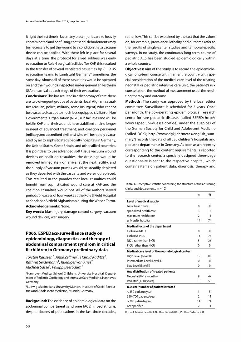
50
Anaesthesiol Intensive Ther 2017, Supplement 1
it right the first time in fact many blast injuries are so heavily contaminated and confusing, that serial debridements may be necessary to get the wound to a condition that a vacuum device can be applied. With these left in place for several days at a time, the protocol for allied soldiers was early evacuation to Role 4 surgical facilities “for KAF, this resulted in the transfer of several ventilated casualties by C119 US evacuation teams to Landstuhl Germany” sometimes the same day. Almost all of these casualties would be operated on and their wounds inspected under general anaesthesia (GA) on arrival at each stage of their evacuation.Conclusions: This has resulted in a dichotomy of care: there are two divergent groups of patients: local Afghani casual-ties (civilian, police, military, some insurgent) who cannot be evacuated except to much less equipped civilian or Non Governmental Organization (NGO) run facilities and will be held in KAF until their wounds have stabilized and no longer in need of advanced treatment; and coalition personnel (military and accredited civilians) who will be rapidly evacu-ated by air to sophisticated specialty hospitals in Germany, the United States, Great Britain, and other allied countries. It is pointless to use advanced soft tissue vacuum wound devices on coalition casualties: the dressings would be removed immediately on arrival at the next facility, and the supply of vacuum pumps would be steadily depleted as they departed with the casualty and were not replaced. This resulted in the paradox that local casualties could benefit from sophisticated wound care at KAF and the coalition casualties would not. All of the authors served periods of excess of four weeks at the Role 3 Field Hospital at Kandahar Airfield Afghanistan during the War on Terror.Acknowledgements: None. Key words: blast injury, damage control surgery, vacuum wound devices, war surgery
P065. ESPEDacs-surveillance study on epidemiology, diagnostics and therapy of abdominal compartment syndrom in critical ill children in Germany: preliminary data
Torsten Kaussen1, Anke Zellmer1, Harald Käditzz1, Kathrin Seidemann1, Ruediger von Kries2, Michael Sasse1, Philipp Beerbaum1
1Hannover Medical School Childrens University Hospital, Depart-ment of Pediatric Cardiology and Intensive Care Medicine, Hannover, Germany2Ludwig-Maximilians-University Munich, Institute of Social Paedia-trics and Adolescent Medicine, Munich, Germany
Background: The evidence of epidemiological data on the abdominal compartment syndrome (ACS) in pediatrics is, despite dozens of publications in the last three decades,
rather low. This can be explained by the fact that the values on, for example, prevalence, lethality and outcome refer to the results of single-center studies and temporal-specific surveys. In no study, the continuous long-term course of pediatric ACS has been studied epidemiologically within a whole country.Objectives: Aim of the study is to record the epidemiolo-gical long-term course within an entire country with spe-cial consideration of the medical care level of the treating neonatal or pediatric intensive care unit, the patient’s risk constellation, the method of measurement used, the resul-ting therapy and outcome. Methods: The study was approved by the local ethics committee. Surveillance is scheduled for 2 years. Once per month, the co-operating epidemiological research center for rare pediatric diseases (called ESPED; http://www.esped.uni-duesseldorf.de) under the auspices of the German Society for Child and Adolescent Medicine (called DGKJ; http://www.dgkj.de/metas/english_sum-mary/) records the data of all 530 children’s hospitals and pediatric departments in Germany. As soon as a rare entity corresponding to the content requirements is reported to the research center, a specially designed three-page questionnaire is sent to the respective hospital, which contains items on patient data, diagnosis, therapy and
Table 1. Descriptive statistic concerning the structure of the answering clinics and departments (n = 19)
n %
Level of medical supply basic health carespecialized health care maximum health care university hospital
03 2
14
0 16 11 74
Medical focus of the departmentExclusive NICUExclusive PICUNICU rather than PICUPICU rather than NICU
01450
074260
Medical care level of the neonatological center High Level (Level BI)Intermediate Level (Level IL)Low Level (Level I)
1900
10000
Age distribution of treated patientsNeonatal (0–12 months)Pediatric (1–18 years)
910
4753
ICU size/number of patients treated< 350 patients/year350–700 patients/year> 700 patients/yearnot specified
12
142
5117411
ICU — Intensive Care Unit; NICU — Neonatal ICU; PICU — Pediatric ICU
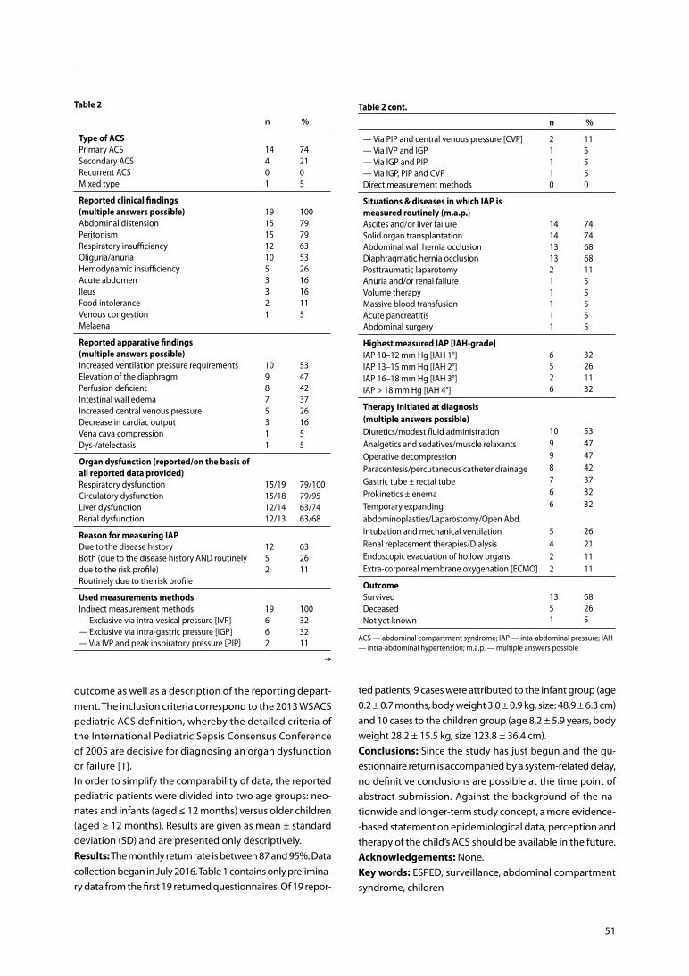
51
Table 2
n %
Type of ACSPrimary ACSSecondary ACS Recurrent ACS Mixed type
14401
742105
Reported clinical findings (multiple answers possible)Abdominal distension PeritonismRespiratory insufficiencyOliguria/anuriaHemodynamic insufficiencyAcute abdomen IleusFood intoleranceVenous congestionMelaena
191515121053321
10079796353261616115
Reported apparative findings (multiple answers possible)Increased ventilation pressure requirementsElevation of the diaphragm Perfusion deficientIntestinal wall edemaIncreased central venous pressureDecrease in cardiac outputVena cava compressionDys-/atelectasis
109875311
53474237261655
Organ dysfunction (reported/on the basis of all reported data provided)Respiratory dysfunction Circulatory dysfunctionLiver dysfunctionRenal dysfunction
15/1915/1812/1412/13
79/10079/9563/7463/68
Reason for measuring IAP Due to the disease historyBoth (due to the disease history AND routinely due to the risk profile)Routinely due to the risk profile
1252
632611
Used measurements methodsIndirect measurement methods— Exclusive via intra-vesical pressure [IVP]— Exclusive via intra-gastric pressure [IGP]— Via IVP and peak inspiratory pressure [PIP]
19662
100323211
n %
— Via PIP and central venous pressure [CVP]— Via IVP and IGP — Via IGP and PIP— Via IGP, PIP and CVP Direct measurement methods
21110
115550
Situations & diseases in which IAP is measured routinely (m.a.p.)Ascites and/or liver failureSolid organ transplantationAbdominal wall hernia occlusionDiaphragmatic hernia occlusionPosttraumatic laparotomyAnuria and/or renal failureVolume therapy Massive blood transfusionAcute pancreatitisAbdominal surgery
14141313211111
747468681155555
Highest measured IAP [IAH-grade]IAP 10–12 mm Hg [IAH 1°]IAP 13–15 mm Hg [IAH 2°]IAP 16–18 mm Hg [IAH 3°]IAP > 18 mm Hg [IAH 4°]
6526
32261132
Therapy initiated at diagnosis (multiple answers possible)Diuretics/modest fluid administrationAnalgetics and sedatives/muscle relaxantsOperative decompressionParacentesis/percutaneous catheter drainageGastric tube ± rectal tubeProkinetics ± enemaTemporary expanding abdominoplasties/Laparostomy/Open Abd.Intubation and mechanical ventilationRenal replacement therapies/DialysisEndoscopic evacuation of hollow organsExtra-corporeal membrane oxygenation [ECMO]
10998766
5422
53474742373232
26211111
OutcomeSurvivedDeceasedNot yet known
1351
68265
ACS — abdominal compartment syndrome; IAP — inta-abdominal pressure; IAH — intra-abdominal hypertension; m.a.p. — multiple answers possible
Table 2 cont.
Æ
outcome as well as a description of the reporting depart-ment. The inclusion criteria correspond to the 2013 WSACS pediatric ACS definition, whereby the detailed criteria of the International Pediatric Sepsis Consensus Conference of 2005 are decisive for diagnosing an organ dysfunction or failure [1].In order to simplify the comparability of data, the reported pediatric patients were divided into two age groups: neo-nates and infants (aged ≤ 12 months) versus older children (aged ≥ 12 months). Results are given as mean ± standard deviation (SD) and are presented only descriptively.Results: The monthly return rate is between 87 and 95%. Data collection began in July 2016. Table 1 contains only prelimina-ry data from the first 19 returned questionnaires. Of 19 repor-
ted patients, 9 cases were attributed to the infant group (age 0.2 ± 0.7 months, body weight 3.0 ± 0.9 kg, size: 48.9 ± 6.3 cm) and 10 cases to the children group (age 8.2 ± 5.9 years, body weight 28.2 ± 15.5 kg, size 123.8 ± 36.4 cm).Conclusions: Since the study has just begun and the qu-estionnaire return is accompanied by a system-related delay, no definitive conclusions are possible at the time point of abstract submission. Against the background of the na-tionwide and longer-term study concept, a more evidence--based statement on epidemiological data, perception and therapy of the child’s ACS should be available in the future.Acknowledgements: None. Key words: ESPED, surveillance, abdominal compartment syndrome, children
52
Anaesthesiol Intensive Ther 2017, Supplement 1
Reference: 1. Goldstein B, Giroir B, Randolph A; International Consensus Conference
on Pediatric Sepsis. International pediatric sepsis consensus conference: definitions for sepsis and organ dysfunction in pediatrics. Pediatr Crit Care Med. 2005; 6: 2–8.
P066. ChimneyVAC for entero-atmospheric fistuals. Early experience with a novel VAC system for open abdomen
Arne Seternes1, 2, Lars Cato Rekstad1, Knut Magne Augestad3, Ola Rokke3, 4, Hans H Wasmuth1
1Department of Surgery, St. Olavs Hospital, Trondheim University Hospital, Trondheim, Norway2NTNU, Norwegian University of Science and Technology, Tron-dheim, Norway3Department of Gastroenterologic Surgery, Akershus University Hospital, Norway4Faculty of Medicine, University of Oslo, Norway
Background: Entero-atmospheric fistula (EAF) is a severe complication in patients with an open abdomen (OA), and several methods have been developed to treat the enteric leak, especially when early deviation is impossible.Objectives: The objective of this study is to present our early results with ChimneyVAC treatment. ChimneyVAC represents a novel method for EAF treatment. A chimney with a base is made out of white foam and placed directly on the intestines with the intestinal fistula in the center of the chimney. A protective plastic sheet is placed to cover the rest of the abdominal content. If possible, a temporary polypropylene mesh is sutured to the rectus fascia and covered with black foam on top and around the chimney. Adhesive drape is then used to seal the abdominal wound, and a negative pressure of 25–75 mm Hg is then placed on top of the chimney. This facilitate drainage of the fistula and the OA, and might prevent leakage of intestinal content into the abdominal wound. The dressing is changed completely every second or third day. The goal of the treatment is to establish an entero-cutaneous fistula. Methods: Retrospective study of patients treated with open abdomen and ChimneyVAC at two university hospi-tals from 2012–2017. Thirteen consecutive patients, eight males, median age 51 years (range 22–73) were included. The reason for open abdomen (OA) treatment were com-plications after incisional hernia repair (n = 3), vascular disease (n = 3), mechanical obstruction (n = 3), pancreatitis (n = 2), trauma (n = 1) and self-injury (n = 1). One patient was treated for abdominal compartment syndrome. OA treatment was initiated after a median of two (range 0–45) days after initial surgery. Eleven fistulas were defined as high out-put fistulas.Results: Eleven patients survived, two died due to multi--organ failure after 55 and 98 days with OA. Median length
of stay in hospital was 74 (range 10–212) days. Median num-ber of dressing changes after established EAF were nine (4-31). In ten out of eleven surviving patients, a controlled entero-cutaneous stoma was established. In one patient, the ChimneyVAC treatment was discontinued due to diffi-culties with occlusion of the vacuum pump due to intestinal content, and he had an abdominal wall reconstruction with a musculo-cutaneous flap to cover the fistula.Conclusions: ChimneyVAC seems to be a feasible method for treatment of EAF with acceptable figures for survival, entero-cutaneous fistula formation and closure of the OA.Acknowledgements: None. Key words: open abdomen, entero-atmospheric fistula, VAC, survival
P068. Polycompartment syndrome: from theory to reality
Zsolt Bodnar1, Edit Tidrenczel2
1Letterkenny University Hospital Letterkenny Co. Donegal, Ireland2Torrevieja University Hospital, Spain
Background: Intra-abdominal hypertension (IAP) and ab-dominal compartment syndrome (ACS) have detrimental effects on all organ systems and are associated with signifi-cant morbidity and mortality. In the last years the diagnosis and management of these syndromes has envolved tremen-dously, and the importance of comprehensive strategies to reduce IAP has been recognized.Objectives: There are four major compartments in the hu-man body: the head, the chest, the abdominal and the limb compartments. The direct correlation among intra-cranial, intra-thoracic and intra-abdominal compartments was dem-onstrated in some small studies during the last ten years. Methods: We present three cases of polycompartment syndrome (PCS) successfully treated with abdominal de-compression. The etiology of each cases was different: blunt thoracic trauma, head injury, intra-thoracic hematoma. Fol-lowing the urgent laparotomies the patients were treated in ICU and discharged after complication free recovery period.Results: The urgent decompressive laparotomy was suc-cessful in decreasing IAP, intra-thoracic pressure (ITP) and intra-cranial pressure (ICP), supporting the complex interac-tion between body compartments.Conclusions: The measurement of IAP is essential in devel-opment of polycompartment syndrome. Clinicians need to be aware of the real existence of PCS and the complex and constant interplays of raised pressures between compart-ments. The presentation of our real clinical cases is to justify the existence of polycompartment syndrome and underline the clinical importance of this complex entity.
53
Acknowledgements: None. Key words: polycompartment syndrome, abdominal com-partment syndrome, intra-abdominal pressure, decompres-sive laparotomy
P071. Does sepsis influence intra-abdominal pressure in critical patients? A pilot study
Alcir Escocia Dorigatti, Bruno Monteiro Pereira, Jennifer Leme dos Santos, Fernanda Dias Teramoto, Marcos Aurelio Boes, Roberta Nazario Aoki, Cesar Vanderlei Carmona, Thiago Martins Santos, Gustavo Pereira FragaUniversity of Campinas, Campinas, Brazil
Background: Currently there is no data regarding the im-pact of sepsis on intra-abdominal pressure (IAP) available.Objectives: This pilot study aims to look for a correlation between both IAP and sepsis in severe ICU patients Methods: This is a prospective observational study enrolling all adult septic shock patients admitted to the ICU, between 2016 April to October in a single institution, that fill both SEP-SIS-3 and Surviving Sepsis criteria for septic shock. Patients with primary abdominal pathologies were excluded from study to avoid intra-abdominal bias. IAP was measured in mm Hg, according to WSACS protocols on every 6 hours, using AbViser®. Continuous variables were expressed as mean and confidence intervals represented in brackets with confidence level of 95% and were compared with the Kruskal-Wallis and RM-One-way ANOVA with Geisser-Greenhouse correction tests. For groups comparison Mann-Whitney test were used.Results: 351 IAP measures were performed in 19 pa-tients. Mean age: 52.58 [43.52 to 61.65]; 84.21% male. Most commons septic shock etiologies were: pulmonary (78.94%), meningitis (10.53%), cellulitis (10.53%). In ten consecutive days Abdominal Perfusion Pressure (APP) was: D1: 75.49 [71.7 to 79.28], D2: 70.22 [66.37 to 74.06], D3: 77.35 [73.45 to 81.25], D4: 76.36 [71.55 to 81.17], D5: 74.39 [68.09 to 80.7], D6: 84.13 [77.14 to 91.11], D7: 76.13 [68.18 to 84.07], D8: 62.36 [48.81 to 75.91], D9: 86.19 [79.7 to 92.67], D10: 85.26 [77.77 to 92.76]. Differences between AAP measurements were found (P < 0.0001), lower APP was found between days 5 to 8. Also differences bete-ween IAP were found (P = 0.0142). Mortality during treat-ment was found to be as high as 47.36%. On patients with death-outcome IAP mean value at a cut point of 72 hours before death was of 20,5 [18,31 to 22,69]; mean IAP in 48 hours before death was 20,47 [17,97 to 22,97] and in last 24 hours before death was of 21,08 [16,88 to 25,28]. No sta-tistical differences were found (p-value = 0.7193). APP value 72 hours before death was 24.73 [10.81 to 38.66]; in 48 hours prior to death 41.95 [29.13 to 55.77] and in last 24 hours before
death 33.22 [16.48 to 49.96] with no statistical differences found (p-value = 0.0734). Mean SOFA Score was: 7.56 [7.06 to 8.06], no statistical differences were found between sepsis days (P-value = 0.831); neither in last three days before death statistical differences were found (P-value = 0.435). In survivor
group mean SOFA score was: 7.44 [7.09 to 7.78] and in death
group was: 8.95 [8.27 to 9.63] (P-value = 0.0017); mean IAP in
survivor group was: 11.42 [10.61 to 12.22] and in death group
was: 13.15 [12.36 to 13.95] (P-value < 0.0001); mean APP in
survivor group was: 78.71 [76.08 to 81.34] and in death group
was: 72.37 [68.64 to 76.1] (P-value = 0.002).
Conclusions: Septic shock patients seem to have amended
IAP and APP measures which seems to change according
sepsis treatment and hospital lenght of stay, although the
SOFA Score does not change in the same repercussion. High
IAP has the potential to be a strong marker for death in sep-tic shock even without having any abdominal pathology.Acknowledgements: Thanks to all the patients and families who allowed this study to be possible, as well as to all the assistance staff — physicians, physiotherapists and nurses — who have assisted in the execution of this project.Key words: sepsis, intra-abdominal hypertension, critical care
P072. Experience of upper gastrointestinal massive bleeding patients requiring open abdominal management
Yuzuru Mochida, Koji Morishita, Kosuke Sekiya, Yasuhiro OtomoTokyo Medical and Dental University Hospital, Japan
Background: Open abdomen has been proven to be ad-vances in the management of abdominal compartment syndrome, damage control surgery and staged laparotomy for general surgery. A limited number of cases have reported on upper gastrointestinal bleeding (GI bleeding) requiring open abdominal management (OAM).Objectives: The purpose of our study is to analyze treat-ment results of patients with upper GI bleeding managed open abdomen.Methods: A retrospective review of 3 patients with upper GI bleeding requring OAM during 3 years (2015–2017) at our hospital.Results: The present study reviewed 3 cases of upper GI bleeding (2 gastric peptic ulcer and 1 duodenal peptic ulcer) requiring OAM. 2 of the patients were male. Ages was ranging from 63 to 73 (mean 68 years). The indications for the OAM were hemorrhagic shock, coagulopathy, and intestinal edema. 2 free omental plug (Graham patch) and 1 total gastrectomy were performed. 30-days post-operative mortality was 33%.
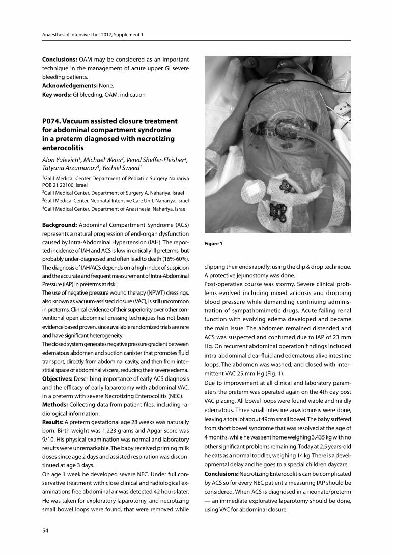
54
Anaesthesiol Intensive Ther 2017, Supplement 1
Conclusions: OAM may be considered as an important technique in the management of acute upper GI severe bleeding patients.Acknowledgements: None. Key words: GI bleeding, OAM, indication
P074. Vacuum assisted closure treatment for abdominal compartment syndrome in a preterm diagnosed with necrotizing enterocolitis
Alon Yulevich1, Michael Weiss2, Vered Sheffer-Fleisher3, Tatyana Arzumanov4, Yechiel Sweed1
1Galil Medical Center Department of Pediatric Surgery Nahariya POB 21 22100, Israel2Galil Medical Center, Department of Surgery A, Nahariya, Israel3Galil Medical Center, Neonatal Intensive Care Unit, Nahariya, Israel4Galil Medical Center, Department of Anasthesia, Nahariya, Israel
Background: Abdominal Compartment Syndrome (ACS) represents a natural progression of end-organ dysfunction caused by Intra-Abdominal Hypertension (IAH). The repor-ted incidence of IAH and ACS is low in critically ill preterms, but probably under-diagnosed and often lead to death (16%-60%). The diagnosis of IAH/ACS depends on a high index of suspicion and the accurate and frequent measurement of Intra-Abdominal Pressure (IAP) in preterms at risk. The use of negative pressure wound therapy (NPWT) dressings, also known as vacuum-assisted closure (VAC), is still uncommon in preterms. Clinical evidence of their superiority over other con-ventional open abdominal dressing techniques has not been evidence based proven, since available randomized trials are rare and have significant heterogeneity.The closed system generates negative pressure gradient between edematous abdomen and suction canister that promotes fluid transport, directly from abdominal cavity, and then from inter-stitial space of abdominal viscera, reducing their severe edema.Objectives: Describing importance of early ACS diagnosis and the efficacy of early laparotomy with abdominal VAC, in a preterm with severe Necrotizing Enterocolitis (NEC). Methods: Collecting data from patient files, including ra-diological information.Results: A preterm gestational age 28 weeks was naturally born. Birth weight was 1,223 grams and Apgar score was 9/10. His physical examination was normal and laboratory results were unremarkable. The baby received priming milk doses since age 2 days and assisted respiration was discon-tinued at age 3 days. On age 1 week he developed severe NEC. Under full con-servative treatment with close clinical and radiological ex-aminations free abdominal air was detected 42 hours later. He was taken for exploratory laparotomy, and necrotizing small bowel loops were found, that were removed while
clipping their ends rapidly, using the clip & drop technique. A protective jejunostomy was done.Post-operative course was stormy. Severe clinical prob-lems evolved including mixed acidosis and dropping blood pressure while demanding continuing adminis-tration of sympathomimetic drugs. Acute failing renal function with evolving edema developed and became the main issue. The abdomen remained distended and ACS was suspected and confirmed due to IAP of 23 mm Hg. On recurrent abdominal operation findings included intra-abdominal clear fluid and edematous alive intestine loops. The abdomen was washed, and closed with inter-mittent VAC 25 mm Hg (Fig. 1).Due to improvement at all clinical and laboratory param-eters the preterm was operated again on the 4th day post VAC placing. All bowel loops were found viable and mildly edematous. Three small intestine anastomosis were done, leaving a total of about 49cm small bowel. The baby suffered from short bowel syndrome that was resolved at the age of 4 months, while he was sent home weighing 3.435 kg with no other significant problems remaining. Today at 2.5 years-old he eats as a normal toddler, weighing 14 kg. There is a devel-opmental delay and he goes to a special children daycare.Conclusions: Necrotizing Enterocolitis can be complicated by ACS so for every NEC patient a measuring IAP should be considered. When ACS is diagnosed in a neonate/preterm — an immediate explorative laparotomy should be done, using VAC for abdominal closure.
Figure 1
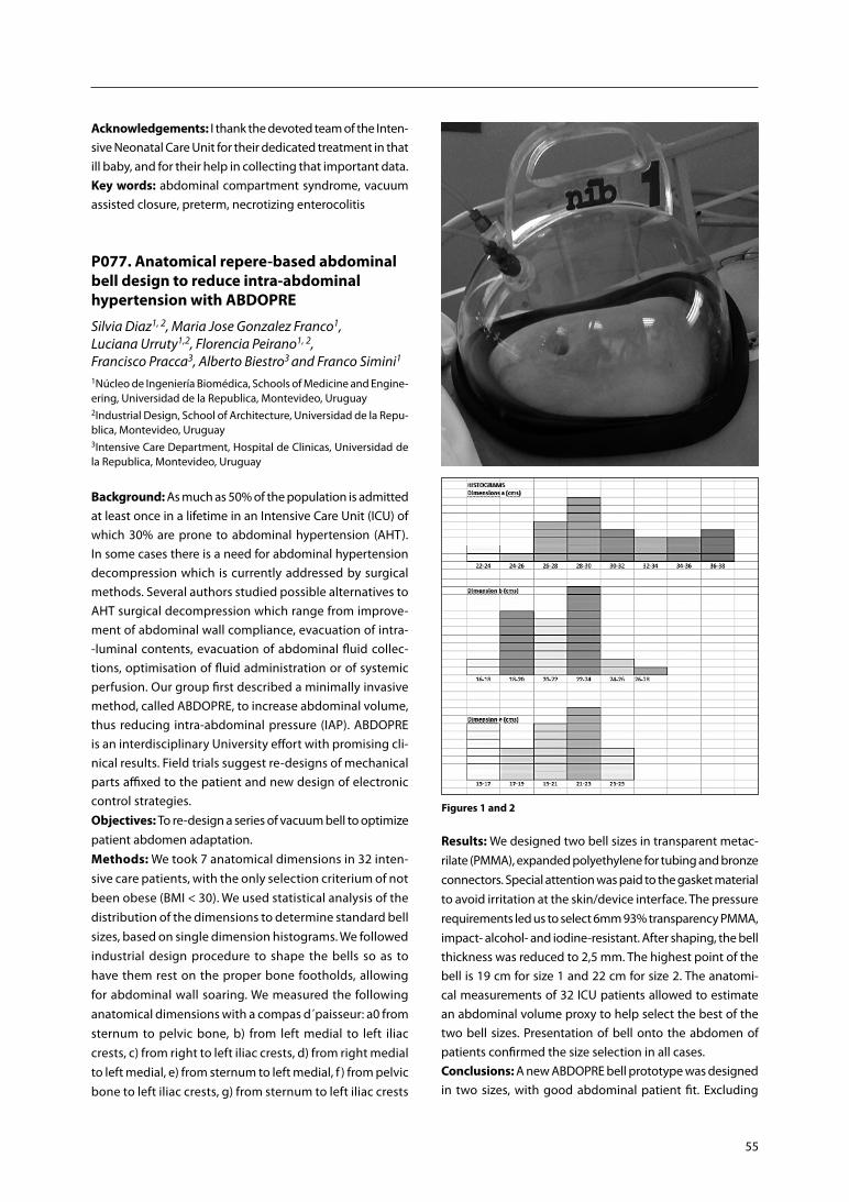
55
Acknowledgements: I thank the devoted team of the Inten-sive Neonatal Care Unit for their dedicated treatment in that ill baby, and for their help in collecting that important data.Key words: abdominal compartment syndrome, vacuum assisted closure, preterm, necrotizing enterocolitis
P077. Anatomical repere-based abdominal bell design to reduce intra-abdominal hypertension with ABDOPRE
Silvia Diaz1, 2, Maria Jose Gonzalez Franco1, Luciana Urruty1,2, Florencia Peirano1, 2, Francisco Pracca3, Alberto Biestro3 and Franco Simini1
1Núcleo de Ingeniería Biomédica, Schools of Medicine and Engine-ering, Universidad de la Republica, Montevideo, Uruguay2Industrial Design, School of Architecture, Universidad de la Repu-blica, Montevideo, Uruguay3Intensive Care Department, Hospital de Clinicas, Universidad de la Republica, Montevideo, Uruguay
Background: As much as 50% of the population is admitted at least once in a lifetime in an Intensive Care Unit (ICU) of which 30% are prone to abdominal hypertension (AHT). In some cases there is a need for abdominal hypertension decompression which is currently addressed by surgical methods. Several authors studied possible alternatives to AHT surgical decompression which range from improve-ment of abdominal wall compliance, evacuation of intra--luminal contents, evacuation of abdominal fluid collec-tions, optimisation of fluid administration or of systemic perfusion. Our group first described a minimally invasive method, called ABDOPRE, to increase abdominal volume, thus reducing intra-abdominal pressure (IAP). ABDOPRE is an interdisciplinary University effort with promising cli-nical results. Field trials suggest re-designs of mechanical parts affixed to the patient and new design of electronic control strategies.Objectives: To re-design a series of vacuum bell to optimize patient abdomen adaptation. Methods: We took 7 anatomical dimensions in 32 inten-sive care patients, with the only selection criterium of not been obese (BMI < 30). We used statistical analysis of the distribution of the dimensions to determine standard bell sizes, based on single dimension histograms. We followed industrial design procedure to shape the bells so as to have them rest on the proper bone footholds, allowing for abdominal wall soaring. We measured the following anatomical dimensions with a compas d´paisseur: a0 from sternum to pelvic bone, b) from left medial to left iliac crests, c) from right to left iliac crests, d) from right medial to left medial, e) from sternum to left medial, f ) from pelvic bone to left iliac crests, g) from sternum to left iliac crests
Figures 1 and 2
Results: We designed two bell sizes in transparent metac-rilate (PMMA), expanded polyethylene for tubing and bronze connectors. Special attention was paid to the gasket material to avoid irritation at the skin/device interface. The pressure requirements led us to select 6mm 93% transparency PMMA, impact- alcohol- and iodine-resistant. After shaping, the bell thickness was reduced to 2,5 mm. The highest point of the bell is 19 cm for size 1 and 22 cm for size 2. The anatomi-cal measurements of 32 ICU patients allowed to estimate an abdominal volume proxy to help select the best of the two bell sizes. Presentation of bell onto the abdomen of patients confirmed the size selection in all cases.Conclusions: A new ABDOPRE bell prototype was designed in two sizes, with good abdominal patient fit. Excluding
56
Anaesthesiol Intensive Ther 2017, Supplement 1
obese patients (BMI > 30), ICU adult patient population can be fitted with either a size 1 or size 2 bell.Acknowledgements: The authors thank Marcelo David, M.Sc. for active counselling and specific work on previous ABDOPRE prototypes.Key words: abdominal hypertension, vacuum bell, ABDO-PRE, bell design
P079. Surviving sepsis, multi organ failure, open abdomen, enterocutanous fistula and intestinal failure
Maria Gaard1, G. Carlson2, N. Meidell3, K. Sunde4 1Consultant Surgeon, Department of Gastroenterological Surgery, Oslo University Hospital, Norway2Professor, Intestinal Failure Unit, Salford Trust Hospital, Manchester, UK3Consultant Intensivist, Department of Anaesthesiology, Oslo University Hospital, Norway4Professor, Consultant Intensivist, Department of Anaesthesiology, Oslo University Hospital, Norway
Background: A 36 year old woman with complications to colorectal cancer surgery.Objectives: Tips and tricks on the challenges of treating sep-sis, multi organ failure, open abdomen, enteroatmospheric fistula and intestinal failure. Methods: Case history on long term treatment of open abdomen.Results: Multivisceral organ transplant as a solution to a GI failure situation.Conclusions: Never, ever give up.Acknowledgements: The patient has agreed to the pre-sentation of her storyKey words: intestinal failure, open abdomen, enterocuta-nous fistulae, multivisceral transplantation
P081. Are anthropomorphic parameters able to predict the presence of intra- -abdominal hypertension: a datastories-based prediction model
Manu L.N.G. Malbrain1, Brecht De Tavernier2, Tine Opsomer2, Pieter-Jan Van Gaal2, Valerie Van Biervliet2
1ICU director, Ziekenhuis Netwerk Antwerpen, ZNA Stuivenberg, Intensive Care Unit, Antwerp, Belgium2Department of Intensive Care and High Care Burn Unit, Ziekenhuis Netwerk Antwerpen, ZNA Stuivenberg, Lange Beeldekensstraat 267, 2060 Antwerpen, Belgium
Background: The abdominal compartment society has listed a number of risk factors for intra-abdominal hypertension (IAH). Obesity and a high body mass index have been asso-ciated with increased intra-abdominal pressure (IAP) [1, 2].
Objectives: The aim of this study is to examine which an-thropomorphic parameters are closely associated with the presence or not of IAH in critically ill patients that are me-chanically ventilated. Methods: Prospective study in 96 mechanically-ventilated patients equipped with a Foley bladder catheter connected to a FoleyManometer (Holtech Medical, Charlottenlund, Denmark) to measure intrabladder pressure (IBP). The pres-ence of IAH is defined according to the WSACS consensus definitions as an endexpiratory IBP equal to or above 12 mm Hg. The following body anthropomorphic parameters were measured: distances (ear-xiphoid, ear-nose, xiphoid-pubis, and ribcage-crista), diameters (rib cage, umbilical, waist, and hip), circumference (rib cage, abdominal, waist, and hip), height (patient, rib cage, hip, and sagittal abdominal diam-eter). A simulation model to predict the presence or not of IAH, based on the above listed anthropomorphic parameters was developed with Datastories (www.datastories.com) in order to identify independent parameters (out of a total of 25) able to predict the presence of IAH. We had to create and challenge 66537 predictive models to deeply learn which metrics are necessary and sufficient to predict IAH. A half of the computational effort was spent on meticulous cross- -validations to make sure to avoid over-fitting and maximizing the predictive power of models. At the end, we were able to build a final ensemble of 100 models with a minimal number of metrics. With respect to predicting IAH presence, the health of our data was excellent. DataStories focuses on finding reli-able relationships between numeric metrics and IBP. Therefore, we had to look at 25 metrics remaining after omitting 5. We first looked at how our metrics impact the prediction of IAH individually and performed a standard correlation analysis as well as a more involved analysis of the mutual information content between IAH and all other data inputs individually. Because the data studied only had 25 parameters on top of IAH (binary labelled as present or not according to WSCAS defini-tion) we also computed all individual pairwise relationships (correlation and mutual information) among the metrics to see how parameters are connected to each other (Fig. 1). Based on initial results we could conclude that 1 out of 25 inputs could be removed from the consideration whatsoever, because it did not have even slight independent relationship to IAH.Results: SAPS-II was 55.4 ± 12.9; APACHE-II 26.4 ± 9.6, SOFA 11.3 ± 5.2; age 57.5 ± 13.9; height 174 ± 9cm; weight 85 ± 20; BMI 27.9 ± 7. There were 55 patients with IAH. The final en-semble of parameters after data analysis able to predict IAH has the following characteristics: average cross-validation prediction accuracy of 88.6% with the use of 1 metric (out of the initial 25 that were entered into the model). From this preliminary analysis, we could conclude that 25 inputs are individually related to IAH, but many of them are cor-related to each other (Fig. 2). Therefore, further datamining
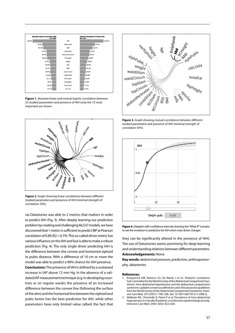
57
via Datastories was able to 2 metrics that matters in order to predict IAH (Fig. 3). After deeply learning our prediction problem by creating and challenging 66,537 models, we have discovered that 1 metric is sufficient to predict IBP at Pearson correlation of 0.89 (R2 = 0.79). This so-called driver metric has various influence on the IAH and but is able to make a robust prediction (Fig. 4). The only single driver predicting IAH is the difference between the convex and horizontal xiphoid to pubis distance. With a difference of 10 cm or more the model was able to predict a 90% chance for IAH presence.Conclusions: The presence of IAH is defined by a sustained increase in IAP above 12 mm Hg. In the absence of a vali-dated IAP measurement technique (e.g. in developing coun-tries or on regular wards) the presence of an increased difference between the convex line (following the surface of the skin) and the horizontal line between the xiphoid and pubic bones has the best prediction for IAH, while other parameters have only limited value (albeit the fact that
Figure 1. Absolute linear and mutual logistic correlation between 25 studied parameters and presence of IAH (only the 15 most important are shown
Figure 2. Graph showing linear correlations between different studied parameters and presence of IAH (minimal strength of correlation 20%)
Figure 3. Graph showing mutual correlations between different studied parameters and presence of IAH (minimal strength of correlation 50%)
Figure 4. Dataplot with confidence intervals showing the “What-If” scenario to see the evolution in prediction for IAH when main driver changes
they can be significantly altered in the presence of IAH). The use of Datastories seems promising for deep learning and understanding relations between different parameters.Acknowledgements: None.Key words: abdominal pressure, prediction, anthropomor-phy, datastories
References: 1. Kirkpatrick AW, Roberts DJ, De Waele J et al.; Pediatric Guidelines
Sub-Committee for the World Society of the Abdominal Compartment Syn-drome. Intra-abdominal hypertension and the abdominal compartment syndrome: updated consensus definitions and clinical practice guidelines from the World Society of the Abdominal Compartment Syndrome. Inten-sive Care Med. 2013;39(7): 1190–206. doi: 10.1007/s00134-013-2906-z.
2. Malbrain ML, Chiumello D, Pelosi P et al. Prevalence of intra-abdominal hypertension in critically ill patients: a multicentre epidemiological study. Intensive Care Med. 2004; 30(5): 822–829.
58
Anaesthesiol Intensive Ther 2017, Supplement 1
P082. Early, definitive repair of traumatic lumbar hernia with a titanium, twinfix, bone-anchored suture technique
Thomas Clements, Derek J. Roberts, Ryan Martin, Chad G. Ball, Andrew W. Kirkpatrick, Ruphus Rajakumar, Rohan Lall
Background: Traumatic lumbar hernias are uncommon and may be challenging to repair, especially because con-comitant intraperitoneal contamination from coexisting intestinal injuries frequently precludes the use of mesh. Objective: We report a novel, mesh-free technique for closure of traumatic lumbar hernia. The technique utilizes 5.5 mm titanium, Twinfix bone-anchored sutures to prima-rily approximate the oblique musculature to the iliac wing. Methods: We obtained informed consent from the patient to present their case.Results: An 37-year old, severely injured male presented to our quaternary-care, level 1, academic trauma center after a head on collision at highway speed. As he was hemodyna-mically stable, he immediately underwent a computed to-mography (CT) scan of his head, neck, chest, abdomen, and pelvis. The CT of his abdomen and pelvis revealed an acute
dissection of his left common iliac artery, an American As-sociation for the Surgery of Trauma (AAST) grade IV liver laceration, lumbar transverse process fracture, intraperito-neal bladder rupture, and a large left sided traumatic hernia thought to exist between the quadratus laborum/oblique musculature and iliac wing. The patient was taken to the operating room and underwent an endovascular iliac ar-tery repair and laparotomy, during which his bladder was repaired. A very large posterior abdominal wall defect was encountered intraoperatively. The oblique musculature was seen to be torn from the pelvis. We subsequently utilized a novel hernia repair during which 5.5 mm titanium Twinfix bone anchored sutures were secured to the pelvis. A mayo needle was then used to suture the anchors to all muscular and fascial layers and the peritoneum was closed to exclude the repair from the intraperitoneal space. A closed suction drain was then inserted to drain the area. The patient did well postoperatively, had minimal pain, and showed no signs of clinical recurrence at discharge.Conclusion: Early, definitive repair of traumatic lumbar hernia using bone-anchored sutures may be a promising technique for management of this uncommon condition in appropriately selected patients.
59
Invited abstracts
I001. Introduction to the International Fluid Academy and the 4 phases, 4 D’s and 4 questions in relation to fluid management
Manu L.N.G. MalbrainICU and High Care Burn Unit Director, Ziekenhuis Netwerk Antwer-pen, ZNA Stuivenberg, Antwerp, BelgiumICU Director, University Hospital Brussels (UZB), Jette, BelgiumProfessor at Vrije Universiteit Brussel (VUB), Faculty of Medicine, Brussels, Belgium
Learning objectives: To understand the mission of the International Fluid Academy (iFA), as part of iMERiT (In-ternational Medical Education and Research Initiative). To describe the International Fluid Academy Days (iFAD) and the available member resources on the fluidacademy.org website within the FOAM (Free Open Access Medical educa-tion) philosophy. To describe the 4 phases of fluid manage-ment. To describe the 4 D’s of fluid therapy and the analogy to antibiotic therapy. To describe the 4 questions that need to be answered during fluid management. Introduction: The iFA started as local initiative from the pharmaceutical working group on fluids from the Ziekenhu-is Netwerk Antwerpen (www.zna.be). Today iFA and iFAD are integrated within the not-for-profit charitable organization iMERiT, International Medical Education and Research Ini-tiative, under Belgian law. The mission of the iFA is to foster education and promote research on fluid management and monitoring in critically ill patients, and thereby improve the survival of critically ill patients by bringing together physicians, nurses, and others from a variety of clinical di-sciplines. The primary goal of the iFA is to establish an inter-national collaboration group with the final aim to improve and standardize care and outcome of critically ill patients with an emphasis on fluids, fluid management, monitoring
Table 1. Social and scientific impact of iFA-endorsed publications
Publication date
doi Shares downloads citations altmetric score
Wise et al. [2] 5 Jan 2017 http: //link.springer.com/article/10.1007/s00268-016-3865-7
193 8000 1 92
Reintam Blaser et al. [1]
6 Feb 2017 http: //link.springer.com/article/10.1007/s00134-016-4665-0
176 20000 1 96
Van Regenmortel et al. [3]
20 May 2017 https: //doi.org/10.1093/bja/aex118 20 33 0 11
and organ support. The iFA is proud to announce that the fluidacademy.org website is now an official SMACC-affiliated site (Social Media and Critical Care) adhering to the FOAM (Free Open Access Medical education) principles. Recently the first scientific papers endorsed by an unrestricted iFA--educational grant have been published under the FOAM label [1–3]. The impact of these publications in the scientific community has been significant as shown by their Altmetric scores (Table 1).Discussion: The application of what we already know will have a bigger impact than any drug or fluid or technology likely to be introduced in the next decade. Therefore we should consider and treat fluids as drugs and take into ac-count the 4 D’s of fluid therapy [4, 5]. Fluids are drugs. There are different types of fluids (crystalloids vs colloids, synthetic vs blood derived, balanced vs unbalanced, intravenous vs oral administration). Each fluid comes with its indications, contraindications and possible adverse effects. Possible indications are fluids for resuscitation, maintenance or repla-cement. Fluid therapy needs to be appropriate and in some cases combination therapy is needed. It is all about giving the right fluid for the right patient at the right time. The dose of fluids is important. As Paracelsus nicely stated already back in 16th century: “All things are poison, and nothing is without poison; only the dose permits something not to be poisonous”. This also refers to the pharmacodynamics and kinetics of fluids within the body. The response to IV fluids needs to be assessed by means of hemodyanmic monitoring and dynamic tests (like passive leg raising, pulse pressure variation,…). Appropriate duration is important. Fluid admini-stration needs to be tailored to response and stopped when no longer needed. Finally de-escalation needs to be conside-red. This follows the principles of the ROSE concept and the 4 phases of fluid management [6]. The 4 basic questions
60
Anaesthesiol Intensive Ther 2017, Supplement 1
that need to be answered are when to start and stop fluid administration and when to start and stop fluid removal?Take-home messages: 1. The mission of the iFA is to foster education and pro-
mote research on fluid management and monitoring in critically ill patients, and thereby improve the survival of critically ill patients by bringing together physicians, nurses, and others from a variety of clinical disciplines.
2. The iFA and iFAD are part of iMERiT (a not-for-profit charitable organization under Belgian law).
3. The iFA website is now an official SMACC affiliated we-bsite adhering to the FOAM principles (Free Open Access Medical eduction).
4. Each ICU clinician should be aware of the 4 phases of fluid management within the ROSE concept.
5. Each ICU physician should be aware of the 4 D’s of fluid therapy in analogy to antibiotic therapy: Drug–Dose–Du-ration–De-escalation.
6. Each ICU physician needs to answer 4 questions in relation to fluid management: When to start fluids? When to stop fluids? When to start fluid removal? When to stop fluid removal?
Conflict of interest: The author declares a possible con-flict of interest in relation to the content of this abstract and presentation as he is member of the medical advisory board of Pulsion Medical Systems (now part of Maquet Getinge group).
References: 1. Reintam Blaser A, Starkopf J, Alhazzani W et al. Early enteral nutrition in
critically ill patients: ESICM clinical practice guidelines. Intensive Care Med. 2017; 43: 380–398.
2. Wise R, Faurie M, Malbrain ML, Hodgson E. Strategies for intravenous fluid resuscitation in trauma patients. World J. Surg. 2017.
3. Van Regenmortel N, De Weerdt T, Van Craenenbroeck AH et al. Effect of isotonic versus hypotonic maintenance fluid therapy on urine output, fluid balance, and electrolyte homeostasis: a crossover study in fasting adult volunteers. Br J Anaesth. 2017.
4. Malbrain ML, Van Regenmortel N, Owczuk R. It is time to consider the four D’s of fluid management. Anaesthesiol Intensive Ther. 2015; 47 Spec No: 1–5.
5. Malbrain MLNG. The great Fluid Debate: Faith and Evidence. Intensive Care Monitor. 2014; 21: 1.
6. Malbrain ML, Marik PE, Witters I et al. Fluid overload, de-resuscitation, and outcomes in critically ill or injured patients: a systematic review with suggestions for clinical practice. Anaesthesiol Intensive Ther. 2014; 46: 361–380. doi: 10.5603/AIT.2014.0060.
I002. Introduction to DAMPS and PAMPS and why they matter in critical intra- -abdominal injury/illness
Carl J. HauserAcademic faculty member at the University of Southern California, the University of Mississippi Medical Center, University of Medicine and Dentistry of New Jersey (now Rutgers Medical School) and Harvard Medical School
Learning objectives: To get an understanding of DAMPS and PAMPS and to understand why the matter in intraab-dominal injury.
Introduction: The importance of systemic inflammatory response syndromes (SIRS) in clinical medicine is now well--established. SIRS underlies most acute organ dysfunction syndromes and likely also underlies the predilection to in-fection seen in surgery and trauma patients. Discussion: Whereas we formerly assumed that all febrile patients had infections, we now know that only a minority are infected. Current understanding of inflammatory respon-ses is based on our awareness that the immune system uses preformed “pattern recognition receptors” (PRR) to respond to a wide array of “danger signals” commonly found under conditions of cell stress or injury. In this paradigm, bacteria are seen as presenting specific molecular patterns to the host immune system that are called “pathogen associated molecular patterns,” or PAMPs. On the contrary, essentially all surgical and trauma patients manifest some degree of sterile inflammation. In these circumstances, inflammation is elicited by host responses to non-pathogenic, danger--associated molecular patterns, or DAMPs. DAMPs are often motifs derived from cell contents that are released by surgery or injury. These can also engage PRR, which may often be the self-same PRR that respond to PAMPs. These similarities in cell signaling explain why clinical responses can be the same whether initiated by sterile for infective means. Take-home messages: This modern understanding of in-flammation is a critical adjunct to our approach to patients with abdominal injuries. It can explain why patients with sterile blunt liver injuries will develop a SIRS response that looks almost indistinguishable from the response to a liver abscess. Moreover, in the future better understanding of these key molecular processes should give us diagnostic and therapeutic insights into abdominal illnesses and injuries. Conflict of interest: none provided with abstract.
I003. Mild to moderate intra-abdominal hypertension: does it matter?
Annika Reintam BlaserDepartment of Anaesthesiology and Intensive Care, University of Tartu, Estonia
Learning objectives: This lecture addresses mild and mo-derate intra-abdominal hypertension (IAH) and discusses the importance of differentiation of IAH based on severity. Introduction: IAH occurs in about one third of intensive care (ICU) patients. In 60–70% of these cases intra-abdominal pressure (IAP) stays between 12 to 15 mm Hg (Grade I IAH), in 20–30% IAP reaches 16 to 20 mm Hg (Grade II IAH), and only minority have higher levels of IAP (Grade III — IAP 21–25 or Grage IV — IAP >25 mm Hg) [1].Discussion: Studies have shown different results regarding the impact of IAH on outcome. Several earlier studies have
61
shown that IAH independently impairs outcome in ICU pa-tients [2, 3]. Two more recent studies reported no differen-ce in IAH vs. noIAH regarding survival, whereby Iyer et al. observed adverse impact on survival associated with IAH Grade II–IV [4, 5]. These results may possibly be explained by increased attention towards and better management of IAH in more recent studies, but possibly also in decreased incidence of more severe IAH in respective studies. Overall prevalence of IAH in the ICU does not seem much reduced over time, but severity of IAH was often not reported in earlier studies. A consensus cut-off value of 12 mm Hg for IAH is supported by several studies and is well established. Whether current grading system of IAH is enough to identify gradually increasing risk of adverse outcome and can be translated into a treatment algorithm, still needs to be cla-rified. Lower grades of IAH may be relevant in terms of both pathophysiology and clinical consequences, but the influen-ce of certain value of IAP on outcome will depend on patient characteristics (e.g. obese vs not; mechanically ventilated vs spontaneously breathing, critically ill vs recovery room patients), concomitant macro- and microcirculation, disease characteristics (e.g. pancreatitis vs pneumonia) and possibly duration of IAH [6]. Accordingly, mild IAH sometimes may require specific treatment and sometimes not. Most of the options suggested to be effectively reducing IAP are not without risks (e.g., drainages) and side effects (e.g., sedation, neuromuscular blockade) [6], whereas prolonged periods with increased IAP may lead to multiple serious consequen-ces [1]. Therefore, decisions to treat or not mild to moderate IAH in an individual patient need to be carefully considered. Future studies should aim more detailed approach to IAH moving away from IAH/ACS as a yes-or-no phenomenon.Take-home messages: 1. IAH is a continuum.2. The term “IAH/ACS” describing a yes-or-no phenomenon
should be abandoned.3. Mild IAH may sometimes necessitate specific treatment
aiming reduction of IAP and sometimes not, whereas any specific value of IAP alone is insufficient to justify respective decisions.
Conflict of interest: The author declares no possible conflict of interest in relation to the content of this abstract and presentation.
References: 1. Kirkpatrick AW, Roberts DJ, De Waele J et al. Intra-abdominal hyperten-
sion and the abdominal compartment syndrome: updated consensus definitions and clinical practice guidelines from the World Society of the Abdominal Compartment Syndrome. Intensive Care Med. 2013; 39: 1190–1206. doi: 10.1007/s00134-013-2906-z.
2. Malbrain ML, Chiumello D, Pelosi P et al. Incidence and prognosis of intraabdominal hypertension in a mixed population of critically ill pa-tients: a multiple-center epidemiological study. Crit Care Med. 2005; 33: 315–322. doi: 10.1097/01.CCM.0000153408.09806.1B.
3. Reintam A, Parm P, Kitus R, Kern H, Starkopf J. Primary and secondary intra-abdominal hypertension — different impact on ICU outcome. In-tensive Care Med. 2008; 34: 1624–1631. doi: 10.1007/s00134-008-1134-4.
4. Iyer D, Rastogi P, Åneman A, D’Amours S. Early screening to identify pa-tients at risk of developing intra-abdominal hypertension and abdominal compartment syndrome. Acta Anaesthesiol Scand. 2014; 58: 1267–1275 doi: 10.1111/aas.12409.
5. Kim IB, Prowle J, Baldwin I, Bellomo R. Incidence, risk factors and outcome associations of intra-abdominal hypertension in critically ill patients. Ana-esth Intensive Care. 2012; 40: 79–89.
6. Maddison L, Starkopf J, Reintam Blaser A. Mild to moderate intra-abdomi-nal hypertension: Does it matter? World J Crit Care Med. 2016; 5: 96–102. doi: 10.5492/wjccm.v5.i1.96.
I004. Epidemiology of IAH and the ACS in acute general surgery
Rob Wise Head Clinical Unit Critical Care, Edendale Hospital, Pietermaritzburg, South Africa; Clinical Fellow, Discipline of Anaesthesiology and Critical Care, Nelson Mandela School of Medicine, University of Kwazulu Natal, Durban, South Africa
Learning objectives: Gaining an understanding of the epidemiology (namely the incidence, distribution, and po-ssible control of intra-abdominal hypertension/ abdominal compartment syndrome and it’s related issues) will assist in laying the foundation for an understanding in what is required to change clinical practice, education, and research. This lecture will focus these issues in the context of acute general surgery and how these components come together in improving our patients’ outcomes.Introduction: Awareness and research in IAH and ACS have increased since the first publication described the associa-tion between raised intra-abdominal pressure and renal function. Growing evidence since then has accumulated, highlighting the importance of this often undiagnosed con-dition. The morbidity and mortality associated with IAH and ACS prompted the publication of 2007 and updated 2013 consensus guidelines from the Abdominal Compart-ment Society (WSACS). These evidence based consensus definitions and clinical practice guidelines, together with advances in clinical therapies and equipment, have enabled better monitoring and physiological manipulation of pa-tients with IAH or ACS. In this review, we examine changes in the epidemiological issues (awareness, clinical practice and the physiological importance of IAH and ACS). In particular, we review how far we have come and the journey ahead.Discussion: Although the journals reflect an increase in publications, it is unclear whether significant clinical appli-cation of knowledge and awareness has occurred following the publication of the updated 2013 WSACS consensus guidelines. Many surveys have investigated the clinical un-derstanding, awareness and practice associated with ACS, and have shown a lack of clinical knowledge and under-standing. Results from the largest of these surveys revealed many critical care physicians never measure intra-abdominal pressures, and there is no uniform management of patho-physiology across medical disciplines or geographically. It is
62
Anaesthesiol Intensive Ther 2017, Supplement 1
vital that further efforts are spent on improving education and awareness, and ensuring the routine and protocolised management of patients at risk of IAH/ACS. New and in-novative methods to achieve these objectives need to be explored. These should include incorporation into under-graduate training systems, and use of new education plat-forms such as social media. Following this, renewed efforts and continued research needs to encourage practical and evidence-based review of current guidelines — guidelines that can be achieved even in resource limited environments.Take-home messages: 1. The incidence of IAH/ACS is most often greater than what
healthcare providers realize. 2. Awareness of IAH/ACS and adherence to current clini-
cal guidelines needs to improve worldwide in order to better patient outcomes.
3. Research needs to focus of education, changing health-care providers approach to patients at risk of IAH/ACS, and incorporation of novel and innovative ideas for training (at both pre- and post-graduate levels).
Conflict of interest: The author declares no possible conflict of interest in relation to the content of this abstract and presentation.
References: 1. Wendt E. Ueber den einfluss des intraabdominalen druckes auf die ab-
sonderungsgeschwindigkeit des harness. Arch Physiologische Heilkunde. 1876; 57: 525–527.
2. Emerson H. Intra-abdominal pressures. Arch Intern Med. 1911; 7: 754–758. 3. Saggi BH, Sugerman HJ, Ivatury RR, Bloomfield GL. Abdominal compart-
ment syndrome. J Trauma. 1998; 45: 597–609. 4. Kimball EJ, Kim W, Cheatham ML, Malbrain ML. Clinical awareness of
intra-abdominal hypertension and abdominal compartment syndrome in 2007. Acta clinica Belgica Supplementum. 2007; (1): 66–73. PubMed PMID: 17469703.
5. Balogh ZJ, Leppaniemi A. The neglected (abdominal) compartment: what is new at the beginning of the 21st century? World J Surg. 2009; 33: 1109. PubMed PMID: 19363696.
6. Kirkpatrick AW, Roberts DJ, De Waele J et al. Intra-abdominal hyperten-sion and the abdominal compartment syndrome: updated consensus definitions and clinical practice guidelines from the World Society of the Abdominal Compartment Syndrome. Intensive Care Med. 2013; 39: 1190–1206. PubMed PMID: 23673399. Pubmed Central PMCID: 3680657.
7. Wise R, Roberts D, Vandervelden S et al. Awareness and knowledge of intra-abdominal hypertension and abdominal compartment syndrome: results of an international survey. Anaesthesiol Intensive Ther. 2015; 47: 14–29. doi: 10.5603/AIT.2014.0051.
I005. Under appreciated champions: nursing awareness and advocacy
Rosemary K. Lee Clinical Nurse SpecialistCritical Care & Progressive Care DepartmentHomestead, Florida, USA
Learning objective: Identify the opportunities that the Abdominal Compartment Society has opened for nurses. Introduction: Critical Care Nurses have always been first in line to adapt new technology that can improve patients’
outcomes. The frustration comes when their physician colle-agues do not have the same understanding. The Abdominal Compartment Society has opened that door to collegiality and has supported nurses in developing the nurses’ role in managing and monitoring for IAH. Discussion: Florence Nightingale is to nurses, what Hip-pocrates is to physicians. Her wisdom and insights are still relevant today. Her remarks will be related to the nurses’ role in managing the patient at risk of IAH. With the support of the Abdominal Compartment Society nurses have made inroads in developing nurse driven protocols, educating the interdisciplinary team, publishing, and presenting at national symposia. Nurses now need to add to the body of knowledge by performing research relevant to the nurse’s role in IAH. Take home message: Like Florence Nightingale, nurses want to put patients in the best possible condition for nature to act. Nurses have valuable contributions to add to the management of the patient at risk for IAH.Conflict of interest: The author is a consultant for Becton--Dickinson (BD).
References: 1. Hardin SR, Kaplow R. Synergy for Clinical Excellence: The AACN Synergy
Model for Patient Care, 2nd Ed. Jones & Bartlett Learning, Burlington 2017. 2. Lee R. The effects of nursing activities on the intra-abdominal pressure of
patients at risk for intra-abdominal hypertension. Anaesthesiol Intensive Ther. 2017; 49: 116–121. doi: 10.5603/AIT.a2017.0020.
3. Lee R. Comprehensive overview of intra-abdominal hypertension and abdominal compartment syndrome. Crit Care Nurse. 2012; 32: 19–31.
4. Nightingale F. Notes on nursing: what it is and what it is not. John W Parker & Son, London 1860.
I006. Implications of IAH on renal failure in the critically ill
Bart L. De KeulenaerAssociate Professor, Department of Surgery, University of Western Australia, WA, Australia; Intensive Care Medicine, Fiona Stanley Hospital, Murdoch, WA, Australia
Learning objectives: 1. To review the mechanisms by which renal derange-
ments occur in IAH.2. To review the effects of reduced cardiac output (CO),
increased real venous pressure and increased renal pa-renchymal pressure on renal function.
3. To discuss how early intervention in patients with IAH and ACS can prevent acute renal failure.
Introduction: We know in the acute setting, that with ra-pid elevation of IAP, that the patient first becomes oliguric followed by anuria with eventually progression to overt renal failure.Going back to basics, we know that 25% of our cardiac output goes to the kidneys and that our postglomerular
63
intrarenal vascular network is a low-pressure system and that with raised IAP significant renal venous congestion occurs (cardiorenal syndrome). What are the mechanisms by which renal derangements occur in IAH. It is thought to be through either one or more of the following: reduced CO (hypoperfusion of the kidney), elevated renal venous pres-sure (RVP) and elevated renal parenchymal pressure RPP).Reduced CO is secondary to pressure on the inferior vena cava, which reduces preload pressure on the resistance vessels, increasing afterload, and increased intrathoracic pressure, which further decreases venous return to the he-art. With decreased CO, there is decreased renal blood flow (RBF), causing decreased glomerulofiltration rate (GFR) and activation of renin-angiotensin-ALD system. If we assume reduced CO was the only mechanism, than fluid resuscita-tion to baseline CO would restore renal function in IAH to some extenrt. However that does not happen, renal function does not respond to volume resuscitation or diuretics alone, except if IAP is promptly released.(Volume resuscitation only partially decrease raised renin and aldosterone levels (increased renin and aldosterone causing fluid retention and angiotensin induced vasocon-striction further reducing RBF and GFR).Discussion: Harman (in 1982) studied the effect of increased IAP on cardiac output and renal function using anaesthe-tised dogs into whom inflatable intraperitoneal bags were placed.When increasing IAP to 20 and 40 mm Hg, there was an in-crease in renal vein pressure, drop in RBF and GFR and drop in CO. After fluid resuscitation but maintaining pressure up to 40 mm Hg only the CO improved. It did not influence GFR or RBF. So high IAP causes renal dysfunction probable by inducing high renal vein pressures as restoring of the CO did not improve GFR or RBF That only improved when IAP was reduced. So they concluded that that renal function in IAH/ACS is caused by increased RVP or increased RPP. Doty et al investigated the effects of increased renal parenchy-mal pressure on renal function. A compressive device was placed about the right kidney and needle was inserted into the parenchyma to measure compartment. Left kidney was removed. The compressive device consisted of two acrylic plastic plates transversed by four screws placed beyond the confines of the kidney. The screws were tightened increasing the RPP measured with the needle. There was no difference in RBF or GFR. However a study done in monkeys but in an injured kidney model did show that increased RPP caused renal failure. In conclusion, renal dysfunction secondary to IAH is due to a combination of renal venous congestion, raised renal parenchymal pressures, reduce CO and raised plasma renin aldosterone system. Prompt reduction of ve-nous congestion (by diuretics, dialysis and paracentesis),
restoration of CO and early abdominal decompression will prevent renal failure.Take-home messages: 1. IAH and ACS causes early renal failure.2. A combination of reduced cardiac output, increased
renal venous pressure and to some extent a raised re-nal parenchymal pressure is responsible for early renal impairment.
3. Early intervention is of utmost importance in the pre-vention of renal failure in IAH/ACS by improving CO, reducing renal venous pressure via paracentesis, diu-retics or renal. replacement therapy and other medical and/or surgical management strategies to reduce IAP.
Conflict of interest: None to declare.
References: 1. Harman PK, Kron IL, McLachlan HD, Freedlender AE, Nolan SP. Elevated
intra-abdominal pressure and renal function. Ann Surg 1982; 196: 594.2. Doty JM, Saggi BH, Sugerman HJ et al. Effect of increased renal venous
pressure on renal function. J Trauma 1999; 47: 1000–1003.3. De Keulenaer BL, De Waele JJ, Powell B, Malbrain ML. What is a normal
intra-abdominal pressure and how is it affected by positioning, body mass and PEEP? Intensive Care Med. 2009; 35: 969–976. doi: 10.1007/s00134-009-1445-0.
4. Bloomfield GL, Blocher CR, Fakhry IF, Sica DA, Sugerman HJ. Elevated In-tra-abdominal Pressure Increases Plasma Renin Activity and Aldosterone Levels. J Trauma. 1997; 42: 997–1005.
5. Doty JM, Saggi BH, Blocher CR. Effects of increased renal parenchymal pressure on renal function. J Trauma. 1997; 48: 874.
I007. Co-morbidities among those requiring AWR: when is “no” a hard-line?
Ari LeppäniemiChief of Emergency Surgery, Helsinki University Hospital, Finland
Learning objectives: 1. To describe the most common complications after ab-
dominal wall reconstruction.2. To describe the most commonly used and useful clas-
sification systems of comorbidities in surgical patients.3. To discuss the effect of age on outcomes after elective
ventral hernia repair.4. To summarize the contraindications for patients in need
of complex abdominal wall reconstruction.Introduction: Repair of giant ventral hernias or perfor-ming abdominal wall reconstruction following open ab-domen management are major operations with high risk of complications. The risk increases if the abdominal wall reconstruction is combined with a procedure of restoring the gastrointestinal tract continuity, closing a stoma or ope-rating in a surgical field that is already contaminated, such as in patients with an infected mesh. A careful assessment of potential risks and benefits of the planned operation must be carried out before surgery, and in some cases conserva-tive approach could be a better option.
64
Anaesthesiol Intensive Ther 2017, Supplement 1
Discussion: Among patients undergoing large and complex ventral hernias, the most common postoperative complica-tions include seroma or hematoma formation, surgical site or mesh infection and wound necrosis. Wound dehiscence and enteric fistula are more serious but fortunately less common complications. Extra-abdominal complications, such as pulmonary or cardiac complications can occur espe-cially in patients with cardiopulmonary risk factors, and all patients are at risk of thromboembolic events. Finally, ab-dominal compartment syndrome can occur postoperatively in patients undergoing abdominal wall reconstruction after prolonged preoperative loss of abdominal domain.To decrease the incidence of incisional hernias, using conti-nuous suturing techniques and avoiding rapidly absorbable sutures are strongly recommended by the European Hernia Society guidelines. The component separation technique in the management of large and complex ventral hernias ena-bles fascial closure with low rates of morbidity and hernia recurrence. Some of the current risk factor stratification tools overemphasize patient factors ignoring the importance of surgical techniques used. In general age should not be a contradiction to elective ventral hernia repair, even in patients over 70 years old. Large hernia volume as calcula-ted from a standard abdominal CT-scan is a risk factor for postoperative pulmonary complications.Take-home messages: 1. Abdominal wall reconstruction is a major operation
where the potential risks and benefits should be care-fully evaluated before surgery.
2. Age is not an absolute contraindication for abdominal wall reconstruction.
3. Comorbidities play a role in increasing the risk of posto-perative complications, but the selection of the recon-struction technique has also great impact on the risk of complications, and therefore the technique used should be individualized.
Conflict of interest: The author declares no possible conflict of interest in relation to the content of this abstract and presentation
References: 1. Muysoms FE, Antoniu SA, Bury K et al. European Hernia Society guidelines
on the closure of abdominal wall incisions. Hernia 2015: 19: 1–24. doi: 10.1007/s10029-014-1342-5.
2. Torregrosa-Gallud A, Muriel JS, Bueno-Lledo J et al. Modified components separation technique: experience treating large, complex ventral hernias at university hospital. Hernia. 2017; doi: 10.1007/s10029-017-1619-6.
3. Fligor JE, Lanier ST, Dumanian GA. Current risk stratification systems are not generalizable across surgical technique in midline ventral hernia repair. Plast Reconstr Surg Glob Open. 2017; 5: e1206. doi: 10.1097/GOX.0000000000001206.
4. Neupane R, Fayezizadeh M, Majumder A, Novitsky YW. Is old age a con-traindication to elective ventral hernia repair? Surg Endosc. 2017; doi: 10.1007/s00464-017-5492-2.
5. Mommers EHH, Wegdam JA, van der Wolk S et al. Impact of hernia volume on pulmonary complications following complex hernia repair. J Surg Res. 2017; 211: 8–13.
I008. Abdominal wall reconstruction: managing the skin envelope
Duncan NickersonSection of Plastic Surgery, Department of Surgery, University of Calgary
Learning objectives: 1. Review strategies for dealing with the skin envelope in
abdominal wall reconstruction.2. Review the literature regarding concurrent skin-reduc-
tion procedures and abdominal wall reconstruction.Introduction: Patients undergoing abdominal wall recon-struction often have a real or perceived need for tailoring of their abdominal wall skin. Current literature suggests that this should be undertaken with caution, particularly in the obese patient.Discussion: Patients with a normal body mass index may undergo concurrent abdominal wall reconstruction and skin tailoring with an expectation of a reasonable outcome.Patients with an elevated body mass index are much more likely to experience added morbidity when abdominal wall reconstruction is performed concurrently with skin tailoring procedures.Conflict of interest: The author declares no possible conflict of interest in relation to the content of this abstract and presentation
References: 1. Fadel MG, Chatzikonstantinou M, Gilchrist C, Andrews B. Panniculus
morbidus: obesity-related abdominal wall lymphoedema. BMJ Case Rep. 2017; 2017. pii: bcr-2016-219023. doi: 10.1136/bcr-2016-219023.
2. Giordano SA, Garvey PB, Baumann DP, Liu J, Butler CE. The impact of body mass index on abdominal wall reconstruction outcomes: a comparative study. Plast Reconstr Surg. 2017; 139: 1234–1244. doi: 10.1097/PRS.0000000000003264.
3. Seretis K, Goulis D, Demiri EC, Lykoudis EG. Prevention of seroma formation following abdominoplasty: a systematic review and meta--analysis. Aesthet Surg J. 2017; 37: 316–323. doi: 10.1093/asj/sjw192.
4. Mericli AF, Garvey PB, Giordano S, Liu J, Baumann DP, Butler CE. Abdo-minal wall reconstruction with concomitant ostomy-associated hernia repair: outcomes and propensity score analysis. J Am Coll Surg. 2017; 224: 351–361.e2. doi: 10.1016/j.jamcollsurg.2016.11.013.
5. Colavita PD, Wormer BA, Belyansky I et al. Intraoperative indocyanine gre-en fluorescence angiography to predict wound complications in complex ventral hernia repair. Hernia. 2016; 20: 139–149. doi: 10.1007/s10029-015-1411-4.
6. Warren JA, Epps M, Debrux C et al. Surgical site occurrences of simul-taneous panniculectomy and incisional hernia repair. Am Surg. 2015; 81: 764–769.
7. Cheesborough JE, Dumanian GA. Simultaneous prosthetic mesh abdo-minal wall reconstruction with abdominoplasty for ventral hernia and severe rectus diastasis repairs. Plast Reconstr Surg. 2015; 135: 268–276. doi: 10.1097/PRS.0000000000000840.
8. Koolen PG, Ibrahim AM, Kim K. Patient selection optimization fol-lowing combined abdominal procedures: analysis of 4925 patients undergoing panniculectomy/abdominoplasty with or without con-current hernia repair. Plast Reconstr Surg. 2014; 134: 539e–50e. doi: 10.1097/PRS.0000000000000519.
9. Shubinets V, Fox JP, Tecce MG. Concurrent panniculectomy in the obese ventral hernia patient: assessment of short-term complications, hernia recurrence, and healthcare utilization. J Plast Reconstr Aesthet Surg. 2017; 70: 759–767. doi: 10.1016/j.bjps.2017.01.001.
10. Moreno-Egea A, Campillo-Soto Á, Morales-Cuenca G. Does abdominopla-sty add morbidity to incisional hernia repair? A Randomized Controlled Trial. Surg Innov. 2016; 23: 474–480. doi: 10.1177/1553350616646480.
65
11. Wormer BA et al; J Surg Res12. Fischer JP, Basta MN, Wink JD, Wes AM, Kovach SJ. Optimizing patient
selection in ventral hernia repair with concurrent panniculectomy: an analysis of 1974 patients from the ACS-NSQIP datasets. J Plast Reconstr Aesthet Surg. 2014; 67: 1532–1540. doi: 10.1016/j.bjps.2014.07.001.
13. Harth KC, Blatnik JA, Rosen MJ. Optimum repair for massive ventral her-nias in the morbidly obese patient--is panniculectomy helpful? Am J Surg. 2011; 201: 396–400; discussion 400. doi: 10.1016/j.amjsurg.2010.08.023.
I009. Global impacts of sepsis upon human health
Carl J. HauserAcademic faculty member at the University of Southern California, the University of Mississippi Medical Center, University of Medicine and Dentistry of New Jersey (now Rutgers Medical School) and Harvard Medical School
Learning objectives: to understand the global impact on human health.Introduction: Sepsis is a major challenge both to clinicians and researchers. It is clearly a major global healthcare burden altho-ugh the exact degree of that global burden is very hard to assess. Discussion: First, data is only available from developed countries and can only be extrapolated for developing na-tions. Second, sepsis is often a ‘final common pathway’ to-ward death in other primary disease processes. Thus the in-cidence of sepsis may actually increase as the management of other disease processes (for example cancer or trauma) improves. Third, the definition of sepsis may vary widely. But in general, current estimates of the population incidence of sepsis range from 149 to 240 cases per 100,000 patient--years. The rate of severe sepsis probably varies from 56 to 91 cases per 100,000 patient-years. The rate of septic shock is probably around 11/ 100,000 patient-years but there is a high degree of variability between studies. Case-fatality rates depend on the setting and severity of disease, but overall mortality reaches up to 30% for sepsis, 50% for severe sepsis and 80% for septic shock. In addition, there is growing evidence of long-term morbidity and increased long-term mortality rates in survivors of sepsis both in developed and developing countries. Thus improvements in the manage-ment of sepsis will clearly impact global health. Take-home messages: 1. Efforts to improve sepsis outcomes should focus on
several key areas: 2. Early molecular diagnosis (generating biomarkers for
both infection and SIRS). 3. Improved resuscitation and vital organ functional support. 4. Antibiotic management (both husbandry and discovery
of new agents).5. Understanding the immunologic response to infection,
both as it relates to improving the host response to sepsis and to diminishing injurious host inflammatory responses.
I010. Advances and challenges in modulating the inflammatory response within the peritoneal cavity: negative pressure therapy
Andrew W. KirkpatrickProfessor of Surgery and Critical Care Medicine, Foothills Medical Centre, Calgary, Alberta; Major, Canadian Forces Medical Services, Canada
Learning objectives: 1. To review the Biomediator potential/implications of
peritoneal fluid in critical intra-abdominal infection/in-flammation.
2. To discuss potential interventions to address the adverse potential of Biomediator containing intra-peritoneal fluid.
3. To review evidence concerning negative pressure peri-toneal therapy (NPPT) in relation to potential effects on intra-peritoneal and systemic inflammatory mediators.
Introduction: Severe complicated intra-peritoneal infec-tions remain often lethal especially when sepsis occurs with resultant multi-system organ failure. The actual effector of such MSOF are cascades of inflammatory mediators released from the intra-peritoneal (IP) pathology and subsequently the entire endothelium. When studied IP mediator levels have often found to be many-fold higher than serum levels implying that the abdominal cavity may be a reservoir or sink for inflammation. A number of different techniques have thus examined theoretical and variably practical techniques to remove or block the systemic spread of IP Biomediators. Discussion: Microcirculatory disruption after septic or hemorrhagic shock may lead to loss of intestinal barrier function, bowel edema, and formation of pro-inflamma-tory-mediator-rich ascites. This inflammatory ascites may serve as a motor for sepsis, which may perpetuate systemic inflammation and result in multi-organ dysfunction syndro-me. Previous scientific methods to ameliorate the spread on IP Biomediators have included experimentally isolating the abdominal cavity from the thorax through division of the lymphatic flow through lymphatic duct ligation with reduced distant mediator-induced organ damage. Intra--peritoneally, when ischemic bowel was contained within physical barrier precautions there was less remote lung injury, presumable due to containment of the toxic media-tors. In an large animal RCT which involved the application of NPPT to a ischemia/sepsis model, the NPPT therapy was remarkably effective in reducing systemic toxic Biomedia-tor levels, abrogating visceral damage, and in preserving organ function. In a human RCT which largely replicated this methodology there was a marked survival advantage to NPPT compared to a less effective peritoneal drainage method, but differences in peritoneal fluid drainage and Biomediator levels were not shown.
66
Anaesthesiol Intensive Ther 2017, Supplement 1
Take-home messages: 1. The peritoneal cavity appears to be a functional reservoir
for inflammatory biomediators in the setting of intra--peritoneal sepsis/ischemia/injury.
2. Experimental techniques that remove or block the sys-temic transmission of these mediators appear to reduce remote organ injury.
3. Negative pressure peritoneal therapy is one method of efficiently draining the peritoneal cavity that is remar-kably effective in animal models.
4. Human studies utilizing NPPT have demonstrated su-rvival advantages without obvious mechanistic expla-nations.
Conflict of interest: The author has consulted for the In-novative Trauma Care and Acelity Corporations. The Acelity Corporation supported the Peritoneal VAC trial.
References: 1. Kubiak BD, Albert SP, Gatto LA et al. Peritoneal negative pressure therapy
prevents multiple organ injury in a chronic porcine sepsis and ische-mia/reperfusion model. Shock 2010; 34: 525–534.
2. Kirkpatrick AW, Roberts DJ, Faris PD et al. Active negative pressure perito-neal therapy after abbreviated laparotomy: the Intraperitoneal Vacuum Randomized Controlled Trial. Ann Surg. 2015; 262: 38–46.
3. Cheatham ML, Demetriades D, Fabian TC et al. Prospective study exa-mining clinical outcomes associated with a negative pressure wound therapy system and Barker’s vacuum packing technique. World J Surg 2013; 37: 2018–2030.
4. Hendriks T, Bleichrodt RP, Lomme RM et al. Peritoneal cytokines predict mortality after surgical treatment of secondary peritonitis in the rat. J Am Coll Surg. 2010; 211: 263–270.
I011. Advances and challenges in modulating the inflammatory response within the peritoneal cavity: extracorporeal therapies
Jan J. De WaeleIntensivist, Ghent University Hospital, Gent, Belgium
Learning objectives: 1. To understand the physiological rationale for extracor-
poreal therapies.2. To recognize the different modalities of removing cyto-
kines from the circulation.3. To appreciate the limitations of current technologies
and clinical data.4. To apply the techniques discussed in clinical practice.Introduction: Sepsis remains an important cause of mor-bidity and mortality despite new insights and treatment modalities and the complexity of sepsis as well as our incom-plete understanding of the processes involved undoubtedly contributes to that. The inflammatory mediators released by various cells and tissues play a critical role in this process and removing these mediators from the circulation seems an attractive treatment option in both abdominal and non--abdominal sources of sepsis.
Discussion: Several technologies have been devised to remove mediators from the circulation; these can be divi-ded in different categories. First, techniques based on (and evolved from) renal replacement therapy have been inve-stigated for a long time. Classical techniques appear to be insufficient but also high volume RRT has not been able to demonstrate a clear benefit. High cutoff membranes may be more efficient and are under study. Secondly, hemoabsorp-tion techniques involve the use of an absorber to eliminate solutes from the circulation, and have been improved over time. The most extensively studied absorbent is PolymyxinB (PMX) and has been studied mostly in Japan. A recent large European study however could not find a benefit and the exact role remains to be determined. Finally, new appro-aches include the use of polymer based sorbents Conflict of interest: The author declares no possible conflict of interest in relation to the content of this abstract and presentation.
I012. Intra-abdominal hypertension and the human microbiome
Braedon McDonaldDepartment of Critical Care Medicine, Cumming School of Medicine, University of Calgary, Calgary, Canada
Learning objectives: This presentation will describe the human microbiome and its role in critical illness, and discuss the relationship between the microbiome and the abdomi-nal compartment syndrome.Introduction: The human microbiome is a community of billions of bacteria that reside in/on our bodies, and contri-butes to innumerable biological processes in the host. An expanding literature on the consequences of disrupting the microbiome has demonstrated a profound influence on diseases ranging from diabetes and cancer, to infections and critical illness, primarily through interactions with the host immune system [1]. Discussion: Within the intensive care unit, significant alte-rations in the human microbiome have been identified in critically ill patients [2]. Acquired abnormalities of the micro-biome have been correlated with increased susceptibility to infection and organ dysfunction. While our understanding of the human microbiome is in its infancy, animal experiments have demonstrated that depletion of the microbiome results in dysregulated host response to sepsis, including increased circulating levels of inflammatory cytokines, dysfunctional leukocytes, and defective host defense against infection. Therapeutic strategies aimed at restoring/modulating the microbiome in critical illness (digestive decontamination, probiotics, pre-biotics, fecal microbiota transplantation) have been found to reduce nosocomial infections, and may

67
even impact mortality in the ICU [3]. Finally, little is known about the role of the human microbiome in the pathoge-nesis of abdominal compartment syndrome, thus revealing an important area for future research. Take-home messages: Disruption of microbiome home-ostasis during critical illness results in immune dysregula-tion and ICU-acquired complications. Further translational research and clinical trials are needed to understand how manipulation of the microbiome can be used to treat cri-tically ill patients.Conflict of interest: The author declares no conflicts of inte-rest in relation to the content of this abstract or presentation.
References: 1. Thaiss CA, Zmora N, Levy M, Elinav E. The microbiome and innate immu-
nity. Nature 2016; 535: 65–74.2. King J, Vis-Kampen C, Knight R, Wischmeyer PE. Extreme dysbiosis of the
microbiome in critical illness. mSphere 2016.3. Haak BW, Levi M, Wiersinga WJ. Microbiota-targeted therapies on the
intensive care unit. Curr Opin Crit Care. 2017; 23: 167–174.
I013. Occult abdominal compartment syndrome
Inneke De LaetIntensive Care Unit, Hospital Network Antwerp (ZNA), Antwerp, Belgium
Learning objectives: 1. To understand the definition of occult ACS.2. To understand what the consequences of delayed or
missed diagnosis of ACS can be.3. To analyze published case reports concerning the
reasons for delayed diagnosis of ACS.4. To understand how cases of occult ACS can be
prevented or treated when they occur.Introduction: Abdominal compartment syndrome (ACS) is defined as a sustained increase of intra-abdominal pressure (IAP) above 20 mm Hg associated with new organ dyn-function/failure. In patients with established risk factors for development of IAH/ACS, IAP and organ function are usually monitored and a diagnosis of occult ACS is very unlikely. In patients who were not identified as being at risk for ACS, delayed measurement of IAP and diagnosis of ACS can lead to death, increased morbidity or permanent disability. A proposed definition of occult ACS could be defined as unexpected abdominal compartment syndrome, caused by an intra- or extra-abdominal event in a patient not identified as being at risk for IAH/ACS. This talk will cover some relatively frequent causes of occult ACS and how they can be avoided or treated.Discussion: Like all other forms of ACS, occult ACS is usually caused by an increase in intra-abdominal volume, a decrease in abdominal compliance or a combination of both, even outside of the risk factors specifically mentioned in the
WSACS guidelines [1]. A literature search was performed on rare causes of ACS that were reported independently from each other at least twice. Cases were classified in groups according to the mechanism that caused ACS. Some (or most) of these can be classified under one or more typical risk factors but they occur under circumstances that usually do not raise suspicion for IAH/ACS. The most frequently cited causes of occult ACS were: pneumoperitoneum after upper GI endoscopy, colonoscopy [2], intestinal perforation or difficult airway management with extended bag-mask ventilation [3], ischemia-reperfusion injury of the gut [4], Clostridium difficile colitis [5], cardiopulmonary bypass [6] or extracorporeal membrane oxygenation (ECMO) [7] and retroperitoneal bleeding [8]. Increased IAP also seems to be an important contributing factor in some chronic diseases that are not usually associated with ACS, such as hepatorenal syndrome [9] and idiopathic intracranial hypertension [10]. Take-home messages: 1. Occult abdominal compartment syndrome can be defi-
ned as unexpected abdominal compartment syndrome, caused by an intra- or extra-abdominal event, in a pa-tient not identified as being at risk for IAH/ACS.
2. Delayed measurement of IAP and diagnosis of ACS can lead to death, increased morbidity or permanent disability.
3. Like all other forms of ACS, occult ACS is usually caused by an increase in intra-abdominal volume, a decrease in abdominal compliance or a combination of both.
4. Almost all reported cases of occult ACS can be cata-logued under the known risk factors for IAH/ACS as published by the WSACS, but they occur in situations that do not raise suspicion of IAH/ACS.
5. The most frequently reported causes of occult ACS are pneumoperitoneum, cardiopulmonary bypass, ische-mia-reperfusion injury of the gut and Clostridium dif-ficile colitis.
6. Increased IAP may also contribute to more chronic con-ditions such as idiopathic intracranial hypertension and hepatorenal syndrome
Conflict of interest: The author has no conflicts of interest.
References: 1. Kirkpatrick AW, Roberts DJ, De Waele J et al. Intra-abdominal hyperten-
sion and the abdominal compartment syndrome: updated consensus definitions and clinical practice guidelines from the World Society of the Abdominal Compartment Syndrome. Intensive Care Med. 2013; 39: 1190–206. doi: 10.1007/s00134-013-2906-z.
2. Ng, E, Kim HB, Lillehei CW, Seefelder C. Life threatening tension pneu-moperitoneum from intestinal perforation during air reduction of intussusception. Paediatr Anaesth 2002; 12: 798–800.
3. Ahmed K, Amine EG, Abdelbaki A et al. Airway management: indu-ced tension pneumoperitoneum. Pan Afr Med J. 2016; 25; 125. doi: 10.11604/pamj.2016.25.125.9038.
4. Sullivan KM, Battey PM, Miller JS, McKinnon WM, Skardasis, GM. Abdo-minal compartment syndrome after mesenteric revascularization. J Vasc Surg. 2001; 34: 559–561.
5. Thai H, Guerron AD, Bencsath KP, Liu X, Loor, M. Fulminant Clostridium Difficile enteritis causing abdominal compartment syndrome. Surg Infect (Larchmt) 2014.
68
Anaesthesiol Intensive Ther 2017, Supplement 1
6. Shiiya N, Matsuzaki K, Miyatake T, Yoshimoto K, Yasuda, K. Abdominal compartment syndrome causing respiratory failure during surgery for a ruptured descending thoracic aneurysm: report of a case. Surg Today 2005; 35: 320–322.
7. McKee CT, Vricella LA, Harris ZL, Easley RB. Abdominal compartment syn-drome contributing to failure of extracorporeal membrane oxygenation in an infant with congenital heart disease and sepsis. Pediatr Crit Care Med. 2006; 7: 180–182.
8. Andrade MM, Pimenta MB, Belezia Bde F, Xavier RL, Neiva AM. Abdominal compartment syndrome due to warfarin-related retroperitoneal hema-toma. Clinics (Sao Paulo) 2007; 62: 781–784.
9. Umgelter A, Reindl W, Wagner KS et al. Effects of plasma expansion with albumin and paracentesis on haemodynamics and kidney function in cri-tically ill cirrhotic patients with tense ascites and hepatorenal syndrome: a prospective uncontrolled trial. Crit Care 2008; 12: R4.
10. Nedelmann M, Kaps M, Mueller-Forell W. Venous obstruction and jugular valve insufficiency in idiopathic intracranial hypertension. J Neurol 2009; 256: 964–969.
I014. Systemic mediators and a local problem: interactions between systemic inflammation and the abdominal compartment
Bruno M. Pereira Professor of Surgery, Department of Surgery, University of Cam-pinas, Brazil
Learning objectives: 1. To review the influences serum inflammatory mediators
in the abdominal compartment.2. To review theoretical interactions between IAH/SCA with
systemic inflammatory mediators.Introduction: Inflammatory and protein mediators (cyto-kine, chemokine, acute phase proteins) play an important, but still not completely understood, role in morbidity and mortality. The interaction of actual effect of inflammatory systemic mediators (ISM) may influence worsening of clinical evaluation. It is already know that intra-peritoneal mediator levels have often found to be many-fold higher than serum levels implying that the abdominal cavity may be a reservoir or sink for inflammation. Local or systemic inflammatory may also play a hole on influencing IAH/ACS. Discussion: In surgical ICU patients, traumatic injuries and septic conditions can be simultaneously or subsequently present. Each can be associated with profound and dynamic production of bioactive mediators that are at the present poorly understood, particularly in terms of their kinetics and overall interaction with the host inflammatory response. This inflammatory response is a complex and multifaceted process. Traumatic injuries induce overwhelming reactions in the immunological and neurohormonal systems. Innate immunocytes are activated by hypoxia stress and endoge-nous signals released by damaged tissues. These reactions are thought to represent attempts to adjust physiology for maintenance of homeostasis. These responses however often result in positive feedback loops leading to excessive cytokine production and uncontrolled inflammation. In
the early phase following injury, the response is regula-ted by acute phase reactants, proinflammatory mediators (TNF-α, IL-1, -6, -8, -18), and the activation of endothelial cells (expression of P- and E-selectins, ICAM-1, VCAM-1), leading to a so- called “sterile” systemic inflammatory response or SIRS. At the same time, anti-inflammatory mediators (IL-10) are released in an attempt to balance the proinflammatory reaction. Polytrauma patients with excessive SIRS can not only progress to multiple organ damage and failure (MOF), but also develop persistent inflammation, immunosuppres-sion, and catabolism syndrome. The effects of long-exposure to ISM also may play important role on the presence or sustained IAH. Take-home messages: 1. Systemic mediators may play an important role on the
presence or sustained IAH.2. Further high evidence graded studies aren’t available
to date. Conflict of interest: The author has consulted for the Acelity Corporations, Convatec and Hartmann Bace Healthcare.
References: 1. Tsukamoto T, Chanthaphavong RS, Pape HC. Current theories on the
pathophysiology of multiple organ failure after trauma. Injury. 2010; 41: 21–6.
2. Cohen MJ, Brohi K, Calfee CS et al. Early release of high mobility group box nuclear protein 1 after severe trauma in humans: role of injury severity and tissue hypoperfusion. Crit Care. 2009; 13: R174.
3. Zhang Q, Raoof M, Chen Y et al. Circulating mitochondrial DAMPs cause inflammatory responses to injury. Nature. 2010; 464: 104–107.
4. Xiao Z, Wilson C, Robertson HL et al. Inflammatory mediators in intra-ab-dominal sepsis or injury — a scoping review. Critical Care. 2015; 19: 373
I015. The polycompartment syndrome and organ-organ interactions
Janeth Chiaka EjikeLoma Linda University, Department of Pediatrics, Loma Linda, Ca-lifornia, USA
Learning objectives: 1. List important definitions related to compartment syn-
drome.2. Pathophysiology of compartment syndrome.3. How compartments interact.4. Management of polycompartment syndrome. Introduction: Polycompartment syndrome is a condition where two or more anatomical compartments have elevated compartmental pressures [1]. A primary compartment syn-drome may be defined as a rise of compartmental pressure with physical tissue or organ injury [2]. The body can be artificially divided into four major compartments, namely cranial, thoracic, abdominal and limb compartments [2]. These compartments are interrelated by their proximity, and by blood vessels and nerves that travel between com-
69
partments. Within each major compartment there are even smaller compartments in which tissues are more adversely affected when compartment syndrome develops. However, we will not focus on this aspect in this discussion. Discussion: The pathophysiology of polycompartment syn-drome is very complex and not yet fully understood. The compliance of each compartment will determine the ease with which pressure rises in the compartment and its impact on intra-compartment organ function. Developing a model to understand the pathophysiology of compartment syndrome requires certain assumptions: (1) The compartment is a “closed box” [3, 4]. (2) Each compartment has some degree of com-pliance, which can be defined as the change in intra-compart-ment volume per unit change in intra-compartment pressure. For example, abdominal compliance is defined as a measure of the ease of abdominal expansion, which is determined by the elasticity of the abdominal wall and diaphragm. It is expressed as the change in intra-abdominal volume per unit change in IAP [2]. (3) In the healthy state, the flow of blood into and out of the compartment is occurring at the same rate [4]. Given these assumptions, pressure within a compartment may rise because of addition of volume to that confined compartment (in the form of air, fluid or solid masses), or as a result of loss of compliance of the wall or other struc-tures within the compartment. In the healthy state, the four major compartments have different degrees of compliance. For example, the cranial compartment is enclosed within the rigid skull, and is therefore less compliant than the ab-dominal space which being enclosed by more distensible structures is more compliant. Thus, for the same absolute change in volume, the pressure in the cranial compartment will increase more rapidly. All of the major compartments have inlets for blood flow via arteries, and outlets for flow via veins, with capillaries forming the bridging interconnection within the organs connecting inflow and outflow. Net Alte-rations in flow, whether due to an excess inflow or decreased outflow will result in increased intra-compartmental pres-sure and impact the compartmental space and the organs within it. High intra-compartment pressure, initially causes displacement of fluids (such as cerebral spinal fluid in the cranial compartment) or air (in collapsible organs such as the intestines in the intra-abdominal space), in an attempt to compensate for the elevation in pressure. Subsequently, thin walled capillaries and veins become compressed first before compression of muscular arteries, reducing the rate of blood outflow relative to inflow and leading to vascular stasis. In addition, the systemic vascular resistance becomes elevated due to the surrounding pressure on the arterial bed. The resulting increase in the volume within that non--compliant compartment further propagates a rise in intra--compartment pressure in an ever-escalating manner. Blood flow to the compartment eventually decreases due to the
Starling-resistor effect. Perfusion pressure within the com-partment can be calculated using the equation, mean arte-rial pressure (MAP) minus intra-compartment pressure. For example, in the abdominal compartment, the abdominal perfusion pressure equals MAP minus IAP, and for the cra-nial compartment, cerebral perfusion pressure equals MAP minus intracranial pressure (ICP). However, these equations do not take into consideration the impact of obstruction to venous flow which has been shown in the intracranial compartment to have a major effect on ICP4. Should the intra-compartment pressure rise above the arterial pres-sure to that compartment, flow will stop and ischemia of intra-compartmental structures will ensue. In addition, the metabolic effects of hypoxia and acidosis, and release of inflammatory markers would propagate organ injury and swelling, all further contributing to a vicious cycle. The abdominal compartment is centrally located with walls that are either rigid (costal arch, spine, and pelvis) or flexible (abdominal wall and diaphragm). IAH has been identified as a continuum of pathophysiologic changes beginning with regional blood flow disturbances and culminating in frank end-organ failure with development of ACS [5].In compartments inferior to the abdomen, ACS impedes venous return via compression of the inferior vena cava, causing congestion of all organs upstream in the abdominal and lower limb compartments. The congested organs swell and occupy more space further increasing intra-compart-mental pressure ultimately reducing arterial blood flow into the compartment leading to ischemia and necrosis of tissues downstream. ACS also affects the compartments located superiorly, namely the thoracic and cranial com-partments. IAP exerts upward pressure on the diaphragm displacing it cranially and encroaching on the intrathoracic compartment. This impedes venous return to the heart. The tamponade-like effect that the intrathoracic pressure (ITP) places on the heart elevates central venous pressure and impairs ventricular compliance. Systemic vascular resistance (afterload) is increased through compression of both the aorta and systemic vasculature and pulmonary vascular resistance through compression of the pulmonary paren-chyma [5]. Subsequently the decreased preload, increased afterload, and decreased ventricular compliance impairs cardiac output, decreasing perfusion pressures. This is ma-nifested clinically as need for escalation in cardiac support in the form of preload and inotropic support. Oxygenation and ventilation become impaired due to lung parenchymal compression resulting in alveolar atelectasis, decreased oxygen transport across the pulmonary capillary membrane, and an increased intrapulmonary shunt fraction. Restricted excursion of the diaphragm and upward displacement of the ribcage impair the mechanics of the thoracic pump, dimi-nishing tidal volumes, and thus minute ventilation leading
70
Anaesthesiol Intensive Ther 2017, Supplement 1
to hypercarbia. Ischemia in these major compartments due to hypoxemia and impaired perfusion manifest as severe metabolic acidosis, electrolyte imbalances, rhabdomyolysis, multiorgan dysfunction and likely death if intervention is not effective and timely. Management of polycompartment syndrome is based on three basic principles, including: i. lowering of compart-ment pressure medically and/or surgically, ii. Optimizing and supporting organ perfusion and iii. treating the underlying cause and preventing specific adverse events [2].Take home-messages: Polycompartment syndrome is a condition where two or more anatomical compartments have elevated compartmental pressures [1].The body can be artificially divided into four major com-partments, namely cranial, thoracic, abdominal and limb compartments [2]. These compartments are interrelated by their proximity, and interconnected via blood vessels and nerves that travel between them.The abdominal compartment is centrally located and ACS can impact other compartments and lead to polycompart-ment syndrome resulting in multi-organ dysfunction in organs outside the abdominal compartment.Management of polycompartment syndrome is based on three basic principles, including: i. lowering of compart-ment pressure medically and/or surgically, ii. Optimizing and supporting organ perfusion and iii. treating the underlying cause and preventing specific adverse events [2].Conflict of interest: The author declares no possible conflict of interest in relation to the content of this abstract and presentation.
References: 1. Kirkpatrick AW, Roberts DJ, De Waele J et al. Intra-abdominal hyperten-
sion and the abdominal compartment syndrome: updated consensus definitions and clinical practice guidelines from the World Society of the Abdominal Compartment Syndrome. Intensive Care Med 2013; 39: 1190–206.
2. Malbrain ML, Roberts DJ, Sugrue M et al. The polycompartment syndrome: a concise state-of-the-art review. Anaesthesiol Intensive Ther. 2014; 46: 433–450. doi: 10.5603/AIT.2014.0064.
3. Malbrain ML, Peeters Y, Wise R. The neglected role of abdominal com-pliance in organ-organ interactions. Critical Care. 2016; 20: 67.
4. Wilson MH. Monro-Kellie 2.0: The dynamic vascular and venous patho-physiological components of intracranial pressure. J Cereb Blood Flow Metab. 2016; 36: 1338–1350.
5. Cheatham ML. Abdominal Compartment Syndrome: pathophysiology and definitions. Scand J Trauma Resusc Emerg Med. 2009; 17: 10.
I016. The closed or open abdomen after laparotomy (cool) FOR source control of severe complicated intra-abdominal sepsis study
Andrew W. KirkpatrickProfessor of Surgery and Critical Care Medicine, Foothills Medical Centre, Calgary, Alberta; Major, Canadian Forces Medical Services, Canada
Learning objectives: 1. To review the potential benefits of an open abdomen
strategy after initial source control laparotomy in severe complicated intra-abdominal sepsis.
2. To review the potential complications and controversies of an open abdomen strategy after initial source control lapa-rotomy (SCL) in severe complicated intra-abdominal sepsis.
3. To discuss the logistics and research plan for the Closed versus Open Abdomen Study for Severe Complicated Intra-abdominal sepsis.
Introduction: Severe complicated intra-abdominal sepsis (SCIAS) is a World-Wide challenge, with mortality rates up to 40% when shock is present, and ever increasing incidence. Most cases result from secondary peritonitis in which there is a physical disruption of the integrity of the gastrointestinal (GI) tract leading to contamination of the peritoneal cavity. Ultimately, however the resultant organ damage that fre-quently becomes progressive and self-perpetuating results in auto-amplifying biomediator generation and systemic inflammation. Leaving the abdominal cavity open after SCL offers the opportunity to utilize negative peritoneal pressure (NPPT) which may profoundly mitigate the in-flammatory milieu within the abdominal cavity. However there are potential complications of an open abdomen OA strategy such as a theoretical risk of enteroatmosphe-ric fistulae and typical requirement for increased ICU care. Conversely, there are competing risks of primary closure as SCL including inadequate source control and severe intra--abdominal hypertension.Discussion: The Closed or Open after Laparotomy (COOL) for source control in the Surgical Treatment of Severe Secon-dary Peritonitis trial is an ethically approved internationally registered RCT examining outcomes after either the open abdomen versus closed abdominal approaches (https: //clinicaltrials.gov/ct2/show/NCT03163095?term=NC-T03163095&rank=1). Foothills Medical Centre in Calgary is positioned to be te initial recruiting site, but it is anticipated that an international collaboration between the WSACS- the Abdominal Compartment Society and the World Society of Emergency Surgery will facilitate world-wide collaboration and multi-site recruitment. With multi-centre recruitment, the primary outcome will be powered to demonstrate a re-alistic mortality difference. Secondary outcomes will feature standard clinical differences in morbidity and organ dysfunc-tion as well as markers of both intra-peritoneal and serum inflammatory Biomediators. This is a pragmatic trial with any method of open abdominal management that permits some degree of NPPT being considered (eg Barkers, VAC, or AbThera) to fulfill the requirement for OA therapy. Site parti-cipation is also pragmatic with any site that can randomize severe complicated intra-peritoneal sepsis to either a closed
71
or open strategy being invited, although a greater degree of commitment with collection of both intra-peritoneal and serum Biomediators being encouraged. Take-home messages: 1. The open abdomen ameliorates intra-abdominal hy-
pertension, and potentially facilitates improved source control and negative peritoneal pressure therapy.
2. Controversy remains as to whether the potential side effets of an OA strategy are outweighted by the potential clinical benefits.
3. The Closed or Open after Laparotomy (COOL) for source control in the Surgical Treatment of Severe Secondary Pe-ritonitis trial is about to commence to address this critical question and world-wide collaborators are welcomed.
Conflict of interest: The author has consulted for the In-novative Trauma Care and Acelity Corporations. The Acelity Corporation supported the Peritoneal VAC trial.
References: 1. Xiao J, Kirkpatrick AW. Closed or Open Abdomen for the Management of
Abdominal Sepsis [Clinicaltrials.gov web site]. 2017. Available at: https: //clinicaltrials.gov/ct2/show/NCT03163095?term=NCT03163095&rank=1. Accessed May 22 2017, 2017.
2. Kirkpatrick AW, Roberts DJ, Faris PD et al. Active Negative pressure peri-toneal therapy after abbreviated laparotomy: the intraperitoneal vacuum randomized controlled trial. Ann Surg 2015; 262: 38–46.
3. Coccolini F, Biffl W, Catena F et al. The open abdomen, indications, management and definitive closure. World J Emerg Surg 2015; 10: 32.
4. Sartelli M, Catena F, Ansaloni L et al. Complicated intra-abdominal infections worldwide: the definitive data of the CIAOW Study. World J Emerg Surg 2014; 9: 37.
I017. When to leave the abdomen open after general surgery in 2016
Michael SugrueDepartment of Surgery Letterkenny University Hospital Donegal Ireland www.easccourse.com
Learning objectives: The challenge is choosing the right patient in whom to leave the abdomen open and this pre-sentation will deal with the indications, techniques and pitfalls relating to the open abdomen (OA) in general sur-gery patients.Introduction: The fine balance between good and evil, optimizing outcome without increase in complications is the key. Currently the OA is used in about 5% of patients undergoing emergency laparotomy. Despite the Trojan ef-forts of the World Society of the Abdominal Comaprtment (WSACS), abdominal compartment syndrome (ACS) and intra-abdominal hypertension (IAH) remains a consistent significant clinical challenge [1]. Mortality post emergency abdominal surgery is still high at 13% with wide variability and new key performance indicators for ACS have been pro-duced [2]. The main indications, which are not set in stone, for the OA in General Surgery are shown in Table 1 [2, 3].
Discussion: The OA acts to prevent or reduce IAH and ACS incidence, and to facilitate drainage and evacuation of toxic intra-peritoneal fluid. Ideally the OA should have a nega-tive pressure component, which optimizes both patient outcome and ability to close the abdomen. In the more critical general surgical abdomen the addition of a dynamic component will aid closure. Early closure is a key and de--resuscitation and peritoneal resuscitation with renal dialysis fluid will be discussed. A cautious very selective approach to the OA in general surgery will optimize outcome, in conjunc-tion with a clear closure plan and aggressive sepsis source control. Avoiding mistake and lack of planning is vital [3].Take home-messages: Use the OA in nearly all cases of bowel ischaemia and patients who remain hypotensive despite being on ionotropes. Don’t over use the OA as in general no more than 5% of patients should have an OA.Conflict of interest: The presenting author has undertaken consulting work for Smith and Nephew relating to optimi-zing wound and abdominal outcomes.
References:1. Kirkpatrick AW, Sugrue M, Jessica L. McKee JL et al. Update from the
Abdominal Compartment Society (WSACS) on intra-abdominal hyper-tension and abdominal compartment syndrome: past, present, and future beyond Banff 2017.
2. Sugrue M, Maier R (ed.) Emergency Surgery Performance, Quality and Outcome Consensus Summit-Resource for optimal care of emergency surgery. Ireland, Donegal 2016.
3. Sugrue, M. Abdominal compartment syndrome and the open abdomen any unresolved issues? Curr Opin Crit Care. 2017; 23: 73–38.
4. Sugrue M. Open abdomen strategies in acute care surgery: When and how. In: Di Saverio S, Catena F, Ansaloni L, Coccolini F, Velmahos G. (ed.) Acute care surgery handbook. Springer International Publishing 2016.
I018. Implications of IAH on feeding and weaning the critically ill
Inneke De Laet Intensive Care Unit, Hospital Network Antwerp (ZNA), Antwerp, Belgium
Learning objectives: 1. To understand the ways in which IAH could adversely
impact feeding tolerance and weaning in critically ill patients.
2. To review available clinical literature data on weaning and feeding in patients with IAH.
Table 1. Indication for open abdomen
Massively resuscitated laparotomy patients one can’t physically close
Unstable patients undergoing laparotomy for severe peritonitis
Mesenteric ischaemic with resected bowel
Abdominal wall loss secondary to fascitis
Gross faecal contamination

72
Anaesthesiol Intensive Ther 2017, Supplement 1
3. To provide experience-based suggestions for a practical ap-proach in areas where no reliable literature data are available.
Introduction: Early enteral nutrition (EEN) has been advo-cated by all major guidelines and societies for critically ill patients [1, 2]. However, we know that IAH leads to impaired gut perfusion and gastrointestinal failure, potentially inclu-ding feeding intolerance [3]. Weaning from mechanical ven-tilation is another major issue in critical care medicine. IAH is known to cause decreased functional residual capacity and increased atelectasis volume during mechanical ventilation [4], possibly complicating weaning. This talk will explore the topics of feeding and weaning in critically ill patients with IAH, including an overview of available literature data and suggestions for a practical approach.Discussion: IAH leads to impaired gut perfusion, gastro-intestinal failure and feeding intolerance in critically ill pa-tients. In fact, IAH has been identified as an independent risk factor for feeding intolerance in several studies [3, 5]. There has been only one randomized study on early vs. delayed enteral feeding in critically ill patients, specifically patients with severe acute pancreatitis. In this study, patients with EEN had a higher incidence of feeding intolerance, but en-teral feeding did lead to a lower incidence of IAH [6]. De-spite the paucity of reliable data, the European Society for Intensive Care Medicine (ESICM) and the European Society for Parenteral and Enteral Nutrition (ESPEN) incorporated recommendations for patients with IAH and ACS in their recently published guidelines on early enteral nutrition [1]. There are many ways in which IAH may have a negative impact on weaning from mechanical ventilation. Apart from the direct effects on the respiratory system, fluid overload, decreased cardiac contractility and increased need for se-dation and analgesia can interfere with successful weaning. There are no published studies on weaning strategies for pa-tients with IAH, but a practical approach based on non-IAH related weaning studies, non-weaning related IAH studies and clinical experience will be suggested.Take-home messages: 1. Intra-abdominal hypertension can be both cause and
effect of feeding intolerance and weaning difficulty in critically ill patients.
2. There are very few published studies on how to deal with IAH related feeding intolerance and IAH related weaning failure in the ICU.
3. The European Society for Intensive Care Medicine (ESICM) and the European Society for Parenteral and Enteral Nutrition (ESPEN) have included suggestions on early enteral nutrition in patients with IAH and ACS in their recently published guidelines. The Society for Critical Care Medicine (SCCM) and the American Society for Parenteral and Enteral Nutrition (A.S.P.E.N.) have not made any recommendations.
4. There are no studies on how to wean patients with IAH from mechanical ventilation. Practical suggestions can be made based on circumstantial literature and clinical experience.
Conflict of interest: The author has no conflicts of interest.
References: 1. Reintam Blaser A, Starkopf J, Alhazzani W et al. Early enteral nutrition in
critically ill patients: ESICM clinical practice guidelines. Intensive Care Med. 2017; 43: 380–398.
2. McClave SA, Taylor BE, Martindale RG, et al. Guidelines for the provision and assessment of nutrition support therapy in the adult critically ill patient: Society of Critical Care Medicine (SCCM) and American Society for Parenteral and Enteral Nutrition (A.S.P.E.N.). JPEN J Parenter Enteral Nutr. 2016; 40: 159–211.
3. Reintam A, Parm P, Kitus R, Starkopf J, Kern H. Gastrointestinal failure score in critically ill patients: a prospective observational study. Crit Care. 2008; 12: R90.
4. Andersson LE, Baath M, Thorne A, Aspelin P, Odeberg-Wernerman S. Effect of carbon dioxide pneumoperitoneum on development of ate-lectasis during anesthesia, examined by spiral computed tomography. Anesthesiology 2005; 102: 293–299.
5. Padar M, Uusvel G, Starkopf L, Starkopf J, Reintam Blaser A. Implementa-tion of enteral feeding protocol in an intensive care unit: before-and-after study. World J Crit Care Med. 2017; 6: 56–64.
6. Sun JK, Li WQ, Ke L et al. Early enteral nutrition prevents intra-abdomi-nal hypertension and reduces the severity of severe acute pancreatitis compared with delayed enteral nutrition: a prospective pilot study. World J Surg. 2013; 37: 2053–2060.
I019. Fluid overload and deresuscitation: what, why, when and how?
Manu L.N.G. MalbrainICU and High Care Burn Unit Director, Ziekenhuis Netwerk Antwer-pen, ZNA Stuivenberg, Antwerp, Belgium; ICU Director, University Hospital Brussels (UZB), Jette, Belgium; Professor at Vrije Universiteit Brussel (VUB), Faculty of Medicine, Brussels, Belgium
Learning objectives: To understand the deleterious effects of fluid overload on end-organ function (brain, heart, lungs, kidneys, gut). To learn the definitions on fluid balance, fluid overload, early adequate, late conservative fluid manage-ment and late goal directed fluid removal. The latter also known as deresuscitation. To describe the impact of fluid overload on morbidity and mortality. To describe different diagnostic methods to assess fluid overload and its impact on organ function.Introduction: What? Fluid overload (FO) is defined by a cu-toff value of 10% (increase BW) of fluid accumulation. Why? While we are aware that hypovolemia may be bad for tissue perfusion as oxygen cannot get to the tissue because of convective problems, hypervolemia is equally bad as oxygen diffusion is impeded by interstitial edema. Interstitial edema increases the oxygen diffusion distance, pulmonary edema, intra-abdominal pressure (IAP), intra-abdominal hyperten-sion (IAH) and abdominal compartment syndrome (ACS), morbidity and mortality; while it decreases wound healing and recovery from infection. Recently a systematic review and 2 meta-analysis have been performed that confirmed that fluid overload is independently associated with incre-
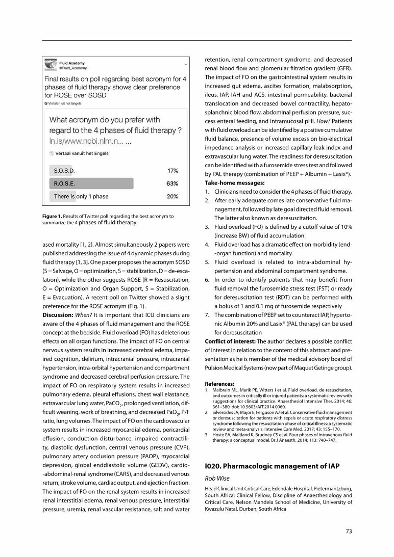
73
ased mortality [1, 2]. Almost simultaneously 2 papers were published addressing the issue of 4 dynamic phases during fluid therapy [1, 3]. One paper proposes the acronym SOSD (S = Salvage, O = optimization, S = stabilization, D = de-esca-lation), while the other suggests ROSE (R = Resuscitation, O = Optimization and Organ Support, S = Stabilization, E = Evacuation). A recent poll on Twitter showed a slight preference for the ROSE acronym (Fig. 1). Discussion: When? It is important that ICU clinicians are aware of the 4 phases of fluid management and the ROSE concept at the bedside. Fluid overload (FO) has deleterious effects on all organ functions. The impact of FO on central nervous system results in increased cerebral edema, impa-ired cognition, delirium, intracranial pressure, intracranial hypertension, intra-orbital hypertension and compartment syndrome and decreased cerebral perfusion pressure. The impact of FO on respiratory system results in increased pulmonary edema, pleural effusions, chest wall elastance, extravascular lung water, PaCO2, prolonged ventilation, dif-ficult weaning, work of breathing, and decreased PaO2, P/F ratio, lung volumes. The impact of FO on the cardiovascular system results in increased myocardial edema, pericardial effusion, conduction disturbance, impaired contractili-ty, diastolic dysfunction, central venous pressure (CVP), pulmonary artery occlusion pressure (PAOP), myocardial depression, global enddiastolic volume (GEDV), cardio--abdominal-renal syndrome (CARS), and decreased venous return, stroke volume, cardiac output, and ejection fraction. The impact of FO on the renal system results in increased renal interstitial edema, renal venous pressure, interstitial pressure, uremia, renal vascular resistance, salt and water
retention, renal compartment syndrome, and decreased renal blood flow and glomerular filtration gradient (GFR). The impact of FO on the gastrointestinal system results in increased gut edema, ascites formation, malabsorption, ileus, IAP, IAH and ACS, intestinal permeability, bacterial translocation and decreased bowel contractility, hepato-splanchnic blood flow, abdominal perfusion pressure, suc-cess enteral feeding, and intramucosal pHi. How? Patients with fluid overload can be identified by a positive cumulative fluid balance, presence of volume excess on bio-electrical impedance analysis or increased capillary leak index and extravascular lung water. The readiness for deresuscitation can be identified with a furosemide stress test and followed by PAL therapy (combination of PEEP + Albumin + Lasix®).Take-home messages: 1. Clinicians need to consider the 4 phases of fluid therapy.2. After early adequate comes late conservative fluid ma-
nagement, followed by late goal directed fluid removal. The latter also known as deresuscitation.
3. Fluid overload (FO) is defined by a cutoff value of 10% (increase BW) of fluid accumulation.
4. Fluid overload has a dramatic effect on morbidity (end--organ function) and mortality.
5. Fluid overload is related to intra-abdominal hy-pertension and abdominal compartment syndrome.
6. In order to identify patients that may benefit from fluid removal the furosemide stress test (FST) or ready for deresuscitation test (RDT) can be performed with a bolus of 1 and 0.1 mg of furosemide respectively
7. The combination of PEEP set to counteract IAP, hyperto-nic Albumin 20% and Lasix® (PAL therapy) can be used for deresuscitation
Conflict of interest: The author declares a possible conflict of interest in relation to the content of this abstract and pre-sentation as he is member of the medical advisory board of Pulsion Medical Systems (now part of Maquet Getinge group).
References: 1. Malbrain ML, Marik PE, Witters I et al. Fluid overload, de-resuscitation,
and outcomes in critically ill or injured patients: a systematic review with suggestions for clinical practice. Anaesthesiol Intensive Ther. 2014; 46: 361–380. doi: 10.5603/AIT.2014.0060.
2. Silversides JA, Major E, Ferguson AJ et al. Conservative fluid management or deresuscitation for patients with sepsis or acute respiratory distress syndrome following the resuscitation phase of critical illness: a systematic review and meta-analysis. Intensive Care Med. 2017; 43: 155–170.
3. Hoste EA, Maitland K, Brudney CS et al. Four phases of intravenous fluid therapy: a conceptual model. Br J Anaesth. 2014; 113: 740–747.
I020. Pharmacologic management of IAP
Rob WiseHead Clinical Unit Critical Care, Edendale Hospital, Pietermaritzburg, South Africa; Clinical Fellow, Discipline of Anaesthesiology and Critical Care, Nelson Mandela School of Medicine, University of Kwazulu Natal, Durban, South Africa
Figure 1. Results of Twitter poll regarding the best acronym to summarize the 4 phases of fluid therapy
74
Anaesthesiol Intensive Ther 2017, Supplement 1
Learning objectives: Despite the relative elasticity of the abdominal compartment, the area is prone to increases in pressure caused by both surgical and medical patho-physiology. Resulting intra-abdominal hypertension (IAH) and abdominal compartment syndrome (ACS) are serious conditions associated with both local and systemic compli-cations. While surgical intervention is often required once abdominal compartment syndrome is established, medical management plays an important part in the prevention of the evolution of intra-abdominal hypertension to ACS. This lecture will review the pharmacological strategies available for the management of intra-abdominal pressure, how to optimise therapy, and where the future of medical mana-gement of IAP may take us. Introduction: Non-surgical management of intra-abdo-minal hypertension has previously been described under 5 headings: 1. Improvement of abdominal wall compliance. 2. Evacuation of intra-luminal contents. 3. Evacuation of abdominal fluid collections. 4. Optimisation of fluid administration. 5. Optimisation of systemic and regional perfusion.Pharmacologic management plays an important role in several of these aspects, particularly improving wall com-pliance and evacuation of intra-luminal contents. Additional targets for pharmacologic agents include reduction of the inflammatory injury resulting from raised IAP, and novel therapies that may offer the gastrointestinal tract protection.Discussion: The goal of instituting non-surgical strategies to patients developing IAH is to slow, or halt progression to ACS and decrease the extent of organ injury and dysfunc-tion. Several pharmacologic therapies have been included in the WSACS guidelines for the management of IAH/ACS and include: • Evacuation of intra-luminal contents: - Prokinetics, neostigmine and enemas; • Imrpovement of abdominal wall compliance:- Neuromuscular blocking agents,- Sedation and analgesia.Many of these strategies have been shown to aid in signi-ficant reductions in IAP and should form part of routine practice. Other pharmacological agents, such as choice of re-suscitation fluid, has only small studies to suggest particualr benefit, and this is an area that requires further research. In addition, several new medical treatments options are being researched, but currently do not have sufficient evidence for incorporation into everyday clinical practice. Take-home messages: 1. Routine and protocolised measurement of patients at
risk for IAH and ACS is required to first identify patients requiring the necessary pharmacological treatment strategies to reduce IAP.
2. Early initiation of non-surgical strategies should be em-ployed in patients with IAH and these include several pharmacological therapies.
3. Vitamin C, melotonin, octreotide, theophylline, all show interesting recent results that suggest a reduction in damage from oxidative process and free radicals, but insufficient to be introduced into routine practice. Tissue plasminogen activator may be useful in managing raised IAP from haematomas. Chinese traditional medicine also has interesting results, but insufficient to support routine use.
4. The type of fluid used to resuscitate patients at risk for developing IAH/ACS may influence outcome.
Conflict of interest: The author declares no possible conflict of interest in relation to the content of this abstract and presentation.
References: 1. Chang M, Tang H, Liu D, Li Y, Zhang L. Comparison of melatonin, hy-
pertonic saline, and hydroxyethyl starch for resuscitation of secondary intra-abdominal hypertension in an animal model. PLoS One. 2016; 11: e0161688. doi: 10.1371/journal.pone.0161688. eCollection 2016.
2. Leng Y, Li H, Bai Y, Ge Q, Yao G. Effect of traditional Chinese medicine on intra-abdominal hypertension and abdominal compartment syndrome: a systematic review and Meta-analysis. J Crit Care. 2016; 34: 24–29. doi: 10.1016/j.jcrc.2016.03.021.
3. Chang M, Li Y, Liu D, Zhang L, Zhang H, Tang H, Zhang H. Melatonin prevents secondary intra-abdominal hypertension in rats possibly thro-ugh inhibition of the p38 MAPK pathway. Free Radic Biol Med. 2016; 97: 192–203. doi: 10.1016/j.freeradbiomed.2016.06.001.
4. Hecker A, Hecker B, Hecker M, Riedel JG, Weigand MA, Padberg W. Acute abdominal compartment syndrome: current diagnostic and the-rapeutic options. Langenbecks Arch Surg. 2016 Feb; 401(1): 15-24. doi: 10.1007/s00423-015-1353-4. Epub 2015 Oct 30.
5. De Keulenaer B, Regli A, De Laet I, Roberts D, Malbrain ML. What’s new in medical management strategies for raised intra-abdominal pressure: eva-cuating intra-abdominal contents, improving abdominal wall compliance, pharmacotherapy, and continuous negative extra-abdominal pressure. Anaesthesiol Intensive Ther. 2015; 47: 54–62. doi: 10.5603/AIT.a2014.0065.
I021. Positioning to manage IAP
Bart de KeulenaerAssociate Professor, Department of Surgery, University of Western Australia, WA, Australia; Intensive Care Medicine, Fiona Stanley Hospital, Murdoch, WA, Australia
Learning objectives: 1. To review if the abdomen behaves as a hydraulic system
and simply follows the law of fluid physics.2. To review if body positioning affects intra-abdominal
pressure (IAP) measurement.3. To discuss if body positioning can help in the manage-
ment of raised intra-abdominal pressures.Introduction: If the abdomen behaves as a hydraulic sys-tem it would follow the laws of Pascal. Pascal’s law states that enclosed fluid transmits pressure equally in all direc-tions. However, in reality the abdominal contents remain a heterogeneous mix of solid, liquid and gaseous compo-nents, so maybe the pressure transmission is rather complex.
75
We have to consider three factors affecting abdominal pres-sure, gravity, uniform compression and shear deformation. Gravity produces hydrostatic pressure gradients throughout the abdomen, uniform compression produces a spatially homogenous change in pressure and shear deformation is associated with spatially inhomogeneous pressure gra-dients. Some reports have identified significant pressure differences between upper and lower abdomen suggesting the abdomen does at times not behave as a hydraulic sys-tem. However, in the impact of shear deformation on the measurement of IAP is probably not significant in the fully sedated mechanically ventilated patient with sepsis, capil-lary leak and a positive fluid balance. Discussion: Many studies have investigated the effects of body positioning on IAP measurement with the largest reported difference in positional pressure of 5 mm Hg at 30 and 7.4 at 45 degrees.This is mainly due to a gravitational effect of the abdominal contents on the bladder. Cheatham et al showed that in patients with pressures above 20 mm Hg that pressure dif-ference in different body positions becomes less significant, hence patient can remain in head of bed (HOB) elevation at 30 degrees. In general head of bed elevation increases IAP, trendelenberg position underestimates IAP, reverse tren-delenberg overestimate IAP and prone positioning also increases IAP. So the question “can we use body positio-ning to manage IAP” depends if the abdomen behaves as a hydraulic system or not. Head of bed position should not be used if the abdomen follows the principle of fluid phy-sics. The patient with impending abdominal compartment syndrome should be placed in the supine position. If shear deformation plays an important factor one should consider HOB elevation in certain clinical conditions as it might re-duce IAP in the upper abdomen.Take-home messages: 1. Body positioning significantly affects IAP measurements.2. The abdomen does not always behaves as a hydraulic
system.3. Supine position is better than HOB elevation with impe-
ding ACS, however if upper abdomen pathology HOB elevation might reduce IAP.
Conflict of interest: None to declare.
References: 1. Loring SH, Yoshino K, Kimball WR, Barnas GM. Gravitational and shear-
-associated pressure gradients in the abdomen. J Applied Phys. 1994; 77: 1175–1182.
2. Cheatham ML, De Waele JJ, De Laet I et al. Effect of body positioning on intra-abdominal pressure measurement. A multi centre analysis. Crit Care Med. 2009; 37: 2187–2190.
3. De Keulenaer BL, De Waele JJ, Powell B, Malbrain ML. What is a normal intra-abdominal pressure and how is it affected by positioning, body mass and PEEP? Intensive Care Med. 2009; 35: 969–976.
4. Cresswell AB, Jassem W, Srinivasan P et al. The effect of body position on compartmental intra-abdominal pressure following liver transplantation. Ann Intensive Care 2012; 2 (Suppl. 1): S12.
I022. Comprehensive approaches to the problem
Ari LeppaniemiChief of Emergency Surgery, Helsinki University Hospital, Finland
Learning objectives: 1. To list the most common reasons for open abdomen.2. To describe the available techniques for abdominal clo-
sure after open abdomen.3. To evaluate the appropriate timing for closure in
patients with open abdomen.4. To describe the tools for decision making for se-
lecting closure technique and timing.Introduction: There are two reasons to have open abdomen, either as a treatment method (part of damage control strategy, prevention or treatment of abdominal compartment syndrome) or as a complication or surgical sequelae (burst abdomen, loss of abdominal wall due to necrotizing infection or tumor resec-tion). Early management of open abdomen requires a method for temporary abdominal closure, of which there are several options. The aim with open abdomen is to close the wound (fascial layer and skin) as soon as possible to reduce the risk of complications related to the open abdomen (anastomotic dehiscence, enteric fistula) and to close the “catabolic drain”. Discussion: The two important things to consider when planning the closure after open abdomen are timing and availability of patient’s tissues for midline closure. Open ab-domens can usually not be closed after 2–3 weeks, in which case the most common approach is to resort to a planned hernia approach. If there is enough available skin, a “skin only” closure can be performed. If not, then the abdominal viscera can be covered with a split-thickness skin graft, accept a maturation period of 9–12 months followed by abdominal wall reconstruction. The main techniques for abdominal wall reconstruction, either at the early stage or following a planned hernia approach, include component separation, use of mesh, tissue flaps, or a combination of them. Availability to cover the repair with patient’s own skin and the presence of contamination (iatrogenic enteric lesions, closure of stoma, infected mesh) determine the most appropriate type of reconstruction method. When closing the fascia at midline, component separation can be used as an adjunct to decrease tension in the midline and facilitate fascial closure. It is also important to measure intra-abdominal pressure after any kind of abdominal wall reconstruction procedure to detect the possible develop-ment of quaternary abdominal compartment syndrome.Take-home messages: Aim for delayed fascial closure as early as possible.If not possible within 2-3 weeks, move to planned hernia strategy.
76
Anaesthesiol Intensive Ther 2017, Supplement 1
Component separation, mesh, tissue flaps or a combination of them are the most common techniques to be used for abdominal wall reconstruction after open abdomen.Availability if skin to cover the repair and potential con-tamination determine the most appropriate method for definitive closure.Conflict of interest: The author declares no possible conflict of interest in relation to the content of this abstract and presentation.
References: 1. Leppäniemi A. Laparostomy, why and when? Crit Care. 2010; 14: 216.2. De Waele JJ, Leppäniemi A. Temporary abdominal closure techniques. Am
Surg 2011; 77: S46–S50.3. Björck M, Kirkpatrick AW, Cheatham M, Kaplan M, Leppäniemi A, De
Waele JJ. Amended classification of the open abdomen. Scand J Surg. 2016; 105: 5–10.
4. Rasilainen S, Mentula P, Leppäniemi A. Components separation technique is feasible for assisting delayed primary fascial closure of open abdomen. Scand J Surg. 2016; 105: 17–21.
5. Leppäniemi A, Tukiainen E. Planned hernia repair and late abdominal wall reconstruction. World J Surg. 2012; 36: 511–515, doi: 10.1007/s00268-011-1177-5.
6. Leppäniemi A, Tukiainen E. Reconstruction of complex abdominal wall defects. Scand J Surg. 2013; 102: 14–19.
7. Tukiainen E, Leppäniemi A. Reconstruction of extensive abdominal wall defects with microvascular tensor fascia latae flap. Br J Surg. 2011; 98: 880–884.
8. Coccolini F, Biffl W, Catena F et al. The open abdomen, indications, ma-nagement and definitive closure. World J Emerg Surg. 2015; 10: 32, doi: 10.1186/s13017-015-0026-5.
9. Rasilainen S, Mentula P, Leppäniemi A. Components separation technique is feasible for assisting delayed primary fascial closure of open abdomen. Scand J Surg. 2016; 105: 17–21.
10. Kirkpatrick AW, Nickerson D, Roberts DJ et al. Intra-abdominal hyper-tension and abdominal compartment syndrome after abdominal wall reconstruction: quaternary syndromes? Scand J Surg. 2017; 106: 97–106, doi: 10.1177/1457469916660036.
I023. Management of enteroatmospheric fistulas: from initial conservative treatment to definitive surgery
Daniel WainsteinMember of Surgical Argentinian Academy; Staff Surgeon of Pirova-no Hospital, Buenos Aires, Argentina
Learning objectives: To review the main characteristics of enteroatmospheric fistulas (EAF). To review the highlights of enterocutaneous fistulas (ECF) management. To show our specific local conservative and surgical treatment strategy on EAF’s.Introduction: The Open Abdomen procedure (OA) is a the-rapeutic approach that has become widespread in recent years mainly in the prevention and treatment of abdominal compartment syndrome, as a damage control strategy for abdominal trauma, and in the management of severe intra--abdominal sepsis. However, this method can also cause complications, enteroatmospheric fistulas (EAFs) being among one of the most feared. This serious postoperative complication is a kind of enterocutaneous fistula (ECF) that discharges the enteric content onto an open abdominal
wound. The average incidence rate is estimated at 20% (range 4–75%), while mortality has been reported between 5% and 60 %. The main purpose of this presentation is to describe the strategy and procedures implemented in the management of such postoperative complications and to show a local conservative treatment specifically designed for patients with EAFs.Discussion: The general principles of the ECF treatment must be adapted to the EA ones: correction of electrolyte imbalance, septic sources control, nutritional support and output control. Regarding this, the following criteria should be considered, 1) the intake of fluids and nutrients should be enough to balance all the losses; 2) the leakage should neither be partially retained as an intermediate cavity nor contact with superficial tissues; 3) effluent reduction, to improve metabolic and nutritional handle, simplifies wound management and increases the chance of spontaneous closure. Even though each patient would require individual treatment, we have managed most of EAF in 2 steps. The first one, up to the first 15 to 30 postoperative days, when viscera are more exposed and unprotected. The objecti-ve is: abdominal containment, prevent new fistulas, avoid dermatitis and wound infection and enteric fluid aspira-tion. The treatment is PTN + Fast + octreotide selectively, mesh retrieve and vacuum aspiration system. The second step, starting beyond such period, once a strong layer of granulation tissue on the viscera has been developed and the risk of a new fistula is less. The goal is to reduce the wound size and temporary fistula closure. The treatment, progression from PN to EN or oral if it were feasible and fistula’s temporary occlusion by vacuum. Reconstructive surgery should be indicated when, 1) the patient reaches clinical and nutritional recovery; 2) detainment in local he-aling process is observed; 3) enough time has gone by for a less risky surgical approach. Regarding the surgical treat-ment, we usually apply similar strategy and a technique for most cases, incision surrounding the wound, release and in block resection of the fistula and adjacent intestine together with the granulation tissue, anastomosis on healthy bowel. Finally, abdominal wall closure with its own tissue and if it is not possible, which happens frequently, closure with absorbable prosthesis. Take-home messages: • Strategic and multidisciplinary approach in the hands of
groups specialized in the management of the EAF opti-mize the results in terms of healing and mortality.
• Conservative treatment in two consecutive steps by aspi-ration and occlusive vacuum are practical and provide a good recovery to reach spontaneous closure or to a suc-cessful definitive surgery.
• The recovery of nutritional status, essential for a successful treatment, is complex but feasible in most cases thro-
77
ugh an adequate tactic. For this it is very important the indication and progression of the AE without losing the fistula’s control.
• Prolongation of conservative treatment is key to achieving spontaneous closure of the fistula in selected cases and, in most of the remaining patients, a successful surgical repair.
• A systematized surgical technique, with the lowest num-ber of anastomosis as possible and prioritizing the closure of the abdominal wall with own tissues, would be the most recommended surgical strategy.
Conflict of interest: The author declares no possible conflict of interest in relation to the content of this abstract and presentation.
References: 1. Majercik M, Kinikini M, White T. Enteroatmospheric fistula: from soup
to nuts.2. Nutr Clin Pract. 2012; 27: 507–512. 3. Davis K, Johnson EK. Controversies in the care of the enterocutaneous
fistula. Surg Clin North Am. 2013; 93: 231–250.4. Wainstein, DE, Tüngler V, Ravazzola C, et al. Management of external small
bowel fistulae: challenges and controversies confronting the general surgeon. Int J Surg. 2011; 9: 198–203.
5. Di Saverio S, Tarasconi A , Walczak D et al. Classification, prevention and management of entero-atmospheric fistula: a state-of-the-art review. Langenbecks Arch Surg. 2016; 401: 1–13.
6. Fernandez ER, Cornalo AO, Gonzalez V et al. Newapproach in the tre-atment of postoperative enterocutaneous fistulas. Rev Argent Cirug. 1992; 62: 117–127.
7. Wainstein DE, Fernandez E, Gonzalez D et al. Treatment of high-output enterocutaneous fistulas with a vacuum-compaction device. A ten-year experience. World J Surg. 2008; 32: 430–435.
8. Wainstein D, Sisco P, Deforel M et al. Systematic and specific treatment of patients with enteroatmospheric fistulas: from initial conservative treatment to definitive surgery. Surg Tech Int. 2015; XXXVIII: 73–80.
9. Wainstein DE, Manolisi H, Delgado M et al. Complex external small bowel fistulas: Reconstructive surgery of the digestivetract. Rev Argent Cirug. 2010; 99: 40–53.
10. Lynch AC, Delaney C, Senagore A et al. Clinical outcome and factors predictive of recurrence after enterocutaneous fistula. World J Surg. 2004; 240: 825–831.
I024. Diuresis and renal replacement therapy to manage IAP
Jan de WaeleIntensivist, Ghent University Hospital, Gent, Belgium
Learning objectives: 1. To describe the contribution of fluid overload to IAH
and ACS.2. To understand the mechanisms of diuresis and RRT in
the management of patients with IAH and ACS.3. To identify indications for diuresis and ultrafiltration in
patients with IAH and ACS.Introduction: Fluid (over)resuscitation has been identified as a contributor to IAH and ACS in critically ill patients. The-refore, fluid removal through diuresis using frusemide or analogues, or ultrafiltration using renal replacement therapy
could be simple and convenient non-surgical strategy to reduce the IAP. Discussion: Achieving a negative fluid balance is incre-asingly recognized as associated with improved outcomes but the role of IAH in this observation remains unclear. Several reports have described the effect of administe-ring diuretics or fluid removal through RRT, but mostly this has been described in patients after massive fluid resuscitation, for either trauma, burns or septic shock. The reduction in IAP is most often modest and may take several hours which limits its use in the context of abdo-minal compartment syndrome. The data documenting the impact of these interventions remain limited and as resuscitation strategies are changing, its role may be more limited in 2017 for treatment. Moreover, using diuretics in patients with established acute kidney injury may likely be ineffective. In patients with decompensated heart failure, elevated IAP was associated with diuretic resistance. For prevention though, a correct fluid balance management is important and diuretics or ultrafiltration may be a tool to achieve this.Take-home messages: 1. Fluid overresuscitation contributes to IAH and ACS but
its importance is decreasing.2. Fluid removal trough diuresis and ultrafiltration may
have little or no impact in most patients with IAH and ACS; in selected patients, there may be a role for this, but an acute effect should not be expected.
3. Aiming for a negative fluid balance after initial resuscitation may decrease IAP and improve outcomes.
Conflict of interest: The author declares no possible conflict of interest in relation to the content of this abstract and presentation.
References: 1. Kula R, Szturz P, Sklienka P, Neiser J. Negative fluid balance in patients
with abdominal compartment syndrome — case reports. Acta Chir Belg. 2008; 108: 346–349.
2. Oda S, Hirasawa H, Shiga H et al. Management of intra-abdominal hy-pertension in patients with severe acute pancreatitis with continuous hemodiafiltration using a polymethyl methacrylate membrane hemo-filter. Ther Apher Dial. 2005; 9: 355–361.
3. Kirkpatrick AW, Roberts DJ, De Waele J et al. Intra-abdominal hyperten-sion and the abdominal compartment syndrome: updated consensus definitions and clinical practice guidelines from the World Society of the Abdominal Compartment Syndrome. Intensive Care Med. 2013; 39: 1190–1206.
4. Nguyen VQ, Gadiraju TV, Patel H, Park M, Le Jemtel TH, Jaiswal A. Intra-ab-dominal hypertension: an important consideration for diuretic resistance in acute decompensated heart failure. Clin Cardiol. 2016; 39: 37–40.
5. De Keulenaer B, Regli A, De Laet I, Roberts D, Malbrain ML. What’s new in medical management strategies for raised intra-abdominal pressure: eva-cuating intra-abdominal contents, improving abdominal wall compliance, pharmacotherapy, and continuous negative extra-abdominal pressure. Anaesthesiol Intensive Ther. 2015; 47: 54–62. doi: 10.5603/AIT.a2014.0065.
6. Regli A, De Keulenaer B, De Laet I, Roberts D, Dabrowski W, Malbrain ML. Fluid therapy and perfusional considerations during resuscitation in critically ill patients with intra-abdominal hypertension. Anaesthesiol Intensive Ther. 2015; 47: 45–53. doi: 10.5603/AIT.a2014.0067.
78
Anaesthesiol Intensive Ther 2017, Supplement 1
I025. Gut mucosal injury from IAH
Patrick MurphyDepartment of General Surgery, London Health Sciences Center, London, Ontario, Canada
Learning objectives: Consolidate the modern evidence of gut mucosal injury in the critically ill. This lecture will review the concept of elevated intra-abdominal pressure (IAP) and ischemia-reperfusion injury that ultimately leads to multi--organ dysfunction and sepsis in the critically ill. Introduction: The gut represents > 300 m2 of surface area exposed to the environment. Described as “the motor” which drives multiple organ failure the gut acts as an im-mense reservoir of bacteria which is incredibly sensitive to changes in blood flow, particularly ischemia-reperfusion (IR) injury. Clinical intra-abdominal hypertension can be susta-ined or fluctuant resulting in varying periods of ischemia and reperfusion. Discussion: Nearly 50% of critically ill patients are diagno-sed with IAH in modern series. Splanchnic hypoperfusion initiates a cascade of events which undermine the native intestinal protections including barrier function, innate im-munity and alteration of intestinal microflora. Even short periods of ischemia increase mucosal permeability and epi-thelial apoptosis prompting the release of pro-inflammatory molecules and leading to bacterial translocation. When per-fusion is restored a second hit injury occurs from oxidative stress. Numerous animal models have demonstrated the IR associated with IAH even when blood pressure is maintained suggesting the gut is sensitive to not only the systemic but local environment. IAH therefore may be the missing piece for late development of multiple organ dysfunction and sepsis after the initial period of shock and resuscitation in critically ill patients.Take-home messages: 1. IAH is common and often unrecognized and untreated.2. IAH directly impacts gut mucosa even when patient
normotensive.3. Good animal evidence to suggest the gut is a source of
multi-organ dysfunction and sepsis4. Further study in critically ill patients is required.Conflict of interest: The author declares no possible conflict of interest in relation to the content of this abstract and presentation.
References: 1. Cheng J, Wei Z, Liu X et al. The role of intestinal mucosa injury induced
by intra-abdominal hypertension in the development of abdominal compartment syndrome and multiple organ dysfunction syndrome. Crit Care. 2013; 17: R283.
2. Kirkpatrick AW, Roberts DJ, De Waele J et al. Is intra-abdominal hyperten-sion a missing factor that drives multiple organ dysfunction syndrome? Crit Care. 2014; 18: 124.
3. Sertaridou E, Papaioannou V, Kolios G et al. Gut failure in critical care: old school versus new school. Ann Gastroenterol. n.d.; 28: 309–322.
4. Diebel LN, Dulchavsky SA, Wilson RF. Effect of increased intra-abdomi-nal pressure on mesenteric arterial and intestinal mucosal blood flow. J Trauma. 1992; 33: 45–48; discussion 48–49.
5. Diebel LN, Dulchavsky SA, Brown WJ. Splanchnic ischemia and bacterial translocation in the abdominal compartment syndrome. J Trauma. 1997; 43: 852–855.
I026. Mechanical open abdomen systems
Sam MinorAssociate Professor of Surgery and Critical Care Medicine; Dalhousie University; QEII Medical Director of Trauma
Learning objectives: The open abdomen is a life saving tool in the appropriate patient population. Once the reasons for having the abdomen open have resolved, it is essential to get it closed as quickly as possible. The length of time the abdomen is left open is associated with prolonged hospi-talization, development of intestinal fistula and mortality. Failure to close the fascia primarily results in a massive hernia that has a significant impact on the patient’s quality of life and may require a large surgery to reconstruct the abdomi-nal wall. This lecture will discuss the use of the Abdominal Reapproximation Anchor (ABRA) mechanical system in con-junction with the traditional ABthera, VAC therapy in closing the abdomen in patients at high risk for failure to achieve primary fascial closure with ABThera alone.Introduction: The use of the ABThera and other VAC tech-nologies as temporary abdominal closure has become stan-dard. However, approximately 40% of these patients will not achieve primary fascial closure [1, 2]. The ABRA system uses elastomers through the full thickness of the abdominal wall to progressively pull the fascial edges together. The ABRA system can be used in patients who appear to be at risk for not achieving primary fascial closure with ABThera alone.Discussion: A number of factors have been associated with higher risk for failing to achieve primary fascial closure after utilizing an open abdomen [3, 4]. The most important of which may be progressive evisceration and loss of domain following the first or second ABThera change. The applica-tion of the ABRA in these patients has been associated with a higher rate of primary fascial closure than all comers with ABThera alone [5–9]. Take-home messages:1. Patients treated with VAC temporary abdominal closure
alone are at risk for not achieving primary fascial closure.2. Mechanical systems such as the ABRA can increase the
rate of primary fascial closure in patients who are at high risk for primary non-closure.
References:1. Atema JJ. Systematic review and meta-analysis of the open abdomen.
World J Surg. 2015; 39: 912–925.2. Cirocchi R, Birindelli A, Biffl WL et al. What is the effectiveness of the
negative pressure wound therapy (NPWT) in patients treated with open
79
abdomen technique? A systematic review and meta-analysis. J Trauma Acute Care Surg. 2016; 81: 575–584. doi: 10.1097/TA.0000000000001126.
3. Roberts DJ, Zygun DA, Grendar J et al. Negative-pressure wound therapy for critically ill adults with open abdominal wounds: a systematic review. J Trauma Acute Care Surg. 2012; 73: 629–639.
4. Salman AE, Yetisir F, Aksoy M, Tokac M, Yildirium MB, Kilic M. Use of dynamic closure system in conjunction with vacuum-assisted closure therapy in delayed closure of open abdomen. Hernia 2012; 18: 99–104. doi: 10.1007/s10029-012-1008-0.
5. Verdam FJ, Dolmans DE, Loos MJ et al. Delayed primary closure of the sep-tic abdomen with a dynamic closure system. J Surg. 2011; 35: 2348–2355.
6. Reimer MW, Yelle YD, Reitsma B, Doumit G, Allen MA, Bell MSG. Manage-ment of open abdominal wounds using a dynamic fascial closure system. Can. J Surg. 2008; 51: 209–214.
7. Haddock C, Konkin DE, Blair NP. Management of the open abdomen with the abdominal reapproximation anchor dynamic fascial closure system. Am J Surg. 2013; 205: 528–533.
8. Okullo A, Siriwardhane M, Pang T, Lam V, Richardson A, Pleass H, Johnston E. The abdominal reapproximation anchor device: a single australian te-riary hospital experience. Surgical Innovation 2017; 24: 29–54.
9. Long K, Hamilton D, Davenport D, Bernard A, Kearney P, Chang P. A prospective, controlled evaluation of the abdominal reapproximation anchor abdominal wall closure system in combination with VAC therapy compared with vac alone in the management of an open abdomen. Am Surg. 2014; 80: 567–571.
I027. Introduction to the Concept of “GIPS” and “AIDS
Manu L.N.G. MalbrainICU and High Care Burn Unit Director, Ziekenhuis Netwerk Antwer-pen, ZNA Stuivenberg, Antwerp, Belgium; ICU Director, University Hospital Brussels (UZB), Jette, BelgiumProfessor at Vrije Universiteit Brussel (VUB), Faculty of Medicine, Brussels, Belgium
Learning objectives: To understand the deleterious effects of fluid overload in the setting of capillary leak and poor source control. To learn the definitions of global increased permeability syndrome (GIPS) and acute intestinal distress syndrome (AIDS). To describe the vicious cycle of fluid over-load. To describe the pathophysiology of shock after injury, resulting in ongoing increased inflammation and hyper-permeability leading to accumulation of second and third space fluids. To describe a 3-hit model of shock. To describe diagnostic tools for GIPS.Introduction: What? Global Increased Permeability Syn-drome (GIPS) is defined as a combination of a persistently positive cumulative fluid balance together with new onset organ failure. It is characterized by the absence of transgres-sion from Ebb to Flow phase of shock. Why? Some colleagues have described the presence of anasarca, peripheral edema as only being of cosmetic concern [1]. Less is true. Hypervole-mia leads to impaired oxygen diffusion by interstitial edema. Recently a systematic review and 2 meta-analysis have been performed that confirmed that fluid overload is indepen-dently associated with increased mortality in the setting of GIPS [2, 3]. Fluid resuscitation triggers a vicious cycle leading to intestinal edema, visceral swelling, increased intra-abdo-minal pressure (IAP), mesenteric vein compression, venous hypertension leading to administration f even more fluids [4]. Within this context, the gut can be considered as the
motor of multiple organ failure and the abdominal com-partment syndrome (ACS) can be considered as the acute respiratory distress syndrome (ARDS) of the gut. This mecha-nism of injury (i.e. increased vascular permeability) is widely recognized and accepted in the lung and kidneys, where it is classified as acute lung and kidney injury (ALI/AKI). The same pathological process occurs in the gut, but this concept is much slower to seep through. However, the role of the gut as the motor of organ dysfunction syndrome cannot be denied and difficulties in assessing gut function should not deter us from recognizing that concept [5]. Therefore the term AIDS, acute intestinal distress syndrome was coined to address these important issues [6]. However due to the confusion in relation to the consequences of HIV infection the term was afterwards changed into AGI, acute gastro-intestinal injury in analogy to AKI and ALI [7].Discussion: When? It is important that ICU clinicians are aware of the existence of GIPS, AIDS and AGI. Fluid over-load (FO) has deleterious effects on all organ functions and persistent FO in combination with organ failure has been defined as GIPS. After an insult (either trauma, sepsis, pancreatitis, bleeding, burns...) referred to as the first hit, the patient enters the Ebb phase of shock where fluids are lifesaving (R-phase within ROSE concept). Following ischemia and reperfusion distant organ injury may occur referred to as the second hit (O-phase within ROSE con-cept). Afterwards the patient may stabilize when source control (correct surgical intervention, correct antibiotics) has been properly performed (S-phase within ROSE con-cept). Finally, two things can happen, either the patient enters the Flow phase spontaneously and will mobilize the excess edema resulting in negative fluid balance (polyuria phase after AKI), or the patient may enter GIPS and remain in a state of persistent capillary leak and inflammation with FO. Here active deresuscitation may be advocated (E-phase within ROSE concept). How? Patients with GIPS in need for deresuscitation can be identified by an increase in cumulative fluid balance, volume excess, ECW/ICW ratio (bio-electrical impedance analysis), capillary leak index and extravascular lung water. The readiness for deresu-scitation can be identified with a furosemide stress test and followed by PAL therapy (combination of PEEP + Al-bumin + Lasix®).Take-home messages: 1. Fluid overload (FO) is defined by a cutoff value of 10%
(increase BW) of fluid accumulation and it is associated with increased morbidity (organ failure) and mortality.
2. Global increased permeability syndrome (GIPS) is defi-ned as FO without spontaneous transition from Ebb to Flow phase of shock.
3. Acute intestinal distress syndrome should now be called acute gastrointestinal injury (AGI).
80
Anaesthesiol Intensive Ther 2017, Supplement 1
4. Increased capillary leak index (CLI) and ECW/ICW ratio identify GIPS.
5. EVLWI may help to guide late conservative fluid management and late goal directed fluid removal.
Conflict of interest: The author declares a possible con-flict of interest in relation to the content of this abstract and presentation as he is member of the medical advisory board of Pulsion Medical Systems (now part of Maquet Getinge group).
References: 1. Pinsky MR. Hemodynamic evaluation and monitoring in the ICU. Chest.
2007; 132: 2020–2029.2. Malbrain ML, Marik PE, Witters I et al. Fluid overload, de-resuscitation,
and outcomes in critically ill or injured patients: a systematic review with suggestions for clinical practice. Anaesthesiol Intensive Ther. 2014; 46: 361–380. doi: 10.5603/AIT.2014.0060.
3. Silversides JA, Major E, Ferguson AJ et al. Conservative fluid management or deresuscitation for patients with sepsis or acute respiratory distress syndrome following the resuscitation phase of critical illness: a systematic review and meta-analysis. Intensive Care Med. 2017; 43: 155–170.
4. Duchesne JC, Kaplan LJ, Balogh ZJ, Malbrain ML. Role of permissive hypo-tension, hypertonic resuscitation and the global increased permeability syndrome in patients with severe hemorrhage: adjuncts to damage con-trol resuscitation to prevent intra-abdominal hypertension. Anaesthesiol Intensive Ther. 2015; 47: 143–55. doi: 10.5603/AIT.a2014.0052.
5. Malbrain ML, De laet I. It’s all in the gut: introducing the concept of acute bowel injury and acute intestinal distress syndrome. Crit Care Med. 2009; 37: 365–366.
6. Malbrain ML, De Laet I. AIDS is coming to your ICU: be prepared for acute bowel injury and acute intestinal distress syndrome. Intensive Care Med. 2008; 34: 1565–1569.
7. Reintam Blaser A, Malbrain ML, Starkopf J et al. Gastrointestinal function in intensive care patients: terminology, definitions and management. Recommendations of the ESICM Working Group on Abdominal Pro-blems. Intensive Care Med. 2012; 38: 384–394.
I028. Guidelines on open abdomen from the European Hernia Society
Frederik BerrevoetAssociate Professor of Surgery, Dept. of general and HPB Surge-ry and Liver Transplantation, Ghent University Hospital, Ghent, Belgium
Learning objectives: To be able to evaluate abdomen wall closure options for open abdomen patients according to current evidence.Introduction: The main goals of the guidelines are the mana-gement of abdominal wall closure in the context of an open abdomen, with primary fascial closure versus delayed fascial closure and following the classification scheme of Open Ab-domen complexity grades proposed by Björck et al. [1]. It is not the aim to cover all other aspects in the management of intraabdominal hypertension, abdominal compartment syndrome and open abdomen therapy or enteroatmosferic fistulae in connection with open abdomen.Discussion: Although evidence regarding the different options of open abdomen closure is still rather scarce, the formulated key questions may give an evidence based guidance on how to handle open abdomen abdominal wall closure in these complicated patient cohort according to GRADE methodology.
Take-home messages: 1. Continuous monofilament sutures following a SW: WL
ratio of 4 could be used as a good practice statement.2. Mesh augmentation could be advised after definitive
(primary) fascial closure, but mesh would be related with an increased incidence of SSO.
3. The use of techniques that separate the compo-nents of the abdominal wall in any way must be carefully and judiciously balanced.
4. Mesh bridging (inlay) should be avoided as a static closure due to the high incidence of fistula formation observed.
5. Dynamic closure techniques should be used in patients with Grade 1 or 2 open abdomen and the use of dynamic closure techniques is preferred over static closure techniques (where possible).
Conflict of interest: No possible conflict of interest in rela-tion to the content of this abstract and presentation.
Reference: 1. Björck M, Bruhin A, Cheatham M et al. Classification — important step to
improve management of patients with an open abdomen. World J Surg. 2009; 33: 1154−1157.
I029. IAP and its potential role in diseases of pregnancy
Diane SawchuckAdjunct Professor, Faculty Applied Sciences, UBC; Adjunct Associate Professor, Human and Social Development, University of Victoria (BC)
Learning objectives: 1. To understand the potential role of IAH in diseases of
pregnancy.2. To understand the potential role of IAH specific to pre-
-eclampsia. Introduction: The current scientific paradigm for the etio-logy of pre-eclampsia supposes that the placenta is the mediating factor, yet the disease and causative pathways remain an enigma. We propose that IAH in pregnancy is the underlying cause and that pre-eclampsia is mediated by the liver. Delivery cures pre-eclampsia not because of delivery of the placenta, as currently believed, but because of the mechanical effect of abdominal decompression. Discussion: By applying principles and physics and evi-dence from the critical care literature, we postulate that pre-eclampsia is caused by IAPP of P12 mm Hg, that when sustained or increasing, leads to hemodynamic shifts, in-testinal ischemia reperfusion injury, translocation of gram negative bacteria and lipopolysaccharide endotoxin from the maternal gut to the liver, systemic cytotoxic immune response, multi-organ dysfunction, and poly-compartment syndrome. We hypothesize that the placenta exposed to
81
TNF-α results in deficient trophoblastic invasion and spi-ral artery remodeling (< 22 weeks), elevated sFlt1, sEng and other antiangiogenic proteins, ischemia reperfusion injury, and apoptosis, foam cell and fibrin deposition. We challenge the current scientific paradigm and consider the role of IAHP in development of pre-eclampsia, eclampsia, intra-uterine growth restriction, and associated maternal and fetal mortality. Take-home messages: 1. The pathological pathways of IAH and abdominal com-
partment syndrome parallel the pathways observed in pre-eclampsia.
2. There is scientific and physical justification for inve-stigating the role of IAHP and its contribution to both maternal and fetal morbidity and mortality.
Conflict of interest: The author declares no possible conflict of interest in relation to the content of this abstract and presentation.
Reference: 1. Sawchuck D, Wittmann BK. Pre-eclampsia renamed and reframed: Intra-
-abdominal hypertension in pregnancy. Medical Hypotheses 2014; 83: 619–632.
I030. Overview of advanced surgical techniques to avoid IAH/ACS in AWR
Frederik BerrevoetAssociate Professor of Surgery, Dept. of general and HPB Surge-ry and Liver Transplantation, Ghent University Hospital, Ghent, Belgium
Learning objective: 1. To be able to distinguish between and judge the effi-
cient use of Botulinum Toxin Type A injection, preope-rative progressive pneumoperitoneum and transverse abdominis release (TAR) or combinations in managing complex abdominal wall reconstructions.
Introduction: Extensive Abdominal Wall Reconstruction (AWR) represents a surgical challenge due to lateral muscle retraction and possible loss of domain. The ideal objective is to perform a tension-free closure, with abdominal wall dynamic stability and optimizing aesthetic appearance. Different tools can be used to minimize tension, reduce wound morbidity and to prevent IAH and ACS. Ideally, these techniques should be considered and used if necessary; prior to surgery.Progressive preoperative pneumoperitoneum and the application of Botulinum Toxin Type A (BTA) in the lateral abdominal wall muscles have been reported to reduce po-stoperative incisional tension [1, 2]. Discussion: Different surgical techniques have been added to the toolkit of the abdominal wall or even general surgeon,
the preoperative planning of a giant incisional hernia with loss of domain remains essential. Up-to-date knowledge of these tools is necessary to prevent intraabdominal hyper-tension and secondary abdominal compartment syndrome.Take-home messages: 1. BTA is useful for large defects without excessive loss
of domain.2. PPP needs to be combined with BTA in case of loss of
domain > 20%.3. TAR is a relatively new, but very helpful and pro-
mising technique to cover lateral defects. However, in case of loss of domain this should always be combined with BTA and/or PPP.
Conflict of interest: No possible conflict of interest in rela-tion to the content of this abstract and presentation.
References: 1. Goni Moreno I. Rational treatment of chronic massive hernias and even-
trations; preparation of a patient with progressive pneumoperitoneum; original technic. Prensa Medica Argentina. 1951; 38: 10–21.
2. Ibarra-Hurtado TR, Nuno-Guzman CM, Echeagaray-Herrera JE, Robles--Velez E, de Jesus Gonzalez-Jaime J. Use of botulinum toxin type a before abdominal wall hernia reconstruction. World J Surg. 2009; 33: 2553–2556.
I031. Maternal positioning and it’s potential role in intra-abdominal pressure
Bernd Wittmann, Diane SawchuckMFM, Amara Women’s Health Clinic, Surrey, BC, Canada; Adjunct Professor, Faculty Applied Sciences, UBC; Adjunct Associate Pro-fessor, Human and Social Development, University of Victoria (BC)
Learning objectives: 1. To review the physical evidence for the maternal abdo-
men as a mathematical object.2. To consider the role of maternal positioning in pre-
gnancy.Introduction: Evidence suggests that intra-abdominal pres-sure in pregnancy (IAPP) alters with change in maternal position due to the physical laws of vector force [1–5]. Vec-tor force maternal position is one of four variables during pregnancy (along with physiologic increase in abdominal volume, decreased abdominal compliance and decreased atmospheric pressure), that we propose contributes to intra--abdominal hypertension in pregnancy (IAHP). By altering maternal position, we may alter the effects of IAHP. Discussion: Recent human data are very limited, and were only obtained in term pregnancies at the time of Cesa-rean delivery after spinal anesthesia. They confirmed that the antepartum IAP was significantly higher than the post-partum (pp) IAP, and that it was significantly lower in the left lateral than in the supine position. However, the only serial IAP measurements during pregnancy were obtained in 1913 by Paramore, who believed that IAH was the cau-
82
Anaesthesiol Intensive Ther 2017, Supplement 1
se of pre-eclampsia. He obtained serial measures of IAP in 26 incarcerated women, 24 of whom were pregnant, with 2 non-pregnant ones used for baseline data. Over 160 IAP measurements were obtained rectally between 9 — 40 weeks, and up to 6 weeks pp in 4 positions: standing, supine, left lateral, knee-chest. Analysis of Paramore’s raw data6 indicates that standing resulted in the highest, and left lateral in the lowest IAP, while knee- chest, the inversion of the abdominal orientation, actually resulted in a negati-ve IAP value7. Considering these data, the conclusion that knee-chest position may modify pathology associated with increased IAP is obvious.Take-home messages: 1. IAP in pregnancy is a neglected area of research, with
effectively no solid data published.2. Establishment of a scientific data base on all aspects of
the physiology of IAP in pregnancy and postpartum is urgently required, and essential for the understanding of a series of pathological conditions in pregnancy.
3. Considering the available data, there is compelling evi-dence that the inversion of the abdominal orientation may benefit patients with IAHP, specifically with preeclampsia.
Conflict of interest: The author declares no possible conflict of interest in relation to the content of this abstract and presentation.
References: 1. Malbrain ML, De Laet IE, De Waele JJ, Kirkpatrick AW. Intra-abdominal
hypertension: definitions, monitoring, interpretation and management. Best Pract Res Clin Anaesthesiol 2013; 27: 249–270.
2. Fuchs F, Bruyere M, Senat MV, Purenne E, Benhamou D, Fernandez H. Are standard intra-abdominal pressure values different during pregnancy? PLOS ONE 2013; 8: 1–4.
3. Al-Khan A, Shah M, Altabban M et al. Measurement of intra-abdominal pressure in pregnant women at term. J Reprod Med. 2011; 56: 53–57.
4. Abdel-Razeq SS, Campbell K, Funai EF, Kaplan LJ, Ozan Bahtiyar M. Nor-mative postpartum intra-abdominal pressure: potential implications in the diagnosis of abdominal compartment syndrome. Am J Obstet Gynecol. 2010; 203: 149e 1–4.
5. Chun R, Baghirzada L, Tiruta C, Kirkpatrick AW. Measurement of intra-ab-dominal pressure in term pregnancy: a pilot study. Int J Obstet Anesth. 2012; 21: 135–139.
6. Paramore RH. The intra-abdominal pressure in pregnancy. Obstetrical and Gynaecological Section. Proc R Soc Med. 1913; 6: 291–334.
7. Sawchuck D, Wittmann BK. Pre-eclampsia renamed and reframed: intra--abdominal hypertension in pregnancy. Medical Hypotheses 2014; 83: 619–632.
I032. Surgical simulation of the abdominal cavity: the abdominal component of the cut-suit
Anthony LaPorta
Learning objectives: Mass casualty events require deeply coordinated events with teams including first responders to the surgical teams themselves. This talk will describe training in a coordinated fashion with all interventions along the way.
Introduction: Open abdominal surgery and damage control surgery has become an art in itself far beyond the typical general surgery training. However because of new sanctions on the use of the animals for training, and because of societal barriers training as a true trauma unit has become more and more difficult. The use of a new exoskeleton known as the cut suit has allowed for training without silos and with true human interaction from the point of injury to recovery room. It enhances personal skills, and dramatically increasing team skills.Methods: The United States military especially the United States Marine and Navy I have utilized the device known as the cut suit to allow for continuous training. This simulator allows personal interaction with a site of injury, and allows the invasive technical maneuvers necessary to train first responders, emergency room teams, and operating room teams. This training has now been performed at multiple locations both in the US military and with civilian tactical teams. This training will be described including its usage in mass casualty event’s and full trauma team training.Results: Nearly 30 Army and Navy forward surgical teams have now undergone training utilizing this device. Mass casualty events in Denver including a 400 person mass casualty event, and mass casualty training in Vail Colorado for the world championship in skiing will be shown. The methodology has been proven with critical evidence of re-suscitation time and critical error measurements. Objective hormonal and heart rate variability measurements have already been studied in some of our learner groups and is now being studied on full surgical teams. In this brief talk, video showing the dramatic effect of the training on stress and operating team-training results will be given.Conflict of interest: No conflict of interest. These are not the opinions of the US Military.
References: 1. Hoang TN, Kang J, Siriratsivawong K et al. Hyper-realistic, team-centered
fleet surgical team training provides sustained improvements in perfor-mance J Surg Education. 2016; 73: 668–674.
2. Hoang T, LaPorta AJ, Wrenn K, Lucero A, Mullinax R, Ross D. Methods of team training when all members of the interdisciplinary team are experts. Proceedings of the 7th International Clinical Skills Conference. 2017: 97.
3. LaPorta AJ, McKee J, Hoang T, Mcbeth P, Gillman L, Kirkpatrick A. Stress inoculation: preparing outside the box in surgical resuscitation and edu-cation. Curr Trauma Rep. 2017; 3: 135. doi: 10.1007/s40719-017-0090-2
4. Kirkpatrick A, Tien H, LaPorta A et al. The marriage of surgical simulation and tele-mentoring for Damage Control Surgical training of operational first-responders. Can J Surg. 2015; 58: S10.
5. LaPorta AJ, Hoang T, Moloff A et al. How blended hyper-realism in the real and virtual worlds can better prepare surgeons. Medicine Meets Virtual Reality 21. Ed. James Westwood. Stud Health Technol Inform. 2014; 196: 233–237. IOS-Press 2014, 233.
6. Hoang, MD, FACS, CDR, MC, USN; Jeff Kang, MD, LCDR, MC, USN; Anthony J. LaPorta TN, Makler WI, Chalut C. Filling in the gaps of pre-deployment fleet surgical team training using a team-centered approach. J Special Operations Med. 2013 .
7. Lipshy K, LaPorta AJ. Leadership skills in the or, part iii: operating room crisis management leadership training: guidance for surgical team edu-cation. Bull Am Coll Surg. 2013.
83
I033. Posterior component release and laparoscopic approaches to decompress for IAH/ACS
Ari LeppaniemiChief of Emergency Surgery, Helsinki University Hospital, Finland
Learning objectives: 1. To describe the various options to treat IAH/ACS.2. To describe the minimally invasive surgical techniques
for decompression.3. To assess the available results of minimally inva-
sive decompression techniques.Introduction: The first line treatment of intra-abdominal hy-pertension (IAH) is conservative consisting of the reduction of gastrointestinal and intra-abdominal volumes including percutaneous drainage of ascites, increasing abdominal wall compliance and aiming for negative fluid balance. In full--blown abdominal compartment syndrome (ACS) surgical decompression is indicated if nonoperative management fails. In fulminant situations a standard full-thickness midli-ne laparostomy is the safest method although in patients with severe acute pancreatitis, a transverse midline incision can also be used. In some patients with borderline IAH not responsive to conservative management, less invasive sur-gical decompression techniques are available.Discussion: Most of the techniques that are potentially suitable for abdominal decompression are techniques used for abdominal wall reconstruction and include various mo-difications of the component separation technique or other release techniques of the abdominal wall musculature. Whe-reas there are numerous reports of their use to reconstruct abdominal wall defects, there is little evidence that they actually help to decrease intra-abdominal pressure (IAP) in patients with IAH/ACS. Subcutaneous linea alba fasciotomy procedure has been used to manage ACS in patients with severe acute pancreatitis, and it seems to reduce IAP suffi-ciently in about 50% of the patients. Originally the linea alba was divided with the help of laparoscopy but subsequently the fasciotomy can be completed openly through three short transverse incisions. Although the fasciotomy will inevitably result in an incisional hernia, its correction later on is quite simple. There is unpublished data of one patient where bilateral minimally invasive anterior component se-paration technique was used in an attempt to reduce IAP, but this has not been reproduced since then. Overall, with the improved nonoperative management of ACS, the role of minimally invasive techniques has diminished in the last decade.Take-home messages: 1. Nonoperative management of IAH/ACS should be tried
first.
2. Fulminant ACS usually requires formal full-thickness midline laparostomy for decompression.
3. Minimally invasive techniques for abdominal de-compression are available but their role is limited.
Conflict of interest: The author declares no possible conflict of interest in relation to the content of this abstract and presentation
References: 1. Kirkpatrick AW, Roberts D, De Waele J et al. Intra-abdominal hyperten-
sion and the abdominal compartment syndrome: updated consensus definitions and clinical practice guidelines from the World Society of the Abdominal Compartment Syndrome. Intensive Care Med. 2013; 39: 1190–1206. doi: 10.1007/s00134-013-2906-z.
2. Mentula P, Hienonen P, Kemppainen E, Puolakkainen P, Leppäniemi A. Surgical decompression for abdominal compartment syndrome in severe acute pancreatitis. Arch Surg. 2010; 145: 764–769.
3. Jones CM, Winder JS, Potochny JD, Pauli EM. Posterior component separa-tion with transversus abdominis release: Technique, utility, and outcomes in complex abdominal wall reconstruction. Plast Reconstr Surg. 2016; 137: 636–646. doi: 10.197/01.prs.0000475778.45783.e2.
4. Leppäniemi A, Mentula P, Hienonen P, Kemppainen E. Transverse laparostomy is feasible and effective in the treatment of abdominal compartment syndrome in severe acute pancreatitis. World J Emerg Surg. 2008; 3: 6.
5. Leppäniemi A, Hienonen P, Mentula P, Kemppainen E. Subcutaneous linea alba fasciotomy, does it really work? Am Surg. 2011; 77: 99–102.
6. Ouellet JF, Leppäniemi A, Ball CG, Cheatham M, D’Amours S, Kirkpatrick AW. Alternatives to formal abdominal decompression. Am Surg. 2011; 77: S51–S57.
I034. Abdominal simulators in training for far-forward damage control surgery in extreme environments
Andrew W. KirkpatrickProfessor of Surgery and Critical Care Medicine, Foothills Medical Cen-tre, Calgary, Alberta; Major, Canadian Forces Medical Services, Canada
Learning objectives: 1. To review the technical requirements to study interven-
tional techniques to address torso exsanguination using surgical simulators.
2. To review initial efforts examining open surgical techni-ques and percutaneous hemostatic foam insufflation to address torso exsanguination using surgical simulators
3. To review comparative differences in undertaking inte-rventional techniques to address torso exsanguination using surgical simulators in variable gravity.
4. To review initial efforts to mentor non-surgeons to undertake interventional techniques to address torso exsanguination using surgical simulators, guided by remote tele-medical guidance.
Introduction: Bleeding to death is the most potentially preventable cause of post-traumatic death, with most de-aths occurring in the pre-hospital environment, especially in austere and extreme environments. Training and research to address these catastrophic conditions is hampered by their criticality precluding novices to learn by trial and error and their occurrence outside of “business hours”. With less
84
Anaesthesiol Intensive Ther 2017, Supplement 1
clinical exposure every year high fidelity surgical simulators with realistic 3-D printed organs and a vascular system that can be perfused create ever more realistic training tools for surgeons and non-surgeons to learn and study torso exsan-guination. Extreme environments can also be utilized such as hyper-realistic settings with all the special effects a movie lot can provide and parabolic flight environments to vary the actual gravity that is present to challenge operators.Discussion: The Damage Control Surgery in Austere Envi-ronments Research Group (DCSAERG) collaborated with Strategic Operations (STOPS) a former movie lot and graphic design studio who utilizes the custom designed Cut-Suit surgical simulator as the central tool of hyper-realistic tra-ining scenarios in extreme environments. A customized model of torso exsanguination simulating a hepatic arterial injury was iteratively created and tested. Randomized Con-trol trial (RCT) thereafter examined the ability of Military Medics to perform damage control laparotomies with and with remote telementoring oversight by experienced surge-ons. Further studies with the same model were conducted onboard a Research Aircraft capable of parabolic flight to allow comparisons of surgical interventions in both normal gravity and weightlessness. The relative attributes of open damage control surgery and intra-corporeal hemostatic foam insufflation were assed.Take-home messages: 1. An inanimate anatomically correct perfused surgical si-
mulator can be used to greatly aid training and research in torso exsanguination.
2. Non-surgically trained military medics can be remotely mentored using similar technology to perform damage control surgery on such a simulator.
3. Trained surgeons can perform open damage control surgery on such a simulator in weightlessness with little performance degradation compared to normal gravity.
4. Further development of highly realistic perfused simulators should continue to aid in efforts to address exsanguination in extreme and austere environments.
Conflict of interest: The author has consulted for the Inno-vative Trauma Care and Acelity Corporations.
References: 1. Kirkpatrick AW, McAllister V, Dawe P et al. A Dynamic Canadian Forces
Medical Services/Canadian Space Agency Sponsored Program to facilitate real-time tele-mentoring/tele-diagnosis to address exsanguination in austere environments. J Trauma Acute Care Surg (in press).
2. Kirkpatrick AW, McKee JL, Tien H et al. Damage control surgery in weigh-tlessness: a comparative study of simulated torso hemorrhage control comparing terrestrial and weightless conditions. J Trauma Acute Care Surg. 2017; 82: 392–399.
3. Kirkpatrick AW, Tien H, LaPorta AT et al. The marriage of surgical simulation and telementoring for damage-control surgical training of operational first responders: a pilot study. J Trauma Acute Care Surg 2015; 79: 741–747.
4. Kirkpatrick AW, LaPorta A, Brien S et al. Technical innovations that may facilitate real-time telementoring of damage control surgery in austere environments: a proof of concept comparative evaluation of the im-portance of surgical experience, telepresence, gravity and mentoring in the conduct of damage control laparotomies. Can J Surg. 2015; 58 (3 Suppl 3): S88–90.
I035. Biomesh selection during hernia repair
Bruno PereiraProfessor of Surgery, Department of Surgery, University of Cam-pinas, Brazil
1. Learning objectives: 2. To review the types of Biomesh available in the market.3. Elucidate when Biomesh could demonstrate better
performance.Introduction: The use of a prosthetic material to streng-then or replace host tissue during hernia repair is today practically mandatory. The more recently developed bio-logical materials contain animal or human tissues, and when used to repair an abdominal wall defect, behave similarly to autologous tissues. These bio- logical meshes are composed of denatured acellular collagens and are gradually absorbed in the host. Besides repairing damaged tissue, biomeshes induce the formation of a well- organi-zed neotissue with characteristics similar to those of the healthy tissue that allows for adequate angiogenesis. Ho-wever, other authors have reported some inconveniences, including adverse effects that have been described after implantation of xenograft biologic meshes, including acute mechanical failure, mesh disintegration, and poor mesh integration. Several biological prostheses are currently available on the market. These biomeshes are classified according to the origin of the collagens from which they are made, xenogeneic (animal) or allogeneic (human), the type of tissue matrix used, dermis, pericardium, or intestinal submucosa, and whether this tissue matrix is crosslinked or not. Discussion: Bioprostheses are a good option for tissue--engineering since they promote the regeneration of host tissue at the repair site, giving rise to new connective tissue. This process involves both the induction of growth factors and synthesis of new extracellular matrix constituents. It has been well established that the degradation and absorption of an implanted bioprostheses will condition the tissue remodeling process in that area and therefore the develop-ment of new tissue. The triple-helix collagen cross linking procedures that biomeshes are subjected to confer them resistance to degradation. This is important since rather than rapidly degrading, a prosthetic mesh should remain stable until it becomes fully incorporated in the host tissue. Otherwise, its mechanical resistance will be compromised. Several experimental studies have tried to elucidate the behavior of these porcine prostheses. Those examining the biocompatibility, immunogenicity, and host tissue in-corporation of porcine biomeshes have served to explain the potential benefits of these biomaterials over synthetic materials. Despite these characteristics of biomeshes, few studies have so far addressed the behavior of the different biological prosthetic implants currently on the market in
85
terms of the host tissue response that elicit and their degra-dation or absorption once implanted in the host. Moreover, no study has yet examined the effect on these factors of the site of implantation.Take-home messages: 1. Biomesh may be useful in contaminated scenarios.2. Using biomesh as a bridge to fascia synthesis may have
insufficient performance.3. Long-therm follow up of patients showed increase
of laxity.Conflict of interest: The author has consulted for the Acelity Corporations, Convatec and Hartmann Bace Healthcare.
References: 1. Pascual G, Rodrıguez M, Sotomayor S, Moraleda E, Bellon JM. Effects
of collagen prosthesis cross-linking on long-term tissue regeneration following the repair of an abdominal wall defect. Wound Repair Regen. 2012; 20: 402–413.
2. Pascual G, Sotomayor S, Rodrıguez M, Perez-Kholer B, Bellon JM. Repair of abdominal wall defects with biodegradable laminar prostheses: Po-lymeric or biological? PLoS One 2012; 7: e52628.
3. Bryan N, Ashwin H, Smart N, Bayon Y, Scarborough N, Hunt JA. The innate oxygen dependant immune pathway as a sensitive parameter to predict the performance of biological graft materials. Biomaterials 2012; 33: 6380–6392.
4. Ditzel M, Deerenberg EB, Grotenhuis N et al. Biologic meshes are not su-perior to synthetic meshes in ventral hernia repair: an experimental study with long-term follow-up evaluation. Surg Endosc. 2013; 27: 3654–3662.
I036. Long-term absorbable meshes
Frederik BerrevoetAssociate Professor of Surgery, Dept. of general and HPB Surge-ry and Liver Transplantation, Ghent University Hospital, Ghent, Belgium
Learning objectives: 1. To get familiar with a sub-set of meshes that are long-
-term resorbable with initial properties similar to current synthetics.
2. To identify the pro- and cons of this mesh-family.Introduction: Long-term mesh related complications are increasingly reported as using synthetic meshes has become the gold standard for almost every abdominal wall defect. Furthermore, the use of synthetics in contaminated fields remains controversial. It is known that the early wound--healing period is critical for a lasting repair of any hernia or laparotomy. To limit the negative consequences of using synthetic materials in a human body, synthetic resorbable materials with a long-term strength retention profile, pre-serving 50% of the initial breaking strength for 6 months or longer have been introduced. Discussion: Abdominal wall reconstruction and hernia re-pair in the contaminated setting remains an area of need and intense study as well as possibilities to reduce mesh complications. Although biologic mesh use has resulted in decreased morbidity from the chronic complications due
to infected mesh, these materials come with only moderate durability and at substantial financial cost. Slowly absor-bable biosynthetic implants as a class of hernia meshes represent a possible cost-effective solution. Rigorous, well--designed clinical studies are lacking for most products and are clearly warranted to further evaluate these materials to define their appropriate application. In the final analysis, if their performance is determined to be equivalent to that of tissue-derived biologic meshes, given their lower cost, reduced handling requirements, and relatively predictable behavior, their use could be supported for these difficult clinical situations. Take-home messages: 1. Slowly absorbable biosynthetic implants as a class of
hernia meshes represent a possible cost-effective solu-tion in contaminated fields.
2. These ‘semi-synthetics’ might diminish longterm sequ-ellae of current synthetics.
3. Longer-term data is needed on durability, biocompatibi-lity, and appropriate indications for each of the products.
Conflict of interest: Speaker’s bureau and educational grant from Novus Scientific.
I037. The role of social media and FOAM in critical care education
Manu L.N.G. MalbrainICU and High Care Burn Unit Director, Ziekenhuis Netwerk Antwer-pen, ZNA Stuivenberg, Antwerp, Belgium; ICU Director, University Hospital Brussels (UZB), Jette, BelgiumProfessor at Vrije Universiteit Brussel (VUB), Faculty of Medicine, Brussels, Belgium
Learning objectives: To learn about the different types of social media. To describe the anatomy of a Tweet. To explain the FOAM movement (Free Open Access Medical education). To define SMICC and SMACC (social media in and critical care). To understand whether or not social media can replace peer review. To describe the symplur health-care hashtag project and the IFAD case study. To describe possible dangers.Introduction: FOAM is a collection of resources, a commu-nity and an ethos. The FOAM community spontaneously emerged from the collection of constantly evolving, collabo-rative and interactive open access medical education reso-urces being distributed on the web with one objective — to make the world a better place [1]. FOAM is independent of platform or media — FOAM is a personalised continuously expanding database of resources for medical education: it includes blogs, podcasts, tweets, Google hangouts, online videos, text documents, photographs, facebook groups, and a whole lot more [2].
86
Anaesthesiol Intensive Ther 2017, Supplement 1
Discussion: Traditional media are organized by few sources for many receivers. The quality is mediated by publishers and training is required. It is expensive to publish and access is limited. Publication process takes time and the publication is permanent once published. Social Media (SoMe) on the other hand has many sources (eg SMICC and SMACC) for many receivers. Quality is mediated by participants and the-re is no training. It is cheap or free to publish with unlimited accessibility. Publication is immediate and SoMe are flexible even after publication. By sharing our specific competencies, protocols and experiences we can shift to a new online lear-ning paradigm that will carry medical education via internet 2.0 to a new era. Peer review seems to be broken as half-mil-lion papers published per year (> 1 per minute). The question therefore arises whether FOAM could possibly replace peer review? The answer is maybe: FOAM ignores traditional hierarchy, it is free and has equitable access 24/7, it crosses professional boundaries, it is multi-national, transparent, robust and finally FOAM is apolitical. Recently because of the increase in and awareness surrounding FOAM a social media index (SMI) has been suggested [3] while others suggested the so-called Kardashian index (KI) [4]. The SMI enables to assess the impact and quality of FOAM resour-ces, and enables educators to receive scholarly credit and learners to identify respected resources. The KI a measure of discrepancy between a scientist’s social media profile and publication record based on the direct comparison of numbers of citations and Twitter followers. Possible dangers of FOAM are related to the reliability and correctness of the information provided. Recently a quality label for medical websites has been launched (the so called HONcode by the
Health on the Net foundation https: //www.healthonnet.org/HONcode/Conduct.html). Another danger related to SoMe and FOAM is reductive education: First we read the textbook, then we just read the chapter, then just the paper, then just the abstract and now we just read the Tweet.Take-home messages: 1. FOAM stand for Free Open Access Medical education.2. The FOAM movement is steadily increasing and repla-
cing traditional media and traditional sources for me-dical knowledge dissemination.
3. In the future FOAM has a possibility to replace peer review.
4. The SMI and KI have been developed to quantify SoMe scientific output.
5. Quality of FOAM content needs to be validated. 6. Reductive education and fake news are potential dan-
gers.Conflict of interest: The author declares no possible conflict of interest in relation to the content of this abstract and presentation.
References: 1. Nickson C. FOAM — Free Open Access Meducation — Medical education
for anyone, anywhere, anytime. https: //lifeinthefastlane.com/foam/.2. Cadogan M, Thoma B, Chan TM, Lin M. Free Open Access Meduca-
tion (FOAM): the rise of emergency medicine and critical care blogs and podcasts (2002-2013). Emerg Med J. 2014; 31(e1): e76–77. doi: 10.1136/emermed-2013-203502.
3. Thoma B, Sanders JL, Lin M, Paterson QS, Steeg J, Chan TM. The so-cial media index: measuring the impact of emergency medicine and criti-cal care websites. West J Emerg Med. 2015; 16: 242–249. doi: 10.5811/we-stjem.2015.1.24860.
4. Hall N. The Kardashian index: a measure of discrepant social media profile for scientists. Genome Biol. 2014; 15: 424. doi: 10.1186/s13059-014-0424-0.