Emotional Voice Areas: Anatomic Location, Functional Properties, and Structural Connections Revealed...
10
Cerebral Cortex January 2012;22:191--200 doi:10.1093/cercor/bhr113 Advance Access publication May 30, 2011 Emotional Voice Areas: Anatomic Location, Functional Properties, and Structural Connections Revealed by Combined fMRI/DTI Thomas Ethofer 1,2 , Johannes Bretscher 1 , Markus Gschwind 2 , Benjamin Kreifelts 1 , Dirk Wildgruber 1 and Patrik Vuilleumier 2,3 1 Department of General Psychiatry, University of Tu¨ bingen, 72076 Tu¨ bingen, Germany, 2 Department of Neuroscience and Clinic of Neurology, Laboratory for Behavioral Neurology and Imaging of Cognition, Medical School and 3 Department of Neuroscience, Neuroscience Center, University of 1211 Geneva Switzerland Address correspondence to Thomas Ethofer, Department of General Psychiatry, University of Tu¨bingen, Osianderstraße 24, 72076 Tu¨bingen, Germany. Email: [email protected]. We determined the location, functional response profile, and structural fiber connections of auditory areas with voice- and emotion-sensitive activity using functional magnetic resonance imaging (fMRI) and diffusion tensor imaging. Bilateral regions responding to emotional voices were consistently found in the superior temporal gyrus, posterolateral to the primary auditory cortex. Event-related fMRI showed stronger responses in these areas to voices-expressing anger, sadness, joy, and relief, relative to voices with neutral prosody. Their neural responses were primarily driven by prosodic arousal, irrespective of valence. Probabilistic fiber tracking revealed direct structural connections of these ‘‘emotional voice areas’’ (EVA) with ipsilateral medial geniculate body, which is the major input source of early auditory cortex, as well as with the ipsilateral inferior frontal gyrus (IFG) and inferior parietal lobe (IPL). In addition, vocal emotions (compared with neutral prosody) increased the functional coupling of EVA with the ipsilateral IFG but not IPL. These results provide new insights into the neural architecture of the human voice processing system and support a crucial involvement of IFG in the recognition of vocal emotions, whereas IPL may subserve distinct auditory spatial functions, consistent with distinct anatomical substrates for the processing of ‘‘how’’ and ‘‘where’’ information within the auditory pathways. Keywords: connectivity, diffusion, probabilistic fiber tracking, prosody, superior temporal gyrus Introduction Speech melody (prosody) provides important cues about the emotional state of our conversational partners during social interactions. Previous functional magnetic resonance imaging (fMRI) studies have provided consistent evidence that the middle part of the human associative auditory cortex holds a temporal voice area (TVA; Belin et al. 2000; Ethofer, Kreifelts, et al. 2009), which exhibits stronger responses to utterances spoken in an emotional rather than neutral tone (Grandjean et al. 2005; Ethofer, Anders, Wiethoff, et al. 2006; Beaucousin et al. 2007). These fMRI results converge with electrophysio- logical data demonstrating a differentiation of neutral and emotional vocal stimuli over temporal cortical areas as early as 200 ms after stimulus onset (Paulmann and Kotz 2008). The increases observed in this area occur for a broad range of emotional categories and cannot be explained on the basis of single acoustic parameters (Wiethoff et al. 2008). Furthermore, an increased activation to voices spoken in alluring prosody by speakers of the opposite (but not same) sex indicates that these modulations reflect a particular sensitivity to the behavioral relevance of voices for the listener (Ethofer et al. 2007). However, the recognition and appraisal of affective signals in voices implicate widespread brain networks beyond auditory areas. Explicit judgment of prosody type or intensity consistently activates the inferior frontal gyrus (IFG; e.g., Buchanan et al. 2000; Wildgruber et al. 2005; Ethofer, Anders, Erb, et al. 2006; Ethofer, Kreifelts, et al. 2009). In addition, lesions of the IFG lead to impaired comprehension of vocal emotions (Hornak et al. 1996, 2003; Rymarczyk and Grabowska 2007). Accordingly, models of prosody processing have long postulated a role for important interactions between temporal and frontal regions of the right hemisphere, similar to the networks associated with language functions in the left hemisphere (Ross and Mesulam 1979). In the latter case, strong connections between the posterior part of the left superior temporal gyrus (STG) and left IFG via the arcuate fasciculus have been identified using diffusion tensor imaging (DTI) in humans (Catani et al. 2005). Furthermore, a strong left--right asymmetry for connections between phonological areas in STG and frontal areas has been demonstrated, with more consistent fiber bundles in the left than right hemisphere (Glasser and Rilling 2008). However, the anatomical substrates mediating interactions between temporal and frontal cortices for comprehension of emotional prosody still remain unknown. This study was designed to clarify the following 3 questions: First, we wanted to determine the precise location of the emotion-sensitive auditory cortices in relation to other anatomical structures, particularly the primary auditory cortex (PAC) and the voice-sensitive associative auditory cortex in the TVA. Voice-sensitive areas were defined using a validated fMRI localizer experiment (Belin et al. 2000). Second, we wanted to systematically investigate any differential influence of valence and arousal on emotional responses in these areas. Third, we aimed at determining the structural and functional connectivity between the emotion-sensitive auditory areas and other distant regions in the brain. To this end, we combined fMRI and DTI measures in the same volunteers. Sentences spoken in neutral and various emotional categories were presented during an event-related fMRI experiment with a 2 3 2 factorial design including valence (positive vs. negative emotions) and arousal (low- vs. high-arousing emotions) as within-subject factors. The fMRI activations were subsequently employed as seed areas to examine not only how the network underlying prosody perception is modulated by the emotional content of voices (effective connectivity) but also how areas within this network are structurally interconnected (using probabilistic Ó The Author 2011. Published by Oxford University Press. All rights reserved. For permissions, please e-mail: [email protected] at University of Geneva on August 10, 2012 http://cercor.oxfordjournals.org/ Downloaded from
Transcript of Emotional Voice Areas: Anatomic Location, Functional Properties, and Structural Connections Revealed...

Cerebral Cortex January 2012;22:191--200
doi:10.1093/cercor/bhr113
Advance Access publication May 30, 2011
Emotional Voice Areas: Anatomic Location, Functional Properties, and StructuralConnections Revealed by Combined fMRI/DTI
Thomas Ethofer1,2, Johannes Bretscher1, Markus Gschwind2, Benjamin Kreifelts1, Dirk Wildgruber1 and Patrik Vuilleumier2,3
1Department of General Psychiatry, University of Tubingen, 72076 Tubingen, Germany, 2Department of Neuroscience and Clinic of
Neurology, Laboratory for Behavioral Neurology and Imaging of Cognition, Medical School and 3Department of Neuroscience,
Neuroscience Center, University of 1211 Geneva Switzerland
Address correspondence to Thomas Ethofer, Department of General Psychiatry, University of Tubingen, Osianderstraße 24, 72076 Tubingen,
Germany. Email: [email protected].
We determined the location, functional response profile, andstructural fiber connections of auditory areas with voice- andemotion-sensitive activity using functional magnetic resonanceimaging (fMRI) and diffusion tensor imaging. Bilateral regionsresponding to emotional voices were consistently found in thesuperior temporal gyrus, posterolateral to the primary auditorycortex. Event-related fMRI showed stronger responses in theseareas to voices-expressing anger, sadness, joy, and relief, relativeto voices with neutral prosody. Their neural responses wereprimarily driven by prosodic arousal, irrespective of valence.Probabilistic fiber tracking revealed direct structural connections ofthese ‘‘emotional voice areas’’ (EVA) with ipsilateral medialgeniculate body, which is the major input source of early auditorycortex, as well as with the ipsilateral inferior frontal gyrus (IFG) andinferior parietal lobe (IPL). In addition, vocal emotions (comparedwith neutral prosody) increased the functional coupling of EVA withthe ipsilateral IFG but not IPL. These results provide new insightsinto the neural architecture of the human voice processing systemand support a crucial involvement of IFG in the recognition of vocalemotions, whereas IPL may subserve distinct auditory spatialfunctions, consistent with distinct anatomical substrates for theprocessing of ‘‘how’’ and ‘‘where’’ information within the auditorypathways.
Keywords: connectivity, diffusion, probabilistic fiber tracking, prosody,superior temporal gyrus
Introduction
Speech melody (prosody) provides important cues about the
emotional state of our conversational partners during social
interactions. Previous functional magnetic resonance imaging
(fMRI) studies have provided consistent evidence that the
middle part of the human associative auditory cortex holds a
temporal voice area (TVA; Belin et al. 2000; Ethofer, Kreifelts,
et al. 2009), which exhibits stronger responses to utterances
spoken in an emotional rather than neutral tone (Grandjean
et al. 2005; Ethofer, Anders, Wiethoff, et al. 2006; Beaucousin
et al. 2007). These fMRI results converge with electrophysio-
logical data demonstrating a differentiation of neutral and
emotional vocal stimuli over temporal cortical areas as early as
200 ms after stimulus onset (Paulmann and Kotz 2008). The
increases observed in this area occur for a broad range of
emotional categories and cannot be explained on the basis of
single acoustic parameters (Wiethoff et al. 2008). Furthermore,
an increased activation to voices spoken in alluring prosody by
speakers of the opposite (but not same) sex indicates that
these modulations reflect a particular sensitivity to the
behavioral relevance of voices for the listener (Ethofer et al.
2007).
However, the recognition and appraisal of affective signals in
voices implicate widespread brain networks beyond auditory
areas. Explicit judgment of prosody type or intensity consistently
activates the inferior frontal gyrus (IFG; e.g., Buchanan et al.
2000; Wildgruber et al. 2005; Ethofer, Anders, Erb, et al. 2006;
Ethofer, Kreifelts, et al. 2009). In addition, lesions of the IFG lead
to impaired comprehension of vocal emotions (Hornak et al.
1996, 2003; Rymarczyk and Grabowska 2007). Accordingly,
models of prosody processing have long postulated a role for
important interactions between temporal and frontal regions of
the right hemisphere, similar to the networks associated with
language functions in the left hemisphere (Ross and Mesulam
1979). In the latter case, strong connections between the
posterior part of the left superior temporal gyrus (STG) and left
IFG via the arcuate fasciculus have been identified using
diffusion tensor imaging (DTI) in humans (Catani et al. 2005).
Furthermore, a strong left--right asymmetry for connections
between phonological areas in STG and frontal areas has been
demonstrated, with more consistent fiber bundles in the left
than right hemisphere (Glasser and Rilling 2008). However, the
anatomical substrates mediating interactions between temporal
and frontal cortices for comprehension of emotional prosody
still remain unknown.
This study was designed to clarify the following 3 questions:
First, we wanted to determine the precise location of the
emotion-sensitive auditory cortices in relation to other
anatomical structures, particularly the primary auditory cortex
(PAC) and the voice-sensitive associative auditory cortex in the
TVA. Voice-sensitive areas were defined using a validated fMRI
localizer experiment (Belin et al. 2000). Second, we wanted to
systematically investigate any differential influence of valence
and arousal on emotional responses in these areas. Third, we
aimed at determining the structural and functional connectivity
between the emotion-sensitive auditory areas and other distant
regions in the brain. To this end, we combined fMRI and DTI
measures in the same volunteers. Sentences spoken in neutral
and various emotional categories were presented during an
event-related fMRI experiment with a 2 3 2 factorial design
including valence (positive vs. negative emotions) and arousal
(low- vs. high-arousing emotions) as within-subject factors.
The fMRI activations were subsequently employed as seed
areas to examine not only how the network underlying
prosody perception is modulated by the emotional content of
voices (effective connectivity) but also how areas within this
network are structurally interconnected (using probabilistic
� The Author 2011. Published by Oxford University Press. All rights reserved.
For permissions, please e-mail: [email protected]
at University of G
eneva on August 10, 2012
http://cercor.oxfordjournals.org/D
ownloaded from

fiber tracking). Based on previous neuroimaging studies in
humans (Buchanan et al. 2000; Meyer et al. 2004; Wildgruber
et al. 2005; Ethofer, Anders, Erb, et al. 2006; Ethofer, Kreifelts,
et al. 2009) as well as anatomical data obtained in human
primates (Hackett et al. 1998; Romanski et al. 1999), we
hypothesized strong connections of the emotional voice areas
(EVA) with inferior frontal regions that are modulated by
emotional information expressed by prosody.
Materials and Methods
Subjects, Stimulus Material, and Experimental DesignTwenty-two right-handed healthy subjects (13 females; 26.3 ± 7.7
years) participated in the fMRI experiment. The study was approved by
the local ethical committee. The stimulus material (Banziger et al.
2009) consisted of recordings of 10 actors expressing the pseudosen-
tence ‘‘Ne kalibam sout molem’’ in angry (high-arousing negative), sad
(low-arousing negative), joyful (high-arousing positive), relieved (low-
arousing positive), and neutral prosody. These recordings were
normalized to the same mean sound intensity and evaluated by 24
subjects (12 females; 28.5 ± 4.5 years) to ensure that the intended
emotion was recognized by at least 70% of the subjects. In addition, all
stimuli were evaluated by another 14 subjects (7 females; 28.6 ± 4.6
years) with respect to valence and arousal expressed by prosody using
a 9-point Likert scale. For each stimulus, the mean pitch (F0) was
determined with Praat software (http://www.praat.org). Mean F0 and
mean duration, as well as valence and arousal ratings, are presented in
Supplementary Table 1. During fMRI, these stimuli were presented
twice in pseudorandomized order and jittered relative to scanning in
steps of 850 ms (intertrial interval: 6.8--10.2 s). Subjects were instructed
to identify the gender of the speaker as accurately and quickly as
possible.
Prior to the main fMRI experiment, a voice localizer was also run in
each participant using a passive-listening block design with 32
stimulation and 16 silent epochs (each 8 s), as validated in previous
research (Belin et al. 2000; Kreifelts et al. 2009). These stimuli included
16 blocks with human voices (HV; e.g., speech, sighs, laughs, etc.),
8 blocks with animal sounds (cries of various animals), and 8 blocks
with environmental sounds (ES; e.g., doors, telephones, cars, etc.).
Image AcquisitionStructural T1-weighted images (time repetition [TR] = 1900 ms,
time echo [TE] = 2.32 ms, time to inversion [TI] = 900 ms, voxel size:
0.9 3 0.9 3 0.9 mm3) and functional images (30 axial slices, slice
thickness 4 + 1 mm gap, TR = 1.7 s, TE = 30 ms, voxel size: 3 3 3 3 5
mm3) were acquired with a 3-T scanner (Siemens TIM TRIO, Erlangen,
Germany). Time series consisted of 509 images for the main
experiment and 242 images for the voice localizer. For correction of
image distortions, a field map (36 slices, slice thickness 3 + 1 mm gap,
TR = 400 ms, TE(1) = 5.19 ms, TE(2) = 7.65 ms, voxel size: 3 3 3 3
4 mm3) was acquired. Diffusion-weighted images were acquired using
a ‘‘Skejskal-Tanner’’ sequence (TR = 8.3 s, TE = 82 ms, flip angle = 90�, 64axial slices, 2 acquisitions) with a voxel size of 2 3 2 3 2 mm3 along 30
independent directions. The b value for these images was 1000 s/mm2.
Additionally, an image with a b value of 0 s/mm2 was acquired for
coregistration with the fMRI data.
Conventional fMRI AnalysisImages were analyzed with statistical parametric mapping software
(SPM5, Wellcome Department of Imaging Neuroscience, London, UK).
Preprocessing comprised realignment, unwarping, slice time correc-
tion, normalization into Montreal Neurological Institute space (MNI,
resampled voxel size: 3 3 3 3 3 mm3), and smoothing with a Gaussian
filter (10 mm full width at half maximum). In the main fMRI
experiment, statistical analysis was based on a general linear model
with the 5 prosodic intonations as distinct event-related regressors
defined by a stick function, convolved with the hemodynamic response
function (hrf), in SPM5. Events with missed responses ( <1% of trials)
were excluded from analysis. For the voice localizer experiment,
responses to HV, AV, and ES were separately modeled using a boxcar
function of 8 s duration convolved with the hrf For both experiments,
data from the individual first-level models were employed to create
contrast images for each subject that compared brain responses with
emotional prosody (all categories together) versus neutral prosody in
the main experiment and voices versus other sounds (animals and
environmental) in the voice localizer experiment. These contrast
images were then submitted to a second-level random effect to enable
population inference for localization of emotion-sensitive voxels
(emotional > neutral prosody) and voice-sensitive voxels (HV > AV
and ES) at the group level. To ensure that only voxels with
a reliable response were included in the subsequent DTI analysis (see
below), a conservative threshold of P < 0.05, family-wise error (FWE)
corrected for the whole brain was applied for our analysis of both
experiments.
To examine more subtle effects of valence and arousal on neural
activity in the emotion-sensitive areas, a region-of-interest (ROI)
analysis was conducted. Using an ROI approach was most appropriate
given our main focus on EVA and the 2 3 2 factorial distribution of our
stimulus conditions. To this end, beta estimates averaged across all
significant voxels of each emotion-sensitive cluster were submitted to
a 2-by-2 factorial analysis of variance (ANOVA) with valence (positive
and negative) and arousal (high and low) as within-subject factors.
The optimal strategy for defining activation seed masks for sub-
sequent fiber tracking critically depends on the signal-to-noise ratio
(SNR) of the differential responses (i.e., emotional vs. neutral prosody).
While individual seed masks might accommodate differences in
functional anatomy across subjects, averaging across subjects can
suppress noise, which can be crucial for measures with a low SNR such
event-related fMRI. Hence, to formally determine the average ROI
activation strength to auditory stimulation as well as the effect size of
response enhancements due to emotion, we evaluated the SNR of all
event-related responses versus baseline and the differential contrast
emotional versus neutral prosody averaged across the EVAs for each
subject. To this end, the corresponding beta or contrast estimates were
divided by the standard deviation of the noise (as calculated by the
square root of the residual mean square image created during
estimation of the first-level statistical model in SPM). SNRs are given
in mean ± standard error.
Effective Connectivity Analysis of fMRI DataA psychophysiological interaction (PPI; Friston et al. 1997) analysis
was conducted to identify brain areas showing a modulation of
their functional coupling with the EVAs as a function of vocal
emotional information. To this end, subject-wise PPI models were
run, which contained 3 regressors for the physiological variable, the
psychological variable, and the PPI, respectively. The physiological
variable was defined as the time course of activity in the EVA clusters as
obtained by the conventional SPM analysis above. The psychological
variable was defined by the contrast emotional > neutral prosody. The
PPI was obtained by deconvolving the hemodynamic time course of
the physiological variable, multiplying it with the psychological
variable, and then reconvolving it with the hrf. This deconvolution--
reconvolution procedure was specifically developed for measures of
effective connectivity using PPI analyses in event-related designs
(Gitelman et al. 2003). Significance was assessed at a voxel level of
P < 0.05, FWE corrected for multiple comparisons across the whole
brain.
DTI AnalysisVoxelwise estimates of fiber orientations and their uncertainty were
calculated using Oxford Centre for Functional MRI of the Brain
Diffusion Toolbox (part of FSL 4.1, http://fsl.fmrib.ox.ac.uk/fsl/) on the
basis of a model that accounts for the possibility of crossing fibers
within each voxel (Behrens et al. 2003, 2007). The 2 most probable
directions within each voxel of the white matter were determined for
each individual subject.
Two different seed masks were generated using activation clusters:
The first included the emotion-sensitive voxels in auditory cortex that
were obtained from the main fMRI experiment. The second included
192 Location, Properties, and Connections of the Emotional Voice Area d Ethofer et al.
at University of G
eneva on August 10, 2012
http://cercor.oxfordjournals.org/D
ownloaded from
the voxels in auditory cortex that showed significant voice sensitivity
in the localizer experiment (HV > AV \ HV > ES) but no significant
emotion sensitivity (emotional > neutral prosody) in the main fMRI
experiment. These 2 seed masks contained only voxels within the
gray matter (determined by segmentation of the high-resolution
anatomical images, Ashburner and Friston 2005), which overlapped
with activation clusters obtained in the fMRI group analysis, and
were then transferred to individual DTI space (resampled voxel size:
2 3 2 3 2 mm3). A small exclusion mask consisting of the temporal
pole gray matter in the STG and middle temporal gyrus was used to
avoid spurious fiber shortcuts from the temporal pole to the inferior
frontal lobe. Otherwise, probabilistic fiber tracking was not restricted
by target or waypoint masks. The results were then corrected for
the size of the masks (i.e., divided by the number of seed voxels),
and connectivity maps of the emotion- and voice-sensitive cortices
were compared using a paired t-test. To ascertain that the comparison
of tracking results were not influenced by the fact that the EVA
was localized in the central part of the auditory cortex whereas the
region with more general responses to voices (TVA) was more
widespread across the temporal lobe (see Results), we performed
additional fiber tracking separately for each of 3 subparts of the TVA
that were situated anterior, posterior, and at the same level as the EVA.
To locate fibers that were specifically connected to EVA, we
statistically compared the tracking results for each of these 3 subparts
of the voice area with those obtained for the emotion area using
a minimum T-statistic (conjunction null hypothesis, Nichols et al.
2005).
Results
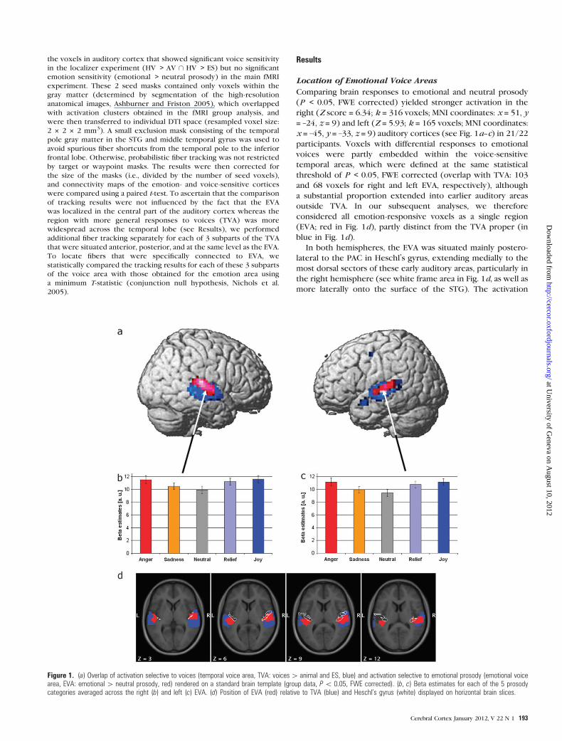
Location of Emotional Voice Areas
Comparing brain responses to emotional and neutral prosody
(P < 0.05, FWE corrected) yielded stronger activation in the
right (Z score = 6.34; k = 316 voxels; MNI coordinates: x = 51, y
= –24, z = 9) and left (Z = 5.93; k = 165 voxels; MNI coordinates:
x = –45, y = –33, z = 9) auditory cortices (see Fig. 1a--c) in 21/22
participants. Voxels with differential responses to emotional
voices were partly embedded within the voice-sensitive
temporal areas, which were defined at the same statistical
threshold of P < 0.05, FWE corrected (overlap with TVA: 103
and 68 voxels for right and left EVA, respectively), although
a substantial proportion extended into earlier auditory areas
outside TVA. In our subsequent analyses, we therefore
considered all emotion-responsive voxels as a single region
(EVA; red in Fig. 1d), partly distinct from the TVA proper (in
blue in Fig. 1d).
In both hemispheres, the EVA was situated mainly postero-
lateral to the PAC in Heschl’s gyrus, extending medially to the
most dorsal sectors of these early auditory areas, particularly in
the right hemisphere (see white frame area in Fig. 1d, as well as
more laterally onto the surface of the STG). The activation
Figure 1. (a) Overlap of activation selective to voices (temporal voice area, TVA: voices[ animal and ES, blue) and activation selective to emotional prosody (emotional voicearea, EVA: emotional[ neutral prosody, red) rendered on a standard brain template (group data, P\ 0.05, FWE corrected). (b, c) Beta estimates for each of the 5 prosodycategories averaged across the right (b) and left (c) EVA. (d) Position of EVA (red) relative to TVA (blue) and Heschl’s gyrus (white) displayed on horizontal brain slices.
Cerebral Cortex January 2012, V 22 N 1 193
at University of G
eneva on August 10, 2012
http://cercor.oxfordjournals.org/D
ownloaded from

maxima were located in the middle part of the STG on both
sides (see Table 1). Individual activation maps revealed a high
spatial congruency of this organization for the contrast
emotional versus neutral prosody across subjects. The most
significant activation was situated within, or in voxels directly
adjacent to, the cluster obtained in the SPM group analysis for
17/22 participants in the right hemisphere and for 19/22
participants in the left hemisphere.
Taken together, these data indicate that emotional effects in
auditory cortex arose partly in the voice-selective cortex but
also extended to earlier auditory stages on the right more than
on the left side.
Functional Properties of Emotional Voice Areas
In both hemispheres, the EVA exhibited a robust main effect of
auditory stimulation, with positive responses in all subjects and
a mean SNR of 11.0 ± 0.6 and 11.5 ± 0.6 for the right and left
EVA, respectively. Stronger responses to emotional than neutral
prosody were found bilaterally in all but one subject, with an
average SNR of 1.3 ± 0.1 (right EVA) and 1.4 ± 0.2 (left EVA).
Post hoc paired t-tests on the beta estimates averaged across
the whole EVA clusters indicated that responses to emotional
categories were significantly stronger than responses to neutral
stimuli (for right EVA, anger vs. neutral: t21 = 7.0, sadness vs.
neutral: t21 = 2.8, relief vs. neutral: t21 = 6.4, joy vs. neutral: t21 =9.8; and for left EVA, anger vs. neutral: t21 = 9.0, sadness vs.
neutral: t21 = 2.4, relief vs. neutral: t21 = 5.5, joy vs. neutral: t21 =8.7; all P < 0.05, see Fig. 1b,c).
A 2-factorial ANOVA on neural responses of the EVA
demonstrated a significant main effect for both valence and
arousal, as well as an interaction between these 2 factors for
both sides (right EVA—main effect of valence: F1,21 = 4.7,
P < 0.05; main effect of arousal: F22 = 12.2, P < 0.01; interaction
valence 3 arousal: F1,21 = 4.4, P < 0.05; left EVA—main effect of
valence: F1,21 = 5.6, P < 0.05; main effect of arousal: F1,21 = 23.4,
P < 0.001; interaction valence 3 arousal: F1,21 = 6.6, P < 0.05).
To test whether the observed effects of valence and the
interaction between valence and arousal were due to the fact
that sad prosody expressed lower arousal than relief prosody
(see Supplementary Table 1), we removed the variance
correlated with arousal ratings from fMRI responses and
submitted the regression residuals of this new analysis to a 2-
factorial ANOVA again. Neither the left nor the right EVA now
showed any significant main effect for valence or interaction
between valence and arousal (all F1,21 < 3.0, all P > 0.05).
Conversely, removing the variance correlated with valence
ratings did not change significance of the main effect of arousal
or the interaction of valence and arousal (right EVA—main
effect of arousal: F22 = 10.8, P < 0.01, interaction valence 3
arousal: F1,21 = 6.5, P < 0.05; left EVA—main effect of arousal:
F1,21 = 22.1, P < 0.001, interaction valence 3 arousal: F1,21 = 8.6,
P < 0.01). These findings indicate that arousal was the main
factor influencing EVA responses.
Effective Connectivity of Emotional Voice Areas
A PPI analysis performed on activity of the right and left EVA
separately showed that emotion enhanced the functional
coupling of these auditory areas with a widespread network
of distant brain regions. Remarkably, a very similar set of
regions was found in the 2 hemispheres (Fig. 2 and Table 2),
including bilateral frontal and parietal areas such as the
supramarginal gyrus (SMG), frontal operculum, and IFG, which
all showed higher connectivity with both the right and the left
EVA during perception of emotional prosody. Vocal emotions
also increased effective connectivity of both EVAs with bilateral
posterior thalamus, bilateral visual areas, left middle frontal
gyrus, right putamen, and right cerebellum (see Table 2 and
Fig. 2a--f). In addition, for the right EVA, we also found an
increased effective connectivity with the left cerebellum and
left IFG, whereas the left EVA exhibited an enhanced coupling
with the right posterior middle temporal gyrus and right medial
prefrontal cortex.
Taken together, these PPI results reveal that processing of
emotional (compared with neutral) voice prosody enhanced
the synchronization of neural activity in EVA with a distributed
and bilateral network of cortical and subcortical regions,
including the IFG and SMG on both sides.
Structural Connectivity of Emotional Voice Areas
Fiber connections originating in emotion-sensitive auditory
areas are presented in Fig. 3 (yellow/red areas in Fig. 3a--f,g--l
for the left and right EVA, respectively). Consistent connec-
tions across subjects (voxel-by-voxel overlap > 50%) were
found with the ipsilateral IFG via the superior longitudinal
fasciculus (SLF) and the external capsule (EC, see Fig. 3a--d,g--j)
in both hemispheres, as well as with the ipsilateral medial
geniculate body (MGB) via the acoustic radiation (AR, see Fig.
3a,g). Additional connections with the ipsilateral occipital
cortex (Fig. 3a,g) and bilateral inferior parietal lobe (IPL,
Fig. 3c--e,i--k) were also observed. Connections of right and left
EVA toward the contralateral IPL crossed the corpus callosum
within its posterior part (see Fig. 3f,l).
The projections of the EVA were further assessed by a voxel-
by-voxel statistical comparison of fiber connections originating
in the EVA proper against those originating in the surrounding
TVA (light blue/blue areas in Fig. 3a--f,g--l for the left and right
EVA, respectively). This analysis revealed that the connections
with ipsilateral IFG via the EC (but not SLF), as well as those to
ipsilateral MGB via the AR, and the projections to ipsilateral IPL
were significantly stronger when tested from the EVA than
from the TVA, in both the right and the left hemispheres (P <
0.05, corrected at the level of spatial extent). All these
Table 1Location of activations (emotional[ neutral prosody) used to define the right and left emotional
voice area (EVA)
MNI coordinates Z score Cluster size
Right EVA x 5 51 y 5 �24 z 5 9 6.34 316Composition of right EVA % Cluster % Region Number of voxelsRight STG 47.1 31.5 149Right Heschl’s gyrus 4.75 56.8 15Right rolandic operculum 2.53 3.70 8White matter 45.3
MNI coordinates Z score Cluster size
Left EVA x 5 �45 y 5 �33 z 5 9 5.93 165Composition of left EVA % Cluster % Region Number of voxelsLeft STG 31.5 14.4 52Left middle temporal gyrus 8.48 1.28 14Left rolandic operculum 1.21 1.43 2Left Heschl’s gyrus 1.21 9.03 2White matter 57.5
Note: Percentage of cluster denotes the contribution of white matter and different gray matter
structures to the activation clusters (emotional[ neutral prosody). Percentage of regions
denotes the fraction of different gray matter structures as defined by automatic anatomic labeling
covered by the activation clusters (emotional[ neutral prosody).
194 Location, Properties, and Connections of the Emotional Voice Area d Ethofer et al.
at University of G
eneva on August 10, 2012
http://cercor.oxfordjournals.org/D
ownloaded from

connections of EVA were also significantly stronger when
separately compared with those originating in different TVA
subparts anterior, lateral, or posterior to the EVA (see
Supplementary Fig. 1). Taken together, these data indicate that
connections with both the IFG and the IPL predominated with
auditory regions exhibiting significant emotion effects rather
than with more widespread auditory areas showing voice-
selective responses but no emotion effects.
Discussion
We provide novel findings on the exact location, functional
properties, and connectivity of EVA within the auditory cortex,
which show preferential increases to affective prosody in-
formation and partly overlap with the TVA. We employed both
structural and functional neuroimaging methods to character-
ize the connectivity of this cortical region. Structural connec-
tivity approaches are well suited to reveal the anatomical
connection profile of brain regions but cannot determine
which of these connections are active during processing of
certain stimulus types or tasks. Effective connectivity analyses,
on the other hand, provide information on networks showing
an increased coupling of functional activity but cannot
determine whether the enhancement of connectivity occurs
directly or via one or several other mediating neural nodes
(Friston et al. 1997). Here, by combining both structural fiber
tracking and effective connectivity analysis, we were able not
only to uncover the direct structural fiber connections of the
EVAs but also pinpoint their functional recruitment during the
perception of emotional prosody.
Anatomical Location of Emotional Voice Areas
It has been consistently replicated across different studies and
laboratories that responses within the auditory cortex situated in
the middle part of STG are modulated by the emotion expressed
in the voice of humans (Grandjean et al. 2005; Ethofer, Anders,
Wiethoff, et al. 2006; Beaucousin et al. 2007; Ethofer et al. 2007;
Dietrich et al. 2008; Wiethoff et al. 2008; Ethofer, Kreifelts, et al.
2009; Ethofer, Van De Ville, et al. 2009) or animals (Belin et al.
2008). However, the exact location of emotion-sensitive areas in
relation to anatomical landmarks and other auditory areas has
not been precisely defined yet. Here, we demonstrate that the
majority of the emotion-sensitive cortex is located posterolateral
to the PAC and situated within Brodmann’s area (BA) 22, which
covers the posterior two-thirds of the free surface of the STG
(Brodmann 1909). In the right hemisphere, the emotion-
sensitive areas additionally comprised a large part of the PAC
itself, suggesting a lateralized involvement of early auditory
processing stages and thus providing a possible explanation for
the typical left-ear superiority observed for emotionally relevant
information in dichotic listening tasks (Erhan et al. 1998). These
findings also accord with electrophysiological data demonstrat-
ing a modulation of early event-related potentials within the first
200 ms, which occurs more consistently in the right than in the
left hemisphere (Paulmann and Kotz 2008). Enhanced responses
of early sensory cortices to emotionally relevant information are
a well-known phenomenon in the visual modality, as it has been
shown that emotional cues in scenes (Lang et al. 1998) and faces
(Pourtois et al. 2004; Vuilleumier and Pourtois 2007) modulate
responses in the primary visual cortex (see also Vuilleumier
2005). Thus, our fMRI findings provide new evidence for the
Figure 2. Brain regions showing an increase of functional connectivity (P\ 0.05, FWE corrected) with the right EVA (a--c) and left EVA (d--f) rendered on a standard braintemplate (a, d) and on transversal (b, d, z 5 0) and sagittal slices (c, x 5 45; f, x 5 �54).
Table 2Brain regions showing increased effective connectivity with right and left EVA during perception
of emotional stimuli (PPI: emotional[ neutral)
MNI (x) MNI (y) MNI (z) Z score
Brain areas modulated from right EVALeft posterior thalamus �6 �18 6 5.52Right posterior thalamus 6 �15 6 5.34Left visual cortex �3 �93 24 5.97Right visual cortex 9 �93 27 5.67Left IFG �51 12 0 4.92Right IFG 51 42 0 5.20Left SMG �54 �12 21 5.24Right SMG 63 �33 30 5.02Left cerebellum �21 �60 �42 5.45Right cerebellum 42 �72 �33 5.12Right putamen 27 3 18 6.28Left middle frontal gyrus �36 45 15 5.09
Brain areas modulated from left EVALeft posterior thalamus �9 �18 6 5.85Right posterior thalamus 15 �18 0 5.77Left visual cortex �9 �93 12 5.44Right visual cortex 15 �90 6 5.38Left IFG �45 21 0 5.02Right cerebellum 39 �75 �33 5.14Right putamen 30 �9 0 5.85Right SMG 45 �48 18 5.44Left middle frontal gyrus �33 39 9 5.17Right posterior middle temporal gyrus 57 �42 0 5.08Right medial prefrontal cortex 0 45 21 5.00
Cerebral Cortex January 2012, V 22 N 1 195
at University of G
eneva on August 10, 2012
http://cercor.oxfordjournals.org/D
ownloaded from

suggestion that increased activation within primary areas might
contribute to efficient processing of behaviorally relevant
information across different sensory modalities.
The EVAs were functionally defined by contrasting activity to
emotional and neutral prosody. Note that this approach
allowed us to assess sensitivity to emotional information but
does not imply that this area exclusively subserves the
representation of vocal emotions. Indeed, posterior lateral
regions of the auditory cortex are also implicated in other
auditory functions including speech perception, particularly in
the left hemisphere (for review, see Price et al. 2009). Posterior
auditory areas also participate in the representation of auditory
space and motion (for review, see Recanzone and Sutter 2008).
Thus, it is likely that the EVA regroups primary and higher
order auditory regions that operate as a computational hub
(Griffiths and Warren 2002) underlying several discriminatory
functions among which processing of prosodic emotions is just
one. Further research investigating different auditory functions
in the same group of participants is needed to examine to what
extent these processes share overlapping representations
within posterior auditory areas or can instead be spatially
segregated using high-resolution fMRI.
Functional Response Profile of Emotional Voice Areas
In both hemispheres, the activation of EVA was significantly
stronger for all emotional categories (i.e., anger, sadness, joy, and
relief) than neutral prosody. This confirms previous observations
that this area is sensitive to a broad range of emotional
information (Ethofer et al. 2007; Wiethoff et al. 2008), an effect
that may occur irrespective of spatial attention (Grandjean et al.
2005) or task instructions (Ethofer, Anders, Wiethoff, et al.
2006). Again, these effects are reminiscent of those found in the
visual domain, where various facial expressions are known to
modulate the fusiform face area (FFA; see Vuilleumier et al. 2002;
Surguladze et al. 2003; Sato et al. 2004). This similarity in neural
response profiles may suggest equivalent functional roles for
these 2 areas in the analysis of facial and vocal affect, respectively
(Campanella and Belin 2007).
Our study sought to disentangle effects due to the emotional
arousal and valence of prosody on EVA using a 2 3 2 factorial
design. As expected, a conventional ANOVA revealed a strong
main effect of arousal. In addition, we also found a significant
main effect of valence and an interaction between valence and
arousal, suggesting at first glance that this area may respond
stronger to positive than to negative emotions and that arousal-
related enhancement is stronger for negative than positive
emotions. Further inspection of activation to the different
emotional categories revealed, however, that these effects can
be explained by the different levels of arousal elicited by
positive and negative low-arousing emotions (i.e., sadness and
relief). When correcting for arousal-related activity by re-
gression analysis, both the main effect of valence as well as the
interaction between valence and arousal were completely
abolished, indicating that arousal was the most important factor
modulating EVA. Likewise, in the visual modality, responses of
the FFA increase linearly with the intensity of computer-
morphed facial expressions of fear, disgust, happiness, and
sadness (Surguladze et al. 2003), although the strongest
enhancement is often observed for fearful faces—a finding
that may reflect greater sensitivity to signals of imminent
danger or greater arousing value of fear compared with other
emotion categories. However, direct comparisons between
sensory modalities might be hampered by many differences in
stimulus properties across studies, and future studies with
high-resolution techniques and multivoxel analysis might be
valuable to investigate more subtle differences in the cortical
representation of different emotions expressed by voices
(Ethofer, Van De Ville, et al. 2009) or faces (Peelen et al. 2010).
Figure 3. Fiber connections from the right (a--f) and left (g--l) EVA as determined by either the degree of overlap ($50%) across subjects (red/yellow) or a direct pairwisestatistical comparison (P\ 0.05) of tracking probabilities from EVA versus TVA (EVA[ TVA, blue/light blue). The MGBs in the posterior thalamus as determined by probabilisticcytoarchitectonic maps are marked in green.
196 Location, Properties, and Connections of the Emotional Voice Area d Ethofer et al.
at University of G
eneva on August 10, 2012
http://cercor.oxfordjournals.org/D
ownloaded from

Modulation of Connectivity of Emotional Voice Areas
We found that emotion expressed by prosody did not only
activate the EVA but also modulated its connectivity with
a widespread and bilateral network, including downstream areas
in the auditory processing pathway, such as bilateral posterior
thalamus, but also occipital, parietal, motor, and—most crit-
ically—inferior frontal areas. All these regions may reflect
distinct facets of the impact of salient emotional prosody on
cognitive processes. The interplay between auditory and visual
regions during the perception of emotional prosody might
constitute the neural substrate of enhanced cross-modal
representations of behaviorally relevant auditory stimuli (Sander
et al. 2005; Kayser et al. 2010). Increased connectivity between
the EVA and the frontoparietal opercular areas could reflect an
engagement of the audio--motor loop to evoke representations
that covertly simulate the perceived vocal emotions (Vigneau
et al. 2006; Warren et al. 2006), whereas the enhanced coupling
with the SMG might have mediated a stronger activation of the
phonological working memory system (Vigneau et al. 2006;
Buchsbaum and D’Esposito 2008) during perception of
emotionally relevant information. Modulation of connectivity
with core components of the motor system (cerebellum
and putamen) converges with electrophysiological findings
(Paulmann et al. 2008) and clinical observations, indicating that
emotional prosody is impaired in Parkinson’s disease (Pell and
Leonard 2003) or after cerebrovascular lesions in the basal
ganglia (Cancelliere and Kertesz 1990; Starkstein et al. 1994).
More critically, in agreement with our hypothesis, we found
that vocal emotions produced a distinctive pattern of enhanced
connectivity with inferior frontal regions that were also found
to activate during prosody recognition in previous work
(Ethofer, Kreifelts, et al. 2009). Very similar effects were
observed in both hemispheres. Importantly, the current study
indicates that the same areas in IFG showed direct fiber
connections with the ipsilateral EVA, as demonstrated by our
DTI fiber tracking results (see Fig. 3). Thus, our findings
provide novel and converging evidence for both structural and
functional interactions between auditory and inferior frontal
areas that are typically associated with the ‘‘how’’ auditory
pathway (Belin and Zatorre 2000).
Structural Connection Profile of the Emotional VoiceAreas
To our knowledge, our study is the first investigating structural
fiber connections within the network underlying emotional
voice processing. Probabilistic fiber tracking revealed that both
the right and the left EVA were directly connected with the
ipsilateral MGB, ipsilateral IFG, and bilateral IPL. To further
determine the specificity of these connections, we statistically
compared the structural connection profile of the EVA with
those of adjacent cortex (in TVA) that was more generally
sensitive to voices but did not show any significant enhance-
ment to emotional prosody. These different analyses converged
to show a strong selectivity of these connections in both
hemispheres.
Direct fiber bundles connected the EVA and the MGB via
the AR as verified by comparison with probabilistic maps for
these brain structures (Burgel et al. 2006). Our finding that
these connections were significantly stronger for EVA than
surrounding TVA accords with the fact that EVAs were located
in the central section of the auditory cortex and also comprised
parts of the PAC. The MGB represents the dominant source of
auditory afferents to PAC (Burton and Jones 1976), although
several other posterior thalamic nuclei also provide inputs to
higher order posterior auditory areas in nonhuman primates
(Hackett et al. 2007).
In agreement with our hypothesis, strong connections were
found between the EVA and the inferior frontal cortex, running
through the EC. These connections were also significantly
stronger for seeds originating in EVA than surrounding TVA and
terminated mostly in the cortex of BA 44--46, which has
previously been found to activate during working memory for
emotional prosody (Mitchell 2007) and explicit judgments of
affective information expressed in the voice (Wildgruber et al.
2005; Ethofer, Anders, Erb, et al. 2006). The finding of direct
structural connections between these areas accords with data
obtained from tracer studies in rhesus monkeys (Seltzer and
Pandya 1989; Padberg et al. 2003) and lends new support to the
hypothesis of a how pathway for voice processing in the human
brain (Belin and Zatorre 2000).
The third, and among the strongest and most consistent,
fiber bundle originating in EVA terminated in bilateral IPL. This
result agrees with tracing studies in the macaque monkey
demonstrating strong connections between posterior auditory
areas and IPL (Lewis and Van Essen 2000; Rozzi et al. 2006;
Smiley et al. 2007). However, it is important to note that we did
not observe any modulation of the IPL during the perception of
vocal emotions, unlike the PPI results for IFG. Therefore, it
seems that this pathway was not actively engaged during
identification of emotional information in the voice. Results
obtained in a dichotic listening paradigm using emotional
utterances, however, suggest that the IPL might be involved in
directing spatial auditory attention to emotional voices (Sander
et al. 2005). This interpretation is also consistent with single-
cell recordings demonstrating auditory responses within the
cortex of the IPL in macaque monkeys (Mazzoni et al. 1996;
Cohen et al. 2004; Gifford and Cohen 2004), as well as human
fMRI data during auditory spatial tasks (Griffiths et al. 1998;
Zatorre et al. 2002).
Finally, fibers originating in EVA also included a posterior
bundle projecting toward the occipital cortex. However, these
fibers were equally found for the surrounding TVA and thus not
specific for the EVA. These findings might reflect the observation
made in anatomical studies of nonhuman primates that both PAC
and higher order auditory cortex are reciprocally connected
with early visual areas (Falchier et al. 2002; Rockland and Ojima
2003) and thus represent a possible neural substrate for cross-
modal integration in early auditory cortices, as evidenced by
electrophysiological studies (Ghazanfar et al. 2005).
Taken together, our DTI results highlight several important
direct pathways through which emotion processing in auditory
cortex may influence cognition and behavior and converge
with our effective connectivity analysis to demonstrate how
some of these pathways with prefrontal areas are differentially
recruited by the current task demands.
Methodological Considerations
Combining data obtained across different imaging modalities
can help interpret results from each modality and thus greatly
advance our understanding of the functional brain architecture
(for review, see Bandettini 2009). However, it is important to
keep several methodological issues in mind when interpreting
fMRI-guided fiber tracking results based on DTI data.
Cerebral Cortex January 2012, V 22 N 1 197
at University of G
eneva on August 10, 2012
http://cercor.oxfordjournals.org/D
ownloaded from

In our study, seed areas for probabilistic fiber tracking were
defined on the basis of fMRI activation maps using a conserva-
tive threshold that controls for false positives for each and
every included voxel (i.e., FWE correction at the voxel level).
While this approach guarantees that the whole seed area shows
significant fMRI activation for the effect of interest (i.e.,
stronger responses to emotional than neutral prosody), it is
possible that the true extent of emotional effects in auditory
cortex is underestimated. However, this is a general problem
in functional imaging as it is impossible to determine the
exact spatial extent and borders of functional areas responding
to a certain type of stimulus based on statistical parametric
maps.
Increased activity in EVA in response to vocal emotions was
a very consistent phenomenon across subjects (21 out of 22
participants showed stronger responses to emotional than
neutral prosody within both the right and the left EVA),
with a high spatial congruency across subjects (at least 75% of
them showed an activation peak within or voxels directly
adjacent to the cluster defined by the random-effect group
analysis). However, peaks in a statistical parametric map may
not necessarily reflect the true activation maximum for an
individual subject given that the difference in activation
amplitude between conditions (emotional and neutral pros-
ody) was only slightly larger than the average noise level
(SNR of 1.3--1.4). Therefore, here we preferred a definition
of seed areas based on group rather than individual activation
data, since it would not have been possible to select a constant
statistical threshold to identify the EVA for each and
every subject and at the same time reliably control for false
positives.
Conclusions
By combining structural and functional neuroimaging approaches,
our study yields novel insights on neural networks engaged by the
perception of vocal emotions. We demonstrate that emotion-
sensitive areas in auditory cortex are situated posterolateral to the
PAC but also partially include primary auditory areas. Neural
activity in the EVA is mainly driven by the arousal expressed by
vocal emotions, with less or no influence of valence. These
findings suggest a key role for this area in detection of acoustic
information with high behavioral relevance irrespective of
emotion category.
Both right and left EVAs were found to be directly
connected with the MGB, presumably reflecting the main
input source of early auditory areas. In both hemispheres, EVAs
were also strongly connected with the ipsilateral IFG and
bilateral IPL, consistent with an important role of these areas in
higher level processing of the emotional nature and spatial
position of auditory information, respectively. These connec-
tions might constitute the anatomical substrates of the ‘‘where’’
and how processing streams activated by voices and be
differentially modulated depending on stimulus characteristics
and task demands.
Funding
Societe Academique de Geneve; Swiss National Science
Foundation (51NF40-104897) to the National Center of
Competence in Research for Affective Sciences; Fortune-
Program of the University of Tubingen (fortune 1874-0-0).
Supplementary Material
Supplementary material can be found at http://www.cercor
.oxfordjournals.org/
Notes
Conflict of Interest: None declared.
References
Ashburner J, Friston KJ. 2005. Unified segmentation. Neuroimage.
26:839--851.
Bandettini PA. 2009. What’s new in neuroimaging methods? Ann N Y
Acad Sci. 1156:260--293.
Banziger T, Grandjean D, Scherer KR. 2009. Emotion recognition from
expressions in face, voice, and body: the Multimodal Emotion
Recognition Test (MERT). Emotion. 9:691--704.
Beaucousin V, Lacheret A, Turbelin MR, Morel M, Mazoyer B, Tzourio-
Mazoyer N. 2007. FMRI study of emotional speech comprehension.
Cereb Cortex. 17:339--352.
Behrens TE, Berg HJ, Jbabdi S, Rushworth MF, Woolrich MW. 2007.
Probabilistic diffusion tractography with multiple fibre orientations:
what can we gain? Neuroimage. 34:144--155.
Behrens TE, Woolrich MW, Jenkinson M, Johansen-Berg H, Nunes RG,
Clare S, Matthews PM, Brady JM, Smith SM. 2003. Characterization
and propagation of uncertainty in diffusion-weighted MR imaging.
Magn Reson Med. 50:1077--1088.
Belin P, Fecteau S, Charest I, Nicastro N, Hauser MD, Armony JL. 2008.
Human cerebral response to animal affective vocalizations. Proc Biol
Sci. 275:473--481.
Belin P, Zatorre RJ. 2000. ’What’, ’where’ and ’how’ in auditory cortex.
Nat Neurosci. 3:965--966.
Belin P, Zatorre RJ, Lafaille P, Ahad P, Pike B. 2000. Voice-selective areas
in human auditory cortex. Nature. 403:309--312.
Brodmann K. 1909. Vergleichende Lokalisationslehre der Grosshirn-
rinde: in ihren Principien dargestellt auf Grund des Zellenbaues.
Leipzig (Germany): Johann Ambrosius Barth Verlag.
Buchanan TW, Lutz K, Mirzazade S, Specht K, Shah NJ, Zilles K,
Jancke L. 2000. Recognition of emotional prosody and verbal
components of spoken language: an fMRI study. Brain Res Cogn
Brain Res. 9:227--238.
Buchsbaum BR, D’Esposito M. 2008. The search for the phonological
store: from loop to convolution. J Cogn Neurosci. 20:762--778.
Burgel U, Amunts K, Hoemke L, Mohlberg H, Gilsbach JM, Zilles K.
2006. White matter fiber tracts of the human brain: three-
dimensional mapping at microscopic resolution, topography and
intersubject variability. Neuroimage. 29:1092--1105.
Burton H, Jones EG. 1976. The posterior thalamic region and its cortical
projections in new world and old world monkeys. J Comp Neurol.
168:249--301.
Campanella S, Belin P. 2007. Integrating face and voice in person
perception. Trends Cogn Sci. 11:535--543.
Cancelliere AE, Kertesz A. 1990. Lesion localization in acquired deficits
of emotional expression and comprehension. Brain Cogn.
13:133--147.
Catani M, Jones DK, ffytche DH. 2005. Perisylvian language networks of
the human brain. Ann Neurol. 57:8--16.
Cohen YE, Cohen IS, Gifford GW, 3rd. 2004. Modulation of LIP
activity by predictive auditory and visual cues. Cereb Cortex. 14:
1287--1301.
Dietrich S, Hertrich I, Alter K, Ischebeck A, Ackermann H. 2008.
Understanding the emotional expression of verbal interjections:
a functional MRI study. Neuroreport. 19:1751--1755.
Erhan H, Borod JC, Tenke CE, Bruder GE. 1998. Identification of
emotion in a dichotic listening task: event-related brain potential
and behavioral findings. Brain Cogn. 37:286--307.
Ethofer T, Anders S, Erb M, Herbert C, Wiethoff S, Kissler J, Grodd W,
Wildgruber D. 2006. Cerebral pathways in processing of affective
prosody: a dynamic causal modeling study. Neuroimage. 30:580--587.
Ethofer T, Anders S, Wiethoff S, Erb M, Herbert C, Saur R, Grodd W,
Wildgruber D. 2006. Effects of prosodic emotional intensity on
activation of associative auditory cortex. Neuroreport. 17:249--253.
198 Location, Properties, and Connections of the Emotional Voice Area d Ethofer et al.
at University of G
eneva on August 10, 2012
http://cercor.oxfordjournals.org/D
ownloaded from

Ethofer T, Kreifelts B, Wiethoff S, Wolf J, Grodd W, Vuilleumier P,
Wildgruber D. 2009. Differential influences of emotion, task, and
novelty on brain regions underlying the processing of speech
melody. J Cogn Neurosci. 21:1255--1268.
Ethofer T, Van De Ville D, Scherer K, Vuilleumier P. 2009. Decoding of
emotional information in voice-sensitive cortices. Curr Biol.
19:1028--1033.
Ethofer T, Wiethoff S, Anders S, Kreifelts B, Grodd W, Wildgruber D.
2007. The voices of seduction: cross-gender effects in processing of
erotic prosody. Soc Cogn Affect Neurosci. 2:334--337.
Falchier A, Clavagnier S, Barone P, Kennedy H. 2002. Anatomical
evidence of multimodal integration in primate striate cortex.
J Neurosci. 22:5749--5759.
Friston KJ, Buechel C, Fink GR, Morris J, Rolls E, Dolan RJ. 1997.
Psychophysiological and modulatory interactions in neuroimaging.
Neuroimage. 6:218--229.
Ghazanfar AA, Maier JX, Hoffman KL, Logothetis NK. 2005. Multisensory
integration of dynamic faces and voices in rhesus monkey auditory
cortex. J Neurosci. 25:5004--5012.
Gifford GW, 3rd, Cohen YE. 2004. Effect of a central fixation light on
auditory spatial responses in area LIP. J Neurophysiol.
91:2929--2933.
Gitelman DR, Penny WD, Ashburner J, Friston KJ. 2003. Modeling
regional and psychophysiologic interactions in fMRI: the impor-
tance of hemodynamic deconvolution. Neuroimage. 19:200--207.
Glasser MF, Rilling JK. 2008. DTI tractography of the human brain’s
language pathways. Cereb Cortex. 18:2471--2482.
Grandjean D, Sander D, Pourtois G, Schwartz S, Seghier ML, Scherer KR,
Vuilleumier P. 2005. The voices of wrath: brain responses to angry
prosody in meaningless speech. Nat Neurosci. 8:145--146.
Griffiths TD, Rees G, Rees A, Green GG, Witton C, Rowe D, Buchel C,
Turner R, Frackowiak RS. 1998. Right parietal cortex is involved in
the perception of sound movement in humans. Nat Neurosci.
1:74--79.
Griffiths TD, Warren JD. 2002. The planum temporale as a computa-
tional hub. Trends Neurosci. 25:348--353.
Hackett TA, De La Mothe LA, Ulbert I, Karmos G, Smiley J,
Schroeder CE. 2007. Multisensory convergence in auditory cortex,
II. Thalamocortical connections of the caudal superior temporal
plane. J Comp Neurol. 502:924--952.
Hackett TA, Stepniewska I, Kaas JH. 1998. Subdivisions of auditory
cortex and ipsilateral cortical connections of the parabelt auditory
cortex in macaque monkeys. J Comp Neurol. 394:475--495.
Hornak J, Bramham J, Rolls ET, Morris RG, O’Doherty J, Bullock PR,
Polkey CE. 2003. Changes in emotion after circumscribed surgical
lesions of the orbitofrontal and cingulate cortices. Brain.
126:1691--1712.
Hornak J, Rolls ET, Wade D. 1996. Face and voice expression
identification in patients with emotional and behavioural changes
following ventral frontal lobe damage. Neuropsychologia.
34:247--261.
Kayser C, Logothetis NK, Panzeri S. 2010. Visual enhancement of
the information representation in auditory cortex. Curr Biol. 20:
19--24.
Kreifelts B, Ethofer T, Shiozawa T, Grodd W, Wildgruber D. 2009.
Cerebral representation of non-verbal emotional perception: fMRI
reveals audiovisual integration area between voice- and face-
sensitive regions in the superior temporal sulcus. Neuropsychologia.
47:3059--3066.
Lang PJ, Bradley MM, Fitzsimmons JR, Cuthbert BN, Scott JD, Moulder B,
Nangia V. 1998. Emotional arousal and activation of the visual
cortex: an fMRI analysis. Psychophysiology. 35:199--210.
Lewis JW, Van Essen DC. 2000. Corticocortical connections of visual,
sensorimotor, and multimodal processing areas in the parietal lobe
of the macaque monkey. J Comp Neurol. 428:112--137.
Mazzoni P, Bracewell RM, Barash S, Andersen RA. 1996. Spatially tuned
auditory responses in area LIP of macaques performing delayed
memory saccades to acoustic targets. J Neurophysiol. 75:1233--1241.
Meyer M, Steinhauer K, Alter K, Friederici AD, von Cramon DY. 2004.
Brain activity varies with modulation of dynamic pitch variance in
sentence melody. Brain Lang. 89:277--289.
Mitchell RL. 2007. fMRI delineation of working memory for emotional
prosody in the brain: commonalities with the lexico-semantic
emotion network. Neuroimage. 36:1015--1025.
Nichols T, Brett M, Andersson J, Wager T, Poline JB. 2005. Valid
conjunction inference with the minimum statistic. Neuroimage.
25:653--660.
Padberg J, Seltzer B, Cusick CG. 2003. Architectonics and cortical
connections of the upper bank of the superior temporal sulcus in
the rhesus monkey: an analysis in the tangential plane. J Comp
Neurol. 467:418--434.
Paulmann S, Kotz SA. 2008. Early emotional prosody perception based
on different speaker voices. Neuroreport. 19:209--213.
Paulmann S, Pell MD, Kotz SA. 2008. Functional contributions of the
basal ganglia to emotional prosody: evidence from ERPs. Brain Res.
1217:171--178.
Peelen MV, Atkinson AP, Vuilleumier P. 2010. Supramodal representa-
tions of perceived emotions in the human brain. J Neurosci.
30:10127--10134.
Pell MD, Leonard CL. 2003. Processing emotional tone from speech in
Parkinson’s disease: a role for the basal ganglia. Cogn Affect Behav
Neurosci. 3:275--288.
Pourtois G, Grandjean D, Sander D, Vuilleumier P. 2004. Electrophys-
iological correlates of rapid spatial orienting towards fearful faces.
Cereb Cortex. 14:619--633.
Price CJ. 2009. The anatomy of language: a review of 100 fMRI studies
published in 2009. Ann N Y Acad Sci. 1191:62--88.
Recanzone GH, Sutter ML. 2008. The biological basis of audition. Annu
Rev Psychol. 59:119--142.
Rockland KS, Ojima H. 2003. Multisensory convergence in calcarine
visual areas in macaque monkey. Int J Psychophysiol. 50:19--26.
Romanski LM, Bates JF, Goldman-Rakic PS. 1999. Auditory belt and
parabelt projections to the prefrontal cortex in the rhesus monkey.
J Comp Neurol. 403:141--157.
Ross ED, Mesulam MM. 1979. Dominant language functions of the right
hemisphere? Prosody and emotional gesturing. Arch Neurol.
36:144--148.
Rozzi S, Calzavara R, Belmalih A, Borra E, Gregoriou GG, Matelli M,
Luppino G. 2006. Cortical connections of the inferior parietal
cortical convexity of the macaque monkey. Cereb Cortex.
16:1389--1417.
Rymarczyk K, Grabowska A. 2007. Sex differences in brain control of
prosody. Neuropsychologia. 45:921--930.
Sander D, Grandjean D, Pourtois G, Schwartz S, Seghier ML, Scherer KR,
Vuilleumier P. 2005. Emotion and attention interactions in social
cognition: brain regions involved in processing anger prosody.
Neuroimage. 28:848--858.
Sato W, Kochiyama T, Yoshikawa S, Naito E, Matsumura M. 2004.
Enhanced neural activity in response to dynamic facial expressions
of emotion: an fMRI study. Brain Res Cogn Brain Res. 20:81--91.
Seltzer B, Pandya DN. 1989. Frontal lobe connections of the superior
temporal sulcus in the rhesus monkey. J Comp Neurol. 281:97--113.
Smiley JF, Hackett TA, Ulbert I, Karmas G, Lakatos P, Javitt DC,
Schroeder CE. 2007. Multisensory convergence in auditory cortex, I.
Cortical connections of the caudal superior temporal plane in
macaque monkeys. J Comp Neurol. 502:894--923.
Starkstein SE, Federoff JP, Price TR, Leiguarda RC, Robinson RG. 1994.
Neuropsychological and neuroradiologic correlates of emotional
prosody comprehension. Neurology. 44:515--522.
Surguladze SA, Brammer MJ, Young AW, Andrew C, Travis MJ,
Williams SC, Phillips ML. 2003. A preferential increase in the
extrastriate response to signals of danger. Neuroimage.
19:1317--1328.
Vigneau M, Beaucousin V, Herve PY, Duffau H, Crivello F, Houde O,
Mazoyer B, Tzourio-Mazoyer N. 2006. Meta-analyzing left hemi-
sphere language areas: phonology, semantics, and sentence pro-
cessing. Neuroimage. 30:1414--1432.
Vuilleumier P. 2005. How brains beware: neural mechanisms of
emotional attention. Trends Cogn Sci. 9:585--594.
Vuilleumier P, Armony JL, Clarke K, Husain M, Driver J, Dolan RJ. 2002.
Neural response to emotional faces with and without awareness:
Cerebral Cortex January 2012, V 22 N 1 199
at University of G
eneva on August 10, 2012
http://cercor.oxfordjournals.org/D
ownloaded from

event-related fMRI in a parietal patient with visual extinction and
spatial neglect. Neuropsychologia. 40:2156--2166.
Vuilleumier P, Pourtois G. 2007. Distributed and interactive brain
mechanisms during emotion face perception: evidence from
functional neuroimaging. Neuropsychologia. 45:174--194.
Warren JE, Sauter DA, Eisner F, Wiland J, Dresner MA, Wise RJ, Rosen S,
Scott SK. 2006. Positive emotions preferentially engage an auditory-
motor ‘‘mirror’’ system. J Neurosci. 26:13067--13075.
Wiethoff S, Wildgruber D, Kreifelts B, Becker H, Herbert C, Grodd W,
Ethofer T. 2008. Cerebral processing of emotional prosody—influence
of acoustic parameters and arousal. Neuroimage. 39:885--893.
Wildgruber D, Riecker A, Hertrich I, Erb M, Grodd W, Ethofer T,
Ackermann H. 2005. Identification of emotional intonation evalu-
ated by fMRI. Neuroimage. 24:1233--1241.
Zatorre RJ, Bouffard M, Ahad P, Belin P. 2002. Where is ’where’ in the
human auditory cortex? Nat Neurosci. 5:905--909.
200 Location, Properties, and Connections of the Emotional Voice Area d Ethofer et al.
at University of G
eneva on August 10, 2012
http://cercor.oxfordjournals.org/D
ownloaded from