Elucidating the Conformational Dependence of Calculated p K a Values
Upload
independentCategory
view
5download
0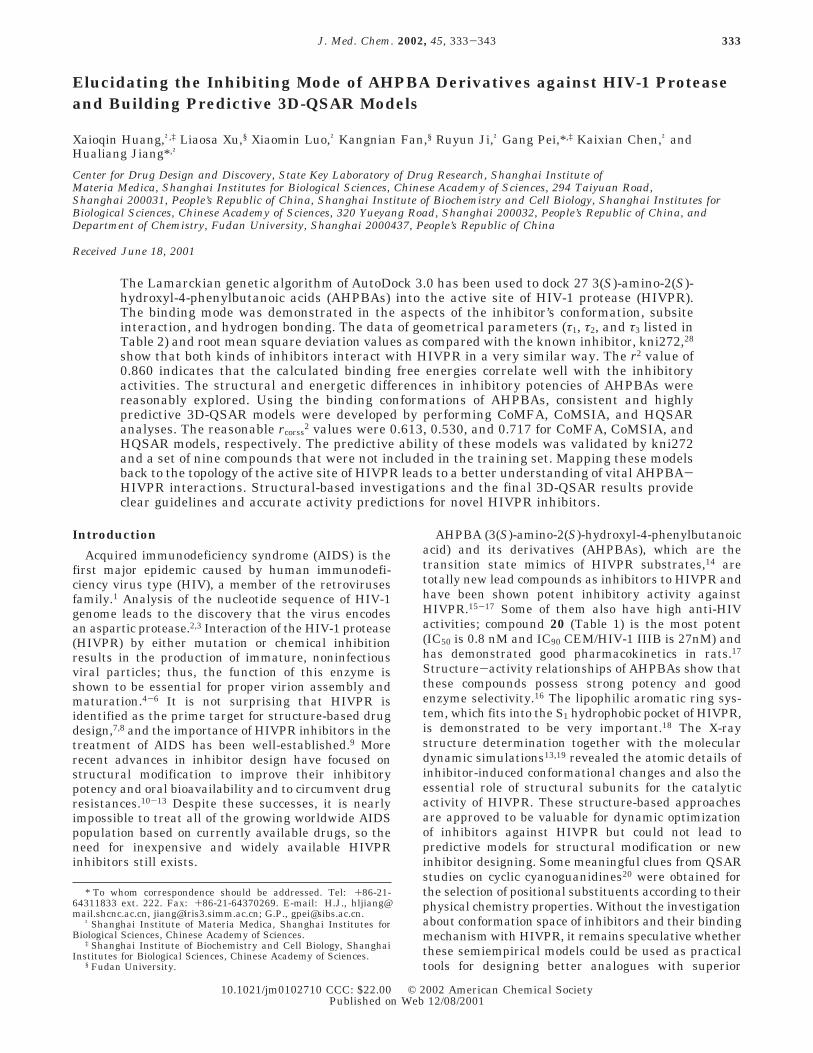
Elucidating the Inhibiting Mode of AHPBA Derivatives against HIV-1 Proteaseand Building Predictive 3D-QSAR Models
Xaioqin Huang,†,‡ Liaosa Xu,§ Xiaomin Luo,† Kangnian Fan,§ Ruyun Ji,† Gang Pei,*,‡ Kaixian Chen,† andHualiang Jiang*,†
Center for Drug Design and Discovery, State Key Laboratory of Drug Research, Shanghai Institute ofMateria Medica, Shanghai Institutes for Biological Sciences, Chinese Academy of Sciences, 294 Taiyuan Road,Shanghai 200031, People’s Republic of China, Shanghai Institute of Biochemistry and Cell Biology, Shanghai Institutes forBiological Sciences, Chinese Academy of Sciences, 320 Yueyang Road, Shanghai 200032, People’s Republic of China, andDepartment of Chemistry, Fudan University, Shanghai 2000437, People’s Republic of China
Received June 18, 2001
The Lamarckian genetic algorithm of AutoDock 3.0 has been used to dock 27 3(S)-amino-2(S)-hydroxyl-4-phenylbutanoic acids (AHPBAs) into the active site of HIV-1 protease (HIVPR).The binding mode was demonstrated in the aspects of the inhibitor’s conformation, subsiteinteraction, and hydrogen bonding. The data of geometrical parameters (τ1, τ2, and τ3 listed inTable 2) and root mean square deviation values as compared with the known inhibitor, kni272,28
show that both kinds of inhibitors interact with HIVPR in a very similar way. The r2 value of0.860 indicates that the calculated binding free energies correlate well with the inhibitoryactivities. The structural and energetic differences in inhibitory potencies of AHPBAs werereasonably explored. Using the binding conformations of AHPBAs, consistent and highlypredictive 3D-QSAR models were developed by performing CoMFA, CoMSIA, and HQSARanalyses. The reasonable rcorss
2 values were 0.613, 0.530, and 0.717 for CoMFA, CoMSIA, andHQSAR models, respectively. The predictive ability of these models was validated by kni272and a set of nine compounds that were not included in the training set. Mapping these modelsback to the topology of the active site of HIVPR leads to a better understanding of vital AHPBA-HIVPR interactions. Structural-based investigations and the final 3D-QSAR results provideclear guidelines and accurate activity predictions for novel HIVPR inhibitors.
Introduction
Acquired immunodeficiency syndrome (AIDS) is thefirst major epidemic caused by human immunodefi-ciency virus type (HIV), a member of the retrovirusesfamily.1 Analysis of the nucleotide sequence of HIV-1genome leads to the discovery that the virus encodesan aspartic protease.2,3 Interaction of the HIV-1 protease(HIVPR) by either mutation or chemical inhibitionresults in the production of immature, noninfectiousviral particles; thus, the function of this enzyme isshown to be essential for proper virion assembly andmaturation.4-6 It is not surprising that HIVPR isidentified as the prime target for structure-based drugdesign,7,8 and the importance of HIVPR inhibitors in thetreatment of AIDS has been well-established.9 Morerecent advances in inhibitor design have focused onstructural modification to improve their inhibitorypotency and oral bioavailability and to circumvent drugresistances.10-13 Despite these successes, it is nearlyimpossible to treat all of the growing worldwide AIDSpopulation based on currently available drugs, so theneed for inexpensive and widely available HIVPRinhibitors still exists.
AHPBA (3(S)-amino-2(S)-hydroxyl-4-phenylbutanoicacid) and its derivatives (AHPBAs), which are thetransition state mimics of HIVPR substrates,14 aretotally new lead compounds as inhibitors to HIVPR andhave been shown potent inhibitory activity againstHIVPR.15-17 Some of them also have high anti-HIVactivities; compound 20 (Table 1) is the most potent(IC50 is 0.8 nM and IC90 CEM/HIV-1 IIIB is 27nM) andhas demonstrated good pharmacokinetics in rats.17
Structure-activity relationships of AHPBAs show thatthese compounds possess strong potency and goodenzyme selectivity.16 The lipophilic aromatic ring sys-tem, which fits into the S1 hydrophobic pocket of HIVPR,is demonstrated to be very important.18 The X-raystructure determination together with the moleculardynamic simulations13,19 revealed the atomic details ofinhibitor-induced conformational changes and also theessential role of structural subunits for the catalyticactivity of HIVPR. These structure-based approachesare approved to be valuable for dynamic optimizationof inhibitors against HIVPR but could not lead topredictive models for structural modification or newinhibitor designing. Some meaningful clues from QSARstudies on cyclic cyanoguanidines20 were obtained forthe selection of positional substituents according to theirphysical chemistry properties. Without the investigationabout conformation space of inhibitors and their bindingmechanism with HIVPR, it remains speculative whetherthese semiempirical models could be used as practicaltools for designing better analogues with superior
* To whom correspondence should be addressed. Tel: +86-21-64311833 ext. 222. Fax: +86-21-64370269. E-mail: H.J., [email protected], [email protected]; G.P., [email protected].
† Shanghai Institute of Materia Medica, Shanghai Institutes forBiological Sciences, Chinese Academy of Sciences.
‡ Shanghai Institute of Biochemistry and Cell Biology, ShanghaiInstitutes for Biological Sciences, Chinese Academy of Sciences.
§ Fudan University.
333J. Med. Chem. 2002, 45, 333-343
10.1021/jm0102710 CCC: $22.00 © 2002 American Chemical SocietyPublished on Web 12/08/2001
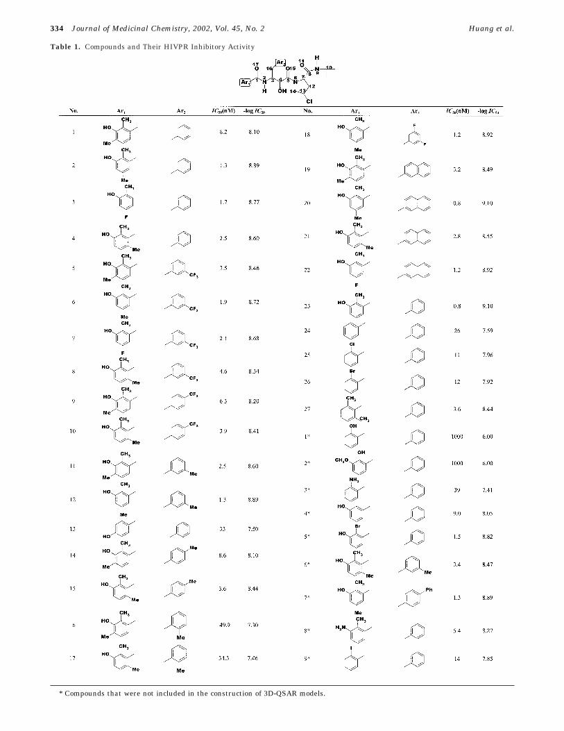
Table 1. Compounds and Their HIVPR Inhibitory Activity
* Compounds that were not included in the construction of 3D-QSAR models.
334 Journal of Medicinal Chemistry, 2002, Vol. 45, No. 2 Huang et al.
pharmacokinetic and efficacy profiles. Developing 3D-QSAR models under the guide of information from thecatalytic site of enzymes21 has been proven as one ofthe more rational methodologies for binding affinityprediction of new inhibitors.
Taking account of the pioneering works in the fieldof designing and synthesis of HIVPR inhibitors, itbecomes fascinating for us to study the inhibitorymechanism of AHPBAs against HIVPR and investigatethe three-dimensional quantitative structure-activityrelationships (3D-QSAR) of AHPBAs. To the best of ourknowledge, there has been no previous effort carried outto seek new insight into the relationship between thestructure information and the inhibitory potency ofAHPBAs at the level of binding free energy prediction,especially by theoretical methods, such as automatedmolecular docking, comparative molecular field analysis(CoMFA),22 comparative molecular similarity analysis(CoMSIA),23 and hologram quantitative structure-activity relationship (HQSAR)24 approaches.
The aim of the present research is to demonstrate thecommon binding mode of AHPBAs with HIVPR and topredict the binding free energies relative to the inhibi-tory potencies of these compounds. The further impor-tant goal is to obtain not only stable and predictive butalso fast and convenient QSAR models, which arelocated at the 3D level about the main intermolecularinteractions involved in the HIVPR inhibition.
Computational Details
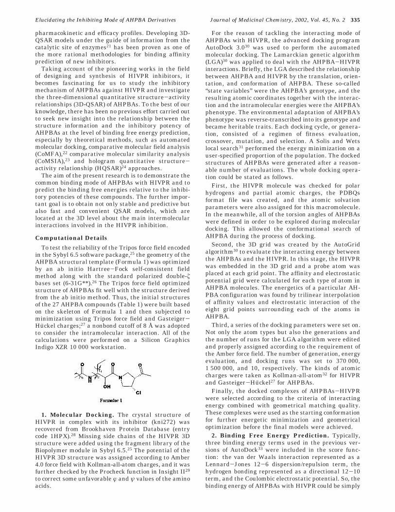
To test the reliability of the Tripos force field encodedin the Sybyl 6.5 software package,25 the geometry of theAHPBA structural template (Formula 1) was optimizedby an ab initio Hartree-Fock self-consistent fieldmethod along with the standard polarized double-úbases set (6-31G**).26 The Tripos force field optimizedstructure of AHPBAs fit well with the structure derivedfrom the ab initio method. Thus, the initial structuresof the 27 AHPBA compounds (Table 1) were built basedon the skeleton of Formula 1 and then subjected tominimization using Tripos force field and Gasteiger-Huckel charges;27 a nonbond cutoff of 8 Å was adoptedto consider the intramolecular interaction. All of thecalculations were performed on a Silicon GraphicsIndigo XZR 10 000 workstation.
1. Molecular Docking. The crystal structure ofHIVPR in complex with its inhibitor (kni272) wasrecovered from Brookhaven Protein Database (entrycode 1HPX).28 Missing side chains of the HIVPR 3Dstructure were added using the fragment library of theBiopolymer module in Sybyl 6.5.25 The potential of theHIVPR 3D structure was assigned according to Amber4.0 force field with Kollman-all-atom charges, and it wasfurther checked by the Procheck function in Insight II29
to correct some unfavorable æ and ψ values of the aminoacids.
For the reason of tackling the interacting mode ofAHPBAs with HIVPR, the advanced docking programAutoDock 3.030 was used to perform the automatedmolecular docking. The Lamarckian genetic algorithm(LGA)30 was applied to deal with the AHPBA-HIVPRinteractions. Briefly, the LGA described the relationshipbetween AHPBA and HIVPR by the translation, orien-tation, and conformation of AHPBA. These so-called“state variables” were the AHPBA’s genotype, and theresulting atomic coordinates together with the interac-tion and the intramolecular energies were the AHPBA’sphenotype. The environmental adaptation of AHPBA’sphenotype was reverse-transcribed into its genotype andbecame heritable traits. Each docking cycle, or genera-tion, consisted of a regimen of fitness evaluation,crossover, mutation, and selection. A Solis and Wetslocal search31 performed the energy minimization on auser-specified proportion of the population. The dockedstructures of AHPBAs were generated after a reason-able number of evaluations. The whole docking opera-tion could be stated as follows.
First, the HIVPR molecule was checked for polarhydrogens and partial atomic charges, the PDBQsformat file was created, and the atomic solvationparameters were also assigned for this macromolecule.In the meanwhile, all of the torsion angles of AHPBAswere defined in order to be explored during moleculardocking. This allowed the conformational search ofAHPBA during the process of docking.
Second, the 3D grid was created by the AutoGridalgorithm30 to evaluate the interacting energy betweenthe AHPBAs and the HIVPR. In this stage, the HIVPRwas embedded in the 3D grid and a probe atom wasplaced at each grid point. The affinity and electrostaticpotential grid were calculated for each type of atom inAHPBA molecules. The energetics of a particular AH-PBA configuration was found by trilinear interpolationof affinity values and electrostatic interaction of theeight grid points surrounding each of the atoms inAHPBA.
Third, a series of the docking parameters were set on.Not only the atom types but also the generations andthe number of runs for the LGA algorithm were editedand properly assigned according to the requirement ofthe Amber force field. The number of generation, energyevaluation, and docking runs was set to 370 000,1 500 000, and 10, respectively. The kinds of atomiccharges were taken as Kollman-all-atom32 for HIVPRand Gasteiger-Huckel27 for AHPBAs.
Finally, the docked complexes of AHPBAs-HIVPRwere selected according to the criteria of interactingenergy combined with geometrical matching quality.These complexes were used as the starting conformationfor further energetic minimization and geometricaloptimization before the final models were achieved.
2. Binding Free Energy Prediction. Typically,three binding energy terms used in the previous ver-sions of AutoDock33 were included in the score func-tion: the van der Waals interaction represented as aLennard-Jones 12-6 dispersion/repulsion term, thehydrogen bonding represented as a directional 12-10term, and the Coulombic electrostatic potential. So, thebinding energy of AHPBAs with HIVPR could be simply
Elucidating the Inhibiting Mode of AHPBA Derivatives Journal of Medicinal Chemistry, 2002, Vol. 45, No. 2 335
described as the electrostatic, van der Waals, andhydrogen bonding interaction energy, respectively.
On the basis of the traditional molecular force fieldmodel of interaction energy, a new score function at thelevel of binding free energy was derived and adopted inthe version of AutoDock 3.0.30 Not only the restrictionof internal rotors, the global rotation, and translationwere modeled depending on the number of torsionangles of the ligand but also the desolvation uponbinding and the hydrophobic effect (solvent entropychanges at solute-solvent interfaces) were calculated.The total binding free energy was empirically calibratedbased on the above-stated terms and a set of coefficientfactors.30 Thus, the new score function was sufficientto rank the inhibitors in the different levels of bindingaffinities.
The same rationale was applied to the system ofAHPBAs-HIVPR in order to evaluate the bindingproperties more precisely than the traditional molecularmechanics method did, and the total binding free energybetween AHPBAs and HIVPR was calculated accordingto the algorithm in the AuotDock 3.0 program.30
3. 3D-QSAR Studies. To more fully explore thespecific contributions of electrostatic, steric, and hydro-phobic effects in the binding of AHPBAs to HIVPR andto build predictive QSAR models, CoMFA,22 CoMSIA,23
and HQSAR24 studies were performed by using theconformations and their alignment at the binding siteof the HIVPR, which resulted from the moleculardocking.
3.1. CoMFA. Usually, steric and electrostatic fieldenergies were probed using an sp3 carbon atom and a+1 net charge atom, respectively. Steric and electro-static interactions were calculated using a Tripos forcefield with a distance-dependent dielectric constant atall intersections in a regularly spaced (2 Å) grid. Theminimum-σ (column filtering) was set to 2.0 kcal/molto improve the signal-to-noise ratio by omitting thoselattice points whose energy variation was below thisthreshold. A cutoff of 30 kcal/mol was adopted, and theregression analysis was carried out using the partialleast-squares (PLS) method. The final model was de-veloped with the optimum number of components equalto that yielding the highest rcv
2.3.2. CoMSIA. Three physicochemical properties,
steric, electrostatic, and hydrophobic fields, have beenevaluated. The steric contribution was reflected by thethird power of the atomic radii of the atoms. Electro-static properties were introduced as atomic charges thatresulted from molecular docking. An atom-based hydro-phobicity was assigned according to the parametri-zation developed by Viswanadhan et al.34 The latticedimensions were selected with a sufficiently large margin(>4 Å) to enclose all aligned molecules. Any singularitieswere avoided at atomic positions in CoMSIA fieldsbecause a Gaussian type distance dependence of thephysicochemical properties was adopted; thus, no arbi-trary cutoffs were required. In general, similarityindices (AF,K) between the compounds of interest and aprobe atom placed at the intersections of the latticecould be calculated with eq 1
where q represents a grid point; i is the summationindex over all atoms of the molecule j under computa-tion; wik is the actual value of the physicochemicalproperty k of atom i; and wprob,k is the value of the probeatom. In the present study, similarity indices werecomputed using a probe atom (wprob,k) with a charge of+1, a radius of 1 Å, a hydrophobicity of +1, and anattenuation factor R 0.3 for the Gaussian type distance.The statistical evaluation for the CoMSIA analyses wasperformed in the same way as described in CoMFA.
3.3. HQSAR. The construction of a molecular holo-gram containing the HQSAR descriptors was completedas following this procedure: at first, the molecule washashed to a molecular fingerprint that encoded thefrequency of occurrence of various molecular fragmenttypes using a predefined set of rules. Then, the molec-ular fingerprint was cut into strings at a fixed intervalas specified by a hologram length (HL) parameter, andat last, all of the generated strings were hashed into afixed length array. The Sybyl line notation for eachstring was mapped to a unique integer in the range of0-231 using a cyclic redundancy check algorithm. Thenumerical representation of molecules was exploited bya subsequent correlation analysis; typically, a PLSQSAR model was constructed. The optimal HQSARmodel was constructed by screening the 12 default HLvalues, which were a set of prime numbers ranging from53 to 401.
Results and Discussion1. Interacting Mode with HIVPR. 1.1. Inhibitor’s
Conformation. Figure 1 illustrates the probable bind-ing conformations for the 27 AHPBAs extracted fromAHPBA-HIVPR complexes. Figure 2A shows the 3Dmodel of the AHPBAs-HIVPR complex, and Figure 2Bis the conformational comparison for the most potentinhibitors, compound 12 (Table 1) and compoundkni272.28 The main conformational difference betweenthe AHPBAs and the kni272 could be represented asthe three torsion angles (τ1, τ2, and τ3 in Formula 1) andthe root mean square deviation (RMSD) values basedon the parts of similar structure including the Ar2 group.These data are summarized in Table 2 and shown inFigure 2C. Figure 3 generally represents the interacting
AF,Kq(j) ) - ∑
i)1
n
wprobe,k wik e-ariq2
(1)
Figure 1. Probable binding conformations of AHPBAs andtheir alignment in the binding site of HIVPR.
336 Journal of Medicinal Chemistry, 2002, Vol. 45, No. 2 Huang et al.
mode of AHPBAs with HIVPR. Just like the inhibitorkni27228 and most of the other transition state mimicinhibitors cocrystallized with HIVPR,35,36 AHPBAs lo-cate in the center of the typical binding pocket of HIVPRand share some common binding features for each other.All of the AHPBAs are bound in the active site of HIVPRin an extended conformation (Figures 1 and 2A), andthe binding conformations of AHPBAs could be alignedquite well overall. Following a similar binding patternwith compound kni272,28 the Ar2 group (Table 1) of allof the 27 AHPBAs is situated at the S1 subsite of thebinding pocket, and the Ar1-CONH group occupies theS2 subsite. Meanwhile, the NH-tertiary butyl groupinteracts with the S2′ subsite, and the proline-CO partis in match with the S1′ subsite.
1.2. Subsite Interactions. The open mouth of thesmall hydrophobic pocket formed by residues Leu23′,Gly48, Gly49, Ile50, Pro81′, Val82′, and Ile84′ of HIVPR
is directly toward the Ar2 group and wraps the mostpart of the latter (Figure 3). They interact with eachother tightly through hydrophobic interaction. Theoutside edge of the Ar2 group is almost blocked off bythe hydrogen network formed between Arg8′ and Asp29.Interestingly, the two terminal methyl groups of the side
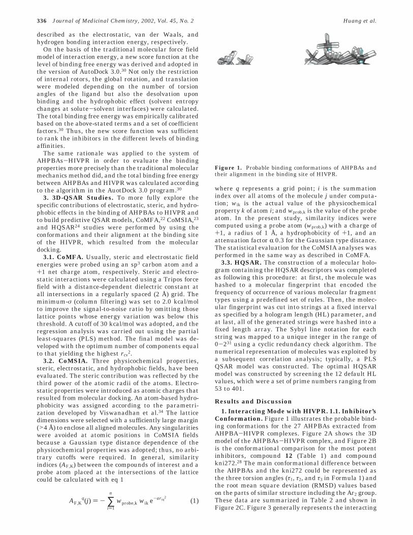
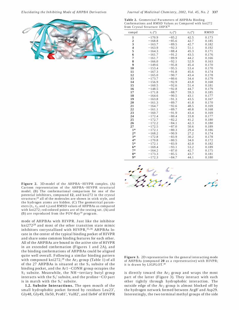
Figure 2. 3D-model of the AHPBA-HIVPR complex. (A)Cartoon representation of the AHPBA-HIVPR structuralmodel. (B) The conformational comparison for one of thepotential inhibitors, compound 12, and kni272 in the crystalstructure;28 all of the molecules are shown in stick style, andthe hydrogen atoms are hidden. (C) The geometrical param-eters (τ1, τ2, and τ3) and RMSD values of AHPBAs as comparedwith kni272; red-colored points are of the testing set. (A) and(B) are reproduced from the POV-Ray39 program.
Table 2. Geometrical Parameters of AHPBAs BindingConformations and RMSD Values as Compared with kni272from Crystal Structure 1HPX28
compd τ1 (°) τ2 (°) τ3 (°) RMSD
1 -170.9 -85.2 42.5 0.1732 -168.8 -85.6 42.7 0.1853 -163.7 -89.5 42.7 0.1824 -163.9 -92.3 51.1 0.1925 -164.3 -88.4 45.3 0.1716 -161.7 -91.2 43.5 0.1767 -161.7 -89.9 44.2 0.1668 -166.0 -92.1 52.9 0.1639 -149.6 -95.8 45.4 0.170
10 -153.4 -95.5 53.4 0.17011 -167.3 -91.8 45.6 0.17412 -165.0 -90.7 43.4 0.17813 -173.7 -80.6 34.4 0.17914 -156.9 -92.9 43.8 0.16815 -160.5 -92.6 51.4 0.18116 -148.5 -92.8 44.7 0.17917 -171.8 -88.7 59.3 0.18518 -164.6 -90.5 43.1 0.17719 -163.8 -91.3 43.5 0.16720 -161.3 -89.7 41.8 0.17021 -164.7 -92.6 48.5 0.16922 -161.1 -89.7 40.8 0.16823 -160.7 -91.9 43.4 0.16924 -172.4 -80.4 33.8 0.17725 -172.7 -82.2 41.2 0.18026 -172.2 -84.1 42.3 0.18027 -172.5 -87.0 50.6 0.1891* -172.1 -80.3 29.4 0.1862* -168.2 -90.9 27.2 0.1743* -172.8 -83.9 30.2 0.1784* -170.8 -80.5 34.0 0.1725* -172.1 -83.9 42.0 0.1826* -169.4 -93.1 53.2 0.1897* -164.2 -87.0 42.7 0.1758* -173.3 -85.5 43.7 0.1819* -172.3 -84.7 44.1 0.180
Figure 3. 2D representative for the general interacting modeof AHPBAs (compound 20 as a representative) with HIVPR;it is drawn by LIGPLOT.40
Elucidating the Inhibiting Mode of AHPBA Derivatives Journal of Medicinal Chemistry, 2002, Vol. 45, No. 2 337
chains of residues Val82′ and Ile84′, respectively, in-teract with the two aromatic rings of the Ar2 group ofcompound 20 or the benzyl ring of the other inhibitorsthrough nonpolar alkane-π interaction37 (Figure 3).This kind of alkane-π interaction has not yet beenappreciated in inhibitor-enzyme binding. To estimatethe strength of the alkane-π interaction, we performeda theoretical calculation employing the ab initio quan-tum chemistry methods of Moler-Plesset second-ordercorrelation method (MP2)38 at the 6-31G* basis set level,taking methane-benzene as model systems. The dis-tance between the methyl carbon and the center ofbenzene is 3.7 Å, which is in agreement with thedistances of the two methyl carbons to the centers ofthe two rings of Ar2 (Figure 3). The binding energy ofMP2/6-31G* between methyl and benzene is -1.3 kcal/mol. This indicates that the alkane-π interactioncontributes about 2.6 kcal/mol energy for compound 20-HIVPR binding, or at least 1.3 kcal/mol energy for theother compounds-HIVPR binding, and points out theimportance of the aromaticity of Ar2.
The Ar1-CONH group binds with the side chains ofresidues Ala28, Asp29, Asp30, Val32, Ile47, Gly48, andIle84 of HIVPR through not only hydrophobic interac-tion but also electrostatic interaction to some extent.The carbonyl group (>CdO) of residue Gly48 is in aposition almost perpendicular to the plane of thearomatic ring of the Ar1 group. The distance from thecarbonyl oxygen of Gly48 to the center of the aromaticring of the Ar1 group is 3.51 Å. So, the residue Gly48may be one of the important factors determining the3D positioning of the Ar1 group in the binding pocket.
The NH-tertiary butyl group at the other end of theAHPBA molecules is surrounded by residues Gly27′,Ala28′, Asp29′, Ile47′, Gly48′, Gly49′, and Ile50 ofHIVPR. Although the hydrophobic space is not fullyoccupied by the tertiary butyl group, they are in goodgeometrical match through the side chains of Ala28′ andIle50. The proline-CO group of AHPBAs situates in asmall hydrophobic pocket of the subsite S1′ and interactswith the side chains of residues Leu23, Val82, and Ile84of HIVPR; the chloride points directly to the gapbetween Leu23, Val82, and Ile84.
1.3. Hydrogen-Bonding Interactions. Another im-portant characteristic of the interaction between AHP-BAs and HIVPR is the hydrogen bonding (Figure 3).There are several hydrogen bonds formed between theAHPBAs and the side chains of some residues inHIVPR. The hydroxyl group at position 4 of AHPBAs(Table 1) could form hydrogen bonds with Oδ1 and Oδ2
atoms of Asp25 or Oδ2 of Asp25′, and the >CdO groupat position 5 (Table 1) forms a hydrogen bond with theprotonated Oδ1 of Asp25′. This network of hydrogenbonds in the catalytic site of HIVPR must play a vitalrole in determining the level of binding affinities forAHPBAs with HIVPR, and this may be the importantreason why the AHPBAs could inhibit the HIVPR muchmore potently. The oxygen atom of the hydroxyl groupat Ar1 might be an acceptor to form a hydrogen bondwith the -NH group at the backbone of residue Asp29on one hand; the hydrogen atom of the hydroxyl groupat Ar1 hydrogen bonds with Oδ1 of the same residue onthe other hand. These hydrogen bonds greatly strengthenthe interaction of the aromatic ring Ar1 with the
surrounding hydrophobic field produced by side chainsof residues Ala28, Val32, Ile47, and Ile84 in HIVPR.Interestingly, the -NH group at position 9 of AHPBAsalso forms a hydrogen bond with the >CdO group ofresidue Gly48′ in HIVPR. This hydrogen bonding in-tensely determines the 3D space position of the tertiarybutyl group in the binding pocket and stabilizes thehydrophobic interaction of the tertiary butyl group withthe side chains of residues Ala28′, Ile47′, and Ile50 inHIVPR.
On further inspection of the AHPBA-HIVPR complexmodel, the >CdO groups at positions 1 and 8 ofAHPBAs are located adjacent to the -NH groups ofIle50/Ile50′. The distances between them are within 4Å, and the conformation of this part in the complex issymmetric to some extent. There may be a hydrogenbond network formed between the >CdO groups atpositions 1 and 8 of AHPBAs and the -NH groups ofIle50/Ile50′ through a water molecule if the complex iscrystallized. This is just like the hydrogen bondingtetrahedral network and a water molecule named asWat301 found in the kni272-HIVPR28 and other inhibi-tor-HIVPR crystal structures.41,42
As a whole, the AHPBAs interact with HIVPR througha hydrophobic, hydrogen bonding interaction and a local,weak electrostatic interaction. The hydroxyl group atposition 4 of AHPBAs is located at the center of theelectrostatic field produced by the negatively chargedside chains of an active catalytic triad of HIVPR. Thebinding of the AHPBAs may introduce significant influ-ence on the conformation of those regions that definethe binding site of HIVPR, and the different ionizationstates of the catalytic aspartyl groups of Asp25 andAsp25′. The presence of the inhibitor makes the dimericstructure of HIVPR stabilized, especially the flap regionthat becomes conformationally closed.43,44
2. Correlation between Binding Free Energyand Inhibitory Activity. Table 3 lists the predictedbinding free energy of AHPBAs with HIVPR. Satisfiedthat the 3D structures of the AHPBA-HIVPR com-plexes were practically reasonable, the multiple regres-sion analysis45 was performed to explore whether theinhibitory potencies of AHPBAs could be correlated withthe energetic information. The regression equation was
Table 3. Predicted Binding Free Energy (kcal/mol) vs theExperimental Activity (-logIC50) of AHPBAs and kni272
compd -logIC50
∆G(kcal/mol) compd -logIC50
∆G(kcal/mol)
1 8.10 -14.81 19 8.49 -15.022 8.89 -15.31 20 9.10 -16.023 8.77 -15.48 21 8.55 -14.964 8.60 -15.01 22 8.92 -15.265 8.46 -15.25 23 9.10 -15.056 8.72 -15.28 24 7.59 -14.057 8.68 -15.15 25 7.96 -14.158 8.34 -15.03 26 7.92 -13.919 8.20 -14.90 27 8.44 -14.92
10 8.41 -14.98 1* 6.00 -11.1811 8.60 -15.18 2* 6.00 -11.2912 8.89 -15.66 3* 7.41 -13.3113 7.50 -13.60 4* 8.05 -14.4214 8.10 -14.76 5* 8.82 -15.0815 8.44 -14.93 6* 8.47 -15.0616 7.30 -13.67 7* 8.89 -15.4017 7.46 -13.86 8* 8.27 -14.7818 8.92 -15.50 9* 7.85 -13.89
338 Journal of Medicinal Chemistry, 2002, Vol. 45, No. 2 Huang et al.
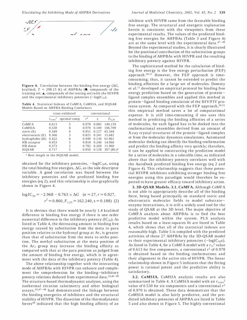
obtained for the inhibitory potencies, -logIC50s, usingthe total binding free energies, ∆G, as the sole descriptorvariable. A good correlation was found between theinhibitory potencies and the predicted binding freeenergies (eq 2), and this relationship is also graphicallyshown in Figure 4.
It is obvious that there would be nearly 1.4 kcal/moldifference in binding free energy if there is one ordernumerical difference in the inhibitory potency (IC50). Aslisted in Table 4, the decreasing amount in binding freeenergy caused by substitution from the meta to paraposition relative to the hydroxyl group at Ar1 is greaterthan that of substitution from the meta to ortho posi-tion. The methyl substitution at the meta position ofthe Ar2 group may increase the binding affinity ascompared with that at the para position as judged fromthe amount of binding free energy, which is in agree-ment with the data of the inhibitory potency (Table 4).
The above relationship together with the interactingmode of AHPBAs with HIVPR can enhance and comple-ment the comprehension for the binding-inhibitorypotency relations deduced from experimental data.10,46-49
The structure-based thermodynamic analyses, using theisothermal titration calorimetry and other biologicalassays,10,47-48 had demonstrated the linkage betweenthe binding energetics of inhibitors and the structuralstability of HIVPR. The dissection of the thermodynamicforces49 indicated that the high binding affinity of an
inhibitor with HIVPR came from the favorable bindingfree energy. The structural and energetic explanationherein is consistent with the viewpoints from theseexperimental results. The values of the predicted bind-ing free energies for AHPBAs (Table 3 and Figure 4)are at the same level with the experimental data.47-49
Beyond the experimental studies, it is clearly illustratedfor the positional contribution of the substitution groupsto the binding of AHPBAs with HIVPR and the resultinginhibitory potency against HIVPR.
The sophisticated method for the calculation of bind-ing free energy is the free energy perturbation (FEP)approach.50,51 However, the FEP approach is time-consuming; thus, it cannot be extended to predict thebinding affinities for a large set of molecules. Dominyet al.11 developed an empirical protocol for binding freeenergy prediction based on the generation of protein-ligand complex ensembles and applied this method inprotein-ligand binding simulation of the HIV/FIV pro-tease system. As compared with the FEP approach,50,51
this empirical method saves a lot of computationalexpense. It is still time-consuming if one uses thismethod in predicting the binding affinities of a seriesof molecules, for each ligand has to be docked into theconformational ensembles derived from an amount ofX-ray crystal structures of the protein-ligand complexor from the molecular dynamics simulations. Automatedmolecular docking can identify the binding conformationand predict the binding affinity very quickly; therefore,it can be applied in constructing the prediction modelfor a series of molecules in a tolerable time, as indicatedabove that the inhibitory potency correlates well withthe AutoDock predicted binding free energy (eq 2 andFigure 4). This relationship suggests that those poten-tial HIVPR inhibitors exhibiting stronger binding freeenergies using this paradigm would therefore be ex-pected to have greater efficacy toward inhibitory action.
3. 3D-QSAR Models. 3.1. CoMFA. Although CoMFAis not able to appropriately describe all of the bindingforce, being based principally on standard steric andelectrostatic molecular fields to model substrate-enzyme interactions, it is still a widely used tool for thestudy of QSAR at the 3D level. The major objective ofCoMFA analysis about AHPBAs is to find the bestpredictive model within the system. PLS analysisresults based on a least-squares fit are listed in Table4, which shows that all of the statistical indexes arereasonably high. Table 5 is compiled with the predictedactivities of these 27 AHPBAs by the 3D-QSAR modelvs their experimental inhibitory potencies (-logIC50s).As listed in Table 4, for a CoMFA model with a rcv
2 valueof 0.613 for five components, a conventional r2 of 0.978is obtained based on the binding conformations andtheir alignment in the active site of HIVPR. The linearrelationship shown in Figure 5 indicates that the fittingpower is rational potent and the predictive ability issatisfactory.
3.2. CoMSIA. CoMSIA analysis results are alsosummarized in Table 4. A CoMSIA model with an rcross
2
value of 0.530 for six components and a conventional r2
of 0.970 is obtained. These data demonstrate that theCoMSIA model is also fairly predictive, and the pre-dicted inhibitory potencies of AHPBA are listed in Table5 and also shown in Figure 5. The highly conventional
Figure 4. Correlation between the binding free energy (∆G,kcal/mol, T ) 298.15 K) of AHPBAs (b, compounds of thetraining set; 2, compounds of the testing set) with the HIVPRand the experimental inhibitory potencies (-logIC50).
Table 4. Statistical Indexes of CoMFA, CoMSIA, and HQSARModels Based on AHPBA Binding Conformers
cross-validated conventional
rcross2 optimal comp r2 s F6,20
CoMFA 0.613 5 0.978 0.085 149.159CoMSIA 0.530 6 0.970 0.100 106.709steric (S) 0.349 6 0.951 0.127 65.344electrostatic (E) 0.366 3 0.825 0.241 15.682hydrophobic (H) 0.422 6 0.949 0.130 61.861HB acceptor 0.453 5 0.818 0.246 14.966HB donor 0.373 2 0.782 0.269 11.960HQSAR 0.717 5 0.950 0.128 307 (BL)a
a Best length in the HQSAR model.
logIC50 ) -2.968 - 0.763 × ∆G (n ) 27, r ) 0.927,
r2 ) 0.860, F1,25 ) 162.240, s ) 0.188) (2)
Elucidating the Inhibiting Mode of AHPBA Derivatives Journal of Medicinal Chemistry, 2002, Vol. 45, No. 2 339
r2 results relating to five different descriptor variables(steric, electrostatic, hydrophobic, and hydrogen bonddonor and acceptor) (Table 4) illustrate that thesevariables are necessary not only to fully describe thefield properties around the AHPBA molecules but alsoto fully describe the interaction mode of AHPBAs withHIVPR.
3.3. HQSAR. Table 4 also shows a summary of theHQSAR calculation results. These data show that theleast standard error occurs at a cross-validated r2 (q2)of 0.717 with five optimal components. The hologramthat gives the lowest standard error has a length of 307.The PLS analysis yields a conventional r2 of 0.950 forthe studied compounds. The predicted inhibitory poten-cies of AHPBAs against HIVPR are also listed in Table5, and their correlation is shown in Figure 5. It isimportant to have a QSAR technique that offers not onlya consistent and reproducible prediction but also a fastand convenient procedure. The HQSAR model in thestudy appears well-suited for such application.
3.4. Testing of 3D-QSAR Models. To test thestability and predictive ability of the 3D-QSAR resultsof AHPBAs, nine analogous compounds14-17 togetherwith compound kni272,28 which was not included in theconstruction of CoMFA, CoMSIA, and HQSAR models,were selected as a set of testing for validation. The
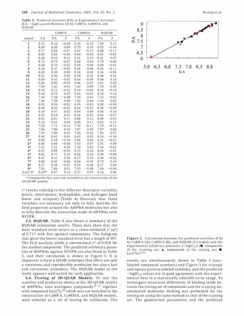
results are simultaneously shown in Table 5 (star-labeled compound numbers) and Figure 5 (in triangleand square pattern-labeled symbols), and the predicted-logIC50 values are in good agreement with the experi-mental data in a statistically tolerable error range. Toinvestigate structural differences of binding mode be-tween the testing set of compounds and the training set,automated molecular docking was performed for thetesting set using the same method as that of the trainingset. The geometrical parameters and the predicted
Table 5. Predicted Activities (PA) vs Experimental Activities(EA, -logIC50) and Residues (δ) by CoMFA, CoMSIA, andHQSAR
CoMFA CoMSIA HQSAR
compd EA PA δ PA δ PA δ
1 8.10 8.18 -0.08 8.20 -0.10 7.99 0.112 8.89 8.98 0.09 8.79 0.10 8.93 -0.043 8.77 8.84 -0.07 8.92 -0.15 8.88 -0.114 8.60 8.66 -0.06 8.64 -0.04 8.65 -0.055 8.46 8.34 0.12 8.31 0.15 8.41 0.056 8.72 8.79 -0.07 8.68 0.04 8.78 -0.067 8.68 8.70 -0.02 8.68 0.00 8.69 -0.018 8.34 8.37 -0.03 8.40 -0.06 8.45 -0.119 8.20 8.20 0.00 8.18 0.02 8.26 -0.06
10 8.41 8.36 0.05 8.39 0.02 8.40 0.0111 8.60 8.62 -0.02 8.64 -0.04 8.44 0.1612 8.89 8.98 -0.09 8.96 -0.07 8.81 0.0813 7.50 7.45 0.05 7.42 0.08 7.50 0.0014 8.10 8.12 -0.02 8.14 -0.04 8.24 -0.1415 8.44 8.39 0.05 8.45 -0.01 8.34 0.1016 7.30 7.38 -0.08 7.29 0.01 7.35 -0.0517 7.46 7.38 0.08 7.42 0.04 7.44 0.0218 8.92 8.94 -0.02 8.95 -0.03 8.98 -0.0619 8.49 8.50 -0.01 8.52 -0.03 8.58 -0.0920 9.10 9.07 0.03 9.04 0.06 8.96 0.1421 8.55 8.54 0.01 8.56 -0.01 8.62 -0.0722 8.92 8.81 0.11 8.80 0.12 8.90 0.0223 9.10 9.01 0.09 8.99 0.11 8.95 0.1524 7.59 7.71 -0.12 7.70 -0.11 7.70 -0.1125 7.96 7.86 0.10 7.87 0.09 7.87 0.0926 7.92 7.90 0.02 7.94 -0.02 7.85 0.0727 8.44 8.43 0.01 8.42 0.02 8.54 -0.101* 6.00 6.18 -0.18 5.96 0.04 6.21 -0.212* 6.00 6.08 -0.08 5.93 0.07 5.91 0.093* 7.41 7.21 0.20 7.39 0.02 7.43 -0.024* 8.05 8.08 -0.03 8.15 -0.10 8.06 -0.015* 8.82 8.72 0.10 8.68 0.14 8.90 -0.086* 8.47 8.41 0.06 8.57 0.10 8.49 -0.027* 8.89 8.95 -0.06 8.94 -0.05 8.79 0.108* 8.27 8.28 -0.01 8.33 -0.06 8.27 0.009* 7.85 7.84 0.01 7.93 -0.08 7.89 -0.04
kni272* 8.2046 8.07 0.13 8.17 0.03 8.14 0.06
* Compounds that were not included in the construction of the3D-QSAR models.
Figure 5. Correlation between the predicted activities (PA)by CoMFA (A), CoMSIA (B), and HQSAR (C) models and theexperimental inhibitory potencies (-logIC50) (b, compoundsof the training set; 2, compounds of the testing set; 9,kni272).28,46
340 Journal of Medicinal Chemistry, 2002, Vol. 45, No. 2 Huang et al.
binding free energies for these compounds were alsocompiled in Tables 2 and 3 and graphically shown inFigures 2C and 4, respectively. As listed in Table 2 andshown in Figure 2C, the three torsion angles and theconformational RMSD values as compared with kni27228
are in the same levels as that of the training set. Thetorsion angle τ3 for compounds 1*-3*, which indicatesthe relative position of the aromatic ring in the Ar1group and the adjacent carbonyl amide backbone, isalmost 30 degrees less than that of all others. Thismeans that the energy consumed for the conformationalchange from the coplanar to out-of-planar state (bindingconformation) of these three compounds is much lessthan that of the 3-OH’s of the Ar1 compound. In otherwords, the binding energies for compounds 1*-3* mustbe much lower. Inspecting the feature of the bindingconformation of compounds 1*-3*, one intramolecularhydrogen bond is formed between the -OH or the -NH2of the Ar1 group and the oxygen atom of the adjacentamide carbonyl group. Meanwhile, the typical hydrogenbond between the -OH of the Ar1 group of AHPBAs andthe Oδ1 of the Asp29 of HIVPR could not be formed. Thismay be the structural and energetic source for the greatdecrease of binding affinities and therefore the lowerinhibitory potencies for compounds 1*-3*. The muchhigher level of predicted binding free energies shownin Table 3 and Figure 4 is the direct reflection andtestimony of the difference in the binding mode forcompounds 1*-3* as compared with other inhibitors.
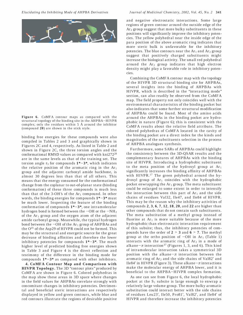
4. CoMFA Contour Map and Comparison withHIVPR Topology. The 3D “contour plots” produced byCoMFA are shown in Figure 6. Colored polyhedras inthe map show these areas in 3D space where changesin the field values for AHPBAs correlate strongly withconcomitant changes in inhibitory potencies. Detrimen-tal and beneficial steric interactions are respectivelydisplayed in yellow and green contours, while blue andred contours illustrate the regions of desirable positive
and negative electrostatic interactions. Some largeregions of green contour around the outside edge of theAr2 group suggest that more bulky substituents in thesepositions will significantly improve the inhibitory poten-cies. The yellow polyhedral near the inside edge of thepara position of the above aromatic ring indicates thatmore steric bulk is unfavorable for the inhibitorypotencies. The blue contours near the Ar1 and Ar2 groupsuggest that positively charged substituents mightincrease the biological activity. The small red polyhedralaround the Ar2 group indicates that high electrondensity might play a favorable role in inhibitory poten-cies.
Combining the CoMFA contour map with the topologyof the HIVPR 3D structural binding site for AHPBAs,several insights into the binding of AHPBAs withHIVPR, which is described in the “interacting mode”section, can also readily be observed from the CoMFAmap. The field property not only coincides well with theenvironmental characteristics of the binding pocket butalso indicates that some further structural modificationof AHPBAs could be found. Most of the amino acidsaround the AHPBAs in the binding pocket are hydro-phobic in nature (Figure 6); this is consistent with theCoMFA results about the relative field attribute. Thecolored polyhedras of CoMFA located in the cavity ofthe binding pocket are a direct index for the kinds andmagnitudes of the substituents selection in the processof AHPBA analogues synthesis.
Furthermore, some SARs of AHPBAs could highlightthe consistency between the 3D-QSAR results and thecomplementary features of AHPBAs with the bindingsite of HIVPR. Introducing a hydrophobic substituentto the meta position of the hydroxyl group at Ar1significantly increases the binding affinity of AHPBAswith HIVPR.17 The green polyhedral around the hy-droxyl group of Ar1 coincides with the hydrophobicpocket enwrapping the Ar1 group. The meta substituentcould be enlarged to some extent in order to intensifythe interaction between this part of Ar1 and the sidechains of residues Val32, Ile47, and Ile84 of HIVPR.This may be the reason why the inhibitory activities ofcompounds 2, 3, 6, 7, 12, 18, 20, and 22 are higher thanother compounds that do not contain such substituents.The meta substitution of a methyl group instead offluorine at Ar1 is more suitable because of the morehydrophobic than electrostatic interaction requirementof this subsite; thus, the inhibitory potencies of com-pounds have the order of 2 > 3 and 6 > 7. The methylgroup at the ortho position of -OH in Ar1 (Table 1)interacts with the aromatic ring of Ar2 in a mode ofalkane-π interaction37 (Figures 1, 3, and 6). This kindof intramolecular interaction takes a symmetrical 3Dposition with the alkane-π interaction between thearomatic ring of Ar2 and the side chains of Val82′ andIle84′ in HIVPR (Figure 3). These alkane-π interactionsmake the molecular energy of AHPBA lower, and it isbeneficial to the AHPBA-HIVPR complex formation.
As one can see from Figure 6, the local hydrophobicpocket at the S1 subsite is large enough to enwrap arelatively large volume group. The more bulky aromaticsubstitution could interact better with the side chainsof residues Leu23′, Ile50, Pro81′, Val82′, and Ile84′ ofHIVPR and therefore increase the inhibitory potencies
Figure 6. CoMFA contour maps as compared with thestructural topology of the binding site in the AHPBA-HIVPRcomplex; only the residues within 5 Å around the inhibitor(compound 20) are shown in the stick style.
Elucidating the Inhibiting Mode of AHPBA Derivatives Journal of Medicinal Chemistry, 2002, Vol. 45, No. 2 341
of AHPBAs. The two large green polyhedras situatedaround the Ar2 group could demonstrate this point ofview, and the meta and/or para substitutions are thebest choice to intensify this local hydrophobic interac-tion. Therefore, it is natural that the inhibitory activitiesof the compounds in Table 1 have the order 19 > 1, 20> 2, 22 > 7, 11 > 9, 12 > 6, and 14 > 16.
Conclusion
We have obtained not only the probable bindingconformations but also the reasonable prediction ofbinding free energies of AHPBAs with HIVPR employ-ing the LGA algorithm of the AutoDock 3.0 program.30
Modeling results indicate that the binding free energiesof AHPBAs calculated by this method correlate very wellwith the reported inhibitory potencies against HIVPR14-17
and provide a structural and energetic explanation forthe differences in the binding affinities of AHPBAs withHIVPR. On the basis of the binding conformations ofAHPBAs, we have developed stable and predictive 3D-QSAR models with acceptable rcross
2 values by under-taking CoMFA, CoMSIA, and HQSAR techniques, andthese models could be mapped back to the structuraltopology of the active site in HIVPR. This leads to abetter understanding of important AHPBA-HIVPRinteractions and thus provides guidelines for the struc-tural modifications of the inhibitors and a predictivemodel for scoring novel synthetic candidates.
Typically, structure-based design is focused on theelucidation of enzyme-substrate interactions but doesnot always lead to predictive models. On the other hand,3D-QSAR models do not necessarily reflect topologicalfeatures of the protein structure. These models aregenerally constructed using alignment rules, which arenot always consistent with the characteristics of thebinding conformations. In this study, we successfullycombined these two approaches. The 3D-QSAR resultsallow focus on those regions, where electrostatic, steric,or hydrophobic effects have a dominant role in AHPBA-HIVPR interactions. The predictive ability testing forthe models has validated their robustness, so theapplication of these models for quantitative predictionof inhibitory potencies against HIVPR is possible withina structurally limited range. Hence, for new candidatesas potential HIVPR inhibitors, reliable inhibitory activi-ties can be computed by “interpolation”, and less reliableIC50’s by “extrapolation” might be obtained for candi-dates with lower structural similarity to the trainingset molecules.
Acknowledgment. The authors are grateful for thefinancial support from the National Natural ScienceFoundation of China (Grant 29725203) and the StateKey Program of Basic Research of China (Grant1998051115). We also acknowledge Prof. Arthur J.Olson of the Scripps Research Institute in La Jolla, CAfor his provision of the AutoDock 3.0 program.
References(1) Pearl, L. H.; Taylor, W. R. A structural model for the retroviral
proteases. Nature 1987, 329, 351-354.(2) Ratner, L.; Haseltine, W.; Patarca, R.; Livak, K. J.; Starcich,
B.; Josephs, S. F.; Doran, E. R.; Rafalski, J. A.; Whitehorn, E.A.; Baumeister, K. Complete nucleotide sequence of the AIDSvirus, HTLV-III. Nature 1985, 313, 277-84.
(3) Seelmeier, S.; Schmidt, H.; Turk, V.; von der Helm, K. Humanimmunodeficiency virus has an aspartic-type protease that canbe inhibited by pepstatin A. Proc. Natl. Acad. Sci. U.S.A. 1988,85 (18), 6612-6616.
(4) Appett, K. Crystal structures of HIV-1 protease-inhibitorscomplexes. Perspect. Drug Discovery Des. 1993, 1, 23-48.
(5) Kohl, N. E.; Emini, E. A.; Schleif, W. A.; Davis, L. J.; Heimbach,J. C.; Dixon, R. A.; Scolnick, E. M.; Sigal, I. S. Active humanimmunodeficiency virus protease is required for viral infectivity.Proc. Natl. Acad. Sci. U.S.A. 1988, 85, 4686-4690.
(6) McQuade, T. J.; Tomasselli, A. G.; Liu, L.; Karacostas, V.; Moss,B.; Sawyer, T. K.; Heinrikson, R. L.; Tarpley, W. G. A syntheticHIV-1 protease inhibitor with antiviral activity arrests HIV-likeparticle maturation. Science 1990, 247, 454-456.
(7) Pyring, D.; Lindberg, J.; Rosenquist, A.; Zuccarello, G.; Kvarn-strom, I.; Zhang, H.; Vrang, L.; Unge, T.; Classon, B.; Hallberg,A.; Samuelsson, B. Design and synthesis of potent C(2)-sym-metric diol-based HIV-1 protease inhibitors: effects of fluorosubstitution. J. Med. Chem. 2001, 44 (19), 3083-3091.
(8) Rozzelle, J. E.; Dauber, D. S.; Todd, S.; Kelle, R.; Craik, C. S.Macromolecular inhibitors of HIV-1 protease. J. Biol. Chem.2000, 275, 7080-7086.
(9) Lee, T.; Laco, G. S.; Torbett, B. E.; Fox, H. S.; Lerner, D. L.;Elder, J. H.; Wong, C. H. Analysis of the s3 and s3′ subsitespecificities of feline immunodeficiency virus (FIV) protease:development of a broad-based protease inhibitors efficaciousagainst FIV, SIV, and HIV in vitro and ex vivo. Proc. Natl. Acad.Sci. U.S.A. 1998, 95, 934-944.
(10) Todd, J. M.; Freire, E. The effect of inhibitor binding on thestructural stability and cooperativity of the HIV-1 protease.Proteins: Struct., Funct., Genet. 1999, 36, 147-156.
(11) Dominy, B. N.; Brooks, C. L., III. Methodology for protein-ligandbinding studies: appilication to a model for drug resistance, theHIV/FIV protease system. Proteins: Struct., Funct., Genet. 1999,36, 318-331.
(12) Hagen, S. E.; Domagala, J.; Gajda, C.; Lovdahl, M.; Tait, B. D.;Wise, E.; Holler, T.; Hupe, D.; Nouhan, C.; Urumov, A.; Zeikus,G.; Zeikus, E.; Lunney, E. A.; Pavlovsky, A.; Gracheck, S. J.;Saunders, J.; VanderRoest, S.; Brodfuehrer, J. 4-Hydroxy-5,6-dihydropyrones as inhibitors of HIV protease: the effect ofheterocyclic substituents at C-6 on antiviral potency and phar-macokinetic parameters. J. Med. Chem. 2001, 44 (14), 2319-2332.
(13) Hong, L.; Zhang, X. J.; Foundling, S.; Hartsuck, J. A.; Tang, J.Structure of a G48H mutant of HIV-1 protease explains howglycine-48 replacements produce mutants resistant to inhibitordrugs. FEBS Lett. 1997, 420, 11-16.
(14) Sakurai, M.; Higashida, S.; Sugano, M.; Komai, T.; Yagi, R.;Ozawa, Y.; Handa, H.; Nishigaki, T.; Yabe, Y. Structure-activityrelationships of HIV-1 PR inhibitors containing AHPBA. Bioorg.Med. Chem. 1994, 2, 807-825.
(15) Komai, T.; Higashida, S.; Sakurai, M.; Nitta, T.; Kasuya, A.;Miyamaoto, S.; Yagi, R.; Ozawa, Y.; Handa, H.; Mohri, H.;Yasuoka, A.; Oka, S.; Nishigaki, T.; Kimura, S.; Shimada, K.;Yabe, Y. Structure-activity relationships of HIV-1 PR inhibitorscontaining AHPBA?II. Modification of pyrrolidine ring at P1′proline. Bioorg. Med. Chem. 1996, 4, 1365-1377.
(16) Takashiro, E.; Watanabe, T.; Nitta, T.; Kasuya, A.; Miyamoto,S.; Ozawa, Y.; Yagi, R.; Nishigaki, T.; Shibayama, T.; Nakagawa,A.; Iwamoto, A.; Yabe, Y. Structure-activity relationship ofHIV-1 protease inhibitors containing AHPBA. Part III: Modi-fication of P2 site. Bioorg. Med. Chem. 1998, 6, 595-604.
(17) Takashiro, E.; Hayakawa, I.; Nitta, T.; Kasuya, A.; Miyamoto,S.; Ozawa, Y.; Yagi, R.; Yamamoto, I.; Shibayama, T.; Nakagawa,A.; Yabe, Y. Structure-activity relationship of HIV-1 proteaseinhibitors containing R-hydroxy-â-amino acids. Detailed studyof P1 site. Bioorg. Med. Chem. 1999, 7, 2063-2072.
(18) Kaldor, S. W.; Kalish, V. J.; Davies, J. F.; Shetty, B. V.; Fritz, J.E.; Appelt, K.; Burgess, J. A.; Campanale, K. M.; Chirgadze, N.Y.; Clawson, D. K.; Dressman, B. A.; Hatch, S. D.; Khalil, D. A.;Kosa, M. B.; Lubbehusen, P. P.; Muesing, M. A.; Patick, A. K.;Reich, S. H.; Su, K. S.; Tatlock, J. H. Viracept (nelfinavirmesylate, AG1343): a potent, orally bioavailable inhibitor ofHIV-1 protease. J. Med. Chem. 1997, 40, 3979-3985.
(19) Ringhofer, S.; Kallen, J.; Dutzler, R.; Billoch, A.; Visser, A. J.W. G.; Scholz, D.; Steinhauser, O.; Schreiber, H.; Auer, M.;Kungl, A. J. X-ray structure and conformational dynamics of theHIV-1 protease in complex with the inhibitor sdz283-910:agreement of time-resolved spectroscopy and molecular dynam-ics simulations. J. Mol. Biol. 1999, 286, 1147-1159.
(20) Gupta, S. P.; Babu, M. S. Quantitative structure-activityrelationship studies on cyclic cyanoguanidines acting as HIV-1protease inhibitors. Bioorg. Med. Chem. 1999, 7, 2549-2553.
(21) Jiang, H. L.; Chen, K. X.; Tang, Y.; Chen, J. Z.; Li, Q.; Wang, Q.M.; Ji, R. Y. Molecular modeling and 3D-QSAR studies on theinteraction mechanism of tripeptidyl thrombin inhibitors withhuman a-thrombin. J. Med. Chem. 1997, 40, 3085-3090.
342 Journal of Medicinal Chemistry, 2002, Vol. 45, No. 2 Huang et al.
(22) Cramer, M.; Cramer, R. D., III; Jones, D. M. Comparativemolecular field analysis. 1. Effect of shape on binding of steroidsto carrier proteins. J. Am. Chem. Soc. 1988, 110, 5959-5967.
(23) Klebe, G.; Abraham, U.; Mietzner, T. Molecular similarity indicesin a comparative analysis(CoMSIA) of drug molecules to cor-relate and predict their biological activity. J. Med. Chem. 1994,37, 4130-4146.
(24) Tong, W.; Lowis, D. R.; Perkins, R.; Chen, Y.; Welsh, W. J.;Goddette, D. W.; Heritage, T. W.; Sheehan, D. M. Evaluation ofquantitative structure-activity relationship methods for large-scale prediction of chemicals binding to the estrogen receptor.J. Chem. Inf. Comput. Sci. 1998, 38, 669-677.
(25) Sybyl, version 6.5; Tripos Associates: St. Louis, MO, 1998.(26) Frisch, M. J.; Trucks, G. W.; Schlegel, H. B.; Scuseria, G. E.;
Robb, M. A.; Cheeseman, J. R.; Zakrzewski, V. G.; Montgomery,J. A., Jr.; Stratmann, R. E.; Burant, J. C.; Dapprich, S.; Millam,J. M.; Daniels, A. D.; Kudin, K. N.; Strain, M. C.; Farkas, O.;Tomasi, J.; Barone, V.; Cossi, M.; Cammi, R.; Mennucci, B.;Pomelli, C.; Adamo, C.; Clifford, S.; Ochterski, J.; Petersson, G.A.; Ayala, P. Y.; Cui, Q.; Morokuma, K.; Malick, D. K.; Rabuck,A. D.; Raghavachari, K.; Foresman, J. B.; Cioslowski, J.; Ortiz,J. V.; Stefanov, B. B.; Liu, G.; Liashenko, A.; Piskorz, P.;Komaromi, I.; Gomperts, R.; Martin, R. L.; Fox, D. J.; Keith, T.;Al-Laham, M. A.; Peng, C. Y.; Nanayakkara, A.; Gonzalez, C.;Challacombe, M.; Gill, P. M. W.; Johnson, B. G.; Chen, W.; Wong,M. W.; Andres, J. L.; Head-Gordon, M.; Replogle, E. S.; Pople,J. A. Gaussian 98; Gaussian, Inc.: Pittsburgh, PA, 1998.
(27) Purcel, W. P.; Singer, J. A. J. Chem. Eng. Data 1967, 12, 235-246. Details of the implementation are given in Sybyl 6.5 TheoryManual; Tripos: St. Louis, MO, 1998; p 69.
(28) Baldwin, E. T.; Bhat, T. N.; Gulnik, S.; Liu, B.; Topol, I. A.; Kiso,Y.; Mimoto, T.; Mitsuya, H.; Erickson, J. W. Structure of HIV-1protease with KNI-272, a tight-binding transition-state analoguecontaining allophenylnorstatine. Structure 1995, 3 (6), 581-590.
(29) InsightII, version 98; Molecular Simulation Inc.: California,1998.
(30) Morris, G. M.; Goodsell, D. S.; Halliday, R. S.; Huey, R.; Hart,W. E.; Belew, R. K.; Olson, A. J. Automated docking usingLamarckian genetic algorithm and empirical binding free energyfunction. J. Comput. Chem. 1998, 19, 1639-1662.
(31) Solis, F. J.; Wets, R. J. B. Minimization by random searchtechniques. Maths Opera. Res. 1981, 6, 19-30.
(32) Weiner, S. J.; Kollman, P. A.; Case, D. A.; Singh, C.; Ghio, G.;Alagona, S.; Profeta, P.; Weiner, P. J. Am. Chem. Soc. 1984, 106,765-784. Details of the implementation are given in Sybyl 6.5Theory Manual; Tripos: St. Louis, MO, 1998; p 441.
(33) Morris, G. M.; Goodsell, D. S.; Huey, R.; Olson, A. J. Distributedautomated docking of flexible ligands to proteins: parallelapplications of AutoDock 2.4. J. Comput.-Aided Mol. Des. 1996,10, 293-304.
(34) Ghose, A. K.; Viswanadhan, V. N.; Wendoloski, J. J. A knowledge-based approach in designing combinatorial or medicinal chem-istry libraries for drug discovery. 1. A qualitative and quanti-tative characterization of known drug databases. J. Comb. Chem.1999, 1, 55-68.
(35) Thompson, W. J.; Fitzgerald, P. M.; Holloway, M. K.; Emini,E. A.; Darke, P. L.; McKeever, B. M.; Schleif, W. A.; Quintero,J. C.; Zugay, J. A.; Tucker, T. J.; Schwering, J. E.; Homnick, C.F.; Nunberg, J.; Springer, J. P.; Juff, J. R. Synthesis andantiviral activity of a series of HIV-1 protease inhibitors withfunctionality tethered to the P1 or P1′ phenyl substituents:X-ray crystal structure assisted design. J. Med. Chem. 1992, 35,1685-1701.
(36) Thanki, N.; Rao, J. K.; Foundling, S. I.; Howe, W. J.; Moon, J.B.; Hui, J. O.; Tomasselli, A. G.; Heinrikson, R. L.; Thaisrivongs,S.; Wlodawer, A. Crystal structure of a complex of HIV-1 pro-tease with a dihydroxyethylene-containing inhibitor: compari-sons with molecular modeling. Protein Sci. 1992, 1, 1061-1072.
(37) Kim, K. S.; Tarakeshwar, P.; Lee, J. Y. Molecular clusters ofπ-systems: theoretical studies of structures, spectra, and originof interaction energies. Chem. Rev. 2000, 100, 4145-4185.
(38) Head-Gordon, M.; Pople, J. A. A Method for two-electronGaussian integral and integral derivative evaluation usingrecurrence relations. J. Chem. Phys. 1988, 89, 5777.
(39) POV-ray-Team; POV-ray, version 3; 1999 (www.povray.org).(40) Wallace, A. C.; Laskowski, R. A.; Thornton, J. M. LIGPLOT: A
program to generate schematic diagrams of protein-ligandinteractions. Protein Eng. 1995, 8, 127-134.
(41) Erickson, J.; Neidhart, D. J.; VanDrie, J.; Kempf, D. J.; Wang,X. C.; Norbeck, D. W.; Plattner, J. J.; Rittenhouse, J. W.; Turon,M.; Wideburg, N.; Kohlbrenner, W. E.; Simmer, R.; Helfrich, R.;Paul, D. A.; Knigge, M. Design, activity, and 2.8 A crystalstructure of a C2 symmetric inhibitor complexed to HIV-1protease. Science 1990, 527-533.
(42) Rosin, C. D.; Belew, R. K.; Walker, W. L.; Morris, G. M.; Olson,A. J.; Goodsell, D. S. Coevolution and subsite decomposition forthe design of resistance-evading HIV-1 protease inhibitors. J.Mol. Biol. 1999, 287, 77-92.
(43) Nicholson, L. K.; Yamazaki, T.; Torchia, D. A.; Grzesiek, S.; Bax,A.; Stahl, S. J.; Kaufman, J. D.; Wingfield, P. T.; Lam, P. Y.;Jadhav, P. K. Flexibility and function in HIV-1 protease. Nat.Struct. Biol. 1995, 2, 274-280.
(44) Rosin, C. D.; Belew, R. K.; Morris, G. M.; Olson, A. J.; Goodsell,D. S. Coevolutionary analysis of resistance-evading peptidomi-metic inhibitors of HIV-1 protease. Proc. Natl. Acad. Sci. U.S.A.1999, 96, 1369-1374.
(45) Stahle, L.; Wold, S. Multivariate data analysis and experimentaldesign in biomedical research. Prog. Med. Chem. 1988, 25, 291-338.
(46) Mimoto, T.; Imai, J.; Kisanuki, S.; Enomoto, H.; Hattori, N.;Akaji, K.; Kiso, Y. Kynostatin (kni)-227 and -272, highly potentanti-HIV agents: conformationally constrained inhiboitors ofHIV protease containing allophenorstatine. Chem. Pharm. Bull.1992, 40 (8), 2251-2253.
(47) Rick, S. W.; Topol, I. A.; Erickson, J. W.; Burt, S. K. Molecularmechanisms of resistance: free energy calculations of mutationeffects on inhibitor binding to HIV-1 protease. Protein Sci. 1998,7 (8), 1750-1756.
(48) Trylska, J.; Antosiewicz, J.; Geller, M.; Hodge, C. N.; Klabe, R.M.; Head, M. S.; Gilson, M. K. Thermodynamic linkage betweenthe binding of protons and inhibitors to HIV-1 protease. ProteinSci. 1999, 8 (1), 180-195.
(49) Velazquez-Campoy, A.; Luque, I.; Todd, M. J.; Milutinovich, M.;Kiso, Y.; Freire, E. Thermodynamic dissection of the bindingenergetics of KNI-272, a potent HIV-1 protease inhibitor. ProteinSci. 2000, 9 (9), 1801-1809.
(50) Kollman, P. Free energy calculations: applications to chemicaland biochemical phenomena. Chem. Rev. 1993, 93, 3 (7), 2395-2417.
(51) Wang, L.; Duan, Y.; Stouten, P.; De Lucca, G. V.; Klabe, R. M.;Kollman, P. Does a diol cyclic urea inhibitor of HIV-1 proteasebind tighter than its corresponding alcohol form? A study by freeenergy perturbation and continuum electrostatics calculations.J. Comput.-Aided Mol. Des. 2001, 15 (2), 145-156.
JM0102710
Elucidating the Inhibiting Mode of AHPBA Derivatives Journal of Medicinal Chemistry, 2002, Vol. 45, No. 2 343
Copyright © 2022 FDOKUMEN