
Elizabeth Natkanski PhD Thesis.pdf - UCL Discovery
144
Investigating Novel Components and Mechanisms Involved in B Cell Receptor-Antigen Internalisation Elizabeth Mary Natkanski Division of Immune Cell Biology National Institute for Medical Research London UCL A thesis presented for the degree of Doctor of Philosophy, 2013
-
Upload
khangminh22 -
Category
Documents
-
view
1 -
download
0
Transcript of Elizabeth Natkanski PhD Thesis.pdf - UCL Discovery

Investigating Novel Components and Mechanisms Involved in B Cell
Receptor-Antigen Internalisation
Elizabeth Mary Natkanski
Division of Immune Cell Biology National Institute for Medical Research
London
UCL
A thesis presented for the degree of Doctor of Philosophy, 2013

1
Declaration
I, Elizabeth Mary Natkanski, confirm that the work presented in this
thesis is my own. Where information has been derived from other
sources, I confirm that this has been indicated in the thesis.

2
Publications arising from this thesis
Elizabeth Natkanski, Wing-Yiu Lee, Bhakti Mistry, Antonio Casal, Justin E.
Malloy, and Pavel Tolar. (2013). B cells use Mechanical Energy to Discriminate
Antigen Affinities. Science, 340, p 1587-1590

3
Abstract
The elimination of a wide variety of infections requires the production of high
affinity antibodies specific for the invading pathogen. The generation of these
high affinity antibodies depends on the ability of B cells to recognise and
internalise antigens from the surface of antigen-presenting cells (APCs) in an
affinity-dependent manner. B cells expressing high affinity B cell receptors
(BCRs) internalise, process and present more antigen, obtain better T cell help,
and are selectively expanded over lower affinity equivalents. However, the
molecular mechanisms by which B cells extract antigens for presentation remain
unclear.
Using a new fluid and flexible membrane substrate to mimic APCs, we show that
B cells acquire antigen by dynamic myosin IIA-mediated contractions that pull out
and invaginate the presenting membranes. Invaginations containing high affinity
BCR-antigen microclusters were able to withstand the force of these contractions
and recruit clathrin resulting in endocytosis. In contrast, low affinity BCR-antigen
bonds quickly ruptured aborting internalisation.
Thus we conclude that coupling contractility to endocytosis permits B cells to
discriminate between antigen affinities, which provides a mechanism for the
selective advantage seen for high affinity B cell clones in vivo.
Disruptions in BCR-antigen internalisation has been linked to the growth of
malignant B cells and the development of autoimmunity. Therefore, ultimately,
our results could contribute to the effective design of future therapeutics for B cell
diseases.

4
Acknowledgements First of all, and most importantly, I should like to thank my supervisor Dr Pavel
Tolar for giving me the opportunity to work on this project and for encouraging
me to aim high and to be the best scientist I could be. I also would like to
acknowledge my Thesis Committee: Dr Ben Seddon, Dr Victor Tybulewicz and
Dr Tom Carter for taking the time during my PhD to critically examine my project
and to give their invaluable advice.
Dr Wing-Yiu Lee has been an admirable fountain of knowledge from day one,
and I will always be grateful for all the time he gave in helping me with the
project. My thanks also go to all my friends, both within NIMR and out, for being
there to support me and for their continual encouragement.
Thanks too, to my parents for all their support throughout my years of full time
education. Their ideals of working hard, being thoughtful and having a
dedication to seeing things through in many areas of their lives has always
been an inspiration. Last, but by no means least, a big thank-you to Paul
Eastwood, I couldn’t have done it without you.
.
5
Table of contents Declaration 1
Publications arising from this thesis 2
Abstract 3
Acknowledgements 4
Table of contents 5
List of figures and tables 9
List of abbreviations 11
Chapter 1 Introduction 15
1.1 The Immune System 15
1.2 BCR-antigen internalisation – an overview 16
1.3 Antigen encounter 19
1.3.1 Soluble antigen encounter 20
1.3.2 Membrane-bound antigen encounter 22
1.3.3 The capture of antigen by antigen-presenting cells 23
1.3.4 The transport of antigen by antigen-presenting cells to B cells 24
1.3.5 Antigen-presenting cells in maintaining the B cell follicle and
supporting germinal centre function 26
1.4 B cell development 26
1.5 BCR signalling 28
1.5.1 BCR signalling in the absence of antigen 28
1.5.2 Antigen-induced BCR signalling 29
1.5.3 Translocation to lipid rafts 31
1.5.4 Synapse formation in response to membrane-bound antigen 32
1.6 BCR-antigen internalisation 34
1.7 Affinity-dependent antigen internalisation 36
1.8 Defects in membrane-bound antigen internalisation can lead to
autoimmunity or cancer 40
1.8.1 Autoimmunity 40
1.8.2 Cancer 43

6
1.9 Thesis Aims 44
Chapter 2 Materials and Methods 45
2.1 Mice 45
2.2 B cell purification and cell culture 45
2.3 Antigens 46
2.4 Antibodies 47
2.5 Inhibitors 48
2.6 Planar lipid bilayers (PLB) 49
2.7 Plasma membrane sheets (PMS) 50
2.8 Plasma membrane planar lipid bilayers (PMPLB) – Wing-Liu Lee 50
2.9 Imaging 50
2.10 Atomic force microscopy (AFM) force spectroscopy – Pavel Tolar 51
2.11 Internalisation assays 52
2.11.1 Soluble antigen internalisation using flow cytometry 52
2.11.2 Soluble antigen internalisation using microscopy 52
2.11.3 Membrane-bound antigen internalisation 52
2.12 Fluorescence recovery after photobleaching (FRAP) 53
2.13 3D particle localisation – Pavel Tolar 54
2.14 Analysis of membrane invaginations 54
2.15 Protein knock-down using siRNA 55
2.16 Western blot 55
2.17 Lentiviral production and infection 56
2.18 Retroviral production and infection 57
2.19 Analysis of clathrin light chain-GFP, myosin IIA RLC-GFP, and LifeAct
activity 58
2.20 B cell phenotyping of Cre+MyH9fl/fl mice – in collaboration with Harold
Hartweger 58
Chapter 3 Plasma membrane sheets are a new artificial substrate that can mimic antigen-presenting cells when studying BCR-antigen internalisation 60
3.1 Introduction 60
3.2 Results 61

7
3.2.1 B cells are unable to internalise antigen tethered to planar lipid
bilayers 61
3.2.2 B cells are able to internalise antigen from plasma membrane
sheets 64
3.2.3 Plasma membrane sheets are similar in their viscoelastic properties
to antigen-presenting cells 69
3.2.4 B cells pull on and invaginate the presenting membrane during
antigen internalisation 71
3.2.5 Antigen internalisation occurs only at sites of long-lived invagination
formation 74
3.3 Discussion 76
Chapter 4 B cells use myosin IIA contractility combined with clathrin-mediated endocytosis to internalise membrane-bound antigen 79
4.1 Introduction 79
4.2 Results 80
4.2.1 SiRNA is not a reliable method of protein knockdown for the genetic
screen 80
4.2.2 Lentiviral delivery of shRNA mediates protein knockdown of
endocytic mediators in B cells 82
4.2.3 Inhibition of internalisation following protein knockdown can be
assessed by flow cytometry or microscopy 84
4.2.4 BCR-antigen internalisation is reduced in Ramos cells following
shRNA mediated knockdown of myosin IIA and clathrin 86
4.2.5 BCR-antigen internalisation is reduced in Bl6 B cells following
pharmacological inhibition of myosin IIA and clathrin 89
4.2.6 Clathrin is recruited to the synapse at the point of antigen
internalisation 92
4.2.7 Myosin IIA is recruited to the synapse immediately before
invagination formation 97
4.3 Discussion 100
Chapter 5 Myosin IIA contractility enables B cells to discriminate between antigen affinities 106
8
5.1 Introduction 106
5.2 Results 107
5.2.1 B cells internalise antigen presented by plasma membrane sheets
in an affinity-dependent manner 107
5.2.2 Antigen clustering and invagination lifetime depends on antigen
affinity 109
5.2.3 Affinity discrimination is mediated by myosin IIA contractility 111
5.2.4 Deletion of myosin IIA in B cells prevents B cell development past
the PrePro B cell stage 112
5.3 Discussion 117
Chapter 6 Conclusions 121
References 126
Appendix – Movie figure legends 143

9
List of figures and tables
Figure 1: The BCR 16
Figure 2: BCR-antigen internalisation 18
Figure 3: Antigen encounter in the lymph node 22
Figure 4: B cell development 27
Figure 5: B cells form synapses with antigen-presenting surfaces 33
Figure 6: B cells cannot internalise antigen when presented on PLB 63
Figure 7: Bl6 B cells can internalise antigen presented by PMS 66
Figure 8: Primary B cells internalise antigen with portions of the presenting
membrane from PMS and DC, but not from PLB 68
Figure 9: PMS are similar in their fluidity compared to PLB and PMPLB as
shown by FRAP 69
Figure 10: PMS are flexible and similar in their viscoelastic properties to APCs
71
Figure 11: Bl6 B cells pull on and invaginate the PMS during antigen
internalisation 73
Figure 12: Increased spots of DiI fluorescence are associated with the upward
movement of antigen during antigen internalisation 73
Figure 13: Invaginations produced by Bl6 B cells are dynamic and have varying
lifetimes. Long-lived invaginations begin at sites of antigen microclustering and
lead to antigen internalisation 75
Figure 14: Nucleofection of siRNA produces a modest protein knockdown in B
cells 82
Figure 15: Infection of lentivirus-containing shRNA can mediate protein
knockdown in Ramos cells and Bl6 B cells 84
Figure 16: Soluble antigen internalisation is inhibited following dynamin2
knockdown by shRNA-containing lentivirus 85
Figure 17: A pilot customised genetic screen was used to identify components
involved in antigen internalisation from the PMS 88
Figure 18: Ramos cells need to invaginate the PMS using myosin IIA
contraction before internalising the antigen in a clathrin-dependent manner 88
Figure 19: Bl6 B cells invaginate the PMS using myosin IIA contraction before
internalising the antigen in a clathrin-dependent manner 91

10
Figure 20: Inhibition of myosin IIA, Rock1 or dynamin2 affects the extent of Bl6
B cell spreading on the PMS 92
Figure 21: Retroviral infection of clathrin light chain-GFP in Bl6 B cells show
clathrin recruitment to Bl6 B cell synapse, and colocalisation of clathrin with
long-lived invaginations during antigen internalisation from the PMS 94
Figure 22: Bl6 B cells internalise antigen from the PMS following the sequential
formation of antigen microclusters, long-lived invaginations and CCSs 96
Figure 23: Myosin IIA is recruited to the Bl6 B cell synapse at the onset of
invagination formation 98
Figure 24: F-actin is recruited to the Bl6 B cells synapse in a biphasic manner
99
Figure 25: B1-8 B cells can discriminate between antigen affinities when
presented on PMS 108
Figure 26: B1-8 B cells produce more long-lived invaginations, produce
microclusters faster, and have increased amounts of invagination-associated
antigen, when presented with NIP antigen over NP antigen 110
Figure 27: Modulation of myosin IIA activity affects the ability of B1-8 B cells to
internalise membrane-bound antigen in an affinity-dependent manner 112
Table 1: B cell markers used to identify different B cell subsets harvested from
mice 113
Figure 28: Cre+MyH9fl/fl mice have a block in B cell development 116
Figure 29: Cre+MyH9fl/fl mice have smaller spleens compared to controls 116
Figure 30: Cre+MyH9fl/fl B cells which develop past the PrePro stage in the
bone marrow are larger 117
Figure 31: BCR internalisation of membrane-bound antigen 123

11
List of abbreviations
AFM Atomic force microscopy
AP2 Activating protein 2
APC Antigen-presenting cell
Arh-GAP Rho GTPase-activating protein
Arh-GEF Rho GTPase-guanine exchange factor
B1-8 B1-8fl/flIgκ-Ctm1Cgn/tm1Cgn
BAFF B cell activating factor
BCR B cell receptor
Bl6 C57 black 6
BSA Bovine serum albumin
C-terminal Carboxyl-terminal
C-type lectin Calcium-binding lectin
CCL Chemokine (C-C motif) ligand
CCP Clathrin-coated pit
CCR C-C chemokine receptor
CCS Clathrin-coated structure
CD Cluster of differentiation
CO2 Carbon dioxide
CpG Cytosine-phosphate-guanine
Cre Cre recombinase
CXCL Chemokine (C-X-C motif) ligand
CXCR Cy
C-X-C chemokine receptor
Cyanine dye
DC Dendritic cell
DiI/ DiD Dialkylcarbocyanines
DNA Deoxyribonucleic acid
dSTORM Direct stochastic optical reconstruction microscopy
Erk Extracellular signal-regulated kinases
et al and others
Fab FBS
Antigen binding fragment
Foetal bovine serum

12
Fc Crystallisable fragment
FcγRIIb Low affinity immunoglobulin gamma FC region receptor IIb
FDC Follicular dendritic cell
Fl Flox
FRAP Fluorescence recovery after photobleaching
FRC Fibroblastic reticular cell
FRET Fluorescence resonance energy transfer
GFP Green fluorescent protein
HBSS Hank's buffered saline solution
HEK Human embryonic kidney
HEL Hen egg lysozyme
HEV High endothelial venule
Ig Immunoglobulin
IL Interleukin
ITAM Immunoreceptor tyrosine-based activation motif
kDa Kilo dalton
LPS Lipopolysaccharide
MHC Class II Major histocompatibility complex class two
MyH9 Myosin heavy chain 9
Myl Myosin light chain
Myo Myosin
N-terminal Amino-terminal
NP/NIP Nitrophenyl hapten
Oligo Oligonucleotides
Pax Paried box protein
PBS Phosphate buffered saline
PCR Polymerase chain reaction
PI3 kinase Phosphoinositide 3 kinase
PKC Protein kinase C
PLAT-E Platinum-E
PLB Planar lipid bilayers
PMPLB Plasma membrane planar lipid bilayers
PMS Plasma membrane sheets

13
Rho Ras homolog gene family member
RLC Regulatory light chain
RNA Ribonucleic acid
ROCK Rho-associated coiled-coil containing protein kinase
SCS subsacpsular sinus
shRNA Short-hairpin ribonucleic acid
siRNA Small-interfering ribonucleic acid
TCR T cell receptor
TEL Turkey egg lysozyme
Tfh T follicular helper cell
TI T cell-independent antigens
TIRF Total internal reflection fluorescence
TLR Toll-like receptor
YFP Yellow fluorescent protein
α Alpha
α- Anti-
β Beta
δ Delta
κ Kappa
µ Micro
°C Degrees Celsius
% Percent
g Gram
Ka Affinity constant
m Metre
mg Milligram
ml Millilitre
mM Milimolar
mm Millimetre
mN Millinewton
ng Nanogram
nM Nanomolar
Nm Nanometre

14
PFA Paraformaldehyde
pN Piconewton
rpm Revolutions per minute
U Units
x g G-force
µg Microgram
µl Microlitre
µM Micromolar
µm Micrometre

15
Chapter 1 Introduction
1.1 The Immune System The immune system is a complex network of specialised cells, tissues and
molecular components designed to efficiently recognise, control and clear
infections.
The immune system is comprised of innate and adaptive immune cells. Innate
immune cells initially recognise patterns in the invading pathogens as “non-self”
and initiate the immune reaction by the production of cytokines and chemokines,
which activate other cellular members of the immune system and recruit them to
the site of infection. Some innate cells such as macrophages and dendritic cells
(DCs) are also able to present the peptides from the pathogen, or the pathogen
itself in its native conformation, for recognition by T cells and B cells,
respectively.
T and B cells constitute the adaptive immune system and are able to “adapt” their
responses to be specific for the infectious agent following recognition of antigen
by their highly specialised antigen receptors. Each and every B cell receptor
(BCR) and T cell receptor (TCR) is unique due to somatic recombination and
introduction of point mutations into the genes encoding both receptors during
development. This greatly increases the chances of antigen recognition of
unforeseen pathogens, which may have evolved to evade detection by the innate
immune cells. B and T cell clones, which bind to the antigen with high affinity, are
then selectively expanded to elicit antigen-specific mechanisms of pathogen
elimination and differentiate into memory cells in preparation for any subsequent
reinfection. How B cells are able to initiate these antigen-specific responses will
be the focus of this study.

16
1.2 BCR-antigen internalisation – an overview B cells begin their development in the bone marrow, and then migrate to the
spleen to become fully mature naïve B cells primed for antigen encounter. B cells
circulate in the blood and through secondary lymphoid organs in search of their
cognate antigen. B cells bind to antigen in its native form and can recognise it
both in solution and tethered to a cell of the innate immune system, which can act
as an antigen-presenting cell (APC).
B cells bind to antigen through the variable region of the membrane
immunoglobulin (Ig) component of the BCR (Lanzavecchia, 1987) (Figure 1).
Binding of antigen to the BCR stimulates antigen-induced BCR signalling through
the cytoplasmic domains of the IgαIgβ heterodimer, which is non-covalently
associated with membrane Ig (Figure 1) (Hombach et al., 1990). This signalling,
switches on the B cell nuclear activation programme and acts as the first signal
required for B cell activation.
Figure 1: The BCR
As well as initiating signalling, antigen binding stimulates the BCR to enter the
endocytic pathway and internalise the BCR-antigen complex (Figure 2). The BCR

17
traffics the antigen to intracellular degradative compartments where it is
degraded into peptides and loaded onto major histocompatibility complex class
two molecules (MHC Class II) (Davidson et al., 1991) (Figure 2). These MHC
class II proteins are then presented on the B cell surface for recognition by
cognate T cells (Lanzavecchia, 1985). Specific recognition between TCR and
MHC Class II-peptide permits the release of T cell co-stimulatory factors such as
interleukins (IL) and CD40 ligand (CD40L) (Figure 2), which give context-
dependent stimulation and act as second signals required for full B cell activation
and movement of the B cell into germinal centres (Han et al., 1995; Lane et al.,
1992; Parker, 1993). B cells in the germinal centre are highly proliferative and go
through rounds of somatic hypermutation on their BCRs to increase diversity of
the antigen binding. B cells containing mutations improving their affinity for
antigen are selectively expanded, released into the circulation, and differentiate
into plasma cells to produce high affinity antibodies for pathogen elimination, or
into memory cells in preparation for any subsequent reinfection (Rajewsky, 1996;
Victora and Nussenzweig, 2012).
In addition to antigen processing and presentation, BCR-antigen internalisation
also exposes antigen to endosomal toll-like receptors 7 and 9 (TLR7 and 9)
(Figure 2), which are able to recognise RNA (Lau et al., 2005) and DNA (Bauer et
al., 2001) sequences from viral and bacterial infections. Recognition of antigen by
TLR7 or 9 can augment the B cell activation and aid pathogen recognition and
clearance.
BCR-antigen internalisation also has role in regulating BCR signalling,
(Chaturvedi et al., 2011). For example, internalisation has been associated with
the termination of calcium and MAP kinase activity and the promotion of AKT
stimulation. Therefore, disregulation of BCR internalisation and trafficking may
contribute to the inappropriate BCR signalling, which has been observed in B cell
lymphomas (Davis et al., 2010).
BCR-antigen internalisation is therefore the gateway for efficient B cell activation
and stringent regulation of this process is vital in ensuring an efficient humoral
response, while preventing pathology.

18
Figure 2: BCR-antigen internalisation
A) BCRs can recognise antigen in solution
B) BCRs can recognise antigen tethered to an APC
C) Antigen binding initiates BCR signalling and stimulates the B cell’s nuclear activation
programme (signal 1)
D) Antigen binding stimulates internalisation of BCR-antigen complexes
E) Internalised BCR-antigen complexes are trafficked to lysosomes, processed, and loaded
onto MHC Class II molecules
F) Antigen loaded MHC Class II molecules are presented on the B cell surface for specific
recognition by cognate T cells
G) Recognition of the antigen-loaded MHC Class II by T cells provides activation factors
(signal 2)
H) Internalisation of antigen also permits antigen recognition by intracellular pathogen
recognitions receptors such as TLRs

19
1.3 Antigen encounter B cells primarily reside in the lymph nodes and the spleen. Considering the
diverse range of the BCR repertoire, the chances of random contact between
antigen and cognate B cell would be rare. Therefore secondary lymphoid organs
are specifically structured to facilitate efficient antigen exposure to rare cognate B
cells and to control cell-cell interactions (Batista and Harwood, 2009). In the
lymph node, two areas can be identified based on their histological features: the
cortex and the medulla. The cortex can be sub-divided into B cell follicles, which
contains B cells, and the paracortex, also called the T cell zone, which contains T
cells and DCs (Andrian and Mempel, 2003). The architecture of these regions is
maintained by non-haematopoietic stromal cells such as fibroblastic reticular cells
(FRCs) and follicular dendritic cells (FDCs). Apart from structural support, these
cells also produce CXCL13 or CCL19, the defining chemokines that attract B and
T cells to their respective zones (Andrian and Mempel, 2003; Gonzalez et al.,
2011).
Lymph nodes are linked to both the lymphatic and circulatory systems and are
strategically positioned around the body to ensure efficient trafficking and capture
of lymphocytes arriving by blood and antigen draining from tissues (Batista and
Harwood, 2009). B cells circulate around the body and enter the lymph nodes
through the high endothelial venule (HEV). B cells then follow the CXCL13
gradient and reside in the follicle just below the subcapsular sinus (SCS), at the
site of lymphatic antigen entry (Cyster, 2010). Whilst screening for antigen in the
follicle, B cells are active and migrate at approximately 6 µm/ minute (Okada et
al., 2005). B cells are able to recognise both soluble antigen, which can filter into
the follicle, or antigen retained on the surface of an APC (discussed below).
Following specific antigen binding, B cells upregulate CCR7 (receptor for CCL19)
and move towards the T cell zone for T cell help and the initiation of the B cell
response (Okada et al., 2005). If the B cell does not come into contact with
specific antigen after approximately 24 hours it re-enters the circulation in search
of the next lymph node and cognate antigen (Goodnow, 1997).

20
The white pulp of the spleen is similar in its structure to the lymph node with B
cell follicles and the T cell zone. Surrounding the white pulp is the red pulp, which
is rich in vasculature and functions to filter blood for cellular debris, foreign
substances, and microorganisms. An area called the marginal zone, containing
specialised marginal zone B cells, macrophages, and DCs, controls the entry of
blood-borne antigen from the red pulp into the white pulp and exposure to the
residing lymphocytes (Mebius and Kraal, 2005).
1.3.1 Soluble antigen encounter Soluble antigen can arise through the protease-mediated cleavage of larger
microorganisms (Catron et al., 2010) or secreted bacterial products (Roozendaal
et al., 2009), or alternatively, from an injection or vaccination. These soluble
antigens drain into the lymphatic vessels and are carried in the lymphatic fluid to
the lymph node. The lymph node itself is protected from the unregulated flow of
soluble antigens into the cortex by the SCS, which separates the afferent
lymphatic vessel and the B cell follicle (Gretz et al., 2000). However, it has been
shown that B cells are able to respond to these soluble antigens indicating the
presence of a controlled mechanism permitting the antigens to cross this barrier.
Early electron microscopy images of the lymph node revealed small holes (0.1-1
µm) in the lining of the SCS (van Ewijk et al., 1988) indicating the possibility that
soluble antigen could filter through these holes into the follicle (Figure 3A). In
support of this theory Pape et al 2007 intradermally injected hen egg lysozyme
(HEL) antigen, tagged with green fluorescent protein (GFP), into the ears of
recipient mice, which had previously been injected with HEL-specific MD4 B cells
(Pape et al., 2007). Flash frozen cryosections of these lymph nodes
demonstrated that MD4 B cells 150 µm away from the SCS and site of antigen
entry, were able to acquire these antigens within minutes of injection. As B cells
are unable to move more than approximately 6 µm/ minute without antigen, and
are known to transiently decrease their mobility after specific antigen recognition
(Okada et al., 2005), the authors suggest that antigen-positive B cells must have
acquired antigen which diffused passively through the holes in the SCS.

21
More recently studies have shown that soluble antigens enter the B cell follicles
through a conduit system (Figure 3A). Conduits are comprised of a core of
collagen fibres surrounded by FRCs covering a basement membrane sheath.
Conduits are abundant in the T cell zone, and in neonatal lymph nodes there are
also ample levels in the B cell follicle, however during development, FDCs
replace the majority of conduits in the B cell zone, leaving a much less dense
network compared to the paracortex (Bajénoff and Germain, 2009). Using
intravital imaging Roozendaal et al 2009 demonstrated that small soluble
fluorescently-labelled turkey egg lysozyme (TEL) antigen injected
subcutaneously into recipient mice, entered the lymph node in a centripetal
fashion, highlighting the presence of a follicular conduit network (Roozendaal et
al., 2009). Observations of B cells acquiring this antigen, and electron
microscopy images showing B cell pseudopods directly sampling the antigen-
filled conduit core, demonstrated the use of these conduits in delivering antigen
to B cells. The authors also revealed that if the TEL antigen was conjugated to a
large fluorescent label (250 kDa), it was initially arrested at the SCS and then
entered the lymph node with a delay. These findings mirror earlier studies
showing that conduits have a size exclusion limit of 70 kDa (Gretz et al., 2000),
and suggest that bigger antigens, require alternate mechanisms for entry into the
follicle.
22
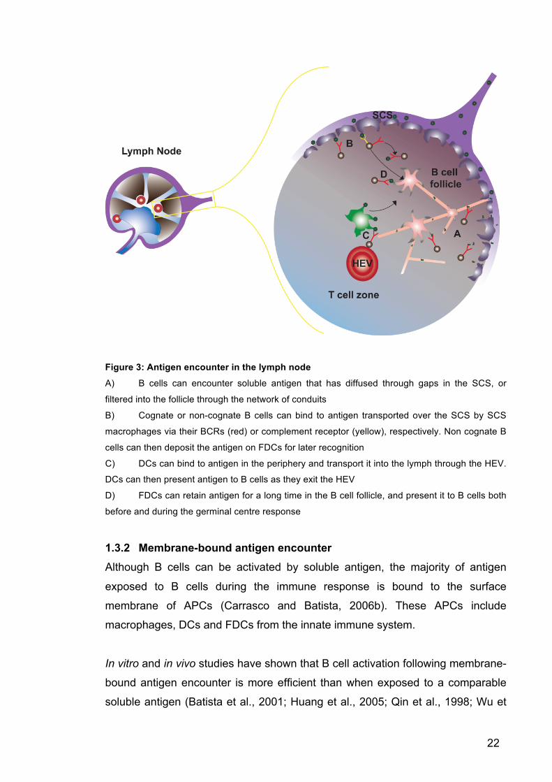
Figure 3: Antigen encounter in the lymph node
A) B cells can encounter soluble antigen that has diffused through gaps in the SCS, or
filtered into the follicle through the network of conduits
B) Cognate or non-cognate B cells can bind to antigen transported over the SCS by SCS
macrophages via their BCRs (red) or complement receptor (yellow), respectively. Non cognate B
cells can then deposit the antigen on FDCs for later recognition
C) DCs can bind to antigen in the periphery and transport it into the lymph through the HEV.
DCs can then present antigen to B cells as they exit the HEV
D) FDCs can retain antigen for a long time in the B cell follicle, and present it to B cells both
before and during the germinal centre response
1.3.2 Membrane-bound antigen encounter Although B cells can be activated by soluble antigen, the majority of antigen
exposed to B cells during the immune response is bound to the surface
membrane of APCs (Carrasco and Batista, 2006b). These APCs include
macrophages, DCs and FDCs from the innate immune system.
In vitro and in vivo studies have shown that B cell activation following membrane-
bound antigen encounter is more efficient than when exposed to a comparable
soluble antigen (Batista et al., 2001; Huang et al., 2005; Qin et al., 1998; Wu et

23
al., 2008). This could be because of the expression of integrin receptors by the
APC to facilitate long-lived and stable interactions with the cognate B cells (Allen
and Cyster, 2008; Carrasco and Batista, 2006a; Carrasco et al., 2004). Or it
could be because APCs concentrate and cluster antigen thereby increasing its
avidity (Suzuki et al., 2009). APCs can also augment B cell activation through
stimulation of the B cell co-receptors or through the secretion of soluble survival
and activation factors (Allen and Cyster, 2008; Balázs et al., 2002; Depoil et al.,
2007; Gonzalez et al., 2010a; Qin et al., 1998; Weber et al., 2008), as well as
providing a platform for B-T cell interaction (Bergtold et al., 2005).
As well as increasing B cell signalling and activation, APCs can also act as
antigen transporters to ensure B cells are able to see antigen that cannot diffuse
passively into the lymph node, and they can support the continual improvement
of the humoral response during affinity maturation in the germinal centres
following infection.
1.3.3 The capture of antigen by antigen-presenting cells Before B cells can become activated by APCs, APCs must first capture antigen
and retain it on the surface of their cell membrane. B cells need to be able to
recognise bacteria, viruses, fungi, and parasites, as well as environmental or
synthetic chemicals. Although APCs don’t need to recognise these antigens
specifically, they do need to identify them as non-self so they can present them
as pathogenic entities to the adaptive immune system. To be able to do this they
have a selection of cell surface receptors to mediate this task. These include
complement receptors and receptors which can recognise the Fc portions of
soluble antibody, for example FcγRIIb receptors. These receptors recognise
immune complexes, which are comprised of soluble antigen bound by natural or
specific antibody and complement – complement are serum proteins which, when
activated produce a protease cascade whose products bind to antigens and mark
them for subsequent recognition by other components of the immune system
(Bergtold et al., 2005; Carroll, 1998; Gonzalez et al., 2010a; Heesters et al.,
2013; Qin et al., 1998). Another important receptor group, which can mediate
antigen presentation on APCs, are C-type lectins. These receptors can bind to

24
carbohydrate moieties of infectious agents such as viruses and yeast (Gonzalez
et al., 2010c; Taylor et al., 2004).
As BCRs recognise antigen in its native structure, APCs need to hold antigen in
its native from. FcγRIIb and complement receptors have been shown able to
internalise antigen into non-degradative endosomes and recycle them to their cell
surface to allow stable and long-lived presentation of native antigen to B cells
(Bergtold et al., 2005; Delamarre et al., 2005; Heesters et al., 2013).
1.3.4 The transport of antigen by antigen-presenting cells to B cells As mentioned above, antigen can only diffuse into the lymph node if it is smaller
than 70 kDa. Microorganisms such as viruses or bacteria are too big to filter into
follicles, and small soluble protein antigens are thought to be quickly bound up
into immune complexes by serum antibodies and complement. Therefore, APCs
must transport these particulate antigens from the lymph into the follicle.
One set of APCs able to mediate transport of antigen into B cell follicles are
macrophages, which line the SCS. These SCS macrophages were first identified
using light and electron microscopy as monocyte-type cells which were able to
transfer immune complexes, produced through subcutaneous injection of antigen
into passively immunised mice, across their cell surface and along their cell
processes, which penetrated the B cell follicle (Szakal et al., 1983). More
recently, intravital imaging demonstrated that within minutes of subcutaneous
injection of immune complexes, SCS macrophages are decorated with antigen,
which can be directly presented to cognate B cells for specific recognition (Figure
3B), resulting in B cell activation and movement to the B-T cell border (Carrasco
and Batista, 2007; Phan et al., 2007). Studies using vesicular stomatitis virus as
antigen, demonstrated that the disruption of SCS macrophages following
clodronate liposomes treatment, prevented the initiation of an adaptive immune
response and caused an increase in systemic viral load (Junt et al., 2007).
Interestingly, non-cognate B cells have also been observed acquiring antigen
from the SCS macrophages (Figure 3B). These B cells were able to bind to

25
antigen via their complement receptors and carry this antigen for deposition onto
another family of APCs; FDCs, for later detection by cognate B cells (Heesters et
al., 2013). The importance of non-cognate B cells transporting antigen was
shown in both the spleen and lymph nodes of mice reconstituted with
complement-receptor deficient B cells (Arnon et al., 2012; Cinamon et al., 2007;
Ferguson et al., 2004; Phan et al., 2007). These mice had decreased antigen
deposition on FDCs, which is required for germinal centre formation and
maintenance, and therefore they also had a reduction in the subsequent humoral
response. Although the ability of B cells to transport antigen has been known for
a long time (Heinen et al., 1986), exactly how B cells are able to “give up” the
antigen to FDCs is unknown, although reports suggest that the increased levels
of complement receptor found on FDCs could outcompete the complement
receptors on B cells (Carroll, 1998).
DCs are also important in bringing antigen into the follicle or spleen for
presentation to B cells (Figure 3C). DCs can bind to pathogens in the peripheral
tissues or in the blood stream and bring them into the follicle or spleen to
stimulate a B cell response (Balázs et al., 2002; Berney et al., 1999; Chappell et
al., 2012; Qi et al., 2006; Wykes et al., 1998). This is especially important if the
infecting microorganisms are large and may not enter the lymphatic drainage
(Balázs et al., 2002; Manolova et al., 2008). Intravital imaging of DCs pulsed with
soluble antigen and transferred into recipient mice, showed that they homed to
the lymph nodes and crowded around the HEVs, to maximise the chance of
cognate B cell encounter as they entered the lymph node (Qi et al., 2006). In
addition to presentation of intact antigen important for B cell recognition, DCs
also process and present antigen on MHC Class II molecules, which is important
for antigen recognition by cognate T cells (Bergtold et al., 2005; Delamarre et al.,
2005). Expression of both processed and intact antigens facilitates cellular
interactions between DCs, T cells and B cells and increases the efficiently of T
cell help required for full B cell activation (Bergtold et al., 2005).

26
1.3.5 Antigen-presenting cells in maintaining the B cell follicle and supporting germinal centre function
Another important APC involved in stimulating effective B cell responses in the
lymph node are FDCs (Figure 3D). As with the macrophages and DCs, cognate
interactions of B cells with antigen presented on these APCs leads to B cell
activation (Heesters et al., 2013; Suzuki et al., 2009). Through the continual
recycling of antigen via complement and FcγRIIb receptors, FDCs have been
shown able to hold the antigen on their cell surface weeks after infection (Fang et
al., 1998; Heesters et al., 2013; Schwickert et al., 2007; Suzuki et al., 2009; Wu
et al., 2008). This long-lived antigen retention is particularly important in
orchestrating the somatic hypermutation and clonal selection of high affinity B
cells during the germinal centre response (Allen and Cyster, 2008; Chan and
Brink, 2012; Wu et al., 2008).
FDCs also have a role in maintaining the structural support of the B cell follicle
and produce CXCL13 required to attract B cells to the follicle after entry into the
lymph node (Cyster et al., 2000). Selective ablation of FDCs caused loss of
structural integrity in the lymph node and the B cell follicles became disorganised
bands of cells around the T cell zone (Wang et al., 2011).
Collectively, these studies reveal intricate mechanisms ensuring that B cells can
respond to antigen irrelevant of size or site of infection (Manolova et al., 2008)
and with appropriate co-stimulatory signals. Loss or reduction of the humoral
response when activity of these APCs or their antigen-binding receptors are
disrupted, demonstrates the vital role of membrane-presented antigen for the
development of an efficient antibody response (Fang et al., 1998; Gonzalez et al.,
2010b; Phan et al., 2007).
1.4 B cell development Apart from B cell function in the periphery, BCR signalling is also instrumental
during B cell development (Hardy and Hayakawa, 2001; Kurosaki et al., 2010;
Mårtensson et al., 2010; Nagasawa, 2006; Wang and Clark, 2003). B cells

27
develop from common lymphoid progenitor cells and during this developmental
process B cells interact with stromal cells, which give survival signals and
developmental cues. The first B cell progenitor, the PrePro B cell (Figure 4),
upregulates the cell surface marker B220. Following this, B cells move into the
Pro B cell stage by upregulating the transcription factor Pax 5, which then
stimulates Igα and CD19 surface expression, whilst the heavy chain locus of the
BCR begins to rearrange (Nagata et al., 1997; Nutt et al., 1999; Wang and Clark,
2003). Rearrangement of the variable, diverse and joining genes in the heavy
chain locus is mediated by the Rag1/2 recombinase, and then non-templated
nucleotides are inserted into joints of the newly rearranged gene segments by
deoxynucleotidyl transferase. Expression of a newly-rearranged signalling-
competent Pre-BCR (Igµ paired with surrogate light chains) on the cell surface, is
required for the transition from the Pro B cell stage into Pre B cell stage and is
followed by proliferation (Gong and Nussenzweig, 1996; Kitamura et al., 1991;
Pelanda et al., 2002) (Figure 4). Although it is possible that ligand-induced BCR
signalling could mediate this proliferation stimulation, it has been widely
suggested that this signal is BCR intrinsic and depends on the unique clustering
properties of the surrogate light chains (Mårtensson et al., 2010; Ohnishi and
Melchers, 2003). Light chains of the BCR are then rearranged producing an
immature B cell.
Figure 4: B cell development
Stage 1: PrePro B cells develop from common lymphoid progenitors in the bone marrow and
interact with CXCL12 expressing stromal cells

28
Stage 2: Pro B cells interact with IL-7 expressing stromal cells and upregulate the IgαIgβ
heterodimer, which signals for transcription and rearrangement of Igµ heavy chain
Stage 3: Pre B cells express Igµ heavy chain with surrogate light chains, which stimulates
proliferation of the B cell and light chain rearrangement
Stage 4: Immature B cells express a fully assembled BCR, which stimulates B cell clonal deletion,
BCR rearrangement, or release into the periphery
Stage 5: Mature B cells enter circulation
Immature B cells undergo negative selection, which depends on BCR-mediated
recognition of autoantigens and is thought to prevent autoreactive B cells from
entering the periphery. This was shown in MD4 mice whose B cells are specific
for HEL and which were also engineered to express HEL tethered to stromal
cells, as they had no peripheral B cells (Hartley et al., 1991). A similar phenotype
was seen after the introduction of an anti-DNA BCR transgene into non-
autoimmune mice (Chen et al., 1994). Separately, physical crosslinking of BCRs
on immature B cells in vitro by αIgM antibodies, stimulated immature B cells to
die (Norvell et al., 1995).
However, although these studies show the requirement for effective BCR
signalling following autoantigen binding in sorting out the autoreactive B cells, we
still don’t know in what form the developing B cells recognise the autoantigens
and whether some specialised APCs are involved. It is also unknown whether
this developmental step requires BCR-antigen internalisation or whether cell-
surface BCR crosslinking is sufficient. It has been shown that mutations in TLR-
signalling pathways result in defects in negative selection, suggesting a
possibility that antigen internalisation and trafficking to TLR-containing
compartments may play an important role (Isnardi et al., 2008). Therefore, the
role of antigen internalisation from APCs and APC-B cell interaction in B cell
development is an interesting topic for future study.
1.5 BCR signalling 1.5.1 BCR signalling in the absence of antigen Once B cells become mature and populate the periphery, they require
constitutive BCR signalling even in the absence of antigen. This was shown by

29
the apoptotic loss of B cells following inducible Cre-mediated deletion of the
heavy chain of the IgM, or deletion or mutation of Igα in peripheral B cells (Kraus
et al., 2004; Lam et al., 1997). This loss of mature B cells was restored by
constitutive PI3 kinase signalling, indicating that BCRs signal through this
pathway to regulate B cell survival (Srinivasan et al., 2009).
The exact trigger for the survival signalling from the BCR is not well understood.
It was originally hypothesised that these survival signals were initiated from the
exposure of BCR to low levels of ligand or self antigen, or through spontaneous
BCR tonic signalling (Kraus et al., 2004; Lam et al., 1997; Reth and Wienands,
1997). More recently, signalling in response to BAFF has been implicated. BAFF
is a cytokine, which binds to its receptor (BAFFR) on the surface of follicular and
marginal zone B cells. This binding induces BAFFR-mediated signalling, critical
for B cell survival (Mackay et al., 2010). Inducible deletion of the BCR or the
BCR-proximal signalling molecule Syk, led to unresponsiveness of B cells to
BAFF and B cell death (Schweighoffer et al., 2013). These studies suggest that
in this context the BCR can act as an adaptor protein in the BAFFR signalling
pathway and functions following BAFFR-mediated phosphorylation of its Igα
chain, by activating Syk and subsequently PI3 kinase (Schweighoffer et al.,
2013).
1.5.2 Antigen-induced BCR signalling The first biochemical steps after antigen binding to the BCR is phosphorylation of
immunoreceptor tyrosine-based activation motifs (ITAM) found in the cytoplasmic
domains of the IgαIgβ heterodimer by Src and Syk family tyrosine kinases (Dal
Porto et al., 2004). Exactly how this is happens is a matter of debate. Three
possible scenarios have been suggested recently.
Firstly, total internal reflection fluorescence (TIRF) imaging of labelled BCRs has
shown that in the absence of antigen approximately 80% of BCRs are monomeric
and freely diffuse on the plasma membrane (Tolar et al., 2005). This was
demonstrated using fluorescence energy resonance transfer (FRET) analysis of
BCR components labelled with FRET donor or acceptor proteins. In the absence

30
of antigen a very low FRET signal was observed between labelled components
suggesting the majority of BCRs were not in close enough proximity to elicit a
FRET signal. However, after antigen binding the FRET signal dramatically
increased clearly illustrating the formation of oligomers, and then decreased
suggesting ‘opening’ of the BCR cytoplasmic domain. This decrease in FRET did
not reach levels seen without antigen, and coincided with the recruitment and
activation of signalling molecules to the BCR (Tolar et al., 2005).
Targeted deletion of IgM segments identified the Cµ4 domain as the part
responsible for mediating the change from monomer to oligomer. BCRs missing
this portion were unable to oligomerise and could not signal in response to
membrane-bound monovalent antigen, and constructs containing Cµ4 alone
spontaneously clustered and signalled independently of antigen. These studies
suggest that following antigen binding the BCR alters its conformation through
the Cµ4 domain, facilitating oligomerisation. Once oligomerised, the cytoplasmic
domain of the BCR opens permitting interactions with signalling molecules and
initiating the activated-BCR signalling cascade. (Tolar et al., 2009b).
Secondly, it has been suggested that BCRs are inactive oligomers at rest
(Schamel and Reth, 2000) and activated BCR signalling is prevented by auto-
inhibition of the cytoplasmic domains. Binding of multivalent antigen then forces
these oligomers apart into monomers, freeing the cytoplasmic domains to initiate
signalling (Yang and Reth, 2010). Oligomerisation was recently demonstrated
using confocal microscopy and bifluorescent complementation. Components of
the BCR were tagged with either the N- or C- terminal of yellow fluorescent
protein (YFP) and if the components came close enough together i.e. following
dimerisation, a fluorescent signal was generated. In these studies a YFP signal
was detected without antigen, suggesting oligomers were present on the cell
surface. However it is difficult to understand the functionality of these oligomers
as the bifluorescent interaction is irreversible, and therefore does not take into
account the possibility of transient association due to the high number of BCRs
found on the B cell surface. These studies also indicate that for BCRs to become
fully activated, a multivalent antigen is needed to hold the BCRs apart, which
31
does not accommodate the results demonstrating BCR activation following
stimulation with membrane-bound monovalent antigens (Tolar et al., 2009b).
However, the percentage of total BCRs held in these oligomers was not
assessed, and therefore could account for the 20% of immobile BCRs seen in
other studies (Tolar et al., 2005; Treanor et al., 2010).
The third theory suggests that interactions between the Igβ chain and actin
cytoskeleton allow the BCRs to diffuse freely in the membrane but in regions
defined by cytoskeleton (Treanor et al., 2010). These actin-defined regions are
thought to prevent the association of activating kinases or deactivating
phosphatases, required for antigen-induced signalling. This was shown by the
initiation of BCR signalling, comparable to that elicited by antigen binding,
following cytoskeleton disruption (Treanor et al., 2010; 2011).
Within these defined regions BCRs were observed in nanoclusters containing
approximately 30-150 IgD or 25-50 IgM molecules, using direct stochastic optical
reconstruction microscopy (dSTORM) (Mattila et al., 2013). dSTORM allows the
visualisation of BCRs at a much higher accuracy of 20 nm compared to 200 nm
for TIRF and confocal microscopy (Pierce and Liu, 2010) used in the earlier
studies. Whether these nanoclusters can mediate activated BCR signalling
individually, or possibly could join with other nanoclusters to produce larger
signalling-active microclusters, is not yet clear. Also it is not possible to conclude
whether these nanoclusters are oligomers mediated by direct protein-protein
interactions, or whether they are individual BCRs localised in close proximity
(Mattila et al., 2013). One of the limitations of dSTORM is that it is carried out on
fixed cells, which does not account for the dynamic nature of BCRs. Therefore
the change in BCR organisation from resting and tonic, to antigen-bound and
activated, is still open for debate.
1.5.3 Translocation to lipid rafts Once the BCR has bound antigen, BCR-antigen complexes (whether oligomers,
monomers or nanoclusters) decrease their rate of diffusion (Tolar et al., 2005;
Treanor et al., 2011), group together to form microclusters, and translocate to

32
detergent-insoluble cholesterol-rich lipid rafts (Cheng et al., 1999; Sohn, 2006;
Sohn et al., 2008). How the antigen-bound BCR moves to the lipid rafts is also
unknown. The conformational change through the Cµ4 domain may alter the
structure of the BCR so that dwelling in the lipid rafts is more energetically
favourable (Tolar et al., 2009a), which could explain studies showing BCR-
antigen translocation to lipid rafts independently of actin or canonical BCR
signalling (Cheng et al., 2001). Alternatively, other studies suggest that this
movement is dependent on actin activity stimulated by BCR binding to antigen.
This insinuates some form of very early BCR signalling even before the BCR
comes into contact with its established signalling mediators (Dal Porto et al.,
2004; Treanor et al., 2011). Once in the lipid rafts, BCR microclusters can
interact with Lyn, a Src family tyrosine kinase, which resides in the lipid rafts
(Sohn et al., 2008). IgαIgβ ITAM phosphorylation by Lyn then recruits and
activates Syk, which initiates the BCR signalling cascade (Bonnerot et al., 1995;
Dal Porto et al., 2004; Ma et al., 2001). This antigen-induced signalling cascade
stimulates many intracellular signalling pathways, which lead to a multitude of
outcomes including B cell proliferation, survival, differentiation as well as
internalisation and presentation of antigen. (Dal Porto et al., 2004; Kurosaki et al.,
2010).
1.5.4 Synapse formation in response to membrane-bound antigen The majority of antigen in vivo will be tethered to an APC, and in vitro studies
have suggested that once B cells recognise membrane-bound antigen they form
BCR-antigen microclusters arranged in an immunological synapse (Batista et al.,
2001). To study the molecular requirements for this process, antigen tethered to
planar lipid bilayers (PLB), has been used as a surrogate for APCs (Figure 5).
PLB are very flat structures and together with TIRF microscopy allow high-
resolution 2D imaging of molecular events occurring at the plasma membrane,
which cannot be achieved in vivo (Pierce and Liu, 2010).
The formation of BCR-antigen microclusters, and the initiation of BCR signalling
in response to PLB-bound antigen, stimulates F-actin dependent B cell spreading
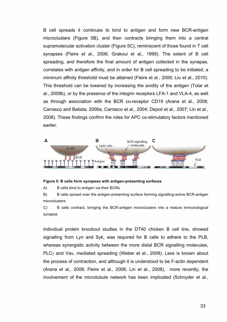
over the antigen-presenting surface (Figure 5A and B) (Fleire et al., 2006). As the
33
B cell spreads it continues to bind to antigen and form new BCR-antigen
microclusters (Figure 5B), and then contracts bringing them into a central
supramolecular activation cluster (Figure 5C), reminiscent of those found in T cell
synapses (Fleire et al., 2006; Grakoui et al., 1999). The extent of B cell
spreading, and therefore the final amount of antigen collected in the synapse,
correlates with antigen affinity, and in order for B cell spreading to be initiated, a
minimum affinity threshold must be attained (Fleire et al., 2006; Liu et al., 2010).
This threshold can be lowered by increasing the avidity of the antigen (Tolar et
al., 2009b), or by the presence of the integrin receptors LFA-1 and VLA-4, as well
as through association with the BCR co-receptor CD19 (Arana et al., 2008;
Carrasco and Batista, 2006a; Carrasco et al., 2004; Depoil et al., 2007; Lin et al.,
2008). These findings confirm the roles for APC co-stimulatory factors mentioned
earlier.
Figure 5: B cells form synapses with antigen-presenting surfaces
A) B cells bind to antigen via their BCRs
B) B cells spread over the antigen-presenting surface forming signalling-active BCR-antigen
microclusters
C) B cells contract, bringing the BCR-antigen microclusters into a mature immunological
synapse
Individual protein knockout studies in the DT40 chicken B cell line, showed
signalling from Lyn and Syk, was required for B cells to adhere to the PLB,
whereas synergistic activity between the more distal BCR signalling molecules,
PLCγ and Vav, mediated spreading (Weber et al., 2008). Less is known about
the process of contraction, and although it is understood to be F-actin dependent
(Arana et al., 2008; Fleire et al., 2006; Lin et al., 2008), more recently, the
involvement of the microtubule network has been implicated (Schnyder et al.,
34
2011). This was shown by the recruitment of the microtubule-motor Dynein to the
contracting microclusters by a Grb2-Dok3-cbl complex.
1.6 BCR-antigen internalisation
While the formation of signalling active BCR microclusters immediately after
antigen binding has been extensively observed in vitro in response to multivalent
soluble antigen (Ma et al., 2001; Niiro et al., 2004) as well as both monovalent
and multivalent PLB-bound antigen (Depoil et al., 2007; Fleire et al., 2006; Liu et
al., 2010; Weber et al., 2008). How BCR-antigen microclusters are internalised
remains unclear.
Studies have shown that BCR signalling is required for efficient antigen
internalisation, suggesting a crosstalk between BCR signalling and endocytic
machinery. For instance, antigen internalisation has been shown to be dependent
on the signalling capacity of the BCR’s IgαIgβ cytoplasmic domains. Using
chimera constructs containing the cytoplasmic domains of either the Igα or the
Igβ proteins, studies have shown that both chains contained residues that could
individually mediate BCR internalisation (Bonnerot et al., 1995; Jang et al., 2010;
Lankar et al., 1998). Point mutation studies have also shown that activity from
both ITAM (Cassard et al., 1998; Gazumyan et al., 2006) and non-ITAM tyrosines
within the IgαIgβ heterodimer (Hou et al., 2006; Patel and Neuberger, 1993) are
required for this process.
Similarly, BCR proximal signalling molecules have been connected to this
process. Antigen-stimulated Lyn knockout B cells were inhibited in their ability to
internalise antigen, shown by retention of antigen on the cell surface (Chan et al.,
1998; Ma et al., 2001). Downstream of Lyn, BCR signalling also activates
regulators of the actin cytoskeleton, and a role for actin polymerisation in BCR-
antigen internalisation has been proposed. For example, results from knockout
and inhibitor studies on Bam32 (Niiro et al., 2004) and WASp (Becker-Herman et
al., 2011) showed similarities in phenotype to Lyn knockout cells, and BCR-
antigen internalisation is also reduced following disruption of Vav mediated

35
activation of Rac (Malhotra et al., 2009a; 2009b). Consistently with these studies,
disruption of actin polymerisation itself also decreased BCR-antigen
internalisation (Stoddart et al., 2005).
The primary endocytic mediator involved in BCR-antigen internalisation is
thought to be clathrin. This was first shown by early electron microscopy studies,
which demonstrated the endocytosis of antigen-bound BCRs through clathrin
coated pits (Salisbury, 1980). More recently this has been supported by a
correlation between BCR-antigen internalisation and lipid raft-associated clathrin
phosphorylation, and a reduction in BCR-antigen internalisation after the
inducible deletion of the clathrin heavy chain in DT40 cells. These studies use
biochemical analysis to propose that antigen-induced BCR signalling stimulates
phosphorylation of clathrin within the lipid raft scaffold initiating internalisation
(Stoddart et al., 2002; 2005). Clathrin-mediated endocytosis is dependent on the
recruitment and organisation of an array of specific adaptor proteins to the site of
clathrin coated pit formation. Some of these adaptors are indispensible for
clathrin action such as AP2, but others can vary depending on the cargo, cell
type or environment (McMahon and Boucrot, 2011; Taylor et al., 2011). However,
to date, the involvement of any cargo-specific adaptors required for BCR-antigen
internalisation through clathrin-mediated endocytosis, remains unknown.
Although studies of BCR-antigen internalisation have identified roles for BCR
signalling, lipid rafts, the actin cytoskeleton, and clathrin-mediated endocytosis,
they have also indicated a significant level of redundancy in this process. Some
studies suggest that lipid rafts act as a signalling platform only and antigen-bound
BCRs are not internalised from these structures (Hou et al., 2006; Putnam et al.,
2003). Also, deletion of clathrin, inhibition of actin activity or disruption of lipid raft
heterogeneity, does not completely abolish BCR-antigen internalisation (Stoddart
et al., 2005), suggesting alternative mechanisms can partially compensate for
their loss. In addition, it is unknown whether the same components are required
for internalisation of membrane-bound antigen as described for antigen in
solution. In fact, the almost absolute requirement for actin activity for synapse
formation in response to membrane-bound antigen (Fleire et al., 2006),
36
compared to a redundant role when faced with soluble antigen (Stoddart et al.,
2005), suggests that the two internalisation processes may be different.
Therefore, although key players in BCR-antigen internalisation have been
identified, their precise contribution and mechanism of action, and how they
interlink together, requires further study.
1.7 Affinity-dependent antigen internalisation B cells need to be able to internalise antigen in an affinity-dependent manner as
affinity-dependent BCR-antigen internalisation allows the selective expansion of
high affinity B cell clones. These B cells can then produce high affinity antibody,
specific for the invading pathogen, marking it for elimination. This happens at two
primary checkpoints of the B cell response. Firstly, at the point of antigen
encounter in the B cell follicle to provide an immediate antibody response (Paus
et al., 2006), and secondly during affinity maturation in the germinal centre to
encourage the expansion of high affinity B cells and their differentiation into
plasma cells and memory B cells (Chan and Brink, 2012; Rajewsky, 1996).
In vitro studies have shown that B cells can recognise and discriminate between
a range of different antigen affinities (Batista and Neuberger, 1998). B cells
respond with increasing efficiency to soluble monovalent antigen with affinities
from Ka > 106 M-1 up until Ka > 1010 M-1, where an affinity ceiling threshold is
reached and the corresponding B cell response does not continue to grow
irrelevant of any further increases in affinity (Batista and Neuberger, 1998). This
increased B cell response to high antigen affinity was shown firstly at the level of
BCR signalling by increased levels of calcium flux and phosphorylation of early
BCR signalling mediators (Kouskoff et al., 1998), and secondly at the level of
antigen presentation by higher levels of IL-2 production by cognate T cell specific
hybridomas, which had been co-cultured with MD4 B cells and HEL antigens
(Batista and Neuberger, 1998).

37
Similar dependence on affinity has been shown for membrane-tethered antigens.
TIRF microscopy of antigen-loaded PLB revealed that cognate B cells had
increased levels of spreading, microcluster formation, and subsequent antigen
accumulation and synapse formation, when BCRs of high affinity were settled on
the same antigen compared to the lower affinity equivalent BCR (Fleire et al.,
2006; Liu et al., 2010). Taken together these studies suggest that high affinity
monovalent antigen stimulates a greater level of signalling and BCR-antigen
accumulation, which results in a higher level of antigen internalisation and
presentation, and subsequent cognate T cell activation.
However, these measurements and studies only look at an individual reaction
between a single Fab molecule of the BCR and a single epitope in the antigen. In
vivo, considering the bivalent nature of the BCR, and the multivalency of antigen
associated with the infections B cells must recognise, such as bacteria, viruses,
fungi, and parasites, the majority of interactions between the BCR and the
antigen will be multivalent. This means that B cell recognition of antigen is also
affected by avidity (number of bonds between BCRs and antigen). High avidity
antigen can be formed not only through the particulate nature of the antigen itself
but also through complexing of antigen with natural antibodies or complement in
the blood stream, or by clustering on the surface of APCs as mentioned earlier
(Batista et al., 2001; Chan and Brink, 2012; Gonzalez et al., 2010a). In vitro,
increasing the avidity of antigen raises its immunogenicity above the threshold
required for maximum B cell stimulation, which prevents B cells from
discriminating between antigen affinities in solution (Batista and Neuberger,
2000). However, in vivo, where antigens are primarily displayed on APCs, B cells
respond to antigen in an affinity-dependent manner (Batista and Neuberger,
2000; Chan and Brink, 2012; Schwickert et al., 2011; Shih et al., 2002a; 2002b;
Wan et al., 2013).
Observations of adoptively transferred B1-8 B cells with either a high or low BCR
affinity for the nitrophenyl hapten (NP) into immunised recipient mice, showed
that the rate of antigen internalisation and subsequent presentation correlated
with affinity. This difference in internalisation rate didn’t cause the observed
affinity discrimination itself, as when low affinity B cells were transferred alone,

38
they were able to elicit a normal B cell response (Schwickert et al., 2011). Only
when competition was introduced by transferring both high and low affinity B cells
together, was there affinity discrimination and an almost exclusive expansion of
high affinity clones which dominated the majority of the germinal centre response
(Schwickert et al., 2011; Shih et al., 2002a). This was due to the increased levels
of presented antigen on high affinity B cells, which monopolised T cell help at the
B-T cell boarder and prevented lower affinity B cells from receiving the correct
survival signals (Schwickert et al., 2011). Thus affinity discrimination at the B-T
cell border selects BCRs with the relatively highest affinity to enter into the
germinal centre (Schwickert et al., 2011). This relative selection mechanism,
therefore, allows antigens of low affinity to still produce a successful immune
response (Dal Porto et al., 1998; 2002), which is particularly important in the case
of a primary infection, when the typical affinity of the BCR for a previously unseen
pathogen is low (Chan and Brink, 2012).
Once in the germinal centre, B cells cycle between the light and dark zones.
Within the dark zones B cells proliferate and activate somatic hypermutation.
Somatic hypermutation introduces point mutations into the variable region of the
BCR, diversifying the affinity for the stimulating antigen (Jacob et al., 1991;
McKean et al., 1984). After one or more rounds of division B cells move to the
light zone and test the fitness of the newly mutated BCR by binding to antigen
displayed by light zone-resident FDCs (Allen and Cyster, 2008; Victora and
Nussenzweig, 2012). B cells with a higher affinity are thought to internalise more
of the antigen and present more to follicular helper T cells (Tfh) (Khalil et al.,
2012; Victora et al., 2010). This T cell help has again been shown to be limiting
and only those B cells with high enough affinity for the antigen receive T cell help,
which stimulates not only their survival but also their movement back into the
dark zone to continue another round of proliferation and somatic hypermutation
(Victora et al., 2010). This mechanism ensures the continual selective expansion
of high affinity B cells as the immune response progresses (Jacob et al., 1991;
Rajewsky, 1996). In contrast, low affinity B cells internalise less antigen from
FDCs, receive less help from Tfh, accumulate in the light zone, and die.

39
New studies by Khalil et al 2012 have recently provided evidence showing that
BCR signalling in the germinal centre reaction is inhibited by phosphatase
activity, which may favour internalisation and presentation of the antigen to Tfh
and possibly augments high affinity B cell expansion (Khalil et al., 2012).
Some antigens are highly stimulative to B cells and do not require T cell help to
elicit antibody production; these are called T cell-independent (TI) antigens. TI
antigens are usually large, polyvalent structures with repeating patterns such as
carbohydrates from a bacterial capsid or virus. The polyvalent nature of these TI
antigens allows extensive crosslinking of BCRs, which augments B cell signalling
and intrinsically provides the stimulation needed for B cell activation and
proliferation (Vos et al., 2000). These responses differ from T cell-dependent
antigens as they do not form germinal centres, and function to quickly release
soluble antibody against circulating pathogens (Shih et al., 2002b). Despite the
lack of T cell help when faced with TI antigens, high affinity B cell clones are still
able to outcompete low affinity clones. It is unclear how this competition happens,
but it is possible that high affinity B cells internalise antigen more quickly, which
in turn stimulates faster B cell proliferation, increases access to other stimulatory
factors such as cytokines, and therefore facilitates the selective expansion of
high affinity B cell clones.
The ability of B cells to internalise antigen in an affinity-dependent manner is vital
during the immune response. This is important both at the point of initial antigen
encounter, and to support the continual improvement of BCR affinity in germinal
centres as the immune response progresses. Collectively, these studies show
that the ability of B cells to discriminate between antigen affinities is due to the
increased rate of antigen internalisation of high affinity B cells. However, the
molecular mechanisms controlling this process are still unknown.

40
1.8 Defects in membrane-bound antigen internalisation can lead to autoimmunity or cancer
BCR-antigen internalisation is a tightly regulated process to ensure effective B
cell function, and disturbances have been linked to B cell-mediated autoimmune
diseases and cancer. These diseases can be a result of a range of either genetic
factors, such as mutations in negative regulators of BCR signalling or they can be
induced by the environment, for example by B cell responses to infection that
cross-reacts with self-components. It is, therefore, important to consider how
BCR-antigen internalisation prevents pathology, and how disturbances in this
process can cause these diseases.
1.8.1 Autoimmunity 70% of newly developed immature B cells are autoreactive and have the
potential to cause autoimmune pathologies by the production of autoantibodies
specific for self-proteins, which then target self-cells for destruction (O'Neill et al.,
2011; Plotz, 2003).
B cell interactions with APCs, in addition to their function in stimulating B cell
activation, are also important in deleting these autoreactive B cells. Membrane-
bound autoantigen is more efficient at this deletion than comparable soluble
autoantigen. For example, during development, cognate B cells, which react with
autoantigen expressed by stromal cells, are deleted and prevented from exiting
the bone marrow, whereas their reaction to soluble autoantigen induces antigen-
unresponsiveness (anergy) in the B cells, but they are still permitted to reach the
periphery (Goodnow et al., 1988; Hartley et al., 1991). A similar deletion is seen
after encounter with membrane-bound autoantigen in the periphery, which
demonstrates an additional layer of control to ensure deletion of B cells specific
for an autoantigen that is not present in the bone marrow (Russell et al., 1991;
Taylor et al., 2012).
In the germinal centre, there is a risk that the newly mutated BCRs may cross-
react with autoantigen and become autoimmune. Antigen presented by APCs in

41
the germinal centre can communicate to the B cell that the antigen is pathogenic
and not self. This was shown by the rapid deletion of B cell clones in the germinal
centre when soluble antigen, representing autoantigen, was added (Chan et al.,
2012; Pulendran et al., 1995; Shokat and Goodnow, 1995).
Therefore, disruptions in the molecules that facilitate B cell-APC interaction, can
lead to inappropriate activation of these B cells and the development of
autoimmunity. For example, chronic expression of the integrins LFA-1 and VLA-
4, which reduce the B cell activation threshold by facilitating adhesion between B
cells and APCs, could promote B cell activation to low affinity autoantigens which
should not normally stimulate a B cell response (Carrasco and Batista, 2006a;
Carrasco et al., 2004). In addition, elevated expression of CD19, the BCR co-
receptor shown to augment B cell activation in response to membrane-bound
antigen (Depoil et al., 2007), has also been linked to an autoimmune phenotype
by B cell hyper-responsiveness and autoantibody production (Hasler and Zouali,
2001). Conversely, loss of inhibitory factors, which prevent B cell response to
autoantigens, can also cause autoimmunity. This was shown following the loss of
CD22. CD22 is a receptor for α2,6 sialogglycoconjugates, which are commonly
expressed on cell types which B cells are likely to encounter in an infection
setting i.e. other lymphocytes and activated endothelium (Lanoue et al., 2002).
The interaction between CD22 and α2,6 sialogglycoconjugates is thought to
inhibit antigen-bound BCR signalling and reduce the expression of the B cell
activation markers CD69 and CD86. As a result, B cells deficient in this BCR
negative regulator, are prone to the production of autoantibodies and the initiation
of autoimmune disease (O'Keefe et al., 1999; 1996).
Autoimmunity can also be caused by disturbances in BCR signalling, which
normally controls antigen internalisation. Surprisingly, both Lyn knockout and Lyn
gain of function mice are prone to autoimmune phenotypes characterised by
hyper-responsive B cells and an increase in autoantibody production (Chan et al.,
1997; Hibbs et al., 2002). It is difficult to ascertain how much of this phenotype is
due to loss of internalisation as Lyn’s role in B cell activation goes beyond
stimulating internalisation. Lyn activity is required for phosphorylating both

42
positive and negative BCR regulators (Xu et al., 2005). Therefore when Lyn is
lost, so is the activation of molecules, which inhibit B cell activation such as
CD22, FcγRIIb, and SHIP-1 (Chan et al., 1998; O'Neill et al., 2011), and when
Lyn is over expressed BCR activating molecules such as Syk are increased
(Hibbs et al., 2002). These studies illustrate the complexity of BCR signalling and
internalisation, and clearly demonstrate the difficulties in elucidating the exact
mechanism and role of BCR-antigen internalisation in vivo.
One of the ways B cells prevent autoimmunity is through the process of anergy.
Anergy is a non-reactive state of B cells, which occurs following recognition of
autoantigen during development or in the periphery. Anergy can be initiated
through chronic low-level stimulation of BCRs without T cell help (Cambier et al.,
2007; O'Neill et al., 2011), but can also be controlled at the level of BCR-antigen
internalisation. For example, when anergic B cells recognise DNA-containing
autoantigens, they do not recruit TLR9 for amplification of the B cell response,
but prevent antigen from meeting TLR9 before merging into late endosomes
(O'Neill et al., 2009). However this altered trafficking in anergic B cells is reverted
in autoimmune B cells, possibly by BCRs mistaking hypomethylated DNA from
apoptotic cells for bacterial DNA (Leadbetter et al., 2002), or over active TLR9
signalling (Christensen, 2005). This results in the recruitment of TLR9 to
internalised BCR-antigen complexes (Chaturvedi et al., 2008) and production of
autoantibodies against self-DNA (Viglianti et al., 2003).
It has also been suggested that accelerated BCR-antigen internalisation from
lipid rafts may help to maintain the unresponsiveness of anergic B cells. Studies
showed that anergic B cells have a large intracellular pool of BCRs, present in a
recycling endosomal compartment (Bléry et al., 2006). These anergic B cells had
a faster rate of internalisation compared to naïve B cells, which prevented
initiation of the signalling pathway leading to NFκB activation and subsequently B
cell activation through the transcription of factors such as c-myc and bcl-XL. If
lipid rafts were disrupted in these anergic cells by methyl-β-cyclodextrin, then
BCRs quickly relocated to the cell surface and regained their responsiveness to
antigen.
43
1.8.2 Cancer In addition to antigen processing and engagement of antigen with intracellular
TLRs, BCR-antigen internalisation also functions to alter BCR signalling to
prevent the inappropriate survival of B cells, which could lead to cancer. Inhibition
of BCR-antigen internalisation has been shown to maintain activate antigen-
induced signalling complexes at the cell surface, which downregulates
transcription factors controlling proapoptotic genes, and promotes B cell survival
(Chaturvedi et al., 2011; Srinivasan et al., 2009).
This inhibition of internalisation together with the resulting hyper-responsive BCR
signalling is seen in activated B cell like subtype of diffuse large B cell lymphoma
cells (ABC DLBCL) (Davis et al., 2010). This is thought to be caused by
mutations in the IgαIgβ ITAMs frequently found in ABL DLBCL biopsies. These
mutations were shown to prevent the full activity of Lyn and inhibitory
phosphatases, and inhibited BCR internalisation, causing a malignant phenotype.
These studies collectively show that although BCR-antigen internalisation is vital
in the production of high affinity antibodies required for specific pathogen
elimination, it is also important in preventing inappropriate B cell survival or B cell
activation to autoantigens that could lead to the development of cancer or
autoimmunity. By understanding how BCR-antigen internalisation works in
normal physiology we hope to better understand how these processes are
disregulated in disease. This knowledge could potentially be applied in the
manipulation of BCR internalisation in order to treat B cell diseases.

44
1.9 Thesis Aims
It is well acknowledged that B cells require antigen internalisation in order to
become activated and develop an efficient humoral response, as well as to
prevent excessive BCR signalling. It is known that in vivo the primary source of
antigen is tethered to APCs, and this presentation allows context-dependent
antigen recognition by B cells, which augments and fine-tunes the B cell
response. It is also known that this internalisation occurs in an affinity-dependent
manner, which allows B cells to selectively expand high affinity B cell clones for
the production of specific antibodies.
However, the mechanisms of B cell antigen extraction from APCs are still
unknown. Studies using soluble antigen have identified specific components and
pathways that are involved in BCR-antigen internalisation from solution, such as
clathrin-mediated endocytosis, the actin cytoskeleton, lipid raft scaffolds and BCR
signalling molecules. However, to date, there has not been a technique available
to study the cellular and molecular mechanisms of BCR-antigen internalisation
from an APC.
To address this question, we aimed to design and develop an antigen-presenting
substrate from which B cells can internalise antigen, and using this substrate, we
hoped to identify novel components and mechanisms involved in BCR-antigen
internalisation.

45
Chapter 2 Materials and Methods
2.1 Mice C57Bl/6 mice from an internal colony (called Bl6 throughout the thesis) were
used for all experiments and as wild-type controls unless otherwise stated. B1-
8fl/fl mice (a gift from K. Rajewsky (Sonoda et al., 1997)) were crossed with Igκ-
deficient Igκ-Ctm1Cgn/tm1Cgn mice (a gift from J. Langhorne) to produce the B1-8fl/fl
Igκ-Ctm1Cgn/tm1Cgn strain (called B1-8 throughout the thesis) and the strain was
backcrossed onto C57Bl/6 background. The majority of B cells in these mice are
specific for the nitrophenyl hapten (NP or NIP). MyH9fl/fl mice (purchased from
Mutant Mouse Regional Resource Centers (Jacobelli et al., 2010)) were crossed
with Mb1-Cre mice (a gift from V. Tybulewicz (Hobeika et al., 2006)) and the
strain was backcrossed onto C57Bl/6 background (called Cre+MyH9fl/fl throughout
the thesis). These mice express Cre recombinase at the point of Igα expression
in Pro B cells. Genotypes were confirmed using PCR analysis of DNA from ear
snips. Mice were bred and treated in accordance with the guidelines set by the
UK Home Office and UK NIMR Ethical Review Panel.
2.2 B cell purification and cell culture Naïve B cells were obtained from splenocytes following red blood cell lysis (ACK
buffer, Gibco) for all experiments except siRNA-mediated knockdown for which, a
ficoll density gradient (GE Healthcare) was used. Naïve B cells were then
negatively selected for using anti-CD43 microbeads on an autoMACs Pro
separator (Miltenyi Biotec). Primary B cells and the Ramos human lymphoma cell
line were cultured in RPMI 1640 media (Sigma) supplemented with 10% foetal
bovine serum (FBS, Biosera), 1% MEM Non-Essential Amino Acids (Gibco), 2
mM L-glutamine (PAA Laboratories), 50 µM 2-mercaptoethanol (Gibco), 100
U/ml Penicillin and 100 µg/ml Streptomycin (Gibco). Adherent HEK293A,
HEK293FT (both from Invitrogen), PLAT-E cells (a gift from V. Tybulewicz (Morita
et al., 2000)) and bone marrow derived DCs were cultured in DMEM media
(Gibco) supplemented with 10% FBS, 1% MEM Non-Essential Amino Acids, and

46
6 mM L-glutamine. 10 µg/ml blasticidin and 1 µg/ml puromycin (both InvivoGen)
were added to PLAT-E media, 0.5 mg/ml Geneticin (Gibco) was added to
HEK293FT media, and DCs differentiated from bone marrow cells using Flt3
ligand (a gift from G. Stockinger) for 7 days, were matured using 1 µg/ml LPS
(Sigma) for 24 hours. Suspension HEK293A cells were maintained at 5 x 10⁵
cells/ml, agitated at 120 rpm in Pro293S-CDM media (Lonza) supplemented with
1.5% FBS and 2 mM L-Glutamine. All cells were cultured in incubators humidified
with 5% CO2 at 37°C.
2.3 Antigens
BCR-specific antigen Cellular target Source
Goat F(ab’)2 α-mouse Igκ Bl6 B cells Southern biotec
Goat F(ab’)2 α-human IgM (µ chain specific) Ramos cells Southern Biotec
NP (4-Hydroxy-3-nitrophenylacetic acid active ester)- or NIP (4-Hydroxy-3-iodo-5-nitrophenylacetic active ester)- Osu
B1-8 B cells Biosearch
Unless otherwise stated αIgκ and αIgM antigens were labelled with Cy5 or Cy3
using the Monoreactive dye kit (GE Healthcare) and biotinylated using EZ-link
sulfo-NHS-LC-LC-Biotin (Thermo scientific).
To generate NIP-Qdot antigens, fluorescent Quantum dots (Qdot, Invitrogen)
were haptenated with NIP-Osu in dimethylformamide (Sigma) for 3 hours and
biotinylated. αIgM-Qdots were produced by mixing Qdots conjugated to
streptavidin 1:1 with biotinylated αIgM.
NP or NIP antigens were produced from the haptenation of AlexaFluor647-
labelled goat F(ab’)2 (Jackson Immunoresearch) with NP- or NIP-Osu and
biotinylated. To load cognate immune complexes onto DCs for recognition by B1-
8 B cells, NP/NIP antigen was mixed 1:1 with anti-biotin IgG1 antibodies (Sigma)
for 30 minutes on ice. For AFM experiments, DCs were loaded with immune-

47
complexes consisting of αNP antibodies (Serotec) and NIP antigens for 1 hour
on ice.
For 3D localisation of antigen particles, biotinylated-αIgκ was loaded onto PMS
and then a small number of antigen molecules were labelled with a low
concentration of streptavidin-conjugated Qdots.
2.4 Antibodies
Antibody Clone Source Final concentration
α-mouse CD19 FITC 1D3 BD biosciences 1 µg/ml
α-human GAPDH-Alexa647 14C10 Cell Signaling 1:500 dilution
α-mouse CD45R/B220 RA3-6B2 BD biosciences 1 µg/ml
α- clathrin heavy chain 23 BD Transduction Laboratories 0.25 µg/ml
α-Dynamin2 Polyclonal Abcam 1 µg/ml
α-β-actin AC-74 Sigma 1:5000 dilution
α-AP2 µ1 EP2695Y Epitomics 1:1000 dilution
α-mouse IgM µ chain specific-FITC
Catalog number: 115-097-020
Jackson Immunoresearch 5 µg/ml
α-mouse CD93 (AA4.1)-APC AA4.1 eBioscience 2 µg/ml
α-mouse CD23-PE B3B4 eBioscience 5 µg/ml
α-mouse/human CD45R/B220 PerCP-
Cy5.5 RA3-6B2 Biolegend 1 µg/ml
α-mouse CD8a-brilliant violet 570 53-6.7 Biolegend 0.5 µg/ml
α-mouse CD4-efluor 450 RM4-5 eBioscience 1µg/ml
α-mouse CD19-APC 6D5 Biolegend 2 µg/ml
α-mouse CD2 (LFA-2)-PE RM2-5 BD Biosciences 2.5 µg/ml
α-mouse IgD efluor 450 11-26 eBioscience 1 µg/ml

48
α-mouse IgD-PE 11-26 eBioscience 0.5 µg/ml
α-mouse TCR-β-APC H57-597 eBioscience 1 µg/ml
α-mouse CD5 (Ly-1)-PE 53-7.3 eBioscience 2 µg/ml
α-mouse CD23-APC B3B4 Invitrogen 1:100 dilution
α-human/mouse CD45/R (B220)-efluor
450 RA3-6B2 eBioscience 1 µg/ml
α-mouse/human CD45R (B220)-PE RA3-6B2 ebioscience 1 µg/ml
2.5 Inhibitors Inhibitors were incubated with B cells in Hank’s buffered saline solution (HBSS)
supplemented with 0.01% bovine serum albumin (BSA, Sigma). Dynasore was
incubated with HBSS containing no BSA. All inhibitors were preincubated with B
cells for 15 minutes at 37°C before addition to antigen-loaded PMS or soluble
antigen. Solvent in the appropriate buffer was used as control.
Inhibitor Target Source Final concentration
PP2 Lyn Invitrogen 20 µM
LFM-A13 Btk Caymann Chemical 50 µM
Bisindolylmaleimide PKC Caymann Chemical 20 µM
PD184352 Erk1/2 Selleckchem 2 µM
SMIFH2 Formin Sigma 10 µM
NSC-23766 Rac1 Biovision 100 µM
Dynasore Dynamin2 T Kirchhausen 80 µM
Blebbistatin Myosin IIA Caymann Chemical 50 µM
Y-27632 Rock1 Sigma 50 µM
49
2.6 Planar lipid bilayers (PLB) DOPS (1,2-Dioleoyl-sn-Glycero-3-[Phospho-L-Serine] (Sodium Salt)) or DOPE-
biotin (1,2-Dioleoyl-sn-Glycero-3-phosphoethanolamine-N-(Cap Biotinyl) (Sodium
Salt)), were mixed at a molar ratio of 1:10 with DOPC lipid (1,2-dihexanoyl-sn-
glycero-3-phosphocholine) (All from Avanti) in choloform. The lipids were dried
overnight using argon and resuspended in degassed phosphate buffered saline
(PBS). The lipids were then sonicated to produce unilamellar vesicles, clarified by
ultracentrifugation at 36 000 rpm for 16 hours, and prepared in a 0.1 mM lipid
solution in PBS. Glass coverslips were cleaned in sulfuric acid and hydrogen
peroxide at a ratio of 3:1 and attached to Labtek imaging chambers (Nunc). Lipid
bilayers were formed by fusing the unilaminar vesicles onto the clean coverslips.
For PLB in Figure 6, PLB formed from DOPE-biotin and DOPC were incubated
with 5 µg/ml streptavidin (Southern Biotec) in PBS, washed, and incubated with
NIP-Qdot antigens.
For PLB in all subsequent experiments, PLB from DOPS and DOPC were
incubated with 24 nM biotinylated annexin V (BioVision) in HBSS. After washing,
PLB were incubated with 5 µg/ml streptavidin, washed again, and incubated with
indicated antigens. When not explicitly stated, PLB were loaded with antigens at
1 µg/ml, resulting typically in 50 antigen molecules per µm2. In some cases, the
concentration of antigen was titrated to achieve the density specified in the
experiments. The calculation of antigen density on the substrates was performed
as previously described (Tolar et al., 2009b). Briefly, antigen was diluted to a
concentration that allowed resolution of single antigen molecules. The average
fluorescence intensity of the single antigen molecules was used to convert
fluorescence intensity of experimental antigen concentrations to number of
antigen molecules per µm2.
50
2.7 Plasma membrane sheets (PMS) Suspension HEK293A cells were seeded onto poly-L-lysine (Sigma)-coated
Labtek imaging chambers (Nunc) and cultured overnight to 100% confluency.
Cells were then washed in PBS and sonicated with a probe sonicator placed 5
mm above the cells for 20 seconds at room temperature. This shears off the
cells, leaving the ventral plasma membrane attached to the coverslip with its
cytoplasmic side exposed. PMS were blocked with PBS supplemented with 1%
BSA and then incubated in HBSS 0.1% BSA with 24 nM biotinylated annexin V.
After washing, PMS were incubated with 5 µg/ml streptavidin, washed again, and
incubated with indicated antigens as on the PLB.
2.8 Plasma membrane planar lipid bilayers (PMPLB) – Wing-Liu Lee
Suspension HEK293A cells were lysed in 5 mM NaH2PO4/NaHPO4, pH 8 at 10 x
106 cells/ ml for 30 minutes on ice, and then homogenised using Dounce tissue-
grinder pestle (Sigma). Nuclei were removed by centrifugation at 500 x g at 4°C
for 10 minutes and plasma membranes were isolated by centrifugation at 20 000
x g at 4°C for 30 minutes. The membrane pellet was resuspended in a buffer
containing 500 µM NaH2PO4/NaHPO4, 100 µM MgSO4, pH 8, on ice for 1 hour.
Membrane vesicles were then purified by centrifugation at 100 000 x g at 4°C.
The vesicle pellet was resuspended in the same buffer and used immediately.
PMPLB were formed by fusing the vesicles onto glass coverslips. Antigen was
attached via biotinylated annexin V and streptavidin and its density was
quantified as on the PLB and PMS.
2.9 Imaging All microscopy analysis and live-cell imaging was carried out on an Olympus
IX81 microscope, with an Andor iXon EMCCD Camera and 100x objective
(Olympus). Live-cell imaging was done at 37°C on a heated stage with an
objective heater.
51
TIRF or epifluorescence timelapse images were acquired with time resolution of
2-5 seconds for 20 minutes, and corrected for photo-bleaching. The TIRF angle
was setup as shallow as possible to extend the depth of field further away from
the coverslip. All images were aligned, background-subtracted and corrected for
spectral bleedthrough and photobleaching. Images were analysed using ImageJ
and Matlab (Mathworks). All Matlab algorithms were written by Pavel Tolar.
Reconstructed images showing antigen internalisation from PMS, PLB and DCs
were generated from z-stacks by first deconvoluting the stacks, and then using
maximal projection of the middle of the cell along the y axis (PLB and PMS) or z
axis (DCs).
2.10 Atomic force microscopy (AFM) force spectroscopy – Pavel Tolar
AFM was performed using a setup integrating AFM and optical imaging (JPK
instruments). To carry out force spectroscopy with PMS, PLB, PMPLB and DCs,
gold-coated Biolever cantilevers (nominal spring constant 6 mN/m, Olympus)
were exposed to UV light for 30 minutes and then conjugated to DOTP (Pierce)-
activated streptavidin. The cantilevers were then washed and used immediately.
Spring constant was measured for each tip using the JPK software and varied
between 5.2 and 6.0 mN/m. PMS, PLB, PMPLB and DCs, were loaded with
antigens, washed, and attached to poly-L-lysine coated coverslips. AFM force
spectroscopy was carried out on PMS, PLB or PM-PLB visualised using DiI or
DiD (Invitrogen), and on the dorsal surfaces of the DCs’ lamellopodia, visualised
by brightfield imaging. Negligible binding was observed in samples lacking the
biotinylated annexin V or antigen. Data were processed using the JPK analysis
software.

52
2.11 Internalisation assays 2.11.1 Soluble antigen internalisation using flow cytometry Internalisation of soluble antigens was measured using flow cytometry. Cells
were incubated with biotinylated antigens for 30 minutes on ice, washed, and
stimulated to internalise for indicated times at 37°C. Control cells were kept on
ice. Then, cells were fixed with 2% paraformaldehyde (PFA, Alfa Aesar) and
stained with 1 µg/ml of fluorescently labelled streptavidin (Jackson
Immunoresearch) to visualise any antigen remaining on the cell surface. Antigen
internalisation was calculated by subtracting the streptavidin fluorescence of the
heated sample from the cold sample and dividing by the fluorescence of the cold
sample. Data was analysed using FlowJo.
2.11.2 Soluble antigen internalisation using microscopy To quantify internalisation of soluble NP/NIP antigens, B1-8 B cells were
attached to poly-L-lysine-coated coverslips, incubated with NP/NIP antigens for
20 minutes, fixed with 2% PFA and stained with 1 µg/ml Cy3-labelled streptavidin
(Jackson Immunoreserach). To determine the intensity of the internalised antigen
in each cell, background from regions outside the cells was subtracted. Z-stack
images of antigen and streptavidin on cells were processed through a 3D
bandpass filter and thresholded to create 3D masks. The antigen mask was
applied to the original antigen stack. Next, the streptavidin mask was used to
exclude antigen present on the cell surface and therefore not internalised. The
resulting antigen fluorescence was integrated. The percentage of internalisation
was expressed as the intensity of internalised antigen minus the antigen
colocalised with streptavidin divided by total antigen intensity. Data was analysed
using Matlab.
2.11.3 Membrane-bound antigen internalisation Internalisation from PMS, PMPLB and PLB was quantified from z-stack images,
similarly as for the NP/NIP soluble antigens, but integrating antigen fluorescence
only from image planes above the substrate. Specifically, background was
subtracted using regions of interest outside of presenting membranes. Z-stack

53
images of antigen were processed through a 3D bandpass filter and thresholded
to create a 3D mask. The antigen mask was applied to the original unfiltered
antigen stack to include endosomal antigen and exclude scattered fluorescence
background. Internalised antigen fluorescence was then calculated by integrating
antigen fluorescence in the masked antigen images in stacks above the
substrate. In experiments with primary cells, we found that all antigen above the
substrate was inaccessible to cell-impermeable secondary 1 µg/ml fluorescently
labelled streptavidin staining and was therefore considered internalised. In
experiments with Ramos B cells, we found that some antigen above the
substrate was accessible to this staining. Therefore, secondary streptavidin
staining was used to exclude the streptavidin-colocalised antigen from the
analysis as described for soluble antigens. The percentage of internalisation was
expressed as the intensity of internalised antigen divided by total antigen
intensity, which included the internalised antigen and the antigen left on the
substrate underneath the cell. The percentage of internalised antigen colocalised
with DiI-stained lipid, was expressed as the intensity of internalised antigen,
minus antigen not colocalised with DiI, divided by total antigen intensity. Data
was analysed using Matlab.
2.12 Fluorescence recovery after photobleaching (FRAP) FRAP imaging was performed using TIRF microscopy at 37°C. Substrates were
labelled with DiI or loaded with antigen, imaged for 10 seconds, and then a
circular region of 13 µm in diameter was bleached at maximum laser power for 1
minute using the field diaphragm. Substrates were then imaged every 10
seconds for an additional 10 minutes. Fluorescence recovery was analysed using
ImageJ by measuring fluorescently-labelled antigen or DiI intensity in the FRAP
region throughout the experiment. The intensity in the FRAP region was
normalised to fluorescence in a non-bleached control region.

54
2.13 3D particle localisation – Pavel Tolar DiI-labelled PMS were loaded with Qdot-antigen. Imaging was performed in
epifluorescence mode. 3D localisation of the Qdots was performed using a
cylindrical lens (f = 1 m) inserted into the infinity space of the emission path
(Huang et al., 2008). Gaussian fitting was used to extract the elipticity of the
fluorescent spots. To convert the data to vertical distances, we acquired
calibration images of Qdots attached to coverslips, while stepping up the
microscope z-stage in 10 nm increments. Positional accuracy was estimated
from timelapse images of stationary Qdots on coverslips and was ~40 nm.
Vertical position of the Qdots in B cell synapses were expressed as their vertical
distance from the mean position of Qdots present on the coverslip outside of
synapses.
2.14 Analysis of membrane invaginations Membrane invaginations were detected by staining the lipids in the PMS with DiI
or DiD. The invaginations were equally detectable in epifluorescence illumination
and in TIRF. For 3D particle localisation, the membrane dyes were excited in
epifluorescence mode. For timelapse TIRF imaging, we used p-polarised laser,
which produces evanescent field that is mostly vertically polarised and therefore
preferentially excites DiI dyes in vertically oriented membranes (Sund et al.,
1999).
Membrane invaginations were tracked in space and time from timelapse images
of DiI or DiD stained PMS using previously developed tracking algorithms (Tolar
et al., 2009b). Briefly, image sequences were aligned and normalised to the
image of the PMS prior to cell spreading, and then processed using a bandpass
filter. Fluorescent spots were tracked and their lifetime was calculated from the
length of the tracks using Matlab.
Intensity of antigen associated with the invaginations was calculated as the mean
intensity in 5 by 5 pixel regions centred on the track coordinates, including 1
55
frame before the start and 1 frame after the end of the track. Antigen intensities
were normalised to the average antigen intensity on the PMS before cell
spreading.
2.15 Protein knock-down using siRNA
2 x 10⁶ Ramos cells, or 3 x 10⁶ primary B cells stimulated with 1 µg/ml of CpG
(Sigma) for 24 hours, were nucleofected (Amaxa Nucleofector II) with 3 µM
siRNA oligonucleotides (oligos, Ambion) using the Amaxa Mouse B Cell
Nucleofector Kit or the Amaxa Cell Line Nucleofector Kit V (Lonza), respectively.
Following nucleofection primary B cells were cultured with 200 ng/ml BAFF
(PeproTech). Efficiency of knock-down was assessed 96 hours after
nucleofection by western blot or flow cytometry.
siRNA target siRNA oligo ID Catalog number Negative control No.1 - AM4635
GAPDH - 4404025 CD19 S63587 4390815
Clathrin heavy chain S475 4390825
AP2M1 S3114 4392421 Dynamin2 S4212 4390825
2.16 Western blot Ramos or primary B cells were lysed with RIPA buffer containing 1% NP-40
(Sigma), 0.5% sodium deoxycholate (Sigma), 0.1% SDS (Sigma), protease
inhibitor cocktail diluted 1:100 (Sigma) for 20 min on ice. Samples were then
spun at 13 000 rpm 4°C, and proteins were separated using 10% SDS-PAGE
(Invitrogen) in reducing conditions. Proteins were transferred to nitrocellulose
membranes (Millipore), blocked in 5% milk in Tris Buffered Saline with 0.1%
Tween20, and probed in the same buffer with primary antibodies against proteins
of interest, followed by secondary antibodies conjugated to hydrogen peroxidase.

56
2.17 Lentiviral production and infection Recombinant lentiviruses were produced by lipofectamine 2000 (Invitrogen)-
mediated co-transfection of HEK293FT cells with the VSVg and δ8.9 helper
plasmids, together with the TRC RNAi Consortium’s shRNA-containing pLKO.1
plasmids (Sigma). Empty pLKO.1 was used as control. Lentivirus-containing
media were harvested 72 hours following transfection and centrifuged with
Ramos cells in the presence of 8 µg/ml polybrene (Sigma) for 90 minutes at 1350
x g. 24 hours after spinfection, infected cells were selected using 3 µg/ml
puromycin.
shRNA Clones (Sigma)
Target Clones Dynmain2 TRCN0000006649
AP2M1 TRCN0000060238 TRCN0000060239 TRCN0000060241 TRCN0000060242
RHOA TRCN0000047710 TRCN0000047708 TRCN0000047711 TRCN0000047712
ROCK1 TRCN0000002159 TRCN0000121092 TRCN0000121093 TRCN0000121094
HIP1R TRCN0000117222 TRCN0000117223 TRCN0000117225 TRCN0000117226
ARH-GAP4 TRCN0000047189 TRCN0000047190 TRCN0000047191 TRCN0000047192
ARH-GAP15 TRCN0000047253 TRCN0000047254 TRCN0000047255 TRCN0000047256
ARH-GAP17 TRCN0000047448 TRCN0000047450 TRCN0000047451 TRCN0000047452
ARH-GAP26 TRCN0000029754 TRCN0000029755 TRCN0000029756 TRCN0000029758

57
ARH-GAP27 TRCN0000048354 TRCN0000048355 TRCN0000048356 TRCN0000048357
ARH-GAP30 TRCN0000149213 TRCN0000179792 TRCN0000183676 TRCN0000183499
ARH-GEF1 TRCN0000033564 TRCN0000033566 TRCN0000033567 TRCN0000033568
ARH-GEF2 TRCN0000003173 TRCN0000003174 TRCN0000003176 TRCN0000010764
ARH-GEF6 TRCN0000007400 TRCN0000007401 TRCN0000007403 TRCN0000007404
ARH-GEF18 TRCN0000047533 TRCN0000047535 TRCN0000047536 TRCN0000047537
MYL12B TRCN0000053654 TRCN0000053655 TRCN0000053656 TRCN0000053657
MYL6 TRCN0000054138 TRCN0000054139 TRCN0000054140 TRCN0000054142
MYH9 TRCN0000029465 TRCN0000029466 TRCN0000029467 TRCN0000029468
MYO1E TRCN0000152890 TRCN0000150353 TRCN0000151296 TRCN0000152421
MYO1C TRCN0000122924 TRCN0000122926 TRCN0000122927 TRCN0000122928
2.18 Retroviral production and infection Retrovirus was produced by lipofectamine 2000-mediated transfection of PLAT-E
cells (Morita et al., 2000) with pMSCV containing one of the following constructs:
clathrin light chain-GFP (Ehrlich et al., 2004), LifeAct (Riedl et al., 2008) (a gift
from J. Burkhardt), or myosin IIA regulatory light chain (RLC) 12B (MYL12B)-
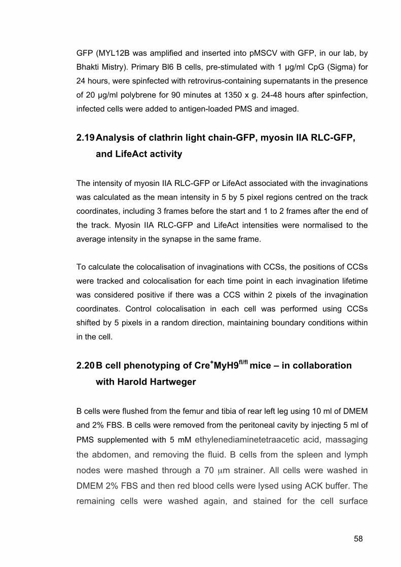
58
GFP (MYL12B was amplified and inserted into pMSCV with GFP, in our lab, by
Bhakti Mistry). Primary Bl6 B cells, pre-stimulated with 1 µg/ml CpG (Sigma) for
24 hours, were spinfected with retrovirus-containing supernatants in the presence
of 20 µg/ml polybrene for 90 minutes at 1350 x g. 24-48 hours after spinfection,
infected cells were added to antigen-loaded PMS and imaged.
2.19 Analysis of clathrin light chain-GFP, myosin IIA RLC-GFP, and LifeAct activity
The intensity of myosin IIA RLC-GFP or LifeAct associated with the invaginations
was calculated as the mean intensity in 5 by 5 pixel regions centred on the track
coordinates, including 3 frames before the start and 1 to 2 frames after the end of
the track. Myosin IIA RLC-GFP and LifeAct intensities were normalised to the
average intensity in the synapse in the same frame.
To calculate the colocalisation of invaginations with CCSs, the positions of CCSs
were tracked and colocalisation for each time point in each invagination lifetime
was considered positive if there was a CCS within 2 pixels of the invagination
coordinates. Control colocalisation in each cell was performed using CCSs
shifted by 5 pixels in a random direction, maintaining boundary conditions within
in the cell.
2.20 B cell phenotyping of Cre+MyH9fl/fl mice – in collaboration with Harold Hartweger
B cells were flushed from the femur and tibia of rear left leg using 10 ml of DMEM
and 2% FBS. B cells were removed from the peritoneal cavity by injecting 5 ml of
PMS supplemented with 5 mM ethylenediaminetetraacetic acid, massaging
the abdomen, and removing the fluid. B cells from the spleen and lymph
nodes were mashed through a 70 µm strainer. All cells were washed in
DMEM 2% FBS and then red blood cells were lysed using ACK buffer. The
remaining cells were washed again, and stained for the cell surface

59
markers described in Chapter 5 Table 1 for 20 minutes on ice in PBS.
Absolute cell numbers were ascertained using FlowJo. Alive cells were
identified as negative for the Live/Dead stain (Invitrogen).

60
Chapter 3 Plasma membrane sheets are a new artificial substrate that can mimic antigen-presenting
cells when studying BCR-antigen internalisation
3.1 Introduction
B cells are exposed to antigen primarily in the secondary lymphoid organs. B
cells can respond to antigen in solution following antigen filtration into the B cell
follicles (Pape et al., 2007; Roozendaal et al., 2009), but the majority of antigen
encountered by B cells in vivo will be retained on the surface of an APC (Batista
and Harwood, 2009; Cyster, 2010).
Intravital imaging has captured the interaction of B cells with antigen-loaded
APCs in the lymph node throughout the immune response (Phan et al., 2007; Qi
et al., 2006; Schwickert et al., 2007; Suzuki et al., 2009) and both in vivo and in
vitro studies have confirmed that antigen acquisition from these APCs is a
prerequisite to B cell activation, differentiation and antibody production (Batista et
al., 2001; Carrasco and Batista, 2007; Chappell et al., 2012; Junt et al., 2007;
Moseman et al., 2012; Phan et al., 2007; Qi et al., 2006; Schwickert et al., 2007;
Suzuki et al., 2009; Wang et al., 2011).
To delineate the molecular mechanisms involved in B cell recognition of
membrane-bound antigen, PLB have been used in vitro as a surrogate for APCs.
Live-cell TIRF images of B cells interacting with antigen-loaded PLB has revealed
that B cells initially spread over the antigen-presenting surface collecting BCR-
antigen microclusters, and then contract to bring them into the centre of the
synapse (Depoil et al., 2007; Fleire et al., 2006). This process has been shown to
be dependent on the structure of the BCR (Tolar et al., 2009b), BCR and CD19
signalling (Depoil et al., 2007; Schnyder et al., 2011; Weber et al., 2008), the
actin cytoskeleton (Liu et al., 2012; Mattila et al., 2013; Treanor et al., 2011), and
also on the affinity, multivalency, and concentration of the antigen (Fleire et al.,

61
2006; Tolar et al., 2009b). However the mechanisms and components required
for the subsequent antigen acquisition and internalisation are still unknown.
Here we aimed to develop an antigen-presenting substrate, which can support B
cell internalisation of membrane-bound antigen in vitro. We hoped that
visualisation of this process using TIRF microscopy would allow us to investigate
the dynamics of BCR-antigen internalisation, and support the use of a
membrane-surrogate in future investigations into the molecular components
governing this process.
3.2 Results 3.2.1 B cells are unable to internalise antigen tethered to planar lipid
bilayers To visualise internalisation of membrane-bound antigen by B cells, we initially
used PLB as a surrogate for APCs. PLB were formed following the fusion of
unilamellar vesicles on glass coverslips. We attached NIP-Qdot antigen to biotin-
containing lipids within the PLB using a streptavidin linker (Figure 6A). To the
antigen-loaded PLB we then added primary splenic B1-8 B cells that express a
BCR specific for the NIP hapten (Sonoda et al., 1997). Live cell imaging using
TIRF microscopy showed B1-8 B cell spreading over the antigen-loaded PLB and
the production of antigen microclusters, which was followed by contraction of the
B1-8 B cells pulling the antigen into the centre of the synapse (Figure 6B, Movie
1). Loss of fluorescence from the TIRF field would be indicative of internalisation,
since movement of antigen up into the B1-8 B cell would remove it from the
evanescent field exciting the Qdots. However, no loss of antigen fluorescence
from the synapse was seen during the imaging, suggesting the B1-8 B cells were
arrested at the point of synapse formation and internalisation didn’t take place.
This was confirmed using epifluorescent z-stack images taken every 300 nm
throughout the B1-8 B cell interacting with PLB. Side view reconstruction of these
images showed that antigen is not extracted from the PLB by the B cell, but is
retained in the synapse (Figure 6C).

62
The incapability of B1-8 B cells to internalise antigen wasn’t due to the type of B
cells chosen, or the antigen produced, as Ramos B cells were also unable to
extract antigen from the PLB (Figure 6C), and flow cytometry showed B1-8 B
cells were able to internalise the NIP-Qdot antigen from solution (Figure 6D). In
an attempt to optimise this system we increased the affinity between the BCR
and antigen by using MuMT-MD4 B cells, which have a BCR with over 1000
times higher affinity for HEL antigen than the B1-8 BCR has for NIP (KA ~ 5 ×
1010 M-1 versus 3 × 10⁶ M-1) (Carrasco and Batista, 2006a; Natkanski et al.,
2013). We also decreased the strength of the interaction between the antigen
and lipid bilayer by conjugating peptides containing a six histidine tag to the NIP-
Qdots and anchoring the Qdots to the PLB via nickel salt-containing lipid, instead
of using biotinylated lipids and streptavidin (Reznik et al., 1998). However, in both
circumstances the same phenotype was seen as in Figure 6B and C.
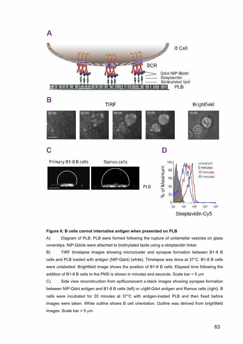
63
Figure 6: B cells cannot internalise antigen when presented on PLB
A) Diagram of PLB. PLB were formed following the rupture of unilamellar vesicles on glass
coverslips. NIP-Qdots were attached to biotinylated lipids using a streptavidin linker
B) TIRF timelapse images showing microcluster and synapse formation between B1-8 B
cells and PLB loaded with antigen (NIP-Qdot) (white). Timelapse was done at 37°C. B1-8 B cells
were unlabelled. Brightfield image shows the position of B1-8 B cells. Elapsed time following the
addition of B1-8 B cells to the PMS is shown in minutes and seconds. Scale bar = 5 µm
C) Side view reconstruction from epifluorescent z-stack images showing synapse formation
between NIP-Qdot antigen and B1-8 B cells (left) or αIgM-Qdot antigen and Ramos cells (right). B
cells were incubated for 20 minutes at 37°C with antigen-loaded PLB and then fixed before
images were taken. White outline shows B cell orientation. Outline was derived from brightfield
images. Scale bar = 5 µm
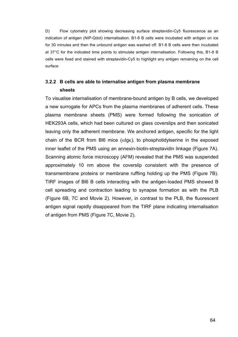
64
D) Flow cytometry plot showing decreasing surface streptavidin-Cy5 fluorescence as an
indication of antigen (NIP-Qdot) internalisation. B1-8 B cells were incubated with antigen on ice
for 30 minutes and then the unbound antigen was washed off. B1-8 B cells were then incubated
at 37°C for the indicated time points to stimulate antigen internalisation. Following this, B1-8 B
cells were fixed and stained with streptavidin-Cy5 to highlight any antigen remaining on the cell
surface
3.2.2 B cells are able to internalise antigen from plasma membrane sheets
To visualise internalisation of membrane-bound antigen by B cells, we developed
a new surrogate for APCs from the plasma membranes of adherent cells. These
plasma membrane sheets (PMS) were formed following the sonication of
HEK293A cells, which had been cultured on glass coverslips and then sonicated
leaving only the adherent membrane. We anchored antigen, specific for the light
chain of the BCR from Bl6 mice (αIgκ), to phosphotidylserine in the exposed
inner leaflet of the PMS using an annexin-biotin-streptavidin linkage (Figure 7A).
Scanning atomic force microscopy (AFM) revealed that the PMS was suspended
approximately 10 nm above the coverslip consistent with the presence of
transmembrane proteins or membrane ruffling holding up the PMS (Figure 7B).
TIRF images of Bl6 B cells interacting with the antigen-loaded PMS showed B
cell spreading and contraction leading to synapse formation as with the PLB
(Figure 6B, 7C and Movie 2). However, in contrast to the PLB, the fluorescent
antigen signal rapidly disappeared from the TIRF plane indicating internalisation
of antigen from PMS (Figure 7C, Movie 2).

65

66
Figure 7: Bl6 B cells can internalise antigen presented by PMS
A) Diagram of PMS. PMS were formed following sonication of adherent HEK293A cells
cultured on glass coverslips. Antigen (αIgκ) was attached to the exposed phosphatidylserine in
the inner leaflet of the remaining HEK293A plasma membrane using an annexin-biotin-
streptavidin linker
B) Scanning AFM of PMS. Left, fluorescent image of PMS. PMS lipids were labelled with the
hydrophobic carbocyanine dye, DiI. Middle, the area shown by the yellow rectangle scanned by
AFM. Different colour intensities indicate different heights of the PMS above the coverslip. Right,
vertical profile of the dotted line indicated in AFM scan. Experiment done by Pavel Tolar
C) TIRF timelapse images showing microcluster and synapse formation between Bl6 B cells
and antigen (αIgκ)-loaded PMS. The BCRs on Bl6 B cells were stained with αIgM Fab for 5
minutes before addition to the imaging chamber. Timelapse was done at 37°C. Disappearance of
antigen from the TIRF view indicates internalisation. Elapsed time following the addition of Bl6 B
cells to the imaging chamber is shown in minutes and seconds. Scale bar = 10 µm
Antigen internalisation was confirmed by side view reconstructions of
epifluorescent z-stack images taken throughout the Bl6 B cell following
interaction with antigen-loaded PMS or PLB. These images demonstrated the
presence of antigen inside the Bl6 B cells when incubated with PMS but not with
PLB (Figure 8A and B). The difference in internalisation from the different
substrates was not due to the additional components found in the PMS, as PLB
produced from the plasma membranes of HEK293A cells (PMPLB) did not
support internalisation either (Figure 8B).
To visualise the fate of membranes in the different substrates during BCR-
antigen internalisation, we labelled the lipids in the PMS and PLB using the
hydrophobic carbocyanine dye, DiI. This revealed that during antigen
internalisation, Bl6 B cells also internalised portions of the antigen-presenting
membrane (Figure 8A and C). Imaging of B1-8 B cells acquiring NIP-antigens
along with portions of the cell membrane from matured DCs suggested this
process is likely to also happen during antigen acquisition from APCs in vivo
(Figure 8C and D).
In addition, fluorescence recovery after photobleaching (FRAP) experiments
confirmed there was no qualitative difference in the fluidity of the lipids or
diffusion of the antigen in any of the substrates. The lipid was stained with DiI,

67
and was seen to be mobile in all substrates shown by the recovery of lipid
fluorescence to the bleached spot (Figure 9A), whereas the antigen was seen to
be immobile in all substrates (Figure 9B). The immobility of the antigen is likely
due to the multivalent nature of the biotin-streptavidin linkage and the
oligomerisation of annexin anchoring the antigen to the substrate.
Collectively these studies support the use of PMS instead of PLB as a surrogate
for APCs when studying BCR-antigen internalisation. We show that B cells
internalise antigen with portions of the presenting membrane from PMS and
APCs but not from PLB, and this difference is not due to the composition of the
PMS or due to its fluidity.
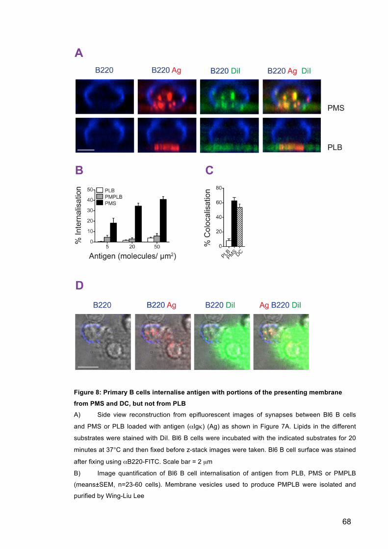
68
Figure 8: Primary B cells internalise antigen with portions of the presenting membrane
from PMS and DC, but not from PLB A) Side view reconstruction from epifluorescent images of synapses between Bl6 B cells
and PMS or PLB loaded with antigen (αIgκ) (Ag) as shown in Figure 7A. Lipids in the different
substrates were stained with DiI. Bl6 B cells were incubated with the indicated substrates for 20
minutes at 37°C and then fixed before z-stack images were taken. Bl6 B cell surface was stained
after fixing using αB220-FITC. Scale bar = 2 µm
B) Image quantification of Bl6 B cell internalisation of antigen from PLB, PMS or PMPLB
(means±SEM, n=23-60 cells). Membrane vesicles used to produce PMPLB were isolated and
purified by Wing-Liu Lee

69
C) Image quantification of colocalisation of antigen with DiI-labelled lipid in primary B cells
after internalisation from the substrates (Means±SEM, n=12-21 cells)
D) Epifluorescent image of a B1-8 B cell interacting with a DC loaded with NIP antigen (Ag).
The plasma membrane of the DC was labelled with DiI. B1-8 B cells were incubated with the DCs
for 20 minutes at 37°C and then fixed before images were taken. B1-8 B cell surface was labelled
after fixing with αB220-FITC. Scale bar = 5 µm
Figure 9: PMS are similar in their fluidity compared to PLB and PMPLB as shown by
FRAP
A) Lipids of the substrates were stained with DiI and then bleached for 1 minute. Arrow
shows point of bleaching. Recovery of DiI fluorescence to the bleached spot was measured for 10
minutes at 37°C
B) Antigen (αIgκ) was loaded onto substrates and then bleached for 1 minute. Arrow shows
point of bleaching. Recovery of antigen fluorescence to the bleached spot was measured for 10
minutes at 37°C
Membrane vesicles used to produce PMPLB were isolated and purified by Wing-Liu Lee
3.2.3 Plasma membrane sheets are similar in their viscoelastic properties to antigen-presenting cells
Since we have shown that B cells can internalise antigen from PMS but not PLB,
we wondered whether the 10 nm gap between the lipids of the PMS and
coverslip allowed the substrate to be more flexible, and whether lack of this
space, and therefore flexibility, was the reason B cells couldn’t internalise antigen
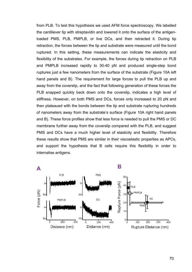
70
from PLB. To test this hypothesis we used AFM force spectroscopy. We labelled
the cantiliever tip with streptavidin and lowered it onto the surface of the antigen-
loaded PMS, PLB, PMPLB, or live DCs, and then retracted it. During tip
retraction, the forces between the tip and substrate were measured until the bond
ruptured. In this setting, these measurements can indicate the elasticity and
flexibility of the substrates. For example, the forces during tip retraction on PLB
and PMPLB increased rapidly to 30-40 pN and produced single-step bond
ruptures just a few nanometers from the surface of the substrate (Figure 10A left
hand panels and B). The requirement for large forces to pull the PLB up and
away from the coverslip, and the fact that following generation of these forces the
PLB snapped quickly back down onto the coverslip, indicates a high level of
stiffness. However, on both PMS and DCs, forces only increased to 20 pN and
then plateaued with the bonds between the tip and substrate rupturing hundreds
of nanometers away from the substrate’s surface (Figure 10A right hand panels
and B). These force profiles show that less force is needed to pull the PMS or DC
membrane further away from the coverslip compared with the PLB, and suggest
PMS and DCs have a much higher level of elasticity and flexibility. Therefore
these results show that PMS are similar in their viscoelastic properties as APCs,
and support the hypothesis that B cells require this flexibility in order to
internalise antigens.
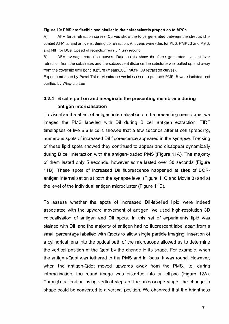
71
Figure 10: PMS are flexible and similar in their viscoelastic properties to APCs
A) AFM force retraction curves. Curves show the force generated between the streptavidin-
coated AFM tip and antigens, during tip retraction. Antigens were αIgκ for PLB, PMPLB and PMS,
and NIP for DCs. Speed of retraction was 0.1 µm/second
B) AFM average retraction curves. Data points show the force generated by cantilever
retraction from the substrates and the subsequent distance the substrate was pulled up and away
from the coverslip until bond rupture (Means±SD, n=31-109 retraction curves).
Experiment done by Pavel Tolar. Membrane vesicles used to produce PMPLB were isolated and
purified by Wing-Liu Lee
3.2.4 B cells pull on and invaginate the presenting membrane during antigen internalisation
To visualise the effect of antigen internalisation on the presenting membrane, we
imaged the PMS labelled with DiI during B cell antigen extraction. TIRF
timelapses of live Bl6 B cells showed that a few seconds after B cell spreading,
numerous spots of increased DiI fluorescence appeared in the synapse. Tracking
of these lipid spots showed they continued to appear and disappear dynamically
during B cell interaction with the antigen-loaded PMS (Figure 11A). The majority
of them lasted only 5 seconds, however some lasted over 30 seconds (Figure
11B). These spots of increased DiI fluorescence happened at sites of BCR-
antigen internalisation at both the synapse level (Figure 11C and Movie 3) and at
the level of the individual antigen microcluster (Figure 11D).
To assess whether the spots of increased DiI-labelled lipid were indeed
associated with the upward movement of antigen, we used high-resolution 3D
colocalisation of antigen and DiI spots. In this set of experiments lipid was
stained with DiI, and the majority of antigen had no fluorescent label apart from a
small percentage labelled with Qdots to allow single particle imaging. Insertion of
a cylindrical lens into the optical path of the microscope allowed us to determine
the vertical position of the Qdot by the change in its shape. For example, when
the antigen-Qdot was tethered to the PMS and in focus, it was round. However,
when the antigen-Qdot moved upwards away from the PMS, i.e. during
internalisation, the round image was distorted into an ellipse (Figure 12A).
Through calibration using vertical steps of the microscope stage, the change in
shape could be converted to a vertical position. We observed that the brightness

72
of the DiI spots correlated with the upward movement of antigen-Qdots (Figure
12B). Therefore, these studies demonstrated that the increase in DiI fluorescence
in the B cell synapse was due to the B cell pulling up and invaginating the PMS
during antigen internalisation.
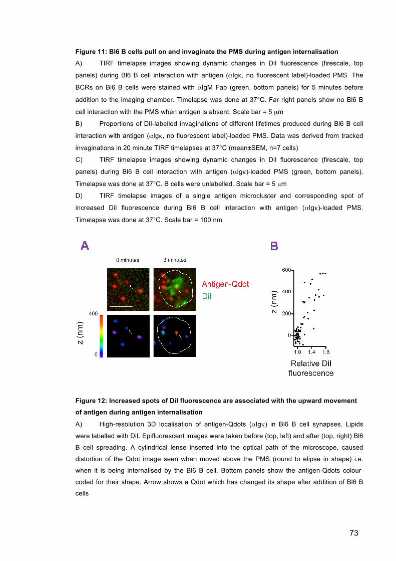
73
Figure 11: Bl6 B cells pull on and invaginate the PMS during antigen internalisation
A) TIRF timelapse images showing dynamic changes in DiI fluorescence (firescale, top
panels) during Bl6 B cell interaction with antigen (αIgκ, no fluorescent label)-loaded PMS. The
BCRs on Bl6 B cells were stained with αIgM Fab (green, bottom panels) for 5 minutes before
addition to the imaging chamber. Timelapse was done at 37°C. Far right panels show no Bl6 B
cell interaction with the PMS when antigen is absent. Scale bar = 5 µm
B) Proportions of DiI-labelled invaginations of different lifetimes produced during Bl6 B cell
interaction with antigen (αIgκ, no fluorescent label)-loaded PMS. Data was derived from tracked
invaginations in 20 minute TIRF timelapses at 37°C (mean±SEM, n=7 cells)
C) TIRF timelapse images showing dynamic changes in DiI fluorescence (firescale, top
panels) during Bl6 B cell interaction with antigen (αIgκ)-loaded PMS (green, bottom panels).
Timelapse was done at 37°C. B cells were unlabelled. Scale bar = 5 µm
D) TIRF timelapse images of a single antigen microcluster and corresponding spot of
increased DiI fluorescence during Bl6 B cell interaction with antigen (αIgκ)-loaded PMS.
Timelapse was done at 37°C. Scale bar = 100 nm
Figure 12: Increased spots of DiI fluorescence are associated with the upward movement
of antigen during antigen internalisation
A) High-resolution 3D localisation of antigen-Qdots (αIgκ) in Bl6 B cell synapses. Lipids
were labelled with DiI. Epifluorescent images were taken before (top, left) and after (top, right) Bl6
B cell spreading. A cylindrical lense inserted into the optical path of the microscope, caused
distortion of the Qdot image seen when moved above the PMS (round to elipse in shape) i.e.
when it is being internalised by the Bl6 B cell. Bottom panels show the antigen-Qdots colour-
coded for their shape. Arrow shows a Qdot which has changed its shape after addition of Bl6 B
cells

74
B) Vertical position of antigen-Qdots plotted against the colocalised DiI fluorescence
following Bl6 B cell spreading (n=10 cells) ***,p<0.001 for Pearson correlation test.
Experiment done by Pavel Tolar
3.2.5 Antigen internalisation occurs only at sites of long-lived invagination formation
Characterisation studies of individual invaginations revealed that short-lived
invaginations colocalised with background levels of antigen and terminated
abruptly suggesting physical rupture (Figure 13A, left hand panel). However,
long-lived invaginations started at points of high antigen fluorescence, indicative
of an antigen microcluster, and both fluorescent traces disappeared from the
TIRF field together, suggesting internalisation (Figure 13A, right hand panel). By
grouping the invaginations by their lifetimes, we saw that the level of antigen
fluorescence at the start of the invagination, corresponded to the subsequent
invagination lifetime. We also confirmed that there was a significant drop of
antigen fluorescence at the end of all invaginations lasting over 20 seconds,
which was not seen with short-lived invaginations (Figure 13B).
These results show that B cells pulled on and invaginated the presenting
membrane during BCR-antigen internalisation. The sites of invagination
contained variable amounts of antigen, but only those with a lifetime longer than
20 seconds ended with productive internalisation, and the lifetime of the
invaginations, depended on prior formation of antigen microclusters.

75
Figure 13: Invaginations produced by Bl6 B cells are dynamic and have varying lifetimes.
Long-lived invaginations begin at sites of antigen microclustering and lead to antigen
internalisation
A) Antigen (αIgκ) and DiI fluorescence in a single short-lived invagination (left) or a long-
lived invagination (right). Arrow indicates internalisation
B) Antigen (αIgκ) (Ag) fluorescence associated with tracked DiI-labelled invaginations
grouped by their lifetime. Data was derived from 20 minute TIRF timelapses of Bl6 B cells
interacting with antigen-loaded PMS at 37°C. The associated antigen fluorescence was analysed
5 seconds before (dotted lines), during the lifetime (solid lines), and 5 seconds after the
disappearance of the invagination (dotted lines) (mean±SEM, n=10 cells). *, P<0.01 in paired t-
tests between the penultimate time point of the invagination, and the time point 5 seconds after
invagination disappearance. Matlab analysis was done by Pavel Tolar
C) DiI fluorescence in tracked invaginations of the indicated lifetimes from the same dataset
as in B). Matlab analysis done by Pavel Tolar

76
3.3 Discussion We initially attempted to study BCR internalisation of membrane-bound antigen
using PLB as surrogates for APCs. Our observations of the interaction between B
cells and antigen-loaded PLB match the synapse formation previously reported
(Fleire et al., 2006). However, we observed that B cells were inhibited in their
ability to internalise antigen from these synapses, therefore, we developed a new
antigen-presenting substrate using immobilised plasma membranes from
adherent cells.
PLB are very flat surfaces (Attwood et al., 2013; Sackmann, 1996), which have
made them ideal substrates for high-resolution TIRF microscopy of the initial
molecular events involved in BCR recognition of membrane-bound antigen. In
order to expand these studies we originally intended to use these same
techniques to study the next step in the B cell activation process: BCR-antigen
internalisation. However, the inability of B cells to internalise antigen from PLB
was shown both by the formation of stable synapses and absence of antigen
detected in the B cells following synapse formation.
We initially thought this was because the biotin-streptavidin-biotin linkage was
anchoring the antigen too strongly to the PLB. In an attempt to overcome this
limitation, we then used B cells from MuMT-MD4 mice, which have a very high
affinity for the HEL antigen, but we still did not observe any internalisation. This
experimental set up has been used in previous studies, which reported
successful antigen internalisation and presentation by B cells through activation
of co-cultured cognate T cell hybridomas (Fleire et al., 2006). The internalisation
of antigen described in these studies could be due to the release of small
amounts of antigen from the PLB during the long incubation times with B cells
and cognate T cells, before assessing T cell activation. If small amounts of
antigen were released by the PLB, and internalised and presented by B cells, it is
possible that this was enough to activate antigen-specific T cell hybridomas,
which are very sensitive to peptide-MHC Class II molecules. This internalisation
could also be explained by other recent studies showing that when B cells come
77
into contact with immobilised antigen, they secrete lysosomes releasing
hydrolases and acidifying the synapse, which causes antigen liberation from the
presenting surface and facilities internalisation (Yuseff et al., 2011). As PLB
completely cover the surface of the coverslip with only a 1 nm space between the
bilayer and the glass (Sackmann, 1996), we hypothesised that this surface could
be very inflexible and so this release of lysosomes may occur as a consequence
of B cells being unable to generate big enough forces to extract the antigen.
Therefore, we set out to develop a new more flexible antigen-presenting surface.
Here we show the development of PMS, a new APC surrogate for microscopy
studies into B cell antigen acquisition from antigen-presenting surfaces. PMS
have been recently used to investigate the formation of clathrin coated pits after
reconstitution with cytoplasmic components (Wu et al., 2010). The observed
production of endocytic vesicles in these studies indicated a level of flexibility
within the PMS, and as predicted, following antigen tethering, B cells were able to
internalise antigen from this substrate.
We used AFM force spectroscopy to assess the differences in flexibility between
the PLB and PMS, and compared them to the viscoelastic properties of APCs.
These studies demonstrated B cells would need to overcome forces of 20 pN to
extract antigens from the PMS or DCs, which supports the values required to pull
out nanotubes from plasma membranes previously reported (Krieg et al., 2008).
However, in order to extract antigen from PLB the B cells would need to generate
forces over 40 pN. This supports our hypothesis that PLB are inflexible
substrates and suggests that the antigen-presenting surface needs to have a
certain level of flexibility to reduce the force required by B cells to facilitate BCR-
antigen internalisation.
Scanning AFM revealed that PMS were suspended approximately 10 nm above
the coverslip. This 10 nm space is consistent with the presence of
transmembrane proteins or ruffling of the adherent cell membranes, which are
the source of PMS, and could account for the increased flexibility seen when
comparing PMS with PLB. This is also the only primary difference between the

78
PMS and PMPLB further supporting flexibility in the antigen-presenting surface
as an essential factor in antigen internalisation.
To visualise any changes in the topology of the presenting membrane during
BCR-antigen internalisation, we used the hydrophobic carbocyanine dye, DiI,
which incorporates into the lipid bilayer and acts as a reporter for changes in
membrane shape (Sund et al., 1999). TIRF studies showed B cells actively pulled
on and invaginated the PMS during BCR-antigen internalisation. The number of
invaginations was more frequent and dynamic compared to detectable antigen
microclusters, with only long-lived invaginations resulting in internalisation. The
high number of non-productive short-lived invaginations suggests a mechanism
whereby B cells continually screen the antigen-presenting surface, and only
when they bind to pre-formed microclusters and overcome a force of 20pN, can
they produce a long-lived invagination and internalise the antigen. This could
explain how B cells acquire APC membrane proteins during BCR-antigen
internalisation in vivo (Suzuki et al., 2009).
PMS do not fully resemble APCs as they lack any adhesion and co-stimulatory
molecules found on APCs in vivo, which could further regulate BCR-antigen
internalisation (Carrasco and Batista, 2006a; Carrasco et al., 2004; Suzuki et al.,
2009), however, they do resemble APCs in their viscoelastic properties and are
closer in their membrane structure than the historically used PLB. In addition, the
use of PMS has allowed us to visualise antigen internalisation previously unseen
at the molecular level.
Therefore, although, in the future it will be important to continually optimise this
technique to more closely resemble APCs, or use APCs themselves, to confirm
the findings shown here, the PMS can be considered a reasonable surrogate for
APCs, and used for the identification of molecular components and mechanisms
involved in BCR-antigen internalisation.
79
Chapter 4 B cells use myosin IIA contractility combined with clathrin-mediated endocytosis to
internalise membrane-bound antigen
4.1 Introduction Antigen binding stimulates BCR-antigen internalisation from either a cap of BCR-
antigen complexes in response to soluble antigen, (Ma et al., 2001; Salisbury,
1980) or from a synapse when encountered with an antigen tethered to an APC
(Batista et al., 2001).
The process of BCR-antigen internalisation is thought to begin with BCR-antigen
microclustering and translocation to lipid rafts (Cheng et al., 1999; Sohn, 2006;
Sohn et al., 2008; Tolar et al., 2005). Once associated with the lipid rafts BCRs
are exposed to the Src family tyrosine kinase Lyn (Cheng et al., 1999; 2001) and
a BCR signalling cascade is initiated resulting in BCR-antigen internalisation.
Studies using soluble antigen to stimulate B cells have identified many BCR
signalling molecules as regulators of antigen internalisation. These proteins
include Lyn itself (Ma et al., 2001; Stoddart et al., 2002), the adaptor proteins
Bam32 and WASp (Becker-Herman et al., 2011; Niiro et al., 2004), the guanine
exchange factors Vav 1 and 3, and the GTPase Rac (Malhotra et al., 2009a;
2009b). These signalling proteins have also been linked to the control of the actin
cytoskeleton, therefore also strongly indicating a requirement for actin activity in
BCR-antigen internalisation. In addition, these studies have identified clathrin as
the primary endocytic mediator in this process (Salisbury, 1980; Stoddart et al.,
2002). However, the precise contribution clathrin, actin and BCR signalling make
towards BCR-antigen internalisation is still being discussed.
More recently, attention has been turned to mechanisms, by which B cells extract
antigen from APCs (Batista and Harwood, 2009; Batista et al., 2001). To model
antigen acquisition from APCs, high-resolution live-cell imaging of B cells
80
interacting with antigen-loaded PLB have been done. These studies have
revealed some signalling molecules involved in B cell spreading and antigen
gathering in response to membrane-bound antigen (Arana et al., 2008; Carrasco
and Batista, 2006a; Carrasco et al., 2004; Depoil et al., 2007; Fleire et al., 2006;
Mattila et al., 2013; Schnyder et al., 2011; Tolar et al., 2009b; Treanor et al.,
2011; Weber et al., 2008). However, as these B cells are inhibited in their ability
to internalise antigen from the PLB (Chapter 3), it is unknown how gathering of
antigen in the B cell-APC synapse leads to internalisation.
Therefore, while these data have identified components involved in BCR-antigen
internalisation and the mechanisms of synapse formation, it is still unknown how
the signalling cascade initiated by BCR-antigen binding is activating and
recruiting the endocytic mediators. We also do not know whether the mechanism
of BCR-antigen internalisation changes depending on the context of antigen
encounter.
For this chapter our aim was to begin to delineate the mechanisms of antigen
internalisation by using PMS as surrogates for APCs. PMS allow live-cell imaging
of membrane-bound antigen internalisation, which has not been previously
possible in the studies using soluble antigen or PLB. We hoped to identify novel
components involved in BCR-antigen internalisation using a customised genetic
screen and small-molecule inhibition, and visualise their spatial and temporal
activity in real-time with high-resolution TIRF microscopy.
4.2 Results 4.2.1 SiRNA is not a reliable method of protein knockdown for the genetic
screen To identify novel components involved in BCR-antigen internalisation we planned
to set up a customised genetic screen. This screen would allow us to
systematically test a large number of protein candidates for a role in BCR-antigen
internalisation and could allow the unbiased identification of new signalling
pathways involved in this process.

81
We initially attempted to set up this technique using a siRNA-mediated protein
knockdown approach. Although B cells are thought to be highly resistant to non-
viral methods of siRNA-mediated knockdown (Goffinet and Keppler, 2006;
Gresch, 2004), a new amaxa nucleofection technique has been recently shown
to increase the transfection efficiency and cell viability following siRNA
transfection, and has proved useful in studying the effect of gene knockdowns in
T lymphocytes (Goffinet and Keppler, 2006; Gresch, 2004; Mantei et al., 2008;
Stallwood et al., 2006).
Initial attempts to optimise siRNA nucleofection in B cells were successful.
Approximately 50% reduction of CD19 in Bl6 B cells, and GAPDH in Ramos
cells, was seen following siRNA nucleofection of their specific, but not control,
oligonucleotides (Figure 14A and B). We then tested this technique using
oligonucleotides specific for clathrin, dynamin2, or AP2. These proteins are
known to be involved in endocytosis and BCR-antigen internalisation, and
therefore would act as positive controls in the genetic screen (Collinet et al.,
2010; Stoddart et al., 2005; Taylor et al., 2011). However, no reduction in protein
levels was observed (Figure 14C). Its unlikely the absence of knockdown was
due to high stability of the proteins, as other studies reported 90% reduction of
clathrin under a tetracycline-sensitive promoter, in the same time frame as our
knockdown studies, in a B cell line following doxycycline treatment (Stoddart et
al., 2005). The unsuccessful knockdown could be because these particular
oligonucleotides were not efficiently reducing mRNA levels. However, because of
the high rate of failure and also high levels of cell toxicity observed, we decided
to abandon siRNA as a method of protein knockdown and use lentiviral delivery
of shRNA instead. This process is labour intensive compared to siRNA
nucleofection, however the resulting transduction is reported to be higher than
nonviral methods and successful knockdown of proteins in B cells has been
previously reported (Gresch, 2004; Pavri et al., 2010).

82
Figure 14: Nucleofection of siRNA produces a modest protein knockdown in B cells
A) Flow cytometry plots (left) and quantification (right) showing CD19 protein levels following
siRNA-mediated knockdown. Protein levels were assessed using αCD19-FITC 4 days following
siRNA nucleofection. Grey = unstained, black = control siRNA, red = CD19 siRNA (mean±SEM,
n=3 independent expeirments) *,p<0.05 in Mann-Whitney test compared to Control siRNA
B) Same as A but with Ramos cells and siRNA-mediated knockdown of GAPDH
C) Western blot showing protein levels 4 days after siRNA-mediated knockdown of indicated
proteins
4.2.2 Lentiviral delivery of shRNA mediates protein knockdown of endocytic mediators in B cells
To produce shRNA-lentiviral particles, we transfected the pLKO.1 lentiviral
vectors from The RNAi Consortium (Moffat et al., 2006; Root et al., 2006) which
contain shRNA constructs specific for our proteins of interest, into HEK293FT

83
cells. Viral supernatants were collected 3 days later and used to infect Ramos
cells.
To ensure infectious particles were being produced, Ramos cells were infected
with a control lentivirus containing a GFP construct. An increase in GFP
fluorescence provided evidence of successful viral production and infection
(Figure 15A). To check if infection of shRNA-lentivirus could mediate protein
knockdown, Ramos cells were infected with dynamin2 shRNA-lentivirus. 4 days
following infection, knockdown of dynamin2 protein levels was observed in both
Bl6 B cells and Ramos cells (Figure 15B and C). Better knockdown efficiency
was seen in Ramos cells compared to primary Bl6 B cells. This may be due to a
reduced susceptibility of primary B cells to lentiviral infection compared to
retroviral infection, which has been described anecdotally (Pavri et al., 2010). For
this reason, we chose to use Ramos cells for the continual development of the
genetic screen.

84
Figure 15: Infection of lentivirus-containing shRNA can mediate protein knockdown in
Ramos cells and Bl6 B cells
A) Flow cytometry plot showing GFP fluorescence in Ramos cells 1 day following infection of
cells with pLKO.1-GFP lentivirus (red) or uninfected controls (grey)
B) Western blot showing dynamin2 protein levels 4 days after infection with lentivirus-
containing dynamin2-shRNA
C) Quantification of B (mean±SEM, n=3 independent experiments) *, p<0.05, ***,p<0.001 in
Mann-Whitney test compared to Empty pLKO.1
4.2.3 Inhibition of internalisation following protein knockdown can be assessed by flow cytometry or microscopy
To understand whether the dynamin2 knockdown mediated by shRNA-lentivirus
transduction caused a decrease in BCR-antigen internalisation, we stimulated the
infected cells to internalise soluble antigen at 37°C. Control cells were left on ice.
The cells were then fixed and stained with fluorescent streptavidin to visualise
any antigen remaining on the cell surface. While the streptavidin signal
decreased after 37°C incubation of the mock-infected samples, which is
indicative of fewer antigens on the cell surface due to internalisation, dynamin2
knockdown prevented this decrease indicating antigen retention on the cell
surface, and therefore a decrease in BCR-antigen internalisation (Figure 16A and
B). This was further confirmed by microscopy, which revealed little colocalisation
between antigen and streptavidin in mock infected samples, as the majority of the
antigen was inside the B cell and protected from the streptavidin binding.
However, in the dynamin2 knockdown cells, antigen was retained on the cell
surface resulting in a high level of antigen colocalisation with streptavidin (Figure
16C and D). These results support both the previous studies showing reduced
BCR-antigen internalisation following dynamin2 inhibition (Chaturvedi et al.,
2011; Malhotra et al., 2009b), as well as the use of this technique in the
identification of proteins involved in BCR-antigen internalisation.
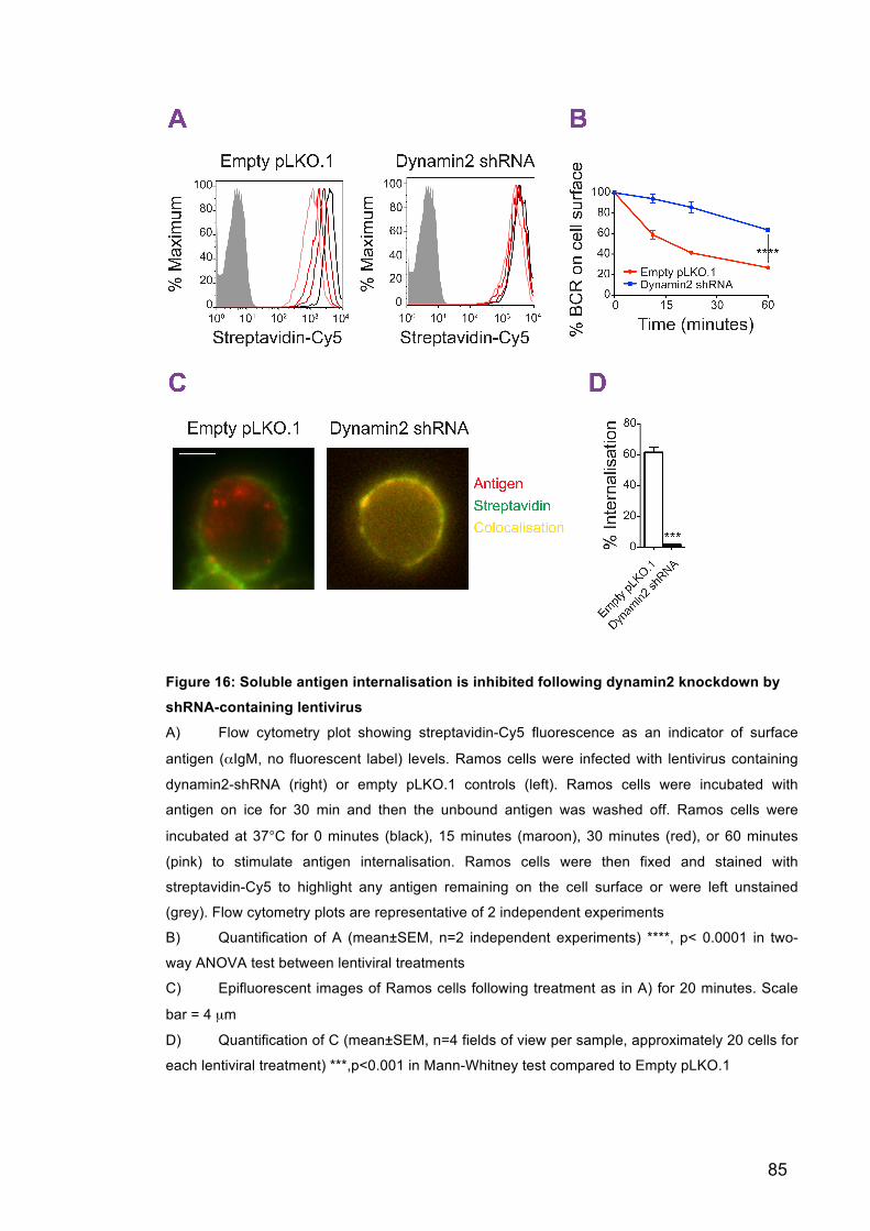
85
Figure 16: Soluble antigen internalisation is inhibited following dynamin2 knockdown by
shRNA-containing lentivirus
A) Flow cytometry plot showing streptavidin-Cy5 fluorescence as an indicator of surface
antigen (αIgM, no fluorescent label) levels. Ramos cells were infected with lentivirus containing
dynamin2-shRNA (right) or empty pLKO.1 controls (left). Ramos cells were incubated with
antigen on ice for 30 min and then the unbound antigen was washed off. Ramos cells were
incubated at 37°C for 0 minutes (black), 15 minutes (maroon), 30 minutes (red), or 60 minutes
(pink) to stimulate antigen internalisation. Ramos cells were then fixed and stained with
streptavidin-Cy5 to highlight any antigen remaining on the cell surface or were left unstained
(grey). Flow cytometry plots are representative of 2 independent experiments
B) Quantification of A (mean±SEM, n=2 independent experiments) ****, p< 0.0001 in two-
way ANOVA test between lentiviral treatments
C) Epifluorescent images of Ramos cells following treatment as in A) for 20 minutes. Scale
bar = 4 µm
D) Quantification of C (mean±SEM, n=4 fields of view per sample, approximately 20 cells for
each lentiviral treatment) ***,p<0.001 in Mann-Whitney test compared to Empty pLKO.1

86
4.2.4 BCR-antigen internalisation is reduced in Ramos cells following shRNA mediated knockdown of myosin IIA and clathrin
20 genes were chosen for a pilot run of the genetic screen. The genes chosen
included known and predicted endocytic mediators that are expressed highly in
murine primary B cells. 4 days following lentiviral infection, Ramos cells were
incubated with antigen-loaded PMS and the extent of internalisation was
analysed. If 3 out of 4 clones used for each gene showed a difference in
internalisation by 2 standard deviations from the normalised Z score, the gene
was counted as a positive hit. Normalised z scores demonstrated that knockdown
of dynamin2 (an enzyme required for the pinching off of clathrin-coated vesicles),
Ap2 (AP2M1, an essential clathrin adapter protein), myosin IIA (MYH9, a motor
protein involved in the actomyosin cytoskeleton), and Rock1 (a myosin IIA
activator) all caused significant decreases in BCR-antigen internalisation (Figure
17). This inhibition was validated by subsequent repetitions (Figure 18A)
showing that the clathrin pathway (dynamin2 and Ap2) as well as the myosin IIA
pathway (myosin IIA and Rock1) are important in mediating membrane-bound
antigen internalisation in Ramos cells.
The ability of Ramos cells to pull on and invaginate the PMS during antigen-
internalisation was analysed by tracking changes in DiI fluorescence in DiI-
stained antigen-loaded PMS (Chapter 3). Interestingly the number of
invaginations decreased only if myosin IIA or Rock1 were knocked-down and
were unaffected by Ap2 or dynamin2 knockdown (Figure 18B). These results
indicate B cells require myosin IIA contractility to invaginate the PMS before
internalising antigen in a clathrin-dependent manner.

87

88
Figure 17: A pilot customised genetic screen was used to identify components involved
in antigen internalisation from the PMS
Ramos cells were infected with shRNA-lentivirus and 4 days later were incubated with antigen
(αIgM)-loaded PMS for 20 minutes at 37°C. Cells were then fixed before internalisation was
analysed. Each gene was targeted by 4 separate shRNA sequences. Red bars indicate Z
values 2 standard deviations or more away from the mean (n=>20 cells, n=1 experiment for
each gene)
Figure 18: Ramos cells need to invaginate the PMS using myosin IIA contraction before internalising the antigen in a clathrin-dependent manner
A) Repetition of positive hits shown in Figure 17. Antigen (αIgM) internalisation from PMS by
Ramos B cells expressing shRNA against AP2, dynamin2, Rock1, or myosin IIA. Ramos cells
were incubated with PMS for 20 minutes at 37°C and then fixed before internalisation was
analysed. Values shown are pooled results from all 4 shRNA clones (mean±SEM n=>60 cells
over 3 independent experiments). f.u., fluorescent units, ****, p<0.0001 in Mann-Whitney test
compared to Empty pLKO.1
B) Number of tracked DiI-labelled invaginations pulled from the PMS by Ramos B cells
expressing shRNA against AP2, dynamin2, Rock1, or myosin IIA. Tracks were taken from a 20
minute timelapse of Ramos cells interacting with antigen (αIgM)-loaded PMS at 37°C.
(mean±SEM n=>20 cells over 2 independent experiments). ****, p<0.0001, ***, p<0.001 in Mann-
Whitney test compared to Empty pLKO.1

89
4.2.5 BCR-antigen internalisation is reduced in Bl6 B cells following pharmacological inhibition of myosin IIA and clathrin
To confirm the results of the pilot screen in Ramos cells independently, we ran
another pilot screen using Bl6 B cells and small molecule inhibitors against
known and possible endocytic mediators, as well as BCR signalling molecules.
Inhibition of Rock1 as well as myosin IIA itself, supported the requirement of the
myosin IIA pathway in BCR-antigen internalisation and PMS invagination, and
dynamin2 inhibition supported the need for clathrin-mediated endocytosis (Figure
19A). Lyn and formin inhibitors confirmed previous reports showing BCR
signalling and the actin cytoskeleton are also needed in BCR-antigen
internalisation (Brown and Song, 2001; Fleire et al., 2006; Ma et al., 2001;
Stoddart et al., 2005), whereas PKC and Rac1 were seen to be dispensable
(Arana et al., 2008; Weber et al., 2008). Erk1/2 signalling was also seen to be
important in BCR-antigen internalisation, which supports recent findings
suggesting BCR signalling continues following BCR-antigen internalisation
(Chaturvedi et al., 2011).
Comparing internalisation of soluble or membrane-bound antigen by Bl6 B cells
supported the idea that clathrin and myosin IIA have independent roles in BCR-
antigen internalisation. Dynamin2 inhibition (mediated by dynasore) prevented
internalisation of antigen whether in solution or tethered to the PMS (Figure 19B).
However, it did not stop the B cell from invaginating the PMS (Figure 19C) or
BCR signalling, as Bl6 B cells inhibited by dynasore, were able to spread over
the PMS, form microclusters and contract bringing them into a central synapse
(Figure 20). This suggests clathrin acts as the endoytic mediator in this process
but is not required for BCR signalling or PMS invagination prior to internalisation.
In contrast, internalisation following a reduction in the activity of the myosin IIA
pathway (mediated by blebbistatin and Y-27632) was only decreased when the
antigen was membrane-bound, and antigen internalisation from solution was
unaffected (Figure 19B). These inhibited cells were also unable to invaginate the
PMS (Figure 19C) or contract in response to membrane-bound antigen shown by
an increase in synapse size (Figure 20). These results suggest myosin IIA is the
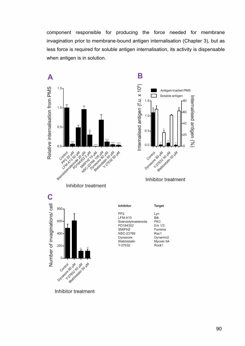
90
component responsible for producing the force needed for membrane
invagination prior to membrane-bound antigen internalisation (Chapter 3), but as
less force is required for soluble antigen internalisation, its activity is dispensable
when antigen is in solution.
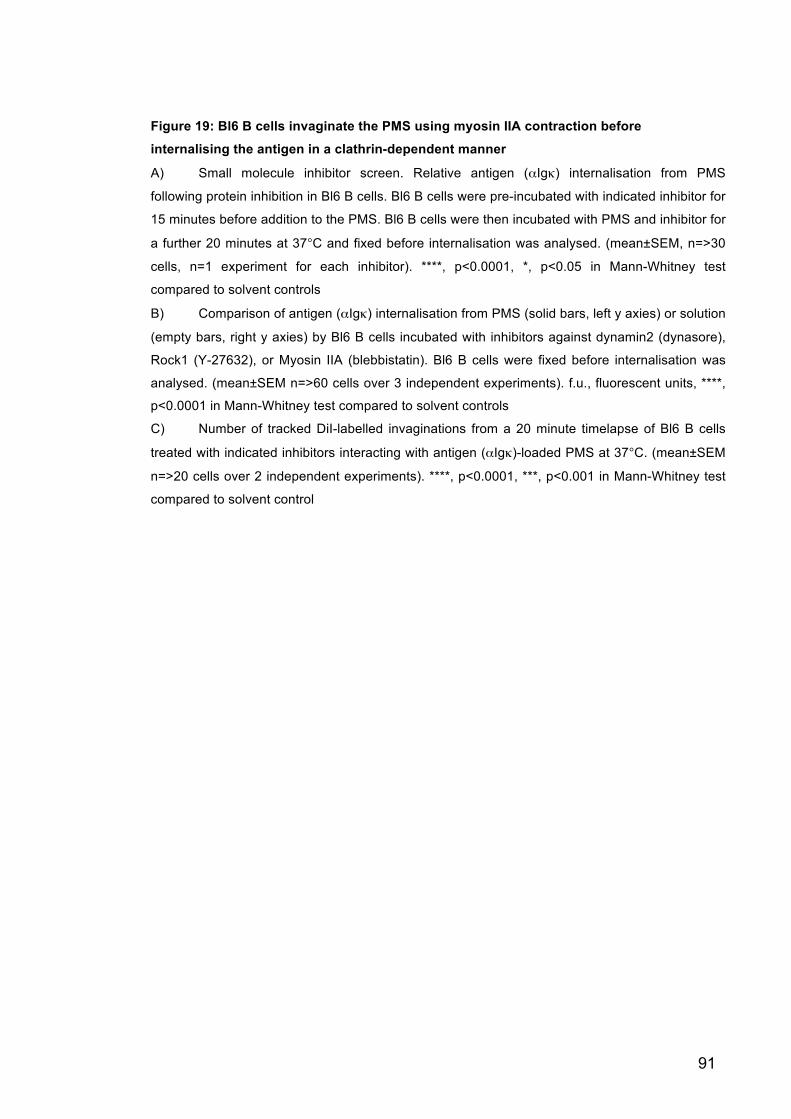
91
Figure 19: Bl6 B cells invaginate the PMS using myosin IIA contraction before
internalising the antigen in a clathrin-dependent manner
A) Small molecule inhibitor screen. Relative antigen (αIgκ) internalisation from PMS
following protein inhibition in Bl6 B cells. Bl6 B cells were pre-incubated with indicated inhibitor for
15 minutes before addition to the PMS. Bl6 B cells were then incubated with PMS and inhibitor for
a further 20 minutes at 37°C and fixed before internalisation was analysed. (mean±SEM, n=>30
cells, n=1 experiment for each inhibitor). ****, p<0.0001, *, p<0.05 in Mann-Whitney test
compared to solvent controls
B) Comparison of antigen (αIgκ) internalisation from PMS (solid bars, left y axies) or solution
(empty bars, right y axies) by Bl6 B cells incubated with inhibitors against dynamin2 (dynasore),
Rock1 (Y-27632), or Myosin IIA (blebbistatin). Bl6 B cells were fixed before internalisation was
analysed. (mean±SEM n=>60 cells over 3 independent experiments). f.u., fluorescent units, ****,
p<0.0001 in Mann-Whitney test compared to solvent controls
C) Number of tracked DiI-labelled invaginations from a 20 minute timelapse of Bl6 B cells
treated with indicated inhibitors interacting with antigen (αIgκ)-loaded PMS at 37°C. (mean±SEM
n=>20 cells over 2 independent experiments). ****, p<0.0001, ***, p<0.001 in Mann-Whitney test
compared to solvent control
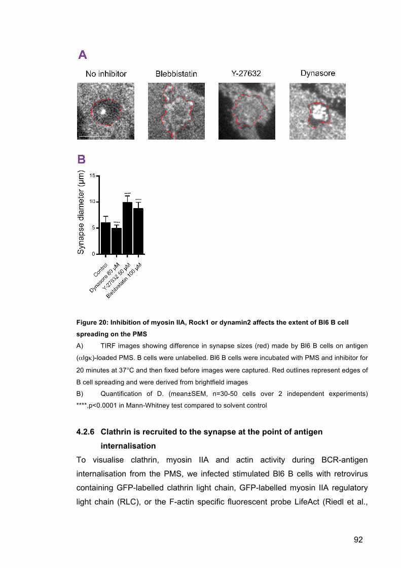
92
Figure 20: Inhibition of myosin IIA, Rock1 or dynamin2 affects the extent of Bl6 B cell
spreading on the PMS
A) TIRF images showing difference in synapse sizes (red) made by Bl6 B cells on antigen
(αIgκ)-loaded PMS. B cells were unlabelled. Bl6 B cells were incubated with PMS and inhibitor for
20 minutes at 37°C and then fixed before images were captured. Red outlines represent edges of
B cell spreading and were derived from brightfield images
B) Quantification of D. (mean±SEM, n=30-50 cells over 2 independent experiments)
****,p<0.0001 in Mann-Whitney test compared to solvent control
4.2.6 Clathrin is recruited to the synapse at the point of antigen
internalisation To visualise clathrin, myosin IIA and actin activity during BCR-antigen
internalisation from the PMS, we infected stimulated Bl6 B cells with retrovirus
containing GFP-labelled clathrin light chain, GFP-labelled myosin IIA regulatory
light chain (RLC), or the F-actin specific fluorescent probe LifeAct (Riedl et al.,
93
2008), respectively. Successful infection of Bl6 B cells by retrovirus was shown
by increased GFP fluorescence using flow cytometry compared to uninfected
controls (Figure 21A).
TIRF images of Bl6 B cells infected with clathrin light chain-GFP on antigen-
loaded PMS showed an increase in the number and brightness of clathrin-coated
structures (CCS) in the synapse during B cell spreading and antigen-
internalisation (Figure 21B and C, Movie 4). Tracking of invaginations allowed us
to analyse the colocalisation of invaginations with CCSs, and showed us that
short-lived invaginations contained little clathrin, but that this colocalisation
increased up to 20% with long-lived invaginations, peaking towards the end of
the invagination lifetime (Figure 21D and E). Observations of clathrin and antigen
activity in two separate long-lived invaginations revealed the sequential formation
of antigen microclusters, invagination of the membrane, and recruitment of
clathrin (Figure 22A and B). Although comparisons between the 2 invaginations
revealed different temporal regulation (possibly due to differences in antigen
microcluster size), both followed the same recruitment pattern of antigen,
invagination, and clathrin, and in both cases simultaneous disappearance of all
three fluorescent traces was observed indicating the point of internalisation.
These observations support the hypothesis suggested in Chapter 3, that
successful internalisation occurs by sustaining long-lived invaginations at sites of
antigen microclustering. If the invagination is sustained long enough, CCSs
assemble allowing successful internalisation. In contrast, short-lived
invaginations terminate before clathrin is recruited and endocytosis can occur.

94
Figure 21: Retroviral infection of clathrin light chain-GFP in Bl6 B cells show clathrin
recruitment to Bl6 B cell synapse, and colocalisation of clathrin with long-lived
invaginations during antigen internalisation from the PMS
A) Flow cytometry plot showing GFP fluorescence in Bl6 B cells 48 hours following retroviral
infection of clathrin light chain-GFP (red) or uninfected controls (grey)
B) Images from a TIRF timelapse showing clathrin recruitment to Bl6 B cell synapse with
antigen (αIgκ)- loaded PMS. Bl6 B cells were infected with clathrin light chain-GFP retrovirus and
imaged 48 hours later. Timelapse images were taken at 37°C. B cells were unlabelled. White
outline represents edges of B cell spreading derived from brightfield images. Scale bars = 5 µm
95
C) Quantification of the number (red, left hand y axis) and brightness (blue, right hand y
axis) of CCSs in Bl6 B cell synapses on antigen (αIgκ)-loaded PMS. CCSs were tracked over a
20 minute TIRF timelapse 48 hours after Bl6 B cells were infected with clathrin light chain-GFP
(mean±SEM, n=15-21 cells). *, p<0.05 in Mann-Whitney tests for time>1 minute against no-
antigen controls
D) Images from a TIRF timelapse showing colocalisation of CCSs in a Bl6 B cell synapse
with DiD-labelled invaginations on an antigen (αIgκ, no fluorescent label)- loaded PMS. Bl6 B
cells were infected with clathrin light chain-GFP retrovirus and imaged 48 hours later. Timelapse
images were taken at 37°C. Image shown was taken 13 minutes after Bl6 B cells were added to
the PMS. Scale bars = 5 µm
E) Percentage of CCS fluorescence associated with tracked DiD-labelled invaginations
grouped by their lifetime. Data was derived from 20 minute TIRF timelapses of clathrin light chain-
GFP expressing Bl6 B cells, interacting with antigen (Igκ, no fluorescent label)-loaded PMS at
37°C. The associated clathrin light chain-GFP fluorescence was analysed 5 seconds before
(dotted lines), during the lifetime (solid lines), and 5 seconds after the disappearance of the
invagination (dotted lines) (mean±SEM, n=12 cells). Black lines, colocalisation of CCSs with
randomly scrambled controls. *, p<0.01 in Mann-Whitney tests against scrambled controls.
Matlab analysis was done by Pavel Tolar.

96
Figure 22: Bl6 B cells internalise antigen from the PMS following the sequential
formation of antigen microclusters, long-lived invaginations and CCSs
A) Formation of a single antigen (αIgκ) microcluster (arrows, top panels), followed by
invagination of DiI-labelled PMS (second row panels), and recruitment of clathrin (third row
panels). All three fluorescent images disappear at the same time indicating internalisation.

97
Images were taken from a 20 minute TIRF timelapse of Bl6 B cell expressing clathrin-GFP on
antigen (αIgκ)-loaded and DiI-stained PMS at 37°C. Scale bar = 100 nm
B) Fluorescent traces of a separate antigen microcluster showing same sequential formation
of antigen microcluster, DiI invagination, and CCS as above. Arrows show point of recruitment/
formation, and dotted line shows point of internalisation
4.2.7 Myosin IIA is recruited to the synapse immediately before invagination formation
TIRF timelapse images of myosin IIA RLC-GFP and LifeAct activity also support
myosin IIA and actin involvement in BCR-antigen internalisation as both showed
specific patterns of localisation in the B cell synapse (Figure 23A, 24A, Movie 5
and Movie 6). Analysis of myosin IIA colocalisation with membrane invaginations
revealed that myosin IIA recruitment peaked just before the onset of all
invaginations irrelevant of their lifetime, in agreement with the idea that the force
required for invagination formation is myosin IIA-dependent (Figure 23B and C).
In contrast actin was recruited in a biphasic manner, firstly at the onset of the
invagination, and then secondly at the end of the invagination if it lasted 20
seconds or longer (Figure 24B and C). The first wave of recruitment supports the
involvement of actomyosin cytoskeleton in invagination formation and the second
wave supports previous studies showing actin assists in clathrin-mediated
endocytosis when force is required to extract the cargo (Boulant et al., 2011;
Cureton et al., 2010).
Collectively these results suggest that following membrane-bound antigen
encounter, B cells use forces generated by the actomyosin cytoskeleton to pull
on and invaginate the presenting membrane. The invagination lifetime depends
on the prior formation of antigen microclusters and if the invagination persists
long enough, CCS are recruited, and antigen internalisation occurs in a clathrin-
and actin-dependent manner.
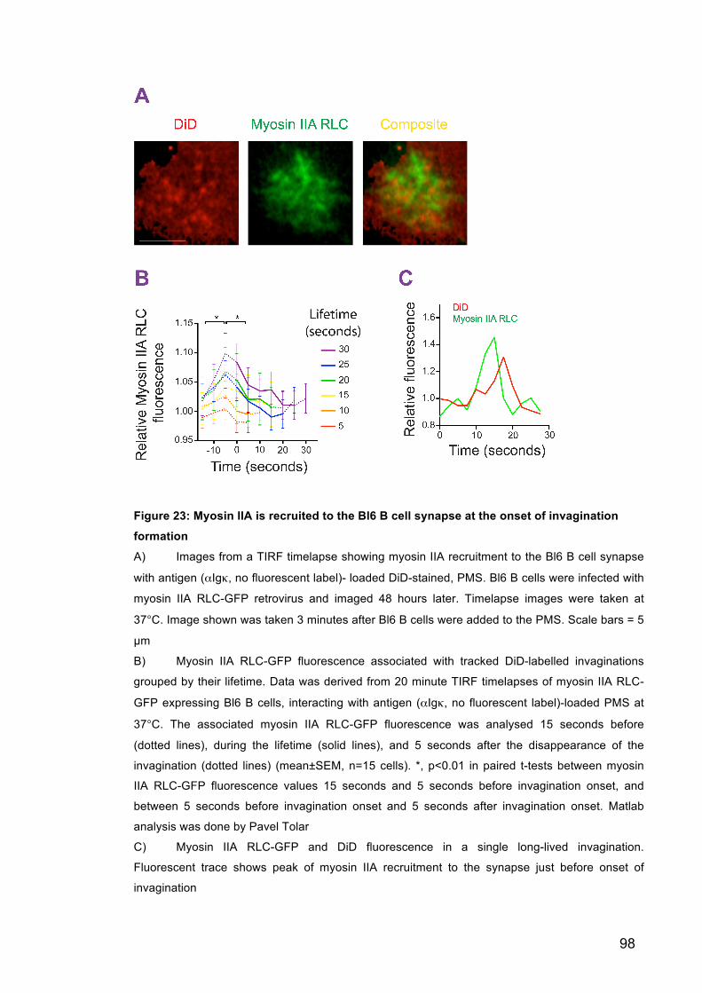
98
Figure 23: Myosin IIA is recruited to the Bl6 B cell synapse at the onset of invagination
formation
A) Images from a TIRF timelapse showing myosin IIA recruitment to the Bl6 B cell synapse
with antigen (αIgκ, no fluorescent label)- loaded DiD-stained, PMS. Bl6 B cells were infected with
myosin IIA RLC-GFP retrovirus and imaged 48 hours later. Timelapse images were taken at
37°C. Image shown was taken 3 minutes after Bl6 B cells were added to the PMS. Scale bars = 5
µm
B) Myosin IIA RLC-GFP fluorescence associated with tracked DiD-labelled invaginations
grouped by their lifetime. Data was derived from 20 minute TIRF timelapses of myosin IIA RLC-
GFP expressing Bl6 B cells, interacting with antigen (αIgκ, no fluorescent label)-loaded PMS at
37°C. The associated myosin IIA RLC-GFP fluorescence was analysed 15 seconds before
(dotted lines), during the lifetime (solid lines), and 5 seconds after the disappearance of the
invagination (dotted lines) (mean±SEM, n=15 cells). *, p<0.01 in paired t-tests between myosin
IIA RLC-GFP fluorescence values 15 seconds and 5 seconds before invagination onset, and
between 5 seconds before invagination onset and 5 seconds after invagination onset. Matlab
analysis was done by Pavel Tolar
C) Myosin IIA RLC-GFP and DiD fluorescence in a single long-lived invagination.
Fluorescent trace shows peak of myosin IIA recruitment to the synapse just before onset of
invagination

99
Figure 24: F-actin is recruited to the Bl6 B cells synapse in a biphasic manner
A) Images from a TIRF timelapse showing LifeAct (a fluorescent probe which binds to F-
actin) recruitment to the Bl6 B cell synapse with antigen (αIgκ, no fluorescent label)- loaded DiD-
stained, PMS. Bl6 B cells were infected with LifeAct retrovirus and imaged 48 hours later.
Timelapse images were taken at 37°C. Image shown was taken 5 minutes after Bl6 B cells were
added to the PMS. Scale bars = 5 µm
B) LifeAct fluorescence associated with tracked DiD-labelled invaginations grouped by their
lifetime. Data was derived from 20 minute TIRF timelapses of LifeAct expressing Bl6 B cells,
interacting with antigen (αIgκ, no fluorescent label)-loaded PMS at 37°C. The associated LifeAct
fluorescence was analysed 15 sec before (dotted lines), during the lifetime (solid lines), and 10
seconds after the disappearance of the invagination (dotted lines) (mean±SEM, n=13 cells). *,
p<0.015 in paired t-tests between LifeAct fluorescence values before invagination onset and 10
seconds prior to invagination onset (black bracket) and between values at invagination
termination and 10 seconds after invagination termination (colour brackets). Matlab analysis was
done by Pavel Tolar
C) LifeAct-GFP and DiD fluorescence in a single long-lived invagination. Fluorescent trace
shows two peaks of actin recruitment to the synapse; the first with invagination onset and the
second as the invagination is terminated

100
4.3 Discussion Using both shRNA-mediated knockdown and protein inhibition we have been
able to identify molecular components involved in membrane-bound antigen
internalisation for the first time. Live cell imaging of B cells interacting with the
PMS allowed us to visualise the temporal and spatial regulation of these
identified proteins and therefore understand their distinct roles in BCR-antigen
internalisation. These molecular components were shown to be important for
antigen internalisation in both primary mouse cells and a human cell line
indicating conservation of this mechanism across different species.
Internalisation of both soluble antigen and antigen tethered to the PMS were
reduced following inhibition of the components of CCSs; dynamin2 and AP2
(Figures 16, 17, 18 and 19). These results confirmed previous studies identifying
clathrin as the main endocytic mediator in soluble antigen internalisation
(Salisbury, 1980; Stoddart et al., 2002; 2005), and also indicated a previously
unknown role for clathrin in membrane-bound antigen internalisation. Although
we demonstrated clathrin was involved in both types of antigen internalisation, it
is unlikely that both types of endocytosis occur in the same way. Recent studies
suggest that the mode of clathrin-mediated endocytosis can alter depending on
the force required for internalisation (Kirchhausen, 2009). For instance,
endocytosis of small soluble cargo from plasma membranes with low surface
tension, occurs using canonical clathrin coated pits (CCPs) (Boulant et al., 2011;
Kirchhausen, 2009) which are restricted in their size (approximately 30-100 nm
(Cureton et al., 2010; 2009)) and generally take approximately 30 seconds to
form and endocytose (Kirchhausen, 2009). In contrast, endocytosis of very large
cargo (over 120 nm) (Cureton et al., 2009; 2010) require much bigger CCSs,
which can persist for minutes, and are dependent on force generation (Merrifield
et al., 2002; 2005; Taylor et al., 2011). CCSs are also required to internalise
cargo from membranes under high surface tension (Boettner et al., 2011; Boulant
et al., 2011). This force generation is thought to be actin dependent and
examples of interactions between clathrin and actin during endocytosis have
been shown (Boulant et al., 2011; Calabia-Linares et al., 2011; Kirchhausen,
101
2009; Merrifield et al., 2002; 2005). Considering all studies of soluble antigen
internalisation to date have been done using small protein antigens, together with
the assumption that little or no force is required for internalisation from solution, it
is likely that soluble BCR-antigen internalisation happens independently of force
generation, and through canonical CCPs. In this case, actin polymerisation is
needed only to overcome the tension of B cell plasma membrane and could
explain the modest reduction in soluble antigen internalisation reported following
actin inhibition (Stoddart et al., 2005). In contrast, since BCR-antigen
internalisation from the PMS requires force generation to extract the antigen from
the opposing membrane (Chapter 3), it is likely this method of clathrin-mediated
endocytosis happens in a non-canonical way using CCSs, which is reminiscent of
maturation and disassembly of focal adhesions (Ezratty et al., 2009; Levayer et
al., 2011).
The requirement for actin-dependent non-canonical CCS involvement in BCR-
antigen internalisation is supported by our live-cell imaging of clathrin light chain-
GFP. In these studies we observed the recruitment and development of CCSs, a
few of which lasted 30 seconds or more, and the internalisation of antigen
microclusters which may have been too big to fit into a CCP (Figure 16C and
Movie 4). Independently we also noted that inhibition of some actin
polymerisation proteins (formins) inhibited BCR internalisation of membrane-
bound antigen, and visualisation of actin through LifeAct showed the second
wave of actin assembly (after the membrane invagination) coincided with clathrin
recruitment (Figure 19B, 22B and 24C). However, we were not able to analyse
internalisation from the PMS following complete inhibition of actin polymerisation
because this treatment severely reduces the ability of B cells to spread and
attach to the PMS.
Although our studies strongly indicate clathrin is the primary endocytic mediator
of BCR-antigen internalisation, we still do not know the mechanics behind this
process. For instance, knockdown and inhibition of dynamin2 and AP2
demonstrate clathrin involvement, as these are universally required for clathrin-
mediated endocytosis (Boucrot et al., 2010; McMahon and Boucrot, 2011;
Merrifield et al., 2002), but it remains unclear whether AP2 binds directly to the

102
BCR or whether other adaptors are involved (Boucrot et al., 2010; Hou et al.,
2006; McMahon and Boucrot, 2011). These studies also do not shed light on
whether BCR signalling recruits and initiates clathrin assembly, which has been
seen in response to some cargo (Cureton et al., 2009), or whether BCR
engagement of antigen merely stabilises CCSs which initiate randomly (Ehrlich et
al., 2004). In addition, our live-cell TIRF imaging of clathrin light chain-GFP
activity does not discriminate between endosomal clathrin close to the cell
surface and clathrin involved in endocytosis. It has been suggested that
endosomal clathrin in T lymphocytes has a separate role to the clathrin mediating
endocytosis (Calabia-Linares et al., 2011) and therefore it would be important in
the future to discriminate between endosomal and plasma membrane forms of
clathrin in B cells to fully understand the dynamins of clathrin during BCR-antigen
internalisation.
In addition to clathrin, we also observed that myosin IIA activity was vital in BCR-
antigen internalisation from the PMS. Nonmuscle myosin IIA is a motor protein
highly expressed in B cells and through its actin binding properties can control
the cytoskeleton. Myosin IIA activity has been linked with a broad range of
cellular processes such as cell migration, cytokinesis and adhesion, and in each
of these roles myosin IIA contraction during interaction with actin provides the
force needed for these processes to occur (Conti and Adelstein, 2008; Lecuit et
al., 2011; Vicente-Manzanares et al., 2009). Therefore, it is likely myosin IIA
contraction is the force generator needed for antigen extraction from the PMS.
This idea is supported firstly by the fact that soluble antigen internalisation, which
does not require high levels of force, is not affected by myosin IIA inhibition
(Figure 19B), and secondly by the live-cell imaging of myosin IIA RLC-GFP in B
cells interacting with the PMS where we observed the peak in myosin IIA
recruitment occurring just before invagination onset. This leads us to hypothesise
that myosin IIA organises actin to form a contractile ring at the site of force
generation, then myosin IIA contracts the bound-actin leading to invagination of
the membrane. Myosin IIA has also been seen to increase its contractility
strength under increasing reverse loads (Kovács et al., 2007), which could
explain how B cells are able to withstand the opposing tension of the PMS long
enough for sufficient clathrin recruitment and productive antigen internalisation.
103
How myosin IIA activity and localisation are controlled at the B cell synapse is
unknown. One possibility is that, as in the maintenance of epithelial cell junctions,
B cell adhesion to the PMS stimulates actin adaptor protein recruitment and actin
polymerisation, this in turn stimulates myosin IIA activity, which provides the
required contractile force (Lecuit et al., 2011). We have also identified the
activator of myosin IIA, Rock1 (Pellegrin and Mellor, 2007) as an important
protein in the regulation of BCR-antigen internalisation. Rock1 controls myosin
IIA activity by the phosphorylation of myosin IIA regulatory light chains and is
activated by RhoA, which is stimulated following BCR binding to antigen (Dal
Porto et al., 2004; Vicente-Manzanares et al., 2009). Reports have also
suggested that myosin IIA can be controlled through phosphorylation of its heavy
chain, however one of the proteins known to mediate this phosphorylation; PKC
(Figure 19A) was seen to be dispensable for BCR-antigen internalisation in our
studies (Vicente-Manzanares et al., 2009). Therefore, further investigations into
other components that are able to phosphorylate the myosin IIA heavy chains will
allow better understanding of whether this type of myosin IIA regulation plays a
prominent role in BCR-antigen internalisation.
Interestingly B cells were able to spread and form large synapses independently
of myosin IIA activity. This supports other reports suggesting that B cell
spreading is an actin-dependent process (Fleire et al., 2006) and suggests that
myosin IIA contractility is required after initial synapse formation for invagination
of BCR-antigen microclusters. The biphasic recruitment of actin observed during
BCR-antigen internalisation, and its ability to interact with both myosin IIA and
clathrin pathways, indicates different roles for the cytoskeleton depending on
which point of the internalisation process is under investigation. However as
LifeAct binds to all filamentous actin we cannot decipher between these roles in
these experiments.
Taken together it is clear that the force generated by myosin IIA is a vital
component involved in BCR-antigen internalisation. However, the molecular
mechanisms controlling and regulating myosin IIA in this system will require
further study.

104
ShRNA-mediated knockdown experiments are limited in their use, as successful
knockdown following transduction of shRNA constructs cannot be guaranteed. In
addition, if knockdown is achieved, another protein may compensate for the initial
protein loss. Off targets effects are also possible, which could lead to the
identification of false positive hits. For example, our shRNA studies did not pick
up RhoA even though it is a well-known activator of Rock1 and therefore myosin
IIA. Despite these limitations, continuing the shRNA screen by using 4 clones per
gene, and by targeting many genes per pathway, should reduce the chance of
mis-identifying protein hits.
Whilst the myosin IIA, clathrin and actin pathways identified are involved in a
large array of different cellular processes, their situation-specific function
depends on the adaptor proteins and signalling molecules controlling them in a
particular circumstance. Identification of these regulators by continuing the
screen, and subsequent visualisation by using live-cell microscopy, will allow us
to delineate the precise roles by which actin polymerisation, myosin IIA
contraction, and clathrin recruitment can orchestrate endocytosis of membrane-
bound antigen. Ultimately we hope that continuing this genetic screen will allow
us to gain a better understanding of how these processes are regulated and
linked together, and what translates antigen-stimulated BCR signalling to antigen
internalisation, so we can begin to piece together the parts of this complex
molecular puzzle.
Collectively these results support a mechanism which links contractility to
endocytosis in BCR-antigen internalisation (Levayer et al., 2011). Here we show
that following antigen recognition, myosin IIA is activated to pull on the BCR-
antigen microclusters causing invagination of the PMS. If the invagination is
sustained for over 20 seconds, successful assembly of the CCS can occur and
endocytosis of antigen follows. Interestingly, as shown in Chapter 3, the majority
of invaginations are short-lived (approximately 5 seconds), occur at sites of
minimal antigen clustering, and do not end with a productive internalisation. This
suggests BCR-antigen bonds found in small microclusters (low avidity) rupture
under the high force generated by myosin IIA before clathrin can be assembled,

105
and only microclusters with enough BCR-antigen bonds (high avidity) can
maintain the invagination and stimulate productive internalisation.
However, In vivo, B cells not only need to discriminate between antigens of
different avidities, but also between antigens of different affinities, to ensure the
selection of high affinity B cell clones. The size of the BCR-antigen microcluster
has been shown to depend on the BCR affinity for the antigen (Liu et al., 2010),
therefore, we can assume that BCR-antigen bonds of low affinities will produce
smaller microclusters that will rupture under the myosin IIA contractility, whereas
BCR-antigen complexes of high affinity will produce bigger microclusters that are
able to withstand the contractile forces and become internalised. This leads us to
hypothesise that this mechanical testing immediately before internalisation could
provide a possible proofreading mechanism for affinity discrimination in vivo.

106
Chapter 5 Myosin IIA contractility enables B cells to discriminate between antigen affinities
5.1 Introduction The development of an effective immune response depends on the selective
expansion of high affinity B cell clones to produce high affinity antibodies in
response to infection.
In vitro studies have shown that B cells with high affinity BCRs are seen to have
increased levels of signalling (Kouskoff et al., 1998), antigen accumulation (Liu et
al., 2010), and antigen internalisation and presentation (Batista et al., 2001)
compared to B cells with low affinity BCRs. This shows BCRs have the intrinsic
ability to discriminate between antigen affinities. However, in vivo both high and
low affinity BCRs can elicit comparable antibody responses, and only when
competition between BCRs of different affinities are introduced, is affinity
discrimination shown by a selective expansion of high affinity B cells (Schwickert
et al., 2011; Shih et al., 2002a; 2002b).
This shows that affinity discrimination in vivo also requires a competition-based B
cell extrinsic mechanism. Studies have suggested that this results from limiting T
cell help to B cells presenting the highest level of antigen either at the B-T cell
boarder or within the germinal centre (Schwickert et al., 2011; Victora et al.,
2010). Therefore, taken together, these studies indicate that the increased levels
of signalling through high affinity BCRs leads to more antigen internalisation and
monopolisation of T cell help, demonstrating a link between affinity-dependent
antigen internalisation and affinity discrimination.
In this set of studies we investigated whether myosin IIA contraction and
invagination of PMS during BCR internalisation of membrane-bound antigen
shown in Chapter 3 and 4, could contribute to the affinity-dependent
internalisation and therefore affinity discrimination shown in vivo. We
hypothesised that the interaction between BCR and high-affinity antigen would be

107
able to withstand the myosin IIA contractility, and internalise antigen, more
successfully compared to low affinity antigen.
5.2 Results 5.2.1 B cells internalise antigen presented by plasma membrane sheets
in an affinity-dependent manner To test the ability of B cells to discriminate between antigen affinities presented
on the PMS we used B1-8 B cells which are specific for the low affinity NP, and
the high affinity NIP, antigens. These antigens differ approximately 10 times in
their monovalent affinity for the Fab fragment of the B1-8 BCR (Natkanski et al.,
2013). In solution, B1-8 B cells were able to internalise both antigens to a similar
extent (Figure 25A). This could be due to the high avidity nature of NP/ NIP
antigens complexed to a protein carrier (goat (Fab’)2), which may raise the
immunogenicity of the antigens above the Ka ~ 1010 M-1 threshold, so the B cells
would not be able to discriminate between them (Batista and Neuberger, 1998;
2000). However, when they were tethered to the PMS, B1-8 B cells internalised
significantly more of the high affinity NIP antigen compared to the low affinity NP
(Figure 25B and C).

108
Figure 25: B1-8 B cells can discriminate between antigen affinities when presented on
PMS
A) Antigen (NP/NIP) internalisation from solution by Bl6 or B1-8 B cells. B cells were
attached to the coverslip and incubated with antigen for 20 minutes at 37°C. B cells were then
fixed and any antigen remaining on the cell surface was labelled with streptavidin-Cy3 before
analysing the extent of internalisation (mean±SEM, n= 25-118 cells, over >2 individual
experiments)
B) Antigen (NP/NIP) internalisation from PMS by Bl6 or B1-8 B cells. B cells were incubated
with antigen-loaded PMS for 20 minutes at 37°C. Cells were then fixed before analysing the
extent of antigen internalisation (mean±SEM, n= 25-118 cells, over >2 individual experiments). *,
p<0.001 in Mann-Whitney test between NP and NIP antigens
C) Side view reconstruction from epifluorescent images of synapses between B1-8 B cells
and antigen (NP/NIP)-loaded PMS. B1-8 B cells were incubated with PMS for 20 minutes at
37°C, and then fixed before z-stack images were taken. White outline shows B1-8 B cell
orientation. Outline was derived from brightfield images. Scale bar = 4 µm

109
5.2.2 Antigen clustering and invagination lifetime depends on antigen affinity
To assess the differences in synaptic activity between the two antigens we
tracked both invaginations and BCR-microclusters produced by B1-8 B cells over
20 minute timelapses. We observed that similar numbers of invaginations were
produced in response to both antigens (Figure 26A). However, the NIP antigens
stimulated the production of more long-lived invaginations (Figure 26B), greater
BCR microcluster formation (Figure 26C), and we observed more NIP antigen
associated with the long-lived invaginations than NP antigen (Figure 26D). These
results suggest that the reduced levels of BCR microcluster formation in
response to NP antigen, together with the already reduced strength of the low-
affinity BCR-antigen bond, prevents B1-8 B cells from successful long-lived
invagination formation, clathrin assembly and antigen internalisation (Chapter 3
and 4).

110
Figure 26: B1-8 B cells produce more long-lived invaginations, produce microclusters
faster, and have increased amounts of invagination-associated antigen, when presented
with NIP antigen over NP antigen
A) Total number of invaginations tracked from 20 minute TIRF timelapses of B1-8 B cells
interacting with NIP- or NP-loaded PMS (mean±SEM, n = 25-118 cells over >2 experiments)
B) Proportion of invaginations with indicated lifetimes tracked from 20 minute TIRF
timelapses of B1-8 B cells interacting with NP- or NIP-loaded PMS (mean±SEM, n = 25-118 cells
over >2 experiments). *, p<0.01 in Mann-Whitney test between NP and NIP antigens
C) Rate of antigen microcluster growth. Antigen microclusters were tracked from 20 minute
TIRF timelapses of B1-8 B cells interacting with NP or NIP-loaded PMS (mean±SEM, n=9 cells).
***, p<0.001 in Mann-Whitney test between NP and NIP antigens
D) Levels of antigen associated with invaginations of indicated lifetimes. Invaginations were
tracked from 20 minute TIRF timelapse images of B1-8 B cells interacting with NP- or NIP-loaded
PMS (mean±SEM, n = 25-118 cells over >2 experiments). *, p<0.05 in Mann-Whitney test
between NP and NIP antigens
B-D Matlab analysis done by Pavel Tolar

111
5.2.3 Affinity discrimination is mediated by myosin IIA contractility In Chapter 4 we observed that myosin IIA generated the force required to
invaginate the presenting membrane during internalisation of membrane-bound
antigen. As we have seen that high and low affinity antigens have different
proportions of long-lived invaginations, we wanted to see if myosin IIA modulation
affected the ability of B1-8 B cells to discriminate between high and low antigen
affinities. To do this we inhibited myosin IIA activity in B1-8 B cells using
blebbistatin treatment and analysed the extent of NP or NIP internalisation from
the PMS. Interestingly we observed that although the internalisation of both
antigens was inhibited at high concentrations of blebbistatin, lower
concentrations seemed to improve internalisation levels of the low affinity NP
antigen (Figure 27). This suggests that lower concentrations of blebbistatin may
reduce the force exerted on the BCR-antigen complexes enough to improve the
low affinity binding between the BCR and NP antigen, which would have quickly
ruptured under normal myosin IIA activity, thereby increasing the chances of
internalisation.
Collectively these results show that presentation of antigen by the PMS allows
discrimination between antigens of different affinities that are indistinguishable in
solution. High affinity antigen stimulates quicker production of BCR microclusters
thereby also improving the antigen avidity. Increased affinity and avidity allow the
formation of stronger and more stable BCR-antigen complexes, better resistance
against the myosin IIA-generated force and improved internalisation. Therefore,
these results provide a possible mechanism for how linking mechanical activity to
antigen internalisation can allow the discrimination of membrane-bound antigen.

112
Figure 27: Modulation of myosin IIA activity affects the ability of B1-8 B cells to
internalise membrane-bound antigen in an affinity-dependent manner
B1-8 B cells were treated with the indicated concentration of blebbistatin, and then incubated with
NP- or NIP- loaded-PMS for 20 minutes at 37°C. B1-8 B cells were fixed before NP/NIP
internalisation was analysed (means±SEM, n= 25-118 cells over 3 independent experiments). ***,
p<0.01 in Mann-Whitney test between NP and NIP antigens
5.2.4 Deletion of myosin IIA in B cells prevents B cell development past the PrePro B cell stage
To begin to understand whether the ability of B cells to discriminate between
antigen affinities was also lost following myosin IIA disruption in vivo, we
generated mice with a B cell specific deletion of the MyH9 gene. Mice
expressing floxed myosin IIA heavy chain (Myh9fl/fl) were crossed with mice that
expressed Cre recominbase (Cre) under the Mb1 promoter. MyH9 is the only
myosin IIA heavy chain expressed in lymphocytes therefore this deletion is
expected to prevent any myosin IIA expression, and the Mb1 gene encodes the
Igα subunit of the BCR. Thus, at the point of Igα expression, which occurs at the
very early Pro B cell stage in the bone marrow, Cre is expressed, and myosin IIA
heavy chain is deleted. Mice need to be heterozygote for MB1-Cre, as
homozygote mice have no B cells due to loss of Igα expression (Hobeika et al.,
2006).
Before assessing whether or not B cells from these mice (Cre+MyH9fl/fl) are
impaired in their ability to internalise antigen and discriminate between different

113
antigen affinities, we wanted to assess whether they had a normal B cell
compartment. We isolated immune cells from the bone marrow, peritoneal
exudate, inguinal and brachial lymph nodes, and spleen, and identified the B cell
subsets in these organs using the cell markers shown in Table 1.
Table 1: B cell markers used to identify different B cell subsets harvested from mice
hi = high levels of expression, lo = low levels of expression, int = intermediate levels of
expression.
In the bone marrow PrePro B cell numbers were found to be normal in
Cre+MyH9fl/fl mice but surprisingly Pro B cells were severely reduced (Figure
28A) and all subsets of B cells following Pro B cells in the bone marrow, spleen,
lymph nodes and peritoneal exudate were also reduced by at least 90%
compared to wild type B cells. This indicates that as soon as Cre+MyH9fl/fl B cells
express Igα and begin to loose myosin IIA, they can no longer survive and/or
develop revealing a non-redundant function for myosin IIA at the Pro B cell stage
(Figure 28). T cell numbers were unaffected in the lymph nodes suggesting that
the myosin IIA defect was restricted to B cells as expected (Figure 28C).

114
However, T cell numbers were slightly reduced in the spleen (Figure 28B), which
is likely due to the reduction in spleen size (Figure 29).
Because of the severe reduction in peripheral B cells from Cre+MyH9fl/fl mice, we
were unable to determine whether any surviving cells had deleted myosin IIA,
and if they had, whether they were inhibited in their ability to internalise antigen
and discriminate between antigen affinities, as seen following myosin IIA
inhibition in vitro. However, we did observe that any Cre+MyH9fl/fl B cells, which
developed past the PrePro stage were consistently bigger in their size through all
subsequent developmental stages, suggesting they were physiologically different
to the wild type B cells and therefore possibly had deleted Myosin IIA (Figure 30).
To fully assess the role of myosin IIA in antigen extraction and affinity
discrimination, an inducible deletion in mature B cells will be necessary.
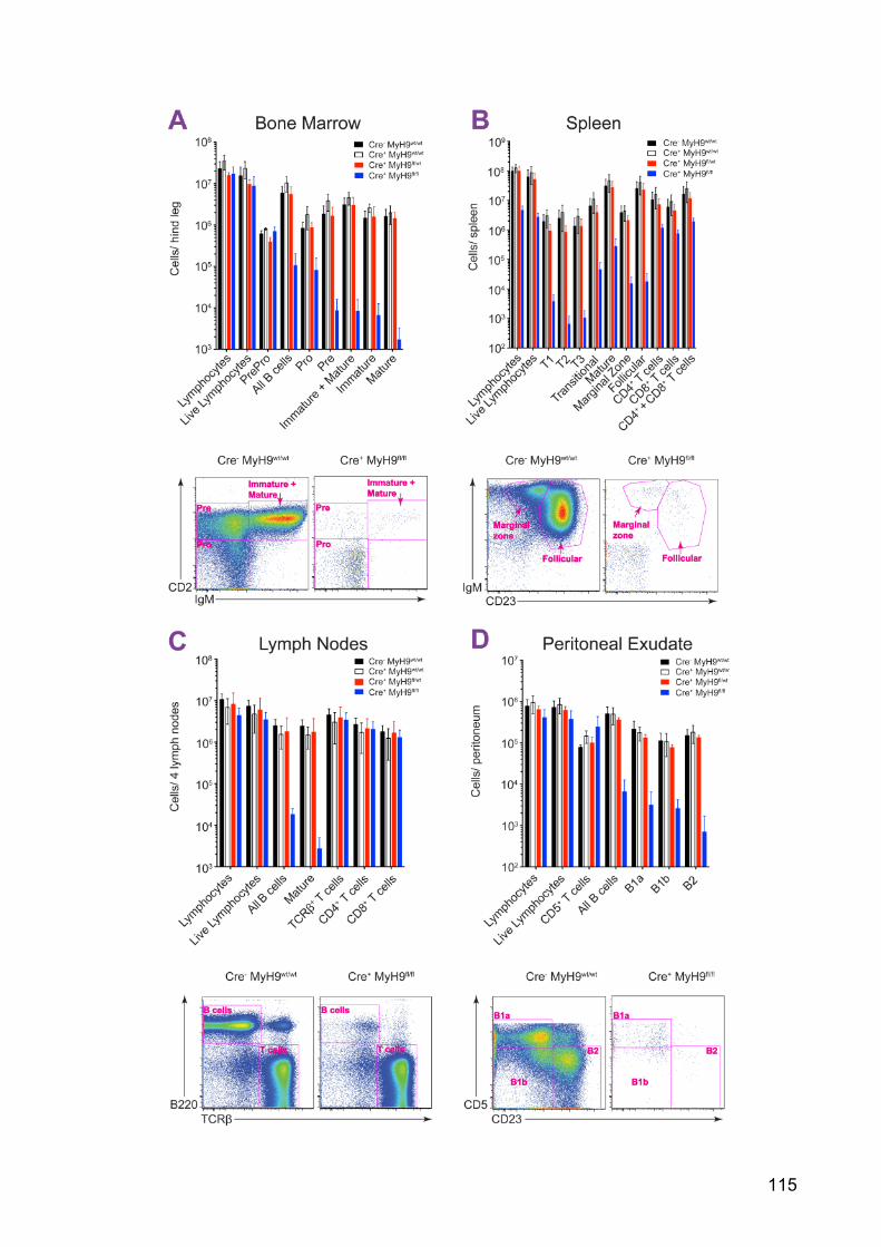
115

116
Figure 28: Cre+MyH9fl/fl mice have a block in B cell development
A) Top panel: Absolute B cell numbers flushed from femur and tibia of rear left leg. Cells
were stained for cell markers for 20 minutes on ice and absolute numbers were analysed using
FlowJo. Bottom: Representative dot plot showing absence of B cells following the PrePro
developmental stage. Cells shown were gated on B220hiCD19hi population
B) Top panel: Absolute B cell numbers and T cell numbers harvested from mashed spleen.
Cells were stained for cell markers for 20 minutes on ice and absolute numbers were analysed
using FlowJo. Bottom: Representative dot plot showing absence of follicular and marginal zone B
cells. Cells shown were gated on B220hiCD93lo population
C) Top panel: Absolute B cell and T cell numbers harvested from mashed brachial and
inguinal lymph nodes. Cells were stained for cell markers for 20 minutes on ice and absolute
numbers were analysed using FlowJo. Bottom: Representative dot plot showing absence of B
cells but an intact T cell compartment
D) Top panel: Absolute B cell numbers flushed from the peritoneal cavity. Cells were stained
for cell markers for 20 minutes on ice and absolute numbers were analysed using FlowJo.
Bottom: Representative dot plot showing absence of B1 and B2 cells. Cells shown were gated on
IgMhiCD5int population
In all panels live B cells were identified as negative for the Live/Dead stain and then B cell
subsets were identified using the cell markers described in Table 1 (mean±SEM, n= 3-4 mice for
each genotype). Mice were used at 7-9 weeks of age
These experiments were done in collaboration with Harold Hartweger.
Figure 29: Cre+MyH9fl/fl mice have smaller spleens compared to controls

117
Figure 30: Cre+MyH9fl/fl B cells which develop past the PrePro stage in the bone marrow
are larger A) Cre+MyH9fl/fl (red) and wild type (grey) PrePro B cells. Cells shown were gated on
B220hiCD19lo
B) Cre+MyH9fl/fl (red) and wild type (grey) Pro B cells. Cells shown were gated on
B220hiCD19hiCD2loIgMlo
Representative plots from 1 of 3 experiments shown in Figure 28
5.3 Discussion
By using PMS as surrogates for APCs, we have demonstrated that B cells
internalise membrane-bound antigen depending on their affinity.
The antigens used in this study consist of 15 NP or NIP haptens complexed with
a protein carrier. These antigens have much higher avidity than monomeric
antigen, therefore, the subtle differences in affinity were masked when exposed
to B cells in solution (Batista and Neuberger, 2000). However, when these same
antigens were tethered to the PMS, B cells were able to discriminate between
their affinities and they internalised the higher affinity antigen to a greater extent.
By tracking the invaginations and BCR microclusters formed in the B1-8 B cell
synapse, we saw that B1-8 B cells sustained more long-lived invaginations,
produced BCR-antigen microclusters faster, and had more antigen associated
with the long-lived invaginations. These studies support other investigations

118
using PLB as a surrogate for APCs, which show that BCRs have increased
spreading and signalling in response to higher affinity antigen and suggest that
affinity discrimination is an intrinsic function of the BCR (Fleire et al., 2006; Liu et
al., 2010; Wan et al., 2013).
B cells can discriminate between a large range of antigen affinities. Due to the
bivalency of the BCR and the high avidity nature of most physiologically relevant
antigens, it is possible that the half-lives of BCR-antigen complexes could reach
many hours (Batista and Neuberger, 1998). We found that by inhibiting myosin
IIA contractility and decreasing the mechanical pressure exerted on the BCR-
antigen bonds, the internalisation rate of the low affinity antigens was improved.
This suggests that under normal conditions, myosin IIA contractility shortens the
lifetime of the BCR-antigen bonds thereby ensuring affinity discrimination occurs
over seconds rather than hours and acts as a checkpoint so that only antigens of
high enough affinity are internalised. Therefore this mechanism could be
important in preventing the internalisation of lower affinity antigens, which could
lead to inappropriate B cell activation.
Collectively, these results suggest a model whereby BCRs with a higher affinity
for the antigens, produce bigger BCR microclusters faster, this together with the
increased strength of the individual high-affinity BCR-antigen bonds, better
resists the myosin IIA-generated force, and the invaginations last long enough to
allow the full assembly of the CCSs and internalisation. In contrast, low affinity
antigens produce BCR microclusters more slowly, which combined with the
weaker BCR-antigen interactions, cannot withstand the myosin IIA contractility
and the bonds rupture, aborting internalisation. This mechanism provides a
possible link for how the increased B cell signalling in response to high-affinity
membrane-bound antigen leads to increased antigen internalisation and
presentation, required for higher levels of T cell help and B cell proliferation to
ensure a competitive advantage over low affinity antigens in vivo (Khalil et al.,
2012; Schwickert et al., 2011; Shih et al., 2002a; 2002b; Victora et al., 2010).
In vivo, when B cells come into contact with APC-tethered antigen they will also
see many other signals such as integrin ligands, complement, and other co-

119
stimulatory molecules, which may fine tune the B cell response (Carrasco and
Batista, 2006a; Carrasco et al., 2004; Chan and Brink, 2012; Suzuki et al., 2009;
Wan et al., 2013). In addition, active forces generated by APCs may also
influence this process. Therefore, future studies will be necessary to understand
how various APCs can further regulate antigen extraction and B cell affinity
discrimination in vivo.
To assess the role of myosin IIA in BCR-antigen internalisation in vivo we
produced mice with B cell specific deletion of the MyH9 gene and therefore
myosin IIA (Cre+MyH9fl/fl). However, analysis of B cell responses was not
possible, as these mice had very few peripheral B cells. The loss of B cells during
development was observed at the Pro B cell stage in the bone marrow, exactly
when the Mb1-controlled Cre recombinase is expressed (Hobeika et al., 2006).
This was not due to loss of Igα, as Igα deficient B cells are arrested at the later
Pro B cell to Pre B cell transition (Pelanda et al., 2002), therefore supporting the
finding that the loss of B cells was due to myosin IIA deletion by the Cre
recombinase.
The loss of B cells in Cre+MyH9fl/fl mice before signalling from the Pre-BCR,
which occurs at the Pre B cell stage (Figure 4) (Mårtensson et al., 2010; Wang
and Clark, 2003), indicates a role for myosin IIA in B cell development that is
separate from its role in BCR-antigen internalisation. Given the important roles
myosin IIA has in cell biology, there are many possibilities for why loss of myosin
IIA is incompatible with the survival or differentiation of B cell progenitors. In our
study we observed that the few Cre+MyH9fl/fl B cells, which made it past the
PrePro stage, were consistently larger than wild type B cells. This could suggest
that myosin IIA may be required for cytokinesis during B cell division (Matsumura,
2005), which would prevent B cells from completing mitosis needed during B cell
development. However, it is generally thought that B cells do not proliferate until
exiting the Pro B cell stage and entering the Pre B cell stage, therefore this is
unlikely to be the sole cause of the defect seen in our studies (Hardy et al.,
1991). This could be resolved by staining the DNA in these cells to understand if
they have been arrested at the telophase stage of mitosis. Alternatively, the

120
enlarged B cells could be explained by loss of cortical tension, which may
stimulate cell death due to loss of internal cellular structure and organisation.
Another possible reason for loss of B cells at the Pro B cell stage could be due to
lack of survival signals. Myosin IIA activity has been shown to be important for
cell migration and adhesion (Vicente-Manzanares et al., 2009). If Cre+MyH9fl/fl B
cells were unable to migrate across, or interact with, the stromal cells in the bone
marrow during development, this could lead to developmental arrest. For
instance, B cells have been shown to migrate between CXCL12-producing
stromal cells and IL-7-producing cells during development to receive
differentiation and survival components (Nagasawa, 2006). So, if the B cells are
unable to migrate across these stromal cells they may die due to lack of
appropriate stimulation. It would be interesting in the future to investigate
downstream signalling in these myosin IIA deficient B cells to understand
whether they are able to respond to survival signals such as IL-7. Therefore, the
exact cause for Cre+MyH9fl/fl B cell loss during development will have to be
addressed by further study.
Independently, to address the question of whether myosin IIA contractility is
involved in antigen internalisation and affinity discrimination in vivo, MyH9fl/fl mice
will need to be crossed with mice expressing Cre recombinase under a promoter
which becomes active only in mature B cells, such as the promoter for CD23, or
is induced by drug treatment, such as doxycycline. We will then be able to take
mature B cells from these mice and observe their internalisation and affinity
discrimination on antigen-loaded PMS in vitro, as well as stimulating B cells in
vivo to see whether loss of myosin IIA inhibits the ability of B cells to respond to
antigen in the lymph node.
In conclusion, here we have demonstrated an important role for myosin IIA-
generated force in affinity-dependent antigen internalisation in vitro. The
understanding of whether this mechanism is still relevant in vivo is still to be
elucidated and is actively being investigated in the lab.

121
Chapter 6 Conclusions The aim of this thesis was to investigate and identify novel components and
mechanisms involved in BCR-antigen internalisation, particularly focusing on
acquisition of antigen from APCs. Antigen internalisation from APCs has been
previously shown at the cellular level by intravital imaging. These studies
demonstrated that in response to immunisation, cognate B cells interact with
APCs over several minutes, during which they become activated and extract the
antigen (Carrasco and Batista, 2007; Phan et al., 2007; Qi et al., 2006; Suzuki et
al., 2009). This antigen extraction and subsequent internalisation is critical for the
production of T cell-dependent B cell responses. However the molecular
mechanisms controlling antigen internalisation from APCs are unknown.
To investigate the mechanisms of membrane-bound antigen acquisition, we
developed a new antigen-presenting substrate from adherent HEK293A cells,
PMS. These PMS were able to support membrane-bound antigen internalisation
by B cells in an in an in vitro setting. Live-cell imaging of B cell interaction with
antigen-loaded PMS revealed that B cells spread over the antigen forming BCR-
antigen microclusters. This spreading was followed by B cell invagination of the
PMS membrane. The lifetime of these invaginations depended on antigen affinity
for the BCR and the extent of BCR-antigen microclustering. BCR-antigen
complexes with sufficient affinity and avidity were able to withstand the ~20 pN
force exerted by the B cells during invagination formation long enough for
internalisation to occur.
To identify the pathways controlling this process we used 2 approaches. The first
was shRNA-mediated knockdown of target proteins in Ramos B cells, and the
second was inhibition of these proteins using small molecule inhibitors in primary
Bl6 B cells. Both strategies identified a sequential role for myosin IIA and clathrin
in antigen extraction and endocytosis.
We then used live-cell TIRF microscopy to visualise the activity of these proteins
during the internalisation process. These experiments showed that myosin IIA

122
activity peaked immediately before the onset of membrane invagination, whereas
clathrin was only recruited during the termination of long-lived invaginations.
Comparisons of the internalisation process between antigens of low and high
affinity revealed that B cells internalised antigen from the PMS in an affinity-
dependent manner. High affinity antigens stimulated faster BCR-antigen
microcluster formation, more long-lived invaginations, and higher levels of
invagination-associated antigen, which led to a greater amount of antigen
internalisation.
Taking these results together we present a model by which, after initial antigen
binding, B cells spread over the antigen-presenting surface forming BCR-antigen
microclusters and invaginating the presenting surface in a myosin IIA-dependent
manner. If an invagination is initiated on a big enough BCR-microcluster with a
sufficient level of affinity within the BCR-antigen complex, the BCR is able to
withstand the force generated by myosin IIA and the invagination persists long
enough for clathrin recruitment and BCR-antigen internalisation (Figure 31).

123
Figure 31: BCR internalisation of membrane-bound antigen

124
Therefore, we can conclude that coupling myosin IIA contractility to endocytosis
permits B cells to internalise antigen in an affinity-dependent manner. This is
particularly important at the initiation of the immune response in the B cell follicle,
and during affinity maturation in the germinal centre, when the selective
expansion of B cell clones depends on the relative amount of antigen internalised
and presented to cognate T cells (Paus et al., 2006; Schwickert et al., 2011;
Victora et al., 2010).
The in vitro based studies presented in this thesis of BCR-antigen internalisation,
gave us an insight into the components and mechanisms involved in BCR-
antigen internalisation in the context of B cell synapses with APCs. However,
BCR-antigen internalisation has the potential to be highly flexible, making
investigations into this process difficult. This is supported by studies showing that
loss of clathrin, actin or lipid rafts, or any other protein identified as mediating
internalisation, decreased the level of BCR antigen-internalisation but did not
inhibit it completely, suggesting a high level of redundancy within this process
(Becker-Herman et al., 2011; Ma et al., 2001; Malhotra et al., 2009a; 2009b; Niiro
et al., 2004; Stoddart et al., 2002; 2005). This is likely to have evolved as B cells
are required to recognise and internalise many different possible infections such
as bacteria, viruses, fungi, and parasites as well as any type of environmental or
synthetic chemical they may come across, which would suggest a necessary
level of flexibility in antigen internalisation to be able to accommodate a wide
variety antigen sizes and shapes (Chan and Brink, 2012).
Different subtypes of B cells themselves may also internalise antigens in different
ways. For example, memory B cells are likely to recognise antigen with a much
higher affinity than naïve B cells, and the increase in BCR signalling following
high affinity antigen binding, may stimulate a different pathway of internalisation
(Kouskoff et al., 1998; Liu et al., 2010).
Finally studies have shown that molecules on the surface of APCs can fine tune
and alter the B cell response (Allen and Cyster, 2008; Carrasco and Batista,
2006a; Carrasco et al., 2004; Lanoue et al., 2002), but the types of co-stimulatory
molecules can differ in their type and expression between different types of APC,

125
which may also change the way B cells recognise and internalise antigen (Batista
and Harwood, 2009).
In nearly all of the BCR-antigen internalisation studies done to date with soluble
antigen, or antigen presented on PLB or PMS, protein antigens with either
primary naïve B cells or B cell lines have been used. It would be of great interest
in the future to build on these studies by completing the genetic screen begun in
the course of this thesis, to put together a molecular pathway for how this
process occurs in these conditions, and then develop these findings by seeing
how the molecular requirements can change depending on B cell type, APC and
antigen used. This would give us a clearer indication for how B cells undergo
BCR-antigen internalisation and activation in vivo.
Ultimately the knowledge gained from studying BCR-antigen internalisation will
hopefully contribute to the design and development of immunotherapy and
effective vaccine design. BCR-antigen internalisation is an intricately controlled
process that initiates the antibody response. Therefore, inhibition or
augmentation of this process is an attractive target in the treatment of
autoimmunity and cancer, or vaccine design, respectively. For example, studies
have shown that antigens targeted to APCs can boost the effectiveness of
immunisations (Caminschi and Shortman, 2012), therefore understanding the
relevance of membrane-presented antigen is likely going to be highly applicable
in rational design of future vaccines. Similarly, as disruptions in internalisation
can potentially contribute to the growth of malignant B cells, targeting B cell
trafficking may also be of therapeutic potential (Davis et al., 2010; Young and
Staudt, 2013).
These studies signify that by increasing our understanding of BCR-antigen
internalisation, we can open the door to a range of possible new targets for the
treatment or prevention of B cell diseases.

126
References
Allen, C.D.C., and Cyster, J.G. (2008). Follicular dendritic cell networks of primary follicles and germinal centers: phenotype and function. Seminars in Immunology 20, 14–25.
Andrian, von, U.H., and Mempel, T.R. (2003). Homing and cellular traffic in lymph nodes. Nature Reviews Immunology 3, 867–878.
Arana, E., Vehlow, A., Harwood, N.E., Vigorito, E., Henderson, R., Turner, M., Tybulewicz, V.L.J., and Batista, F.D. (2008). Activation of the Small GTPase Rac2 via the B Cell Receptor Regulates B Cell Adhesion and Immunological-Synapse Formation. Immunity 28, 88–99.
Arnon, T.I., Horton, R.M., Grigorova, I.L., and Cyster, J.G. (2012). Visualization of splenic marginal zone B-cell shuttling and follicular B-cell egress. Nature 493, 684–688.
Attwood, S., Choi, Y., and Leonenko, Z. (2013). Preparation of DOPC and DPPC Supported Planar Lipid Bilayers for Atomic Force Microscopy and Atomic Force Spectroscopy. International Journal of Molecular Sciences 14, 3514–3539.
Bajénoff, M., and Germain, R.N. (2009). B-cell follicle development remodels the conduit system and allows soluble antigen delivery to follicular dendritic cells. Blood 114, 4989–4997.
Balázs, M., Martin, F., Zhou, T., and Kearney, J.F. (2002). Blood Dendritic Cells Interact with Splenic Marginal Zone B Cells to Initiate T-Independent Immune Responses. Cell 17, 341–352.
Batista, F.D., and Neuberger, M.S. (1998). Affinity Dependence of the B Cell Response to Antigen: A Threshold, a Ceiling, and the Importance of Off-Rate. Cell 8, 751–759.
Batista, F.D., and Neuberger, M.S. (2000). B cells extract and present immobilized antigen: implications for affinity discrimination. EMBO Journal 19, 513–520.
Batista, F., and Harwood, N. (2009). The who, how and where of antigen presentation to B cells. Nature Reviews Immunology 9, 15–27.
Batista, F., Iber, D., and Neuberger, M. (2001). B cells acquire antigen from target cells after synapse formation. Nature 411, 489–494.
Bauer, S., Kirschning, C.J., Häcker, H., Redecke, V., Hausmann, S., Akira, S., Wagner, H., and Lipford, G.B. (2001). Human TLR9 confers responsiveness to bacterial DNA via species-specific CpG motif recognition. Proceedings of the National Academy of Sciences 98, 9237–9242.

127
Becker-Herman, S., Meyer-Bahlburg, A., Schwartz, M.A., Jackson, S.W., Hudkins, K.L., Liu, C., Sather, B.D., Khim, S., Liggitt, D., Song, W., et al. (2011). WASp-deficient B cells play a critical, cell-intrinsic role in triggering autoimmunity. The Journal of Experimental Medicine 208, 2033–2042.
Bergtold, A., Desai, D.D., Gavhane, A., and Clynes, R. (2005). Cell Surface Recycling of Internalized Antigen Permits Dendritic Cell Priming of B Cells. Cell 23, 503–514.
Berney, C., Herren, S., Power, C.A., Gordon, S., Martinez-Pomares, L., and Kosco-Vilbois, M.H. (1999). A member of the dendritic cell family that enters B cell follicles and stimulates primary antibody responses identified by a mannose receptor fusion protein. The Journal of Experimental Medicine 190, 851–860.
Bléry, M., Tze, L., Miosge, L.A., Jun, J.E., and Goodnow, C.C. (2006). Essential role of membrane cholesterol in accelerated BCR internalization and uncoupling from NF-κB in B cell clonal anergy. The Journal of Experimental Medicine 203, 1773–1783.
Boettner, D.R., Chi, R.J., and Lemmon, S.K. (2011). Lessons from yeast for clathrin-mediated endocytosis. Nature Cell Biology 14, 2–10.
Bonnerot, C., Lankar, D., Hanau, D., Spehner, D., Davoust, J., Salamero, J., and Fridman, W.H. (1995). Role of B cell receptor Ig alpha and Ig beta subunits in MHC class II-restricted antigen presentation. Immunity 3, 335–347.
Boucrot, E., Saffarian, S., Zhang, R., and Kirchhausen, T. (2010). Roles of AP-2 in clathrin-mediated endocytosis. PLoS ONE 5, e10597.
Boulant, S., Kural, C., Zeeh, J.-C., Ubelmann, F., and Kirchhausen, T. (2011). Actin dynamics counteract membrane tension during clathrin-mediated endocytosis. Nature Cell Biology 13, 1124–1131.
Brown, B.K., and Song, W. (2001). The Actin Cytoskeleton is Required for the Trafficking of the B Cell Antigen Receptor to the Late Endosomes. Traffic 2, 414–427.
Calabia-Linares, C., Robles-Valero, J., la Fuente, de, H., Perez-Martinez, M., Martín-Cofreces, N., Alfonso-Pérez, M., Gutierrez-Vázquez, C., Mittelbrunn, M., Ibiza, S., Urbano-Olmos, F.R., et al. (2011). Endosomal clathrin drives actin accumulation at the immunological synapse. Journal of Cell Science 124, 820–830.
Cambier, J.C., Gauld, S.B., Merrell, K.T., and Vilen, B.J. (2007). B-cell anergy: from transgenic models to naturally occurring anergic B cells? Nature Reviews Immunology 7, 633–643.
Caminschi, I., and Shortman, K. (2012). Boosting antibody responses by targeting antigens to dendritic cells. Trends in Immunology 33, 71–77.
Carrasco, Y.R., and Batista, F.D. (2006a). B-cell activation by membrane-bound antigens is facilitated by the interaction of VLA-4 with VCAM-1. EMBO Journal

128
25, 889–899.
Carrasco, Y.R., and Batista, F.D. (2007). B cells acquire particulate antigen in a macrophage-rich area at the boundary between the follicle and the subcapsular sinus of the lymph node. Immunity 27, 160–171.
Carrasco, Y., and Batista, F. (2006b). B cell recognition of membrane-bound antigen: an exquisite way of sensing ligands. Current Opinion in Immunology 18, 286–291.
Carrasco, Y., Fleire, S., Cameron, T., Dustin, M., and Batista, F. (2004). LFA-1/ICAM-1 interaction lowers the threshold of B cell activation by facilitating B cell adhesion and synapse formation. Immunity 20, 589–599.
Carroll, M.C. (1998). The role of complement and complement receptors in induction and regulation of immunity. Annual Review of Immunology 16, 545–568.
Cassard, S., Salamero, J., Hanau, D., Spehner, D., Davoust, J., Fridman, W.H., and Bonnerot, C. (1998). A tyrosine-based signal present in Ig alpha mediates B cell receptor constitutive internalization. The Journal of Immunology 160, 1767–1773.
Catron, D.M., Pape, K.A., Fife, B.T., van Rooijen, N., and Jenkins, M.K. (2010). A protease-dependent mechanism for initiating T-dependent B cell responses to large particulate antigens. The Journal of Immunology 184, 3609–3617.
Chan, T.D., and Brink, R. (2012). Affinity-based selection and the germinal center response. Immunological Reviews 247, 11–23.
Chan, T.D., Wood, K., Hermes, J.R., Butt, D., Jolly, C.J., Basten, A., and Brink, R. (2012). Elimination of Germinal-Center-Derived Self-Reactive B Cells Is Governed by the Location and Concentration of Self-Antigen. Cell 37, 893–904.
Chan, V.W.F., Lowell, C.A., and DeFranco, A.L. (1998). Defective negative regulation of antigen receptor signaling in Lyn-deficient B lymphocytes. Current Biology 8, 545–553.
Chan, V., Meng, F., Soriano, P., DeFranco, A., and Lowell, C. (1997). Characterization of the B lymphocyte populations in Lyn-deficient mice and the role of Lyn in signal initiation and down-regulation. Immunity 7, 69–81.
Chappell, C.P., Draves, K.E., Giltiay, N.V., and Clark, E.A. (2012). Extrafollicular B cell activation by marginal zone dendritic cells drives T cell-dependent antibody responses. The Journal of Experimental Medicine 209, 1825–1840.
Chaturvedi, A., Dorward, D., and Pierce, S. (2008). The B cell receptor governs the subcellular location of Toll-like receptor 9 leading to hyperresponses to DNA-containing antigens. Immunity 28, 799–809.
Chaturvedi, A., Martz, R., Dorward, D., Waisberg, M., and Pierce, S.K. (2011).

129
Endocytosed BCRs sequentially regulate MAPK and Akt signaling pathways from intracellular compartments. Nature Immunology 12, 1119–1126.
Chen, C., Radic, M.Z., Erikson, J., Camper, S.A., Litwin, S., Hardy, R.R., and Weigert, M. (1994). Deletion and editing of B cells that express antibodies to DNA. The Journal of Immunology 152, 1970–1982.
Cheng, P.C., Brown, B.K., Song, W., and Pierce, S.K. (2001). Translocation of the B cell antigen receptor into lipid rafts reveals a novel step in signaling. The Journal of Immunology 166, 3693–3701.
Cheng, P.C., Dykstra, M.L., Mitchell, R.N., and Pierce, S.K. (1999). A role for lipid rafts in B cell antigen receptor signaling and antigen targeting. The Journal of Experimental Medicine 190, 1549–1560.
Christensen, S.R. (2005). Toll-like receptor 9 controls anti-DNA autoantibody production in murine lupus. The Journal of Experimental Medicine 202, 321–331.
Cinamon, G., Zachariah, M., Lam, O., Foss, F., and Cyster, J. (2007). Follicular shuttling of marginal zone B cells facilitates antigen transport. Nature Immunology 9, 54–62.
Collinet, C., Stöter, M., Bradshaw, C., Samusik, N., Rink, J., Kenski, D., Habermann, B., Buchholz, F., Henschel, R., and Mueller, M. (2010). Systems survey of endocytosis by multiparametric image analysis. Nature 464, 243–249.
Conti, M.A., and Adelstein, R.S. (2008). Nonmuscle myosin II moves in new directions. Journal of Cell Science 121, 11–18.
Cureton, D.K., Massol, R.H., Saffarian, S., Kirchhausen, T.L., and Whelan, S.P.J. (2009). Vesicular stomatitis virus enters cells through vesicles incompletely coated with clathrin that depend upon actin for internalization. PLoS Pathogens 5, e1000394.
Cureton, D.K., Massol, R.H., Whelan, S.P.J., and Kirchhausen, T. (2010). The length of vesicular stomatitis virus particles dictates a need for actin assembly during clathrin-dependent endocytosis. PLoS Pathogens 6, e1001127.
Cyster, J.G., Ansel, K.M., Reif, K., Ekland, E.H., Hyman, P.L., Tang, H.L., Luther, S.A., and Ngo, V.N. (2000). Follicular stromal cells and lymphocyte homing to follicles. Immunological Reviews 176, 181–193.
Cyster, J. (2010). B cell follicles and antigen encounters of the third kind. Nature Immunology 11, 989–996.
Dal Porto, J.M., Haberman, A.M., Shlomchik, M.J., and Kelsoe, G. (1998). Antigen drives very low affinity B cells to become plasmacytes and enter germinal centers. The Journal of Immunology 161, 5373–5381.
Dal Porto, J.M., Gauld, S.B., Merrell, K.T., Mills, D., Pugh-Bernard, A.E., and Cambier, J. (2004). B cell antigen receptor signaling 101. Molecular

130
Immunology 41, 599–613.
Dal Porto, J.M., Haberman, A.M., Kelsoe, G., and Shlomchik, M.J. (2002). Very low affinity B cells form germinal centers, become memory B cells, and participate in secondary immune responses when higher affinity competition is reduced. The Journal of Experimental Medicine 195, 1215–1221.
Davidson, H.W., Reid, P.A., Lanzavecchia, A., and Watts, C. (1991). Processed antigen binds to newly synthesized MHC class II molecules in antigen-specific B lymphocytes. Cell 67, 105–116.
Davis, R., Ngo, V., Lenz, G., Tolar, P., Young, R., Romesser, P., Kohlhammer, H., Lamy, L., Zhao, H., and Yang, Y. (2010). Chronic active B-cell-receptor signalling in diffuse large B-cell lymphoma. Nature 463, 88–92.
Delamarre, L., Pack, M., Chang, H., Mellman, I., and Trombetta, E.S. (2005). Differential lysosomal proteolysis in antigen-presenting cells determines antigen fate. Science 307, 1630–1634.
Depoil, D., Fleire, S., Treanor, B., Weber, M., Harwood, N., Marchbank, K., Tybulewicz, V., and Batista, F. (2007). CD19 is essential for B cell activation by promoting B cell receptor–antigen microcluster formation in response to membrane-bound ligand. Nature Immunology 9, 63–72.
Ehrlich, M., Boll, W., Van Oijen, A., Hariharan, R., Chandran, K., Nibert, M.L., and Kirchhausen, T. (2004). Endocytosis by random initiation and stabilization of clathrin-coated pits. Cell 118, 591–605.
Ezratty, E.J., Bertaux, C., Marcantonio, E.E., and Gundersen, G.G. (2009). Clathrin mediates integrin endocytosis for focal adhesion disassembly in migrating cells. The Journal of Cell Biology 187, 733–747.
Fang, Y., Xu, C., Fu, Y.X., Holers, V.M., and Molina, H. (1998). Expression of complement receptors 1 and 2 on follicular dendritic cells is necessary for the generation of a strong antigen-specific IgG response. The Journal of Immunology 160, 5273–5279.
Ferguson, A.R., Youd, M.E., and Corley, R.B. (2004). Marginal zone B cells transport and deposit IgM-containing immune complexes onto follicular dendritic cells. International Immunology 16, 1411–1422.
Fleire, S.J., Goldman, J.P., Carrasco, Y.R., Weber, M., Bray, D., and Batista, F.D. (2006). B cell ligand discrimination through a spreading and contraction response. Science 312, 738–741.
Gazumyan, A., Reichlin, A., and Nussenzweig, M.C. (2006). Igβ tyrosine residues contribute to the control of B cell receptor signaling by regulating receptor internalization. The Journal of Experimental Medicine 203, 1785–1794.
Goffinet, C., and Keppler, O.T. (2006). Efficient nonviral gene delivery into primary lymphocytes from rats and mice. The FASEB Journal 20, 500–502.

131
Gong, S., and Nussenzweig, M.C. (1996). Regulation of an early developmental checkpoint in the B cell pathway by Ig beta. Science 272, 411–414.
Gonzalez, S.F., Degn, S.E., Pitcher, L.A., Woodruff, M., Heesters, B.A., and Carroll, M.C. (2011). Trafficking of B cell antigen in lymph nodes. Annual Review of Immunology 29, 215–233.
Gonzalez, S.F., Lukacs-Kornek, V., Kuligowski, M.P., Pitcher, L.A., Degn, S.E., Turley, S.J., and Carroll, M.C. (2010a). Complement-dependent transport of antigen into B cell follicles. The Journal of Immunology 185, 2659–2664.
Gonzalez, S., Kuligowski, M., Pitcher, L., Roozendaal, R., and Carroll, M. (2010b). The Role of Innate Immunity in B Cell Acquisition of Antigen Within LNs. Advances in Immunology 106, 1–19.
Gonzalez, S., Lukacs-Kornek, V., Kuligowski, M., Pitcher, L., Degn, S., Kim, Y., Cloninger, M., Martinez-Pomares, L., Gordon, S., and Turley, S. (2010c). Capture of influenza by medullary dendritic cells via SIGN-R1 is essential for humoral immunity in draining lymph nodes. Nature Immunology 11, 427–434.
Goodnow, C.C. (1997). Chance encounters and organized rendezvous. Immunological Reviews 156, 5–10.
Goodnow, C.C., Crosbie, J., Adelstein, S., Lavoie, T.B., Smith-Gill, S.J., Brink, R.A., Pritchard-Briscoe, H., Wotherspoon, J.S., Loblay, R.H., and Raphael, K. (1988). Altered immunoglobulin expression and functional silencing of self-reactive B lymphocytes in transgenic mice. Nature 334, 676–682.
Grakoui, A., Bromley, S.K., Sumen, C., Davis, M.M., Shaw, A.S., Allen, P.M., and Dustin, M.L. (1999). The immunological synapse: a molecular machine controlling T cell activation. Science 285, 221–227.
Gresch, O. (2004). New non-viral method for gene transfer into primary cells. Methods 33, 151–163.
Gretz, J.E., Norbury, C.C., Anderson, A.O., Proudfoot, A.E., and Shaw, S. (2000). Lymph-borne chemokines and other low molecular weight molecules reach high endothelial venules via specialized conduits while a functional barrier limits access to the lymphocyte microenvironments in lymph node cortex. The Journal of Experimental Medicine 192, 1425–1440.
Han, S., Hathcock, K., Zheng, B., Kepler, T.B., Hodes, R., and Kelsoe, G. (1995). Cellular interaction in germinal centers. Roles of CD40 ligand and B7-2 in established germinal centers. The Journal of Immunology 155, 556–567.
Hardy, R.R., and Hayakawa, K. (2001). B cell development pathways. Annual Review of Immunology 19, 595–621.
Hardy, R.R., Carmack, C.E., Shinton, S.A., Kemp, J.D., and Hayakawa, K. (1991). Resolution and characterization of pro-B and pre-pro-B cell stages in normal mouse bone marrow. The Journal of Experimental Medicine 173, 1213–1225.

132
Hartley, S.B., Crosbie, J., Brink, R., Kantor, A.B., Basten, A., and Goodnow, C.C. (1991). Elimination from peripheral lymphoid tissues of self-reactive B lymphocytes recognizing membrane-bound antigens. Nature 353, 765–769.
Hasler, P., and Zouali, M. (2001). B cell receptor signaling and autoimmunity. The FASEB Journal 15, 2085–2098.
Heesters, B.A., Chatterjee, P., Kim, Y.-A., Gonzalez, S.F., Kuligowski, M.P., Kirchhausen, T., and Carroll, M.C. (2013). Endocytosis and Recycling of Immune Complexes by Follicular Dendritic Cells Enhances B Cell Antigen Binding and Activation. Cell 38, 1164–1175.
Heinen, E., Braun, M., Coulie, P.G., Van Snick, J., Moeremans, M., Cormann, N., Kinet-Denoël, C., and Simar, L.J. (1986). Transfer of immune complexes from lymphocytes to follicular dendritic cells. European Journal of Immunology 16, 167–172.
Hibbs, M.L., Harder, K.W., Armes, J., Kountouri, N., Quilici, C., Casagranda, F., Dunn, A.R., and Tarlinton, D.M. (2002). Sustained Activation of Lyn Tyrosine Kinase In Vivo Leads to Autoimmunity. The Journal of Experimental Medicine 196, 1593–1604.
Hobeika, E., Thiemann, S., Storch, B., Jumaa, H., Nielsen, P.J., Pelanda, R., and Reth, M. (2006). Testing gene function early in the B cell lineage in mb1-cre mice. Proceedings of the National Academy of Sciences 103, 13789–13794.
Hombach, J., Tsubata, T., Leclercq, L., Stappert, H., and Reth, M. (1990). Molecular components of the B-cell antigen receptor complex of the IgM class. Nature 343, 760–762.
Hou, P., Araujo, E., Zhao, T., Zhang, M., Massenburg, D., Veselits, M., Doyle, C., Dinner, A., and Clark, M. (2006). B cell antigen receptor signaling and internalization are mutually exclusive events. PLoS Biology 4, e200.
Huang, B., Wang, W., Bates, M., and Zhuang, X. (2008). Three-dimensional super-resolution imaging by stochastic optical reconstruction microscopy. Science 319, 810–813.
Huang, N.-N., Han, S.-B., Hwang, I.-Y., and Kehrl, J.H. (2005). B cells productively engage soluble antigen-pulsed dendritic cells: visualization of live-cell dynamics of B cell-dendritic cell interactions. The Journal of Immunology 175, 7125–7134.
Isnardi, I., Ng, Y.-S., Srdanovic, I., Motaghedi, R., Rudchenko, S., Bernuth, von, H., Zhang, S.-Y., Puel, A., Jouanguy, E., Picard, C., et al. (2008). IRAK-4- and MyD88-dependent pathways are essential for the removal of developing autoreactive B cells in humans. Immunity 29, 746–757.
Jacob, J., Kelsoe, G., Rajewsky, K., and Weiss, U. (1991). Intraclonal generation of antibody mutants in germinal centres. Nature 354, 389–392.
133
Jacobelli, J., Friedman, R.S., Conti, M.A., Lennon-Duménil, A.-M., Piel, M., Sorensen, C.M., Adelstein, R.S., and Krummel, M.F. (2010). Confinement-optimized three-dimensional T cell amoeboid motility is modulated via myosin IIA–regulated adhesions. Nature Immunology 11, 953–961.
Jang, C., Machtaler, S., and Matsuuchi, L. (2010). The role of Ig-α/β in B cell antigen receptor internalization. Immunology Letters 134, 75–82.
Junt, T., Moseman, E.A., Iannacone, M., Massberg, S., Lang, P.A., Boes, M., Fink, K., Henrickson, S.E., Shayakhmetov, D.M., Di Paolo, N.C., et al. (2007). Subcapsular sinus macrophages in lymph nodes clear lymph-borne viruses and present them to antiviral B cells. Nature 450, 110–114.
Khalil, A.M., Cambier, J.C., and Shlomchik, M.J. (2012). B cell receptor signal transduction in the GC is short-circuited by high phosphatase activity. Science 336, 1178–1181.
Kirchhausen, T. (2009). Imaging endocytic clathrin structures in living cells. Trends in Cell Biology 19, 596–605.
Kitamura, D., Roes, J., Kühn, R., and Rajewsky, K. (1991). A B cell-deficient mouse by targeted disruption of the membrane exon of the immunoglobulin mu chain gene. Nature 350, 423–426.
Kouskoff, V., Famiglietti, S., Lacaud, G., Lang, P., Rider, J.E., Kay, B.K., Cambier, J.C., and Nemazee, D. (1998). Antigens varying in affinity for the B cell receptor induce differential B lymphocyte responses. The Journal of Experimental Medicine 188, 1453–1464.
Kovács, M., Thirumurugan, K., Knight, P.J., and Sellers, J.R. (2007). Load-dependent mechanism of nonmuscle myosin 2. Proceedings of the National Academy of Sciences 104, 9994–9999.
Kraus, M., Alimzhanov, M.B., Rajewsky, N., and Rajewsky, K. (2004). Survival of resting mature B lymphocytes depends on BCR signaling via the Igα/β heterodimer. Cell 117, 787–800.
Krieg, M., Helenius, J., Heisenberg, C.-P., and Müller, D.J. (2008). A Bond for a Lifetime: Employing Membrane Nanotubes from Living Cells to Determine Receptor-Ligand Kinetics. Angewandte Chemie International Edition 47, 9775–9777.
Kurosaki, T., Shinohara, H., and Baba, Y. (2010). B Cell Signaling and Fate Decision. Annual Review of Immunology 28, 21–55.
Lam, K.P., Kühn, R., and Rajewsky, K. (1997). In vivo ablation of surface immunoglobulin on mature B cells by inducible gene targeting results in rapid cell death. Cell 90, 1073–1083.
Lane, P., Traunecker, A., Hubele, S., Inui, S., Lanzavecchia, A., and Gray, D. (1992). Activated human T cells express a ligand for the human B cell‐associated antigen CD40 which participates in T cell‐dependent activation of B

134
lymphocytes. European Journal of Immunology 22, 2573–2578.
Lankar, D., Briken, V., Adler, K., Weiser, P., Cassard, S., Blank, U., Viguier, M., and Bonnerot, C. (1998). Syk tyrosine kinase and B cell antigen receptor (BCR) immunoglobulin-alpha subunit determine BCR-mediated major histocompatibility complex class II-restricted antigen presentation. The Journal of Experimental Medicine 188, 819–831.
Lanoue, A., Batista, F.D., Stewart, M., and Neuberger, M.S. (2002). Interaction of CD22 with α2,6-linked sialoglycoconjugates: innate recognition of self to dampen B cell autoreactivity? European Journal of Immunology 32, 348–355.
Lanzavecchia, A. (1985). Antigen-specific interaction between T and B cells. Nature 314, 537–539.
Lanzavecchia, A. (1987). Antigen Uptake and Accumulation in Antigen‐Specific B Cells. Immunological Reviews 99, 39–51.
Lau, C.M., Broughton, C., Tabor, A.S., Akira, S., Flavell, R.A., Mamula, M.J., Christensen, S.R., Shlomchik, M.J., Viglianti, G.A., Rifkin, I.R., et al. (2005). RNA-associated autoantigens activate B cells by combined B cell antigen receptor/Toll-like receptor 7 engagement. The Journal of Experimental Medicine 202, 1171–1177.
Leadbetter, E., Rifkin, I., Hohlbaum, A., Beaudette, B., Shlomchik, M., and Marshak-Rothstein, A. (2002). Chromatin–IgG complexes activate B cells by dual engagement of IgM and Toll-like receptors. Nature 416, 603–607.
Lecuit, T., Lenne, P.-F., and Munro, E. (2011). Force generation, transmission, and integration during cell and tissue morphogenesis. Annual Review of Cell and Developmental Biology 27, 157–184.
Levayer, R., Pelissier-Monier, A., and Lecuit, T. (2011). Spatial regulation of Dia and Myosin-II by RhoGEF2 controls initiation of E-cadherin endocytosis during epithelial morphogenesis. Nature Cell Biology 13, 529–540.
Lin, K., Freeman, S., Zabetian, S., Brugger, H., Weber, M., Lei, V., Dang-Lawson, M., Tse, K., Santamaria, R., and Batista, F. (2008). The rap GTPases regulate B cell morphology, immune-synapse formation, and signaling by particulate B cell receptor ligands. Immunity 28, 75–87.
Liu, C., Miller, H., Orlowski, G., Hang, H., Upadhyaya, A., and Song, W. (2012). Actin reorganization is required for the formation of polarized B cell receptor signalosomes in response to both soluble and membrane-associated antigens. The Journal of Immunology 188, 3237–3246.
Liu, W., Meckel, T., Tolar, P., Sohn, H.W., and Pierce, S.K. (2010). Antigen affinity discrimination is an intrinsic function of the B cell receptor. The Journal of Experimental Medicine 207, 1095–1111.
Ma, H., Yankee, T.M., Hu, J., Asai, D.J., Harrison, M.L., and Geahlen, R.L. (2001). Visualization of Syk-antigen receptor interactions using green

135
fluorescent protein: differential roles for Syk and Lyn in the regulation of receptor capping and internalization. The Journal of Immunology 166, 1507–1516.
Mackay, F., Figgett, W.A., Saulep, D., Lepage, M., and Hibbs, M.L. (2010). B-cell stage and context-dependent requirements for survival signals from BAFF and the B-cell receptor. Immunological Reviews 237, 205–225.
Malhotra, S., Kovats, S., Zhang, W., and Coggeshall, K.M. (2009a). Vav and Rac Activation in B Cell Antigen Receptor Endocytosis Involves Vav Recruitment to the Adapter Protein LAB. Journal of Biological Chemistry 284, 36202–36212.
Malhotra, S., Kovats, S., Zhang, W., and Coggeshall, K.M. (2009b). B cell antigen receptor endocytosis and antigen presentation to T cells require Vav and dynamin. Journal of Biological Chemistry 284, 24088–24097.
Manolova, V., Flace, A., Bauer, M., Schwarz, K., Saudan, P., and Bachmann, M.F. (2008). Nanoparticles target distinct dendritic cell populations according to their size. European Journal of Immunology 38, 1404–1413.
Mantei, A., Rutz, S., Janke, M., Kirchhoff, D., Jung, U., Patzel, V., Vogel, U., Rudel, T., Andreou, I., and Weber, M. (2008). siRNA stabilization prolongs gene knockdown in primary T lymphocytes. European Journal of Immunology 38, 2616–2625.
Matsumura, F. (2005). Regulation of myosin II during cytokinesis in higher eukaryotes. Trends in Cell Biology 15, 371–377.
Mattila, P.K., Feest, C., Depoil, D., Treanor, B., Montaner, B., Otipoby, K.L., Carter, R., Justement, L.B., Bruckbauer, A., and Batista, F.D. (2013). The Actin and Tetraspanin Networks Organize Receptor Nanoclusters to Regulate B Cell Receptor-Mediated Signaling. Immunity 38, 1–14.
Mårtensson, I.-L., Almqvist, N., Grimsholm, O., and Bernardi, A.I. (2010). The pre-B cell receptor checkpoint. FEBS Letters 584, 2572–2579.
McKean, D., Huppi, K., Bell, M., Staudt, L., Gerhard, W., and Weigert, M. (1984). Generation of antibody diversity in the immune response of BALB/c mice to influenza virus hemagglutinin. Proceedings of the National Academy of Sciences 81, 3180–3184.
McMahon, H.T., and Boucrot, E. (2011). Molecular mechanism and physiological functions of clathrin-mediated endocytosis. Nature Reviews Molecular Cell Biology 12, 517–533.
Mebius, R.E., and Kraal, G. (2005). Structure and function of the spleen. Nature Reviews Immunology 5, 606–616.
Merrifield, C., Feldman, M., Wan, L., and Almers, W. (2002). Imaging actin and dynamin recruitment during invagination of single clathrin-coated pits. Nature Cell Biology 4, 691–698.

136
Merrifield, C., Perrais, D., and Zenisek, D. (2005). Coupling between clathrin-coated-pit invagination, cortactin recruitment, and membrane scission observed in live cells. Cell 121, 593–606.
Moffat, J., Grueneberg, D., Yang, X., Kim, S., Kloepfer, A., Hinkle, G., Piqani, B., Eisenhaure, T., Luo, B., and Grenier, J. (2006). A lentiviral RNAi library for human and mouse genes applied to an arrayed viral high-content screen. Cell 124, 1283–1298.
Morita, S., Kojima, T., and Kitamura, T. (2000). Plat-E: an efficient and stable system for transient packaging of retroviruses. Gene Therapy 7, 1063–1066.
Moseman, E.A., Iannacone, M., Bosurgi, L., Tonti, E., Chevrier, N., Tumanov, A., Fu, Y.-X., Hacohen, N., and Andrian, von, U.H. (2012). B Cell Maintenance of Subcapsular Sinus Macrophages Protects against a Fatal Viral Infection Independent of Adaptive Immunity. Immunity 36, 415–426.
Nagasawa, T. (2006). Microenvironmental niches in the bone marrow required for B-cell development. Nature Reviews Immunology 6, 107–116.
Nagata, K., Nakamura, T., Kitamura, F., Kuramochi, S., Taki, S., Campbell, K.S., and Karasuyama, H. (1997). The Igα/Igβ heterodimer on µ-negative proB cells is competent for transducing signals to induce early B cell differentiation. Immunity 7, 559–570.
Natkanski, E., Lee, W.Y., Mistry, B., Casal, A., Molloy, J.E., and Tolar, P. (2013). B Cells Use Mechanical Energy to Discriminate Antigen Affinities. Science 340, 1587–1590.
Niiro, H., Allam, A., Stoddart, A., Brodsky, F.M., Marshall, A.J., and Clark, E.A. (2004). The B lymphocyte adaptor molecule of 32 kilodaltons (Bam32) regulates B cell antigen receptor internalization. The Journal of Immunology 173, 5601–5609.
Norvell, A., Mandik, L., and Monroe, J.G. (1995). Engagement of the antigen-receptor on immature murine B lymphocytes results in death by apoptosis. The Journal of Immunology 154, 4404–4413.
Nutt, S.L., Heavey, B., Rolink, A.G., and Busslinger, M. (1999). Commitment to the B-lymphoid lineage depends on the transcription factor Pax5. Nature 401, 556–562.
O'Keefe, T.L., Williams, G.T., Batista, F.D., and Neuberger, M.S. (1999). Deficiency in CD22, a B Cell-specific Inhibitory Receptor, Is Sufficient to Predispose to Development of High Affinity Autoantibodies. The Journal of Experimental Medicine 189, 1307–1313.
O'Keefe, T.L., Williams, G.T., Davies, S.L., and Neuberger, M.S. (1996). Hyperresponsive B cells in CD22-deficient mice. Science 274, 798–801.
O'Neill, S.K., Getahun, A., Gauld, S.B., Merrell, K.T., Tamir, I., Smith, M.J., Dal Porto, J.M., Li, Q.-Z., and Cambier, J.C. (2011). Monophosphorylation of CD79a

137
and CD79b ITAM Motifs Initiates a SHIP-1 Phosphatase-Mediated Inhibitory Signaling Cascade Required for B Cell Anergy. Immunity 35, 746–756.
O'Neill, S.K., Veselits, M.L., Zhang, M., Labno, C., Cao, Y., Finnegan, A., Uccellini, M., Alegre, M.-L., Cambier, J.C., and Clark, M.R. (2009). Endocytic sequestration of the B cell antigen receptor and toll-like receptor 9 in anergic cells. Proceedings of the National Academy of Sciences 106, 6262–6267.
Ohnishi, K., and Melchers, F. (2003). The nonimmunoglobulin portion of λ5 mediates cell-autonomous pre-B cell receptor signaling. Nature Immunology 4, 849–856.
Okada, T., Miller, M.J., Parker, I., Krummel, M.F., Neighbors, M., Hartley, S.B., O'Garra, A., Cahalan, M.D., and Cyster, J.G. (2005). Antigen-engaged B cells undergo chemotaxis toward the T zone and form motile conjugates with helper T cells. PLoS Biology 3, e150.
Pape, K., Catron, D., Itano, A., and Jenkins, M. (2007). The humoral immune response is initiated in lymph nodes by B cells that acquire soluble antigen directly in the follicles. Immunity 26, 491–502.
Parker, D.C. (1993). T cell-dependent B cell activation. Annual Review of Immunology 11, 331–360.
Patel, K.J., and Neuberger, M.S. (1993). Antigen presentation by the B cell antigen receptor is driven by the alpha/beta sheath and occurs independently of its cytoplasmic tyrosines. Cell 74, 939–946.
Paus, D., Phan, T.G., Chan, T.D., Gardam, S., Basten, A., and Brink, R. (2006). Antigen recognition strength regulates the choice between extrafollicular plasma cell and germinal center B cell differentiation. The Journal of Experimental Medicine 203, 1081–1091.
Pavri, R., Gazumyan, A., Jankovic, M., Di Virgilio, M., Klein, I., Ansarah-Sobrinho, C., Resch, W., Yamane, A., Reina San-Martin, B., Barreto, V., et al. (2010). Activation-induced cytidine deaminase targets DNA at sites of RNA polymerase II stalling by interaction with Spt5. Cell 143, 122–133.
Pelanda, R., Braun, U., Hobeika, E., Nussenzweig, M.C., and Reth, M. (2002). B cell progenitors are arrested in maturation but have intact VDJ recombination in the absence of Ig-alpha and Ig-beta. The Journal of Immunology 169, 865–872.
Pellegrin, S., and Mellor, H. (2007). Actin stress fibres. Journal of Cell Science 120, 3491–3499.
Phan, T., Grigorova, I., Okada, T., and Cyster, J. (2007). Subcapsular encounter and complement-dependent transport of immune complexes by lymph node B cells. Nature Immunology 8, 992–1000.
Pierce, S., and Liu, W. (2010). The tipping points in the initiation of B cell signalling: how small changes make big differences. Nature Reviews
138
Immunology 10, 767–777.
Plotz, P.H. (2003). The autoantibody repertoire: searching for order. Nature Reviews Immunology 3, 73–78.
Pulendran, B., Kannourakis, G., Nouri, S., Smith, K.G., and Nossal, G. (1995). Soluble antigen can cause enhanced apoptosis of germinal-centre B cells. Nature 375, 331–334.
Putnam, M.A., Moquin, A.E., Merrihew, M., Outcalt, C., Sorge, E., Caballero, A., Gondré-Lewis, T.A., and Drake, J.R. (2003). Lipid raft-independent B cell receptor-mediated antigen internalization and intracellular trafficking. The Journal of Immunology 170, 905–912.
Qi, H., Egen, J.G., Huang, A.Y., and Germain, R.N. (2006). Extrafollicular activation of lymph node B cells by antigen-bearing dendritic cells. Science 312, 1672–1676.
Qin, D., Wu, J., Carroll, M.C., Burton, G.F., Szakal, A.K., and Tew, J.G. (1998). Evidence for an important interaction between a complement-derived CD21 ligand on follicular dendritic cells and CD21 on B cells in the initiation of IgG responses. The Journal of Immunology 161, 4549–4554.
Rajewsky, K. (1996). Clonal selection and learning in the antibody system. Nature 381, 751–758.
Reth, M., and Wienands, J. (1997). Initiation and processing of signals from the B cell antigen receptor. Annual Review of Immunology 15, 453–479.
Reznik, G.O., Vajda, S., Sano, T., and Cantor, C.R. (1998). A streptavidin mutant with altered ligand-binding specificity. Proceedings of the National Academy of Sciences 95, 13525–13530.
Riedl, J., Crevenna, A.H., Kessenbrock, K., Yu, J.H., Neukirchen, D., Bista, M., Bradke, F., Jenne, D., Holak, T.A., Werb, Z., et al. (2008). Lifeact: a versatile marker to visualize F-actin. Nature Chemical Biology 5, 605–607.
Root, D., Hacohen, N., Hahn, W., Lander, E., and Sabatini, D. (2006). Genome-scale loss-of-function screening with a lentiviral RNAi library. Nature Methods 3, 715–719.
Roozendaal, R., Mempel, T.R., Pitcher, L.A., Gonzalez, S.F., Verschoor, A., Mebius, R.E., Andrian, von, U.H., and Carroll, M.C. (2009). Conduits Mediate Transport of Low-Molecular-Weight Antigen to Lymph Node Follicles. Cell 30, 264–276.
Russell, D.M., Dembić, Z., Morahan, G., Miller, J.F., Bürki, K., and Nemazee, D. (1991). Peripheral deletion of self-reactive B cells. Nature 354, 308–311.
Sackmann, E. (1996). Supported Membranes: Scientific and Practical Applications. Science 271, 43–48.

139
Salisbury, J.L. (1980). Role of coated vesicles, microfilaments, and calmodulin in receptor- mediated endocytosis by cultured B lymphoblastoid cells. The Journal of Cell Biology 87, 132–141.
Schamel, W.W.A., and Reth, M. (2000). Monomeric and Oligomeric Complexes of the B Cell Antigen Receptor. Cell 13, 5–14.
Schnyder, T., Castello, A., Feest, C., Harwood, N.E., Oellerich, T., Urlaub, H., Engelke, M., Wienands, J., Bruckbauer, A., and Batista, F.D. (2011). B cell receptor-mediated antigen gathering requires ubiquitin ligase Cbl and adaptors Grb2 and Dok-3 to recruit dynein to the signaling microcluster. Immunity 34, 905–918.
Schweighoffer, E., Vanes, L., Nys, J., Cantrell, D., McCleary, S., Smithers, N., and Tybulewicz, V.L.J. (2013). The BAFF Receptor Transduces Survival Signals by Co-opting the B Cell Receptor Signaling Pathway. Immunity 38, 475–488.
Schwickert, T.A., Lindquist, R.L., Shakhar, G., Livshits, G., Skokos, D., Kosco-Vilbois, M.H., Dustin, M.L., and Nussenzweig, M.C. (2007). In vivo imaging of germinal centres reveals a dynamic open structure. Nature 446, 83–87.
Schwickert, T.A., Victora, G.D., Fooksman, D.R., Kamphorst, A.O., Mugnier, M.R., Gitlin, A.D., Dustin, M.L., and Nussenzweig, M.C. (2011). A dynamic T cell-limited checkpoint regulates affinity-dependent B cell entry into the germinal center. The Journal of Experimental Medicine 208, 1243–1252.
Shih, T.-A.Y., Meffre, E., Roederer, M., and Nussenzweig, M.C. (2002a). Role of BCR affinity in T cell dependent antibody responses in vivo. Nature Immunology 3, 570–575.
Shih, T.-A.Y., Roederer, M., and Nussenzweig, M.C. (2002b). Role of antigen receptor affinity in T cell-independent antibody responses in vivo. Nature Immunology 3, 399–406.
Shokat, K.M., and Goodnow, C.C. (1995). Antigen-induced B-cell death and elimination during germinal-centre immune responses. Nature 375, 334–338.
Sohn, H.W. (2006). Fluorescence resonance energy transfer in living cells reveals dynamic membrane changes in the initiation of B cell signaling. Proceedings of the National Academy of Sciences 103, 8143–8148.
Sohn, H.W., Tolar, P., and Pierce, S.K. (2008). Membrane heterogeneities in the formation of B cell receptor-Lyn kinase microclusters and the immune synapse. The Journal of Cell Biology 182, 367–379.
Sonoda, E., Pewzner-Jung, Y., Schwers, S., Taki, S., Jung, S., Eilat, D., and Rajewsky, K. (1997). B cell development under the condition of allelic inclusion. Immunity 6, 225–233.
Srinivasan, L., Sasaki, Y., Calado, D.P., Zhang, B., Paik, J.H., DePinho, R.A., Kutok, J.L., Kearney, J.F., Otipoby, K.L., and Rajewsky, K. (2009). PI3 kinase

140
signals BCR-dependent mature B cell survival. Cell 139, 573–586.
Stallwood, Y., Briend, E., Ray, K.M., Ward, G.A., Smith, B.J., Nye, E., Champion, B.R., and McKenzie, G.J. (2006). Small interfering RNA-mediated knockdown of notch ligands in primary CD4+ T cells and dendritic cells enhances cytokine production. The Journal of Immunology 177, 885–895.
Stoddart, A., Dykstra, M., Brown, B., Song, W., Pierce, S., and Brodsky, F. (2002). Lipid rafts unite signaling cascades with clathrin to regulate BCR internalization. Immunity 17, 451–462.
Stoddart, A., Jackson, A.P., and Brodsky, F.M. (2005). Plasticity of B cell receptor internalization upon conditional depletion of clathrin. Molecular Biology of the Cell 16, 2339–2348.
Sund, S.E., Swanson, J.A., and Axelrod, D. (1999). Cell membrane orientation visualized by polarized total internal reflection fluorescence. Biophysical Journal 77, 2266–2283.
Suzuki, K., Grigorova, I., Phan, T.G., Kelly, L.M., and Cyster, J.G. (2009). Visualizing B cell capture of cognate antigen from follicular dendritic cells. The Journal of Experimental Medicine 206, 1485–1493.
Szakal, A.K., Holmes, K.L., and Tew, J.G. (1983). Transport of immune complexes from the subcapsular sinus to lymph node follicles on the surface of nonphagocytic cells, including cells with dendritic morphology. The Journal of Immunology 131, 1714–1727.
Taylor, J.J., Martinez, R.J., Titcombe, P.J., Barsness, L.O., Thomas, S.R., Zhang, N., Katzman, S.D., Jenkins, M.K., and Mueller, D.L. (2012). Deletion and anergy of polyclonal B cells specific for ubiquitous membrane-bound self-antigen. The Journal of Experimental Medicine 209, 2065–2077.
Taylor, M.J., Perrais, D., and Merrifield, C.J. (2011). A high precision survey of the molecular dynamics of mammalian clathrin-mediated endocytosis. PLoS Biology 9, e1000604.
Taylor, P.R., Brown, G.D., Herre, J., Williams, D.L., Willment, J.A., and Gordon, S. (2004). The role of SIGNR1 and the beta-glucan receptor (dectin-1) in the nonopsonic recognition of yeast by specific macrophages. The Journal of Immunology 172, 1157–1162.
Tolar, P., Sohn, H., and Pierce, S. (2005). The initiation of antigen-induced B cell antigen receptor signaling viewed in living cells by fluorescence resonance energy transfer. Nature Immunology 6, 1168–1176.
Tolar, P., Sohn, H., Liu, W., and Pierce, S. (2009a). The molecular assembly and organization of signaling active B‐cell receptor oligomers. Immunological Reviews 232, 34–41.
Tolar, P., Hanna, J., Krueger, P.D., and Pierce, S.K. (2009b). The constant region of the membrane immunoglobulin mediates B cell-receptor clustering

141
and signaling in response to membrane antigens. Immunity 30, 44–55.
Treanor, B., Depoil, D., Bruckbauer, A., and Batista, F.D. (2011). Dynamic cortical actin remodeling by ERM proteins controls BCR microcluster organization and integrity. The Journal of Experimental Medicine 208, 1055–1068.
Treanor, B., Depoil, D., Gonzalez-Granja, A., Barral, P., Weber, M., Dushek, O., Bruckbauer, A., and Batista, F.D. (2010). The membrane skeleton controls diffusion dynamics and signaling through the B cell receptor. Immunity 32, 187–199.
van Ewijk, W., Brekelmans, P.J., Jacobs, R., and Wisse, E. (1988). Lymphoid microenvironments in the thymus and lymph node. Scanning Microscopy 2, 2129–2140.
Vicente-Manzanares, M., Ma, X., Adelstein, R.S., and Horwitz, A.R. (2009). Non-muscle myosin II takes centre stage in cell adhesion and migration. Nature Reviews Molecular Cell Biology 10, 778–790.
Victora, G.D., and Nussenzweig, M.C. (2012). Germinal centers. Annual Review of Immunology 30, 429–457.
Victora, G., Schwickert, T., Fooksman, D., Kamphorst, A., Meyer-Hermann, M., Dustin, M., and Nussenzweig, M. (2010). Germinal Center Dynamics Revealed by Multiphoton Microscopy with a Photoactivatable Fluorescent Reporter. Cell 143, 592–605.
Viglianti, G., Lau, C., Hanley, T., Miko, B., Shlomchik, M., and Marshak-Rothstein, A. (2003). Activation of autoreactive B cells by CpG dsDNA. Immunity 19, 837–847.
Vos, Q., Lees, A., Wu, Z.Q., Snapper, C.M., and Mond, J.J. (2000). B-cell activation by T-cell-independent type 2 antigens as an integral part of the humoral immune response to pathogenic microorganisms. Immunological Reviews 176, 154–170.
Wan, Z., Zhang, S., Fan, Y., Liu, K., Du, F., Davey, A.M., Zhang, H., Han, W., Xiong, C., and Liu, W. (2013). B cell activation is regulated by the stiffness properties of the substrate presenting the antigens. The Journal of Immunology 190, 4661–4675.
Wang, L.D., and Clark, M.R. (2003). B-cell antigen-receptor signalling in lymphocyte development. Immunology 110, 411–420.
Wang, X., Cho, B., Suzuki, K., Xu, Y., Green, J.A., An, J., and Cyster, J.G. (2011). Follicular dendritic cells help establish follicle identity and promote B cell retention in germinal centers. The Journal of Experimental Medicine 208, 2497–2510.
Weber, M., Treanor, B., Depoil, D., Shinohara, H., Harwood, N.E., Hikida, M., Kurosaki, T., and Batista, F.D. (2008). Phospholipase C-gamma2 and Vav

142
cooperate within signaling microclusters to propagate B cell spreading in response to membrane-bound antigen. The Journal of Experimental Medicine 205, 853–868.
Wu, M., Huang, B., Graham, M., Raimondi, A., Heuser, J.E., Zhuang, X., and De Camilli, P. (2010). Coupling between clathrin-dependent endocytic budding and F-BAR-dependent tubulation in a cell-free system. Nature Cell Biology 12, 902–908.
Wu, Y., Sukumar, S., Shikh, El, M.E., Best, A.M., Szakal, A.K., and Tew, J.G. (2008). Immune complex-bearing follicular dendritic cells deliver a late antigenic signal that promotes somatic hypermutation. The Journal of Immunology 180, 281–290.
Wykes, M., Pombo, A., Jenkins, C., and MacPherson, G.G. (1998). Dendritic cells interact directly with naive B lymphocytes to transfer antigen and initiate class switching in a primary T-dependent response. The Journal of Immunology 161, 1313–1319.
Xu, Y., Harder, K.W., Huntington, N.D., Hibbs, M.L., and Tarlinton, D.M. (2005). Lyn tyrosine kinase: accentuating the positive and the negative. Immunity 22, 9–18.
Yang, J., and Reth, M. (2010). Oligomeric organization of the B-cell antigen receptor on resting cells. Nature 467, 465–469.
Young, R.M., and Staudt, L.M. (2013). Targeting pathological B cell receptor signalling in lymphoid malignancies. Nature Reviews Drug Discovery 12, 229–243.
Yuseff, M.-I., Reversat, A., Lankar, D., Diaz, J., Fanget, I., Pierobon, P., Randrian, V., Larochette, N., Vascotto, F., Desdouets, C., et al. (2011). Polarized Secretion of Lysosomes at the B Cell Synapse Couples Antigen Extraction to Processing and Presentation. Immunity 35, 361–374.

143
Appendix – Movie figure legends
Movie 1. TIRF timelapse showing B1-8 B cells (unlabelled) spreading over the
NIP-Qdot (white)-loaded PLB and contracting to form a mature synapse.
Elapsed time is shown in minutes and seconds.
Movie 2. TIRF timelapse showing Bl6 B cells labelled with αIgM Fab (green)
spreading and extracting antigen (αIgκ, red) from a PMS. Disappearance of
antigen from the TIRF view indicates internalisation. Elapsed time is shown in
minutes and seconds.
Movie 3. TIRF timelapse showing membrane invaginations and antigen
microclustering in a DiI-stained (fire scale, left panel) and antigen (αIgκ)-loaded
(green, right panel) PMS, during Bl6 B cell synapse formation. Antigen
disappearance indicates internalisation. DiI intensity was normalised to the DiI
image before B cell spreading. Bl6 B cell was unlabelled. Elapsed time is shown
in minutes and seconds.
Movie 4. TIRF timelapse showing a Bl6 B cell expressing clathrin light chain-
GFP (green) during spreading on an antigen (αIgκ)-loaded PMS (red). Elapsed
time is shown in minutes and seconds.
Movie 5. TIRF timelapse showing a Bl6 B cell expressing myosin IIA RLC-GFP
(green) during spreading on a DiD-labelled PMS (red) loaded with antigen
(αIgκ, no fluorescent label). DiD intensity was normalised to the DiD image
before B cell spreading. Elapsed time is shown in minutes and seconds.
Movie 6. TIRF timelapse showing a Bl6 B cell expressing LifeAct (green) during
spreading on a DiD-labelled PMS (red) loaded with antigen (αIgκ, no
fluorescent label). DiD intensity was normalised to the DiD image before B cell
spreading. Elapsed time is shown in minutes and seconds.