Effects of osmotic and matric potential on radial growth and accumulation of endogenous reserves in...
26
Biocontrol Science and Technology, Volume 19, Issue 2 February 2009, pages 185-199 DOI: 10.1080/09583150802650175 1 1 2 Effects of osmotic and matric potential on radial growth and accumulation of 3 endogenous reserves in three isolates of Pochonia chlamydosporia 4 5 6 I. ESTEVES 1 , B. PETEIRA 2 , S. POWERS 3 , N. MAGAN 4 & B. R. KERRY 1 7 8 9 10 11 1 Nematode Interactions Unit, Department of Plant Pathology and Microbiology, Rothamsted 12 Research, Harpenden, Hertfordshire, AL5 2JQ, UK; 2 National Research Institute for Animal 13 and Plant Heath (CENSA), P.O Box 10, San Jose de Las Lajas, Havana, Cuba; 3 Department 14 of Biomathematics and Bioinformatics, Rothamsted Research, Harpenden, Hertfordshire, AL5 15 2JQ, UK; 4 Applied Mycology Group, Cranfield Health, Cranfield University, Bedford MK43 16 0AL, U.K. 17 Corresponding author: [email protected] 18
Transcript of Effects of osmotic and matric potential on radial growth and accumulation of endogenous reserves in...

Biocontrol Science and Technology, Volume 19, Issue 2 February 2009, pages 185-199DOI: 10.1080/09583150802650175
1
1
2
Effects of osmotic and matric potential on radial growth and accumulation of3
endogenous reserves in three isolates of Pochonia chlamydosporia4
5
6
I. ESTEVES1 , B. PETEIRA2, S. POWERS3, N. MAGAN4 & B. R. KERRY17
8
9
10
111Nematode Interactions Unit, Department of Plant Pathology and Microbiology, Rothamsted12
Research, Harpenden, Hertfordshire, AL5 2JQ, UK; 2National Research Institute for Animal13
and Plant Heath (CENSA), P.O Box 10, San Jose de Las Lajas, Havana, Cuba; 3 Department14
of Biomathematics and Bioinformatics, Rothamsted Research, Harpenden, Hertfordshire, AL515
2JQ, UK; 4Applied Mycology Group, Cranfield Health, Cranfield University, Bedford MK4316
0AL, U.K.17
Corresponding author: [email protected]
18
Abstract
For the first time, the effects of varying osmotic and matric potential on fungal radial growth and
accumulation of polyols were studied in three isolates of Pochonia chlamydosporia. Fungal radial
growth was measured on potato dextrose agar modified osmotically using potassium chloride or
glycerol. PEG 8000 was used to modify matric potential. When plotted, the radii of the colonies
were found to grow linearly with time, and regression was applied to estimate the radial growth
rate (mm/day-). Samples of fresh mycelia from 25 day-old-cultures were collected and the quantity
(mg g-1 fresh biomass) of four polyols (glycerol, erythritol, arabitol and mannitol) and one sugar
(glucose) was determined using HPLC. Results revealed that fungal radial growth rates decreased
with increased osmotic or matric stress. Statistically significant differences in radial growth were
found between isolates in response to matric stress (p<0.006) but not in response to osmotic stress
(p=0.759). Similarly, differences in the total amounts of polyols accumulated by the fungus were
found between isolates in response to matric stress (p<0.001), but not in response to osmotic stress
(p=0.952). Under water stress, the fungus accumulated a combination of different polyols
important in osmoregulation, which depended on the solute used to generate the stress. Arabitol
and glycerol were the main polyols accumulated in osmotically modified media, whereas erythritol
was the main polyol that was accumulated in media amended with PEG. The results found that
Pochonia chlamydosporia may use different osmoregulation mechanisms to overcome osmotic and
matric stresses.
Keywords: Biological control, water stress; Pochonia chlamydosporia; root-knot nematodes; cyst
nematodes; endogenous reserves.
Word count: 245

Biocontrol Science and Technology, Volume 19, Issue 2 February 2009, pages 185-199DOI: 10.1080/09583150802650175
3
Introduction
The anamorphic and facultative parasitic fungus, Pochonia chlamydosporia (Goddard) Zare &
Gams has been associated with soils which suppress the multiplication of cyst nematode
populations (Kerry, Crump and Mullen, 1982). Atkins et al. (2003a) suggested the fungus has
potential as a biological control agent against root-knot (Meloidogyne spp.) and cyst nematodes
(Globodera spp. and Heterodera spp.) (Atkins, Clark, Kerry and Sosnowska, 2002, Tobin,
Haydock, Hare, Woods and Crump, 2008). In order to provide effective control of nematode
multiplication, P. chlamydosporia must establish in the soil and rhizosphere and survive, even in
the absence of nematode hosts, and should infect, parasitise and consume nematode eggs when
they are produced on roots (Kerry and Jaffee, 1997). However, the successful development of P.
chlamydosporia as a biocontrol agent is not only dependent on the ability of the organism to kill
the nematode target pest; other factors including those involved in the production of the fungus are
important, as they affect the growth of the fungus in soil after its release (Kerry et al., 1993). The
efficacy of some fungal biological control agents in soil has not been consistent because fungi can
be markedly influenced by environmental conditions, including the availability of water, which
affects survival and performance (Cook and Baker, 1983). Additionally, the shelf-life of a
formulated fungal-based biocontrol agent can be much affected by its ability to withstand drying.
Some information is available on the water relations of P. chlamydosporia, particularly the effects
of water stress on hyphal growth and production of chlamydospores (Kerry, Irving and Hornsey,
1986, Bourne and Kerry, 1999, Noreen, Amer-Zareen, Zaki and Shaukat, 2001). However,
tolerance to solute and matric stress has been little studied in this fungus. In soil and crop residues,
matric potential is the major component of total water potential (Magan and Lynch, 1986) and
therefore, matric stress is thought to have greater effect on fungal growth in soil and spore
germination than osmotic stress (Brownell and Schneider, 1985).
In order to overcome water stress, microbial cells must be able to take up water when their
internal water potential is less than that of the environment. The water potential gradient across the
cell membrane results in the movement of water from the side with greater potential until, by the
development of cell turgor, water potential inside the cell is in equilibrium with that outside (Cook
and Baker, 1983). Osmoregulation is an energy-requiring process and may be accomplished
internally by the accumulation of compatible solutes. When grown under low water potentials
(water-stress conditions), fungi are able to lower their intracellular water potential accordingly, by
synthesis of polyhydroxyalcohols (polyols), organic acids and sugars (Brown, 1978, Luard, 1982,
Meikle, Chudek, Reed and Gadd, 1991, Van Eck, Prior and Brandt, 1993, Hallsworth and Magan,
1994a, Hallsworth and Magan, 1994b). These solutes are accumulated in protoplasmic and
vacuolar spaces in fungal propagules (Brown and Simpson, 1972), and regulate the intracellular

Biocontrol Science and Technology, Volume 19, Issue 2 February 2009, pages 185-199DOI: 10.1080/09583150802650175
4
osmotic potential (Pascual, Melgarejo and Magan, 2000). By altering the conditions of growth,
such as reducing the availability of water in the medium, fungi are seen to change the quantity
and/or proportion of the endogenous reserves accumulated (Ramos, Magan and Sanchis, 1999),
which may improve spore germination rate (Hallsworth and Magan, 1995) and change the
virulence of certain species (Chandler, Andersen and Magan, 2005). Therefore, effective
physiological manipulation of inocula may improve spore quality and the establishment of the
fungus in soil, thus leading to improved efficacy of fungal-based biocontrol agents (Magan, 2001).
The objectives of this study were to: (a) quantify the effect of varying levels of solute (ionic, non-
ionic water stress) and matric stress on the radial growth of P. chlamydosporia and (b) measure the
accumulation of endogenous low and high molecular weight polyols (glycerol, erythritol, arabitol
and mannitol) and a sugar (glucose) in response to osmotic and matric water stress, in three isolates
of the fungus.
Materials and methods
Fungal isolates
The identity of three isolates (10, 280 and 392) of P. chlamydosporia was checked using specific
diagnostic primers derived from the ß-tubulin gene, and confirmed to be P. chlamydosporia using
PCR (Hirsch, Mauchline, Mendum and Kerry, 2000). DNA fingerprinting enabled the
discrimination between different isolates of P. chlamydosporia grown in pure culture (Arora,
Hirsch and Kerry, 1996). The isolate 392, originally isolated from Cuba, was identified as P.
chlamydosporia var. catenulata, and could also be distinguished from isolates of P.
chlamydosporia var. chlamydosporia using specific PCR primers (Atkins et al., 2003b). The three
fungal isolates are maintained as freeze-dried samples in a collection at Rothamsted Research, UK.
Media
The medium used in this study was potato dextrose agar (PDA), prepared according to the
instructions of the manufacturer (Oxoid). The osmotic potential of the PDA (-0.7 Mpa) was
modified to -1.4, -2.8 and -7.1 MPa (Magan, 1997) by the addition of the ionic solute, potassium
chloride (KCl) or the non-ionic solute, glycerol (Dallyn and Fox, 1980).
For modification of the matric potential, PDA was omitted and known amounts of PEG
8000 were used, resulting in matric potentials of -1.4, -2.8 and -4.2 MPa. The water potential
generated by PEG 8000 is mainly (99%) due to matric forces (Steuter, Mozafar and Goodin, 1981).
The medium was enriched with peptone (10 g l-1) and glucose (40 g l-1) and was autoclaved prior to
use. Sterile disks of capillary matting (8.5 cm diameter) were placed in sterile 9 cm Petri dishes to
which 20 ml of the room temperature cooled medium was added. The matting was overlaid with a
Biocontrol Science and Technology, Volume 19, Issue 2 February 2009, pages 185-199DOI: 10.1080/09583150802650175
5
sterile disk of black polyester lining cloth and then a cellophane disk (Ramos et al., 1999), to
provide support for fungal growth in the liquid medium.
Inoculation and measurement
Petri dishes were inoculated centrally with a 5 mm diameter agar plug taken from the edge of
seven-day-old colonies growing on PDA (Ramos et al., 1999). Using a completely randomised
design, five dishes per treatment combination (solute × water potential) were inoculated and
incubated at 25°C; those at the same water potential were placed in sealed polyethylene bags
(Marín, Sanchis and Magan, 1995). The growing colonies were measured along two diameters at
right angles after 5, 7, 10, 12, 14, 18 and 25 days, which was prior to any colony reaching the edge
of its dish. Additionally, the numbers of spores (chlamydospores) produced after 25 days of growth
in osmotically modified media were recorded in all isolates, at each level of water potential.
Quantification of polyols and glucose
A sample of 100 mg of fresh, aerial mycelia from each of three randomly selected replicate dishes
of each treatment combination of the 25 day-old-cultures was mixed with 1 ml of HPLC grade
water in a 2 ml microtube (Eppendorf) and sonicated for two minutes using a Soniprep 150 (Fisher
Scientific UK), at 28 µm amplitude. After immersion in a boiling water bath for 5.5 minutes, the
samples were left to cool (Hallsworth and Magan, 1994a) and 667 µl of acetonitrile were added to
maintain a 40:60 acetonitrile:water ratio, the same ratio as in the mobile phase of the HPLC. The
tubes were then centrifuged for 10 minutes at 1150× g (Microfuge). The supernatant was filtered
through 0.2 µm aperture membrane filters (13 mm diameter, Whatman) and injected into a Gilson
HPLC for quantification of solutes. The HPLC was fitted with a Hamilton HC-75 Ca 2+ column,
and a refraction index detector. The mobile phase was 40:60 acetonitrile:water and the flow rate
was 1 ml min-1. The solutes of particular interest from this analysis were four polyols (glycerol,
erythritol, arabitol and mannitol) and one sugar (glucose). Solute quantification was measured
using the Gilson software adapted to the HPLC equipment. Peak areas were integrated by the
software and amounts expressed as mg g-1 fresh biomass based on the calibration curves and the
pure sugar alcohols or glucose.
Statistical analysis
Effects of osmotic and matric potential on the radial growth rate in three isolates of P.
chlamydosporia
A two-stage modelling approach was used for analysis of fungal radial growth (Mead, Curnow and
Hasted, 2003). Firstly, the radii of the colonies were plotted against time for each replicate dish and
Biocontrol Science and Technology, Volume 19, Issue 2 February 2009, pages 185-199DOI: 10.1080/09583150802650175
6
were found to increase linearly. Linear regression was applied to estimate the radial growth rate
(mm/day) for each colony. As the second stage of the analysis, the radial growth rates were
modelled on the different water potentials for osmotic and matric stress, in separate analyses for
each type of stress.
Modification of osmotic potential
A parallel curve regression and exponential model was used to describe the radial growth rates for
osmotic stress. The form of this model was
Y= a + b exp (c (MPa + 0.7))
where Y represents the estimated radial growth rates, a + b represents the radial growth rate in the
control condition (MPa = -0.7) and c is the rate of exponential decay in the radial growth rate with
decreasing MPa. The statistical significance of differences between the two types of solute used
(KCl or glycerol) and the three isolates were then assessed in terms of whether separate values for
the a, b or c parameters were required in the model for the categories of these different factors.
Modification of the matric potential
A parallel lines regression was applied to describe the radial growth rates for matric stress. A
linear model and analysis was used to determine whether separate intercept or slope parameters
were required for the three isolates.
The estimated MPa for zero radial growth was also estimated using the fitted regression curves or
lines.
The radial growth rates at each level of matric or osmotic potential were compared separately by
analysis of variance (ANOVA). The data were checked for Normality and constant variance by
plotting histograms of residuals and plotting the residuals against the fitted values. No
transformation was required. Following ANOVA, least significant differences (LSD) were used to
statistically separate the means, at the 5% level of significance. The Genstat® (2007) statistical
system was used for all analyses.
Effects of osmotic and matric potential on the accumulation of endogenous reserves in three
isolates of P. chlamydosporia
Modification of osmotic potential
For polyol data from HPLC, ANOVA was applied to assess the significance of differences between
isolates, media and variation in the osmotic potential. Due to two of the isolates (isolate 10 and
392) having no observations for erythritol and total polyols for the treatment using glycerol, at -2.8
MPa, the method of Residual Maximum Likelihood (REML) was applied (Patterson and
Thompson, 1971) as implemented in GenStat (2007). This method takes account of the unbalance,

Biocontrol Science and Technology, Volume 19, Issue 2 February 2009, pages 185-199DOI: 10.1080/09583150802650175
7
while still providing an assessment of treatment effects via approximate F-tests, standard errors of
difference (SED) between means and LSD (5%).
Modification of matric potential
Similar analysis was applied to data from the experiment investigating the effect of matric potential
in the accumulation of endogenous reserves. In this case, there were no observations for glycerol,
at -2.8 MPa, therefore, REML was used. As the polyol data showed a clear skewed distribution,
data were transformed to the natural logarithm scale.
Results
Effects of osmotic and matric potential on the radial growth rates in three isolates of P.
chlamydosporia
Modification of osmotic potential
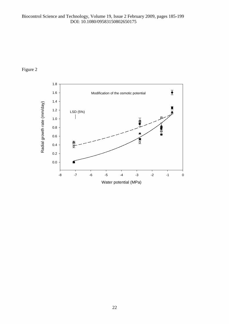
The mean radial growth of colonies for each of the three fungal isolates, in response to osmotic
stress was found to be linear over time (Figure 1). At -7.1 MPa, none of the P. chlamydosporia
isolates tested grew in the medium modified with glycerol, but continued growing at this water
potential in the medium modified with KCl (Figure 1). Having applied the linear regression model
to each replicate, the average of the estimated rates of radial growth of P. chlamydosporia, for each
combination of solute, isolate and water potential level was calculated (Figure 2). The exponential
model revealed significant differences between the solutes KCl and glycerol (p<0.001), but no
differences (p>0.05) were found in the rates of decrease for the three isolates (Figure 2). The model
shows that the exponential rate of decrease in radial growth using KCl (0.115) was approximately
half of the rate of decrease using glycerol (0.228). Analysis of Variance of the estimated radial
growth rates at different osmotic potentials showed a three-way interaction between solute, isolate
and MPa level (p<0.001); although the overall main effect of isolate was not significant (p=0.759),
which supports the result found using the exponential model.
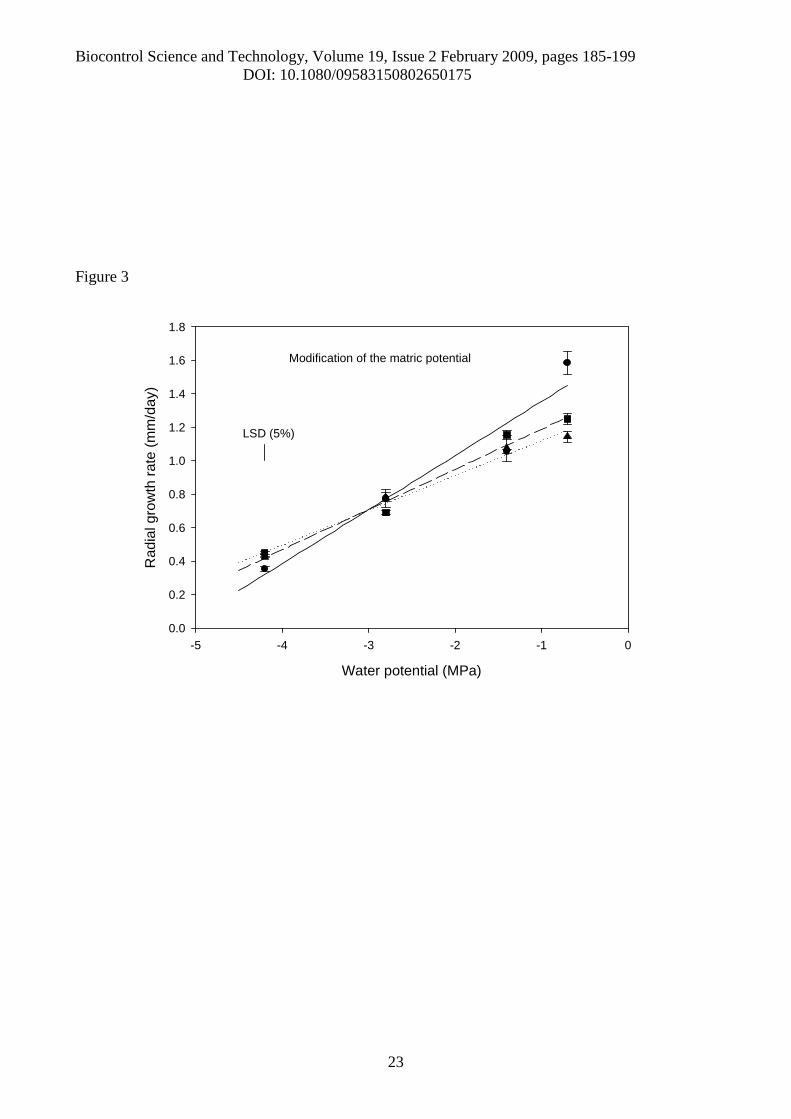
Modification of matric potential
When PEG 8000 was added to the medium for modification of the matric potential, the
variation in the radial growth rates with decreasing MPa was linear (p<0.001), and separate lines
for isolates were fitted (Figure 3). The Analysis of Variance of radial growth rates for matric
potential gave a significant interaction between isolate and MPa level (p<0.001), with both main
effects also being significant (p<0.006).
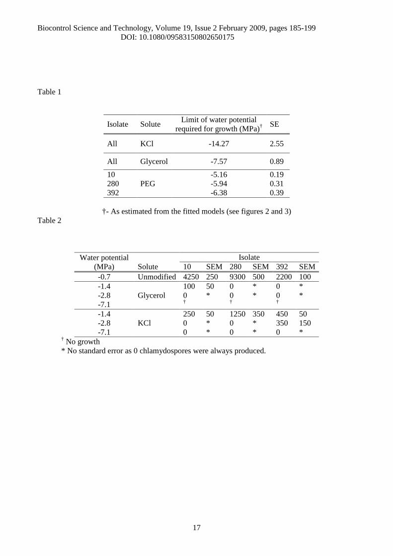
Given the models, the limits of radial growth potential were estimated (Table 1). The
fungus was generally more tolerant to the ionic solute KCl than to glycerol. In matrically modified
media, the limits for growth differed from -5.16 ± 0.19 to -6.38 ± 0.39 MPa, depending on the

Biocontrol Science and Technology, Volume 19, Issue 2 February 2009, pages 185-199DOI: 10.1080/09583150802650175
8
isolate (Table 1). Chlamydospores were produced by all isolates in the unmodified medium but few
or none were produced at lower osmotic potentials (Table 2).
Effects of osmotic and matric potential on the accumulation of endogenous reserves in three
isolates of P. chlamydosporia
Modification of osmotic potential
The total amounts of polyols accumulated intracellularly were greater and significantly different in
osmotically modified media (p<0.05, using LSD), when compared with those endogenous reserves
accumulated in unmodified media, with the exception of media modified with KCl at -1.4 MPa
(Table 3-A). However, no significant differences in the amounts of total polyols were found
between isolates in response to modification of the osmotic potential (p=0.952).
The proportion of polyols accumulated by the fungus depended on the solute used to
generate the water stress (Figure 4). When grown in media osmotically modified with glycerol, the
fungus accumulated proportionately greater amounts of glycerol compared to the other polyols and
in the control medium (Figure 4). In media osmotically modified with KCl, the accumulation of
arabitol at -1.4 and -2.8 MPa was proportionately greater than in unmodified media (Figure 4). In
the media modified with glycerol, greater amounts of glucose were detected, compared with the
amounts accumulated in colonies growing in media where water was freely available (Table 3-A).
However, fungal growth in media modified with KCl did not result in a significant change in the
accumulation of glucose (p>0.05) (Table 3-A). There was a highly significant interaction (p<0.001)
between isolates, media and level of water potential in the accumulation of arabitol and mannitol.
Comparing specific quantities (on the natural logarithm scale), there was a greater (p <0.05)
accumulation of arabitol at -2.8 and and -7.1 MPa, in all isolates compared to the control (Table 4-
A). However, no significant differences (p>0.05) between isolates were found in the accumulation
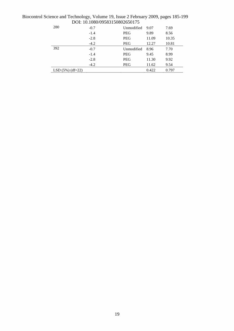
of glycerol and erythritol (Table 4-A).Modification of matric potential
The quantity of polyols accumulated by the fungus in media modified with PEG was greater and
significantly different (p<0.05) compared to that accumulated in unmodified media, in all the
isolates tested (Table 3-B). Significant differences in the amounts of total polyols were found
between isolates in response to matric stress levels (p<0.001). However, the three-way interaction
between isolates, matric stress levels and solute was not significant (p=0.247). The total amounts of
polyols accumulated were greatest when the fungus grew at water potentials of -4.2 MPa, in media
amended with PEG (Table 3-B). In matrically modified media, a large quantity of erythritol was
accumulated in all the isolates, and this increased substantially with the reduction of the water
potential (Figure 4-B). There was a significant interaction (p<0.001) between isolates and level of
water potential in all types of polyols (Table 4-B).

Biocontrol Science and Technology, Volume 19, Issue 2 February 2009, pages 185-199DOI: 10.1080/09583150802650175
9
Discussion
For the first time, the effects of osmotic and matric potential on radial growth and
accumulation of polyols were studied in colonies from different isolates of P. chlamydosporia. In
this study, the fungus grew faster in a non-modified medium, when water was freely available (-0.7
MPa). Similar results were found by Kerry et al.(1986), who studied the effects of pH and water
potential on the growth of six isolates of the fungus on a basal medium amended with a mixture of
salts or with sucrose. However, matric stress, a component of the water potential relevant to growth
and colonisation in soil (Brownell and Schneider, 1985), was not considered in their study. In the
present study, radial growth rates decreased substantially with the increase of osmotic or matric
stress. Although radial growth rates were lower in PEG-modified media, the fungus was relatively
tolerant to matric stress. Interestingly, differences between isolates were found in response to
matric stress on the PEG-modified media but isolates responded similarly on the medium modified
with the ionic and non ionic solute, used to subject the fungal isolates to osmotic stress. Tolerance
to matric stress may have been related to the accumulation of high amounts of erythritol in PEG-
modified media, which enables enzyme systems to work effectively in such conditions. Low-
molecular weight erythritol is known to regulate intracellular water potential efficiently, and is
accumulated by xerophylic fungi in response to water stress (Hallsworth, 1995).
All the isolates tended to be more sensitive to the non-ionic solute glycerol than to the ionic
KCl. At -7.1 MPa, the fungus was able to continue growing in a medium amended with the ionic
solute, but not in a medium amended with glycerol at this level of MPa. Low concentrations of
ionic solutes, such as KCl, may stimulate growth in some fungi (Larsen, 1986), as K ions aid
transport across the mycelial cell walls to enable better intracellular osmotic adjustment. Therefore,
it is possible that KCl from the medium may have been utilised to provide increased tolerance to
osmotic stress.
In media modified with glycerol, P. chlamydosporia accumulated large amounts of
glycerol, which were much greater than in unmodified media. This accumulation was possibly a
response to regulate intracellular osmotic potential. As glycerol was used to modify the medium, it
may have been passively accumulated by the fungus which in turn helped it to tolerate the increase
in stress. Glycerol appears to be of crucial importance in many fungi, as it protects enzymes from
accumulation of sodium and from loss of water, both of which may denature the enzymes (Luard,
1982).
The quantities of the major sugar alcohol accumulated in colonies of P. chlamydosporia
depended on the solute used to generate the water stress. Whereas arabitol and glycerol were the
Biocontrol Science and Technology, Volume 19, Issue 2 February 2009, pages 185-199DOI: 10.1080/09583150802650175
10
main polyols accumulated in osmotically modified media with ionic and non-ionic solutes,
respectively, erythritol was the main sugar accumulated in media amended with PEG.. The
versatility to accumulate different polyols and perhaps different sugars under different stress
conditions might bring advantage of allowing the fungus to survive in soil when environmental
conditions are less favourable or highly variable. In some fungi, enhanced accumulation of
compatible solutes contributed to an improvement in viability (Hallsworth and Magan, 1995,
Mokiou, 2005, Teixidó et al., 2005) and even virulence (Chandler et al., 2005). Therefore, the
viability of the spores produced under different stress regimes might have been different, although
this aspect was not covered in the study, as few chlamydospores were produced on osmotically
modified media, after 25 days of growth (Table 2). As chlamydospores are the preferred type of
inoculum to establish the fungus in soil (De Leij and Kerry, 1991) it was considered that any
improvement through the incorporation of polyols could be greatly outweighed by the reduction in
chlamydospore production. To conclude, the presence of free water available in the agar media
favoured the growth of P. chlamydosporia, when compared to growth rates in media where water
was restricted. Fungal growth was affected by both osmotic and matric stress, although this species
can tolerate matric stress levels relevant to those that occur in soil. Under water stress, the fungus
accumulated a combination of different sugar alcohols important in osmoregulation, the differential
and proportional amounts of which were dependent on the solute used. The research presented in
this paper provides some new information suggesting the involvement of different mechanisms of
osmoregulation in P. chlamydosporia as a response to osmotic and matric stress on mycelial
growth and the accumulation of endogenous reserves in spores. Further research is needed to study
the effects of such changes on fungal survival and activity in soil.
Acknowledgements
The authors are grateful for the financial support received from the EU for the project, ICA4-CT-
2002-10044, MiCoSPA - Microbial Pest Control for Sustainable Peri-urban/ urban Agriculture in
Latin America (Cuba and Mexico). We also thank Rothamsted International, who sponsored Belkis
Peteira. Rothamsted Research receives grant aided support from the Biotechnology and Biological
Sciences Research Council of the UK.

Biocontrol Science and Technology, Volume 19, Issue 2 February 2009, pages 185-199DOI: 10.1080/09583150802650175
11
Figure 1. Effect of osmotic potential (glycerol and KCl modified media) on the average radialdiameter (mm) over time (days) in three isolates of Pochonia chlamydosporia; [isolates 10 (A, B),280 (C, D) and 392 (E, F)], at 25°C; (×, -7.1 MPa; ▲, -2.8 MPa; ■, -1.4 MPa and ♦, -0.7 MPa).Solid lines show the fitted regressions through the means.
Figure 2. Comparison of radial growth rates (mm/day) in three isolates of Pochoniachlamydosporia in response to osmotic potential (MPa). [● – glycerol, isolate 10; ■ – glycerol,isolate 280; ▲- glycerol, isolate 392; ○- KCl, isolate 10; □ - KCl, isolate 280; ∆ - KCl, isolate 392;___ -Fitted curve for glycerol: Y = -0.294 + 1.402 exp(0.228 (MPa + 0.7)); ---- -Fitted curve forKCl: Y = -0.294 + 1.402 exp(0.115 (MPa + 0.7)); R2 for model = 74.4%, s2 = 0.042, df =99]. Theleast significant difference (LSD) at the 5% level for comparisons of means is taken from theANOVA of these data.
Figure 3. Comparison of radial growth rates (mm/day) in three isolates of Pochoniachlamydosporia in response to matric potential (MPa). [● - PEG, isolate 10; ■ - PEG, isolate 280;▲- PEG, isolate 392; ___ - Fitted line for isolate 10: Y = 1.677+ 0.323 MPa; ----- -Fitted line forisolate 280: Y = 1.429 + 0.241 MPa; ……. -Fitted line for isolate 392: Y= 1.327 + 0.208 MPa; R2 formodel = 92.7%, s2 = 0.010, df =40]. The least significant difference (LSD) at the 5% level forcomparisons of means is taken from the ANOVA of these data.
Figure 4. Relative proportion (%) of polyols [ - glycerol, - erythritol, - arabitol and -mannitol] accumulated in biomass of three isolates of Pochonia chlamydosporia [A- isolate 10,glycerol; B-isolate 10, KCl; C- isolate 280, glycerol; D- isolate 280, KCl, E- isolate 392, glycerol;F- isolate 392, KCl; G- isolate 10, PEG; H- isolate 280, PEG; I- isolate 392, PEG], after 25 days ofgrowth, at 25 ºC.

Biocontrol Science and Technology, Volume 19, Issue 2 February 2009, pages 185-199DOI: 10.1080/09583150802650175
12
Table 1. Estimated limits of water potential (MPa) required for Pochonia chlamydosporia radialgrowth, in different solutes with standard errors (SE).
Table 2. Mean and standard error of mean (SEM) number of chlamydospores produced per Petridish, in each treatment, after 25days of growth, at 25ºC.
Table 3. Effect of ionic (KCl) and non ionic (glycerol) osmotic potential (A) and matric potential(B) on the intracellular accumulation of total polyols and glucose in three isolates of Pochoniachlamydosporia, at 25ºC. Means in each column for statistical comparison given the ANOVAs arefollowed by the least significant difference (LSD) at the 5% level of significance. Differencesbetween isolate were not statistically significant (p>0.05) for total polyols, so means forcomparison in this case (in bold) are taken across isolate.
Table 4. Effect of ionic (KCl) and non ionic (glycerol) osmotic potential (A) and matric potential(B) on the intracellular accumulation of glycerol, erythritol, arabitol and mannitol in three isolatesof Pochonia chlamydosporia, at 25ºC. Means in each column for statistical comparison given theANOVAs are followed by the least significant difference (LSD) at the 5% level of significance.Differences between isolate were not statistically significant (p>0.05) for glycerol and erythritol, someans for comparison in this case (in bold) are taken across isolate.
Biocontrol Science and Technology, Volume 19, Issue 2 February 2009, pages 185-199DOI: 10.1080/09583150802650175
13
References
Arora, D.K., Hirsch, P.R. and Kerry, B.R. (1996), "PCR-based discrimination of Verticilliumchlamydosporium isolates," Mycological Research, 100, 801-809.
Atkins, S., Clark, I.M., Kerry, B.R. and Sosnowska, D. (2002), "Initial testing of potential fungalbiological control agents for potato cyst nematodes," in The BCPC conference- Pests & Diseases,pp. 79-84.
Atkins, S., Hidalgo-Diaz, L., Kalisz, H., Mauchline, T.H., Hirsch, P.R. and Kerry, B.R. (2003a),"Development of a new management strategy for the control of root-knot nematodes (Meloidogyespp.) in organic vegetable production," Pest Management Science, 59, 183-189.
Atkins, S., Hidalgo-Diaz, L., Clark, I., Morton, O., Montes de Oca, N., Gray, P. and Kerry, B.R.(2003b), "Approaches for monitoring the release of Pochonia chlamydosporia var. catenulata, abiocontrol agent for root-knot nematodes," Mycological Research, 107 (2), 206-212.
Bourne, J.M. and Kerry, B.R. (1999), "Effect of the host plant on the efficacy of Verticilliumchlamydosporium as a biological control agent of root-knot nematodes at different nematodedensities and fungal application rates," Soil biology and biochemistry, 31, 75-84.
Brown, A.D. (1978), "Compatible solutes and extreme water stress in eukaryotic microorganisms,"Advances in Microbial Physiology, 17, 181-242.
Brown, A.D. and Simpson, J.R. (1972), "Water relations of sugar- tolerant yeasts: the role ofintracellular polyols," Journal of General Microbiology, 72, 589-591.
Brownell, K.H. and Schneider, R.W. (1985), "Roles of matric and osmotic components of waterpotential and their interaction with temperature in the growth of Fusarium oxysporum in syntheticmedia and soil," Phytopathology, 75, 53-57.
Chandler, D., Andersen, M. and Magan, N. (2005), "A population based threshold model todemonstrate that physiological manipulation of endogenous reserves can increase the virulence ofinsect pathogenic fungi," in The British Crop Protection Council (BCPC) International Congress -Crop Science & Technology 2005., Glasgow, Scotland.
Cook, R.J. and Baker, K.F. (1983), The nature and practise of biological control of plantpathogens, Minnesota, USA: The American Phytopathological Society.
Dallyn, H. and Fox, A. (1980), "Spoilage of material of reduced water activity by xerophilic fungi,"in Microbial growth and survival in extreme environments, eds. G.H. Gould and E.L. Corry,London and New York: Academic Press, pp. 129-139.
De Leij, F.A.A.M. and Kerry, B.R. (1991), "The nematophagous fungus, Verticilliumchlamydosporium, as a biological control agent for Meloidogyne arenaria," Révue de Nematologie,14, 157-194.
GenStat® (2007), UK: VSN International Ltd.

Biocontrol Science and Technology, Volume 19, Issue 2 February 2009, pages 185-199DOI: 10.1080/09583150802650175
14
Hallsworth, J.E. (1995), "Manipulation of intracellular polyols and trehalose for successfulbiological control," Ph.D. dissertation, Cranfield University.
Hallsworth, J.E. and Magan, N. (1994a), "Effect of carbohydrate type and concentration onpolyhydroxy alcohol and trehalose content of conidia of three entomopathogenic fungi,"Microbiology, 140, 2705-2713.
Hallsworth, J.E. and Magan, N. (1994b), "Effects of KCl concentration on accumulation of acyclicsugar alcohols and trehalose in conidia in three entomopathogenic fungi," Letters in AppliedMicrobiology, 18, 8-11.
Hallsworth, J.E. and Magan, N. (1995), "Manipulation of intercellular glycerol and erythritolenhances germination of conidia at low water availability," Microbiology, 141, 1109-1115.
Hirsch, P.R., Mauchline, T.H., Mendum, A. and Kerry, B.R. (2000), "Detection of thenematophagous fungus Verticillium chlamydosporium in nematode-infested plant roots using PCR,"Mycological Research, 104, 435-439.
Kerry, B.R., Crump, D.H. and Mullen, L.A. (1982), "Studies of the cereal cyst nematodeHeterodera avenae, under continuous cereals 1975-1978 II .Fungal parasitism of nematode femalesand eggs," Annals of Applied Biology, 100, 489-499.
Kerry, B.R., Irving, F. and Hornsey, J.C. (1986), "Variation between strains of the nematophagousfungus Verticillium chlamydosporium Goddard I. Factors affecting growth in vitro," Nematologica,32, 461-473.
Kerry, B.R. and Jaffee, B.A. (1997), "Fungi as biological control agents for plant parasiticnematodes," in The Mycota IV Environmental and Microbial Relationships, eds. D.T. Wilcklow andB. Söderström, Berlim, Germany: Springer-Verlag pp. 203-218.
Kerry, B.R., Kirkwood, I.A., de Leij, F.A.A.M., Barba, J., Leijdens, M.B. and Brookes, P.C.(1993), "Growth and survival of Verticillium chlamydosporium Goddard, a parasite of nematodes,in soil," Biocontrol science and technology, 3, 355-365.
Larsen, H. (1986), "Halophilic and halotolerant microorganisms - An overview and historicalperspective," FEMS Microbiology Reviews, 39, 3-7.
Luard, E.J. (1982), "Effect of osmotic shock on some intracellular solutes in two filamentousfungi," Journal of General Microbiology, 128, 2575-2581.
Magan, N. (1997), "Fungi in extreme environments.," in The Mycota IV: Environmental andMicrobial Relationships., eds. D.T. Wicklow and B. Söderström, Berlin, Germany: SpringerVerlag, pp. 99-113.
Magan, N. (2001), "Physiological approaches to improving the ecological fitness of fungalbiocontrol agents in fungi as biocontrol agents.," in Fungi as Biocontrol Agents -Progress,Problems and Potential, eds. T. Butt, C. Jackson and N. Magan, London: CAB International, pp.239-251.

Biocontrol Science and Technology, Volume 19, Issue 2 February 2009, pages 185-199DOI: 10.1080/09583150802650175
15
Magan, N. and Lynch, J.M. (1986), "Water potential, growth and cellulolysis of fungi involved indecomposition of cereal residues," Journal of General Microbiology, 132, 1181-1187.
Marín, S., Sanchis, V. and Magan, N. (1995), "Water activity, temperature and pH effects ongrowth of Fusarium moniliforme and Fusarium proliferatum isolates from maize," CanadianJournal of Microbiology, 41, 1063-1070.
Mead, R., Curnow, R.N. and Hasted, A.M. (2003), Statistical methods in agriculture andexperimental biology Chapman & Hall
Meikle, A.J., Chudek, J.A., Reed, R.H. and Gadd, G.M. (1991), "Natural abundance 13C-nuclearmagnetic resonance spectroscopic analysis of acyclic polyol and trehalose accumulation by severalyeast species in response to salt stress," FEMS Microbiology letters, 66, 163-167.
Mokiou, S. (2005), "Ecophysiological approaches to production and formulation of the biocontrolyeast Pichia anomala," Cranfield University, Institute of BioScience and Technology.
Noreen, A., Amer-Zareen, Zaki, M.J. and Shaukat, S.S. (2001), "Factors affecting survival ofnematophagous fungus Verticillium chlamydosporium. I. Soil moisture and temperature,"International journal of nematology, 11, 274-278.
Pascual, S., Melgarejo, P. and Magan, N. (2000), "Accumulation of compatible solutes inPenicillium frequentans grown at reduced water activity and biocontrol of Monilinia laxa,"Biocontrol science and technology, 10, 71-80.
Patterson, H.D. and Thompson, R. (1971), "Recovery of inter-block information when block sizesare unequal," Biometrika, 58, 545-554.
Ramos, A.J., Magan, N. and Sanchis, V. (1999), "Osmotic and matric potential effects on growth,sclerotia and partioning of polyols and sugars in colonies and spores of Aspergillus ochraceus,"Mycological Research, 103, 141-147.
Steuter, A.A., Mozafar, A. and Goodin, J.R. (1981), "Water potential of aqueous polyethyleneglycol," Plant Physiology, 67, 64-67.
Teixidó, N., Canamás, T., Usall, J., Torres, A.M., Magan, N. and Vinas, I. (2005), "Accumulationof the compatible solutes, glycine-betaine and ectoine, as an osmotic stress adaptation and heatshock cross-protection in the biocontrol agent Pantoea agglomerans CPA-2," Letters in AppliedMicrobiology, 41, 248-252.
Tobin, J.D., Haydock, P.P.J., Hare, M.C., Woods, S.R. and Crump, D.H. (2008), "Effect of thefungus Pochonia chlamydosporia and fosthiazate on the multiplication rate of potato cyst nematodes(Globodera pallida and G. rostochiensis) in potato crops grown under UK field conditions,"Biological control, In Press, Corrected Proof.
Van Eck, J.H., Prior, B.A. and Brandt, E.V. (1993), "The water relations of growth andpolyhydroxy alcohol production by ascomycetous yeasts," Journal of General Microbiology, 139,1047-1054.
Biocontrol Science and Technology, Volume 19, Issue 2 February 2009, pages 185-199DOI: 10.1080/09583150802650175
16
Biocontrol Science and Technology, Volume 19, Issue 2 February 2009, pages 185-199DOI: 10.1080/09583150802650175
17
Table 1
Isolate Solute Limit of water potentialrequired for growth (MPa)† SE
All KCl -14.27 2.55
All Glycerol -7.57 0.89
10PEG
-5.16 0.19280 -5.94 0.31392 -6.38 0.39
†- As estimated from the fitted models (see figures 2 and 3)Table 2
Water potential(MPa) Solute
Isolate10 SEM 280 SEM 392 SEM
-0.7 Unmodified 4250 250 9300 500 2200 100-1.4 100 50 0 * 0 *-2.8 Glycerol 0 * 0 * 0 *-7.1 † † †
-1.4KCl
250 50 1250 350 450 50-2.8 0 * 0 * 350 150-7.1 0 * 0 * 0 *
† No growth* No standard error as 0 chlamydospores were always produced.

Biocontrol Science and Technology, Volume 19, Issue 2 February 2009, pages 185-199DOI: 10.1080/09583150802650175
18
Table 3
(A) Amount (mg/g) mycelial colonies (Log-transformed)
Isolate
Waterpotential(MPa) Solute
TotalPolyols(df=30)
Glucose(df=34)
10 -0.7 Unmodified 9.26 8.14
-1.4 Glycerol 10.31 9.86
-1.4 KCl 9.38 9.22
-2.8 Glycerol ND† 10.38
-2.8 KCl 10.11 8.57
-7.1 KCl 10.30 8.50
280 -0.7 Unmodified 9.07 7.69
-1.4 Glycerol 10.88 9.56
-1.4 KCl 9.19 7.94
-2.8 Glycerol ND† 8.91
-2.8 KCl 9.50 7.98
-7.1 KCl 10.09 8.58
392 -0.7 Unmodified 8.96 7.70
-1.4 Glycerol 10.63 9.60
-1.4 KCl 9.27 7.75
-2.8 Glycerol ND† 8.63
-2.8 KCl 9.75 8.12
-7.1 KCl 10.85 9.76
LSD (5%) 1.000-0.7 Unmodified 9.10-1.4 Glycerol 10.61-1.4 KCl 9.28-2.8 KCl 9.79-7.1 KCl 10.41
LSD (5%) 0.361†ND- Not determined
(B) Amount (mg/g) mycelial colonies (Log-transformed)
IsolateWater potential(MPa) Solute
TotalPolyols Glucose
10 -0.7 Unmodified 9.26 8.14-1.4 PEG 10.39 9.77-2.8 PEG 11.27 10.81-4.2 PEG 12.48 9.49
Biocontrol Science and Technology, Volume 19, Issue 2 February 2009, pages 185-199DOI: 10.1080/09583150802650175
19
280 -0.7 Unmodified 9.07 7.69-1.4 PEG 9.89 8.56-2.8 PEG 11.09 10.35-4.2 PEG 12.27 10.81
392 -0.7 Unmodified 8.96 7.70-1.4 PEG 9.45 8.99-2.8 PEG 11.30 9.92-4.2 PEG 11.62 9.54
LSD (5%) (df=22) 0.422 0.797

Biocontrol Science and Technology, Volume 19, Issue 2 February 2009, pages 185-199DOI: 10.1080/09583150802650175
20
Table 4(A)Amount (mg/g) mycelial colonies (Log- transformed)
IsolateWaterpotential(MPa)
Solute Glycerol(df =35)
Erythritol(df =28)
Arabitol(df=36)
Mannitol(df=35)
10 -0.7 Unmodified 7.43 7.68 7.46 8.47-1.4 Glycerol 9.73 7.75 7.49 9.06-1.4 KCl 5.43 7.87 8.06 8.34-2.8 Glycerol 12.8 9.45 8.49 9.73-2.8 KCl 7.86 8.09 9.58 8.19-7.1 KCl 7.87 10.49 8.63 8.19
280 -0.7 Unmodified 7.75 7.72 7.46 7.76-1.4 Glycerol 10.46 8.07 7.78 8.75-1.4 KCl 7.91 7.64 7.80 7.77-2.8 Glycerol 12.19 8.79 8.20 9.06-2.8 KCl 7.59 7.69 8.81 7.79-7.1 KCl 7.9 9.39 8.24 8.31
392 -0.7 Unmodified 7.38 7.66 7.49 7.73-1.4 Glycerol 10.40 7.95 7.63 8.07-1.4 KCl 8.03 7.68 7.83 7.77-2.8 Glycerol 12.61 8.73 7.79 8.10-2.8 KCl 7.62 7.93 9.21 8.08-7.1 KCl 8.60 9.14 10.14 8.98
LSD (5%) 0.676 0.582-0.7 Unmodified 7.52 7.69-1.4 Glycerol 10.12 7.93-1.4 KCl 7.92 7.73-2.8 Glycerol 12.53 ND†
-2.8 KCl 7.69 7.90-7.1 KCl 8.12 9.67
LSD (5%) 0.472 0.373†-Not determined
(B)Isolate Water potential
(MPa) Solute Glycerol(df=14)
Erythritol(df=22)
Arabitol(df=22)
Mannitol(df=22)
10 -0.7 Unmodified 7.43 7.68 7.46 8.47-1.4 PEG 7.38 10.12 7.97 7.83-2.8 PEG 8.36 11.21 7.69 7.74-4.2 PEG 8.38 12.43 8.41 8.36
280 -0.7 Unmodified 7.75 7.72 7.46 7.76-1.4 PEG 7.47 8.68 7.47 9.20-2.8 PEG ND† 10.91 8.17 8.52-4.2 PEG 7.69 12.23 7.80 7.96
392 -0.7 Unmodified 7.38 7.66 7.49 7.73-1.4 PEG 7.45 7.81 8.26 8.31-2.8 PEG 7.51 10.75 10.03 9.02

Biocontrol Science and Technology, Volume 19, Issue 2 February 2009, pages 185-199DOI: 10.1080/09583150802650175
21
-4.2 PEG 7.46 10.87 10.48 9.99LSD (5%) 0.311 0.478 0.645 0.640
Figure 1
(E) -Isolate 392 -Glycerol
0
5
10
15
20
25
30
35
40
5 7 10 12 14 18 25
Days
Rad
iale
xten
sion
(mm
)
(C) -Isolate 280-Glycerol
0
5
10
15
20
25
30
35
40
5 7 10 12 14 18 25
Days
Rad
iale
xten
sion
(mm
)
(D) -Isolate 280 -KCl
0
5
10
15
20
25
30
35
40
5 7 10 12 14 18 25
Days
Rad
iale
xten
sion
(mm
)
(F) - Isolate 392- KCl
0
5
10
15
20
25
30
35
40
5 7 10 12 14 18 25
Days
Rad
iale
xten
sion
(mm
)
(A) -Isolate 10- Glycerol
05
1015202530354045
5 7 10 12 14 18 25
Days
Rad
iale
xten
sion
(mm
)
(B)- Isolate 10- KCl
05
101520
253035
4045
5 7 10 12 14 18 25
Days
Rad
iale
xten
sion
(mm
)
Biocontrol Science and Technology, Volume 19, Issue 2 February 2009, pages 185-199DOI: 10.1080/09583150802650175
22
Figure 2
Modification of the osmotic potential
Water potential (MPa)
-8 -7 -6 -5 -4 -3 -2 -1 0
Rad
ialg
row
thra
te(m
m/d
ay)
0.0
0.2
0.4
0.6
0.8
1.0
1.2
1.4
1.6
1.8
LSD (5%)
Biocontrol Science and Technology, Volume 19, Issue 2 February 2009, pages 185-199DOI: 10.1080/09583150802650175
23
Figure 3
Modification of the matric potential
Water potential (MPa)
-5 -4 -3 -2 -1 0
Rad
ialg
row
thra
te(m
m/d
ay)
0.0
0.2
0.4
0.6
0.8
1.0
1.2
1.4
1.6
1.8
LSD (5%)

Biocontrol Science and Technology, Volume 19, Issue 2 February 2009, pages 185-199DOI: 10.1080/09583150802650175
24

Biocontrol Science and Technology, Volume 19, Issue 2 February 2009, pages 185-199DOI: 10.1080/09583150802650175
25
(B) - Biotype 10 - KCl
0%
20%
40%
60%
80%
100%
-0.7 -1.4 -2.8 -7.1
Media w ater potential (-MPa)
Rel
ativ
epr
opot
ion
(%)
(A)- Biotype 10 - Glycerol
0%
20%
40%
60%
80%
100%
-0.7 -1.4 -2.8
Media w ater potential (-MPa)
Rel
ativ
epr
opot
ion
(%)
(G)- Biotype 10 - PEG 8000
0%
20%
40%
60%
80%
100%
-0.7 -1.4 -2.8 -4.2
Media w ater potential (-MPa)
Rel
ativ
epr
opot
ion
(%)
(H)- Biotype 280 - PEG 8000
0%
20%
40%
60%
80%
100%
-0.7 -1.4 -2.8 -4.2
Media w ater potential (-MPa)
Rel
ativ
epr
opot
ion
(%)
(I)- Biotype 392 - PEG 8000
0%
20%
40%
60%
80%
100%
-0.7 -1.4 -2.8 -4.2
Media w ater potential (-MPa)
Rel
ativ
epr
opot
ion
(%)
Glycerol Erythritol Arabitol Mannitol
(D)- Biotype 280 - KCl
0%
20%
40%
60%
80%
100%
-0.7 -1.4 -2.8 -7.1
Media w ater potential (-MPa)
Rel
ativ
epr
opot
ion
(%)
(C)- Biotype 280 - Glycerol
0%
20%
40%
60%
80%
100%
-0.7 -1.4 -2.8
Media w ater potential (-MPa)
Rel
ativ
epr
opot
ion
(%)
(F) - Biotype 392 - KCl
0%
20%
40%
60%
80%
100%
-0.7 -1.4 -2.8 -7.1
Media w ater potential (-MPa)
Rel
ativ
epr
opot
ion
(%)
(E)- Biotype 392 - Glycerol
0%
20%
40%
60%
80%
100%
-0.7 -1.4 -2.8
Media w ater potential (-MPa)
Rel
ativ
epr
opot
ion
(%)

Biocontrol Science and Technology, Volume 19, Issue 2 February 2009, pages 185-199DOI: 10.1080/09583150802650175
26