Effects of Long-Term Pioglitazone Treatment on Peripheral and Central Markers of Aging
14
Effects of Long-Term Pioglitazone Treatment on Peripheral and Central Markers of Aging Eric M. Blalock 1. , Jeremiah T. Phelps 1. , Tristano Pancani 1 , James L. Searcy 1 , Katie L. Anderson 1 , John C. Gant 1 , Jelena Popovic 1 , Margarita G. Avdiushko 2 , Don A. Cohen 2 , Kuey-Chu Chen 1 , Nada M. Porter 1 , Olivier Thibault 1 * 1 Department of Molecular and Biomedical Pharmacology, University of Kentucky Medical Center, Lexington, Kentucky, United States of America, 2 Department of Microbiology and Immunology, University of Kentucky Medical Center, Lexington, Kentucky, United States of America Abstract Background: Thiazolidinediones (TZDs) activate peroxisome proliferator-activated receptor gamma (PPARc) and are used clinically to help restore peripheral insulin sensitivity in Type 2 diabetes (T2DM). Interestingly, long-term treatment of mouse models of Alzheimer’s disease (AD) with TZDs also has been shown to reduce several well-established brain biomarkers of AD including inflammation, oxidative stress and Ab accumulation. While TZD’s actions in AD models help to elucidate the mechanisms underlying their potentially beneficial effects in AD patients, little is known about the functional consequences of TZDs in animal models of normal aging. Because aging is a common risk factor for both AD and T2DM, we investigated whether the TZD, pioglitazone could alter brain aging under non-pathological conditions. Methods and Findings: We used the F344 rat model of aging, and monitored behavioral, electrophysiological, and molecular variables to assess the effects of pioglitazone (PIO-ActosH a TZD) on several peripheral (blood and liver) and central (hippocampal) biomarkers of aging. Starting at 3 months or 17 months of age, male rats were treated for 4–5 months with either a control or a PIO-containing diet (final dose approximately 2.3 mg/kg body weight/day). A significant reduction in the Ca 2+ -dependent afterhyperpolarization was seen in the aged animals, with no significant change in long- term potentiation maintenance or learning and memory performance. Blood insulin levels were unchanged with age, but significantly reduced by PIO. Finally, a combination of microarray analyses on hippocampal tissue and serum-based multiplex cytokine assays revealed that age-dependent inflammatory increases were not reversed by PIO. Conclusions: While current research efforts continue to identify the underlying processes responsible for the progressive decline in cognitive function seen during normal aging, available medical treatments are still very limited. Because TZDs have been shown to have benefits in age-related conditions such as T2DM and AD, our study was aimed at elucidating PIO’s potentially beneficial actions in normal aging. Using a clinically-relevant dose and delivery method, long-term PIO treatment was able to blunt several indices of aging but apparently affected neither age-related cognitive decline nor peripheral/ central age-related increases in inflammatory signaling. Citation: Blalock EM, Phelps JT, Pancani T, Searcy JL, Anderson KL, et al. (2010) Effects of Long-Term Pioglitazone Treatment on Peripheral and Central Markers of Aging. PLoS ONE 5(4): e10405. doi:10.1371/journal.pone.0010405 Editor: Silvana Gaetani, Sapienza University of Rome, Italy Received February 12, 2010; Accepted April 7, 2010; Published April 29, 2010 Copyright: ß 2010 Blalock et al. This is an open-access article distributed under the terms of the Creative Commons Attribution License, which permits unrestricted use, distribution, and reproduction in any medium, provided the original author and source are credited. Funding: Funding for this work was provided in part by a gift from the Neurosciences Education and Research Foundation (OT), and research grants AG029268 (OT), AG0033649 (OT) and NCRR-P20-RR15592 (OT). The funders had no role in study design, data collection and analysis, decision to publish, or preparation of the manuscript. Competing Interests: The authors have declared that no competing interests exist. * E-mail: [email protected] . These authors contributed equally to this work. Introduction Adjunct therapy against type 2 diabetes mellitus (T2DM) with thiazolidinediones (TZDs) is on the rise, with increasing numbers of patients prescribed the TZDs rosiglitazone (ROSI, AvandiaH) or pioglitazone (PIO, ActosH). These agents are in the top 50 prescribed drugs in North America, and together account for approximately 20 million prescriptions (2008 data, rxlist.com). Results from over two decades of studies have shown that untreated T2DM can negatively impact brain function. Depend- ing on the severity and the duration of the disease, as well as on the age of the individual, the condition is associated with varying degrees of cognitive deficits, motor dysfunction, and depression [1,2,3,4,5,6,7]. While aging worsens the impact of diabetes on cognitive function, it is not clear how diabetes and accompanying peripheral metabolic dysregulation exacerbate this process. Proposed mechanisms underlying cognitive decline when aging and diabetes coexist include insulin resistance, vascular disease, and inflammation resulting from the release of adipose tissue- derived cytokines. In clinical and animal studies, the brain, and the hippocampus in particular, appear sensitive to peripheral cytokine levels or metabolic stressors [8,9,10,11], with enhanced sensitivity seen in aged animals [8]. Given the role of the hippocampus in memory acquisition, processing and consolidation (reviewed in PLoS ONE | www.plosone.org 1 April 2010 | Volume 5 | Issue 4 | e10405
-
Upload
independent -
Category
Documents
-
view
4 -
download
0
Transcript of Effects of Long-Term Pioglitazone Treatment on Peripheral and Central Markers of Aging
Effects of Long-Term Pioglitazone Treatment onPeripheral and Central Markers of AgingEric M. Blalock1., Jeremiah T. Phelps1., Tristano Pancani1, James L. Searcy1, Katie L. Anderson1, John C.
Gant1, Jelena Popovic1, Margarita G. Avdiushko2, Don A. Cohen2, Kuey-Chu Chen1, Nada M. Porter1,
Olivier Thibault1*
1 Department of Molecular and Biomedical Pharmacology, University of Kentucky Medical Center, Lexington, Kentucky, United States of America, 2 Department of
Microbiology and Immunology, University of Kentucky Medical Center, Lexington, Kentucky, United States of America
Abstract
Background: Thiazolidinediones (TZDs) activate peroxisome proliferator-activated receptor gamma (PPARc) and are usedclinically to help restore peripheral insulin sensitivity in Type 2 diabetes (T2DM). Interestingly, long-term treatment of mousemodels of Alzheimer’s disease (AD) with TZDs also has been shown to reduce several well-established brain biomarkers ofAD including inflammation, oxidative stress and Ab accumulation. While TZD’s actions in AD models help to elucidate themechanisms underlying their potentially beneficial effects in AD patients, little is known about the functional consequencesof TZDs in animal models of normal aging. Because aging is a common risk factor for both AD and T2DM, we investigatedwhether the TZD, pioglitazone could alter brain aging under non-pathological conditions.
Methods and Findings: We used the F344 rat model of aging, and monitored behavioral, electrophysiological, andmolecular variables to assess the effects of pioglitazone (PIO-ActosH a TZD) on several peripheral (blood and liver) andcentral (hippocampal) biomarkers of aging. Starting at 3 months or 17 months of age, male rats were treated for 4–5months with either a control or a PIO-containing diet (final dose approximately 2.3 mg/kg body weight/day). A significantreduction in the Ca2+-dependent afterhyperpolarization was seen in the aged animals, with no significant change in long-term potentiation maintenance or learning and memory performance. Blood insulin levels were unchanged with age, butsignificantly reduced by PIO. Finally, a combination of microarray analyses on hippocampal tissue and serum-basedmultiplex cytokine assays revealed that age-dependent inflammatory increases were not reversed by PIO.
Conclusions: While current research efforts continue to identify the underlying processes responsible for the progressivedecline in cognitive function seen during normal aging, available medical treatments are still very limited. Because TZDshave been shown to have benefits in age-related conditions such as T2DM and AD, our study was aimed at elucidating PIO’spotentially beneficial actions in normal aging. Using a clinically-relevant dose and delivery method, long-term PIO treatmentwas able to blunt several indices of aging but apparently affected neither age-related cognitive decline nor peripheral/central age-related increases in inflammatory signaling.
Citation: Blalock EM, Phelps JT, Pancani T, Searcy JL, Anderson KL, et al. (2010) Effects of Long-Term Pioglitazone Treatment on Peripheral and Central Markers ofAging. PLoS ONE 5(4): e10405. doi:10.1371/journal.pone.0010405
Editor: Silvana Gaetani, Sapienza University of Rome, Italy
Received February 12, 2010; Accepted April 7, 2010; Published April 29, 2010
Copyright: � 2010 Blalock et al. This is an open-access article distributed under the terms of the Creative Commons Attribution License, which permitsunrestricted use, distribution, and reproduction in any medium, provided the original author and source are credited.
Funding: Funding for this work was provided in part by a gift from the Neurosciences Education and Research Foundation (OT), and research grants AG029268(OT), AG0033649 (OT) and NCRR-P20-RR15592 (OT). The funders had no role in study design, data collection and analysis, decision to publish, or preparation of themanuscript.
Competing Interests: The authors have declared that no competing interests exist.
* E-mail: [email protected]
. These authors contributed equally to this work.
Introduction
Adjunct therapy against type 2 diabetes mellitus (T2DM) with
thiazolidinediones (TZDs) is on the rise, with increasing numbers
of patients prescribed the TZDs rosiglitazone (ROSI, AvandiaH) or
pioglitazone (PIO, ActosH). These agents are in the top 50
prescribed drugs in North America, and together account for
approximately 20 million prescriptions (2008 data, rxlist.com).
Results from over two decades of studies have shown that
untreated T2DM can negatively impact brain function. Depend-
ing on the severity and the duration of the disease, as well as on the
age of the individual, the condition is associated with varying
degrees of cognitive deficits, motor dysfunction, and depression
[1,2,3,4,5,6,7]. While aging worsens the impact of diabetes on
cognitive function, it is not clear how diabetes and accompanying
peripheral metabolic dysregulation exacerbate this process.
Proposed mechanisms underlying cognitive decline when aging
and diabetes coexist include insulin resistance, vascular disease,
and inflammation resulting from the release of adipose tissue-
derived cytokines. In clinical and animal studies, the brain, and the
hippocampus in particular, appear sensitive to peripheral cytokine
levels or metabolic stressors [8,9,10,11], with enhanced sensitivity
seen in aged animals [8]. Given the role of the hippocampus in
memory acquisition, processing and consolidation (reviewed in
PLoS ONE | www.plosone.org 1 April 2010 | Volume 5 | Issue 4 | e10405
[12,13]), inflammation within the structure likely contributes to
memory and cognitive deficits with age and/or AD. Nevertheless,
almost nothing is known about the mechanisms through which
peripheral metabolic dysregulation as those seen in T2DM impact
hippocampal function and cognition.
TZDs are best known for their peripheral actions, where these
synthetic PPARc agonists selectively bind nuclear receptors and
enhance lipid accumulation in adipocytes, thereby helping to
decrease free fatty acid and lipid levels in plasma [14,15,16,17,18].
This mechanism helps to reestablish insulin sensitivity in T2DM
by working on fat, liver, and muscle tissues. Additionally, TZDs
are compounds with significant anti-inflammatory actions [19,20].
Recent evidence indicates that TZDs can have beneficial central
effects. In particular, ROSI was shown to improve cognition and
verbal memory in patients with mild cognitive impairment (MCI)
[21]. Further, a recent preliminary study on patients diagnosed
with MCI and diabetes also reported improved cognition following
6-months of PIO [22]. Finally, AD patients lacking the ApoEe4allele appear to be selectively sensitive to the beneficial effects of
chronic treatment with ROSI [23]. The mechanisms underlying
these effects in humans are not clear, but are likely to reflect
changes in inflammation, vascular function, insulin and/or glucose
levels, energy metabolism or beta amyloid clearance. Whether
these improvements are due to changes in the periphery, direct
effects in the brain, or some combination, is still unknown. In AD
animal models (APPV717I, Tg2576, 36TG), TZDs have been
shown to decrease Ab deposition [24,25,26,27] (but see [28]),
inflammation [24,29,30], and oxidative stress [28].
The predominant information we have regarding the beneficial
effects of TZDs in the brain comes from clinical studies in AD
patients [21,23]. However, given that most patients with diabetes
do not have AD, at least in the earlier stages of the disease, it seems
important to determine what the potential effects of these drugs
are in the context of normal aging, or in the absence of clinically
defined cognitive deficits. Compared to research conducted in AD
models, however, little is known about the functional consequences
of TZDs on cognition in animal models of normal aging.
Therefore, the present studies were undertaken to determine
whether PIO, the more brain permeant TZD [26], confers
significant benefits within the context of normal brain aging.
Further, because of our prior work identifying new roles of ROSI
and PIO in cultured neurons [31], and the work of others
suggesting that targets of TZD actions may include Ca2+-mediated
pathways in the brain [32,33,34], we investigated select biomark-
ers of aging including the Ca2+-dependent afterhyperpolarization
(AHP), long-term potentiation (LTP), and hippocampal-dependent
spatial memory. Other examined variables in the brain and in the
periphery included inflammatory cytokine levels, hippocampal
gene signatures, and insulin signaling. Our results suggest that at
the dose and duration tested, PIO caused expected beneficial
effects including reduced peripheral insulin and lipid marker
levels, and reduced a central biomarker of aging, namely the AHP.
However, other major markers of aging, including increased
inflammatory signaling (based on cytokine array measures in the
periphery and microarray measures in the hippocampus),
impaired cognition, and altered synaptic plasticity, were not
altered with PIO treatment.
Methods
Ethics StatementAll procedures were carried out under a University of Kentucky
IACUC approved protocol and are in accordance with NIH
guidelines for the care and use of laboratory animals.
AnimalsThirty-six male F344/NIA rats were purchased (Harlan,
Indianapolis, IN) in 2 groups of 18 with a 6 week stagger between
groups. All animals were fed TD94045 diet (Harlan Teklad,
Madison, WI) for one week prior to initiation of PIO or control
diets. To limit potential cohort effects across two separate animal
purchases, treatment groups were balanced across both cohorts.
Each group consisted of 8 young (3 months old), and 10 aged (17
months old) animals. Twelve animals from each group were
assigned to either the young control (YC, n = 4), young PIO (YP,
n = 4), or aged control (AC, n = 4) treatment groups, and the
remaining six animals were placed in the aged PIO (AP) group. In
all, the study was comprised of 8 YC, 8 YP, 8 AC, and 12 AP
animals. Animals were maintained on the diets for 15–20 weeks,
and were 7–8 months old, and 21–22 months old at the time of
study completion. Five aged animals died in the course of the
study. One AP animal had to be euthanized because of an
unresolved mandibular/eye infection, and two AP animals
stopped eating, lost considerable weight and were euthanized.
Based on two gross necropsy reports, another AP animal died of
chronic renal failure, and 1 AC animals died of granular
lymphocytic leukemia, both major causes of mortality in the aging
F344 [35]. The thirty one remaining animals were active, well-
groomed and appeared healthy, and were used for behavioral and
electrophysiology studies.
Blood collections and analysisOver the course of the study, three in vivo glucose measures were
taken. Animals were placed in a decapiconeH restraint (Braintree
Scientific, Braintree, MD) while their tails were washed with warm
soapy water and dried under a heat lamp. The lateral tail vein was
pricked with a 22 gauge needle and a FreeStyle Lite glucometer
(Abott Diabetes Care Inc., Alameda, CA) was used to measure
blood glucose levels (mg/dL). Trunk blood from twenty nine
animals was collected at the time of hippocampal slice preparation
(two samples were lost). Briefly, 2–3 mL was collected in a BD
Vacutainer SSD centrifuge tube and allowed to clot at room
temperature for one hour. To collect serum, samples were
centrifuged at 4000 rpm for 10 min. Half of the serum was sent
on dry ice for standard chemistry panel analysis (Comparative
Pathology Laboratory, University of California Davis, CA). The
remaining serum was frozen (280uC) and later used to monitor
insulin concentrations using the manufacturer’s protocol for an
ELISA-based assay (Millipore, Billerica, MA) and our bioassay
reader (HTS plus 7000, Perkin Elmer, Wellesley, MA), as well as
to monitor for the presence of three proinflammatory cytokines
using a Multiplex Bio Assay Analyzer (Millipore).
Insulin Receptor signalingLiver and brain cortices were used to quantify total and
phosphorylated insulin receptor levels according to the manufac-
turer’s protocol (Calbiochem, San Diego, CA), and using
duplicates for each sample. Frozen samples were removed from
the 280uC freezer and thawed on ice. After a 2 min homogeni-
zation period in PBS followed by centrifugation at 300 rpm for
5 min, the pellet was resuspended in Cell Extraction Buffer
(BioSource FNN0011) and left to lyse for 30 min with vortexing
every 10 min. At the end of this process the suspension was
centrifuged at 14K rpm for 10 min and the protein content in the
supernatant was determined using a Bradford assay. Detection of
the phosphorylated insulin receptor (IR) was accomplished
following the Phosphodetect ELISA kit protocol (CBA038,
Calbiochem). Total IR present in the samples was measured
using IR b-subunit ELISA Kit (CBA039, Calbiochem). Briefly,
Pioglitazone in Normal Aging
PLoS ONE | www.plosone.org 2 April 2010 | Volume 5 | Issue 4 | e10405
samples containing the same concentration of total protein were
incubated for 2 h in a 96 wells plate coated with IR b–subunit-
specific monoclonal antibody. After washing, an antibody specific
for IR phosphorylated at Tyr1162/1163 (CBA038) or specific for IR
b-subunit (CBA039) was added (detection antibody). The excess
detection antibody was removed after 1 hr and a horseradish
peroxidase-conjugated antibody (anti-rabbit Ig-HRP) was added
to the wells for 30 min. Following a final washing step to remove
the excess anti-rabbit Ig-HRP, a substrate was added and
absorbance was read at 450nm.
Experimental dietsPioglitazone (PIO-ActosH) was purchased through our DLAR
facility, and was incorporated into standard, color-coded purified
rodent diets (TD94045 Harlan Teklad). TD94045 was chosen to
approximate the NIH31 diet fed to animals since adulthood
(18.8% Kcal from protein, 63.9% Kcal from carbohydrates, and
17.2% Kcal from fat vs. 24% Kcal from protein, 62% Kcal from
carbohydrates and 14% Kcal from fat in the NIH31). Because of
differences in animal weights and food consumption, two PIO
diets were used, one formulated at 84 ppm for older animals and
another, formulated at 37 ppm for younger animals. Final PIO
dosages based on animal food consumption and body weight (each
measured 3 times a week throughout the course of the study) were
,2.3 mg/Kg/day. ActosH is available in 15–45 mg tablets and in
humans, serum concentrations following a single 30 mg oral dose
reach approximately 1 mg/mL [36], and following a 10 day
treatment with once a day 45 mg oral dosing regimen, peak
plasma level was measured at 1.6 mg/mL, as reported in The
pharmacological basis of therapeutics [37]. The dose used here
(,2.3 mg/Kg/day), is relatively low compared to other reports in
animals, and we estimate steady state blood PIO levels at
approximately 1.3 mg/mL. This is based on published human
clearance values for PIO (1.2 mL/min/Kg) given that the
pharmacokinetic properties of PIO in rodents are not available.
Electrophysiology, AHP and LTPElectrophysiological data were recorded between 1.5 and 5
weeks after the end of the Morris water maze training to limit the
impact of learning and arousal on transient (about one week [38])
hippocampal excitability changes, and because only a single
animal could be monitored daily on the electrophysiology setup.
Hippocampal slices taken from the medial half of the hippocam-
pus were obtained according to previously published protocols
[39], briefly, animals were anesthetized in a CO2 chamber prior to
decapitation, hippocampi were removed and transverse slices
prepared (350 mm in ice cold low-calcium artificial cerebrospinal
fluid (ACSF) composed of (in mM): 128 NaCl, 1.25 KH2PO4, 10
Glucose, 26 NaHCO3, 3 KCl, 0.1 CaCl2, 2 MgCl2.) using a
VibratomeH (series 3000, TPI, Saint Louis, MO). For AHP
experiments, slices were then transferred to a heated (32uC)
interface-type chamber, maintained in oxygenated (95% O2, 5%
CO2) normal-calcium ACSF containing 2mM CaCl2 and 2mM
MgCl2 for least 2 h prior to recording. For LTP experiments, a
modified ACSF containing 2.5 mM CaCl2 and 1.3 mM MgCl2was used.
AHP experimentsEach hippocampal slice was placed in a recording chamber
(RC22C, Warner Instruments, Co., Hamden, CT) and main-
tained in a continuous flow of oxygenated ACSF pre-heated at
32uC using a TC2Bip/HPRE2 in line heating system (Cell Micro
Controls, Northfolk, VA). This setup was mounted on the stage of
a Nikon E600FN inverted microscope. As previously described
[39], cells were impaled with sharp microelectrodes filled with 2M
KMeSO4 and 10mM HEPES, pH 7.4 (tip resistance
108.264.7 MV), pulled from borosilicate glass capillaries (World
Precision Instruments, Sarasota, FL) using a P80 pipette puller
(Sutter Instruments, Novato, CA). All experiments were performed
in current clamp mode with bridge balance compensation and
capacitance neutralization. Signal was digitized at 2 kHz and low-
pass filtered at 1 kHz. Recordings of membrane input resistance
(IR) were obtained in response to 800 ms, 200 pA hyperpolarizing
current injections using an Axoclamp 2B amplifier (Molecular
Devices, MDS, Toronto, Canada) while holding the cell at
270 mV. To generate an afterhyperpolarization (AHP) cells were
held at 265 mV (baseline) and depolarized with a 100 ms current
injection in order to generate three Na+ action potentials. AHPs
were elicited every 30 s and at least 6 AHPs were averaged for
each cell. The medium AHP (mAHP) was measured as the peak
hyperpolarization immediately after the offset of the depolarizing
current injection, the slow AHP (sAHP) was measured 800 ms
after the end of the current injection. The AHP duration was
measured from the end of the depolarizing step until return to
baseline. Neurons with input resistance ,40 MV, holding current
.500 pA and action potential height ,0 mV, were excluded from
in this study. Data were acquired using pClamp 8.0 (Molecular
Devices) software through a Digidata 1320A A/D converter
(Molecular Devices), and quantification of potentials (e.g.,
amplitude and duration of AHPs) was obtained with Clampfit
software (Molecular Devices).
LTP experimentsSlices were recorded from within a heated and oxygenated
interface-type chamber (32uC) after at least 2 h of recovery.
Recording electrodes were 5–10 MV (filled with ACSF), and the
stimulating electrode was made from twisted insulated stainless
steel wire (A-M Systems, Inc. Everett, WA). Stimulation (baseline
and LTP) was delivered through a pair of SD9K stimulators (Astro
Med Inc., Grass Instr., Warwick, RI). During baseline and after
LTP induction, stimulation rate was set to 0.33 Hz. LTP was
elicited using a 2 s theta-burst pattern such that eight pulses at
100 Hz (50 ms each) were delivered at 5 Hz [40] in stratum
radiatum. Stimulation intensity was set at 33% of the maximum
response (determined from an I/O curve prior to LTP induction).
This LTP induction protocol was chosen to accentuate the age-
dependent decrease in LTP induction and maintenance
[13,41,42,43,44,45,46]. For each slice, baseline EPSP slopes
averaged across the 20 min prior to LTP induction were used to
normalize EPSP slopes after LTP induction. Post-tetanic poten-
tiation (PTP) was derived from EPSP slope measures taken
immediately after LTP induction (2 min average) and LTP was
derived from EPSP slopes averaged 25–30 min after tetanization
(5 min average). A slice was removed from the analysis if the
percent change in EPSP slope during the baseline period
fluctuated more than 25% (up or down), or if the EPSP was
contaminated with a spike following LTP induction.
Morris Water Maze (MWM)The maze (black circular pool, 190 cm in diameter) was placed
equidistant (,60 cm) to a continuous wall of black curtains
hanging from the ceiling, making the environment relatively
neutral. Three high contrast black and white cues (90 cm690 cm,
representing a circle, triangle and vertical lines), were placed on
the curtains. Each day, the animals were placed in one of the four
quadrants; this allowed the animal to learn to map the position of
the escape platform relative to the cues on the curtain. Pool
temperature was maintained at 25–26uC. One quadrant contained
Pioglitazone in Normal Aging
PLoS ONE | www.plosone.org 3 April 2010 | Volume 5 | Issue 4 | e10405

a 15 cm diameter escape platform covered with black neoprene
for improved traction. Illumination in the room was set such that
the Videomex-V water maze monitoring system (Columbus
Instrument, Columbus, OH) could reliably monitor animal
movements with no artifacts.
For all training days (days 1–4), three trials were run with animals
placed in the pool for 60 s. During the early training days (1–2),
animals that did not find the platform within the allotted 60 s were
gently guided to the platform. All animals were allowed to stay on
the platform for 60 s. Following this 60 s rest period, animals were
taken to a drying cage outside the MWM enclosure for 45 s, and
then returned to the MWM for a second trial. The intertrial interval
was approximately 165 s, with ,60 s of swimming and 105 s of
rest. On day 1, three cue trials were run with animals released in the
same quadrant for each trial. In these first trials, a hanging white
cup was positioned over the platform (,30 cm above the water
surface), and the platform was set right at, or slightly above the
water level, providing the animals with salient clues for a mean of
escape. On the next 3 days of training (days 2–4) animals were
placed in a different starting location along the periphery of the
maze for each trial (3 trials/day), and the platform was submerged
(,2.5 cm below the water surface). Animals were never placed in
the pool within the quadrant containing the platform. On the last
day (day 5), a single 60 s probe trial was run with the platform
removed. Animals were considered visually impaired if they failed to
find the platform within the allotted 60 s on 2 out of 3 trials on day 1
(cue learning) , and on 3 out of 3 trials for learning days 3 and 4.
Using this criterion, 6 aged animals, 3 AC and 3 AP, were excluded
from the behavioral analysis.
MicroarraysMicroarray analysis. During preparation of hippocampal
tissue for electrophysiology, dorsal and ventral quarters from both
hippocampi were placed in RNAse-free Eppendorf tubes on dry
ice, and transferred to a 280uC freezer until further use. For each
animal (n = 7–8/group), this tissue was treated as a single sample.
Each sample underwent RNA extraction, purification, and cDNA
labeling separately, as described previously [47,48,49,50],
according to standard Affymetrix procedures. Labeled cDNA for
each region from each subject was individually hybridized to an
Affymetrix rat microarray (RAE230 2.0, 31099 probe sets). All
arrays passed standard Affymetrix quality control: GAPDH 39–59
ratio 1.0760.005, RawQ 2.7360.02, Background noise 79.360.6.
Scaling factor, based on target intensity of 500, YC: 0.9560.03,
YP: 0.9160.04, AC: 0.9260.04, AC: 0.9060.04; as well as %
Present- YC: 69.760.44, YP: 70.060.33, AC: 69.860.51, AP:
70.160.35 were not significantly different across treatment groups
(two-way ANOVA p.0.4 for main effects of age, drug, and
interaction). Visual inspection of residual sign images (Affy PLM
[51]) revealed no major image defects.
The MAS5 probe level algorithm (Gene Expression Console v
1.1, Affymetrix) calculated signal intensity and presence/absence
calls. Only unique probe sets/genes with ‘A’ grade annotation and
.2 presence calls were retained for further analysis. Values were
transferred to Excel (2007, Microsoft), Bioconductor [52], Multi-
Experiment Viewer (MEV, [53]) and the DAVID suite of
bioinformatic tools [54] for subsequent analysis. All data are
MIAME compliant and the raw data has been deposited in a
MIAME compliant database (Gene Expression Omnibus - GEO
accession #GSE20219).
Proinflammatory Cytokine AnalysisSerum samples were analyzed by multiplex bead array using
Milliplex rat cytokine kits (RCYTO-80K) according to procedures
recommended by the manufacturer (Millipore). Just prior to
analysis, frozen sera were thawed and maintained on ice
throughout the assay setup. Briefly, all serum samples were
diluted 1:5 in sample diluent and were then incubated in duplicate
overnight with capture beads specific for IL-1b, IL-6 and TNFa.
Beads were subsequently washed and incubated for 2h with biotin-
conjugated detection antibody and then for 30 min with
streptavidin-phycoerythrin. Bead fluorescence was then analyzed
on a Luminex 100 IS Multiplex Bio-Assay Analyzer. Cytokine
concentrations were determined from standard curves of recom-
binant rat cytokines in which 4-parameter logistic curve fitting
analysis was used. All cytokines are reported as pg/ml 6 S.D.
StatisticsFor all electrophysiological measures presented here, outliers
were removed based on the 2 SD rule. For main effects of age or
treatment on these measures, two-way ANOVA with Bonferroni
post-hoc analyses were used. Behavioral and chemical panel
analyses also used two-way ANOVA. For genechip analyses, the
filtered genes (7922 probe sets) were tested statistically by two-way
ANOVA (main effects of age and drug, as well as interaction) using
the False Discovery Rate (FDR [55]) to gauge multiple testing error
(see Results) and post hoc Fisher’s Protected Least Significant
Difference (PLSD) was used for all-pairwise comparisons among
genes with significant main effects/interactions. For all statistical
analyses, significance was considered present of p values were less or
equal to 0.05.
Results
Chemical panelAnalysis of blood serum obtained at time of hippocampal
dissection for each animal, revealed a significant effect of PIO on
lipids, including decreased total cholesterol (F(1,25) = 4.46, p,0.05)
and triglycerides (F(1,25) = 13.9, p,0.001). Insulin levels also were
significantly reduced by PIO in both age groups (F(1,25) = 16.7,
p,0.0005), consistent with similar human studies reported in the
literature [56,57,58,59,60]. A significant age-dependent increase
in HDL was seen (F(1,25) = 5.26, p,0.05) but was not sensitive to
PIO. In vivo glucose measures (from tail pricks) did not change with
age or treatment during the course of the study (Fig. 1C), and
analysis of sera obtained at time of dissection showed no glucose
level change (see Table 1). Levels of alanine aminotransferase, a
marker of liver health, were not affected by age or treatment.
Interestingly, the triglyceride (TG) to HDL ratio (TG/HDL) was
reduced to the same degree by PIO (,50%) in young and aged
animals (YC = 4.1; YP = 2.3; AC = 2.2; AP = 1.2), suggesting that
our use of two PIO diets formulated at different drug
concentration for the younger and older animals to compensate
for different weights, had similar impact on peripheral lipids.
Further, because this ratio is consider a surrogate marker for
insulin resistance in humans, it seems PIO levels here where within
a therapeutically-relevant range, reducing an indirect, yet classic
clinical marker of insulin resistance. Therefore, in the F344 rat and
at the dose tested, PIO provided significant reductions in lipid
profiles and insulin levels, in a manner similar to that seen in
clinical studies [56,58,60].
Organ and animal healthAnimal weights were not different by age or treatment group by
the end of the study, indicating younger animals on either the
control or the PIO diet gained comparable weight (Fig. 1A).
Because of prior reports that TZDs might be associated with
adverse cardiovascular outcome [61], we measured heart weights
Pioglitazone in Normal Aging
PLoS ONE | www.plosone.org 4 April 2010 | Volume 5 | Issue 4 | e10405

in all animals. Normalized heart to body weight ratios showed no
difference across groups (Fig. 1 B). Upon examination, no gross
adiposity or cirrhosis was noted, and no internal organs showed
distinguishable signs of pathology in the PIO group. Overall, it
seems PIO was well-tolerated, with no observed anomaly detected
in the animals’ coat, eyes or skin, as well as internal organs, body,
or heart weights.
DosagePIO doses calculated from individual body weights and
averaged food consumption (taken three times a week across the
duration of the study) were 2.660.12 mg/Kg/day for the young
PIO group and 1.960.07 mg/Kg/day for the aged PIO group.
This difference was significant (Ttest, p,0.05).
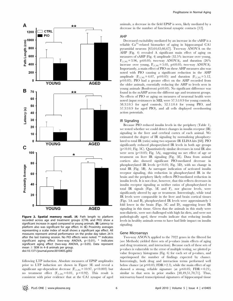
Behavioral characterizationTwo-way ANOVA on conventional outcome measures associ-
ated with the Morris water maze (MWM) including path length
and latency to platform, were tested for significance across the 4
days of training. No age or treatment differences were noted
during this learning phase apart from a significant decrease in
swim speed with age (F(1,21) = 36.2; p,0.0001). Following the last
day of training, a probe test (platform removed) assessed 24 h
retention of platform location. Aged animals showed significantly
longer path length to platform (F(1,21) = 5.06, p,0.05) and latency
to platform (F(1,21) = 13.9, p,0.005), likely explained by a decrease
in swim speed (F(1,21) = 5.73, p,0.05). A proximal analysis which is
not dependent on animals’ speed or their original distance to the
platform at the beginning of each trial [62] also showed that aged
animals were swimming, on average, at a cumulative distance
farther from the target than younger animals (F(1,21) = 6.8,
p,0.05). A proximity average scalar derived from the cumulative
distance data divided by the latency to platform (Fig. 2C) also
revealed significant age-dependent impairment on memory recall
(F(1,21) = 6.7, p,0.05). Thus, irrespective of the analysis used, no
main effect of treatment was found, suggesting that at the doses
tested, PIO could not reverse the age-dependent decrease in
memory recall 24 h after the last training day.
LTPAs described previously [13,41,42,43,44,45,46], an age-depen-
dent deficit in long-term potentiation (LTP) maintenance was seen
25–30 min following LTP induction (F(1,26) = 6.82, p,0.02).
Animals treated with PIO did not show signs of improvement
on measures of LTP induction or maintenance (F(1,26) = 0.21,
p = 0.67). Similar results were seen on measures of post-tetanic
potentiation (PTP) taken immediately following LTP induction,
showing a significant main effect of age (F(1,26) = 5.51, p,0.03), but
no main effect of treatment (F(1,26) = 0.80, p = 0.38). Figure 3
shows group means of normalized EPSP slopes across both age
and treatment during 20 min baseline and for 30 minutes
Table 1. Blood chemistry panel.
YOUNG AGED
CHOL (mg/dl) CTRL 119.4619.7 124.4610.1
PIO 85.468.0 * 108.165.9 *
TG (mg/dl) CTRL 291.2645.5 184.2644.1{
PIO 141.1621.4 ** 88.468.7 **,{
HDL (mg/dl) CTRL 70.367.8 83.265.8{
PIO 62.565.6 76.763.6{
INSULIN (ng/ml) CTRL 7.261.8 6.161.6
PIO 4.461.5 # 4.261.3 #
GLUCOSE (mg/dl) CTRL 112.164.9 102.466.6
PIO 109.568.2 101.468.1
ALT (IU/L) CTRL 48.964.9 51.062.7
PIO 37.563.2 47.764.6
Blood serum markers in control (CTRL) and PIO-treated (PIO) young and agedanimals. Abbreviations: Cholesterol: CHOL; Triglycerides: TG; Alanineaminotransferase: ALT; High Density Lipoprotein: HDL.*, **, and # indicate significant PIO effects (two-way ANOVA, p,0.05, p,0.001,and p,0.0005, respectively).{indicates significant aging effect (two-way ANOVA, p,0.05). Data representmean 6 SEM in 7–8 animals per group.
doi:10.1371/journal.pone.0010405.t001
Figure 1. Growth curves, heart weights and blood glucose levels.(A) Body weights measured across age during the 14-week long study(YC = young control, YP = young PIO, AC = aged control and AP = aged PIO;7–12 animals per group). (B) Heart weight normalized to body weight in 6–8 animals per group fed either control diet (CTRL) and PIO-enriched diet(PIO). (C) Blood glucose levels measured over the course of the study in 3–11 animals per group. Data represent mean 6 SEM.doi:10.1371/journal.pone.0010405.g001
Pioglitazone in Normal Aging
PLoS ONE | www.plosone.org 5 April 2010 | Volume 5 | Issue 4 | e10405
following LTP induction. Absolute measures of EPSP amplitudes
prior to LTP induction are shown in Figure 3E and reveal a
significant age-dependent decrease (F(1,26) = 10.97, p,0.003) but
no treatment effect (F(1,26) = 0.01, p = 0.92). This result is
consistent with prior evidence that at the CA1 synapse of aged
animals, a decrease in the field EPSP is seen, likely mediated by a
decrease in the number of functional synaptic contacts [12].
AHPDecreased excitability mediated by an increase in the sAHP is a
reliable Ca2+-related biomarker of aging in hippocampal CA1
pyramidal neurons [63,64,65,66,67]. Two-way ANOVA on the
AHP (Fig. 4) revealed A significant main effect of aging on
measures of sAHP (Fig. 4) amplitude (32.5% increase over young,
F(1,56) = 3.96, p#0.05; two-way ANOVA), and duration (26%
increase over young, F(1,56) = 5.05, p#0.05; two-way ANOVA).
Importantly, a main effect of PIO on these AHP measures also was
noted with PIO causing a significant reduction in the AHP
amplitude (F(1,56) = 4.07, p#0.05) and duration (F(1,56) = 5.12,
p#0.05). PIO had a greater effect on the AHP recorded from
the older animals, essentially reducing the AHP to levels seen in
young animals (Bonferroni p#0.05). No significant difference was
found in the mAHP across the different age and treatment groups.
No effects of PIO or aging on measures of neuronal health were
noted (input resistances in MV, were 57.560.9 for young controls,
58.360.5 for aged controls, 52.160.4 for young PIO, and
57.360.9 for aged PIO), and all cells displayed overshooting
action potentials.
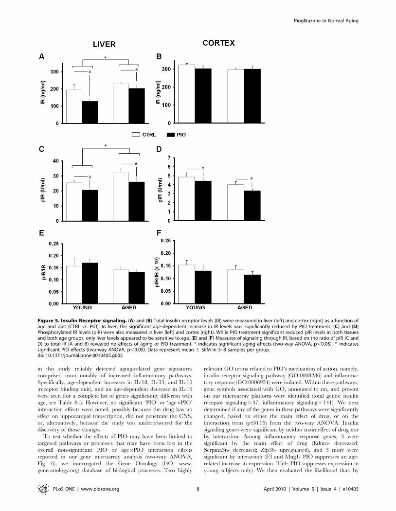
IR SignalingBecause PIO reduced insulin levels in the periphery (Table 1),
we tested whether we could detect changes in insulin receptor (IR)
signaling in the liver and cerebral cortex of each animal. We
estimated the degree of IR signaling by normalizing phosphory-
lated to total IR (ratio) using two separate IR ELISA kits [68]. PIO
significantly reduced phosporylated IR levels in both age groups
(p,0.01; Fig. 5C). Quantitatively similar decreases in total IR also
were seen (p,0.05; Fig. 5A), suggesting no net effect of age or
treatment on liver IR signaling (Fig. 5E). Data from animal
cortices also showed significant PIO-mediated decrease in
phosphorylated IR levels (p,0.05; Fig. 5D), with no change in
total IR (Fig. 5B). As surrogate indication of activated insulin
receptor signaling, this reduction in phosphorylated IR in the
brain and the periphery likely reflects PIO-mediated reduction in
insulin levels. It is not clear, however, that this reflects decreases in
insulin receptor signaling as neither ratios of phosphorylated to
total IR signals (Figs. 5E and F), nor glucose levels, were
significantly altered by age or treatment. Interestingly, while total
IR levels were comparable in the liver and brain cortical tissues
(Figs. 5A and B), phosphorylated IR levels were approximately 5
fold lower in the brain (Figs. 5C and D), suggesting lesser IR
signaling in this tissue. Given that the animals in this study were
non-diabetic, were not challenged with high fat diets, and were not
pathologically aged, these results indicate that reducing insulin
levels in healthy animals seems to have had little impact on insulin
signaling.
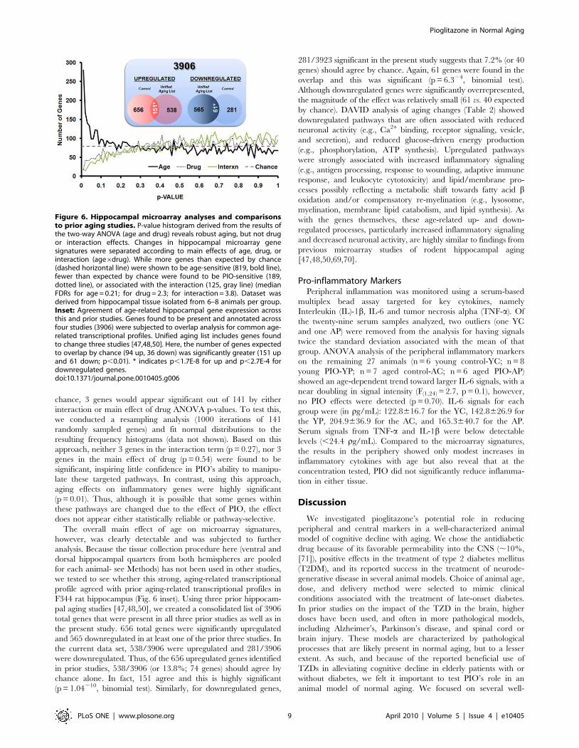
Gene MicroarraysTwo-way ANOVA applied to the 7922 genes in the filtered list
(see Methods) yielded three sets of p-values (main effects of aging
and drug treatment, and interaction). Because each of these sets of
p-values is vulnerable to the error of multiple testing, we plotted p-
value frequency histograms (Fig. 6) for each set of p-values, and
superimposed the number of findings expected by chance.
Interestingly, both drug and interaction terms performed well
below chance (at p#0.05; FDR.2.3), while the main effect of age
showed a strong, reliable signature (at p#0.05; FDR = 0.21),
similar to that seen in prior studies [48,49,51,70,71]. Thus,
microarray-based transcriptional signatures of hippocampal tissue
Figure 2. Spatial memory recall. (A) Path length to platformrecorded across age and treatment groups (CTRL and PIO) show asignificant increase in aged compared to young animals. (B) Latency toplatform also was significant for age effect. In (C) Proximity averagesrepresenting a scalar index of recall shows a significant age effect. Allmeasures represent animal performance on the probe day taken 24 hafter the last training session. No PIO effects were noted. ** Indicatessignificant aging effect (two-way ANOVA, p,0.01), * indicatessignificant aging effect (two-way ANOVA, p,0.05). Data representmean 6 SEM in 4–8 animals per group.doi:10.1371/journal.pone.0010405.g002
Pioglitazone in Normal Aging
PLoS ONE | www.plosone.org 6 April 2010 | Volume 5 | Issue 4 | e10405
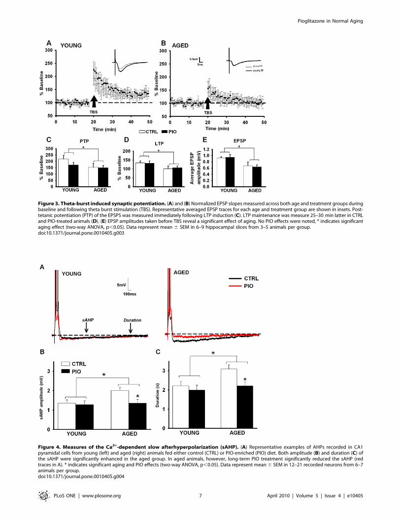
Figure 3. Theta-burst induced synaptic potentiation. (A) and (B) Normalized EPSP slopes measured across both age and treatment groups duringbaseline and following theta burst stimulation (TBS). Representative averaged EPSP traces for each age and treatment group are shown in insets. Post-tetanic potentiation (PTP) of the EPSPS was measured immediately following LTP induction (C). LTP maintenance was measure 25–30 min latter in CTRLand PIO-treated animals (D). (E) EPSP amplitudes taken before TBS reveal a significant effect of aging. No PIO effects were noted, * indicates significantaging effect (two-way ANOVA, p,0.05). Data represent mean 6 SEM in 6–9 hippocampal slices from 3–5 animals per group.doi:10.1371/journal.pone.0010405.g003
Figure 4. Measures of the Ca2+-dependent slow afterhyperpolarization (sAHP). (A) Representative examples of AHPs recorded in CA1pyramidal cells from young (left) and aged (right) animals fed either control (CTRL) or PIO-enriched (PIO) diet. Both amplitude (B) and duration (C) ofthe sAHP were significantly enhanced in the aged group. In aged animals, however, long-term PIO treatment significantly reduced the sAHP (redtraces in A). * indicates significant aging and PIO effects (two-way ANOVA, p,0.05). Data represent mean 6 SEM in 12–21 recorded neurons from 6–7animals per group.doi:10.1371/journal.pone.0010405.g004
Pioglitazone in Normal Aging
PLoS ONE | www.plosone.org 7 April 2010 | Volume 5 | Issue 4 | e10405
in this study reliably detected aging-related gene signatures
comprised most notably of increased inflammatory pathways.
Specifically, age-dependent increases in IL-18, IL-33, and IL-10
(receptor binding unit), and an age-dependent decrease in IL-16
were seen (for a complete list of genes significantly different with
age, see Table S1). However, no significant ‘PIO’ or ‘age6PIO’
interaction effects were noted, possibly because the drug has no
effect on hippocampal transcription, did not penetrate the CNS,
or, alternatively, because the study was underpowered for the
discovery of those changes.
To test whether the effects of PIO may have been limited to
targeted pathways or processes that may have been lost in the
overall non-significant PIO or age6PIO interaction effects
reported in our gene microarray analysis (two-way ANOVA,
Fig. 6), we interrogated the Gene Ontology (GO; www.
geneontology.org) database of biological processes. Two highly
relevant GO terms related to PIO’s mechanism of action, namely,
insulin receptor signaling pathway (GO:0008286) and inflamma-
tory response (GO:0006954) were isolated. Within these pathways,
gene symbols associated with GO, annotated to rat, and present
on our microarray platform were identified (total genes: insulin
receptor signaling = 37; inflammatory signaling = 141). We next
determined if any of the genes in these pathways were significantly
changed, based on either the main effect of drug, or on the
interaction term (p#0.05) from the two-way ANOVA. Insulin
signaling genes were significant by neither main effect of drug nor
by interaction. Among inflammatory response genes, 3 were
significant by the main effect of drug (Ednra- decreased;
Serpina3n- decreased; Zfp36- upregulated), and 3 more were
significant by interaction (F3 and Mug1- PIO suppresses an age-
related increase in expression, Tlr4- PIO suppresses expression in
young subjects only). We then evaluated the likelihood that, by
Figure 5. Insulin Receptor signaling. (A) and (B) Total insulin receptor levels (IR) were measured in liver (left) and cortex (right) as a function ofage and diet (CTRL vs. PIO). In liver, the significant age-dependent increase in IR levels was significantly reduced by PIO treatment. (C) and (D)Phosphorylated IR levels (pIR) were also measured in liver (left) and cortex (right). While PIO treatment significant reduced pIR levels in both tissuesand both age groups, only liver levels appeared to be sensitive to age. (E) and (F) Measures of signaling through IR, based on the ratio of pIR (C andD) to total IR (A and B) revealed no effects of aging or PIO treatment. * indicates significant aging effects (two-way ANOVA, p,0.05). # indicatessignificant PIO effects (two-way ANOVA, p,0.05). Data represent mean 6 SEM in 5–8 samples per group.doi:10.1371/journal.pone.0010405.g005
Pioglitazone in Normal Aging
PLoS ONE | www.plosone.org 8 April 2010 | Volume 5 | Issue 4 | e10405
chance, 3 genes would appear significant out of 141 by either
interaction or main effect of drug ANOVA p-values. To test this,
we conducted a resampling analysis (1000 iterations of 141
randomly sampled genes) and fit normal distributions to the
resulting frequency histograms (data not shown). Based on this
approach, neither 3 genes in the interaction term (p = 0.27), nor 3
genes in the main effect of drug (p = 0.54) were found to be
significant, inspiring little confidence in PIO’s ability to manipu-
late these targeted pathways. In contrast, using this approach,
aging effects on inflammatory genes were highly significant
(p = 0.01). Thus, although it is possible that some genes within
these pathways are changed due to the effect of PIO, the effect
does not appear either statistically reliable or pathway-selective.
The overall main effect of age on microarray signatures,
however, was clearly detectable and was subjected to further
analysis. Because the tissue collection procedure here (ventral and
dorsal hippocampal quarters from both hemispheres are pooled
for each animal- see Methods) has not been used in other studies,
we tested to see whether this strong, aging-related transcriptional
profile agreed with prior aging-related transcriptional profiles in
F344 rat hippocampus (Fig. 6 inset). Using three prior hippocam-
pal aging studies [47,48,50], we created a consolidated list of 3906
total genes that were present in all three prior studies as well as in
the present study. 656 total genes were significantly upregulated
and 565 downregulated in at least one of the prior three studies. In
the current data set, 538/3906 were upregulated and 281/3906
were downregulated. Thus, of the 656 upregulated genes identified
in prior studies, 538/3906 (or 13.8%; 74 genes) should agree by
chance alone. In fact, 151 agree and this is highly significant
(p = 1.04210, binomial test). Similarly, for downregulated genes,
281/3923 significant in the present study suggests that 7.2% (or 40
genes) should agree by chance. Again, 61 genes were found in the
overlap and this was significant (p = 6.324, binomial test).
Although downregulated genes were significantly overrepresented,
the magnitude of the effect was relatively small (61 vs. 40 expected
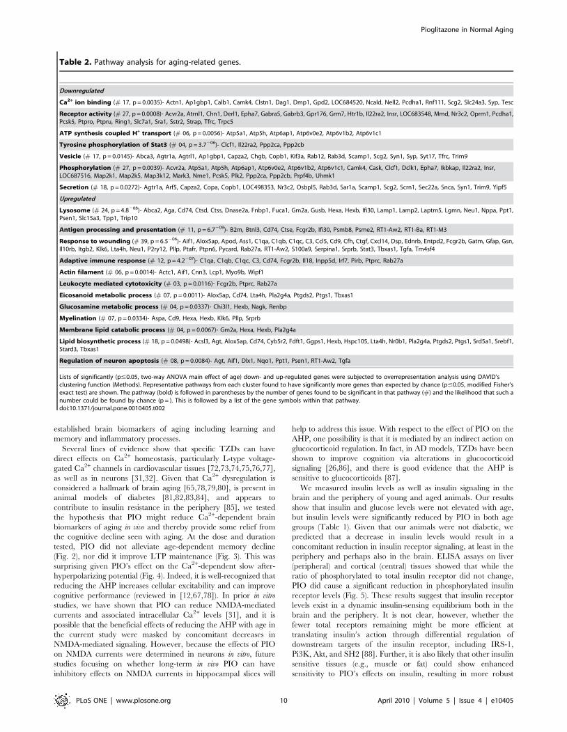
by chance). DAVID analysis of aging changes (Table 2) showed
downregulated pathways that are often associated with reduced
neuronal activity (e.g., Ca2+ binding, receptor signaling, vesicle,
and secretion), and reduced glucose-driven energy production
(e.g., phosphorylation, ATP synthesis). Upregulated pathways
were strongly associated with increased inflammatory signaling
(e.g., antigen processing, response to wounding, adaptive immune
response, and leukocyte cytotoxicity) and lipid/membrane pro-
cesses possibly reflecting a metabolic shift towards fatty acid boxidation and/or compensatory re-myelination (e.g., lysosome,
myelination, membrane lipid catabolism, and lipid synthesis). As
with the genes themselves, these age-related up- and down-
regulated processes, particularly increased inflammatory signaling
and decreased neuronal activity, are highly similar to findings from
previous microarray studies of rodent hippocampal aging
[47,48,50,69,70].
Pro-inflammatory MarkersPeripheral inflammation was monitored using a serum-based
multiplex bead assay targeted for key cytokines, namely
Interleukin (IL)-1b, IL-6 and tumor necrosis alpha (TNF-a). Of
the twenty-nine serum samples analyzed, two outliers (one YC
and one AP) were removed from the analysis for having signals
twice the standard deviation associated with the mean of that
group. ANOVA analysis of the peripheral inflammatory markers
on the remaining 27 animals (n = 6 young control-YC; n = 8
young PIO-YP; n = 7 aged control-AC; n = 6 aged PIO-AP)
showed an age-dependent trend toward larger IL-6 signals, with a
near doubling in signal intensity (F(1,24) = 2.7, p = 0.1), however,
no PIO effects were detected (p = 0.70). IL-6 signals for each
group were (in rg/mL): 122.8616.7 for the YC, 142.8626.9 for
the YP, 204.9636.9 for the AC, and 165.3640.7 for the AP.
Serum signals from TNF-a and IL-1b were below detectable
levels (,24.4 rg/mL). Compared to the microarray signatures,
the results in the periphery showed only modest increases in
inflammatory cytokines with age but also reveal that at the
concentration tested, PIO did not significantly reduce inflamma-
tion in either tissue.
Discussion
We investigated pioglitazone’s potential role in reducing
peripheral and central markers in a well-characterized animal
model of cognitive decline with aging. We chose the antidiabetic
drug because of its favorable permeability into the CNS (,10%,
[71]), positive effects in the treatment of type 2 diabetes mellitus
(T2DM), and its reported success in the treatment of neurode-
generative disease in several animal models. Choice of animal age,
dose, and delivery method were selected to mimic clinical
conditions associated with the treatment of late-onset diabetes.
In prior studies on the impact of the TZD in the brain, higher
doses have been used, and often in more pathological models,
including Alzheimer’s, Parkinson’s disease, and spinal cord or
brain injury. These models are characterized by pathological
processes that are likely present in normal aging, but to a lesser
extent. As such, and because of the reported beneficial use of
TZDs in alleviating cognitive decline in elderly patients with or
without diabetes, we felt it important to test PIO’s role in an
animal model of normal aging. We focused on several well-
Figure 6. Hippocampal microarray analyses and comparisonsto prior aging studies. P-value histogram derived from the results ofthe two-way ANOVA (age and drug) reveals robust aging, but not drugor interaction effects. Changes in hippocampal microarray genesignatures were separated according to main effects of age, drug, orinteraction (age6drug). While more genes than expected by chance(dashed horizontal line) were shown to be age-sensitive (819, bold line),fewer than expected by chance were found to be PIO-sensitive (189,dotted line), or associated with the interaction (125, gray line) (medianFDRs for age = 0.21; for drug = 2.3; for interaction = 3.8). Dataset wasderived from hippocampal tissue isolated from 6–8 animals per group.Inset: Agreement of age-related hippocampal gene expression acrossthis and prior studies. Genes found to be present and annotated acrossfour studies (3906) were subjected to overlap analysis for common age-related transcriptional profiles. Unified aging list includes genes foundto change three studies [47,48,50]. Here, the number of genes expectedto overlap by chance (94 up, 36 down) was significantly greater (151 upand 61 down; p,0.01). * indicates p,1.7E-8 for up and p,2.7E-4 fordownregulated genes.doi:10.1371/journal.pone.0010405.g006
Pioglitazone in Normal Aging
PLoS ONE | www.plosone.org 9 April 2010 | Volume 5 | Issue 4 | e10405
established brain biomarkers of aging including learning and
memory and inflammatory processes.
Several lines of evidence show that specific TZDs can have
direct effects on Ca2+ homeostasis, particularly L-type voltage-
gated Ca2+ channels in cardiovascular tissues [72,73,74,75,76,77],
as well as in neurons [31,32]. Given that Ca2+ dysregulation is
considered a hallmark of brain aging [65,78,79,80], is present in
animal models of diabetes [81,82,83,84], and appears to
contribute to insulin resistance in the periphery [85], we tested
the hypothesis that PIO might reduce Ca2+-dependent brain
biomarkers of aging in vivo and thereby provide some relief from
the cognitive decline seen with aging. At the dose and duration
tested, PIO did not alleviate age-dependent memory decline
(Fig. 2), nor did it improve LTP maintenance (Fig. 3). This was
surprising given PIO’s effect on the Ca2+-dependent slow after-
hyperpolarizing potential (Fig. 4). Indeed, it is well-recognized that
reducing the AHP increases cellular excitability and can improve
cognitive performance (reviewed in [12,67,78]). In prior in vitro
studies, we have shown that PIO can reduce NMDA-mediated
currents and associated intracellular Ca2+ levels [31], and it is
possible that the beneficial effects of reducing the AHP with age in
the current study were masked by concomitant decreases in
NMDA-mediated signaling. However, because the effects of PIO
on NMDA currents were determined in neurons in vitro, future
studies focusing on whether long-term in vivo PIO can have
inhibitory effects on NMDA currents in hippocampal slices will
help to address this issue. With respect to the effect of PIO on the
AHP, one possibility is that it is mediated by an indirect action on
glucocorticoid regulation. In fact, in AD models, TZDs have been
shown to improve cognition via alterations in glucocorticoid
signaling [26,86], and there is good evidence that the AHP is
sensitive to glucocorticoids [87].
We measured insulin levels as well as insulin signaling in the
brain and the periphery of young and aged animals. Our results
show that insulin and glucose levels were not elevated with age,
but insulin levels were significantly reduced by PIO in both age
groups (Table 1). Given that our animals were not diabetic, we
predicted that a decrease in insulin levels would result in a
concomitant reduction in insulin receptor signaling, at least in the
periphery and perhaps also in the brain. ELISA assays on liver
(peripheral) and cortical (central) tissues showed that while the
ratio of phosphorylated to total insulin receptor did not change,
PIO did cause a significant reduction in phosphorylated insulin
receptor levels (Fig. 5). These results suggest that insulin receptor
levels exist in a dynamic insulin-sensing equilibrium both in the
brain and the periphery. It is not clear, however, whether the
fewer total receptors remaining might be more efficient at
translating insulin’s action through differential regulation of
downstream targets of the insulin receptor, including IRS-1,
Pi3K, Akt, and SH2 [88]. Further, it is also likely that other insulin
sensitive tissues (e.g., muscle or fat) could show enhanced
sensitivity to PIO’s effects on insulin, resulting in more robust
Table 2. Pathway analysis for aging-related genes.
Downregulated
Ca2+ ion binding (# 17, p = 0.0035)- Actn1, Ap1gbp1, Calb1, Camk4, Clstn1, Dag1, Dmp1, Gpd2, LOC684520, Ncald, Nell2, Pcdha1, Rnf111, Scg2, Slc24a3, Syp, Tesc
Receptor activity (# 27, p = 0.0008)- Acvr2a, Atrnl1, Chn1, Derl1, Epha7, Gabra5, Gabrb3, Gpr176, Grm7, Htr1b, Il22ra2, Insr, LOC683548, Mmd, Nr3c2, Oprm1, Pcdha1,Pcsk5, Ptpro, Ptpru, Ring1, Slc7a1, Sra1, Sstr2, Strap, Tfrc, Trpc5
ATP synthesis coupled H+ transport (# 06, p = 0.0056)- Atp5a1, Atp5h, Atp6ap1, Atp6v0e2, Atp6v1b2, Atp6v1c1
Tyrosine phosphorylation of Stat3 (# 04, p = 3.7206)- Clcf1, Il22ra2, Ppp2ca, Ppp2cb
Vesicle (# 17, p = 0.0145)- Abca3, Agtr1a, Agtrl1, Ap1gbp1, Capza2, Chgb, Copb1, Kif3a, Rab12, Rab3d, Scamp1, Scg2, Syn1, Syp, Syt17, Tfrc, Trim9
Phosphorylation (# 27, p = 0.0039)- Acvr2a, Atp5a1, Atp5h, Atp6ap1, Atp6v0e2, Atp6v1b2, Atp6v1c1, Camk4, Cask, Clcf1, Dclk1, Epha7, Ikbkap, Il22ra2, Insr,LOC687516, Map2k1, Map2k5, Map3k12, Mark3, Nme1, Pcsk5, Plk2, Ppp2ca, Ppp2cb, Prpf4b, Uhmk1
Secretion (# 18, p = 0.0272)- Agtr1a, Arf5, Capza2, Copa, Copb1, LOC498353, Nr3c2, Osbpl5, Rab3d, Sar1a, Scamp1, Scg2, Scrn1, Sec22a, Snca, Syn1, Trim9, Yipf5
Upregulated
Lysosome (# 24, p = 4.8208)- Abca2, Aga, Cd74, Ctsd, Ctss, Dnase2a, Fnbp1, Fuca1, Gm2a, Gusb, Hexa, Hexb, Ifi30, Lamp1, Lamp2, Laptm5, Lgmn, Neu1, Nppa, Ppt1,Psen1, Slc15a3, Tpp1, Trip10
Antigen processing and presentation (# 11, p = 6.7209)- B2m, Btnl3, Cd74, Ctse, Fcgr2b, Ifi30, Psmb8, Psme2, RT1-Aw2, RT1-Ba, RT1-M3
Response to wounding (# 39, p = 6.5206)- Aif1, Alox5ap, Apod, Ass1, C1qa, C1qb, C1qc, C3, Ccl5, Cd9, Cfh, Ctgf, Cxcl14, Dsp, Ednrb, Entpd2, Fcgr2b, Gatm, Gfap, Gsn,Il10rb, Itgb2, Klk6, Lta4h, Neu1, P2ry12, Pllp, Ptafr, Ptpn6, Pycard, Rab27a, RT1-Aw2, S100a9, Serpina1, Srprb, Stat3, Tbxas1, Tgfa, Tm4sf4
Adaptive immune response (# 12, p = 4.2207)- C1qa, C1qb, C1qc, C3, Cd74, Fcgr2b, Il18, Inpp5d, Irf7, Pirb, Ptprc, Rab27a
Actin filament (# 06, p = 0.0014)- Actc1, Aif1, Cnn3, Lcp1, Myo9b, Wipf1
Leukocyte mediated cytotoxicity (# 03, p = 0.0116)- Fcgr2b, Ptprc, Rab27a
Eicosanoid metabolic process (# 07, p = 0.0011)- Alox5ap, Cd74, Lta4h, Pla2g4a, Ptgds2, Ptgs1, Tbxas1
Glucosamine metabolic process (# 04, p = 0.0337)- Chi3l1, Hexb, Nagk, Renbp
Myelination (# 07, p = 0.0334)- Aspa, Cd9, Hexa, Hexb, Klk6, Pllp, Srprb
Membrane lipid catabolic process (# 04, p = 0.0067)- Gm2a, Hexa, Hexb, Pla2g4a
Lipid biosynthetic process (# 18, p = 0.0498)- Acsl3, Agt, Alox5ap, Cd74, Cyb5r2, Fdft1, Ggps1, Hexb, Hspc105, Lta4h, Nr0b1, Pla2g4a, Ptgds2, Ptgs1, Srd5a1, Srebf1,Stard3, Tbxas1
Regulation of neuron apoptosis (# 08, p = 0.0084)- Agt, Aif1, Dlx1, Nqo1, Ppt1, Psen1, RT1-Aw2, Tgfa
Lists of significantly (p#0.05, two-way ANOVA main effect of age) down- and up-regulated genes were subjected to overrepresentation analysis using DAVID’sclustering function (Methods). Representative pathways from each cluster found to have significantly more genes than expected by chance (p#0.05, modified Fisher’sexact test) are shown. The pathway (bold) is followed in parentheses by the number of genes found to be significant in that pathway (#) and the likelihood that such anumber could be found by chance (p = ). This is followed by a list of the gene symbols within that pathway.doi:10.1371/journal.pone.0010405.t002
Pioglitazone in Normal Aging
PLoS ONE | www.plosone.org 10 April 2010 | Volume 5 | Issue 4 | e10405
changes in insulin receptor signaling. Because insulin levels were
decreased in both young and aged animals treated with PIO, and
AHP reductions were only seen in the aged group, we do not
believe insulin levels directly influenced the AHP under the
condition tested. There is, however, evidence supporting a
decrease in insulin sensitivity in the brain of AD patients and in
AD models [89,90,91], and we are currently testing this hypothesis
in our aging model. However, it is unclear whether the aging F344
rat is a good model of T2DM, and responds to high fat diets with
signs of insulin resistance in both the periphery and the brain
[92,93]. Here, therefore, we believe that PIO’s effects on insulin
signaling were somewhat blunted because animals were healthy
and non-diabetic. On the other hand, one would predict that
under conditions more representative of human aging, where
accumulated exposure to high fat diets and a sedentary life style
contribute to T2DM, PIO might significantly reduce insulin levels
[56,57,58,59], increase insulin sensitivity, and likely increase
insulin signaling.
Prior studies in CNS and peripheral cell types modeling trauma
and insult (e.g., LPS, PMA) demonstrate that PPAR-c activators
play a critical role in reducing inflammatory cytokines (interleukin-
6, interleukin 1-b and TNF-a), including activation of inducible
nitric oxide synthase (iNOS) [94,95,96,97,98,99]. Similarly, in
animal models of ischemia, stroke, hypertension, stress, and
Parkinson’s disease, which are also characterized as pro-inflam-
matory conditions, PPAR-c agonists provide significant neuropro-
tection [100,101,102,103,104,105,106,107,108,109,110,111,112].
In AD animal models also, PPAR-c agonists appear to reduce
baseline inflammatory levels [24,26,113,114]. Only one prior
animal study examined the effects of TZDs under non-patholog-
ical aging conditions. The authors reported that the increase in
proinflammatory cytokine levels (IL-1b) in the hippocampus of
aged F344 rats was not sensitive to the actions of the TZD
rosiglitazone (10 mg/Kg/day) yet the drug caused significant
improvement in contextual fear conditioning [115]. Similarly, a
prior publication using 20 mg/Kg/day PIO for four months in
transgenic mouse models of AD (Tg2576) revealed very little anti-
inflammatory effects of PIO (based on microglial activation,
soluble Ab levels, and plaque burden) when compared to
ibuprofen treatment [114]. The same group, however, convinc-
ingly showed that a 40 mg/Kg/day PIO dose could significantly
reduced brain inflammation in the APPV717I transgenic mouse
[24]. Here, therefore, we examined inflammatory cytokines in the
serum, and inflammatory signaling in the brain using hippocampal
microarray analyses in the context of normal aging, and at low
doses of PIO. While a clear inflammatory signature was present in
the brain as previously reported in microarray studies of aging
[11,47,48,50,116,117,118,119,120], PIO did not significantly
reduce inflammatory markers in the hippocampus. In the
periphery, no robust age-dependent change in inflammatory
cytokine levels was seen (although a trend in increased IL-6 levels
was noted in aged animals), precluding an effect of PIO.
Importantly, reductions in peripheral insulin and lipids indicate
the target therapeutic window for PIO was reached. Under these
conditions, thus, it is unclear that PIO was able to reduce central
and peripheral inflammatory markers in an animal model of
aging, and together, these results suggest that higher PIO doses
might be necessary to reduce inflammatory pathways and exert
beneficial cognitive effects.
Using an unbiased microarray analysis approach on hippo-
campal tissue, our study compared the effects of a brain permeant
TZD treatment in younger and older animals, and showed that
while age-dependent gene signatures were clearly present at both
the gene (Fig. 6) and pathway (Table 2) levels, PIO effects on
gene expression were virtually absent. Possible reasons for this
include: low statistical detection power, insufficient drug expo-
sure, or lack of influence of this treatment regimen on
hippocampal transcription. Low statistical power is a possibility;
however, the treatment main-effect histogram dips well below
chance at small p values (Fig. 6), and it seems unlikely that
increasing the number of subjects would allow us to detect
significant transcriptional effects of PIO. Regarding drug
exposure, PIO reduced peripheral blood chemistry measures
significantly and in the direction predicted by prior work (see
discussion above and Table 1), suggesting that treatment levels
were appropriate. Thus, it seems reasonable to conclude that PIO
did not exert a detectable transcriptional effect on hippocampal
gene transcription, and that this lack of central influence may be
due to either reduced blood-brain barrier penetration or a frank
lack of response from hippocampal tissue. Nevertheless, because
PIO has established effects involving insulin and inflammatory
processes, we also directly investigated genes associated with these
processes and tested their sensitivity to PIO in the brain.
Although it is not possible to evaluate the biological importance
of the 6 genes that were identified without functional genetic
manipulation (e.g., knock-in), our resampling analysis revealed
that these targeted pathways were not statistically significant. It is
interesting to note, however, that of the six genes identified, one
of them (Tlr4) recently was found to be sensitive to PIO in
monocytes and macrophages [121], reinforcing the role of PIO as
an anti-inflammatory agent, and suggesting Tlr4 might be a
common target of PIO in peripheral and central tissues.
Compared to prior aging studies, our overlap analysis (Fig. 6
inset) suggests that, irrespective of where along the dorsal-ventral
axis it is sampled, the hippocampus shows increased inflammatory
markers with age, and validates the use of hippocampal extremities
in future microarray studies. Interestingly, upregulated inflamma-
tory categories, historically the most consistent and largest
magnitude of the aging brain transcriptional signatures, remain
largely unperturbed by PIO administration.
To our knowledge, this is the first study testing long-term PIO
treatment in the F344 rat with age, specifically investigating
whether a commonly prescribed drug may have off-target
cognition enhancing effects in a rat model of aging. The study
was designed using a clinically-relevant dose and delivery method.
As expected, several signatures of aging were present in older
animals, characterized by weak peripheral and robust central
inflammatory increases, reduced spatial memory and LTP
maintenance, and increased Ca2+-dependent AHPs. Peripheral
insulin levels, phosphorylated insulin receptors in the CNS and the
periphery, and the AHP were significantly reduced by PIO. While
the mechanism through which PIO may mediate its central effects
(AHP reduction, reduced phosphorylated insulin receptor) is not
clear, it does not appear to occur via a transcriptional process.
Given the increased incidence in metabolic syndrome and T2DM
seen in the aging population, together with the high numbers of
prescriptions written for TZDs, our study has direct therapeutic
relevance and suggests future experiments testing the use of these
agents at clinically-relevant doses for the treatment of neurological
or cognitive conditions are needed. Nevertheless, the results of our
study do not preclude the beneficial effects of TZDs in the elderly
where metabolic dysregulation and diabetes are often reported.
Supporting Information
Table S1
Found at: doi:10.1371/journal.pone.0010405.s001 (5.20 MB
XLS)
Pioglitazone in Normal Aging
PLoS ONE | www.plosone.org 11 April 2010 | Volume 5 | Issue 4 | e10405

Acknowledgments
We thank Dr. Robert H. Hadley for his critical reading of the manuscript
and his valuable input.
Author Contributions
Conceived and designed the experiments: EMB NMP OT. Performed the
experiments: JTP TP JLS MGA KCC OT. Analyzed the data: EMB JTP
TP JLS KLA JP KCC OT. Contributed reagents/materials/analysis tools:
JCG DAC NMP OT. Wrote the paper: EMB TP NMP OT.
References
1. Gradman TJ, Laws A, Thompson LW, Reaven GM (1993) Verbal learning
and/or memory improves with glycemic control in older subjects with non-
insulin-dependent diabetes mellitus. J Am Geriatr Soc 41: 1305–1312.
2. Awad N, Gagnon M, Messier C (2004) The relationship between impaired
glucose tolerance, type 2 diabetes, and cognitive function. J Clin Exp
Neuropsychol 26: 1044–1080.
3. Gregg EW, Yaffe K, Cauley JA, Rolka DB, Blackwell TL, et al. (2000) Is
diabetes associated with cognitive impairment and cognitive decline among
older women? Study of Osteoporotic Fractures Research Group. Arch Intern
Med 160: 174–180.
4. Messier C (2005) Impact of impaired glucose tolerance and type 2 diabetes on
cognitive aging. Neurobiol Aging 26 Suppl 1: 26–30.
5. Reagan LP (2002) Glucose, stress, and hippocampal neuronal vulnerability. Int
Rev Neurobiol 51: 289–324.
6. Ryan CM, Geckle M (2000) Why is learning and memory dysfunction in Type
2 diabetes limited to older adults? Diabetes Metab Res Rev 16: 308–315.
7. Zhao WQ, Alkon DL (2001) Role of insulin and insulin receptor in learning
and memory. Mol Cell Endocrinol 177: 125–134.
8. Barrientos RM, Frank MG, Hein AM, Higgins EA, Watkins LR, et al. (2009)
Time course of hippocampal IL-1 beta and memory consolidation impairments
in aging rats following peripheral infection. Brain Behav Immun 23: 46–54.
9. Fishel MA, Watson GS, Montine TJ, Wang Q, Green PS, et al. (2005)
Hyperinsulinemia provokes synchronous increases in central inflammation and
beta-amyloid in normal adults. Arch Neurol 62: 1539–1544.
10. Godbout JP, Chen J, Abraham J, Richwine AF, Berg BM, et al. (2005)
Exaggerated neuroinflammation and sickness behavior in aged mice following
activation of the peripheral innate immune system. FASEB J 19: 1329–1331.
11. Prolla TA (2002) DNA microarray analysis of the aging brain. Chem Senses 27:
299–306.
12. Rosenzweig ES, Barnes CA (2003) Impact of aging on hippocampal function:
plasticity, network dynamics, and cognition. Prog Neurobiol 69: 143–179.
13. Barnes CA, McNaughton BL (1985) An age comparison of the rates of
acquisition and forgetting of spatial information in relation to long-term
enhancement of hippocampal synapses. Behav Neurosci 99: 1040–1048.
14. Berger J, Moller DE (2002) The mechanisms of action of PPARs. Annu Rev
Med 53: 409–435.
15. Hauner H (2002) The mode of action of thiazolidinediones. Diabetes Metab
Res Rev 18 Suppl 2: S10–15.
16. Martin G, Schoonjans K, Lefebvre AM, Staels B, Auwerx J (1997) Coordinate
regulation of the expression of the fatty acid transport protein and acyl-CoA
synthetase genes by PPARalpha and PPARgamma activators. J Biol Chem 272:
28210–28217.
17. Schoonjans K, Peinado-Onsurbe J, Lefebvre AM, Heyman RA, Briggs M,
et al. (1996) PPARalpha and PPARgamma activators direct a distinct tissue-
specific transcriptional response via a PPRE in the lipoprotein lipase gene.
Embo J 15: 5336–5348.
18. Berger JP, Akiyama TE, Meinke PT (2005) PPARs: therapeutic targets for
metabolic disease. Trends Pharmacol Sci 26: 244–251.
19. Hofmann C, Lorenz K, Braithwaite SS, Colca JR, Palazuk BJ, et al. (1994)
Altered gene expression for tumor necrosis factor-alpha and its receptors during
drug and dietary modulation of insulin resistance. Endocrinology 134:
264–270.
20. Hong C, Tontonoz P (2008) Coordination of inflammation and metabolism by
PPAR and LXR nuclear receptors. Curr Opin Genet Dev 18: 461–467.
21. Watson GS, Cholerton BA, Reger MA, Baker LD, Plymate SR, et al. (2005)
Preserved cognition in patients with early Alzheimer disease and amnestic mild
cognitive impairment during treatment with rosiglitazone: a preliminary study.
Am J Geriatr Psychiatry 13: 950–958.
22. Hanyu H, Sato T, Kiuchi A, Sakurai H, Iwamoto T (2009) Pioglitazone
improved cognition in a pilot study on patients with Alzheimer’s disease and
mild cognitive impairment with diabetes mellitus. J Am Geriatr Soc 57:
177–179.
23. Risner ME, Saunders AM, Altman JF, Ormandy GC, Craft S, et al. (2006)
Efficacy of rosiglitazone in a genetically defined population with mild-to-
moderate Alzheimer’s disease. Pharmacogenomics J 6: 246–254.
24. Heneka MT, Sastre M, Dumitrescu-Ozimek L, Hanke A, Dewachter I, et al.
(2005) Acute treatment with the PPAR{gamma} agonist pioglitazone and
ibuprofen reduces glial inflammation and A{beta}1–42 levels in APPV717I
transgenic mice. Brain.
25. Jiang Q, Heneka M, Landreth GE (2008) The role of peroxisome proliferator-
activated receptor-gamma (PPARgamma) in Alzheimer’s disease: therapeutic
implications. CNS Drugs 22: 1–14.
26. Pedersen WA, McMillan PJ, Kulstad JJ, Leverenz JB, Craft S, et al. (2006)
Rosiglitazone attenuates learning and memory deficits in Tg2576 Alzheimer
mice. Exp Neurol 199: 265–273.
27. Lacombe P, Mathews PM, Schmidt SD, Breidert T, Heneka MT, et al. (2004)
Effect of anti-inflammatory agents on transforming growth factor beta over-
expressing mouse brains: a model revised. J Neuroinflammation 1: 11.
28. Nicolakakis N, Aboulkassim T, Ongali B, Lecrux C, Fernandes P, et al. (2008)
Complete rescue of cerebrovascular function in aged Alzheimer’s disease
transgenic mice by antioxidants and pioglitazone, a peroxisome proliferator-
activated receptor gamma agonist. J Neurosci 28: 9287–9296.
29. Landreth GE, Heneka MT (2001) Anti-inflammatory actions of peroxisome
proliferator-activated receptor gamma agonists in Alzheimer’s disease.
Neurobiol Aging 22: 937–944.
30. Feinstein DL (2003) Therapeutic potential of peroxisome proliferator-activated
receptor agonists for neurological disease. Diabetes Technol Ther 5: 67–73.
31. Pancani T, Phelps JT, Searcy JL, Kilgore MW, Chen KC, et al. (2009) Distinct
modulation of voltage-gated and ligand-gated Ca2+ currents by PPAR-gamma
agonists in cultured hippocampal neurons. J Neurochem 109: 1800–1811.
32. Inestrosa NC, Godoy JA, Quintanilla RA, Koenig CS, Bronfman M (2005)
Peroxisome proliferator-activated receptor gamma is expressed in hippocampal
neurons and its activation prevents beta-amyloid neurodegeneration: role of
Wnt signaling. Exp Cell Res 304: 91–104.
33. Uryu S, Harada J, Hisamoto M, Oda T (2002) Troglitazone inhibits both post-
glutamate neurotoxicity and low-potassium-induced apoptosis in cerebellar
granule neurons. Brain Res 924: 229–236.
34. Zhao X, Ou Z, Grotta JC, Waxham N, Aronowski J (2006) Peroxisome-
proliferator-activated receptor-gamma (PPARgamma) activation protects
neurons from NMDA excitotoxicity. Brain Res 1073–1074: 460–469.
35. Goodman DG, Ward JM, Squire RA, Chu KC, Linhart MS (1979) Neoplastic
and nonneoplastic lesions in aging F344 rats. Toxicol Appl Pharmacol 48:
237–248.
36. Gillies PS, Dunn CJ (2000) Pioglitazone. Drugs 60: 333–343; discussion 344–
335.
37. Brunton LL, Sazo JS, Parker KL (2005) Goodman and Gilman’s The
pharmacological basis of therapeutics. Eleventh edition. 1860 p.
38. Moyer JR, Jr., Thompson LT, Disterhoft JF (1996) Trace eyeblink conditioning
increases CA1 excitability in a transient and learning-specific manner.
J Neurosci 16: 5536–5546.
39. Gant JC, Sama MM, Landfield PW, Thibault O (2006) Early and simultaneous
emergence of multiple hippocampal biomarkers of aging is mediated by Ca2+-
induced Ca2+ release. J Neurosci 26: 3482–3490.
40. Larson J, Wong D, Lynch G (1986) Patterned stimulation at the theta
frequency is optimal for the induction of hippocampal long-term potentiation.
Brain Res 368: 347–350.
41. deToledo-Morrell L, Geinisman Y, Morrell F (1988) Age-dependent alterations
in hippocampal synaptic plasticity: relation to memory disorders. Neurobiol
Aging 9: 581–590.
42. Rex CS, Kramar EA, Colgin LL, Lin B, Gall CM, et al. (2005) Long-term
potentiation is impaired in middle-aged rats: regional specificity and reversal by
adenosine receptor antagonists. J Neurosci 25: 5956–5966.
43. Tombaugh GC, Rowe WB, Chow AR, Michael TH, Rose GM (2002) Theta-
frequency synaptic potentiation in CA1 in vitro distinguishes cognitively
impaired from unimpaired aged Fischer 344 rats. J Neurosci 22: 9932–9940.
44. Shankar S, Teyler TJ, Robbins N (1998) Aging differentially alters forms of
long-term potentiation in rat hippocampal area CA1. J Neurophysiol 79:
334–341.
45. Barnes CA, Rao G, McNaughton BL (1996) Functional integrity of NMDA-
dependent LTP induction mechanisms across the lifespan of F-344 rats. Learn
Mem 3: 124–137.
46. Deupree DL, Bradley J, Turner DA (1993) Age-related alterations in
potentiation in the CA1 region in F344 rats. Neurobiol Aging 14: 249–258.
47. Rowe WB, Blalock EM, Chen KC, Kadish I, Wang D, et al. (2007)
Hippocampal expression analyses reveal selective association of immediate-
early, neuroenergetic, and myelinogenic pathways with cognitive impairment
in aged rats. J Neurosci 27: 3098–3110.
48. Kadish I, Thibault O, Blalock EM, Chen KC, Gant JC, et al. (2009)
Hippocampal and cognitive aging across the lifespan: a bioenergetic shift
precedes and increased cholesterol trafficking parallels memory impairment.
J Neurosci 29: 1805–1816.
49. Blalock EM, Geddes JW, Chen KC, Porter NM, Markesbery WR, et al. (2004)
Incipient Alzheimer’s disease: microarray correlation analyses reveal major
transcriptional and tumor suppressor responses. Proc Natl Acad Sci U S A 101:
2173–2178.
Pioglitazone in Normal Aging
PLoS ONE | www.plosone.org 12 April 2010 | Volume 5 | Issue 4 | e10405
50. Blalock EM, Chen KC, Sharrow K, Herman JP, Porter NM, et al. (2003) Gene
microarrays in hippocampal aging: statistical profiling identifies novel processes
correlated with cognitive impairment. J Neurosci 23: 3807–3819.
51. Bolstad BM, Collin F, Brettschneider J, Simpson K, Cope L, et al. (2005)
Quality Assessment of Affymetrix GeneChip Data. In: Gentleman R, Carey V,
Huber W, Irizarry R, Dudoit S, eds. Bioinformatics and Computational
Biology Solutions Using R and Bioconductor Springer.
52. Gentleman RC, Carey VJ, Bates DM, Bolstad B, Dettling M, et al. (2004)
Bioconductor: open software development for computational biology and
bioinformatics. Genome Biol 5: R80.
53. Saeed AI, Sharov V, White J, Li J, Liang W, et al. (2003) TM4: a free, open-
source system for microarray data management and analysis. Biotechniques 34:
374–378.
54. Huang da W, Sherman BT, Tan Q, Collins JR, Alvord WG, et al. (2007) The
DAVID Gene Functional Classification Tool: a novel biological module-centric
algorithm to functionally analyze large gene lists. Genome Biol 8: R183.
55. Benjamini Y, Drai D, Elmer G, Kafkafi N, Golani I (2001) Controlling the false
discovery rate in behavior genetics research. Behav Brain Res 125: 279–284.
56. Boyle PJ, King AB, Olansky L, Marchetti A, Lau H, et al. (2002) Effects of
pioglitazone and rosiglitazone on blood lipid levels and glycemic control in
patients with type 2 diabetes mellitus: a retrospective review of randomly
selected medical records. Clin Ther 24: 378–396.
57. Deeg MA, Buse JB, Goldberg RB, Kendall DM, Zagar AJ, et al. (2007)
Pioglitazone and rosiglitazone have different effects on serum lipoprotein
particle concentrations and sizes in patients with type 2 diabetes and
dyslipidemia. Diabetes Care 30: 2458–2464.
58. Goldberg RB, Kendall DM, Deeg MA, Buse JB, Zagar AJ, et al. (2005) A
comparison of lipid and glycemic effects of pioglitazone and rosiglitazone in
patients with type 2 diabetes and dyslipidemia. Diabetes Care 28: 1547–1554.
59. Szapary PO, Bloedon LT, Samaha FF, Duffy D, Wolfe ML, et al. (2006) Effects
of pioglitazone on lipoproteins, inflammatory markers, and adipokines in
nondiabetic patients with metabolic syndrome. Arterioscler Thromb Vasc Biol
26: 182–188.
60. Spanheimer R, Betteridge DJ, Tan MH, Ferrannini E, Charbonnel B (2009)
Long-term lipid effects of pioglitazone by baseline anti-hyperglycemia
medication therapy and statin use from the PROactive experience (PROactive
14). Am J Cardiol 104: 234–239.
61. Lipscombe LL, Gomes T, Levesque LE, Hux JE, Juurlink DN, et al. (2007)
Thiazolidinediones and cardiovascular outcomes in older patients with
diabetes. Jama 298: 2634–2643.
62. Gallagher M, Burwell R, Burchinal M (1993) Severity of spatial learning
impairment in aging: development of a learning index for performance in the
Morris water maze. Behav Neurosci 107: 618–626.
63. Moyer JR, Jr., Thompson LT, Black JP, Disterhoft JF (1992) Nimodipine
increases excitability of rabbit CA1 pyramidal neurons in an age- and
concentration-dependent manner. J Neurophysiol 68: 2100–2109.
64. Landfield PW, Pitler TA (1984) Prolonged Ca2+-dependent afterhyperpolar-
izations in hippocampal neurons of aged rats. Science 226: 1089–1092.
65. Disterhoft JF, Thompson LT, Moyer JR, Jr., Mogul DJ (1996) Calcium-
dependent afterhyperpolarization and learning in young and aging hippocam-
pus. Life Sci 59: 413–420.
66. Gant JC, Thibault O (2008) Action potential throughput in aged rat
hippocampal neurons: Regulation by selective forms of hyperpolarization.
Neurobiol Aging.
67. Foster TC (2007) Calcium homeostasis and modulation of synaptic plasticity in
the aged brain. Aging Cell 6: 319–325.
68. Storozhevykh TP, Senilova YE, Persiyantseva NA, Pinelis VG, Pomytkin IA
(2007) Mitochondrial respiratory chain is involved in insulin-stimulated
hydrogen peroxide production and plays an integral role in insulin receptor
autophosphorylation in neurons. BMC Neurosci 8: 84.
69. Lee CK, Weindruch R, Prolla TA (2000) Gene-expression profile of the ageing
brain in mice. Nat Genet 25: 294–297.
70. Burger C, Lopez MC, Feller JA, Baker HV, Muzyczka N, et al. (2007) Changes
in transcription within the CA1 field of the hippocampus are associated with
age-related spatial learning impairments. Neurobiol Learn Mem 87: 21–41.
71. Maeshiba Y, Kiyota Y, Yamashita K, Yoshimura Y, Motohashi M, et al. (1997)
Disposition of the new antidiabetic agent pioglitazone in rats, dogs, and
monkeys. Arzneimittelforschung 47: 29–35.
72. Arikawa M, Takahashi N, Kira T, Hara M, Yoshimatsu H, et al. (2004)
Attenuated inhibition of L-type calcium currents by troglitazone in fructose-fed
rat cardiac ventricular myocytes. J Cardiovasc Pharmacol 44: 109–116.
73. Asano M, Nakajima T, Iwasawa K, Morita T, Nakamura F, et al. (1999)
Troglitazone and pioglitazone attenuate agonist-dependent Ca2+ mobilization
and cell proliferation in vascular smooth muscle cells. Br J Pharmacol 128:
673–683.
74. Heppner TJ, Bonev AD, Eckman DM, Gomez MF, Petkov GV, et al. (2005)
Novel PPARgamma agonists GI 262570, GW 7845, GW 1929, and
pioglitazone decrease calcium channel function and myogenic tone in rat
mesenteric arteries. Pharmacology 73: 15–22.
75. Ikeda S, Watanabe T (1998) Effects of troglitazone and pioglitazone on the
action potentials and membrane currents of rabbit ventricular myocytes.
Eur J Pharmacol 357: 243–250.
76. Nakajima T, Iwasawa K, Oonuma H, Imuta H, Hazama H, et al. (1999)Troglitazone inhibits voltage-dependent calcium currents in guinea pig cardiac
myocytes. Circulation 99: 2942–2950.
77. Song J, Walsh MF, Igwe R, Ram JL, Barazi M, et al. (1997) Troglitazone
reduces contraction by inhibition of vascular smooth muscle cell Ca2+ currentsand not endothelial nitric oxide production. Diabetes 46: 659–664.
78. Disterhoft JF, Oh MM (2007) Alterations in intrinsic neuronal excitability
during normal aging. Aging Cell 6: 327–336.
79. Thibault O, Gant JC, Landfield PW (2007) Expansion of the calciumhypothesis of brain aging and Alzheimer’s disease: minding the store. Aging
Cell 6: 307–317.
80. Thibault O, Landfield PW (1996) Increase in single L-type calcium channels inhippocampal neurons during aging. Science 272: 1017–1020.
81. Huang TJ, Sayers NM, Fernyhough P, Verkhratsky A (2002) Diabetes-induced
alterations in calcium homeostasis in sensory neurones of streptozotocin-diabetic rats are restricted to lumbar ganglia and are prevented by
neurotrophin-3. Diabetologia 45: 560–570.
82. Huang TJ, Verkhratsky A, Fernyhough P (2005) Insulin enhances mitochon-drial inner membrane potential and increases ATP levels through phospho-
inositide 3-kinase in adult sensory neurons. Mol Cell Neurosci 28: 42–54.
83. Kamal A, Artola A, Biessels GJ, Gispen WH, Ramakers GM (2003) Increasedspike broadening and slow afterhyperpolarization in CA1 pyramidal cells of
streptozotocin-induced diabetic rats. Neuroscience 118: 577–583.
84. Kruglikov I, Gryshchenko O, Shutov L, Kostyuk E, Kostyuk P, et al. (2004)Diabetes-induced abnormalities in ER calcium mobilization in primary and
secondary nociceptive neurons. Pflugers Arch 448: 395–401.
85. Draznin B, Lewis D, Houlder N, Sherman N, Adamo M, et al. (1989)Mechanism of insulin resistance induced by sustained levels of cytosolic free
calcium in rat adipocytes. Endocrinology 125: 2341–2349.
86. Escribano L, Simon AM, Perez-Mediavilla A, Salazar-Colocho P, Del Rio J,
et al. (2009) Rosiglitazone reverses memory decline and hippocampalglucocorticoid receptor down-regulation in an Alzheimer’s disease mouse
model. Biochem Biophys Res Commun 379: 406–410.
87. Kerr DS, Campbell LW, Hao SY, Landfield PW (1989) Corticosteroidmodulation of hippocampal potentials: increased effect with aging. Science 245:
1505–1509.
88. Bonini JA, Colca JR, Dailey C, White M, Hofmann C (1995) Compensatoryalterations for insulin signal transduction and glucose transport in insulin-
resistant diabetes. Am J Physiol 269: E759–765.
89. de la Monte SM, Tong M, Lester-Coll N, Plater M, Jr., Wands JR (2006)Therapeutic rescue of neurodegeneration in experimental type 3 diabetes:
Relevance to Alzheimer’s disease. J Alzheimers Dis 10: 89–109.
90. Steen E, Terry BM, Rivera EJ, Cannon JL, Neely TR, et al. (2005) Impairedinsulin and insulin-like growth factor expression and signaling mechanisms in
Alzheimer’s disease–is this type 3 diabetes? J Alzheimers Dis 7: 63–80.
91. Watson GS, Craft S (2003) The role of insulin resistance in the pathogenesis ofAlzheimer’s disease: implications for treatment. CNS Drugs 17: 27–45.
92. Barzilai N, Rossetti L (1995) Relationship between changes in body
composition and insulin responsiveness in models of the aging rat.
Am J Physiol 269: E591–597.
93. Levin BE, Triscari J, Sullivan AC (1983) Relationship between sympathetic
activity and diet-induced obesity in two rat strains. Am J Physiol 245:
R364–371.
94. Petrova TV, Akama KT, Van Eldik LJ (1999) Cyclopentenone prostaglandins
suppress activation of microglia: down-regulation of inducible nitric-oxide
synthase by 15-deoxy-Delta12,14-prostaglandin J2. Proc Natl Acad Sci U S A96: 4668–4673.
95. Combs CK, Johnson DE, Karlo JC, Cannady SB, Landreth GE (2000)
Inflammatory mechanisms in Alzheimer’s disease: inhibition of beta-amyloid-stimulated proinflammatory responses and neurotoxicity by PPARgamma
agonists. J Neurosci 20: 558–567.
96. Heneka MT, Feinstein DL, Galea E, Gleichmann M, Wullner U, et al. (1999)Peroxisome proliferator-activated receptor gamma agonists protect cerebellar
granule cells from cytokine-induced apoptotic cell death by inhibition of
inducible nitric oxide synthase. J Neuroimmunol 100: 156–168.
97. Jiang C, Ting AT, Seed B (1998) PPAR-gamma agonists inhibit production of
monocyte inflammatory cytokines. Nature 391: 82–86.
98. Li M, Pascual G, Glass CK (2000) Peroxisome proliferator-activated receptor
gamma-dependent repression of the inducible nitric oxide synthase gene. MolCell Biol 20: 4699–4707.
99. Ricote M, Li AC, Willson TM, Kelly CJ, Glass CK (1998) The peroxisome
proliferator-activated receptor-gamma is a negative regulator of macrophageactivation. Nature 391: 79–82.
100. Tureyen K, Kapadia R, Bowen KK, Satriotomo I, Liang J, et al. (2007)
Peroxisome proliferator-activated receptor-gamma agonists induce neuropro-tection following transient focal ischemia in normotensive, normoglycemic as
well as hypertensive and type-2 diabetic rodents. J Neurochem 101: 41–56.
101. Morgenweck J, Abdel-Aleem OS, McNamara KC, Donahue RR, Badr MZ,et al. (2009) Activation of peroxisome proliferator-activated receptor gamma in
brain inhibits inflammatory pain, dorsal horn expression of Fos, and localedema. Neuropharmacology.
102. Churi SB, Abdel-Aleem OS, Tumber KK, Scuderi-Porter H, Taylor BK (2008)
Intrathecal rosiglitazone acts at peroxisome proliferator-activated receptor-
gamma to rapidly inhibit neuropathic pain in rats. J Pain 9: 639–649.
Pioglitazone in Normal Aging
PLoS ONE | www.plosone.org 13 April 2010 | Volume 5 | Issue 4 | e10405

103. Aoun P, Watson DG, Simpkins JW (2003) Neuroprotective effects of
PPARgamma agonists against oxidative insults in HT-22 cells.
Eur J Pharmacol 472: 65–71.
104. Garcia-Bueno B, Madrigal JL, Lizasoain I, Moro MA, Lorenzo P, et al. (2005)
Peroxisome proliferator-activated receptor gamma activation decreases neu-
roinflammation in brain after stress in rats. Biol Psychiatry 57: 885–894.
105. Heneka MT, Klockgether T, Feinstein DL (2000) Peroxisome proliferator-
activated receptor-gamma ligands reduce neuronal inducible nitric oxide
synthase expression and cell death in vivo. J Neurosci 20: 6862–6867.
106. Hunter RL, Choi DY, Ross SA, Bing G (2008) Protective properties afforded
by pioglitazone against intrastriatal LPS in Sprague-Dawley rats. Neurosci Lett
432: 198–201.
107. Romera C, Hurtado O, Mallolas J, Pereira MP, Morales JR, et al. (2007)
Ischemic preconditioning reveals that GLT1/EAAT2 glutamate transporter is
a novel PPARgamma target gene involved in neuroprotection. J Cereb Blood
Flow Metab 27: 1327–1338.
108. Breidert T, Callebert J, Heneka MT, Landreth G, Launay JM, et al. (2002)
Protective action of the peroxisome proliferator-activated receptor-gamma
agonist pioglitazone in a mouse model of Parkinson’s disease. J Neurochem 82:
615–624.
109. Victor NA, Wanderi EW, Gamboa J, Zhao X, Aronowski J, et al. (2006)
Altered PPARgamma expression and activation after transient focal ischemia
in rats. Eur J Neurosci 24: 1653–1663.
110. Dehmer T, Heneka MT, Sastre M, Dichgans J, Schulz JB (2004) Protection by
pioglitazone in the MPTP model of Parkinson’s disease correlates with I kappa
B alpha induction and block of NF kappa B and iNOS activation. J Neurochem
88: 494–501.
111. Kapadia R, Yi JH, Vemuganti R (2008) Mechanisms of anti-inflammatory and
neuroprotective actions of PPAR-gamma agonists. Front Biosci 13: 1813–1826.
112. Sundararajan S, Gamboa JL, Victor NA, Wanderi EW, Lust WD, et al. (2005)
Peroxisome proliferator-activated receptor-gamma ligands reduce inflamma-tion and infarction size in transient focal ischemia. Neuroscience 130: 685–696.
113. Pedersen WA, Flynn ER (2004) Insulin resistance contributes to aberrant stress
responses in the Tg2576 mouse model of Alzheimer’s disease. Neurobiol Dis17: 500–506.
114. Yan Q, Zhang J, Liu H, Babu-Khan S, Vassar R, et al. (2003) Anti-inflammatory drug therapy alters beta-amyloid processing and deposition in an
animal model of Alzheimer’s disease. J Neurosci 23: 7504–7509.
115. Gemma C, Stellwagen H, Fister M, Coultrap SJ, Mesches MH, et al. (2004)Rosiglitazone improves contextual fear conditioning in aged rats. Neuroreport
15: 2255–2259.116. Aenlle KK, Kumar A, Cui L, Jackson TC, Foster TC (2009) Estrogen effects
on cognition and hippocampal transcription in middle-aged mice. NeurobiolAging 30: 932–945.
117. Burger C, Lopez MC, Baker HV, Mandel RJ, Muzyczka N (2008) Genome-
wide analysis of aging and learning-related genes in the hippocampal dentategyrus. Neurobiol Learn Mem 89: 379–396.
118. Parachikova A, Agadjanyan MG, Cribbs DH, Blurton-Jones M, Perreau V,et al. (2007) Inflammatory changes parallel the early stages of Alzheimer
disease. Neurobiol Aging 28: 1821–1833.
119. Stranahan AM, Lee K, Becker KG, Zhang Y, Maudsley S, et al. (2008)Hippocampal gene expression patterns underlying the enhancement of
memory by running in aged mice. Neurobiol Aging.120. Wang X, Zaidi A, Pal R, Garrett AS, Braceras R, et al. (2009) Genomic and
biochemical approaches in the discovery of mechanisms for selective neuronalvulnerability to oxidative stress. BMC Neurosci 10: 12.
121. Dasu MR, Park S, Devaraj S, Jialal I (2009) Pioglitazone inhibits Toll-like
receptor expression and activity in human monocytes and db/db mice.Endocrinology 150: 3457–3464.
Pioglitazone in Normal Aging
PLoS ONE | www.plosone.org 14 April 2010 | Volume 5 | Issue 4 | e10405