Lateral steps reveal adaptive biomechanical strategies in adolescent idiopathic scoliosis
INT. J. BIOL. BIOTECH. 12 (2): 317-328, 2015
¶Main contribution of PhD dissertation of the second author, registered from Department of Mathematics, University of
Karachi.
*Correspondence: Prof. Dr. S. A. Kamal (http://ngds-ku.org/kamal), PhD (Mathematical Neuroscience), MA, Johns
Hopkins, Baltimore, MD, USA; Professor and Head, Anthromathematics Group ( http://anthromath.uok.edu.pk ); Chairman, Department of Mathematics (http://math.uok.edu.pk) and Project Director, the NGDS Pilot Project (http://ngds.uok.edu.pk); University of Karachi (http://www.uok.edu.pk), Karachi 75270, Pakistan; Telephone: +92 21 9926 1300-6 ext. 2380
$MPhil Candidate, Research Center for Mathematical Sciences, Federal Urdu University of Arts Sciences and Technologies
(FUUAST), Gulshan-é-Iqbal Campus, Karachi 75330, Pakistan.
EFFECTIVE DECISION MAKING FOR PRESENCE OF SCOLIOSIS¶
Syed Arif Kamal*, Maqsood Sarwar and Urooj A. Razzaq$
SF-Growth-and-Imaging Laboratory, the NGDS Pilot Project and Anthromathematics Group,
Department of Mathematics, University of Karachi, Karachi 75270, Pakistan; e-mail: [email protected]
ABSTRACT
Scoliosis, a body-disfiguring disease, is associated with lateral curvatures and rotations of a person’s spine. It is, generally,
detectable around the age of 8 years. A two-minute-stripped-orthopedic examination of students, in the age group seven- to
ten-years, may alert the health-care provider to early-warning signals, which are expressed as a mathematical index, named
as ‘Cumulative-Scoliosis-Risk Weightage (CSRW)’. CSRW is based on family history, age, statuses of being tall and/or
wasted, forward-bending tests, non-alignment of plumb-line, shoulder drooping, uneven scapulae, shape of midline of
back, unequal body triangles, uneven spinal dimples and positive moiré. A high CSRW calls for further examination before
sending the child for X rays. Effective methods are needed to eliminate need for unnecessary X rays, which damage bone
marrow of children. A mathematical model is proposed and tested on seven- and eight-year old students of a local school.
Four tests were conducted, visual (standing), visual (sitting), forward bending (standing) and forward bending (sitting) —
postural problem suspected through positive visual examinations (standing and sitting), indicated through positive mid-
stretching test; leg-length in-equality suspected though positive visual and forward-bending tests (both standing), indicated
through uneven spinal dimples; hip weakness suspected though positive visual and forward-bending tests (both sitting),
indicated through positive Tredelenburg sign; spinal rotation suspected through positive forward-bending tests (standing
and sitting), indicated through positive moiré. This paper reports effectiveness of CSRW in predicting lateral curvatures
and spinal rotatoions.
Keywords: Modeling of spinal column, spinal-deformity screening, cumulative-scoliosis-risk weightage
LIST OF ABBREVIATIONS
mm: millimeter(s) • cm: centimeter(s) • kg: kilogram(s) AP: Anteroposterior SF: The Syed Firdous Growth-and-Imaging
CSRW: Cumulative-Scoliosis-Risk Weightage Laboratory, University of Karachi
NGDS: National Growth and Developmental Standards SGPP: Sibling Growth Pilot Project — a subproject of
for the Pakistani Children the NGDS Pilot Project
INTRODUCTION
From the ancient civilization man was interested in maintaining an upright posture. Military parades and
exercises, also, serve the same purpose. The original meaning of ‘orthopedics’ is straight (ortho) child (pedics).
Hence, surveillance of scoliosis in primary-school children is of utmost importance (Persson-Bunke et al., 2012;
Saikia et al., 2002). Scoliosis is defined as lateral curvatures and rotations of the spinal column and is commonly
checked through visual examination of back in the attention position and Adam’s forward-bending test (Anderson,
2007; Sengupta and Webb, 2010).
The above-mentioned tests generate a large number of false positives in young children under the age of 11
years. These children are, then, subjected to X rays to either confirm or rule out scoliosis. Unnecessary X-ray exposure to children, who have a delicate bone marrow, is of concern to clinicians and they have been looking for
alternate methods to limit the number of children, who are sent for X rays. Moiré fringe topography is one such
technique, which highlights minimal left-right asymmetries on the back of child using ordinary light (non-ionizing
radiation). In fact, AP-X ray of the spinal column and moiré fringe topography of the entire back provide different
sets of information about the spinal column — the first one shows lateral curvatures and the second one rotations.
Hence, it is recommended that both of these techniques should be used in conjunction to obtain a complete picture.
The world of orthopedic surgeons is going beyond study of localized curvatures to modeling of the entire spinal
column, all the way to vertebral and sub-vertebral levels. This paper lists some attempts in this direction as well as
mathematical modeling for scoliosis-risk indicators and a decision matrix to select cases for follow up using moiré
fringe topography, radiography and other imaging techniques.
318 S. A. KAMAL et al.
INTERNATIONAL JOURNAL OF BIOLOGY AND BIOTECHNOLOGY 12 (2): 317-328, 2015
MODELING OF THE SPINAL COLUMN
Anthromathematics of the human spinal column is going to be one of the most active areas of research in this
century, which should involve modeling of the spinal column (Kamal et al., 2013c).
2-D Modeling
2-D-spinal-column models should be able to generate frontal view from projections of spine obtained from AP-
X rays or back moiré topographs in the attention position (Oxborrow, 2000). An attempt was made to determine
Cobb angles from moiré topographs of back (Kamal, 1982a; El-Sayyad and Kamal, 1981).
Need for 3-D Modeling Spinal column is a structure, which exists in three dimensions. AP-X rays of the spinal column show the entire
spinal column from the external auditory meatus to hip joint (patient in the attention position). These X-ray pictures
exhibit spinal projection only in the frontal plane. Hence, they are not capable of showing kyphosis or lordosis. 3-D-
spinal-column models should have the ability to synthesize full view from projections of spine in the frontal and the
sagittal planes, generated from AP- and lateral-X-ray pictures or moiré topographs of back in the attention position (Kamal 1982b). Recently, Bella et al. (2014) have tried to define shape of spine using moiré method.
3-D-Static Modeling 3-D static modeling was started in simultaneously in 1982 in Germany (Hierholzer and Lüxmann, 1982) and in
USA (Kamal, 1982b; 1983 a; b). Natural curvatures of the spinal column, as seen in the sagittal-plane projection, were incorporated in a later work (Kamal, 1987). A comprehensive model was published in 1996, which is, briefly,
described here (Kamal, 1996c). From X-ray or moiré measurements, a parabolic curve was generated, relating x, y
and z, where x = x(), y = y(), z = z(), best fitted to discrete measurements performed at different locations
represented by the parameters, i; i = 1,…,33; corresponding to 33 vertebrae of backbone, consisting of cervical, thoracic, lumbar and sacral regions. The parabolic curve was represented by
(1) 22
2
1
2
1),( czbyzayzyfx
The cross term (yz) vanished if the coördinate system was rotated through an angle given by
(2) ac
b
2
tan2
1 1
Coefficients of squares of y and z, then, gave half of curvatures. Degree of correction of spinal deformity was
defined in terms of these curvatures, taking into account normal curvatures of the human spinal column. Trunk
deformity was classified as ‘severe’, ‘intermediate’ or ‘mild’ depending on the value of degree of correction of
spinal deformity in the ranges 0-33.33, 33.34-66.66, 66.67-100, respectively. This 3-D-static model was found to be
useful in the study of posture of children.
3-D-Dynamic Modeling The dynamic model was a generalization of 3-D-static model, to study movement of the human spinal column
during a gait cycle (Kamal, 1996a; Yosufzai et al., 1995). The human spinal column in three dimensions was
generated from moiré topographs of back in the attention position as well as during each of the four steps of the gait cycle (Kamal, 1996d). Spinal column in the attention position was, then, linked to first step through edge-based
algorithm (Kamal, 1997b). Similarly, position in the second step was linked to the first step through edge-based
algorithm and so on.
3-D-Crystal-Structure-Based Modeling The human spinal column was considered as a crystal structure — a collection of vertebrae in the cervical, the
thoracic, the lumbar and the sacral regions, which are located at specific distances from each other. The center-of-
mass of each vertebra was expressed in terms of positional coördinates (x, y, z) in the body-coördinate system. It
could be considered as ‘form factor’, being used in crystallography. Study of vertebral-surface structure using moiré
fringe topography, which provides height map of surface of child’s back (Kamal et al., 2013b), rasterstereography,
which gives local curvatures at various points of child’s back (Hackenberg et al., 2003a; b; 2006; Kamal et al.,
2013a) or the enhanced version, dotted-rasterstereography, which generates better information of these curvatures
(Wasim et al., 2013) as well as combined moiré, raster and backscatter-X ray (Kamal, 2013; Kamal et al., 2014b),
which draws height and curvature maps on spinal-column surface. If rotational (in terms of Euler angles) and inter-

EFFECTIVE DECISION MAKING FOR PRESENCE OF SCOLIOSIS 319
INTERNATIONAL JOURNAL OF BIOLOGY AND BIOTECHNOLOGY 12 (2): 317-328, 2015
vertebral-spacing information is added, the analysis may be visualized as ‘structure factor’, used by solid-state
physicists to study crystal structure (Kamal et al., 2012).
CUMULATIVE-SCOLIOSIS-RISK WEIGHTAGE (CSRW)
There is a dire need to study factors associated with scoliosis in school children (Baroni et al., 2015). A
mathematical index ‘Cumulative-Scoliosis-Risk Weigtage’ (CSRW) was devised by our group, which assigned a
weight to each early-warning signal, e. g., family history (scoliosis in parents or one of the siblings increases the
risk), age group, tallness, wasting, positive forward-bending tests, plumb-line non-alignment, positive indicators in
visual examination of back (drooping shoulders, uneven scapulae, curved shape of midline of back, unequal body
triangles, uneven spinal dimples) and positive moiré (front and back), with the weightage increasing if the condition exists for more than one checkup. Table 1 lists all these factors and their respective weightages for a single or
multiple checkups (Kamal et al., 2013d).
Differential-spinal-function testing (see the following section) should be performed to rule out scoliosis if
CSRW is equal to or more than 5.0 after 1st checkup, 7.0 after 2nd checkup and 9.0 after 3rd checkup.
DIFFERENTIAL-SPINAL-FUNCTION TESTING
A mandatory two-minute-stripped-scoliosis screening, which includes moiré examination of back, for school-
going children in the age group 7-10 years and a follow-up of at-risk cases may prevent suffering of a lifetime
(Akram and Kamal, 1991; Horn, 2012; Luk et al., 2010; Kamal and Lindseth, 1980).
Table 1. Formulae for Assigning Cumulative-Scoliosis-Risk Weightage (CSRW)§
.§ The student should be subjected to differential-spinal-function testing if CSRW is equal to or more than 5.5 after the
first checkup, 6.5 after the second checkup and 7.5 after the third checkup .
. 1st checkup or 2
nd checkup or 3
rd checkup
(1
st + 2
nd) checkup or (2
nd + 3
rd) checkup or (1
st + 3
rd) checkup
(1
st + 2
nd + 3
rd) checkup
[x, y) means x (3 years in the first entry) is included, but y (6.5 years) is not. Hence, a 6.5-year old student is rated according to criterion 03.
#. The superscript P denotes percentile
Second value is applicable, if the front and the back asymmetries are on the opposite side ¥
. Second value is applicable, if the deformity is not corrected upon asking the child to assume mild-stretching posture
Scoliosis-Risk Weightage A…. B
…. C
…
01. Family history 2.0 2.0 2.0
02. Age [3, 6.5) years 0.5 0.5 0.5
03. Age [6.5, 7.5) years 1.0 1.0 1.0
04. Age [7.5, 8.5) years 1.5 1.5 1.5
05. Age [8.5, 11) years 2.0 2.0 2.0
06. Tall (above 50P)
# 1.0 1.5 2.0
07. Tall (above 75P)
# 1.5 2.0 2.5
08. Tall (above 97P)
# 2.0 2.5 3.0
09. Wasted (more than 10%)# 1.0 1.5 2.0
10. Wasted (more than 20%)# 1.5 2.0 2.5
11. Wasted (more than 30%)# 2.0 2.5 3.0
12. FBTF (lumbar asymmetry) 1.0/1.5 1.5/2.0
2.0/2.5
13. FBTB (thoracic asymmetry) 1.0/1.5 1.5/2.0
2.0/2.5
14. Plumb-line non-alignment 1.0 1.5 2.0
15. Shoulder drooping 0.5 1.0 1.5 16. Uneven scapulae 0.5 1.0 1.5
17. Midline of back C-shaped 0.5/1.0¥ 1.0/1.5
¥ 1.5/2.0
¥
18. Midline of back S-shaped 1.0/1.5¥ 1.5/2.0
¥ 2.0/2.5
¥
19. Unequal body triangles 0.5 1.0 1.5 20. Uneven spinal dimples 0.5 1.0 1.5
21. Positive moiré (back) 1.0 1.5 2.0 22. Positive moiré (front) 0.5 1.0 1.5
320 S. A. KAMAL et al.
INTERNATIONAL JOURNAL OF BIOLOGY AND BIOTECHNOLOGY 12 (2): 317-328, 2015
Mathematical Concepts Involved in Scoliosis Screening
Scoliosis screening would generate interest in the students to be screened (end users of this exercise), if they are
taught in a classroom lesson (prior to being subjected to screening) to visualize anatomy of the spinal column as a
geometrical figure, offering them opportunity to measure Cobb angles as part of their mathematics classes (Kurz et
al., 2015).
Kamal et al. (1998) discussed physics of scoliosis screening. Here, we list some of the mathematical concepts involved in scoliosis screening (Kamal, 2011):
Symmetry: Symmetry about sagittal plane (left-right) is the main concept behind screening for scoliosis — in the
context of visual exam, the screener observed scapulae, body triangles, spinal dimples, shoulders/neck line, nipples and
knee joints. If transverse axis were taken as the x axis, anteroposterior axis as the y axis and longitudinal axis as the z
axis, sagittal plane could be identified as the yz plane. Mathematically, symmetry about the yz plane (described by the
equation, x = 0) was expressed as the condition — for every point (x, y, z), which was on body surface, there existed a
mirror point (–x, y, z) on the same surface.
Inverse Problem: The screener tried to determine properties of source (condition of spinal column) from the
properties of field generated, e. g., thermogram indicating crooked spine or determination of shape of organ from
back-surface analysis using moiré fringe topography or rasterstereography
Precedence Graph: Precedence Graph illustrates the procedures, which must ‘precede’ the others, e. g., heart
was checked before Adam’s forward bending test. The later was omitted for a patient, who underwent multiple cardiac surgeries.
Influence Graph: Influence Graph shows various procedures ‘influenced’ by others. Visual examination of
spine (in the attention position) did not give proper results, when the student was tired after vigorous activity, e. g.,
right after recess or physical-education class.
Decision Matrix for Presence of Scoliosis
Figure 1 illustrates decision matrix to detect possible existence of lateral curvatures and rotation of the spinal
column. The decision was made in two levels. In the first level 2 tests were conducted and the results compared to
suspect a possible condition. Then a 3rd test was administered to indicate that condition. 4 tests were selected for this
purpose: visual (standing), visual (sitting), forward bending (standing) and forward bending (sitting) — postural
problem suspected through positive visual examinations (standing and sitting), indicated through positive mild-
stretching test (if the deformity was not corrected after mild stretching, it was indicative of lateral curvatures); leg-
length in-equality suspected though positive visual and forward-bending tests (both standing), indicated through
uneven spinal dimples; hip weakness suspected though positive visual and forward-bending tests (both sitting),
indicated through positive Tredelenburg sign; spinal rotation suspected through positive forward-bending tests
(standing and sitting), indicated through either positive moiré or positive forward-bending tests (back and front
views) on opposite sides (Kamal et al., 2014a).
MATERIAL AND METHODS
Data Collection
In 1998, the NGDS (National Growth and Developmental Standards for the Pakistani Children) Pilot Project (http://ngds.uok.edu.pk) was launched after ‘Institutional Review Process’, which incorporated existing ethical and
human-right standards applicable in Pakistan employing ‘opt-in policy’. Participation was possible after filled in and
signed ‘Informed Consent Form’ (http://www.ngds-ku.org/ngds_folder/Protocols/NGDS_form.pdf) was received.
Those students, who required further examinations, were called in SF-Growth-and-Imaging Laboratory, along with
their parents and siblings. ‘SGPP Participation Form’ (http://www.ngds-ku.org/SGPP/SGPP_form.pdf) was filled
out for detailed checkup — SGPP (Sibling Growth Pilot Project) is a subproject of the NGDS Pilot Project
(http://www.ngds-ku.org/ngds_URL/subprojects.htm#SGPP), in which Growth-and-Obesity Profiles of all siblings
as well as Obesity Profiles of parents are generated (Kamal et al., 2011). This paper reports analysis of spinal
examinations performed on the students of a civilian school located in Karachi for the checkups performed during
2011-2013. Students studying in KG (4- to 5-year old) were enrolled in the study. They were followed up till they left class 2 (7- to 8-year old). Data of 68 boys and 65 girls are presented here. Posture examinations and brief
scoliosis screening was performed in KG and Class 1 to find out cases of early onset of scoliosis (Fletcher and
Brace, 2012; Tis et al., 2012) and a detailed scrutiny of the spinal column performed in class 2 (Kamal et al.,
2014a).
Students were required to undress totally except briefs or panties. Spinal examinations were performed in
standing position — visual and forward bending. Visual examination of back in the attention position consisted of

EFFECTIVE DECISION MAKING FOR PRESENCE OF SCOLIOSIS 321
INTERNATIONAL JOURNAL OF BIOLOGY AND BIOTECHNOLOGY 12 (2): 317-328, 2015
Visual (Sitting) & Forward Bending (Sitting)
POSITIVE
Hip Weakness Suspected
(Tredelenburg Sign)
Visual (Standing) & Forward Bending (Standing)
POSITIVE
Leg-Length Inequality
Suspected (Spinal Dimples)
Visual (Sitting) &
Visual (Standing)
POSITIVE
Postural Problem Suspected (Mild-Stretching Exam)
Forward Bending (Sitting) &
Forward Bending (Standing)
POSITIVE
Spinal-Rotation Suspected (Moiré Fringe Topography)
Fig. 1. Decision matrix to detect possible presence of spinal rotation, which may contribute to scoliosis, based on 4 tests, visual (standing), visual (sitting), forward bending (standing) and forward bending (sitting)
322 S. A. KAMAL et al.
INTERNATIONAL JOURNAL OF BIOLOGY AND BIOTECHNOLOGY 12 (2): 317-328, 2015
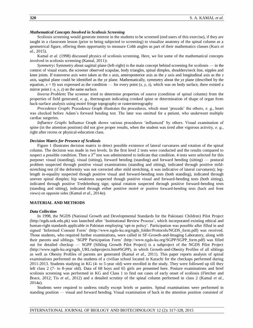
Fig. 2. Visual examination from front — drooping shoulder, nipples not level,
side and back — drooping shoulder, uneven scapulae
noting presence of drooping shoulders, level of scapulae, symmetry of body triangles, level of spinal dimples and
shape of midline of back — straight, C (most probably due to postural problem) or S (pathological — may be indica-
tive of scoliosis). In addition, visual examination of child facing the examiner was conducted to confirm shoulder drooping, unequal body triangles and shape of sternum (if observed during back examination) as well as level of
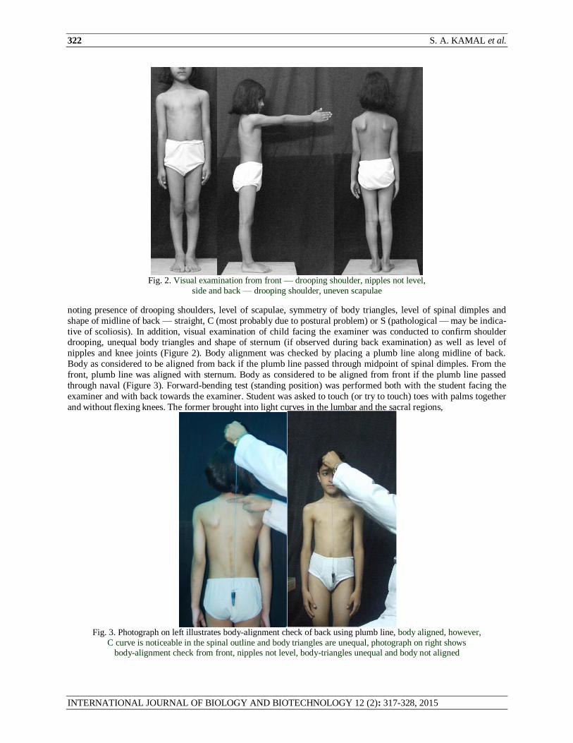
nipples and knee joints (Figure 2). Body alignment was checked by placing a plumb line along midline of back.
Body as considered to be aligned from back if the plumb line passed through midpoint of spinal dimples. From the
front, plumb line was aligned with sternum. Body as considered to be aligned from front if the plumb line passed
through naval (Figure 3). Forward-bending test (standing position) was performed both with the student facing the
examiner and with back towards the examiner. Student was asked to touch (or try to touch) toes with palms together
and without flexing knees. The former brought into light curves in the lumbar and the sacral regions, whekkreas
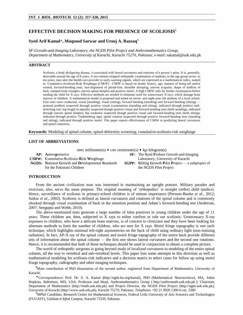
Fig. 3. Photograph on left illustrates body-alignment check of back using plumb line, body aligned, however,
C curve is noticeable in the spinal outline and body triangles are unequal, photograph on right shows body-alignment check from front, nipples not level, body-triangles unequal and body not aligned

EFFECTIVE DECISION MAKING FOR PRESENCE OF SCOLIOSIS 323
INTERNATIONAL JOURNAL OF BIOLOGY AND BIOTECHNOLOGY 12 (2): 317-328, 2015
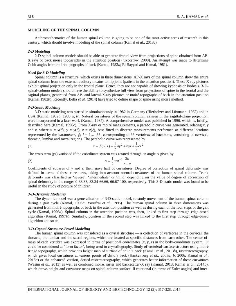
Fig. 4. Forward-bending test from front (lumber region highlighted) — left side elevated,
side and back (thoracic region highlighted) — right side elevated; S curve suspected
whereas the later highlighted curves in the cervical and the thoracic regions. The asymmetry observed in forward-
bending test (student facing the examiner or back towards the examiner) was confirmed by observing the student in
forward-bending position observed from the side opposite to elevated back, i. e., a student showing left side of back
elevated was observed from the right side and vice versa. The side observation highlighted elevated portion (Figure 4 — figure on extreme left shows forward-bending test, with student facing the examiner, in which left side is
elevated, whereas figure on extreme right shows forward-bending test, with student’s back towards the examiner, in
which right side is elevated; strong indication of S curve and rotation of the spinal column). Students were asked to
lift each foot for a count of 3 to check for hip weaknesses — the pelvis tilted downward on the side of unaffected
hip, when weight was borne on weak hip abductors (positive Tredelenburg sign). Uneven spinal dimples indicated
leg-length inequality.
In the sitting position visual and forward-bending examinations were performed by asking the child to sit on a
stool with back straight. To make sure that the thighs were positioned perpendicular to back and feet were not
hanging, wooden planks were placed under the feet to stabilize sitting posture. Body triangles could not been in the
sitting-position-visual examination.
Moiré examination (Figure 5) was performed by asking the child to stand behind a shadow-type moiré frame of
opening 13357 cm (grating dimensions 10852 cm) constructed from fishing line of black nylon thread of dddddiameter
Fig. 5. Use of moiré fringe topography to look for symmetric curves about the
sagittal plane of child’s back and front for possible detection of scoliosis

324 S. A. KAMAL et al.
INTERNATIONAL JOURNAL OF BIOLOGY AND BIOTECHNOLOGY 12 (2): 317-328, 2015
V V
Compare
Results
Both Positive
Mild-Stretching Exam
Postural Problem Suspected
Reduction of Curvature
Postural Problem Indicated
V FBT
Compare
Results
Both Positive
Tredelenberg Sign
Hip Weakness Suspected
Tredelenberg Positive
Hip Weakness Indicated
V FBT
Compare
Results
Both Positive
Spinal Dimples Observed
Leg-Length Inequality Suspected
Dimples not Level
Leg-Length Inequality Indicated
FBT FBT
Compare
Results
Both Positive
Moiré Exam
Spinal Rotation Suspected
Moiré Positive
Spinal Rotation Indicated
Fig. 6. Flowchart for decision matrix, which may be used to plan
for efficient detection and effective treatment of scoliosis
diameter 0.85 mm wound along the longer side with spacing maintained through a spring of pitch 0.75 mm. Distance
between the camera and the moiré grating was kept as 170 cm and that from the light source and the camera as 70
cm (Akram, 1989). Overhead projector was used as light source. A transparency was placed next to moiré grating to focus the light beam. The transparency was, later removed and the child was instructed to stand in the attention
position touching the moiré grating.
Heights and masses of students were measured by reproducible anthropometrists to accuracies of 0.01 cm and
0.01 kg, in the morning hours, as per protocols described elsewhere (Kamal et al., 2011; 2015). Equipments were
calibrated using a standard 100-cm ruler and a standard 2-kg mass. Zero errors were determined before starting each
session and subtracted from the measured values.
Data Processing
Figure 6 gives a flow chart of the decision-making process (Kamal et al., 2014a). We would like to study correlation
of those cases in which lateral curvatures, spinal rotations or both were indicated with CSRW. Let us denote
A :... ..set of students, in whom lateral curvatures of spinal column were indicated
B :... sset of students, in whom rotations of spinal column were indicated
BA :... SS set of students, in whom both lateral curvatures and rotations were indicated

EFFECTIVE DECISION MAKING FOR PRESENCE OF SCOLIOSIS 325
INTERNATIONAL JOURNAL OF BIOLOGY AND BIOTECHNOLOGY 12 (2): 317-328, 2015
Table 2. Mean Cumulative-Scoliosis-Risk Weightage (CSRW) for students suspected of scoliosis
Mean of individual CSRW scores of each student
BA :... S set of students, in whom lateral curvatures were indicated, but rotations were not
AB :.:: Sset of students, in whom rotations were indicated, but lateral curvature were not
From the above description, it could be easily concluded
(3) )()()( ABBABABA
Arithmetic mean of CSRW was computed corresponding to elements of each set.
RESULTS
Figure 7 shows the results of differential-spinal-function testing. Table 2 gives the number of students, who
indicated lateral curvatures or rotations and the corresponding mean Cumulative-Scoliosis-Risk Weightage
(CSRW). This analysis indicates that a higher CSRW increases the risk of student acquiring scoliosis — girls are
approximately at a double risk of acquiring scoliosis as compared to boys.
DISCUSSION AND CONCLUSION
The authors recommend posture examinations for boys and girls in the age group 4-7 years (Kamal and El-
Sayyad, 1981) and scoliosis-screening examinations for children in the age group 7-10 years (Kamal et al., 2013b)
employing visual inspection and moiré fringe topography. Child should be totally stripped except short underpants
and barefoot for these exams (Kamal et al., 2015). At-risk cases, determined through high CSRW (5.5 after the first
0
20
40
60
80
100
120
Postural
Problem
Leg Length
Inequality
Hip
Weakness
Spinal
Rotation
Postural
Problem
Leg Length
Inequality
Hip Weakness Spinal
Rotation
Fig. 7. Data analysis based on decision matrix, suspected conditions illustrated in indigo (boys) and pink (girls), indicated
conditions in blue (boys) and red (girls) — postural problem suspected through positive visual examinations (standing and sitting), indicated through positive mid-stretching test; leg-length inequality suspected though positive visual
and forward-bending tests (both standing), indicated through uneven spinal dimples; hip weakness suspected
though positive visual and forward-bending tests (both sitting), indicated through positive Tredelenburg sign; spinal rotation suspected through positive forward-bending tests (standing and sitting),
indicated through either positive moiré or positive forward-bending tests (back and front views) on opposite sides
Differential-Spinal-Function Testing Result Number of Boys Number of Girls Total Mean CSRW
Lateral curvatures indicated, no rotations 21 52 53 11.92
Rotations indicated, no lateral curvatures 27 50 77 18.98 Both lateral curvatures and rotations 20 51 51 17.50

326 S. A. KAMAL et al.
INTERNATIONAL JOURNAL OF BIOLOGY AND BIOTECHNOLOGY 12 (2): 317-328, 2015
checkup, 6.5 after the second checkup and 7.5 after the third checkup), should be followed up till the end of growth
period (An et al., 2015; Schulte et al., 2008).
Future work should be focused on formulating a coördinate system, which should reduce the degrees-of-
freedom of spinal column from a total of 231 = (33)(3 + 3 + 1) to, possibly, one by employing techniques similar to
those used in reducing degrees-of-freedom of two-body problem of planetary motion from 12 to one (Kamal,
1997b). The product (33)(3 + 3 + 1) comes from multiplying the number of vertebrae in the spinal column (33) with the sum of positional degrees-of-freedom (3), rotational degrees-of-freedom (3) + inter-vertebral-spacing degree-of-
freedom (1). The problem of human spinal column, one of the leading problems of ‘orthopedics’ (considered as a
branch of medical science dealing with the study of bones, joints and skeletal deformations), therefore, is described
by the branch of mathematics called ‘algebraic topology’. One must realize that ‘orthopedics’ is derived from
‘anatomy’ (study of structures of human body), whereas ‘algebra’ is the understanding of mathematical structures
and ‘topology’, studies invariance under deformations. ‘Anthrotopology’ may be the new branch of mathematics,
which could deal with the mathematical framework related to spinal deformities (Kamal et al., 2012).
ACKNOWLEDGEMENTS
The authors would like to thank Prof. Dr. Anisuddin Bhatti, Professor of Orthopedic Surgery and Managing
Director, Jinnah Postgraduate Medical Center, Karachi for visiting SF-Growth-and-Imaging Laboratory, and taking
a keen interest in our group’s work. The protocols of differential-spinal-function testing were discussed with him on
September 4, 2013, when the first author (SAK) gave Z. K. Kazi and M. A. Shah memorial lecture (Kamal et al.,
2013c). The authors are indebted to Mr. Sanaullah Kazi, Mrs. Azra Anwar Ahmed, Mrs. Yasmeen Salman, Mrs. Anis
Hasan, Mrs. Farkhunda Ghufran and Mrs. Nilofer Inam of Beacon Light Academy, ‘O’ Levels, Gulshan-é-Iqbal, Karachi, for providing data-collection facilities and our associate, Samira Sahar Jamil, for assisting in laboratory work. Shakeel
Ahmed Ansari developed software, which was needed to compute CSRW. Muhammed Wasim helped in drawing diagrams for this paper. The authors declare that they don’t have any financial/non-financial conflict of interest in the
work presented in this paper. This work is dedicated to the loving memory of Stig Willner (1931-1999), Orthopedic Surgeon, who was host of the first author during the fall of 1988, when he visited Malmö General Hospital as part of
research collaboration, which resulted in a publication (Kamal et al., 1994) with the deceased.
REFERENCES
Akram, M. (1989). Development of a shadow-type moiré apparatus. MSc Project, Department of Physics, Uni-versity of
Karachi, Karachi, Pakistan (unpublished) Akram, M. and S. A. Kamal (1991, April 16-20). Role of moiré fringe topography in the skeletal examination of school athlete.
The International Congress and Exposition on Sports Medicine and Human Performance, Vancouver, Canada, p. 2, abstract: http://www.ngds-ku.org/pub/confabst0.htm#C36:
An, K. C., D. H. Park, G. M. Kong, J. Y. Kim, S. Y. Jin, W. S. Lee et al. (2015). Prevalence study of adolescent idiopathic scoliosis in ten-, eleven-year olds for 10 years. Journal of Korean Orhopaedic Association, 50: 25-30
Anderson, S. M. (2007). Spinal curves and scoliosis. Radiologic Technology, 79: 44-65 Balla, P., G. Manhertz and A. Antal (2014). Defining of the shape of the spine using moiré method in case of patients with
Scheuermann disease. World Academy of Science, Engineering and Technology International Journal of Medical,
Health, Biomedical and Pharmaceutical Engineering, 8: 338-343 Baroni, M. P., G. J. B. Sanchis, S. J. C. de Assis, R. G. dos Santos, S. A. Pereira, K. G. Sousa et al. (2015). Factors
associated with scoliosis in school children: A cross-sectional population-based study. Journal of Epidemiology, 25:
212-220
El-Sayyad, M. M. and S. A. Kamal (1981, September 21-23). Cobb’s angle measurement by moiré topographs. Proceedings of Thirty-Fourth Annual Conference on Engineering in Medicine and Biology, Houston, Texas, USA, 23: 311, abstract#30.7:
http://www.ngds-ku.org/pub/confabst1.htm#C12: Fletcher, N. D. and R. W. Brace (2012). Early onset scoliosis: Current concepts and controversies. Current Reviews in
Musculoskeletal Medicine, 5: 102-110 Hackenberg, L., E. Hierholzer, V. Bullmann, U. Liljenqvist and C. Götze (2006). Raserstereographic analysis of axial back
surface rotation in standing versus forward bending posture in idiopathic scoliosis. European Spine Journal, 15: 1144-1149
Hackenberg, L., E. Hierholzer, W. Pötzi, C. Götze and U. Liljenqvist (2003a). Rasterstereographic back shape analysis in idiopathic scoliosis after anterior correction and fusion. Clinical Biomechanics, 18: 1-8
Hackenberg, L., E. Hierholzer, W. Pötzi, C. Götze and U. Liljenqvist (2003b). Rasterstereographic back shape analysis in idiopathic scoliosis after posterior correction and fusion. Clinical Biomechanics, 18: 883-889

EFFECTIVE DECISION MAKING FOR PRESENCE OF SCOLIOSIS 327
INTERNATIONAL JOURNAL OF BIOLOGY AND BIOTECHNOLOGY 12 (2): 317-328, 2015
Hierholzer, E. and G. Lüxmann (1982). Three-dimensional shape analysis of the scoliotic spine using invariant shape
parameters. Journal of Biomechanics, 15: 583-598 Horn, P. (2012). Scoliosis: Early identification of affected patients. Clinician Reviews, 22: 17-22
Kamal, S. A. (1982a). Measurement of angle of spinal curvature by moiré topographs. Journal of Islamic Medical Association (USA), 14: 145-149, full text: http://www.ngds-ku.org/Papers/J04.pdf
Kamal, S. A. (1982b, March 8-12). Moiré topography for the measurement of spinal curvature in three dimensions. March Meeting of the American Physical Society, Dallas, Texas, USA — in Bulletin of the American Physical Society, 27:
301, abstract#GY15: http://www.ngds-ku.org/pub/confabst1.htm#C16: Kamal, S. A. (1983a, October 30-November 3). Moiré fringe topography and the spinal deformity. The National Guard
Eighth Saudi Medical Conference, Riyadh, Kingdom of Saudi Arabia, p. 192, full text: http://www.ngds-ku.org/Papers/C22.pdf
Kamal, S. A. (1983b). Determination of degree of correction of spinal deformity by moiré topographs. Moiré Fringe Topography and Spinal Deformity (Proceedings of the Second International Symposium, Münster, Germany,
September 12-15, 1982), edited by B. Drerup, W. Frobin and E. Hierholzer, Gustav Fischer, Stuttgart and New York, 117-126, full text: http://www.ngds-ku.org/Papers/C23.pdf
Kamal, S. A. (1987). Moiré topography for the study of multiple curves of spine. Surface Topography and Spinal Deformity (Proceedings of the Fourth International Symposium, Mont Sainte Marie, Québec, Canada, September 27-
30, 1986), edited by I. A. F. Stokes, J. R. Pekelsky and M. S. Moreland, Gustav Fischer, Stuttgart and New York, 43-49, full text: http://www.ngds-ku.org/Papers/C26.pdf
Kamal, S. A. (1996a). Gait analysis using moiré fringe topography and rasterstereography (simultaneous recording). Karachi University Journal of Science, 24: 7-18, full text: http://www.ngds-ku.org/Papers/J16.pdf
Kamal, S. A. (1996b). Combination of moiré contours and edge-based algorithm to study motion in the sagittal plane. Karachi University Journal of Science, 24: 53-60, full text: http://www.ngds-ku.org/Papers/J17.pdf
Kamal, S. A. (1996c). A 3-D-static model of the human spinal column. Karachi University Journal of Science, 24: 29-34, full text: http://www.ngds-ku.org/Papers/J18.pdf
Kamal, S. A. (1996d, June 27-July 11). 3-D-dynamic modeling of the human spinal column. The Twenty-First International Summer College on Physics and Contemporary Needs, Nathiagali, KP, Pakistan, abstract:
http://www.ngds-ku.org/pub/confabst0.htm#C42: Kamal, S. A., (1997b, March 30, April 3). Planetary-orbit modeling based on astrodynamical coördinates. The Pakistan
Institute of Physics International Conference (PIPC 1997), Government College, Lahore, Pakistan, pp. 28-29,
abstract#P2: http://www.ngds-ku.org/pub/confabst0.htm#C45: Kamal, S. A. (2011, May 9, 10). Anthromathematics: A new branch of mathematics. The Sixth Symposium on Compu-
tational Complexities, Innovations and Solutions (CCIS 2011), the COMSATS Institute of Information Technology, Abbotabad, KP, Pakistan, pp. 13, 14, abstract#1: http://www.ngds-ku.org/Presentations/Anthromath.pdf
Kamal, S. A. (2013, May 30). 3-D-spinal-column-surface analysis (height and curvature maps) by combining moiré fringe topography and rasterstereography with backscatter-X-ray scanning technology. The First Undergraduate National
Computing Conference, Usman Institute of Technology, Karachi, Pakistan (keynote lecture), abstract: http://www.ngds-ku.org/Presentations/Backscatter.pdf
Kamal, S. A., A. Haider and M. Sarwar (2013a, September 4, 5). Rasterstereography in scoliosis detection and manage-ment. The First Conference on Anthromathematics in the Memory of (Late) Syed Firdous (ANTHROMATHEMATICS
2013), Department of Mathematics, University of Karachi, Karachi, Pakistan and Government College, Hyderabad, Pakistan, p. 10, abstract#Anthro13-04: http://www.ngds-ku.org/Presentations/Rasterstereography.pdf
Kamal, S. A. G. Benoni and S. Willner (1994). A study to test the reproducibility of moiré topographs. Karachi University Journal of Science, 22: 67-74, full text: http://www.ngds-ku.org/Papers/J15.pdf
Kamal, S. A. and M. M. El-Sayyad (1981, August 9-13). The use of moiré topographs for detection of orthopedic defects in children of age group four to seven years. The Twenty-Third Annual Meting of the American Association of Physicists in
Medicine, Boston, Massachusetts, USA — presented by title, Medical Physics, 8: 549, papert#T7:
http://www.ngds-ku.org/pub/confabst1.htm#C11:
Kamal, S. A. M. Sarwar and A. Haider (2014a, March 20). Effective decision making for presence of scoliosis based on moiré fringe topography. The Second Conference on Mathematical Sciences (CMS 2014), Department of Mathema-
tics, University of Karachi, Karachi, Pakistan, p. 2, abstract#CMS14-05: http://www.ngds-ku.org/Presentations/Decision.pdf
Kamal, S. A., M. Sarwar and M. K. Rajput (2012, December 27-29). Crystal-structure concepts applied to static and dyna-mic modeling of the human-spinal column. The International Conference on Condensed-Matter Physics and Engi-
neering, the Bahauddin Zakaria University, Multan, Pakistan p. 81, abstract: http://www.ngds-ku.org/Presentations/BZU1.pdf
Kamal, S. A., M. Sarwar and M. K. Rajput (2014b, September 4). Crystal-structure-concept-based modeling of the human spinal column validated through 3-D-bone scanning. The Second Conference on Anthromathematics in the Memory of

328 S. A. KAMAL et al.
INTERNATIONAL JOURNAL OF BIOLOGY AND BIOTECHNOLOGY 12 (2): 317-328, 2015
(Late) Hussain Ahmed Bilgirami (ANTHROMATHEMATICS 2014), Department of Mathematics, University of Kara-
chi, Karachi, Pakistan and Government College, Hyderabad, Pakistan, p. 8, abstract#Anthro14-04:
http://www.ngds-ku.org/Presentations/Spinal.pdf
Kamal, S. A., M. Sarwar and S. K. Raza (2013b, September 4, 5). Moiré fringe topography in scoliosis detection and management. The First Conference on Anthromathematics in the Memory of (Late) Syed Firdous (ANTHROMATHE-
MATICS 2013), Department of Mathematics, University of Karachi, Karachi, Pakistan and Government College, Hyderabad, Pakistan, p. 16, abstract#Anthro13-10: http://www.ngds-ku.org/Presentations/Moire.pdf
Kamal, S. A., M. Sarwar and U. A. Razzaq (2013c, September 4, 5). Anthromathematics of the human spinal column. The First Conference on Anthromathematics in the Memory of (Late) Syed Firdous (ANTHROMATHEMATICS 2013),
Department of Mathematics, University of Karachi, Karachi, Pakistan and Government College, Hyderabad, Pakistan, p. 7 (Prof. Dr. Z. K. Kazi and Prof. Dr. M. A. Shah memorial lecture), abstract#Anthro13-01:
http://www.ngds-ku.org/Presentations/Scoliosis.pdf Kamal, S. A., M. Sarwar and U. A. Razzaq (2013d, September 4, 5). Cumulative-Scoliosis-Risk Weightage (CSRW) —
designing preventive strategies. The First Conference on Anthromathematics in the Memory of (Late) Syed Firdous (ANTHROMATHEMATICS 2013), Department of Mathematics, University of Karachi, Karachi, Pakistan and Gover-
nment College, Hyderabad, Pakistan, p. 11, abstract#Anthro13-05: http://www.ngds-ku.org/Presentations/CSRW.pdf Kamal, S. A., Naseeruddin, M. Wasim and S. Firdous (1998). Physics of scoliosis screening in school-going children.
Karachi University Journal of Science, 26: 5-12, full text: http://www.ngds-ku.org/Papers/J22.pdf Kamal S. A., N. Jamil and S. A. Khan (2011). Growth-and-obesity profiles of children of Karachi using box-interpolation
method. International Journal of Biology and Biotechnology, 8: 87-96, full text:
http://www.ngds-ku.org/Papers/J29.pdf
Kamal, S. A. and R. E. Lindseth (1980, July 29-August 1). Moiré topography for the detection of orthopedic defects.
Periodic Structures, Moiré Patterns and Diffraction Phenomena (Proceedings of the Society of Photo-Optical Instru-
mentation Engineers, San Diego, California, USA), edited by C. H. Chi, E. G. Loewen and C. L. O’Bryan III, 240: 293-295, full text: http://www.ngds-ku.org/Papers/C08.pdf
Kamal, S. A., S. A. Ansari and S. S. Jamil (2015). Generating and validating Growth-and-Obesity Roadmaps for the Pakistani children. International Journal of Biology and Biotechnology, 12: 47-61, full text:
http://www.ngds-ku.org/Papers/J35.pdf Kurz, T. L., H. B. Yanik and M. Y. Lee (2015). The geometry of scoliosis. Teaching Children Mathematics, 21: 372-375
Luk, K. D., C. F. Lee, K. M. C. Cheung, J. C. Y. Cheng, B. K. W. Ng, T. P. Lam et al. (2010). Clinical effectiveness of
school screening for adolescent idiopathic scoliosis: A large population-based retrospective cohort study. Spine, 35: 1607-1614
Oxborrow, N. J. (2000). Assessing the child with scoliosis: The role of surface topography. Archives of Diseases in Child-hood, 83: 453-455
Persson-Bunke, M., G. Hägglund, H. Lauge-Pedersen, W. Philippe and L. Westborn (2012). Scoliosis in a total population of children with cerebral palsy. Spine, 37: E708-E713
Saikia, K. C., A. Duggal, P. K. Bhattacharya and M. Borgohain (2002). Scoliosis: An epidemiological study of school children in lower Assam. Indian Journal of Orhopaedics, 36: 243-245
Schulte, T. L., E. Hierholzer, A. Boerke, T. Lerner, U. Liljenqvist, V. Bullmann and L. Hackenberg (2008). Rasterstereo-graphy versus radiography in the long-term follow-up of idiopathic scoliosis. Journal of Spinal Disorders and Tech-
niques, 21: 23-28 Sengupta, D. P. and J. K. Webb (2010). Scoliosis — The current concepts. Indian Journal of Orhopaedics, 44: 5-8
Tis, J. E., L. I. Karlin, B. A. Akbarnia, L. C. Blakemore, G. H. Thompson, R. E. McCarthy et al. (2012). Early onset sco-liosis: Modern treatment and results. Journal of Pediatric Orthopedics, 32: 647-657
Wasim, M., S. A. Kamal and A. Shaikh (2013). A security system employing edge-based rasterstereography. International Journal of Biology and Biotechnology, 10: 613-630, full text: http://www.ngds-ku.org/Papers/J31.pdf
Yosufzai, M. A. K., S. A. Kamal and J. A. Zubairi (1995, January 2-5). Computer-based analysis of human gait using moiré fringe topography. Proceedings of the Second International Workshop on Computer Vision and Parallel Pro-
cessing, edited by G. N. Khan, A. A. Naqvi and M. A. Shah, the Quaid-é-Azanm University, Islamabad, Pakistan, pp. 60-71, full text: http://www.ngds-ku.org/Papers/C41.pdf
(Accepted for Publication: March 2015)
Copyright © 2022 FDOKUMEN