Intermittent aerobic-resistance interval training versus ... - PLOS

1
Effect of high-intensity intermittent training on lactate and H+ release from human skeletal muscle
Carsten Juel, Christina Klarskov, Jens Jung Nielsen, Peter Krustrup, Magni Mohr and Jens Bangsbo.
Copenhagen Muscle Research Centre
August Krogh Institute, University of Copenhagen
Running head: Training-induced changes in lactate and H+ release
Correspondence: Carsten Juel, August Krogh Institute, Universitetsparken 13, DK-2100 Copenhagen
Denmark.
Tlf: +45 35 32 16 82.
Fax: +45 35 32 15 67.
Email: [email protected]
Copyright (c) 2003 by the American Physiological Society.
Articles in PresS. Am J Physiol Endocrinol Metab (October 14, 2003). 10.1152/ajpendo.00303.2003

2
ABCTRACT
The study investigated the effect of training on lactate and H+ release from human skeletal muscle
during one-legged knee-extensor exercise. Six subjects were tested after 7-8 weeks of training (fifteen
1-min bouts at ∼150 % of thigh VO2max per day). Blood samples, blood flow, and muscle biopsies were
obtained during and after a 30-W exercise bout and an incremental test to exhaustion of both trained
(T-leg) and untrained (UT-leg) leg. Peak blood-flow was 16 % higher in T-leg than in UT-leg. In the
30-W test venous lactate and lactate release were lower in T-leg compared to UT-leg. In the
incremental test time to fatigue was 10.6±0.7 and 8.2±0.7 min in T-leg and UT-leg (P<0.05). At
exhaustion venous blood lactate was 10.7±0.4 and 8.0±0.9 mmol l-1 in T-leg and UT-leg (P<0.05), and
lactate release was 19.4±3.6 and 10.6±2.0 mmol min-1 (P<0.05). H+ release at exhaustion was higher in
T-leg than in UT-leg. Muscle lactate content was 59.0±15.1 and 96.5 ± 14.5 mmol kg-1 dry weight in
T-leg and UT-leg, and muscle pH was 6.82±0.05 and 6.69±0.04 in T-leg and UT-leg (P = 0.06). The
membrane contents of the monocarboxylate transporters MCT1 and MCT4 and the Na+/H+ exchanger
were 115±5 (P<0.05), 111±11 and 116±6 % (P<0.05), respectively, in T-leg compared to UT-leg. The
reason for the training-induced increase in peak lactate and H+ release during exercise is a combination
of an increased density of the lactate and H+ transporting systems, an improved blood flow and blood
flow distribution, as well as an increased systemic lactate and H+ clearance.
Keywords: MCT1, MCT4, Na+/H+ exchange, NHE1

3
INTRODUCTION
During intense exercise lactate and H+ accumulate in the contracting muscle. The accumulated lactate
is either removed by oxidation or gluconeogenesis in the muscle or is transported to the blood and
removed by other cells (6, 14). The fluxes of lactate and H+ across the muscle membrane are facilitated
by two monocarboxylate transporter proteins, MCT1 and MCT4 (21). These transporters contribute to
the pH regulation during intense muscle activity, whereas pH regulation at rest is mainly dependent on
an efflux of H+ mediated by Na+/H+ exchange or transport systems involving bicarbonate (11, 12).
However, it has been demonstrated that the H+ transport systems not involving lactate are also further
activated during muscle activity and in the recovery phase after exercise (4).
Training induces numerous adaptive changes in skeletal muscle, among these a few reports have
described changes in lactate transporters at the protein level. The density of MCT1 and MCT4 in
human skeletal muscle has been demonstrated to be elevated after a period of endurance training (7),
high intensity training (5, 16), and after a single prolonged exercise bout (9). In the study by Pilegaard
et al. (16) the training-induced elevation in MCT density was associated with a reduction in muscle
lactate during exercise. The effect of training on the Na+/H+ exchanger in muscle is less investigated.
One study in rats has demonstrated that the amount of the Na+/H+ exchanger proteins is elevated after a
period of high intensity treadmill training (13), but no studies have examined the effect of training on
the Na+/H+ exchanger in human skeletal muscle.
It is hypothesized that intense training increases the release of H+ and lactate from muscle, and that
these changes are related to an increased expression of the MCT and Na+/H+ exchanger proteins.
MATERIALS AND METHODS

4
Subjects
Six healthy male subjects participated in the study. Age, height, weight and VO2max before training was
25.3 ± 2.9 (±SD) yrs, 185.0 ± 3.9 cm, 82.8 ± 11.8 kg and 50.2 ± 1.2 ml O2 min-1 kg-1, respectively. The
subjects were fully informed of any risk and discomfort associated with the experiments before giving
their written consent to participate. The study conforms to the code of Ethics of the World Medical
Association (Declaration of Helsinki) and was approved by the Ethical Committee of the Copenhagen
and Frederiksberg Communities, Denmark.
Training
Training was performed for 7-8 weeks with one leg (T-leg), whereas the untrained leg (UT-leg) served
as control. Before training the subjects were familiarized to the one-legged knee-extensor model and
measurements were carried out to confirm that the work was only performed with m. quadriceps
femoris (2). The leg to be trained was selected randomly. At least two incremental tests to fatigue were
performed with each leg in order to determine the initial load in the training period. The subjects
performed one-legged knee-extensor exercise (60 kicks min-1) on an ergonometer in a supine position.
Each training session consisted of 5-min warm up at a load of 10 W, 3 min of rest, and fifteen 15 1-min
exercise bouts at ∼150 % of thigh VO2max separated by 3 min of rest. The training was performed 3
times per week in week 1-2, 4 times per week in week 3-4, and 5 times per week for the last weeks.
The work-load was adjusted in order to keep the relative load constant.

5
Main experiment
Muscle mass was determined before and after training from thigh length, 3 circumferences of the thigh
and skin fold thickness.
An identical protocol was carried out for T-leg and UT-leg on separate days. The subjects rested in the
supine position and a catheter was placed in the femoral vein under local anesthesia. The tip of the
catheter was positioned approximately 1-2 cm proximal to the inguinal ligament. This catheter was
used for femoral venous blood sampling. Through the catheter a thermistor was placed and advanced 8-
10 cm proximal to the tip of the catheter for measurement of femoral venous blood flow by the constant
infusion thermodilution technique. Briefly, ice-cold saline was infused at a constant rate for 10-15 s
and leg blood flow could be calculated from the temperature decrease. An occlusion cuff placed below
the knee was inflated during exercise in order to avoid contribution of blood from the lower leg. A
second catheter was inserted in the femoral artery of the resting leg.
Each protocol consisted of warm up and 30 min of sub-maximal exercise at 30 W, which makes up
approximately 28 % of the final load (106 ± 9 W) obtained in the incremental test. After 60 min of rest
exercise was performed at 10 W, followed by 10 min rest, and an incremental test starting with 50 W
for 4 min, subsequently the load was increased every 2 min by 10 W until exhaustion. Time to fatigue
was defined from the time when the kicking frequency reached below 55 rpm. Heparinized syringes
were used to draw blood simultaneously from the femoral artery and vein. The blood samples were
placed on ice-cold water.
Blood analysis

6
Plasma pH, plasma HCO3-, whole blood hemoglobin and hematocrit were measured in an ABL 615
blood analyzer (Radiometer Denmark). Actual base excess (ABE) for oxygenated blood was calculated
from plasma HCO3-, blood pH and hemoglobin according to the method of Siggaard-Andersen (19),
and total H+ release was calculated from blood flow and the V-A concentration difference for ABE,
which was corrected for the changes in buffer capacity due to various levels of hemoglobin
oxygenation. A 100 µl sample of each blood sample was treated with 20 g l-1 of Triton-X-100 and used
for determination of whole blood lactate on an YSI lactate analyzer (Yellow Spring Instruments)
Muscle analysis
The needle biopsy technique was used to obtain muscle samples from vastus lateralis. Muscle biopsies
were obtained at rest before training, after 2 and 4 weeks of training as well as after the training period
(7-8 weeks). In the main experiment biopsies were obtained before and immediately after exercise in
the incremental test.
The samples were immediately frozen in liquid nitrogen and stored at –80 0C. One part of each muscle
sample was used for the Western blotting technique to measure membrane proteins and for fiber type
determination. The rest was freeze-dried and water content was determined. One part was homogenized
and used for pH-determination using a small glass electrode; another part was extracted with 0.6 M
perchloric acid, neutralized, and used for a fluorometric lactate determination (in mmol kg-1 dry
weight).

7
Western blot
Approximately 30 mg of each muscle sample was homogenized in a sucrose buffer (250 mM sucrose,
30 mM HEPES, 2 mM EGTA, 40 mM NaCl, 2 mM PMSF, pH 7.4) with a Polytron 2100 and
centrifuged at 1000 g for 5 min, this procedure removes heavy material including a fraction of the
mitochondria. The pellet was used for determination of fibertypes based on the distribution of myosin
heavy chain (MHC) isoforms using SDS-PAGE (1, 19). The supernatant was spun at 190.000 g for 90
min at 40C, and the new pellet was re-suspended in Tris-SDS (10 mM Tris, 4 % SDS, 1 mM EDTA, 2
mM PMSF, pH 7.4) and protein content determined with a BSA standard (DC protein assay, Bio-Rad).
10 µg of protein from each sample was subjected to SDS-PAGE (Excell 8-18 % gradient gel) and
electroblotted to a Millipore Immobilon-P polyvinylidiene diflouride membrane. The membrane was
blocked by BSA, 0.5 % Tween-20, and low fat milk and incubated with the primary antibody diluted in
a BSA containing buffer. After treatment with the HRP-coupled secondary antibody and repeated
washing, the membrane were incubated with ECL reagent (Amersham) and visualized on a film. The
quantification of protein was performed by scanning the film and analyzing band intensities with the
SigmaGel software. Samples from the same subject were always run on the same gel and with identical
amount of protein per lane.
The Na+/H+ exchanger isoform NHE1 was detected by the antibody MAB3080 (Chemicon). The
antibodies to the human lactate/H+ co-transporter isoforms MCT1 and MCT4 were a gift from prof. A.
Halestrap, Bristol UK (21).

8
Calculations
The concentration of lactate in cell water was calculated from muscle lactate (mmol kg-1 dry weight),
muscle water content (%) and corrected for extracellular lactate assuming an interstitial space of 15 %.
The concentration gradient for lactate from muscle to blood at exhaustion from the incremental test was
calculated as the difference between the intracellular muscle lactate concentration (mmol l-1 of cell
water) and the arterial plasma lactate concentration. The H+ gradient was calculated from the H+
concentration in muscle (pH in the homogenate was chosen to represent intracellular pH) and in the
arterial blood. Lactate and H+ release were calculated from the A-V differences and blood flow.
Statistics
Two-way analysis of variance was used for comparison between data sets from trained and untrained
legs. Student’s paired t-test was used to locate the differences if data passed a normality test; otherwise
a Wilcoxon rank test was used. One-way repeated measures analysis of variance (ANOVA) was used
to test the changes in protein content. The differences were located using the Tukey Test (SigmaStat
software). A significance level of P = 0.05 was chosen.
RESULTS

9
Muscle mass
The thigh muscle mass in the T-leg increased (P<0.05) by 4.5% (from 2.44 ± 0.13 to 2.55 ± 0.13 kg)
during the training period, whereas the muscle mass of the UT-leg remained unaltered.
Fiber type distribution
The fiber type distribution after training in T- and UT-leg was 44/47/9 % (MHC-I/MHC-IIa/MHC-IIx )
and 44/50/6 %, respectively.
Performance
The work-load increased (P<0.05) during the training period from 92 ± 8 to 106 ± 9 W. The time to
fatigue in the incremental test was longer (P<0.05) in the T-leg than in the UT-leg (10.6 ± 0.7 vs. 8.2 ±
0.70 min).
Leg blood flow
In the 30-W test leg blood flow reached the peak value ∼3.5 l min-1 approximately 3 min after onset of
exercise after which the flow remained constant in both T- and UT-leg.
In the incremental test thigh blood flow increased with increasing work-load with no differences
between T- and UT-leg. However, the peak blood flow was higher (P<0.05) in T-leg than in UT-leg
(7.1 ± 0.5 vs. 5.7 ± 0.6 l min-1). In the first sample in recovery (approximately 5 s from exhaustion) leg
blood flow was still higher (P<0.05) in T-leg (6.7 ± 1.0 l min-1) than in UT-leg (5.7 ± 0.3 l min-1). Later
in recovery blood flow was gradually reduced towards the resting level without any difference between
T- and UT-leg.

10
Blood lactate and lactate release
In the 30-W test femoral venous blood lactate increased rapidly at onset of exercise and reached a
lower (P<0.05) peak value (1.5 ± 0.2 mmol l-1) in T-leg than in UT-leg (2.0 ± 0.3 mmol l-1) after 3 min,
after which blood lactate declined towards resting level at the end of the exercise period (Fig. 1A).
The venous-arterial (v-a) concentration difference reached the peak value 3 min after onset of exercise
and declined to zero at the end of the exercise period (Fig. 2A). In the first 5 min of exercise the v-a
lactate concentration difference was lower (P<0.05) in T-leg than in UT-leg (Fig. 2A). The peak thigh
lactate release was 2.1 ± 0.5 mmol min-1 in T-leg and 3.2 ± 0.5 mmol min-1 in UT-leg (P<0.05) during
the 30-W exercise test. Lactate release decreased towards the end of exercise and was only slightly
higher at 30 min than at rest (Fig. 3A).
In the incremental test blood lactate increased with increasing power output (Fig. 1B). At exhaustion
the femoral venous blood lactate was higher (P<0.05) in T-leg than in UT-leg (10.7 ± 0.4 vs. 8.0 ± 0.9
mmol l-1). Also in the first part of recovery venous blood lactate was higher (P<0.05) in T-leg
compared to UT-leg (Fig. 1B). The v-a lactate concentration difference at exhaustion was 3.2 ± 0.5
mmol l-1 and 1.9 ± 0.3 mmol l-1 in T- and UT-leg, respectively (P<0.05), whereas no differences
between T- and UT-leg were observed in recovery (Fig. 2B). Lactate release increased with increasing
exercise intensity (Fig. 3B). At exhaustion lactate release was greater (P<0.05) in T-leg than in UT-leg
(19.4 ± 3.6 vs. 10.6 ± 2.0 mmol min-1), whereas no difference between T- and UT-leg was observed 5 s
into recovery (9.3 ± 1.2 and 7.9 ± 1.1 mmol min-1, respectively. In both T- and UT-leg lactate release
decreased towards zero during the 10-min recovery period.

11
Blood pH
At onset of the 30-W exercise femoral venous blood pH declined rapidly and reached the lowest value
after 3 min where after it increased slowly during the rest of the exercise (Fig. 4A). However, venous
blood pH was still reduced after 30 min. Arterial blood pH did not change during exercise.
In the incremental test venous blood pH was gradually reduced during exercise (Fig. 4B). At
exhaustion venous blood pH was lower (P<0.05) in T-leg than in UT-leg (7.07 ± 0.02 vs. 7.13 ± 0.02).
Also in the first part of recovery venous blood pH was lower (P<0.05) in T-leg compared to UT-leg
(Fig. 4B).
H+ release
The H+ release in the 30-W exercise test increased rapidly at onset of exercise and reached a plateau
after 3 min, which lasted for the rest of the 30 min exercise period. The peak H+ release (10-11 mmol
min-1) was not different in T- and UT-leg (Fig. 5A).
In the incremental test H+ release (Fig. 5B) increased gradually with exercise intensity and reached
36.9 ± 3.1 and 24.2 ± 1.5 mmol min-1 at exhaustion in T- and UT-leg, respectively (P<0.05). H+ release
was also higher (P<0.05) in T-leg compared to UT-leg in the first two measurements (5 and 75 sec) in
recovery.
Muscle lactate and muscle pH
Muscle lactate after the 30-W exercise bout was 8.4 ± 1.7 and 5.9 ± 1.0 mmol kg-1 dry weight in T- and
UT-leg, respectively. At exhaustion in the incremental test muscle lactate in T- and UT-leg was 59.0

12
±15.1 and 96.5 ± 14.5 mmol kg-1 dry weight, respectively, corresponding to 22.3 and 29.8 mmol l-1 of
muscle water (P = 0.06).
Muscle pH at the end of the 30-W exercise test was 7.13 ± 0.04 in T-leg and 7.09 ± 0.03 in UT- leg. At
exhaustion in the incremental test muscle pH was 6.82 ± 0.05 in T-leg and 6.69 ± 0.04 in UT- leg.
Muscle to blood gradients for lactate and H+
In the incremental test the muscle to blood lactate gradient at exhaustion was 15.3 ± 4.4 mmol l-1 in T-
leg and 23.5 ± 4.5 mmol l-1 in UT-leg (P = 0.1). Fig. 6A shows the individual relationship between
lactate release and muscle to blood lactate gradient.
The H+ gradient from muscle to arterial blood at exhaustion was lower (P<0.05) in T- than UT-leg (101
(range: 54-132) vs. 158 (207 - 121) nM). In Fig. 6B is plotted the individual H+ release as a function of
the muscle to blood H+ gradient.
MCT1, MCT4 and NHE1 protein expression
After 2, 4 and 7-8 weeks of training the MCT1 content was 108 ± 10 %, 138 ± 19 % and 115 ± 5%
(P<0.05), respectively, compared to the pre-training value (Fig. 7). The MCT4 content was 98 ± 9 %,
112 ± 7 %, and 111 ± 11 % after 2, 4, 7-8 weeks of training, respectively, compared to the pre-training
values (Fig. 7).
The content of the Na+/H+ exchanger protein NHE1 was 131 ± 21 %, 131 ± 13 % (P<0.05) and 116 ± 6
% (P<0.05) after 2, 4 and 7-8 weeks, respectively, relative to the individual values before training (Fig.
7).
13
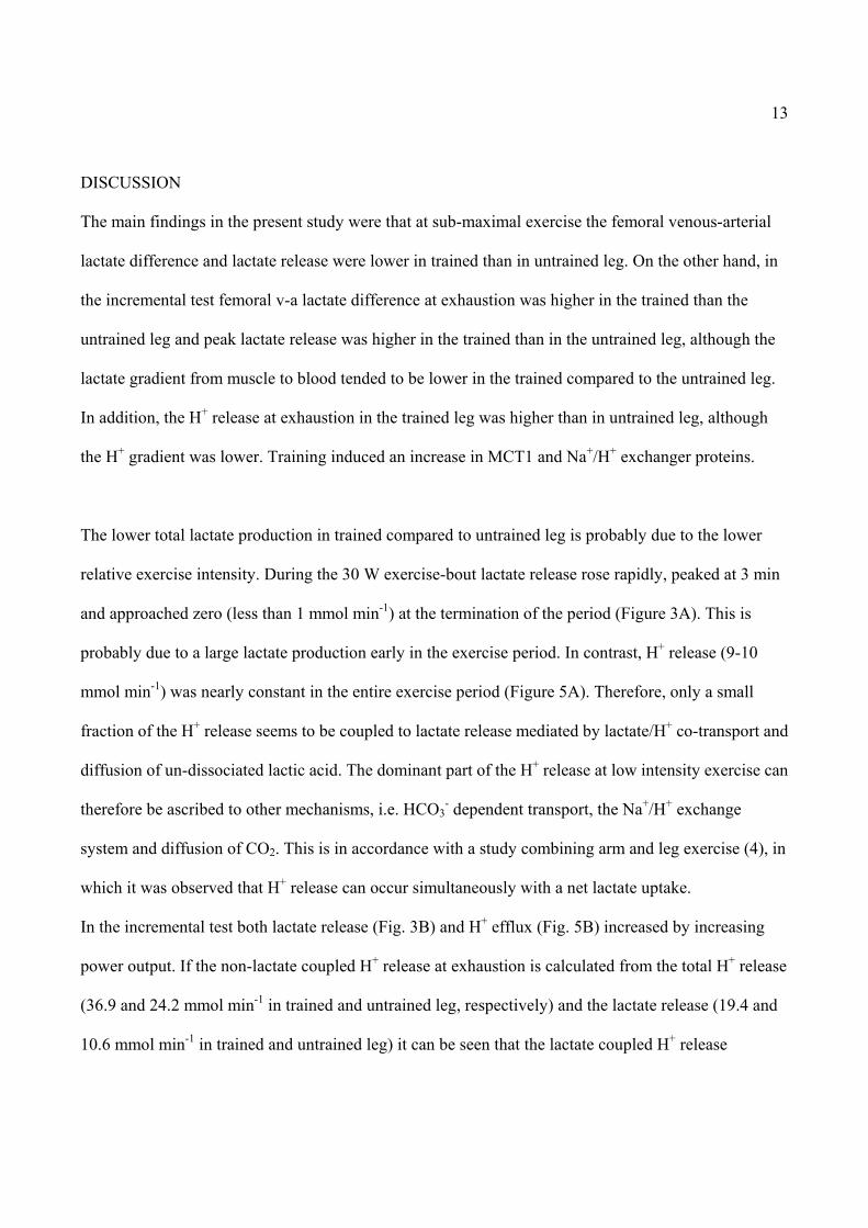
DISCUSSION
The main findings in the present study were that at sub-maximal exercise the femoral venous-arterial
lactate difference and lactate release were lower in trained than in untrained leg. On the other hand, in
the incremental test femoral v-a lactate difference at exhaustion was higher in the trained than the
untrained leg and peak lactate release was higher in the trained than in the untrained leg, although the
lactate gradient from muscle to blood tended to be lower in the trained compared to the untrained leg.
In addition, the H+ release at exhaustion in the trained leg was higher than in untrained leg, although
the H+ gradient was lower. Training induced an increase in MCT1 and Na+/H+ exchanger proteins.
The lower total lactate production in trained compared to untrained leg is probably due to the lower
relative exercise intensity. During the 30 W exercise-bout lactate release rose rapidly, peaked at 3 min
and approached zero (less than 1 mmol min-1) at the termination of the period (Figure 3A). This is
probably due to a large lactate production early in the exercise period. In contrast, H+ release (9-10
mmol min-1) was nearly constant in the entire exercise period (Figure 5A). Therefore, only a small
fraction of the H+ release seems to be coupled to lactate release mediated by lactate/H+ co-transport and
diffusion of un-dissociated lactic acid. The dominant part of the H+ release at low intensity exercise can
therefore be ascribed to other mechanisms, i.e. HCO3- dependent transport, the Na+/H+ exchange
system and diffusion of CO2. This is in accordance with a study combining arm and leg exercise (4), in
which it was observed that H+ release can occur simultaneously with a net lactate uptake.
In the incremental test both lactate release (Fig. 3B) and H+ efflux (Fig. 5B) increased by increasing
power output. If the non-lactate coupled H+ release at exhaustion is calculated from the total H+ release
(36.9 and 24.2 mmol min-1 in trained and untrained leg, respectively) and the lactate release (19.4 and
10.6 mmol min-1 in trained and untrained leg) it can be seen that the lactate coupled H+ release

14
accounted for 53 and 44 % of the total H+ release in trained and untrained leg, respectively. In addition
it appears that training affected both the lactate-coupled H+ release (+52%) and the non lactate-coupled
H+ release (+83%).
The present exercise model involved a small muscle mass, which only produced limited changes in
arterial H+ and lactate concentrations. It is therefore possible to study the effect of the muscle to blood
gradient for H+ and lactate release. The concentration gradients for lactate and H+ are important factors
for the transporter-mediated flux of lactate from muscle to blood. However, in the present study in the
trained leg a higher lactate release at exhaustion was observed in spite of a tendency towards a lower
muscle to blood gradient for lactate (Fig. 6). Similarly, a large H+ gradient is known to stimulate
lactate/H+ co-transport (11) and the Na+/H+ exchange system (12). In the present study the H+ gradient
was lower in the trained than in the untrained leg whereas the H+ release was higher. Thus, higher
driving forces for lactate and H+ cannot explain the higher release of lactate and H+ in the trained leg.
These findings are in accordance with a previous study showing that a given lactate release after
training was obtained at smaller lactate and H+ gradients (16).
In the present study the MCT1 content was and MCT4 content tended to be higher after than before
training. These findings are in accordance with observations in other studies. It has been demonstrated
that a period of training can increase the density of the MCT1 proteins (1, 7, 9, 16), and MCT4 has also
been shown to increase by high-intensity training (16), although a short-term (7 days) training study (5)
and a long-term (5 months) training study (8) did not observed any increase in MCT4. Thus, MCT4
seems to be less sensitive to training than MCT1. In one study plasma membrane MCT4, but not
MCT1, has been demonstrated to be acutely down-regulated after one bout of exercise, whereas the
membrane transport capacity for lactate was elevated (20). The moderate changes (+15%, +11%) in
MCTs in the present study can probably only explain a fraction of the improved lactate release ability

15
after training. The Na+/H+ exchanger protein NHE1, which in the present study for the first time was
quantified in human muscle in association with training, was significantly elevated (+16%) after the
training period. Thus, the increase in the total H+ transport capacity (estimated from the increase in
NHE1 and MCTs) was approximately 15 %. It is a possibility that training-induced changes in intrinsic
activity of the transporters proteins are also involved, but this has not been studied. Apparently, the
improved lactate and H+ release can partly be explained by an increased density of membrane proteins,
but others mechanisms are probably involved, as the training-induced improvement in H+ release is
larger than the increase in protein densities.
The higher lactate and H+ release in the trained leg may also in part be due to a higher blood flow. At
exhaustion the thigh blood flow was approximately 16 % higher in the trained leg, which by itself
should increase lactate release since elevated blood flow has been shown to increase lactate release
(15). In the present study, an increased capillary-to-fiber ratio from 1.7 ± 0.1 to 2.5 ± 0.1 (+47 %) after
the training (10) may also in part explain the higher lactate release. The greater relative increase in
capillarization than in leg blood flow in the trained leg may indicate that the mean transit-time for
blood was elevated allowing a higher degree of equilibration. Thus, the release of lactate to the blood
stream would be higher at a given concentration gradient.
Other factors might also be of importance. It has been demonstrated that passive leg movements
increase thigh blood flow from approximately 0.4 l min-1 to 1.2 l min-1 (17). Blood flow can therefore
be expected to increase in the passive muscle of the thigh during the knee-extensor exercise.
Consequently, the increase in blood flow in the active muscle is more than 16 % if it is assumed that
the training-induced increase in blood flow is located to the active muscle.

16
The possibility also exist that training changes the ratio between flow to passive and active muscle. It is
striking in connection with this possibility that the V-A concentration difference and lactate release in
the trained leg are reduced at the transition from exercise to the first part of recovery (despite leg blood
flow is still high), whereas this is less marked in the untrained leg (Fig. 2B and 3B). This could indicate
a different distribution of local flow during exercise in trained compared to untrained leg. In summary,
higher leg blood flow and altered flow distribution are expected to account for a fraction of the higher
lactate and H+ release with training.
The total lactate release during exercise can be estimated from the area under the curves in Fig. 3B.
Release in the trained leg was considerably higher (105 ± 24 mmol) than in untrained leg (48 ± 4
mmol). With a blood volume of 5.5 l the theoretical arterial lactate concentrations at exhaustion are
approximately 19 and 9 mM in trained and untrained leg, respectively. Since the arterial lactate
concentration increased by only 7.5 and 6.1 mM during exercise in trained and untrained leg,
respectively, it can be concluded that a considerable higher amount of lactate is removed from the
circulation during exercise in the trained compared to the untrained leg (63 vs. 16 mmol). This large
difference can only partly be explained by the increased (+29%) time to fatigue in the trained leg. Thus,
although training was performed with an only small muscle mass there was a considerable effect on
systemic lactate clearance.
Arterial and venous lactate concentrations stayed high in the first part of recovery, suggesting a balance
between lactate release and clearance. Since release is reduced within the first 10 min of recovery there
must also be a parallel fall in clearance from the blood. This is probably due to the gradually reduced
blood flow since uptake in resting muscle is related to blood flow (3). Furthermore, lactate uptake in

17
the heart is expected to be reduced during the recovery period. Together, these mechanisms are
responsible for the small changes in arterial and venous lactate in the first part of recovery.
In summary, the reason for the higher peak lactate and H+ release during exercise in the trained muscle
is a combination of an increased density of the lactate and H+ transporting proteins MCT1 and NHE1,
an improved blood flow and blood flow distribution, as well as an increased systemic lactate and H+
clearance.
ACHNOWLEDGEMENTS
The study was supported by Team Danmark and The Danish National Research Foundation

18
REFERENCES
1. Andersen JL and Aagaard P. Myosin heavy chain IIX overshoot in human skeletal muscle.
Muscle & Nerve 23: 1095-1104, 2000.
2. Andersen P, Adams RP, Sjøgaard G, Thorboe A, and Saltin B. Dynamic knee extension as
a model for the study of isolated exercising muscle in humans. J Appl Physiol 59: 1647-1653,
1985.
3. Bangsbo J, Aagaard T, Olsen M, Kiens B, Turcotte LP, and Richter EA. Lactate and H+
uptake in inactive muscle during intense exercise in man. J Physiol 488: 219-229, 1995.
4. Bangsbo J, Juel C, Hellsten Y, and Saltin B. Dissociation between lactate and proton
exchange in muscle during intense exercise in man. J Physiol 504: 489-499, 1997.
5. Bonen A, McCullagh KJA, Putman CT, Hultman E, Jones NL, and Heigenhauser GJF.
Short-term training increases human MCT1 and femoral venous lactate in relation to muscle
lactate. Am J Physiol Endocrinol Metab 274: E102-E107, 1998.
6. Bonen A, McDermott JC, and Tan MH. Glycogenesis ans glyconeogenesis in skeletal
muscle: effect of pH and hormones. Am J Physiol Endocrinol Metab 258: E693-E700, 1990.

19
7. Dubouchaud H, Butterfield GE, Wolfe EE, Bergman BC, and Brooks GA. Endurance
training, expression, and physiology of LDH, MCT1 and MCT4 in human skeletal muscle. Am
J Physiol Endocrinol Metab 278: E571-E579, 2000.
8. Evertsen F, Medbø J, and Bonen A. Effect of training intensity on muscle lactate transporters
and lactate threshold of cross-country skiers. Acta Physiol Scand 173, 195-205, 2001.
9. Green H, Halestrap A, Mockett C, O´Toole D, Grant S, and Ouyang J. Increases in muscle
MCT are associated with reductions in muscle lactate after a single exercise session in humans.
Am J Physiol Endocrinol Metab 282: E154-E160, 2002.
10. Höffner L, Krustrup P, Klarskov C, Hudson D, Juel C, Bangsbo J, and Hellsten Y. Four
weeks of anaerobic training induce capillarization in human skeletal muscle. The FASEB
Journal 16(4): 384.3, 2002
11. Juel C. Lactate-proton cotransport in skeletal muscle. Physiol Rev 77, 321-358, 1997.
12. Juel C. Muscle pH regulation: role of training. Acta Physiol Scand 162: 359-366, 1998.
13. Juel C. Skeletal muscle Na+/H+ exchange in rats: pH dependency and the effect of training.
Acta Physiol Scand 164: 135-140, 1998.

20
14. Juel C, Bangsbo J, Graham T, and Saltin B. Lactate end potassium fluxes from human
skeletal muscle during and after intense, dynamic, knee extensor exercise. Acta Physiol Scand
140: 147-159, 1990.
15. Pilegaard H, Bangsbo J, Henningsen P, Juel C, and Richter EA. Effect of blood flow on
muscle lactate release studied in perfused rat hindlimb. Am J Physiol Endocrinol Metab 269:
E1044-E1051, 1995.
16. Pilegaard H, Domino K, Noland T, Juel C, Hellsten Y, Halestrap AP, and Bangsbo J.
Effect of high-intensity exercise training on lactate/H+ transport capacity in human skeletal
muscle. Am J Physiol Endocrinol Metab 276: E255-E261, 1999.
17. Rådegran G and Saltin B. Muscle blood flow at onset of dynamic exercise in humans. Am J
Physiol Heart Circ Physiol 274: H314-H322, 1998.
18. Siggaard-Andersen O. The Acid-Base Status of the Blood. Copenhagen: Munksgaard, 1974.
19. Smerdu V, Karch-Mizrachi I, Campoine M, Leinwand LA, and Schiaffino S. Type IIx
myosin heavy chain transcripts are expressed in type IIb fibers in human skeletal muscle. Am J
Physiol Cell Physiol 267: C1723-C1728, 1994.
21
20. Tonouchi M, Hatta H, and Bonen A. Muscle contraction increases lactate transport while
reducing sarcolemmal MCT4, but not MCT1. Am J Physiol Endocrinol Metab 282: E1062-
E1069, 2002.
21. Wilson MC, Jackson VN, Heddle C, Price NT, Pilegaard H, Juel C, Montgommery I,
Hutter, OF and Halestrap AP. Lactic acid efflux from white skeletal muscle is catalized by
the monocarboxylate transporter isoform MCT3. J Biol Chem 273: 15920-15926, 1998.

22
FIGURE LEGENDS
Figure 1. Arterial and venous blood lactate in the 30 W test (A) and in the incremental test (B). Due to
the individual time to fatigue not all subjects were represented in the last three mean values of the
incremental test, whereas the mean value (Exh) is calculated as mean of all individual values at
exhaustion (n=6). Open circles: untrained leg, venous blood. Closed circles: trained leg, venous blood.
Open squares: untrained leg, arterial blood. Filled squares: trained leg, arterial blood. * Mean value
from trained and untrained leg significantly different.
Figure 2. Venous-arterial lactate concentration difference in the 30 W test (A) and in the incremental
test (B). Due to the individual time to fatigue not all subjects were represented in the last three mean
values of the incremental test. Exh: mean value at exhaustion (n=6). Open circles: untrained leg. Filled
circles: trained leg. * Mean value from trained and untrained leg significantly different.
Figure 3. Lactate release in the 30 W test (A) and in the incremental test (B). Due to the individual time
to fatigue not all subjects were represented in the last three mean values of the incremental test,
whereas the mean value (Exh) is calculated as mean of all individual values at exhaustion (n=6). Open
circles: untrained leg. Filled circles: trained leg. * Mean value from trained and untrained leg
significantly different.
Figure 4. Blood pH during exercise in the 30 W test (A) and in the incremental test (B). Due to the
individual time to fatigue not all subjects were represented in the last three mean values of the
incremental test, whereas the mean value (Exh) is calculated as mean of all individual values at
exhaustion (n=6). Open circles: untrained leg, venous blood. Closed circles: trained leg, venous blood.

23
Open squares: untrained leg, arterial blood. Filled squares: trained leg, arterial blood. * Mean value
from trained and untrained leg significantly different.
Figure 5. H+ release during the 30 W exercise test (A) and during the incremental test (B). Due to the
individual time to fatigue not all subjects were represented in the last three mean values of the
incremental test. Exh: mean of the individual values at exhaustion (n=6). Open circles: untrained leg.
Filled circles: trained leg. * Mean value from trained and untrained leg significantly different.
Figure 6. Release of lactate and H+ compared to the muscle to arterial concentration gradients for
lactate and H+ at exhaustion from the incremental test. Lactate release (A). The lactate gradient was
calculated as the concentration difference between arterial blood and muscle lactate (mmol l-1 of cell
water) calculated from water content and lactate concentration per kg dry weight (n=5). H+ release (B).
Muscle H+ concentration gradient (in nM, n=6) was calculated as the concentration difference between
the muscle H+ concentration and arterial blood H+ concentration at exhaustion. Open circles: untrained
leg. Filled circles: trained leg.
Figure 7. Training-induced changes in the lactate/H+ co-transporter proteins MCT1, MCT4, and the
Na+/H+ exchanger protein NHE1 determined by Western blotting. The protein contents were measured
in biopsy material obtained before (white bar), after 2 (gray bar) and 4 weeks (dark gray bar) of
training as well as after the last training (7-8 weeks) (black bar). Y-axis: arbitrary density units. The
individual values were calculated relative to the individual value before training. * Significantly
different from mean value before training.

24
-10 0 10 20 30 40 50
Lact
ate
(mm
ol l-1
)
0
1
2
3
trained leg, venoustrained leg, arterialuntrained leg, venousuntrained leg, arterial
Time (min)
-5 0 5 10 15 20 25 30
Lact
ate
(mm
ol l-1
)
-2
0
2
4
6
8
10
12
14
Figure 1A + 1B
30 W exercise
50 W70W 90W
recovery
** *
**
Exh.
*
*

25
-10 0 10 20 30 40V-A
conc
entra
tion
diffe
renc
e (m
mol
min
-1)
0
1untrained legtrained leg
-5 0 5 10 15 20 25 30
V-A
conc
entra
tion
diffe
renc
e (m
mol
min
-1)
-1
0
1
2
3
4
30 W
Fig 2A+
Time (min)
Time (min)
*
*
*
50 W
90 WExh.
*

26
-10 0 10 20 30 40
Lact
ate
rele
ase
(mm
ol m
in-1
)
0
1
2
3
4
5
6
trained leguntrained leg
-5 0 5 10 15 20 25 30
Lact
ate
rele
ase
(mm
ol m
in-1
)
0
5
10
15
20
25
30
30 W
Fig 3 A+B
Time (min)
Time (min)
*
*
**
*
*
50 W90 W
Exh.
*

27
Time (min)-5 0 5 10 15 20 25 30
bloo
d pH
6.9
7.0
7.1
7.2
7.3
7.4
7.5
Figure 4A+B
*
*
Time (min)
-10 0 10 20 30 40 50
bloo
d pH
7.3
7.3
7.3
7.4
arterial, trained legvenous, trained legarterial, untrained legvenous, untrained leg
30 W
* *
50 W
90 W
recovery
Exh
*

28
-10 0 10 20 30 40
H+ re
leas
e (m
mol
min
-1)
0123456789
101112131415
trained leguntrained leg
-5 0 5 10 15 20 25 30
H+ re
leas
e (m
mol
min
-1)
-5
0
5
10
15
20
25
30
35
40
45
50
55
30 W
Fig 5 A+B
Time (min)
Time (min)
50 W 90 WExh.
recovery
*
*
*

29
Lactate gradient (mmol)
0 5 10 15 20 25 30 35 40
Lact
ate
rele
ase
(mm
ol m
in-1
)
0
5
10
15
20
25
30
35
40
trained leguntrained leg
H+ gradient (nM)
40 60 80 100 120 140 160 180 200 220
H+
rele
ase
(mm
ol m
in-1
)
15
20
25
30
35
40
45
50
55
Figure 6

30
Fig. 7
MCT1 MCT4 NHE1
Rel
ativ
e de
nsity
(arb
itraq
ry u
nits
)
0
20
40
60
80
100
120
140
160
*
*
*
0 2 4 7-8 weeks0 2 4 7-8 0 2 4 7-8
Copyright © 2022 FDOKUMEN