The effects of nicotine and cigarette smoking on cardiac ...

Effect of Cigarette Smoking on Prefrontal Cortical Function inNondeprived Smokers Performing the Stroop Task
Jiansong Xu1, Adrianna Mendrek1, Mark S Cohen1,2,3, John Monterosso1, Sara Simon1,Murray Jarvik1, Richard Olmstead1, Arthur L Brody1, Monique Ernst4, and Edythe DLondon*,1,3,51Department of Psychiatry and Biobehavioral Sciences, David Geffen School of Medicine, Universityof California Los Angeles, Los Angeles, CA, USA2Department of Neurology, Radiological Sciences, and Biomedical Physics, David Geffen Schoolof Medicine, University of California Los Angeles, Los Angeles, CA, USA3Brain Research Institute, David Geffen School of Medicine, University of California Los Angeles,Los Angeles, CA, USA4Mood and Anxiety Disorders Program, National Institute of Mental Health, Bethesda, MD, USA5Department of Molecular and Medical Pharmacology, David Geffen School of Medicine, Universityof California Los Angeles, Los Angeles, CA, USA
AbstractSome reports indicate that cigarette smoking can help smokers focus attention, even when they havenot abstained from smoking for a substantial period of time (eg, >1 h). Understanding the mechanismsby which smoking affects attention may help in designing smoking cessation treatments. Thirteennonsmokers and nine smokers participated in two tests of blood oxygen level dependent (BOLD)functional magnetic resonance imaging (fMRI). During fMRI, the participants performed the StroopTask. There was a 15-min break between the two tests. During the break, each smoker smoked onecigarette. For smokers, the first test began 45–60 min after the last cigarette of ad libitum smoking.The differences in BOLD signal changes between Stroop conditions (ie, incongruent minuscongruent) showed a group × test interaction in the right precentral sulcus, including the putativehuman frontal eye field (FEF). Smokers, but not nonsmokers, showed greater changes (relative torest) in BOLD signal in the incongruent than in the congruent condition in the first fMRI test but notin the second. Even after brief abstinence from smoking, therefore, smokers exhibit compromisedfunctional efficiency in the right FEF and adjacent precentral sulcus in a test of selective attention;and smoking ameliorates this condition.
KeywordsfMRI; tobacco; nicotine; attention; brain imaging; prefrontal cortex
© 2007 Nature Publishing Group All rights reserved*Correspondence: Dr ED London, NPI-Semel Institute for Neuroscience, University of California Los Angeles, 760 Westwood Plaza,C8-528, Los Angeles, CA 90024, USA, Tel: + 1 310 825 0606, Fax: + 1 310 825 0812, [email protected].
NIH Public AccessAuthor ManuscriptNeuropsychopharmacology. Author manuscript; available in PMC 2010 May 25.
Published in final edited form as:Neuropsychopharmacology. 2007 June ; 32(6): 1421–1428. doi:10.1038/sj.npp.1301272.
NIH
-PA Author Manuscript
NIH
-PA Author Manuscript
NIH
-PA Author Manuscript

INTRODUCTIONSmokers feel that cigarette smoking helps them concentrate, and that acute abstinence fromcigarettes impairs their concentration (Heishman, 1999; Newhouse et al, 2004). The effects ofsmoking on concentration can be assessed with the Stroop Color-Word Task, which tests theability to maintain focus in the face of interference (Stroop, 1935). Color-naming words, suchas RED, are presented either in congruent (the word matching the color in which it appears)or in incongruent (the word mismatching the color in which it appears) conditions, andparticipants are instructed to name the color (rather than reading the word) as fast as they can.In the incongruent condition, successful performance of the task involves suppression ofinterference due to the mismatch of the stimulus word with the color in which it is presented.Most investigators measure the Stroop effect as the difference in reaction time (RT) betweenincongruent and congruent conditions, and use this measure as an index of interference(MacLeod, 1991).
While enhanced performance on the Stroop Task (ie, reduced Stroop effect) after cigarettesmoking has been observed in abstinent smokers (>12 h) (Mancuso et al, 1999; Pomerleau etal, 1994), it was not clear whether this effect was due to relief from nicotine withdrawal, afacilitating effect of nicotine on selective attention (beyond the preabstinence level), or acombination of both (for review see Heishman et al (1994)). Some studies found thatadministration of nicotine per se (Provost and Woodward, 1991) or cigarette smoking(Hasenfratz and Battig, 1992; Provost and Woodward, 1991) decreased the Stroop effect innonsmokers (Provost and Woodward, 1991) or smokers smoking ad libitum (Hasenfratz andBattig, 1992; Provost and Woodward, 1991). These reports suggested that nicotine had anabsolute facilitating effect on focusing of attention, but other observations did not support thisview. For example, one study found that cigarette smoking decreased the RT for both congruentand incongruent conditions, but did not change the Stroop effect in abstinent smokers (Rustedet al, 2000); and another found that nicotine injection (0.6 mg, i.v.) did not reduce the Stroopeffect in either nonsmokers or abstinent smokers (Foulds et al, 1996).
Blood oxygen level dependent (BOLD) functional magnetic resonance imaging (fMRI) hasbeen used to investigate the neural systems mediating the effects of nicotine on selectiveattention of abstinent smokers (Jacobsen et al, 2004) and nonsmokers (Thiel et al, 2005).Abstinent smokers (>15 h) showed less activity in the left thalamus while they performed anauditory task of selective attention after receiving transdermal nicotine (28 or 35 mg) than afterapplication of a placebo patch (Jacobsen et al, 2004). After chewing nicotine gum (1 and 2mg), nonsmokers showed improved performance (decreased RT) as well as decreased activityin the prefrontal and parietal cortices while they performed an attention reorienting task (Thielet al, 2005). Both of these studies of selective attention showed decreases in task-relatedactivity, but in different brain regions (ie, thalamus (Jacobsen et al, 2004) or prefrontal andparietal cortices (Thiel et al, 2005)) after nicotine administration (also see Ernst et al (2001)).
Although these fMRI studies have assessed the effects of nicotine administration on selectiveattention, comparable tests of the effects of cigarette smoking have not been reported. Notably,smoking provides a manipulation that includes more than the administration of nicotine perse, such as the ritual of smoking and associated conditioned reinforcers (Rose et al, 2003). Wetherefore, tested nonsmokers and nicotine-dependent smokers with fMRI while they performedthe Stroop Color-Word naming task. The objective was to determine the effects of smoking inthe absence of substantial withdrawal and cigarette craving so that potential facilitating effects(other than relief of withdrawal) could be observed.
Previous neuroimaging studies found that while performing the Stroop Task, healthy subjectsshowed higher activity in the presupplementary motor cortex (pre-SMC), anterior cingulate
Xu et al. Page 2
Neuropsychopharmacology. Author manuscript; available in PMC 2010 May 25.
NIH
-PA Author Manuscript
NIH
-PA Author Manuscript
NIH
-PA Author Manuscript

cortex (ACC), and inferior frontal gyrus (IFG) in the incongruent than in the congruentcondition (Banich et al, 2000; Bench et al, 1993; Brown et al, 1999; Carter et al, 1995; Gruberet al, 2002; Mead et al, 2002; Milham and Banich, 2005; Pardo et al, 1990b; Taylor et al,1997). Based on these findings, we predicted that participants in both groups would showhigher BOLD signal in these brain regions (ie, pre-SMC, ACC, and IFG) in the incongruentthan in the congruent condition. As nicotine administration reduced task-related BOLD signalin participants performing tests of selective attention (Jacobsen et al, 2004; Thiel et al, 2005),we expected smokers to show decreases in task-related BOLD signal change in the pre-SMC,ACC, and IFG after smoking. These brain areas have been implicated in mediating responsepreparation, distracter inhibition, and conflict monitoring during the Stroop Task (Banich etal, 2000; Bench et al, 1993; Brown et al, 1999; Carter et al, 1995; Gruber et al, 2002; Meadet al, 2002; Milham and Banich, 2005; Pardo et al, 1990a; Taylor et al, 1997).
MATERIALS AND METHODSExperimental Subjects
We recruited potential research participants through flyers and newspaper advertisements.Those who passed an initial telephone screening were invited for further assessment afterproviding written informed consent, as approved by the Institutional Review Board of theUniversity of California Los Angeles. The participants completed questionnaires related tocigarette smoking psychiatric diagnosis (including the Fagerström Test for NicotineDependence as a measure of nicotine dependence (Heatherton et al, 1991), the Wender UtahRating Scale (WURS) as an index of childhood attention deficit hyperactivity disorder (ADHD,(Ward et al, 1993), the Beck Depression Inventory (BDI) as an assessment of depressivesymptoms (Beck and Beamesderfer, 1974)). Psychiatric disorders were determined using theStructured Clinical Interview for DSM-IV (First et al, 1996) to exclude participants withcurrent psychotic disorders and to determine diagnoses of drug abuse disorders.
We excluded individuals who were younger than 18 or older than 55 years of age, reported adebilitating medical condition, or had a score of ≥46 on the WURS. Another exclusion factorwas illicit substance abuse, as indicated by self-reports and by results on urine drug screensfor cocaine, methamphetamine, opiates, and benzodiazepines at the time of enrollment and atall test sessions. Consumption of ≤10 standard drinks of alcohol per week (one standard drink= 12 ounces of beer, 6 ounces of wine, or a 1.5 ounce shot of 80-proof hard liquor), as indicatedby self-report, was allowed. In addition, marijuana use of ≤1 joint per week was allowed, buturine was required to test negative for cannabinoids at each session, and the participants wereinstructed to refrain from using marijuana for the 72 h prior to each test session. Right-handedness, as indicated by a self report of using the right hand to perform at least six out ofseven tasks on the Edinburgh Handedness Questionnaire (Oldfield, 1971), and a self-report ofsmoking at least 10 cigarettes a day for the 2 years prior to enrollment were inclusionrequirements.
Thirteen nonsmokers (smoked ≤5 cigarettes life time) and nine smokers were studied withfMRI. Seven of the nonsmokers were men, 18–53 years of age (mean = 34.9, SD = 10.4). TheirBDI scores ranged from 0 to 10 (mean = 4.2, SD = 3.1), and WURS ranged from 1 to 27 (mean= 11.8, SD = 9.1). The number of days they used alcohol ranged 0 to 5 within the last 30 days(mean = 1.7, SD = 1.9). Five of the smokers were men, 22-50 years of age = (mean = 37.5, SD= 8.8). Their BDI scores ranged from 0 to 9 (mean = 2.4, SD = 3.2), and their WURS rangedfrom 4 to 39 (mean = 12.4, SD = 4.4). The number of days they used alcohol ranged 0 to 10within the last 30 days (mean = 2.6, SD = 3.6). None of the participants in either group reportedusing marijuana within the 30 days preceding the study. The groups did not differ significantlyin age, BDI score, WURS, and alcohol use. The smokers reported smoking 13–28 cigarettesper day (mean = 20, SD 4.2) and had smoked regularly for 5–30 years (mean = 17, SD = 8.9).
Xu et al. Page 3
Neuropsychopharmacology. Author manuscript; available in PMC 2010 May 25.
NIH
-PA Author Manuscript
NIH
-PA Author Manuscript
NIH
-PA Author Manuscript

Their scores on the Fagerström Test for Nicotine Dependence, a 10-point scale (Heatherton etal, 1991), ranged from 3 to 8 (mean = 4.9, SD = 1.5).
Experimental DesignEach subject was scanned in two tests, separated by a 15-min break. Testing began between1400 and 1600 h, when normal daily smoking behavior produces a relatively stable plateau ofnicotine in blood (Benowitz et al, 1983). Smokers were allowed to smoke ad libitum up to 45–60 min before the first test. After acquisition of the first set of fMRI data, each participant wasremoved from the scanner for 15 min. During this time, nonsmokers went outdoors and relaxed,and smokers did the same but also each smoked a cigarette of their usual brand. Then all subjectswere re-positioned in the scanner for acquisition of the second test. For smokers, the timebetween cigarette smoking during the break and the beginning of the second test was <20 min.
Before acquisition of each set of fMRI data, smokers were evaluated for cigarette withdrawalusing the 25-item Shiffman/Jarvik Withdrawal Scale (SJWS) (Jarvik et al, 2000; Schuh andStitzer, 1995). Cigarette craving was also assessed using the Urge to Smoke (UTS) scale(Jarvik et al, 2000) before each test. The score of each subscale of either SJWS or UTS rangesfrom ‘1’ to ‘7’, with score ‘1’, ‘4’, and ‘7’ corresponding to ‘definitely no withdrawal or cravingsymptom’, ‘possibly withdrawal or craving symptom’, and ‘definitely withdrawal or cravingsymptom’, respectively. The concentration of CO in expired air was also measured (parts permillion (ppm)).
Task DesignThis study used a block design with congruent, incongruent, and rest conditions. Three words,RED, BLUE, and GREEN were displayed in color as the congruent (matching) and incongruent(mismatched) stimuli. During rest, a cross was displayed at the center of the screen, and subjectsfixed their eyes on this cross without responding. All events were programmed into two scriptswith different sequences of congruent and incongruent blocks. Each script consisted of fourcongruent, four incongruent, and nine rest blocks. There were seven trials in each task block,and each stimulus was presented for 1 s with an inter-stimulus interval of 2 s. Thus, each taskblock lasted 21 s. All rest blocks lasted 17 s, except for the first one, which lasted 19 s. Beforeeach task block, the instruction, ‘Identify the Color’ was presented; and before each rest block,the instruction was ‘Rest’. All instructions were presented for 2 s. Each entire script lasted 367s. Each participant was instructed to respond to the displayed color as fast as possible bypressing a button with his or her right hand. Button presses by the index, middle, and ring fingercorresponded to red, blue, and green, respectively.
Scanning ParametersFunctional images were acquired on a 3 T MRI scanner (GE, Signa with the EPI upgrade fromAdvanced NMR Systems), using T2* weighted gradient-recalled EPI, with blood oxygen level-dependent (BOLD) contrast (TR = 3000 ms, TE = 42 ms, flip angle = 80°, slice thickness = 4mm with a 1 mm inter-slice interval, matrix of 64 × 64, in-plane resolution = 3.12 mm2). Onehundred nineteen images were acquired for each of 16 axial slices through the brain. Sixteenslices cover the majority of the brain except the most dorsal portion of the frontoparietal cortexand the most ventral portion of frontal and temporal cortices. High-resolution, T2-weighted,EPI, anatomical images of the whole brain (23–25 slices, 4-mm thick) were acquired in eachscanning session to help define the locations of the BOLD signals.
Statistical AnalysisBehavioral data—Two repeated measures Analyses of Variance (ANOVAs) wereconducted, with one modeling reaction time and the other modeling accuracy. For both
Xu et al. Page 4
Neuropsychopharmacology. Author manuscript; available in PMC 2010 May 25.
NIH
-PA Author Manuscript
NIH
-PA Author Manuscript
NIH
-PA Author Manuscript

analyses, task condition (congruent vs incongruent) and tests (first vs second) were within-subject variables and group (smoker vs nonsmoker) was a between-subject variable. As testswere confounded with the smoking manipulation, we looked to the interaction between testsand study groups (smoker vs nonsmoker) for evidence of an effect of smoking, apart from anyeffects of order. The Pearson correlations between the scores of Fagerström Test and Stroopeffect exhibited by smokers in the two tests were also assessed.
Imaging data—We used Statistical Parametric Mapping (SPM2, Welcome Department ofCognitive Neurology, London) for motion correction, spatial normalization, smoothing, andstatistical analysis. For each subject, the functional scans were aligned to the first functionalimage and corrected for motion, co-registered, spatially normalized to the EPI template fromSPM2, then smoothed with a 12-mm FWHM Gaussian filter. Then, the functional data werefiltered with a 128-s high pass temporal filter. We constructed model time courses for eachevent block, that is, congruent, incongruent, rest, and instruction, by convolving a boxcarwaveform representing the times of the presentation of each block with the canonicalhemodynamic response function of SPM2. We then analyzed the data in comparison with thesemodel time courses in the context of the general linear model.
Our statistical tests involved two levels, first at the single subject level (fixed effect model),and then at the group level (random effects model). First, SPM(T) maps of desired contrasts(eg, incongruent minus rest, congruent minus rest, incongruent minus congruent), for theimaging data from first and second test, and the combined data from both tests were generatedfor each subject. In addition, comparisons between tests were made for the aforementionedcontrasts between conditions for each subject. Then these contrasts were entered into a secondlevel analysis. One sample t-tests were used to assess the group mean BOLD signal changesrelated to the congruent (congruent minus rest) or the incongruent (incongruent minus rest)condition, or the group mean of the difference in BOLD signal change between the twoconditions (incongruent minus congruent) of nonsmokers and smokers, respectively, in thefirst and second tests, and the combined data from both tests. One sample t-tests were also usedto identify regions where there were differences between the first and second tests in contrastof incongruent minus congruent condition of nonsmokers and smokers, separately. Two samplet-tests were used to make group comparisons (smokers vs nonsmokers) in BOLD signalchanges between conditions (incongruent minus congruent). Two sample t-tests were also usedto make between-group (nonsmokers vs smokers) comparisons of differences in BOLD signalchanges between tests related to the contrast of incongruent minus congruent condition. Avoxel-level height threshold of p<0.001 (uncorrected), and a cluster level extent threshold ofp<0.05, corrected for multiple comparisons, were used to identify significant task-relatedactivity in a whole brain analyses. To quantify the magnitudes of BOLD signal changes withinsignificant clusters, these clusters were functionally defined as regions of interest (ROIs), andthe mean signal change in each ROI was extracted with the SPM2-compatible ROI analysistool Marsbar (Brett et al, 2002). The Pearson correlations between the scores of FagerströmTest for Nicotine Dependence and the differences of percent changes of BOLD signal betweenincongruent and congruent conditions of each ROI of smokers in the two tests were assessedwith SPSS.
RESULTSSubjective Withdrawal and Craving in Smokers
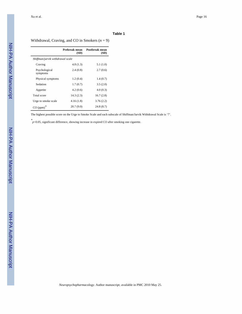
Before the first test, the self reported mean scores on the SJWS subscales ranged from 1.2 to4.8, and the mean UTS score was 4.16 (Table 1). These scores indicated that smokers did notexperience severe withdrawal or craving symptoms before the first test (see Materials andmethods for the definition of scores). Self-reports of withdrawal and craving before the first
Xu et al. Page 5
Neuropsychopharmacology. Author manuscript; available in PMC 2010 May 25.
NIH
-PA Author Manuscript
NIH
-PA Author Manuscript
NIH
-PA Author Manuscript

and second fMRI tests did not show significant differences (Table 1), in keeping with the factthat abstinence from smoking was brief in both cases: 45–60 min for the first test and <20 minfor the second. After smoking a cigarette during the break, the average concentration of CO inexpired air showed a small, statistically significant increase (Table 1, df = 8, t = 4.11, p<0.05).
Stroop Task Performance of Nonsmokers and SmokersBoth groups showed a significant Stroop effect, that is, longer RT during the incongruent thanthe congruent condition in both first and second tests (paired t-test, for nonsmokers, first test:mean Stroop effect = 115.1 ms, SD = 46.3, t = 3.5, p<0.05, df = 12; second test: mean Stroopeffect = 121.6 ms, SD = 70.3, t = 5.0, p<0.05, df = 12; for smokers, first test: mean Stroopeffect = 138.4 ms, SD = 50.2, t = 8.7, p<0.05, df = 8; second test: mean Stroop effect = 124.1ms, SD = 40, t = 8.0, p<0.05, df = 8). There was no significant difference between tests; norwas there a significant group difference in either test. There also were no significant interactionsinvolving the Stroop effect. Scores on the Fagerström Test for Nicotine Dependence were notsignificantly correlated with the Stroop effect exhibited by the smokers in either test. Neithergroup showed a significant difference in error rates between the incongruent and congruentcondition in either test or between tests.
Task-Related Changes in BOLD SignalThe imaging data of the two tests of nonsmokers showed no difference and, therefore, werecombined. Nonsmokers exhibited significantly higher BOLD signal in the incongruent than inthe congruent condition in the left IFG (Figure 1, cluster size = 12609 mm3, MNI coordinatesof the peak voxel: x = −33, y = 0, z = 21), and in the left pre-SMC (Figure 1, size = 1728mm3, MNI coordinate of the peak voxel: x = −9, y = 18, z = 48). As with the nonsmokers, thedata from the combined two tests of smokers also showed significantly higher BOLD signalin the incongruent than in the congruent condition in the left IFG (Figure 2, cluster size = 1971mm3, MNI coordinates of the peak voxel: x = −42, y = 9, z = 21), but also in the left intraparietalsulcus (Figure 2, cluster size = 3942 mm3, MNI coordinates of the peak voxel: x = −30, y =−75, z = 30). Two sample t-tests not did show significant difference in task-related changes ofBOLD signal in the brain between nonsmokers and smokers in either incongruent or congruentconditions, or in the contrast of incongruent minus congruent condition in either test.
A cluster in the right precentral sulcus, including the putative human frontal eye field (FEF,Figure 3a, cluster size = 6075 mm3, MNI coordinates of the peak voxel: x = 43, y = −22, z =50) showed a significant interaction between groups (smokers vs nonsmokers) and tests (firstvs second) in the contrast of incongruent minus congruent condition. We defined this clusteras an ROI and assessed changes (relative to rest) in BOLD signal in both the incongruent andthe congruent conditions in each test of each subject. ROI analysis indicated that this interactionin the right precentral sulcus reflected greater BOLD signal changes in the incongruent thanin the congruent condition of the smokers only in the first test (paired t-test, t = 2.5, p<0.05,df = 8), but not in the second test (after smoking). Nonsmokers did not exhibit a differencebetween the two conditions in either test (Figure 3b). There was no significant correlationbetween nicotine dependence scores (Fagerström Test) and the difference in BOLD signalchanges in this ROI between incongruent and congruent conditions of smokers in either test.
DISCUSSIONWhen abstinent <1 h, smokers showed a greater change in BOLD signal in the right precentralsulcus, including the FEF, in the incongruent than in the congruent condition of the StroopTask. This difference between the two task conditions was not observed after smokers smokeda cigarette, nor was it exhibited by nonsmokers in either test. Literature regarding selectiveattention indicates that activity of the FEF (including the adjacent regions around the precentral
Xu et al. Page 6
Neuropsychopharmacology. Author manuscript; available in PMC 2010 May 25.
NIH
-PA Author Manuscript
NIH
-PA Author Manuscript
NIH
-PA Author Manuscript

sulcus) mediates shifts of attention and discrimination between targets and distracters (Astafievet al, 2003; Hopfinger et al, 2000). For example, FEF neurons in monkeys are activated duringdiscrimination between targets and distracters (Bichot and Schall, 1999; Thompson and Bichot,2005); and the FEF is activated by distracters while human subjects perform an oddball task(Bledowski et al, 2004). This activation is believed to reflect discrimination of distracters fromtargets and diversion of attention from distracters (Bledowski et al, 2004). Previous fMRIstudies found that activity in the FEF is associated with the diversion of attention in space(Astafiev et al, 2003; Hopfinger et al, 2000), the shifting of attention between objects at thesame location, and between features of the same object (Liu et al, 2003; Serences et al,2004).
Activity of FEF neurons mediates selection, generation, and inhibition of responses afterperipheral cues (Astafiev et al, 2003; Connolly et al, 2002; Merriam et al, 2001). The successfulperformance of the incongruent condition requires inhibition of the interference introduced bythe semantic meaning of the word during signal perception, and perhaps also inhibition of thetendency to respond to the semantic meaning during response selection (MacLeod, 1991; Meadet al, 2002). The activity in the FEF observed in the present study may reflect either or bothof these types of inhibition. Our study did not, however, distinguish between potential effectsof brief abstinence or smoking on these types of inhibition.
Relative to young adults, aged subjects have deficits in inhibition of distracters on a selectiveattention task (Maylor and Lavie, 1998). They also exhibit larger differences in the activity inthe right FEF between the incongruent and congruent conditions than young adults while theyperform the Stroop Task during fMRI (Langenecker et al, 2004), suggesting that aging isassociated with reduced functional efficiency of the FEF during performance of the Strooptask. The observed interaction in the present study reflects greater activity in the more difficultcondition of smokers only and only in the test block before acute smoking. It suggests that theinhibitory function of the right FEF is compromised in smokers abstinent for only 45–60 min;and that this compromise is counteracted by smoking a cigarette.
In the present study, smokers did not report severe withdrawal and craving symptomsimmediately before the first test. They also did not show significant differences in withdrawaland craving scores between the first and second tests. Therefore, it is unlikely that the effectof cigarette smoking on the BOLD signal at the right FEF and adjacent precentral sulcus ofsmokers is due to the relief of subjective nicotine withdrawal or craving for cigarettes. Ourfindings suggest that chronic smoking, short abstinence (45–60 min) from cigarette smoking,conditions that existed before the initiation of smoking, or any combination of these factors,impaired the functional efficiency of the right FEF of nicotine-dependent smokers on selectiveattention tasks, and that acute smoking a cigarette ameliorated this functional deficit. This acutereversal effect of cigarette smoking on the function of FEF may be one of several factors thathelp maintain smoking behavior.
In a previous study, smokers, who abstained from cigarette smoking for about 45 min, showedless activation in the parietal cortex and striatum than nonsmokers while they performed acontinuous performance task; and that this hypoactivation in smokers was reversed afterapplication of transdermal nicotine (21 mg, >3 h) (Lawrence et al, 2002). In that study and inours, smokers who were abstinent from cigarette smoking <60 min, exhibited abnormal brainactivation while performing attentional tasks; and this abnormality was reversed by eithercigarette smoking or nicotine administration. The two studies, however, show differentdirections of effects. We found that, before cigarette smoking, smokers showed higher task-related activity than nonsmokers (see Figure 3b) although this difference did not reach our apriori threshold for statistical significance. Notably the two studies used different tasks, andthe abnormalities in activation were in different brain regions.
Xu et al. Page 7
Neuropsychopharmacology. Author manuscript; available in PMC 2010 May 25.
NIH
-PA Author Manuscript
NIH
-PA Author Manuscript
NIH
-PA Author Manuscript

Acetylcholine enhances both bottom-up signal detection and top-down control of signalprocessing, thereby facilitating the processing of goal-related signals, suppressing theprocessing of distracters, and increasing the signal/noise ratio in the cortex (for review, seeSarter et al (2005)). Such actions could increase the functional efficiency of cortex, reducingcortical activity (ie, BOLD signal) required for demanding attentional tasks. When deliveredthrough cigarette smoking, nicotine mimics the action of acetylcholine by binding to nicotinicacetylcholine receptors (nAChRs) and promoting acetylcholine release. By this action,cigarette smoking may have reduced BOLD signal at the right FEF and adjacent precentralsulcus in the incongruent condition after cigarette smoking in our study. The same mechanismmay explain why nicotine administration decreased task related BOLD signals in the frontaland parietal cortices of nonsmokers performing a selective attention task (Thiel et al, 2005).
It is plausible that reduced functional efficiency in the brain following brief abstinence fromsmoking ad libitum until the afternoon of testing may reflect desensitization of nAChRs. About70% of α4β2 nAChRs in cultured cells are desensitized after brief exposure to 10 nM (1.6 ng/ml) (Paradiso and Steinbach, 2003). By comparison, smoking one cigarette can elevate arterialplasma nicotine concentrations above 180 nM (30 ng/ml) for more than 20 min (Gourlay andBenowitz, 1997). In addition, positron emission tomographic assessment of in vivo nAChRoccupancy in smokers indicated that smoking just one cigarette can produce >88% occupancyof α4β2* nAChRs for at least 4 h after smoking (Brody et al, 2006). To the extent that nAChRsin human brain respond to nicotine with desensitization as shown in vitro, smoking adlibitum until 45–60 min before testing could render central nAChRs less responsive tostimulation by endogenous acetylcholine, and thereby decrease functional efficiency. Ifsmokers in this study had a large proportion of their cerebral α4β2* nAChRs desensitized atthe time of testing, the positive effect of smoking one cigarette on selective attention couldreflect activation of a population α4β2* (or other) nAChRs that are resistant to desensitization.Alternatively, the effect may reflect non-nicotine components of tobacco smoke or of thesmoking experience.
In the present study, we did not find significant improvement in task performance of smokersafter they each smoked a cigarette. At the same time, we observed a decrease in BOLD signalin the incongruent condition of the Stroop task, suggesting improved functional efficiency inthe right FEF and adjacent pre-central sulcus. A possible reason for the inconsistency betweenfMRI and behavioral findings is a greater sensitivity of neuroimaging methods as comparedwith purely behavioral tests, as observed previously (Bolla et al, 2003, 2004; Eldreth et al,2004; Goldberg and Weinberger, 2004).
Consistent with our hypothesis and relevant literature (Banich et al, 2000; Brown et al, 1999;Langenecker et al, 2004; Taylor et al, 1997; Zysset et al, 2001), both test groups showed largerincreases of BOLD signal from rest in the left IFG during the incongruent than during thecongruent condition. Neuroimaging studies have indicated that the left IFG mediates inhibitionin verbal working memory (Jonides et al, 1998). Studies using the Stop-signal task found thatthe right IFG is the main cortical site involved inhibition of motor responses (Aron et al,2003; Aron and Poldrack, 2006). The findings of different brain regions mediating inhibitionin different tasks suggest that the inhibition, or cognitive control, involves multiple brain areas,and the exact location is task dependent.
There are limitations in this study. First, as the sample size is small, the results should beconsidered as preliminary. In addition, as CO and nicotine from cigarette smoking may havecomplex effects on cerebral perfusion (Domino et al, 2004; Ghatan et al, 1998; Rose et al,2003), they may affect BOLD signal, which is an essential feature of fMRI signal. Nicotineadministration, however, did not alter the coupling between the BOLD signal and activity ofthe visual cortex in response to photic stimulation in a previous study (Jacobsen et al, 2002),
Xu et al. Page 8
Neuropsychopharmacology. Author manuscript; available in PMC 2010 May 25.
NIH
-PA Author Manuscript
NIH
-PA Author Manuscript
NIH
-PA Author Manuscript

suggesting that this potential confound may not contribute to our findings. In our study,significant interaction between subject groups and tests was only observed in the rightprecentral sulcus and the adjacent FEF, which previous work implicated as contributing toselective attention and response selection. This selective effect is not easily explained bynonspecific effects of CO and/or nicotine on cerebral perfusion. Lastly, our study does notseparate the effects of nicotine from other effects of the smoking experience (physical andemotional). Answering this question would require a larger sample and assay of plasmanicotine.
Our findings suggest that nicotine-dependent smokers have impaired functional efficiency inthe right FEF and adjacent precentral sulcus, as evidenced by greater BOLD signal changeswhen tested in the incongruent than in the congruent condition of the Stroop Task. Nonsmokersdid not show this difference between conditions. As smoking a cigarette removed the differencein smokers, it appears that smoking after even brief abstinence can improve functionalefficiency. This effect appears to reflect normalization of deficient function rather than afacilitation of brain function beyond the level exhibited by healthy nonsmokers. To the extentthat such recovery reflects effects of nicotine per se, smokers might achieve this improvementwith products that deliver nicotine through routes other than smoking.
AcknowledgmentsThis work was supported by NIH grants RO1 DA014093 (EDL), RO1 DA015059 (ALB), MOI RR 00865, UCTobacco-Related Disease Research Program awards 10RT-0091 (EDL) and 11RT-0024 (ALB), VA Merit ReviewType I Award (ALB), T32 DA 07272 (JX), and Philip Morris External Research Program and Philip MorrisInternational contract 02066286 (EDL).
REFERENCESAron AR, Fletcher PC, Bullmore ET, Sahakian BJ, Robbins TW. Stop-signal inhibition disrupted by
damage to right inferior frontal gyrus in humans. Nat Neurosci 2003;6:115–116. [PubMed: 12536210]Aron AR, Poldrack RA. Cortical and subcortical contributions to stop signal response inhibition: role of
the subthalamic nucleus. J Neurosci 2006;26:2424–2433. [PubMed: 16510720]Astafiev SV, Shulman GL, Stanley CM, Snyder AZ, Van E, Corbetta M. Functional organization of
human intraparietal and frontal cortex for attending, looking, and pointing. J Neurosci 2003;23:4689–4699. [PubMed: 12805308]
Banich MT, Milham MP, Atchley RA, Cohen NJ, Webb A, Wszalek T, et al. Prefrontal regions play apredominant role in imposing an attentional ’set’: evidence from fMRI. Brain Res Cogn Brain Res2000;10:1–9. [PubMed: 10978687]
Beck AT, Beamesderfer A. Assessment of depression: The depression inventory. Psychol MeasurePsychopharmacol 1974;7:151–169.
Bench CJ, Frith CD, Grasby PM, Friston KJ, Paulesu E, Frackowiak R, et al. Investigations of thefunctional anatomy of attention using the Stroop test. Neuropsychologia 1993;31:907–922. [PubMed:8232848]
Benowitz NL, Hall SM, Herning RI, Jacob P III, Jones RT, Osman A-L. Smokers of low-yield cigarettesdo not consume less nicotine. N Engl J Med 1983;309:139–142. [PubMed: 6866013]
Bichot NP, Schall JD. Effects of similarity and history on neural mechanisms of visual selection. NatNeurosci 1999;2:549–554. [PubMed: 10448220]
Bledowski C, Prvulovic D, Goebel R, Zanella FE, Linden DE. Attentional systems in target and distractorprocessing: a combined ERP and fMRI study. Neuroimage 2004;22:530–540. [PubMed: 15193581]
Bolla K, Ernst M, Kiehl K, Mouratidis M, Eldreth D, Contoreggi C, et al. Prefrontal cortical dysfunctionin abstinent cocaine abusers. J Neuropsychiatry Clin Neurosci 2004;16:456–464. [PubMed:15616172]
Xu et al. Page 9
Neuropsychopharmacology. Author manuscript; available in PMC 2010 May 25.
NIH
-PA Author Manuscript
NIH
-PA Author Manuscript
NIH
-PA Author Manuscript

Bolla KI, Eldreth DA, London ED, Kiehl KA, Mouratidis M, Contoreggi C, et al. Orbitofrontal cortexdysfunction in abstinent cocaine abusers performing a decision-making task. Neuroimage2003;19:1085–1094. [PubMed: 12880834]
Brett M, Anton JL, Valabregue R, Poline JB. Region of interest analysis using an SPM toolbox.Neuroimage 2002;16:1140–1141. Ref Type: Abstract.
Brody AL, Mandelkern MA, London ED, Olmstead RE, Farahi J, Scheibal D, et al. Cigarette smokingsaturates brain α4β2 nicotinic acetylcholine receptors. Arch Gen Psychiatry 2006;63:907–915.[PubMed: 16894067]
Brown GG, Kindermann SS, Siegle GJ, Granholm E, Wong EC, Buxton RB. Brain activation and pupilresponse during covert performance of the Stroop Color Word task. J Int Neuropsychol Soc1999;5:308–319. [PubMed: 10349294]
Carter CS, Mintun MA, Cohen JD. Interference and facilitation effects during selective attention: AnH2
15O PET study of Stroop task performance. Neuroimage 1995;2:264–272. [PubMed: 9343611]Connolly JD, Goodale MA, Menon RS, Munoz DP. Human fMRI evidence for the neural correlates of
preparatory set. Nat Neurosci 2002;5:1345–1352. [PubMed: 12411958]Domino EF, Ni L, Xu Y, Koeppe RA, Guthrie S, Zubieta J-K. Regional cerebral blood flow and plasma
nicotine after smoking tobacco cigarettes. Prog Neuropsychopharmacol Biol Psychiatry2004;28:319–327. [PubMed: 14751429]
Eldreth DA, Matochik JA, Cadet J-L, Bolla KI. Abnormal brain activity in prefrontal brain regions inabstinent marijuana users. Neuroimage 2004;23:914–920. [PubMed: 15528091]
Ernst M, Heishman SJ, Spurgeon L, London ED. Smoking history and nicotine effects on cognitiveperformance. Neuropsychopharmacology 2001;25:313–319. [PubMed: 11522460]
First, MB.; Spitzer, RL.; Gibbon, M.; Williams, J.; Biometrics Research Department. Structured ClinicalInterview for DSM-IV Axis I Disorders—Patient Edition (SCID-I/P, Version 2.0). New York StatePsychiatric Institute; New York: 1996.
Foulds J, Stapleton J, Swettenham J, Bell N, McSorley K, Russell MAH. Cognitive performance effectsof subcutaneous nicotine in smokers and never-smokers. Psychopharmacology (Berlin)1996;127:31–38. [PubMed: 8880941]
Ghatan PH, Ingvar M, Eriksson L, Stone-Elander S, Serrander M, Ekberg K, et al. Cerebral effects ofnicotine during cognition in smokers and non-smokers. Psychopharmacology (Berlin)1998;136:179–189. [PubMed: 9551775]
Goldberg TE, Weinberger DR. Genes and the parsing of cognitive processes. Trends Cogn Sci2004;8:325–335. [PubMed: 15242692]
Gourlay SG, Benowitz NL. Arteriovenous differences in plasma concentration of nicotine andcatecholamines and related cardiovascular effects after smoking, nicotine nasal spray, andintravenous nicotine. Clin Pharmacol Ther 1997;62:453–463. [PubMed: 9357397]
Gruber SA, Rogowska J, Holcomb P, Soraci S, Yurgelun-Todd D. Stroop performance in normal controlsubjects: an fMRI study. Neuroimage 2002;16:349–360. [PubMed: 12030821]
Hasenfratz M, Battig K. Action profiles of smoking and caffeine: Stroop effect, EEG, and peripheralphysiology. Pharmacol Biochem Behav 1992;42:155–161. [PubMed: 1528938]
Heatherton TF, Kozlowski LT, Frecker RC, Fägerström KO. The Fägerström Test for NicotineDependence: a revision of the Fägerström Tolerance Questionnaire. Br J Addict 1991;86:1119–1127.[PubMed: 1932883]
Heishman SJ. Behavioral and cognitive effects of smoking: relationship to nicotine addiction. NicotineTob Res 1999;1(Suppl 2):S143–S147. [PubMed: 11768172]
Heishman SJ, Taylor RC, Henningfield JE. Nicotine and smoking: a review of effects on humanperformance. Exp Clin Psychopharmacol 1994;2:345–395.
Hopfinger JB, Buonocore MH, Mangun GR. The neural mechanisms of top-down attentional control.Nat Neurosci 2000;3:284–291. [PubMed: 10700262]
Jacobsen LK, Cyril D’Souza D, Einar Mencl W, Pugh KR, Skudlarski P, Krystal JH. Nicotine effects onbrain function and functional connectivity in schizophrenia. Biol Psychiatry 2004;55:850–858.[PubMed: 15050867]
Jacobsen LK, Gore JC, Skudlarski P, Lacadie CM, Jatlow P, Krystal JH. Impact of intravenous nicotineon BOLD signal response to photic stimulation. Magn Reson Imag 2002;20:141–145.
Xu et al. Page 10
Neuropsychopharmacology. Author manuscript; available in PMC 2010 May 25.
NIH
-PA Author Manuscript
NIH
-PA Author Manuscript
NIH
-PA Author Manuscript

Jarvik M, Madsen D, Olmstead R, Iwamoto-Schaap P, Elins J, Eisenberger N, et al. Blood nicotine levelsand subjective craving for cigarettes. Pharmacol Biochem Behav 2000;66:553–558. [PubMed:10899369]
Jonides J, Smith EE, Marshuetz C, Koeppe RA, Reuter-Lorenz PA. Inhibition in verbal working memoryrevealed by brain activation. Proc Natl Acad Sci USA 1998;95:8410–8413. [PubMed: 9653200]
Langenecker SA, Nielson KA, Rao SM. fMRI of healthy older adults during Stroop interference.Neuroimage 2004;21:192–200. [PubMed: 14741656]
Lawrence NS, Ross TJ, Stein EA. Cognitive mechanisms of nicotine on visual attention. Neuron2002;36:539–548. [PubMed: 12408855]
Liu T, Slotnick SD, Serences JT, Yantis S. Cortical mechanisms of feature-based attentional control.Cereb Cortex 2003;13:1334–1343. [PubMed: 14615298]
MacLeod CM. Half a century of research on the Stroop effect: an integrative review. Psychol Bull1991;109:163–203. [PubMed: 2034749]
Mancuso G, Warburton DM, Melen M, Sherwood N, Tirelli E. Selective effects of nicotine on attentionalprocesses. Psychopharmacology (Berlin) 1999;146:199–204. [PubMed: 10525756]
Maylor EA, Lavie N. The influence of perceptual load on age differences in selective attention. PsycholAging 1998;13:563–573. [PubMed: 9883457]
Mead LA, Mayer AR, Bobholz JA, Woodley SJ, Cunningham JM, Hammeke TA, et al. Neural basis ofthe Stroop interference task: response competition or selective attention? J Int Neuropsychol Soc2002;8:735–742. [PubMed: 12240737]
Merriam EP, Colby CL, Thulborn KR, Luna B, Olson CR, Sweeney JA. Stimulus-responseincompatibility activates cortex proximate to three eye fields. Neuroimage 2001;13:794–800.[PubMed: 11304076]
Milham MP, Banich MT. Anterior cingulate cortex: an fMRI analysis of conflict specificity and functionaldifferentiation. Hum Brain Mapp 2005;25:328–335. [PubMed: 15834861]
Newhouse PA, Potter A, Singh A. Effects of nicotinic stimulation on cognitive perfomance. Curr OpinPharmacol 2004;4:36–46. [PubMed: 15018837]
Oldfield RC. The assessment and analysis of handedness: the Edinburgh inventory. Neuropsychologia1971;9:97–113. [PubMed: 5146491]
Paradiso KG, Steinbach JH. Nicotine is highly effective at producing desensitization of rat alpha4beta2neuronal nicotinic receptors. J Physiol 2003;553:857–871. [PubMed: 14555718]
Pardo JV, Pardo PJ, Janer KW, Raichle ME. The anterior cingulate cortex mediates processing selectionin the Stroop attentional conflict paradigm. Proc Natl Acad Sci USA 1990a;87:256–259. [PubMed:2296583]
Pardo JV, Pardo PJ, Janer KW, Raichle ME. The anterior cingulate cortex mediates processing selectionin the Stroop attentional conflict paradigm. Proc Natl Acad Sci USA 1990b;87:256–259. [PubMed:2296583]
Pomerleau CS, Teuscher F, Goeters S, Pomerleau OF. Effects of nicotine abstinence and menstrual phaseon task performance. Addict Behav 1994;19:357–362. [PubMed: 7992670]
Provost SC, Woodward R. Effects of nicotine gum on repeated administration of the Stroop test.Psychopharmacology (Berlin) 1991;104:536–540. [PubMed: 1780425]
Rose JE, Behm FM, Westman EC, Mathew RJ, London ED, Hawk TC, et al. PET studies of the influencesof nicotine on neural systems i cigarette smokers. Am J Psychiatry 2003;160:323–333. [PubMed:12562580]
Rusted JM, Caulfield D, King L, Goode A. Moving out of the laboratory: does nicotine improve everydayattention? Behav Pharmacol 2000;11:621–629. [PubMed: 11198133]
Sarter M, Hasselmo ME, Bruno JP, Givens B. Unraveling the attentional functions of cortical cholinergicinputs: interactions between signal-driven and cognitive modulation of signal detection. Brain ResBrain Res Rev 2005;48:98–111. [PubMed: 15708630]
Schuh KJ, Stitzer ML. Desire to smoke during spaced smoking intervals. Psychopharmacology (Berlin)1995;120:289–295. [PubMed: 8524976]
Serences JT, Schwarzbach J, Courtney SM, Golay X, Yantis S. Control of object-based attention in humancortex. Cereb Cortex 2004;14:1346–1357. [PubMed: 15166105]
Xu et al. Page 11
Neuropsychopharmacology. Author manuscript; available in PMC 2010 May 25.
NIH
-PA Author Manuscript
NIH
-PA Author Manuscript
NIH
-PA Author Manuscript

Stroop JR. Studies of interference in serial verbal reactions. J Exp Psychol 1935;18:643–662.Taylor SF, Kornblum S, Lauber EJ, Minoshima S, Koeppe RA. Isolation of specific interference
processing in the Stroop task: PET activation studies. Neuroimage 1997;6:81–92. [PubMed:9299382]
Thiel CM, Zilles K, Fink GR. Nicotine modulates reorienting of visuospatial attention and neural activityin human parietal cortex. Neuropsychopharmacology 2005;30:810–820. [PubMed: 15668726]
Thompson KG, Bichot NP. A visual salience map in the primate frontal eye field. Prog Brain Res2005;147:251–262. [PubMed: 15581711]
Ward MF, Wender PH, Reimherr FW. The Wender Utah Rating Scale: an aid in the retrospectivediagnosis of childhood attention deficit hyperactivity disorder. Am J Psychiatry 1993;150:885–890.[PubMed: 8494063]
Zysset S, Muller K, Lohmann G, von Cramon DY. Color-word matching stroop task: separatinginterference and response conflict. Neuroimage 2001;13:29–36. [PubMed: 11133306]
Xu et al. Page 12
Neuropsychopharmacology. Author manuscript; available in PMC 2010 May 25.
NIH
-PA Author Manuscript
NIH
-PA Author Manuscript
NIH
-PA Author Manuscript

Figure 1.Nonsmokers (n = 13) showed greater increases in BOLD signal from the rest condition in theleft inferior frontal gyrus and presupplementary cortex in the incongruent than in the congruentcondition. Colors superimposed on the gray scale image, from the single subject T1 braintemplate of SPM2, indicate values of t according to the color bar. Voxel level height thresholdp<0.001, uncorrected. Abbreviations: IFG: inferior frontal gyrus, L: left, pre-SMA:presupplementary motor area, R: right.
Xu et al. Page 13
Neuropsychopharmacology. Author manuscript; available in PMC 2010 May 25.
NIH
-PA Author Manuscript
NIH
-PA Author Manuscript
NIH
-PA Author Manuscript

Figure 2.Smokers (n = 9) showed greater increases in BOLD signal in the left inferior frontal gyrus andintraparietal sulcus from the rest condition in the incongruent than in the congruent condition.Colors superimposed on the gray scale image, from the single subject T1 brain template ofSPM2, indicate values of t according to the color bar. Voxel level height threshold p<0.001,uncorrected. Abbreviations: IFG: inferior frontal gyrus, IPS: intraparietal sulcus, L: left, R:right.
Xu et al. Page 14
Neuropsychopharmacology. Author manuscript; available in PMC 2010 May 25.
NIH
-PA Author Manuscript
NIH
-PA Author Manuscript
NIH
-PA Author Manuscript

Figure 3.Significant interaction of contrast of incongruent minus congruent condition in the rightprecentral sulcus, including the putative human frontal eye field (smokers (n = 9) vsnonsmokers (n = 13) and first vs second test). (a) Colors superimposed on the single subjectT1 template from SPM2 indicate values of t according to the color bar. (b) Difference inpercentage signal change (relative to rest) between the incongruent and congruent conditions.Bar: mean, Error Bar: SD. Voxel level height threshold p<0.001, uncorrected. Abbreviations:L: left, R: right.
Xu et al. Page 15
Neuropsychopharmacology. Author manuscript; available in PMC 2010 May 25.
NIH
-PA Author Manuscript
NIH
-PA Author Manuscript
NIH
-PA Author Manuscript
NIH
-PA Author Manuscript
NIH
-PA Author Manuscript
NIH
-PA Author Manuscript
Xu et al. Page 16
Table 1
Withdrawal, Craving, and CO in Smokers (n = 9)
Prebreak mean(SD)
Postbreak mean(SD)
Shiffman/jarvik withdrawal scale
Craving 4.8 (1.3) 5.1 (1.0)
Psychological symptoms
2.4 (0.8) 2.7 (0.6)
Physical symptoms 1.2 (0.4) 1.4 (0.7)
Sedation 1.7 (0.7) 3.5 (2.0)
Appetite 4.2 (0.6) 4.0 (0.3)
Total score 14.3 (2.3) 16.7 (2.8)
Urge to smoke scale 4.16 (1.8) 3.76 (2.2)
CO (ppm)* 20.7 (9.0) 24.8 (8.7)
The highest possible score on the Urge to Smoke Scale and each subscale of Shiffman/Jarvik Withdrawal Scale is ‘7’.
*p<0.05, significant difference, showing increase in expired CO after smoking one cigarette.
Neuropsychopharmacology. Author manuscript; available in PMC 2010 May 25.
Copyright © 2022 FDOKUMEN