Neural Responses to BEGIN and END-Stimuli of the Smoking Ritual in Nonsmokers, Nondeprived Smokers,...
55
Neural responses to BEGIN- and END-stimuli of the smoking-ritual in non- smokers, non-deprived smokers and deprived smokers Bastian Stippekohl 1 , Markus Winkler 2 , Ronald F. Mucha 2 , Paul Pauli 2 , Bertram Walter 1 , Dieter Vaitl 1 & Rudolf Stark 1 1 Bender Institute of Neuroimaging, University of Giessen, Germany 2 Department of Psychology, University of Würzburg, Germany Correspondence to: Bastian Stippekohl Bender Institute of Neuroimaging Otto-Behaghel-Str. 10 H 35394 Giessen Germany Phone: +49-641-9926087 Fax: +49-641-9926309 Email: [email protected] peer-00504141, version 1 - 20 Jul 2010 Author manuscript, published in "Neuropsychopharmacology n/a, n/a (2010) n/a-n/a" DOI : 10.1038/npp.2009.227
-
Upload
uni-wuerzburg -
Category
Documents
-
view
0 -
download
0
Transcript of Neural Responses to BEGIN and END-Stimuli of the Smoking Ritual in Nonsmokers, Nondeprived Smokers,...
Neural responses to BEGIN- and END-stimuli of the smoking-ritual in non-
smokers, non-deprived smokers and deprived smokers
Bastian Stippekohl1, Markus Winkler2, Ronald F. Mucha2, Paul Pauli2, Bertram
Walter1, Dieter Vaitl1 & Rudolf Stark1
1 Bender Institute of Neuroimaging, University of Giessen, Germany
2 Department of Psychology, University of Würzburg, Germany
Correspondence to:
Bastian Stippekohl
Bender Institute of Neuroimaging
Otto-Behaghel-Str. 10 H
35394 Giessen
Germany
Phone: +49-641-9926087
Fax: +49-641-9926309
Email: [email protected]
peer
-005
0414
1, v
ersi
on 1
- 20
Jul
201
0Author manuscript, published in "Neuropsychopharmacology n/a, n/a (2010) n/a-n/a"
DOI : 10.1038/npp.2009.227

Stippekohl 2
Abstract
Drug associated stimuli (cues) play a prominent role in addiction research because
they are able to provoke craving and relapses. Generally, drug cues are seen as
conditioned excitatory stimuli, which elicit drug seeking and usage. However, newer
data suggest differential effects for smoking stimuli depending on their stage in the
smoking ritual. Specifically, stimuli associated with the terminal stage of smoke
consumption (END-stimuli) may evoke reactivity opposite to the reactivity evoked by
stimuli associated with the beginning of smoke consumption (BEGIN-stimuli).
This fMRI-study compared twenty non-deprived smokers with 20 non-smokers to
unravel the influence of smoking related pictures displaying the beginning (BEGIN-
stimuli) and termination (END-stimuli) of the smoking ritual on neural activity in the
addiction network. Additionally, 20 deprived smokers (12 h deprivation) were
investigated to explore the effects of deprivation on the processing of these stimuli.
In non-deprived smokers, BEGIN-stimuli reliably activated the addiction network (e.g.
ventral striatum, orbitofrontal cortex, anterior cingulate cortex). In contrast, END-
stimuli triggered a differential pattern of activations as well as deactivations;
deactivations were found in the ventral striatum and the ACC. Deprivation had no
clear effect on the responses triggered by BEGIN-stimuli, but affected the reactivity to
END-stimuli.
Our data clearly suggest that stimuli associated with different stages of the smoking-
ritual trigger differential neuronal responses. While BEGIN-stimuli generally seem to
activate the addiction network, END-stimuli presumably have some inhibitory
properties. This new finding might add to a more differentiated understanding of cue-
reactivity and addiction.
peer
-005
0414
1, v
ersi
on 1
- 20
Jul
201
0
Stippekohl 3
Keywords: addiction, smoking, cue-reactivity, fMRI, deprivation, terminal stimuli
peer
-005
0414
1, v
ersi
on 1
- 20
Jul
201
0

Stippekohl 4
Introduction
Core features of addiction are excessive craving, compulsive drug intake and high
rates of relapse (Self and Nestler, 1998; Kalivas and Volkow, 2005; Lubman et al,
2004). Current learning theories of addiction emphasize the prominent role of drug
cues - stimuli associated with drug intake, which may induce craving, lapses and
relapses (Wikler, 1948; Siegel, 1989; Stewart et al, 1984; Robinson and Berridge,
1993, 2003). Empirical data support these assumptions for smokers and smoking
cues (Carter and Tiffany, 1999; Niaura et al, 1988; O´Brien et al, 1998).
Brain research confirmed that addiction is a disorder of learning and memory (Kauer
and Malenka, 2007; Hyman, 2005; Everitt et al, 2001). Evidence suggests that drugs
lead to pathological adaptations in a widely distributed neuronal network divided into
four sub-circuits: reward/salience (ventral tegmental area, ventral striatum);
motivation/drive/craving (orbitofrontal cortex, insula); learning/memory (hippocampus,
amygdala); and control (dorsolateral prefrontal cortex, anterior cingulate cortex)
(Everitt and Robbins, 2005; Volkow et al, 2003, 2004; Naqvi and Bechara, 2009).
Smoking cues were found to activate this network in smokers but not in non-smokers
(Due et al, 2002; David et al, 2005; Brody et al, 2002; Smolka et al, 2006; Franklin et
al, 2007).
However, stimuli associated with different stages of the smoking-ritual seem to
trigger differential responses. Using pictures of different stages of smoking, Mucha et
al (1999, 2008) found evidence that pictures representing the beginning and the
process of smoking (BEGIN-stimuli, e.g. scenes of lighting a cigarette) elicit high
craving. Pictures representing the terminal phase of smoking (END-stimuli, e.g.
scenes of the disposal of a finished cigarette) elicit low craving, although they are
peer
-005
0414
1, v
ersi
on 1
- 20
Jul
201
0
Stippekohl 5
associated with a high nicotine level in the body (see also Bushnell et al, 2000). A
difference was also demonstrated using a physiological measure of motivational
state: contrary to END-stimuli, BEGIN-stimuli attenuate the startle-response similar to
positive pictures (Geier et al, 2000; Mucha et al, 2008; Mucha et al, 2006). However,
END-stimuli are probably not simply weak cues since they are able to reduce the
effects of BEGIN-stimuli when both are presented together (Mucha et al, 2008).
Therefore, END-stimuli seem to trigger a unique reactivity, which even may oppose
responses triggered by BEGIN-stimuli. Mucha et al (2008) suggested this to be an
effect of the perceived smoke availability, which is high for BEGIN- and low for END-
stimuli. Previous studies demonstrated increased subjective, behavioral,
physiological, and neuronal responses to smoking-cues when smoking seems
available (Wertz and Sayette, 2001; Carter and Tiffany, 2001; Wilson et al, 2004;
McBride et al, 2006). Other studies showed that the end of drug consumption and
stimuli predicting the non-occurrence of the drug reduce drug-seeking and wanting
(Kearns et al, 2005; see also Panlilio et al, 2008). In sum, these results point to an
inhibitory nature of END-stimuli.
There are however alternative explanations for the low cue reactivity of END-stimuli
(Mucha et al, 2008). Therefore, it is important that research on END-stimuli has
focused on subjective and physiological responses, but nothing is known about their
neuronal representation. Accordingly, this fMRI-study was designed to investigate
whether END-stimuli have an impact on neuronal activity in the addiction-network
and whether they lead to inhibitory effects. Prototypical BEGIN-(“lighting a cigarette”)
and END-stimuli (“stubbing out a cigarette butt”) were presented to non-deprived
smokers, deprived smokers (12 h deprivation), and non-smokers. Furthermore, our
study included an expansion of the concept of BEGIN- and END-stimuli. Several
animal conditioning studies suggest that conditioned stimuli (CS) lead to different
peer
-005
0414
1, v
ersi
on 1
- 20
Jul
201
0
Stippekohl 6
behavioral responses depending on their temporal proximity to the unconditioned
stimulus (UCS) (Timberlake, 1997; Silva et al, 1996, 1998; Silva & Timberlake,
1997). For example, with food as reinforcer, long CS-UCS intervals lead to general
search behavior, whereas short intervals lead to focal search behavior. A similar
functional organization may also be present in the smoking-ritual. Thus, we added
two exploratory stimuli (“taking a cigarette out of its box”, “the last puff”) to represent
a wider range of the smoking-ritual.
Several directional hypotheses were put forward for BEGIN- and END-stimuli: for
non-deprived and deprived smokers we hypothesized that BEGIN-stimuli lead to
neuronal activations, whereas END-stimuli should result in deactivations. Regarding
group differences, we expected that these effects are significantly different between
non-deprived smokers and non-smokers. Similar group differences were expected
between deprived and non-deprived smokers, because learning theories suggest an
enhancement of the incentive-value of drug-associated stimuli under deprivation
(Stewart et al, 1984; Toates, 1994; Berridge, 2004). We had no directional
hypotheses for the effects of the additional stimuli. However, we also expected
significant group differences for them.
peer
-005
0414
1, v
ersi
on 1
- 20
Jul
201
0
Stippekohl 7
Method
Subjects
Sixty right-handed subjects participated in the study: 20 non-deprived smokers (11
female), 20 deprived smokers (10 female), and 20 non-smokers (10 female). Non-
deprived and deprived smokers were included if they had smoked for at least three
years and a mean of at least 15 cigarettes per day in the last 12 months; non-
smokers had to have smoked less than 10 cigarettes in their lifetime. The Edinburgh
Inventory of Handedness (Oldfield, 1971) ensured that only right-handed subjects
participated. A self-generated questionnaire was used to assess general information
regarding smoking history. The Fagerström Test of Nicotine Dependence (FTND;
Schumann et al, 2003a) was used to assess the severity of addiction. Deprived
smokers were instructed to go to bed before 12:00 pm the day before the experiment
and were scheduled after 12:00 am the day of the experiment. This resulted in a
deprivation phase of at least 12 hours. The procedure was the same for non-deprived
smokers and non-smokers.
Most subjects were students receiving either money (10 Euro/hour) or course credit.
No subject was taking regular medication or had a history of psychiatric or
neurological illness. None of the participants reported abuse of other drugs. The
study was performed in accordance with the ethical standards of the fifth revision of
the Declaration of Helsinki and approved by the Ethical Committee of the German
Psychological Association.
Stimulus material
peer
-005
0414
1, v
ersi
on 1
- 20
Jul
201
0

Stippekohl 8
We used self-generated pictures and videos depicting different stages of the
smoking-ritual without showing many irrelevant or distracting features (e.g., faces,
surroundings, cigarette brands). As neutral controls, stimuli depicting equivalent
temporal stages of teeth-brushing were used. Teeth-brushing can be matched to the
smoking-process with respect to temporal stages, body parts, and equipment. It can
be considered as neutral because it is an over-learned every day routine. All props
(e.g., ashtrays, lighters, toothbrushes, beakers) were matched for color.
We recorded 12 video clips using 12 different models (6 female) while smoking or
teeth-brushing. We then cut 4 sequences (4 seconds each) out of each clip. Each
represented one of four different stages of smoking or teeth-brushing. Then, one
representative static picture from each clip was selected. Smoking-stages were:
“taking a cigarette out of its box” (S1), “lighting a cigarette” (S2), “last puff on a
cigarette” (S3), and “stubbing out a cigarette butt” (S4). Teeth-brushing stages were:
“preparing a toothbrush with toothpaste” (T1), “putting a toothbrush into ones mouth”
(T2), “taking a toothbrush out of ones mouth” (T3), and “putting a toothbrush back
into the beaker” (T4). Overall, eight different picture categories were used, with every
smoking-stage having its own teeth-brushing stage as control (S1, S2, S3, S4, T1,
T2, T3, T4). Among these stimuli, we focused on S2 and S4, since they are
prototypical BEGIN- (S2) and END-stimuli (S4) of smoking. S1 and S3 were
considered as exploratory to account for a wider range of the smoking-ritual.
To study differential effects of pictures and video clips, both were presented in
separate and counterbalanced sessions on different days one week apart. The data
regarding the picture stimuli will be the focus of this paper. Results regarding the
video stimuli will be reported separately. All pictures were presented in 800 x 600
pixel resolution. An LCD projector (model EPSON EMP-7250) projected them onto a
peer
-005
0414
1, v
ersi
on 1
- 20
Jul
201
0

Stippekohl 9
screen at the end of the scanner (visual field=18°) where they were viewed through a
mirror mounted on the head coil. Pictures were presented for 4 seconds in
randomized order (restriction: not more than two stimuli of the same category after
one another).
Participants rated the stimuli right after their presentation on the dimensions craving,
valence and arousal on 9-point Likert scales using a three-button keypad (left, right,
enter). For craving, the scale was visualized with bars of different heights
representing the amount of craving. For valence and arousal, a computerized version
of the Self Assessment Manikin (SAM; Bradley and Lang, 1994) was applied. Ratings
of craving were assessed after each stimulus. Valence and arousal ratings
interchanged to keep the experiment short (each stimulus category was rated half on
valence and half on arousal).
Procedure
Subjects participated in three different sessions.
Session 1. The study was explained, possible contraindications were checked and
written consent was obtained. Smokers were randomly assigned to the non-deprived
or the deprived group. All subjects were instructed to go to bed before 12 pm on the
days before the experiments (sessions 2 and 3). Deprived smokers were instructed
not to smoke from bedtime to the end of the experimental sessions. After reading the
instructions, all smokers were told to smoke one cigarette. This was necessary to
measure a carbon monoxide (CO)- and craving-baseline (German version of the
Questionnaire on Smoking Urges, QSU-G; Müller et al, 2001) when non-deprived.
CO-values and craving were also assessed in non-smokers. All subjects had to fill in
a self-generated questionnaire regarding their individual smoking history. Afterwards,
peer
-005
0414
1, v
ersi
on 1
- 20
Jul
201
0

Stippekohl 10
an anatomical measurement took place in the MRI scanner to get participants
accustomed. Afterwards, smokers completed different smoking-related
questionnaires (analyses not included).
Session 2 and 3. Each experimental session started between noon and 6 pm. Non-
deprived smokers were allowed to smoke one cigarette before the procedure started.
Then, all subjects completed a CO-measurement. Subsequently, they filled in the
German version of the Questionnaire on Smoking Urges (QSU). CO-levels were
used to confirm deprivation. All deprived smokers had CO-levels ≤ 13 CO-ppm,
which is similar to the mean value found in other studies with overnight deprivation
(Due et al, 2002; Della Casa et al, 1999; Mucha et al, 1999). Next, participants were
familiarized with the stimulus-presentation and rating procedure outside the scanner.
Only neutral pictures from the International Affective Picture System (IAPS; Lang et
al, 1995) were presented. Subjects were then placed in the scanner. Another test-
rating took place before the experiment. After the experiment all smokers were
allowed to smoke. They then filled in personality questionnaires (analyses not
included). After completing all sessions, participants were paid and debriefed.
Data acquisition and analysis
Brain images were acquired using a 1.5 T whole-body tomography (Siemens
Symphony) with a standard head coil. Structural image acquisition consisted of 160
T1-weighted sagittal images (MPRage, 1 mm slice thickness). A gradient echo field
map sequence was measured before the functional run to get information for
unwarping B0 distortions. For functional imaging a total of 780 volumes were
registered using a T2*-weighted gradient echo-planar imaging sequence (EPI) with
25 transversal slices covering the whole brain (slice thickness = 5 mm; 1 mm gap;
peer
-005
0414
1, v
ersi
on 1
- 20
Jul
201
0

Stippekohl 11
descending; TR = 2.5 s; TE = 55 ms; flip angle = 90°; field of view = 192 x 192 mm;
matrix size = 64 x 64). The first three volumes were discarded due to an incomplete
steady state of magnetisation. Orientation of slices was parallel to the AC-PC line.
The statistical parametric mapping software package (SPM5, Wellcome Department
of Cognitive Neurology, London, UK) implemented in Matlab (Mathworks, Inc.,
Natick, MA, USA, Release 2007b) was used for preprocessing and statistical
analyses. Origin coordinates were adjusted to the anterior commissure (AC), and
realignment (third order B-spline) and unwarping, slice time correction, and
normalization to the standard brain of the Montreal Neurological Institute (MNI) were
performed. Smoothing was executed with an isotropic three-dimensional Gaussian
filter with a full width at half maximum (FWHM) of 9 mm.
For the event-related design, each picture presentation was modeled with a box-car
function convolved with a hemodynamic response function (duration 4 s) in the
General Linear Model of SPM. The six movement parameters of the rigid body
transformation applied by the realignment procedure were introduced as covariates
in the model. The serial correlation in the voxel-based time series was considered as
a first order autoregressive process. A high pass filter (time constant = 128 s) was
implemented by using cosine functions in the design matrix.
For analyses of hemodynamic responses, contrasts between smoking and
corresponding control stimuli (smoking minus control) were calculated on the first
level: S1-T1 is called TAKING OUT, S2-T2 BEGIN, S3-T3 LAST PUFF, and S4-T4
END. These contrasts were then used as dependent variables on the second level.
Activations were defined as positive differences between smoking and control stimuli;
deactivations as negative differences. The second level model was a variance
analytical model with the factors group and stage (four stages). Our data analysis
comprised a confirmatory part and an exploratory part: in the confirmatory part,
peer
-005
0414
1, v
ersi
on 1
- 20
Jul
201
0

Stippekohl 12
appropriate contrasts for each hypothesis were constructed within this model and T-
tests were applied to these contrasts. Group comparisons (non-deprived smokers vs.
non-smokers; deprived smokers vs. non-deprived smokers) were done for 1) BEGIN-
stimuli, 2) END-stimuli and 3) differences between BEGIN- and END-stimuli.
Afterwards, exploratory analyses were performed by comparing the groups (non-
deprived smokers vs. non-smokers; deprived smokers vs. non-deprived smokers) for
all four smoking stimuli in two 2 by 4 plans.
Regions of interest (ROI) analyses were performed for: dorsolateral prefrontal cortex
(DLPFC), orbitofrontal cortex (OFC), anterior cingulate cortex (ACC), ventral
tegmental area (VTA), ventral striatum, amygdala, hippocampus, and insula. Results
were corrected for multiple testing in each ROI according to Worsley (2001).
Ratings were analyzed with SPSS 17.0. The analysis was analogous to that of the
brain data but without computing differences between smoking and control stimuli
beforehand. For the repeated measure factor stage, the Greenhouse-Geisser
correction was applied. Post hoc comparisons were performed using the Bonferroni-
adjustment for multiple comparisons. For all statistical analyses α was set to .05.
peer
-005
0414
1, v
ersi
on 1
- 20
Jul
201
0

Stippekohl 13
Results
Group characteristics
The final analyses are based on 20 deprived smokers (10 female), 19 non-deprived
smokers (11 female), and 17 non-smokers (9 female). Four subjects had to be
excluded: one smoker and one non-smoker due to technical problems and two non-
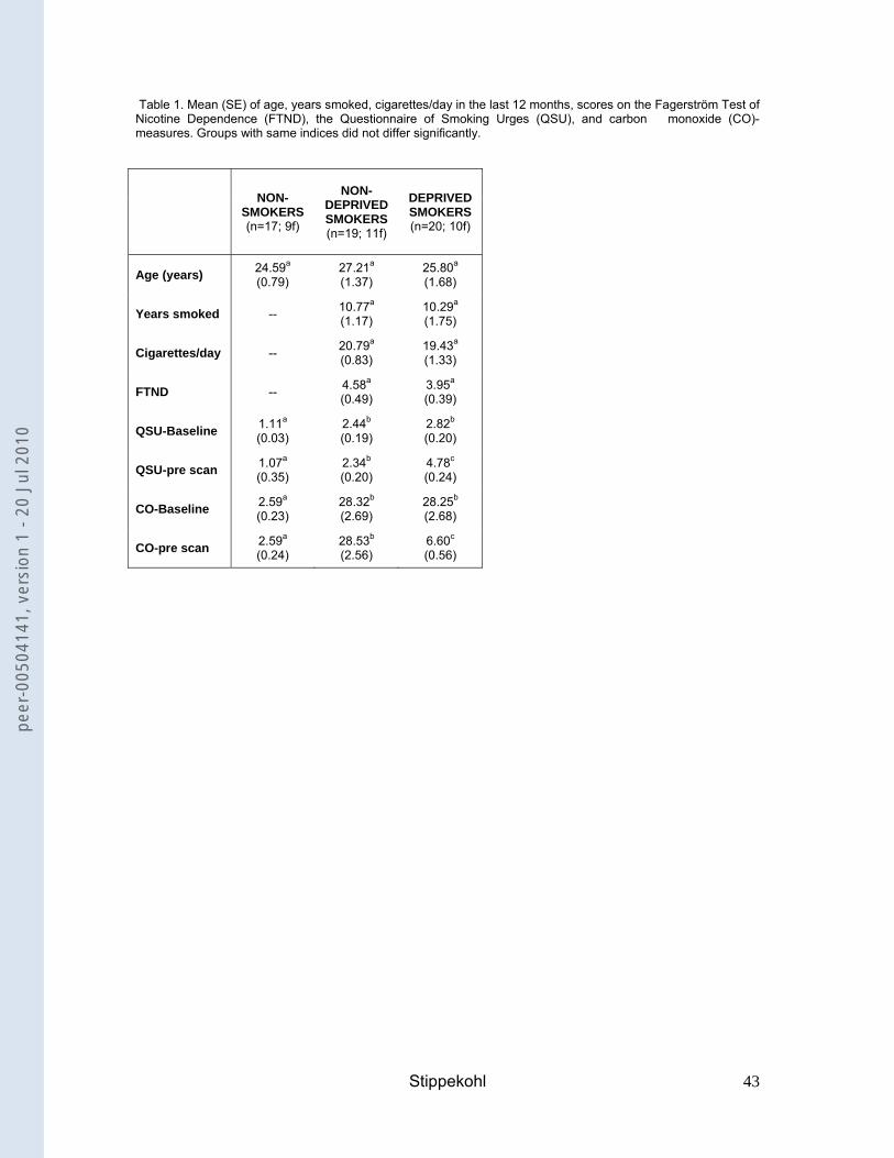
smokers because they had smoked more than 10 cigarettes in their lifetime. Table 1
summarises relevant group characteristics.
As intended, the groups did not differ in age. Non-deprived smokers and deprived
smokers did not differ in years smoked, mean number of cigarettes per day within the
last 12 months and FTND-score. As expected, the three groups differed in craving
and CO-levels at baseline measured in session 1 (QSU-baseline: F(2, 52) = 26.261,
p<.001; CO-baseline: F(2, 53) = 39.694, p<.001). Craving scores and CO-levels were
higher in non-deprived smokers and deprived smokers than in non-smokers (QSU-
baseline: non-deprived smokers vs. non-smokers: mean difference = 1.33, p<.001,
deprived smokers vs. non-smokers: mean difference = 1.71, p<.001; CO-baseline:
non-deprived smokers vs. non-smokers: mean difference = 25.73, p<.001, deprived
smokers vs. non-smokers: mean difference = 25.66, p<.001). On the day of the
scanning session, deprived smokers showed higher craving scores (t(37) = 7.740,
p<.001) and lower CO-levels (t(37) = -8.585, p<.001) than non-deprived smokers.
They also had significantly higher craving scores and lower CO-levels than at
baseline (QSU: t(19) = 7.976, p<.001; CO-levels: t(19) = -8.386, p<.001). Thus, CO-
and QSU-levels confirmed the effectiveness of the deprivation manipulation.
peer
-005
0414
1, v
ersi
on 1
- 20
Jul
201
0

Stippekohl 14
--- insert Table 1 ---
Picture ratings for BEGIN- and END-stimuli
The following section focuses on the rating data for smoking stimuli (differences to
control stimuli, differences to other smoking stimuli, group differences). Differences
with regard to control stimuli (differences to other control stimuli, group differences)
are not reported in the text but can be seen in Table 2.
Non-deprived smokers compared to non-smokers
Craving (Figure 1, Table 2). In non-deprived smokers, BEGIN- and END-stimuli led to
more craving than the corresponding control stimuli (BEGIN: t(18)=4.40, p<.001;
END: t(18)=2.57, p=.019). BEGIN-stimuli led to higher craving than END-stimuli
(t(18)=4.22; p=.001). For non-smokers, no significant results were found. Comparing
both groups, non-deprived smokers revealed higher craving ratings than non-
smokers for BEGIN- and END-stimuli (BEGIN: t(18)=6.96, p<.001; END: t(18)=6.63,
p<.001).
--- insert Figure 1 ---
Valence (Table 2). Non-deprived smokers rated END-stimuli as significantly less
pleasurable than the corresponding control stimuli (t(18)=-3.39, p=.003). BEGIN-
stimuli were rated as more pleasurable than END-stimuli (t(18)=2.69, p=.015). Non-
smokers rated BEGIN- and END-stimuli significantly less pleasurable than the
corresponding control stimuli (BEGIN: t(16)=-2.49, p=.024; END: t(16)=-3.21,
peer
-005
0414
1, v
ersi
on 1
- 20
Jul
201
0

Stippekohl 15
p=.005). Comparing both groups, non-deprived smokers rated BEGIN-stimuli as
significantly more pleasurable than non-smokers (t(23.09)=3.57, p=.002).
Arousal (Table 2). Non-deprived smokers rated BEGIN-stimuli as significantly more
arousing than the corresponding control stimuli (t(18)=3.65, p=.002). The same was
true for non-smokers (t(16)=2.17, p=.046). Comparing both groups, no significant
differences were observed.
--- insert Table 2 ---
Deprived smokers compared to non-deprived smokers
The ratings of one deprived smoker had to be excluded from the analysis due to
excessive missing values.
Craving (Figure 1, Table 2). In deprived smokers, BEGIN- and END-stimuli led to
more craving than the corresponding control stimuli (BEGIN: t(18)=5.96, p<.001;
END: t(18)=4.88, p<.001). BEGIN-stimuli led to higher craving than END-stimuli
(t(18)=4.38; p<.001). In comparison with non-deprived smokers, deprived smokers
showed higher craving ratings for BEGIN- and END-stimuli (BEGIN: t(36)=2.86,
p=.007; END: t(36)=3.71, p=.001).
Valence (Table 2). Deprived smokers rated BEGIN-stimuli as significantly more
pleasurable than the corresponding control stimuli (t(18)=3.07, p=.007) and END-
stimuli (t(18)=2.87, p=.010). In comparison with non-deprived smokers, no significant
differences were observed.
peer
-005
0414
1, v
ersi
on 1
- 20
Jul
201
0
Stippekohl 16
Arousal (Table 2). Deprived smokers rated BEGIN- and END-stimuli as significantly
more arousing than the corresponding control stimuli (BEGIN: t(18)=4.34, p<.001;
END: t(18)=4.48, p<.001). BEGIN-stimuli were rated as more arousing than END-
stimuli (t(18)=4.82, p<.001). In comparison with non-deprived smokers, deprived
smokers rated BEGIN- as well as END-stimuli as significantly more arousing (BEGIN:
t(36)=5.58, p<.001; END: t(36)=4.08, p<.001).
Ratings of additional stimuli
Craving (Table 2). “Taking a cigarette out of its box” led to higher craving in non-
deprived smokers and deprived smokers than the corresponding control stimuli (non-
deprived smokers: t(18)=4.43, p<.001; deprived smokers: t(18)=5.99, p<.001). No
such differences were found for non-smokers. Similar results were found for “the last
puff” (non-deprived smokers: t(18)=4.00, p=.001; deprived smokers: 5.60, p<.001).
Valence (Table 2). Deprived smokers rated “taking a cigarette out of its box” as
significantly more pleasurable than the corresponding control stimuli (t(18)=3.64,
p=.002). Non-smokers rated this stimulus as significantly less pleasurable than
control stimuli (t(16)=-2.72, p=.015). No significant results were found for non-
deprived smokers. Similar results were found for “the last puff” (deprived smokers:
t(18)=3.54, p=.002; non-smokers: t(16)= -3.09, p=.007).
Arousal (Table 2). “Taking a cigarette out of its box” was rated significantly more
arousing than the corresponding control stimuli by non-deprived smokers and
deprived smokers (non-deprived smokers: t(18)=4.62, p<.001; deprived smokers:
t(18)=4.49, p<.001). No significant results were found for non-smokers. “The last
peer
-005
0414
1, v
ersi
on 1
- 20
Jul
201
0
Stippekohl 17
puff” was rated as significantly more arousing than the corresponding control stimuli
by deprived smokers (t(18)=4.41, p<.001).
Exploratory analyses of differences between the four smoking stimuli
To account for group differences in changes over time, the interaction between group
and stage was analyzed in two 2 x 4 plans.
Craving (Table 2). Comparing non-deprived smokers and non-smokers, significant
main effects of group and stage as well as a significant group x stage interaction
were found (for all F-values p<.001). Post hoc analyses revealed that for non-
deprived smokers, END-stimuli led to significantly lower craving than all other
smoking stimuli (all p<.01). BEGIN-stimuli led to significantly more craving than “the
last puff” (mean difference = 0.65, p=.045), whereas no significant differences were
found between BEGIN-stimuli and “taking a cigarette out of its box”.
Comparing deprived smokers and non-deprived smokers, significant main effects of
group and stage were found, with deprived smokers showing higher craving (all
p<.004). Post hoc analyses in both groups combined revealed that END-stimuli led to
significantly lower craving than all other smoking stimuli (all p<.001). BEGIN-stimuli
led to significantly more craving than “taking a cigarette out of its box” (mean
difference = 0.25, p=.038) and “the last puff” (mean difference = 0.58, p=.007),
whereas no significant differences were found between “taking a cigarette out of its
box” and “the last puff”.
Valence (Table 2). Comparing non-deprived smokers and non-smokers, main effects
of group and stage were found, with non-deprived smokers showing higher ratings
peer
-005
0414
1, v
ersi
on 1
- 20
Jul
201
0

Stippekohl 18
(all p<.003). Post hoc analyses in both groups combined revealed that “taking a
cigarette out of its box” was rated as significantly more pleasurable than “the last
puff” and END-stimuli. BEGIN-stimuli were rated as significantly more pleasurable
than “the last puff” (all p<.05).
Comparing deprived smokers and non-deprived smokers, a significant main effect of
stage was found (F(1.90, 68.33)=11.39, p<.001). Post hoc analyses in both groups
combined revealed that END-stimuli were rated as significantly less pleasurable than
all other smoking stimuli (all p<.01).
Arousal (Table 2). Comparing non-deprived smokers and non-smokers, no significant
results were found.
Comparing deprived smokers and non-deprived smokers, significant main effects of
group and stage were found, with deprived smokers showing higher ratings (all
p<.002). Post hoc analyses in both groups combined revealed that END-stimuli were
rated as significantly less arousing than all other smoking stimuli (all p<.05).
Brain responses to BEGIN- and END-stimuli
Non-deprived smokers compared to non-smokers
BEGIN
peer
-005
0414
1, v
ersi
on 1
- 20
Jul
201
0
Stippekohl 19
For the contrast BEGIN, non-deprived smokers showed activations in the VTA, the
ventral striatum, the OFC, the ACC, the DLPFC, the hippocampus, and the insula
(Table 3). In non-smokers we found activations in the OFC, the ACC and in the
DLPFC. Comparing both groups, non-deprived smokers exhibited significantly
greater activations than non-smokers in the VTA, the DLPFC, and the insula (Table
4).
--- insert Table 3 ---
---insert Table 4---
END
For the contrast END, non-deprived smokers showed deactivations in the ventral
striatum, the ACC, the DLPFC, and the insula (Table 3). Yet despite deactivations,
they also showed activations in the OFC, the DLPFC, the hippocampus, and the
insula. No activations or deactivations were found for non-smokers. Comparing both
groups, non-deprived smokers showed stronger activations than non-smokers in the
OFC and the DLPFC (Table 4).
Differences between the contrasts BEGIN and END (BEGIN-END).
Non-deprived smokers showed stronger neuronal activity for BEGIN than for END in
the ventral striatum and the ACC (Table 5). Non-smokers showed no differences
between these contrasts. Comparing both groups, non-deprived smokers revealed
smaller differences between BEGIN and END in the OFC than non-smokers (Table
4).
peer
-005
0414
1, v
ersi
on 1
- 20
Jul
201
0
Stippekohl 20
--- insert Table 5 ---
Figure 2 visualises the most important findings. “Lighting up a cigarette”, a
prototypical BEGIN-stimulus, led to activations in the ACC, the DLPFC and the
ventral striatum of non-deprived smokers. In contrast, “stubbing out a cigarette butt”,
a prototypical END-stimulus, triggered deactivations in exactly the same structures.
--- insert Figure 2 ---
Deprived smokers compared to non-deprived smokers
BEGIN
For the contrast BEGIN, deprived smokers showed activations in the VTA, the OFC,
the ACC, the DLPFC, the hippocampus, and the insula (Table 3). In comparison with
non-deprived smokers, deprived smokers showed a significantly stronger activation
in the DLPFC (Table 4). However, at a different voxel-coordinate, non-deprived
smokers also showed a significantly stronger activation than deprived smokers in this
structure.
END
For the contrast END, deprived smokers showed deactivations in OFC and DLPFC.
They also showed activations in the OFC and the ventral striatum (Table 3). In
comparison with non-deprived smokers, an effect of deprivation was found in the
OFC, the DLPFC, and the insula, with deprived smokers showing significantly lower
neuronal activity than non-deprived smokers (Table 4).
peer
-005
0414
1, v
ersi
on 1
- 20
Jul
201
0

Stippekohl 21
Differences between the contrasts BEGIN and END
In deprived smokers, differences between BEGIN and END were found in the VTA,
the OFC, the ACC, the DLPFC, and the insula (Table 5). In comparison with non-
deprived smokers, deprived smokers revealed significantly greater differences in the
OFC and the DLPFC (Table 4).
Brain responses to additional smoking stimuli (TAKING OUT, LAST PUFF).
For the contrast TAKING OUT, non-deprived smokers showed activations in the
OFC, the ACC, the DLPFC, and the insula (Table 3). Additionally, they showed
deactivations in the ventral striatum, the ACC, and the DLPFC. Deprived smokers
showed activations in the OFC, the ACC, the DLPFC, the hippocampus, and the
insula (Table 3). No activations or deactivations were found for non-smokers.
For the contrast LAST PUFF, non-deprived smokers showed activations in the VTA,
the ventral striatum, the OFC, the ACC, the DLPFC, the amygdala, the hippocampus,
and the insula (Table 3). Deprived smokers showed activations in the ventral
striatum, the OFC, the ACC, the DLPFC, the amygdala, the hippocampus, and the
insula (Table 3). No activations or deactivations were found for non-smokers.
Exploratory analysis of differences between the four smoking stimuli
To account for group differences in changes over time, the interaction between group
and stage was analyzed in two 2 x 4 plans.
peer
-005
0414
1, v
ersi
on 1
- 20
Jul
201
0

Stippekohl 22
Comparing non-deprived smokers and non-smokers, a significant interaction was
found in the right amygdala (Table 6); the greatest differences between both groups
appeared for BEGIN and LAST PUFF. Non-deprived smokers showed strong
amygdala activations for LAST PUFF and deactivations for BEGIN; the reverse was
true for non-smokers. Contrast values for TAKING out and END were close to zero in
both groups.
Comparing deprived smokers and non-deprived smokers, a significant main effect of
stage was found in the OFC, the ACC, and the insula. Figure 3 shows the contrast
estimates for the combined group (non-deprived and deprived smokers) and the four
conditions for the ACC. Notably, contrast values decreased over the time course of
the smoking-ritual and showed deactivations for END. Similar progressions were
found for the OFC and the insula.
--- insert Table 6 ---
--- insert Figure 3 ---
Additionally to this main effect, we found significant interactions between group and
stage in the DLPFC and the hippocampus (Table 6). Figure 4 shows the DLPFC-
contrast estimates for both groups in the four conditions. In the DLPFC, deprived
smokers showed a decrease in contrast estimates over the four conditions resulting
in deactivations for LAST PUFF and END. Non-deprived smokers showed an
increase in contrast estimates up to the point of LAST PUFF (deactivations for
TAKING OUT and BEGIN) and deactivations for END. Notably, for END, deprived
smokers showed a stronger deactivation than non-deprived smokers. Opposing
trends in both groups can be observed for TAKING OUT and LAST PUFF. The
greatest difference between the groups appeared for TAKING OUT and only a small
peer
-005
0414
1, v
ersi
on 1
- 20
Jul
201
0
Stippekohl 23
difference can be seen for BEGIN. Responses in the hippocampus showed a less
regular progression. For deprived smokers, activations were found for TAKING OUT
and LAST PUFF, whereas deactivations were found for BEGIN and END. In non-
deprived smokers, deactivations occurred for TAKING OUT, whereas there were
activations in response to the other conditions. The greatest differences between
non-deprived smokers and deprived smokers appeared for TAKING OUT and END.
--- insert Figure 4 ---
peer
-005
0414
1, v
ersi
on 1
- 20
Jul
201
0
Stippekohl 24
Discussion
This study addressed two main questions: firstly, we wanted to explore whether
stimuli associated with the beginning of smoking (BEGIN-stimuli) and stimuli
associated with the terminal stage of smoking (END-stimuli) differentially activate the
neural addiction network of smokers. This was based on the hypothesis that the
reactivity evoked by a drug-associated stimulus is critically influenced by its position
in the drug-consumption ritual. Specifically, we expected BEGIN-stimuli to be
excitatory as reflected in activations of the addiction network whereas END-stimuli
should have no excitatory or even inhibitory effects as reflected in deactivations.
Secondly, we were interested in the effects of deprivation on these processes and
expected an enhancement of differential effects of BEGIN- and END-stimuli under
deprivation. Additional exploratory questions addressed the issue whether other
stimuli from the smoking ritual also evoke differential responses.
Craving, valence and arousal
Generally, the used stimuli were rated in accordance with our hypotheses.
All smoking stimuli elicited higher craving than the corresponding control stimuli in
non-deprived and deprived smokers. Furthermore and in line with our previous
findings (Mucha et al, 1999, 2008), in both groups BEGIN-stimuli elicited more
craving than END-stimuli. Interestingly, stimuli depicting events immediately before
the termination of smoking (“the last puff”) also evoked more craving than END-
stimuli even though they show a similar association with the peak of the nicotine-
effect. In contrast, stimuli depicting events immediately before the start of actual
peer
-005
0414
1, v
ersi
on 1
- 20
Jul
201
0
Stippekohl 25
smoke-consumption (“taking a cigarette out of its box”) elicited less craving than
BEGIN-stimuli. Overall, deprived smokers reported more craving in response to
smoking stimuli than non-deprived smokers; this pattern also emerged in response to
control stimuli, most probably due to craving baseline differences (see QSU pre-scan
scores). Differences between the craving ratings elicited by smoking vs. control
stimuli were similar for non-deprived and deprived smokers. Thus, considering
craving, we conclude that stimuli associated with different stages of the smoking
ritual differentially affect craving in smokers. These effects are only slightly affected
by deprivation.
A difference between BEGIN- and END-stimuli was also found for valence: similar to
Mucha and colleagues (1999, 2008), non-deprived and deprived smokers rated
BEGIN-stimuli as more pleasurable than END-stimuli. At least for deprived smokers,
this difference also occurred for arousal ratings. These results demonstrate that
differences between BEGIN- and END-stimuli do not only exist for craving, but also
for two important dimensions of emotional experience. For deprived smokers we
found that they were significantly more aroused by smoking stimuli than non-deprived
smokers. This observation supports the assumption of an enhancement of the
incentive value of smoking stimuli under deprivation (Stewart et al, 1984, Toates,
1994; Berridge, 2004). In sum, we conclude that BEGIN- and END-stimuli lead to
pronounced differences in ratings and therefore should also differentially activate the
neural addiction network.
Neural impact of BEGIN- and END-stimuli in non-deprived smokers
In non-deprived smokers, prototypical BEGIN-stimuli (“lighting a cigarette”) triggered
activations in almost the entire addiction-network. Significant stronger activations
peer
-005
0414
1, v
ersi
on 1
- 20
Jul
201
0
Stippekohl 26
than in non-smokers were found in the VTA, the DLPFC, and the insula, three
structures critically involved in reward processing, the generation and maintenance of
consumption related goals, and craving (Everitt and Robbins, 2005; Wilson et al,
2004; Naqvi and Bechara, 2009). This is in accordance with our hypothesis and
resembles results obtained by others (e.g. Due et al, 2002; Brody et al, 2002). In
contrast, prototypical END-stimuli (“stubbing out a cigarette butt)” led to a more
complex pattern of activations and deactivations. Importantly, in non-deprived
smokers, deactivations in response to END-stimuli were found in parts of the reward-
and the control-sub-circuit: ventral striatum and ACC. Contrasting BEGIN- and END-
stimuli, we also found higher neural activity towards BEGIN-stimuli in these two
structures. This shows that END-stimuli have indeed a differential impact on the
neuronal activity in the addiction-network of non-deprived smokers. Since the ventral
striatum and the ACC play important roles for addiction in terms of incentive
motivation, reward detection, and disinhibition (e.g. Lubman et al, 2004; Robinson
and Berridge, 1993, 2003; Volkow et al, 2003, 2004), the observed deactivations in
these structures may indicate inhibitory effects of prototypical END-stimuli.
The role of the ventral striatum in reward processing and reward expectancy is widely
acknowledged (e.g., Spanagel and Weiss, 1999; Wise, 2004) and its activity has
been shown to be correlated with the magnitude of a received reward (Knutson et al,
2001, 2005). Moreover, unexpected omissions of a reward as well as stimuli
predictive of reward omissions (i.e. a conditioned inhibitor) lead to an inhibition of
dopaminergic neurons (Schultz, 1998, 2002; Tobler et al, 2003). Furthermore,
previous behavioral studies demonstrated that stimuli predicting the absence of drug
can suppress drug-seeking (Kearns et al 2005). Therefore, the deactivations
observed in our study might point to a reduction of an incentive motivational state in
response to prototypical END-stimuli in smokers.
peer
-005
0414
1, v
ersi
on 1
- 20
Jul
201
0
Stippekohl 27
The ACC is part of the limbic affect-response system (Wexler et al, 2001) and
interconnected with the ventral striatum (Vogt et al, 1992; Devinsky et al, 1995; Bush
et al, 2000). In addicts, it is one of the structures, in which activity in response to
stimuli paired with a variety of drugs such as nicotine (Brody et al, 2002; Due et al,
2002; McBride et al, 2006; McClernon et al, 2005), cocaine (Maas et al, 1998;
Garavan et al, 2000; Kilts et al, 2001; Childress et al, 1999; Wexler et al, 2001) and
heroin (Daglish et al, 2001) is most frequently reported. Moreover, its importance for
addiction is emphasized by the fact that surgical lesioning of the ACC can decrease
drug consumption in addicts (Sharma, 1974; Kanaka and Balasubramaniam, 1978).
Likewise, smokers treated with bupropion showed an attenuation of both, cue-
induced cigarette craving and ACC activation (Brody et al, 2004). Like the ventral
striatum, the ACC is involved in reward processing and it also seems to be sensitive
to the magnitude of a received reward (Shidara et al, 2002; Peoples, 2002; Bush et
al, 2002; Kirsch et al, 2003). It has also been shown to play a role in anticipation
(Murtha et al, 1996). In conditioning studies, the ACC was found to discriminate
between a reinforced CS (CS+) and a non-reinforced CS (CS-) (Martin-Soelch et al,
2007). Therefore, the differential activation of the ACC by BEGIN- and END-stimuli
could be due to different anticipatory states, resulting from the discrimination of
stimuli with different predictive values. Finally, the ACC plays a role in the detection
of conflicts (Braver et al, 2001) and thus is activated in tasks that generate conflicting
response tendencies (e.g. Carter et al, 1999). As Wexler (2001) notes, subjects in
cue-reactivity paradigms may be faced with an internal response conflict and the
need to inhibit certain response tendencies. This also could contribute to our data,
with very pronounced conflicts in response to BEGIN-stimuli but not in response to
END-stimuli.
peer
-005
0414
1, v
ersi
on 1
- 20
Jul
201
0
Stippekohl 28
Surprisingly, although these differences between BEGIN- and END-stimuli were
significant in non-deprived smokers but not in non-smokers, there were no significant
differences between both groups. This could possibly be due to a motivational impact
of smoking-stimuli on non-smokers. For example, BEGIN-stimuli could be aversive
because they are associated with smoke; END-stimuli might have a relieving effect.
The ventral striatum and the ACC are known to be involved in a wide range of
positive as well as negative emotional processes (Rolls, 1999; Martin-Soelch, 2007).
Thus, although stimuli can have different motivational impacts, they can lead to
activations in the same brain structures.
Neural impact of BEGIN- and END-stimuli in deprived smokers
The comparison between deprived smokers and non-deprived smokers revealed
some interesting results. Regarding BEGIN-stimuli (“lighting a cigarette”), we
expected that deprived smokers would show enhanced neuronal activity compared to
non-deprived smokers. However, both groups barely differed (see also Geier et al,
2000 and Mueller et al, 1998 for similar results regarding the modulation of the
startle-response). Regarding END-stimuli (“stubbing out a cigarette butt”), we also
expected enhanced effects under deprivation, i.e. lower responses in deprived
smokers compared to non-deprived smokers. Indeed, we observed lower responses
in OFC, DLPFC, and insula in deprived smokers compared to non-deprived smokers.
Moreover, we found deactivations as well as activations in the OFC and exclusive
deactivations in the DLPFC of deprived smokers. These structures are associated
with craving, drug-expectancy, and planning of drug consumption (Goldstein and
Volkow, 2002; Wilson et al, 2004; Naqvi and Bechara, 2009). A review regarding the
role of drug-expectancy on neuronal responses to drug-cues (Wilson et al, 2004)
peer
-005
0414
1, v
ersi
on 1
- 20
Jul
201
0
Stippekohl 29
found that only active drug-users showed OFC and DLPFC activations in response to
cues. Drug-users in treatment, who are supposed to have no expectancy of their
drug in response to cues, showed no such activations. This was further confirmed by
McBride et al (2006) who explicitly manipulated the expectancy to smoke after a cue
exposure experiment. Similar effects could account for our data. One essential
feature of END-stimuli may be that the perceived availability of smoke is reduced
(Mucha et al, 2008). This could lead to a lower drug- expectancy and less planning of
drug-consumption, especially in deprived smokers. Overall, deprivation particularly
seems to affect the responses to END-stimuli, whereas responses to BEGIN-stimuli
seem to be only slightly affected. As a consequence, we found significantly greater
differences between BEGIN- and END-stimuli in deprived smokers than in non-
deprived smokers in OFC and DLPFC.
Thus, deprivation does not simply enhance responses to all drug stimuli. Rather, the
effect of deprivation strongly depends on the stage of the consumption ritual a
smoking stimulus is associated with.
Neural impact of additional stimuli
Two additional stimuli were included in our study, “taking a cigarette out of its box”
and “the last puff”. In non-deprived smokers, “taking a cigarette out of its box” led to a
complex pattern of activations and deactivations. Activations as well as deactivations
occurred in the reward- and control-sub-circuits (ventral striatum, ACC, DLPFC). “The
last puff” led to activations in the entire network. Surprisingly, although “the last puff”
directly precedes the prototypical END-stimuli, neural responses are more similar to
those of prototypical BEGIN-stimuli. Likewise, “taking a cigarette out of its box”
peer
-005
0414
1, v
ersi
on 1
- 20
Jul
201
0
Stippekohl 30
directly precedes prototypical BEGIN-stimuli, but the results are more similar to those
of prototypical END-stimuli.
Comparing non-deprived smokers and non-smokers (group x stage analysis), we
found stronger amygdala activation to “the last puff” in non-deprived smokers and to
BEGIN-stimuli in non-smokers. Amongst other functions, the amygdala is involved in
the facilitation of attention to salient events and processes stimuli predictive of
aversive as well as positive events (Phelps and LeDoux, 2005; LeDoux, 2000; Baxter
and Murray, 2002). In addiction research amygdala activity is related to the
processing of discrete cues (e.g. Everitt and Robbins, 2005). The activation in non-
smokers emphasizes the motivational significance of BEGIN-stimuli in this group as
mentioned before. The activation in non-deprived smokers suggests “the last puff” to
be a very salient stimulus for this group. This might be explained by the fact that it
displays acute smoke availability and intake. Also, nicotine intake might be higher
with “the last puff” due to the reduced air resistance of a cigarette butt. “Taking a
cigarette out of its box” and BEGIN-stimuli predict a rather prospective nicotine
availability. In a non-deprived state, this may not be as salient as “the last puff”.
In the group x stage analysis for deprived smokers and non-deprived smokers, a
significant main effect for the factor stage occurred in the ACC, the OFC and the
insula. OFC- and ACC-responses diminished over the time course of smoking,
leading to pronounced deactivations in response to END-stimuli. The role of the ACC
in addiction was discussed above. The OFC is linked to similar functions, particular to
craving and to the expectation of drug (Goldstein and Volkow, 2002). This again
shows that END-stimuli seem to yield a unique response in the addiction network that
is different from prototypical BEGIN-stimuli and other stimuli of the smoking ritual. In
this analysis, deactivations to END-stimuli have also been found in the insula. The
peer
-005
0414
1, v
ersi
on 1
- 20
Jul
201
0

Stippekohl 31
role of the insula in addiction has only recently been emphazised (Naqvi and
Bechara, 2009) and like the OFC it has become linked to craving. However, the OFC
seems to be more involved in the cognitive aspects of craving like drug expectation
(Goldstein and Volkow, 2002; London et al, 2000; see also Cardinal et al, 2002). The
insula, due to its function in monitoring physiological processes (Craig, 2009; Naqvi
and Bechara, 2009), seems to be more involved in the bodily aspects. Furthermore,
significant interactions in the DLPFC and the hippocampus show a remarkably
difference between deprived and non-deprived smokers. As mentioned above, the
DLPFC is crucially involved in the generation and maintenance of consume related
goals. Its activity is thereby modulated by the perceived availability to consume the
drug (Wilson et al., 2004). In deprived smokers, neuronal activity in the DLPFC is
highest in response to “taking a cigarette out of its box” and diminishes over time.
“The last puff” and END-stimuli are accompanied with deactivations of DLPFC
activity. We therefore conclude that in a deprived state the most salient smoking
stimuli are those that predict more prospective drug availability. As noted above, in a
non-deprived, i.e. satiated state, stimuli predicting rather acute drug availability seem
to be the most salient cues. This can also be seen in the DLPFC activity of non-
deprived smokers that is deactivated in response to ”taking a cigarette out of its box”,
BEGIN-stimuli, and END-stimuli. Only “the last puff” leads to strong DLPFC
activations in this group. It is well known that drug availability plays an important role
in the modulation of cue-reactivity (Wertz and Sayette, 2001; Carter and Tiffany,
2001; Wilson et al, 2004, McBride et al, 2006). Furthermore, Mucha et al (2008)
suggested that the perceived smoke availability is an important factor for the
differential reactivity to BEGIN- and END-stimuli. Thus, on a neural level, our results
seem to confirm their findings but also lead to a possible extension: in non-deprived
smokers particularly stimuli depicting the acute availability of smoke lead to
peer
-005
0414
1, v
ersi
on 1
- 20
Jul
201
0
Stippekohl 32
activations in the addiction network. In contrast, in a deprived state mainly stimuli
predicting a prospective availability lead to the strongest activations. Similar effects
were found in a study on food deprivation (Siep et al, 2009); subjects saw pictures of
high or low calorie food while being either hungry or satiated. The results revealed an
increased reward processing in response to low calorie food in satiated subjects but
in response to high calorie food in deprived subjects.
Previous findings regarding the effects of drug deprivation on subjective (Gross et al,
1993; Waters and Feyerabend, 2000; Mucha et al, 1999), physiological (Geier et al,
2000), and neuronal responses are mixed. For neuronal activity, deprived smokers
(compared to non-deprived smokers) have been found to show weaker (David et al,
2007), equal (McBride et al, 2006; McClernon et al, 2005) or greater activity
(McClernon et al, 2008) in response to smoking cues. Yet, these studies did not
differentiate their stimuli regarding to the position in the smoking ritual and the
perceived acute or prospective drug availability. This might be one factor that can
explain differences between studies. We have however to note that these brain data
are not fully in accordance with subjective data. For example, both, non-deprived
smokers and deprived smokers gave higher craving ratings in response to BEGIN-
stimuli than in response to the other smoking stimuli.
Limitations
This study was the first investigation of neuronal responses to stimuli related to
different stages of the smoking-ritual. Careful attention was paid to the development
of the stimulus and control material and the composition of three participant groups:
non-smokers, non-deprived smokers and deprived smokers. However, some
limitations may lie in possible habituation effects developing over the experimental
course. Further, an unwanted transfer effect due to the random arrangement of the
peer
-005
0414
1, v
ersi
on 1
- 20
Jul
201
0
Stippekohl 33
different stages of the smoking ritual, which normally has a strictly determined
sequence, might have occurred. Eventually, we were able to show some deprivation
effects, yet more pronounced effects might have been observed with a longer
deprivation-period.
Conclusions
Our results support the notion that stimuli depicting the terminal stage of the
smoking-ritual (END-stimuli) modulate neuronal activity in the addiction network of
smokers in a different way than stimuli depicting the beginning of the smoking ritual
(BEGIN-stimuli). BEGIN-stimuli lead to significantly stronger activations than END-
stimuli and, furthermore, END-stimuli lead to partial deactivations in the network. As
expected, deprivation leads to a stronger differentiation between BEGIN- and END-
stimuli in deprived than in non-deprived smokers. Furthermore, activity in response to
END-stimuli is significantly lower in deprived smokers, whereas responses to BEGIN-
stimuli differ only marginally. Deprived smokers seem to respond particularly strong
to stimuli signalling prospective drug availability, whereas non-deprived smokers
respond particularly strong to stimuli signalling acute drug availability.
peer
-005
0414
1, v
ersi
on 1
- 20
Jul
201
0

Stippekohl 34
Disclosure/Conflict of Interest
Ronald F. Mucha has invested in a marketing venture to use pictures of the end of
smoking for symbolizing smoking with little craving. This could be perceived as a
conflict of interest, although the present study was conceived and carried out
independent of this.
All other authors do not have any conflicts of interest, financial or otherwise, that
might bias this work.
peer
-005
0414
1, v
ersi
on 1
- 20
Jul
201
0

Stippekohl 35
Acknowledgements
The study was supported by the German Research Foundation (DFG):
Forschergruppe ‘‘Emotion and Behaviour’’ (STA475/5-1) and Graduiertenkolleg
‘‘NeuroAct’’.
peer
-005
0414
1, v
ersi
on 1
- 20
Jul
201
0

Stippekohl 36
References
Baxter MG, Murray EA (2002). The amygdala and reward. Nature Reviews Neuroscience 3: 563-573. Berridge KC (2004). Motivation concepts in behavioral neuroscience. Physiology and Behavior 81: 179-209. Bradley MM, Lang PJ (1994). Measuring emotion: the self-assessment manikin and the semantic differential. Journal of Behavior Therapy and Experimental Psychiatry 25: 49-59. Braver TS, Barch DM, Gray JR, Molfese DL, Snyder A (2001). Anterior cingulate cortex and response conflict: effects of frequency, inhibition and errors. Cerebral Cortex 11: 825-836. Brody AL, Mandelkern MA, London ED, Childress AR, Lee GS, Bota RG, et al (2002). Brain metabolic changes during cigarette craving. Archives of General Psychiatry 59: 1162-1172. Brody AL, Mandelkern MA, Lee G, Smith E, Sadeghi M, Saxena S (2004). Attenuation of cue-induced cigarette craving and anterior cingulate cortex activation in bupropion-treated smokers: a preliminary study. Psychiatry Research: Neuroimaging 130: 269-281. Bush G, Luu P, Posner MI (2000). Cognitive and emotional influences in anterior cingulate cortex. Trends in cognitive sciences 4: 215-222. Bush G, Vogt BA, Holmes J, Dale AM, Greve D, Jenike MA et al (2002). Dorsal anterior cingulate cortex: a role in reward-based decision making. Proceedings of the national academy of sciences of the United States of America 99: 523-528. Bushnell PJ, Levin ED, Marrocco RT, Sarter MF, Strupp BJ, Warburton DM (2000). Attention as a target of intoxication: insights and methods from studies of drug abuse. Neurotoxicology and Teratology 22: 487-502. Cardinal RN, Parkinson JA, Hall J, Everitt BJ (2002). Emotion and motivation: the role of the amygdala, ventral striatum, and prefrontal cortex. Neuroscience and Biobehavioral Reviews 26: 321-352. Carter BL, Tiffany ST (1999). Meta-analysis of cue-reactivity in addiction research. Addiction 94: 327-340. Carter BL, Tiffany ST (2001). The cue-availability paradigm: The effects of cigarette availability on cue reactivity in smokers. Experimental and Clinical Psychopharmacology 9: 183-190. Carter CS, Botvinick MM, Cohen JD (1999). The contribution of the anterior cingulate cortex to executive processes in cognition. Reviews of Neuroscience 10: 49-57.
peer
-005
0414
1, v
ersi
on 1
- 20
Jul
201
0

Stippekohl 37
Childress AR, Mozley PD, McElgin W, Fitzgerald J, Reivich M, O´Brien CP (1999). Limbic activation during cue-induced cocaine craving. American Journal of Psychiatry 156: 11-18. Craig AD (2009). How do you feel – now? The anterior insula and human awareness. Nature Reviews Neuroscience 10: 59-70. Daglish MRC, Weinstein A, Malizia AL, Wilson S, Melichar JK, Britten S et al (2001). Changes in regional cerebral blood flwo elicited by craving memories in abstinent opiate-dependent subjects. American Journal of Psychiatry 158: 1680-1686. David SP, Munafò MR, Johansen-Berg H, Smith SM, Rogers RD, Matthews PM, et al (2005). Ventral striatum/nucleus accumbens activation to smoking-related pictorial cues in smokers and nonsmokers: a functional magnetic resonance imaging study. Biological Psychiatry 58: 488-494. David SP, Munafò MR, Johansen-Berg H, MacKillop J, Sweet LH, Cohen RA, et al (2007). Effects of acute nicotine abstinence on cue-elicited ventral striatum/nucleus accumbens activation in female cigarette smokers: a functional magnetic resonance imaging study. Brain Imaging and Behavior 1: 43-57. Della Casa V, Höfer I, Weiner I, Feldon J (1999). Effects of smoking status and schizotypy on latent inhibition. Journal of Psychopharmacology 13: 45-57. Devinsky O, Morrell MJ, Vogt BA (1995). Contributions of anterior cingulate cortex to behaviour. Brain 118: 279-306. Due DL, Huettel SA, Hall WG, Rubin DC (2002). Activation in mesolimbic and visuospatial neural circuits elicited by smoking cues: evidence for functional magnetic resonance imaging. American Journal of Psychiatry 159: 954-960. Everitt BJ, Dickinson A, Robbins TW (2001). The neuropsychological basis of addictive behaviour. Brain Research Reviews 36: 129-138. Everitt BJ, Robbins TW (2005). Neural systems of reinforcement for drug addiction: from actions to habits to compulsion. Nature Neuroscience 8: 1481-1489. Franklin TR, Wang Z, Wang J, Sciortino N, Harper D, Li Y, et al (2007). Limbic activation to cigarette smoking cues independent of nicotine withdrawal: a perfusion fMRI study. Neuropsychopharmacology 32: 2301-2309. Garavan H, Pankiewicz J, Bloom A, Cho JK, Sperry L, Ross TJ et al (2000). Cue-induced cocaine craving: neuroanatomical specificity for drug users and drug stimuli. American Journal of Psychiatry 157: 1789-1798. Geier A, Mucha RF, Pauli P (2000). Appetitive nature of drug cues confirmed with physiological measures in a model using pictures of smoking. Psychopharmacology 150: 283-291.
peer
-005
0414
1, v
ersi
on 1
- 20
Jul
201
0

Stippekohl 38
Goldstein RZ, Volkow ND (2002). Drug addiction and its underlying neurobiological basis: neuroimaging evidence for the involvement of the frontal cortex. American Journal of Psychiatry 159: 1642-1652. Gross TM, Jarvik, ME, Rosenblatt, MR (1993). Nicotine abstinence produces content-specific stroop interference. Psychopharmacology 110: 333-336. Hyman SE (2005). Addiction: a disease of learning and memory. American Journal of Psychiatry 162: 1414-1422. Kalivas PW, Volkow ND (2005). The neural basis of addiction: a pathology of motivation and choice. American Journal of Psychiatry 162: 1403-1413. Kanaka TS, Balasubramaniam V (1978). Stereotactic cingulotomy for drug addiction. Applied Neurophysiology 41: 86-92. Kauer JA, Malenka RC (2007). Synaptic plasticity and addiction. Nature Reviews Neuroscience 8: 844-858. Kearns DN, Weiss SJ, Schindler CW, Panlilio LV (2005). Conditioned inhibition of cocaine seeking in rats. Journal of Experimental Psychology 31: 247-253. Kilts CD, Schweitzer JB, Quinn CK, Gross RE, Faber TL, Muhammad F et al (2001). Neural activity related to drug craving in cocaine addiction. Archives of general Psychiatry 58: 334-341. Kirsch P, Schienle A, Stark R, Sammer G, Blecker C, Walter B et al (2003). Anticipation of reward in a nonaversive differential conditioning paradigm and the brain reward system: an event-related fMRI study. Neuroimage 20: 1086-1095. Knutson B, Adams CA, Fong GW, Hommer D (2001). Anticipation of increasing monetary reward selectively recruits nucleus accumbens. The Journal of Neuroscience 21: 1-5. Knutson B, Taylor J, Kaufman M, Peterson R, Glover G (2005). Distributed neural representation of expected value. The Journal of Neuroscience 25: 4806-4812. Lang PJ, Bradley, MM, Cuthbert (1995). International Affective Picture System. Gainsville, Florida: Center for Research in Psychophysiology, University of Florida. LeDoux JE (2000). Emotion circuits in the brain. Annual Review of Neuroscience 23: 155-184. London ED, Ernst M, Grant S, Bonson K, Weinstein A (2000). Orbitofrontal Cortex and Human Drug Abuse: Functional Imaging. Cerebral Cortex 10: 334-342. Lubman DJ, Yücel M, Pantelis C (2004). Addiction, a condition of compulsive behaviour? Neuroimaging and neuropsychological evidence of inhibitory dysregulation. Addiction 99: 1491-1502.
peer
-005
0414
1, v
ersi
on 1
- 20
Jul
201
0
Stippekohl 39
Maas LC, Lukas SE, Kaufman MJ, Weiss RD, Daniels SL, Rogers VW et al (1998). Functional magnetic resonance imaging of human brain activation during cue-induced cocaine craving. American Journal of Psychiatry 155: 124-126. Martin-Soelch C, Linthicum J, Ernst M (2007). Appetitive conditioning: neural bases and implications for psychopathology. Neuroscience and Biobehavioral Reviews 31: 426-440. McBride D, Barret SP, Kelly JT, Aw A, Dagher A (2006). Effects of expectancy and abstinence on the neural response to smoking cues in cigarette smokers: an fMRI study. Neuropsychopharmacology 31: 2728-2738. McClernon FJ, Hiott FB, Huettel SA, Rose JE (2005). Abstinence-induced changes in self-report craving correlate with event-related fMRI responses to smoking cues. Neuropsychopharmacology 30: 1940-1947. McClernon FJ, Kozink RV, Lutz AM, Rose JE (2008). 24-h smoking abstinence potentiates fMRI-BOLD activation to smoking cues in cerebral cortex and dorsal striatum. Psychopharmacology published online Mucha RF, Geier A, Pauli P (1999). Modulation of craving by cues having differential overlap with pharmacological effect: Evidence for cue approach in smokers and social drinkers. Psychopharmacology 147: 306-113. Mucha RF., Pauli P, Weyers P (2006). Psychophysiology and implicit cognition in drug use: significance and measurement of motivation for drug use with emphasis on startle tests. In: Wiers RW, Stacy AW (eds). Handbook of implicit cognition and addiction. Sage Publications: Thousand Oaks. pp. 201 214. Mucha RF, Pauli P, Weber M, Winkler M (2008). Smoking stimuli from the terminal phase of cigarette consumption may not be cues for smoking in healthy smokers. Psychopharmacology 201: 81-95. Mueller V, Mucha RF, Pauli P (1998). Dependence on smoking and the acoustic startle response in healthy smokers. Pharmacology, Biochemistry and Behavior 59: 1031-1038. Müller V, Mucha RF, Ackermann K, Pauli P (2001). Die Erfassung des Cravings bei Rauchern mit einer deutschen Version des “Questionnaire on Smoking Urges” (QSU-G). Zeitschrift für Klinische Psychologie und Psychotherapie 30: 164-171. Murtha S, Chertkow H, Beauregard M, Dixon R, Evans A (1996). Anticipation causes increased blood flow to the anterior cingulate cortex. Human Brain Mapping 4: 103-112. Naqvi NH, Bechara A (2009). The hidden island of addiction: the insula. Trends in Neurosciences 32: 56-67. Niaura RS, Rohsenow DJ, Binkoff JA, Monti PM, Pedraza M, Abrams DB (1988). Relevance of cue reactivity to understanding alcohol and smoking relapse. Journal of Abnormal Psychology 97: 133-152.
peer
-005
0414
1, v
ersi
on 1
- 20
Jul
201
0

Stippekohl 40
O´Brien CP, Childress AR, Ehrman R, Robbins SJ (1998). Conditioning factors in drug abuse: can they explain compulsion? Journal of Psychopharmacology 12: 15-22. Oldfield R (1971). The assessment and analysis of handedness: The Edingburgh Inventory. Neuropsychologia 9: 97-113. Panlilio LV, Thorndike, EB, Schindler CW (2008). A stimulus-control account of regulated drug intake in rats. Psychopharmacology 196: 441-450. Peoples L (2002). Will, anterior cingulate cortex, and addiction. Science 296: 1623-1624. Phelps EA, LeDoux, JE (2005). Contributions of the amygdala to emotion processing: from animal models to human behaviour. Neuron 48: 175-187. Robinson TE, Berridge KC (1993). The neural basis of drug craving: an incentive-sensitization theory of addiction. Brain Research and Brain Research Reviews 18: 247-291. Robinson TE, Berridge KC (2003). Addiction. Annual Reviews of Psychology 54: 25-53. Rolls ET (1999). The Brain and Emotion. Oxford University Press: New York. Schultz W (1998). Predictive reward signal of dopamine neurons. Journal of Neurophysiology 80: 1-27. Schultz W (2002). Getting formal with dopamine and reward. Neuron 36: 241-263. Schumann A, Rumpf HJ, Meyer C, Hapke U, John U (2003a). Deutsche Version des Fagerström-Test for Nicotine Dependence (FTND-G) und des Heaviness of Smoking Index (HIS-G). In Glöckner-Rist A, Rist F, Küfner H (eds). Elektronisches Handbuch zu Erhebungsinstrumenten im Suchtbereich (EHES). Version 3.00. Mannheim: Zentrum für Umfragen, Methoden und Analysen. Self DW, Nestler EJ (1998). Relapse to drug-seeking: neural and molecular mechanisms. Drug and Alcohol Dependence 51: 49-60. Sharma T (1974). Abolition of opiate hunger in humans following bilateral anterior cingulotomy. Texas Medicine 70: 49-52. Shidara M, Richmond BJ (2002). Anterior Cingulate: Single neuronal signals related to degree of reward expectancy. Science 296: 1709-1711. Siegel S (1989). Pharmacological conditioning and drug effects, In: Goudie, AJ, & Emmet-Oglesby, MW (eds.) Psychoactive Drugs: tolerance and sensitization. Humana Press: Clifton, NJ. pp. 115-180.
peer
-005
0414
1, v
ersi
on 1
- 20
Jul
201
0

Stippekohl 41
Siep N, Roefs A, Roebroeck A, Havermans R, Bonte ML, Jansen A (2009). Hunger is the best spice: An fMRI study of the effects of attention, hunger and calorie content on food rewards processing in the amygdala and orbitofrontal cortex. Behavioural Brain Research 198: 149-158. Silva FJ, Timberlake W, Koehler TL (1996). A behavior systems apporach to bidirectional excitatory conditioning. Learning and Motivation 27: 130-150. Silva KM, Timberlake W (1997). A behavior systems view of conditioned states during long and short CS-US intervals. Learning and motivation 28: 465-490. Silva FJ, Timberlake W, Cevik MO (1998). A behaviour systems approach to the expression of backward associations. Learning and Motivation 29: 1-22. Smolka MN, Bühler M, Klein S, Zimmermann U, Mann K, Heinz A et al (2006). Severity of nicotine dependence modulates cue-induced brain activity in regions involved in motor preparation and imagery. Psychopharmacology 184: 577-588. Spanagel R, Weiss F (1999). The dopamine hypothesis of reward: past and current status. Trends in Neurosciences 22: 521-527. Stewart J, de Wit H, Eikelboom R (1984). Role of conditioned and unconditioned drug effects in the self-administration of opiates and stimulants, Psychological Review 91: 251-268. Toates F (1994). Comparing motivational systems--An incentive motivation perspective. (1994). In: Booth DA, Legg CR (eds). Appetite: Neural and behavioural bases. Oxford University Press: London. pp 305-327 Tobler PN, Dickinson A, Schultz, W (2003). Coding of predicted reward omission by dopamine neurons in a conditioned inhibition paradigm. The Journal of Neuroscience 23: 10402-10410. Timberlake W (1997). An animal-centered, causal-system approach to the understanding and control of bahvior. Applied Animal Behaviour Science 53: 107-129. Vogt BA, Finch DM, Olson CR (1992). Functional heterogeneity in cingulate cortex: the anterior executive and posterior evaluative regions. Cerebral Cortex 2: 435-443. Volkow ND, Fowler JS, Wang GJ (2003). The addicted human brain: insights from imaging studies. Journal of Clinical Investigations 111: 1444-1451. Volkow ND, Fowler JS, Wang GJ (2004). The addicted human brain viewed in the light of imaging studies: brain circuits and treatmet strategies. Neuropharmacology 47: 3-13. Waters AJ, Feyerabend C (2000). Determinants and effects of attentional bias in smokers. Psychology of Addictive Behaviors 14: 111-120. Wertz JM, Sayette MA (2001). A review of the effects of perceived drug use
peer
-005
0414
1, v
ersi
on 1
- 20
Jul
201
0

Stippekohl 42
opportunity on self-reported urge. Experimental and Clinical Psychopharmacology 9: 3-13. Wexler BE, Gottschalk CH, Fulbright RK, Prohovnik I, Lacadie, CM, Rounsaville BJ et al (2001). Functional magnetic resonance imaging of cocaine craving. American Journal of Psychiatry 158: 86-95. Wikler A (1948). Recent progress in research on the neurophysiologic basis of morphine addiction. American Journal of Psychiatry 105: 329-338. Wilson SJ, Sayette MA, Fiez JA (2004). Prefrontal responses to drug cues: a neurocognitive analysis. Nature Neuroscience 7: 211-214 Wise RA (2004). Dopamine, learning and motivation. Nature Reviews Neuroscience 5: 1-12. Worsley, K J (2001). Statistical analysis of activation images. In Jezzard P, Matthews PM and Smith SM(eds). Functional MRI: An Introduction to methods. Oxford: Oxford University Press. pp. 251-270.
peer
-005
0414
1, v
ersi
on 1
- 20
Jul
201
0
Stippekohl 43
Table 1. Mean (SE) of age, years smoked, cigarettes/day in the last 12 months, scores on the Fagerström Test of Nicotine Dependence (FTND), the Questionnaire of Smoking Urges (QSU), and carbon monoxide (CO)-measures. Groups with same indices did not differ significantly.
NON-
SMOKERS (n=17; 9f)
NON-
DEPRIVED SMOKERS (n=19; 11f)
DEPRIVED SMOKERS (n=20; 10f)
Age (years) 24.59a (0.79)
27.21a (1.37)
25.80a (1.68)
Years smoked -- 10.77a (1.17)
10.29a (1.75)
Cigarettes/day -- 20.79a (0.83)
19.43a (1.33)
FTND -- 4.58a (0.49)
3.95a (0.39)
QSU-Baseline 1.11a (0.03)
2.44b (0.19)
2.82b (0.20)
QSU-pre scan 1.07a (0.35)
2.34b (0.20)
4.78c (0.24)
CO-Baseline 2.59a (0.23)
28.32b (2.69)
28.25b (2.68)
CO-pre scan 2.59a (0.24)
28.53b (2.56)
6.60c (0.56)
peer
-005
0414
1, v
ersi
on 1
- 20
Jul
201
0
Stippekohl 44
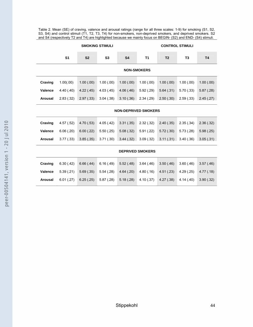
Table 2. Mean (SE) of craving, valence and arousal ratings (range for all three scales: 1-9) for smoking (S1, S2, S3, S4) and control stimuli (T1, T2, T3, T4) for non-smokers, non-deprived smokers, and deprived smokers. S2 and S4 (respectively T2 and T4) are highlighted because we mainly focus on BEGIN- (S2) and END- (S4) stimuli.
SMOKING STIMULI
CONTROL STIMULI
S1
S2
S3
S4
T1
T2
T3
T4
NON-SMOKERS Craving 1.00(.00) 1.00 (.00) 1.00 (.00) 1.00 (.00) 1.00 (.00) 1.00 (.00) 1.00 (.00) 1.00 (.00) Valence 4.40 (.40) 4.22 (.45) 4.03 (.45) 4.06 (.46) 5.92 (.29) 5.64 (.31) 5.70 (.33) 5.87 (.28) Arousal 2.83 (.32) 2.97 (.33) 3.04 (.38) 3.10 (.36) 2.34 (.29) 2.50 (.30) 2.59 (.33) 2.45 (.27) NON-DEPRIVED SMOKERS Craving 4.57 (.52) 4.70 (.53) 4.05 (.42) 3.31 (.35) 2.32 (.32) 2.40 (.35) 2.35 (.34) 2.36 (.32) Valence 6.06 (.20) 6.00 (.22) 5.50 (.25) 5.08 (.32) 5.91 (.22) 5.72 (.30) 5.73 (.28) 5.98 (.25) Arousal 3.77 (.33) 3.85 (.35) 3.71 (.30) 3.44 (.32) 3.09 (.32) 3.11 (.31) 3.40 (.36) 3.05 (.31) DEPRIVED SMOKERS Craving 6.30 (.42) 6.66 (.44) 6.16 (.49) 5.52 (.48) 3.64 (.46) 3.50 (.46) 3.60 (.46) 3.57 (.46) Valence 5.39 (.21) 5.69 (.35) 5.54 (.28) 4.64 (.20) 4.80 (.16) 4.51 (.23) 4.29 (.25) 4.77 (.18) Arousal 6.01 (.27) 6.25 (.25) 5.87 (.28) 5.18 (.28) 4.10 (.37) 4.27 (.38) 4.14 (.40) 3.90 (.32)
peer
-005
0414
1, v
ersi
on 1
- 20
Jul
201
0
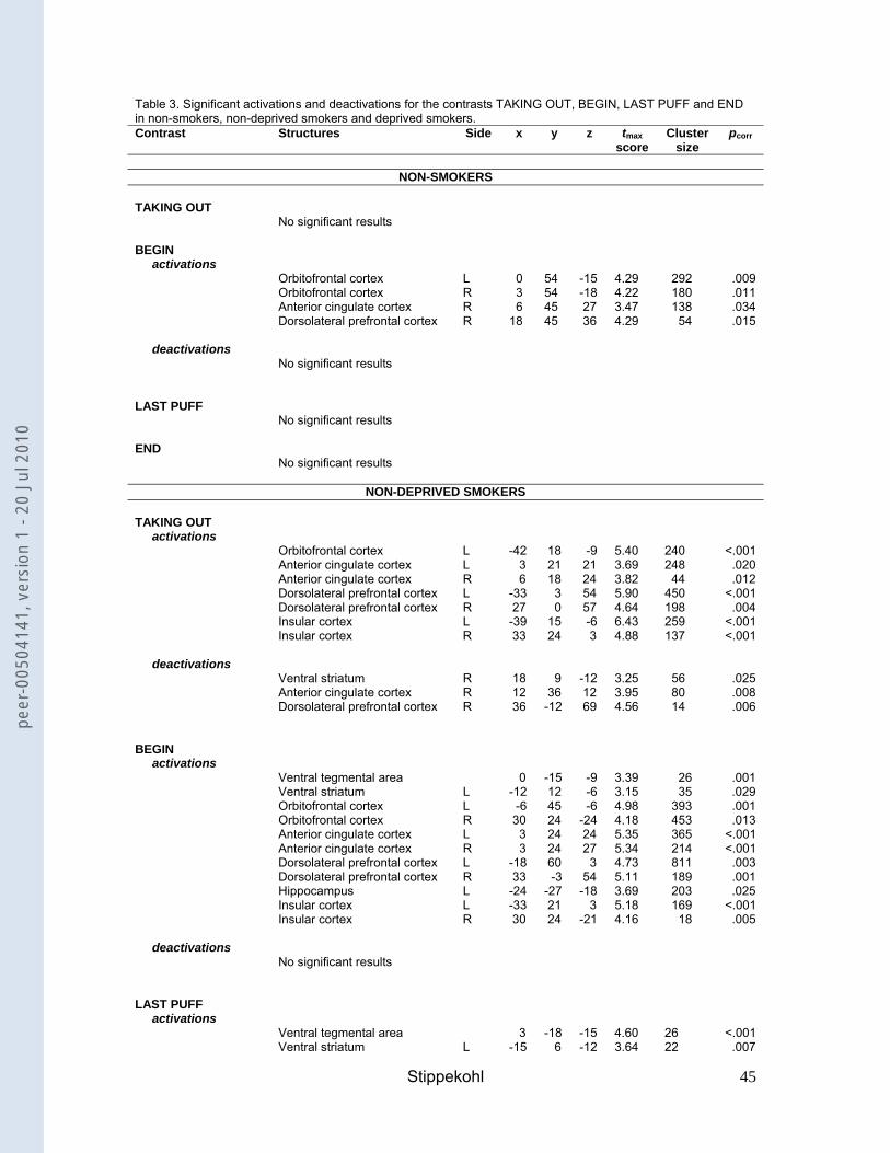
Stippekohl 45
Table 3. Significant activations and deactivations for the contrasts TAKING OUT, BEGIN, LAST PUFF and END in non-smokers, non-deprived smokers and deprived smokers. Contrast Structures Side x y z tmax
score Cluster
size pcorr
NON-SMOKERS
TAKING OUT No significant results BEGIN activations Orbitofrontal cortex L 0 54 -15 4.29 292 .009 Orbitofrontal cortex R 3 54 -18 4.22 180 .011 Anterior cingulate cortex R 6 45 27 3.47 138 .034 Dorsolateral prefrontal cortex R 18 45 36 4.29 54 .015 deactivations No significant results LAST PUFF No significant results END No significant results
NON-DEPRIVED SMOKERS
TAKING OUT activations Orbitofrontal cortex L -42 18 -9 5.40 240 <.001 Anterior cingulate cortex L 3 21 21 3.69 248 .020 Anterior cingulate cortex R 6 18 24 3.82 44 .012 Dorsolateral prefrontal cortex L -33 3 54 5.90 450 <.001 Dorsolateral prefrontal cortex R 27 0 57 4.64 198 .004 Insular cortex L -39 15 -6 6.43 259 <.001 Insular cortex R 33 24 3 4.88 137 <.001 deactivations Ventral striatum R 18 9 -12 3.25 56 .025 Anterior cingulate cortex R 12 36 12 3.95 80 .008 Dorsolateral prefrontal cortex R 36 -12 69 4.56 14 .006 BEGIN activations Ventral tegmental area 0 -15 -9 3.39 26 .001 Ventral striatum L -12 12 -6 3.15 35 .029 Orbitofrontal cortex L -6 45 -6 4.98 393 .001 Orbitofrontal cortex R 30 24 -24 4.18 453 .013 Anterior cingulate cortex L 3 24 24 5.35 365 <.001 Anterior cingulate cortex R 3 24 27 5.34 214 <.001 Dorsolateral prefrontal cortex L -18 60 3 4.73 811 .003 Dorsolateral prefrontal cortex R 33 -3 54 5.11 189 .001 Hippocampus L -24 -27 -18 3.69 203 .025 Insular cortex L -33 21 3 5.18 169 <.001 Insular cortex R 30 24 -21 4.16 18 .005 deactivations No significant results LAST PUFF activations Ventral tegmental area 3 -18 -15 4.60 26 <.001 Ventral striatum L -15 6 -12 3.64 22 .007
peer
-005
0414
1, v
ersi
on 1
- 20
Jul
201
0
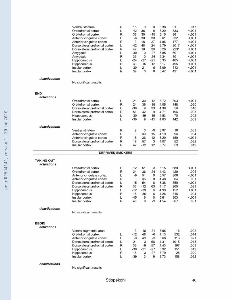
Stippekohl 46
Ventral striatum R 15 9 0 3.38 91 .017 Orbitofrontal cortex L -42 36 -6 7.20 833 <.001 Orbitofrontal cortex R 36 30 -15 5.15 861 <.001 Anterior cingulate cortex L -6 30 30 6.61 332 <.001 Anterior cingulate cortex R 3 18 27 4.86 177 <.001 Dorsolateral prefrontal cortex L -42 48 24 6.79 2017 <.001 Dorsolateral prefrontal cortex R 42 18 39 6.26 2231 <.001 Amygdala L -30 0 -27 5.85 69 <.001 Amygdala R 36 0 -24 5.34 80 <.001 Hippocampus L -24 -27 -21 5.33 465 <.001 Hippocampus R 33 -15 -12 6.17 495 <.001 Insular cortex L -30 21 -9 6.09 513 <.001 Insular cortex R 39 0 6 5.47 421 <.001 deactivations No significant results END activations Orbitofrontal cortex L -21 30 -12 6.72 393 <.001 Orbitofrontal cortex R 24 36 -15 4.05 148 .020 Dorsolateral prefrontal cortex L -39 9 33 4.39 56 .010 Dorsolateral prefrontal cortex R 51 42 9 4.71 196 .003 Hippocampus L -30 -39 -15 4.53 70 .002 Insular cortex L -36 9 -15 4.03 142 .009 deactivations Ventral striatum R 9 3 -9 3.97 16 .003 Anterior cingulate cortex L 3 39 15 4.19 96 .004 Anterior cingulate cortex R 15 36 12 5.20 159 <.001 Dorsolateral prefrontal cortex R 18 57 3 4.07 52 .032 Insular cortex R 42 -12 12 3.77 59 .019
DEPRIVED SMOKERS
TAKING OUT activations Orbitofrontal cortex L -12 51 -3 5.15 680 <.001 Orbitofrontal cortex R 24 36 -24 4.43 429 .005 Anterior cingulate cortex L -9 51 0 5.57 306 <.001 Anterior cingulate cortex R 3 36 0 4.48 84 .001 Dorsolateral prefrontal cortex L -15 54 6 5.38 859 <.001 Dorsolateral prefrontal cortex R 33 -12 63 4.17 260 .023 Hippocampus L -12 -39 6 4.99 152 <.001 Hippocampus R 15 -36 6 4.26 316 .004 Insular cortex L -45 6 0 5.61 303 <.001 Insular cortex R 48 9 -3 4.54 367 .001 deactivations No significant results BEGIN activations Ventral tegmental area 3 -18 -21 3.66 16 .002 Orbitofrontal cortex L -12 48 -6 4.13 532 .014 Anterior cingulate cortex L -9 48 -3 3.68 113 .021 Dorsolateral prefrontal cortex L -21 -3 66 4.31 1015 .013 Dorsolateral prefrontal cortex R 36 -9 57 4.43 197 .009 Hippocampus L -30 -21 -27 3.92 151 .012 Hippocampus R 18 -3 -27 3.79 25 .020 Insular cortex L -39 3 9 3.73 158 .022 deactivations No significant results
peer
-005
0414
1, v
ersi
on 1
- 20
Jul
201
0
Stippekohl 47
LAST PUFF activations Ventral striatum L -3 12 -6 3.12 77 .031 Ventral striatum R 6 3 -9 3.26 63 .024 Orbitofrontal cortex L -36 42 -9 3.91 227 .029 Anterior cingulate cortex L -6 48 6 3.54 207 .030 Dorsolateral prefrontal cortex L -42 48 6 4.29 755 .014 Dorsolateral prefrontal cortex R 39 45 3 4.48 354 .008 Amygdala R 24 -9 -15 3.02 41 .029 Hippocampus L -18 -6 -33 3.71 211 .023 Hippocampus R 21 -6 -33 4.01 287 .010 Insular cortex L -42 3 9 4.12 305 .006 Insular cortex R 39 0 3 4.02 191 .009 deactivations No significant results END activations Orbitofrontal cortex L -9 18 -15 3.80 235 .041 Orbitofrontal cortex R 3 27 -21 3.97 81 .026 Ventral striatum L -3 12 -18 4.24 25 .001 deactivations Orbitofrontal cortex R 27 54 -3 4.14 125 .015 Dorsolateral prefrontal cortex L -18 6 66 3.91 206 .049 Dorsolateral prefrontal cortex R 24 51 0 4.58 648 .005
peer
-005
0414
1, v
ersi
on 1
- 20
Jul
201
0
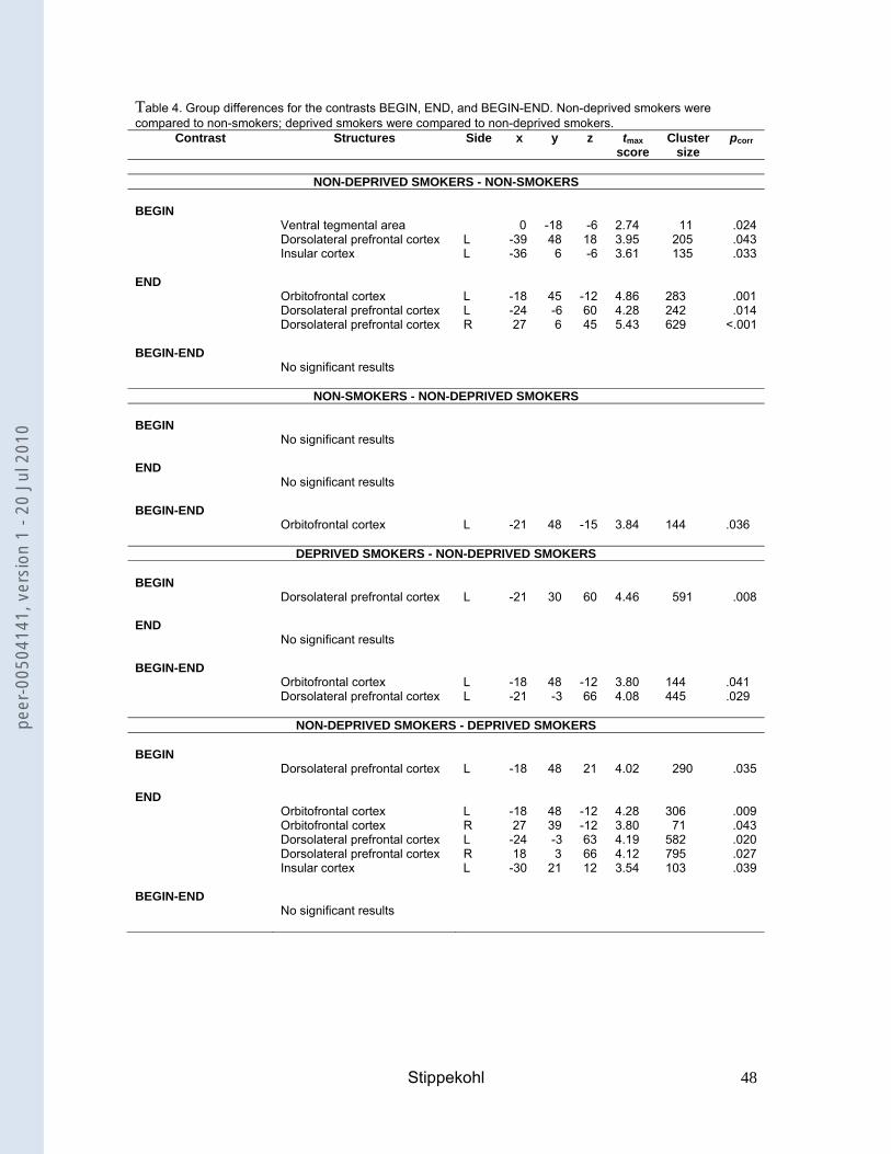
Stippekohl 48
Table 4. Group differences for the contrasts BEGIN, END, and BEGIN-END. Non-deprived smokers were compared to non-smokers; deprived smokers were compared to non-deprived smokers.
Contrast Structures Side x y z tmax score
Cluster size
pcorr
NON-DEPRIVED SMOKERS - NON-SMOKERS
BEGIN Ventral tegmental area 0 -18 -6 2.74 11 .024 Dorsolateral prefrontal cortex L -39 48 18 3.95 205 .043 Insular cortex L -36 6 -6 3.61 135 .033 END Orbitofrontal cortex L -18 45 -12 4.86 283 .001 Dorsolateral prefrontal cortex L -24 -6 60 4.28 242 .014 Dorsolateral prefrontal cortex R 27 6 45 5.43 629 <.001 BEGIN-END No significant results
NON-SMOKERS - NON-DEPRIVED SMOKERS BEGIN No significant results END No significant results BEGIN-END Orbitofrontal cortex L -21 48 -15 3.84 144 .036
DEPRIVED SMOKERS - NON-DEPRIVED SMOKERS BEGIN Dorsolateral prefrontal cortex L -21 30 60 4.46 591 .008 END No significant results BEGIN-END Orbitofrontal cortex L -18 48 -12 3.80 144 .041 Dorsolateral prefrontal cortex L -21 -3 66 4.08 445 .029
NON-DEPRIVED SMOKERS - DEPRIVED SMOKERS BEGIN Dorsolateral prefrontal cortex L -18 48 21 4.02 290 .035 END Orbitofrontal cortex L -18 48 -12 4.28 306 .009 Orbitofrontal cortex R 27 39 -12 3.80 71 .043 Dorsolateral prefrontal cortex L -24 -3 63 4.19 582 .020 Dorsolateral prefrontal cortex R 18 3 66 4.12 795 .027 Insular cortex L -30 21 12 3.54 103 .039 BEGIN-END No significant results
peer
-005
0414
1, v
ersi
on 1
- 20
Jul
201
0
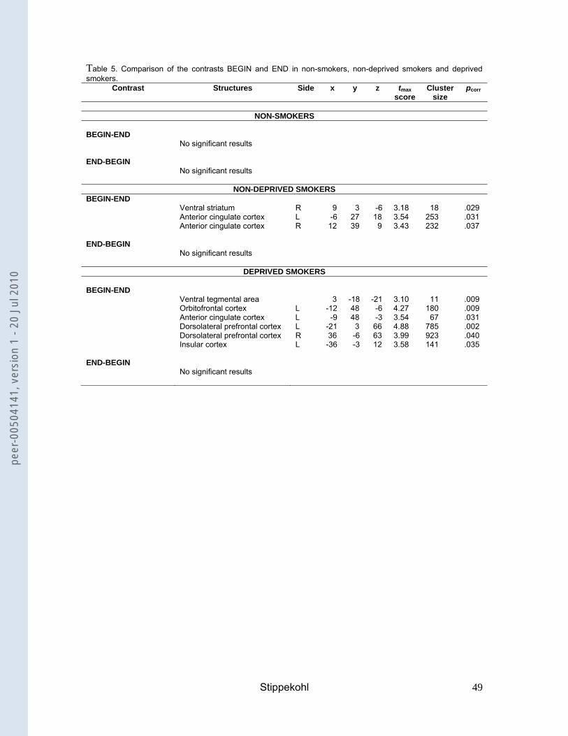
Stippekohl 49
Table 5. Comparison of the contrasts BEGIN and END in non-smokers, non-deprived smokers and deprived smokers.
Contrast Structures Side x y z tmax score
Cluster size
pcorr
NON-SMOKERS
BEGIN-END No significant results END-BEGIN No significant results
NON-DEPRIVED SMOKERS BEGIN-END Ventral striatum R 9 3 -6 3.18 18 .029 Anterior cingulate cortex L -6 27 18 3.54 253 .031 Anterior cingulate cortex R 12 39 9 3.43 232 .037 END-BEGIN No significant results
DEPRIVED SMOKERS BEGIN-END Ventral tegmental area 3 -18 -21 3.10 11 .009 Orbitofrontal cortex L -12 48 -6 4.27 180 .009 Anterior cingulate cortex L -9 48 -3 3.54 67 .031 Dorsolateral prefrontal cortex L -21 3 66 4.88 785 .002 Dorsolateral prefrontal cortex R 36 -6 63 3.99 923 .040 Insular cortex L -36 -3 12 3.58 141 .035 END-BEGIN No significant results
peer
-005
0414
1, v
ersi
on 1
- 20
Jul
201
0
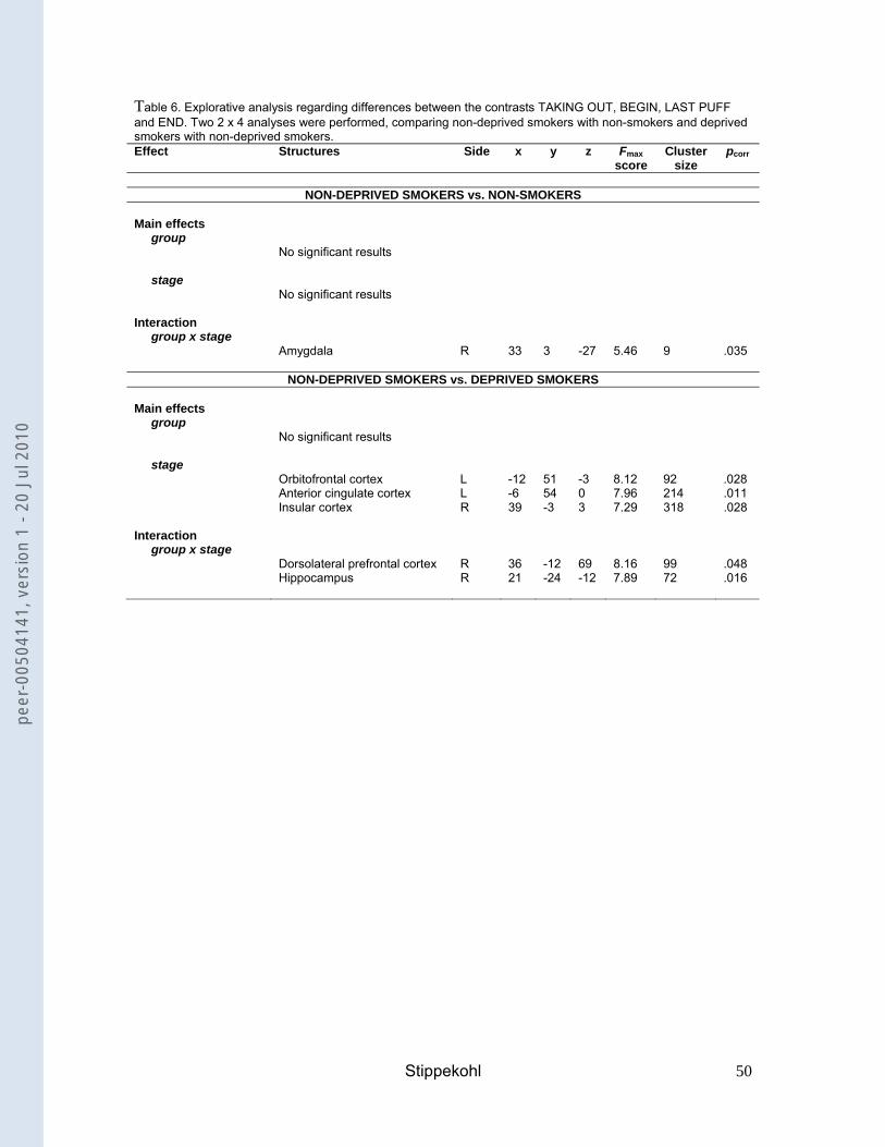
Stippekohl 50
Table 6. Explorative analysis regarding differences between the contrasts TAKING OUT, BEGIN, LAST PUFF and END. Two 2 x 4 analyses were performed, comparing non-deprived smokers with non-smokers and deprived smokers with non-deprived smokers. Effect Structures Side x y z Fmax
score Cluster
size pcorr
NON-DEPRIVED SMOKERS vs. NON-SMOKERS
Main effects group No significant results stage No significant results Interaction group x stage Amygdala R 33 3 -27 5.46 9 .035
NON-DEPRIVED SMOKERS vs. DEPRIVED SMOKERS Main effects group No significant results stage Orbitofrontal cortex L -12 51 -3 8.12 92 .028 Anterior cingulate cortex L -6 54 0 7.96 214 .011 Insular cortex R 39 -3 3 7.29 318 .028 Interaction group x stage Dorsolateral prefrontal cortex R 36 -12 69 8.16 99 .048 Hippocampus R 21 -24 -12 7.89 72 .016
peer
-005
0414
1, v
ersi
on 1
- 20
Jul
201
0
Stippekohl 51
Titles and legends to figures Figure 1. Craving ratings [scale-range: 1-9] for BEGIN- and END-stimuli.
Figure 2. Neural activation and deactivation patterns for BEGIN and END in non-
deprived smokers.
Figure 3. Main effect of stage in the ACC for non-deprived smokers and deprived
smokers combined.
Figure 4. Group x stage interaction in the DLPFC for deprived smokers and non-
deprived smokers.
peer
-005
0414
1, v
ersi
on 1
- 20
Jul
201
0
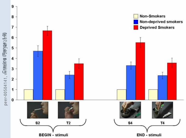
peer
-005
0414
1, v
ersi
on 1
- 20
Jul
201
0

peer
-005
0414
1, v
ersi
on 1
- 20
Jul
201
0
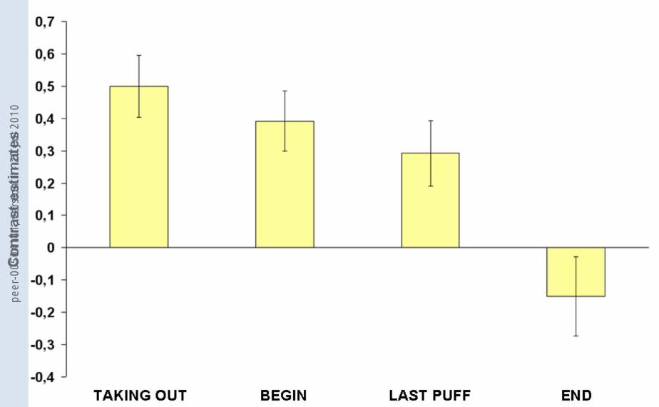
peer
-005
0414
1, v
ersi
on 1
- 20
Jul
201
0
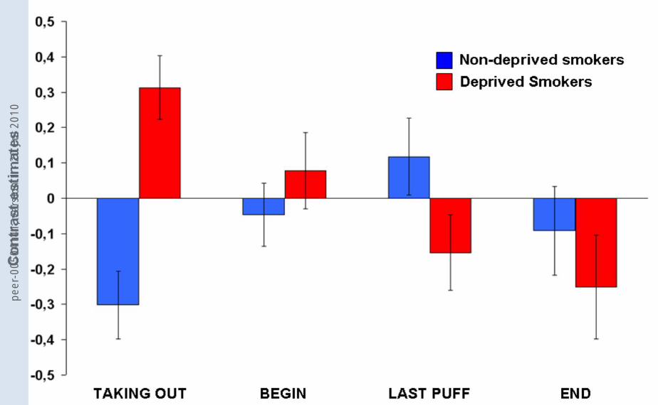
peer
-005
0414
1, v
ersi
on 1
- 20
Jul
201
0