
THE FUNCTION OF THE KIDNEY WHEN DEPRIVED ... - NCBI
17
THE FUNCTION OF THE KIDNEY WHEN DEPRIVED OF ITS NERVES.* BY WILLIAM C. QUINBY, M.D. (From the James B. Brady UrologicalInstitute, Johns Hopkins Hospital, Baltimore.) PLATES 81 To 83. (Received for publication, February 14, 1916.) The question as to what may be the function of a kidney excluded from all nervous influences is not settled at present, in spite of many experimental attempts by various observers. The reason for this lies in the fact that none of the methods employed to solve the question have been free from criticism. That the kidney is plentifully sup- plied with nerves is well known; but that these have any further function than that of vasomotor control has not been proved. At- tempts to demonstrate secretory fibers to the kidney in either the vagus or splanchnic nerves or their branches have given only con- flicting evidence. Removal of all the nerve filaments at the renal hilus by dissection would exclude the kidney from the nervous system, so that its func- tion could be compared with that of a normal organ. But on examin- ing the situation more closely one finds that such method of nerve removal has been found most uncertain in its results? This is due to the fact that the nerve filaments supplying the organ not only lie in a network closely applied to the renal vessels, but also run partly within the walls of these vessels. It has therefore been sug- gested to use some chemical means of destruction, such as painting with carbolic acid after the dissection. But even under these cir- cumstances one is never absolutely sure that all nerves have been removed. Also, in such thin walled vessels as the renal vein, car- * Presented in abstract at the meeting of the Federation of American Societies for Experimental Biology, Boston, Dec. 26, 1915. 1 Cohnheim, J., and Roy, C. S., Untersuchungen fiber die Circulation in den Nieren, Virckows Arch. f. Path. Anat., 1883, xcii, 443. 535
-
Upload
khangminh22 -
Category
Documents
-
view
3 -
download
0
Transcript of THE FUNCTION OF THE KIDNEY WHEN DEPRIVED ... - NCBI

THE FUNCTION OF THE KIDNEY WHEN DEPRIVED OF ITS NERVES.*
BY WILLIAM C. QUINBY, M.D.
(From the James B. Brady Urological Institute, Johns Hopkins Hospital, Baltimore.)
PLATES 81 To 83.
(Received for publication, February 14, 1916.)
The question as to what may be the function of a kidney excluded from all nervous influences is not settled at present, in spite of many experimental attempts by various observers. The reason for this lies in the fact that none of the methods employed to solve the question have been free from criticism. That the kidney is plentifully sup- plied with nerves is well known; but that these have any further function than that of vasomotor control has not been proved. At- tempts to demonstrate secretory fibers to the kidney in either the vagus or splanchnic nerves or their branches have given only con- flicting evidence.
Removal of all the nerve filaments at the renal hilus by dissection would exclude the kidney from the nervous system, so that its func- tion could be compared with that of a normal organ. But on examin- ing the situation more closely one finds that such method of nerve removal has been found most uncertain in its results? This is due to the fact that the nerve filaments supplying the organ not only lie in a network closely applied to the renal vessels, but also run partly within the walls of these vessels. I t has therefore been sug- gested to use some chemical means of destruction, such as painting with carbolic acid after the dissection. But even under these cir- cumstances one is never absolutely sure that all nerves have been removed. Also, in such thin walled vessels as the renal vein, car-
* Presented in abstract at the meeting of the Federation of American Societies for Experimental Biology, Boston, Dec. 26, 1915.
1 Cohnheim, J., and Roy, C. S., Untersuchungen fiber die Circulation in den Nieren, Virckows Arch. f. Path. Anat., 1883, xcii, 443.
535

536 FUNCTION OF KIDNEY WI-IEN DEPRIVED O~" ITS NERVES
bolic acid extensively used m a y cause a local react ion leading to thrombosis. For these or other reasons, none of the evidence thus far brought forward in regard to the function of such kidneys is convincing.
I t has been shown by workers on the methods and possibilities o f blood vessel suture tha t a k idney removed from the body of an experi- mental animal, and later re implanted by restorat ion of the cir- culation, is able to support life in a presumably normal fashion. With one exception, however, all such investigators have been en- gaged in demonstra t ing the possibility of the operation, and have not concerned themselves with the detailed physiological function of the organ.
In the present investigation i t was desired to examine in detail the function of a k idney which had been removed from the body and subsequently replaced. By this method i t is certain tha t the organ is entirely outside the sphere of all nervous influences for a t ime at least, if not permanent ly . Fur thermore , the response of such a kidney to the various functional tests gives a t least indirect evidence on the question of secretory innervation.
HISTORICAL.
Asher~ is one of the most recent investigators to attempt the demonstration of secretory nerves to the kidney. With his coworkers, Pearce and Jost, he has made observations which seem to show that the vagus nerve carries secretory fibers, while the splanchnic carries inhibitory ones; at least so far as the water output of the kidney is concerned. Jungmann and Meyer, a after cutting the splanchnic, found an increase of urine and of sodium chloride delivered from the kidney on the same side as the severed nerve. Rhode and Ellinger, 4 however, obtained different results, which led them to believe that the splanchnlc has an inhibitory action. The difficulty seems to lie in the fact that in the methods
Asher, L., Die Innervation der Niere, Deutsch. reed. Wchnschr., 1915, xli, 1000. Asher, L., and Pearce, R. G., Die sekretorische Innervation der Niere, Ztschr. f. Biol., 1914, lxiii, 83. Jost, W., Die sympathische Innervation der Niere, Ztschr. f. Biol., 1914, lxiv, 441.
3Jungmann, P., and Meyer, E., Experimentelle Untersuchungen tiber die AbhAngigkeit der Nierenfunktion vom Nervensystem, Arch. f. exper. Path. u. Pharm., 1913, lxxiii, 49.
* Rhode, E., and Ellinger, P., t3ber die Funktion der Nierennerven, Zentralbl. f. Physiol., 1913-14, xxvii, 12.

WILLIAM C. QUINB¥ 537
employed to elucidate the problem, the circulatory effects of vagus or splanchnic stimulation completely mask other possible effects.
More recently Pearce 5 has employed the method of Barcroft in which the oxygen consumption of the kidney is used as the index of cellular activity, and he has been unable to confirm his previous work done with Asher.
Transplantation or reimplantation of the kidney has been found surgically possible by Carrel, Stich, and a few others. ~ The most recent worker on this subject has been Lobenhoffer. His is the only work which attempts in any detailed way, to study the function of a kidney so treated. He united the severed renal vessels to those of the spleen in dogs, and was successful in ten instances. After removal of the other kidney, the water and salt output, as well as that of lactose and sugar caused by phloridzin, was studied. His results show that such a kidney is able to meet not only the ordinary demands of life, but also the excessive ones set up by the experimental injections. Further details of his work will be discussed later.
Zaaijer ~ has recently reported the survival and complete health of a dog bearing a single kidney, which had been transplanted to the iliac vessels 6 years pre- viously.
Method.
The exper iments were carried out on large dogs of bo th sexes.
After examinat ion of the var ious regions where the k idney migh t be placed, i t was decided not to t r ansp lan t the organ, bu t to reunite it
to its own severed vessels; for in this way the neares t approach to
normal physiological conditions is secured. Al though the left k idney of the dog is somewhat more accessible
than the right, and has slightly longer vessels, the renal a r t e ry on the
left is of ten bifurcated, or even m a y leave the aor ta as two separa te vessels. This reduces the caliber of the arteries and doubles the
a m o u n t of t ime spent in sewing. The r ight side was chosen, there- fore, in near ly all instances.
Pearce, R. G., and Carter, E. P., The Influence of the Vagus Nerve on the Gaseous Metabolism of the Kidney, Am. Jour. Physiol., 1915, xxxvlii, 350.
Carrel, A., Doppelte Nephrektomie und Reimplantation einer Niere, Arch. f . klin. Ch#., 1908-09, lxxxviii, 379. Stich, R., ~ber Gef~ss- und Organtrans- plantation mittelst GefAssnaht, Ergebn. d. Chit. u. Orthop., 1910, i, 1. Lobenhoffer, W., Funkfionspriifungen an transplantierten Nieren, Mitt. a. d. Grenzgeb. d. Med. u. Chir., 1913, xxvi, 197.
7 Zaaijer, J. H., Dauerresultat einer autoplastischen Nierentransplantation bei einem Hunde, Beitr. ~. klin. Chir., 1914, xciii, 223.

538 FUNCTION OF KIDNEY WHEN DEPRIVED OF ITS NERVES
Ether was used by the intratracheal method in all the experi- ments. This method of administration is important, because by its use the respiratory movements can be reduced to a minimum, or even abolished. A motionless field is thus obtained which is of great aid in accurate and speedy sewing.
The animals were prepared for operation by shaving over the abdomen and far down onto the flanks. This area was then made sterile by soap and water, alcohol, ether, and tincture of iodine. A catheter was placed in the bladder, and in the case of male dogs, was carried off to the left side where it remained during the operation. The surgical asepsis was in every respect as strict as possible; gowns and gloves were worn, although during the actual suture of the blood vessels the gloves had to be removed, following which the hands were carefully coated with sterile vaselin.
Transverse Abdominal Incision.--Incision is made transversely across the abdomen just above the level of the umbilicus, and extend- ing from the outer border of the left rectus abdominis across the right side down to the erector spinm group of muscles (Fig. 1). This is well shown in the illustrations. 8 Failure to prolong the incision far enough towards the posterior wall of the abdominal cavity prevents easy access to the whole kidney region, and makes the suture diffi- cult or impossible.
The abdomen is entered in layers, careful attention being paid to hemostasis. After incision of the skin and subcutaneous tissues, the fascia of the external oblique is picked up on the right, at the outer border of the rectus. I t is lifted up, incised, and the incision pro- longed well toward the flank. The internal oblique is similarly treated. The rectus is dissected bluntly from its underlying sheath as far as the middle line, when it is cut through. The left rectus is similarly divided. After entering the abdomen through the linea alba, the transversalis can be quickly cut down in either direction (Fig. 2). The wound gapes widely and needs no retraction. All the intestines
s This method of entrance to the abdominal cavity is of great aid in the per- formance of other operations, such as Eck fistula and those involving the biliary passages or adrenal glands. It is illustrated not because the procedure is in any way new, but because I have found that its value is not generally recognized by laboratory investigators.

WILLIAM C. QUINBY 539
except the duodenum and descending colon, are then lifted out of the abdomen to the left, where they lie covered by silk handkerchiefs impregnated with liquid vaselin (Fig. 3).
Preparation of Kidney and Renal Vessels for Suture.--After section of the peritoneum about the kidney, it is lifted out and turned toward the middle line of the body where, surrounded with gauze, it is gently held by the assistant. In this position the renal artery comes first to view, and it is carefully cleaned as far as the aorta of all surround- ing tissue and nerves. This is done by blunt dissection and by wiping with dry gauze. The kidney is allowed to fall back into place, and the vein is similarly cleaned as far as the vena cava. The ureter is next stripped downward for about 6 or 7 cm. The field is then carefully washed wi th salt solution, every bleeding point caught and tied, and after drying, the whole is coated with liquid vaselin, including the kidney and its vessels. The two rubber-covered serrefines are then placed on the artery, two others on the vein, and the vessels are cut between them. The ureter is cut long. The kidney is then removed from the body and placed on a clean gauze pad. The gloves are now removed.
Vessel Suture.--The ends of the severed vessels are washed quickly with salt solution from a bulb pipette, fill every trace of blood is removed, They are then coated with liquid vaselin. The advenfitia is removed from the ends of the arteries in the usual way, after which the whole field within the abdomen is covered with vase- lined silk handkerchiefs leaving only the stumps of the renal vessels protruding. These handkerchiefs are held in place by brass clips used by stationers, called "O.K. paper fasteners."
The most exacting and important part of the vascular suture lles in placing the three primary guide or tension stitches. This is made much easier by using a suture armed with two needles, one at either end. In this way the suture can always be passed from within the vessel outward. The circumference of the vessel ends must be accurately divided into thirds by these tension sutures.
The artery, being behind the vein, is sewed first, using an over- and-over stitch. This is followed by suture of the vein. The serre- fine clamps are then removed from the vein and the vessel is allowed to fill under moderate pressure, rolling it a bit between the thumb
FUNCTION OF KIDNEY WHEN DEPRIVED OF ITS NERVES
and finger. The artery is next freed while held between the thumb and finger so as partially to control the tension during the first few moments of blood flow? After the vascular suture is ended, the ureter is reunited by the invagination method of Van Hook. Fol- lowing this, the operative field is cleaned of blood, and the peritoneum readjusted by a few interrupted sutures about the periphery of the kidney. Special pains should be taken to see that the kidney is replaced high enough toward the liver so that there is no angulation of the vein.
Closure of the Abdomen.--The next step consists in closing the abdomen after the bowels have been replaced. This is made certain by using first a mattress suture of stout silk placed in the linea alba. After tying this, the severed abdominal muscles can easily be approxi- mated in layers. A continuous suture is used for the transversalis, including at the same time the peritoneum. Mattress stitches are used for the recti, while the obliques are held by interrupted sutures. Closure of the subcutaneous tissue and skin completes the operation. Silk is used throughout the operation, both for ties and sutures.
The dog is placed in a metabolism cage so that the urine can be accurately collected and measured.
The operation is a difficult one, but after some experience it was found possible to restore the circulation in about an hour following its interruption. Forty-three dogs were subjected to operation, of which sixteen survived in suitable condition for further physiological observations.
Examination of the Renal Function.
The operated animals were divided into two series. In the first, the function of the reimplanted kidney was compared with that of the intact one, at periods varying from 2 days to 3 weeks after the primary operation. For this purpose the animal was anesthetized with paraldehyde, each ureter brought out onto the flank through
0 The method of suture used is that elaborated by Carrel. Carrel, A., La tech- nique op~ratoire des anestomoses vasculalres et la transplantation des visc~res, Lyon m~d., 1902, xcviii, 859; Anastomosis and Transplantation of Blood Vessels, Am. Med., 1905, x, 284; The Surgery of Blood Vessels, Etc., Bull. Johns ttopkins ttosp., 1907, xviii, 18.

WILLIAM C. QUINBY 541
lumbar incisions, and the urines were compared. This method has been described in a previous communication. ~°
The dogs of the second series were subjected to removal of the unoperated kidney at times varying from 5 to 14 days after the pri- mary operation. The work of their remaining, reimplanted kidney was later examined and compared with that of control dogs, in whom a single nephrectomy had been done.
Series I.
Two typical protocols will suffice to show the results of this series. June 17, 1915. Dog 66. Weight 10,454 gin. Operation at 10 a.m.; reim-
plantation of fight kidney. June 18. Has made a good recovery. Blood urea is 0.302 mg. per liter.n
Excretion of phenolsulphonephthalein given intravenously is 51 per cent in 2 hours. 11
June 19. Given paraldchydc, n 1.7 cc. per kilo of body weight, by stomach tube, followed by 200 cc. of water. Ureters exposed in loins, and cannulm introduced delivering into test-tubes. Carotid blood pressure recorded on kymograph.
11.10 a.m. Urine appears without delay from each side. Collected for I hour. 12.10 p.m. 5.0 gin. of sodium chloride in 20 cc. of water given intravenously.
Good diurcsis from each side; urine collected for 1 hour. 1.10 p.m. Animal killed. The blood pressure during the experiment varied
from 128 to 138 ram. of mercury, cxccpt just after the injection of the hyper- tonic salt solution. The sutured vesscls were frec from obstruction.
Microscopic examination of thc reimplanted kidney showed the capsular spaces wide for the most part, and the glomerular tufts containing a considerable amount
10 Quinby, W. C., and Fitz, R., Observations on Renal Function in Acute Experimental Unilateral Nephritis, Arch. Int. Med., 1915, xv, 303.
n As was shown in the communication noted above (Quinby and Fitz 1°) the two most dependable tests of renal function are the excretion of phenolsulphone- phthalein and the estimation of the blood nitrogen or blood urea. The urease method of Marshall (Jour. Biol. Chem., 1913, xv, 487) was used. By this the normal amount of blood urea of the dog is found to lie between 0.200 and 0.380 or 0.400 mg. per liter. The phenolsulphonephthalein excretion was estimated by the method of Rowntree and Geraghty (Jour. Pharmacol. and Exper. Therap., 1909-10, i, 579). Paraldehyde was used, because in proper dosage it causes neither diuresis nor lowered blood pressure. I t must be used fresh, however, because it loses strength on exposure to the air, thus making the dosage uncertain.

542 FUNCTION OF KIDNEY WHEN DEPRIVED OF ITS NERVES
of blood. T h e cells and nuclei of the tubules were well preserved except in a very few areas where a smal! amount of cellular desquamation had taken place. The blood vessels were everywhere dilated, but no evidence of thrombosis or infarction could be found. A considerable number of hyaline bodies were seen in the collecting tubules, evidently casts. The uninvolved kidney was normal in all respects. Its capsules showed active diuresis, but the blood vessels were not so evidently dilated as were those of the operated organ.
1st hr. t *
Before diuresis ,) Right.
£ Left.. 2nd hr.
£
After diuresis ) Right.
"~ Left..
Amount of urine.
5.0 2.5
60.0 22.0
Percentage of NaCI.
0.90 0.55
0.95 0.95
Amount of NaCI.*
gm.
0.045 0.013
0. 570 0. 209
* The chlorides were determined by the Volhard method at first; later by that of McLean and Van Slyke (Jour. Biol. Chem., 1915, xxi, 361).
We see that here the kidney without nerves shows an increased function over the normal one, both before and after diuresis.
June 30, 1915. Dog 67. Weight 11,363 gm. Left kidney reimplanted. July 12. Has made a good recovery. Urine free from a!bumin and sugar.
Average daily amount, 195 cc. Given paraldehyde, 1.7 cc. per kilo of body weight, followed by 200 cc. of water. Ureters exposed on flanks and cannulated. Blood pressure tracing from carotid.
11.05 a.m. Flow from right kidney begins a little later than from left which started immediately on delivering ureter. Urine collected for 1 hour after flow from each was established.
12.16 p.m. 5.0 gin. of sodium chloride in 20 cc. of water injected intravenously. Marked diuresis.
1.20 p.m. Animal killed. Blood pressure average was 133 mm. of mercury. Renal vessels without trace of clot, though there were a few adhesions about the kidney. A sample of blood drawn just before death showed 0.354 rag. of urea per liter.
Microscopic examination showed that both kidneys were normal in all respects except that in the one previously reimplanted a few mitotic figures could be found in the tubular cells after careful search. These were evidently the sequel of previous cellular degeneration.

WILLIAM C. QUINBY 543
1st hr.
diuresis ,f Right . . . . . . . . . . . . . . . . . Before Left . . . . . . . . . . . . . . . . . .
2nd hr. /,
After diuresis J Right . . . . . . . . . . . . . . . . . ) Left . . . . . . . . . . . . . . . . . .
Amount of Percentage of Amount of urine. NaCI. NaC1.
2.5 3.0
40.0 36.0
1.12 0.93
0.82 0.96
gm.
0.028 0.028
0.328 0.345
In this experiment the operated kidney has a function about the same as that of the normal side.
These two protocols fairly represent the findings in this first series which comprised eleven dogs. For a period following operation varying from 10 to 14 days the denervated kidney shows an increased absolute function both for fluid and salt, as compared with the normal kidney. At times this increase is relative as well. This is true of the unstimulated organ, and especially so of the one subjected to the diuretic action of sodium chloride. Beyond this period, however, the balance is regained, so that each organ, operated and intact, divides the labor in very nearly equal parts.
We see here also that absence of the renal nerves abolishes the temporary inhibition of flow so often seen normally after the handling of the ureters necessary for their exposure in the loin. Urine from the denervated side always flowed immediately on section of the ureter; but in some instances the normal side showed an inhibition lasting for as long as 5 minutes. This is analogous to the temporary inhibition occasionally seen on passage of a ureteral catheter in man.
Series II.
The following is a typical protocol. Nov. 20, 1915. Large male, of Newfoundland type. Weight 15,900 gin.
Intratracheal ether and reimplantation of right kidney. Circulation restored after being interrupted for 1 hour.
Nov. 23, 1915. Has made a good recovery; eats well and does not vomit. Nov. 30, 1915. Has entirely recovered. Wound healed by first intention. Dec. 4, 1915. Ether. Lateral incision in left flank through which the kidney
was removed after ligation of its vessels with silk. Wound closed in layers. Dee. 7, 1915. Has made an excellent recovery. Passes 320 to 340 cc. of urine
daily. Blood urea, 0.456 rag. per liter. Phenolsuphonephthalein, 60 per cent in

544 FUNCTION OF KIDNEY WHEN DEPI~IVED OF ITS NERVES
2 hours. Urinary sediment shows a rare blood corpuscle; no casts; a few leukocytes. Sugar and albumin absent.
Dec. 11, 1915. Animal well. The 24 hour amount of urine has fallen some- what and its concentration has increased.
Dec. 14, 1915. Has a rather marked balanitis, and does not urinate until the demand is imperative. After about 48 hours during which no urine was passed, voided a little over 700 cc.
Dec. 18,1915. Balanitis has responded to irrigation with boric acid and animal is now well. Output of phenolsulphonephthalein, 50 per cent in 1 hour. No albumin or sugar.
Dec. 22, 1915. Intravenous injection of 500 cc. of normal (0.8 per cent) salt solution. Urine withdrawn by catheter at 30 minute intervals showed the fol- lowing:
i
1st half hr . . . . . . . . . . . . . . . . . . . . . . . . . . . . . . . . . . . . . . . . . . . . . . . 2nd " " . . . . . . . . . . . . . . . . . . . . . . . . . . . . . . . . . . . . . . . . . . . . i
3rd " " . . . . . . . . . . . . . . . . . . . . . . . . . . . . . . . . . . . . . . . . . . . . i
4th " " . . . . . . . . . . . . . . . . . . . . . . . . . . . . . . . . . . . . . . . . . . . .
Amount. NaCI per liter.
83 62 48 11
gra.
10.6 12.3 13.0 17.2
The urine before diuresis contained 19.10 grn. of sodium chloride
per liter. I t is thus seen that the kidney responds quickly to diuresis
and regains its equilibrium within a normal time limit.
Jan. 31, 1916. Given 2 gin. of lactose intravenously. At the end of 5 hours 1.72 gm. were found in the urine.
Feb. 7, 1916. The 24 hour amount of urine has been measured for 65 days, giving an average of 180 cc. It contains neither albumin nor casts.
Feb. 9, 1916. Blood urea 0.320 mg. per liter. 61 per cent of phenolsulphone- phthalein is excreted in 1 hour. Animal seems to be perfectly normal.
Four other auimals of this series were killed after having shown
normal kidney function for a month or longer. In each instance
the kidney showed microscopic evidences of some hype r t rophy of
the elements, which usually occurs after unilateral nephrectomy.
In two instances the kidney also showed a small depressed scar in the cortex with sclerosis of the normal elements and infiltration by
connective tissue. These areas seemed to be the result of small
focal necroses caused by interrupt ion of the blood supply during
operation. T h e y were never of any considerable size, so tha t the
function of the organ remained uninfluenced.

WILLIAM C. QUINBY 545
The results of this second series show that the life of dogs having a single reimplanted kidney is maintained in a normal manner, as estimated by renal functional tests as well as by other more general methods of observation.
DISCUSSION.
The experiments of the first series show that the immediate effect of loss of nerve control over the kidney is a period of overaction. This occurs in all cases, and in the presence of apparent health, as judged by the general condition of the animal, by the normal content of the blood in urea, and by a rlormal output of phenolsulphonephthalein. This period exists for a varying time, but balance has always been restored by the end of 2 weeks. The kidney recently deprived of its nerves is without vasomotor control; the organ is tense and appreciably enlarged; its vessels are dilated, and following the in- crease of blood flow there is an increase of function over that of the normal organ. Resumption of tone on the part of the blood vessels brings again normal function.
The results here would seem to be analogous to those vasomotor changes occurring in the splanchnic area after section of the cord. Following this operation there occurs a marked dilatation of the mesenteric vessels, but in a short time vasomotor control is again established. Vascular tone may be resumed through the interven- tion of other more peripheral nerve ganglia, or the smooth muscle fibers of the vessel wall may possibly regain their tone without such intervention. Also, in the kidney there are ganglion cells, especially in the region of the renal sinus, which may be responsible for the resumption of vasomotor control. Although we know that fibers of the sympathetic type are able to regenerate much more quickly than are those of the peripheral nerves, that they should be able to grow to the renal blood vessels and resume control over them within 2 weeks after section seems improbable. Certainly nerve control by the normal pathways could never have been regained in Zaaijer's dog whose kidney was sutured to the iliac vessels, or in those of Lobenhoffer who used the splenic vessels.
The time variation in regaining normal function is probably to be explained by the greater or less degree of surgical insult in the indi-

546 FUNCTION OF KIDNEY W H E N DEPRIVED OF ITS NERVES
vidual case. No kidney showed normal function after being ex- cluded from the circulation for longer than 1 hour and 20 minutes.
The second series of observations indicates that a single kidney which has been removed from the body and subsequently reimplanted, can maintain normal life for apparently indefinite periods. Also such a kidney is able to respond to the excessive demands made on it by the injection of various test substances. M y results in this group of experiments fairly coincide with those of Lobenhoffer, except in a few details. He found that the 24 hour amount of urine passed by his dogs varied between 1,500 and 2,000 cc. This is quite unusual. Normal cage dogs in a large number of observations made by us, are found to pass from 200 to 450 or 500 cc. of urine daily, having a specific gravity of about 1.030. Of course this is but a rough average, since all our animals had water continuously at hand, and must have taken varying amounts from day to day. Their diet was of meat. I feel, therefore, that the continuous excretion of such large amounts of urine tends to suggest the absence of com- plete return to normal conditions.
Again, in the three infusion experiments reported by Lobenhoffer, his animals put out amounts of water and salt which varied widely, though the quantities infused were the same. In the few infusions done by me the resulting outputs were all within 10 or 12 per cent of each other.
The observations on the relative values of the different methods used for testing renal function made by Quinby and Fitz 1° showed that the estimation of the blood nitrogen, or that part of it composing the blood urea, and the output of phenolsulphonephthklein, together form the best means of measuring renal function. Further experi- ence with these tests in clinical work by many observers has confirmed this opinion. I have therefore been content to follow the dogs of this second series by means of these tests, rather than by phloridzin or lactose, as did Lobenhoffer.
The above results, though they throw no direct evidence on any possible secretory function of either the vagus or splanchnic nerves, seem to suggest that if this exists it must play a minor and infre- quent part. Under all the conditions produced both by my experi- ments and by those of Lobenhoffer, the denervated kidney has been

WILLIAM C. QUINBY 547
seen to react in an entirely normal manner. One may ask, there- fore, if secretory nerves to the kidney are assumed to exist, under what conditions they are manifest; for no lack of such action seems" to be demonstrable. Added to the inability of the present observations to show any failure of kidney function which might be ascribed to lack of secretory nerve influence, is the recent work of Cow, x2 who finds in the duodenal mucosa some substance which has a definite diuretic effect on the kidney by means of a hormone action.
I t is probable that vasomotor conditions in the kidney, added to the chemical and hormone action of substances contained in the circuIat- ing blood, will be found entirely adequate to explain all variations and types of normal renal function.
SUMMARY.
1. By means of vascular suture it is possible to remove the dog's kidney from the body and later to restore it to its former position.
2. Such a kidney is removed from the control of the nervous system, at least for a time.
3. Examination of the function of a kidney so treated shows an initial period of overaction, as compared with that of the normal kidney.
4. This is followed by balanced action. 5. The more recent tests of renal function show that a single, re-
implanted kidney is able to maintain normal life indefinitely. 6. The results of these experiments, together with the evidence
already at hand, suggest strongly that secretory nerves to the kidney do not exist.
EXPLANATION OF PLATES.
PLATE 81.
FIG. 1. The transverse abdominal incision extending from the outer border of the left rectus muscle well down into the right flank.
PLATE 82.
FIG. 2. The oblique muscles and both recti have been divided and the ab- domen is being opened by incision of the transversalis in a direction parallel to its fibers.
12 Cow, D., Jour. Physiol., 1914-15, xlix, 441.
54~ FUNCTION OF KIDN]E¥ WHEN DEPRIVED OF ITS NERVES
PLATE 83.
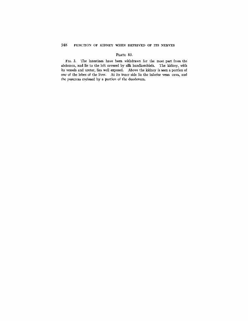
FIG. 3. The intestines have been withdrawn for the most part from the abdomen, and lie to the left covered by silk handkerchiefs. The kidney, with its vessels and ureter, lies well exposed. Above the kidney is seen a portion of one' of the lobes of the liver. At its inner side lie the inferior vena cava, and the pancreas enclosed by a portion of the duodenum.

THE JOURNAL OF EXPERIMENTAL MEDICINE VOL. XX I I I , PLATE 81.
F l a . 1. (Quinby: Kidney Deprived of Nerves.)

THE JOURNAL OF E X P E R I M E N T A L MEDICINE VOL. X X I l l . PLATE 82.
FIG. 2.
Quinby: Kidney Deprived of Nerves.)

THE JOURNAL OF EXPERIMENTAL MEDICINE VOL. XX I I I . PLATE 83.
FIG. 3.
(Quinby: Kidney Deprived of Nerves.)


