Effect of 2,4-Diacetylphloroglucinol on Pythium : Cellular Responses and Variation in Sensitivity...
10
966 PHYTOPATHOLOGY Biological Control Effect of 2,4-Diacetylphloroglucinol on Pythium: Cellular Responses and Variation in Sensitivity Among Propagules and Species Jorge T. de Souza, Christine Arnould, Chrystel Deulvot, Philippe Lemanceau, Vivienne Gianinazzi-Pearson, and Jos M. Raaijmakers First and sixth authors: Wageningen University and Research Centre (WUR), Department of Plant Sciences, Laboratory of Phytopathology, Binnenhaven 5, P.O. Box 8025, 6709 PD, Wageningen, The Netherlands; second, third, and fourth authors: UMR INRA/Université de Bourgogne BBCE-IPM, Centre de Microbiologie du Sol et de I’Environment, INRA, BV 1540, 21034 Dijon Cedex, France; and fifth author: INRA-CMSE, Laboratoire de Recherches sur la Flore Pathogène dans de Sol, 17 rue Sully, 21034 Dijon Cedex, France. Accepted for publication 12 March 2003. ABSTRACT de Souza, J. T., Arnould, C., Deulvot, C., Lemanceau, P., Gianinazzi- Pearson, V., and Raaijmakers, J. M. 2003. Effect of 2,4-diacetylphloro- glucinol on Pythium: Cellular responses and variation in sensitivity among propagules and species. Phytopathology 93:966-975. The antibiotic 2,4-diacetylphloroglucinol (2,4-DAPG) plays an im- portant role in the suppression of plant pathogens by several strains of Pseudomonas spp. Based on the results of this study, there is variation within and among Pythium spp. to 2,4-DAPG. Also, various propagules of Pythium ultimum var. sporangiiferum, that are part of the asexual stage of the life cycle, differ considerably in their sensitivity to 2,4-DAPG. Mycelium was the most resistant structure, followed by zoosporangia, zoospore cysts, and zoospores. Additionally, we report for the first time that pH has a significant effect on the activity of 2,4-DAPG, with a higher activity at low pH. Furthermore, the level of acetylation of phloro- glucinols is also a major determinant of their activity. Transmission electron microscopy studies revealed that 2,4-DAPG causes different stages of disorganization in hyphal tips of Pythium ultimum var. sporangiiferum, including alteration (proliferation, retraction, and disrup- tion) of the plasma membrane, vacuolization, and cell content disinte- gration. The implications of these results for the efficacy and consistency of biological control of plant-pathogenic Pythium spp. by 2,4-DAPG- producing Pseudomonas spp. are discussed. Additional keywords: microbial ecology, soilborne pathogens, ultra- structure. Phloroglucinols (PG) are phenolic, secondary metabolites pro- duced by plants, algae, and bacteria (2,3,6,22,25,58,59). More than 60 PG derivatives have been described and were reported to have antiviral, antimicrobial, ichthyotoxic, insect and mammal antifeedant, antihelminthic, phytotoxic, antioxidant, cytotoxic, antitumor, and plant growth-regulating activities (1,11,15,23– 26,30,50,51,60). The antimicrobial activity of these compounds, and in particular of 2,4-diacetylphloroglucinol (2,4-DAPG), has received considerable attention in the area of biological control of plant diseases. Numerous studies have demonstrated that 2,4-DAPG-producing Pseudomonas spp. can suppress a wide variety of plant pathogens, including fungi, bacteria, and nematodes (11,12,17,20,26,27,32, 47,49,59). Among the plant-pathogenic fungi, Gaeumannomyces graminis var. tritici, Thielaviopsis basicola, Fusarium oxysporum f. sp. radicis-lycopersici, and the oomycete Pythium ultimum can be effectively controlled by 2,4-DAPG-producing Pseudomonas strains (17,20,47,49,59). The determinative role of 2,4-DAPG in disease suppression by Pseudomonas strains has been demon- strated by (i) the use of mutants deficient in 2,4-DAPG production (11,12,26,45,59), (ii) complementation of 2,4-DAPG-deficient mutants and concomitant restoration of biocontrol activity (11,12, 26,59), and (iii) expression of 2,4-DAPG biosynthetic genes in heterologous, nonproducing strains, thereby conferring biocontrol activity or enhanced activity (2,46,59). Reporter gene systems (33) and analytical techniques (53) have further demonstrated that 2,4-DAPG is produced in situ by both introduced Pseudomonas strains and indigenous Pseudomonas populations (7,17,26,38,41). Pseudomonas strains that produce 2,4-DAPG also produce monoacetylphloroglucinol (MAPG) and, depending on the nutritional environment, the ratio of MAPG to 2,4-DAPG may change (17). In addition to MAPG and 2,4- DAPG, PG has been purified from cultures of a bacterium, classi- fied as Aeromonas hydrophila, found on decayed roots of red pine seedlings (48). In spite of significant progress in our understanding of the biosynthesis and regulation of 2,4-DAPG-production in Pseudo- monas strains (2,3,5,44,62), little attention has been given to fungal responses to 2,4-DAPG and to its mode of action. Most studies on the sensitivity of plant-pathogenic fungi to 2,4-DAPG take into account only one species or isolate of the target pathogen and consider only one particular stage in the life cycle of a patho- gen, usually mycelial growth (26,32,36). Most life cycles of pathogens, however, are complex and comprise numerous patho- gen structures and infectious propagules. For example, oomycete pathogens can infect host tissues by means of multiple propagules, including mycelium, zoosporangia, zoospores, and oospores. Understanding the variation in sensitivity of different phases of the life cycle of a pathogen to a specific biocontrol trait will give more insight into the potential efficacy of biocontrol agents har- boring that trait. In this study, we investigated the response of Pythium spp. to 2,4-DAPG. Pythium spp. are ubiquitous pathogens of many im- portant crops around the world and possess a high level of diver- sity with more than 200 species described (16,57). Many of these species can occur simultaneously at the same site and, often, more than one species can infect a certain host plant (9). We determined the variation in sensitivity of 14 Pythium isolates to 2,4-DAPG; the isolates were obtained from multiple hosts and represent eight Corresponding author: J. M. Raaijmakers; E-mail address: [email protected] Publication no. P-2003-0527-02R © 2003 The American Phytopathological Society
-
Upload
u-bourgogne -
Category
Documents
-
view
1 -
download
0
Transcript of Effect of 2,4-Diacetylphloroglucinol on Pythium : Cellular Responses and Variation in Sensitivity...
966 PHYTOPATHOLOGY
Biological Control
Effect of 2,4-Diacetylphloroglucinol on Pythium: Cellular Responses and Variation in Sensitivity Among Propagules and Species
Jorge T. de Souza, Christine Arnould, Chrystel Deulvot, Philippe Lemanceau, Vivienne Gianinazzi-Pearson, and Jos M. Raaijmakers
First and sixth authors: Wageningen University and Research Centre (WUR), Department of Plant Sciences, Laboratory of Phytopathology, Binnenhaven 5, P.O. Box 8025, 6709 PD, Wageningen, The Netherlands; second, third, and fourth authors: UMR INRA/Université de Bourgogne BBCE-IPM, Centre de Microbiologie du Sol et de I’Environment, INRA, BV 1540, 21034 Dijon Cedex, France; and fifth author: INRA-CMSE, Laboratoire de Recherches sur la Flore Pathogène dans de Sol, 17 rue Sully, 21034 Dijon Cedex, France.
Accepted for publication 12 March 2003.
ABSTRACT
de Souza, J. T., Arnould, C., Deulvot, C., Lemanceau, P., Gianinazzi-Pearson, V., and Raaijmakers, J. M. 2003. Effect of 2,4-diacetylphloro-glucinol on Pythium: Cellular responses and variation in sensitivity among propagules and species. Phytopathology 93:966-975.
The antibiotic 2,4-diacetylphloroglucinol (2,4-DAPG) plays an im-portant role in the suppression of plant pathogens by several strains of Pseudomonas spp. Based on the results of this study, there is variation within and among Pythium spp. to 2,4-DAPG. Also, various propagules of Pythium ultimum var. sporangiiferum, that are part of the asexual stage of the life cycle, differ considerably in their sensitivity to 2,4-DAPG. Mycelium was the most resistant structure, followed by zoosporangia, zoospore cysts, and zoospores. Additionally, we report for the first time
that pH has a significant effect on the activity of 2,4-DAPG, with a higher activity at low pH. Furthermore, the level of acetylation of phloro-glucinols is also a major determinant of their activity. Transmission electron microscopy studies revealed that 2,4-DAPG causes different stages of disorganization in hyphal tips of Pythium ultimum var. sporangiiferum, including alteration (proliferation, retraction, and disrup-tion) of the plasma membrane, vacuolization, and cell content disinte-gration. The implications of these results for the efficacy and consistency of biological control of plant-pathogenic Pythium spp. by 2,4-DAPG-producing Pseudomonas spp. are discussed.
Additional keywords: microbial ecology, soilborne pathogens, ultra-structure.
Phloroglucinols (PG) are phenolic, secondary metabolites pro-
duced by plants, algae, and bacteria (2,3,6,22,25,58,59). More than 60 PG derivatives have been described and were reported to have antiviral, antimicrobial, ichthyotoxic, insect and mammal antifeedant, antihelminthic, phytotoxic, antioxidant, cytotoxic, antitumor, and plant growth-regulating activities (1,11,15,23–26,30,50,51,60). The antimicrobial activity of these compounds, and in particular of 2,4-diacetylphloroglucinol (2,4-DAPG), has received considerable attention in the area of biological control of plant diseases.
Numerous studies have demonstrated that 2,4-DAPG-producing Pseudomonas spp. can suppress a wide variety of plant pathogens, including fungi, bacteria, and nematodes (11,12,17,20,26,27,32, 47,49,59). Among the plant-pathogenic fungi, Gaeumannomyces graminis var. tritici, Thielaviopsis basicola, Fusarium oxysporum f. sp. radicis-lycopersici, and the oomycete Pythium ultimum can be effectively controlled by 2,4-DAPG-producing Pseudomonas strains (17,20,47,49,59). The determinative role of 2,4-DAPG in disease suppression by Pseudomonas strains has been demon-strated by (i) the use of mutants deficient in 2,4-DAPG production (11,12,26,45,59), (ii) complementation of 2,4-DAPG-deficient mutants and concomitant restoration of biocontrol activity (11,12, 26,59), and (iii) expression of 2,4-DAPG biosynthetic genes in heterologous, nonproducing strains, thereby conferring biocontrol activity or enhanced activity (2,46,59).
Reporter gene systems (33) and analytical techniques (53) have further demonstrated that 2,4-DAPG is produced in situ by both
introduced Pseudomonas strains and indigenous Pseudomonas populations (7,17,26,38,41). Pseudomonas strains that produce 2,4-DAPG also produce monoacetylphloroglucinol (MAPG) and, depending on the nutritional environment, the ratio of MAPG to 2,4-DAPG may change (17). In addition to MAPG and 2,4-DAPG, PG has been purified from cultures of a bacterium, classi-fied as Aeromonas hydrophila, found on decayed roots of red pine seedlings (48).
In spite of significant progress in our understanding of the biosynthesis and regulation of 2,4-DAPG-production in Pseudo-monas strains (2,3,5,44,62), little attention has been given to fungal responses to 2,4-DAPG and to its mode of action. Most studies on the sensitivity of plant-pathogenic fungi to 2,4-DAPG take into account only one species or isolate of the target pathogen and consider only one particular stage in the life cycle of a patho-gen, usually mycelial growth (26,32,36). Most life cycles of pathogens, however, are complex and comprise numerous patho-gen structures and infectious propagules. For example, oomycete pathogens can infect host tissues by means of multiple propagules, including mycelium, zoosporangia, zoospores, and oospores. Understanding the variation in sensitivity of different phases of the life cycle of a pathogen to a specific biocontrol trait will give more insight into the potential efficacy of biocontrol agents har-boring that trait.
In this study, we investigated the response of Pythium spp. to 2,4-DAPG. Pythium spp. are ubiquitous pathogens of many im-portant crops around the world and possess a high level of diver-sity with more than 200 species described (16,57). Many of these species can occur simultaneously at the same site and, often, more than one species can infect a certain host plant (9). We determined the variation in sensitivity of 14 Pythium isolates to 2,4-DAPG; the isolates were obtained from multiple hosts and represent eight
Corresponding author: J. M. Raaijmakers; E-mail address: [email protected]�
Publication no. P-2003-0527-02R © 2003 The American Phytopathological Society
Vol. 93, No. 8, 2003 967
species. The sensitivity of various infectious propagules of Py-thium ultimum var. sporangiiferum to 2,4-DAPG was studied in detail, with special emphasis on those propagules that are part of the asexual life cycle. The effects of pH and level of acetylation on activity of phloroglucinols against mycelial growth of Pythium ultimum var. sporangiiferum were assessed also. To understand the mode of action of 2,4-DAPG, ultrastructural changes induced by 2,4-DAPG in hyphal tips of Pythium ultimum var. sporangi-iferum were studied by transmission electron microscopy (TEM).
MATERIALS AND METHODS
Organisms and culture conditions. Characteristics of the Py-thium isolates used in this study are shown in Table 1. Pythium isolates were grown routinely on potato dextrose agar (PDA; Oxoid Ltd., Basingstoke, Hampshire, England) or on V8-juice agar (V8; N. V. Campbell Foods, Puurs, Belgium) at 25°C. For production of zoospores, Pythium ultimum var. sporangiiferum was grown on V8 amended with CaCO3 at 10 g liter–1 (V8+). Mycelial plugs of all Pythium isolates were stored in sterile mineral oil at 15°C. Pseudomonas fluorescens strain P60 was isolated from the wheat rhizosphere and identified with the API 20 NE test (BioMérieux sa., Lyon, France). Strain P60 was grown on Pseudomonas agar F (PSA; Difco Laboratories, Detroit) or on King’s medium B (KMB) agar (29). The bacterial strain was stored at –80°C in KMB broth supplemented with glycerol to a final concentration of 40% (vol/vol). For TEM studies, Pythium ultimum var. sporangiiferum was grown on KMB agar.
Antibiotics. The antibiotics used were PG, MAPG, and 2,4-DAPG. PG (1,3,5-trihydroxybenzene) and MAPG (2,4,6-trihy-droxyacetophenone monohydrate) were obtained from Sigma-Aldrich Chemie GmbH (Steinheim, Germany). 2,4-DAPG was purified from cultures of Pseudomonas fluorescens strain P60 by high pressure liquid chromatography (HPLC) followed by photo-diode array spectroscopy to determine peak purity and identity. Purification protocols and the HPLC-photodiode array spectro-scopy system were described previously (41). All antibiotics were dissolved in 96% ethanol (Merck KGaA, Darmstadt, Germany). In experiments where activity of the antibiotics was determined, all treatments, including controls, had the same concentration of ethanol.
Wheat root exudates. Seed of wheat cv. Pagode were surface sterilized by soaking in 96% ethanol for 30 s and washing once with sterile demineralized water, followed by agitation in 1.5%
NaOCl supplemented with 1% Tween 20. After 1.5 h, seed were washed three times in sterile demineralized water, incubated again for 15 min in 1.5% NaOCl, and washed three times in sterile demineralized water. Seed were germinated on one-fifth-strength PDA plates at 18ºC in a climate-controlled chamber with a 12-h photoperiod. Plates containing germinated seed showing any sign of contamination were discarded. After 11 days, germinated seed-lings without seed remnants were transferred to culture tubes (De Wit culture tubes; LAB Associated BV, Oudenbosch, The Nether-lands) containing 5 ml of sterile diluted (2:3, vol/vol) Hoaglund’s solution. After 7 days of incubation at 18°C in a climate-con-trolled chamber with a 12-h photoperiod, plants were removed and the nutrient solutions containing the root exudates were checked for contamination by plating samples onto KMB and PDA. Con-taminated samples were discarded. Sterile root exudates were stored at –20°C.
Effect of 2,4-DAPG on mycelial growth of different Pythium spp. Effect of 2,4-DAPG on mycelial growth of 14 different Pythium isolates was tested on PDA plates amended with 2,4-DAPG. The initial pH of the agar was adjusted to 6.5. 2,4-DAPG concentrations ranged from 0 to 300 µg ml–1 with intervals of 8 µg ml–1. Mycelial plugs (0.5 cm in diameter) were obtained from 3-day-old cultures grown on PDA and transferred to PDA plates amended with different concentrations of 2,4-DAPG. Mycelial growth was recorded every 24 h for 7 days. The concentration necessary to inhibit 50% of the mycelial growth (EC50 value) was assessed. The minimal inhibitory concentration (MIC), defined as the minimum concentration necessary to completely (100%) in-hibit mycelial growth, was determined. Each treatment had six replicates and the experiment was performed three times.
Effect of 2,4-DAPG on zoosporangia. Mycelial plugs (1.5 cm in diameter) were cut from Pythium ultimum var. sporangiiferum agar cultures grown for 3 days on V8+ at 25°C. Mycelial plugs were transferred to petri plates and flooded with 15 ml of a solution containing 2,4-DAPG at final concentrations of 0, 2, 4, 8, and 16 µg ml–1. After 4 days at 25°C, the number of zoosporangia formed in one microscopic field at magnification of ×100 (Leitz Wetzlar Dialux 20 EB, Germany), corresponding to an area of 3.14 mm2, was determined. The diameter of 10 randomly selected zoosporangia formed per treatment after 4 days was measured with an ocular micrometer (Periplan 10X M, Leitz). Each treat-ment had five replicates and the experiment was performed twice.
Effect of 2,4-DAPG on zoospores and zoospore cysts. Zoospores of Pythium ultimum var. sporangiiferum were produced
TABLE 1. Effect of 2,4-diacetylphloroglucinol (2,4-DAPG) on mycelial growth of different Pythium spp.
Pythium spp. Isolatex Origin EC50 (µg ml–1)y MIC (µg ml–1)z
P. aphanidermatum� 311–PPO Cucumber 36 b 48 P. aristosporum CBS 263.38 Wheat 20 c 32 P. deliense V1–PPO Cucumber 300 a >300 P. intermedium 294–LBO Lily 36 b 56 P. intermedium LBO Hyacinth 28 bc 48 P. irregulare ATCC 1120 Wheat 28 bc 48 P. irregulare LBO Crocus 20 c 56 P. macrosporum 111–LBO Lily 20 c 40 P. ultimum P17–LBO Tulip 28 bc 72 P. ultimum 266 Lily 28 bc 72 P. ultimum IRS Sugar beet 34.7 b 56 P. ultimum var. sporangiiferum CBS 219.65 Wheat 20 c 32 P. ultimum var. ultimum CBS 725.94 Wheat 20 c 32 P. volutum CBS 699.83 Wheat <8 8
x CBS, Collection of Fungal Cultures, Utrecht, The Netherlands; LBO, Applied Plant Research, section Flower Bulbs, Lisse, The Netherlands; PPO, Applied Plant Research, Naaldwijk, The Netherlands; ATCC, American Type Culture Collection, University Blvd., Manassas, Virginia; IRS, Institute for Rational Sugar Production, Bergen op Zoom, The Netherlands.
y EC50 = concentration of 2,4-DAPG necessary to inhibit radial mycelial growth by 50%. Mean values followed by the same letter are not statistically different according to Tukey’s test (P = 0.05).
z MIC = minimal concentration of 2,4-DAPG causing 100% inhibition of mycelial growth. Medium used was potato dextrose agar (pH 6.5) and inhibition was assessed every 24 h during 7 days of incubation at 25°C. Concentrations of 2,4-DAPG varied from 0 to 300 µg ml–1 with intervals of 8 µg ml–1. Each value is based on three experiments with six replicates per treatment.
968 PHYTOPATHOLOGY
using a modification of a method described by Zhou and Paulitz (63). Pythium ultimum var. sporangiiferum was grown on V8+ for 3 days at 25°C. The full-grown agar plates were cut into 2-cm-wide strips and one-half of the strip was transferred to another petri plate; both plates were flooded with 20 ml of sterile water and kept at 25°C. After 1 h, the water was discarded and replaced with the same volume of water. Plates were incubated at 18°C for 4 days, water was removed and replaced with the same volume and incubated at 18°C for 2 h, and zoospores were collected and concentrated using a 0.45-µm nylon filter (ZapCap; Schleicher & Schuell GmbH, Dassel, Germany). Suspensions containing 20,000 to 40,000 zoospores ml–1 typically were obtained. Aliquots of 50 µl of a suspension containing Pythium ultimum var. spor-angiiferum at 25,000 zoospores ml–1 were transferred to 96-well microtiter plates (Greiner BV, Alphen aan den Rijn, The Nether-lands) and mixed in a 1:1 ratio with solutions of 2,4-DAPG to obtain final 2,4-DAPG concentrations of 0.0, 0.2, 0.4, 0.8, 1.6, and 3.2 ng ml–1. The number of swimming, encysted, germinated, and disintegrated zoospores was counted under an inverted micro-scope (Axiovert 10; Carl Zeiss, Jena, Germany). Treatments had five replicates and the experiment was performed five times.
The effect of 2,4-DAPG on germination of encysted zoospores was studied in more detail in separate experiments. These experi-ments were performed in both water and wheat root exudates. To study germination in water, encystment of zoospores of Pythium ultimum var. sporangiiferum was induced by vortexing a suspen-sion containing 25,000 zoospores ml–1 for 25 s. Encysted zoo-spores were transferred to 96-well microtiter plates and mixed with 2,4-DAPG solutions to obtain final 2,4-DAPG concentra-tions of 0, 0.8, 1.6, 2.0, 2.4, 2.8, 3.2, 4.0, 5.2, and 6.4 µg ml–1. To study the effect of wheat root exudates, a zoospore suspension containing 50,000 zoospores ml–1 was vortexed for 25 s; after which encysted zoospores were transferred to microtiter plates and diluted with root exudates to obtain the same final 2,4-DAPG concentrations described above. The final concentration of root exudates was half of the initial root exudates obtained as de-scribed previously. Microtiter plates were incubated at 25°C and, after 2 h, a drop of 96% ethanol was added to each well to stop germination. The number of germinated zoospore cysts was deter-mined microscopically. Prior to addition of 2,4-DAPG and wheat root exudates to zoospore suspensions, the initial number of ger-minated encysted zoospores was determined microscopically. Treatments had five replicates each and the experiment was per-formed twice.
Effect of acetylation on activity of PG derivatives. The effect of the level of acetylation on the activity of PG derivatives was tested on mycelial growth of Pythium ultimum var. sporangi-iferum. PG, MAPG, and 2,4-DAPG have no, one, and two acetyl groups, respectively (Fig. 1). Pythium ultimum var. sporangiiferum was grown on PDA amended with PG, MAPG, or 2,4-DAPG to final concentrations of 0, 16, 32, 48, 64, 80, 100, 200, and 300 µg ml–1. Mycelial growth was measured every 24 h for 7 days and growth rates (millimeters per day) were calculated. Treatments had five replicates and the experiment was performed three times.
Effect of pH on activity of 2,4-DAPG. Pythium ultimum var. sporangiiferum was grown on PDA buffered with 0.0015 M citric acid (0.001 M C6H8O7·1H2O; 0.002 M Na2HPO4·2H2O) to a pH of 4.5, 5.5, 6.5, 7.5, or 8.0. PDA plates with different pH were amended with 2,4-DAPG to final concentrations of 0, 16, 24, 32, and 40 µg ml–1. Plates were incubated at 25°C and mycelial growth was measured every 24 h for 7 days. Each treatment had 10 replicates and the experiment was performed three times.
Static effect of 2,4-DAPG on mycelial growth. Pythium ultimum var. sporangiiferum was grown on PDA for 3 days and 0.5-cm-diameter mycelial plugs were transferred to PDA plates amended with 2,4-DAPG to final concentrations of 0, 8, 16, 32, 48, 64, 96, 128, 200, and 300 µg ml–1. After 7 days, mycelial plugs that failed to grow on PDA amended with 2,4-DAPG were transferred to PDA (pH 6.5) without 2,4-DAPG. Growth was recorded up to 7 days after transfer. Treatments had five replicates and the experiment was performed twice.
TEM studies. Mycelial plugs (6 mm in diameter) were cut from Pythium ultimum var. sporangiiferum agar cultures grown for 3 days on KMB at 25°C. Mycelial plugs were transferred to the center of petri plates containing KMB. Sterile antibiotic assay disks (9 mm in diameter; Whatman, Springfield Mill, England) were imbibed with solutions containing 2,4-DAPG at final concentrations of 0, 25, 50, 100, 200, and 400 µg disk–1. Discs were placed at a distance of 3 cm from the mycelial plugs. Petri dishes were sealed with parafilm and incubated 5 to 6 days in the dark at 25°C. Five samples (2 mm2) of agar were collected at the
����
�� ��
����
����
�
����
����
�
�
�
���
��
�����������������������������������������������������������������
Fig. 1. Chemical structure of the phloroglucinol derivatives used in this study: A, phloroglucinol, B, monoacetylphloroglucinol, and C, 2,4-diacetylphloro-glucinol.
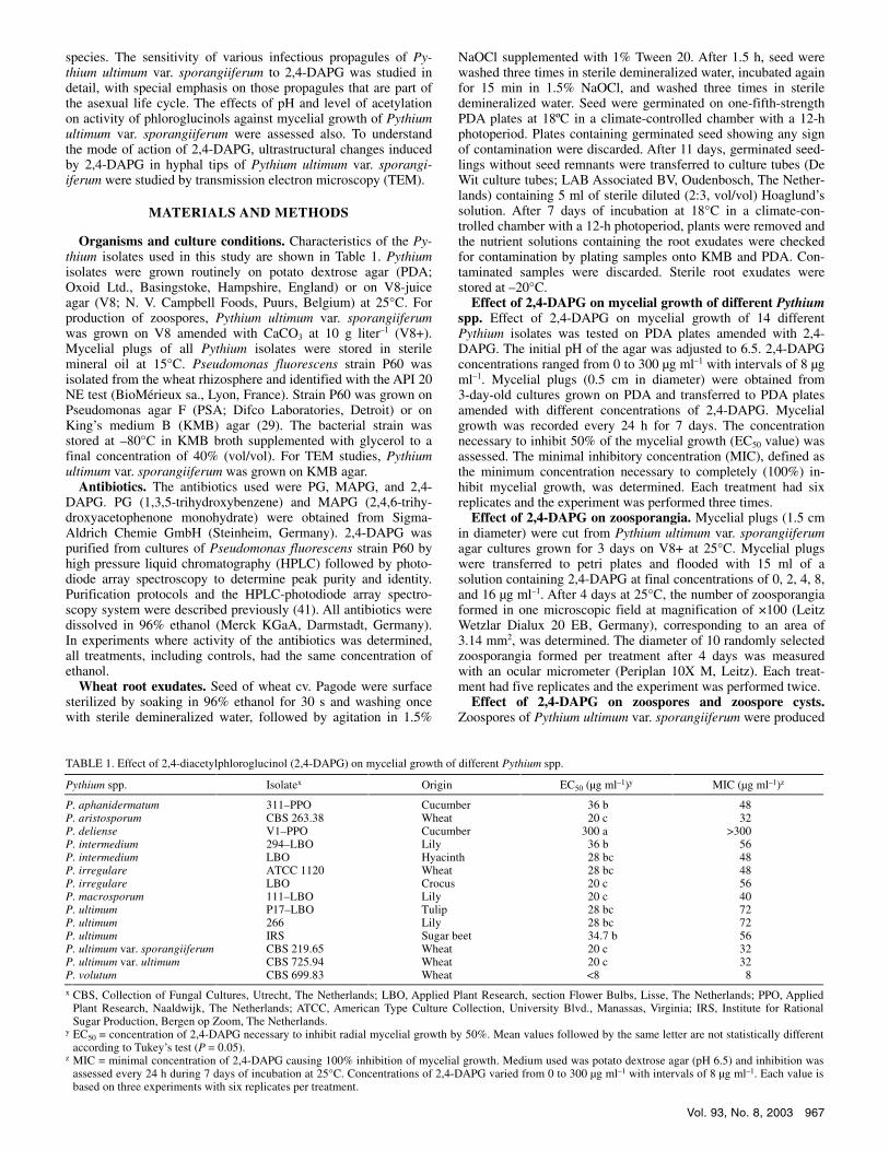
Fig. 2. Effect of 2,4-diacetylphloroglucinol (2,4-DAPG) on formation and diameter of zoosporangia of Pythium ultimum var. sporangiiferum. Mycelial plugs of Pythium ultimum var. sporangiiferum grown on medium without the antibiotic were flooded with solutions of different 2,4-DAPG concentrations. After 4 days of incubation at 25°C, the number and diameter of 10 sporangia formed per treatment was determined microscopically. Mean values of five replicates are shown. Error bars represent the standard error of the mean. For each of the two parameters, means followed by the same letter are not significantly different according to Tukey’s studentized range test (P = 0.05). The experiment was performed twice and representative results are shown.
Vol. 93, No. 8, 2003 969
margin of Pythium ultimum var. sporangiiferum cultures exposed or not to 400 µg of 2,4-DAPG per disk. Samples were fixed over-night at 4°C in 2% glutaraldehyde buffered in 0.1 M cacodylate (pH 7.2), washed in the same buffer, then dehydrated in a graded ethanol series and embedded in Epon (Prolabo, France) or LR White medium grade resin (Oxford Instrument, France).
Epon-embedded samples were postfixed with 1% osmium tetroxide before ethanol dehydration (21). Ultrathin sections (0.1 µm) were cut with a diamond knife (Reicher Ultracut micro-tone). For ultrastructural observations, Epon sections were cut onto copper grids and double contrasted by post-staining with 3% uranyl acetate in 50% ethanol (56) followed by lead citrate (42). To locate �(1,4)- and �(1,6)-glycosylated compounds (glyco-proteins and polysaccharides), Epon-embedded samples were subjected to the periodic acid thiocarbohydrazide silver proteinate (PATAg) reaction according to Thièry (52).
LR White-embedded sections were collected on golden grids coated with collodion for analytical immunocytochemistry using an indirect immunogold labeling technique for the detection of �(1,3)-glucans (31). Samples on grids were incubated overnight at 4°C in a mouse monoclonal antibody raised against �(1,3)-glucopyranose polymers (Biosupplies Australia, Australia) which has previously been shown to be specific for �(1,3)-glucans of the
cell wall (31). The antibody was diluted 1,000 or 500 times in Tris buffer saline with 1% bovine serum albumin. After rinsing, sec-tions were incubated for 1 h in a gold-labeled (15 nm) goat anti-mouse polyclonal antibody (Tebu Biocell, France) diluted 20 times to detect the primary antibody. Immunological control sec-tions were prepared by omitting the monoclonal antibody during the first incubation step. All sections were counterstained for 10 min with 3% aqueous uranyl acetate before observation in a transmission electron microscope (Hitachi H7500) at 80 kV.
The effect of 2,4-DAPG on Pythium ultimum var. sporangiiferum mycelium was evaluated by quantifying hyphal responses on Epon-embedded double-contrasted sections in the following categories: (i) dense cytoplasm with few vacuoles (young), (ii) many vacuoles in the cell lumen, (iii) beginning of cell content disorganization, and (iv) deteriorated or empty cell contents (dead). The effect of 2,4-DAPG on Pythium ultimum var. sporangiiferum cell wall synthesis was determined by observing LR White immunogold labeled sections. All experiments were performed twice.
Statistical analysis. Data were analyzed by analysis of variance followed by Tukey’s studentized range test after certifying normal distribution and homogeneity of variances (SAS institute, Inc., Cary, NC). All experiments were performed at least twice and representative results are shown.
RESULTS
Effect of 2,4-DAPG on mycelial growth of different Pythium spp. Mycelial growth of 14 Pythium isolates, obtained from multiple host plants and representing eight different species, was completely inhibited at 2,4-DAPG concentrations ranging from 8 to more than 300 µg ml–1 (Table 1). Pythium volutum CBS 699.83 was the most sensitive isolate, whereas Pythium deliense V1-PPO was relatively insensitive to 2,4-DAPG. Mycelial growth of the other Pythium isolates was completely inhibited at 2,4-DAPG concentrations ranging from 32 to 72 µg ml–1. Pythium ultimum var. sporangiiferum isolate CBS 219.65 was used for a detailed analysis of the effect of 2,4-DAPG on different propag-ules, including zoosporangia, zoospores, and zoospore cysts.
Effect of 2,4-DAPG on zoosporangia. The antibiotic 2,4-DAPG had a significant effect on both the formation and the diameter of zoosporangia of Pythium ultimum var. sporangiiferum (Fig. 2). The number of newly formed zoosporangia decreased by approximately 50% when exposed to 2,4-DAPG at 2.0 µg ml–1. The diameter of the newly formed zoosporangia, an indicative measure of their ripening, decreased from 26 to 16 µm with increasing concentrations of 2,4-DAPG.
Effect of 2,4-DAPG on zoospores and zoospore cysts. The behavior of Pythium ultimum var. sporangiiferum zoospores was
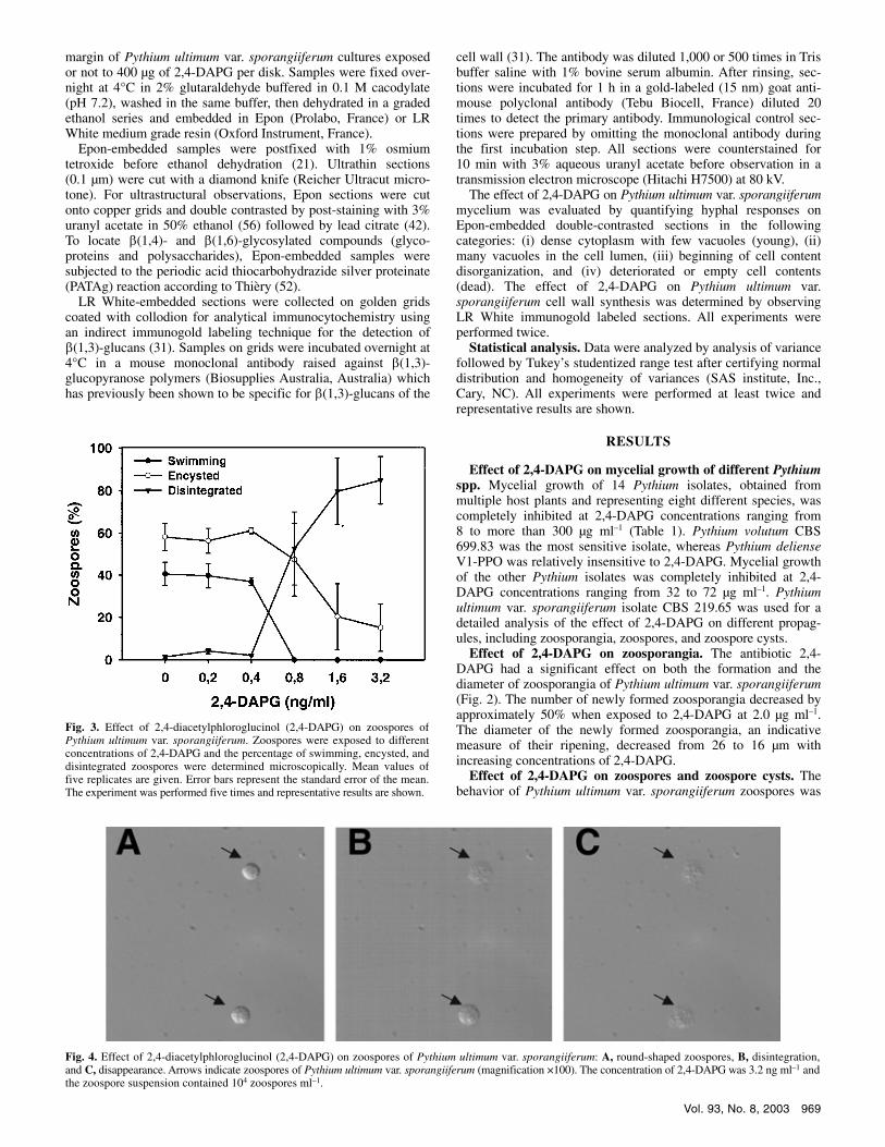
Fig. 4. Effect of 2,4-diacetylphloroglucinol (2,4-DAPG) on zoospores of Pythium ultimum var. sporangiiferum: A, round-shaped zoospores, B, disintegration, and C, disappearance. Arrows indicate zoospores of Pythium ultimum var. sporangiiferum (magnification ×100). The concentration of 2,4-DAPG was 3.2 ng ml–1 and the zoospore suspension contained 104 zoospores ml–1.
Fig. 3. Effect of 2,4-diacetylphloroglucinol (2,4-DAPG) on zoospores of Pythium ultimum var. sporangiiferum. Zoospores were exposed to different concentrations of 2,4-DAPG and the percentage of swimming, encysted, and disintegrated zoospores were determined microscopically. Mean values of five replicates are given. Error bars represent the standard error of the mean. The experiment was performed five times and representative results are shown.
970 PHYTOPATHOLOGY
adversely affected by 2,4-DAPG at concentrations of 0.8 ng ml–1 and higher (Fig. 3). At a concentration of 0.8 ng ml–1, all zoospores stopped swimming and approximately 50% of the zoospores disintegrated (Fig. 4). The time for disintegration to occur depended on the concentration of 2,4-DAPG. At concen-trations of 0.8 ng ml–1, disintegration of the zoospores occurred in approximately 20 min and at concentrations of 3.2 ng ml–1 it only took 3 min (data not shown).
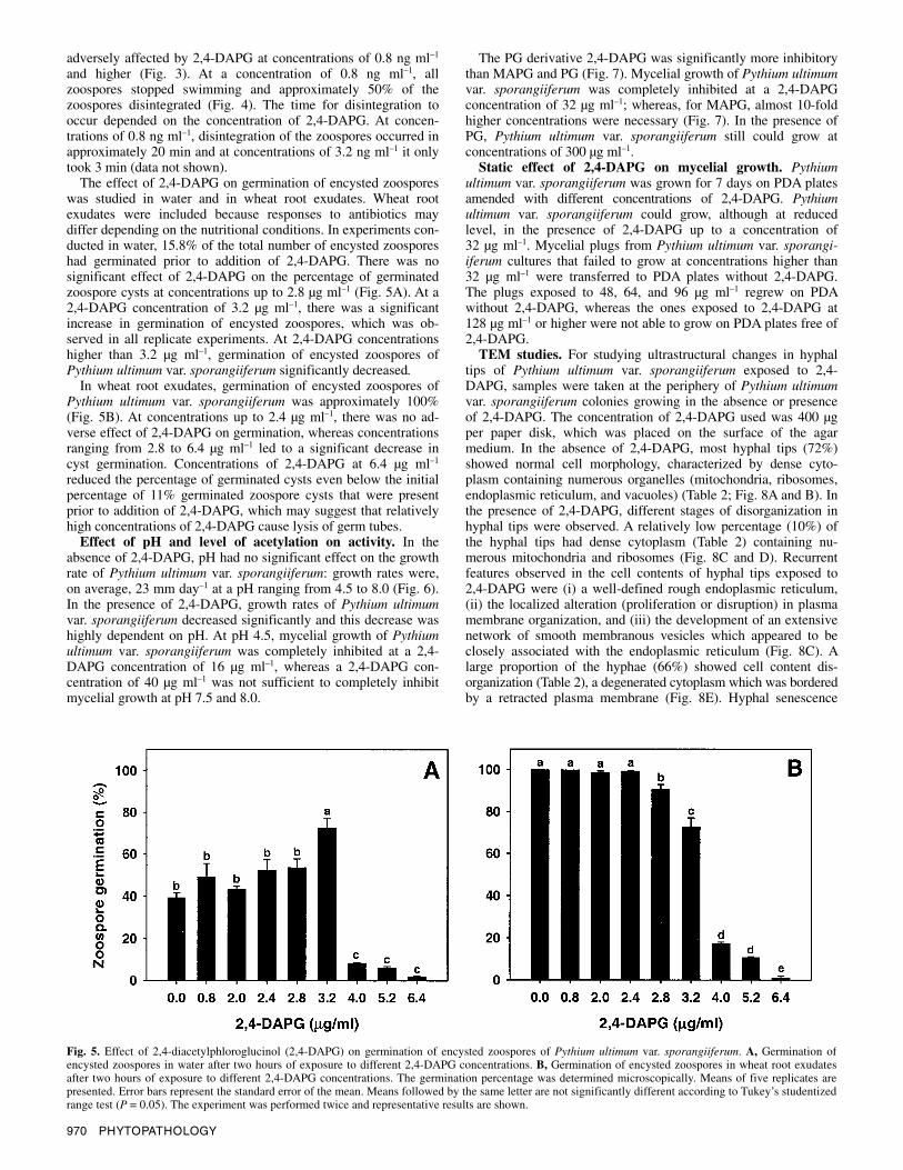
The effect of 2,4-DAPG on germination of encysted zoospores was studied in water and in wheat root exudates. Wheat root exudates were included because responses to antibiotics may differ depending on the nutritional conditions. In experiments con-ducted in water, 15.8% of the total number of encysted zoospores had germinated prior to addition of 2,4-DAPG. There was no significant effect of 2,4-DAPG on the percentage of germinated zoospore cysts at concentrations up to 2.8 µg ml–1 (Fig. 5A). At a 2,4-DAPG concentration of 3.2 µg ml–1, there was a significant increase in germination of encysted zoospores, which was ob-served in all replicate experiments. At 2,4-DAPG concentrations higher than 3.2 µg ml–1, germination of encysted zoospores of Pythium ultimum var. sporangiiferum significantly decreased.
In wheat root exudates, germination of encysted zoospores of Pythium ultimum var. sporangiiferum was approximately 100% (Fig. 5B). At concentrations up to 2.4 µg ml–1, there was no ad-verse effect of 2,4-DAPG on germination, whereas concentrations ranging from 2.8 to 6.4 µg ml–1 led to a significant decrease in cyst germination. Concentrations of 2,4-DAPG at 6.4 µg ml–1 reduced the percentage of germinated cysts even below the initial percentage of 11% germinated zoospore cysts that were present prior to addition of 2,4-DAPG, which may suggest that relatively high concentrations of 2,4-DAPG cause lysis of germ tubes.
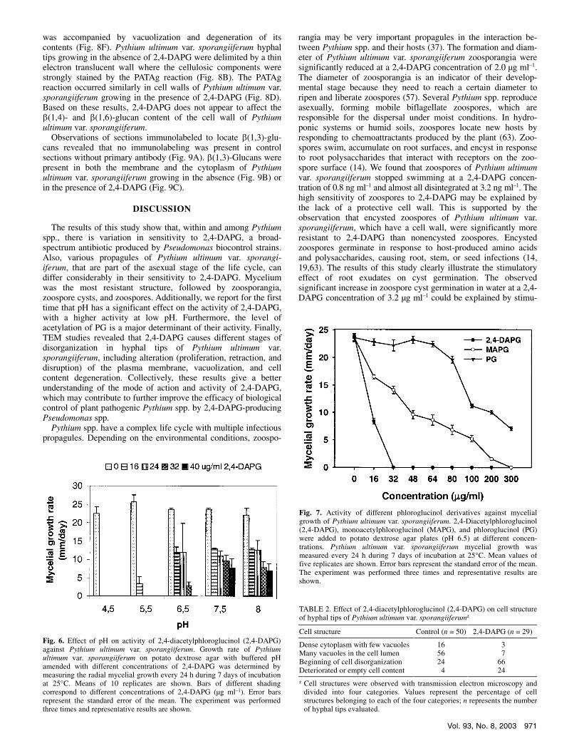
Effect of pH and level of acetylation on activity. In the absence of 2,4-DAPG, pH had no significant effect on the growth rate of Pythium ultimum var. sporangiiferum: growth rates were, on average, 23 mm day–1 at a pH ranging from 4.5 to 8.0 (Fig. 6). In the presence of 2,4-DAPG, growth rates of Pythium ultimum var. sporangiiferum decreased significantly and this decrease was highly dependent on pH. At pH 4.5, mycelial growth of Pythium ultimum var. sporangiiferum was completely inhibited at a 2,4-DAPG concentration of 16 µg ml–1, whereas a 2,4-DAPG con-centration of 40 µg ml–1 was not sufficient to completely inhibit mycelial growth at pH 7.5 and 8.0.
The PG derivative 2,4-DAPG was significantly more inhibitory than MAPG and PG (Fig. 7). Mycelial growth of Pythium ultimum var. sporangiiferum was completely inhibited at a 2,4-DAPG concentration of 32 µg ml–1; whereas, for MAPG, almost 10-fold higher concentrations were necessary (Fig. 7). In the presence of PG, Pythium ultimum var. sporangiiferum still could grow at concentrations of 300 µg ml–1.
Static effect of 2,4-DAPG on mycelial growth. Pythium ultimum var. sporangiiferum was grown for 7 days on PDA plates amended with different concentrations of 2,4-DAPG. Pythium ultimum var. sporangiiferum could grow, although at reduced level, in the presence of 2,4-DAPG up to a concentration of 32 µg ml–1. Mycelial plugs from Pythium ultimum var. sporangi-iferum cultures that failed to grow at concentrations higher than 32 µg ml–1 were transferred to PDA plates without 2,4-DAPG. The plugs exposed to 48, 64, and 96 µg ml–1 regrew on PDA without 2,4-DAPG, whereas the ones exposed to 2,4-DAPG at 128 µg ml–1 or higher were not able to grow on PDA plates free of 2,4-DAPG.
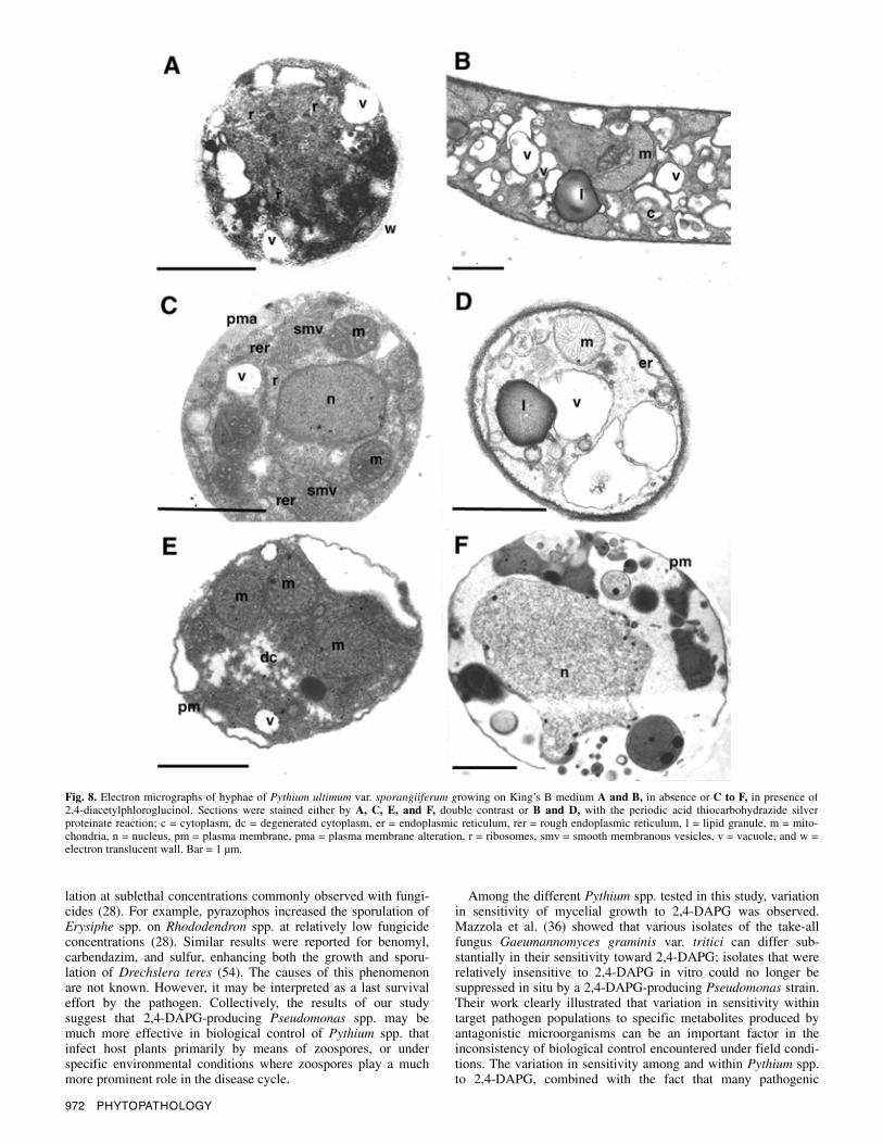
TEM studies. For studying ultrastructural changes in hyphal tips of Pythium ultimum var. sporangiiferum exposed to 2,4-DAPG, samples were taken at the periphery of Pythium ultimum var. sporangiiferum colonies growing in the absence or presence of 2,4-DAPG. The concentration of 2,4-DAPG used was 400 µg per paper disk, which was placed on the surface of the agar medium. In the absence of 2,4-DAPG, most hyphal tips (72%) showed normal cell morphology, characterized by dense cyto-plasm containing numerous organelles (mitochondria, ribosomes, endoplasmic reticulum, and vacuoles) (Table 2; Fig. 8A and B). In the presence of 2,4-DAPG, different stages of disorganization in hyphal tips were observed. A relatively low percentage (10%) of the hyphal tips had dense cytoplasm (Table 2) containing nu-merous mitochondria and ribosomes (Fig. 8C and D). Recurrent features observed in the cell contents of hyphal tips exposed to 2,4-DAPG were (i) a well-defined rough endoplasmic reticulum, (ii) the localized alteration (proliferation or disruption) in plasma membrane organization, and (iii) the development of an extensive network of smooth membranous vesicles which appeared to be closely associated with the endoplasmic reticulum (Fig. 8C). A large proportion of the hyphae (66%) showed cell content dis-organization (Table 2), a degenerated cytoplasm which was bordered by a retracted plasma membrane (Fig. 8E). Hyphal senescence
Fig. 5. Effect of 2,4-diacetylphloroglucinol (2,4-DAPG) on germination of encysted zoospores of Pythium ultimum var. sporangiiferum. A, Germination of encysted zoospores in water after two hours of exposure to different 2,4-DAPG concentrations. B, Germination of encysted zoospores in wheat root exudates after two hours of exposure to different 2,4-DAPG concentrations. The germination percentage was determined microscopically. Means of five replicates are presented. Error bars represent the standard error of the mean. Means followed by the same letter are not significantly different according to Tukey’s studentized range test (P = 0.05). The experiment was performed twice and representative results are shown.
Vol. 93, No. 8, 2003 971
was accompanied by vacuolization and degeneration of its contents (Fig. 8F). Pythium ultimum var. sporangiiferum hyphal tips growing in the absence of 2,4-DAPG were delimited by a thin electron translucent wall where the cellulosic components were strongly stained by the PATAg reaction (Fig. 8B). The PATAg reaction occurred similarly in cell walls of Pythium ultimum var. sporangiiferum growing in the presence of 2,4-DAPG (Fig. 8D). Based on these results, 2,4-DAPG does not appear to affect the �(1,4)- and �(1,6)-glucan content of the cell wall of Pythium ultimum var. sporangiiferum.
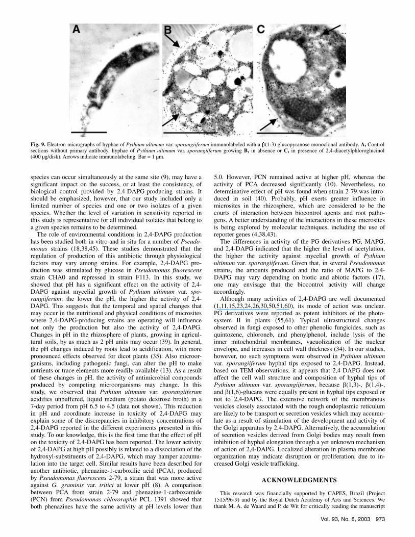
Observations of sections immunolabeled to locate �(1,3)-glu-cans revealed that no immunolabeling was present in control sections without primary antibody (Fig. 9A). �(1,3)-Glucans were present in both the membrane and the cytoplasm of Pythium ultimum var. sporangiiferum growing in the absence (Fig. 9B) or in the presence of 2,4-DAPG (Fig. 9C).
DISCUSSION
The results of this study show that, within and among Pythium spp., there is variation in sensitivity to 2,4-DAPG, a broad-spectrum antibiotic produced by Pseudomonas biocontrol strains. Also, various propagules of Pythium ultimum var. sporangi- iferum, that are part of the asexual stage of the life cycle, can differ considerably in their sensitivity to 2,4-DAPG. Mycelium was the most resistant structure, followed by zoosporangia, zoospore cysts, and zoospores. Additionally, we report for the first time that pH has a significant effect on the activity of 2,4-DAPG, with a higher activity at low pH. Furthermore, the level of acetylation of PG is a major determinant of their activity. Finally, TEM studies revealed that 2,4-DAPG causes different stages of disorganization in hyphal tips of Pythium ultimum var. sporangiiferum, including alteration (proliferation, retraction, and disruption) of the plasma membrane, vacuolization, and cell content degeneration. Collectively, these results give a better understanding of the mode of action and activity of 2,4-DAPG, which may contribute to further improve the efficacy of biological control of plant pathogenic Pythium spp. by 2,4-DAPG-producing Pseudomonas spp.
Pythium spp. have a complex life cycle with multiple infectious propagules. Depending on the environmental conditions, zoospo-
rangia may be very important propagules in the interaction be-tween Pythium spp. and their hosts (37). The formation and diam-eter of Pythium ultimum var. sporangiiferum zoosporangia were significantly reduced at a 2,4-DAPG concentration of 2.0 µg ml–1. The diameter of zoosporangia is an indicator of their develop-mental stage because they need to reach a certain diameter to ripen and liberate zoospores (57). Several Pythium spp. reproduce asexually, forming mobile biflagellate zoospores, which are responsible for the dispersal under moist conditions. In hydro-ponic systems or humid soils, zoospores locate new hosts by responding to chemoattractants produced by the plant (63). Zoo-spores swim, accumulate on root surfaces, and encyst in response to root polysaccharides that interact with receptors on the zoo-spore surface (14). We found that zoospores of Pythium ultimum var. sporangiiferum stopped swimming at a 2,4-DAPG concen-tration of 0.8 ng ml–1 and almost all disintegrated at 3.2 ng ml–1. The high sensitivity of zoospores to 2,4-DAPG may be explained by the lack of a protective cell wall. This is supported by the observation that encysted zoospores of Pythium ultimum var. sporangiiferum, which have a cell wall, were significantly more resistant to 2,4-DAPG than nonencysted zoospores. Encysted zoospores germinate in response to host-produced amino acids and polysaccharides, causing root, stem, or seed infections (14, 19,63). The results of this study clearly illustrate the stimulatory effect of root exudates on cyst germination. The observed significant increase in zoospore cyst germination in water at a 2,4-DAPG concentration of 3.2 µg ml–1 could be explained by stimu-
Fig. 6. Effect of pH on activity of 2,4-diacetylphloroglucinol (2,4-DAPG)against Pythium ultimum var. sporangiiferum. Growth rate of Pythiumultimum var. sporangiiferum on potato dextrose agar with buffered pH amended with different concentrations of 2,4-DAPG was determined by measuring the radial mycelial growth every 24 h during 7 days of incubation at 25°C. Means of 10 replicates are shown. Bars of different shading correspond to different concentrations of 2,4-DAPG (µg ml–1). Error bars represent the standard error of the mean. The experiment was performed three times and representative results are shown.
TABLE 2. Effect of 2,4-diacetylphloroglucinol (2,4-DAPG) on cell structure of hyphal tips of Pythium ultimum var. sporangiiferumz
Cell structure Control (n = 50) 2,4-DAPG (n = 29)
Dense cytoplasm with few vacuoles 16 3 Many vacuoles in the cell lumen 56 7 Beginning of cell disorganization 24 66 Deteriorated or empty cell content 4 24
z Cell structures were observed with transmission electron microscopy and divided into four categories. Values represent the percentage of cell structures belonging to each of the four categories; n represents the number of hyphal tips evaluated.
Fig. 7. Activity of different phloroglucinol derivatives against mycelial growth of Pythium ultimum var. sporangiiferum. 2,4-Diacetylphloroglucinol (2,4-DAPG), monoacetylphloroglucinol (MAPG), and phloroglucinol (PG) were added to potato dextrose agar plates (pH 6.5) at different concen-trations. Pythium ultimum var. sporangiiferum mycelial growth was measured every 24 h during 7 days of incubation at 25°C. Mean values of five replicates are shown. Error bars represent the standard error of the mean.The experiment was performed three times and representative results are shown.
972 PHYTOPATHOLOGY
lation at sublethal concentrations commonly observed with fungi-cides (28). For example, pyrazophos increased the sporulation of Erysiphe spp. on Rhododendron spp. at relatively low fungicide concentrations (28). Similar results were reported for benomyl, carbendazim, and sulfur, enhancing both the growth and sporu-lation of Drechslera teres (54). The causes of this phenomenon are not known. However, it may be interpreted as a last survival effort by the pathogen. Collectively, the results of our study suggest that 2,4-DAPG-producing Pseudomonas spp. may be much more effective in biological control of Pythium spp. that infect host plants primarily by means of zoospores, or under specific environmental conditions where zoospores play a much more prominent role in the disease cycle.
Among the different Pythium spp. tested in this study, variation in sensitivity of mycelial growth to 2,4-DAPG was observed. Mazzola et al. (36) showed that various isolates of the take-all fungus Gaeumannomyces graminis var. tritici can differ sub-stantially in their sensitivity toward 2,4-DAPG; isolates that were relatively insensitive to 2,4-DAPG in vitro could no longer be suppressed in situ by a 2,4-DAPG-producing Pseudomonas strain. Their work clearly illustrated that variation in sensitivity within target pathogen populations to specific metabolites produced by antagonistic microorganisms can be an important factor in the inconsistency of biological control encountered under field condi-tions. The variation in sensitivity among and within Pythium spp. to 2,4-DAPG, combined with the fact that many pathogenic
Fig. 8. Electron micrographs of hyphae of Pythium ultimum var. sporangiiferum growing on King’s B medium A and B, in absence or C to F, in presence of 2,4-diacetylphloroglucinol. Sections were stained either by A, C, E, and F, double contrast or B and D, with the periodic acid thiocarbohydrazide silver proteinate reaction; c = cytoplasm, dc = degenerated cytoplasm, er = endoplasmic reticulum, rer = rough endoplasmic reticulum, l = lipid granule, m = mito-chondria, n = nucleus, pm = plasma membrane, pma = plasma membrane alteration, r = ribosomes, smv = smooth membranous vesicles, v = vacuole, and w = electron translucent wall. Bar = 1 µm.
Vol. 93, No. 8, 2003 973
species can occur simultaneously at the same site (9), may have a significant impact on the success, or at least the consistency, of biological control provided by 2,4-DAPG-producing strains. It should be emphasized, however, that our study included only a limited number of species and one or two isolates of a given species. Whether the level of variation in sensitivity reported in this study is representative for all individual isolates that belong to a given species remains to be determined.
The role of environmental conditions in 2,4-DAPG production has been studied both in vitro and in situ for a number of Pseudo-monas strains (18,38,45). These studies demonstrated that the regulation of production of this antibiotic through physiological factors may vary among strains. For example, 2,4-DAPG pro-duction was stimulated by glucose in Pseudomonas fluorescens strain CHA0 and repressed in strain F113. In this study, we showed that pH has a significant effect on the activity of 2,4-DAPG against mycelial growth of Pythium ultimum var. spo-rangiiferum: the lower the pH, the higher the activity of 2,4-DAPG. This suggests that the temporal and spatial changes that may occur in the nutritional and physical conditions of microsites where 2,4-DAPG-producing strains are operating will influence not only the production but also the activity of 2,4-DAPG. Changes in pH in the rhizosphere of plants, growing in agricul-tural soils, by as much as 2 pH units may occur (39). In general, the pH changes induced by roots lead to acidification, with more pronounced effects observed for dicot plants (35). Also microor-ganisms, including pathogenic fungi, can alter the pH to make nutrients or trace elements more readily available (13). As a result of these changes in pH, the activity of antimicrobial compounds produced by competing microorganisms may change. In this study, we observed that Pythium ultimum var. sporangiiferum acidifies unbuffered, liquid medium (potato dextrose broth) in a 7-day period from pH 6.5 to 4.5 (data not shown). This reduction in pH and coordinate increase in toxicity of 2,4-DAPG may explain some of the discrepancies in inhibitory concentrations of 2,4-DAPG reported in the different experiments presented in this study. To our knowledge, this is the first time that the effect of pH on the toxicity of 2,4-DAPG has been reported. The lower activity of 2,4-DAPG at high pH possibly is related to a dissociation of the hydroxyl-substituents of 2,4-DAPG, which may hamper accumu-lation into the target cell. Similar results have been described for another antibiotic, phenazine-1-carboxilic acid (PCA), produced by Pseudomonas fluorescens 2-79, a strain that was more active against G. graminis var. tritici at lower pH (8). A comparison between PCA from strain 2-79 and phenazine-1-carboxamide (PCN) from Pseudomonas chlororaphis PCL 1391 showed that both phenazines have the same activity at pH levels lower than
5.0. However, PCN remained active at higher pH, whereas the activity of PCA decreased significantly (10). Nevertheless, no determinative effect of pH was found when strain 2-79 was intro-duced in soil (40). Probably, pH exerts greater influence in microsites in the rhizosphere, which are considered to be the courts of interaction between biocontrol agents and root patho-gens. A better understanding of the interactions in these microsites is being explored by molecular techniques, including the use of reporter genes (4,38,43).
The differences in activity of the PG derivatives PG, MAPG, and 2,4-DAPG indicated that the higher the level of acetylation, the higher the activity against mycelial growth of Pythium ultimum var. sporangiiferum. Given that, in several Pseudomonas strains, the amounts produced and the ratio of MAPG to 2,4-DAPG may vary depending on biotic and abiotic factors (17), one may envisage that the biocontrol activity will change accordingly.
Although many activities of 2,4-DAPG are well documented (1,11,15,23,24,26,30,50,51,60), its mode of action was unclear. PG derivatives were reported as potent inhibitors of the photo-system II in plants (55,61). Typical ultrastructural changes observed in fungi exposed to other phenolic fungicides, such as quintozene, chloroneb, and phenylphenol, include lysis of the inner mitochondrial membranes, vacuolization of the nuclear envelope, and increases in cell wall thickness (34). In our studies, however, no such symptoms were observed in Pythium ultimum var. sporangiiferum hyphal tips exposed to 2,4-DAPG. Instead, based on TEM observations, it appears that 2,4-DAPG does not affect the cell wall structure and composition of hyphal tips of Pythium ultimum var. sporangiiferum, because �(1,3)-, �(1,4)-, and �(1,6)-glucans were equally present in hyphal tips exposed or not to 2,4-DAPG. The extensive network of the membranous vesicles closely associated with the rough endoplasmic reticulum are likely to be transport or secretion vesicles which may accumu-late as a result of stimulation of the development and activity of the Golgi apparatus by 2,4-DAPG. Alternatively, the accumulation of secretion vesicles derived from Golgi bodies may result from inhibition of hyphal elongation through a yet unknown mechanism of action of 2,4-DAPG. Localized alteration in plasma membrane organization may indicate disruption or proliferation, due to in-creased Golgi vesicle trafficking.
ACKNOWLEDGMENTS
This research was financially supported by CAPES, Brazil (Project 1515/96-9) and by the Royal Dutch Academy of Arts and Sciences. We thank M. A. de Waard and P. de Wit for critically reading the manuscript
Fig. 9. Electron micrographs of hyphae of Pythium ultimum var. sporangiiferum immunolabeled with a �(1-3) glucopyranose monoclonal antibody. A, Control sections without primary antibody, hyphae of Pythium ultimum var. sporangiiferum growing B, in absence or C, in presence of 2,4-diacetylphloroglucinol (400 µg/disk). Arrows indicate immunolabeling. Bar = 1 µm.
974 PHYTOPATHOLOGY
and for their valuable suggestions, and T. Corberand and D. Kraaijeveld for their practical assistance.
LITERATURE CITED
1. Arisawa, M., Fujita, A., Hayashi, T., Hayashi, K., Ochiai, H., and Morita, N. 1990. Cytotoxic and antiherpetic activity of phloroglucinol deriva-tives from Mallotus japonicus (Euphorbiaceae). Chem. Pharm. Bull. 38:1624-1626.
2. Bangera, M. G., and Thomashow, L. S. 1996. Characterization of a ge-nomic locus required for synthesis of the antibiotic 2,4-diacetylphloro-glucinol by the biological control agent Pseudomonas fluorescens Q2-87. Mol. Plant-Microbe Interact. 9:83-90.
3. Bangera, M. G., and Thomashow, L. S. 1999. Identification and charac-terization of a gene cluster for synthesis of the polyketide antibiotic 2,4-diacetylphloroglucinol from Pseudomonas fluorescens Q2-87. J. Bacteriol. 181:3155-3163.
4. Bloemberg, G. V., Wijfjes, A. H. M., Lamers, G. E. M., Stuurman, N., and Lugtenberg, B. J. J. 2000. Simultaneous imaging of Pseudomonas fluorescens WCS365 populations expressing three different autofluorescent proteins in the rhizosphere: New perspectives for studying microbial communities. Mol. Plant-Microbe Interact. 13:1170-1176.
5. Blumer, C., and Haas, D. 2000. Iron regulation of the hcnABC genes encoding hydrogen cyanide synthase depends on the anaerobic regulator ANR rather than on the global activator GacA in Pseudomonas fluorescens CHA0. Microbiology 146:2417-2424.
6. Bokesch, H. R., Groweiss, A., McKee, C. T., and Boyd, M. R. 1999. Laxifloranone, a new phloroglucinol derivative from Marila laxiflora. J. Nat. Prod. 62:1197-1199.
7. Bonsall, R. F., Weller, D. M., and Thomashow, L. S. 1997. Quantifica-tion of 2,4-diacetylphloroglucinol produced by fluorescent Pseudomonas sp. in vitro and in the rhizosphere of wheat. Appl. Environ. Microbiol. 63:951-955.
8. Brisbane, P. G., Janik, L. J., Tate, M. E., and Warren, R. F. O. 1987. Revised structure for the phenazine antibiotic from Pseudomonas fluorescens 2-79 (NRRL B-15132). Antimicrob. Agents Chemother. 31: 1967-1971.
9. Chamswarng, C., and Cook, R. J. 1985. Identification and comparative pathogenicity of Pythium species from roots and wheat-field soils in the Pacific Northwest. Phytopathology 75:821-827.
10. Chin-A-Woeng, T. F. C., Bloemberg, G., van der Bij, A., van der Drift, K. M. G. M., Schripsema, J., Kroon, B., Scheffer, R. J., Keel, C., Bakker, P. A. H. M., Tichy, H.-V., de Bruijn, F. J., Thomas-Oates, J. E., and Lugtenberg, B. J. J. 1998. Biocontrol by phenazine-1-carboxamide-producing Pseudomonas chlororaphis PCL1391 of tomato root and crown rot caused by Fusarium oxysporum f. sp. radicis-lycopersici. Mol. Plant-Microbe Interact. 11:1069-1077.
11. Cronin, D., Moenne-Loccoz, Y., Fenton, A., Dunne, C., Dowling, D. N., and O’Gara, F. 1997. Role of 2,4-diacetylphloroglucinol in the interactions of the biocontrol pseudomonad strain F113 with the potato cyst nematode Globodera rostochiensis. Appl. Environ. Microbiol. 63:1357-1361.
12. Cronin, D., Moenne-Loccoz, Y., Fenton, A., Dunne, C., Dowling, D. N., and O’Gara, F. 1997. Ecological interaction of a biocontrol Pseudo-monas fluorescens strain producing 2,4-diacetylphloroglucinol with the soft rot potato pathogen Erwinia caratovora subsp. atroseptica. FEMS Microbiol. Ecol. 23:95-106.
13. Cunningham, J. E., and Kuiack, C. 1992. Production of citric and oxalic acids and solubilization of calcium-phosphate by Penicillium bilaii. Appl. Environ. Microbiol. 58:1451-1458.
14. Deacon, J. W., and Donaldson, S. P. 1993. Molecular recognition in the homing responses of zoosporic fungi, with special reference to Pythium and Phytophthora. Mycol. Res. 97:1153-1171.
15. Debabrata, S., and Naik, P. 2000. Phloroglucinol enhances growth and rate of axillary shoot proliferation in potato shoot tip cultures in vitro. Plant Cell Tissue Organ Cult. 60:139-149.
16. Dick, M. W. 1990. Keys to Pythium. Reading University Press, Reading, England.
17. Duffy, B. K., and Défago, G. 1997. Zinc improves biocontrol of fusarium crown and root rot of tomato by Pseudomonas fluorescens and represses the production of pathogen metabolites inhibitory to bacterial antibiotic biosynthesis. Phytopathology 87:1250-2157.
18. Duffy, B. K., and Défago, G. 1999. Environmental factors modulating antibiotic and siderophore biosynthesis by Pseudomonas fluorescens biocontrol strains. Appl. Environ. Microbiol. 65:2429-2438.
19. Estrada-Garcia, T., Ray, T. C., Green, J. R., Callow, J. A., and Kennedy, J. F. 1990. Encystment of Pythium aphanidermatum zoospores is in-duced by root mucilage polysaccharides, pectin and a monoclonal antibody to a surface antigen. J. Exp. Bot. 41:693-699.
20. Fenton, A. M., Stephens, P. M., Crowley, J., O’Callaghan, M., and O’Gara, R. 1992. Exploitation of gene(s) involved in 2,4-diacetylphloro-glucinol biosynthesis to confer a new biocontrol capability to a Pseudo-monas strain. Appl. Environ. Microbiol. 58:3873-3878.
21. Gianinazzi, S., and Gianinazzi-Pearson, V. 1992. Cytology, histochemis-try and immunocytochemistry as tools for studying structure and function in endomycorrhiza. Methods Microbiol. 24:109-139.
22. Ishiguro, K., Nagareya, N., and Fukumoto, H. 1998. A phloroglucinol derivative from cell suspension cultures of Hypericum palatum. Phyto-chemistry 47:1041-1043.
23. Ito, H., Muranaka, T., Mori, K., Jin-ZheXiong, Tokuda, H., Nishino, H., Yoshida, T., and Jin, Z. X. 2000. Ichthyotoxic phloroglucinol derivatives from Dryopteris fragrans and their anti-tumor promoting activity. Chem. Pharm. Bull. 1190-1195.
24. James, D. J. 1979. The role of auxins and phloroglucinol in adventitious root formation in Rubus and Fragaria in vitro. J. Hortic. Sci. 54:273-277.
25. Jimenez-Escrig, A., Jimenez-Jimenez, I., Pulido, R., and Saura-Calixto, F. 2001. Antioxidant activity of fresh and processed edible seaweed. J. Sci. Food Agric. 81:530-534.
26. Keel, C., Schnider, U., Maurhofer, M., Voisard, C., Laville, J., Burger, U., Wirthner, P. H., Haas, D., and Défago, G. 1992. Suppression of root diseases by Pseudomonas fluorescens CHA0: Importance of the secon-dary metabolite 2,4-diacetylphloroglucinol. Mol. Plant-Microbe Interact. 5:4-13.
27. Keel, C., Wirthner, P. H., Oberhansli, T. H., Voisard, C., Burger, U., Haas, D., and Défago, G. 1990. Pseudomonads as antagonists of plant pathogens in the rhizosphere: Role of the antibiotic 2,4-diacetylphloro-glucinol in the suppression of black root rot of tobacco. Symbiosis 9:327-342.
28. Kenyon, D. M., Dixon, G. R., and Helfer, S. 1997. The repression and stimulation of growth of Erysiphe sp. on Rhododendron by fungicidal compounds. Plant Pathol. 46:425-431.
29. King, E. O., Ward, M. K., and Raney, D. E. 1954. Two simple media for the demonstration of pyocyanin and fluorescin. J. Lab. Clin. Med. 44:301-307.
30. Lawler, I. R., Eschler, B. M., Schliebs, D. M., and Foley, W. J. 1999. Relationship between chemical functional groups on Eucalyptus secon-dary metabolites and effectiveness as marsupial antifeedants. J. Chem. Ecol. 25:2561-2573.
31. Lemoine, M. C., Gollotte, A., and Gianinazzi-Pearson, V. 1995. Locali-zation of beta(1-3) glucan in walls of the endomycorrhizal fungi Glomus mosseae (Nicol. & Gerd.) Gerd. & Trappe and Acaulospora laevis Gerd. & Trappe during colonization of host roots. New Phytol. 129:97-105.
32. Levy, E., Gough, F. J., Berlin, K. D., Guiana, P. W., and Smith, J. T. 1992. Inhibition of Septoria tritici and other phytopathogenic fungi and bacteria by Pseudomonas fluorescens and its antibiotics. Plant Pathol. 41:335-341.
33. Loper, J. E., and Lindow, S. E. 1997. Reporter gene systems useful in evaluating in situ gene expression by soil and plant-associated bacteria. Pages 482-492 in: Manual of Environmental Microbiology. C. J. Hurst, G. R. Knudsen, M. J. McInerney, L. D. Stetzenbach, and M. V. Walter, eds. American Society for Microbiology, Washington.
34. Lyr, H. 1995. Aromatic hydrocarbon fungicides and their mechanism of action. Pages 76-98 in: Modern Selective Fungicides: Properties, Appli-cations, Mechanisms of Action, 2nd ed. H. Lyr, ed. Gustav Fischer Verlag, Jena, Germany.
35. Marschner, H., and Römheld, V. 1983. In vivo measurement of root-induced pH changes at the soil-root interface: Effect of plant species and nitrogen source. Z. Pflanzenphysiol. 111:241-251.
36. Mazzola, M. F., Fujimoto, D. K., Thomashow, L. S., and Cook, R. J. 1995. Variation in sensitivity of Gaeumannomyces graminis to anti-biotics produced by Pseudomonas spp. and effect on biological control of take-all of wheat. Appl. Environ. Microbiol. 61:2554-2559.
37. Nelson, E. B., and Hsu, J. S. T. 1994. Nutritional factors affecting responses of sporangia of Pythium ultimum to germination stimulants. Phytopathology 84:677-683.
38. Notz, R., Maurhofer, M., Schnider-Keel, U., Duffy, B., Haas, D., and Défago, G. 2001. Biotic factors affecting expression of the 2,4-diacetyl-phloroglucinol biosynthesis gene phlA in Pseudomonas fluorescens strains CHA0 in the rhizosphere. Phytopathology 91:873-881.
39. Nye, P. H. 1981. Changes in the pH across the rhizosphere induced by roots. Plant Soil 61:7-26.
40. Ownley, B. H., Weller, D. M., and Thomashow, L. S. 1992. Influence of in situ and in vitro pH on suppression of Gaeumannomyces graminis var. tritici by Pseudomonas fluorescens 2-79. Phytopathology 82:178-184.
41. Raaijmakers, J. M., Bonsall R. F., and Weller, D. M. 1999. Effect of population density of Pseudomonas fluorescens on production of 2,4-diacetylphloroglucinol in the rhizosphere of wheat. Phytopathology 89:470-475.
Vol. 93, No. 8, 2003 975
42. Reynolds, E. S. 1983. The use of lead citrate at high pH as an electron opaque stain in electronic microscopy. J. Cell Biol. 17:208-212.
43. Sankarasubramanian, S., and Kaushik, B. D. 2001. Development of genetic markers in cyanobacteria and stability of genetically marked strains in soil. World J. Microbiol. Biotechnol. 17:535-544.
44. Sarniguet, A., Kraus, J., Henkels, M. D., Muehlchen, A. M., and Loper, J. E. 1995. The sigma factor �S affects antibiotic production and biological control activity of Pseudomonas fluorescens Pf-5. Proc. Natl. Acad. Sci. USA 92:12255-12259.
45. Shanahan, P., O’Sullivan, D. J., Simpson, P., Glennon, J. D., and O’Gara, F. 1992. Isolation of 2,4-diacethylphloroglucinol from a fluorescent pseudomonad and investigation of physiological parameters influencing its production. Appl. Environ. Microbiol. 58:353-358.
46. Shanahan, P., O’Sullivan, D. J., Simpson, P., Glennon, J. D., and O’Gara, F. 1993. Liquid chromatographic assay of microbially derived phloro-glucinol antibiotics for establishing the biosynthetic route to produc- tion, and the factors affecting their regulation. Anal. Chim. Acta 272:271-277.
47. Sharifi-Tehrani, A., Zala, M., Natsch, A., Moënne-Loccoz, Y., and Défago, G. 1998. Biocontrol of soil-borne fungal diseases by 2,4-diacetylphloroglucinol-producing pseudomonads with different restric-tion profiles of amplified 16S rDNA. Eur. J. Plant Pathol. 104:631-643.
48. Strunz, G. M., Wall, R. E., and Holder-Franklin, M. A. 1978. Phloro-glucinol derivatives from Aeromonas hydrophila. J. Antibiot. (Tokyo) 31:1201-1202.
49. Stutz, E., Défago, G., and Kern, H. 1986. Naturally occurring fluorescent pseudomonad involved in suppression of black root rot of tobacco. Phytopathology 76:181-185.
50. Tada, M., Takakuwa, T., Nagai, M., and Yoshii, T. 1990. Antiviral and antimicrobial activity of 2,4-diacetylphloroglucinols, 2-acylcyclohexane-1,3-diones and 2-carboxamidocyclohexane-1,3-diones. Agric. Biol. Chem. 54:3061-3063.
51. Te-Chato, S., and Lim, M. 1999. Plant regeneration of mangosteen via nodular callus formation. Plant Cell Tissue Organ Cult. 59:89-93.
52. Thièry, J. P. 1967. Mise en évidence des polysaccharides sur des coupes fines en microscopie électronique. J. Microsc. (Oxf.) 6:987-1018.
53. Thomashow, L. S., Bonsall, R. F., and Weller, D. M. 1997. Antibiotic
production by soil and rhizosphere microbes in situ. Pages 493-499 in: Manual of Environmental Microbiology. C. J. Hurst, G. R. Knudsen, M. J. McInerney, L. D. Stetzenbach, and M. V. Walter, eds. American Society for Microbiology, Washington.
54. Toubia-Rahme, H., Ali-Haimoud, D. E., Barrault, G., and Albertini, L. 1995. Effect of four fungicides on barley net blotch caused by Drecheslera teres. J. Phytopathol. 143:335-339.
55. Trebst, A., Donner, W., and Draber, W. 1984. Structure activity corre-lation of herbicides affecting plastoquinone reduction by photosystem II: Electron density distribution in inhibitors and plastoquinone species. Z. Naturforsch. C. Biosci. 39:405-411.
56. Valentine, R. C. 1961. Contrast enhancement in the electron microscopy of viruses. Adv. Virus Res. 8:287-290.
57. Van der Plaats-Niterink, A. J. 1981. Monograph of the genus Pythium. Page 244 in: Studies in Mycology, 21. Centraalbureau voor Schimmel-cultures, Baarn, The Netherlands.
58. Verrota, L., Appendino, G., Belloro, E., Jakupovic, J., and Bombardelli, E. 1999. Furohyperforin, a prenylated phloroglucinol from St. John’s Wort (Hypericum perforatum). J. Nat. Prod. 62:770-772.
59. Vincent, M. N., Harrison, L. A., Brackin, J. M., Kovacevich, P. A., Mukerji, P., Weller, D. M., and Pierson, E. A. 1991. Genetic analysis of the antifungal activity of a soilborne Pseudomonas aureofacens strain. Appl. Environ. Microbiol. 57:2928-2934.
60. Yajima, T., and Munakata, K. 1979. Phloroglucinol-type furocumarins, a group of potent naturally-occurring insect antifeedants. Agric. Biol. Chem. 43:1701-1706.
61. Yoneyama, K., Konnai, M., Honda, I., Yoshida, S., Takahashi, N., Koike, H., and Inoue, Y. 1990. Phloroglucinol derivatives as potent photosystem inhibitors. Z. Naturforsch. C. Biosci. 45:317-321.
62. Whistler, C. A., Corbell, N. A., Sarniguet, A., Ream, W., and Loper, J. E. 1998. The two-component regulators gacS and gacA influence accumu-lation of the stationary-phase sigma factor �S and the stress response in Pseudomonas fluorescens Pf-5. J. Bacteriol. 180:6635-6641.
63. Zhou, T., and Paulitz, T. C. 1993. In vitro and in vivo effects of Pseudomonas spp. on Pythium aphanidermatum: Zoospore behavior in exudates and on the rhizoplane of bacteria-treated cucumber roots. Phytopathology 83:872-876.


![3-[( E )-2,4-Dichlorobenzylidene]-1-methylpiperidin-4-one](https://static.fdokumen.com/doc/165x107/631368d0c32ab5e46f0c6810/3-e-24-dichlorobenzylidene-1-methylpiperidin-4-one.jpg)