MTOC Reorientation Occurs during Fc R-mediated Phagocytosis in Macrophages
Upload
independentCategory
view
2download
0
Efavirenz Promotes b-Secretase Expression and IncreasedAb1-40,42 via Oxidative Stress and Reduced MicroglialPhagocytosis: Implications for HIV AssociatedNeurocognitive Disorders (HAND)Lecia A. M. Brown1,2, Jingji Jin1, Darren Ferrell1,2, Edin Sadic1, Demian Obregon3, Adam J. Smith3,
Jun Tan2,3,4,5, Brian Giunta1,2,4,5*
1 Department of Psychiatry and Behavioral Neurosciences, Neuroimmunology Laboratory, University of South Florida, Morsani College of Medicine, Tampa, Florida, United
States of America, 2 Department of Molecular Medicine, University of South Florida, Morsani College of Medicine, Tampa, Florida, United States of America, 3 Department
of Psychiatry and Neurosciences, Rashid Developmental Neurobiology Laboratory, Silver Child Development Center, University of South Florida, Morsani College of
Medicine, Tampa, Florida, United States of America, 4 Center of Excellence for Aging and Brain Repair, Department of Neurosurgery and Brain Repair, University of South
Florida, Morsani College of Medicine, Tampa, Florida, United States of America, 5 James A. Haley Veterans Administration Hospital, Tampa, Florida, United States of
America
Abstract
Efavirenz (EFV) is among the most commonly used antiretroviral drugs globally, causes neurological symptoms thatinterfere with adherence and reduce tolerability, and may have central nervous system (CNS) effects that contribute in partto HIV associated neurocognitive disorders (HAND) in patients on combination antiretroviral therapy (cART). Thus weevaluated a commonly used EFV containing regimen: EFV/zidovudine (AZT)/lamivudine (3TC) in murine N2a cellstransfected with the human ‘‘Swedish’’ mutant form of amyloid precursor protein (SweAPP N2a cells) to assess forpromotion of amyloid-beta (Ab) production. Treatment with EFV or the EFV containing regimen generated significantlyincreased soluble amyloid beta (Ab), and promoted increased b-secretase-1 (BACE-1) expression while 3TC, AZT, or, vehiclecontrol did not significantly alter these endpoints. Further, EFV or the EFV containing regimen promoted significantly moremitochondrial stress in SweAPP N2a cells as compared to 3TC, AZT, or vehicle control. We next tested the EFV containingregimen in Ab - producing Tg2576 mice combined or singly using clinically relevant doses. EFV or the EFV containingregimen promoted significantly more BACE-1 expression and soluble Ab generation while 3TC, AZT, or vehicle control didnot. Finally, microglial Ab phagocytosis was significantly reduced by EFV or the EFV containing regimen but not by AZT,3TC, or vehicle control alone. These data suggest the majority of Ab promoting effects of this cART regimen are dependentupon EFV as it promotes both increased production, and decreased clearance of Ab peptide.
Citation: Brown LAM, Jin J, Ferrell D, Sadic E, Obregon D, et al. (2014) Efavirenz Promotes b-Secretase Expression and Increased Ab1-40,42 via Oxidative Stress andReduced Microglial Phagocytosis: Implications for HIV Associated Neurocognitive Disorders (HAND). PLoS ONE 9(4): e95500. doi:10.1371/journal.pone.0095500
Editor: Michelle L. Block, Virginia Commonwealth University, United States of America
Received January 27, 2014; Accepted March 27, 2014; Published April 23, 2014
Copyright: � 2014 Brown et al. This is an open-access article distributed under the terms of the Creative Commons Attribution License, which permitsunrestricted use, distribution, and reproduction in any medium, provided the original author and source are credited.
Funding: BG is supported by NIMH/NIH grant (1R01MH098737-02) (PI). JT is supported by NIH grants (1R41AG031586-01), (1R43AG033417-01), and1R43AT004871-01 as well as a Veterans Administration grant (MH080168). The funders had no role in study design, data collection and analysis, decision topublish, or preparation of the manuscript.
Competing Interests: The authors have declared that no competing interests exist.
* E-mail: [email protected]
Introduction
There has been considerable growth in patient’s receiving
combination antiretroviral therapy (cART) in recent years [1]. Up
to 50% of long-term HIV-infected patients experience HIV
associated neurocognitive disorders (HAND) [2]. Most recently it
was shown the Non-Nucleoside Reverse Transcriptase Inhibitor
(NNRTI) efavirenz (EFV) is associated with cognitive disorders
even in asymptomatic HIV-infected patients [3]. A randomized
controlled study [4] found subjects receiving EFV-containing
regimens showed less improvement from baseline on instruments
examining speed of information processing and executive function
than patients not on EFV. Further, patients with preserved
immune function on EFV regimens showed greater improvement
on Trail-Making Tests A and B and the Wechsler Adult
Intelligence Scale digital symbol test after EFV interruption than
the non-EFV control group [5]. EFV has substantial rates of
central nervous system (CNS) side effects aside of cognitive
impairment including sleep and dreaming disturbances and
anxiety [6–8] that interfere with adherence and tolerability as
well [9].
Amyloid-beta (Ab) peptide generation and aggregation as
plaques are traditionally known as key events in the development
of Alzheimer’s Disease (AD; [10–13]). The peptides have been
evidenced to be neurotoxic, as they are reported mediators of
inflammation [14,15], and oxidative stress [16]. Ab peptides are
produced via the amyloidogenic pathway of amyloid precursor
protein (APP) proteolysis, which involves the actions of b and c-
secretases [13,17]. Initially, b-secretase (BACE-1) cleaves APP,
creating an Ab-containing carboxyl-terminal fragment known as
b-C-terminal fragment (b-CTF) [18]. This proteolysis also
PLOS ONE | www.plosone.org 1 April 2014 | Volume 9 | Issue 4 | e95500

generates an amino-terminal, soluble APP-b (sAPP-b) fragment,
which is released extracellularly. Intracellularly, b-CTF is then
cleaved by a multi-protein c-secretase complex that results in
generation of the Ab peptide and a smaller c-CTF [19,20]. In the
human brain Ab1-40 is the predominant form whereas Ab1-42
represents about 10% of Ab in brain and has a greater propensity
to form neurotoxic oligomeric and aggregated species [21].
The rapid, early clinical phase-in of cART required dose de-
escalations secondary to toxicities suggested to be related to
mitochondrial drug side effects [22]. Mitochondrial dysfunction
can result in an elevation of reactive oxygen species (ROS) that in
turn promotes amyloidogenic APP processing by promoting
BACE-1 activity [18]. Such mitochondrial stress has also been
reported occurs in patients taking lamivudine (3TC), zidovudine
(AZT) and especially EFV [23–28]; a commonly used cART
regimen [29–31]. In light of the increasing life-span’s imparted by
cART, the mitochondrial promoted by cART [23–27,32–34], and
the age associated risk for developing amyloid pathology [35], it is
not surprising that a body of epidemiological data suggests
significant numbers of long-term HIV survivors are at elevated
risk of developing early brain aging in the form of AD like
pathology including Ab deposition [36–42].
As a result, we hypothesized that Ab pathology may be
produced via the amyloidogenic pathway of APP proteolysis,
which involves the actions of BACE-1 [13] in patients on such
regimen and sought to test this with in vitro and in vivo models. Our
results indicate that EFV is the primary antiretroviral in this
commonly used EFV containing regimen: EFV/3TC/AZT [29–
31] which is responsible for its promotion of Ab pathology.
Materials and Methods
All animal work was approved by the University of South
Florida Institutional Animal Care and Use Committee (IACUC).
ReagentsAb1-40 and Ab1-42 peptides and control peptide (Ab40-1) were
obtained from QCB (Hopkinton, MA) and freshly solubilized in
distilled H2O immediately before use. To determine the
oligomeric state of Ab in our assays, Ab was immunoprecipitated
from cell supernatants after incubation with microglia and/or
neurons, and Western blot analysis was performed at time points
of 12, 24, and 48 hr. Data revealed that both Ab1-40 and Ab1-42,
irrespective of the time points assayed, existed as a ladder of SDS-
stable oligomers, with a predominant species of ,32 kDa. Immun-
Blot polyvinylidene difluoride (PVDF) membranes were purchased
from Bio-Rad (Hercules, CA). Tris-buffered saline was obtained
from Bio-Rad (Hercules, CA) and luminol reagent was obtained
from Pierce Biotechnology. Anti-actin antibody was obtained from
Roche. Antiretrovirals were obtained from The National Institutes
of Health (NIH) AIDS Research and Reference Reagent Program
(Rockville, MD). Regarding dosages administered, cART effects in
vivo are likely to occur over long- term exposures [43]. Thus,
chronic, low dose, in vivo effects of any reagent are often very
appropriately modeled in vitro, by proportionally higher doses of
the same reagent, over more acute time frames [43]. For these
reasons we used 10 mM cART doses throughout our in vitro works
and per our previous study [44]. The doses of cART administered
in vivo were based on based on human clinical therapy [45], the
body weight of the mice, the short dosing period of 10 ten days,
the administration method being in chow as opposed to
intravenous administration, as well as those reported in previous
publications: AZT 50 mg/kg [46-48], 3TC 40 mg/kg [47,48],
and EFV 15 mg/kg [46,49].
Neuronal Ab Production AssayThis was performed according to our previous works [44].
Briefly, SweAPP N2a cells were treated with EFV, AZT, and 3TC
both alone (10 mM) and in combination (10 mM) for 18 hours.
Ab1-40, 42 peptides were detected directly from the conditioned
media and quantified in these samples using Ab1-40, 42 ELISA kits
(Life Technologies) in accordance with the manufacturer’s
instructions.
Western immunoblottingWestern blot was performed as described previously [50,51].
Briefly, total protein content was estimated using the Bio-Rad
protein assay in strict accordance with manufacturer’s directions.
Immunoblotting was performed with a primary antibody followed
by an anti-mouse HRP-conjugated IgG secondary antibody as a
tracer. Primary antibodies used included: 6E10 monoclonal anti-
Ab antibody (Covance, 1:1000), polyclonal Rabbit anti- BACE-1
(Sigma1:1000), C-terminus monoclonal anti-BACE-1 (Millipore
1:1000), and anti-actin antibody (Sigma, 1:1500).
For the in vivo studies of Ab associated pathology we employed
our previous methods [50,51]. Left hemispheres of 3 month old
transgenic and nontransgenic mouse brains were lysed in ice-cold
lysis buffer and aliquots were electrophoretically separated using
16.5% Tris–tricine gels. Electrophoresed proteins were then
transferred to PVDF membranes (Bio-Rad), washed in dH2O,
then blocked in Tris-buffered saline containing 5% (w/v) non-fat
dry milk. Membranes were then hybridized with various primary
antibodies followed by washing in dH2O and then incubation for
1 h at ambient temperature with the appropriate HRP-conjugated
secondary antibody (1:1000). For both in vitro and in vivo studies,
blots were developed and then assessed densitometrically analyzed
using the Fluor-S MultiImager with Quantity One software (Bio-
Rad).
Mitochondrial Stress Analysis: Adenosine triphosphate(ATP), mitochondrial membrane potential (MMP), andreactive oxygen species (ROS)
ATP determination was performed using the Invitrogen ATP
determination kit (A22066). MMP analysis was performed using a
JC-1 (excitation filter 530/25, emission filter 590/35) MMP
detection kit (Biotium). Cellular ROS generation was analyzed
using 2,7-dichloro dihydrofluorescein diacetate (excitation filter
485/20, emission filter528/20) from the Invitrogen ROS detection
kit. For all three analyses of mitochondrial stress, the reagents and
reaction mixture were combined according to the supplied
protocol. All fluorescence measurements were read using a Biotek
Synergy H1 microplate reader.
Microglial Phagocytosis AssayThis was performed according to our previous studies [44,50].
Briefly, primary mouse microglia were treated with ‘‘aged’’ Ab1-42
peptide conjugated with FITC (BioSource Life TechnologiesTM)
with antiretroviral drugs both alone (10 mM) and in combination
(10 mM). The total cellular protein of all groups was quantified and
adjusted using the Bio-Rad protein assay. Extracellular and cell
associated FITC-tagged Ab was quantified using an SPECTRA-
max GEMINI microplate fluorometer (Molecular Devices Corp.)
with an emission wavelength of 538 nm and an excitation
wavelength of 485 nm. Microglial cells were rinsed 3 times in
Ab-free complete medium, and the media was exchanged with
fresh Ab-free complete medium for 10 min both to allow for
removal of non-incorporated Ab and to promote concentration of
the Ab into phagosomes. The relative mean fluorescence values
HIV, Cognitive, Antiretrovirals, Mitochondria, cART
PLOS ONE | www.plosone.org 2 April 2014 | Volume 9 | Issue 4 | e95500

for each sample at 37uC and 4uC at the indicated time points were
determined by fluorometric analysis. Relative mean values were
calculated as: (mean fluorescence value for each sample at 37uC -
mean fluorescence value for each sample at 4uC). In this manner,
both extracellular and cell associated FITC-labeled Ab were
quantified.
Statistical analysisAll data were normally distributed; therefore, in instances of
single mean comparisons, Levene’s test for equality of variances
followed by t-test for independent samples was used to assess
significance. In instances of multiple mean comparisons, analysis of
variance (ANOVA) was used, followed by post-hoc comparison using
Bonferonni’s method/correction. Alpha levels were set at 0.05 for
all analyses. The statistical package for the social sciences release
10.0.5 (SPSS Inc., Chicago, IL, USA) was used for all data analysis.
Results
Epidemiological reports indicate that HAND persists in patients
even with good viremic control who take EFV [3]. Previous studies
have shown that cART imparts mitochondrial toxicity in the form
of elevate ROS [23,24]. A high ROS microenvironment has been
shown to promote the activity of BACE-1, a key enzyme the
generation of Ab in the brain [52]. Brain oligomeric [53] and Ab1-
40,42 [54] have been correlated with cognitive impairment. Since
the EFV containing regimen may promote mitochondrial
dysfunction [23,24,27,34,55] which could result in increased
BACE-1 activity, we investigated the effect of a commonly used
EFV containing cART regimen [29–31] for its ability to
upregulate Ab production via activation of BACE-1 and
amyloidogenic APP processing and also for its ability to reduce
microglial phagocytosis of Ab1-40,42.
BACE-1 is involved in Ab generation promoted by theEFV containing cART regimen in cultured SweAPP N2acells (Fig. 1)
Using similar conditions as in our prior investigations [44],
SweAPP N2a cells were treated with the EFV containing regimen:
3TC, AZT, EFV or each drug singly at 10 mM in addition to PBS
control for 18 hours. Ab40 and Ab42 peptides were then measured
in conditioned media from these cells by ELISA (Fig. 1A–C) while
BACE-1 expression was measured in cell lysates by Western Blot
analysis (Fig. 1D–E). The EFV containing regimen increased Ab40
and Ab42 production in SweAPP N2a cells significantly
(**P,0.05). Importantly, we found that EFV alone was more
potent than the EFV containing regimen in terms of significantly
increasing Ab40 and Ab42 production by these cells (***P,0.001).
Additionally EFV or the EFV containing regimen increased
BACE-1 expression in SweAPP N2a cells significantly
(***P,0.001). These data would suggest that 3TC and/or AZT
somehow reduces the toxicity of EFV in terms of promoting
amyloidogenic APP processing and that EFV is the primary agent
promoting Ab production in SweAPP N2a cells. There is some
evidence to indicate that AZT may indeed have a neuroprotective
effect [56–58] which could explain why the EFV containing
regimen is less potent in its amyloid producing effects compared to
EFV alone.
Cerebral amyloidosis in Tg2576 mice is increased by EFVor the EFV containing cART regimen (Fig 2)
Brain Ab deposition is a pathognomonic feature of AD [59],and
oligomeric Ab species are thought to be a driving force in AD-type
neurodegeneration [60–63]. They may also play a role in HAND
development [37–42] The transgenic Tg2576 mouse [64] is a
widely used model of cerebral amyloidosis, and we purchased
them from Taconic (Germantown, NY) at 8 months of age. They
were evaluated for changes in cerebral Ab after 10 days treatment
with each antiretroviral singly or combined as well as vehicle
control. Data are represented as mean 6 SD with n = 5 females
per group at 8 months of age. Western blot analysis of brain
homogenates revealed significantly increased Ab species in both
the EFV and EFV containing regimen groups (**p,0.01 and 0.05
respectively); again suggesting that EFV accelerates cerebral
amyloidosis as opposed to having a cumulative effect with 3TC
and AZT. Indeed AZT is most likely behind the reduced potency
of the EFV containing regimen compared to EFV alone in terms
of Ab pathology in light of reports that it may be neuroprotective
[56–58]. Additionally EFV or the EFV containing regimen
increased BACE-1 expression in SweAPP N2a cells significantly
(***P,0.001).
EFV promotes mitochondrial stress in SweAPP N2a cells(Fig. 3)
To determine if EFV or the EFV containing cART regimen
could promote mitochondrial stress in an amyloid producing
model, SweAPP N2a cells were treated with EFV, 3TC, AZT, or
all three antiretrovirals combined in addition to vehicle control
(PBS) for 48 hours. We performed three separate assays to
determine general mitochondrial function. These included anal-
yses of cellular ATP production, MMP, and ROS production.
EFV or the EFV containing regimen were most potent in reducing
mitochondrial function. Mitochondria produce approximately
90% of the total cellular ATP in neurons [65]. We therefore first
examined ATP levels in SweAPP N2a cells as a measure of
mitochondrial function. Cells treated with EFV or the EFV-
containing regimen had greatly decreased ATP levels
(***P,0.001) although the EFV containing regimen had slightly
less ATP depletion than EFV alone. Mitochondria from SweAPP
N2a cells treated with EFV or the EFV containing regimen
showed significantly reduced maximal respiratory rates compared
to 3TC or AZT treated SweAPP N2a cells; mirroring the results
with the ATP analysis. The MMP is an indicator of electron
transport chain function [65].
Mitochondria are the main source of cellular ROS in the brain,
thus the rate of ROS reflects the efficiency of mitochondrial
function as well [65] (Fig. 3C–F). EFV or the EFV containing
regimen caused a large increase in ROS production (P,0.001 and
P,0.05 respectively). AZT and 3TC did not cause a significant
rise thus explaining the reduced potency in terms of promoting
ROS production of the three drug combination versus EFV alone.
From the three cell-based assays that were utilized to monitor
different parameters of mitochondrial function, EFV was identified
as the most deleterious compound in our screen of this commonly
used cART regimen [29–31]. From all three assays we see that
AZT and 3TC reduce this effect promoted by EFV.
Microglial phagocytosis of Ab1-42 peptides is opposed byEFV (Fig. 4)
Amyloid load in the brain is affected not only by production, but
also by its clearance from the brain via microglia mediated
mechanisms [66]. To determine whether the EFV containing
regimen could affect microglial clearance of Ab and further
promote amyloidosis, we performed a phagocytosis assay with
primary mouse microglia in the presence of EFV, 3TC, AZT or all
three antiretrovirals combined in addition to PBS control.
HIV, Cognitive, Antiretrovirals, Mitochondria, cART
PLOS ONE | www.plosone.org 3 April 2014 | Volume 9 | Issue 4 | e95500

Following detection of FITC-tagged Ab1-42 in extracellular and
cell associated fractions, we again found that EFV or the EFV
containing regimen inhibited microglial phagocytosis/clearance.
These two treatments significantly inhibited microglial phagocy-
tosis of Ab1-42 peptides as determined by high levels of peptide
remaining in the cultured media (extracellular) (p,0.001 and
p,0.05 respectively). In addition, EFV or the EFV containing
regimen tested also significantly reduced levels of phagosomal (cell
associated) Ab1-42 (p,0.001 and p,0.05 respectively). Also, when
comparing cell associated Ab1-42 levels of the EFV compared to
the three drug combination to levels of these compound alone, the
differences suggest the major reduction in phagocytosis is imparted
by EFV and the addition of the other two antiretrovirals of the
regimen are not additive in nature. Importantly, when comparing
the levels of extracellular Ab1-42 to that of cell associated we can
see that the phagocytosis/clearance profiles are relatively congru-
ent for each treatment condition. That is to say, when a given
treatment maintains high levels of extracellular Ab1-42, the
corresponding cell associated levels are relatively low. Not only
does this apparent relationship between extracellular and cell
associated Ab1-42 levels confirm the accuracy of the assay, but also
furthers the overall significance of the inhibition of microglial
phagocytosis by the antiretrovirals [44].
Discussion
Here, we elucidate a potential mechanism whereby EFV may
have neurotoxic effects via promotion of brain Ab. As shown in
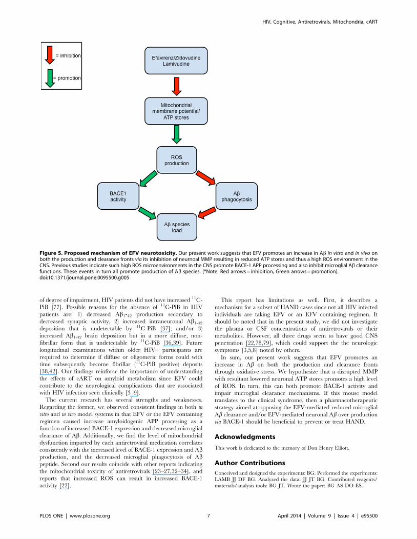
Figure 5, our present study has led to the proposed mechanism of
neurotoxicity in which EFV promotes an increase in Ab in vitro and
in vivo on both the production and clearance fronts via its inhibition
of proper MMP resulting in reduced ATP stores and thus a high
ROS environment in the CNS. It is proposed that EFV induced
high ROS microenvironments (Fig. 3) in the CNS promote
BACE-1 APP processing ([18]; Fig. 1) and also inhibits microglial
phagocytic functions (Fig. 4; [67]). These events in turn all
promote production of Ab species.
EFV has been associated with serious adverse reactions, most of
which can in part be attributed directly or indirectly to dysfunction
of mitochondria [22–25,33]. We found that EFV, or the EFV
containing regimen consistently and significantly promoted
mitochondrial oxidative stress in the form of reduced cellular
ATP stores (Fig. 3A) and MMP (Fig. 3B), as well as increased
release of ROS (Fig. 3C–F). These observations suggest the
mitochondrial stress imparted by this cART regimen is largely
dependent upon EFV and that 3TC and/or AZT may have some
protective effect. Indeed there is some evidence that the latter
antiretroviral may help to preserve cognitive function [56–58]
Reactive microgliosis can be associated with the formation of
microglial phenotypes that are unfavorable to phagocytic activities
[68]. ROS are an important signal for cellular activation and
proliferation. Over the long term lead to microglial dysfunction,
rendering the phagocytes unable to perform their vital clearance
functions [68]. This may underlie the reduced microglial
phagocytosis of Ab observed in microglia treated with EFV or
the EFV containing regimen (Fig. 4).
Several lines of epidemiological evidence signal a role for Ab in
HAND development while some studies have not yet fully
implicated over production of the protein as a contributor to
Figure 1. EFV or EFV/3TC/AZT treatment promotes Ab generation in cultured neuronal cells via BACE-1 activation in vitro. Ab specieswere analyzed in cell lysates from SweAPP N2a cells (A) by ELISA. Data are represented as the mean 6 of a percentage of Ab peptides secreted 24 hafter 3TC, AZT, EFV, or 3TC/EFV/AZT administration, relative fold over control (PBS treated). Significant increases in Ab were observed in EFV or EFV/3TC/AZT treated cells were observed compared to control (***P,0.001 and **P,0.05 respectively by ANOVA). (B) Western blot (6E10 antibody) ofconditioned media shows increased oligomeric Ab species vs. s-APP-a (control) in the EFV or EFV/3TC/AZT treated cells (***P,0.001 and **P,0.05respectively). (D) BACE-1 expression in cultured media revealed significant differences between EFV or EFV/3TC/AZT treated cells compared tountreated control (***P,0.001). b-actin is used for the internal loading control. Results are representative of three independent experiments.doi:10.1371/journal.pone.0095500.g001
HIV, Cognitive, Antiretrovirals, Mitochondria, cART
PLOS ONE | www.plosone.org 4 April 2014 | Volume 9 | Issue 4 | e95500

Figure 2. EFV/3TC/AZT increases soluble Ab levels in Tg2576 mice via BACE-1 activation in vivo. (A) Ab40, 42 peptides were analyzed inbrain homogenates from 8 month old Tg2576 mice by ELISA (n = 5 mice for each group). One-way ANOVA followed by post hoc comparison revealedsignificant differences between control (Tg2576mice treated with PBS) and EFV or EFV/3TC/AZT -treated Tg2576 mice (P,0.001 and 0.05 respectivelywith n = 5 mice/group). (B) Western Blot of brain homogenates using anti-Ab1-17 antibody (6E10) shows total APP and a bands corresponding tosoluble Ab oligomer species. b-actin was an internal control. A t-test revealed significant differences in soluble Ab species between EFV-treatedcompared to 3TC/AZT/EFV, 3TC or AZT treated Tg2576 mice (P,0.01) (C) BACE-1 expression in brain homogenate of Tg2576 mice significantly wasincreased in EFV or EFV/3TC/AZT -treated Tg2576 mice (P,0.001).doi:10.1371/journal.pone.0095500.g002
Figure 3. cART treatment of SweAPP N2a cells promotes mitochondrial dysfunction. (A) ATP levels are reduced in EFV or EFV/3TC/AZTtreated SweAPP N2a neuron cells: SweAPP N2a cells were grown with 10 mM of each medication or all three medications combined for 48 h. We founda significant decrease in ATP levels in cells treated with EFV or 3TC/AZT/EFV (***P,0.001). (B)MMP is reduced in EFV or EFV/3TC/AZT SweAPP N2a cells:In accord with reduced ATP levels we found a similar reduction in MMP in the EFV or EFV/3TC/AZT treated groups (***P,0.001) (C–F) ROS levels areincreased in EFV or EFV/3TC/AZT treated SweAPP N2a cells: EFV-treated primary neuron cells have significantly higher ROS contents (**P,0.001) afterincubation for 60 min than untreated primary neuron. (C–E)The average relative fluorescence units of DCFDA in neurons from each treatment groupas indicated by the mean 6 standard deviations (D, F) The ROS content in the antiretroviral treatment is expressed as % RFU 6 standard deviationsfor each group compared to untreated control primary neuron cells (100%). (*P,0.05, *** P,0.001).doi:10.1371/journal.pone.0095500.g003
HIV, Cognitive, Antiretrovirals, Mitochondria, cART
PLOS ONE | www.plosone.org 5 April 2014 | Volume 9 | Issue 4 | e95500

HAND. It is known that pathological similarities exist between
HAND and AD [37–42]. The latter is more so characterized by
extracellular deposits of Ab1-42 in the form of plaques and
aggregations of microtubule-associated tau yielding neurofibrillary
tangles (NFT). In contrast, with HIV infection, the plaques are
more diffuse [38] rather than neuritic [41].
Cerebrospinal fluid (CSF) biomarkers can mirror pathogenic
cerebral amyloid deposition. Decreased CSF Ab1-42 and increased
CSF tau can differentiate symptomatic AD participants and
cognitively normal individuals at high risk for symptomatic AD
from cognitively normal individuals at low risk for symptomatic
AD [69,70]. In that regard, at least some HAND patients have
CSF Ab1-42 values comparable to symptomatic AD individuals,
that is, reduced [34,41]. This is salient because reductions in CSF
Ab42 have been found in almost all individuals with increased
fibrillar amyloid deposition within the brain as assessed with
positron emission tomography (PET) amyloid binding of N-
methyl-[11C]2-(4-methylaminophenyl)-6-hydroxybenzothiazole
(11C-PiB) [71–73]. Likewise, AIDS dementia complex (ADC)
patients had significantly decreased CSF Ab1-42 and increased
total and phospho (t-tau and p-tau respectively) concentrations
similar to AD [38]. Achim and colleague’s (2009) reported
increased Ab by both autopsy examination and PET imaging of
HIV patients. Specifically, cases with HIV encephalitis (HIV-E)
were about twice as likely to have amyloid detected (72%) than
HIV+ patients without HIV-E (38%; [37]). In the same year
Clifford and colleagues reported Ab1-42 measurements in CSF of
cognitively impaired patients with HIV were similarly reduced as
in in patients with mild dementia of the Alzheimer type (DAT).
Normal or slightly depressed CSF tau and p-tau measurements
distinguished these patients with HAND from patients with DAT
[42].
Further analysis as to why low CSF Ab1-42 in patients with
HAND is needed. However, there are several reasons which may
explain altered Ab metabolism in HIV disease [42] in addition to
the data presented in this report. First, HIV-1 transactivator of
transcription (Tat) protein may compete with APP and/or
apolipoprotein E (an Ab chaperone) for binding to the low density
lipoprotein receptor related protein (LRP), thus inhibiting LRP
mediated clearance of Abfrom brain interstitial fluid to periphery
[74]. Second, APP cleavage products (sAPPaand sAPPb) have
been reported to be reduced in the CSF of patients with HAND
compared to those with DAT or HIV-negative controls, with
sAPPa (a neurotrophic protein) showing a slight decline in the
asymptomatic HIV state [75].
In 2010 Ances and colleagues reported cognitively unimpaired
HIV+ participants, even with low CSF Ab1-42 (,500 pg/mL), did
not have (11)C-PiB parameters suggesting brain fibrillar amyloid
deposition. This dissimilarity between cognitively unimpaired
HIV+ and preclinical AD may reflect differences in Ab1-42
production and/or formation of diffuse plaques [76]. This same
group, in 2012, reported symptomatic AD patients were signifi-
cantly older, had significantly lower CSF Ab1-42, and had
significantly higher CSF tau levels than other groups. Regardless
Figure 4. EFV/3TC/AZT inhibits microglial phagocytosis of Ab1-42 peptide. (A) Primary microglia (16105 cells/well in 24-well tissue cultureplates) were treated with aged FITC tagged Ab1-42 (50 nM) in complete medium for 60 min with antiretroviral medications (10uM) combined or singlyas indicated, or PBS (control). As a control for nonspecifically incorporated Ab, microglial cells were incubated at 4uC with the same treatmentfollowed by DAPI staining. EFV or 3TC/AZT/EFV inhibited microglia-colocalization by fluorescence microscopy. Green indicates Ab1-42 positive; blueindicates microglia nuclei. Addition of heat inactivated HIV-1 Tat yielded similar results as vehicle control (data not shown) (B) Cell supernatants andlysates were analyzed for extracellular (top) and cell associated (bottom) FITC-Ab using a fluorimeter. Data are represented as the relative fold ofmean fluorescence change (mean 6 SD), calculated as the mean fluorescence for each sample at 37uC divided by mean fluorescence at 4uC (n = 6 foreach condition presented). One-way ANOVA followed by post-hoc comparison showed a significant difference between EFV (***P,0.001) or EFV/3TC/AZT (**P,0.05) but not 3TC or AZT compared to control.doi:10.1371/journal.pone.0095500.g004
HIV, Cognitive, Antiretrovirals, Mitochondria, cART
PLOS ONE | www.plosone.org 6 April 2014 | Volume 9 | Issue 4 | e95500
of degree of impairment, HIV patients did not have increased 11C-
PiB [77]. Possible reasons for the absence of 11C-PiB in HIV
patients are: 1) decreased Ab1-42 production secondary to
decreased synaptic activity, 2) increased intraneuronal Ab1-42
deposition that is undetectable by 11C-PiB [37]; and/or 3)
increased Ab1-42 brain deposition but in a more diffuse, non-
fibrillar form that is undetectable by 11C-PiB [36,39]. Future
longitudinal examinations within older HIV+ participants are
required to determine if diffuse or oligomeric forms could with
time subsequently become fibrillar (11C-PiB positive) deposits
[38,42]. Our findings reinforce the importance of understanding
the effects of cART on amyloid metabolism since EFV could
contribute to the neurological complications that are associated
with HIV infection seen clinically [3–9].
The current research has several strengths and weaknesses.
Regarding the former, we observed consistent findings in both in
vitro and in vivo model systems in that EFV or the EFV containing
regimen caused increase amyloidogenic APP processing as a
function of increased BACE-1 expression and decreased microglial
clearance of Ab. Additionally, we find the level of mitochondrial
dysfunction imparted by each antiretroviral medication correlates
consistently with the increased level of BACE-1 expression and Abproduction, and the decreased microglial phagocytosis of Abpeptide. Second our results coincide with other reports indicating
the mitochondrial toxicity of antiretrovirals [23–27,32–34], and
reports that increased ROS can result in increased BACE-1
activity [22].
This report has limitations as well. First, it describes a
mechanism for a subset of HAND cases since not all HIV infected
individuals are taking EFV or an EFV containing regimen. It
should be noted that in the present study, we did not investigate
the plasma or CSF concentrations of antiretrovirals or their
metabolites. However, all three drugs seem to have good CNS
penetration [22,78,79], which could support the the neurologic
symptoms [3,5,8] noted by others.
In sum, our present work suggests that EFV promotes an
increase in Ab on both the production and clearance fronts
through oxidative stress. We hypothesize that a disrupted MMP
with resultant lowered neuronal ATP stores promotes a high level
of ROS. In turn, this can both promote BACE-1 activity and
impair microglial clearance mechanisms. If this mouse model
translates to the clinical syndrome, then a pharmacotherapeutic
strategy aimed at opposing the EFV-mediated reduced microglial
Ab clearance and/or EFV-mediated neuronal Ab over production
via BACE-1 should be beneficial to prevent or treat HAND.
Acknowledgments
This work is dedicated to the memory of Don Henry Elliott.
Author Contributions
Conceived and designed the experiments: BG. Performed the experiments:
LAMB JJ DF BG. Analyzed the data: JJ JT BG. Contributed reagents/
materials/analysis tools: BG JT. Wrote the paper: BG AS DO ES.
Figure 5. Proposed mechanism of EFV neurotoxicity. Our present work suggests that EFV promotes an increase in Ab in vitro and in vivo onboth the production and clearance fronts via its inhibition of neurnoal MMP resulting in reduced ATP stores and thus a high ROS environment in theCNS. Previous studies indicate such high ROS microenvironments in the CNS promote BACE-1 APP processing and also inhibit microglial Ab clearancefunctions. These events in turn all promote production of Ab species. (*Note: Red arrows = inhibition, Green arrows = promotion).doi:10.1371/journal.pone.0095500.g005
HIV, Cognitive, Antiretrovirals, Mitochondria, cART
PLOS ONE | www.plosone.org 7 April 2014 | Volume 9 | Issue 4 | e95500

References
1. Schouten EJ, Jahn A, Ben-Smith A, Makombe SD, Harries AD, et al. (2011)
Antiretroviral drug supply challenges in the era of scaling up ART in Malawi.J Int AIDS Soc 14 Suppl 1: S4.
2. Heaton RK, Franklin DR, Ellis RJ, McCutchan JA, Letendre SL, et al. (2011)
HIV-associated neurocognitive disorders before and during the era ofcombination antiretroviral therapy: differences in rates, nature, and predictors.
J Neurovirol 17: 3–16.
3. Ciccarelli N, Fabbiani M, Di Giambenedetto S, Fanti I, Baldonero E, et al.
(2011) Efavirenz associated with cognitive disorders in otherwise asymptomaticHIV-infected patients. Neurology 76: 1403–1409.
4. Winston A, Duncombe C, Li PC, Gill JM, Kerr SJ, et al. (2010) Does choice of
combination antiretroviral therapy (cART) alter changes in cerebral functiontesting after 48 weeks in treatment-naive, HIV-1-infected individuals commenc-
ing cART? A randomized, controlled study. Clin Infect Dis 50: 920–929.
5. Robertson KR, Smurzynski M, Parsons TD, Wu K, Bosch RJ, et al. (2007) Theprevalence and incidence of neurocognitive impairment in the HAART era.
Aids 21: 1915–1921.
6. Velasco M, Pareja JA, Losa JE, Valverde JF, Espinosa A, Gujarro C Dreamchanges following initiation of efavirenz treatment. Med Clin (Barc) 136: 103–
105.
7. Waters L, Fisher M, Winston A, Higgs C, Hadley W, et al. (2001) A phase IV,double-blind, multicentre, randomized, placebo-controlled, pilot study to assess
the feasibility of switching individuals receiving efavirenz with continuing central
nervous system adverse events to etravirine. AIDS 25: 65–71.
8. Romao PR, Lemos JC, Moreira J, de Chaves G, Moretti M, et al., (2011) Anti-HIV drugs nevirapine and efavirenz affect anxiety-related behavior and
cognitive performance in mice. Neurotox Res 19: 73–80.
9. Wintergerst U, Hoffmann F, Jansson A, Notheis G, Huss K, et al. (2008)Antiviral efficacy, tolerability and pharmacokinetics of efavirenz in an unselected
cohort of HIV-infected children. J Antimicrob Chemother 61: 1336–1339.
10. Sambamurti K, Greig NH, Lahiri DK (2002) Advances in the cellular andmolecular biology of the beta-amyloid protein in Alzheimer’s disease.
Neuromolecular Med 1: 1–31.
11. Golde TE, Eckman CB, Younkin SG (2000) Biochemical detection of Abetaisoforms: implications for pathogenesis, diagnosis, and treatment of Alzheimer’s
disease. Biochim Biophys Acta 1502: 172–187.
12. Huse JT, Doms RW (2000) Closing in on the amyloid cascade: recent insightsinto the cell biology of Alzheimer’s disease. Mol Neurobiol 22: 81–98.
13. Selkoe DJ (1996) Cell biology of the beta-amyloid precursor protein and the
genetics of Alzheimer’s disease. Cold Spring Harb Symp Quant Biol 61: 587–596.
14. Bradt BM, Kolb WP, Cooper NR (1998) Complement-dependent proinflam-
matory properties of the Alzheimer’s disease beta-peptide. J Exp Med 188: 431–
438.15. Suo Z, Tan J, Placzek A, Crawford F, Fang C, et al. (1998) Alzheimer’s beta-
amyloid peptides induce inflammatory cascade in human vascular cells: the roles
of cytokines and CD40. Brain Res 807: 110–117.
16. Murakami K, Irie K, Ohigashi H, Hara H, Nagao M, et al. (2005) Formationand stabilization model of the 42-mer Abeta radical: implications for the long-
lasting oxidative stress in Alzheimer’s disease. J Am Chem Soc 127: 15168–15174.
17. Schenk D, Games KD, McConlogue L (1995) The potential utility of transgenic
mice harboring beta-amyloid precursor protein. Neurobiol Aging 16: 711–713;discussion 715–718.
18. Sinha S, Lieberburg I (1999) Cellular mechanisms of beta-amyloid production
and secretion. Proc Natl Acad Sci U S A 96: 11049–11053.
19. Steiner H, Capell A, Haass C (1999) Proteolytic processing and degradation ofAlzheimer’s disease relevant proteins. Biochem Soc Trans 27: 234–242.
20. De Strooper B, Beullens M, Contreras B, Levesque L, Craessaerts K, et al.
(1997) Phosphorylation, subcellular localization, and membrane orientation ofthe Alzheimer’s disease-associated presenilins. J Biol Chem 272: 3590–3598.
21. Jacobsen JS, Reinhart P, Pangalos MN (2005) Current concepts in therapeutic
strategies targeting cognitive decline and disease modification in Alzheimer’sdisease. NeuroRx 2: 612–626.
22. Cossarizza A, Moyle G (2004) Antiretroviral nucleoside and nucleotide
analogues and mitochondria. AIDS 18: 137–151.
23. Apostolova N, Blas-Garcia A, Esplugues JV (2011) Mitochondrial interferenceby anti-HIV drugs: mechanisms beyond Pol-gamma inhibition. Trends
Pharmacol Sci 32: 715–725.
24. Apostolova N, Blas-Garcia A, Esplugues JV (2011) Mitochondrial toxicity in
HAART: an overview of in vitro evidence. Curr Pharm Des 17: 2130–2144.25. Apostolova N, Gomez-Sucerquia LJ, Gortat A, Blas-Garcia A, Esplugues JV
(2011) Compromising mitochondrial function with the antiretroviral drug
efavirenz induces cell survival-promoting autophagy. Hepatology 54: 1009–1019.
26. Blas-Garcia A, Esplugues JV, Apostolova N (2011) Twenty years of HIV-1 non-
nucleoside reverse transcriptase inhibitors: time to reevaluate their toxicity. CurrMed Chem 18: 2186–2195.
27. Blas-Garcia A, Apostolova N, Esplugues JV (2011) Oxidative stress and
mitochondrial impairment after treatment with anti-HIV drugs: clinicalimplications. Curr Pharm Des 17: 4076–4086.
28. Manda KR, Banerjee A, Banks WA, Ercal N (2011) Highly active antiretroviral
therapy drug combination induces oxidative stress and mitochondrial dysfunc-tion in immortalized human blood-brain barrier endothelial cells. Free Radic
Biol Med 50: 801–810.
29. Riddler SA, Haubrich R, DiRienzo AG, Peeples L, Powderly WG, et al. (2008)
Class-sparing regimens for initial treatment of HIV-1 infection. N Engl J Med2008; 358: 2095–2106.
30. Staszewski S, Morales-Ramirez J, Tashima KT, Rachlis A, Skiest D, et al. (1999)Efavirenz plus zidovudine and lamivudine, efavirenz plus indinavir, and
indinavir plus zidovudine and lamivudine in the treatment of HIV-1 infection
in adults. Study 006 Team. N Engl J Med 341: 1865–1873.
31. Hirschel B, Calmy (2008) A Initial treatment for HIV infection—an
embarrassment of riches. N Engl J Med 358: 2170–2172.
32. Apostolova N, Gomez-Sucerquia LJ, Alegre F, Funes HA, Victor VM, et al.
(2013) ER stress in human hepatic cells treated with Efavirenz: mitochondriaagain. J Hepatol 59: 780–789.
33. Apostolova N, Gomez-Sucerquia LJ, Gortat A, Blas-Garcia A, Esplugues JV(2011) Autophagy as a rescue mechanism in efavirenz-induced mitochondrial
dysfunction: a lesson from hepatic cells. Autophagy 7: 1402–1404.
34. Blas-Garcia A, Apostolova N, Ballesteros D, Monleon D, Morales JM, et al.
(2010) Inhibition of mitochondrial function by efavirenz increases lipid contentin hepatic cells. Hepatology 52: 115–125.
35. Rodrigue KM, Rieck JR, Kennedy KM, Devous MD, Sr., Diaz-Arrastia R, etal. (2010) Risk factors for beta-amyloid deposition in healthy aging: vascular and
genetic effects. JAMA Neurol 2013; 70: 600–606.
36. Anthony IC, Ramage SN, Carnie FW, Simmonds P, Bell JE (2006) Accelerated
Tau deposition in the brains of individuals infected with human immunodefi-
ciency virus-1 before and after the advent of highly active anti-retroviral therapy.Acta Neuropathol 111: 529–538.
37. Achim CL, Adame A, Dumaop W, Everall IP, Masliah E (2009) Increasedaccumulation of intraneuronal amyloid beta in HIV-infected patients.
J Neuroimmune Pharmacol 4: 190–199.
38. Brew BJ, Pemberton L, Blennow K, Wallin A, Hagberg L (2005) CSF amyloid
beta42 and tau levels correlate with AIDS dementia complex. Neurology 65:1490–1492.
39. Green DA, Masliah E, Vinters HV, Beizai P, Moore DJ, et al. (2005) Braindeposition of beta-amyloid is a common pathologic feature in HIV positive
patients. AIDS 19: 407–411.
40. Alisky JM (2007) The coming problem of HIV-associated Alzheimer’s disease.
Med Hypotheses 69: 1140–1143.
41. Esiri MM, Biddolph SC, Morris CS (1998) Prevalence of Alzheimer plaques in
AIDS. J Neurol Neurosurg Psychiatry 65: 29–33.
42. Clifford DB, Fagan AM, Holtzman DM, Morris JC, Teshome M, et al. (2009)
CSF biomarkers of Alzheimer disease in HIV-associated neurologic disease.
Neurology 73: 1982–1987.
43. Kiebala M, Polesskaya O, Yao Z, Perry SW, Maggirwar SB (2010) Nuclear
factor-kappa B family member RelB inhibits human immunodeficiency virus-1Tat-induced tumor necrosis factor-alpha production. PLoS One5: e11875.
44. Giunta B, Ehrhart J, Obregon DF, Lam L, Le L, et al. (2011)Antiretroviralmedications disrupt microglial phagocytosis of beta-amyloid and increase its
production by neurons: implications for HIV-associated neurocognitivedisorders. Mol Brain 4: 23.
45. Lewis W, Kohler JJ, Hosseini SH, Haase CP, Copeland WC, et al. (2006)Antiretroviral nucleosides, deoxynucleotide carrier and mitochondrial DNA:
evidence supporting the DNA pol gamma hypothesis. AIDS 20: 675–684.
46. Chandra S, Murthy SN, Mondal D, Agrawal KC (2009) Therapeutic effects of
Nigella sativa on chronic HAART-induced hyperinsulinemia in rats.
Can J Physiol Pharmacol 87: 300–309.
47. Torres SM, Divi RL, Walker DM, McCash CL, Carter MM, et al. (2010) In
utero exposure of female CD-1 mice to AZT and/or 3TC: II. Persistence offunctional alterations in cardiac tissue. Cardiovasc Toxicol 10: 87–99.
48. Torres SM, Divi RL, Walker DM, McCash CL, Carter MM, et al. (2010) Inutero exposure of female CD-1 mice to AZT and/or 3TC: II. Persistence of
functional alterations in cardiac tissue. Cardiovasc Toxicol 10: 87–99.
49. Balani SK, Kauffman LR, deLuna FA, Lin JH (1999) Nonlinear pharmacoki-
netics of efavirenz (DMP-266), a potent HIV-1 reverse transcriptase inhibitor, inrats and monkeys. Drug Metab Dispos 27: 41–45.
50. Giunta B, Hou H, Zhu Y, Rrapo E, Tian J, et al. (2009) HIV-1 Tat contributesto Alzheimer’s disease-like pathology in PSAPP mice. Int J Clin Exp Pathol 2:
433–443.
51. Giunta B, Hou H, Zhu Y, Salemi J, Ruscin A, et al. (2010) Fish oil enhancesanti-amyloidogenic properties of green tea EGCG in Tg2576 mice. Neurosci
Lett 471: 134–138.
52. Tamagno E, Guglielmotto M, Aragno M, Borghi R, Autelli R, et al. (2008)
Oxidative stress activates a positive feedback between the gamma- and beta-secretase cleavages of the beta-amyloid precursor protein. J Neurochem 104:
683–695.
53. Cleary JP, Walsh DM, Hofmeister JJ, Shankar GM, Kuskowski MA, et al. (2005)
Natural oligomers of the amyloid-beta protein specifically disrupt cognitivefunction. Nat Neurosci 8: 79–84.
HIV, Cognitive, Antiretrovirals, Mitochondria, cART
PLOS ONE | www.plosone.org 8 April 2014 | Volume 9 | Issue 4 | e95500
54. Graff-Radford NR, Crook JE, Lucas J, Boeve BF, Knopman DS, et al. (2007)
Association of low plasma Abeta42/Abeta40 ratios with increased imminent riskfor mild cognitive impairment and Alzheimer disease. Arch Neurol 64: 354–362.
55. Gomez-Sucerquia LJ, Blas-Garcia A, Marti-Cabrera M, Esplugues JV,
Apostolova N (2012) Profile of stress and toxicity gene expression in humanhepatic cells treated with Efavirenz. Antiviral Res 94: 232–241.
56. Pizzo PA, Eddy J, Falloon J, Balis FM, Murphy RF, et al. (1988) Effect ofcontinuous intravenous infusion of zidovudine (AZT) in children with
symptomatic HIV infection. N Engl J Med 319: 889–896.
57. Rausch DM, Heyes M, Eiden LE (1994) Effects of chronic zidovudineadministration on CNS function and virus burden after perinatal SIV infection
in rhesus monkeys. Adv Neuroimmunol 4: 233–237.58. Schmitt FA, Bigley JW, McKinnis R, Logue PE, Evans RW, et al. (1988)
Neuropsychological outcome of zidovudine (AZT) treatment of patients withAIDS and AIDS-related complex. N Engl J Med 319: 1573–1578.
59. Selkoe DJ (2001) Alzheimer’s disease results from the cerebral accumulation and
cytotoxicity of amyloid beta-protein. J Alzheimers Dis 3: 75–80.60. Klyubin I, Betts V, Welzel AT, Blennow K, Zetterberg H, et al. (2008) Amyloid
beta protein dimer-containing human CSF disrupts synaptic plasticity:prevention by systemic passive immunization. J Neurosci 28: 4231–4237.
61. Walsh DM, Klyubin I, Fadeeva JV, Cullen WK, Anwyl R, et al. (2002) Naturally
secreted oligomers of amyloid beta protein potently inhibit hippocampal long-term potentiation in vivo. Nature 416: 535–539.
62. Walsh DM, Klyubin I, Fadeeva JV, Rowan MJ, Selkoe DJ (2002) Amyloid-betaoligomers: their production, toxicity and therapeutic inhibition. Biochem Soc
Trans 30: 552–557.63. Walsh DM, Klyubin I, Shankar GM, Townsend M, Fadeeva JV, (2005) The role
of cell-derived oligomers of Abeta in Alzheimer’s disease and avenues for
therapeutic intervention. Biochem Soc Trans 33: 1087–1090.64. Hsiao K, Chapman P, Nilsen S, Eckman C, Harigaya Y, et al. (1996) Correlative
memory deficits, Abeta elevation, and amyloid plaques in transgenic mice.Science 274: 99–102.
65. Dragicevic N, Smith A, Lin X, Yuan F, et al. (2011) Green tea epigallocatechin-
3-gallate (EGCG) and other flavonoids reduce Alzheimer’s amyloid-inducedmitochondrial dysfunction. J Alzheimers Dis 26: 507–521.
66. Mandrekar S, Jiang Q, Lee CY, Koenigsknecht-Talboo J, Holtzman DM, et al.(2009) Microglia mediate the clearance of soluble Abeta through fluid phase
macropinocytosis. J Neurosci 29: 4252–4262.
67. Mosser DM, Edwards JP (2008) Exploring the full spectrum of macrophage
activation. Nat Rev Immunol 8: 958–969.68. Meyer-Luehmann M, Spires-Jones TL, Prada C, Garcia-Alloza M, de Calignon
A, et al. (2008) Hyman BT Rapid appearance and local toxicity of amyloid-beta
plaques in a mouse model of Alzheimer’s disease. Nature 451: 720–724.69. Craig-Schapiro R, Perrin RJ, Roe CM, Xiong C, Carter D, et al. (2010) YKL-
40: a novel prognostic fluid biomarker for preclinical Alzheimer’s disease. BiolPsychiatry 68: 903–912.
70. Okonkwo OC, Mielke MM, Griffith HR, Moghekar AR, O’Brien RJ, et al.
(2011) Cerebrospinal fluid profiles and prospective course and outcome inpatients with amnestic mild cognitive impairment. Arch Neurol 68: 113–119.
71. Fagan AM, Mintun MA, Shah AR, Aldea P, Roe CM, et al. (2009)Cerebrospinal fluid tau and ptau(181) increase with cortical amyloid deposition
in cognitively normal individuals: implications for future clinical trials ofAlzheimer’s disease. EMBO Mol Med 1: 371–380.
72. Fagan AM, Head D, Shah AR, Marcus D, Mintun M, et al. (2009) Decreased
cerebrospinal fluid Abeta(42) correlates with brain atrophy in cognitively normalelderly. Ann Neurol 65: 176–183.
73. Grimmer T, Riemenschneider M, Forstl H, Henriksen G, Klunk WE, et al.(2009) Beta amyloid in Alzheimer’s disease: increased deposition in brain is
reflected in reduced concentration in cerebrospinal fluid. Biol Psychiatry 65:
927–934.74. Liu Y, Jones M, Hingtgen CM, Bu G, Laribee N, et al. (2000) Uptake of HIV-1
tat protein mediated by low-density lipoprotein receptor-related protein disruptsthe neuronal metabolic balance of the receptor ligands. Nat Med 6: 1380–1387.
75. Gisslen M, Krut J, Andreasson U, Blennow K, Cinque P, et al. (2009) Amyloidand tau cerebrospinal fluid biomarkers in HIV infection. BMC Neurol 9: 63.
76. Ances BM, Christensen JJ, Teshome M, Taylor J, Xiong C, A et al. (2010)
Cognitively unimpaired HIV-positive subjects do not have increased 11C-PiB: acase-control study. Neurology 75: 111–115.
77. Ances BM, Benzinger TL, Christensen JJ, Thomas J, Venkat R, et al. (2012)11C-PiB imaging of human immunodeficiency virus-associated neurocognitive
disorder. Arch Neurol 69: 72–77.
78. Wynn HE, Brundage RC, Fletcher CV (2002) Clinical implications of CNSpenetration of antiretroviral drugs. CNS Drugs 16: 595–609.
79. Gibbs JE, Gaffen Z, Thomas SA (2006) Nevirapine uptake into the centralnervous system of the Guinea pig: an in situ brain perfusion study. J Pharmacol
Exp Ther 317: 746–751.
HIV, Cognitive, Antiretrovirals, Mitochondria, cART
PLOS ONE | www.plosone.org 9 April 2014 | Volume 9 | Issue 4 | e95500
Copyright © 2022 FDOKUMEN