Ectodomain Shedding of Epidermal Growth Factor Receptor Ligands Is Required for Keratinocyte...
11
The Rockefeller University Press, 0021-9525/2000/10/209/11 $5.00 The Journal of Cell Biology, Volume 151, Number 2, October 16, 2000 209–219 http://www.jcb.org/cgi/content/full/151/2/209 209 Ectodomain Shedding of Epidermal Growth Factor Receptor Ligands Is Required for Keratinocyte Migration in Cutaneous Wound Healing Sho Tokumaru,* § Shigeki Higashiyama,* i Takeshi Endo,* Takatoshi Nakagawa,* Jun-ichiro Miyagawa, ‡ Katsumi Yamamori, ‡ Yasushi Hanakawa, § Hiroshi Ohmoto, ¶ Kohichiro Yoshino, ¶ Yuji Shirakata, § Yuji Matsuzawa, ‡ Koji Hashimoto, § and Naoyuki Taniguchi* *Department of Biochemistry, ‡ Department of Internal Medicine and Medical Science, Osaka University Medical School, Suita, Osaka 565-0871, Japan; § Department of Dermatology, Ehime University School of Medicine, Shitsukawa, Shigenobucho, Onsengun, Ehime 791-0295, Japan; i Department of Biochemistry, School of Allied Health Science, Osaka University Faculty of Medicine, Suita, Osaka 565-0871; and ¶ Nippon Organon K.K., Osaka 534-0016, Japan Abstract. Keratinocyte proliferation and migration are essential to cutaneous wound healing and are, in part, mediated in an autocrine fashion by epidermal growth factor receptor (EGFR)–ligand interactions. EGFR ligands are initially synthesized as membrane-anchored forms, but can be processed and shed as soluble forms. We provide evidence here that wound stimuli induce keratinocyte shedding of EGFR ligands in vitro, partic- ularly the ligand heparin-binding EGF-like growth fac- tor (HB-EGF). The resulting soluble ligands stimulated transient activation of EGFR. OSU8-1, an inhibitor of EGFR ligand shedding, abrogated the wound-induced activation of EGFR and caused suppression of kerati- nocyte migration in vitro. Soluble EGFR–immunoglob- ulin G-Fcg fusion protein, which is able to neutralize all EGFR ligands, also suppressed keratinocyte migration in vitro. The application of OSU8-1 to wound sites in mice greatly retarded reepithelialization as the result of a failure in keratinocyte migration, but this effect could be overcome if recombinant soluble HB-EGF was added along with OSU8-1. These findings indicate that the shedding of EGFR ligands represents a critical event in keratinocyte migration, and suggest their pos- sible use as an effective clinical treatment in the early phases of wound healing. Key words: TGF-a • HB-EGF • amphiregulin • tissue repair • matrix metalloprotease Introduction Successful wound healing involves a number of processes including inflammation, cell proliferation, cell migration, vascular permeability and angiogenesis, as well as matrix deposition and tissue remodeling (for review see Martin, 1997). Particularly critical are cell proliferation and migra- tion, which are driven by growth factors and cytokines re- leased coordinately into the injury sites. In cutaneous wound healing, keratinocytes play a central role, not only as a key structural cell type in the repairing skin, but also as the source of a number of growth factors. Prominent among these keratinocyte-derived factors are ligands for the epidermal growth factor receptor (EGFR) 1 . The EGFR ligand family is composed of six members in- cluding the epidermal growth factor (Cohen, 1964), trans- forming growth factor-a (TGF-a; Derynck et al., 1984), amphiregulin (AR; Shoyab et al., 1989), heparin-binding EGF-like growth factor (HB-EGF; Higashiyama et al., 1991), betacellulin (Shing et al., 1993), and epiregulin (Toy- oda et al., 1995). All members of this family are synthe- sized as membrane-anchored forms, which are then pro- cessed to give bioactive soluble factors. This processing can be stimulated by treating cells with various agents, includ- ing the phorbol ester 12-O-tetradecanoylphorbol-13-ace- tate (TPA; for review see Massague and Pandiella, 1993). Within the EGFR ligand family, TGF-a, AR, and HB- EGF have been extensively characterized and clearly shown to be autocrine growth factors for keratinocytes (Coffey et al., 1987; Cook et al., 1991; Hashimoto et al., 1994). The transmembrane forms of these factors are also capable of stimulating the growth of adjacent cells includ- ing keratinocytes via cell–cell contact, a process termed juxtacrine stimulation (Brachmann et al., 1989; Wong et al., 1989; Higashiyama et al., 1995; Goishi et al., 1995; Inui et al., 1997). Address correspondence to Shigeki Higashiyama Ph.D., Department of Biochemistry, School of Allied Health Science, Osaka University Faculty of Medicine, 1-7 Yamadaoka, Suita, Osaka 565-0871, Japan. Tel.: 81-6- 6879-2589. Fax: 81-6-6879-2589. E-mail: [email protected] 1 Abbreviations used in this paper: ADAM, a disintegrin and metallopro- tease; AP, alkaline phosphatase; AR, amphiregulin; ECL, enhanced chemilu- minescence; EGFR, epidermal growth factor receptor; EGFR-Fc, epidermal growth factor receptor–immunoglobulin G-Fcg fusion protein; HB-EGF, heparin-binding EGF-like growth factor; MMP, matrix metalloprotease; TACE, tumor necrosis factor–a converting enzyme; TGF-a, transforming growth factor–a; TPA, 12-O-tetradecanoylphorbol-13-acetate. on July 21, 2014 jcb.rupress.org Downloaded from Published October 16, 2000
-
Upload
independent -
Category
Documents
-
view
0 -
download
0
Transcript of Ectodomain Shedding of Epidermal Growth Factor Receptor Ligands Is Required for Keratinocyte...
The Rockefeller University Press, 0021-9525/2000/10/209/11 $5.00The Journal of Cell Biology, Volume 151, Number 2, October 16, 2000 209–219http://www.jcb.org/cgi/content/full/151/2/209 209
Ectodomain Shedding of Epidermal Growth Factor Receptor Ligands Is Required for Keratinocyte Migration in Cutaneous Wound Healing
Sho Tokumaru,*
§
Shigeki Higashiyama,*
i
Takeshi Endo,* Takatoshi Nakagawa,* Jun-ichiro Miyagawa,
‡
Katsumi Yamamori,
‡
Yasushi Hanakawa,
§
Hiroshi Ohmoto,
¶
Kohichiro Yoshino,
¶
Yuji Shirakata,
§
Yuji Matsuzawa,
‡
Koji Hashimoto,
§
and Naoyuki Taniguchi*
*Department of Biochemistry,
‡
Department of Internal Medicine and Medical Science, Osaka University Medical School, Suita, Osaka 565-0871, Japan;
§
Department of Dermatology, Ehime University School of Medicine, Shitsukawa, Shigenobucho,
Onsengun, Ehime 791-0295, Japan;
i
Department of Biochemistry, School of Allied Health Science, Osaka University Faculty of
Medicine, Suita, Osaka 565-0871; and
¶
Nippon Organon K.K., Osaka 534-0016, Japan
Abstract.
Keratinocyte proliferation and migration areessential to cutaneous wound healing and are, in part,mediated in an autocrine fashion by epidermal growthfactor receptor (EGFR)–ligand interactions. EGFRligands are initially synthesized as membrane-anchoredforms, but can be processed and shed as soluble forms.We provide evidence here that wound stimuli inducekeratinocyte shedding of EGFR ligands in vitro, partic-ularly the ligand heparin-binding EGF-like growth fac-tor (HB-EGF). The resulting soluble ligands stimulatedtransient activation of EGFR. OSU8-1, an inhibitor ofEGFR ligand shedding, abrogated the wound-inducedactivation of EGFR and caused suppression of kerati-nocyte migration in vitro. Soluble EGFR–immunoglob-
ulin G-Fc
g
fusion protein, which is able to neutralize allEGFR ligands, also suppressed keratinocyte migrationin vitro. The application of OSU8-1 to wound sites inmice greatly retarded reepithelialization as the result ofa failure in keratinocyte migration, but this effect couldbe overcome if recombinant soluble HB-EGF wasadded along with OSU8-1. These findings indicate thatthe shedding of EGFR ligands represents a criticalevent in keratinocyte migration, and suggest their pos-sible use as an effective clinical treatment in the earlyphases of wound healing.
Key words: TGF-
a
• HB-EGF • amphiregulin • tissue repair • matrix metalloprotease
Introduction
Successful wound healing involves a number of processesincluding inflammation, cell proliferation, cell migration,vascular permeability and angiogenesis, as well as matrixdeposition and tissue remodeling (for review see Martin,1997). Particularly critical are cell proliferation and migra-tion, which are driven by growth factors and cytokines re-leased coordinately into the injury sites. In cutaneouswound healing, keratinocytes play a central role, not onlyas a key structural cell type in the repairing skin, but alsoas the source of a number of growth factors. Prominentamong these keratinocyte-derived factors are ligands forthe epidermal growth factor receptor (EGFR)
1
.
The EGFR ligand family is composed of six members in-cluding the epidermal growth factor (Cohen, 1964), trans-
forming growth factor-
a
(TGF-
a
; Derynck et al., 1984),amphiregulin (AR; Shoyab et al., 1989), heparin-bindingEGF-like growth factor (HB-EGF; Higashiyama et al.,1991), betacellulin (Shing et al., 1993), and epiregulin (Toy-oda et al., 1995). All members of this family are synthe-sized as membrane-anchored forms, which are then pro-cessed to give bioactive soluble factors. This processing canbe stimulated by treating cells with various agents, includ-
ing the phorbol ester 12-
O
-tetradecanoylphorbol-13-ace-tate (TPA; for review see Massague and Pandiella, 1993).
Within the EGFR ligand family, TGF-
a
, AR, and HB-EGF have been extensively characterized and clearlyshown to be autocrine growth factors for keratinocytes(Coffey et al., 1987; Cook et al., 1991; Hashimoto et al.,1994). The transmembrane forms of these factors are alsocapable of stimulating the growth of adjacent cells includ-ing keratinocytes via cell–cell contact, a process termedjuxtacrine stimulation (Brachmann et al., 1989; Wong etal., 1989; Higashiyama et al., 1995; Goishi et al., 1995; Inuiet al., 1997).
Address correspondence to Shigeki Higashiyama Ph.D., Department ofBiochemistry, School of Allied Health Science, Osaka University Facultyof Medicine, 1-7 Yamadaoka, Suita, Osaka 565-0871, Japan. Tel.: 81-6-6879-2589. Fax: 81-6-6879-2589. E-mail: [email protected]
1
Abbreviations used in this paper:
ADAM, a disintegrin and metallopro-tease; AP, alkaline phosphatase; AR, amphiregulin; ECL, enhanced chemilu-minescence; EGFR, epidermal growth factor receptor; EGFR-Fc, epidermalgrowth factor receptor–immunoglobulin G-Fc
g
fusion protein; HB-EGF,heparin-binding EGF-like growth factor; MMP, matrix metalloprotease;
TACE, tumor necrosis factor–
a
converting enzyme; TGF-
a
, transforminggrowth factor–
a
; TPA, 12-
O
-tetradecanoylphorbol-13-acetate.
on July 21, 2014jcb.rupress.org
Dow
nloaded from
Published October 16, 2000

The Journal of Cell Biology, Volume 151, 2000 210
Marikovsky et al. (1993) reported that wound fluid inskin contains processed bioactive EGFR ligands, suggest-ing that regulation of EGFR ligand shedding might be aphysiologically important event in wound healing. There-fore, we investigated the possible role of EGFR ligandshedding in response to wounding, focusing in particularon the early EGFR-related molecular events which areevoked by wound stimuli. We also applied a newly estab-lished inhibitor of EGFR ligand shedding to wound heal-ing models, in both in vitro and in vivo experiments.
Materials and Methods
Materials
Human EGFR antibodies were purchased from Upstate Biotechnology.Phosphotyrosine antibody PY-20 was obtained from Transduction Labora-tories. Neutralizing polyclonal antibody No. 197 was raised in a goat againsta 75–amino acid form of recombinant human HB-EGF and was obtainedfrom D. Damm (Scios Inc., Mountain View, CA; Hashimoto et al., 1994).Anti-EGFR neutralizing antibody was a generous gift from Dr. U. Rodeck(Wister Institute, Philadelphia, PA). AG1478 was obtained from Calbio-chem. TPA was obtained from Wako Pure Chemical Industry. Sulfo-NHS-biotin was purchased from Pierce Chemical Co. An enhanced chemilumi-nescence (ECL) kit was obtained from Amersham Pharmacia Biotech.
Metalloprotease Inhibitors
More than five hundred hydroxamic acid–based inhibitors of metallopro-teases, along with a series of nonhydroxamic derivatives, were synthesizedby Kanebo Ltd. The hydroxamic acid–based inhibitors included the fol-lowing: OSU8-1, [4-(
N
-hydroxyamino)-2R-isobutyl-3S-methylsuccinyl]-
L
-3-(5,6,7,8-tetra-hydro-1-naphthyl)alanine-
N
-methylamide; OSU7-6, [4-(
N
-hydroxyamino)-2R-isobutylsuccinyl]-
L
-3-(1-naphthyl)-alanine-
N
-meth-ylamide; and OSU9-6, [6,7-dihydroxy-3-(
N
-hydroxycarbamoyl)-2-(4-meth-oxybenzenesulfonyl)-1,2,3,4-tetrahydroisoquinoline]. OSU8-1, OSU7-6, andOSU9-6 work as competitive inhibitors.
Construction of Alkaline Phosphatase–EGF-Related Ligand Fusion Vectors
The coding region for human placental alkaline phosphatase (AP) was ob-tained from the APtag-1 expression vector (a gift from Dr. J. Flanagan,Harvard Medical School, Boston, MA) and inserted into the multiplecloning site of pRc/CMV (Invitrogen). A BstBI-MscI fragment of the hu-man HB-EGF cDNA, which encodes the signal sequence and prose-quence, was fused to the 5
9
end of the AP sequence to create the plasmidpSS-AlPh (see Fig. 1 a). As pSS-AlPh has a unique HpaI site overlappingthe termination codon of the AP coding region, one of the following DNAfragments encoding human EGF–related ligands were fused in-frame atthis site: MscI-PstI fragment of the HB-EGF cDNA (0.45 kb); HinfI frag-ment of the AR cDNA (0.5 kb); or MseI-SauIII fragment of the TGF-
a
cDNA (0.38 kb). The plasmids resulting from these ligations were intro-duced into CHO cells using the calcium phosphate method (Chen andOkayama, 1988), and stably transfected clones were isolated.
Cell-surface Biotinylation, Immunoprecipitation, and Western Blotting
CHO cell transfectants stably expressing either wild-type HB-EGF, AR,or TGF-
a
, or AP-tagged fusions of these ligands, were seeded in 6-cmdishes at a density of 2
3
10
5
cells/dish and cultured for 24 h with Ham’sF12/10% FCS. Cells were washed three times with ice-cold Hanks bufferand biotinylated with 0.1 mg/ml of sulfo-NHS-biotin in 50 mM Hepes, pH7.5, and 0.15 M NaCl for 15 min on ice. Excess reagent was quenched andremoved by washing with ice-cold Ham’s F12/10% FCS. Cells were lysedwith a buffer containing 1% Triton X-100, 3 mM EDTA, 1 mM (
p
-amidi-nophenyl) methanesulfonyl fluoride HCl, 1
m
g/ml aprotinin, 5
m
M 3,4-dichloroisocoumarin, and 0.4 M NaCl in 20 mM Hepes, pH 7.2. After cen-trifugation of the lysates at 15,000 rpm for 15 min, supernatants were col-lected and incubated with 2
m
g of human placental alkaline phosphataseantibody (Dako), HB-EGF antibody No. H6 (Higashiyama et al., 1995),
AR antibody No. AAR1 (Hashimoto et al., 1994), or TGF-
a
antibody (agift from Dr. R. Derynck, University of California, San Francisco, SanFrancisco, CA) for 2 h at 4
8
C, followed by incubation with 10
m
l of proteinA trisacryl (50% suspension; Pierce Chemical Co.) for 2 h at 4
8
C. Aftercentrifugation of the mixes, the pellets were analyzed by SDS-PAGE andWestern blotting as described previously (Goishi et al., 1995).
Preparation of EGFR ExtracellularDomain–Immunoglobulin G-Fc
g
Fusion Protein
A fusion protein consisting of the extracellular domain of EGFR and hu-man immunoglobulin G-Fc
g
(EGFR-Fc) was produced as follows. ABglII site was engineered into an EGFR expression plasmid (Gotoh et al.,1994) at the point encoding the extracellular domain/transmembrane do-main junction of this receptor. A SacI-BglII fragment encoding the extra-cellular domain of EGFR was isolated from the resulting plasmid andfused to the 5
9
end of the coding region for IgFc in pcDNA-Fc
g
(a giftfrom Dr. H. Ishiguro, Fujita Health University, Aichi, Japan). The plas-mid obtained as a result of this ligation was introduced into CHO cells us-ing the calcium phosphate method (Chen and Okayama, 1988). A stabletransfectant was cloned, amplified, and incubated in the presence of se-rum-free medium. EGFR-Fc fusion protein was purified from the result-ing conditioned medium using a protein A column (5 ml Pharmacia Hi-Trap) on an FPLC system.
Screening of EGFR Ligand Shedding Inhibitors
Stable transfectants expressing AP-tagged EGFR ligands were seeded in96-well plates at a density of 2
3
10
4
cells/well and incubated for 24 h. Thecells were washed with Mg
1
and Ca
2
1
-free PBS [PBS(
2
)] and incubatedfor 10 min with 1
m
M of an inhibitor. TPA (60 nM) was added to stimulateinducible processing, and the plates were incubated for a further 30 min.100-
m
l aliquots of the conditioned media were transferred to 96-wellplates and heated for 10 min at 65
8
C to inactivate endogenous alkalinephosphatases. An equal volume of 2
3
AP buffer (2 M diethanolamine,pH 9.8, 1 mM MgCl
2
, 20 mM
L
-homoarginine, and 24 mM
p
-nitrophe-nylphosphate) was added to each well and gently mixed at room tempera-ture until color development was apparent in positive control wells. APactivity was determined by the measurement of absorbance at 405 nmwith a microplate reader.
Keratinocyte Culture and Wound Assay
Human keratinocytes were cultured in optimized nutrient mediumMCDB153 (Nissui Co.) supplemented with 5
m
g/ml insulin, 0.5
m
M hydro-cortisone, 0.1 mM ethanolamine, 0.1 mM phosphoethanolamine, and 150
m
g/ml bovine hypothalmic extract (BHE) as previously described (Hash-imoto et al., 1994). The stock solution of MCDB153 used in this case wassupplemented with 0.1 mM CaCl
2
, and contained elevated concentrationsof amino acids (0.75 mM isoleucine, 0.24 mM histidine, 0.09 mM phenyla-lanine, 0.045 mM tryptophan, and 0.075 mM tyrosine) and kanamycin (0.1mg/ml).
For wounding experiments, cells were seeded on type I collagen–coated 10-cm dishes or 6-well plates. After reaching subconfluency, thecells were re-fed with a BHE-free medium and wounded in either of twoways. To obtain conditioned media from wounded keratinocytes, cell lay-ers were wounded by linear, cross-stripe scrapes 5 mm apart using the tipof a micropipette (tip-scraping). For the growth and migration assay, celllayers were scraped with a wide silicone rubber cell scraper (rubber scrap-ing). In either case, the cells were washed with fresh medium once to re-move floating cells and re-fed with fresh medium without BHE in the ab-sence or presence of EGFR neutralizing antibody No. 425 (10
m
g/ml),AG1478 (30 nM), OSU8-1 (1
m
M), or HB-EGF (20 ng/ml). The condi-tioned media from these cultures were collected for analysis after variousperiods of incubation at 37
8
C.
Growth Factor Activity Assays
To measure the mitogenic activities of EGFR ligands, an EP170.7 cellgrowth factor assay was carried out as described previously (Blotnick etal., 1994). Cells were plated in 96-well plates (2
3
10
4
cells/100
m
l/well).100-
m
l aliquots of samples were added to each well, and the plates wereincubated for 36 h. [
3
H]Thymidine (1
m
Ci/well; 1 mCi
5
37 MBq) wasadded to the wells, and the plates were incubated for an additional 6 h.The EP170.7 cells were harvested and analyzed for the incorporation of[
3
H]thymidine into DNA using a 1205 Betaplate system (Amersham Phar-
on July 21, 2014jcb.rupress.org
Dow
nloaded from
Published October 16, 2000
Tokumaru et al.
Ectodomain Shedding of HB-EGF in Wound Healing
211
macia Biotech). To neutralize HB-EGF activity, samples were preincu-bated with human HB-EGF neutralizing antibody No. 197 at a final con-centration of 10
m
g/ml before addition to the assay wells.A juxtacrine growth factor assay was carried out as described previ-
ously (Higashiyama et al., 1995). Keratinocytes (10
5
cells/well) wereplated in 24-well plates and incubated for 12 h. The cell layers werewashed with fresh medium containing 2 M NaCl to remove soluble HB-EGF and AR trapped by cell-surface heparan sulfate proteoglycans, andwere then fixed with 5% buffered formalin for 5 min. The fixed cells werewashed twice with RPMI 1640/10% FCS, and EP170.7 cells (10
5
cells/500
m
l/well) were added to the wells along with [
3
H]thymidine. After a 6-h in-cubation, the EP170.7 cells were harvested and analyzed for incorporationof [
3
H]thymidine into DNA.
Northern Blot Hybridization
Total RNA was isolated from primary cultured human keratinocytes asdescribed previously (Chomczynski and Sacchi, 1987). Total RNA waselectrophoresed on a 1% agarose gel and transferred onto a Zeta-probemembrane (Bio-Rad Laboratories) by capillary action. The membrane fil-ter was hybridized with
32
P-labeled human HB-EGF, AR, or TGF-
a
cDNA probes that corresponded to the HindIII-XbaI, ApaI-XbaI, andHindIII-XbaI fragments, respectively. The filter was washed once in 2
3
SSC, 0.1% SDS, and then twice in 0.2
3
SSC, 0.1% SDS at 55
8
C, beforeexposure to a scientific imaging film (Eastman Kodak Co.).
Migration Assay
The migration of keratinocytes was assayed in a Boyden chamber as pre-viously described (Boyden, 1962; Higashiyama et al., 1993). Keratinocytes(5
3
10
5
cells/dish) were plated on type I collagen–coated 10-cm dishesand incubated for 72 h. The subconfluent keratinocytes were detachedfrom the dishes by incubation with trypsin-EDTA (0.05% trypsin and 0.5mM EDTA) at 37
8
C. To avoid excess exposure to trypsin, the incubationwas carried out for 1 min or less. The cells were washed once with the sup-plemented MCDB153 medium described above, resuspended in the samemedium, and diluted to a final cell concentration of 10
5
cells/ml. To set upthe migration assay, samples were added to the bottom wells of a 48-wellBoyden chamber (Neuro Probe Inc.) and an 8-
m
m pore size polyvinylpyr-rolidone-free polycarbonate membrane (Neuro Probe Inc.) precoatedwith type I collagen (10
m
g/ml in PBS; Nitta Gelatin Co.) was placedabove these wells. 50
m
l of the keratinocyte suspension were added toeach upper well of the 48-well chamber, and the chamber was incubatedfor 4 h at 37
8
C in a humidified atmosphere of air/5% CO
2
. Cells adheringto the upper surface of the filter membrane were removed by scrapingwith a rubber blade. Cells that had migrated through the filter were fixedwith buffered formalin overnight, and then stained with Gill’s hematoxy-lin overnight. The filter was mounted between two glass slides with 90%glycerol, and the number of keratinocytes that had crossed the filter ineach well was determined by counting the stained keratinocytes under amicroscope.
Immunoprecipitations and PhosphotyrosineWestern Blotting
Keratinocytes (5
3
10
5
cells/dish) were plated on type I collagen–coated10-cm dishes and incubated for 72 h. The subconfluent keratinocytes werestarved for 2 h in BHE-free medium with or without 1
m
M of OSU8-1, 50
m
M of cycloheximide, or 10
m
g/ml HB-EGF neutralizing antibody No.197, and then wounded by tip-scraping followed by incubation for the in-dicated times at 37
8
C. Lysates were prepared in 1 ml of PBS(
2
) contain-ing 5 mM EDTA, 100
m
M sodium orthovanadate, 100
m
M sodium pyro-phosphate, 1 mM sodium fluoride, 5
m
M 3,4-dichloroisocumarin, 1 mg/mlaprotinin, and 1% Triton X-100. Cell lysates were centrifuged at 10,000rpm for 10 min at 4
8
C and the supernatants were incubated with 1
m
g ofanti-EGFR antibodies for 12 h at 4
8
C with end-over-end rotation. 15
m
l ofprotein A–Sepharose (50% suspension; Amersham Pharmacia Biotech)were added to each lysate/antibody mixture and incubated for 12 h at 4
8
Cwith end-over-end rotation. The mixtures were centrifuged and the pro-tein A–Sepharose beads were washed three times with lysis buffer, resus-pended in 20
m
l of 2
3
SDS-PAGE sample buffer (0.5 M Tris-HCl, pH 7.2,1% SDS, 100 mM
b
-mercaptoethanol, and 0.01% bromophenol blue), andboiled for 5 min. Aliquots of the boiled samples were fractionated on 6%SDS-PAGE gels and were transferred to PVDF membranes (MilliporeCorporation). The PVDF membranes were blocked with 5% skimmed
milk in PBS(
2
) overnight at 4
8
C. The membranes were incubated for 6 hwith a second antibody as indicated in the text and figures. After threewashes with 0.05% Tween 20 in PBS(
2
) at 10-min intervals, the mem-branes were treated with ABC reagents (Vector Laboratories) for 20 minat room temperature. The membranes were again washed three times with0.05% Tween 20 in PBS(
2
), and were treated with ECL detection re-agents for 1 min at room temperature before exposure to scientific imag-ing films (Eastman Kodak Co.).
In Vivo Wound Assay
An in vivo wound assay was performed as previously described (Tsuboi etal., 1992). Female BALB/c mice (
n
5
15) were housed individually andused at 8 wk of age. A vehicle solution, consisting of 1.5% carboxymethyl-cellulose in sterilized PBS solution, was used for the control. OSU8-1alone (10 mM) and a cocktail of 10 mM OSU8-1 plus 5
m
g/ml HB-EGFwere prepared in the same solution. Mice were anesthetized with diethyl-ether, and two full-thickness-round wounds were prepared on the back ofeach mouse with a punch biopsy instrument (6-mm diam). After the oper-ation, 500
m
l of the test solution was applied to each wound. One woundedside of each mouse was used for vehicle treatment, whereas the other sidereceived either OSU8-1 treatment (
n 5 10) or a cocktail treatment ofOSU8-1 and HB-EGF (n 5 5). The wounds were left open, and the micewere treated daily with these reagents for 5 d. The mice were killed at day6, and the wounds were excised and fixed in 10% buffered formalin. Afterfixation, the samples were sectioned parallel to the anterior-posterior axisand stained with antikeratin/cytokeratin antibody (Nichirei Ltd.).
Results
Screening and Characterization of Inhibitors for EGFR Ligand Shedding
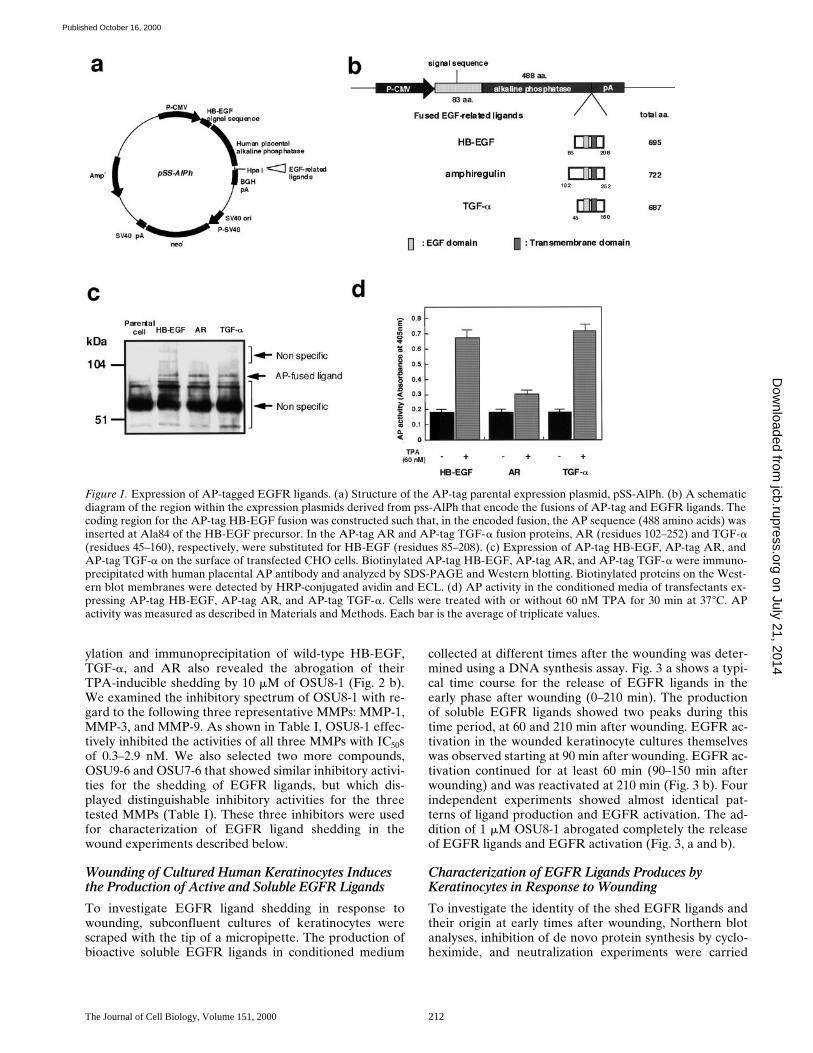
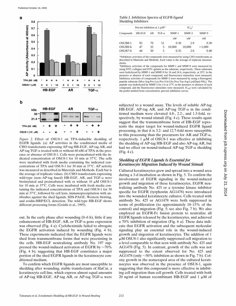
Expression plasmids encoding fusions between AP andthree different EGFR ligands (AP-tag HB-EGF, AP-tagAR, and AP-tag TGF-a) were constructed as shown inFig. 1 (a and b), and were stably transfected into CHOcells. Fusion protein expression on the surface of the trans-fected cells was analyzed by cell-surface biotinylation fol-lowed by immunoprecipitation with an anti-AP antibody.Proteins of the expected size (z88 kD) were expressed al-most equally in the respective transfected cell lines (Fig. 1c). To determine whether the fusions could be processedto release soluble versions of the AP-tagged ligands, TPA-inducible shedding of these ligands was tested. AP activityin the conditioned media of the three transfectants wasmeasured after a 30-min incubation with or without 60 nMTPA. TPA induced the shedding of AP-tag HB-EGF andAP-tag TGF-a (Fig. 1 d), which is consistent with previousobservations on TPA-stimulated shedding (Pandiella andMassague, 1991; Goishi et al., 1995). However, TPA wasless effective, at inducing the shedding of AP-tag AR (Fig.1 d). The shed ligands were also able to activate the EGFreceptor (data not shown).
Using the AP-tag EGFR ligand transfectants, .500 syn-thetic compounds were tested for the ability to inhibitshedding of the ligands. Since it has been reported that amatrix metalloprotease (MMP) is involved in the sheddingof HB-EGF and TGF-a (Suzuki et al., 1997; Izumi et al.,1998; Peschon et al., 1998), these compounds were de-signed as potential MMP inhibitors. The most effectivecompound was OSU8-1. 1 mM OSU8-1 markedly blockedthe shedding of AP-tag HB-EGF and AP-tag AR, andpartially blocked the shedding of AP-tag TGF-a (Fig. 2 a).A 10-fold higher concentration of OSU8-1 (10 mM) signif-icantly blocked the TPA-inducible shedding of all threeAP-tagged EGFR ligands (Fig. 2 a). Cell-surface biotin-
on July 21, 2014jcb.rupress.org
Dow
nloaded from
Published October 16, 2000
The Journal of Cell Biology, Volume 151, 2000 212
ylation and immunoprecipitation of wild-type HB-EGF,TGF-a, and AR also revealed the abrogation of theirTPA-inducible shedding by 10 mM of OSU8-1 (Fig. 2 b).We examined the inhibitory spectrum of OSU8-1 with re-gard to the following three representative MMPs: MMP-1,MMP-3, and MMP-9. As shown in Table I, OSU8-1 effec-tively inhibited the activities of all three MMPs with IC50sof 0.3–2.9 nM. We also selected two more compounds,OSU9-6 and OSU7-6 that showed similar inhibitory activi-ties for the shedding of EGFR ligands, but which dis-played distinguishable inhibitory activities for the threetested MMPs (Table I). These three inhibitors were usedfor characterization of EGFR ligand shedding in thewound experiments described below.
Wounding of Cultured Human Keratinocytes Induces the Production of Active and Soluble EGFR Ligands
To investigate EGFR ligand shedding in response towounding, subconfluent cultures of keratinocytes werescraped with the tip of a micropipette. The production ofbioactive soluble EGFR ligands in conditioned medium
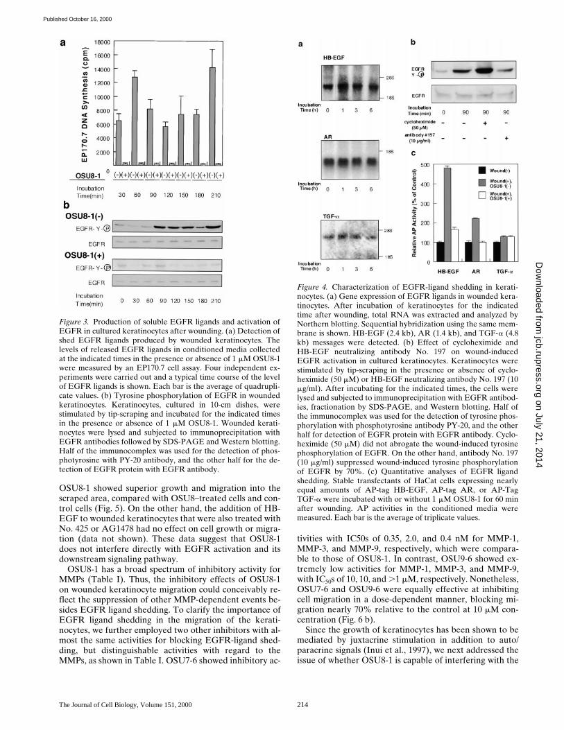
collected at different times after the wounding was deter-mined using a DNA synthesis assay. Fig. 3 a shows a typi-cal time course for the release of EGFR ligands in theearly phase after wounding (0–210 min). The productionof soluble EGFR ligands showed two peaks during thistime period, at 60 and 210 min after wounding. EGFR ac-tivation in the wounded keratinocyte cultures themselveswas observed starting at 90 min after wounding. EGFR ac-tivation continued for at least 60 min (90–150 min afterwounding) and was reactivated at 210 min (Fig. 3 b). Fourindependent experiments showed almost identical pat-terns of ligand production and EGFR activation. The ad-dition of 1 mM OSU8-1 abrogated completely the releaseof EGFR ligands and EGFR activation (Fig. 3, a and b).
Characterization of EGFR Ligands Produces by Keratinocytes in Response to Wounding
To investigate the identity of the shed EGFR ligands andtheir origin at early times after wounding, Northern blotanalyses, inhibition of de novo protein synthesis by cyclo-heximide, and neutralization experiments were carried
Figure 1. Expression of AP-tagged EGFR ligands. (a) Structure of the AP-tag parental expression plasmid, pSS-AlPh. (b) A schematicdiagram of the region within the expression plasmids derived from pss-AlPh that encode the fusions of AP-tag and EGFR ligands. Thecoding region for the AP-tag HB-EGF fusion was constructed such that, in the encoded fusion, the AP sequence (488 amino acids) wasinserted at Ala84 of the HB-EGF precursor. In the AP-tag AR and AP-tag TGF-a fusion proteins, AR (residues 102–252) and TGF-a(residues 45–160), respectively, were substituted for HB-EGF (residues 85–208). (c) Expression of AP-tag HB-EGF, AP-tag AR, andAP-tag TGF-a on the surface of transfected CHO cells. Biotinylated AP-tag HB-EGF, AP-tag AR, and AP-tag TGF-a were immuno-precipitated with human placental AP antibody and analyzed by SDS-PAGE and Western blotting. Biotinylated proteins on the West-ern blot membranes were detected by HRP-conjugated avidin and ECL. (d) AP activity in the conditioned media of transfectants ex-pressing AP-tag HB-EGF, AP-tag AR, and AP-tag TGF-a. Cells were treated with or without 60 nM TPA for 30 min at 378C. APactivity was measured as described in Materials and Methods. Each bar is the average of triplicate values.
on July 21, 2014jcb.rupress.org
Dow
nloaded from
Published October 16, 2000
Tokumaru et al. Ectodomain Shedding of HB-EGF in Wound Healing 213
out. In the early phase after wounding (0–6 h), little if anyenhancement of HB-EGF, AR, or TGF-a gene expressionwas observed (Fig. 4 a). Cycloheximide failed to abrogatethe EGFR activation induced by wounding (Fig. 4 b).These experiments indicated that the EGFR ligands wereshed from transmembrane forms that were preexisting inthe cells. HB-EGF neutralizing antibody No. 197 sup-pressed the wound-induced activation of EGFR by z70%(Fig. 4 b), suggesting that HB-EGF constitutes a majorportion of the shed EGFR ligands in the keratinocyte con-ditioned medium.
To confirm which EGFR ligands are most susceptible toshedding after wounding, stable transfectants of HaCat, akeratinocyte cell line, which express almost equal amountsof AP-tag HB-EGF, AP-tag AR, or AP-tag TGF-a were
subjected to a wound assay. The levels of soluble AP-tagHB-EGF, AP-tag AR, and AP-tag TGF-a in the condi-tioned medium were elevated 4.8-, 2.2-, and 1.3-fold, re-spectively, by wound stimuli (Fig. 4 c). These results againsuggest that the transmembrane form of HB-EGF repre-sents the major target for wound-induced EGFR ligandprocessing, in that it is 3.2- and 12.7-fold more susceptibleto this processing than the precursors for AR and TGF-a,respectively. 1 mM of OSU8-1 was effective at inhibitingthe shedding of AP-tag HB-EGF and also AP-tag AR, buthad no effect on wound-induced AP-tag TGF-a shedding(Fig. 4 c).
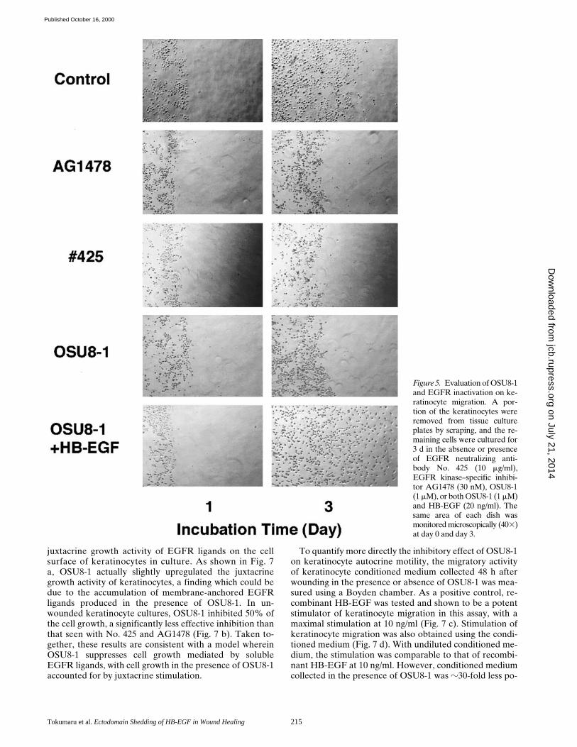
Shedding of EGFR Ligands Is Essential for Keratinocyte Migration Induced by Wound Stimuli
Cultured keratinocytes grew and spread into a wound areaduring a 3-d incubation as shown in Fig. 5. To confirm theinvolvement of EGFR signaling in the wound-inducedgrowth and migration of these keratinocytes, EGFR neu-tralizing antibody No. 425 or a tyrosine kinase inhibitorspecific for EGFR (tyrphostin AG1478) were introducedinto the wounded keratinocyte cultures. Cells treated withantibody No. 425 or AG1478 were both suppressed interms of proliferation (to approximately 10–15% of thecontrol) and migration (Fig. 5; see also Fig. 7 b). We alsoemployed an EGFR-Fc fusion protein to neutralize allEGFR ligands released by the keratinocytes, and achieveda 70% inhibition of migration (Fig. 6 a). These data indi-cate that EGFR activation and the subsequent molecularsignaling play an essential role in the wound-inducedgrowth and migration of keratinocytes. The addition of 1mM OSU8-1 also significantly suppressed cell migration toa level comparable to that seen with antibody No. 425 andAG1478 (Fig. 5). In contrast, growth of the cells was notsuppressed to the extent observed for No. 425 andAG1478 (only z50% inhibition as shown in Fig. 7 b). Col-ony growth in the nonscraped area of the cultured kerati-nocytes was observed in the presence of OSU8-1, againsuggesting that this compound is more effective in inhibit-ing cell migration than cell growth. Cells treated with both20 ng/ml of human recombinant HB-EGF and 1 mM of
Figure 2. Effect of OSU8-1 on TPA-inducible shedding ofEGFR ligands. (a) AP activities in the conditioned media ofCHO transfectants expressing AP-tag HB-EGF, AP-tag AR, andAP-tag TGF-a treated with or without 60 nM of TPA in the pres-ence or absence of OSU8-1. Cells were preincubated with the in-dicated concentration of OSU8-1 for 10 min at 378C. The cellswere incubated with fresh media containing the indicated con-centrations of TPA and OSU8-1 for 30 min at 378C. AP activitywas measured as described in Materials and Methods. Each bar isthe average of triplicate values. (b) CHO transfectants expressingwild-type (non–AP-tag fused) HB-EGF, AR, and TGF-a werebiotinylated and preincubated with or without 10 mM OSU8-1for 10 min at 378C. Cells were incubated with fresh media con-taining the indicated concentrations of TPA and OSU8-1 for 30min at 378C, followed by cell lysis, immunoprecipitation with an-tibodies against the shed ligands, SDS-PAGE, Western blotting,and avidin-HRP/ECL detection. The wild-type HB-EGF showsdifferent processing forms (Goishi et al., 1995).
Table I. Inhibition Spectra of EGFR-ligandShedding Inhibitors
Percent inhibition at 1 mM* IC50‡
Compounds HB-EGF AR TGF-a MMP-1 MMP-3 MMP-9
nM nM nM
OSUB8-1 95 70 51 0.30 2.9 0.30OSUB9-6 47 35 5 10,000 10,000 .1,000OSUB7-6 44 30 5 0.35 2.0 0.40
*Inhibitory activities of the compounds were measured by using CHO transfectants asdescribed in Materials and Methods. Each value is the average of triplicate measure-ments.‡Inhibitory activities of the compounds for MMP-1 and MMP-9 were measured byusing FITC-collagen and FITC-gelatin as the substrate, respectively. These substrateswere hydrolyzed by MMP-1 and MMP-9 for 16 and 24 h, respectively, at 358C in thepresence or absence of each compound, and fluorescence intensities were measured.Inhibitory activities of compounds for MMP-3 were measured by using a fluorogenicpeptide substrate (Mca-Arg-Pro-Lys-Pro-Val-Glu-Nva-Trp-Arg-Lys(Dnp)-NH2). Thepeptide was hydrolyzed by MMP-3 for 2 h at 378C in the presence or absence of eachcompound, and the fluorescence intensities were measured. IC50s were calculated bythe probit method from concentration–percent inhibition curves.
on July 21, 2014jcb.rupress.org
Dow
nloaded from
Published October 16, 2000
The Journal of Cell Biology, Volume 151, 2000 214
OSU8-1 showed superior growth and migration into thescraped area, compared with OSU8–treated cells and con-trol cells (Fig. 5). On the other hand, the addition of HB-EGF to wounded keratinocytes that were also treated withNo. 425 or AG1478 had no effect on cell growth or migra-tion (data not shown). These data suggest that OSU8-1does not interfere directly with EGFR activation and itsdownstream signaling pathway.
OSU8-1 has a broad spectrum of inhibitory activity forMMPs (Table I). Thus, the inhibitory effects of OSU8-1on wounded keratinocyte migration could conceivably re-flect the suppression of other MMP-dependent events be-sides EGFR ligand shedding. To clarify the importance ofEGFR ligand shedding in the migration of the kerati-nocytes, we further employed two other inhibitors with al-most the same activities for blocking EGFR-ligand shed-ding, but distinguishable activities with regard to theMMPs, as shown in Table I. OSU7-6 showed inhibitory ac-
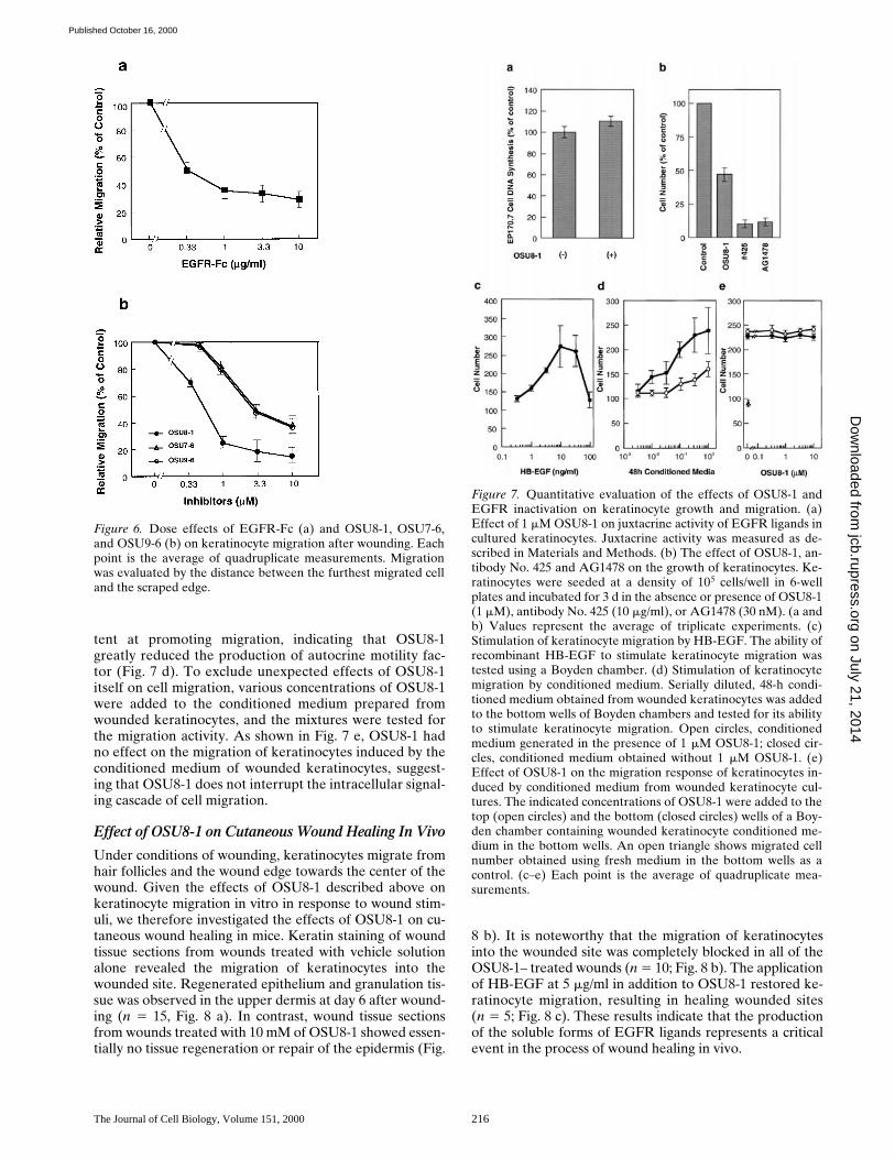
tivities with IC50s of 0.35, 2.0, and 0.4 nM for MMP-1,MMP-3, and MMP-9, respectively, which were compara-ble to those of OSU8-1. In contrast, OSU9-6 showed ex-tremely low activities for MMP-1, MMP-3, and MMP-9,with IC50s of 10, 10, and .1 mM, respectively. Nonetheless,OSU7-6 and OSU9-6 were equally effective at inhibitingcell migration in a dose-dependent manner, blocking mi-gration nearly 70% relative to the control at 10 mM con-centration (Fig. 6 b).
Since the growth of keratinocytes has been shown to bemediated by juxtacrine stimulation in addition to auto/paracrine signals (Inui et al., 1997), we next addressed theissue of whether OSU8-1 is capable of interfering with the
Figure 3. Production of soluble EGFR ligands and activation ofEGFR in cultured keratinocytes after wounding. (a) Detection ofshed EGFR ligands produced by wounded keratinocytes. Thelevels of released EGFR ligands in conditioned media collectedat the indicated times in the presence or absence of 1 mM OSU8-1were measured by an EP170.7 cell assay. Four independent ex-periments were carried out and a typical time course of the levelof EGFR ligands is shown. Each bar is the average of quadrupli-cate values. (b) Tyrosine phosphorylation of EGFR in woundedkeratinocytes. Keratinocytes, cultured in 10-cm dishes, werestimulated by tip-scraping and incubated for the indicated timesin the presence or absence of 1 mM OSU8-1. Wounded kerati-nocytes were lysed and subjected to immunoprecipitation withEGFR antibodies followed by SDS-PAGE and Western blotting.Half of the immunocomplex was used for the detection of phos-photyrosine with PY-20 antibody, and the other half for the de-tection of EGFR protein with EGFR antibody.
Figure 4. Characterization of EGFR-ligand shedding in kerati-nocytes. (a) Gene expression of EGFR ligands in wounded kera-tinocytes. After incubation of keratinocytes for the indicatedtime after wounding, total RNA was extracted and analyzed byNorthern blotting. Sequential hybridization using the same mem-brane is shown. HB-EGF (2.4 kb), AR (1.4 kb), and TGF-a (4.8kb) messages were detected. (b) Effect of cycloheximide andHB-EGF neutralizing antibody No. 197 on wound-inducedEGFR activation in cultured keratinocytes. Keratinocytes werestimulated by tip-scraping in the presence or absence of cyclo-heximide (50 mM) or HB-EGF neutralizing antibody No. 197 (10mg/ml). After incubating for the indicated times, the cells werelysed and subjected to immunoprecipitation with EGFR antibod-ies, fractionation by SDS-PAGE, and Western blotting. Half ofthe immunocomplex was used for the detection of tyrosine phos-phorylation with phosphotyrosine antibody PY-20, and the otherhalf for detection of EGFR protein with EGFR antibody. Cyclo-heximide (50 mM) did not abrogate the wound-induced tyrosinephosphorylation of EGFR. On the other hand, antibody No. 197(10 mg/ml) suppressed wound-induced tyrosine phosphorylationof EGFR by 70%. (c) Quantitative analyses of EGFR ligandshedding. Stable transfectants of HaCat cells expressing nearlyequal amounts of AP-tag HB-EGF, AP-tag AR, or AP-TagTGF-a were incubated with or without 1 mM OSU8-1 for 60 minafter wounding. AP activities in the conditioned media weremeasured. Each bar is the average of triplicate values.
on July 21, 2014jcb.rupress.org
Dow
nloaded from
Published October 16, 2000
Tokumaru et al. Ectodomain Shedding of HB-EGF in Wound Healing 215
juxtacrine growth activity of EGFR ligands on the cellsurface of keratinocytes in culture. As shown in Fig. 7a, OSU8-1 actually slightly upregulated the juxtacrinegrowth activity of keratinocytes, a finding which could bedue to the accumulation of membrane-anchored EGFRligands produced in the presence of OSU8-1. In un-wounded keratinocyte cultures, OSU8-1 inhibited 50% ofthe cell growth, a significantly less effective inhibition thanthat seen with No. 425 and AG1478 (Fig. 7 b). Taken to-gether, these results are consistent with a model whereinOSU8-1 suppresses cell growth mediated by solubleEGFR ligands, with cell growth in the presence of OSU8-1accounted for by juxtacrine stimulation.
To quantify more directly the inhibitory effect of OSU8-1on keratinocyte autocrine motility, the migratory activityof keratinocyte conditioned medium collected 48 h afterwounding in the presence or absence of OSU8-1 was mea-sured using a Boyden chamber. As a positive control, re-combinant HB-EGF was tested and shown to be a potentstimulator of keratinocyte migration in this assay, with amaximal stimulation at 10 ng/ml (Fig. 7 c). Stimulation ofkeratinocyte migration was also obtained using the condi-tioned medium (Fig. 7 d). With undiluted conditioned me-dium, the stimulation was comparable to that of recombi-nant HB-EGF at 10 ng/ml. However, conditioned mediumcollected in the presence of OSU8-1 was z30-fold less po-
Figure 5. Evaluation of OSU8-1and EGFR inactivation on ke-ratinocyte migration. A por-tion of the keratinocytes wereremoved from tissue cultureplates by scraping, and the re-maining cells were cultured for3 d in the absence or presenceof EGFR neutralizing anti-body No. 425 (10 mg/ml),EGFR kinase–specific inhibi-tor AG1478 (30 nM), OSU8-1(1 mM), or both OSU8-1 (1 mM)and HB-EGF (20 ng/ml). Thesame area of each dish wasmonitored microscopically (403)at day 0 and day 3.
on July 21, 2014jcb.rupress.org
Dow
nloaded from
Published October 16, 2000
The Journal of Cell Biology, Volume 151, 2000 216
tent at promoting migration, indicating that OSU8-1greatly reduced the production of autocrine motility fac-tor (Fig. 7 d). To exclude unexpected effects of OSU8-1itself on cell migration, various concentrations of OSU8-1were added to the conditioned medium prepared fromwounded keratinocytes, and the mixtures were tested forthe migration activity. As shown in Fig. 7 e, OSU8-1 hadno effect on the migration of keratinocytes induced by theconditioned medium of wounded keratinocytes, suggest-ing that OSU8-1 does not interrupt the intracellular signal-ing cascade of cell migration.
Effect of OSU8-1 on Cutaneous Wound Healing In Vivo
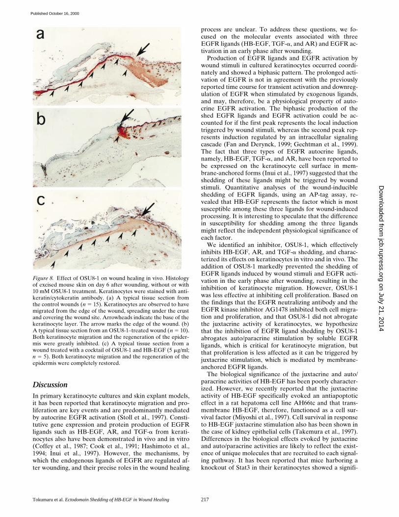
Under conditions of wounding, keratinocytes migrate fromhair follicles and the wound edge towards the center of thewound. Given the effects of OSU8-1 described above onkeratinocyte migration in vitro in response to wound stim-uli, we therefore investigated the effects of OSU8-1 on cu-taneous wound healing in mice. Keratin staining of woundtissue sections from wounds treated with vehicle solutionalone revealed the migration of keratinocytes into thewounded site. Regenerated epithelium and granulation tis-sue was observed in the upper dermis at day 6 after wound-ing (n 5 15, Fig. 8 a). In contrast, wound tissue sectionsfrom wounds treated with 10 mM of OSU8-1 showed essen-tially no tissue regeneration or repair of the epidermis (Fig.
8 b). It is noteworthy that the migration of keratinocytesinto the wounded site was completely blocked in all of theOSU8-1– treated wounds (n 5 10; Fig. 8 b). The applicationof HB-EGF at 5 mg/ml in addition to OSU8-1 restored ke-ratinocyte migration, resulting in healing wounded sites(n 5 5; Fig. 8 c). These results indicate that the productionof the soluble forms of EGFR ligands represents a criticalevent in the process of wound healing in vivo.
Figure 6. Dose effects of EGFR-Fc (a) and OSU8-1, OSU7-6,and OSU9-6 (b) on keratinocyte migration after wounding. Eachpoint is the average of quadruplicate measurements. Migrationwas evaluated by the distance between the furthest migrated celland the scraped edge.
Figure 7. Quantitative evaluation of the effects of OSU8-1 andEGFR inactivation on keratinocyte growth and migration. (a)Effect of 1 mM OSU8-1 on juxtacrine activity of EGFR ligands incultured keratinocytes. Juxtacrine activity was measured as de-scribed in Materials and Methods. (b) The effect of OSU8-1, an-tibody No. 425 and AG1478 on the growth of keratinocytes. Ke-ratinocytes were seeded at a density of 105 cells/well in 6-wellplates and incubated for 3 d in the absence or presence of OSU8-1(1 mM), antibody No. 425 (10 mg/ml), or AG1478 (30 nM). (a andb) Values represent the average of triplicate experiments. (c)Stimulation of keratinocyte migration by HB-EGF. The ability ofrecombinant HB-EGF to stimulate keratinocyte migration wastested using a Boyden chamber. (d) Stimulation of keratinocytemigration by conditioned medium. Serially diluted, 48-h condi-tioned medium obtained from wounded keratinocytes was addedto the bottom wells of Boyden chambers and tested for its abilityto stimulate keratinocyte migration. Open circles, conditionedmedium generated in the presence of 1 mM OSU8-1; closed cir-cles, conditioned medium obtained without 1 mM OSU8-1. (e)Effect of OSU8-1 on the migration response of keratinocytes in-duced by conditioned medium from wounded keratinocyte cul-tures. The indicated concentrations of OSU8-1 were added to thetop (open circles) and the bottom (closed circles) wells of a Boy-den chamber containing wounded keratinocyte conditioned me-dium in the bottom wells. An open triangle shows migrated cellnumber obtained using fresh medium in the bottom wells as acontrol. (c–e) Each point is the average of quadruplicate mea-surements.
on July 21, 2014jcb.rupress.org
Dow
nloaded from
Published October 16, 2000
Tokumaru et al. Ectodomain Shedding of HB-EGF in Wound Healing 217
DiscussionIn primary keratinocyte cultures and skin explant models,it has been reported that keratinocyte migration and pro-liferation are key events and are predominantly mediatedby autocrine EGFR activation (Stoll et al., 1997). Consti-tutive gene expression and protein production of EGFRligands such as HB-EGF, AR, and TGF-a from kerati-nocytes also have been demonstrated in vivo and in vitro(Coffey et al., 1987; Cook et al., 1991; Hashimoto et al.,1994; Inui et al., 1997). However, the mechanisms, bywhich the endogenous ligands of EGFR are regulated af-ter wounding, and their precise roles in the wound healing
process are unclear. To address these questions, we fo-cused on the molecular events associated with threeEGFR ligands (HB-EGF, TGF-a, and AR) and EGFR ac-tivation in an early phase after wounding.
Production of EGFR ligands and EGFR activation bywound stimuli in cultured keratinocytes occurred coordi-nately and showed a biphasic pattern. The prolonged acti-vation of EGFR is not in agreement with the previouslyreported time course for transient activation and downreg-ulation of EGFR when stimulated by exogenous ligands,and may, therefore, be a physiological property of auto-crine EGFR activation. The biphasic production of theshed EGFR ligands and EGFR activation could be ac-counted for if the first peak represents the local inductiontriggered by wound stimuli, whereas the second peak rep-resents induction regulated by an intracellular signalingcascade (Fan and Derynck, 1999; Gechtman et al., 1999).The fact that three types of EGFR autocrine ligands,namely, HB-EGF, TGF-a, and AR, have been reported tobe expressed on the keratinocyte cell surface in mem-brane-anchored forms (Inui et al., 1997) suggested that theshedding of these ligands might be triggered by woundstimuli. Quantitative analyses of the wound-inducibleshedding of EGFR ligands, using an AP-tag assay, re-vealed that HB-EGF represents the factor which is mostsusceptible among these three ligands for wound-inducedprocessing. It is interesting to speculate that the differencein susceptibility for shedding among the three ligandsmight reflect the independent physiological significance ofeach factor.
We identified an inhibitor, OSU8-1, which effectivelyinhibits HB-EGF, AR, and TGF-a shedding, and charac-terized its effects on keratinocytes in vitro and in vivo. Theaddition of OSU8-1 markedly prevented the shedding ofEGFR ligands induced by wound stimuli and EGFR acti-vation in the early phase after wounding, resulting in theinhibition of keratinocyte migration. However, OSU8-1was less effective at inhibiting cell proliferation. Based onthe findings that the EGFR neutralizing antibody and theEGFR kinase inhibitor AG1478 inhibited both cell migra-tion and proliferation, and that OSU8-1 did not abrogatethe juxtacrine activity of keratinocytes, we hypothesizethat the inhibition of EGFR ligand shedding by OSU8-1abrogates auto/paracrine stimulation by soluble EGFRligands, which is critical for keratinocyte migration, butthat proliferation is less affected as it can be triggered byjuxtacrine stimulation, which is mediated by membrane-anchored EGFR ligands.
The biological significance of the juxtacrine and auto/paracrine activities of HB-EGF has been poorly character-ized. However, we recently reported that the juxtacrineactivity of HB-EGF specifically evoked an antiapoptoticeffect in a rat hepatoma cell line AH66tc and that trans-membrane HB-EGF, therefore, functioned as a cell sur-vival factor (Miyoshi et al., 1997). Cell survival in responseto HB-EGF juxtacrine stimulation also has been shown inthe case of kidney epithelial cells (Takemura et al., 1997).Differences in the biological effects evoked by juxtacrineand auto/paracrine activities are likely to reflect the exist-ence of unique molecules that are recruited to each signal-ing pathway. It has been reported that mice harboring aknockout of Stat3 in their keratinocytes showed a signifi-
Figure 8. Effect of OSU8-1 on wound healing in vivo. Histologyof excised mouse skin on day 6 after wounding, without or with10 mM OSU8-1 treatment. Keratinocytes were stained with anti-keratin/cytokeratin antibody. (a) A typical tissue section fromthe control wounds (n 5 15). Keratinocytes are observed to havemigrated from the edge of the wound, spreading under the crustand covering the wound site. Arrowheads indicate the base of thekeratinocyte layer. The arrow marks the edge of the wound. (b)A typical tissue section from an OSU8-1–treated wound (n 5 10).Both keratinocyte migration and the regeneration of the epider-mis were greatly inhibited. (c) A typical tissue section from awound treated with a cocktail of OSU8-1 and HB-EGF (5 mg/ml;n 5 5). Both keratinocyte migration and the regeneration of theepidermis were completely restored.
on July 21, 2014jcb.rupress.org
Dow
nloaded from
Published October 16, 2000
The Journal of Cell Biology, Volume 151, 2000 218
cant retardation of cutaneous wound healing as reflectedin a lack of keratinocyte migration. However, the growthresponse of the keratinocytes in these mice to severalgrowth factors appeared to be normal (Sano et al., 1999).Consistent with these observations, we found that specificactivation of Stat3 was evoked by EGFR ligand sheddingin cultured keratinocytes (data not shown), suggesting thatthe ectodomain shedding of EGFR ligands could triggerintercellular signaling pathways unique for auto/paracrineand juxtacrine stimulation.
Analyses of the effects of OSU8-1 on cutaneous woundhealing in vivo revealed that wounded tissue sectionstreated with 10 mM OSU8-1 showed essentially no tissueregeneration and repair at day 6 after wounding. In partic-ular, there was a marked lack of keratinocyte migration, ascompared with cutaneous healing in control mice. Admin-istration of recombinant HB-EGF, in addition to OSU8-1,restored the migration of keratinocytes in wounded sites.Therefore, we conclude that the shedding of EGFRligands represents a crucial event in the migration of kera-tinocytes to the wound site in vivo. The present findingsmay also have implications not only for our understandingof the molecular mechanism involved in wound healing,but also for other tissue remodeling processes such as tro-phoblast invasion, inflammatory diseases, and, in particu-lar, tumor invasion.
Recently, it has been reported that the broad spectruminhibition of MMPs by a synthetic inhibitor (galardin) inplasminogen-deficient mice completely arrested woundhealing, suggesting an essential role for matrix-degradingproteases in wound healing (Lund et al., 1999). Sincegalardin, like OSU8-1, is a peptide-based zinc-chelatinghydroxamate, it might inactivate not only the MMP fam-ily, but also the ADAM (a disintegrin and a metallopro-tease) family involved in EGFR ligand shedding. Thus, wespeculate that the inhibition of wound healing reported byLund et al. (1999) might, in part, have been due to the in-hibition of EGFR ligand shedding.
Tumor necrosis factor–a converting enzyme (TACE)represents an additional protein involved in ectodomainshedding. Its gene has been cloned (Black et al., 1997;Moss et al., 1997) and disrupted (Peschon et al., 1998), re-sulting in skin and eye defects. This phenotype resemblesthat of TGF-a knockout mice, suggesting that TACEmight function widely as a shedding enzyme beyond its ef-fects on tumor necrosis factor–a processing. TACE is nowclassified in the ADAM family, which is comprised of atleast 31 members. Our most recent studies of inhibitors forEGFR ligand shedding indicate that .1 shedding proteasecould be involved in the shedding of EGFR ligands (Endo,T., and S. Higashiyama, unpublished data). The identifica-tion of shedding enzymes for many types of cell-surfaceproteins, including receptors and ligands, would be a keystep in understanding the normal and pathological tissueremodeling processes described above.
Recently, molecular mechanism in the ectodomainshedding of EGFR ligands including HB-EGF and TGF-ahave been vigorously studied, showing the involvement ofMMP-3 (Suzuki et al., 1997), meltrin-g/MDC9/ADAM9(Izumi et al., 1998), and the MAP kinase cascade (Fan andDerynck, 1999; Gechtman et al., 1999). Further, it also hasbeen shown that the ectodomain shedding of HB-EGF
is a crucial step in the transactivation of the EGFR viaG-protein–coupled receptors (Prenzel et al., 1999). Takentogether with our results herein, it is clear that theectodomain shedding of EGFR ligands is a regulated mo-lecular event involved in different types of physiologicaland pathological phenomena.
In conclusion, wound stimuli trigger EGFR ligand shed-ding, which represents an essential step in the cutaneouswound healing process. OSU8-1 inhibits the shedding ofEGFR ligands, resulting in the strong inhibition of woundhealing in vitro and in vivo. It is clear that shedding en-zymes and their involvement in wound healing are deserv-ing of further investigation.
We greatly acknowledge Dr. J.A. Abraham (Scios Inc.) for editing themanuscript, and Drs. S. Sano and J. Takeda (Osaka University) for help-ful discussion and comments.
Submitted: 9 March 2000Revised: 6 August 2000Accepted: 21 August 2000
References
Black, R.A., C.T. Rauch, C.J. Kozlosky, J.J. Peschon, J.L. Slack, M.F. Wolfson,B.J. Castner, K.L. Stocking, P. Reddy, S. Srinivasan, et al. 1997. A metallo-proteinase disintegrin that releases tumor-necrosis factor-alpha from cells.Nature. 385:729–733.
Blotnick, S., G.E. Peoples, M.R. Freeman, T.J. Eberlein, and M. Klagsbrun.1994. T lymphocytes synthesize and export heparin-binding epidermalgrowth factor-like growth factor and basic fibroblast growth factor, mitogensfor vascular cells and fibroblasts: differential production and release byCD41 and CD81 T cells. Proc. Natl. Acad. Sci. USA. 91:2890–2894.
Boyden, S., 1962. The chemotactic effect of mixtures of antibodies and antigenson polymorphonuclear leukocytes. J. Exp. Med. 115:435–466.
Brachmann, R., P.B. Lindquist, M. Nagashima, W. Kohr, T. Lipari, M. Napier,and R. Derynck. 1989. Transmembrane TGF-a precursors activate EGF/TGF-a receptors. Cell. 56:691–700.
Chen, C.A., and H. Okayama. 1988. Calcium phosphate-mediated gene trans-fer: a highly efficient transfection system for stably transforming cells withplasmid DNA. Biotechniques. 6:632–638.
Chomczynski, P., and N. Sacchi. 1987. Single-step method of RNA isolation byacid guanidinium thiocyanate-phenol-chloroform extraction. Anal. Bio-chem. 162:156–159.
Coffey, R., Jr., R. Derynck, J.N. Wilcox, T.S. Bringman, A.S. Goustin, H.L.Moses, and M.R. Pittelkow. 1987. Production and auto-induction of trans-forming growth factor-a in human keratinocytes. Nature. 328:817–820.
Cohen, S. 1964. Isolation and biological effects of an epidermal growth-stimulat-ing protein. National Cancer Institute Monograph 13. Bethesda, MD. 13–27.
Cook, P.W., P.A Mattox, W.W. Keeble, M.R. Pittelkow, G.D. Plowman, M.Shoyab, J.P. Adelman, and G.D. Shippley. 1991. A heparin sulfate-regulatedhuman keratinocyte autocrine factor is similar or identical to amphiregulin.Mol. Cell. Biol. 11:2547–2557.
Derynck, R., A.B. Roberts, M.E. Winkler, E.Y. Chen, and D.V. Goeddel. 1984.Human transforming growth factor-a: precursor structure and expression inE. coli. Cell. 38:287–297.
Fan, H., and R. Derynck. 1999. Ectodomain shedding of TGF-a and othertransmembrane proteins is induced by receptor tyrosine kinase activationand MAP kinase signaling cascades. EMBO (Eur. Mol. Biol. Organ.) J. 24:6962–6972.
Gechtman, Z., J.L. Alonson, G. Raab, D.E. Ingber, and M. Klagsbrun. 1999.The shedding of membrane-anchored heparin-binding epidermal-likegrowth factor is regulated by the ref/mitogen-activated protein kinase cas-cade and by cell adhesion and spreading. J. Biol. Chem. 274:28828–28835.
Goishi, K., S. Higashiyama, M. Klagsbrun, N. Nakano, T. Umata, M. Ishikawa,E. Mekada, and N. Taniguchi. 1995. Phorbol ester induces the rapid process-ing of cell surface heparin-binding EGF-like growth factor: conversion fromjuxtacrine to paracrine growth factor activity. Mol. Biol. Cell. 6:967–980.
Gotoh, N., A. Tojo, K. Muroya, Y. Hashimoto, S. Hattori, S. Nakamura, T.Takenawa, Y. Yazaki, and M. Shibuya. 1994. Epidermal growth factor-receptor mutant lacking the autophosphorylation sites induces phosphoryla-tion of Shc protein and Shc-Grb2/ASH association and retains mitogenic ac-tivity. Proc. Natl. Acad. Sci. USA. 91:167–171.
Hashimoto, K., S. Higashiyama, H. Asada, E. Hashimura, T. Kobayashi, K.Sudo, T. Nakagawa, D. Damm, K. Yoshikawa, and N. Taniguchi. 1994. Hep-arin-binding EGF-like growth factor is an autocrine growth factor for hu-man keratinocytes. J. Biol. Chem. 269:20060–20066.
Higashiyama, S., J.A. Abraham, J. Miller, J.C. Fiddes, and M. Klagsbrun. 1991.
on July 21, 2014jcb.rupress.org
Dow
nloaded from
Published October 16, 2000
Tokumaru et al. Ectodomain Shedding of HB-EGF in Wound Healing 219
A heparin-binding growth factor secreted by macrophage-like cells that isrelated to EGF. Science. 251:936–939.
Higashiyama, S., J.A. Abraham, and M. Klagsbrun. 1993. Heparin-binding EGF-like growth factor stimulation of smooth muscle cell migration: dependenceon interactions with cell surface heparan sulfate. J. Cell Biol. 122:933–940.
Higashiyama, S., R. Iwamoto, K. Goishi, G. Raab, N. Taniguchi, M. Klagsbrun,and E. Mekada. 1995. The membrane protein CD9/DRAP27 potentiates thejuxtacrine growth factor activity of the membrane-anchored heparin-bindingEGF-like growth factor. J. Cell Biol. 128:929–938.
Inui, S., S. Higashiyama, K. Hashimoto, M. Higashiyama, K. Yoshikawa, and N.Taniguchi. 1997. Possible role of coexpression of CD9 with membrane-anchored heparin-binding EGF-like growth factor and amphiregulin in cul-tured human keratinocyte growth. J. Cell. Physiol. 171:291–298.
Izumi, Y., M. Hirata, H. Hasuwa, R. Iwamoto, T. Umata, K. Miyado, Y. Tamai,T. Kurisaki, A. Sehara-Fujisawa, S. Ohno, and E. Mekada. 1998. A metallo-protease-disintegrin, MDC9/meltrin-gamma/ADAM9 and PKCdelta are in-volved in TPA-induced ectodomain shedding of membrane-anchored hep-arin-binding EGF-like growth factor. EMBO (Eur. Mol. Biol. Organ.) J. 17:7260–7272.
Lund, L.R., J. Romer, T.H. Bugge, B.S. Nielsen, T.L. Frandsen, J.L. Degen,R.W. Stephens, and K. Dano. 1999. Functional overlap between two classesof matrix-degrading proteases in wound healing. EMBO (Eur. Mol. Biol.Organ.) J. 17:4645–4656.
Marikovsky, M., K. Breuing, P.Y. Liu, E. Eriksson, S. Higashiyama, P. Farber,J. Sasse, and M. Klagsbrun. 1993. Appearance of heparin-binding EGF-likegrowth factor in wound fluid as a response to injury. Proc. Natl. Acad. Sci.USA. 90:3889–3893.
Martin, P. 1997. Wound healing: aiming for perfect skin regeneration. Science.276:75–81.
Massague, J., and A. Pandiella. 1993. Membrane-anchored growth factors.Annu. Rev. Biochem. 62:515–541.
Miyoshi, E., S. Higashiyama, T. Nakagawa, N. Hayashi, and N. Taniguchi. 1997.Membrane-anchored heparin-binding epidermal growth factor-like growthfactor acts as a tumor survival factor in a hepatoma cell line. J. Biol. Chem.272:14349–14355.
Moss, M.L., S.L. Jin, M.E. Milla, D.M. Bickett, W. Burkhart, H.L. Carter, W.J.Chen, W.C. Clay, J.R. Didsbury, D. Hassler, et al. 1997. Cloning of a disinte-grin metalloproteinase that processes precursor tumor-necrosis factor-alpha.Nature. 385:733–736.
Pandiella, A., and J. Massague. 1991. Cleavage of the membrane precursor for
transforming growth factor alpha is a regulated process. Proc. Natl. Acad.Sci. USA. 88:1726–1730.
Peschon, J.J., J.L. Slack, P. Reddy, K.L. Stocking, S.W. Sunnarborg, D.C. Lee,W.E. Russell, B.J. Castner, R.S. Johnson, J.N. Fitzner, et al. 1998. An essen-tial role for ectodomain shedding in mammalian development. Science. 282:1281–1284.
Prenzel, N., E. Zwick, H. Daub, M. Leserer, R. Abraham, C. Wallasch, and A.Ullrich. 1999. EGF receptor transactivation by G-protein-coupled receptorsrequires metalloproteinase cleavage of proHB-EGF. Nature. 402:884–888.
Sano, S., S. Itami, K. Takeda, M. Tarutani, Y. Yamaguchi, H. Miura, K.Yoshikawa, S. Akira, and J. Takeda. 1999. Keratinocyte-specific ablation ofStat3 exhibits impaired skin remodeling, but does not affect skin morpho-genesis. EMBO (Eur. Mol. Biol. Organ.) J. 17:4657–4668.
Shing, Y., G. Christofori, D. Hanahan, Y. Ono, R. Sasada, K. Igarashi, and J.Folkman. 1993. Betacellulin: a mitogen from pancreatic b cell tumors. Sci-ence. 259:1604–1614.
Shoyab, M., G.D. Plowman, V.L. McDonald, J.G. Bradley, and G.J. Todaro.1989. Structure and function of human amphiregulin: a member of the epi-dermal growth factor family. Science. 243:1074–1076.
Stoll, S., W. Garner, and J. Elder. 1997. Heparin-binding ligands mediate auto-crine epidermal growth factor receptor activation in skin organ culture. J.Clin. Invest. 100:1271–1281.
Suzuki, M., G. Raab, M.A. Moses, C.A. Fernandez, and M. Klagsbrun. 1997.Matrix metalloproteinase-3 releases active heparin-binding EGF-likegrowth factor by cleavage at a specific juxtamembrane site. J. Biol. Chem.272:31730–31737.
Takemura, T., S. Kondo, T. Homma, M. Sakai, and R.C. Harris. 1997. Themembrane-bound form of heparin-binding epidermal growth factor-likegrowth factor promotes survival of cultured renal epithelial cells. J. Biol.Chem. 272:31036–31042.
Toyoda, H., T. Komurasaki, D. Uchida, Y. Takayama, T. Isobe, T. Okuyama,and K. Hanada. 1995. Epiregulin, a novel epidermal growth factor with mito-genic activity for rat primary hepatocytes. J. Biol. Chem. 270:7495–7500.
Tsuboi, R., S. Chong-ming, B.R. Daniel, and H. Ogawa. 1992. A wound healingmodel using healing-impaired diabetic mice. J. Dermatol. 19:673–675.
Wong, S., L.F. Winchell, B.K. McCune, H.S. Earp, J. Texido, J. Massaague, B.Herman, and D.C. Lee. 1989. The TGF-a precursor expressed on the cellsurface binds to the EGF receptor on adjacent cells, leading to signal trans-duction. Cell. 56:495–506.
on July 21, 2014jcb.rupress.org
Dow
nloaded from
Published October 16, 2000