Agent Based Modelling Helps in Understanding the Rules by Which Fibroblasts Support Keratinocyte...
17
Agent Based Modelling Helps in Understanding the Rules by Which Fibroblasts Support Keratinocyte Colony Formation Tao Sun 1 *, Phil McMinn 2 , Mike Holcombe 2 , Rod Smallwood 2 , Sheila MacNeil 1 * 1 Department of Engineering Materials, University of Sheffield, Sheffield, United Kingdom, 2 Department of Computer Science, University of Sheffield, Sheffield, United Kingdom Abstract Background: Autologous keratincoytes are routinely expanded using irradiated mouse fibroblasts and bovine serum for clinical use. With growing concerns about the safety of these xenobiotic materials, it is desirable to culture keratinocytes in media without animal derived products. An improved understanding of epithelial/mesenchymal interactions could assist in this. Methodology/Principal Findings: A keratincyte/fibroblast o-culture model was developed by extending an agent-based keratinocyte colony formation model to include the response of keratinocytes to both fibroblasts and serum. The model was validated by comparison of the in virtuo and in vitro multicellular behaviour of keratinocytes and fibroblasts in single and co-culture in Greens medium. To test the robustness of the model, several properties of the fibroblasts were changed to investigate their influence on the multicellular morphogenesis of keratinocyes and fibroblasts. The model was then used to generate hypotheses to explore the interactions of both proliferative and growth arrested fibroblasts with keratinocytes. The key predictions arising from the model which were confirmed by in vitro experiments were that 1) the ratio of fibroblasts to keratinocytes would critically influence keratinocyte colony expansion, 2) this ratio needed to be optimum at the beginning of the co-culture, 3) proliferative fibroblasts would be more effective than irradiated cells in expanding keratinocytes and 4) in the presence of an adequate number of fibroblasts, keratinocyte expansion would be independent of serum. Conclusions: A closely associated computational and biological approach is a powerful tool for understanding complex biological systems such as the interactions between keratinocytes and fibroblasts. The key outcome of this study is the finding that the early addition of a critical ratio of proliferative fibroblasts can give rapid keratinocyte expansion without the use of irradiated mouse fibroblasts and bovine serum. Citation: Sun T, McMinn P, Holcombe M, Smallwood R, MacNeil S (2008) Agent Based Modelling Helps in Understanding the Rules by Which Fibroblasts Support Keratinocyte Colony Formation. PLoS ONE 3(5): e2129. doi:10.1371/journal.pone.0002129 Editor: Eshel Ben-Jacob, Tel Aviv University, Israel Received February 4, 2008; Accepted April 2, 2008; Published May 7, 2008 Copyright: ß 2008 Sun et al. This is an open-access article distributed under the terms of the Creative Commons Attribution License, which permits unrestricted use, distribution, and reproduction in any medium, provided the original author and source are credited. Funding: Funding from EPSRC(UK). The funder did not play a role in the design and conduct of the study, in the collection, analysis, and interpretation of the data, and in the preparation of the manuscript. Competing Interests: The authors have declared that no competing interests exist. * E-mail: [email protected] (TS); [email protected] (SM) Introduction While the volume of data in biology is increasing rapidly with outputs from genomic, proteomic and metabolomic approaches it doesn’t follow that it is easy to interpret this growing body of new data-indeed it is becoming increasingly difficult for biologists to integrate such a complexity of information without a holistic view of organisms[1–2]. This study follows on from a previous one from our group [3] in which we used computational modelling as a tool to improve our understanding of one particular biological system, skin cells organise into an epithelium. In this model we closely coupled in vitro and in virtuo approaches to explore different hypotheses about how normal human keratinocytes (NHK) and a transformed keratinocyte cell line (HaCat) formed colonies and achieved new insights into how keratinocytes form colonies [3]. In the current study we now move to the next level of complexity and seek to extend the previous agent based model to investigate the interactions between normal human keratinocytes and fibroblasts both in virtuo and in vitro. There is a plethora of data showing that mesenchymal cells profoundly influence epithelial cell organisation. In the culture of human keratinocytes this information has been used routinely since the initial publications by Rheinwald and Green in 1975 [4] using an irradiated feeder layer of mouse fibroblasts (i3T3) to support the attachment and proliferation of adult human keratinocytes in the presence of a mitogens rich media containing 10% bovine foetal calf serum (FCS). Despite the clinical need to avoid the use of xenobiotic agents (foetal calf serum, cholera toxin) and cells (murine fibroblasts) the use of an irradiated mouse fibroblast feeder layer in a mitogen rich media with 10% bovine serum as first used clinically in the early 1980’s [5] remains the most commonly used methodology for rapid expansion of adult keratinocytes for clinical use to this day [6]. The reason for this is simple. It is a robust and reliable methodology for culturing adult human skin cells often PLoS ONE | www.plosone.org 1 May 2008 | Volume 3 | Issue 5 | e2129
Transcript of Agent Based Modelling Helps in Understanding the Rules by Which Fibroblasts Support Keratinocyte...
Agent Based Modelling Helps in Understanding theRules by Which Fibroblasts Support Keratinocyte ColonyFormationTao Sun1*, Phil McMinn2, Mike Holcombe2, Rod Smallwood2, Sheila MacNeil1*
1 Department of Engineering Materials, University of Sheffield, Sheffield, United Kingdom, 2 Department of Computer Science, University of Sheffield, Sheffield, United
Kingdom
Abstract
Background: Autologous keratincoytes are routinely expanded using irradiated mouse fibroblasts and bovine serum forclinical use. With growing concerns about the safety of these xenobiotic materials, it is desirable to culture keratinocytes inmedia without animal derived products. An improved understanding of epithelial/mesenchymal interactions could assist inthis.
Methodology/Principal Findings: A keratincyte/fibroblast o-culture model was developed by extending an agent-basedkeratinocyte colony formation model to include the response of keratinocytes to both fibroblasts and serum. The modelwas validated by comparison of the in virtuo and in vitro multicellular behaviour of keratinocytes and fibroblasts in singleand co-culture in Greens medium. To test the robustness of the model, several properties of the fibroblasts were changed toinvestigate their influence on the multicellular morphogenesis of keratinocyes and fibroblasts. The model was then used togenerate hypotheses to explore the interactions of both proliferative and growth arrested fibroblasts with keratinocytes.The key predictions arising from the model which were confirmed by in vitro experiments were that 1) the ratio offibroblasts to keratinocytes would critically influence keratinocyte colony expansion, 2) this ratio needed to be optimum atthe beginning of the co-culture, 3) proliferative fibroblasts would be more effective than irradiated cells in expandingkeratinocytes and 4) in the presence of an adequate number of fibroblasts, keratinocyte expansion would be independentof serum.
Conclusions: A closely associated computational and biological approach is a powerful tool for understanding complexbiological systems such as the interactions between keratinocytes and fibroblasts. The key outcome of this study is thefinding that the early addition of a critical ratio of proliferative fibroblasts can give rapid keratinocyte expansion without theuse of irradiated mouse fibroblasts and bovine serum.
Citation: Sun T, McMinn P, Holcombe M, Smallwood R, MacNeil S (2008) Agent Based Modelling Helps in Understanding the Rules by Which Fibroblasts SupportKeratinocyte Colony Formation. PLoS ONE 3(5): e2129. doi:10.1371/journal.pone.0002129
Editor: Eshel Ben-Jacob, Tel Aviv University, Israel
Received February 4, 2008; Accepted April 2, 2008; Published May 7, 2008
Copyright: � 2008 Sun et al. This is an open-access article distributed under the terms of the Creative Commons Attribution License, which permits unrestricteduse, distribution, and reproduction in any medium, provided the original author and source are credited.
Funding: Funding from EPSRC(UK). The funder did not play a role in the design and conduct of the study, in the collection, analysis, and interpretation of thedata, and in the preparation of the manuscript.
Competing Interests: The authors have declared that no competing interests exist.
* E-mail: [email protected] (TS); [email protected] (SM)
Introduction
While the volume of data in biology is increasing rapidly with
outputs from genomic, proteomic and metabolomic approaches it
doesn’t follow that it is easy to interpret this growing body of new
data-indeed it is becoming increasingly difficult for biologists to
integrate such a complexity of information without a holistic view
of organisms[1–2]. This study follows on from a previous one from
our group [3] in which we used computational modelling as a tool
to improve our understanding of one particular biological system,
skin cells organise into an epithelium. In this model we closely
coupled in vitro and in virtuo approaches to explore different
hypotheses about how normal human keratinocytes (NHK) and a
transformed keratinocyte cell line (HaCat) formed colonies and
achieved new insights into how keratinocytes form colonies [3].
In the current study we now move to the next level of
complexity and seek to extend the previous agent based model to
investigate the interactions between normal human keratinocytes
and fibroblasts both in virtuo and in vitro. There is a plethora of data
showing that mesenchymal cells profoundly influence epithelial
cell organisation. In the culture of human keratinocytes this
information has been used routinely since the initial publications
by Rheinwald and Green in 1975 [4] using an irradiated feeder
layer of mouse fibroblasts (i3T3) to support the attachment and
proliferation of adult human keratinocytes in the presence of a
mitogens rich media containing 10% bovine foetal calf serum
(FCS). Despite the clinical need to avoid the use of xenobiotic
agents (foetal calf serum, cholera toxin) and cells (murine
fibroblasts) the use of an irradiated mouse fibroblast feeder layer
in a mitogen rich media with 10% bovine serum as first used
clinically in the early 1980’s [5] remains the most commonly used
methodology for rapid expansion of adult keratinocytes for clinical
use to this day [6]. The reason for this is simple. It is a robust and
reliable methodology for culturing adult human skin cells often
PLoS ONE | www.plosone.org 1 May 2008 | Volume 3 | Issue 5 | e2129

from small initial biopsies and so far it outperforms efforts to
obtain a completely defined culture approach for human
keratinocyte expansion. However with growing concerns about
the transmission of bovine spongiform encephalitis (BSE) from the
use of bovine serum it would be desirable to culture cells under
completely defined culture conditions.
Our previous research demonstrated that NHK could be
expanded by co-culturing these cells with human dermal
fibroblasts (HDFs) in Green’s media without foetal calf serum
[7–8]. We have also shown that human fibroblasts can perform as
well as murine fibroblasts in supporting the expansion of
keratinocytes and indeed keratinocytes expanded on fibroblasts
in the absence of serum tended to show less differentiation than
those expanded with serum [9] which is another desirable property
when expanding cells for clinical use. In the original Rheinwald
and Green methodology the murine fibroblasts were lethally
irradiated so that they could not expand in culture (or if
accidentally transferred to the patient) [4]. However, empirical
data from our laboratory shows that one can get expansion of
keratinocytes on non-irradiated fibroblasts to satisfactory levels if
one pays attention to the ratio between the fibroblasts and
keratinocytes [7].
Agent-based modeling is a computational approach that
simulates the interactions of autonomous entities (agents, or
individual cells) with each other and their local environment to
predict higher level emergent patterns. Outputs of these models
can be visual and easily accessible to biologists (which facilitates
interdisciplinary collaboration) and models can be built more
quickly and at lower cost than laboratory experiments, freeing
resources for a more informed exploration of the hypothesis space
[3]. Our aim in this study was to take our recently established
agent based model of keratinocyte colony expansion and extend it
to look at the interactions between keratinocytes and fibroblasts to
test hypotheses of how fibroblasts interact with keratinocytes to
promote keratinocyte colony formation.
Our approach was to use the extensive literature on
keratinocyte/fibroblast interactions combined with in vitro exper-
imentation (comparing murine and human fibroblasts) to generate
an initial rule set for defining fibroblast behaviour. This was then
incorporated into the previous model for keratinocytes in
monoculture and the model was adapted as necessary to simulate
the macroscopic morphogenesis of NHKs and fibroblasts in vitro.
The model was validated by comparison of the in virtuo model with
in vitro multi-cellular behaviour of NHKs and HDFs both in single
and co-culture conditions in Green’s medium in the presence and
absence of serum. The robustness of the model to simulate the
multicellular morphology of NHKs and various types of HDFs in
co-culture was also tested by varying various properties of HDF
(such as proliferation rate, differentiation rate and motility). The
model was then used to propose a range of hypotheses to explain
the in vitro behaviour of these two cell types. Analysis of the model
demonstrated that the proliferation and differentiation of NHK
would be influenced by the initial ratio of HDF to NHK, the
proliferation rate of the HDF and the timing of when HDF were
introduced to NHKs. From these hypotheses we then focused on
those which could be examined with an in virtuo/in vitro
comparison. Thus, specifically, we looked at to what extent the
ratio of fibroblasts to keratinocytes would promote colony
formation and whether introducing fibroblasts prior to keratino-
cyte differentiation would accelerate keratinocyte colony forma-
tion to a greater extent than if these were introduced when
keratinocytes had begun to differentiate. We also examined
whether proliferative fibroblasts would be more effective than
growth arrested fibroblasts in supporting keratinocyte colony
formation and throughout we examined to what extent the
presence of fibroblasts would allow one to dispense with the
inclusion of foetal calf serum.
This research demonstrated that a closely integrated in virtuo and
in vitro approach is a powerful tool for understanding complex
biological systems. Both in virtuo simulation and in vitro experi-
mentation indicated that: 1) the ratio of fibroblasts to keratinocytes
would critically influence the rate of keratinocyte colony
expansion, 2) this ratio needed to be optimum at the beginning
of the co-culture, 3) proliferative fibroblasts would be more
effective than irradiated cells in expanding keratinocyte colonies
and 4) in the presence of an adequate number of fibroblasts,
keratinocyte colony expansion would be independent of serum.
Development of the Agent Based ModelIn the following sections, the agent based modelling approach is
briefly introduced. We then summarize the biological literature
which was abstracted to derive the rule sets for the agents for the
co-culture model. Finally, our in virtuo predictions and in vitro
experiments of the interactions between keratinocytes and
fibroblasts are described.
Concept of agent-based modellingThe concept of the NHK-HDF co-culture model is similar to
our previous agent-based NHK colony formation model [3].
Briefly, the model is composed of two parts: the agents (representing
cells), and the environment (representing the culture dish in which the
cells reside, along with global factors such as growth factors). Each
cell was modelled as a non-deformable sphere (20 mm in diameter)
governed by a rule set, and cells were capable of migration,
proliferation and differentiation. In this study, the culture dish was
modelled as a user-defined flat square surface (500 mm6500 mm)
with a wall (100 mm high) around it.
The following is the agent rule sequence as summarized in
Table 1. Initially, agents (cells) output their location and type
(NHKs, which are then subdivided into stem cells, transit
amplifying (TA) cells, committed cells or corneocytes or HDFs,
which are subdivided into proliferative or differentiated HDFs) to
the message lists for other cells to read. Each cell then performs
rules specific to its own position in the cell cycle. Following this,
cells follow instructions to change to another cell type based on the
differentiation rules. Cells then execute their migration and
physical rules. All rules are executed in the context of the agent’s
own internal state and its immediate environment as discovered
through interrogation of the message lists. The time step of each
iteration in the co-culture model is 30 minutes. The model
framework used is that of Coakley [10]. (FLAME, http://www.
flame.ac.uk). The framework and a detailed user manual of the
framework are freely available for users to download.
Table 1. Agent rule sequence performed by each cell in eachiteration.
1. Output location and cell type to the message lists
2. Cell cycle & proliferation rules
3. Differentiation rules
4. Migration rules
5. Physical rules
doi:10.1371/journal.pone.0002129.t001
Modeling of Skin Cells
PLoS ONE | www.plosone.org 2 May 2008 | Volume 3 | Issue 5 | e2129
Interactions between HDF and NHKIn a normal healthy human epidermis, the achievement and
maintenance of the equilibrium between proliferation and cell loss of
NHK is precisely controlled by various mechanisms [11–12]. Our
previous NHK colony formation model [3] mainly focused on the
autoregulation of NHK colony formation [13–17]. However, the
complex dermal-epidermal interaction is another crucial regulation
mechanism. This is based on interactions between fibroblasts and
keratinocytes which encompass soluble factors, extracellular matrix
(ECM), and direct cell-cell contacts [11,18–24]. The literature
describing these which is used for this study and the biological rules
abstracted from this literature are reviewed as follows.
HDF-NHK interactions through soluble factorsA variety of soluble factors produced by dermal/epidermal cells
exert complex regulatory effects on both producer and recipient
cells [19,25–26]. This is evident as a different range of cytokines
and growth factors are found to be expressed by NHKs at different
stages of culture or in different proliferation/differentiation states
[24]. NHKs not only stimulate fibroblast proliferation by
producing platelet-derived growth factor (PDGF), basic FGF
(FGF-2) [13], IL-1a and 1b [19], but also inhibit the growth and
induce senescence and apoptosis of fibroblasts through active
synthesis and turnover of cell-permeable ceramide [27]. Recipro-
cally, fibroblasts can influence the proliferation/differentiation
state of NHK through soluble factors [28]. This complex
interaction can be illustrated by a well established double
paracrine mechanism: NHKs release IL-1 [11,21,29–30] and or
parathyroid hormone-related protein (PTHrP) [21,31] to induce
the expression of growth factors such as KGF, GM-CSF and IL-6
in HDF, which in turn regulate both NHK proliferation and
differentiation [21,30]. The expression ratio of KGF and GM-CSF
in fibroblasts is controlled by two AP-1 transcription factors (c-Jun
and Jun B) [20–21,32]. IL-1 can also induce the expression of
PGE2 in fibroblasts, which will suppress the expression of IL-1 in
NHK [33]. This novel type of mutually induced signalling circuits
probably has functional significance in vivo [19,30].
The complex influence of soluble factors on NHK can also be
demonstrated by the spectrum of cytokines that have varied but
overlapping functions on the proliferation/differentiation of NHK
[12–13,34]. For example, EGF and TGF-a both stimulate NHK
proliferation; KGF mediates NHK proliferation compatible with
differentiation [16,35–38]; GM-CSF induces NHK differentiation
compatible with proliferation, while TGF-b has various functions
on NHK [39–40]. Although KGF is the most important ligand to
bind to the KGF receptor, other KGF-R ligands such as FGF10
[21,41] and FGF22 [37,42] have been found to compensate for
the lack of KGF [16,19,43]. Moreover, different factors function
through different mechanisms. For example, both TGF-a and
TGF-b affect NHK through paracrine and autocrine pathways,
while KGF is exclusively a paracrine modulator [36,38].
In this research, all of these soluble factors were simply divided
into stimulatory and inhibitory factors and simulated implicitly. For
example in the early stage of the cell cultures the literature shows that
both NHKs and HDFs tend to express more stimulatory factors,
while more inhibitory factors are produced in late stage cultures.
Serum, which contains several stimulatory factors, was modelled
explicitly as a single stimulatory global factor.
HDF-NHK interactions through ECMThe fibroblast is the main cell type involved in neodermis
formation which it does through depositing ECM components in a
dialogue with NHK regulated through a delicate balance between
synthesis and degradation [25,44–45]. There is a dynamic
interaction and co-operation between epithelial and mesenchymal
cells in all aspects of ECM deposition and subsequent structural
organization. At various stages of wound healing ECM undergoes
transient changes to induce remarkable changes in cell phenotype
[11,18,46–48]. For example, the expression of collagen IV and
laminin 1 are reciprocally stimulated in both NHK and fibroblasts
[48–49].
In this co-culture model, ECMs produced by NHKs were
implicitly simulated using an autoregulation mechanism as in our
previous model [3]. However, the NHK induced ECM expression
of HDF was modelled explicitly: Thus direct cell-cell contact
between NHK and HDF was modelled as inducing the expression
of ECM in HDF to coat the tissue culture surface (changing the
colour of the model surface), NHKs then responded to the ECM
produced by the HDF by being more proliferative when they
attached to the coated surface.
HDF-NHK interactions through direct cell-cell contactIn human skin NHKs and HDFs are separated by the basement
membrane (BM), thus rendering epidermal-mesenchymal cell-cell
contact mediated mechanisms less possible [19–20]. However, in
in vitro co-culture direct contact of both cells is desirable [50], as it
can significantly enhance the expression of cytokines [51] such as
epimorphin in fibroblasts [52] and IL-6 in both cells [53]. Since
cell-cell contact can not only inhibit [54–56] but also enhance cell
proliferation [57–58], it should be considered in a broader
biological context [59–60]. Before confluence, cultured fibroblasts
move apart and most of the cells are proliferative; after confluence,
fibroblasts stop dividing due to contact or density dependent
inhibition of cell division [61–63]. The proliferation of NHK can
be stimulated or inhibited by direct NHK-HDF contact in co-
culture [44].
In our co-culture model the mutual stimulatory/inhibitory
influences between NHKs and HDFs in direct contact were
modelled explicitly. Proliferative HDFs (P-HDFs) stimulated the
attachment and proliferation of proliferative NHKs (i.e. stem cells
and transit amplifying cells) by direct cell-cell contact. Similarly
direct contact with proliferative NHKs enhanced the proliferation
of P-HDFs, while direct contact with differentiated NHKs induced
rapid differentiation of all HDFs.
The heterogeneity of fibroblastsFibroblasts are the most commonly cultured but also possibly
the most poorly understood cells, for the term ‘fibroblast’ covers a
very heterogeneous cell population [64], differing in their
morphology and their ability to proliferate, synthesize macromol-
ecules and contract collagen gel [22,32,65]. HDFs are in a ‘resting’
or inactive state in normal skin but in an injured tissue they change
their phenotype quickly, migrate to the wound area, proliferate
and produce ECMs to repair the injury [18,24,61], then
differentiate into myofibroblasts which play crucial roles in wound
contraction, tissue remodeling and permanent healing [18,45,66–
67]. Because of this heterogeneity, the characterization of
fibroblast populations before their use in vitro has been recognized
as a real challenge [68]. In our in virtuo model, all dermal
fibroblasts were described as active proliferating cells or inactive
non-proliferative differentiated fibroblasts (D-HDF) for simulation
purposes.
Irradiated 3T3 mouse fibroblasts and human dermalfibroblasts
NHK proliferation can be stimulated or inhibited by fibroblasts
in co-culture [44]. To prevent any inhibitory effects of the
Modeling of Skin Cells
PLoS ONE | www.plosone.org 3 May 2008 | Volume 3 | Issue 5 | e2129
overgrowing of proliferative fibroblasts and thus inhibiting the
expansion of NHK colonies, also to avoid the transfer of
proliferative mouse fibroblasts to the patient’s wound (if acciden-
tally transferred to the patient), several methods of growth
arresting fibroblasts have been used. Specifically gamma irradia-
tion, mitomycin C treatment and H2O2 treatment have all been
employed to prepare growth arrested post-mitotic fibroblasts as
feeder cells [9,19,44,53,69]. Growth arrested fibroblasts were
reported to lose contact inhibition but to contribute a combination
of mitogens and matrix proteins to enhance the expansion of
NHK colonies [44,47,53]. During this process the NHK colonies
push aside and finally eliminate these growth arrested fibroblasts.
This phenomenon might also be responsible for the diminished IL-
6 production reported in some older co-cultures in addition to the
possible down regulation of IL-1 receptor expression on these
feeder cells [53].
In our in vitro experiments time lapse microscopy indicated that
less than 50% of the seeded i3T3s or iHDFs attached to the tissue
culture surface. These attached cells did not proliferate or migrate
and most of them died out gradually during the first week of
culture in Green’s medium minus FCS (G-FCS). In contrast, more
than 95% of non-growth arrested HDFs attached readily and
achieved confluence within 5–7 days but then gradually died out
within 8–14 days in G-FCS media. (In Green’s medium plus FCS
(G+FCS), non-growth arrested HDFs survived for months at high
density). Therefore, the irradiated fibroblasts were simulated in
virtuo by knocking out the dividing and migrating rules for the
HDFs. Their ability to attach to the substrate was also weakened
based on our in vitro observations and on the literature [19,53].
Both irradiated mouse fibroblasts and irradiated HDF were
simulated using the same set of biological rules without
introducing any rules to distinguish between them.
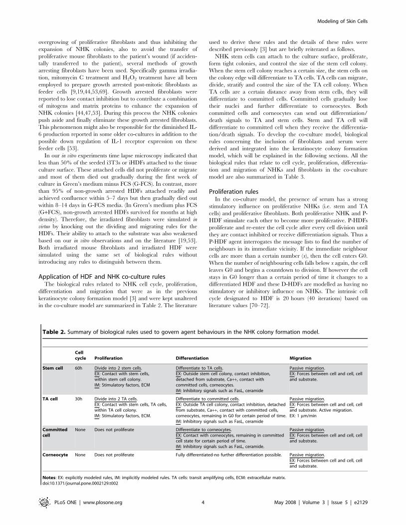
Application of HDF and NHK co-culture rulesThe biological rules related to NHK cell cycle, proliferation,
differentiation and migration that were as in the previous
keratinocyte colony formation model [3] and were kept unaltered
in the co-culture model are summarized in Table 2. The literature
used to derive these rules and the details of these rules were
described previously [3] but are briefly reiterated as follows.
NHK stem cells can attach to the culture surface, proliferate,
form tight colonies, and control the size of the stem cell colony.
When the stem cell colony reaches a certain size, the stem cells on
the colony edge will differentiate to TA cells. TA cells can migrate,
divide, stratify and control the size of the TA cell colony. When
TA cells are a certain distance away from stem cells, they will
differentiate to committed cells. Committed cells gradually lose
their nuclei and further differentiate to corneocytes. Both
committed cells and corneocytes can send out differentiation/
death signals to TA and stem cells. Stem and TA cell will
differentiate to committed cell when they receive the differentia-
tion/death signals. To develop the co-culture model, biological
rules concerning the inclusion of fibroblasts and serum were
derived and integrated into the keratinocyte colony formation
model, which will be explained in the following sections. All the
biological rules that relate to cell cycle, proliferation, differentia-
tion and migration of NHKs and fibroblasts in the co-culture
model are also summarized in Table 3.
Proliferation rulesIn the co-culture model, the presence of serum has a strong
stimulatory influence on proliferative NHKs (i.e. stem and TA
cells) and proliferative fibroblasts. Both proliferative NHK and P-
HDF stimulate each other to become more proliferative. P-HDFs
proliferate and re-enter the cell cycle after every cell division until
they are contact inhibited or receive differentiation signals. Thus a
P-HDF agent interrogates the message lists to find the number of
neighbours in its immediate vicinity. If the immediate neighbour
cells are more than a certain number (x), then the cell enters G0.
When the number of neighbouring cells falls below x again, the cell
leaves G0 and begins a countdown to division. If however the cell
stays in G0 longer than a certain period of time it changes to a
differentiated HDF and these D-HDFs are modelled as having no
stimulatory or inhibitory influence on NHKs. The intrinsic cell
cycle designated to HDF is 20 hours (40 iterations) based on
literature values [70–72].
Table 2. Summary of biological rules used to govern agent behaviours in the NHK colony formation model.
Cellcycle Proliferation Differentiation Migration
Stem cell 60h Divide into 2 stem cells.EX: Contact with stem cells,within stem cell colony.IM: Stimulatory factors, ECM
Differentiate to TA cells.EX: Outside stem cell colony, contact inhibition,detached from substrate, Ca++, contact withcommitted cells, corneocytes.IM: Inhibitory signals such as FasL, ceramide
Passive migration.EX: Forces between cell and cell, celland substrate.
TA cell 30h Divide into 2 TA cells.EX: Contact with stem cells, TA cells,within TA cell colony.IM: Stimulatory factors, ECM.
Differentiate to committed cells.EX: Outside TA cell colony, contact inhibition, detachedfrom substrate, Ca++, contact with committed cells,corneocytes, remaining in G0 for certain period of time.IM: Inhibitory signals such as FasL, ceramide
Passive migration.EX: Forces between cell and cell, celland substrate. Active migration.EX: 1 mm/min
Committedcell
None Does not proliferate Differentiate to corneocytes.EX: Contact with corneocytes, remaining in committedcell state for certain period of time.IM: Inhibitory signals such as FasL, ceramide.
Passive migration.EX: Forces between cell and cell, celland substrate.
Corneocyte None Does not proliferate Fully differentiated-no further differentiation possible. Passive migration.EX: Forces between cell and cell, celland substrate.
Notes: EX: explicitly modeled rules, IM: implicitly modeled rules. TA cells: transit amplifying cells, ECM: extracellular matrix.doi:10.1371/journal.pone.0002129.t002
Modeling of Skin Cells
PLoS ONE | www.plosone.org 4 May 2008 | Volume 3 | Issue 5 | e2129

Differentiation rulesThe following differentiation mechanisms were modelled for
fibroblasts: (1) contact inhibition [2]; (2) presence of differentia-
tion/apoptosis signals, such as ceramide [27] and Fas/L-Fas [73],
(3) loss of cell-matrix contact [74–78]; (4) remaining in a quiescent
state (G0) for longer than a certain period of time [79]. Most of the
NHK/HDF differentiation rules still centre on NHK colony
formation. In addition to an auto-regulation mechanism, the
NHK colony size and the differentiation process are also subject to
regulation by serum and HDF. Our in vitro time lapse experiments
indicated that few NHKs can attach, proliferate or form NHK
colonies on a normal tissue culture plastic surface when single
cultured in serum free Greens media (G-FCS). However, the
presence of HDFs can help NHKs to survive in G-FCS and even
form bigger colonies. The influence of serum and HDF on NHK
was implemented in the co-culture model as follows: Apart from
the auto-regulated keratinocyte colony formation mechanism,
NHK stem cells also check for the presence of serum and HDF.
With serum present, NHKs can attach to the substrate and form
colonies as modelled previously. The presence of HDF, especially
the presence of immediate P-HDF neighbours, can help NHKs to
attach and form colonies in G-FCS. The more immediate P-HDF
neighbours available or the more contacts they make with P-HDF,
the more proliferative the stem cell will be. Consequently the stem
cell colony will be bigger. If the number of stem cell contacts is
relatively low (indicating it is on the edge of the culture), the stem
cell will differentiate into a TA cell.
TA cells undertake a similar process as to whether they should
differentiate into a committed cell by monitoring the presence of
serum and P-HDF. Both serum and P-HDF have stimulatory
effects on TA cells. Stem and TA cells also slightly increase the
proliferative capacity of P-HDF. Both committed cells and
corneocytes send out differentiation signals (e.g. ceramide or
Fas-L) to the neighbouring cells, P-HDFs differentiate to D-HDFs
when they receive these signals. D-HDFs have no stimulatory
effects on either stem or TA cells. (See Table 3 for a summary of
these rule sets).
Strength of cell binding and migration rulesBond strengths between HDF and substrate are modelled as
relatively weak compared with NHK-substrate bonds since HDFs
can be easily pushed or detached by NHK as demonstrated by
time-lapse video microscopy. As HDF were uniformly distributed
on the tissue culture surface and no tight HDF colonies were
observed in in vitro cell culture especially when the cultures were
subconfluent, repelling forces were thus applied among HDFs.
Repelling forces were also applied between HDF and NHK, since
time lapse experiments also demonstrated contact induced
inhibition of movement between HDFs and NHKs.
As the actions of cell migration and division are modelled as
discrete steps that are applied to each agent individually there is a
possibility that the simulated cells may overlap on the virtual
culture plate. In this case, a corrective repulsive force is applied in
order to push the cells apart. This is proportional to the
Table 3. Summary of biological rules used to govern agent behaviours in the NHK/HDF co-culture model.
Cell cycle Proliferation Differentiation Migration
Stem cell 60h Divide into 2 stem cells.EX: Contact with stem cells, withinstem cell colony, presence of serumand/or HDF, contact with P-HDF,coated substrate.IM: Stimulatory factors.
Differentiate to TA cells.EX: Outside stem cell colony, contact inhibition,detached from substrate, Ca++, contact with committedcells, corneocytes, absence of serum and/or HDF.IM: Inhibitory signals such as FasL, ceramide
Passive migration.EX: Forces between cell andcell, cell and substrate.
TA cell 30h Divide into 2 TA cells.EX: Contact with stem cells, TA cells,within TA cell colony, presence ofserum and/or HDF, contact with P-HDF,coated substrate.IM: Stimulatory factors.
Differentiate to committed cells.EX: Outside TA cell colony, contact inhibition, detachedfrom substrate, Ca++, contact with committed cells,corneocytes, remaining in G0 for certain period of time,absence of serum and/or HDF.IM: Inhibitory signals such as FasL, ceramide
Passive migration.EX: Forces between cell andcell, cell and substrate. Activemigration.EX: 1 mm/min
Committed cell None Does not proliferate Differentiate to corneocytes.EX: Contact with corneocytes, remaining in committedcell state for certain period of time, absence of serumand/or HDF.IM: Inhibitory signals such as FasL, ceramide.
Passive migration.EX: Forces between cell andcell, cell and substrate.
Corneocyte None Does not proliferate Fully differentiated-no further differentiation possible. Passive migration.EX: Forces between cell andcell, cell and substrate.
P-HDF 20h Divide into 2 P-HDFs.EX: Contact with stem cells, TA cells,P-HDF, presence of serum and or stemcells and TA cells.IM: Stimulatory factors.
Differentiate to D-HDFs.EX: Contact inhibition, detached from substrate,contact with committed cells, corneocytes, D-HDFs,remaining in G0 for certain period of time, absence ofserum and/or stem cells, TA cells.IM: Inhibitory signals such as FasL, ceramide.
Passive migration.EX: Forces between cell andcell, cell and substrate. Activemigration.EX: 1 mm/min
D-HDF None Does not proliferate Apoptosis or detachment of D-HDFs.EX: Detached by NHKs, remaining in D-HDF state forcertain period of time.
Passive migration.EX: Forces between cell andcell, cell and substrate. Activemigration.EX: 1 mm/min
Notes: EX: explicitly modeled rules, IM: implicitly modeled rules. TA cells: transit amplifying cells, P-HDF: proliferative human dermal fibroblast. D-HDF: differentiatedhuman dermal fibroblast, ECM: extracellular matrix.doi:10.1371/journal.pone.0002129.t003
Modeling of Skin Cells
PLoS ONE | www.plosone.org 5 May 2008 | Volume 3 | Issue 5 | e2129
overlapping area (a higher force for a bigger overlap). If there is no
free surface remaining for cells to move to then an apoptosis
mechanism is applied to HDFs so that essentially the modelled
HDFs are lost from the model.
The essential mechanisms responsible for cell migration are
mainly a cell density driven pressure passive movement [80–81] as
well as a cell active movement. The cell passive movement is
caused by cell-cell or cell-substrate interactions and mitotic
activity. Accordingly a cell passive movement was applied to each
agent corresponding to the forces acting on it. An active migration
speed of approximately 1 mm/minute was also applied to HDF
based on time lapse experiments and literature [70]. (Summarised
in Table 3).
Apoptosis rulesSingle cultures of HDF in G-FCS showed that HDF viability
decreased rapidly after culture for 5–7 days [7–8]. Therefore, the
absence of serum in single HDF culture was assumed to cause
HDF apoptosis. Time lapse experiments of HDF-NHK co-
cultures demonstrated that NHK colonies were surrounded by
HDF and that the NHK colonies push the HDF away. No single
HDFs were observed inside NHK colonies. Accordingly, the
apoptosis or detachment of HDF was explicitly modelled as
follows-after a direct contact with NHK, the HDF will be pushed
away by NHK. If there is no free surface for HDF to migrate to,
the HDF agent will then be removed from the model by simulated
apoptosis or detachment.
The code for this model will be made available from: http://
www.flame.ac.uk.
Results
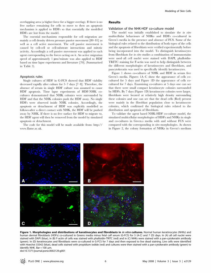
Validation of the NHK-HDF co-culture modelThe model was initially established to simulate the in vitro
multicellular behaviours of NHKs and HDFs co-cultured in
Green’s media in the presence and absence of FCS. Some of the
biological rules related to the distribution of both types of the cells
and the apoptosis of fibroblasts were verified experimentally before
being incorporated into the model. To distinguish keratinocytes
from fibroblasts for in vitro studies a combination of immunostains
were used–all cell nuclei were stained with DAPI, phalloidin-
TRITC staining for F-actin was used to help distinguish between
the different morphologies of keratinocytes and fibroblasts, and
pancytokeratin was used to specifically identify keratinocytes.
Figure 1 shows co-cultures of NHK and HDF in serum free
Green’s media. Figures 1A–C show the appearance of cells co-
cultured for 5 days and Figure 1D the appearance of cells co-
cultured for 7 days. Examining co-cultures at 5 days one can see
that there were small compact keratinocyte colonies surrounded
by HDFs. By 7 days (Figure 1D) keratinocyte colonies were larger,
fibroblasts were located at relatively high density surrounding
these colonies and one can see that the dead cells (Red) present
were mainly in the fibroblast population close to keratinocyte
colonies, which confirmed the biological rules related to the
distribution and apoptosis of fibroblasts.
To validate the agent based NHK-HDF co-culture model, the
simulated multicellular morphologies of HDFs and NHKs in single
and co-cultures in Green.s media with and without FCS were
compared with the corresponding in vitro morphologies. As shown
in Figure 2, the colony formation of NHKs in Green’s medium
Figure 1. Morphologies and distributions of keratinocytes and fibroblasts in in vitro cultures. Normal human keratinocytes (NHKs) andhuman dermal fibroblasts (HDFs) co-cultured in Greens media minus fetal calf serum (G-FCS) for 5 (A-C) and 7 (D) days. In (A) all cell nuclei werestained with DAPI (blue), in (B) F-actin of cells was stained with phalloidin-TRITC (red) and in (C) NHKs were stained with a pan-cytokeratin antibody(green). In (D) keratinocytes and fibroblasts were co-cultured in G-FCS for 7 days and then exposed to live dead staining. Live cells were identifiedwith Hoechst 33342 (blue), dead cells stained with propidium iodide (red) and cultures were then stained with a pan-cytokeratin antibody (green) toidentify NHK. Bar = 100 mm.doi:10.1371/journal.pone.0002129.g001
Modeling of Skin Cells
PLoS ONE | www.plosone.org 6 May 2008 | Volume 3 | Issue 5 | e2129
plus FCS (G+FCS) (Figure 2A) was reproduced in the in virtuo
simulation (Figure 2B). For NHK in monoculture lacking FCS (G-
FCS) hardly any cells survived by 8 days (Figure 1 C). NHK stem
cells in G-FCS were modelled to rapidly withdraw from the cell
cycle and differentiate to committed cells and then become
corneocytes due to the lack of FCS (Figure 2D).
Time lapse experiments indicated that HDFs could initially
survive and proliferate to confluence after culture for 5–7 days in
Green’s media both with and without FCS. As the cultures
continued to 8–14 days, HDFs in G+FCS remained confluent
(Figure 2E), while the cells in G-FCS gradually died out
(Figure 2G). The influence of serum on the proliferation/apoptosis
of HDFs was simulated by the agent based model as shown in
Figure 2F and H. Fibroblasts were depicted as proliferative (pink)
or differentiated (red) in this simulation.
In co-culture, the absence of FCS had relatively little effect on
the expansion of keratinocyte colonies due to the dynamic
interactions between NHKs and HDFs. The macroscopic
morphogenesis of NHKs and HDFs co-cultured in Greens media
with or without FCS were compared using both in vitro and in virtuo
models over 8 days as shown in Figure 3 and very similar results
were obtained in terms of multicellular morphologies and the
Figure 2. In vitro monocultures and the corresponding simulations of keratinocytes and fibroblasts. In virtuo simulations of the growthof normal human keratinocytes (NHKs) and human dermal fibroblasts (HDFs) in monocultures in the presence and absence of foetal calf serum (FCS).(A) and (C) show micrographs of NHKs single cultured for 8 days in Greens medium with and without FCS respectively, while (B) and (D) show thecorresponding model simulations. (E) and (G) show micrographs of HDFs single cultured for 8 days in Greens media with or lacking FCS, while (F) and(H) show the corresponding in virtuo simulation. The initial numbers of keratinocyte stem cells and fibroblasts were both defined as 10 within theuser-defined flat surface (500 mm6500 mm). In the agent based model different colours were used to represent keratinocyte stem cells (blue), TA cells(light green), committed cells (dark green), corneocytes (brown), proliferative fibroblasts (pink) and differentiated fibroblasts (red). Bar = 100 mm.doi:10.1371/journal.pone.0002129.g002
Modeling of Skin Cells
PLoS ONE | www.plosone.org 7 May 2008 | Volume 3 | Issue 5 | e2129
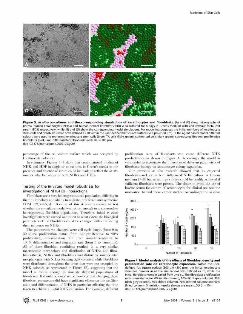
percentage of the cell culture surface which was occupied by
keratinocyte colonies.
In summary, Figures 1–3 show that computational models of
NHK and HDF in single or co-cultures in Green’s media in the
presence and absence of serum could be made to reflect the in vitro
multicellular behaviour of both NHKs and HDFs.
Testing of the in virtuo model robustness forinvestigation of NHK-HDF interactions
Fibroblasts are a very heterogeneous cell population, differing in
their morphology and ability to migrate, proliferate and synthesize
ECM [22,32,65,64]. Because of this it was necessary to test
whether the co-culture model was robust enough to accommodate
heterogeneous fibroblast populations. Therefore, initial in virtuo
investigations were carried out to test to what extent the biological
parameters of the fibroblasts could be changed without affecting
their influence on NHKs.
The parameters we changed were cell cycle length (from 0 to
30 hours) proliferation status (from non-proliferative to 90%
proliferative), differentiation rate (from non-differentiative to
100% differentiative) and migration rate (from 0 to 1um/min).
All of these fibroblast conditions resulted in a very similar
macroscopic morphology and distribution of NHKs and fibro-
blasts-that is, NHKs and fibroblasts had distinctive multicellular
morphologies with NHKs forming tight colonies, while fibroblasts
were distributed throughout the areas that were not occupied by
NHK colonies (as represented in Figure 3B), suggesting that the
model is robust enough to simulate different populations of
fibroblasts. It should be emphasized however that changing these
fibroblast parameters did have significant effects on the prolifer-
ation and differentiation of NHK in particular affecting the time
taken to achieve a useful NHK expansion. For example, different
proliferation rates of fibroblasts can cause different NHK
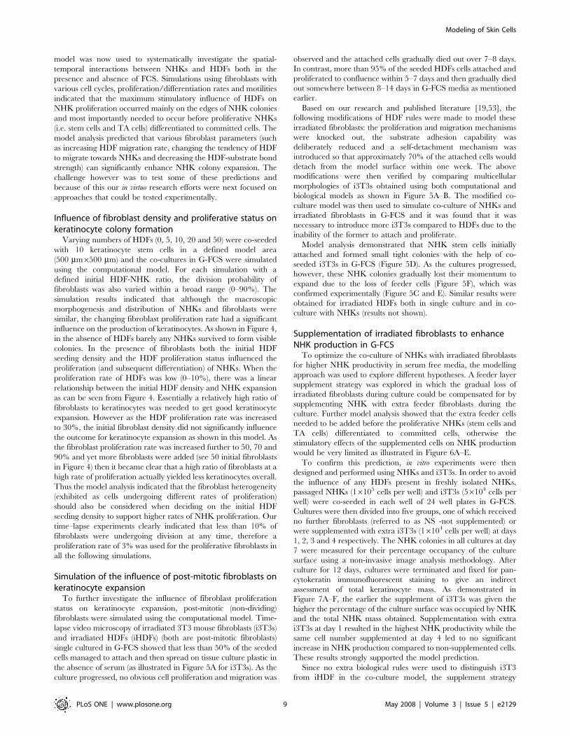
productivities as shown in Figure 4. Accordingly the model is
very useful to investigate the influences of different parameters of
fibroblasts biology on keratinocyte colony expansion.
Our previous in vitro research showed that as expected
fibroblasts and serum both influenced NHK culture in Greens
medium [7–8] but serum free culture could be readily achieved if
sufficient fibroblasts were present. The desire to avoid the use of
bovine serum for culture of keratinocytes for clinical use was the
motivation behind these earlier studies. Accordingly the in virtuo
Figure 3. In vitro co-cultures and the corresponding simulations of keratinocytes and fibroblasts. (A) and (C) show micrographs ofnormal human keratinocytes (NHKs) and human dermal fibroblasts (HDFs) co-cultured for 8 days in Greens medium with and without foetal calfserum (FCS) respectively, while (B) and (D) show the corresponding model simulations. For modelling purposes the initial numbers of keratinocytestem cells and fibroblasts were both defined as 10 within the user-defined flat square surface (500 mm6500 mm). In the agent based model differentcolours were used to represent keratinocyte stem cells (blue), TA cells (light green), committed cells (dark green), corneocytes (brown), proliferativefibroblasts (pink) and differentiated fibroblasts (red). Bar = 100 mm.doi:10.1371/journal.pone.0002129.g003
Figure 4. Model analysis of the effects of fibroblast density andproliferation rate on keratinocyte expansion. Within the user-defined flat square surface (500 mm6500 mm), the initial keratinocytestem cell number in all the simulations was defined as 10, while theinitial fibroblast number varied from 0 to 50. The fibroblast proliferationrates simulated were: 0% (white column), 10% (light grey column), 30%(dark grey column), 50% (black column), 70% (dotted column) and 90%(lined column). Simulation results shown are mean6SD (n = 10).doi:10.1371/journal.pone.0002129.g004
Modeling of Skin Cells
PLoS ONE | www.plosone.org 8 May 2008 | Volume 3 | Issue 5 | e2129
model was now used to systematically investigate the spatial-
temporal interactions between NHKs and HDFs both in the
presence and absence of FCS. Simulations using fibroblasts with
various cell cycles, proliferation/differentiation rates and motilities
indicated that the maximum stimulatory influence of HDFs on
NHK proliferation occurred mainly on the edges of NHK colonies
and most importantly needed to occur before proliferative NHKs
(i.e. stem cells and TA cells) differentiated to committed cells. The
model analysis predicted that various fibroblast parameters (such
as increasing HDF migration rate, changing the tendency of HDF
to migrate towards NHKs and decreasing the HDF-substrate bond
strength) can significantly enhance NHK colony expansion. The
challenge however was to test some of these predictions and
because of this our in virtuo research efforts were next focused on
approaches that could be tested experimentally.
Influence of fibroblast density and proliferative status onkeratinocyte colony formation
Varying numbers of HDFs (0, 5, 10, 20 and 50) were co-seeded
with 10 keratinocyte stem cells in a defined model area
(500 mm6500 mm) and the co-cultures in G-FCS were simulated
using the computational model. For each simulation with a
defined initial HDF-NHK ratio, the division probability of
fibroblasts was also varied within a broad range (0–90%). The
simulation results indicated that although the macroscopic
morphogenesis and distribution of NHKs and fibroblasts were
similar, the changing fibroblast proliferation rate had a significant
influence on the production of keratinocytes. As shown in Figure 4,
in the absence of HDFs barely any NHKs survived to form visible
colonies. In the presence of fibroblasts both the initial HDF
seeding density and the HDF proliferation status influenced the
proliferation (and subsequent differentiation) of NHKs. When the
proliferation rate of HDFs was low (0–10%), there was a linear
relationship between the initial HDF density and NHK expansion
as can be seen from Figure 4. Essentially a relatively high ratio of
fibroblasts to keratinocytes was needed to get good keratinocyte
expansion. However as the HDF proliferation rate was increased
to 30%, the initial fibroblast density did not significantly influence
the outcome for keratinocyte expansion as shown in this model. As
the fibroblast proliferation rate was increased further to 50, 70 and
90% and yet more fibroblasts were added (see 50 initial fibroblasts
in Figure 4) then it became clear that a high ratio of fibroblasts at a
high rate of proliferation actually yielded less keratinocytes overall.
Thus the model analysis indicated that the fibroblast heterogeneity
(exhibited as cells undergoing different rates of proliferation)
should also be considered when deciding on the initial HDF
seeding density to support higher rates of NHK proliferation. Our
time–lapse experiments clearly indicated that less than 10% of
fibroblasts were undergoing division at any time, therefore a
proliferation rate of 3% was used for the proliferative fibroblasts in
all the following simulations.
Simulation of the influence of post-mitotic fibroblasts onkeratinocyte expansion
To further investigate the influence of fibroblast proliferation
status on keratinocyte expansion, post-mitotic (non-dividing)
fibroblasts were simulated using the computational model. Time-
lapse video microscopy of irradiated 3T3 mouse fibroblasts (i3T3s)
and irradiated HDFs (iHDFs) (both are post-mitotic fibroblasts)
single cultured in G-FCS showed that less than 50% of the seeded
cells managed to attach and then spread on tissue culture plastic in
the absence of serum (as illustrated in Figure 5A for i3T3s). As the
culture progressed, no obvious cell proliferation and migration was
observed and the attached cells gradually died out over 7–8 days.
In contrast, more than 95% of the seeded HDFs cells attached and
proliferated to confluence within 5–7 days and then gradually died
out somewhere between 8–14 days in G-FCS media as mentioned
earlier.
Based on our research and published literature [19,53], the
following modifications of HDF rules were made to model these
irradiated fibroblasts: the proliferation and migration mechanisms
were knocked out, the substrate adhesion capability was
deliberately reduced and a self-detachment mechanism was
introduced so that approximately 70% of the attached cells would
detach from the model surface within one week. The above
modifications were then verified by comparing multicellular
morphologies of i3T3s obtained using both computational and
biological models as shown in Figure 5A–B. The modified co-
culture model was then used to simulate co-culture of NHKs and
irradiated fibroblasts in G-FCS and it was found that it was
necessary to introduce more i3T3s compared to HDFs due to the
inability of the former to attach and proliferate.
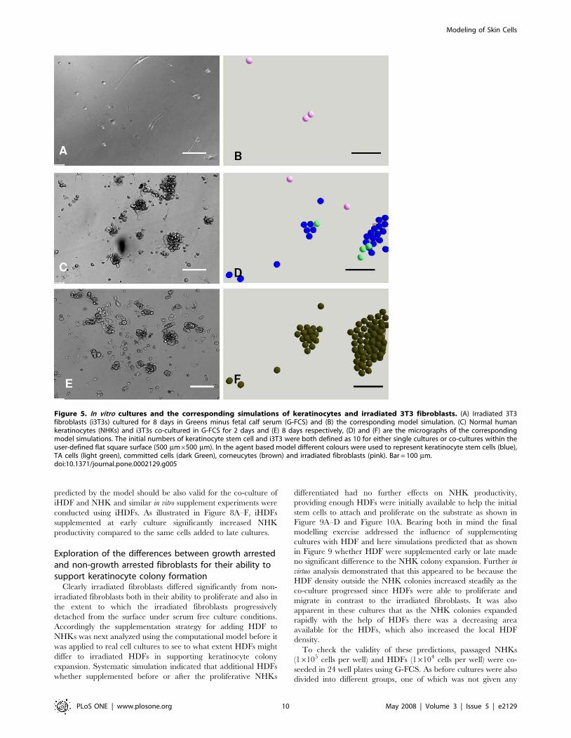
Model analysis demonstrated that NHK stem cells initially
attached and formed small tight colonies with the help of co-
seeded i3T3s in G-FCS (Figure 5D). As the cultures progressed,
however, these NHK colonies gradually lost their momentum to
expand due to the loss of feeder cells (Figure 5F), which was
confirmed experimentally (Figure 5C and E). Similar results were
obtained for irradiated HDFs both in single culture and in co-
culture with NHKs (results not shown).
Supplementation of irradiated fibroblasts to enhanceNHK production in G-FCS
To optimize the co-culture of NHKs with irradiated fibroblasts
for higher NHK productivity in serum free media, the modelling
approach was used to explore different hypotheses. A feeder layer
supplement strategy was explored in which the gradual loss of
irradiated fibroblasts during culture could be compensated for by
supplementing NHK with extra feeder fibroblasts during the
culture. Further model analysis showed that the extra feeder cells
needed to be added before the proliferative NHKs (stem cells and
TA cells) differentiated to committed cells, otherwise the
stimulatory effects of the supplemented cells on NHK production
would be very limited as illustrated in Figure 6A–E.
To confirm this prediction, in vitro experiments were then
designed and performed using NHKs and i3T3s. In order to avoid
the influence of any HDFs present in freshly isolated NHKs,
passaged NHKs (16105 cells per well) and i3T3s (56104 cells per
well) were co-seeded in each well of 24 well plates in G-FCS.
Cultures were then divided into five groups, one of which received
no further fibroblasts (referred to as NS -not supplemented) or
were supplemented with extra i3T3s (16104 cells per well) at days
1, 2, 3 and 4 respectively. The NHK colonies in all cultures at day
7 were measured for their percentage occupancy of the culture
surface using a non-invasive image analysis methodology. After
culture for 12 days, cultures were terminated and fixed for pan-
cytokeratin immunofluorescent staining to give an indirect
assessment of total keratinocyte mass. As demonstrated in
Figure 7A–F, the earlier the supplement of i3T3s was given the
higher the percentage of the culture surface was occupied by NHK
and the total NHK mass obtained. Supplementation with extra
i3T3s at day 1 resulted in the highest NHK productivity while the
same cell number supplemented at day 4 led to no significant
increase in NHK production compared to non-supplemented cells.
These results strongly supported the model prediction.
Since no extra biological rules were used to distinguish i3T3
from iHDF in the co-culture model, the supplement strategy
Modeling of Skin Cells
PLoS ONE | www.plosone.org 9 May 2008 | Volume 3 | Issue 5 | e2129
predicted by the model should be also valid for the co-culture of
iHDF and NHK and similar in vitro supplement experiments were
conducted using iHDFs. As illustrated in Figure 8A–F, iHDFs
supplemented at early culture significantly increased NHK
productivity compared to the same cells added to late cultures.
Exploration of the differences between growth arrestedand non-growth arrested fibroblasts for their ability tosupport keratinocyte colony formation
Clearly irradiated fibroblasts differed significantly from non-
irradiated fibroblasts both in their ability to proliferate and also in
the extent to which the irradiated fibroblasts progressively
detached from the surface under serum free culture conditions.
Accordingly the supplementation strategy for adding HDF to
NHKs was next analyzed using the computational model before it
was applied to real cell cultures to see to what extent HDFs might
differ to irradiated HDFs in supporting keratinocyte colony
expansion. Systematic simulation indicated that additional HDFs
whether supplemented before or after the proliferative NHKs
differentiated had no further effects on NHK productivity,
providing enough HDFs were initially available to help the initial
stem cells to attach and proliferate on the substrate as shown in
Figure 9A–D and Figure 10A. Bearing both in mind the final
modelling exercise addressed the influence of supplementing
cultures with HDF and here simulations predicted that as shown
in Figure 9 whether HDF were supplemented early or late made
no significant difference to the NHK colony expansion. Further in
virtuo analysis demonstrated that this appeared to be because the
HDF density outside the NHK colonies increased steadily as the
co-culture progressed since HDFs were able to proliferate and
migrate in contrast to the irradiated fibroblasts. It was also
apparent in these cultures that as the NHK colonies expanded
rapidly with the help of HDFs there was a decreasing area
available for the HDFs, which also increased the local HDF
density.
To check the validity of these predictions, passaged NHKs
(16105 cells per well) and HDFs (16104 cells per well) were co-
seeded in 24 well plates using G-FCS. As before cultures were also
divided into different groups, one of which was not given any
Figure 5. In vitro cultures and the corresponding simulations of keratinocytes and irradiated 3T3 fibroblasts. (A) Irradiated 3T3fibroblasts (i3T3s) cultured for 8 days in Greens minus fetal calf serum (G-FCS) and (B) the corresponding model simulation. (C) Normal humankeratinocytes (NHKs) and i3T3s co-cultured in G-FCS for 2 days and (E) 8 days respectively, (D) and (F) are the micrographs of the correspondingmodel simulations. The initial numbers of keratinocyte stem cell and i3T3 were both defined as 10 for either single cultures or co-cultures within theuser-defined flat square surface (500 mm6500 mm). In the agent based model different colours were used to represent keratinocyte stem cells (blue),TA cells (light green), committed cells (dark Green), corneucytes (brown) and irradiated fibroblasts (pink). Bar = 100 mm.doi:10.1371/journal.pone.0002129.g005
Modeling of Skin Cells
PLoS ONE | www.plosone.org 10 May 2008 | Volume 3 | Issue 5 | e2129

further HDFs and others were given 16104 extra HDFs per well
on days 1, 2, 3 and 4 respectively. After culture for 7 days the
percentage of keratinocyte colonies occupying the culture surface
was assessed and then at 12 days all cultures were terminated and
pan-cytokeratin was used to get an indication of total keratinocyte
mass. The results are shown in Figure 10 and here it can be seen
that adding extra HDF at days 1 through to 4 had no further effect
on the keratinocyte colonies on the culture surface (Figure 10B) or
overall keratinocyte mass (Figure 10C) as predicted by the in virtuo
modelling. However, the key point to note is that keratinocyte
expansion on HDF was far superior to NHK expansion on iHDF
or i3T3’s even when these cells were initially added at 5 times
higher density. Thus as summarised in Table 4 at least 80% of the
culture well was occupied with keratinocyte colonies (Figure 10B)
within 7 days if these were initially co-cultured with proliferative
HDFs at a ratio of 1 HDF to 10 NHK. A later supplement (after
24 hours) of further HDF did not imrove on this already high rate
of NHK expansion, In contrast if i3T3 and iHDF cells were used
at a ratio of 5 cells to 10 NHK then the rate of NHK expansion
was poor (,10%) but could be improved to 30–55% by the
addition of extra fibroblasts at 24 h (as shown in Figure 7E)). In
summary, the data provide a logical case for using proliferative
Figure 6. Model analysis of the supplement of extra irradiated fibroblasts when cocultured with keratinocytes. Extra irradiatedfibroblasts (50) were added to a co-culture of keratinocytes and irradiated fibroblasts in serum free Greens media (A) before the keratinocyte stemcells started to differentiate and then (B) simulated for 7 more days. In (C) the extra irradiated fibroblasts (50) were supplemented after thekeratinocytes had started to differentiate and then (D) simulated for a further 7 days. In the agent based model different colours were used torepresent keratinocyte stem cells (blue), TA cells (light green), committed cells (dark green), corneocytes (brown) and irradiated fibroblasts (pink).Bar = 100 mm. (E) shows the numerical effect of adding extra irradiated fibroblasts at different time points (iterations in the model) on keratinocyteproliferation. The simulation results shown are mean6SD (n = 10). The initial numbers of keratinocyte stem cells and irradiated fibroblasts in all theabove simulations were both defined as 10 within the user-defined flat square surface (500 mm6500 mm), while 50 extra irradiated fibroblasts weresupplemented at the times (iterations) indicated.doi:10.1371/journal.pone.0002129.g006
Modeling of Skin Cells
PLoS ONE | www.plosone.org 11 May 2008 | Volume 3 | Issue 5 | e2129
fibroblasts rather than irradiated fibroblasts to drive keratinocyte
colony formation.
Discussion
The regulation of epidermal homeostasis involves a complex
interplay between different generic and genetic mechanisms, making
it difficult for biologists to investigate except by focusing on separate
discrete aspects. Fortunately, computational modelling approaches
can handle this complexity [1–2,82–83] and agent-based modelling
in particular is gathering popularity with biologists [84]. The idea of
this modelling approach is to abstract away micro-level details to find
a minimal set of properties sufficient to simulate the local interactions
of components of a system to produce a global behaviour model
which then allows the testing of hypotheses and the designing of new
informative experiments.
Previously we developed an extensible agent-based NHK
colony formation model [3]. In the current study, biological rules
of serum, HDF and their influence on NHK were abstracted from
an extensive published literature and our own in vitro experiments
and then incorporated into the model to simulate the complex
interactions between epidermal-dermal cells. The validity of the
co-culture model was tested by comparing the simulation results of
HDFs and NHKs in single or co-cultures in different environments
with corresponding in vitro experimentation. The results from both
in virtuo and in vitro models indicated that the crucial roles of serum
and fibroblasts in the production of NHK were successfully
simulated by the co-culture model.
As the literature indicates that fibroblasts actually represent a
very heterogeneous cell population [22,32,64–65], several biolog-
ical parameters (cell cycle length, the rates of proliferation,
differentiation and migration) were varied in virtuo to examine
Figure 7. In vitro analysis of the supplement of extra irradiated mouse fibroblasts when cocultured with keratinocytes. (A-D)Micrographs of normal human keratinocytes (NHK) co-cultured with irradiated mouse fibroblasts (i3T3) in serum free Greens medium when extrai3T3s were not supplemented (NS) during the culture, supplemented at day 1(A), 2 (B), 3 (C) or 4 (D) respectively and cultured for 12 days.Bar = 100 mm. Effect of the addition of extra i3T3s at different culture time periods on (E) the percentage of the tissue culture well occupied by NHKcolonies after culture for 7 days and (F) the expression of pan-cytokeratin in NHKs (assessed as an indirect indicator of keratinocyte mass)after culturefor 12 days. Results shown are mean6SD (n = 3).doi:10.1371/journal.pone.0002129.g007
Modeling of Skin Cells
PLoS ONE | www.plosone.org 12 May 2008 | Volume 3 | Issue 5 | e2129

whether the model was robust enough to simulate the different
behaviour of fibroblasts. Our criterion for judging the robustness
of the model was whether the multicellular morphology of NHK-
HDF co-culture could still be simulated when these fibroblast
parameters were varied within broad ranges. Our results
demonstrated that changing the parameters within very broad
ranges or knocking-out some of rules of fibroblasts all ultimately
resulted in very similar macroscopic morphology and distribution
of NHKs and fibroblasts but the time to achieve NHK expansion
was affected as was keratinocyte expansion. In this study the co-
culture model was used to explore different hypotheses of NHK-
HDF interactions to optimize the speed of NHK expansion for
higher productivity for clinical use. Many more in virtuo studies
could now be undertaken with this model to examine for example
how important the different variables are to the expansion of
NHK.
From a modelling perspective the exercise of designing an agent
based approach to understanding fibroblast/keratinocyte interac-
tions allowed us to formulate and test hypotheses in virtuo some of
which we could then test in vitro. The main ‘‘deliverable’’ of this study
is a rational explanation for the differing behaviours of proliferative
versus growth-arrested fibroblasts in supporting keratinocyte expan-
sion. Also as it would be good to avoid the use of bovine serum when
culturing cells for clinical use, a key focus in our integrated in virtuo
and in vitro approaches was the examination of NHK/HDF co-
culture under serum free conditions.
Simulation of the temporal-spatial NHK-HDF dynamic inter-
actions indicated that the presence of HDF on the edges of the
auto-regulated NHK colonies at different culture periods was
crucial for maximum NHK production. Model analysis indicated
that the initial HDF/NHK ratio had a significant influence on the
proliferation/differentiation of NHK, which confirmed our
Figure 8. In vitro analysis of the supplement of irradiated human dermal fibroblasts when cocultured with keratinocytes. (A-D)Micrographs of normal human keratinocytes (NHK) co-cultured with irradiated human dermal fibroblasts (iHDF) in serum free Greens medium whenextra iHDFs were not supplemented (NS) during the culture, supplemented at day 1(A) ,2 (B), 3 (C) and 4 (D) respectively and cultured for 12 days.Bar = 100 mm. Effect of the addition of extra iHDFs at different culture time periods on (E) the percentage of the tissue culture well occupied by NHKcolonies after culture for 7 days and (F) the expression of pan-cytokeratin in NHKs ( assessed as an indirect indicator of keratinocyte mass )afterculture for 12 days. Results shown are mean6SD (n = 3).doi:10.1371/journal.pone.0002129.g008
Modeling of Skin Cells
PLoS ONE | www.plosone.org 13 May 2008 | Volume 3 | Issue 5 | e2129

previous in vitro research [7]. The model also predicted that the
rate of fibroblast proliferation would have a major influence on
NHK colony production. This has not been directly examined
before to the best of our knowledge.
Ever since the seminal work of Rheinwald and Green in
1975 describing a method to culture adult human keratinocytes
using irradiated murine feeder fibroblasts and a mitogen-rich
media [4], feeder cells (i3T3s and iHDFs) have usually been
used as post-mitotic fibroblasts [9,19,53,69] to culture NHKs
for both research and clinical purposes. To model these lethally
irradiated cells three changes were made to the model of
normal fibroblasts- proliferation and migration mechanisms
were knocked out and a new rule of self-detachment based on
literature findings was introduced [19]. The model was
validated by comparing the behaviour of i3T3 and iHDF cells
in virtuo with their behaviour in in vitro time lapse experiments
in single culture in serum free Greens medium. The modified
co-culture model was then used to simulate the co-culture of
NHK and irradiated fibroblasts in G-FCS and the limitations
of these feeder cells to enhance NHK production were
explored in virtuo. Basically most of the irradiated cells in the
presence of expanding NHK colonies were unable to survive
for long (1 week) due to the detachment applied by the
expanding NHK colonies and the self-detachment of the
irradiated cells [19,53].
The model predicted that the addition of more feeder cells
would assist the continuing expansion of NHK colonies but that
this would only work for keratinocytes in a proliferative phase. In
vitro experiments were then designed to examine to what extent the
prediction would hold true. As predicted adding extra i3T3s and
iHDFs at an early keratinocyte culture stage resulted in
significantly higher NHK productivity than if these cultures were
supplemented with fibroblasts once keratinocytes had begun to
differentiate.
The in virtuo effort was then refocused on the optimization of
HDF-NHK co-culture and the same supplementation strategy was
tested. Simulation results indicated that as NHK colonies
expanded during co-culture, the HDF density outside NHK
colonies increased steadily due to HDF division and the
diminished area for HDF. Thus the model predicted that adding
additional HDFs would not achieve any further benefit in terms of
increased colony expansion, which was also confirmed by
subsequent in vitro supplement experiments.
In summary, in this study we have described the development of
an agent based NHK-HDF co-culture model. The model was
developed through close coupling of in virtuo and in vitro
experimentation. It was then used to generate and test different
hypotheses about the rate of NHK colony production. Most
importantly, it is ready to be used by practical experimentalists in a
predictive sense to design and guide new informative in vitro
experiments. Clearly, this co-culture model is still a simplification
of the complex process of NHK homeostasis, thus it is based
primarily on the concept of NHK autoregulation in 2D cultures in
the laboratory (as we previously described [3]) and the ability of
fibroblasts to provide factors (mitogens and attachment proteins)
which facilitate and help accelerate this process to the extent that
there is no need to provide FCS. The model does not go into detail
on which attachment factors or mitogens are produced during the
fibroblast/keratinocyte co-culture, neither does it attempt to
address the issues of 3D cell spatial interactions to describe how
these cells would interact in normal skin organisation. (Skin cells
also interact with melanocytes-and respond to wounding and
trauma such as UV radiation etc-again all challenges for the
future).
Figure 9. Model analysis of the supplement of human dermal fibroblasts when cocultured with keratinocytes. Extra human dermalfibroblasts (HDFs) were added to a co-culture of keratinocytes and HDFs in serum free Greens media (A) before the keratinocyte stem cells started todifferentiate and then (B) simulated for 7 more days. In (C) the extra HDFs were added after the keratinocytes had started to differentiate and then (D)simulated for a further 7 days. The initial numbers of keratinocyte stem cell and fibroblast were both defined as 10 within the user-defined flat squaresurface (500 mm6500 mm), while 50 extra HDFs were supplemented for each simulation. In the agent based model different colours were used torepresent keratinocyte stem cells (blue), TA cells (light green), committed cells (dark green), corneucytes (brown), proliferative fibroblasts (pink) anddifferentiated fibroblasts (red). Bar = 100 mm.doi:10.1371/journal.pone.0002129.g009
Modeling of Skin Cells
PLoS ONE | www.plosone.org 14 May 2008 | Volume 3 | Issue 5 | e2129

Although this model of cell/cell interactions is still at an early
stage, the current study demonstrates the synergy between
computational and experimental models and shows the potential
of such models to become a powerful tool for understanding
complex biological systems at a system level and guiding in vitro
research. Specifically we hope this agent based model finds value
as a new investigative tool for studying epidermal biology. Access
to this software model can be obtained via our website as
mentioned earlier.
Materials and Methods
Cell CultureThe mouse fibroblasts used in this research are an established
cell line-J2 3T3 cells originally obtained from Professor Howard
Green, USA. Normal human keratinocytes and fibroblasts were
isolated from split thickness skin (STS) harvested from theatre
specimens removed following routine plastic surgical abdomino-
plasties and breast reductions skin under a licence (Licensing
Number 12179) granted under Section 16(2)e(ii) of the Human
Tissue Act 2004 (‘the Act’) by the Human Tissue Authority (UK)..
Fully informed written consent was obtained from each patient
prior to operation with explicit permission that removed tissue
could be used for research purposes. The methodologies of cell
isolation, media preparation and cell culture were as described
previously [7–8].
Fibroblast growth arrestBoth mouse and human dermal fibroblasts were growth arrested
by lethally irradiating cells as described previously [9,69]. Briefly,
lethal irradiation was carried out on 90% confluent cultures of
fibroblasts passaged into 20 ml culture media. Mouse fibroblasts
(3T3 cells) were subjected to a gamma irradiation dose of 60 Gy,
while human dermal fibroblasts were subjected to 200 Gy (IBL
437C Gamma Irradiator, CIS Biointernational, Burgess Hill,
United Kingdom). The irradiated cells were then suspended in
FCS with 10% DMSO (Sigma) and chilled at 1uC per minute to
280uC before storage in liquid nitrogen. Before use cells were
thawed at 37uC and then resuspended in prewarmed growth
media and plated at an appropriate density on tissue culture plate.
Immunofluorescence microscopy for multicellularmorphology
Various immunostaining methods were combined to distinguish
NHK and HDF: cell nuclei were labelled with DAPI (300 nM,
Vector Laboratories Inc., Ca, USA) to locate all the cells. F-actin
was stained with phalloidin-TRITC (25 ug/ml, P195, Sigma-
Aldrich) to help distinguish between different cell morphologies.
Pan-cytokeratin immunostaining was used to specifically identify
NHKs.
In brief, after removal of the culture medium, the cells in single
or co-culture in tissue culture plates were washed gently with PBS
(63), fixed in 4% (w/v) paraformaldehyde for 30 minutes and then
labelled with DAPI and phalloidin-TRITC for 1 h as described
recently [85]. After washed thoroughly (63 PBS), NHKs in the
cultures were then specifically identified using pan-cytokeratin
immunostaining [7–8]. After a final wash (63 PBS), all
fluorescently stains were visualized using an ImageXpressTM
system (AXON, USA) and fluorescence micrographs of the labeled
cells were taken using epifluorescent illumination at lex = 358 nm,
lem = 461 nm (for DAPI/nuclei visualization), lex = 550 nm,
lem = 650 nm (for phalloidin-TRITC/cell morphology visualiza-
tion) and lex = 495 nm, lem = 515 nm (for FITC/Pancytokeratin
visualization).
To quantify the pan-cytokeratin expression level in NHKs, all
the fluorescent stain was eluted by incubating the cells in 0.1 M
NaOH for 1 hour at 37uC, transferred to a cuvette and measured
using a fluorescence plate reader (lex = 495 nm, lem = 515 nm).
Figure 10. In vitro and in virtuo analysis of the supplement ofdermal fibroblasts when cocultured with keratinocytes. (A)Effect of the addition of human dermal fibroblasts (HDFs) at differenttime points (iterations in the model) on keratinocyte proliferation. Theinitial numbers of keratinocyte stem cell and fibroblast were bothdefined as 10 within the user-defined flat square surface(500 mm6500 mm), while 50 extra fibroblasts were added for eachsimulation at different simulation time (iterations). Simulation resultsshown are mean6SD (n = 10). Effect of the addition of extra HDFs atdifferent culture time periods on (B) the percentage of the tissue culturewell occupied by NHK colonies after culture for 7 days and (C) theexpression of pan-cytokeratin in NHKs (assessed as an indirect indicatorof keratinocyte mass )after culture for 12 days. Results shown aremean6SD (n = 3).doi:10.1371/journal.pone.0002129.g010
Modeling of Skin Cells
PLoS ONE | www.plosone.org 15 May 2008 | Volume 3 | Issue 5 | e2129
Assessment of cell viabilityIn order to confirm the detachment of HDFs on the edges of
NHK colonies, a cell viability assay and pan-cytokeratin immuno-
staining were combined to identify live/dead cells and NHKs
respectively. Briefly, after co-culture for 7 days, cells were stained
with 8 mM Hoechst 33342 (Sigma Chemical Company Ltd) and
5 mM propidium iodide (Molecular Probes, Cambridge Biochem-
ical, Cambridge UK) for 15 min at 37uC. The culture medium was
then removed and the cells were washed gently with PBS (63), fixed
in 4% (w/v) paraformaldehyde for 30 minutes. NHKs in the cultures
were then specifically identified using pan-cytokeratin immunostain-
ing [7–8]. After a final wash (63 PBS), all the fluorescently stained
cells were visualized using an ImageXpressTM system (AXON,
USA) and fluorescence micrographs of live/dead cells and NHKs
were taken using epifluorescent illumination at lex = 358 nm,
lem = 461 nm (for Hoechst 33342/live cell visualization),
lex = 550 nm, lem = 650 nm (for propidium iodide/dead cell
visualization) and lex = 495 nm, lem = 515 nm (for Pancytokeratin
antibodies/NHK visualization).
Assessment of colony formation by image analysisAssessment of NHK colony formation by image analysis was as
described previously [7]. Briefly, normal light micrographs of
NHK colonies were taken at random using a Leica DM-IRB
inverted microscope. A image analysis software (Openlab v.3.0.2)
was then employed to evaluate the percentage of the tissue culture
area that was occupied by keratinocyte colonies. The fields of
vision were examined in 24 wells to obtain a mean6SD (n = 6) for
each experiment.
Author Contributions
Conceived and designed the experiments: TS SM. Performed the
experiments: TS. Analyzed the data: RS TS PM SM MH. Contributed
reagents/materials/analysis tools: TS PM. Wrote the paper: TS.
References
1. Rashbass J (1996) Modelling tissues on the computer. Trends in Cell Biology 6:
280–281.
2. Galle J, Loeffler M, Drasdo D (2005) Modeling the effect of deregulatedproliferation and apoptosis on the growth dynamics of epithelial cell populations
in vitro. Biophysical Journal 88: 62–75.
3. Sun T, McMinn M, Coakley S, Holcombe M, Smallwood R, et al. (2007) An
integrated systems biology approach to understanding the rules of keratinocyte
colony formation. Journal of the Royal Society Interface 4: 1107–1117.
4. Rheinwald JG, Green H (1975) Serial cultivation of strains of human epidermal
keratinocytes: the formation of keratinizing colonies from single cells. Cell 6:331–344.
5. Gallico GG, O’Connor NE, Compton CC, Kehinde Q, Green H (1984)
Permanent coverage of large burn wounds with autologous cultured humanepithelium. New Eng J Med 311: 448–451.
6. MacNeil S (2007) Progress and Opportunities in Tissue Engineering of Skin.
Nature 445: 874–880.
7. Sun T, Higham M, Layton C, Haycock J, Short R, et al. (2004) Developments in
xenobiotic free culture of human keratinocytes for clinical use. Wound Rep Reg
12: 262–634.
8. Sun T, Mai S, Haycock J, Ryan A, MacNeil S (2005) Self-organisation of
skin cells in 3D-electrospun polystyrene scaffolds. Tissue Engineering 11:1023–1033.
9. Bullock AJ, Higham MC, MacNeil S (2006) Use of human fibroblasts in the
development of a xenobiotic-free culture and delivery system for humankeratinocytes. Tissue Engineering 12: 245–255.
10. Coakley S, Smallwood R, Holcombe M (2006) Using X-machines as a formal
basis for describing agents in agent-based modeling. Proceedings of the Agent-Directed Simulation (ADS’06) Conference April: 2–6.
11. Angel P, Szabowski A (2002) Function of AP-1 target genes in mesenchymal-epithelial cross-talk in skin. Biochemical Pharmacology 64: 949–856.
12. Clavel C, Montero BV, Garric X, Moles JP, Montero JL (2005) Synthesis and
biological activity of M6-P and M6-P analogs of fibroblast and keratinocyteproliferation. Il Farmaco 60: 721–725.
13. Williams IR, Kupper TS (1996) Minireview: Immunity at the surface:
Homeostatic mechanisms of the skin immune system. Life Sciences 58:1485–1507.
14. Owen MR, Sherratt JA (1998) Mathematical modelling of juxtacrine cell
signalling. Mathematical Biosciences 153: 125–150.
15. Sibilia M, Fleischmann A, Behrens A, Stingl L, Carroll J, et al. (2000) The EGF
receptor provides an essential survival signal for SOS-dependent skin tumordevelopment. Cell 102: 211–220.
16. Wearing HJ, Sherratt JA (2000) Keratinocyte growth factor signalling: amathematical model of dermal-epidermal interaction in epidermal wound
healing. Mathematical Biosciences 165: 41–62.
17. Kansra S, Stoll SW, Johnson JL, Elder JT (2004) Autocrine extracellular signal-regulated kinase (ERK) activation in normal human keratinocytes: Metallopro-
teinase-mediated release of Amphiregulin triggers signaling from ErbB1 to ERK.Molecular Biology of the Cell 15: 4299–4309.
18. Gailit J, Clark RAF (1994) Wound repair in the context of extracellular matrix.
Current Opinion in Cell Biology 6: 717–725.
19. Szabowski MN, Shimotoyodome A, Fusenig NE (1999) Keratinocyte growth
regulation in fibroblast co-cultures via a double paracrine mechanism. Journal of
Cell Science 112: 1843–1853.
20. Szabowski A, Szabowski NM, Andrecht S, Kolbus A, Kistner MS, et al. (2000)
C-Jun and Jun-B antagonistically control cytokine-regulated mesenchymal–epidermal interaction in skin. Cell 103: 745–755.
21. Werner S, Smola H (2001) Paracrine regulation of keratinocyte proliferation and
differentiation. TRENDS in Cell Biology 11: 143–146.
22. Okazaki M, Yoshimura K, Uchida G, Harii K (2002) Elevated expression of
hepatocyte and keratinocyte growth factor in cultured buccal-mucosa-derivedfibroblasts compared with normal-skin-derived fibroblasts. Journal of Dermato-
logical Science 30: 108–115.
23. Stark HJ, Willhauck MJ, Miranceab N, Boehnke K, Nord I, et al. (2004)Authentic fibroblast matrix in dermal equivalents normalises epidermal
histogenesis and dermoepidermal junction in organotypic co-culture. Eur. J.
Cell Biol 83: 631–645.
24. Harrison CA, Dalley AJ, MacNeil S (2005) A simple in vitro model for
investigating epithelial/mesenchymal interaction: keratinocyte inhibition offibroblast proliferation and fibronectin synthesis. Wound Rep Reg 13: 543–550.
25. Chang CC, Kuo YF, Chiu HC, Lee JL, Wong TW, et al. (1995) Hydration, not
silicone, modulates the effects of keratinocytes on fibroblasts. Journal of SurgicalResearch 59: 705–711.
26. Dai NT, Williamson MR, Khammo N, Adams EF, Coombes AGA (2004)
Composite cell support membranes based on collagen and polycaprolactone fortissue engineering of skin. Biomaterials 25: 4263–4271.
27. Kolettas E, Skoufos I, Kontargiris E, Markopoulou S, Tzavaras T, et al. (2006)Bcl-2 but not clusterin/apolipoprotein J protected human diploid fibroblasts and
immortalized keratinocytes from ceramide-induced apoptosis: Role of p53 in the
ceramide response. Archives of Biochemistry and Biophysics 445: 184–195.
28. Goulet F, Poitras A, Rouabhia M, Cusson D, Germain L, et al. (1996)
Stimulation of human keratinocyte proliferation through growth factor
exchanges with dermal fibroblasts in vitro. Burns 22: 107–112.
Table 4. Comparison of the ability of proliferative versus growth arrested fibroblasts to support keratinocyte colony growth in theabsence of fetal calf serum.
FibroblastType
Initial fibroblastDensity
Initial NHKDensity
Area of well occupiedby NHKs
Addition of more fibroblastsafter 24h
Area of well occupiedby NHKs
i3T3 56104 16105 ,10% 16104 30%
iHDF 56104 16105 ,10% 16104 55%
HDF 16104 16105 80% 16104 80%
doi:10.1371/journal.pone.0002129.t004
Modeling of Skin Cells
PLoS ONE | www.plosone.org 16 May 2008 | Volume 3 | Issue 5 | e2129
29. Uchi H, Terao H, Koga T, Furue M (2000) Cytokines and chemokines in the
epidermis. Journal of Dermatological Science 24S: S29–S38.30. Witte RP, Kao WJ (2005) Keratinocyte-fibroblast paracrine interaction: the
effects of substrate and culture condition. Biomaterials 26: 3673–3682.
31. Blomme EAG, Sugimoto Y, Lin YC, Capen CC, Rosol TJ (1999) Parathyroidhormone-related protein is a positive regulator of keratinocyte growth factor
expression by normal dermal fibroblasts. Molecular and Cellular Endocrinology152: 189–197.
32. Yamaguchi Y, Hearing VJ, Itami S, Yoshikawa K, Katayama I (2005)
Mesenchymal-epithelial interactions in the skin: aiming for site-specific tissueregeneration. Journal of Dermatological Science 40: 1–9.
33. Bowers W, Blaha M, Alkhyyat A, Sankovich J, Kohl J, et al. (1999) Artificialhuman skin: cytokine, prostaglandin, Hsp 70 and histological responses to heat
exposure. Journal of Dermatological Science 20: 172–182.34. Bhora FY, Dunkin BJ, Batzri S, Aly HM, Bass BL, et al. (1995) Effect of growth
factors on cell proliferation and epithelialization in human skin. Journal of
Surgical Research 59: 236–244.35. Rubin JS, Bottaro DP, Chedid M, Miki T, Ron D, et al. (1995) Keratinocyte
growth factor. Cell Biology International 19: 399–411.36. Sabine W (1998) Keratinocyte growth factor: a unique player in epithelial repair
processes. Cytokine & Growth Factor Reviews 9: 153–165.
37. Keller U, Krampert M, Kumin A, Braun S, Werner S (2004) Keratinocytegrowth factor: effects on keratinocytes and mechanisms of action. Eur. J. Cell
Biol 83: 607–612.38. Kopp J, Wang GY, Kulmburg P, Schultze-Mosgau S, Huan JN, et al. (2004)
Accelerated wound healing by in vivo application of keratinocytes overexpressing KGF. Molecular Therapy 10: 86–96.
39. Hubner G, Werner S (1996) Serum growth factors and proinflammatory
cytokines are potent inducers of activin expression in cultured fibroblasts andkeratinocytes. Experimental Cell Research 228: 106–113.
40. Mishra L, Derynck R, Mishra B (2005) Transforming growth factor-b signalingin stem cells and cancer. Science 310: 68–71.
41. Tagashira S, Harada H, Katsumata T, Itoh N, Nakatsuka M (1997) Cloning of
mouse FGF10 and up-regulation of its gene expression during wound healing.Gene 197: 399–404.
42. Beyer TA, Werner S, Dickson C, Grose R (2003) Fibroblast growth factor 22and its potential role during skin development and repair. Experimental Cell
Research 287: 228–236.43. Sher I, Yeh BK, Mohammadi M, Adir N, Ron D (2003) Structure-based
mutational analyses in FGF7 identify new residues involved in specific
interaction with FGFR2IIIb. FEBS Letters 552: 150–154.44. Dilley TK, Bowden GT, Chen QM (2003) Novel mechanisms of sublethal
oxidant toxicity: induction of premature senescence in human fibroblasts conferstumor promoter activity. Experimental Cell Research 290: 38–48.
45. Bogaerdt AJ, Ghalbzouri AE, Hensbergen PJ, Reijnen L, Verkerk M, et al.
(2004) Differential expression of CRABP-II in fibroblasts derived from dermisand subcutaneous fat. Biochemical and Biophysical Research Communications
315: 428–433.46. Gorelik JV, Blinova MI, Diakonov IA, Kukhareva LV, Pinaev GP (1995) Role of
feeder cells in spreading and cytoskeleton organization of newborn ratkeratinocytes. Cell Biology International 19: 59–64.
47. Dawson RA, Goberdhan NJ, Freedlander E, MacNeil S (1996) Influence of
extracellular matrix proteins on human keratinocyte attachment, proliferationand transfer to a dermal wound model. Burns 22: 93–100.
48. Fleischmajer R, Kuroda K, Utani A, MacDonald ED, Perlish JS, et al. (2000)Differential expression of laminin a chains during proliferative and differenti-
ation stages in a model for skin morphogenesis. Matrix Biology 19: 637–647.
49. Smola H, Stark HJ, Thiekotter G, Mirancea N, Krieg T, et al. (1998) Dynamicsof basement membrane formation by keratinocyte-fibroblast interaction in
organotypic skin culture. Experimental Cell Research 239: 399–410.50. Suzuki K, Saito J, Yanai R, Yamada N, Chikama T, et al. (2003) Cell–matrix
and cell–cell interactions during corneal epithelial wound healing. Progress in
Retinal and Eye Research 22: 113–133.51. Kim BM, Suzuki S, Nishimura Y, Um SC, Morota K, et al. (1999) Cellular
artificial skin substitute by short period simultaneous culture of fibroblasts andkeratinocytes. British Journal of Plastic Surgery 52: 573–578.
52. Zhang LY, Ishikawa O, Takeuchi Y, Yokoyama Y, Miyachi Y (1999) Influencesof keratinocyte-fibroblast interaction on the expression of epimorphin by
fibroblasts in vitro. Journal of Dermatological Science 20: 191–196.
53. Waelti ER, Inaebnit SP, Rast HP, Hunziker T, Limat A, et al. (1992) Co-cultureof human keratinocytes on post-mitotic human dermal fibroblast feeder cells:
Production of large amounts of interleukin 6. J Invest Dermatol 98: 805–808.54. Croix BS, Sheehan C, Rak JW, Florenes VA, Slingerland JM, et al. (1998) E-
Cadherindependent growth suppression is mediated by the cyclin-dependent
kinase inhibitor p27 KIPI. Journal of Cell Biology 142: 557–571.55. Orford K, Orford CC, Byers SW (1999) Exogenous expresion of Beta-Catenin
regulates contact inhibition, anchorage-independent growth, anoikis andradiation-induced cell cycle arrest. Journal of cell biology 146: 855–867.
56. Gizelda TB, Wieser R, Bunge RP, Margitich IS, Katz J, et al. (2000) Densitydependent regulation of human schwann cell proliferation. Glia 30: 165–177.
57. Bhatia SN, Balis UJ, Yarmush ML, Toner M (1999) Effect of cell-cell
interactions in preservation of cellular phenotype: cocultivation of hepatocytesand nonparenchymal cells. Faseb J 13: 1883–900.
58. Nelson CM, Chen CS (2002) Cell-cell signaling by direct contact increases cellproliferation via a PI3K-dependent signal. FEBS Letters 514: 238–42.
59. Scheffrahn I, Singer BB, Sigmundsson K, Lucka L, Obrink B (2005) Control of
density-dependent, cell state-specific signal transduction by the cell adhesionmolecule CEACAM1, and its influence on cell cycle regulation. Experimental
Cell Research 307: 427–435.
60. Walker JL, Fournier AK, Assoian RK (2005) Regulation of growth factorsignaling and cell cycle progression by cell adhesion and adhesion-dependent
changes in cellular tension. Cytokine & Growth Factor Reviews 16: 395–405.
61. Matsuyoshi N, Imamura S (1997) Multiple cadherins are expressed in human
fibroblasts. Biochemical and Biophysical Research Communications 235:
355–358.
62. Casella GTB, Wieser R, Bunge RP, Margitich IS, Katz J, et al. (2000) Density
Dependent Regulation of Human Schwann Cell Proliferation. GLIA 30:165–177.
63. Sakaguchi M, Miyazaki M, Takaishi M, Sakaguchi Y, Makino E, et al. (2003)
S100C/A11 is a key mediator of Ca2+ induced growth inhibition of humanepidermal keratinocytes. The Journal of Cell Biology 163: 825–835.
64. Sorrell JM, Caplan AI (2004) Fibroblast heterogeneity: more than skin deep.
Journal of Cell Science 117: 667–675.
65. Kanzaki Y, Onoue F, Ishikawa F, Ide T (2002) Telomerase rescues the
expression levels of keratinocyte growth factor and insulin-like growth factor-IIin senescent human fibroblasts. Experimental Cell Research 279: 321–329.
66. Osborne CS, Reid WH, Grant MH (1999) Investigation into the biological
stability of collagen/chondroitin-6- sulphate gels and their contraction byfibroblasts and keratinocytes: the effect of crosslinking agents and diamines.
Biomaterials 20: 283–290.
67. Mirastschijski U, Haaksma CJ, Tomasek JJ, Agren MS (2004) Matrixmetalloproteinase inhibitor GM 6001 attenuates keratinocyte migration,
contraction and mofibroblast formation in skin wounds. Experimental CellResearch 299: 465–475.
68. Roguet R, Schaefer H (1997) Overview of in vitro cell culture technologies and
pharmaco-toxicological applications. Toxicology in Vitro 11: 591–599.
69. Higham MC, Dawson R, Szabo M, Short R, Haddow DB, et al. (2003)
Development of a stable chemically defined surface for the culture of humankeratinocytes under serum-free conditions for clinical use. Tissue Engineering 9:
915–930.
70. Absher M, Sylwester D (1981) Effects of silica on human lung fibroblasts:Survival data analysis of time-lapse cinematography data. Environmental
Research 26: 438–452.
71. Macieira-Coelho A, Azzarone B (1982) Aging of human fibroblasts is asuccession of subtle changes in the cell cycle and has a final short stage with
abrupt events. Experimental Cell Research 141: 325–332.
72. Takeuchi F, Hanaoka F, Goto M, Yamada MA, Miyamoto T (1982)
Prolongation of S phase and whole cell cycle in Werner’s syndrome fibroblasts.
Experimental Gerontology 17: 473–480.
73. Tepper CG, Seldin MF, Mudryj M (2000) Fas-mediated apoptosis of
proliferating, transiently growth-arrested, and senescent normal human
fibroblasts. Experimental Cell Research 260: 9–19.
74. Adams JC, Watt FM (1990) Changes in keratinocyte adhe sion during terminal
differentiation: Reduction in fibronectin binding precedes alpha 5 beta 1 integrinloss from the cell surface. Cell 63: 425–435.
75. Ashkenas J, Muschler J, Bissell MJ (1996) The Extracellular Matrix in Epithelial
Biology: Shared Molecules and Common Themes in Distant Phyla. Develop-mental Biology 180: 433–444.
76. Assoian RK (1997) Anchorage-dependent cell cycle progression. J. Cell Biol.
136: 1–4.
77. Santini MT, Rainaldi G, Indovina PL (2000) Apoptosis, cell adhesion and
extracellular matrix in the three-dimensional growth of multicellular tumorspheroids. Crit. Rev. Oncol. Hematol 36: 75–87.
78. Grossmann J, Walther K, Artinger M, Kiessling S, Scholmerich J (2001)
Apoptotic signaling during initiation of detachment-induced apoptosis (‘‘Anoi-kis’’) of primary human intestinal epithelial cells. Cell Growth & Differentiation
12: 147–155.
79. Drasdo D, Hohme S (2005) A single-cell-based model of tumor growth in vitro:monolayers and spheroids. Phys. Biol 2: 133–147.
80. Jensen UB, Lowell S, Watt FM (1999) The spatial relationship between stemcells and their progeny in the basal layer of human epidermis: a new view based
on whole-mount labelling and lineage analysis. Development 126: 2409–2418.
81. Meineke FA, Potten CS, Loeffler M (2001) Cell migration and organization inthe intestinal crypt using a lattice-free model. Cell Prolif 34: 253–266.
82. Morel D, Marcelpoil R, Brugal G (2001) A proliferation control network model:
the simulation of two-dimensional epithelial homeostasis. Acta Biotheoretica 49:219–234.
83. Ponciano JM, Vandecasteele FPJ, Hess TF, Forney LJ, Crawford RL, et al.(2005) Use of stochastic models to assess the effect of environmental factors on
microbial growth. Applied and Environmental Microbiology 71: 2355–2364.
84. Grabe N, Neuber K (2005) A multicellular systems biology model predictsepidermal morphology, kinetics and Ca 2+ flow. Bioinformatics 21: 3541–3547.
85. Sun T, Norton D, Ryan A, MacNeil S, Haycock J (2007b) Making fibroblasts
and keratinocytes walk the plank: investigation of cell scaffold interactions usinga novel 3D cell culture system. Journal of Materials Science : MIM 18: 321–328.
Modeling of Skin Cells
PLoS ONE | www.plosone.org 17 May 2008 | Volume 3 | Issue 5 | e2129