Idiopathic condylar resorption: Diagnosis, treatment protocol, and outcomes
Upload
independentCategory
view
0download
0
Washington University School of MedicineDigital Commons@Becker
Open Access Publications
6-1-2006
Early results of a new method of treatment foridiopathic congenital vertical talusMatthew B. DobbsWashington University School of Medicine in St. Louis
Derek B. PurcellWashington University School of Medicine in St. Louis
Ryan NunleyWashington University School of Medicine in St. Louis
Jose A. MorcuendeUniversity of Iowa Hospitals and Clinics
Follow this and additional works at: http://digitalcommons.wustl.edu/open_access_pubs
Part of the Medicine and Health Sciences Commons
This Open Access Publication is brought to you for free and open access by Digital Commons@Becker. It has been accepted for inclusion in OpenAccess Publications by an authorized administrator of Digital Commons@Becker. For more information, please contact [email protected].
Recommended CitationDobbs, Matthew B.; Purcell, Derek B.; Nunley, Ryan; and Morcuende, Jose A., ,"Early results of a new method of treatment foridiopathic congenital vertical talus." The Journal of Bone and Joint Surgery.88,6. 1192-1200. (2006).http://digitalcommons.wustl.edu/open_access_pubs/929
COPYRIGHT © 2006 BY THE JOURNAL OF BONE AND JOINT SURGERY, INCORPORATED
1192
Early Results of a New Method of Treatment for Idiopathic Congenital Vertical Talus
BY MATTHEW B. DOBBS, MD, DEREK B. PURCELL, MD, RYAN NUNLEY, MD, AND JOSE A. MORCUENDE, MD, PHD
Investigation performed at Washington University School of Medicine, St. Louis, Missouri, and University of Iowa Hospitals and Clinics, Iowa City, Iowa
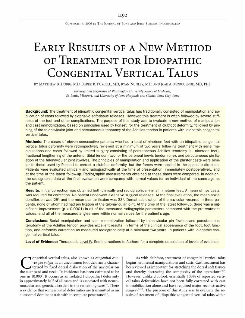
Background: The treatment of idiopathic congenital vertical talus has traditionally consisted of manipulation and ap-plication of casts followed by extensive soft-tissue releases. However, this treatment is often followed by severe stiff-ness of the foot and other complications. The purpose of this study was to evaluate a new method of manipulationand cast immobilization, based on principles used by Ponseti for the treatment of clubfoot deformity, followed by pin-ning of the talonavicular joint and percutaneous tenotomy of the Achilles tendon in patients with idiopathic congenitalvertical talus.
Methods: The cases of eleven consecutive patients who had a total of nineteen feet with an idiopathic congenitalvertical talus deformity were retrospectively reviewed at a minimum of two years following treatment with serial ma-nipulations and casts followed by limited surgery consisting of percutaneous Achilles tenotomy (all nineteen feet),fractional lengthening of the anterior tibial tendon (two) or the peroneal brevis tendon (one), and percutaneous pin fix-ation of the talonavicular joint (twelve). The principles of manipulation and application of the plaster casts were simi-lar to those used by Ponseti to correct a clubfoot deformity, but the forces were applied in the opposite direction.Patients were evaluated clinically and radiographically at the time of presentation, immediately postoperatively, andat the time of the latest follow-up. Radiographic measurements obtained at these times were compared. In addition,the radiographic data at the final evaluation were compared with normal values for an individual of the same age asthe patient.
Results: Initial correction was obtained both clinically and radiographically in all nineteen feet. A mean of five castswas required for correction. No patient underwent extensive surgical releases. At the final evaluation, the mean ankledorsiflexion was 25° and the mean plantar flexion was 33°. Dorsal subluxation of the navicular recurred in three pa-tients, none of whom had had pin fixation of the talonavicular joint. At the time of the latest follow-up, there was a sig-nificant improvement (p < 0.0001) in all of the measured radiographic parameters compared with the pretreatmentvalues, and all of the measured angles were within normal values for the patient’s age.
Conclusions: Serial manipulation and cast immobilization followed by talonavicular pin fixation and percutaneoustenotomy of the Achilles tendon provides excellent results, in terms of the clinical appearance of the foot, foot func-tion, and deformity correction as measured radiographically at a minimum two years, in patients with idiopathic con-genital vertical talus.
Level of Evidence: Therapeutic Level IV. See Instructions to Authors for a complete description of levels of evidence.
ongenital vertical talus, also known as congenital con-vex pes valgus, is an uncommon foot deformity charac-terized by fixed dorsal dislocation of the navicular on
the talar head and neck1. Its incidence has been estimated to beone in 10,0002. It occurs as an isolated (idiopathic) deformityin approximately half of all cases and is associated with neuro-muscular and genetic disorders in the remaining cases2-5. Thereis evidence that some isolated deformities are transmitted as anautosomal dominant trait with incomplete penetrance5-7.
As with clubfoot, treatment of congenital vertical talusbegins with serial manipulations and casts. Cast treatment hasbeen viewed as important for stretching the dorsal soft tissuesand thereby decreasing the complexity of the operation2,5,8,9.However, unlike clubfoot, essentially 100% of reported verti-cal talus deformities have not been fully corrected with castimmobilization alone and have required major reconstructivesurgery3,9-17. The purpose of this study was to evaluate the re-sults of treatment of idiopathic congenital vertical talus with a
C
1193
TH E JO U R NA L OF BONE & JOINT SURGER Y · JBJS .ORG
VO LU M E 88-A · NU M B E R 6 · JU N E 2006EA R LY RESULT S OF A NEW ME T H O D OF TRE A T M EN T FOR ID I OPA T H I C CON GEN ITAL VER T I CAL TA LU S
method of serial manipulations and casts, based on the princi-ples used by Ponseti for the treatment of clubfoot deformity,followed by minimal surgical intervention.
Materials and Methodse retrospectively reviewed the cases of eleven consecu-tive patients with idiopathic congenital vertical talus
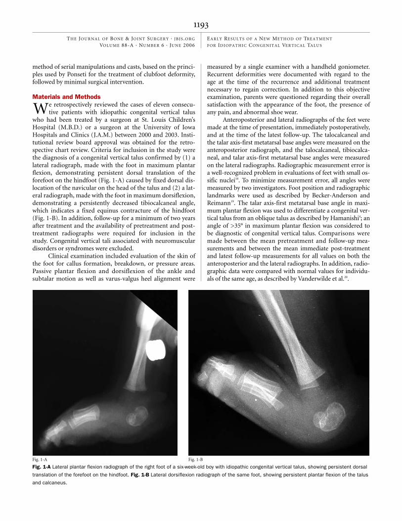
who had been treated by a surgeon at St. Louis Children’sHospital (M.B.D.) or a surgeon at the University of IowaHospitals and Clinics (J.A.M.) between 2000 and 2003. Insti-tutional review board approval was obtained for the retro-spective chart review. Criteria for inclusion in the study werethe diagnosis of a congenital vertical talus confirmed by (1) alateral radiograph, made with the foot in maximum plantarflexion, demonstrating persistent dorsal translation of theforefoot on the hindfoot (Fig. 1-A) caused by fixed dorsal dis-location of the navicular on the head of the talus and (2) a lat-eral radiograph, made with the foot in maximum dorsiflexion,demonstrating a persistently decreased tibiocalcaneal angle,which indicates a fixed equinus contracture of the hindfoot(Fig. 1-B). In addition, follow-up for a minimum of two yearsafter treatment and the availability of pretreatment and post-treatment radiographs were required for inclusion in thestudy. Congenital vertical tali associated with neuromusculardisorders or syndromes were excluded.
Clinical examination included evaluation of the skin ofthe foot for callus formation, breakdown, or pressure areas.Passive plantar flexion and dorsiflexion of the ankle andsubtalar motion as well as varus-valgus heel alignment were
measured by a single examiner with a handheld goniometer.Recurrent deformities were documented with regard to theage at the time of the recurrence and additional treatmentnecessary to regain correction. In addition to this objectiveexamination, parents were questioned regarding their overallsatisfaction with the appearance of the foot, the presence ofany pain, and abnormal shoe wear.
Anteroposterior and lateral radiographs of the feet weremade at the time of presentation, immediately postoperatively,and at the time of the latest follow-up. The talocalcaneal andthe talar axis-first metatarsal base angles were measured on theanteroposterior radiograph, and the talocalcaneal, tibiocalca-neal, and talar axis-first metatarsal base angles were measuredon the lateral radiographs. Radiographic measurement error isa well-recognized problem in evaluations of feet with small os-sific nuclei18. To minimize measurement error, all angles weremeasured by two investigators. Foot position and radiographiclandmarks were used as described by Becker-Anderson andReimann19. The talar axis-first metatarsal base angle in maxi-mum plantar flexion was used to differentiate a congenital ver-tical talus from an oblique talus as described by Hamanishi4; anangle of >35° in maximum plantar flexion was considered tobe diagnostic of congenital vertical talus. Comparisons weremade between the mean pretreatment and follow-up mea-surements and between the mean immediate post-treatmentand latest follow-up measurements for all values on both theanteroposterior and the lateral radiographs. In addition, radio-graphic data were compared with normal values for individu-als of the same age, as described by Vanderwilde et al.20.
W
Fig. 1-A
Fig. 1-A Lateral plantar flexion radiograph of the right foot of a six-week-old boy with idiopathic congenital vertical talus, showing persistent dorsal
translation of the forefoot on the hindfoot. Fig. 1-B Lateral dorsiflexion radiograph of the same foot, showing persistent plantar flexion of the talus
and calcaneus.
Fig. 1-B
1194
TH E JO U R NA L OF BONE & JOINT SURGER Y · JBJS .ORG
VO LU M E 88-A · NU M B E R 6 · JU N E 2006EA R LY RESULT S OF A NEW ME T H O D OF TRE A T M EN T FOR ID I OPA T H I C CON GEN ITAL VER T I CAL TA LU S
At the time of the last follow-up, the outcome for eachpatient was evaluated with the 10-point scale described byAdelaar et al.21. The clinical appearance of the foot is assigneda maximum of 6 points, and radiographic measurements areassigned a maximum of 4 points. The maximum score is 10points, with 1 point subtracted for each abnormality notedeither clinically or radiographically. A score of 10 points isconsidered excellent; a score of 7, 8, or 9 points, good; a scoreof 4, 5, or 6 points, fair; and a score of 0, 1, 2, or 3 points, poor.
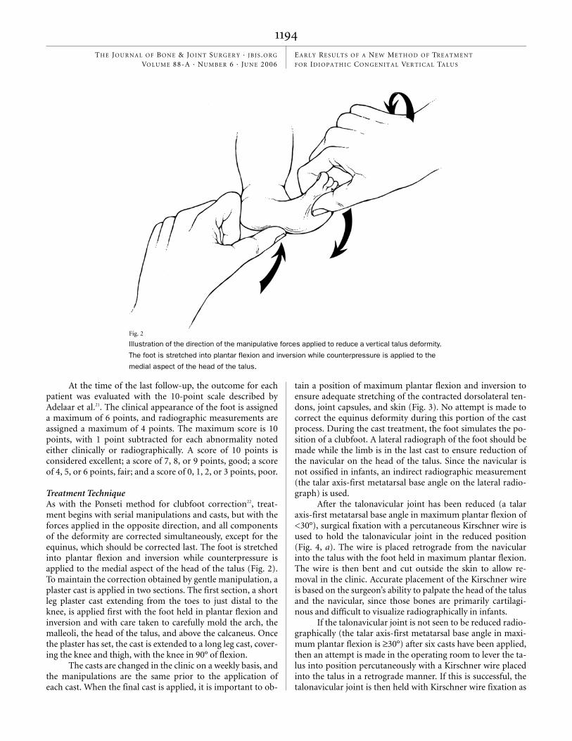
Treatment TechniqueAs with the Ponseti method for clubfoot correction22, treat-ment begins with serial manipulations and casts, but with theforces applied in the opposite direction, and all componentsof the deformity are corrected simultaneously, except for theequinus, which should be corrected last. The foot is stretchedinto plantar flexion and inversion while counterpressure isapplied to the medial aspect of the head of the talus (Fig. 2).To maintain the correction obtained by gentle manipulation, aplaster cast is applied in two sections. The first section, a shortleg plaster cast extending from the toes to just distal to theknee, is applied first with the foot held in plantar flexion andinversion and with care taken to carefully mold the arch, themalleoli, the head of the talus, and above the calcaneus. Oncethe plaster has set, the cast is extended to a long leg cast, cover-ing the knee and thigh, with the knee in 90° of flexion.
The casts are changed in the clinic on a weekly basis, andthe manipulations are the same prior to the application ofeach cast. When the final cast is applied, it is important to ob-
tain a position of maximum plantar flexion and inversion toensure adequate stretching of the contracted dorsolateral ten-dons, joint capsules, and skin (Fig. 3). No attempt is made tocorrect the equinus deformity during this portion of the castprocess. During the cast treatment, the foot simulates the po-sition of a clubfoot. A lateral radiograph of the foot should bemade while the limb is in the last cast to ensure reduction ofthe navicular on the head of the talus. Since the navicular isnot ossified in infants, an indirect radiographic measurement(the talar axis-first metatarsal base angle on the lateral radio-graph) is used.
After the talonavicular joint has been reduced (a talaraxis-first metatarsal base angle in maximum plantar flexion of<30°), surgical fixation with a percutaneous Kirschner wire isused to hold the talonavicular joint in the reduced position(Fig. 4, a). The wire is placed retrograde from the navicularinto the talus with the foot held in maximum plantar flexion.The wire is then bent and cut outside the skin to allow re-moval in the clinic. Accurate placement of the Kirschner wireis based on the surgeon’s ability to palpate the head of the talusand the navicular, since those bones are primarily cartilagi-nous and difficult to visualize radiographically in infants.
If the talonavicular joint is not seen to be reduced radio-graphically (the talar axis-first metatarsal base angle in maxi-mum plantar flexion is ≥30°) after six casts have been applied,then an attempt is made in the operating room to lever the ta-lus into position percutaneously with a Kirschner wire placedinto the talus in a retrograde manner. If this is successful, thetalonavicular joint is then held with Kirschner wire fixation as
Fig. 2
Illustration of the direction of the manipulative forces applied to reduce a vertical talus deformity.
The foot is stretched into plantar flexion and inversion while counterpressure is applied to the
medial aspect of the head of the talus.
1195
TH E JO U R NA L OF BONE & JOINT SURGER Y · JBJS .ORG
VO LU M E 88-A · NU M B E R 6 · JU N E 2006EA R LY RESULT S OF A NEW ME T H O D OF TRE A T M EN T FOR ID I OPA T H I C CON GEN ITAL VER T I CAL TA LU S
described above. If the talonavicular joint cannot be reducedclosed, then a small medial incision is made over the talona-vicular joint and a dorsal capsulectomy of the talonavicularjoint is performed. Traction is applied to the forefoot in theplantar flexed direction, while thumb pressure is applied si-multaneously over the prominence of the head of the talus tomedially push the talar head dorsally. Rarely, if the surgeon
deems them to be obstacles to achieving an adequate talona-vicular reduction, the peroneal brevis tendon and/or tibialisanterior tendon must be fractionally lengthened at the muscu-lotendinous junction.
Once the talonavicular joint is reduced and stabilizedwith the Kirschner wire, a percutaneous tenotomy of theAchilles tendon is used to correct the equinus deformity as de-
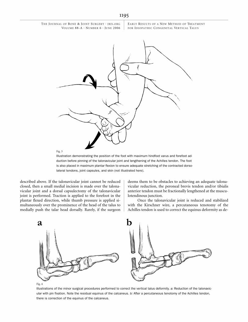
Fig. 4
Illustrations of the minor surgical procedures performed to correct the vertical talus deformity. a: Reduction of the talonavic-
ular with pin fixation. Note the residual equinus of the calcaneus. b: After a percutaneous tenotomy of the Achilles tendon,
there is correction of the equinus of the calcaneus.
Fig. 3
Illustration demonstrating the position of the foot with maximum hindfoot varus and forefoot ad-
duction before pinning of the talonavicular joint and lengthening of the Achilles tendon. The foot
is also placed in maximum plantar flexion to ensure adequate stretching of the contracted dorso-
lateral tendons, joint capsules, and skin (not illustrated here).
1196
TH E JO U R NA L OF BONE & JOINT SURGER Y · JBJS .ORG
VO LU M E 88-A · NU M B E R 6 · JU N E 2006EA R LY RESULT S OF A NEW ME T H O D OF TRE A T M EN T FOR ID I OPA T H I C CON GEN ITAL VER T I CAL TA LU S
scribed by Dobbs et al. for the treatment of a clubfoot23 (Fig. 4,b). A Beaver eye blade (Becton Dickinson, Franklin Lakes,New Jersey) is introduced through the skin onto the medialedge of the Achilles tendon about 1 cm above its calcaneal in-sertion with the cutting surface of the blade pointed proxi-mally. The undersurface of the tendon is palpated with the tipof the blade, which is then rotated 45° to allow the tendon tobe severed from ventral to dorsal. The Kirschner wire preventsloss of reduction of the talonavicular joint as the hindfoot isbrought into dorsiflexion.
A long leg cast is then applied with the foot in a neutralposition and the ankle in 5° of dorsiflexion. The cast ischanged in the clinic at two weeks, at which time a mold ismade for a solid ankle-foot orthosis, in 15° of plantar flexionat the midtarsal joint, to help maintain reduction of the tal-onavicular joint once the Kirschner wire is removed. A newlong leg cast is then applied with the ankle in 10° to 15° of dor-siflexion and is worn for three weeks, after which time the castis removed in the clinic and the Kirschner wire is pulled. Thesolid orthosis is applied, and the parents are instructed regard-ing exercises with emphasis on a range of ankle motion andfoot inversion, to be performed two or three times a day athome. The orthosis is worn for twenty-three hours a day untilthe child reaches walking age, and then it is worn for walking(for twelve to fourteen hours in a twenty-four-hour period)until the age of two years.
Statistical AnalysesMeans and standard deviations are reported. Repeated-measures analysis of variance was used to compare variousradiographic measures before treatment, immediately aftertreatment, and at the time of the latest follow-up. An unpairedt test was used to compare radiographic values at the time oflatest follow-up with published normative radiographic valuesfor children of the same age20. A p value of <0.05 was selectedas significant for this study before data analysis.
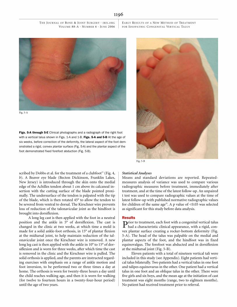
Resultsrior to treatment, each foot with a congenital vertical talushad a characteristic clinical appearance, with a rigid, con-
vex plantar surface creating a rocker-bottom deformity (Fig.5-A). The head of the talus was palpable on the medial andplantar aspects of the foot, and the hindfoot was in fixedequinovalgus. The forefoot was abducted and in dorsiflexionat the midtarsal joint (Fig. 5-B).
Eleven patients with a total of nineteen vertical tali wereincluded in this study (see Appendix). Eight patients had verti-cal talus bilaterally. Two patients had a vertical talus in one footand talipes equinovarus in the other. One patient had a verticaltalus in one foot and an oblique talus in the other. There werefive girls and six boys, and the mean age at the initiation of casttreatment was eight months (range, two to eighteen months).No patient had received treatment prior to referral.
P
Fig. 5-B
Fig. 5-A
Figs. 5-A through 5-E Clinical photographs and a radiograph of the right foot
with a vertical talus shown in Figs. 1-A and 1-B. Figs. 5-A and 5-B At the age of
six weeks, before correction of the deformity, the lateral aspect of the foot dem-
onstrated a rigid, convex plantar surface (Fig. 5-A) and the plantar aspect of the
foot demonstrated fixed forefoot abduction (Fig. 5-B).
1197
TH E JO U R NA L OF BONE & JOINT SURGER Y · JBJS .ORG
VO LU M E 88-A · NU M B E R 6 · JU N E 2006EA R LY RESULT S OF A NEW ME T H O D OF TRE A T M EN T FOR ID I OPA T H I C CON GEN ITAL VER T I CAL TA LU S
A mean of five casts (four, five, or six casts) was requiredto achieve reduction of the navicular onto the head of the talus.All patients required percutaneous Achilles tendon lengtheningfor correction of the hindfoot equinus. Two patients had frac-tional lengthening of the tibialis anterior tendon and one hadfractional lengthening of the peroneal brevis tendon as part ofthe initial treatment to help achieve reduction of the talonavic-ular joint. Seven patients (twelve feet) had percutaneous pin-ning of the talonavicular joint at the time of the Achilles tendonlengthening to maintain correction of the forefoot on the hind-foot. No patient underwent extensive soft-tissue releases.
Six feet in three patients had recurrent deformity, notedradiographically as dorsal subluxation of the navicular on thehead of the talus at a mean of five months (range, three to sixmonths) after the initial correction. None of the six feet hadhad Kirschner wire fixation of the talonavicular joint at thetime of the Achilles tenotomy. All required repeat cast treat-
ment, and four also required Kirschner wire fixation of the tal-onavicular joint to maintain reduction. Open surgery was notrequired in these three patients. Only one patient (one foot)who was not treated initially with talonavicular pinning didnot have a recurrence. In contrast, no patient who had initialtalonavicular pinning had a recurrence by the time of writing.
The mean age at the time of latest follow-up was threeyears (range, two and one-half years to six years). All parentswere satisfied with the appearance of the foot. No patient re-quired custom shoes or complained of abnormal shoe wear,and none had pain. There were no operative complications.According to the Adelaar scoring system, five patients had anexcellent result, six had a good result, and none had a fair orpoor result. There was no significant difference in the Adelaarscore at the time of final follow-up between the patients whohad had a recurrence and needed further treatment and thosewho had not had a recurrence.
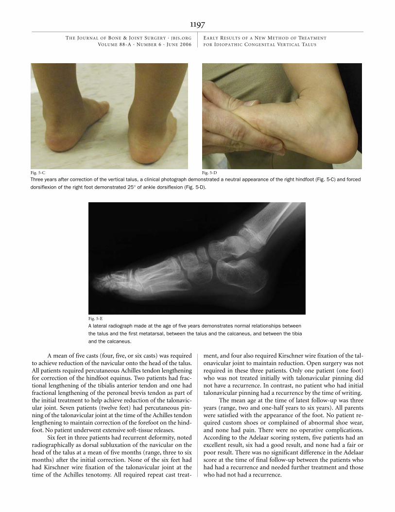
Fig. 5-C
Three years after correction of the vertical talus, a clinical photograph demonstrated a neutral appearance of the right hindfoot (Fig. 5-C) and forced
dorsiflexion of the right foot demonstrated 25° of ankle dorsiflexion (Fig. 5-D).
Fig. 5-D
Fig. 5-E
A lateral radiograph made at the age of five years demonstrates normal relationships between
the talus and the first metatarsal, between the talus and the calcaneus, and between the tibia
and the calcaneus.
1198
TH E JO U R NA L OF BONE & JOINT SURGER Y · JBJS .ORG
VO LU M E 88-A · NU M B E R 6 · JU N E 2006EA R LY RESULT S OF A NEW ME T H O D OF TRE A T M EN T FOR ID I OPA T H I C CON GEN ITAL VER T I CAL TA LU S
At the time of the latest follow-up, the hindfoot was seenclinically to be in neutral in fifteen feet (Fig. 5-C) and in 5° to8° of valgus in four feet. Ankle dorsiflexion averaged 25°(range, 18° to 35°) (Fig. 5-D), and plantar flexion averaged33° (range, 15° to 35°). Subtalar motion was normal in allnineteen feet.
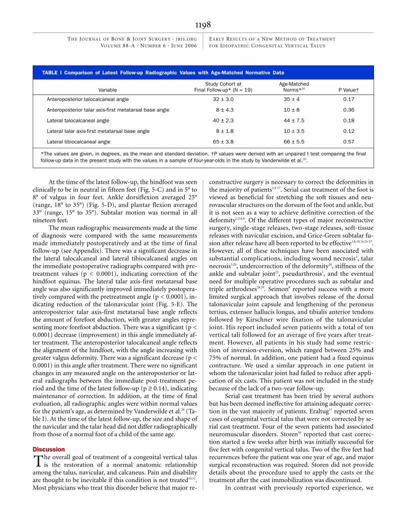
The mean radiographic measurements made at the timeof diagnosis were compared with the same measurementsmade immediately postoperatively and at the time of finalfollow-up (see Appendix). There was a significant decrease inthe lateral talocalcaneal and lateral tibiocalcaneal angles onthe immediate postoperative radiographs compared with pre-treatment values (p < 0.0001), indicating correction of thehindfoot equinus. The lateral talar axis-first metatarsal baseangle was also significantly improved immediately postopera-tively compared with the pretreatment angle (p < 0.0001), in-dicating reduction of the talonavicular joint (Fig. 5-E). Theanteroposterior talar axis-first metatarsal base angle reflectsthe amount of forefoot abduction, with greater angles repre-senting more forefoot abduction. There was a significant (p <0.0001) decrease (improvement) in this angle immediately af-ter treatment. The anteroposterior talocalcaneal angle reflectsthe alignment of the hindfoot, with the angle increasing withgreater valgus deformity. There was a significant decrease (p <0.0001) in this angle after treatment. There were no significantchanges in any measured angle on the anteroposterior or lat-eral radiographs between the immediate post-treatment pe-riod and the time of the latest follow-up (p ≥ 0.14), indicatingmaintenance of correction. In addition, at the time of finalevaluation, all radiographic angles were within normal valuesfor the patient’s age, as determined by Vanderwilde et al.20 (Ta-ble I). At the time of the latest follow-up, the size and shape ofthe navicular and the talar head did not differ radiographicallyfrom those of a normal foot of a child of the same age.
Discussionhe overall goal of treatment of a congenital vertical talusis the restoration of a normal anatomic relationship
among the talus, navicular, and calcaneus. Pain and disabilityare thought to be inevitable if this condition is not treated10,12.Most physicians who treat this disorder believe that major re-
constructive surgery is necessary to correct the deformities inthe majority of patients3,9-17. Serial cast treatment of the foot isviewed as beneficial for stretching the soft tissues and neu-rovascular structures on the dorsum of the foot and ankle, butit is not seen as a way to achieve definitive correction of thedeformity2,5,8,9. Of the different types of major reconstructivesurgery, single-stage releases, two-stage releases, soft-tissuereleases with navicular excision, and Grice-Green subtalar fu-sion after release have all been reported to be effective1,8,10,14,24-27.However, all of these techniques have been associated withsubstantial complications, including wound necrosis2, talarnecrosis5,26, undercorrection of the deformity10, stiffness of theankle and subtalar joint25, pseudarthrosis3, and the eventualneed for multiple operative procedures such as subtalar andtriple arthrodeses28,29. Seimon8 reported success with a morelimited surgical approach that involves release of the dorsaltalonavicular joint capsule and lengthening of the peroneustertius, extensor hallucis longus, and tibialis anterior tendonsfollowed by Kirschner wire fixation of the talonavicularjoint. His report included seven patients with a total of tenvertical tali followed for an average of five years after treat-ment. However, all patients in his study had some restric-tion of inversion-eversion, which ranged between 25% and75% of normal. In addition, one patient had a fixed equinuscontracture. We used a similar approach in one patient inwhom the talonavicular joint had failed to reduce after appli-cation of six casts. This patient was not included in the studybecause of the lack of a two-year follow-up.
Serial cast treatment has been tried by several authorsbut has been deemed ineffective for attaining adequate correc-tion in the vast majority of patients. Eraltug17 reported sevencases of congenital vertical talus that were not corrected by se-rial cast treatment. Four of the seven patients had associatedneuromuscular disorders. Storen30 reported that cast correc-tion started a few weeks after birth was initially successful forfive feet with congenital vertical talus. Two of the five feet hadrecurrences before the patient was one year of age, and majorsurgical reconstruction was required. Storen did not providedetails about the procedure used to apply the casts or thetreatment after the cast immobilization was discontinued.
In contrast with previously reported experience, we
T
TABLE I Comparison of Latest Follow-up Radiographic Values with Age-Matched Normative Data
VariableStudy Cohort at
Final Follow-up* (N = 19)Age-Matched
Norms*20 P Value†
Anteroposterior talocalcaneal angle 32 ± 3.0 35 ± 4 0.17
Anteroposterior talar axis-first metatarsal base angle 8 ± 4.3 10 ± 8 0.36
Lateral talocalcaneal angle 40 ± 2.3 44 ± 7.5 0.18
Lateral talar axis-first metatarsal base angle 8 ± 1.8 10 ± 3.5 0.12
Lateral tibiocalcaneal angle 65 ± 3.8 66 ± 5.5 0.57
*The values are given, in degrees, as the mean and standard deviation. †P values were derived with an unpaired t test comparing the finalfollow-up data in the present study with the values in a sample of four-year-olds in the study by Vanderwilde et al.20.
1199
TH E JO U R NA L OF BONE & JOINT SURGER Y · JBJS .ORG
VO LU M E 88-A · NU M B E R 6 · JU N E 2006EA R LY RESULT S OF A NEW ME T H O D OF TRE A T M EN T FOR ID I OPA T H I C CON GEN ITAL VER T I CAL TA LU S
observed initial success with serial manipulation and casttreatment followed by minor surgical intervention for the de-finitive treatment of idiopathic congenital vertical talus. In ourexperience, patients with neuromuscular and genetic disor-ders have tended to have more rigid deformities that are lessamenable to cast correction.
The principles of manipulation and treatment withplaster casts are similar to those used in the Ponseti method ofclubfoot correction31, but the forces are applied in an oppositedirection. A thorough understanding of the anatomy and thedeformities present is necessary to achieve correction. An av-erage of five casts were required to reduce the forefoot ontothe head of the talus in our study. By the time that the last castis applied, the hindfoot should be in marked varus; thus, atthis stage, the cast is applied so that the foot mimics the ap-pearance of an untreated clubfoot, achieving maximumstretch of the soft tissues that are hindering correction.
Once the forefoot is seen radiographically to be re-duced on the head of the talus, the reduction is best held witha Kirschner wire placed percutaneously across the talonavicu-lar joint with the foot in maximum plantar flexion. Once theforefoot is held in the reduced position, the hindfoot equinuscan be corrected with a percutaneous Achilles tendon length-ening without risking loss of forefoot reduction. No deformi-ties recurred in any of the feet that we initially treated withKirschner wire fixation of the talonavicular joint, but therewas a recurrence in six of seven feet in which the talonavicu-lar joint had not been pinned. We also recommend that asolid ankle-foot orthosis built in 15° of plantar flexion at themidtarsal joint be worn full time until the child reaches walk-ing age and then for walking until the age of two years to keepthe forefoot plantar flexed and to prevent recurrence of thedeformity.
The described technique of serial manipulation and casttreatment, pin fixation of the talonavicular joint, and a percu-taneous Achilles tenotomy provided good early results (at aminimum of two years after correction) in terms of the clini-
cal appearance of the foot, foot function, and radiographicevidence of correction. It is recommended that, as withclubfoot32, treatment with manipulations and casts be initiatedfor idiopathic congenital vertical talus as soon as the diagnosisis made. Longer follow-up is necessary to determine if correc-tion has been maintained. Since 50% of the cases of congenitalvertical talus are idiopathic, this technique may allow manychildren to avoid traditional surgical treatment while main-taining increased flexibility of the foot.
AppendixTables showing the clinical details for all patients and theradiographic measurements obtained at different time-
points are available with the electronic versions of this article,on our web site at jbjs.org (go to the article citation and clickon “Supplementary Material”) and on our quarterly CD-ROM(call our subscription department, at 781-449-9780, to orderthe CD-ROM). �
Matthew B. Dobbs, MDDerek B. Purcell, MDRyan Nunley, MDDepartment of Orthopaedic Surgery, Washington University School of Medicine, One Children’s Place, Suite 4S20, St. Louis, MO 63110. E-mail address for M.B. Dobbs: [email protected]
Jose A. Morcuende, MD, PhDDepartment of Orthopaedic Surgery and Rehabilitation, University of Iowa Hospitals and Clinics, 200 Hawkins Drive, Iowa City, IA 52242
The authors did not receive grants or outside funding in support of their research for or preparation of this manuscript. They did not receive pay-ments or other benefits or a commitment or agreement to provide such benefits from a commercial entity. No commercial entity paid or di-rected, or agreed to pay or direct, any benefits to any research fund, foundation, educational institution, or other charitable or nonprofit or-ganization with which the authors are affiliated or associated.
doi:10.2106/JBJS.E.00402
References
1. Lamy L, Weissman L. Congenital convex pes valgus. J Bone Joint Surg Am. 1939;21:79-91.
2. Jacobsen ST, Crawford AH. Congenital vertical talus. J Pediatr Orthop. 1983;3:306-10.
3. Dodge LD, Ashley RK, Gilbert RJ. Treatment of the congenital vertical talus: a retrospective review of 36 feet with long-term follow-up. Foot Ankle. 1987;7:326-32.
4. Hamanishi C. Congenital vertical talus: classification with 69 cases and new measurement system. J Pediatr Orthop. 1984;4:318-26.
5. Ogata K, Schoenecker PL, Sheridan J. Congenital vertical talus and its familial occurrence: an analysis of 36 patients. Clin Orthop Relat Res. 1979;139:128-32.
6. Stern HJ, Clark RD, Stroberg AJ, Shohat M. Autosomal dominant transmission of isolated congenital vertical talus. Clin Genet. 1989;36:427-30.
7. Dobbs MB, Schoenecker PL, Gordon JE. Autosomal dominant transmission of isolated congenital vertical talus. Iowa Orthop J. 2002;22:25-7.
8. Seimon LP. Surgical correction of congenital vertical talus under the age of 2 years. J Pediatr Orthop. 1987;7:405-11.
9. Drennan JC. Congenital vertical talus. Instr Course Lect. 1996;45:315-22.
10. Coleman SS, Stelling FH 3rd, Jarrett J. Pathomechanics and treatment of con-genital vertical talus. Clin Orthop Relat Res. 1970;70:62-72.
11. Colton CL. The surgical management of congenital vertical talus. J Bone Joint Surg Br. 1973;55:566-74.
12. Drennan JC, Sharrard WJ. The pathological anatomy of convex pes valgus. J Bone Joint Surg Br. 1971;53:455-61.
13. Ellis JN, Scheer GE. Congenital convex pes valgus. Clin Orthop Relat Res. 1974;99:168-74.
14. Eyre-Brook AL. Congenital vertical talus. J Bone Joint Surg Br. 1967;49:618-27.
15. Fitton JM, Nevelos AB. The treatment of congenital vertical talus. J Bone Joint Surg Br. 1979;61:481-3.
16. Griffin DW, Daly N, Karlin JM. Clinical presentation of congenital convex pes valgus. J Foot Ankle Surg. 1995;34:146-52.
17. Eraltug U. Corrective plaster application in the treatment of vertical talus. An analysis of eleven cases. Int Surg. 1966;46:246-9.
18. Stricker SJ, Rosen E. Early one-stage reconstruction of congenital vertical ta-lus. Foot Ankle Int. 1997;18:535-43.
19. Becker-Andersen H, Reimann I. Congenital vertical talus. Reevaluation of early manipulative treatment. Acta Orthop Scand. 1974;45:130-44.
1200
TH E JO U R NA L OF BONE & JOINT SURGER Y · JBJS .ORG
VO LU M E 88-A · NU M B E R 6 · JU N E 2006EA R LY RESULT S OF A NEW ME T H O D OF TRE A T M EN T FOR ID I OPA T H I C CON GEN ITAL VER T I CAL TA LU S
20. Vanderwilde R, Staheli LT, Chew DE, Malagon V. Measurements on radio-graphs of the foot in normal infants and children. J Bone Joint Surg Am. 1988;70:407-15.
21. Adelaar RS, Williams RM, Gould JS. Congenital convex pes valgus: results of an early comprehensive release and a review of congenital vertical talus at Rich-mond Crippled Children’s Hospital and the University of Alabama in Birmingham. Foot Ankle. 1980;1:62-73.
22. Laaveg SJ, Ponseti IV. Long-term results of treatment of congenital club foot. J Bone Joint Surg Am. 1980;62:23-31.
23. Dobbs MB, Gordon JE, Walton T, Schoenecker PL. Bleeding complications fol-lowing percutaneous tendoachilles tenotomy in the treatment of clubfoot defor-mity. J Pediatr Orthop. 2004;24:353-7.
24. Duncan RD, Fixsen JA. Congenital convex pes valgus. J Bone Joint Surg Br. 1999;81:250-4.
25. Zorer G, Bagatur AE, Dogan A. Single stage surgical correction of congenital vertical talus by complete subtalar release and peritalar reduction by using the Cincinnati incision. J Pediatr Orthop B. 2002;11:60-7.
26. Mazzocca AD, Thomson JD, Deluca PA, Romness MJ. Comparison of the pos-terior approach versus the dorsal approach in the treatment of congenital vertical talus. J Pediatr Orthop. 2001;21:212-7.
27. Napiontek M. Congenital vertical talus: a retrospective and critical review of 32 feet operated on by peritalar reduction. J Pediatr Orthop B. 1995;4:179-87.
28. Grice DS. An extra-articular arthrodesis of the subastragalar joint for correc-tion of paralytic flat feet in children. J Bone Joint Surg Am. 1952;34:927-40.
29. Grice DS. Further experience with extra-articular arthrodesis of the subtalar joint. J Bone Joint Surg Am. 1955;37:246-59.
30. Storen H. On the closed and open correction of congenital convex pes valgus with a vertical astragalus. Acta Orthop Scand. 1965;36:352-8.
31. Morcuende JA, Dolan LA, Dietz FR, Ponseti IV. Radical reduction in the rate of extensive corrective surgery for clubfoot using the Ponseti method. Pediatrics. 2004;113:376-80.
32. Dobbs MB, Rudzki JR, Purcell DB, Walton T, Porter KR, Gurnett CA. Factors predictive of outcome after use of the Ponseti method for the treatment of idio-pathic clubfeet. J Bone Joint Surg Am. 2004;86:22-7.
Copyright © 2022 FDOKUMEN