Early and long-term results of subclavian angioplasty in aortoarteritis (takayasu disease):...
6
9 Springer-Verlag New York, Inc. 1998 Cardiovasc Intervent Radiol (1998) 21:219-224 Early and Long-Term Results of Subclavian Angioplasty in Aortoarteritis (Takayasu Disease),: Comparison with Atherosclerosis Sanjay Tyagi, Puneet K Verma, Daljeet S Gambhir, Upkar A Kaul, Renuka Saha, Ramesh Arora Department of Cardiology, G.B. Pant Hospital, Maulana Azad Medical College, New Delhi 110002, India Abstract Purpose: To compare the early and long-term outcomes of subclavian artery angioplasty in patients with aortoarteritis and atherosclerosis. Methods: Sixty-one subclavian artery angioplasties were performed in 55 consecutive patients with aortoarteritis (n = 32) and atherosclerosis (n = 23) between 1986 and 1995. An arch aortogram followed by a selective subclavian artery angiogram was done to profile the site and extent of the lesion, its relation to the vertebral artery, and the distal circulation. Percutaneous transluminal angioplasty (PTA) was performed via the femoral route for 56 stenotic lesions and 5 total occlusions. Results: PTA was successful in 52 (92.8%) stenotic lesions and 3 (60%) total occlusions. Three patients (5.4%) had complications, that could be effectively managed nonsurgi- cally. Compared with atherosclerosis, patients with aortoar- teritis were younger (27.4 + 9.3 years vs 54.5 _+ 10.5 years; p < 0.001), more often female (75% vs 17.4%; p < 0.001), gangrene was uncommon (0% vs 17.4%; p < 0.05), and diffuse involvement was seen more often (43.8% vs 4.4%; p < 0.001). The luminal diameter stenoses were similar before PTA (88.6 ___ 9.7% vs 89.0 + 9.1%; p = NS). Higher balloon inflation pressure was required to dilate the lesions of aortoarteritis (9.9 -+ 4.6 ATM vs 5.5 + 1.0 ATM; p < 0.001). This group had more residual stenosis (15.5 + 12.4% vs 8.3 -+ 9.4%; p < 0.05) after PTA. There were no neuro- logical sequelae, even in PTA of prevertebral lesions. On 3-120 months (mean 43.3 + 28.9 months) follow-up of 40 patients, restenosis was more often observed in patients with aortoarteritis, particularly in those with diffuse arterial nar- rowing. These lesions could be effectively redilated. Clinical Correspondence to: Dr. S. Tyagi symptoms showed marked improvement after successful an- gioplasty. Conclusion: Subclavian PTA is safe and can be performed as effectively in aortoarteritis as in atherosclerosis, with good long-term results. Long-term follow-up shows that it pro- vides good symptomatic relief. Key words: Aortoarteritis--Atherosclerosis--Transluminal subclavian angioplasty--Stenotic lesion--Total occlusion In the last two decades, percutaneous transluminal angio- plasty (PTA) has rapidly gained acceptance for the treatment of peripheral vascular and coronary artery disease. Brachio- cephalic vessels are among the last in which this technique is used, primarily because of the apprehension about cerebral complications. Since the first report of subclavian artery (SA) angioplasty in 1980 by Bachman and Kim [1], it has been increasingly used in the treatment of patients with symptomatic atherosclerotic stenosis of the subclavian artery [2-6]. In the young population, stenosis of arch vessels is commonly due to nonspecific aortoarteritis (Takayasu dis- ease). Only limited information is available on the role of PTA in this disease [7]. We report here our experience with SA angioplasty and compare the results in aortoarteritis with those in atherosclerosis. Materials and Methods Fifty-five patients with 61 stenoocclusive lesions in subclavian arteries underwent PTA between January 1986 and December 1995; 32 had aortoarteritis, 23 had atherosclerotic lesions. Clinical indications included decreased or absent upper-extremity arterial pulses with intermittent clandication, rest pain. subclavian steal syndrome, ischemic changes in the hand due to embolization from the stenosis, Raynaud's phenomenon, and paresthesias. In patients with these clinical findings, PTA was performed for an SA stenosis
Transcript of Early and long-term results of subclavian angioplasty in aortoarteritis (takayasu disease):...

�9 Springer-Verlag New York, Inc. 1998 Cardiovasc Intervent Radiol (1998) 21:219-224
Early and Long-Term Results of Subclavian Angioplasty in Aortoarteritis (Takayasu Disease),: Comparison with Atherosclerosis Sanjay Tyagi, Puneet K Verma, Daljeet S Gambhir, Upkar A Kaul, Renuka Saha, Ramesh Arora
Department of Cardiology, G.B. Pant Hospital, Maulana Azad Medical College, New Delhi 110002, India
Abstract Purpose: To compare the early and long-term outcomes of subclavian artery angioplasty in patients with aortoarteritis and atherosclerosis. Methods: Sixty-one subclavian artery angioplasties were performed in 55 consecutive patients with aortoarteritis (n = 32) and atherosclerosis (n = 23) between 1986 and 1995. An arch aortogram followed by a selective subclavian artery angiogram was done to profile the site and extent of the lesion, its relation to the vertebral artery, and the distal circulation. Percutaneous transluminal angioplasty (PTA) was performed via the femoral route for 56 stenotic lesions and 5 total occlusions. Results: PTA was successful in 52 (92.8%) stenotic lesions and 3 (60%) total occlusions. Three patients (5.4%) had complications, that could be effectively managed nonsurgi- cally. Compared with atherosclerosis, patients with aortoar- teritis were younger (27.4 + 9.3 years vs 54.5 _+ 10.5 years; p < 0.001), more often female (75% vs 17.4%; p < 0.001), gangrene was uncommon (0% vs 17.4%; p < 0.05), and diffuse involvement was seen more often (43.8% vs 4.4%; p < 0.001). The luminal diameter stenoses were similar before PTA (88.6 ___ 9.7% vs 89.0 + 9.1%; p = NS). Higher balloon inflation pressure was required to dilate the lesions of aortoarteritis (9.9 -+ 4.6 ATM vs 5.5 + 1.0 ATM; p < 0.001). This group had more residual stenosis (15.5 + 12.4% vs 8.3 -+ 9.4%; p < 0.05) after PTA. There were no neuro- logical sequelae, even in PTA of prevertebral lesions. On 3-120 months (mean 43.3 + 28.9 months) follow-up of 40 patients, restenosis was more often observed in patients with aortoarteritis, particularly in those with diffuse arterial nar- rowing. These lesions could be effectively redilated. Clinical
Correspondence to: Dr. S. Tyagi
symptoms showed marked improvement after successful an- gioplasty. Conclusion: Subclavian PTA is safe and can be performed as effectively in aortoarteritis as in atherosclerosis, with good long-term results. Long-term follow-up shows that it pro- vides good symptomatic relief.
Key words: Aortoarteritis--Atherosclerosis--Transluminal subclavian angioplasty--Stenotic lesion--Total occlusion
In the last two decades, percutaneous transluminal angio- plasty (PTA) has rapidly gained acceptance for the treatment of peripheral vascular and coronary artery disease. Brachio- cephalic vessels are among the last in which this technique is used, primarily because of the apprehension about cerebral complications. Since the first report of subclavian artery (SA) angioplasty in 1980 by Bachman and Kim [1], it has been increasingly used in the treatment of patients with symptomatic atherosclerotic stenosis of the subclavian artery [2-6]. In the young population, stenosis of arch vessels is commonly due to nonspecific aortoarteritis (Takayasu dis- ease). Only limited information is available on the role of PTA in this disease [7]. We report here our experience with SA angioplasty and compare the results in aortoarteritis with those in atherosclerosis.
Materials and Methods Fifty-five patients with 61 stenoocclusive lesions in subclavian arteries underwent PTA between January 1986 and December 1995; 32 had aortoarteritis, 23 had atherosclerotic lesions. Clinical indications included decreased or absent upper-extremity arterial pulses with intermittent clandication, rest pain. subclavian steal syndrome, ischemic changes in the hand due to embolization from the stenosis, Raynaud's phenomenon, and paresthesias. In patients with these clinical findings, PTA was performed for an SA stenosis
220 S. Tyagi et al.: Subclavian Angioplasty in Aortoarteritis and Atherosclerosis
of >70% diameter reduction, less than 6 cm in length, or total occlusions of < 2 cm in length. Patients in whom the SA stenosis extended across the origin of the vertebral artery and extended into its origin were excluded. However, PTA was indicated for a ste- nosis adjacent to, but not compromising, the origin of the vertebral artery. Stenoses -< 2 cm were classified as discrete, and > 2 cm as diffuse. In order to determine the extent of brachiocephalic artery involvement, an arch aortogram was done by digital subtraction angiography (DSA) or cine angiography. Selective SA angiography was performed to localize the site and extent of vessel involvement, origin of the vertebral artery, and distal circulation.
Angioplasty was performed via the femoral route. In patients with discrete and concentric stenosis, an "over the wire" angio- plasty technique using a 0.035-inch, straight/angled-tip, 260-cm- long Terumo guidewire (Terumo Corp., Tokyo, Japan) was used to c r o s s the lesion. In lesions that were diffuse, eccentric, ulcerated, or near the ostium of SA or vertebral artery, angioplasty was per- formed through an 8 Fr multipurpose or right coronary guiding catheter using a 0.018-inch floppy-tipped, extra-support coronary angioplasty guidewire and a Sub-4 balloon (Medi-tech, Watertown, MA, USA). The size of the balloon to be used for angioplasty was determined after measuring the normal diameter of the lumen adjacent to the stenosis, using the catheter shaft diameter for cali- bration on the cine or unsubtracted DSA image. The balloon diam- eter never exceeded the diameter of the vessel.
In patients with total or near total occlusion, an attempt was made to cross the occluded artery with a 0.014/0.018-inch coronary guidewire through a 4-mm-diameter coronary angioplasty balloon. During this maneuver, the extra support to the balloon-guidewire assembly was obtained by deep-seating the right coronary guiding catheter. After crossing the lesion, predilatation with the 4-ram balloon was followed by use of an appropriately sized balloon, as above. If the coronary guidewire failed to cross the lesion, a 0.035-inch Terumo hydrophilic guidewire (Boston Scientific Corp., Watertown, MA, USA) was advanced through a 5 Fr diagnostic catheter. The final outer balloon diameter and length in this series measured 5-10 mm and 2-4 cm, respectively. In patients requiring dilatation with balloons more than 6 mm in diameter, the Ultrathin or Blue Max high-pressure balloons (Medi-tech) were used.
In the "over the wire" technique, the intraluminal position of the Terumo guidewire was verified by placing a 5 Fr diagnostic catheter distal to the stenosis, withdrawing the guidewire, and injecting the contrast. Heparin (5000 units) and nitroglycerin (100 ~g) were administered intraarterially through the catheter posi- tioned across the stenosis. Balloon inflation pressures were moni- tored throughout the procedure. The balloon was inflated until either the "waist" on the balloon produced by the stenosis disap- peared, or the patient started complaining of severe pain indicating dissection, or maximum inflation pressure of the balloon was reached. Keeping the guidewire in place, the angioplasty catheter was withdrawn and the result of angioplasty was evaluated by selective angiography through the guiding catheter after adminis- tering nitroglycerin (100-200 txg) intraarterially. If the result was satisfactory, the guidewire was withdrawn and a final selective angiogram was obtained. All patients were routinely given aspirin (100 rag/day) prior to and for 6 months following PTA.
Angioplasty was considered to be technically successful if the residual stenosis was less than 30% of the luminal diameter with at least 50% increase from the pretreatment diameter. The long-term results of PTA were assessed clinically from the patient's syrup-
toms, assessment of arterial pulse, and blood pressure in both upper limbs, or other signs of arterial insufficiency in the affected limb. Patients with clinical evidence of restenosis underwent angiogra- phy, and repeat angioplasty if deemed necessary.
The results of PTA between the two patient groups were ana- lyzed for statistically significant differences by the chi-square or Fisher's exact test for proportions, and by the Student's t-test or nonparametric (Mann-Whitney) test for means. A p value of < 0.05 was considered significant.
Results Angiographic Results
Among the 55 patients who underwent angioplasty, two
lesions were dilated in 6 patients in the same or contralateral
SA, and 49 patients had angioplasty of a single stenotic
lesion or occlusion. Of the 61 lesions attempted, 23 were prevertebral and 38 postvertebral. Fifty-six lesions were stenotic and five were short-segment total occlusions. Fifty-
two (92.8%) of 56 stenoses could be successfully dilated
(Figs. 1, 2). Failure to dilate four lesions occurred in patients
with diffuse arterial narrowing, where the stenosis could not
be reduced to < 30% despite maximal balloon inflation
pressure (17 atm). Three (60%) of five short-segment occlu-
sions could also be dilated (Fig. 3). The two failures were
due to the inability to cross the lesion with the guidewire. In
59 lesions that could be crossed with the guidewire and underwent balloon dilatation, the degree of stenosis de-
creased from 88.3 _+ 9.3% to 13.2 + 11.9%; p < 0.001). Three patients (5.4%) had complications, two of whom had
undergone a technically successful primary procedure. One patient with aortoarteritis developed thrombotic occlusion of
the dilated SA within 24 hr. Thrombolysis with intravenous streptokinase lysed the thrombus and restored normal pa- tency. Another patient with atherosclerotic stenosis developed
dissection which occluded the artery in the hour following angioplasty. The lesion was recrossed with a guidewire and an 8-mm x 60-mm Wallstent was implanted which com- pletely normalized the flow. In the third patient, with aorto- arteritis, a diffuse stenotic lesion that could not be adequately dilated developed a flow-limiting dissection which was ad- equately opened up with a 39-mm-long Palmaz stent. No patient had any thromboembolic episode.
Clinical Results
In patients successfully treated with PTA, clinical symptoms
markedly improved in all except one patient who had total
occlusion in distal subclavian and axillary arteries. In this patient, there was partial improvement of claudication; in all
other patients with arm ischemia the symptoms resolved completely. In patients with gangrene, rest pain disappeared and progression of gangrene stopped. In patients with ver- tebro-basilar insufficiency, the symptoms were relieved.

S. Tyagi et al.: Subclavian Angioplasty in Aortoarteritis and Atherosclerosis 221
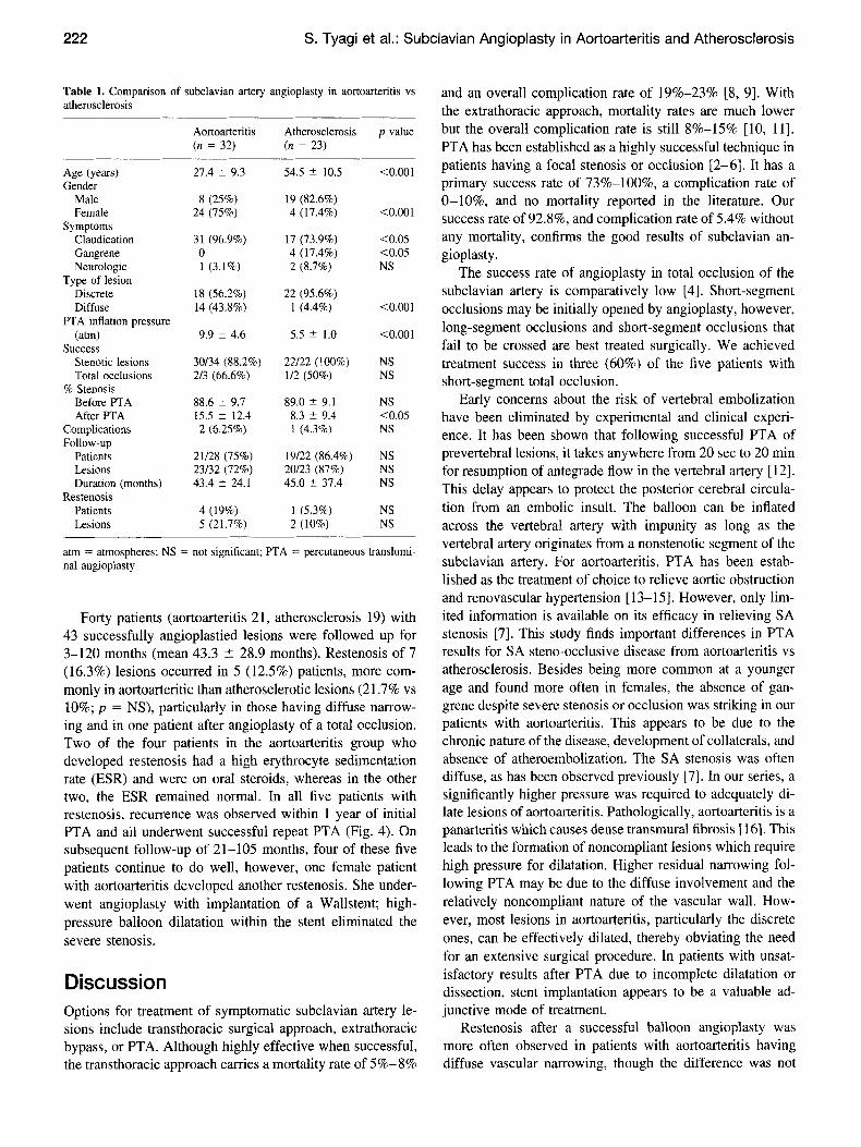
Fig. 1. Selective angiogram of the left subclavian artery in a young female with aortoarteritis shows (A) long-segment stenosis of the postvertebral segment. B Restoration of the normal lumen after angioplasty is shown, resulting in com- plete symptomatic relief.
j
/ g
Fig. 2. Selective angiogram of the left subclavian artery in an elderly male with rest pain and gangrene of the distal pha- langes shows (A) two atherosclerotic stenotic lesions in the post-vertebral segment, the distal being 99%. B Com- plete normalization of the arterial lumen after angioplasty is seen, resulting in marked symptomatic relief.
Fig. 3 Selectiveangiogram oftheleft subclavian artery in a young female with aortoarteritis shows (A) total occlusion just after the origin of vertebral artery. B Good luminal diameter is seen after an- gioplasty.
Comparative hlitial and Long-Term Results
Comparative clinical, angiographic, and angioplasty features in patients with aortoarteritis and atherosclerosis are listed in Table 1. Compared with patients with atherosclerosis, the aortoarteritis group was significantly younger (27.4 + 9.3 years vs 54.5 _+ 10.5 years; p < 0.001), and had more females (75% vs 17.4%; p < 0.001). The patients with aortoarteritis more often presented with claudication (96.9% vs 73.9%; p < 0.05) and gangrene was conspicuously ab- sent. The atherosclerotic patients commonly had gangrene.
On angiography, diffuse narrowing of the subclavian ar- tery was significantly more frequent in aortoarteritis (43.8%
vs 4.4%; p < 0.001). During PTA, aortoarteritis patients often required much higher inflation pressures (9.9 _+ 4.6 arm vs 5.5 + 1.0 atm; p < 0.001) to dilate the lesion. The lesions in both groups could be dilated with a high success rate (86.5% vs 95.8%; p > 0.05). However, residual nar- rowing was more frequent in aortoarteritis compared with "atherosclerosis (15.5 +_ 12.4% vs 8.3 _+ 9.4%; p < 0.05).
Angiographically, an intimal tear after PTA was observed in
all patients with aortoarteritis. The incidence of complica-
tions was the same in the two groups and no transient or permanent neurological complications were seen in any of our patients.
222 S. Tyagi et al.: Subclavian Angioplasty in Aortoarteritis and Atherosclerosis
Table l. Comparison of subclavian artery angioplasty in aortoarteritis vs atherosclerosis
Aortoarteritis Atherosclerosis p value (n = 32) (n = 23)
Age (years) 27.4 _+ 9.3 54.5 _+ 10.5 <0.001 Gender
Male 8 (25%) 19 (82.6%) Female 24 (75%) 4 (17.4%) <0.001
Symptoms Claudication 31 (96.9%) 17 (73.9%) <0.05 Gangrene 0 4 (17.4%) <0.05 Neurologic 1 (3.1%) 2 (8.7%) NS
Type of lesion Discrete 18 (56.2%) 22 (95.6%) Diffuse 14 (43.8%) 1 (4.4%) <0.001
PTA inflation pressure (atm) 9.9 _+ 4.6 5.5 -+ 1.0 <0.001
Success Stenotic lesions 30/34 (88.2%) 22/22 (100%) NS Total occlusions 2/3 (66.6%) 1/2 (50%) NS
% Stenosis Before PTA 88.6 _+ 9.7 89.0 -+ 9.1 NS After PTA 15.5 _+ 12.4 8.3 _+ 9.4 <0.05
Complications 2 (6.25%) 1 (4.3%) NS Follow-up
Patients 21/28 (75%) 19/22 (86.4%) NS Lesions 23/32 (72%) 20/23 (87%) NS Duration (months) 43.4 - 24.1 45.0 --_ 37.4 NS
Restenosis Patients 4 (19%) 1 (5.3%) NS Lesions 5 (21.7%) 2 (10%) NS
atm = atmospheres; NS = not significant; PTA -- percutaneous translumi- nal angioplasty
Forty patients (aortoarteritis 21, atherosclerosis 19) with 43 successfully angioplastied lesions were followed up for 3-120 months (mean 43.3 _ 28.9 months). Restenosis of 7 (16.3%) lesions occurred in 5 (12.5%) patients, more com- monly in aortoarteritic than atherosclerotic lesions (21.7% vs 10%; p = NS), particularly in those having diffuse narrow- ing and in one patient after angioplasty of a total occlusion. Two of the four patients in the aortoarteritis group who developed restenosis had a high erythrocyte sedimentation rate (ESR) and were on oral steroids, whereas in the other two, the ESR remained normal. In all five patients with restenosis, recurrence was observed within 1 year of initial PTA and all underwent successful repeat PTA (Fig. 4). On subsequent follow-up of 21-105 months, four of these five patients continue to do well, however, one female patient with aortoarteritis developed another restenosis. She under- went angioplasty with implantation of a Wallstent; high- pressure balloon dilatation within the stent eliminated the severe stenosis.
Discussion Options for treatment of symptomatic subclavian artery le- sions include transthoracic surgical approach, extrathoracic bypass, or PTA. Although highly effective when successful, the transthoracic approach carries a mortality rate of 5%-8%
and an overall complication rate of 19%-23% [8, 9]. With the extrathoracic approach, mortality rates are much lower but the overall complication rate is still 8%-15% [10, 11]. PTA has been established as a highly successful technique in patients having a focal stenosis or occlusion [2-6]. It has a primary success rate of 73%-100%, a complication rate of 0-10%, and no mortality reported in the literature. Our success rate of 92.8%, and complication rate of 5.4% without any mortality, confirms the good results of subclavian an- gioplasty.
The success rate of angioplasty in total occlusion of the subclavian artery is comparatively low [4]. Short-segment occlusions may be initially opened by angioplasty, however, long-segment occlusions and short-segment occlusions that fail to be crossed are best treated surgically. We achieved treatment success in three (60%) of the five patients with short-segment total occlusion.
Early concerns about the risk of vertebral embolization have been eliminated by experimental and clinical experi- ence. It has been shown that following successful PTA of prevertebral lesions, it takes anywhere from 20 sec to 20 min for resumption of antegrade flow in the vertebral artery [ 12]. This delay appears to protect the posterior cerebral circula- tion from an embolic insult. The balloon can be inflated across the vertebral artery with impunity as long as the vertebral artery originates from a nonstenotic segment of the subclavian artery. For aortoarteritis, PTA has been estab- lished as the treatment of choice to relieve aortic obstruction and renovascular hypertension [13-15]. However, only lim- ited information is available on its efficacy in relieving SA stenosis [7]. This study finds important differences in PTA results for SA steno-occlusive disease from aortoarteritis vs atherosclerosis. Besides being more common at a younger age and found more often in females, the absence of gan- grene despite severe stenosis or occlusion was striking in our patients with aortoarteritis. This appears to be due to the chronic nature of the disease, development of collaterals, and absence of atheroembolization. The SA stenosis was often diffuse, as has been observed previously [7]. In our series, a significantly higher pressure was required to adequately di- late lesions of aortoarteritis. Pathologically, aortoarteritis is a panarteritis which causes dense transmural fibrosis [ 16]. This leads to the formation of noncompliant lesions which require high pressure for dilatation. Higher residual narrowing fol- lowing PTA may be due to the diffuse involvement and the relatively noncompliant nature of the vascular wall. How- ever, most lesions in aortoarteritis, particularly the discrete ones, can be effectively dilated, thereby obviating the need for an extensive surgical procedure. In patients with unsat- isfactory results after PTA due to incomplete dilatation or dissection, stent implantation appears to be a valuable ad- junctive mode of treatment.
Restenosis after a successful balloon angioplasty was more often observed in patients with aortoarteritis having diffuse vascular narrowing, though the difference was not
S. Tyagi et al.: Subclavian Angioplasty in Aortoarteritis and Atherosclerosis 223
found to be statistically significant (probably because of the
small number of patients compared). Less durable results in
more extensive aortoarteritis have also been observed in the
only other reported series [7]. Diffuse vascular involvement, partial dilatation during PTA, and disease activity may be responsible for the higher restenosis rate. It has been ob-
served that disease may remain in the active phase despite normal ESR [17]. In our series, all restenotic lesions could be successfully redilated with good long-term results. Surgical therapy for aortoarteritis carries a high morbidity, mortality,
problems of graft occlusion, and anastomotic narrowing. Involvement of arteries in aortoarteritis is often multifocal
and new lesions may develop on follow-up. The ability to treat multiple lesions safely with PTA, and the ease and efficacy of repeat angioplasty, favors its use over surgery in the management of stenotic lesions. Angioplasty also has the advantages of being cost effective, having lower morbidity, and having a shorter period of hospitalization.
In conclusion, symptomatic patients with significant sub-
clavian artery stenosis can be safely treated by PTA with a
high success rate and good long-term angiographic and clin-
ical results. As in atherosclerotic SA stenosis, PTA should be
considered the treatment of choice, particularly for the dis-
crete lesions of aortoarteritis.
Acknowledgment: The authors acknowledge the help of Mr. Inder Singh for photographic assistance.
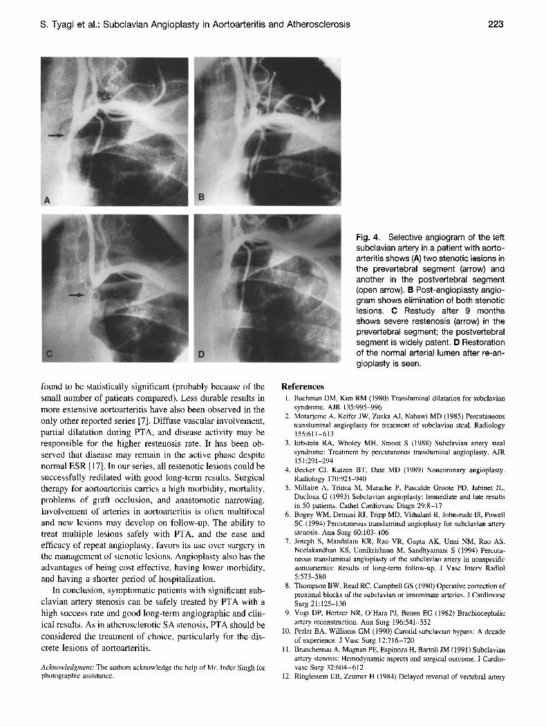
Fig. 4. Selective angiogram of the left subclavian artery in a patient with aorto- arteritis shows (A) two stenotic lesions in the prevertebral segment (arrow) and another in the postvertebral segment (open arrow). B Post-angioplasty angio- gram shows elimination of both stenotic lesions. C Restudy after 9 months shows severe restenosis (arrow) in the prevertebral segment; the postvertebral segment is widely patent. D Restoration of the normal arterial lumen after re-an- gioplasty is s e e n .
References 1. Bachman DM, Kim RM (1980) Transluminal dilatation for subclavian
syndrome. AJR 135:995-996 2. Motarjeme A, Keifer JW, Zuska AJ, Nabawi MD (1985) Percutaneous
transluminal angioplasty for treatment of subclavian steal. Radiology 155:611-613
3. Erbstein RA, Wholey MH, Smoot S (1988) Subclavian artery steal syndrome: Treatment by percutaneous transluminal angioplasty. AJR 151:291-294
4. Becker CJ, Katzen BT, Date MD (1989) Noncoronary angioplasty. Radiology 170:921-940
5. Millaire A, Trinca M, Marache P, Pascalde Groote PD, Jabinet JL, Ducloux G (1993) Subclavian angioplasty: Immediate and late results in 50 patients. Cathet Cardiovasc Diagn 29:8-17
6. Bogey WM, Demasi RJ, Tripp MD, Vithalani R, Johnsrude IS, Powell SC (1994) Percutaneous transluminal angioplasty for subclavian artery stenosis. Ann Surg 60:103-106
7. Joseph S, Mandalam KR, Rao VR, Gupta AK, Unni NM, Rao AS, Neelakandhan KS, Unnikrishnan M, Sandhyamani S (1994) Percuta- neous transluminal angioplasty of the subclavian artery in nonspecific aortoarteritis: Results of long-term follow-up. J Vasc Interv Radiol 5:573-580
8. Thompson BW, Read RC, Campbell GS (1980) Operative correction of proximal blocks of the subclavian or innominate arteries. J Cardiovasc Surg 21:125-130
9. Vogt DP, Hertzer NR, O'Hara PJ, Benen EG (1982) Brachiocepbalic artery reconstruction. Ann Surg 196:541-552
10. Perler BA, Williams GM (1990) Carotid subclavian bypass: A decade of experience. J Vasc Surg 12:716-720
11. Branchereau A, Magnan PE, Espinoza H, Bartoli JM (1991) Subclavian artery stenosis: Hemodynamic aspects and surgical outcome. J Cardio- vasc Surg 32:604-612
12. Ringlestein EB, Zeumer H (1984) Delayed reversal of vertebral artery

224 S. Tyagi et al.: Subclavian Angioplasty in Aortoarteritis and Atherosclerosis
blood flow following percutaneous transluminal angioplasty for sub- clavian steal syndrome. Neuroradiology 26:189-192
13. Tyagi S, Kaul UA, Nair M, Sethi KK, Arora R, Khalilullah M (1992) Balloon angioplasty of the aorta in Takayasu's arteritis: Initial and long-term results. Am Heart J 124:876-882
14. Tyagi S, Singh B, Kaul UA, Sethi KK, Arora R, Khalilullah M (1993) Balloon angioplasty for renovascular hypertension in Takayasu's ar- teritis. Am Heart J 125:1386-1393
15. Tyagi S, Kaul UA, Satsangi DK, Arora R (1997) Percutaneous trans-
luminal angioplasty for renovascular hypertension in children: Initial and long-term results. Pediatrics 99:44-49
16. Virmani R, Lande A, McAllister HA Jr (1986) Pathological aspects of Takayasu's arteritis. In: Lande A, Berkman YM, McAllister HA Jr (eds) Aortitis: Clinical, Pathological and Radiographic Aspects. Raven Press, New York, pp 55-79
17. Kerr GS, Hallahan CW, Giordano J, Leavitt RY, Fauci AS, Rottem M, Hoffman GG (1994) Takayasu arteritis. Ann Intern Med 120: 919 -929