The nucleoside antiviral prodrug remdesivir in treating COVID ...
Upload
independentCategory
view
1download
0
Structure, Vol. 13, 329–338, February 2005, ©2005 Elsevier Ltd All rights reserved. DOI 10.1016/j.str.2004.11.015
dUTPase as a Platform for Antimalarial DrugDesign: Structural Basis for the Selectivityof a Class of Nucleoside Inhibitors
Jean L. Whittingham,1,8 Isabel Leal,2,8
Corinne Nguyen,3 Ganasan Kasinathan,3
Emma Bell,1 Andrew F. Jones,4 Colin Berry,4
Agustin Benito,5 Johan P. Turkenburg,1
Eleanor J. Dodson,1 Luis M. Ruiz Perez,2
Anthony J. Wilkinson,1 Nils Gunnar Johansson,6
Reto Brun,7 Ian H. Gilbert,3
Dolores Gonzalez Pacanowska,2
and Keith S. Wilson1,*1Structural Biology LaboratoryDepartment of ChemistryUniversity of YorkHeslingtonYork YO10 5YWUnited Kingdom2Instituto de Parasitologia y BiomedicinaConsejo Superior de Investigacione CientificasC/Ventanilla 1118001 GranadaSpain3Welsh School of PharmacyUniversity of Cardiff, Redwood BuildingKing Edward VII AvenueCardiff CF10 3XFUnited Kingdom4Cardiff School of BiosciencesBiomedical BuildingMuseum AvenuePO Box 911Cardiff CF10 3USUnited Kingdom5Centro Nacional de MicrobiologíaInstituto de Salud Carlos IIICtra. Majadahonda-Pozuelo km 228220 MajadahondaMadridSpain6Medivir ABLunastigen 7S-141 44 HuddingeSweden7Swiss Tropical InstituteParasite ChemotherapySocinstrasse 57CH-4002 BaselSwitzerland
Summary
Pyrimidine metabolism is a major route for therapeuticintervention against malaria. Here we report inhibitionand structural studies on the deoxyuridine nucleotido-hydrolase from the malaria parasite Plasmodium falci-parum (PfdUTPase). We have identified a series of triphe-nylmethane derivatives of deoxyuridine with antimalarial
*Correspondence: [email protected]
8 These authors contributed equally to this work.activity in vitro which inhibit specifically the Plasmo-dium dUTPase versus the human enzyme. A 2.4 Å crys-tal structure of PfdUTPase in complex with one of theseinhibitors reveals an atypical trimeric enzyme in whichthe triphenylmethane derivative can be seen to selectfor PfdUTPase by way of interactions between the tritylgroup and the side chains of residues Phe46 and Ile117.Immunofluorescence microscopy studies of parasitizedred blood cells reveal that enzyme concentrations arehighest during the trophozoite/schizont stages, sug-gesting that PfdUTPase has a major role in DNA repli-cation. Taken together the data show that PfdUTPasemay be considered as an antimalarial drug target.
Introduction
Malaria is a major threat to human health in large areasof the world, with 300–500 million new clinical casesappearing every year leading to 1.22 million deaths,mainly of children (World Health Organization, 2003).The principal cause of disease and death in humans isthe parasite Plasmodium falciparum, which has a com-plex life-cycle involving insect as well as human hostsand a series of distinct developmental stages (Miller etal., 2002). Treatment of malaria is limited by the ab-sence of an effective vaccine (Richie and Saul, 2002)and the emergence of resistance to the principal drugsthat have been used to control the disease (Ridley,2002). Hence, new antimalarial strategies are urgentlyrequired.
The enzyme dUTPase (deoxyuridine 5#-triphosphatenucleotido-hydrolase, E.C. 3.6.1.23) is essential in botheukaryotes and prokaryotes (Gadsden et al., 1993; el-Hajj et al., 1988). By catalyzing the hydrolysis of dUTPto dUMP it supplies the dUMP substrate for dTTP syn-thesis and, by maintaining low dUTP/dTTP ratios incells, minimizes uracil incorporation into DNA (Richieand Saul, 2002). Although DNA glycosylase repair en-zymes can excise uracil from DNA, too many repairsdestabilize the DNA framework and, in time, are fatal forthe cell. dUTPases occur in several different oligomericforms which are unrelated in sequence or structure:monomeric (in herpes virus and Epstein-Barr virus), di-meric (in trypanosomes and leishmania), and trimeric(in mammals, Plasmodium, and various bacteria andviruses). Crystal structures of five trimeric dUTPases(E. coli [Cedergren-Zeppezauer et al., 1992], human[Mol et al., 1996], equine infectious anemia virus[Dauter et al., 1999], feline immunodeficiency virus[Prasad et al., 1996], and M. jannaschii [Huffman et al.,2003]) and one dimeric dUTPase (T. cruzi [Harkiolaki etal., 2003]) have been published to date. dUTPaseswithin each oligomeric class have similar structures al-though they often share only low-level pair-wise se-quence identities.
The trimeric dUTPases possess five conserved se-quence motifs (Figure 1). These cluster to form the sub-strate recognition site and reaction center, bestowinghigh selectivity toward dUTP to the exclusion of dCTP,

Structure330
Figure 1. Alignment of the Sequences of Trimeric dUTPases
Known structures from: P. falciparum (P. fal), human (H. sap), equine infectious anemia virus (EIAV), feline immunodeficiency virus (FIV),E. coli (E. coli), and M. tuberculosis (M. tub) are shown.The sequences were initially aligned using the program ESPript (Gouet et al., 1999) and then adjusted manually in the light of structural data.The numbering and the secondary structure elements above the alignment refer to the PfdUTPase structure. Strictly conserved residues arehighlighted in red blocks while largely conserved residues are outlined by blue boxes.
dTTP, and UTP. This specificity is achieved through hy- mcdrogen bonding patterns that favor binding of the uracilmbase, and intimate interactions with the sugar and thembase that exclude the 2#-hydroxyl of a ribose sugar andsthe 5-methyl group of thymine. Currently, known inhibi-gtors of the enzyme are mainly confined to isosteres ofadUTP that are not suitable as drug candidates.pPfdUTPase has relatively low sequence similarity
with its human ortholog (28.4% identity) making it aEsuitable drug target. Indeed, this enzyme may assumepextra significance in the parasite given the high AT:CGvratio (approximately 80:20) in its genome. In order torinvestigate PfdUTPase in this capacity, the enzyme hassbeen cloned, overexpressed, and characterized. Sev-
eral drug-like leads have been discovered throughascreening for selective enzyme inhibitors, and theselhave also been shown to inhibit the growth of P. falci-mparum. The crystal structure of PfdUTPase in complexgwith one of these inhibitors shows that inhibitor selec-ztivity toward PfdUTPase is bestowed by way of interac-otions between the trityl group and the side chains ofw
residues Phe46 and Ile117. This structure will provide a2
platform for improvements in the selectivity of the in- phibitors through molecular modeling and synthetic schemistry. c
sResults a
mSequence Analysis and Stage-Specific Expression sof P. falciparum dUTPase aThe Pfdut gene is located on chromosome 11. The nu- mcleotide sequence reveals an ORF encoding a protein s
2of 173 amino acid residues with a relative molecular
ass of 19573 Da and a calculated pI of 6.5. Sequenceomparisons show that PfdUTPase belongs to the tri-eric family of dUTPases and that the five conservedotifs characteristic of this family of proteins are pre-
ent (Figure 1). There is an insertion of 25 residues be-inning at residue 59 which contains characteristics oflow complexity region (LCR) typical of P. falciparum
roteins (Brocchieri, 2001) (Figure 1).Recombinant PfdUTPase was overexpressed in
. coli cells. The purified enzyme exhibited kineticarameters similar to other dUTPases with Vmax and KM
alues for dUTP of 25.75 �mol min−1 mg−1 and 2.4 �M,espectively, and strict substrate specificity (data nothown).Antibodies raised against the recombinant enzyme
llowed immunofluorescence studies and intracellularocation analysis to be carried out. Fluorescence was
inimal in the ring stage, but as the parasite pro-ressed into the proliferative intraerythrocytic tropho-oite/schizont asexual stages, intensive staining wasbserved, suggesting that the enzyme is associatedith forms undergoing active DNA replication (Figure). In this regard, it has been previously shown that ex-ression of genes involved in DNA replication follows atage-specific pattern coinciding with the beginning ofhromosomal replication 28–31 hr after merozoite inva-ion and continuing through most of schizogeny (Whitend Kilbey, 1996; Inselburg and Banyal, 1984). Develop-entally regulated expression of dUTPase and the as-
ociation of the enzyme with the trophozoite/schizontsexual stages have been inferred previously fromicroarray analysis (Le Roch et al., 2003) and suppres-
ion substractive hybridization (Spielmann and Beck,000).

P. falciparum dUTPase as Antimalarial Drug Target331
Figure 2. Indirect Immunofluorescence Mi-croscopy of Intraerythrocytic Stages ofP. falciparum, Using an Asynchronous Cul-ture of the Parasite (3D7 strain) and anti-PfdUTPase Polyclonal Antibodies
The illustration shows the results of costain-ing with FITC (green) and propidium iodide(red) during different stages: (A) trophozoitesand a schizont; (B) an early schizont; (C) alate schizont with emerging merozoites.
Fluorescence obtained with the labeled antibody didnot colocalize with that obtained by treating the cellswith propidium iodide (a nuclear stain), and was notcharacteristic of apicoplast proteins. Rather, an extens-ive fluorescence suggestive of occupation of the para-sitophorous vacuole was evidenced (Figure 2). Hence,PfdUTPase does not appear to be associated with or-ganelles such as the nucleus of the apicoplast. In con-trast, studies with isoforms of human dUTPase haveshown a nuclear and mitochondrial distribution inagreement with the major role of the enzyme in DNAreplication (Ladner and Caradonna, 1997). In recentyears, many observations have indicated a number ofbiologically relevant processes within the vacuole thatare important for parasite survival. It is possible thatdUTPase is secreted by the parasite in order to hy-drolyze host/parasite dUTP to the dUMP necessary forthymidylate biosynthesis.
Inhibition and Antiparasite AssaysNucleoside derivatives were screened for their ability toinhibit PfdUTPase. To evaluate selectivity, parallel as-says using purified recombinant human dUTPase werecarried out. From these assays, the initial lead wascompound 2, which inhibited the parasite enzyme witha Ki of 0.2 µM, compared to 46.3 µM for the humanenzyme, giving a selectivity of the order of 200-fold (Ta-ble 1). Further compounds were then prepared to derive
ing compounds 1–3 with compounds 4 and 5).Table 1. Inhibition of P. falciparum and Human dUTPases and Activity against P. falciparum K1 Strain Cultured in Human Erythrocytes
Inhibition of the Enzyme Activity against Parasites
Human Ki L6-cells IC50
No. X Y R R1 Pf Ki (µM) (µM) Select. Pf IC50 (µM) (µM) Select.
1 O C Ph OH 1.8 17.7 9.8 6 192 322 NH C Ph OH 0.2 46.3 232 4.5 43.5. 9.73 O Si Ph OH 2.8 909 325 1.1 ND4 O C Ph F 4.98 457 91 2.0 35.4 17.75 NH C Ph F 12.45 >1000 >80 5.3 30.3 5.76 O Si i-Pr OH 227.1 >1000 >4.4 >13 25.2
Ph = phenyl, I-Pr = isopropyl.ND = Not detectable.IC50 values for standard drugs against P. falciparum: Chloroquine = 0.195 µM; artemisinin = 0.0057 µM.L-6 cells are rat skeletal myoblasts and compounds are assayed against these cells to give an idea of toxicity against mammalian cells.Selectivity (Select.) values are shown in parentheses, where:Selectivity for the enzyme is defined as Ki (human)/Ki (P. falciparum, Pf). Selectivity for the parasite is defined as IC50 (L-6 cells)/IC50
(P. falciparum, Pf).
structure activity relationships. A number of changes atthe 5# and 3# positions of the deoxyribose ring pro-duced informative changes in both inhibition and selec-tivity (Table 1). The 5# position was varied: by substitut-ing the nitrogen with an oxygen (1, 3, 4, and 6); thecarbon with a silicon (3 and 6), which is larger andshould allow greater flexibility of the phenyl groups;and the phenyl groups with isopropyl groups (6), whichshould probe the necessity of having aromatic substitu-ents. The 3# position was varied by replacing the hy-droxyl with a fluorine (4 and 5). These studies show that2#deoxyuridine derivatives containing either a triphe-nylmethyl or triphenylsilyl substituent at the 5# positionof the deoxyribose ring are selective inhibitors ofPfdUTPase (Table 1). The following conclusions can bedrawn: (1) substitution of three alkyl groups for thethree phenyl groups at the 5# position abolishes activity(compound 6); (2) the central carbon atom of the triphe-nylmethane group can be replaced by a silicon atomwith little effect on inhibition of the enzyme (comparingcompounds 1 and 3), although the selectivity for thePlasmodium enzyme is enhanced considerably; (3) re-placement of the 5# oxygen with a 5# amino group isalso tolerated (comparing compounds 1 and 2 andcompounds 4 and 5); (4) there is some room for varia-tion at the 3# position as the OH group can be replacedby a fluorine atom with little effect on activity (compar-

Structure332
To evaluate further dUTPase as a drug target, com- oRpounds were screened against P. falciparum K1 strainmcultured in erythrocytes. It is not possible to make addirect comparison between inhibition of the P. falci-sparum enzyme and growth inhibition of P. falciparum,fbecause in cellular systems there are many otherifactors to take into account including the rate of pene-ftration of compounds into cells and their metabolic sta-cbility. However, compounds showing activity againstbthe enzyme also showed activity against the parasite,tindicating that PfdUTPase inhibitors have the potentialmto inhibit growth of the organism (Table 1). Compoundslwere also screened against mammalian cells (L-6 cells,
rat myoblasts) to give an indication of selectivity to par-aasitic over mammalian cells. The compounds gave aireasonable degree of selectivity, indicating that thewmode of action is not a general toxic effect, but some-mthing more specific.Pt
Structure of the P. falciparum dUTPase sInhibitor Complex cTo investigate the structural basis for the selectivity of rthe trityl deoxyuridine derivatives against the parasite denzyme, crystals of PfdUTPase in complex with com- ipound 4 were grown and analyzed by X-ray crystallog- traphy. A summary of data processing and refinement cstatistics is shown in Table 2 and Figure 3A shows the wRmerge and I/σI as a function of resolution. During the scourse of the refinement, noncrystallographic restraints 8were not imposed. However, the model building was fcarried out using averaged maps until a stage was rreached (Rcryst 22.8 and Rfree 33.2) at which differences pbetween the three subunits, particularly at the C termini Wand around the low complexity regions, became obvi- 5ous. The refinement finally converged with Rcryst and iRfree values of 19.6 and 29.6, respectively. This differ- sence in R factors is to be expected for a structure of b
Athis resolution (2.4 Å) with weak observations in the
Table 2. Crystallographic Data Processing and Refinement Statistics
Data processing statistics
P41 cell dimensions (Å) a = b = 62.83, c = 121.53Diffraction limits (Å) 40.0–2.40 (2.44–2.40)a
No. unique reflections (Å) 18,324Completeness of data (%) 99.7 (95.0)Rmerge (%) 9.8 (66.9)Multiplicity 4.9 (2.8)I/σI 15.9 (1.4)
Refinement statisticsRcryst (%) 19.6Rfree (%) 29.6Rms � bond length (1–2) (Å) 0.02 (0.02)b
Rms � angles (°) 1.99 (2.02)Rms � chiral volumes (Å) 0.12 (0.20)
Structural information, non-hydrogen atomsNo. of atoms: protein, inhibitor, water 3434, 105, 121Average B (Å2): protein, inhibitor, water 42.4, 36.5, 40.7Disallowed Ramachandran angles in residue: Ser104
Rms � on Cα: PfdUTPase, mol. A versus. mol. B 0.44Rms � on Cα: PfdUTPase, mol. A versus mol. C 0.36Rms � on Cα: PfdUTPase versus Human dUTPase trimer 0.56
a Highest resolution shell statistics given in parentheses.b Average geometric restraints given in parentheses.
uter shell and disordered regions in the molecule.EFMAC, which is based on maximum likelihood mini-ization, is well suited to handling the problem of weakata and weighting them accordingly. Figure 3B, whichhows Rcryst and Rfree as a function of resolution in theinal refinement cycle, indicates that R and Rfree do notncrease significantly at the 2.4 Å resolution limit. Theinal data precision indicator (DPI) value of 0.32 Å indi-ates that the errors in the model are higher than woulde expected for a structure which had excellent datao 2.4 Å. Nevertheless, it is a reliable indicator for theost ordered parts of the structure which include the
igand binding sites.The final model comprises a homotrimer of 429
mino acid residues (82.7% of the actual total), threenhibitor molecules (one at 50% occupancy), and 121ater molecules. Those residues absent from theodel correspond to disordered regions of the PfdUT-ase trimer, particularly in the LCR (residues 64–79) and
he C termini (residues 156–173). In each of the threeubunits of the trimer, the polypeptide fold is the same,onsisting of 13 segments of β strand and two helicalegions (Figure 4A). The strands are arranged in threeiscrete β sheets that pack against one another enclos-
ng a hydrophobic core, representing a variation on theheme of the jelly roll. Extending from an otherwiseompact tertiary structure are the C-terminal residues,hich are invariably disordered in the absence of sub-trate (Nord et al., 2001), and the LCRs (residues 59–3) that form loops at the top of the molecule, awayrom the active site (Figure 4A). Where it is possible foresidues of the LCRs to interact with the rest of therotein, they do in fact make very specific contacts.ithin each subunit the main chain atoms of residues
5–59 hydrogen bond to those of residues 80–84 form-ng a twisted antiparallel β strand. In two of the threeubunits the side chain of residue Asn83 hydrogenonds to the main chain carbonyl group of residuela110, providing additional intramolecular interactions.

P. falciparum dUTPase as Antimalarial Drug Target333
Figure 3. X-Ray Data and Refinement Sta-tistics
(A) Data processing statistics for the PfdUT-Pase 2.4 Å data as a function of resolution(Rm = Rmerge).(B) Rcryst and Rfree in the final refinement cy-cle as a function of resolution.
Those residues on the outside of the loop constitutingtwo thirds of the LCRs are disordered.
Comparison of the PfdUTPase trimer with that of thehuman enzyme (representative of the trimeric dUTPasefamily [Mol et al., 1996]) reveals a common overall sub-unit topology and trimer organization (Figures 4A and4B). However, an unusual difference in folding towardthe C terminus of the Plasmodium enzyme results in arearrangement within the trimer. In each subunit of thehuman enzyme, the final β strand extends away fromthe core of the molecule and is incorporated into a βsheet of a neighboring subunit (Figure 4B). This 3D do-main swapping (Schlunegger et al., 1997) doubtless en-hances trimer stability by increasing the number of in-tersubunit contacts. In contrast, in the P. falciparumenzyme, the C-terminal part of the chain executes asharp turn and the last β strand packs alongside thefirst β strand in the core of the molecule. The redirectionof the chain is facilitated by the presence of residueGly144, which takes the place of bulky side chains in thehuman and other trimeric dUTPases (Figure 1). As aresult, each folding domain in PfdUTPase is made up of
a single polypeptide chain and many of the intersubunitinteractions involving the C terminus are absent. Com-pensating intersubunit interactions include a number ofhydrogen bonds between three symmetry-related Ser104
residues, and van der Waals’ packing interactions be-tween three consecutive tyrosines (residues 62–64 inthe LCR) and residues of a neighboring subunit (His2,Leu56, Tyr58, Pro146, and Ser148).
Inhibitor BindingThe PfdUTPase trimer contains three identical, solvent-accessible substrate binding sites, one at each sub-unit-subunit interface. Three inhibitor molecules (com-pound 4) were built into the structure individually toallow for differences in binding, but all three moleculesbound in an identical manner. Two of the inhibitors wereassigned full occupancies (Figure 5) while the third sitewas estimated to be only half occupied by the inhibitor.This site is the most open, i.e., least enclosed by crys-tal contacts.
Each of the three inhibitor molecules makes anextensive set of interactions with residues of conserved
Structure334
TtFdPr
orcpfsdfpsbTPtPah
D
Potaalnepimdcb
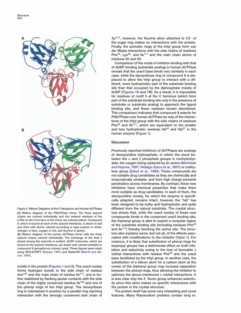
Figure 4. Ribbon Diagrams of the P. falciparum and Human dUTPases dt(A) Ribbon diagram of the PfdUTPase trimer. The three subunit
chains are colored individually and the ordered residues of the cLCRs on the front face of the trimer are colored yellow. Compound t4, which is bound at each of the subunit interfaces, is drawn in ball oand stick with atoms colored according to type (carbon in white,
anitrogen in blue, oxygen in red, and fluorine in green).t(B) Ribbon diagram of the human dUTPase trimer with the threecsubunit chains colored individually. The exchange of the final β
strand among the subunits is evident. dUDP molecules, which are ibound at the subunit interfaces, are drawn and colored similarly to icompound 4 (phosphorus colored cyan). These figures were made busing MOLSCRIPT (Kraulis, 1991) and Raster3D (Merritt and Ba-
ocon, 1997).bs
cmotifs in the protein (Figures 1 and 6). The uracil moietyforms hydrogen bonds to the side chain of residue boAsn103 and the main chain of residue Ile117, and is fur-
ther stabilized by flanking apolar contacts with the side iichain of the highly conserved residue Ile108 and one of
the phenyl rings of the trityl group. The deoxyribose tring is maintained in position by a parallel ring stackinginteraction with the strongly conserved side chain of f
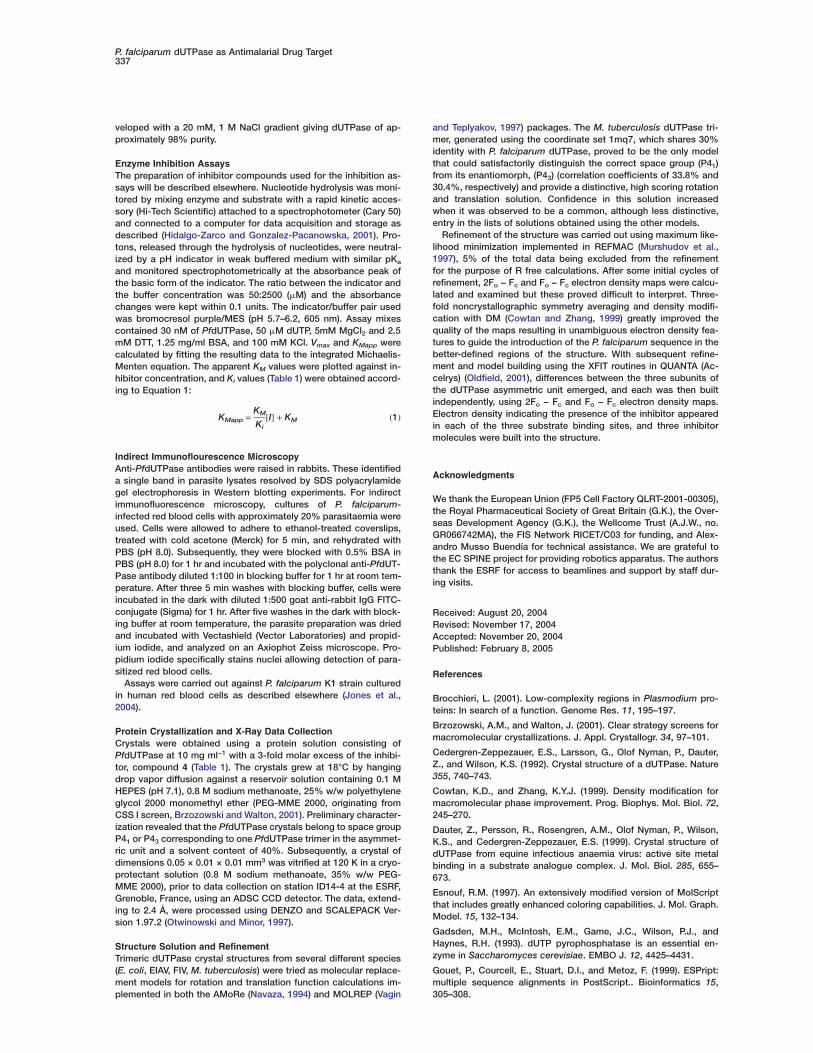
yr112, however, the fluorine atom attached to C3# ofhe sugar ring makes no interactions with the protein.inally, the aromatic rings of the trityl group form vaner Waals interactions with the side chains of residueshe46, Lys96, and Ile117 and the main chain atoms of
esidues 92 and 93.Comparison of the mode of inhibitor binding with that
f dUDP binding (substrate analog) in human dUTPaseeveals that the uracil base binds very similarly in eachase, while the deoxyribose ring of compound 4 is dis-laced to allow the trityl group to interact with a dif-
erent, more hydrophobic part of the substrate bindingite than that occupied by the diphosphate moiety ofUDP (Figures 7A and 7B). As a result, it is impossible
or residues of motif 5 at the C terminus (which formart of the substrate binding site only in the presence ofubstrate or substrate analog) to approach the ligandinding site, and these residues remain disordered.his comparison indicates that compound 4 selects forfdUTPase over human dUTPase by way of the interac-
ions of the trityl group with the side chains of residueshe46 and Ile117, which are equivalent to the smallernd less hydrophobic residues Val42 and Gly87 in theuman enzyme (Figure 1).
iscussion
reviously reported inhibitors of dUTPases are analogsf deoxyuridine triphosphate, in which the bond be-ween the α and β phosphate groups is nonhydrolyz-ble, the oxygen being replaced by an amino (McIntoshnd Haynes, 1997; Hidalgo-Zarco et al., 2001) or methy-
ene group (Zalud et al., 1995). These compounds areot suitable drug candidates as they are chemically andnzymatically unstable, and their high charge preventsenetration across membranes. By contrast, these new
nhibitors have chemical properties that make themore suitable as drug candidates. In each of them, theeoxyuridine moiety, for which the enzyme is specifi-ally adapted, remains intact, however, the “tail” haseen designed to be bulky and hydrophobic and quiteifferent from the natural substrate. The crystal struc-ure shows that, while the uracil moiety of these newompounds binds in the conserved uracil binding site,he triphenyl group is able to exploit a nonpolar regionf the substrate binding site (including residues Phe46
nd Ile117) thereby blocking the active site. The struc-ure also explains some, but not all, of the effects asso-iated with modifications to the inhibitor (Table 1). For
nstance, it is likely that substitution of phenyl rings forsopropyl groups has a detrimental effect on both inhi-ition and selectivity owing to the loss of favorable πrbital interactions with residue Phe46 and the uracilase facilitated by the trityl group. In another case, theubstitution of a silicon atom for a carbon atom at theenter of the triphenyl group may increase separationetween the phenyl rings, thus allowing the inhibitor toptimize the above-mentioned π orbital interactions. It
s less clear why the 3# fluoro group enhances selectiv-ty since this atom makes no specific interactions withhe protein in the crystal structure.
The protein itself has some very interesting and noveleatures. Many Plasmodium proteins contain long in-

P. falciparum dUTPase as Antimalarial Drug Target335
Figure 5. Stereoview Illustration Showing theAtomic Coordinates for the PfdUTPase In-hibitor Compound 4, with Associated Fo − Fc
Electron Density (Omit Map) Contoured at 3σAtoms are colored according to type (carbonin black, oxygen in red, nitrogen in blue, andfluorine in green). This figure was madeusing BOBSCRIPT (Esnouf, 1997).
loops gathered together at the top of the trimer, inde-
Figure 6. Schematic Illustration of the Inhibitor Binding Site inPfdUTPase
The atoms of the inhibitor compound 4 (Dux 1D) are colored ac-cording to type and a water molecule is shown as a turquoisesphere. Residues which interact with the inhibitor, either by way ofhydrogen bonding (dashed lines) or hydrophobic interactions (ra-dial spokes) are labeled by residue number and subunit identifier(A or C). This figure was made using LIGPLOT (Wallace et al., 1995).
they are not totally without structure and probably con-
Figure 7. Stereoview Illustrations of the Inhibitor Binding Sites inthe P. falciparum and Human dUTPases
(A) The human enzyme (green atoms) is superimposed on theP. falciparum enzyme (blue atoms) by aligning the uracil moiety oftheir respective inhibitors.(B) Orthogonal view of the inhibitor binding sites in the two en-zymes, colored as above.
sertions known as low complexity regions (LCRs),which often contain distinctive homopeptide repeats ofhydrophilic amino acid residues. The prevailing wisdomis that these areas lack order, forming loops betweenother secondary structure elements in the protein. Theirfunction, if any, is not known, although one tenable ar-gument is that they contribute to immune evasion dur-ing parasite invasion (Brocchieri, 2001). The LCRs ofPfdUTPase, comprising residues 59 to 83 in each sub-unit, are a typical case in point. They form three large
pendent of the main protein fold. They do not interferewith the essential elements of the protein, namely thesubstrate binding site and trimer formation; however,

Structure336
Figure 8. Stereoview Illustration Showing a Superimposition of Three Subunits from a Selection of Different Trimeric dUTPases
P. falciparum in red, human in green, and M. tuberculosis in blue.Chain termini are labeled N and C, and the beginning and end of the disordered regions of the low-complexity region in PfdUTPase areindicated by residue number. This figure was made using MOLSCRIPT (Kraulis, 1991).
tribute to trimer stability. Their close proximity to the C lDtermini would not, on stereochemical grounds, inhibit
the C-terminal domain swapping observed in other tri- tdmeric dUTPases so it is unlikely that these two features
are linked. adA superimposition of one subunit from three of the
trimeric dUTPase crystal structures shows that, despite crlow sequence homology in many areas of these pro-
teins, the fold is very well conserved (Figures 1 and 8). wgThe only significant conformational variation apart from
the C-terminal flip in the Plasmodium enzyme is toward aathe N terminus, where residues 8–19 in the human en-
zyme and their equivalents in the other structures form aPa variety of distorted β strands. In PfdUTPase this seg-
ment contains four additional residues and forms aEshort α helix. Such variations in conformation are of
little consequence to the enzyme since they occur onE
the outside surface of the trimer where evolutionary re- Cstraints connected with substrate recognition and tri- q
fmer formation are at their lowest. On the other hand, actwo-residue insertion after residue 103 in PfdUTPaseuplaces residue Ser104 at the heart of the trimer, whereTits hydrogen bonding capability is used to maximumC
effect in stabilizing the trimer. This feature is also seen Bin the trimeric dUTPase from M. tuberculosis (acces- s
(sion code 1mq7), and a similar insertion in the E. colienzyme results in van der Waals interactions between
Bthree symmetry-related Leu85 side chains.eAs more information is gathered about dUTPases re-w
garding structure and function, so these enzymes, with mtheir essential cellular activities, are being recognized T
bfor their potential as drug targets against a range ofldiseases (McIntosh and Haynes, 1997; Hidalgo-Zarco4and Gonzalez-Pacanowska, 2001). The present studyefocuses on the dUTPase from P. falciparum as a candi-c
date target for drugs directed against the most severe pform of malaria. We have demonstrated that expression (
oof the enzyme occurs throughout the parasite in the
ate trophozoite and schizont stages coinciding withNA replication, and have discovered a series of selec-
ive inhibitors whose chemical properties give themrug-like properties. These compounds also exhibitntiparasite activity, making them viable leads for drugevelopment. The crystal structure of PfdUTPase inomplex with one of these inhibitors explains the gene-al mode of inhibition as well as the basis for selectivity,hich derives from the combined bulk of the tritylroup and the more subtle hydrogen bonding inter-ctions of the uracil base. This structure will serve as
template for further rounds of drug developmentimed at increasing inhibition of, and selectivity for,fdUTPase.
xperimental Procedures
xpression and Purification of PfdUTPaseonserved motifs of the human dUTPase enzyme were used as auery to identify the Pfdut gene in the sequence of the Plasmodium
alciparum 3D7 strain held in the www.tigr.org database. The entireoding sequence was amplified by PCR from a template cDNAsing the oligonucleotides ATG-PFdut (CATATGCATTTAAAAATTGATGTCTG) and TGA-PFdut (GGATCC-TCAATATTTATTATCGATGTGATC). These primers were designed to introduce NdeI andamHI restriction sites at the 5# and 3# ends of the Pfdut codingequence for convenient cloning in the expression vector pET11Stratagene).
Recombinant P. falciparum dUTPase was expressed in E. coliL21 (DE3) cells which had been transformed with the pET11Pfdutxpression vector. Cell pellets from a two liter IPTG-induced cultureere resuspended in 40 ml of 20 mM sodium acetate (pH 5.5), 50M NaCl, 5 mM MgCl2, 1 mM DTT (buffer A), and 20 �M PMSF.he cells were lysed by sonication, and the cell extract was clearedy centrifugation at 15,000 rpm for 45 min. The supernatant was
oaded onto a 50 ml phosphocellulose (Whatman P-11) column at°C and eluted with a 50 mM, 2 M NaCl gradient in buffer A. Thenzyme was then dialyzed against buffer A, prior to gel filtrationhromatography on a 120 ml Superdex 75 column at 4°C. As a finalurification step, the enzyme was dialyzed against 20 mM Tris-HCl
pH 8.5), 20 mM NaCl, 5 mM MgCl2, and 1 mM DTT and loadednto a Mono Q column at room temperature. The column was de-
P. falciparum dUTPase as Antimalarial Drug Target337
veloped with a 20 mM, 1 M NaCl gradient giving dUTPase of ap-proximately 98% purity.
Enzyme Inhibition AssaysThe preparation of inhibitor compounds used for the inhibition as-says will be described elsewhere. Nucleotide hydrolysis was moni-tored by mixing enzyme and substrate with a rapid kinetic acces-sory (Hi-Tech Scientific) attached to a spectrophotometer (Cary 50)and connected to a computer for data acquisition and storage asdescribed (Hidalgo-Zarco and Gonzalez-Pacanowska, 2001). Pro-tons, released through the hydrolysis of nucleotides, were neutral-ized by a pH indicator in weak buffered medium with similar pKa
and monitored spectrophotometrically at the absorbance peak ofthe basic form of the indicator. The ratio between the indicator andthe buffer concentration was 50:2500 (�M) and the absorbancechanges were kept within 0.1 units. The indicator/buffer pair usedwas bromocresol purple/MES (pH 5.7–6.2, 605 nm). Assay mixescontained 30 nM of PfdUTPase, 50 �M dUTP, 5mM MgCl2 and 2.5mM DTT, 1.25 mg/ml BSA, and 100 mM KCl. Vmax and KMapp werecalculated by fitting the resulting data to the integrated Michaelis-Menten equation. The apparent KM values were plotted against in-hibitor concentration, and Ki values (Table 1) were obtained accord-ing to Equation 1:
KMapp =KM
Ki[I] + KM (1)
Indirect Immunoflourescence MicroscopyAnti-PfdUTPase antibodies were raised in rabbits. These identifieda single band in parasite lysates resolved by SDS polyacrylamidegel electrophoresis in Western blotting experiments. For indirectimmunofluorescence microscopy, cultures of P. falciparum-infected red blood cells with approximately 20% parasitaemia wereused. Cells were allowed to adhere to ethanol-treated coverslips,treated with cold acetone (Merck) for 5 min, and rehydrated withPBS (pH 8.0). Subsequently, they were blocked with 0.5% BSA inPBS (pH 8.0) for 1 hr and incubated with the polyclonal anti-PfdUT-Pase antibody diluted 1:100 in blocking buffer for 1 hr at room tem-perature. After three 5 min washes with blocking buffer, cells wereincubated in the dark with diluted 1:500 goat anti-rabbit IgG FITC-conjugate (Sigma) for 1 hr. After five washes in the dark with block-ing buffer at room temperature, the parasite preparation was driedand incubated with Vectashield (Vector Laboratories) and propid-ium iodide, and analyzed on an Axiophot Zeiss microscope. Pro-pidium iodide specifically stains nuclei allowing detection of para-sitized red blood cells.
Assays were carried out against P. falciparum K1 strain culturedin human red blood cells as described elsewhere (Jones et al.,2004).
Protein Crystallization and X-Ray Data CollectionCrystals were obtained using a protein solution consisting ofPfdUTPase at 10 mg ml−1 with a 3-fold molar excess of the inhibi-tor, compound 4 (Table 1). The crystals grew at 18°C by hangingdrop vapor diffusion against a reservoir solution containing 0.1 MHEPES (pH 7.1), 0.8 M sodium methanoate, 25% w/w polyethyleneglycol 2000 monomethyl ether (PEG-MME 2000, originating fromCSS I screen, Brzozowski and Walton, 2001). Preliminary character-ization revealed that the PfdUTPase crystals belong to space groupP41 or P43 corresponding to one PfdUTPase trimer in the asymmet-ric unit and a solvent content of 40%. Subsequently, a crystal ofdimensions 0.05 × 0.01 × 0.01 mm3 was vitrified at 120 K in a cryo-protectant solution (0.8 M sodium methanoate, 35% w/w PEG-MME 2000), prior to data collection on station ID14-4 at the ESRF,Grenoble, France, using an ADSC CCD detector. The data, extend-ing to 2.4 Å, were processed using DENZO and SCALEPACK Ver-sion 1.97.2 (Otwinowski and Minor, 1997).
Structure Solution and RefinementTrimeric dUTPase crystal structures from several different species(E. coli, EIAV, FIV, M. tuberculosis) were tried as molecular replace-ment models for rotation and translation function calculations im-plemented in both the AMoRe (Navaza, 1994) and MOLREP (Vagin
and Teplyakov, 1997) packages. The M. tuberculosis dUTPase tri-mer, generated using the coordinate set 1mq7, which shares 30%identity with P. falciparum dUTPase, proved to be the only modelthat could satisfactorily distinguish the correct space group (P41)from its enantiomorph, (P43) (correlation coefficients of 33.8% and30.4%, respectively) and provide a distinctive, high scoring rotationand translation solution. Confidence in this solution increasedwhen it was observed to be a common, although less distinctive,entry in the lists of solutions obtained using the other models.
Refinement of the structure was carried out using maximum like-lihood minimization implemented in REFMAC (Murshudov et al.,1997), 5% of the total data being excluded from the refinementfor the purpose of R free calculations. After some initial cycles ofrefinement, 2Fo − Fc and Fo − Fc electron density maps were calcu-lated and examined but these proved difficult to interpret. Three-fold noncrystallographic symmetry averaging and density modifi-cation with DM (Cowtan and Zhang, 1999) greatly improved thequality of the maps resulting in unambiguous electron density fea-tures to guide the introduction of the P. falciparum sequence in thebetter-defined regions of the structure. With subsequent refine-ment and model building using the XFIT routines in QUANTA (Ac-celrys) (Oldfield, 2001), differences between the three subunits ofthe dUTPase asymmetric unit emerged, and each was then builtindependently, using 2Fo − Fc and Fo − Fc electron density maps.Electron density indicating the presence of the inhibitor appearedin each of the three substrate binding sites, and three inhibitormolecules were built into the structure.
Acknowledgments
We thank the European Union (FP5 Cell Factory QLRT-2001-00305),the Royal Pharmaceutical Society of Great Britain (G.K.), the Over-seas Development Agency (G.K.), the Wellcome Trust (A.J.W., no.GR066742MA), the FIS Network RICET/C03 for funding, and Alex-andro Musso Buendía for technical assistance. We are grateful tothe EC SPINE project for providing robotics apparatus. The authorsthank the ESRF for access to beamlines and support by staff dur-ing visits.
Received: August 20, 2004Revised: November 17, 2004Accepted: November 20, 2004Published: February 8, 2005
References
Brocchieri, L. (2001). Low-complexity regions in Plasmodium pro-teins: In search of a function. Genome Res. 11, 195–197.
Brzozowski, A.M., and Walton, J. (2001). Clear strategy screens formacromolecular crystallizations. J. Appl. Crystallogr. 34, 97–101.
Cedergren-Zeppezauer, E.S., Larsson, G., Olof Nyman, P., Dauter,Z., and Wilson, K.S. (1992). Crystal structure of a dUTPase. Nature355, 740–743.
Cowtan, K.D., and Zhang, K.Y.J. (1999). Density modification formacromolecular phase improvement. Prog. Biophys. Mol. Biol. 72,245–270.
Dauter, Z., Persson, R., Rosengren, A.M., Olof Nyman, P., Wilson,K.S., and Cedergren-Zeppezauer, E.S. (1999). Crystal structure ofdUTPase from equine infectious anaemia virus: active site metalbinding in a substrate analogue complex. J. Mol. Biol. 285, 655–673.
Esnouf, R.M. (1997). An extensively modified version of MolScriptthat includes greatly enhanced coloring capabilities. J. Mol. Graph.Model. 15, 132–134.
Gadsden, M.H., McIntosh, E.M., Game, J.C., Wilson, P.J., andHaynes, R.H. (1993). dUTP pyrophosphatase is an essential en-zyme in Saccharomyces cerevisiae. EMBO J. 12, 4425–4431.
Gouet, P., Courcell, E., Stuart, D.I., and Metoz, F. (1999). ESPript:multiple sequence alignments in PostScript.. Bioinformatics 15,305–308.

Structure338
el-Hajj, H.H, Zhang, H., and Weiss, B. (1988). Lethality of a dut Sm(deoxyuridine triphosphatase) mutation in Escherichia coli. J. Bac-
teriol. 170, 1069–1075. b
SHarkiolaki, M., Dodson, E.J., Bernier-Villamor, V., Turkenburg, J.P.,González-Pacanowska, D., and Wilson, K.S. (2003). The crystal t
cstructure of Trypanosoma cruzi dUTPase reveals a novel dUTP/dUDP binding fold. Structure 12, 1–20. t
VHidalgo-Zarco, F., and Gonzalez-Pacanowska, D. (2001). Trypano-somal dUTPases as potential targets for drug design. Curr. Protein g
1Pept. Sci. 4, 389–397.
Hidalgo-Zarco, F., Camacho, A., Bernier-Villamor, V., Ruiz-Perez, WPL.M., and Gonzalez-Pacanowska, D. (2001). Kinetic properties and
inhibition of the dimeric dUTPase-dUDPase from Leishmania ma- gjor. Protein Sci. 10, 1426–1433. W
pHuffman, J.L., Hong, L., White, R.H., and Tainer, J.A. (2003). Struc-tural basis for recognition and catalysis by the bifunctional dCTP Wdeaminase and dUTPase from Methanococcus jannaschii. J. Mol. nBiol. 331, 885–896. ZInselburg, J., and Banyal, H.S. (1984). Synthesis of DNA during the Iasexual cycle of Plasmodium falciparum in culture. Mol. Biochem. sParasitol. 10, 79–87. 1Jones, S.M., Urch, J.E., Brun, R., Harwood, J.L., Berry, C., and Gil-bert, I.H. (2004). Analogues of thiolactomycin as potential anti- Amalarial and anti-trypanosomal agents. Bioorg. Med. Chem. 12,683–692. T
iKraulis, P.J. (1991). MOLSCRIPT—a program to produce both de-tailed and schematic plots of protein structures. J. Appl. Crystal-logr. 24, 946–950.
Ladner, R.D., and Caradonna, S.J. (1997). The human dUTPasegene encodes both nuclear and mitochondrial isoforms. Differentialexpression of the isoforms and characterization of a cDNA encod-ing the mitochondrial species. J. Biol. Chem. 272, 19072–19080.
Le Roch, K.G., Zhou, Y.Y., Blair, P.L., Grainger, M., Moch, J.K.,Haynes, J.D., De La Vega, P., Holder, A.A., Batalov, S., Carucci, D.J.,and Winzeler, E.A. (2003). Discovery of gene function by expressionprofiling of the malaria parasite life cycle. Science 301, 1503–1508.
McIntosh, E.M., and Haynes, R.H. (1997). dUTP pyrophosphataseas a potential target for chemotherapeutic drug development. ActaBiochim. Pol. 44, 159–172.
Merritt, E.A., and Bacon, D.J. (1997). Raster3D: photorealistic mo-lecular graphics. Methods Enzymol. 277, 505–524.
Miller, L.H., Baruch, D.I., Marsh, K., and Doumbo, O.K. (2002). Thepathogenic basis of malaria. Nature 415, 673–679.
Mol, C.D., Harris, J.M., McIntosh, E.M., and Tainer, J.A. (1996). Hu-man dUTP pyrophosphatase: uracil recognition by a β hairpin andactive sites formed by three separate subunits. Structure 4, 1077–1092.
Murshudov, G.N., Vagin, A.A., and Dodson, E.J. (1997). Refinementof macromolecular structures by the maximum-likekihood method.Acta Crystallogr. D Biol. Crystallogr. 53, 240–255.
Navaza, J. (1994). AMoRe—an automated package for molecularreplacement.. Acta Crystallogr. A. 50, 157–163.
Nord, J., Nyman, P., Larsson, G., and Drakenberg, T. (2001). TheC-terminus of dUTPase: observation on flexibility using NMR. FEBSLett. 492, 228–232.
Oldfield, T.J. (2001). A number of real-space torsion-angle refine-ment techniques for proteins, nucleic acids, ligands and solvent.Acta Crystallogr. D Biol. Crystallogr. 57, 82–94.
Otwinowski, Z., and Minor, W. (1997). Processing of X-ray diffrac-tion data collected in oscillation mode. Methods Enzymol. 276,307–326.
Prasad, G.S., Stura, E.A., McRee, D.E., Laco, G.S., HasselkusLight,C., Elder, J.H., and, and Stout, C.D. (1996). Structure of feline immu-nodeficiency virus dUTP pyrophosphatase and its nucleotide com-plexes in three crystal forms. Protein Sci. 5, 2429–2437.
Richie, T.L., and Saul, A. (2002). Progress and challenges for ma-laria vaccines. Nature 415, 694–701.
Ridley, R.G. (2002). Medical need, scientific opportunity and thedrive for antimalarial drugs. Nature 415, 686–693.
chlunegger, M.P., Bennett, M.J., and Eisenberg, D. (1997). Oligo-er formation by 3D domain swapping: a model for protein assem-ly and misassembly. Adv. Protein Chem. 50, 61–122.
pielmann, T., and Beck, H.P. (2000). Analysis of stage-specificranscription on Plasmodium falciparum reveals a set of genes ex-lusively transcribed in ring stage parasites. Mol. Biochem. Parasi-ol. 111, 453–458.
agin, A., and Teplyakov, A. (1997). MOLREP: an automated pro-ram for molecular replacement. J. Appl. Crystallogr. 30, 1022–025.
allace, A.C., Laskowski, R.A., and Thornton, J.M. (1995). LIG-LOT—a program to generate schematic diagrams of protein li-and interactions. Protein Eng. 8, 127–135.
hite, J.H., and Kilbey, B.J. (1996). DNA replication in the malariaarasite. Parasitol. Today 12, 151–155.
orld Health Organization (2003). World Health Report 2003, An-ex 2.
alud, P., Wachs, W.O., Nyman, P.O., and Zeppezauer, M. (1995).nhibition of the proliferation of human cancer cells in-vitro by sub-trate-analogous inhibitors of dUTPase. Adv. Exp. Med. Biol. 370,35–138.
ccession Numbers
he atomic coordinates and structure factors have been depositedn the Protein Data Bank, (www.ebi.ac.uk) with PDB ID code 1VYQ.
Copyright © 2022 FDOKUMEN