
Substituent Effects on the Photogeneration and Selectivity of Triaryl Vinyl Cations
Upload
khangminh22Category
view
0download
0
Understanding Selectivity of Hard and Soft Metal Cationswithin Biological Systems Using the Subvalence
Concept. 1. Application to Blood Coagulation: DirectCation-Protein Electronic Effects versus Indirect
Interactions through Water NetworksB. de Courcy,†,‡ L. G. Pedersen,§ O. Parisel,†,‡ N. Gresh,| B. Silvi,†,‡ J. Pilme,†,‡,⊥ and
J.-P. Piquemal*,†,‡
UPMC UniV Paris 06, UMR 7616, Laboratoire de Chimie Theorique, case courrier137, 4 place Jussieu, F-75005, Paris, France, CNRS, UMR 7616, Laboratoire deChimie Theorique, case courrier 137, 4 place Jussieu, F-75005, Paris, France,
Laboratory of Structural Biology, National Institute of EnVironmental Health, Sciences,Research Triangle Park, North Carolina 27709, Laboratoire de Pharmacochimie
Moleculaire et Cellulaire, U648 INSERM, UFR Biomedicale, UniVersite ParisDescartes, 45, rue des Saints-Peres, 75006, Paris, France, UniVersite de Lyon,
UniVersite Lyon 1, Faculte de Pharmacie, F-69373 Lyon, Cedex 08, FranceReceived October 13, 2009
Abstract: Following a previous study by de Courcy et al. (Interdiscip. Sci. Comput. Life Sci. 2009,1, 55-60), we demonstrate in this contribution, using quantum chemistry, that metal cations exhibita specific topological signature in the electron localization of their density interacting with ligandsaccording to their “soft” or “hard” character. Introducing the concept of metal cation subvalence, weshow that a metal cation can split its outer-shell density (the so-called subvalent domains or basins)according to it capability to form a partly covalent bond involving charge transfer. Such behavior isinvestigated by means of several quantum chemical interpretative methods encompasing thetopological analysis of the Electron Localization Function (ELF) and Bader’s Quantum Theory ofAtoms in Molecules (QTAIM) and two energy decomposition analyses (EDA), namely, the ReducedVariational Space (RVS) and Constrained Space Orbital Variations (CSOV) approaches. Furtherrationalization is performed by computing ELF and QTAIM local properties such as electrostaticdistributed moments and local chemical descriptors such as condensed Fukui functions and dualdescriptors. These reactivity indexes are computed within the ELF topological analysis in additionto QTAIM offering access to a nonatomic reactivity local index, for example, on lone pairs. We applythis “subvalence” concept to study the cation selectivity in enzymes involved in blood coagulation(GLA domains of three coagulation factors). We show that the calcium ions are clearly able to formpartially covalent charge transfer networks between the subdomain of the metal ion and thecarboxylate oxygen lone pairs, whereas magnesium does not have such ability. Our analysis alsoexplains the different role of two groups (high affinity and low affinity cation binding sites) present inGLA domains. If the presence of Ca(II) is mandatory in the central “high affinity” region to conservea proper folding and a charge transfer network, external sites are better stabilized by Mg(II), ratherthan Ca(II), in agreement with the experiment. The central role of discrete water molecules is alsodiscussed in order to understand the stabilities of the observed X-ray structures of the GLA domain.Indeed, the presence of explicit water molecules generating indirect cation-protein interactionsthrough water networks is shown to be able to reverse the observed electronic selectivity occurringwhen cations directly interact with the Gla domain without the need of water.
J. Chem. Theory Comput. 2010, 6, 1048–10631048
10.1021/ct100089s 2010 American Chemical SocietyPublished on Web 03/12/2010

Introduction
Metal cations play a critical role in many biological systems.In most cases, they are specific in their ability to bind toproteins and thereby confer appropriate biological functionor activity. For example, the presence of calcium cations isimportant in blood clotting, signal transduction, and celldivision. Specifically, in the case of blood clotting, it hasbeen experimentally observed that the presence of calciumis required for clot formation.1 Indeed, calcium cationsdirectly participate in the binding and folding of theγ-carboxyglutamic acid (Gla)-rich domain that is commonto the vitamin-K-dependent serine proteases present in theblood coagulation cascade.1 Blood plasma does not coagulatein the sole presence of magnesium ions,2,3 an effect attributedto the concomitant lack of binding of the Gla residues tonegatively charged phospholipids when only magnesium ionsare present. More precisely, recent X-ray crystal structureswith a mixture of both Ca(II) and Mg(II) show that theN-terminus ω-loop segment that is thought to be the keydeterminant for the binding of the GLA domain to mem-branes has a disordered structure when only magnesium ionsare present.4 In the presence of calcium ions, GLA domainshave been crystallized and a strong Gla-calcium network hasbeen observed with varying degrees of calcium ion coordina-tion. However, despite the mandatory presence of calciumions needed to structure the GLA domain and allowing it topoint the three hydrophobic (or anionic) residues forming“the keel” in the direction of negatively charged phospho-lipids found in cellular membranes, it has also beenexperimentally demonstrated that the presence of magnesiumions in addition to calcium enhances the affinity of theenzyme for both the membrane and cofactors.5
The ability of the calcium ion to coordinate watermolecules with flexible coordination is thought to beimportant for this function. At the theoretical level, recentfirst-principle Car-Parrinello6 and force-field simulation7
studies have reported an in-depth description of the hydrationshells and of the preferred coordination numbers for thecalcium and magnesium cations. Although we know that theresidence time for water on a magnesium ion is substantiallylonger than that for a calcium ion,8 we as yet do not knowthe physical origins of the differences between the bindingof calcium and magnesium ions in biological systems, as noclear electron structure-biological actiVity relation has beenuncoVered in realistic model systems. In this context, weproposed to apply modern quantum chemistry to study thenature of the binding of calcium and magnesium ions inmodel systems of factors VII, IX, and X, the structures of
which are based on protein structures extracted from theProtein Data Bank.
Outline
In this work, we will first focus on the fundamentalinteractions occurring in such systems between the metalcations and their environment. Indeed, thanks to X-raystudies, we know that interactions occur between the calciumor magnesium cations and carboxylate moieties. We will thenpresent an extensive ab initio study of the binding of severalhard and soft metal cations to carboxylates in differentposition. Then, applying quantum topological approachessuch as Atoms in Molecules (QTAIM)9 theory and thetopological analysis of the Electron Localization Function(ELF),10 we shed light on the origin of the different behaviorof the electronic structure of metal cation and ligands so asto unravel the specific cation topological signature. Theobserved topological descriptions will be complemented byintermolecular interaction energy decomposition using theReduced Variational Space approach (RVS)11a and theConstrained-Space Orbital variation (CSOV),11b which pro-vide insights about the nature, covalent or electrostatic, ofthe bonding between the cations and carboxylate. To connectthis work to conceptual Density Functional Theory (DFT),we will also provide a detailed analysis by means of localchemical descriptors such as the condensed Fukui functions.In a second part, we will present a study of models of theGLA domain from factors IX, VII, and X of the bloodcoagulation process using ELF computations complementedby multimolecular RVS energy decomposition analyses.
Method
A. Topological Analysis of ELF. The topological analy-sis relies on a partition of the molecular space achieved inthe framework of the theory of gradient dynamics appliedto a scalar potential function, say V(r), called “potentialfunction”, which contains the physical or chemical informa-tion. This partitioning gives rise to a set of nonoverlappingmolecular volumes called basins localized around the maximaof the ELF (the attractors of the vector field). The boundariesbetween these basins, the separatrices, are zero-flux surfacessatisfying the following condition that every point r is a unitvector normal to the surface. In the QTAIM theory of Bader,9
the scalar function is the electron density distribution whosebasins have their attractors located on the nuclei and whichare therefore associated with the atoms that constitute themolecule. In order to recover a chemist’s representation ofa molecule consistent with Lewis’s valence picture, one mustuse another “local” function that is able to describe theelectron pair regions. For almost two decades, the topologicalanalysis of the ELF has been extensively developed and usedto analyze chemical bonding and to investigate chemicalreactivity (for reviews, see refs 12 and 13). The ELF can beinterpreted as a signature of the electron pair distribution.The relationship of the kernel of ELF to pair functions hasbeen established,13 but in contrast to these latter, the ELFvalues are confined in the [0,1] range by a Lorentziantransformation which facilitates the interpretation. The basins
* Corresponding author e-mail: [email protected].† UPMC Univ Paris 06.‡ CNRS, UMR 7616.§ National Institute of Environmental Health Sciences.|Universite Paris Descartes.⊥ Universite Lyon 1.
Selectivity of Hard and Soft Metal Cations J. Chem. Theory Comput., Vol. 6, No. 4, 2010 1049

of the ELF are either core basins, labeled C(A) correspondingto the inner shells of atom A and encompassing its nucleus(if Z > 2), or valence basins denoted by V(A,B,C...), whereA, B, and C are the element symbols. A valence basin canbelong to a single atomic valence shellsin this case, V(A)corresponds to a lone pairsor be shared by several atomsand associated to a bond V(A,B).
The ELF basins closely match the electronic domains of theVSEPR12 model, and it has been shown that the interbasinrepulsion provides a map onto the Gillespie-Nyholm ruleswhich describe molecular geometry.14,15 It has been recentlyshown that non-VSEPR structures which occur around neutralatoms belonging to the fourth and higher periods can beexplained by considering the structure of the external core shellbasins16 and are hereafter referred to as subvalence basins.Details about the ELF analysis can also be found in a recentreview paper dealing with the application of ELF to systemsof biological interest.17
B. Integration of Local Properties within the ELFor QTAIM Partition. 1. Local Electrostatic Moments. Froma quantitative point of view, a population analysis can becarried out by integrating the electron density distributionover the basin volumes. Recently, the distributed momentsanalysis based on the QTAIM partition9 and on the ELFbasins (DEMEP)18a has been introduced, which enables anextended discussion on the nature of bonding in molecules.In this paper, we use more specifically the first moment(denoted as M1), which represents the local dipolar polariza-tion of the density.
That way, the Distributed Electrostatic Moments based onthe ELF Partition (DEMEP) allows the calculation of localmoments located at nonatomic centers such as lone pairs, σbonds, and π systems. Local dipole contributions have beenshown to be useful in rationalizing inductive polarizationeffects and typical hydrogen bond interactions. Moreover,bond quadrupole polarization moments being related to a πcharacter enable a discussion of bond multiplicities andsorting of the families of molecules according to their bondorder.
To summarize, the M0(Ω) monopole term corresponds tothe negative of the population (denoted N):
The first moments or dipolar polarization components ofthe charge distribution are defined by three-dimensionalintegrals for a given basin Ω according to
where Xc, Yc, and Zc are the Cartesian coordinates of thebasin centers.
The five second-moment spherical tensor components canalso be calculated and are defined as the quadrupolarpolarization terms. They can be seen as the ELF basin
equivalents to the atomic quadrupole moments introducedby Popelier9c in the case of an QTAIM analysis:
The first- or second-moment basin magnitude is thendefined as the square root of the sum of squared components:
Thanks to the invariance of the magnitude of any multipolerank (|M1| or |M2|) with respect to the axis for a given bondor lone pair, the approach allows us to compare the dipolaror quadrupolar polarization of a given basin in differentchemical environments.
2. Fukui Functions as Local Chemical Descriptors. Be-yond the computations of local distributed electrostaticmoments, it is also possible to access the topological partitionof local chemical descriptors. Among the numerous chemicalindicators, the Fukui functions,18b,c based on the relativeproperties of the Highest Occupied Molecular Orbital(HOMO) and the Lowest Unoccupied Molecular Orbital(LUMO), are interesting as they are particularly useful forthe interpretation of chemical reactivity, particularly towardnucleophiles or electrophiles.18d Indeed, following Parr andYang, conceptual DFT provides such functions defined interms of the variation of the chemical potential with respectto changes in the external potential V(r) or equivalently asthe derivative of the electron density with respect to changesin the number of electrons N.
Three Fukui functions are usually evaluated: f+(r), f-(r), andf 0(r)
They are sometime also associated with the computation ofanother value called the dual descriptor (denoted ∆f(r)18e,f)and calculated upon the f +(r) and the f -(r) functions:
The f +(r) function usually characterizes the reactivity ofa given species toward nucleophilic attack (in that case, ∆f(r)> 0), whereas the f -(r) function usually characterizes the
M0(Ω) ) -∫ΩF(r) dτ ) -N(Ω) (1)
M1,x(Ω) ) -∫Ω(x - Xc)F(r) dτ
M1,y(Ω) ) -∫Ω(y - Yc)F(r) dτ
M1,z(Ω) ) -∫Ω(z - Zc)F(r) dτ
(2)
M2,zz(Ω) ) -12 ∫Ω
[3(z - Zc)2 - r2]F(r) dτ
M2,x2-y2(Ω) ) -√32 ∫Ω
[(x - Xc)2 - (y - Yc
2)]F(r) dτ
M2,xy(Ω) ) -√3∫Ω(x - Xc)(y - Yc)F(r) dτ
M2,xz(Ω) ) -√3∫Ω(x - Xc)(z - Zc)F(r) dτ
M2,yz(Ω) ) -√3∫Ω(y - Yc)(z - Zc)F(r) dτ
(3)
|M(Ω)| ) ∑i
Mi(Ω)2 (4)
f(r) ) [ δµδV(r)]N
) [∂F(r)∂N ]V(r)
(5)
f +(r) ) (∂F(r)∂N )V(r)
+≈ (δELUMO
δV(r) )N
≈ FLUMO(r)
f -(r) ) (∂F(r)∂N )V(r)
-≈ (δEHOMO
δV(r) )N
≈ FHOMO(r)
f 0(r) ) 12
[f +(r) + f -(r)]
(6)
∆f(r) ) (f +(r) - f -(r))N (7)
1050 J. Chem. Theory Comput., Vol. 6, No. 4, 2010 de Courcy et al.

reactivity of a given species toward electrophilic attack (inthat case, ∆f(r) < 0). When ∆f(r) ) 0, the site’s reactivityshould be equilibrated. As an analytic expression of suchfunction is not available, it remains possible to compute themnumerically using finite differences (see for example ref 18gand references therein).
In that case, f -(r), f +(r), and f 0(r) can be computed asfollows:
qx(N) represents the atomic charges associated with atomx within the N-electron species. Recently, it has beenshown18h,i that it is possible to use a QTAIM condensationscheme for Frontier Molecular Orbitals Fukui functions usingfinite differences within a topological partition. Such anapproach has been shown to be particularly stable, havingsome advantage on other atomic evaluations of the Fukuifunctions scheme:
where h denotes HOMO, and l, LUMO, and the subscript xunder the integration sign indicates that the integration hasto be performed only within the particular atomic domainof atom x.
As previouslsy demonstrated for the computations ofelectrostatic moments, any QTAIM local property computa-tions can be performed using a topological ELF analysis. Inthis contribution, following the studies by Fuenteabla et al.,18j
we present a Fukui analysis performed at both QTAIM andELF levels.
C. Computational Procedures. The geometries of allformate-cation complexes were optimized using the hybridfunctional B3LYP19a,b with the Jaguar 5.5 software.20 Thechoice of the B3LYP functional was motivated by itsobserved good performance in the modeling of biomoleculescontaining Ca(II) and Mg(II) cations compared to MP2.19b,c
We report in Supporting Information S1 some comparisonsbetween different functionals, MP2 and CCSD(T), on optimalCa(II) (or Mg(II))-formate geometries, confirming thesefindings. The LACV3P**21 basis set combining a pseudo-potential for the cation, and the all-electron 6-311G** basisset for the other atoms was employed. All geometriesobtained with this less accurate energy function were thenoptimized further using the hybrid functional B3LYP butapplying the all-electron 6-311++G** basis set22 to allatoms, as provided by the Gaussian 03 software.23 For thecoagulation factors studied in the second part of this paper,single point calculations were employed for the two opti-mized malonate-cation complexes at the B3LYP/6-311++G** level of theory. All topological analyses werecarried out using ELF grids of size 180 × 180 × 180 formoment analysis (300 × 300 × 300 for pictures) with the
last version of the TopMoD9024 package coupled to theTopChem17 program providing DEMEP analysis. To com-pute the total molecular dipole, we have assumed as “global(or molecular) frame” the standard orientation provided byGaussian 03, which computes molecular dipoles at the centerof nuclear charges. B3LYP/CSOV computations were per-formed with the same basis set using an in-house version ofHONDO 95.3,25 whereas the GAMESS26 software providedthe RVS results computed at the Hartree-Fock level.
Results
A. Theoretical Description of Hard and Soft Metal-Cation Interactions with Carboxylate Moieties. For bloodcoagulation proteins, X-ray studies clearly show that the maininteractions involved in the biological activity of suchenzymes involve networks built on the interaction of calciumor magnesium cations with carboxylate groups.27 Moreprecisely, X-rays unravel direct malonate-Ca(II)/or Mg(II)interactions.
To start our quantum chemical description of such proteins,we present here results on the interactions of differents metalcations with formate which are simple malonate models. Asboth monodentate and bidendate formate-cation coordina-tions are found in the structures, we have investigated thetwo cases. In a second part, we will focus on the specificinteraction of calcium and magnesium cations with morerealistic models directly extracted from the PDB structuresof the different available factors.
1. Topological Study of Hard and Soft Metal Cations:The SubValence Concept. In order to study the differencesbetween metal cations, we have performed an ELF analysisupon DFT computations on several metal-cation-formatecomplexes encompassing monovalent cations such as Li(I),Na(I), K(I), and Cu(I) and divalent cations, namely, Mg(II),Ca(II), and Zn(II). They are displayed in Figure 1a and b.Indeed, in a recent study,28 we showed that the density ofZn(II) exhibits a striking plasticity. However, Zn(II) bindingligands were shown to be able to adapt/redistribute theirdensity according to their nature: sulfur atoms were shownto be the softest, being able to spatially delocalize their lonepairs, as oxygen and nitrogen mainly contract their lone pairvolumes. The present study intends to generalize suchobservations. Indeed, striking differences can be observedby visually analyzing the obtained ELF topological picturesthat can be associated with the well-known Parr and Pearsonhardness concept linked to the resistance of an atom tochange or deformity. One can see (Figure 1a and b) that theexpected “hard cations” (high value of the ηA hardnessparameter, see Table 3 of ref 29) such as Li(I), Na(I), orMg(II) have a spatial localization of their electron densitycondensed around the nucleus position. “Soft” cations,usually associated with lower ηA values, exhibit specific splitswithin their outer-shell densities.
It is then possible to use the concept of a “subValence”associated with outer-shell core basins which can be seenas the topological signature for a given hard or soft behaviorof the metal. A quick look at the observed topologicalstructures shows that the observed subvalent ELF basins are
f -(r) ) [qx(N) - qx(N - 1)]
f +(r) ) [qx(N + 1) - qx(N)]
f 0(r) ) 12
[f +(r) + f -(r)]
∆f(r) ) (f +(r) - f -(r))
(8)
∑x
fxR ) ∑
x∫x
|h(l)(r)|2 dr ) ∫x|h(l)(r)|2 dr ) ∫x
f R dr ) 1
(9)
Selectivity of Hard and Soft Metal Cations J. Chem. Theory Comput., Vol. 6, No. 4, 2010 1051

more numerous for soft cations, reflecting a more covalentcharacter of their bonding to formate anions (see Figure 1).
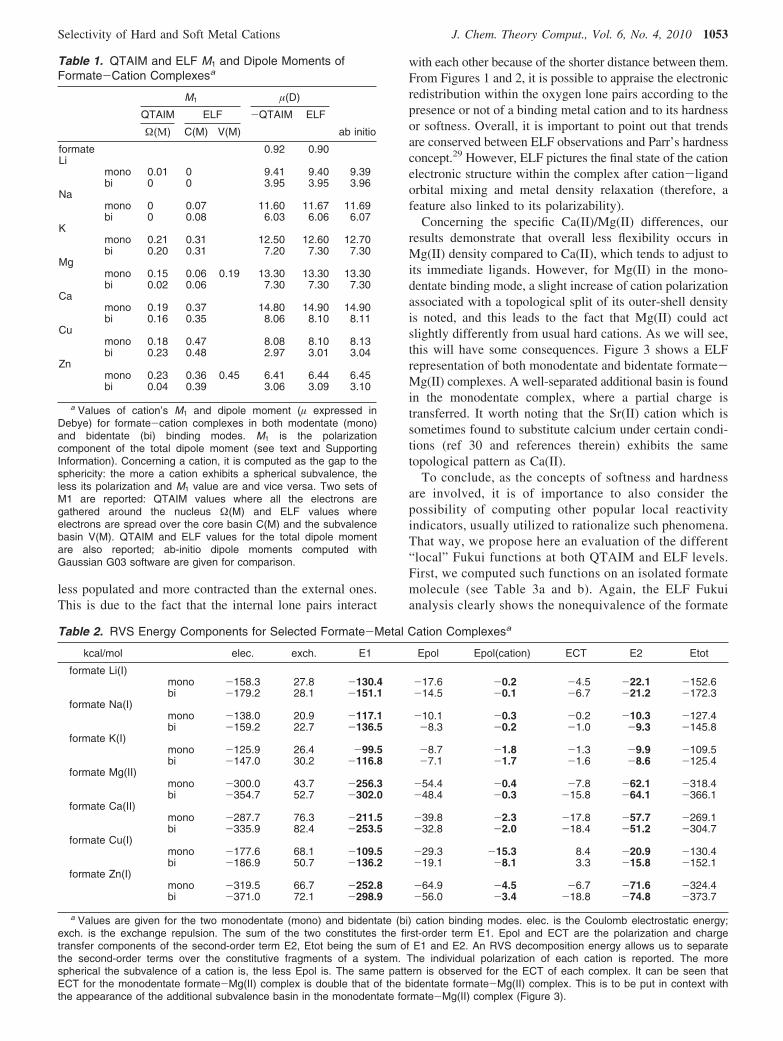
2. SubValence: Understanding Physics at Play. To havea deeper understanding of the physics taking place in suchinteractions, it is possible to extend the ELF analysis to thecomputation of local electrostatic moments (see Table 1) andFukui functions (see Table 3) and to correlate them to RVSenergy decompositions (see Table 2).
From these tables, we see that the more covalent thebonding character of the formate-cation intermolecular bondis, the greater the RVS cation polarization energy and localELF cation dipole moment are. For example, a hard cationsuch as Na(I), which is poorly polarizable (weak polariz-ability), is involved in interactions dominated by electrostatics(Table 2) and does not show any split within its subvalence,whereas cations exhibiting stronger polarization and chargetransfer interactions possess a higher number of basins.Moreover, for a covalently bonded very soft cation such asZn(II), a subvalent Zn-O basin is observed between theformate oxygen lone pair and the metal, a hint of electronsharing. Ca(II), which is less soft and less covalently bonded,still exhibits a split, but subvalent basins remain distributed
around the nucleus (Figure 1) and do not form any bond.Correlated CSOV energy decomposition computations havealso been performed and are fully in line with HF RVSresults. Details can be found in the Supporting Information,S2.
Observations of both monodentate and bidentate coordina-tion modes show that “soft cation” subvalent basins clearlyhave the ability to orient themselves toward the formateoxygen lone pairs. That way, depending on its electronstructure, each cation shows a specific topological signaturewhich enables one to predict specific abilities of the cationto interact with its immediate environment thanks to theplasticity of its valence electron spatial organization. Anindirect measurement of the soft/hard nature of the cationscan be appraised by studying the volumes and density valuesof the formate oxygen lone pairs when interacting withcations. Hard cations such as Li(I) or Mg(II) clearly act onthe lone pair densities which appear lower when comparedto softer cations. Figure 2 exhibits the four oxygen lone pairsas they are when no metal cation interacts with the formate.Volume and density values reveal a dissymmetry betweenthe internal and the external lone pairs, internal ones being
Figure 1. (a) ELF representation of formate interacting in a bidentate mode with metal cations. Topological analysis of interactionsbetween a formate and seven metal cations revealed, at the isosurface, a coefficient of 0.87 (except for Cu(I) and Zn(II) complexes,where it is 0.77). As seen in these pictures, electron densities remain condensed around the nucleus position for hard cationssuch as Li(I), Na(I), and Mg(II), whereas electron densities are split in four distinct subunits (basins), avoiding oxygen lone pairs,for softer cations such as K(I) and Ca(II), and split in two distinct subunits (basins), one of which is inserted between oxygenlone pairs and the cation, for soft cations such as Cu(I) and Zn(II). In soft cation complexes, a blue sphere describes the coreelectrons of the cations. In hard cation complexes, the spherical subvalence obscures the core electrons so that the blue sphereis not visible. (b) ELF representation of formate interacting in a monodentate mode with metal cations. Isosurface coefficientsused to make theses pictures are the same as the ones used for part a. A very similar pattern is observed as for the bidentateformate-cation complexes. The Mg(II) electron density stays spherical. In the Ca(II) complex, three basins have merged into anannular one still avoiding oxygen lone pairs. The same split as for the bidentate complex is shown for Zn(II).
1052 J. Chem. Theory Comput., Vol. 6, No. 4, 2010 de Courcy et al.
less populated and more contracted than the external ones.This is due to the fact that the internal lone pairs interact
with each other because of the shorter distance between them.From Figures 1 and 2, it is possible to appraise the electronicredistribution within the oxygen lone pairs according to thepresence or not of a binding metal cation and to its hardnessor softness. Overall, it is important to point out that trendsare conserved between ELF observations and Parr’s hardnessconcept.29 However, ELF pictures the final state of the cationelectronic structure within the complex after cation-ligandorbital mixing and metal density relaxation (therefore, afeature also linked to its polarizability).
Concerning the specific Ca(II)/Mg(II) differences, ourresults demonstrate that overall less flexibility occurs inMg(II) density compared to Ca(II), which tends to adjust toits immediate ligands. However, for Mg(II) in the mono-dentate binding mode, a slight increase of cation polarizationassociated with a topological split of its outer-shell densityis noted, and this leads to the fact that Mg(II) could actslightly differently from usual hard cations. As we will see,this will have some consequences. Figure 3 shows a ELFrepresentation of both monodentate and bidentate formate-Mg(II) complexes. A well-separated additional basin is foundin the monodentate complex, where a partial charge istransferred. It worth noting that the Sr(II) cation which issometimes found to substitute calcium under certain condi-tions (ref 30 and references therein) exhibits the sametopological pattern as Ca(II).
To conclude, as the concepts of softness and hardnessare involved, it is of importance to also consider thepossibility of computing other popular local reactivityindicators, usually utilized to rationalize such phenomena.That way, we propose here an evaluation of the different“local” Fukui functions at both QTAIM and ELF levels.First, we computed such functions on an isolated formatemolecule (see Table 3a and b). Again, the ELF Fukuianalysis clearly shows the nonequivalence of the formate
Table 1. QTAIM and ELF M1 and Dipole Moments ofFormate-Cation Complexesa
M1 µ(D)
QTAIM ELF -QTAIM ELF
Ω(Μ) C(M) V(M) ab initio
formate 0.92 0.90Li
mono 0.01 0 9.41 9.40 9.39bi 0 0 3.95 3.95 3.96
Namono 0 0.07 11.60 11.67 11.69bi 0 0.08 6.03 6.06 6.07
Kmono 0.21 0.31 12.50 12.60 12.70bi 0.20 0.31 7.20 7.30 7.30
Mgmono 0.15 0.06 0.19 13.30 13.30 13.30bi 0.02 0.06 7.30 7.30 7.30
Camono 0.19 0.37 14.80 14.90 14.90bi 0.16 0.35 8.06 8.10 8.11
Cumono 0.18 0.47 8.08 8.10 8.13bi 0.23 0.48 2.97 3.01 3.04
Znmono 0.23 0.36 0.45 6.41 6.44 6.45bi 0.04 0.39 3.06 3.09 3.10
a Values of cation’s M1 and dipole moment (µ expressed inDebye) for formate-cation complexes in both modentate (mono)and bidentate (bi) binding modes. M1 is the polarizationcomponent of the total dipole moment (see text and SupportingInformation). Concerning a cation, it is computed as the gap to thesphericity: the more a cation exhibits a spherical subvalence, theless its polarization and M1 value are and vice versa. Two sets ofM1 are reported: QTAIM values where all the electrons aregathered around the nucleus Ω(M) and ELF values whereelectrons are spread over the core basin C(M) and the subvalencebasin V(M). QTAIM and ELF values for the total dipole momentare also reported; ab-initio dipole moments computed withGaussian G03 software are given for comparison.
Table 2. RVS Energy Components for Selected Formate-Metal Cation Complexesa
kcal/mol elec. exch. E1 Epol Epol(cation) ECT E2 Etot
formate Li(I)mono -158.3 27.8 -130.4 -17.6 -0.2 -4.5 -22.1 -152.6bi -179.2 28.1 -151.1 -14.5 -0.1 -6.7 -21.2 -172.3
formate Na(I)mono -138.0 20.9 -117.1 -10.1 -0.3 -0.2 -10.3 -127.4bi -159.2 22.7 -136.5 -8.3 -0.2 -1.0 -9.3 -145.8
formate K(I)mono -125.9 26.4 -99.5 -8.7 -1.8 -1.3 -9.9 -109.5bi -147.0 30.2 -116.8 -7.1 -1.7 -1.6 -8.6 -125.4
formate Mg(II)mono -300.0 43.7 -256.3 -54.4 -0.4 -7.8 -62.1 -318.4bi -354.7 52.7 -302.0 -48.4 -0.3 -15.8 -64.1 -366.1
formate Ca(II)mono -287.7 76.3 -211.5 -39.8 -2.3 -17.8 -57.7 -269.1bi -335.9 82.4 -253.5 -32.8 -2.0 -18.4 -51.2 -304.7
formate Cu(I)mono -177.6 68.1 -109.5 -29.3 -15.3 8.4 -20.9 -130.4bi -186.9 50.7 -136.2 -19.1 -8.1 3.3 -15.8 -152.1
formate Zn(I)mono -319.5 66.7 -252.8 -64.9 -4.5 -6.7 -71.6 -324.4bi -371.0 72.1 -298.9 -56.0 -3.4 -18.8 -74.8 -373.7
a Values are given for the two monodentate (mono) and bidentate (bi) cation binding modes. elec. is the Coulomb electrostatic energy;exch. is the exchange repulsion. The sum of the two constitutes the first-order term E1. Epol and ECT are the polarization and chargetransfer components of the second-order term E2, Etot being the sum of E1 and E2. An RVS decomposition energy allows us to separatethe second-order terms over the constitutive fragments of a system. The individual polarization of each cation is reported. The morespherical the subvalence of a cation is, the less Epol is. The same pattern is observed for the ECT of each complex. It can be seen thatECT for the monodentate formate-Mg(II) complex is double that of the bidentate formate-Mg(II) complex. This is to be put in context withthe appearance of the additional subvalence basin in the monodentate formate-Mg(II) complex (Figure 3).
Selectivity of Hard and Soft Metal Cations J. Chem. Theory Comput., Vol. 6, No. 4, 2010 1053

oxygen lone pairs, the external basins having differentindicators from internal lone pairs. If QTAIM tends toshow a uniform Fukui descriptor (the dual descriptor ∆fis always positive), ELF does not, as it provides a ∆fnegative value on the C-H bond, providing insight abouta possible different reactivity.
Concerning the metal cations, Table 3c brings interestinginformation. Let us first consider first the isolated metalcations. If all cations exhibit f - values of 1, they have verydifferent f + values. Again, following chemical intuition,strong differences occur between hard and soft cations. Hardcations such as Li(I) exhibit low values of f +, whereas softcations such as Zn(II) have f + values tending toward 1. Thedual descriptor appears then to be a good global indicatorreflecting the cation’s ranking as ∆f tends to 0 with increasing
Table 3. (a) ELF Integrated Fukui Functions and Dual Descriptor Values for an Isolated Formate Molecule, (b) QTAIMIntegrated Fukui Functions and Dual Descriptor Values for an Isolated Formate Molecule, (c) ELF and QTAIM FukuiFunctions and Dual Descriptor Values for Selected Metal Cationsa
part a
formate basin f -(ELF) f +(ELF) f 0(ELF) ∆f(ELF)
C(C) 0.00 0.00 0.00 0.00C(O1) 0.02 0.00 0.01 0.02C(O2) 0.02 0.00 0.01 0.02V(C,H) 0.22 0.30 0.26 -0.08V(C,O2) 0.04 0.01 0.02 0.03V(C,O1) 0.04 0.01 0.02 0.03V(O1) 0.20 0.01 0.10 0.19V(O1) 0.13 0.03 0.08 0.10V(O2) 0.14 0.03 0.09 0.11V(O2) 0.20 0.01 0.10 0.19
part b
formate atom f -(QTAIM) f +(QTAIM) f 0(QTAIM) ∆f(QTAIM)
H 0.17 0.15 0.16 0.02C 0.05 0.04 0.05 0.02O1 0.38 0.03 0.21 0.35O2 0.38 0.03 0.21 0.35
part c
cations f -(QTAIM) f +(QTAIM) f 0(QTAIM) ∆f(QTAIM), isolated ∆f(ELF), isolated ∆f(QTAIM), complexed ∆f(ELF), complexed
Li 1.00 0.23 0.61 0.77 0.70 -0.14 -0.11Na 1.00 0.44 0.72 0.56 0.45 -0.34 -0.37K 1.00 0.46 0.73 0.54 0.43 -0.30 -0.36Mg 1.00 0.59 0.79 0.41 0.32 -0.67 -0.24Ca 1.00 0.98 0.99 0.02 0.00 -0.55 -0.59Zn 1.00 0.91 0.96 0.09 0.04 -0.69 -0.50
a Values are given for the isolated cations and for a cation within a bidentate formate-metal complex.
Figure 2. ELF representation of a formate without a bindingcation. This picture presents a formate in an uncomplexedstate, where densities are not rearranged through interactionswith a cation. Volumes of the oxygen lone pairs can becompared to the ones of formate-cation complexes shownin Figure 1a and b.
Figure 3. ELF representation of formate-Mg(II) complexesat an isosurface coefficient of 0.22. The electron density ofthe two formate-Mg(II) complexes are compared in thispicture. Contrary to the bidentate formate-Mg(II), where thesubvalence stays spherical, in the monodentate formate-Mg(II),an additional basin appears at the opposite side of thecoordination to the oxygen. This is consistent with theaugmentation of both M1 shown in Table 1 and charge transferenergy shown in Table 2 for this complex.
1054 J. Chem. Theory Comput., Vol. 6, No. 4, 2010 de Courcy et al.

cation softness. Table 3c depicts the results for selectedbidentate formate cations. Global trends are preserved despitestrong, different bonding modes. Again, the softer cationsexhibit smaller ∆f values (here more negative) than harderones. If they agree well, one difference between ELF andQTAIM values is observed: ELF tend to show moredifference between Ca(II) and Mg(II) than QTAIM. This isreflected by a difference of 0.2 (QTAIM) vs 0.3 (ELF) inthe ∆f values between the two cations.
Of course, the numerical values strongly depend on the(never unique) topological partition scheme and thereforeon the density attribution to atoms/centers,18h but the ELFand AIM approaches are not subject to strong basis set/diffuse function dependence18a,h and are quite stable. Overall,beyond the numbers, an interesting qualitative agreement isobserved and supports the previously depicted ELF subva-lence basins and the numerical values extracted from otherinterpretative techniques described above.
B. Metal Cation’s Electron Structure/BiologicalActivity Relationship in Coagulation Proteins. We use herethe commonly accepted terminology, first, to differentiatethe γ-carboxyglutamic acid itself, identified as Gla, from theγ-carboxyglutamic acid-rich domain, identified as GLA,second, to name the first 11 residues at the N-terminalextremity of the ω-loop, and third to specifically identifyresidues 4, 5, and 8 (5, 6, and 9 for FIX) from the previoussequence as the keel. In addition, since we have to compareseveral GLA domain crystal structures, each having theirCa(II) or Mg(II) positions differently numbered, we will usefor clarity purposes the same numbering for all, that is, theone found in the crystal structure of the human factor VII(1DAN, see ref 38).
1. Metal Cation’s Electron Structure/Biological ActiVityRelationship in Coagulation Proteins. When a blood vesselis injured, a cascade of protein-protein interactions1 rapidlyoccurs, leading to the formation of a cross-linked fibrin clot,eventually restoring the integrity of the circulatory system.A number of proteins involved at the early stage of theprocess are vitamin-K-dependent zymogens of serine pro-teases.31 Among this family of coagulation factors, factorVII, once activated and bound to tissue factor (TF), activateszymogen factors IX and X into functional factors IXa andXa. These in turn transform prothrombin into active throm-bin, which is ultimately responsible for the conversion offibrinogen to fibrin.32 A cell-based model of coagulation,which incorporates the important roles of endothelial cellsand platelets, has recently been introduced, and this morephysiological view appears to be gaining acceptance.33 Manyother factors and cofactors are necessary for the completionof the mechanism, but we will focus only on two steps.1
2. Structural Analysis and Biological ActiVity. The above-mentioned three factors are made up by four domains. Atthe N extremity, we find a GLA domain, followed by twoEpidermal Growth Factor (EGF) like domains, namely, EGF1and EGF2, terminated by a Serine Protease (SP) domain.The primary structures of GLA domains (residues 1 to 48)are highly conserved:34 only a few residues differ along thesequences of factors VII, IX, and X. Remarkably, the firstnine Gla residues, as well as two cysteines, are always found
precisely at the same place. Accordingly, for all factors, twohydrophobic residues are adjacent to the first group of twoGla residues: Phe4 and Leu5 for FVIIa and FXa factors andLeu6 and Phe9 for FIXa. We can also notice the presenceof an Asn residue in the second position of the sequence.Moreover, analysis of the PDB files for these factors showsthat the secondary structures are also very similar andessentially superimposable. From the N extremity to the endof the domain, we find successively the ω-loop with its twoabove-mentioned hydrophobic amino acids along with eitheranother hydrophobic residue (FVIIa-Leu8 and FXa-Met8)or a cationic one (FIXa-Lys6) pointing their side chainstoward the exterior of the protein. Three R-helices are alsofound. The first two are parallel and maintained so by adisulfide bridge established by the two cysteines cited before.All Gla residues are distributed along these three subunits:Gla6 and -7 are at the top of the central loop of the ω-loop;Gla14, -16, -19, and 20 belong to the first R helix, whereasGla25, -26, and -29 belong to the second. The third R-helixlinks the GLA domain to the remaining part (3rd helix) ofthe protein. A Gla residue can also be found in this part ofthe domain. In all examined X-ray structures, eight cationsare present.38 One cation is located at the hinge between thethird R helix and the first chain of the EGF1 domain. Theother seven cations are found aligned at the interstice betweenthe base of the two parallel R helices and the top of theω-loop. In this zone, two groups of cation binding sites canbe defined. The first group is called “high affinity” Ca(II)binding sites39 and is constituted by the 5 inner Ca(II) sites(numbered 3, 4, 5, 6, and 8, respectively).38 Within thesesites, Ca(II) were found coordinated 6, 7, 7, 7, and 3 times,respectively, at distances in the 2.4-2.8 Å range in the veryfirst GLA domain ever structurally determined (1992),namely, the GLA domain of Ca-Prothrombin Fragment I(see Figure 4 and Table 3 of ref 35). Subsequently, severalauthors36,37,41 reported very similar coordination numbersand distances for the factors studied here. The second group,called “low affinity” Ca(II) binding sites, defines the twoexternal sites. At these positions, cations can be either Ca(II)or Mg(II). Figure 4 lists the cation binding sites present indifferent PDB structures of the studied factors’ GLAdomains. Occupation by either Ca(II) or Mg(II) in all bindingsites seems to be due to details in the preparation of theprotein crystal during the X-ray crystallization process. Infact, F VIIa (1DAN),38 F IXa (1J35),39 and F Xa (1IOD)40
were prepared using only CaCl2 as crystallographic salt. Itis noticeable that all the binding sites are occupied bycalcium, solely present in the environment. When MgCl2 isadded to CaCl2, at physiological concentration, for thepreparation of F IXa (1J34),39 all the inner binding sites areoccupied by calcium, while the external binding sites areoccupied by magnesium. But, prepared under the sameconditions, a recent X-ray structure deposited in the ProteinData Bank by Bajaj et al. shows Mg(II) residing in the centralsite number 5 replacing Ca(II) in the GLA domain of F VIIa(2A2Q).41 However, when only MgCl2 is used for thepreparation of F Xa (1P0S),4 the two external sites containa Mg(II) cation, but the sole central site number 5 shows athird Mg(II) cation. In addition, contrary to the five other
Selectivity of Hard and Soft Metal Cations J. Chem. Theory Comput., Vol. 6, No. 4, 2010 1055

structures that contain it, no ω-loop is present in the structureof this domain, its constitutive peptide seeming to “float” inthe environment. From these observations, it may bededucted that Ca(II) cations are necessary in the central zoneto structure the ω-loop and that the external sites are usuallyoccupied by magnesium. Close examination of the bindingof the ω-loop to the rest of the GLA domain reveals thenetwork of interactions between the amino acids borne bythe ω-loop, the cations and the amino acids present in thetwo antiparallel R helices. In all factors where the ω-loop ispresent, except for F VIIa with Mg(II) in the central site,the NH3
+ extremity of Ala1 (or Tyr1 for F IXa) establishesthree H bonds with the surrounding residues, namely,carbonyl O of Gln21 (F VIIa), Ala21 (F Xa), or Lys22 (FIXa); Oε4 of Gla20 (F VIIa and F Xa) or Gla21 (F IXa);and Oε4 of Gla26 (F VIIa and F Xa) or Gla27 (F IXa). In FVIIa (2A2Q), however, a single H-bond remains with Oε2
of Gla26. A comparison between coordination numbers andcoordination distances for each cation of the two structuresof F VIIa (1DAN vs 2A2Q), as given in Table 2 of ref 41by Bajaj et al., reveals that, when only Ca(II) is present inthe five inner sites, direct coordinations of cations by thecarbonyl O of Ala1 (or Tyr1 for F IXa); Oδ1 of Asn2; Oε1
and Oε4 of Gla6; and Oε1, Oε2, and Oε4 of Gla7 are observed.By contrast, in F VIIa (2A2Q), where the central Ca(II) issubstituted by Mg(II) with two water molecules (S209 andS363) completing the coordination sphere of this cation, thesame interactions are observed, but this time through anetwork of eight water molecules (S209, S262, S363, S411,S508, S602, S694, and S722), present in between the cationsand the ω-loop. As a consequence, the ω-loop has moveddown approximately 0.5 Å in order to leave enough spacefor water to insert (see Figure 2A of ref 41).
The existence of the ω-loop is of much importance in thatthe very first step of the coagulation process is for coagulationfactors to colocalize on cell surfaces.42 In that step, thebiological function of the GLA domain is first directlyresponsible for the linkage of the factors to the membranes,and ultimately to the fibrin clot. This can be done thanks tothe three hydrophobic residues of the ω-loop43 (or twohydrophobic residues and a cationic one in F IXa), orientedin such a way that they are able to dive deeply inside themembrane and interact through hydrophobic bonds withneighboring lipids,43,44 in addition probably through a buriedsalt bridge involving Lys5 of F IXa.43 The deep insertion ofthe GLA domain inside the membrane also allows thecreation of an interaction between a phosphatidylserine (PS)of the membrane and the Ca(II) cation present in bindingsite number 8, strengthening the anchorage of the coagulationfactor inside the cellular membrane.45 Inhibition of the GLAdomain by direct ligand bonding to the ω-loop (such as snakevenom protein, see ref 39) is responsible for the loss ofmembrane linking with a subsequent loss of the coagulationprocess.
3. Theoretical Study of Interactions of Ca(II) Vs Mg(II)with Malonate Groups. In this section, we systematicallysupplement the ELF analysis with detailed RVS energydecomposition results.
In GLA domains, the transformation of glutamic acids inGla is realized by the action of vitamin K and several specificenzymes with the addition of a carboxylate group to theγ-carbon of the glutamate;46 two carboxylate groups borneby the same γ carbon constitutes a malonate group. Twomalonates coordinating a metal cation is one of the unitstructures observed in GLA domains, displaying as manyas four monodentate formate-cation interactions. Two
Figure 4. Description of metal cation coordination sites of six GLA domains. The picture is a representation of the GLA domainof F VIIa (1DAN), exhibiting its constitutive parts including the seven cation binding sites. For each site, a comparison of sixX-ray geometries shows its occupation by either a Mg(II) or a Ca(II) cation, according to the crystallographic salt used for thepreparation.
1056 J. Chem. Theory Comput., Vol. 6, No. 4, 2010 de Courcy et al.

different computations at the same level of theory (B3LYP/6-311++G**) have been performed on these systems: asingle point calculation using directly extracted geometriesfrom PDB structures on which H atoms were added andgeometry optimizations performed using the previous sys-tems as starting points. At the end of the optimizationprocess, the obtained complexes show the two approximateplanes of malonates perpendicular to each other around the
metal cation, itself four times tetrahedrally coordinated withcoordination distances of approximately 2.1 Å for themagnesium and 2.4 Å for the calcium. A calcium complexwas extracted from the PDB structure of F Xa (1IOD),whereas the magnesium complex was extracted from thePDB structure of F IXa (1J34). From these two systems,external binding sites (number 7 or 9) are selected, in whichcations are surrounded by solvating water molecules: twofor Mg(II) and three for Ca(II). Figures 5a and b show theELF topological analysis on the two optimized geometries.As expected, Mg(II) (Figure 5a) does not exhibit any splitof its subvalence, whereas Ca(II) (Figure 5b) has itssubvalence split into four well separated basins, oriented insuch a manner that no oxygen lone pair faces a cation basin.The same pattern as described above is observed in Figure6a for Mg(II) and Figure 6b for Ca(II), within the extractedgeometries. Thus, upon increasing the coordination numberfrom two in the bidentate formate-cation complexes to fourin the optimized geometries shown in Figures 5a and b andultimately to their maximum in the extracted complexes, eachcation consistently exhibits the same behavior. Table 4reports the results of the RVS analysis, disclosing theindividual contributions of the interaction energies for thetwo malonate-cation complexes shown in Figures 5a and band 6a and b. These results show first a greater electrostaticterm for Mg(II) less compensated by the repulsion term thanfor Ca(II). As a result, the excess of electrostatic energyreflected by the negative first order term of Mg(II) over Ca(II)is -90 kcal/mol for the optimized complexes and less than-77 kcal/mol for the extracted geometries, reflecting thehardness of the Mg(II) cation. Since a RVS analysis canseparate the second order energies for each constitutivemonomer of a many-body system, the individual polarizationenergies of the cations are also reported. It can thus beobserved that, as Ca(II) moves away from its optimalgeometry, its polarization increases, and that Mg(II) isessentially not polarized. The charge transfer contribution inthe Ca(II) complexes decreases upon moving away from theoptimal geometry but remains significant, whereas it is null forthe Mg(II) optimized complex and slightly increases in the
Figure 5. (a) Optimized geometry of a two-malonate-Mg(II)complex. (b) Optimized geometry of a two-malonate-Ca(II)complex. All the ELF pictures were revealed at the 0.87isosurface coefficient. These pictures exhibit the perfecttetrahedral binding mode of the cations. Mg(II) subvalence isspherical, and the Ca(II) one is split in four well separatedbasins recalling the bidentate formate-cation complexes.
Figure 6. (a) Extracted two-malonate-Mg(II) “deformedtetrahedral” complex. (b) Extracted two-malonate-Ca(II) “de-formed tetrahedral” complex. The Mg(II) complex is bindingsite no. 7 of F IXa (1J34); the Ca(II) one is binding site no. 9of F Xa (1IOD). Water molecules complete the coordinationsphere of each cation: six for Mg(II) and seven for Ca(II).Cation coordinations are also disclosed in order to evidencethe “deformed tetrahedral” binding mode of the cations. Eachcation behavior does not change as the number of coordina-tions increases.
Table 4. Theoretical RVS Analysis of Tetrahedral Cation Binding Sitesa
kcal/mol 2 malonates + Mg(II) 2 malonates + Mg(II) 2 malonates + Ca(II) 2 malonates + Ca(II)
geometry optimized F IXa 1J34 optimized F Xa 1IODfigure number 5a 6a 5b 6b
electrostatic -777.7 -703.3 -709.0 -636.8repulsion 90.8 68.2 111.5 78.2first order energies (e1) -686.9 -635.1 -597.5 -558.6
polarization of cations -0.1 -0.3 -0.6 -2.2polarization -94.2 -90.5 -63.0 -61.5charge transfer 0.0 -1.4 -11.2 -9.7second order energies (e2) -94.2 -91.9 -74.2 -71.2
total e1 + e2 -781.1 -720.9 -671.7 -629.8
a RVS energy decomposition has been performed upon theoretical models of tetrahedral binding sites. They are built on the samenumber of atoms (no water molecules are considered) in order to directly compare the energy contributions. Optimized geometries are theextracted ones on which the optimization process was performed. Mg(II) complexes’ first order terms are greater than the ones of Ca(II)complexes, whereas Ca(II) cations are more polarized and generate a larger charge transfer than for Mg(II). This is consistent with theMg(II) cation having a spherical subvalence entering in more electrostatic types of interactions and the Ca(II) cation splitting its subvalenceand creating more interactions of covalent type.
Selectivity of Hard and Soft Metal Cations J. Chem. Theory Comput., Vol. 6, No. 4, 2010 1057

complex extracted from X-ray crystallography. It can bededuced that, contrary to Mg(II), Ca(II) is polarized as it is inthe bidentate formate-cation type of complex and that thecharge transfer contribution becomes selective from one cationto another in such complexes. Thus, Mg(II) enters a moreelectrostatic interaction, whereas a polarized Ca(II), generatinga stronger charge transfer, enters a more covalent interaction.It is important to point out that we also perform such analysisin the presence of an additional PCM implicit solvent. We didnot observe any changes in the topology of the system. Suchresults can be found in Supporting Information S3.
4. Theoretical Study of the SelectiVity Ca(II)/Mg(II).a. Is the Mg(II) Cation Strictly Hard in the Gas Phase? Inthe GLA domain of F Xa (1P0S), in which only magnesium
ions are present in the environment, a Mg(II) is observed tobe three times coordinated to oxygen atoms of Gla 16 and26, in the 2.3-2.5 range of distances. Figures 7a and bcompare the topology of Mg(II) in two positions: the firstone (Figure 7a) comes from an external cation site (site no.7 of F IXa 1J34); the second one (Figure 7b) is the centralsite (no. 5). Due to the low resolution (2.8 Å) of the crystalstructure of F Xa (1P0S), no water molecule is resolved nearthe cation in the central site no. 5. Despite the fact that watermust be present within the coordination sphere of Mg(II) ina real enzyme, no water molecule is considered in the presenttheoretical gas phase study, so that the coordination numberis four for the external site and three for the central site. Ascan be seen in Figure 7a, the cation does not exhibit anysplit of its subvalence, whereas in Figure 7b, the presenceof the additional basin (red circle), recalling the monodentateformate-Mg(II) binding mode, clearly suggests that Mg(II)transfers a part of its density into this additional basin. Thisis confirmed by the magnitude of the RVS charge transfercontribution (see FXa 1P0S in Table 5), namely, -7.6 kcal/mol, instead of a null charge transfer in the optimizedgeometry. In this particular case, it is important to point outthat Mg(II) does not have a hard cation behavior since itexhibits some subvalence capabilities. However, if such afinding is important for understanding the electronic distribu-tion in Mg(II) complexes, could it have an impact whenconsidering more realistic condensed phase GLA domainscontaining explicit solvent molecules?
b. SelectiVity of Ca(II) Vs Mg(II) Cations within the SixGla Domains. From Figure 4, we have found three possiblebinding sites for Mg(II), namely, sites no. 7 and 9 (external)and 5 (central), the latter exhibiting either a Mg(II) or aCa(II). The other sites are exclusively occupied by Ca(II)cations, a necessity to structure the ω-loop in its functionalgeometry. These observations have been confirmed byseveral experiments, in which Mg(II) and Ca(II) were addedat physiological concentration to a previously divalent cation-free environment.2 When only Mg(II) is present, no enzy-matic activity occurs. This is to be put in relation with the
Figure 7. (a) “Hard” Mg(II) binding mode. (b) “Soft” Mg(II)binding mode. (c) Localization of the two binding modes withinF Xa (1P0S). Electron densities are revealed at the isosurfacecoefficient of 0.22. The additional basin shown in the red circleis confirmed by the increase of the charge transfer energyillustrated in Table 4 (-7.6 kcal/mol). As is the case whenonly Mg(II) is present in a vitamin-K-dependent coagulationfactor environment, the cations occupy only the three tetra-hedral binding sites, being unable to structure the ω-loop, andleaving its constitutive peptide floating.
Table 5. RVS Analysis of Selected Cation Binding Sites, Extracted from X-Ray Structuresa
kcal/mol 2 malonates + Mg(II) 2 malonates + Mg(II) Mg(II) hexa coordinated 2 malonates + Ca(II) Ca(II) octa coordinated
Water molecules 2 0 3 3 2
PDB ID F IXa 1J34 F Xa 1P0S F VIIa 2A2Q F Xa 1IOD F IXa 1J34figure number 6a 7b 8a 6b 8b
electrostatic -696.7 -577.9 -746.5 -690.6 -598.5repulsion 130.0 23.7 184.5 188.3 111.1first order energies (e1) -566.7 -554.3 -562.0 -502.3 -487.4
polarization of cations -0.4 -0.4 -0.1 -1.8 -0.5polarization -122.8 -66.5 -92.6 -95.0 -33.0charge transfer -9.8 -7.6 -2.7 -20.7 -4.9second order energies (e2) -132.5 -74.1 -95.3 -115.7 -37.9
total e1 + e2 -699.2 -628.4 -657.3 -618.0 -525.3
a RVS energy decomposition has been performed upon various extracted geometries in order to confirm the theoretical results on realisticsystems. Thus, all the components, including water molecules, were taken into consideration. The number of water molecules is detailed foreach complex, as well as the X-ray structure from which the system was extracted and the corresponding figure referenced. The sametrend is observed, as for theoretical tetrahedral systems, concerning electrostatic energies favoring Mg(II) complexes, and the Ca(II) cationbeing more polarized, producing a greater charge transfer.
1058 J. Chem. Theory Comput., Vol. 6, No. 4, 2010 de Courcy et al.

F Xa (1P0S) structure where the ω-loop is not formed. Whenonly Ca(II) is present, the enzymatic activity is signicantlyhigher but can be enhanced by the addition of Mg(II). Thethree possible Mg(II) sites are constructed from three pairsof malonates provided by site no. 5 Gla16 and 26 (Gla17and 27 for F IXa), site no. 7 Gla14 and 19 (Gla15 and 20for FIXa), and site no. 9 Gla25 and 29 (Gla26 and 30 for FIXa). Close examination of these sites reveals that they allshare the same pattern in which the cation is coordinated bytwo groups of two oxygens residing in the same side of twoalmost parallel malonate groups. Such geometry recalls theoptimized geometry of the two malonates bound to a cationcomplex. Because they are not perfectly tetrahedral, thesethree binding sites will be called “deformed tetrahedral”cation binding sites. Figures 6a and b show ELF pictures ofsuch a “deformed tetrahedral” binding site occupied by aMg(II) cation as extracted from site no. 9 of F IXa (1J34)and a Ca(II) cation as extracted from site no. 7 of F Xa(1IOD), respectively. From Table 4, RVS total energies givethe preferrence of Mg(II) over Ca(II) within this type of siteby a difference of -109.4 kcal/mol for the optimizedgeometries and -91.1 for the extracted ones. This is due tothe excess of first order energies in favor of Mg(II), namely,-89.4 and -76.5 kcal/mol. This is consistent with Mg(II)not splitting its subvalence and thus being involved in moreelectrostatic types of interactions. The presence of watermolecules (two for Mg(II) and three for Ca(II)) within thecoordination sphere of cations does not modify this conclu-sion (see Table 5).
The four other sites, however, are less structured, as theirCa(II) cations are coordinated four to eight times, in bothmonodentate and bidentate mode, by several carboxylategroups belonging to different Gla residues, as well as bycarbonyl groups of the backbone of Ala1 (Tyr1 for F IXa)and the side chain of Asn2. For example, in site no. 6 of FIXa (1J34), Ca(II) is found octa coordinated, by Tyr1, Gla21in a monodentate mode, Gla7 and -17 in a bidentate mode,and two water molecules (Figure 8b). The now familiar splitof the subvalence of Ca(II) is observed. An ELF computationperformed on this model reveals that the net charge of thecation within the system is +1.67 instead of +2 in thefundamental state. This means that up to one-third of anelectron was transferred from the ligand oxygen lone pairsto the Ca(II) cation, with Ca(II) consistently able to adaptits electronic density to its environment by splitting itssubvalence, thus entering into more covalent types ofinteractions. This capacity of adaptation for Ca(II) explainswhy, when only present in a coagulation factor environment,all the binding sites are loaded with Ca(II) (F VIIa 1DAN,F IXa 1J35, and F Xa 1IOD).
Therefore, with respect to the experimental results shownin Figure 5 of ref 2, a mechanism can be proposed for theactivation of vitamin-K-dependent coagulation factors byMg(II) and Ca(II) cations. Upon introducing first Mg(II) toa cation-free environment of an enzyme, one Mg(II) cationgoes to each of the three “deformed tetrahedral” binding sites(nos. 5, 7, and 9), completing its coordination sphere withtwo water molecules. At this stage, the enzyme is not activeand the ω-loop is not structured (F Xa 1P0S). When Ca(II)
is added, one Ca(II) cation goes to each of the four othersites (nos. 3, 4, 6, and 8). This anchors Ala1 (or Tyr1 for FIXa) and Asn2 to the rest of the GLA domain47 and foldsthe peptide in this particular ω geometry through interactionsinvolving Gla6 and -7 (Gla7 and -8 for F IXa) and the restof the domain (F VIIa 2A2Q). However, prepared with thesame mix of cations at physiological concentration39 as theone used to prepare F VIIa (2A2Q)41 for the crystallizationprocess, F IXa (1J34) exhibits a Ca(II) cation in central siteno. 5 instead of a Mg(II) cation. Could the occupation ofthe central binding site no. 5 by either a Mg(II) (2A2Q) ora Ca(II) (1J34) be explained by the topological differenceof the two cations?
c. Direct Interactions with Cations Vs Interactions withCations through Water. We consider here a system consti-tuted by the five central Ca(II) binding sites extracted fromthe geometry of the GLA domain of F VIIa (1DAN), whichis very similar to the one of F IXa (1J34). This complexwas built from the backbone of Ala1, a formamide grouprepresenting the side chain of Asn2, and two malonates forGla6 and -7, with Ala1, Asn2, Gla6, and Gla7 being part ofthe ω-loop, in addition to three malonate groups from Gla16,-20, and -26 and a formate given by Gla29 and the five Ca(II)ions. In addition, up to six water molecules were placed onthe calcium ions according to their position in the X-raystructure. This system, which is globally neutral, is an X-raycrystal snapshot where hydrogens and waters have beenadded, focusing on the interactions between Ca(II) cationsand their ligands. Such a system is found in X-ray crystalstructures 1DAN, 1J34, 1J35, and 1IOD in which cationsdirectly interact with their environment. Figure 9 exhibitsthe ELF study on this system. This picture reveals thenetwork of interactions between the subvalence basins ofthe cations and the lone pairs of the coordinating oxygens.The split of all Ca(II) subvalences indicates that theseinteractions are partially covalent because they are built fromelectronic exchanges between the cations and the lone pairspresent in their immediate environment. This results in a
Figure 8. (a) Extracted geometry of binding site no. 5 of FVIIa (2A2Q) with Mg(II). (b) Extracted geometry of binding siteno. 6 of F IXa (1J34) with Ca(II). Part a shows a tetrahedralbinding site loaded with a Mg(II) cation, that structure beingthe ω-loop through a network of H-bonds established by highlypolarized water molecules coordinated to the cation. Part bshows direct interactions between a Ca(II) cation and someconstitutive segments of the ω-loop. The subvalence patternof the two cations stays consistent with the one observed inall the studied geometries, whatever the size of the systemconsidered.
Selectivity of Hard and Soft Metal Cations J. Chem. Theory Comput., Vol. 6, No. 4, 2010 1059

network of many-body interactions at the center of the GLAdomain. QTAIM values of M1 of each Ca(II), computed withthe ELF function, confirm the intensity of the charge transfer.Ca(II) no. 3 and 5, each being 7-fold coordinated, have thelowest value of the series with a M1 value of 0.041 D and anet charge of +1.66 instead of +2, whereas Ca(II) no. 4and 6, with 6-fold coordination, exhibit enhanced values,namely, 0.060 D and +1.68, for M1 and the charge,respectively. Ca(II) no. 8, being four times coordinated,shows values of M1 very close (0.166 D, +1.72) to the onefound for the small bidentate formate-cation complex (0.160D and +1.70 for M1 and the charge, respectively, see Table1). This indicates that the greater the coordination number,the less the polarization and therefore the residual charge.
In the GLA domain of F VIIa (2A2Q), the central Mg(II)is found six times coordinated: four times tetrahedrally bymalonates of Gla16 and -26 and two times by watermolecules S209 and S363 (Figure 8a). The formamide moietyof Asn2, a formate group from Gla7, and a third watercomplete the network of H-bonds through which this centralMg(II) contributes to structure the ω-loop. As seen in Figure8a, the central Mg(II) subvalence remains almost perfectlyspherical, as in the bidentate formate-Mg(II) complex andin the two-malonate-Mg(II) tetrahedral optimized geometry.Close examination of the RVS polarization contribution(Table 5) reveals that, if the cation is as expected almostnot polarized, the polarization of the three water moleculesaccounts for approximately 30% of the total polarizationof -92.7 kcal/mol. Indeed, one of the water molecules ishighly polarized and bears an Epol(RVS) value of -13.9kcal/mol. Thus, interactions between the cation and theω-loop are established through highly polarized watermolecules.
While the presence of Ca(II) is required2,3,48 in the centralregion for an effective coagulation function, it has beendemonstrated that the affinity of coagulation factors for eithercellular membranes,45,49 tissue factors,5 or anticoagulantagents36,40 is strongly enhanced when external sites areoccupied by Mg(II).40 This supports our findings that thesesites are better stabilized by Mg(II)37 rather than Ca(II).
Therefore, the difference of the two geometries (1J34 and2A2Q) resides in the fact that, in 2A2Q, the central site isoccupied by a Mg(II) which is only able to complete itscoordination shell with water, for which the residence timehas been measured to be on the order of 1 µs.8 This explainsthe greater distance between the two R-helices and the ω-loop(2A2Q case), namely, the interposition of a water layer.Therefore, one of the roles of the cations is to fix water,which in turn binds to the ω-loop through a network ofH-bonds. By contrast, in FIXa 1J34, where all the centralcation binding sites are occupied by Ca(II), interactions takeplace directly between metal cations and the lone pairs borneby its ligating oxygens. In this connection, it was recentlydemonstrated that water layers present either betweenseparate domains of a protein or in between different proteinsare in dynamic short time exchanges with the solvation waterpresent in the environment.50,51 Therefore, it is possible tomake the hypothesis that F VIIa crystallized by Bajaj et al.(2A2Q), with interactions through water (i.e., water mediatedinteractions), could be present, free in the blood plasma sincethe latter is a highly hydrophilic environment. On the otherhand, the GLA domain of F IXa crystallized by Shikamotoet al. (1J34) is found bound to a ligand (in this case, a snakevenom protein); this binding could impose the “directinteractions with cations” geometry. It is, then, perhapsreasonable to suggest that this is the geometry that insertsdeeply into the cell membrane, the interior of which is ahighly hydrophobic environment. Indeed, the insertion withinthe membrane may expel water molecules from the insertedpart of the GLA domain, imposing a switch between “physicsat play”: going from a “through water structure” to a “directelectronic interactions structure”.
Conclusion
In this contribution, we have used several theoretical toolsto illustrate the relationship between the electronicstructure of selected metal-cation complexes and the so-called hard and soft chemical behavior. The ELF analysisallows ranking cations according to their topologicalsignature, namely, their ability to split their valence intosubdomains (subvalent basins). Hard cations will notexhibit such a capability as soft cations do. The covalentcharacter of the ligand-metal cation interaction is as-sociated with a basin between the metal and the neighbor-ing heavy atoms. That way, such a covalent interactioncan be directly affected by the cations’ environment, andas we have seen, an electrostatic interaction, as in Mg(II)monodentate-like complexes, can become covalent, ex-hibiting an extra subvalent localization basin under specificstress conditions. This topological metal cation rankinghas been shown to be relevant when compared to RVSand CSOV energy analyses, as well as in good qualitative
Figure 9. Topological analysis of the five central Ca(II)binding sites of F VIIa (1DAN) GLA domain. This pictureunravels the network of charge transfer interactions betweenthe five Ca(II) cations and the oxygen lone pairs borne byGla residues of the two upper R-helices, on one hand, andthe oxygen lone pairs borne by Ala1 (backbone), Asn2 (sidechain), and Gla6 and -7 of the lower ω-loop, on the other hand.These interactions structure the ω-loop in its functionalgeometry. It can be noticed that Ca(II) cations show differentpatterns of their subvalence split according to their differentnumbers of coordinaton. The numbering of the calcium ionbinding sites of Figure 3 is used.
1060 J. Chem. Theory Comput., Vol. 6, No. 4, 2010 de Courcy et al.

agreement with local Fukui functions extracted from bothQTAIM and ELF analyses.
Our integrated methodological approach uncovered aclear relationship between the underlying metal electronstructure and the biological activity of enzymes such asvitamin-K-dependent coagulation factors. The presentapproach could then be extended to other biologicalsystems involving metal cations. It is also important topoint out that these results on metal cation-ligandcomplexes, such as the dissymmetry between internal andexternal carboxylate oxygen lone pairs, could also provideuseful information for the design of new force fields.7a,52
Moreover, applications to the problem of blood coagula-tion allowed us to address the Ca(II) vs Mg(II) selectivityin their interaction with GLA domains. Only the two X-raystructures, crystallized using the same Mg(II)/Ca(II) mixfound under physiological conditions (1J34 and 2A2Q),have been investigated.
In the first one (1J34), Ca(II) has been shown to bemore covalently bonded to ligand than Mg(II), enablingthe creation of a “direct charge transfer network” betweenits subvalence and the carboxylate oxygen lone pairs. Weshowed that this concept could also uncover the distinctrole of the two cation binding sites present in GLAdomains. Being at the origin of the observed chargetransfer network, calcium cations are mandatory in thecentral region to conserve the folding, whereas externalbinding sites are better stabilized by Mg(II), in agreementwith the experiment, thanks to strong electrostatic interac-tions with the environment.
In the second structure (2A2Q), in which some interac-tions between metal cations and Gla are establishedthrough the presence of crystallized water molecules, wehave shown that magnesium is able to form “an indirectcharge transfer network” with malonates through itsinteraction with two highly polarized water molecules thatare responsible for a structure of the ω-loop similar tothat observed in the first structure (1J34). To conclude,as the two crystal structures have been solved, they couldbe the two interconverting forms of the same system, eachof them having a different water requirement. In bothcases, the ω-loop is present. This is required for the activeenzyme, but the underlying physics is not the same. Inthe first structure, the folding is mainly due to electroniceffects: Ca(II) is preferred in the five central binding sites.By contrast, the presence of water molecules is able toreverse the direct electronic selectivity of cations (Mg(II)could be present if interacting with two water molecules).This clearly indicates that dynamical solvent effects couldplay a key role in the observed structure of the domain.The connection between the two structures might beestablished through a series of molecular dynamicssimulations. This work also emphasizes again53 theimportance of a “discrete” water molecule to understandthe stability of biological systems, as the presence of alimited number of structured water molecules could becritical to obtain meaningful theoretical models.
Acknowledgment. The computations have been per-formed at the national IDRIS (F. 91403 Orsay, France),
CRIHAN (F.76800 Saint-Etienne-du-Rouvray, France),and CINES (F. 34097 Montpellier, France) supercomput-ing centers. Some ELF computations have been run at thelocal CCRE centre at Universite Pierre et Marie Curie,Univ Paris 6 (F. 75252 Paris CEDEX 05, France). Supportfrom the French National Research Agency (ANR) onproject LASIHMODo (BLAN08-3_312754) is acknowl-edged. This research was supported in part by theIntramural Research program of the NIH and NIEHS.
Supporting Information Available: (S1) Performanceof the B3LYP functional. (S2) Effects of PCM solvationover the topology of two bidentate formate-cation com-plexes. (S3) CSOV computations (DFT level). Thisinformation is available free of charge via the Internet athttp://pubs.acs.org/.
References
(1) Furie, B.; Furie, B. C. Cell 1988, 53, 505–518.
(2) Prendergast, F. G.; Mann, K. G. J. Biol. Chem. 1977, 252,840–850.
(3) van den Besselaar, A. M. H. P. Blood Coagulation Fibrin-olysis 2002, 13, 19–23.
(4) Wang, S. X.; Hur, E.; Sousa, C. A.; Brinen, L.; Slivka, E. J.;Fletterick, R. J. Biochemistry 2003, 42, 7959–7966.
(5) Persson, E.; Ostergaard, A. J. Tromb. Haemost. 2007, 5,1977–1978.
(6) (a) Lightstone, F. C.; Schwegler, E.; Hood, R. Q.; Gygi, F.;Galli, G. Chem. Phys. Lett. 2001, 343, 549–555. (b) Naor,M. M.; Van Nostrand, K.; Dellago, C. Chem. Phys. Lett.2003, 369, 159–164. (c) Bako, I.; Hutter, J.; Palinkas, G.J. Chem. Phys. 2002, 117, 9838–9843. (d) Lightstone, F. C.;Schwegler, E.; Allesch, M.; Gygi, F.; Galli, G. Chem. Phys.Chem. 2005, 6, 1745–1749.
(7) (a) Piquemal, J.-P.; Perera, L.; Cisneros, G. A.; Ren, P.;Pedersen, L. G.; Darden, T. A. J. Chem. Phys. 2006, 125,054511-1-7. (b) Babu, C. S.; Lim, C. J. Phys. Chem. A 2006,110, 691–699.
(8) Neely, J.; Connick, R. J. Am. Chem. Soc. 1970, 92, 3476–3478.
(9) (a) Bader, R. F. W. Atoms In Molecules: A Quantum Theory;Oxford University Press: Oxford, U. K., 1990. (b) Matta, C. F.;Boyd, R. J. The Quantum Theory of Atoms in Molecules:From Solid State to DNA and Drug Design; Wiley-VCH:Weinheim, Germany, 2007. (c) Popelier, P. L. A. Atoms InMolecules: An Introduction; Prentice-Hall: Harlow, U. K.,2000.
(10) (a) Becke, A. D.; Edgecombe, K. E. J. Chem. Phys. 1990,92, 5397–5403. (b) Silvi, B.; Savin, A. Nature (London)1994, 371, 683–686.
(11) (a) Stevens, W. J.; Fink, W. Chem. Phys. Lett. 1987, 139,15–22. (b) Bagus, P. S.; Illas, F. J. Chem. Phys. 1992, 96,8962–8970.
(12) Silvi, B.; Fourre, I.; Alikani, M. E. Chemie 2005, 136, 855–879.
(13) Silvi, B. J. Phys. Chem. A 2003, 107, 3081–3085.
(14) Martın Pendas, A.; Francisco, E.; Blanco, M. A. Chem. Phys.Lett. 2008, 454, 396–403.
Selectivity of Hard and Soft Metal Cations J. Chem. Theory Comput., Vol. 6, No. 4, 2010 1061

(15) (a) Gillespie, R. J.; Popelier, P. L. A. Chemical Bonding andMolecular Geometry; Oxford University Press: Oxford U. K.,2001. (b) Gillespie, R. J.; Robinson, E. A. J. Comput. Chem.2007, 34, 396–407.
(16) Gillespie, R. J.; Noury, S.; Pilme, J.; Silvi, B. Inorg. Chem.2004, 43, 3248–3256.
(17) Piquemal, J.-P.; Pilme, J.; Parisel, O.; Gerard, H.; Fourre, I.;Berges, J.; Gourlaouen, C.; de la Lande, A.; van Severen,M. C.; Silvi, B. Int. J. Quantum Chem. 2008, 108, 1951–1969.
(18) (a) Pilme, J.; Piquemal, J.-P. J. Comput. Chem. 2008, 29,1440–1449. (b) Fukui, K.; Yonezawa, T.; Shingu, H. J. Chem.Phys. 1952, 20, 722–725. Fukui, K.; Yonezawa, T.; Nagata,C.; Shingu, H. J. Chem. Phys. 1954, 22, 1433–1442. (c)Woodward, R. B.; Hoffmann, R. The ConserVation ofOrbital Symmetry; Chemie: Weinheim, Germany, 1970. (d)Parr, R. G.; Yang, W. Density Functional Theory of Atomsand Molecules; Oxford University Press: New York, 1989.(e) Morell, C.; Grand, A.; Toro-Labbe, A. J. Phys. Chem. A2005, 109, 205–212. (f) Morell, C.; Grand, A.; Toro-Labbe,A. Chem. Phys. Lett. 2006, 425, 342–346. (g) Cioslowski,J.; Martinov, M.; Mixon, S. T. J. Phys. Chem. 1993, 97,10948–10951. (h) Contreras, R. R.; Fuentealba, P.; Galvan,M.; Perez, P. Chem. Phys. Lett. 1999, 304, 405–413. (i) Bulat,F. A.; Chamorro, E.; Fuentealba, P.; Toro-Labbe, A. J. Phys.Chem. A 2004, 108, 342–349. (j) Tiznado, W.; Chamorro,E.; Contreras, R.; Fuentealba, P. J. Phys. Chem. A 2005, 109,3220–3224.
(19) (a) Lee, C.; Yang, W.; Parr, R. G. Phys. ReV. B 1988, 37,785–789. (b) Becke, A. D. J. Chem. Phys. 1993, 98, 5648–5652. (c) Burda, J. V.; Sponer, J.; Hobza, P. J. J. Phys. Chem.1996, 100, 7250–7256. (d) Russo, N.; Toscano, M.; Grand,A. J. Phys. Chem. A 2003, 107, 11533–11538.
(20) Jaguar 6.5; Schrodinger Inc.: Portland, OR, 2005.
(21) Hay, P. J.; Wadt, W. R. J. Chem. Phys. 1985, 82, 299–310.
(22) Krishnan, R.; Binkley, J. S.; Seeger, R.; Pople, J. A. J. Chem.Phys. 1980, 72, 650–654.
(23) Frisch, M. J.; Trucks, G. W.; Schlegel, H. B.; Scuseria, G. E.;Robb, M. A.; Cheeseman, J. R.; Montgomery, J. J. A.; Vreven,T.; Kudin, K. N.; Burant, J. C.; Millam, J. M.; Iyengar, S. S.;Tomasi, J.; Barone, V.; Mennucci, B.; Cossi, M.; Scalmani,G.; Rega, N.; Petersson, G. A.; Nakatsuji, H.; Hada, M.; Ehara,M.; Toyota, K.; Fukuda, R.; Hasegawa, J.; Ishida, M.;Nakajima, T.; Honda, Y.; Kitao, O.; Nakai, H.; Klene, M.;Li, X.; Knox, J. E.; Hratchian, H. P.; Cross, J. B.; Bakken,V.; Adamo, C.; Jaramillo, J.; Gomperts, R.; Stratmann, R. E.;Yazyev, O.; Austin, A. J.; Cammi, R.; Pomelli, C.; Ochterski,J. W.; Ayala, P. Y.; Morokuma, K.; Voth, G. A.; Salvador,P.; Dannenberg, J. J.; Zakrzewski, V. G.; Dapprich, S.;Daniels, A. D.; Strain, M. C.; Farkas, O.; Malick, D. K.;Rabuck, A. D.; Raghavachari, K.; Foresman, J. B.; Ortiz, J. V.;Cui, Q.; Baboul, A. G.; Clifford, S.; Cioslowski, J.; Stefanov,B. B.; Liu, G.; Liashenko, A.; Piskorz, P.; Komaromi, I.;Martin, R. L.; Fox, D. J.; Keith, T.; Al-Laham, M. A.; Peng,C. Y.; Nanayakkara, A.; Challacombe, M.; Gill, P. M. W.;Johnson, B.; Chen, W.; Wong, M. W.; Gonzalez, C.; Pople,J. A. Gaussian 03, Revision C.02; Gaussian Inc.: Wallingford,CT, 2007.
(24) Noury, S.; Krokidis, X.; Fuster, F.; Silvi, B. J. Comput. Chem.1999, 23, 597–604. The modified TopMod90 program (namedTopChem) is available upon request. See the following Website for details http://www.lct.jussieu.fr/pagesperso/pilme (ac-cessed 01/15/2010).
(25) Piquemal, J.-P.; Marquez, A.; Parisel, O.; Giessner-Prettre,C. J. Comput. Chem. 2005, 26, 1052–1062.
(26) Schmidt, M. W.; Baldridge, K. K.; Boatz, J. A.; Elbert, S. T.;Gordon, M. S.; Jensen, J. H.; Koseki, S.; Matsunaga, N.;Nguyen, K. A.; Su, S.; Windus, T. L.; Dupuis, M.; Mont-gomery Jr, J. A. J. Comput. Chem. 1993, 14, 1347–1363.
(27) Ratcliffe, J. V.; Furie, B.; Furie, B. C. J. Biol. Chem. 1993,268, 24339–24345.
(28) de Courcy, B.; Gresh, N.; Piquemal, J.-P. Interdiscip. Sci.Comput. Life Sci. 2009, 1, 55–60.
(29) Parr, R. G.; Pearson, R. G. J. Am. Chem. Soc. 1983, 105,7512–7516.
(30) Xu, X.; Zhang, L.; Shen, D.; Wu, H.; Peng, L.; Li, J. J. Biol.Inorg. Chem. 2009, 14, 559–571.
(31) Perera, L.; Foley, C.; Darden, T. A.; Stafford, D.; Mather,T.; Esmon, C. T.; Pedersen, L. G. Biophys. J. 2000, 79, 2925–2643.
(32) Hoffman, M. J. Thromb. Thrombolysis 2003, 16, 17–20.
(33) Roberts, H. R.; Hoffman, M.; Monroe, D. M. Semin. Thromb.Hemost. 2006, 32 (Suppl. 1), 32–38.
(34) McDonald, J. F.; Shah, A. M.; Schwalbe, R. A.; Kisiel, W.;Dahlback, B.; Nelsestuen, G. L. Biochemistry 1997, 36, 5120–5127.
(35) Soriano-Garcia, M.; Padmanabhan, K.; de Vos, A. M.;Tulinsky, A. Biochemistry 1992, 31, 2554–2566.
(36) Gopinath, S. C. B.; Shikamoto, Y.; Mizuno, H.; Kumar,P. K. R. Biochem. J. 2007, 405, 351–357.
(37) Sekiya, F.; Yamashita, T.; Atoda, H.; Komiyama, Y.; Morita,T. J. Biol. Chem. 1995, 270, 14325–14331.
(38) Banner, D. W.; D’Arcy, A.; Chene, C.; Winkler, F. K.; Guha,A.; Konigsberg, W. H.; Nemerson, Y.; Kirchhofer, D. Nature(London) 1996, 380, 41–46.
(39) Shikamoto, Y.; Morita, T.; Fujimoto, Z.; Mizuno, H. J. Biol.Chem. 2003, 278, 24090–24094.
(40) Mizuno, H.; Fujimoto, Z.; Atoda, H.; Morita, T. Proc. Natl.Acad. Sci. U.S.A. 2001, 98, 7230–7234.
(41) Bajaj, S. P.; Schmidt, A. E.; Agah, S.; Bajaj, M. S.;Padmanabhan, K. J. Biol. Chem. 2006, 281, 24873–24888.
(42) Falls, L. A.; Furie, B. C.; Jacobs, M.; Furie, B.; Rigby, A. C.J. Biol. Chem. 2001, 276, 23895–23902.
(43) Ohbuko, Y. Z.; Tajkhorshid, E. Structure 2008, 16, 72–81.
(44) Sunnerhagen, M.; Forsen, S.; Hoffren, A.-M.; Drakenberg,T.; Teleman, O.; Stenflo, J. Nat. Struct. Biol. 1995, 2, 504–509.
(45) Taboureau, O.; Olsen, O. H. Eur. Biophys. J. 2007, 36, 133–144.
(46) Davis, C. H.; Deerfield, D., II; Stafford, D. W.; Pedersen, L. G.J. Phys. Chem. A 2007, 111, 7257–7261.
(47) Huang, M.; Furie, B. C.; Furie, B. J. Biol. Chem. 2004, 279,14338–14346.
(48) Huang, M.; Rigby, A. C.; Morelli, X.; Grant, M. A.; Huang,G.; Furie, B.; Seaton, B.; Furie, B. C. Nat. Struct. Biol. 2003,10, 751–756.
(49) Sekiya, F.; Yoshida, M.; Yamashita, T.; Morita, T. J. Biol.Chem. 1996, 271, 8541–8544.
(50) Lin, J.; Balabin, I. A.; Beratan, D. N. Science 2005, 310,1311–1313.
1062 J. Chem. Theory Comput., Vol. 6, No. 4, 2010 de Courcy et al.

(51) de La Lande, A.; Marti, S.; Parisel, O.; Moliner, V. J. Am.Chem. Soc. 2007, 129, 11700–11707.
(52) (a) Gresh, N.; Cisneros, G. A.; Darden, T. A.; Piquemal, J.-P. J. Chem. Theory Comput. 2007, 3, 1960–1986. (b)Piquemal, J.-P.; Chevreau, H.; Gresh, N. J. Chem. TheoryComput. 2007, 3, 824–837. (c) Piquemal, J.-P.; Cisneros,
G. A.; Reinhardt, P.; Gresh, N.; Darden, T. A. J. Chem. Phys.2006, 124, 104101-1-12.
(53) de Courcy, B.; Piquemal, J.-P.; Garbay, C.; Gresh, N. J. Am.Chem. Soc. 2010, 132, 3312-3320.
CT100089S
Selectivity of Hard and Soft Metal Cations J. Chem. Theory Comput., Vol. 6, No. 4, 2010 1063
Copyright © 2022 FDOKUMEN