Histopathological Effects of Ageratum Leaf Extract ... - CORE
Upload
baylorhealthCategory
view
1download
0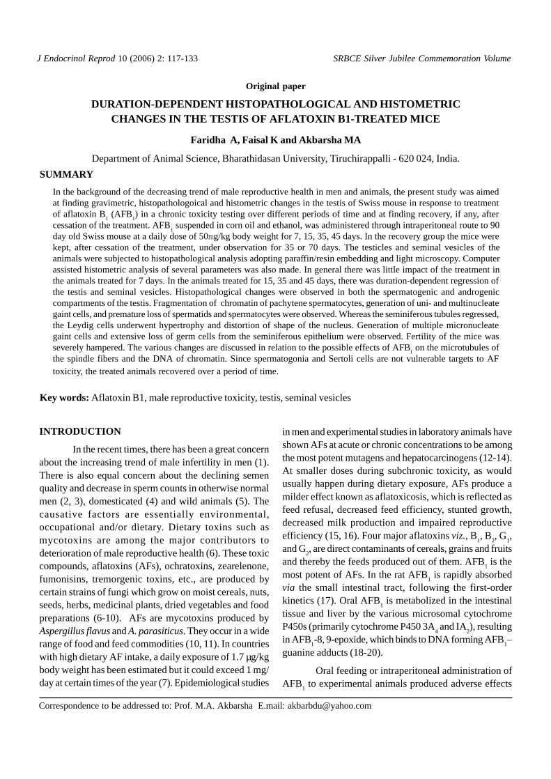
J Endocrinol Reprod 10 (2006) 2: 117-133
Original paper
DURATION-DEPENDENT HISTOPATHOLOGICAL AND HISTOMETRICCHANGES IN THE TESTIS OF AFLATOXIN B1-TREATED MICE
Faridha A, Faisal K and Akbarsha MA
Department of Animal Science, Bharathidasan University, Tiruchirappalli - 620 024, India.
Correspondence to be addressed to: Prof. M.A. Akbarsha E.mail: [email protected]
SUMMARY
In the background of the decreasing trend of male reproductive health in men and animals, the present study was aimedat finding gravimetric, histopathologoical and histometric changes in the testis of Swiss mouse in response to treatmentof aflatoxin B
1 (AFB
1) in a chronic toxicity testing over different periods of time and at finding recovery, if any, after
cessation of the treatment. AFB1 suspended in corn oil and ethanol, was administered through intraperitoneal route to 90
day old Swiss mouse at a daily dose of 50mg/kg body weight for 7, 15, 35, 45 days. In the recovery group the mice werekept, after cessation of the treatment, under observation for 35 or 70 days. The testicles and seminal vesicles of theanimals were subjected to histopathological analysis adopting paraffin/resin embedding and light microscopy. Computerassisted histometric analysis of several parameters was also made. In general there was little impact of the treatment inthe animals treated for 7 days. In the animals treated for 15, 35 and 45 days, there was duration-dependent regression ofthe testis and seminal vesicles. Histopathological changes were observed in both the spermatogenic and androgeniccompartments of the testis. Fragmentation of chromatin of pachytene spermatocytes, generation of uni- and multinucleategaint cells, and premature loss of spermatids and spermatocytes were observed. Whereas the seminiferous tubules regressed,the Leydig cells underwent hypertrophy and distortion of shape of the nucleus. Generation of multiple micronucleategaint cells and extensive loss of germ cells from the seminiferous epithelium were observed. Fertility of the mice wasseverely hampered. The various changes are discussed in relation to the possible effects of AFB
1 on the microtubules of
the spindle fibers and the DNA of chromatin. Since spermatogonia and Sertoli cells are not vulnerable targets to AFtoxicity, the treated animals recovered over a period of time.
Key words: Aflatoxin B1, male reproductive toxicity, testis, seminal vesicles
INTRODUCTION
In the recent times, there has been a great concernabout the increasing trend of male infertility in men (1).There is also equal concern about the declining semenquality and decrease in sperm counts in otherwise normalmen (2, 3), domesticated (4) and wild animals (5). Thecausative factors are essentially environmental,occupational and/or dietary. Dietary toxins such asmycotoxins are among the major contributors todeterioration of male reproductive health (6). These toxiccompounds, aflatoxins (AFs), ochratoxins, zearelenone,fumonisins, tremorgenic toxins, etc., are produced bycertain strains of fungi which grow on moist cereals, nuts,seeds, herbs, medicinal plants, dried vegetables and foodpreparations (6-10). AFs are mycotoxins produced byAspergillus flavus and A. parasiticus. They occur in a widerange of food and feed commodities (10, 11). In countrieswith high dietary AF intake, a daily exposure of 1.7 µg/kgbody weight has been estimated but it could exceed 1 mg/day at certain times of the year (7). Epidemiological studies
in men and experimental studies in laboratory animals haveshown AFs at acute or chronic concentrations to be amongthe most potent mutagens and hepatocarcinogens (12-14).At smaller doses during subchronic toxicity, as wouldusually happen during dietary exposure, AFs produce amilder effect known as aflatoxicosis, which is reflected asfeed refusal, decreased feed efficiency, stunted growth,decreased milk production and impaired reproductiveefficiency (15, 16). Four major aflatoxins viz., B
1, B
2, G
1,
and G2, are direct contaminants of cereals, grains and fruits
and thereby the feeds produced out of them. AFB1 is the
most potent of AFs. In the rat AFB1 is rapidly absorbed
via the small intestinal tract, following the first-orderkinetics (17). Oral AFB
1 is metabolized in the intestinal
tissue and liver by the various microsomal cytochromeP450s (primarily cytochrome P450 3A
4 and IA
2), resulting
in AFB1-8, 9-epoxide, which binds to DNA forming AFB
1–
guanine adducts (18-20).
Oral feeding or intraperitoneal administration ofAFB
1 to experimental animals produced adverse effects
SRBCE Silver Jubilee Commemoration Volume
118
on aspects of male reproduction such as delayed testiculardevelopment, morphological, regressive and degenerativechanges in the testis, hypertrophy of Leydig cells and dropin testosterone, decrease in the percentage of live spermand increase of sperm abnormalities, etc. (21-29).Formation of testicular AFB
1-DNA adducts in rats has
been shown (30). AFs have been detected in the humansemen (31) and boar sperm (32). In the latter case, thehighest AF residues were recorded in March to May andwere related with AF concentration in the feedration.
The various experimental studies, which indicatedeterioration of male reproductive health, do not provideinsight into the impact of AFB
1 on the spermatogenic and
androgenic compartments of the testis when the toxin isadministered over different periods of time. Further, thevarious studies indicate only qualitative histologicalchanges. The present study reports the gravimetric,histological and histometric changes in both thespermatogenic and androgenic compartments of the testisof mice administered AFB
1 over different periods of time
in a chronic toxicity testing. Fertility testing and recoverystudy were also conducted.
MATERIALS AND METHODS
The methodology of chronic male reproductivetoxicity testing was adopted from Linder et al. (33). Swissstrain male albino mice (90 day old), weighingapproximately 30g, were used in the study. Female miceused in the fertility tests also belonged to the same strainand age. Mice were developed from a stock procured fromInternational Institute of Biotechnology and Toxicology(IIBAT), Padappai, India. Mice were housed inpolypropylene cages with metal grill tops, and fed withstandard pellet feed (Gold Mohur Laboratory AnimalFeeds, Lipton India Ltd., Bangalore) and water ad libitum.The toxin, aflatoxin B
1 (AFB
1), was obtained from SIGMA
Chemical Co (St. Louis, MO, USA).
The AFB1 was quantitatively prepared in corn oil
and ethanol (95:5) according to Egbunike et al. (34). AFB1
at a dose of 50µg/kg/day was administered to theexperimental animals through intra-peritoneal route for7, 15, 35 and 45 days. Each experimental group consistedof 45 animals. Control mice were maintained parallel toeach experimental group, and consisted of 45 animals each.At the end of each experimental period, 5 animals fromeach of the experimental and control groups were weighedand killed through cervical dislocation under mild sodium
pentabarbital anasthesia. The animals were dissected andthe testes and seminal vesicles were used for gravimetricas well as histological analysis adopting paraffin-embedding and PAS and hematoxylin staining. In another5 mice from each of the experimental and control groupsthe reproductive system was perfused with Karnovsky’s(35) fluid (glutaraldehyde 1%, paraformaldehyde 1%, inphosphate buffer) and the animals were sacrificed underanasthesia. The right testis was removed and thin slices ofthe tissue were fixed in 2.5% glutaraldehyde in cacodylatebuffer and post-fixed in 1% osmium tetroxide for obtainingsemithin sections (1µm thickness). The sections werestained in toluidine blue O (TBO). 5 mice in each of theexperimental and control groups were tested for fertility.Fertility test was carried out after withdrawal of the toxintreatment or after the period of recovery, by allowing amale mouse to mate with two female mice at estrus.Mounting, if any, was observed overnight. The followingmorning, vaginal smears were examined in a microscopefor spermatozoa. Subsequently, the female mice wereseparated and allowed to go to term and the litter size wasrecorded. Thus, male fertility was evaluated indirectly byregistering female fertility. The remaining 30 animals ineach of the experimental and control groups were left forrecovery. For these mice, treatment of AFB
1 was
discontinued after the defined period. 15 animals weresacrificed and subjected to analysis as above after 35 daysof recovery, the duration of one spermatogenic cycle (36)and the remaining 15 animals sacrificed and subjected toanalysis after 70 days. Fertility testing of animals in therecovery groups was the same as for AFB
1-treatment.
In the case of gravimetric analysis the pairedtesticles and seminal vesicles from each animal wereweighed separately. Data for each organ from each groupwere used to calculate the respective means and the standarddeviations. Light microscopic observation was made in aCarl Zeiss Axioskop 2 plus (Carl Zeiss, Gena, Germany)research microscope connected to a computer through SonyDxC-151a/151HP 2/3 CCD camera (Sony, Tokyo, Japan).Photomicrographs were obtained and processed usingAxiovision software. Histometry/cytometry was conductedusing the same software. The software is a modular image-processing and analysis system for use in modernmicroscopy. Sections stained in PAS and hematoxylin orTBO were viewed in the microscope and fields with sectionsof seminiferous tubules (STs) in circular transverse sectionwere selected at x100 magnification. Using the software,
Faridha et al
119
the boundary of the each of the ST was demarcated, andthe total area of the STs in the field was deducted from thearea of the field. Thus, the area of the STs and the area ofthe interstitium were duduced. This was done for fivedifferent fields from the tissue of each mouse, thus making25 measurements for each sub-group. The perimeter andthe diameter of five different circular transverse sections ofSTs from each mouse were also measured at x400magnification making 25 measurements of each parameterfor five animals in each sub-group. At x400 magnificationthe epithelium of STs was analyzed for number of uninucleategiant cells (UNGCs) and multinucleate giant cells (MNGCs).This was done in STs in semithin sections and a total of 25tubules were assessed in each group. The data were used tofind the total number of cells in 104 mm2 area per tubule,and among them the number of UNGCs and MNGCs.
The number of Leydig cells per 103 mm3 of theinterstitial area was counted in five fields from each animal,at x1000 magnification, making it 25 counts for each sub-group. The perimeter of Leydig cells and the diameter ofthe nuclei of Leydig cells were also measured for 100Leydig cells in the testis of each group. The data weresubjected to analysis using the Carl Zeiss software asabove. The mean and the standard deviation were calculatedfor data in respect of each parameter in each group. Datafor each parameter for the respective control and treatmentgroups were used for application of Students’ ‘t’ test tofind the level of significance of the difference.
RESULTS
Decrease in the weight of the testicles and seminalvesicles
Gravimetric analysis of testicles and seminal vesiclesrevealed duration-dependent decrease in their respectiveweights (Table 1). As calculated from the mean values thedecrease in weight of these organs was not significant in themice treated AFB
1 for 7 days whereas in the mice treated for
15 days, the weight of testicles decreased significantly to 73%,in those treated for 35 days to 68% and in those treated for 45days to 51%. Weight of the seminal vesicles also decreased to76% in mice treated for 15 days, to 69% in those treated for35 days and to 59% in those treated for 45 days.
Table 1. Weight of the paired testicles and seminal vesicles ofcontrol and AFB1 - treated mice (Mean ± SD)
Duration of Weight of testicles Weight of the seminalTreatment (mg) vesicles (mg)
Control Experimental Control Experimental
7 days 213±16 203±13 (95) 85±8 79±7 (93)
15 days 217±14 158±12*(73) 84±9 64±6*(76)
35 days 219±21 148±08*(68) 86±8 59±6*(69)
45 days 220±19 112±06*(51) 88±7 52±6*(59)
* p<0.01
Number in parenthesis, percentage of the control value
Recovery group I - 35 days
Duration of Weight of testicles Weight of the seminalTreatment (mg) vesicles (mg)
Control Experimental Control Experimental
7 days 218±16 212±12 84±6 82±6
15 days 221±18 189±14 86±7 72±5
35 days 222±15 172±16 85±4 69±4
45 days 221±17 163±17 87±6 68±6
Recovery group II - 70 days
Duration of Weight of testicles Weight of the seminalTreatment (mg) vesicles (mg)
Control Experimental Control Experimental
7 days 221±17 218±16 86±7 85±5
15 days 222±18 218±17 82±6 81±6
35 days 221±16 220±18 85±8 78±5
45 days 223±19 221±19 85±6 77±6
Gross histological and histometric changes in the testis
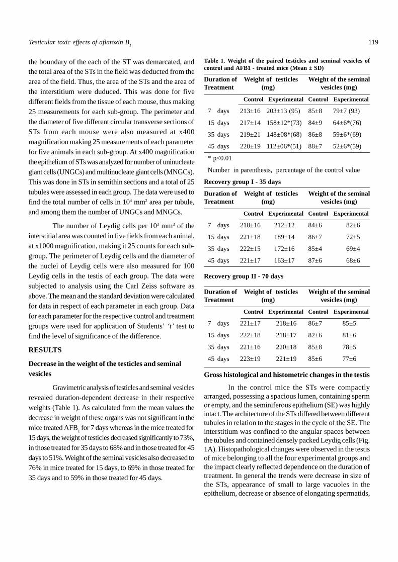
In the control mice the STs were compactlyarranged, possessing a spacious lumen, containing spermor empty, and the seminiferous epithelium (SE) was highlyintact. The architecture of the STs differed between differenttubules in relation to the stages in the cycle of the SE. Theinterstitium was confined to the angular spaces betweenthe tubules and contained densely packed Leydig cells (Fig.1A). Histopathological changes were observed in the testisof mice belonging to all the four experimental groups andthe impact clearly reflected dependence on the duration oftreatment. In general the trends were decrease in size ofthe STs, appearance of small to large vacuoles in theepithelium, decrease or absence of elongating spermatids,
Testicular toxic effects of aflatoxin B1
120
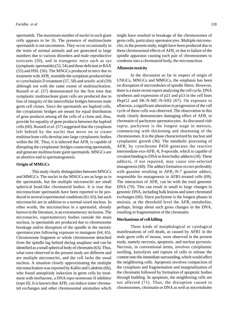
Fig. 1A-E. Section of the testis of control and treated mice. A:Control mouse; paraffin section, PAS & H staining. B-E:AFB
1-treated mice; semithin sections, TBO staining. Note
depletion of lumen. B: 7 day treatment. C: 15 day treatment. Noteoccurrence of giant cells (arrowheads). Interstitium ishypertrophied. D: 35 day treatment. Note hypertrophy ofinterstitium. E: 45 day treatment. Note hypertrophy of interstitium.ST, seminiferous tubules; IN, interstitium. Scale bar 35µm.
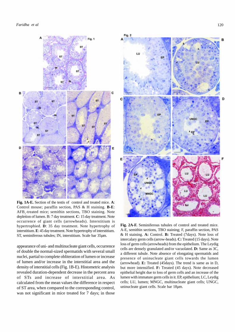
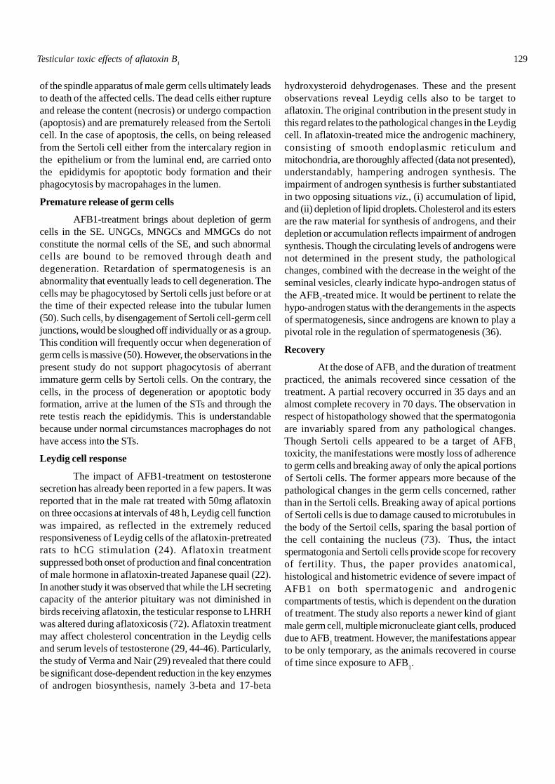
Fig. 2A-F. Seminiferous tubules of control and treated mice.A-E, semithin sections, TBO staining; F, paraffin section, PAS& H staining. A: Control. B: Treated (7days). Note loss ofintercalary germ cells (arrow-heads). C: Treated (15 days). Noteloss of germ cells (arrowheads) from the epithelium. The Leydigcells are densely granulated and/or vacuolated. D: Same as 3C,a different tubule. Note absence of elongating spermatids andpresence of uninucleate giant cells towards the lumen(arrowhead). E: Treated (45days). The trend is same as in D,but more intensified. F: Treated (45 days). Note decreasedepithelial height due to loss of germ cells and an increase of thelumen with immature germ cells in it. EP, epithelium; LC, Leydigcells; LU, lumen; MNGC, multinucleate giant cells; UNGC,uninucleate giant cells. Scale bar 18µm.
appearance of uni- and multinucleate giant cells, occurrenceof double the normal-sized spermatids with several smallnuclei, partial to complete obliteration of lumen or increaseof lumen and/or increase in the interstitial area and thedensity of interstitial cells (Fig. 1B-E). Histometric analysisrevealed duration-dependent decrease in the percent areaof STs and increase of interstitial area. Ascalculated from the mean values the difference in respectof ST area, when compared to the corresponding control,was not significant in mice treated for 7 days; in those
Faridha et al
121
treated for 15 days, the area of STs decreased to 77%, inthose treated for 35 days to 49% and in those treated for45 days to 33% (Table 2).
Table 2. Percent area in transection occupied by theseminiferous tubules and the inter sritium in the testis ofmice treated with AFB1. Each value is mean ± SD of 25determinations atx100 magnifications from sectionsobtained from the right restis of 5 animals.
Treatment groupDuration of Seminiferous tribule Interstitial areaTreatment area
Control AFB1-treated Control AFB1-treated
7 days 86.88±1.93 82.12±1.63* 13.12±1.93 17.88±1.64
15 days 86.94±2.08 76.88±3.20* 13.01±2.04 23.12±2.36*
35 days 86.10±2.04 48.88±6.22* 13.90±2.04 51.12±6.22*
45 days 85.34±2.67 33.38±5.96* 14.66±2.68 66.62±5.96*
*p < 0.001
Recovery group II - 35 days
Duration of Seminiferous tribule Interstitial areaTreatment area
Control AFB1-treated Control AFB1-treated
7 days 88.23±2.15 82.12±1.63 12.15±1.25 22.43±6.72
15 days 87.75±1.32 79.46±2.32 12.32±1.75 28.16±7.09
35 days 87.62±2.55 65.72±2.26 13.05±1.92 32.24±6.24
45 days 87.98±2.62 56.59±1.75 12.75±1.78 45.72±6.35
Recovery group II -70 days
Duration of Seminiferous tribule Interstitial areaTreatment area
Control AFB1-treated Control AFB1-treated
7 days 86.24±2.05 84.69±1.75 13.17±1.89 14.65±1.72
15 days 86.92±2.21 84.23±2.65 13.21±2.65 17.11±1.85
35 days 87.02±1.92 80.72±2.32 13.56±2.42 24.32±1.82
45 days 86.56±1.89 75.02±2.05 13.95±2.35 28.75±2.03
The trend in respect of the interstitial area wasopposite to this. Critical observation of the individual STsrevealed decrease to almost total absence of elongatingspermatids. Spermiated spermatozoa were invariablyabsent in the lumen (Fig. 2A-F). The height of the SE eitherincreased (Fig. 2A-E) or decreased (Fig. 2F) and,correspondingly, the lumen was either almost obliteratedor increased. A duration-dependent appearance of uni- (Fig.2D) and multinucleate (Fig. 2E) giant cells was noticed. The
SE of the mice treated for 15, 35 and 45 days possessed smallto large vacuoles or empty spaces increasing in magnitude inrelation to the duration of treatment. The vacuoles were emptyor contained cell debris. Cell shrinkage and necrosis orpycnosis of the nuclei were also noticed. A few of the giantcells in the epithelium as well as in the lumen possessedvacuolated cytoplasm and pycnotic nuclei or nuclei withmarginalized chromatin. Data on the perimeter and diameterof the STs are presented in Table 3. Both the parametersdecreased. The SE of the control mice did not indicate anydegeneration, whereas in the AFB
1-treated mice the tubules
with indication of epithelial degeneration increased in the orderof the duration of treatment, and in the mice treated for 45days no tubule was spared (Figs. 1D, E, 2B, E,).
Table 3. Perimeter and diameter of the seminiferous tubulesof mice treated AFB1. Each value is mean ± SD of 25measurements made at x400 with sections from the righttestis of 5 animals
Treatment group
Duration of Perimeter (mmmmmm) Diameter (mmmmmm)treatment
Control AFB1-treated Control AFB1-treated
7 days 444.69±10.64 389.92±16.34* 165.65±3.70 120.56±4.71*
15 days 453.00±8.12 332.11±15.09* 164.76±3.19 103.98±8.45*
35 days 462.39±5.34 309.10±19.92* 163.84±2.09 94.01±3.25*
45 days 464.03±2.91 232.53±11.62* 164.56±2.32 81.44±5.71*
* p<0.001
Recovery group I -35 days
Duration of Perimeter (mmmmmm) Diameter (mmmmmm)treatment
Control AFB1-treated Control AFB1-treated
7 days 444.69±8.72 412.73±16.34 164.96±2.86 145.24±4.21
15 days 462.14±9.64 364.72±12.36 164.14±2.36 136.72±6.42
35 days 364.24±4.68 349.52±12.73 165.12±2.39 129.92±4.16
45 days 465.86±4.32 303.16±9.59 164.87±2.86 115.33±4.18
Recovery group II-70 days
Duration of Perimeter (mmmmmm) Diameter (mmmmmm)treatment
Control AFB1-treated Control AFB1-treated
7 days 451.36±8.23 438.17±13.16 163.74±4.21 156.26±3.14
15 days 456.17±12.45 438.76±8.92 163.17±2.85 152.17±6.35
35 days 464.68±8.93 448.86±12.83 165.19±3.28 149.86±4.82
45 days 468.14±6.31 436.73±9.76 166.45±3.05 139.32±4.48
Testicular toxic effects of aflatoxin B1
II-70 days
122
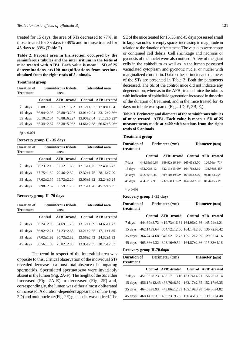
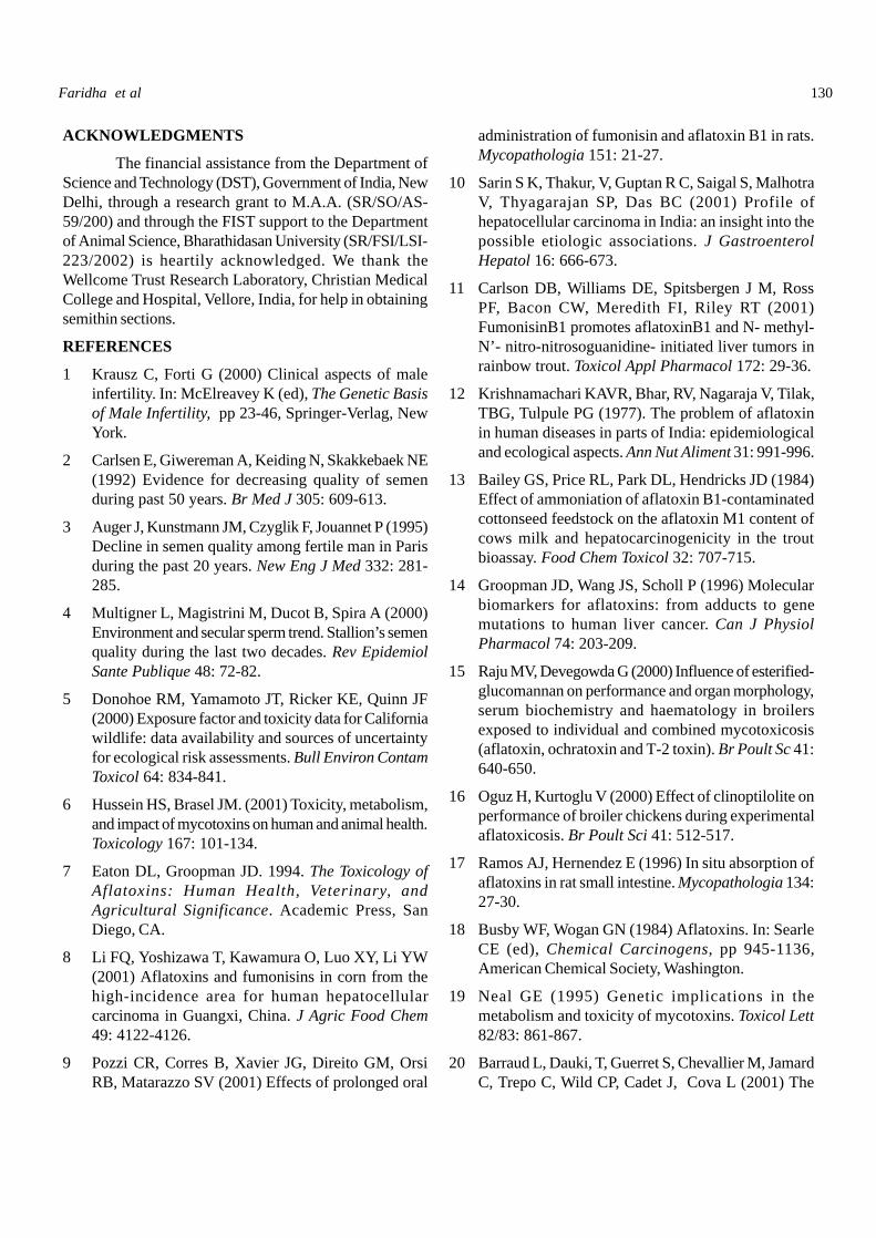
Fig. 3A-D. Seminiferous epithelium of treated mice. Semithinsections, TBO staining. A: Shows uninucleate giant cells(arrowheads) which are spermatids in the epithelium, damageto chromatin of pachytene spermatocytes, and loss of intercalarygerm cells (asterisks). B: The uninucleate giant spermatid(arrowhead) is seen in the lumen of the seminiferous tubule.Necrosis of pachytene spermatocytes is also evident (asterisks).C: The UNGCs (arrowheads) are pachytene spermatocytes.Note doubling of size of the nucleus, compared to those whichunderlie them. The giant cells are in the process of beingreleased and one of them is vacuolated (asterisk). D: The giantcells (arrowheads) are in the process of being released into thelumen. In the area marked with asterisks, germ cells are totallylost. NE, necrosis; PS, pachytene spermatocytes; SC, Sertolicell; SF, Sertoli cell fibrosis. Scale bar, 4µm.
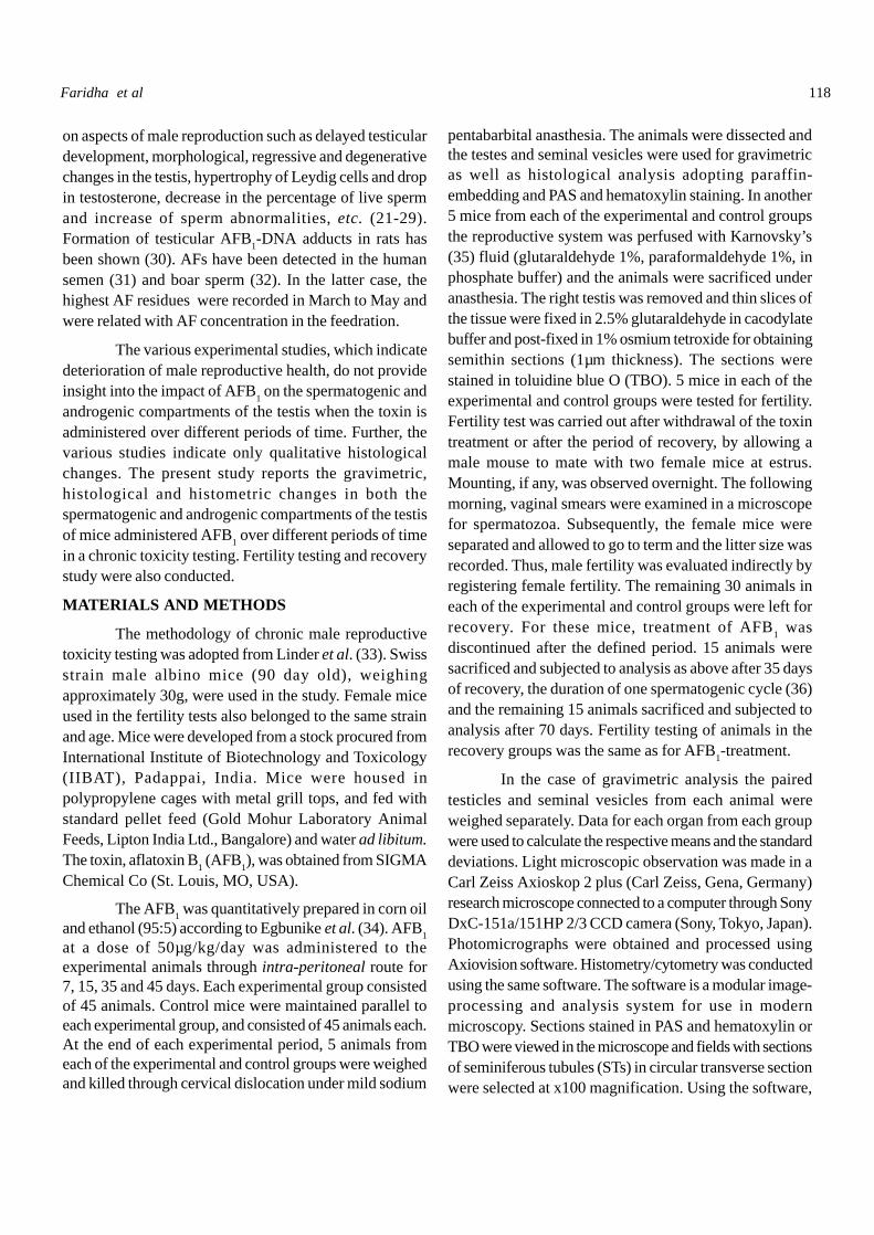
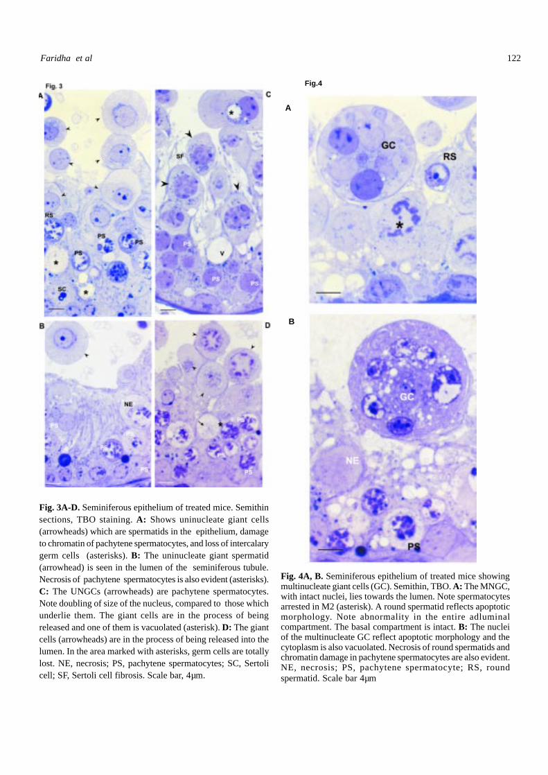
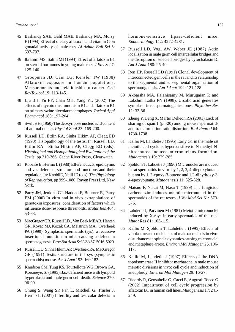
Fig. 4A, B. Seminiferous epithelium of treated mice showingmultinucleate giant cells (GC). Semithin, TBO. A: The MNGC,with intact nuclei, lies towards the lumen. Note spermatocytesarrested in M2 (asterisk). A round spermatid reflects apoptoticmorphology. Note abnormality in the entire adluminalcompartment. The basal compartment is intact. B: The nucleiof the multinucleate GC reflect apoptotic morphology and thecytoplasm is also vacuolated. Necrosis of round spermatids andchromatin damage in pachytene spermatocytes are also evident.NE, necrosis; PS, pachytene spermatocyte; RS, roundspermatid. Scale bar 4µm
Fig.4
A
B
Faridha et al
123
Critical observation of the STs of AFB1-treated mice,
particularly those in the 35 and 45 day treatment groups, revealedoccurrence of pachytene spermatocytes or spermatids of sizedouble that of the respective normal cells (Fig. 2D, E). Suchcells are designated as UNGCs. They were present in theepithelium along the luminal profile (Fig. 3A), some projectinginto the lumen but still adherent to the Sertoli cells (Fig. 3B) orlying loose in the lumen (Fig.3C). In several cases the UNGCpossessed highly vacuolated cytoplasm, and the nucleus wasaltered in morphology (Fig. 3D).
Another observation made in several of the STs ofthe AFB
1-treated mice belonging to 15, 35 and 45 day
treatment groups was occurrence of MNGCs (diameter,40-52 mm dia) (Fig. 2E). Such cells possessed two to 16nuclei. The nuclei were either intact (Fig. 4A) or hadmarginalized chromatin (Fig. 4B). The cytoplasm indicatedlittle (Fig. 4A) to extensive (Fig. 4B) vacuolation. One ofthe observations was appearance of large cells (diameter20-30 mm) containing several micronuclei (Fig. 5A). Suchcells are designated as multiple micronucleate giant cells(MMGC). They were present in the epithelium as well asthe lumen; when present in the epithelium, they wereseparated from the Sertoli cells to a great extent, indicatingthat they were being released into the lumen and wouldresult in the appearance of vacuoles in the epithelium. Themicronuclei had the appearance of dot-like densechromatoid bodies and the cytoplasm formed into twodistinct zones, a thin peripheral zone and a large centralzone containing the micronuclei. In a few tubules UNGC,MNGC and MMGC coexisted (Fig. 5B).
Data on the counts of cells per unit area, andamong them those that were UNGC, MNGC and MMGCare presented in Table 4. The data reveal that in the micetreated AFB1 for 7 days UNGC, MNGC or MMGC wasnot generated, whereas in those treated for 15, 35 and 45days all the three versions of giant cells were generated.Loss of germ cells in a few tubules was so acute that hardlyany germ cell was present in the adluminal compartment,with the epithelium manifesting small to large vacuoles(Figs. 2E, F, 6A). In some of the tubules the Sertoli cellsthemselves, from above the level of the ectoplasmicspecialization, i.e., the tight junction of blood-testis barrier,had broken away and such broken portions were carryingwith them the pachytene spermatocytes, rendering theepithelium comparable to Sertoli cell-only syndrome,though careful observation revealed the presence ofspermatogonia (Fig. 6A, B). The immature germ cells thuslost from the STs could be traced to the rete testis(Fig. 6C). The lumen of the epididymal duct, particularlyin the mice treated AFB1 for 35 and 45 days, containedsuch immature germ cells instead of sperm (Fig. 6D).Several of the immature germ cells in the lumen of the
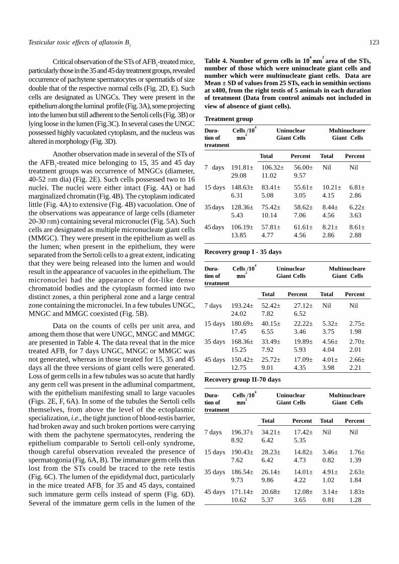
Table 4. Number of germ cells in 104 mmmmmm
2 area of the STs,
number of those which were uninucleate giant cells andnumber which were multinucleate giant cells. Data areMean ± SD of values from 25 STs, each in semithin sectionsat x400, from the right testis of 5 animals in each durationof treatment (Data from control animals not included inview of absence of giant cells).
Treatment group
Dura- Cells /104
Uninuclear Multinuclearetion of mmmmmm
2 Giant Cells Giant Cells
treatment
Total Percent Total Percent
7 days 191.81± 106.32± 56.00± Nil Nil29.08 11.02 9.57
15 days 148.63± 83.41± 55.61± 10.21± 6.81±6.31 5.08 3.05 4.15 2.86
35days 128.36± 75.42± 58.62± 8.44± 6.22±5.43 10.14 7.06 4.56 3.63
45days 106.19± 57.81± 61.61± 8.21± 8.61±13.85 4.77 4.56 2.86 2.88
Recovery group I - 35 days
Dura- Cells /104
Uninuclear Multinuclearetion of mmmmmm
2 Giant Cells Giant Cells
treatment
Total Percent Total Percent
7 days 193.24± 52.42± 27.12± Nil Nil24.02 7.82 6.52
15 days 180.69± 40.15± 22.22± 5.32± 2.75±17.45 6.55 3.46 3.75 1.98
35 days 168.36± 33.49± 19.89± 4.56± 2.70±15.25 7.92 5.93 4.04 2.01
45 days 150.42± 25.72± 17.09± 4.01± 2.66±12.75 9.01 4.35 3.98 2.21
Recovery group II-70 days
Dura- Cells /104
Uninuclear Multinuclearetion of mmmmmm
2 Giant Cells Giant Cells
treatment
Total Percent Total Percent
7 days 196.37± 34.21± 17.42± Nil Nil8.92 6.42 5.35
15 days 190.43± 28.23± 14.82± 3.46± 1.76±7.62 6.42 4.73 0.82 1.39
35 days 186.54± 26.14± 14.01± 4.91± 2.63±9.73 9.86 4.22 1.02 1.84
45 days 171.14± 20.68± 12.08± 3.14± 1.83±10.62 5.37 3.65 0.81 1.28
Testicular toxic effects of aflatoxin B1
124
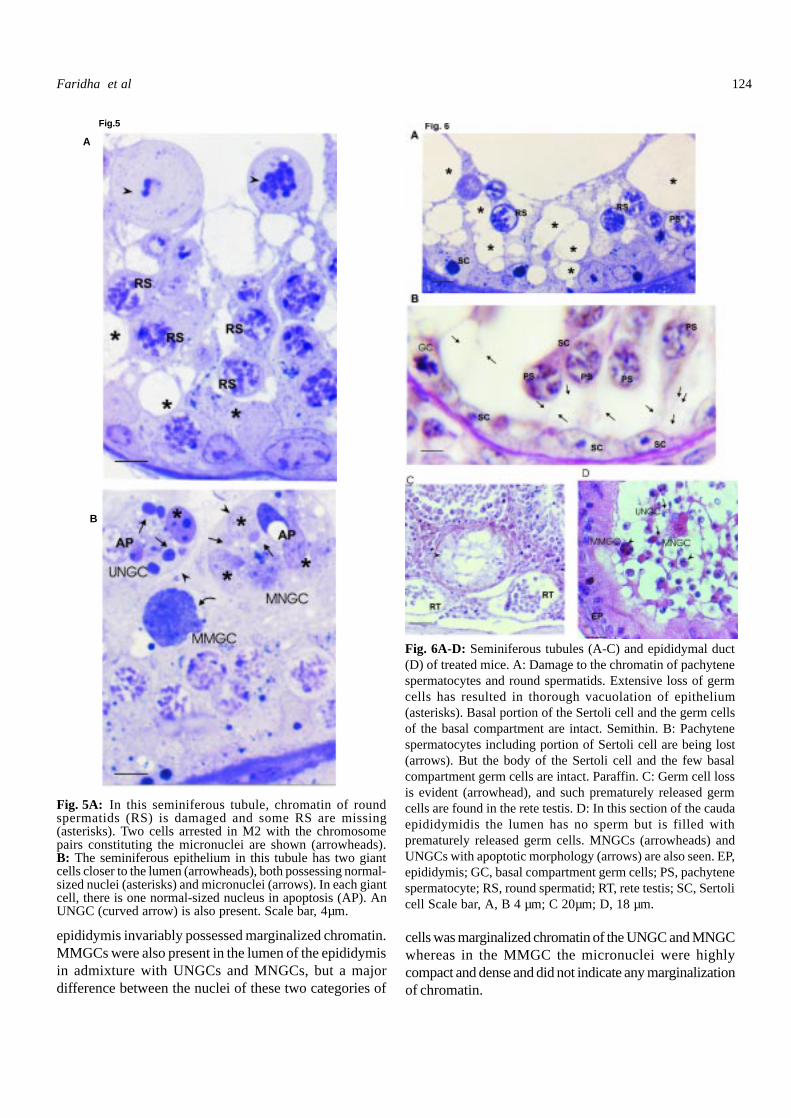
Fig. 5A: In this seminiferous tubule, chromatin of roundspermatids (RS) is damaged and some RS are missing(asterisks). Two cells arrested in M2 with the chromosomepairs constituting the micronuclei are shown (arrowheads).B: The seminiferous epithelium in this tubule has two giantcells closer to the lumen (arrowheads), both possessing normal-sized nuclei (asterisks) and micronuclei (arrows). In each giantcell, there is one normal-sized nucleus in apoptosis (AP). AnUNGC (curved arrow) is also present. Scale bar, 4µm.
Fig. 6A-D: Seminiferous tubules (A-C) and epididymal duct(D) of treated mice. A: Damage to the chromatin of pachytenespermatocytes and round spermatids. Extensive loss of germcells has resulted in thorough vacuolation of epithelium(asterisks). Basal portion of the Sertoli cell and the germ cellsof the basal compartment are intact. Semithin. B: Pachytenespermatocytes including portion of Sertoli cell are being lost(arrows). But the body of the Sertoli cell and the few basalcompartment germ cells are intact. Paraffin. C: Germ cell lossis evident (arrowhead), and such prematurely released germcells are found in the rete testis. D: In this section of the caudaepididymidis the lumen has no sperm but is filled withprematurely released germ cells. MNGCs (arrowheads) andUNGCs with apoptotic morphology (arrows) are also seen. EP,epididymis; GC, basal compartment germ cells; PS, pachytenespermatocyte; RS, round spermatid; RT, rete testis; SC, Sertolicell Scale bar, A, B 4 µm; C 20µm; D, 18 µm.
Fig.5
A
B
epididymis invariably possessed marginalized chromatin.MMGCs were also present in the lumen of the epididymisin admixture with UNGCs and MNGCs, but a majordifference between the nuclei of these two categories of
cells was marginalized chromatin of the UNGC and MNGCwhereas in the MMGC the micronuclei were highlycompact and dense and did not indicate any marginalizationof chromatin.
Faridha et al
125
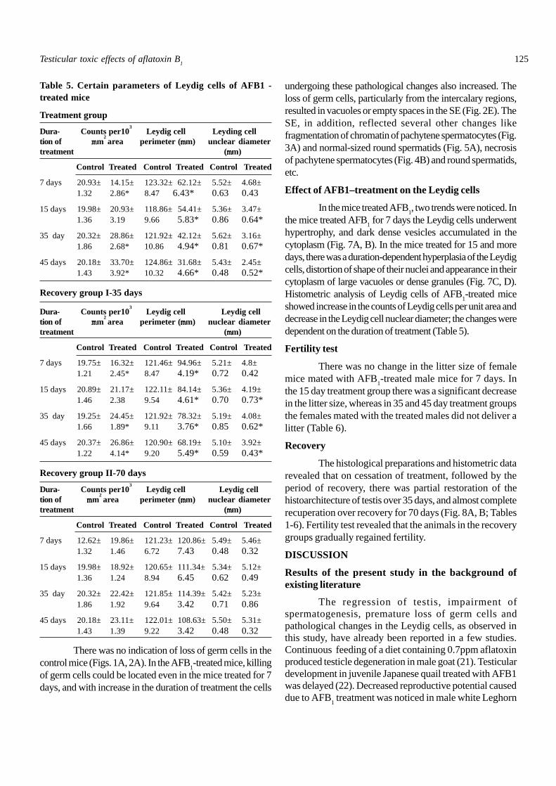
Table 5. Certain parameters of Leydig cells of AFB1 -treated mice
Treatment group
Dura- Counts per103 Leydig cell Leyding cell
tion of m m m m mm2 area perimeter (mmmmmm) unclear diameter
treatment (mmmmmm)
Control Treated Control Treated Control Treated
7 days 20.93± 14.15± 123.32± 62.12± 5.52± 4.68±1.32 2.86* 8.47 6.43* 0.63 0.43
15 days 19.98± 20.93± 118.86± 54.41± 5.36± 3.47±1.36 3.19 9.66 5.83* 0.86 0.64*
35 day 20.32± 28.86± 121.92± 42.12± 5.62± 3.16±1.86 2.68* 10.86 4.94* 0.81 0.67*
45 days 20.18± 33.70± 124.86± 31.68± 5.43± 2.45±1.43 3.92* 10.32 4.66* 0.48 0.52*
Recovery group I-35 days
Dura- Counts per103 Leydig cell Leydig cell
tion of m m m m mm2 area perimeter (mmmmmm) nuclear diameter
treatment (mmmmmm)
Control Treated Control Treated Control Treated
7 days 19.75± 16.32± 121.46± 94.96± 5.21± 4.8±1.21 2.45* 8.47 4.19* 0.72 0.42
15 days 20.89± 21.17± 122.11± 84.14± 5.36± 4.19±1.46 2.38 9.54 4.61* 0.70 0.73*
35 day 19.25± 24.45± 121.92± 78.32± 5.19± 4.08±1.66 1.89* 9.11 3.76* 0.85 0.62*
45 days 20.37± 26.86± 120.90± 68.19± 5.10± 3.92±1.22 4.14* 9.20 5.49* 0.59 0.43*
Recovery group II-70 days
Dura- Counts per103 Leydig cell Leydig cell
tion of m m m m mm2 area perimeter (mmmmmm) nuclear diameter
treatment (mmmmmm)
Control Treated Control Treated Control Treated
7 days 12.62± 19.86± 121.23± 120.86± 5.49± 5.46±1.32 1.46 6.72 7.43 0.48 0.32
15 days 19.98± 18.92± 120.65± 111.34± 5.34± 5.12±1.36 1.24 8.94 6.45 0.62 0.49
35 day 20.32± 22.42± 121.85± 114.39± 5.42± 5.23±1.86 1.92 9.64 3.42 0.71 0.86
45 days 20.18± 23.11± 122.01± 108.63± 5.50± 5.31±1.43 1.39 9.22 3.42 0.48 0.32
There was no indication of loss of germ cells in thecontrol mice (Figs. 1A, 2A). In the AFB
1-treated mice, killing
of germ cells could be located even in the mice treated for 7days, and with increase in the duration of treatment the cells
undergoing these pathological changes also increased. Theloss of germ cells, particularly from the intercalary regions,resulted in vacuoles or empty spaces in the SE (Fig. 2E). TheSE, in addition, reflected several other changes likefragmentation of chromatin of pachytene spermatocytes (Fig.3A) and normal-sized round spermatids (Fig. 5A), necrosisof pachytene spermatocytes (Fig. 4B) and round spermatids,etc.
Effect of AFB1–treatment on the Leydig cells
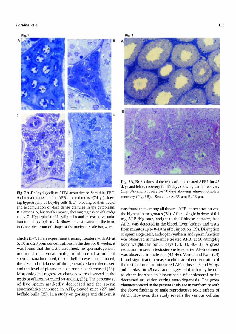
In the mice treated AFB1, two trends were noticed. In
the mice treated AFB1 for 7 days the Leydig cells underwent
hypertrophy, and dark dense vesicles accumulated in thecytoplasm (Fig. 7A, B). In the mice treated for 15 and moredays, there was a duration-dependent hyperplasia of the Leydigcells, distortion of shape of their nuclei and appearance in theircytoplasm of large vacuoles or dense granules (Fig. 7C, D).Histometric analysis of Leydig cells of AFB
1-treated mice
showed increase in the counts of Leydig cells per unit area anddecrease in the Leydig cell nuclear diameter; the changes weredependent on the duration of treatment (Table 5).
Fertility test
There was no change in the litter size of femalemice mated with AFB
1-treated male mice for 7 days. In
the 15 day treatment group there was a significant decreasein the litter size, whereas in 35 and 45 day treatment groupsthe females mated with the treated males did not deliver alitter (Table 6).
Recovery
The histological preparations and histometric datarevealed that on cessation of treatment, followed by theperiod of recovery, there was partial restoration of thehistoarchitecture of testis over 35 days, and almost completerecuperation over recovery for 70 days (Fig. 8A, B; Tables1-6). Fertility test revealed that the animals in the recoverygroups gradually regained fertility.
DISCUSSION
Results of the present study in the background ofexisting literature
The regression of testis, impairment ofspermatogenesis, premature loss of germ cells andpathological changes in the Leydig cells, as observed inthis study, have already been reported in a few studies.Continuous feeding of a diet containing 0.7ppm aflatoxinproduced testicle degeneration in male goat (21). Testiculardevelopment in juvenile Japanese quail treated with AFB1was delayed (22). Decreased reproductive potential causeddue to AFB
1 treatment was noticed in male white Leghorn
Testicular toxic effects of aflatoxin B1
126
Fig. 7 A-D: Leydig cells of AFB1-treated mice. Semithin, TBO.A: Interstitial tissue of an AFB1-treated mouse (7days) show-ing hypertrophy of Leydig cells (LC), bloating of their nucleiand accumulation of dark dense granules in the cytoplasm.B: Same as A, but another mouse, showing regression of Leydigcells. C: Hyperplasia of Leydig cells and increased vacuola-tion in their cytoplasm. D: Shows intensification of the trendin C and distortion of shape of the nucleus. Scale bar, 4µm.
Fig. 8A, B: Sections of the testis of mice treated AFB1 for 45days and left to recovery for 35 days showing partial recovery(Fig. 8A) and recovery for 70 days showing almost completerecovery (Fig. 8B). Scale bar A, 35 µm; B, 18 µm.
chicks (37). In an experiment treating roosters with AF at5, 10 and 20 ppm concentrations in the diet for 8 weeks, itwas found that the testis atrophied, no spermatogenesisoccurred in several birds, incidence of abnormalspermatozoa increased, the epithelium was desquamated,the size and thickness of the generative layer decreasedand the level of plasma testosterone also decreased (28).Morphological regressive changes were observed in thetestis of aflatoxin-treated rat and pig (23). The percentageof live sperm markedly decreased and the spermabnormalities increased in AFB
1-treated mice (27) and
buffalo bulls (25). In a study on goslings and chicken it
was found that, among all tissues, AFB1 concentration was
the highest in the gonads (38). After a single ip dose of 0.1mg AFB
1/Kg body weight to the Chinese hamster, free
AFB1 was detected in the blood, liver, kidney and testis
from minutes up to 8-10 hr after injection (39). Disruptionof spermatogenesis, androgen synthesis and sperm functionwas observed in male mice treated AFB
1 at 50-60mg/kg
body weight/day for 30 days (24, 34, 40-43). A grossreduction in serum testosterone level after AF-treatmentwas observed in male rats (44-46). Verma and Nair (29)found significant increase in cholesterol concentration ofthe testis of mice administered AF at doses 25 and 50mg/animal/day for 45 days and suggested that it may be dueto either increase in biosynthesis of cholesterol or itsdecreased utilization during steroidogenesis. The grosschanges noticed in the present study are in conformity withthe above findings of male reproductive toxic effects ofAFB
1. However, this study reveals the various cellular
Faridha et al

127
targets in the testis to AFB1 toxicity and throws light on
the possible cellular mechanisms of action of the toxin inbringing about these effects. It could be stated at this pointthat AFB
1 brings about the toxic effects in the testis not
through a unified mechanism of action, but through morethan one mechanism.
Table 6. Results of fertility test
Treatment group
Duration of Litter sizeAFB1 treatementControl Experiment
7 days 9.8±1.46 9.8±1.7915 days 9.8±1.48 2.2±1.4835 days 9.9±1.72 Nil *45 days 9.2±2.28 Nil *
Recovery Group I-35 days
Duration of Litter sizeAFB1 treatement
Control Experiment7 days 9.8±1.79 9.8±1.7915 days 10±1.58 5.0±1.035 days 9.8±1.79 4.0±1.2245 days 10±2.0 3.6±0.55
Recovery Group II-70 days
Duration of Litter sizeAFB1 treatement
Control Experiment7 days 10±1.10 9.2±1.1015 days 9.6±1.41 8.0±1.4135 days 9.6±1.10 7.2±1.1045 days 9.6±1.10 7.2±1.10
Justification of the dose of AFB1
As has been stated elsewhere the toxicity testingin the present study has been chronic toxicity testing.According to Groopman et al. (14) and Krishnamachari etal. (12) short exposure to large doses of AFB
1 induced
acute toxicity which may be lethal while exposure to smalldoses over a protracted period of time resulted inaflatoxicosis. The current major concern about aflatoxinsis chronic effect at low level exposure on the general publicand to workers in certain occupations (47, 48). In thechronic male reproductive toxicity testing in the presentstudy, the dose of AFB
1 is only 50 µg/kg body weight,
closer to that administered by Egbunike et al. (34) andmuch less than that those practiced by Verma and Nair(29), which is 25 and 50 mg/animal/day. Dietary exposure
to human as large as 1.7mg per day has been reported (7).Intra peritoneal rather than oral route was preferred in thepresent study since in the latter AFB
1 is mixed in the feed,
which does not ensure uniform dose to all animals.
Origin of UNGCs
The uninucleate giant cells produced in the AFB1-treated mice are typically hypertypic spermatocytes or giantspermatids. Hypertypic spermatocytes are produced whenthe chromosomes, in which DNA synthesis has beencompleted, fail to separate into daughter chromosomes.The UNGCs obtained in the present study appear to be theproducts of failure of paired chromosomes, in which DNAhas already replicated (36, 49), to separate. Thus, theresultant cells are larger than pachytene spermatocytes.In reality, failure of the paired chromosomes to separateshould produce only cells of the size as pachytenespermatocytes. As discussed vide supra, the resultant cellsin this study were double the size of pachytenespermatocytes. It is to be inferred that the cellularmachinery has completed all the biosynthetic processestowards division of the cell in meiotic division I (M1) thatwould contribute to an increase in the volume of the cell.Uninucleate giant spermatids are produced due to failureof cells to divide during M2. It is also to be inferred thatthe cell, for all practical purposes, is abnormal as far astestis is concerned and, therefore, cannot end up asspermatozoa. As the aim of the cellular changes in themale germ cell line is to produce spermatozoa, the abnormalcells do not end up as spermatozoa. Essentially, any cellwhich does not end up as spermatozoon is to die and beremoved (50). The death as far as the UNGC is concernedinvolves swelling through a likely hydropic mechanism.The cells either become necrotic or loose contact with theSertoli cells and are released into the lumen from where,through the rete testis, arrive at the epididymis. Duringthis transit, the cells undergo nuclear pycnosis throughchromatin condensation. It is an established fact thatabnormal cells arriving at the epididymis are removedthrough phagocytic action of the luminal macrophages (51).Thus, it is suggested that the UNGCs in the present caseare generated due to failure of the bivalents to separate, orfailure of division at M2, both probably due to failure ofthe spindle mechanism (52). Apparently, one of themechanisms of AFB
1 action is to disrupt the spindle fibers.
Origin of MNGCs
The present study has clearly established that oneof the mechanisms of action of AFB
1 in the testis is
generation of MNGCs. The organization of the MNGCsthus generated suggests them to be formed of round
Testicular toxic effects of aflatoxin B1
128
spermatids. The maximum number of nuclei in such giantcells appears to be 16. The presence of multinucleatespermatids is not uncommon. They occur occasionally inthe testis of normal animals and are generated in largenumbers due to various disorders and male reproductivetoxicants (50), and in transgenic mice such as sys(symplastic spermatids) (53, 54) and those deficient in BAX(55) and HSL (56). The MNGCs produced in mice due totreatment with AFB
1 resemble the symplasts produced due
to cytochalasin D treatment (57, 58) and ursolic acid (59)although not with the same extent of multinucleation.Russell et al. (57) demonstrated for the first time thatsymplastic multinucleate giant cells are produced due toloss of integrity of the intercellular bridges between malegerm cell clones. Since the spermatids are haploid cells,the cytoplasmic bridges are meant for equal distributionof gene products among all the cells of a clone and, thus,provide for equality of gene products between the haploidcells (60). Russell et al. (57) suggested that the cytoplasmleft behind by the nuclei that move on to createmultinucleate cells develop into large cytoplasmic bodieswithin the SE. Thus, it is inferred that AFB
1 is capable of
disrupting the cytoplasmic bridges connecting spermatids,and generate multinucleate gaint spermatids. MNGCs arean abortive end in spermatogenesis.
Origin of MMGCs
. This study clearly distinguishes between MNGCsand MMGCs. The nuclei in the MNGCs are as large as inthe spermatids, but the multiple micronuclei are smallspherical bead-like chromatoid bodies. It is true thatmicronucleate spermatids have been reported to be pro-duced in several experimental conditions (61-63), but suchmicronuclei are in addition to a normal-sized nucleus. Inother words, the micronucleus in a spermatid, alreadyknown in the literature, is an extranumerary inclusion. Themicronuclei, supernumerary bodies outside the mainnucleus, in spermatids are produced due to chromosomebreakage and/or disruption of the spindle in the meioticspermatocytes following exposure to mutagens (64, 65).Chromosome fragment or whole chromosome detachedfrom the spindle lag behind during anaphase and can beidentified as a small spherical body of chromatin (63). Thus,what were observed in the present study are different andare multiple micronuclei, and the cell lacks the usualnucleus. A situation closely approximating the multiplemicronucleation was reported by Kallio and Lahdetie (66),who found aneuploidy induction in germ cells by treat-ment with merbarone, a DNA topo-isomerase II inhibitor(topo II). It is known that AFB
1 can induce sister chroma-
tid exchanges and other chromosomal anomalies which
might have resulted in breakage of the chromosomes ofgerm cells, particulary spermatocytes. Multiple micronu-clei, in the present study, might have been produced due tothese chromosomal effects of AFB
1 or due to failure of the
spindle apparatus causing each pair of chromosomes tocondense into a chromatoid body, the micronucleus.
Aflatoxin toxicity
In the discussion so far in respect of origin ofUNGCs, MNGCs and MMGCs, the emphasis has beenon disruption of microtubules of spindle fibres. However,there is a more recent report analyzing the cell cycle, DNAsynthesis and expression of p21 and p53 in the cell linesHepG2 and SK-N-MC-N-SH2 (67). On exposure toaflatoxin, a significant alteration in progression of the cellcycle of these cells was observed. The observation in thisstudy clearly demonstrates damaging effect of AFB
1 to
chromatin of pachytene spermatocytes. As discussed videsupra, pachytene is the longest stage in meiosis,commencing with thickening and shortening of thechromosomes. It is the phase characterized by nuclear andcytoplasmic growth (36). The metabolic processing ofAFB
1 by cytochrome P450 generates the reactive
intermediate exo-AFB1-8, 9-epoxide, which is capable of
covalent binding to DNA to form bulky adducts (18). Theseadducts, if not repaired, may cause site-selectedmutagenesis (68). The adduct formation occurs preferablywith guanine resulting in AFB
1-N-7 guanine adduct,
responsible for mutagenesis in AFB1-treated cells (69).The interaction of AFB
1 can be with the total genomic
DNA (70). This can result in small to large changes ingenomic DNA, including bulk lesions and sister chromatidexchanges (66). Since pachytene is the longest phases inmeiosis, at the threshold level the AFB
1-metabolite,
perhaps, brings about such gross changes in the DNA,resulting in fragmentation of the chromatin.
Mechanisms of cell killing
Three kinds of morphological or cytologicalmanifestations of cell death, as caused by AFB1 in themale germ cells of mouse, were observed in the presentstudy, namely necrosis, apoptosis, and nuclear pycnosis.Necrosis, in conventional terms, involves cytoplasmicswelling, karyolysis and rupture of cells to release thecontent onto the immediate surrounding, which would affectthe neighboring cells. Apoptosis involves compaction ofthe cytoplasm and fragmentation and marginalization ofthe chromatin followed by formation of apoptotic bodiesthrough budding. In apoptosis, the neighboring cells arenot affected (71). Thus, the disruption caused tochromosomes, chromatin or DNA as well as microtubules
Faridha et al
129
of the spindle apparatus of male germ cells ultimately leadsto death of the affected cells. The dead cells either ruptureand release the content (necrosis) or undergo compaction(apoptosis) and are prematurely released from the Sertolicell. In the case of apoptosis, the cells, on being releasedfrom the Sertoli cell either from the intercalary region inthe epithelium or from the luminal end, are carried ontothe epididymis for apoptotic body formation and theirphagocytosis by macropahages in the lumen.
Premature release of germ cells
AFB1-treatment brings about depletion of germcells in the SE. UNGCs, MNGCs and MMGCs do notconstitute the normal cells of the SE, and such abnormalcells are bound to be removed through death anddegeneration. Retardation of spermatogenesis is anabnormality that eventually leads to cell degeneration. Thecells may be phagocytosed by Sertoli cells just before or atthe time of their expected release into the tubular lumen(50). Such cells, by disengagement of Sertoli cell-germ celljunctions, would be sloughed off individually or as a group.This condition will frequently occur when degeneration ofgerm cells is massive (50). However, the observations in thepresent study do not support phagocytosis of aberrantimmature germ cells by Sertoli cells. On the contrary, thecells, in the process of degeneration or apoptotic bodyformation, arrive at the lumen of the STs and through therete testis reach the epididymis. This is understandablebecause under normal circumstances macrophages do nothave access into the STs.
Leydig cell response
The impact of AFB1-treatment on testosteronesecretion has already been reported in a few papers. It wasreported that in the male rat treated with 50mg aflatoxinon three occasions at intervals of 48 h, Leydig cell functionwas impaired, as reflected in the extremely reducedresponsiveness of Leydig cells of the aflatoxin-pretreatedrats to hCG stimulation (24). Aflatoxin treatmentsuppressed both onset of production and final concentrationof male hormone in aflatoxin-treated Japanese quail (22).In another study it was observed that while the LH secretingcapacity of the anterior pituitary was not diminished inbirds receiving aflatoxin, the testicular response to LHRHwas altered during aflatoxicosis (72). Aflatoxin treatmentmay affect cholesterol concentration in the Leydig cellsand serum levels of testosterone (29, 44-46). Particularly,the study of Verma and Nair (29) revealed that there couldbe significant dose-dependent reduction in the key enzymesof androgen biosynthesis, namely 3-beta and 17-beta
hydroxysteroid dehydrogenases. These and the presentobservations reveal Leydig cells also to be target toaflatoxin. The original contribution in the present study inthis regard relates to the pathological changes in the Leydigcell. In aflatoxin-treated mice the androgenic machinery,consisting of smooth endoplasmic reticulum andmitochondria, are thoroughly affected (data not presented),understandably, hampering androgen synthesis. Theimpairment of androgen synthesis is further substantiatedin two opposing situations viz., (i) accumulation of lipid,and (ii) depletion of lipid droplets. Cholesterol and its estersare the raw material for synthesis of androgens, and theirdepletion or accumulation reflects impairment of androgensynthesis. Though the circulating levels of androgens werenot determined in the present study, the pathologicalchanges, combined with the decrease in the weight of theseminal vesicles, clearly indicate hypo-androgen status ofthe AFB
1-treated mice. It would be pertinent to relate the
hypo-androgen status with the derangements in the aspectsof spermatogenesis, since androgens are known to play apivotal role in the regulation of spermatogenesis (36).
Recovery
At the dose of AFB1 and the duration of treatment
practiced, the animals recovered since cessation of thetreatment. A partial recovery occurred in 35 days and analmost complete recovery in 70 days. The observation inrespect of histopathology showed that the spermatogoniaare invariably spared from any pathological changes.Though Sertoli cells appeared to be a target of AFB
1
toxicity, the manifestations were mostly loss of adherenceto germ cells and breaking away of only the apical portionsof Sertoli cells. The former appears more because of thepathological changes in the germ cells concerned, ratherthan in the Sertoli cells. Breaking away of apical portionsof Sertoli cells is due to damage caused to microtubules inthe body of the Sertoil cells, sparing the basal portion ofthe cell containing the nucleus (73). Thus, the intactspermatogonia and Sertoli cells provide scope for recoveryof fertility. Thus, the paper provides anatomical,histological and histometric evidence of severe impact ofAFB1 on both spermatogenic and androgeniccompartments of testis, which is dependent on the durationof treatment. The study also reports a newer kind of giantmale germ cell, multiple micronucleate giant cells, produceddue to AFB
1 treatment. However, the manifestations appear
to be only temporary, as the animals recovered in courseof time since exposure to AFB
1.
Testicular toxic effects of aflatoxin B1
130
ACKNOWLEDGMENTS
The financial assistance from the Department ofScience and Technology (DST), Government of India, NewDelhi, through a research grant to M.A.A. (SR/SO/AS-59/200) and through the FIST support to the Departmentof Animal Science, Bharathidasan University (SR/FSI/LSI-223/2002) is heartily acknowledged. We thank theWellcome Trust Research Laboratory, Christian MedicalCollege and Hospital, Vellore, India, for help in obtainingsemithin sections.
REFERENCES
1 Krausz C, Forti G (2000) Clinical aspects of maleinfertility. In: McElreavey K (ed), The Genetic Basisof Male Infertility, pp 23-46, Springer-Verlag, NewYork.
2 Carlsen E, Giwereman A, Keiding N, Skakkebaek NE(1992) Evidence for decreasing quality of semenduring past 50 years. Br Med J 305: 609-613.
3 Auger J, Kunstmann JM, Czyglik F, Jouannet P (1995)Decline in semen quality among fertile man in Parisduring the past 20 years. New Eng J Med 332: 281-285.
4 Multigner L, Magistrini M, Ducot B, Spira A (2000)Environment and secular sperm trend. Stallion’s semenquality during the last two decades. Rev EpidemiolSante Publique 48: 72-82.
5 Donohoe RM, Yamamoto JT, Ricker KE, Quinn JF(2000) Exposure factor and toxicity data for Californiawildlife: data availability and sources of uncertaintyfor ecological risk assessments. Bull Environ ContamToxicol 64: 834-841.
6 Hussein HS, Brasel JM. (2001) Toxicity, metabolism,and impact of mycotoxins on human and animal health.Toxicology 167: 101-134.
7 Eaton DL, Groopman JD. 1994. The Toxicology ofAflatoxins: Human Health, Veterinary, andAgricultural Significance. Academic Press, SanDiego, CA.
8 Li FQ, Yoshizawa T, Kawamura O, Luo XY, Li YW(2001) Aflatoxins and fumonisins in corn from thehigh-incidence area for human hepatocellularcarcinoma in Guangxi, China. J Agric Food Chem49: 4122-4126.
9 Pozzi CR, Corres B, Xavier JG, Direito GM, OrsiRB, Matarazzo SV (2001) Effects of prolonged oral
administration of fumonisin and aflatoxin B1 in rats.Mycopathologia 151: 21-27.
10 Sarin S K, Thakur, V, Guptan R C, Saigal S, MalhotraV, Thyagarajan SP, Das BC (2001) Profile ofhepatocellular carcinoma in India: an insight into thepossible etiologic associations. J GastroenterolHepatol 16: 666-673.
11 Carlson DB, Williams DE, Spitsbergen J M, RossPF, Bacon CW, Meredith FI, Riley RT (2001)FumonisinB1 promotes aflatoxinB1 and N- methyl-N’- nitro-nitrosoguanidine- initiated liver tumors inrainbow trout. Toxicol Appl Pharmacol 172: 29-36.
12 Krishnamachari KAVR, Bhar, RV, Nagaraja V, Tilak,TBG, Tulpule PG (1977). The problem of aflatoxinin human diseases in parts of India: epidemiologicaland ecological aspects. Ann Nut Aliment 31: 991-996.
13 Bailey GS, Price RL, Park DL, Hendricks JD (1984)Effect of ammoniation of aflatoxin B1-contaminatedcottonseed feedstock on the aflatoxin M1 content ofcows milk and hepatocarcinogenicity in the troutbioassay. Food Chem Toxicol 32: 707-715.
14 Groopman JD, Wang JS, Scholl P (1996) Molecularbiomarkers for aflatoxins: from adducts to genemutations to human liver cancer. Can J PhysiolPharmacol 74: 203-209.
15 Raju MV, Devegowda G (2000) Influence of esterified-glucomannan on performance and organ morphology,serum biochemistry and haematology in broilersexposed to individual and combined mycotoxicosis(aflatoxin, ochratoxin and T-2 toxin). Br Poult Sc 41:640-650.
16 Oguz H, Kurtoglu V (2000) Effect of clinoptilolite onperformance of broiler chickens during experimentalaflatoxicosis. Br Poult Sci 41: 512-517.
17 Ramos AJ, Hernendez E (1996) In situ absorption ofaflatoxins in rat small intestine. Mycopathologia 134:27-30.
18 Busby WF, Wogan GN (1984) Aflatoxins. In: SearleCE (ed), Chemical Carcinogens, pp 945-1136,American Chemical Society, Washington.
19 Neal GE (1995) Genetic implications in themetabolism and toxicity of mycotoxins. Toxicol Lett82/83: 861-867.
20 Barraud L, Dauki, T, Guerret S, Chevallier M, JamardC, Trepo C, Wild CP, Cadet J, Cova L (2001) The
Faridha et al

131
role of duck hepatitis B virus and aflatoxin B1 in theinduction of oxidative stress in the liver. Cancer DetectPrev 25: 192-201.
21 Maryamma KI, Sivadas CC (1975) Aflatoxicosis ingoats : An experimental study. Indian Veter J 52: 385-392.
22 Doerr JA, Ottinger MA (1980) Delayed reproductivedevelopment resulting from aflatoxicosis in juvenileJapanese quail. Poult Sci 59: 1995-2001.
23 Piskac A, Drabek J, Halouzka R, Groch L (1982)The effect of long term administration of aflatoxinson the health status of male rats and pigs with respectto morphological changes in the testes. Vet Med 27:101-111.
24 Egbunike GN (1982) Steroidogenic and spermatogenicpotentials of the male rat after acute treatment withaflatoxin B1. Andrologia 14: 440-446.
25 Hafez A H, Megalla SE, Mohamed AA (1982)Aflatoxin and aflatoxicosis: Effect of dietary aflatoxinon the morphology of buffalo bull spermatozoa.Mycopathologia 77: 141-144.
26 Agnes VF, Akbarsha MA (2001) Pale vacuolatedepithelial cells in epididymis of aflatoxin-treated mice.Reproduction 122: 629-641.
27 Agnes VF, Akbarsha MA (2003) Spermatotoxic effectof aflatoxin B1 in the albino mouse. Food ChemToxicol 41: 119-130.
28 Ortatatli M, Ciftci MK, Tuzcu M, Kaya A (2002)The effects of aflatoxin on the reproductive system ofroosters. Res Vet Sci 72: 29-36.
29 Verma RJ, Nair A (2002) Effects of aflatoxins ontesticular steroidogenesis and amelioration by vitamin.Food Chem Toxicol 40: 669-672.
30 Sotomayor RE, Sahu S, Washington M, Hinton DM,Chou M (1999) Temporal patterns of DNA adductformation and glutathione S-transferase activity in thetestes of rats fed aflatoxin B1: A comparison withpatterns in the liver. Environ Mol Mutagen 33: 293-302.
31 Ibeh IN, Saxena DK, Uraih N (2000) Toxicity ofaflatoxin: effects on spermatozoa, oocytes, and in vitrofertilization. J Environ Pathol Toxicol Oncol 19: 357-361.
32 Picha J, Cerovcky J, Pichova D (1986) Fluctuation inthe concentration of sex steroids and aflatoxin B1 in
the seminal plasma of boars and its relation to spermproduction. Vet Med 31: 347-357.
33 Linder RE, Strader LF, Slott VL, Suarez JD (1992)Endpoints of spermatotoxicity in the rat after shortduration exposures to fourteen reproductive toxicants.Reprod Toxicol 6: 491-505.
34 Egbunike GN, Emerole, G O, Aire TA, Ikegwuonu FI(1980) Sperm production rates, sperm physiology andfertility in rats chronically treated with sublethal dosesof aflatoxin B1. Andrologia 12: 467-475.
35 Karnovsky MJ (1965) A formaldehyde-glutaraldehydefixative of high osmolarity for use in electronmicroscopy. J Cell Biol 25: 137A.
36 de Kretser DM, Kerr JB (1994) The cytology of testis.In: Knobil E, Neill JD (eds), The Physiology ofReproduction, pp 1117-1290, Raven Press Ltd, NewYork.
37 Sharlin JS, Howarth B Jr, Thompson FN, Wyatt RD(1981) Decreased reproductive potential and reducedfeed consumption in mature white leghorn males fedaflatoxin. Poult Sci 60: 2701-2703.
38 Marvan F, Vernerova,E, Samek M, Reisnervov H,Nemec J, Martakova R (1983) Aflatoxin B1 residuesin the organs of young poultry. Biologicke Chem Vet(Praha) 24: 85-92.
39 Petr T, Barta I, Turek B (1995) In vitro effect ofmutagenic activity of aflatoxin B1. Hyg EpidemiolMicrobiol Immunol (Prague) 34: 123-128.
40 Egbunike GN (1979) The effects of microdoses ofaflatoxin B1 on sperm production rates, epididymalsperm abnormality and fertility in the rat. ZentralblVet Med A 26: 66-72.
41 Egbunike GN (1985) Sperm maturation and storagein the male rat after acute treatment with aflatoxinB1. Andrologia 17: 379-382.
42 Ikegwuonu F I, Aire TA, Heath EH (1979) Thedevelopment of transaminases and (5(1)-nucleotidase(5(1)-ribonucleotide phosphohydrolase ) in chick testis.Biochem Exp Biol 15: 7-12.
43 Ikegwuonu FL, Egbunike GN, Emerole GO, Aire TA(1980) The effects of aflatoxin on some testicular andkidney enzyme activity in rat. Toxicology 17: 9-16.
44 Srivastava AK, Singh US (1985) Effect of AFB1 onthe androgen receptor and catecholamine in the rattestis. IRCS Med Sci 13: 46-47.
Testicular toxic effects of aflatoxin B1
132
45 Bashandy SAE, Galil MAE, Bashandy MA, MorsyF (1994) Effect of dietary aflatoxin and vitamin C ongonadal activity of male rats. Al-Azhar. Bull Sci 5:697-707.
46 Ibrahim MS, Salim MI (1994) Effect of aflatoxin B1on steroid hormones in young male rats. J Env Sci 7:125-140.
47 Groopman JD, Cain LG, Kensler TW (1988)Aflatoxin exposure in human populations:Measurements and relationship to cancer. CritRevToxicol 19: 113-145.
48 Liu BH, Yu FY, Chan MH, Yang YL (2002) Theeffects of mycotoxins fumonisin B1 and aflatoxin B1on primary swine alveolar macrophages. Toxicol ApplPharmacol 180: 197-204.
49 Swift HH (1950) The deoxyribose nucleic acid contentof animal nuclei. Physiol Zool 23: 169-200.
50 Russell LD, Ettlin RA, Sinha Hikim AP, Clegg ED(1990) Histopathology of the testis. In: Russell LD,Ettlin RA, Sinha Hikim AP, Clegg ED (eds),Histological and Histopathological Evaluation of theTestis, pp 210-266, Cache River Press, Clearwater.
51 Robaire B, Hermo L (1988) Efferent ducts, epididymisand vas deferens: structure and functions and theirregulation. In: KnobilE, Neill JD (eds), The Physiologyof Reproduction, pp 999-1080, Raven Press Ltd, NewYork.
52 Parry JM, Jenkins GJ, Haddad F, Bourner R, ParryEM (2000) In vitro and in vivo extrapolations ofgenotoxin exposures: consideration of factors whichinfluence dose-response thresholds. Mutat Res 464:53-63.
53 MacGregor GR, Russell LD., Van Beek MEAB, HantenGR, Kovac MJ, Kozak CA, Meistrich MA, OverbeekPA (1990). Symplastic spermatids (sys): a recessiveinsertional mutation in mice causing a defect inspermatogenesis. Proc Nat Acad Sci USA 87: 5016-5020.
54 Russell L D, Sinha Hikim AP, Overbeek PA, MacGregorGR (1991) Testis structure in the sys (symplasticspermatids) mouse. Am J Anat 192: 169-182.
55 Knudson CM, Tung KS, Tourtellotte WG, Brown GA,Korsmeye, SJ (1995) Bax-deficient mice with lympoidhyperplasia and male germ cell death. Science 270:96-99.
56 Chung S, Wang SP, Pan L, Mitchell G, Trasler J,Hermo L (2001) Infertility and testicular defects in
hormone-sensitive lipase-deficient mice.Endocrinology 142: 4272-4281.
57 Russell LD, Vogl AW, Weber JE (1987) Actinlocalization in male germ cell intercellular bridges andthe disruption of selected bridges by cytochalasin D.Am J Anat 180: 25-40.
58 Ren HP, Russell LD (1991) Clonal development ofinterconnected gem cells in the rat and its relationshipto the segmental and subsegmental organization ofspermatogenesis. Am J Anat 192: 121-128.
59 Akbarsha MA, Palanisamy M, Murugaian P, andLakshmi Latha PN (1998). Ursolic acid generatessymplasts in rat spermatogenic clones. Phytother Res12: 32-36.
60 Zheng Y, Deng X, Martin Deheon RA (2001) Lack ofsharing of spam1 (ph-20) among mouse spermatidsand transformation ratio distortion. Biol Reprod 64:1730-1738.
61 Kallio M, Lahdetie J (1995) Early G1 in the male ratmeiotic cell cycle is hypersensitive to N-methyl-N-nitrosourea-induced micronucleus formation.Mutagenesis 10: 279-285.
62 Sjoblom T, Lahdetie J (1996) Micronuclei are inducedin rat spermatids in vitro by 1, 2, 3, 4-diepoxybutanebut not by 1, 2-epoxy-3-butene and 1,2-dihydroxy-3,4-epoxybutane. Mutagenesis 11: 525-528.
63 Matsuo F, Nakai M, Nasu T (1999) The fungicidecarbendazim induces meiotic micronuclei in thespermatids of the rat testes. J Vet Med Sci 61: 573-576.
64 Lahdetie J, Parvinen M (1981) Meiotic micronucleiinduced by X-rays in early spermatids of the rats.Mutat Res 81: 103-115.
65 Kallio M, Sjoblom T, Lahdetie J (1995) Effects ofvinblastine and colchicines of male rat meiosis in vivo:disturbances in spindle dynamics causing micronucleiand metaphase arrest. Environ Mol Mutagen 25, 106-117.
66 Kallio M, Lahdetie J (1997) Effects of the DNAtopoisomerase II inhibitor merbarone in male mousemeiotic divisions in vivo: cell cycle and induction ofaneuploidy. Environ Mol Mutagen 29: 16-27.
67 Ricordy R, Gensabella G, Cacci E, Augusti-Tocco G(2002) Impairment of cell cycle progression byaflatoxin B1 in human cell lines. Mutagenesis 17: 241-249.
Faridha et al
133
68 Denissenko MF, Cahill J, Koudriakova TB, GerberN, Pfeifer GP (1999) Qantitation and mapping ofaflatoxin B1-induced DNA damage in genomic DNAusing aflatoxin B1 8,9-epoxide and microsomalactivation systems. Mutat Res 425: 205-211.
69 Bailey EA, Iyer RS, Stone, MP, Harris T.M,Essigmann JM (1996) Mutational properties of theprimary aflatoxin B1- DNA adduct. Proc Natl AcadSci 93: 1535-1539.
70 Choy WN (1993) A review of the dose-responseinduction of DNA adducts by aflatoxin B1 and itsimplications to quantitative cancer-risk assessment.Mutat Res 296: 181-198.
71 Levin S, Bucci TJ, Cohen SM, Fix AS, Hardisty JF,Legrand EK, Maronpot RR, Trump BF (1999) Thenomenclature of cell death: Recommendations of anad hoc committee of the society of toxicologicpathologists. Toxicol Pathol 27: 484-490.
72 Clarke RN, Ottinger MA (1987) The response of theanterior pituitary and testes to synthetic luteinizinghormone-releasing hormone (LHRH) and the effectof castration on pituitary responsiveness in thematuring chicken fed aflatoxin. Biol Reprod 37: 556-563.
73 Nakai M, Hess RA, Netsu J, Nasu T (1995)Deformation of the rat Sertoli cell by oraladministration of carbendazim (methyl2-benzimida zole carbamate). J Androl 16: 410-416.
Testicular toxic effects of aflatoxin B1
Copyright © 2022 FDOKUMEN