L'abbazia di S. Albino di Mortara tra storia e leggenda (1999)

Effect of acrylamide on testis of albino rats
Ultrastructure and DNA cytometry study
Hesham N Mustafa MSc MD
722
ABSTRACT
على لألكريالميد الضارة التأثيرات استكشاف األهداف محاولة في وذلك األبيض اجلرذ في اخلصية أنسجة تركيب
لتوضيح املخاطر احملتملة على صحة اإلنسان
الطريقة أجريت هذه الدراسة في قسم التشريح كلية الطب السعودية خالل العربية اململكة امللك عبدالعزيز جدة جامعة الفترة من ديسمبر 2010م إلى ديسمبر 2011م شملت الدراسة 48 من جرذان ألبينو الذكور والبالغني وزن )300-250 غرام( وقد مت تقسيمهم عشوائيا إلى 6 مجموعات مت استخدام املجهر اإللكتروني والتقنيات النسيجية باستخدام صبغة الفوجلني إلجراء
فحص األنسجة باإلضافة إلى دراسة احلمض النووي اخللوي
النتائج أشارت نتائج الدراسة إلى أن إعطاء جرعة من األكريالميد )25 ملي غرام كيلوغرام10أيام( قد أظهرت تأثيرا طفيفا على داخل احلقن أو الفم طريق عن األكريالميد كان سواء اخلصية الصفاق وعلى اجلانب اآلخر فإن إعطاء األكريالميد بجرعة )50 للخصية عالية ضرر نسبة أظهر قد كيلوغرام10أيام( مجم وخصوصا مع احلقن داخل الصفاق مقارنة بإعطاء اجلرعة بالفم والعديد التناسلية اخلاليا تدمير التالي إلى ذلك أدى حيث من اخلاليا العمالقة متعددة النواة مع وجود جتاويف وفراغات بني
اخلاليا التناسلية
احملتمل وتأثيره األكريالميد أخطار البحث هذا أظهر خامتة أطول فترة مع وضوحا أكثر ستكون والتي اإلنسان صحة على محتوى من باإلقالل نوصي ونحن لألكريالميد التعرض من األطعمة لهذه املادة ومراعاة الطرق املختلفة خلفض مستوى مادة
األكريالميد أثناء حتضير األطعمة املختلفة
Objectives To explore the harmful effects of acrylamide on the structure of testis in albino rats in an attempt to clarify its potential risks on human health
Methods The present study was carried out in the Department of Anatomy Faculty of Medicine King Abdulaziz University Jeddah Kingdom of Saudi
Arabia from December 2010 to December 2011 Forty-eight adult male albino rats (250-300 g) were divided randomly into 6 groups Electron microscopy and histochemical techniques using Feulgen stain were used to conduct the morphological study In addition DNA cytometry method was used
Results Rats treated with acrylamide 25 mgkg body weight for 10 days showed mild affection whether acrylamide was administered orally or intraperitoneally On the other hand the testis of the group treated with a dose of 50 mgkg10 days showed damage especially with intraperitoneal administration in comparison to oral treatment This was in the form of degeneration of germ cells numerous multinucleated giant cells with sloughed seminiferous epithelium and vacuolation in-between the germ cells
Conclusion Exposure to acrylamide produced degenerative changes in the testis which were more prominent with a longer period of exposure Recommendations are necessary to decrease acrylamide level in different foods and ways to decrease the acrylamide formation during preparation of different foods should be advertised
Saudi Med J 2012 Vol 33 (7) 722-731
From the Anatomy Department Faculty of Medicine King Abdulaziz University Jeddah Kingdom of Saudi Arabia
Received 18th February 2012 Accepted 9th June 2012
Address correspondence and reprint request to Dr Hesham N Mustafa Department of Anatomy Faculty of Medicine King Abdulaziz University PO Box 80205 Jeddah 21589 Kingdom of Saudi Arabia Tel +966 566764762 E-mail hesham977hotmailcom
Saudi Med J 2012 Vol 33 (7) wwwsmjorgsa
Disclosure The author declares no conflict of interests and the work was not supported or funded by any drug company
723wwwsmjorgsa Saudi Med J 2012 Vol 33 (7)
Effect of acrylamide on testis of rats Mustafa
Acrylamide is a white crystalline odorless compound soluble in alcohol and water but insoluble in
heptane and benzene It is formed of ldquoacrylic-amiderdquo with its formula C3H5NO Acrylamide exists in 2 forms highly toxic monomer and nontoxic polymer Solid form is stable at room temperature but may polymerize aggressively when melted or exposed to oxidizing agents1 Average daily adult intake of acrylamide in most populations was estimated to be approximately 05 microgkg body weight (BW)2 However intake may vary widely from 03-2 microgkg BWday or may reach even 5 microgkg BWday Certain carbohydrate-rich foods particularly asparagines when reacting with sugar in high temperatures more than 200degC during cooking it is where acrylamide is formed and the reaction is named - Millard reaction3 It was found that when those foodstuffs are heated to temperatures exceeding 120oC it yielded acrylamide concentrations up to one mgkg in carbohydrate-rich foodstuffs in addition the authors added that foods prepared or purchased in restaurants had concentrations up to almost 4 mgkg4 The early findings tended to focus on starch-rich foods such as fried potatoes French fries and crisp-bread all of which showed relatively high levels of acrylamide Besides potatoes particular cereals coffee and crisp-bread were considered as relevant sources of human exposure since they are consumed on a regular basis by a broad group of consumers5 Moreover acrylamide was assessed by the International Agency for Research on Cancer in 1994 as ldquoprobably carcinogenic to humans (IARC Group 2A)rdquo6 Based on the positive bioassay results in mice and rats supported by evidence that acrylamide is biotransformed in mammalian tissues to genotoxic metabolite the biotransformation process by which acrylamide is converted to glycidamide is possible in humans and can be demonstrated to occur efficiently in both human and rodent tissues26 Effects of acrylamide on reproductive system of rats have included decreased sperm count increased abnormal sperm morphology severe testicular damages such as vacuolation and swelling of the round spermatid and break of DNA during specific germ cell stages78 In addition male rats administered with acrylamide exhibited significant reductions in mating fertility as well as transport of sperm in uterus Impaired fertility associated with effects on sperm count and sperm mobility parameters have been demonstrated in male rats exposed to 15 mgkg BWday or more for 5 days It was added that male mice treated with 35 mgkg by gavage 2 timesweek for 8 weeks showed testicular atrophy degenerating spermatids and spermatocytes also multinucleated giant cells were observed910 Therefore the present study was
designed in a dose-dependent manner to investigate the harmful effects of acrylamide on the structure of the testis in albino rats in an attempt to clarify its potential risks on human health In addition to state which way of exposure (intraperitoneally [ip] or oral) is more effective
Methods The study protocol was approved by the Hospital Biomedical and Research Ethics Committee Faculty of Medicine King Abdulaziz University Kingdom of Saudi Arabia and the procedures were carried out according to the Guide for the Care and Use of Laboratory Animals by National Institutes of Health
Acrylamide Acrylamide powder (99 purity) was obtained from Sigma-Aldrich Chemical Company (St Louis MO USA) It was dissolved in saline andor distilled water
Animals care and use The present study was carried out in the Department of Anatomy Faculty of Medicine King Abdulaziz University Jeddah Kingdom of Saudi Arabia from December 2010 to December 2011 Forty-eight adult male albino (Sprague-Dawley strain) rats aged 85-90 days and weighing 250-300 g were used in this study Each rat was housed in one per cage and maintained under a controlled environment with average temperature (20-27oC) throughout the experimental period water and food availability and standard light-dark cycle at the animal house
Study design After one week of acclimatization the animals were divided randomly into 6 groups (n=8) and created as Group I and Group II Group I was subdivided into 3 subgroups Control group - received ip saline injection daily for 10 days Group Ia - received ip injections of acrylamide in a dose of 25 mgkg BW daily for 10 days11 Group Ib - received ip injections of acrylamide in a dose of 50 mgkg BW daily for 10 days12
Group II was subdivided into 3 subgroups Control group - received fresh distilled water (by oral gavage) daily for 10 days Group IIa - received 25 mgkg BW acrylamide orally (by oral gavage) daily for 10 days13 Group IIb - received 50 mgkg BW acrylamide orally (by oral gavage) daily for 10 days14
Tissue sampling and processing At the end of the experiment (10 days) all rats were anesthetized using ether inhalation Specimens of testes were extracted and processed for electron microscopic examinations samples approximately one mm3 were obtained and fixed in 25 glutaraldehyde and processed to obtain Epon capsules Then semithin sections approximately one microm thickness were cut and stained with toluidine blue and examined using a light microscope Ultrathin
724
Effect of acrylamide on testis of rats Mustafa
Saudi Med J 2012 Vol 33 (7) wwwsmjorgsa
sections (50-60 nm thick) were cut using an LKB ultramicrotome (Ultratome NOVA LKB 2188 Bromma Sweden) and stained and examined by Philips 201 transmission electron microscope at 60-80 kv in Transmission Electron Microscope Unit (Philips Industries Eind-hoven The Netherlands) For light microscopic examination samples approximately frac12 cm3 were taken from the mentioned organs and fixed in 10 buffered neutral formalin processed to obtain paraffin blocks then serial sections 5-6 microm thick of the testes were sliced and stained with Feulgen stain
DNA cytometry Feulgen reaction The Feulgen staining reaction specifically stains the DNA to give specific red-purple staining of the nuclear DNA Cytoplasms showed pale or no staining The stained DNA can then be quantitated when analyzed on the Leica Qwin 500 Image Analyzer (LEICA Imaging Systems Ltd Cambridge England) (that is after DNA staining the nuclear-integrated optical density (OD) is the cytometric equivalent of its DNA content)15
Image analysis The nuclear DNA analysis was performed at the Pathology Department National Research Center Cairo Egypt according to the following steps system calibration was carried out before each measurement session and calibration slides provided with the system were used the slides to be examined were placed on the stage of the microscope and focused at high power magnification (X400) The light source is set to the required level successful adjustment of illumination was checked-for on the video monitor15
DNA analysis The DNA content analysis of the basal compartment of the testis was performed on real time image from the microscope on the video monitor The DNA ploidy analysis was performed in normal control specimen using the DNA cytometry software Automatically nuclear boundaries were selected by the image analysis system Only separate intact nuclei were measured Distorted or overlapping nuclei and nuclear fragments were manually eliminated from the measurement All these facilities were supplied as editing function in the Leica Qwin 500 image analysis systems (LEICA Imaging Systems Ltd Cambridge England) The OD of the selected nuclei were measured and automatically converted by the system into the DNA content The OD software was used for quantitative analysis of DNA reaction Color intensity is proportional to DNA content in the nucleus Results were displayed as a frequency histogram on the monitor generated by plotting the DNA content versus the number of nuclei counted Then scaling of the data was automatically performed by the system for optimal resolution interpretation and statistical assessment Histograms
should not be overly compressed so as to obscure rare events neither should they be overly expanded so as to split the data into false or nonexistent populations The percentages of cells within each selected area the DI (deviation index) and the CV (coefficient variation) for these populations were all calculated and determined automatically by the system All collected data were stored to be reanalyzed
Interpretation of DNA histograms The DNA histograms were classified as diploid tetraploid and aneuploid based on the amount of DNA relative to the normal control16 The diploid position (2C) for the study population was determined after calibration of the system The DNA histograms obtained by image analysis were classified as follows diploid type - when a single peak is found in the diploid or near diploid region with a DNA index ranging from 09-11 and fewer than 20 of the cells are present at the tetraploid position tetraploid type - when there is a peak in the diploid region and a second peak with more than 20 of the cells in the tetraploid region with a DNA index ranging from 18-22 and aneuploid type - when at least 10 of the total events show a distinct abnormal peak outside the 2C or 4C position The proliferation index (PI) is automatically expressed as the percentage of cells engaged in the S-phase of the cell cycle Previously it was classified with slight modification into low (lt10) medium (10-20) or high (gt20)17-19
Statistical analysis The quantitative data were expressed as mean and standard deviations while qualitative data were expressed as numbers and percentage as appropriate Quantitative data from different groups were compared using one-way ANOVA followed by Bonferroni correction test while qualitative parameters were compared using Chi-square test Probability value plt005 was considered statistically significant The statistical analysis was conducted at a 95 confidence level All statistical analysis was performed with GraphPad InStat v310 software (GraphPad Software Inc San Diego CA USA)
Results Histopathological observations In the present study the examined specimens of the testes of the control group showed sections of the seminiferous tubules containing numerous spermatozoa These tubules were separated by intervening connective tissue containing Leydig cells Close examination of the wall of the seminiferous tubules showed that it consisted of germinal epithelium supported by Sertoli cells The germinal epithelium comprised different stages of the spermatogenic series namely spermatogonia spermatocytes spermatids and spermatozoa arranged
725wwwsmjorgsa Saudi Med J 2012 Vol 33 (7)
Effect of acrylamide on testis of rats Mustafa
from without inward The testis of the rats receiving 25 mgkg10 days whether oral or ip administration showed some seminiferous tubules with normal appearance of the spermatogenic series and characteristic whorly appearance of the sperms inside the lumen However some of the seminiferous tubules had an irregular outline Other seminiferous tubules showed depletion of germ cells and slightly congested blood vessels were observed (Figure 1a) The histological sections of the testis of the rats receiving 50 mgkg10 days ip showed that most of the seminiferous tubules suffered from marked depletion of the spermatogenic cells many of which were seen sloughed in the lumen Moreover the striking histological change was the appearance of numerous multinucleated giant cells These cells were large rounded cells with abundant eosinophilic cytoplasm and multiple peripherally arranged nuclei They were located free in the lumen of the seminiferous tubule in-between sloughed germ cells and close to the limiting membrane In addition there was marked congestion of the blood vessels in-between the seminiferous tubules (Figure 1b) Detailed examination of the seminiferous
tubules revealed formation of vacuoles of different sizes between spermatogenic cells that were resting over a wavy limiting membrane Moreover the multinucleated giant cells contained multiple nuclei of variable shape and size and their cytoplasm was vacuolated (Figure 1c and 1d) The testis of the rats receiving 50 mgkg10 days orally showed similar histological features to that previously described group but to a lesser extent
Ultrastructural observations Control group Electron microscopic examination revealed that the spermatogonia were resting on the limiting membrane and their nuclei were characterized by their regular nuclear membrane and dispersed chromatin The cytoplasm contained numerous rounded mitochondria with normal cristae (Figure 2a) The primary spermatocytes were identified by their rounded nucleus with clumps of dense chromatin and their pale cytoplasm The cytoplasm was rich in mitochondria Spermatids at different maturation stages were recognized by their rounded nuclei that were covered by the characteristic acrosomal cap The mitochondria were peripherally arranged in the cytoplasm (Figure 2b) Furthermore the
Figure 1 - Photomicrograph of rat testis section from A) Group Ia (receiving 25 mgkg10 daysintraperitoneally [ip]) showing depletion of germ cells in seminiferous tubules (Hematoxylin amp Eosin [HampE] X200) B) Group Ib (receiving 50 mgkg10 daysip) showing depletion of spermatogenic cells Note the numerous multinucleated giant cells (G) between the sloughed spermatogenic cells (HampE x200) Photomicrograph of a semithin rat testis section from C) Group Ib (receiving 50 mgkg10 daysip) showing depletion and separation of the spermatogenic cells multinucleated giant cells (G) and irregular limiting membrane (BM) (Toluidine blue X400) D) Group IIb (receiving 50 mgkg10 daysorally) showing 2 multinucleated giant cells (G) with large cytoplasmic masses containing multiple oval and rounded nuclei Many vacuoles (V) were observed (Toluidine blue X1000)
726
Effect of acrylamide on testis of rats Mustafa
Saudi Med J 2012 Vol 33 (7) wwwsmjorgsa
Figure 2 - Transmission electron micrograph of rat testis section from control group showing A) part of a spermatogonium resting on the limiting membrane (BM) with numerous mitochondria (M) in the cytoplasm (Cy) Note the regular nuclear membrane (nm) and the dispersed chromatin of the nucleus (N) (X6600) B) early spermatid (Sp) with characteristic acrosomal cap (Ac) and peripherally arranged mitochondria (M) in the cytoplasm (X5200)
nuclei of the supporting Sertoli cells were located close to the limiting membrane and were typically pyramidal in shape with dispersed chromatin Their cytoplasm contained mitochondria and lipids
Effect of acrylamide on Group Ia receiving 25 mgkg10 daysip On the ultrastructural level some Sertoli cells contained myelin-like figures and lysosomal dense bodies (Figure 3a) Few primary spermatocytes showed vacuolation of the cytoplasm Many spermatogonia appeared degenerated with vacuolated cytoplasm and mitochondria (Figure 3b) However the spermatids appeared were apparently not affected
Effect of acrylamide on group (IIa) receiving 25-mgkg10 daysorally Electron microscopic examination demonstrated that some primary spermatocytes appeared normal with the characteristic perinuclear chromatoid body (Figure 3c) while other primary spermatocytes showed irregular nuclear membrane aggregated dense chromatin and dense cytoplasm Moreover no significant changes could be identified in the spermatids (Figure 3d)
Effect of acrylamide on Group Ib receiving 50-mgkg10 daysip Ultrastructurally the degenerated spermatogonia showed degenerated vacuolated cytoplasm (Figure 4a) Moreover degenerated spermatocytes with characteristic perinuclear chromatoid body manifested vacuolated cytoplasm In addition many spermatids showed signs of degeneration in the form of vacuolation in the cytoplasm (Figure 4b) On the other hand the degenerated Sertoli cells showed pyknotic-shrunken nuclei and severely vacuolated cytoplasm Again many
ill-defined degenerated cells with irregular pyknotic nuclei were seen close to the limiting membrane (Figure 4c)
Effect of acrylamide on Group IIb receiving 50 mgkg10 daysorally The ultrastructural changes affecting the testis of this group were comparable to those described for Group Ib Degenerated spermatogonia and Sertoli cell manifested pyknotic nuclei and highly vacuolated cytoplasm Moreover some fields showed massive basal cells distortion with degenerated vacuolated cytoplasm containing remnants of mitochondria and rough endoplasmic reticula so that it was difficult to differentiate between cells whether spermatogonia or Sertoli cell (Figure 4d) Degenerated spermatocytes with irregular nuclei and dense chromatin were noticed (Figure 4e)
Apoptosis (Programmed cell death) In an attempt to clarify whether the above-mentioned light and electron microscopic changes in spermatogenic cells were due to necrosis or increased degree of apoptosis sections obtained from various groups of tested animals were subjected to Feulgen stain and studied using image analyzer The apoptotic cells were identified by being light pink in color in comparison to normal cells that showed a red-purple nucleus with dispersed chromatin remnants Data analysis with the image analyzer revealed that in the control group the mean number of apoptotic cells per 10 circular seminiferous tubules cross sections was found to be 510 apoptotic cells With the administration of acrylamide at a dose of 25 mgkg10 days either orally or ip this number slightly increased to be 610 circular seminiferous tubules cross-sections However with a dose of 50 mgkg10days administered orally this number increased by 180 as compared to the control to be 910 circular seminiferous tubules cross-sections An outstanding increase of the mean number of apoptotic cells by 300 occurred with the dose of 50 mgkg10 days administered ip in comparison with the control to reach 1510 circular seminiferous tubules cross sections (Figures 5a amp 5b)
Study of DNA cytometry control group Analysis of the data of control group demonstrated that most of the cells were diploid cells (2C) (57) followed by triploid cells (3C) (228) with high proliferation index monoploid cells (lt15c) were 193 tetraploid cells (4C) were 088 while no aneuploid (gt5c) were found (Table 1) with significant difference between groups (p lt00001) using Chi-square test (Figure 6a)
Group of testes of rats receiving 25 mgkg10 days whether oral or intraperitoneal As shown in Table 2 testes of rats from group (receiving 25 mgkg10 days) whether orally or ip) shows that most of the cells were
727wwwsmjorgsa Saudi Med J 2012 Vol 33 (7)
Effect of acrylamide on testis of rats Mustafa
Figure 3 - Transmission electron micrograph of a rat testis section A) Group Ia (receiving 25 mgkg10 daysintraperitoneally [ip]) showing Sertoli containing myelin-like figures (My) lysosomal dense bodies (Lys) and mitochondria (M) Notice the limiting membrane (BM) (X4000) B) Group Ia (receiving 25 mgkg10 daysip) illustrating part of a spermatogonia (Sg) resting on BM with vacuolated cytoplasm and M (X4000) C) Group IIa (receiving 25 mgkg10 daysorally) showing part of the 1ry spermatocytes with the characteristic perinuclear chromatoid body (Chr) Note many M (X9000) D) Group IIa (receiving 25 mgkg10 daysorally) showing part of a spermatid (Sd) with typical acrosomal cap (Ac) clear nucleoplasm showing no significant changes (X9000)
Figure 4 - Transmission electron micrograph of rat testis section A) Group Ib (receiving 50 mgkg10 daysintraperitoneally [ip]) showing part of a spermatogonium (Sg) adjacent to the limiting membrane (BM) with severely degenerated vacuolated cytoplasm (X4000) B) Group Ib (receiving 50 mgkg10 daysip) showing severely vacuolated cytoplasm (V) of Sertoli cells adjacent to the BM Spermatid (Sd) with degenerated cytoplasm Note normal mitochondria (M) with short cristae Notice the acrosomal cap (A) (X9000) C) Group Ib (receiving 50 mgkg10 daysip) showing a degenerated cell with a pyknotic shrunken nucleus (PN) Note Sertoli cell (St) with its characteristic indentation of the nucleus resting on the BM and lipid droplet (L) and indentation of the nucleus (uarr) (X9000) D) Group IIb (receiving 50 mgkg10 daysorally) showing cell distortion and degenerated vacuolated cytoplasm of basal cells possibly spermatogonium or Sertoli cell since degeneration is massive and cannot differentiate cells Note remnants of rough endoplasmic reticulum (rER) and M (X9000) e) Group IIb (receiving 50 mgkg10 daysorally) showing degenerated spermatocytes with irregular nuclei and dense chromatin Note the vacuolated cytoplasm (V) (X4000)
728
Effect of acrylamide on testis of rats Mustafa
Saudi Med J 2012 Vol 33 (7) wwwsmjorgsa
diploid (2c) (5234) with high proliferation index followed by triploid cells (3c) (2150) monoploid (lt15c) (2056) tetraploid (4c) were 467 then cells with gt45c were 094 meanwhile no aneuploid cells were found with significant difference between groups (plt00001) using Chi-square test (Figure 6b)
Group of testes of rats receiving 50 mgkg10 daysorally As shown in Table 3 DNA ploidy revealed that the highest number of cells with triploid cells (25c-35c) (4615) followed diploid (2C) (2981) with extremely high proliferation index many tetraploid cells (4C) (1442) some aneuploid cells (577) indicating increased mitotic index very high proliferation and some cells with abnormal DNA
content which means positive result for malignancy cells gt45c were 673 cells lt15c were 289 with significant difference between groups (plt00001) using Chi-square test (Figure 6c)
Group of testes of rats receiving 50mgkg10 days intraperitoneal As shown in Table 4 DNA ploidy showed that triploid cells were 3704 followed by diploid (2C) (296) with very high proliferation index tetraploid (4C) (185) and many aneuploid cells (93) Indicating very high proliferation with many cells with abnormal DNA content that means highly positive result for malignancy with significant difference between groups (plt00001) using Chi-square test (Figure 6d)
Figure 5 - Photomicrograph of rat testis section from group receiving 50 mgkg10 daysintraperitoneal showing A) 3 apoptotic cells and B) aggregated degenerated cells (Feulgen X400)
Figure 6 - A deoxyribonucleic acid (DNA) histograms showing A) Control group B) Group receiving 25 mgkg10 days whether oral or intraperitoneal C) Group receiving 50 mgkg10 daysorally D) Group receiving 50 mgkg10 daysintraperitoneally
729wwwsmjorgsa Saudi Med J 2012 Vol 33 (7)
Effect of acrylamide on testis of rats Mustafa
Statistical analysis As shown in Tables 5 amp 6 comparing the groups regarding all cells (total number of cells) there no significant difference between control and group receiving 25 mgkg10 days (pgt005) but there is significant high increase in all cells in the group receiving 50 mgkg10 days orally than control (p=00001) Moreover there is a significant higher increase in all cells in the group receiving 50 mgkg10 days ip than control (p=00001)
Comparing the groups regarding the aneuploid cells (5cER) there no aneuploid cells in the control and group receiving 25 mgkg10 days but the aneuploid cells started to appear with the group receiving 50 mgkg10 days orally but increased in numbers with group receiving 50 mgkg10 days ip
Comparing the groups regarding the diploid cells (15-25c) there insignificant increase in groups receiving 25 mgkg10 days and 50 mgkg10 days orally than control but there is significant increase in group receiving 50 mgkg10 days ip than control (p=00001)
Comparing the groups regarding the diploid cells (25-35c) there significant increase in groups receiving 25 mgkg10 days and 50 mgkg10 days orally and 50 mgkg10 days ip comparing to the control (p=00001)
Discussion The findings presented here add to our knowledge of the histopathological alterations
induced by treatment with acrylamide in the testes In the present work the testis of the rats that received 50 mgkg10 days showed marked depletion of the spermatogenic cells in most of the seminiferous tubules as compared to the control rats Moreover an explicit aggregation of multinucleated giant cells with multiple peripherally arranged nuclei was identified In addition marked congestion of the blood vessels were noticed in-between the seminiferous tubules Scientists explained the appearance of multinucleated giant cells in the seminiferous tubules of rats subjected to acrylamide by the inability of primary spermatocytes to undergo meiotic divisions to generate haploid sperm cells thus the additional DNA replication gives rise to multinucleated giant cells10 A previous study3 used acrylamide in a dose of 50 mgkg10 days observed numerous multinucleated giant cells and sloughed seminiferous epithelium
In the present study numerous vacuoles were observed in-between the spermatogenic cells Similarly scientists observed vacuolation in the seminiferous tubules after acrylamide treatment The impact of these insults on the seminiferous tubules was reflected in the decreased sperm count20 Others also mentioned increased abnormal sperm morphology21 Accordingly it could be postulated that if acrylamide caused this insult in seminiferous tubules of rats such impact could occur in humans and result in fertility problems
Table 1 - The deoxyribonucleic acid (DNA) ploidy and ploidy related-parameters for the control group
Range Number of cells
() DNA index
Mean plusmn SD P-value
All 114 (100) 100 207 plusmn 066 00001lt15c 22 (193) 062 127 plusmn 01615c-25c 65 (570) 094 195 plusmn 03225c-35c 26 (228) 143 296 plusmn 02935c-45c 1 (09) 196 405 plusmn 000gt45c 0 (00) - -5cER (gt50c) 0 (00) - -
5cER - aneuploid cells having DNA content exceeding 5c
Table 2 - The deoxyribonucleic acid (DNA) ploidy and ploidy related-parameters of testis of rats from group receiving 25 mgkg10 days whether oral or intraperitoneal
Range Number of cells
() DNA index
Mean plusmn SD P-value
All 107 (1000) 100 215 plusmn 076 00001lt15c 22 (206) 060 129 plusmn 01515c-25c 56 (523) 091 196 plusmn 03225c-35c 23 (215) 137 294 plusmn 02635c-45c 5 (47) 182 391 plusmn 015gt45c 1 (09) 212 454 plusmn 0005cER (gt50c) 0 (00) - - -
5cER - aneuploid cells having DNA content exceeding 5c
Table 3 - The deoxyribonucleic acid (DNA) ploidy and ploidy related-parameters of testis of rats from group receiving 50 mgkg10 daysorally
Range Number of cells
() DNA index
Mean plusmn SD P-value
All 110 (1000) 138 295 plusmn 101 00001lt15c 3 (27) 061 131 plusmn 01415c-25c 31 (282) 094 202 plusmn 02625c-35c 48 (436) 138 295 plusmn 02835c-45c 15 (136) 187 402 plusmn 030gt45c 7 (64) 256 549 plusmn 0435cER (gt50c) 6 (55) 262 562 plusmn 030
5cER - aneuploid cells having DNA content exceeding 5c
Table 4 - The DNA ploidy and ploidy related-parameters of testis of rats from group receiving 50 mgkg10 daysintraperitoneally
Range Number of cells
() DNA index
Mean plusmn SD P-value
All 108 (1000) 160 331 plusmn 125 00001lt15c 0 (00) - - -15c-25c 32 (271) 105 218 plusmn 03025c-35c 40 (339) 143 295 plusmn 03035c-45c 20 (169) 190 394 plusmn 031gt45c 16 (136) 274 567 plusmn 1005cER (gt50c) 10 (85) 301 623 plusmn 085
5cER - aneuploid cells having DNA content exceeding 5c
730
Effect of acrylamide on testis of rats Mustafa
Saudi Med J 2012 Vol 33 (7) wwwsmjorgsa
The histological findings were confirmed by the ultrastructure examination of the seminiferous tubules It was noticed that with increasing dosages of acrylamide both Sertoli cells and spermatogenic series suffered from degenerative changes in the form of vacuolation of the cytoplasm degeneration of the mitochondria with lost cristae and pyknosis of nuclei which were more marked when the dose was 50 mgkg10 days In male toads it is mentioned that administration of acrylamide resulted in necrosis of the seminiferous tubules with varying degrees of vacuolation of the mitochondria22 Moreover it is observed nuclear vacuolation of germ cells in testes of adult mice subjected to a dose of 100-150 mgkg acrylamide23
In the present investigation apoptosis (programmed cell death) was studied in the seminiferous tubules in order to evaluate whether the depletion of the spermatogenic cells in response to acrylamide administration occurred merely due to degenerative changes or it was accompanied by an increase in the apoptotic process Data analysis with the image analyzer revealed that the mean number of apoptotic cells increased by 180-300 in animals receiving 50 mgkg10 days orally and ip respectively compared to the control rats Such findings correlate with the described affection of the spermatogenic cells and reflect cellular stress due to acrylamide administration In that respect it is reported the presence of significant increase in the number of apoptotic cells in seminiferous tubules of testes isolated from acrylamide-treated rats3 In addition acrylamide-
treated rat showed alteration of the genes related to the function of testis apoptosis cellular redox cell growth cell cycle and nucleic acid binding10
As cells respond to DNA damage by inducing apoptosis DNA cytometry was conducted in the present investigation and revealed the occurrence of a significant degree of aneuploidy in animals receiving oral and intraperitoneal 50 mgkg10 days of acrylamide Such finding demonstrate that the damaging effect of acrylamide on the rat testis reached beyond an inflammatory process to produce severe changes in the cells indicative of high proliferation with many cells with abnormal DNA content that reflects a highly positive result for malignancy Such abnormal DNA content was in agreement with previous findings which observed chromosomal aberrations unscheduled DNA synthesis and DNA breakage of germ cells in male rats exposed to acrylamide1424 Moreover it is reported that cell proliferation and cell cycle delay were found in spermatocytes by acrylamide treatment10 In addition acrylamide disturbs the genes related to cell proliferation and cell cycle which might result in abnormal histopathological features in reproductive organs3 It is worth mentioning at this point that workers exposed to acrylamide in monomer and polymer production plants had double the risk of pancreatic cancer 75 excess incidence of brain cancer 15 increase in lung cancer and 9 increase in all cancers combined25
In conclusion exposure to acrylamide produced degenerative changes in the testis which were more
Table 5 - Results of acrylamide effects at different doses expressed as mean and standard deviation (mean plusmn SD)
Range Control 25 mgkg10 days 50 mgkg10 days oral 50 mgkg10 days intraperitoneal
Number of cells
Mean plusmn SD Numberof cells
Mean plusmn SD Number of cells
Mean plusmn SD Number of cells
Mean plusmn SD
All cells 114 205 plusmn 068 107 213 plusmn 083 104 309 plusmn 113 108 342 plusmn 126lt15c 22 13 plusmn 016 22 13 plusmn 015 3 13 plusmn 014 0 -15c-25c 65 19 plusmn 032 56 20 plusmn 032 31 20 plusmn 026 32 22 plusmn 03025c-35c 26 14 plusmn 029 23 29 plusmn 026 48 3 plusmn 028 40 3 plusmn 03035c-45c 1 - 5 39 plusmn 015 15 4 plusmn 030 20 39 plusmn 031gt45c 0 - 1 - 7 55 plusmn 043 16 57 plusmn 0995cER 0 - 0 - 6 56 plusmn 03 10 62 plusmn 085
Table 6 - Comparison of deoxyribonucleic acid (DNA) aneuploidy results of the control and exposed animals regarding all cells and diploid cells
Groups P-value all cells
95 confidence interval
P-value15c-25c
95 confidence interval
P-value 25c-35c
95 confidence interval
Control versus 25 mg kg10 days
0541 (034 - 034) 0167 (025 - 005) 00001 (172 - 128)
Control versus 50 mgkg10 days oral
00001 (124 - 056) 0184 (028 - 008) 00001 (179 - 141)
Control versus 50 mgkg10 days IP
00001 (154 - 086) 00001 (048 -012) 00001 (179 - 141)
plt005 was considered as statistically significant different from the control IP - intraperitoneal
731wwwsmjorgsa Saudi Med J 2012 Vol 33 (7)
Effect of acrylamide on testis of rats Mustafa
prominent with the longer period of exposure The present study pointed out the hazards of acrylamide and its possible effect on human health and proved that the intraperitoneal route is more dangerous than oral route The present study expands the available information concerning the hazards carried by the consumption of acrylamide on testis Although the doses of acrylamide utilized in the present investigation were higher than the average dietary daily intake in humans 04-5 microgkg body weightday26 yet the cumulative effects of such toxicant on human health are still waiting to be fully identified The work proved that further studies focusing on the influence of acrylamide on different organs in smaller doses for prolonged periods could aid in the full understanding of hazards implicated by this substance
Recommendations are necessary to decrease acrylamide level in different foods and ways to decrease acrylamide formation during preparation of the different foods should be advertised Also further studies concerning cumulative effects of acrylamide exposure are highly needed
Acknowledgment The author gratefully acknowledges Professor Hassan M Serry and Professor Ezz Eldin Helmy Helail for their support and guidance
References 1 Schuur A Health impact assessment of policy measures for
chemicals in non-food consumer products Available from URL http wwwrivmnlbibliotheekrapporten320015001pdf
2 Rice JM The carcinogenicity of acrylamide Mutat Res 2005 580 3-20
3 Yang HJ Lee SH Jin Y Choi JH Han DU Chae C et al Toxicological effects of acrylamide on rat testicular gene expression profile Reprod Toxicol 2005 19 527-534
4 Sharp D Acrylamide in food Lancet 2003 361 361-362 5 Stadler RH Scholz G Acrylamide an update on current
knowledge in analysis levels in food mechanisms of formation and potential strategies of control Nutrition Reviews 2004 62 449-467
6 Pelucchi C La Vecchia C Bosetti C Boyle P Boffetta P Exposure to acrylamide and human cancer--a review and meta-analysis of epidemiologic studies Ann Oncol 2011 22 1487-1499
7 Weiss G Cancer risks Acrylamide in food uncharted territory Science 2002 297 27
8 Richmond P Borrow R Acrylamide in food Lancet 2003 361 361-362
9 Taeymans D Wood J Ashby P Blank I Studer A Stadler RH et al A review of acrylamide an industry perspective on research analysis formation and control Crit Rev Food Sci Nutr 2004 44 323-347
10 Yang HJ Lee SH Jin Y Choi JH Han CH Lee MH Genotoxicity and toxicological effects of acrylamide on reproductive system in male rats J Vet Sci 2005 6 103-109
11 Tareke E Rydberg P Karlsson P Eriksson S Tornqvist M Analysis of acrylamide a carcinogen formed in heated foodstuffs J Agric Food Chem 2002 50 4998-5006
12 Tyl RW Friedman MA Effects of acrylamide on rodent reproductive performance Reprod Toxicol 2003 17 1-13
13 Nordin-Andersson M Walum E Kjellstrand P Forsby A Acrylamide-induced effects on general and neurospecific cellular functions during exposure and recovery Cell Biol Toxicol 2003 19 43-51
14 Paulsson B Graweacute J Toumlrnqvist M Hemoglobin adducts and micronucleus frequencies in mouse and rat after acrylamide or N-methylolacrylamide treatment Mutat Res 2002 516 101-111
15 El-Sharkawy SL Abbas NF EL-Hefnawy NG Prognostic Markers in Prostatic Carcinoma Methods of Cancer Diagnosis Therapy and Prognosis General Methods and Overviews Lung Carcinoma and Prostate Carcinoma 2009 2 465
16 Mi LJ Patil J Hornbuckle WE Cote PJ Gerin JL Tennant BC et al DNA ploidy analysis of hepatic preneoplastic and neoplastic lesions in woodchucks experimentally infected with woodchuck hepatitis virus Hepatology 1994 20 21-29
17 Lauwers GY Grant LD Scott GV Carr NJ Sobin LH Spindle cell squamous carcinoma of the esophagus analysis of ploidy and tumor proliferative activity in a series of 13 cases Hum Pathol 1998 29 863-868
18 Trere D Zilbering A Dittus D Kim P Ginsberg PC Daskal I AgNOR quantity in needle biopsy specimens of prostatic adenocarcinomas correlation with proliferation state Gleason score clinical stage and DNA content Clin Mol Pathol 1996 49 209-213
19 Salim EI Omar KM Abou-Hattab HA Abou-Zaid FA Pituitary toxicity but lack of rat colon carcinogenicity of a DC-magnetic field in a medium-term bioassay Asian Pac J Cancer Prev 2008 9 131-140
20 Oishi S Effects of propyl paraben on the male reproductive system Food Chem Toxicol 2002 40 1807-1813
21 Abramsson-Zetterberg L The dose-response relationship at very low doses of acrylamide is linear in the flow cytometer-based mouse micronucleus assay Mutat Res 2003 535 215-222
22 Friedman M Chemistry biochemistry and safety of acrylamide A review J Agric Food Chem 2003 51 4504-4526
23 Shipp A Lawrence G Gentry R McDonald T Bartow H Bounds J et al Acrylamide review of toxicity data and dose-response analyses for cancer and non-cancer effects Crit Rev Toxicol 2006 36 481-608
24 Sega GA Generoso EE Measurement of DNA breakage in specific germ-cell stages of male mice exposed to acrylamide using an alkaline-elution procedure Mutat Res 1990 242 79-87
25 World Health Organization Health Implications of Acrylamide in Food Geneva (CH) WHO Headquarters 2002
26 Kornbrust BA Stringer MA Lange NEK Hendriksen HV Asparaginase - An Enzyme for Acrylamide Reduction in Food Products In Whitehurst RJ van Oort M editors Enzymes in Food Technology 2nd ed Oxford (UK) Wiley-Blackwell Publishing Ltd 2009 p 59-87
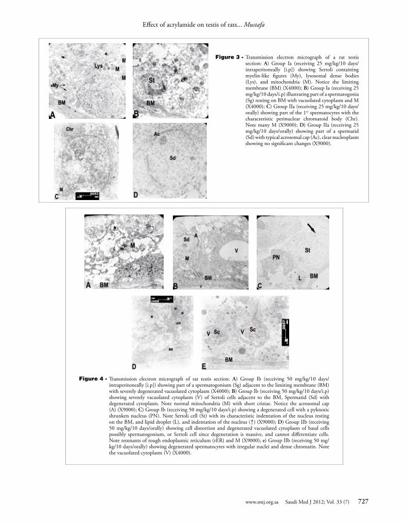
Copyright © 2022 FDOKUMEN