Doctorat en Sciences T H È S E L'Université Paris Sud
216
Doctorat en Sciences T H È S E pour obtenir le grade de docteur délivré par L’Université Paris Sud Spécialité : Microbiologie Présentée et soutenue publiquement par Marcela AZEVEDO le 25 Octobre 2013 Engineering of Lactic Acid Bacteria strains modulating immune response for vaccination and delivery of therapeutics Directeur de thèse : Jean-Marc CHATEL Co-Directeur de la thèse : Anderson MIYOSHI Jury M. Armel GUYONVARCH, Professeur, Université Paris Sud Président du Jury M. Bertrand BELLIER, Maitre de conférence, Université Pierre et Marie Curie Rapporteur M. Jean-Guy LeBlanc, Professeur, Université nationale de Tucuman, Argentine Rapporteur Mme Alexandra GRUSS, Directeur de Recherches, INRA Examinateur M. Jerry WELLS, Professeur, Université de Wageningen, PaysBas Examinateur M. Vasco AZEVEDO, Professeur, Université fédérale du Minais Gerais, Brésil Examinateur M. Philippe LANGELLA, Directeur de Recherches, INRA Examinateur Unité MICALIS UMR 1319, Pôle Ecosystèmes, Equipe « Interactions des bactéries commensales et probiotiques avec l’Hôte » Centre de Recherche INRA de Jouy-en-Josas, Domaine de Vilvert 78 352 Jouy-en-Josas cedex France
-
Upload
khangminh22 -
Category
Documents
-
view
0 -
download
0
Transcript of Doctorat en Sciences T H È S E L'Université Paris Sud

Doctorat en Sciences
T H È S E pour obtenir le grade de docteur délivré par
L’Université Paris Sud Spécialité : Microbiologie
Présentée et soutenue publiquement par
Marcela AZEVEDO
le 25 Octobre 2013
Engineering of Lactic Acid Bacteria strains modulating immune response for vaccination and delivery of
therapeutics
Directeur de thèse : Jean-Marc CHATEL Co-Directeur de la thèse : Anderson MIYOSHI
Jury
M. Armel GUYONVARCH, Professeur, Université Paris Sud Président du Jury
M. Bertrand BELLIER, Maitre de conférence, Université Pierre et Marie Curie Rapporteur
M. Jean-Guy LeBlanc, Professeur, Université nationale de Tucuman, Argentine Rapporteur
Mme Alexandra GRUSS, Directeur de Recherches, INRA Examinateur
M. Jerry WELLS, Professeur, Université de Wageningen, PaysBas Examinateur
M. Vasco AZEVEDO, Professeur, Université fédérale du Minais Gerais, Brésil Examinateur
M. Philippe LANGELLA, Directeur de Recherches, INRA Examinateur
Unité MICALIS UMR 1319, Pôle Ecosystèmes, Equipe « Interactions des bactéries commensales et probiotiques avec l’Hôte » Centre de Recherche INRA de Jouy-en-Josas, Domaine de Vilvert 78 352 Jouy-en-Josas
cedex France

Acknologments
It takes a long time to write a PhD thesis, though not as long as it takes to write a PhD
thesis acknowledgment. I would like here to express my thanks to the people who
helped me during this walk. The first part will be dedicated to my French and Brazilians
supervisors, colleagues and friends that I had the pleasure to meet when I was living in
France and in The Netherlands, after I will switch to Portuguese to thank my colleagues,
friends and family from Brazil.
É preciso muito tempo para escrever uma tese de doutorado, e de mais tempo ainda
para agradecer à todas as pessoas envolvidas direta e indiretamente neste trabalho. Eu
gostaria de expressar aqui a minha gratidão à todos que me ajudaram bastante nesta
caminhada. A primeira parte será dedicada aos meus orientadores Franceses e
Brasileiros, colegas e amigos que eu tive o prazer de conhecer quando estava morando
na França e na Holanda. Posteriormente, farei um agradecimento em Português aos
meus colegas, amigos e familiares Brasileiros.
-‐ First of all I would like to thank my Brazilian supervisor, Prof. Dr. Anderson
Miyoshi, for all the trust, opportunity you gave me, actually, words are not
enough to say how I am grateful to you. Thank you!
-‐ Prof. Dr. Vasco Azevedo for giving me the motivation to carry on and thinking
on new ideas. Thank you for the trust and to be a very special human being,
inspiring all students that are around you to aim high with your creations and
visions. Thank you!
-‐ Dr. Jean-‐Marc Chatel, as my daily supervisor, for thesis, papers and poster
corrections. It was very nice to work in a calm atmosphere that you provided
and most of all I am thankful for your effort to help me to finish this work.
Merci beaucoup!
-‐ Dr. Philippe Langella for the constructive critics, for paper and poster
corrections and to accept me in your research group in France. Merci beaucoup!

-‐ Prof. Dr. Jerry Wells, for your input during meetings, for reviewing the paper
and for the scientific discussions. You will be an inspiration to me forever.
Thank you.
-‐ To the members of this thesis defense for the availability to evaluate this work.
-‐ Post-‐graduation in genetics from UFMG and École Doctorale from Paris-‐Sud 11
University, including the teachers, workers and students.
-‐ or the grant conceded, for the fall schools
and scientific exchange that the program provided, especially to the
coordinators Dr. Emmanuele Maguim and Dr. Hervé Blotierre.
-‐ For all the Cross Talk students coming from different parts of the world for the
amazing work experience and culture exchange! I will miss this!
-‐ Everybody from MICALIS unity from INRA Research Center (Jouy en Josas),
especially Jean-‐Pierre Furret, Denis Marriat, Aurelie Turpin, Claire Cherbuy,
Sebastien Blugeon and Christophe Michon.
-‐ All workers and students from Host Microbe Interactomics -‐ Wageningen
University -‐ Marjolein Meijerink, Linda Loonen, Peter van Baarlen, Jurgen
Karczewski and Nico Taverne! Thank you very much for all of your help!
-‐ A special thanks to Oriana Rossi, for your help when I moved to The
Netherlands and friendship,
Janeiro to do some samba! Miss you guys!
-‐ Brazilian funding FAPEMIG for conceding the grant for the last year of my PhD.
-‐ A todos os alunos e funcionários do Laboratório de Genética Celular e
Molecular (LGCM), especialmente Vanessa Bastos, Tessália Luerce, Kátia Morais,
Thiago Castro, Núbia Seyffert, Anne Pinto, Ulisses Pereira, Fernanda Dorella e
Dayana Ribeiro.
-‐ Aos amigos brasileiros que tornaram minha estadia na França muito mais
divertida. Em especial, Juliana Franco Almeida, Fernanda Dorella, Adriana
Fiorini, Karlla Ribeiro e Daniela Pontes.
-‐ e família por me acolher na França
quando precisei.

-‐ Clarissa Santos Rocha pela grande amizade construída e à Fernanda Machado
pela amizade a la française .
-‐ Amigos de Wageningen pelas festas, divertimento, churrasquinho a -‐10°C e,
acima de tudo amizade, em especial, Carol Mosca (chérie), Carlinhos Veras,
Davi Farias (David Guetta), Anabele Moura, Mauricio Dimitrov, Fernanda
Paganelli, Andreia Gomes e muitos outros
-‐ Aos primos e amigos de BH por estarem sempre ao meu lado de forma online
me dando força durante esses anos difíceis. Em especial, Joana Pacheco,
Antônia Pacheco, Camila Pacheco, grandes amigas de faculdade Bárbara Jardim,
Maria Jimena Amaya, Fernanda Zaidan, Camila Jardim, e todo o pessoal da casa
da vóvó!
-‐ À toda minha família pelo apoio incluindo tios, tias, em especial vóvó, querida
Tia Valquíria, padrinho Valério, Tia Glorinha, Tia Gi e Madrinha Márcia!
-‐ À minha madrinha Júnia, grande professora, por ter sido a inspiração para eu
ter começado tudo isso! Sinto saudades. Jaiminho pela amizade e apoio! Os
domingos com você serão sempre especiais.
-‐ Ao Prof. Rogério P. Martins e família pela amizade.
-‐ Marlon pelo amor, carinho e paciência na parte final da elaboração da tese!
aguentar !
-‐ À minha mãe, pelo carinho, apoio incondicional quando precisei, por ter me
dado a vida e ter feito o máximo por mim. Esses anos de dedicação seriam
impossíveis sem suas palavras acolhedoras, seus gestos de força para eu seguir
em frente. Essa vitória é sua! Te amo!
-‐ Ao meu pai por ter sempre lutado pela minha educação, por todo o interesse,
por sempre me fazer ir mais além, pela incasável torcida para que eu vencesse
os desafios da vida e todas as dificuldades. Essa conquista também é sua! Te
amo!
-‐ À Magui, pelo apoio incondicional, pelo carinho, amor e consideração. Você
tem um lugar especial em meu coração!
-‐ À minha mana, literalmente metade de mim, por me fazer acreditar sempre
que sou capaz!
. Amo você!

SUMMARY
ABSTRACT ....................................................................................................................... I
LIST OF ABREVIATION ..................................................................................................... II
GENERAL INTRODUCTION ............................................................................................. VI
I. COLLABORATIONS .................................................................................................. VI
II. WORK TOPIC ......................................................................................................... VI
III. THESIS OUTLINE ................................................................................................... XI
CHAPTER 1-‐THESIS INTRODUCTION ................................................................................ 1
1.DNA VACCINES ......................................................................................................... 2
1.1 Historical perspective of DNA vaccines .............................................................. 2
1.2 Structural features of DNA vaccines ................................................................... 3
1.3 Safety issues ...................................................................................................... 5
1.4 Immunological aspects of DNA vaccines ............................................................ 6
1.4.1 Routes of administration ................................................................................... 6
1.4.2 Fate of plasmid DNA after injection and antigen presentation ........................ 7
1.4.3 Antigen presentation ......................................................................................... 9
1.4.4 Adaptive immune response: Cellular and Humoral Immunity ........................ 11
1.4.5 Immune memory ............................................................................................. 14
1.5 Preclinical and clinical progress of DNA vaccines .............................................. 15
1.6 Improvement of DNA vaccines immunogenicity............................................... 17
1.7 Non-‐biological delivery systems for DNA vaccines ........................................... 18
1.7.1 Physical approaches ........................................................................................ 18
1.7.2 Chemical vectors .............................................................................................. 19

1.8 Biological delivery systems for DNA vaccines ................................................... 20
1.8.1 Virus as DNA delivery vehicles ......................................................................... 20
1.8.2 Bacteria-‐based vectors .................................................................................... 22
2.BACTERIAL VECTORS AS DNA DELIVERY VEHICLES .................................................. 23
2.1 Basic Principles for Bacteria-‐Mediated DNA delivery at mucosal surfaces ........ 23
2.1. 1 Intestinal mucosa ........................................................................................... 24
2.1.2 Commensal bacteria and the intestinal mucosa ............................................. 26
2.3 Bacterial vectors used for gene transfer ........................................................... 28
2.3.1 Pathogenic bacterial DNA delivery .................................................................. 28
2.3.1.1 Extracellular pathogens ....................................................................... 28
2.3.1.2 Intraphagosomal pathogens ................................................................ 29
2.3.1.3 Intracytosolic pathogens ...................................................................... 29
3.LACTIC ACID BACTERIA (LAB) ................................................................................. 31
3.1 Taxonomy and characteristics .......................................................................... 31
3.1.1 Physiology of lactic acid bacteria into the human gastrointestinal tract ....... 32
3.1.2 The probiotic action ......................................................................................... 33
3.2 The model LAB: Lactococcus lactis ................................................................... 35
3.3 Lactococcus lactis: From cheese making to Heterologous protein Delivery....... 36
3.3.1 Gene expression systems and heterologous protein production in Lactococcus lactis ......................................................................................................................... 41
3.3.2 Lactococcus lactis as mucosal delivery vectors for therapeutic proteins ... Error! Bookmark not defined.
3.4 Lactococcus lactis: From protein to DNA delivery ............................................. 45
3.4.1 Native L. lactis as DNA delivery vectors .......................................................... 45
3.4.2 Recombinant invasive L. lactis as plasmid DNA delivery vehicles ................... 45

CHAPTER 2-‐Aim of the study ........................................................................................ 48
CHAPTER 3-‐In vitro and in vivo characterization of DNA delivery using recombinant Lactococcus lactis expressing a mutated form of L. monocytogenes Internalin A .......... 50
2.1 Introduction ........................................................................................................ 50
2.2 Materials and methods ...................................................................................... 51
2.3 Results and Discussion ........................................................................................ 51
2.4 Conclusions ......................................................................................................... 53
CHAPTER 4-‐Immune response elicited by DNA vaccination using Lactococcus lactis is modified by the production of surface exposed pathogenic protein ............................. 75
3.1 Introduction ........................................................................................................ 76
3.2 Materials and methods ...................................................................................... 76
3.3 Results ................................................................................................................ 77
3.4 Discussion ........................................................................................................... 78
3.5 Conclusions ......................................................................................................... 79
CHAPTER 5-‐Recombinant invasive Lactococcus lactis can transfer DNA vaccines either directly to dendritic cells or across an epithelial cell monolayer ................................. 105
4.1 Introduction ...................................................................................................... 106
4.2 Materials and methods .................................................................................... 107
4.3 Results .............................................................................................................. 107
4.4 Discussion ......................................................................................................... 108
4.5 Conclusions ....................................................................................................... 109
CHAPTER 6-‐GENERAL DISCUSSION, MAIN CONCLUSIONS AND DIRECTIONS FOR FUTURE WORK ........................................................................................................................ 137
CHAPTER 7-‐REFERENCES ............................................................................................. 144
APPENDICE 1-‐Recombinant Lactococcus lactis expressing both Listeria monocytogenes Lysteriolysin O and mutated internalin A applied for DNA vaccination ....................... 160
APPENDICE 2-‐Immunotherapy of allergic diseases using probiotics or recombinant probiotics ................................................................................................................... 174

I
ABSTRACT
The use of Lactic Acid Bacteria (LAB), such as Lactococcus lactis (LL), as DNA delivery vehicles
represents an interesting strategy as they are regarded as safe. Wild type (wt) LL or recombinant
invasive LL, were able to trigger DNA expression by epithelial cells both in vitro and in vivo. However,
important information about how LL can transfer DNA plasmids is still missing. Therefore, we decided
to construct a new recombinant invasive LL strain expressing mutated Internalin A (mInlA) from the
pathogen Listeria monocytogenes to understand the manner by which the DNA is transferred to
mammalian cells. mInlA expression was detected by FACS analysis and LL-‐mInlA strain showed to be
more invasive than the wt strain after co-‐incubation assays with non-‐confluent or polarized intestinal
epithelial cells (IECs). Confocal microscopy confirmed the invasive status of LL-‐mInlA which
demonstrated to deliver more efficiently the eukaryotic expression vector coding the allergen -‐
lactoglobulin, pValac:BLG, in vitro to IECs and to dendritic cells (DCs). LL-‐mInlA was also capable to
transfer pValac:BLG to DCs across a monolayer of differentiated IECs. In vivo, invasive lactococci
tended to increase the number of mice expressing BLG. Moreover, noninvasive or invasive LL-‐mInlA
stimulated the secretion of the pro-‐inflammatory cytokine IL-‐12 in DCs and, in vivo, after oral or
intranasal immunization trials, non-‐invasive LL polarized the immune response more in the type 1
direction while invasive LL generated a Th2-‐type response in immunized animals. All these data gives
new insights on the mechanism of lactococci uptake for delivery of therapeutics.

II
LIST OF ABBREVIATIONS
aa Amino acids
Ads Adenoviruses
APCs Antigen presenting cells
B cells B lymphocytes
BALB/c Laboratory-‐bred strain of the House Mouse Jackson Laboratory
BALT Bronchial/tracheal-‐associated lymphoid tissue
BGH Growth Hormone
BLG ß-‐Lactoglobulin
CD Crohn's disease
cDNA Complementary DNA
CFU Colony Forming Unit
CpG motifs Cytosine-‐phosphate-‐guanine unmethylated
CTL Cytotoxic T lymphocytes
DapD 2,3,4,5-‐tetrahydropyridine-‐2,6-‐dicarboxylate N-‐
succinyltransferase DapD
DCs Dendritic cells
DEAE-‐dextran Poly-‐lysine, diethylaminoethyl-‐dextran
DNA Deoxyribonucleic acid
dsRNA Double stranded RNA
DSS Dextran sulfate sodium
EMEA European Agency for the Evaluation of Medicinal Products
EP Electroporation
ER Endoplasmic reticulum
FAE Follicle-‐associated epithelium
FDA US Food and Drug Administration
FnBPA Fibronectin Biding Protein A
GALT Gut associated-‐lymphoid tissue
GC Guanine-‐cytosine
GF Germ-‐free
gfp Green fluorescent protein
GI Gastrointestinal
GRAS Generally regarded as safe
III
hAAT Human 1-‐antitrypsin
hCMV Human cytomegalovirus
HGF Hepatocyte growth factor
hGH Human growth hormone
HIV-‐1 Human immunodeficiency virus type -‐ 1
HK-‐1 Hemokinin-‐1
HSV Herpes simplex virus
IBD Inflammatory bowel disease
IEC Intestinal epithelial cell
Interferon-‐
Ig Immunoglobulin/antibody
IgA Immunoglobulin A antibody isotype
IgE Immunoglobulin E antibody isotype
IgG1 Immunoglobulin G type 1 antibody isotype
IgG2a Immunoglobulin G type 2a antibody isotype
IHNV Infectious hematopoietic necrosis/Aquatic rhabdovirus
IL-‐12 Interleukine-‐12
IL-‐8 Interleukine-‐8
InlA Internalin A
InlB Internalin B
ISS's Nucleotide hexamers
LAB Lactic acid bacteria
LGG Lactobacillus GG
LLO Cytolysin listeriolysin O
LPS Lipopolysaccharides
LRRs leucin-‐rich repeat motif
LT Lymphotoxin
LTA Lipoteichoic acid
M cells Microfold cells
MALT Mucosa-‐associated lymphoid tissue
MAMPs Microbial-‐associated molecular patterns
MHC I Major histocompatibility complex class I
MHC II Major histocompatibility complex class II

IV
mRNA Messenger RNA
MRP Maximum representation with parsimony
NALT Nose-‐associated lymphoid tissue
NF-‐ B NF-‐ B nuclear factor kappa B
NICE Nisin Controlled Gene Expression
NK Natural killer cells
NL Netherlands
NLR NOD-‐like receptors
NLRs NLRs Nod-‐like receptors
NLS Nuclear localization signal
OD Optical density
ori31 replication origin of 31
31 Bacteriophage 31
PAMPs Pathogen-‐associated molecular patterns
PEI Polyethyleneimine
PGN Peptidoglycan
PM Plasma membrane
polyA tail Polyadenylation sequence
PPH Baculovirus
PRRs Pattern recognition receptors
PRSV Rous sarcoma virus
PTK Thymidine kinase promoter
pValac Vaccination using Lactic acid bacteria
RLRs RIG-‐I like receptors
RNA Ribonucleic acid
rRNA Ribosomal ribonucleic acid
SARS Severe Acute Respiratory Syndrome
SCFA Short chain fatty acid
SDS-‐PAGE Gel denaturing polyacrylamide
SIgA Secretory IgA
SlpA S-‐layer protein
ssRNA Single-‐stranded RNA
SV40 Simian virus 40

V
T CD4+ T helper lymphocytes
T CD8+ Cytotoxic T lymphocytes
T cells T lymphocytes
T3SS Type III secretion system
Th T helper lymphocytes
Th1 T helper cells type 1
Th2 T helper cells type 2
TJ Tight junctions
TLR Toll-‐like receptor
TLR1 Toll-‐like receptor type 1
TNF Tumor necrosis factor-‐
Treg Regulatory T cells
TTFC Fragment C of tetanus toxin
UC Ulcerative colitis
US United States
US Ultrasound
Usp45 Unknown Secreted Protein of 45 kDa
VALT Vulvovaginal-‐associated lymphoid tissue
WHO World Health Organization
wt Wild type
XIES Xylose-‐Inducible Expression System

VI
GENERAL INTRODUCTION
I. COLLABORATIONS
This thesis is a part of a co-‐tutelle PhD program offered jointly by two higher education
institutions; one in Brazil named Universidade Federal de Minas Gerais (UFMG), and the other one in
France called Université Paris-‐Sud 11. This program allows the students to get a double/joint PhD
degree delivered and recognized by both institutions besides exposing students to the international
research community. The major experimental part of this work was conducted in France at Institut
National de la Recherche Agronomique (INRA) facilities located in Jouy en Josas under the
supervision of Dr. Jean Marc Chatel and the co-‐supervision of Dr. Philippe Langella, both researchers
at ProbiHote Team from MICALIS unit (Microbiologie
work was also supervised by Dr. Anderson Miyoshi and co-‐supervised by Dr. Vasco Azevedo; both of
them professors at Instituto de Ciências Biológicas developing research at Laboratório de Genética
Celular e Molecular (LGCM) from UFMG. This research project was financed in the last year by
Fundação de Amparo à Pesquisa de Minas Gerais (FAPEMIG) and, before that, by Cross talk European
project during three years (april 2009-‐2012). Cross-‐Talk was a part of a Marie Curie Initial Training
Network (ITN) focusing to study the interaction between microbiota and the human host. The
network gathered 13 partners and 14 young scientists from more than 10 European and non-‐
European Universities.
The Research Project (RP) comprising this thesis was the Cross Talk RP number 6 (RP6) (for
more details see: http://www.cross-‐talk.eu/index.php?id=63) which aimed to understand the
relationship between immune cells and epithelial cells with recombinant lactic acid bacteria (LAB)
used as DNA delivery vehicle. Training of the fellows was the core activity of Cross Talk project. For
this reason it supported mobility periods (maximum 9 months) abroad to enlarge recently
established collaborations between laboratories with a view to improve students skills and promote
the cross-‐border transfer of knowledge. Therefore, RP6 was also carried out during seven months in
collaboration with Dr. Jerry Wells, a Professor at Host Microbe Interactomic Group (HMI) from
Wageningen University WUR Research Center located in The Netherlands. One of the deliverables
Institut Pasteur situated in Paris. Thus, expertise scientists, such as Dr. Catherine Grillot-‐Courvalin
(MD, PhD, Associate Professor at the Pasteur Institute, Unit of Antibacterial Agents) and Dr. Karine
Adel-‐Patient (Researcher at INRA, UR, Unité d'Immuno-‐Allergie Alimentaire), were also involved in
the project with the intent to give advices and propose actions to improve future publications.

VII
II. WORK TOPIC
Lactococcus lactis (LL) is a bacterium traditionally used for food production and preservation.
Therefore they are safe for human consumption and for this reason they harbor the GRAS (Generally
Regarded As Safe) status. Due to this fact, a number of new applications have been proposed and LL
was extensively engineered to function as a cell factory for many proteins of health interest, such as
antigens for the development of new vaccines (Miyoshi and Azevedo, 2004; Wells and Mercenier,
2008). It was successfully demonstrated that LL is able to express antigenic proteins in different cells
compartments (intracellularly, anchored to the cell wall or expressed to the extracellular medium)
and deliver them at mucosal surfaces (for reviews see Wells, 2011; Pontes et al., 2011; Bermúdez-‐
Humarán et al., 2011). Two great examples are the as -‐lactoglobulin (BLG), one of the major cow's
milk allergen, for the treatment of allergic diseases (Chatel et al., 2001) and E7 antigen from HPV
virus for the treatment of colon cancer (Bermúdez-‐Humarán et al., 2005). More recently it was
demonstrated that this bacterium is able to deliver DNA vaccines at mucosal surfaces (Chatel et al,
2008). This work, therefore, seek to exploit, understand and underline the mechanisms by which LL
can deliver DNA vaccines to mammalian cells.
Briefly, a DNA vaccine consists of a circular DNA plasmid encoding antigenic proteins under
the control of a mammalian promoter. After taking up the DNA, mammalian cells are able to drive
the expression of the vaccine antigens. The plasmid contains an origin of replication that allows its
replication inside bacterial hosts and a resistant marker that permits the selections of clones that
harbors the plasmid DNA (Lichtor and Glick, 2012). DNA immunization can produce stronger and
wider types of immunity within the organism including antibody, T helper cell (CD4+ T cell), and
cytotoxic T lymphocyte (CTL, CD8+ T cell) mediated immunity to cover multiple diseases (Glenting
proteoliposomes (Gurunathan et al., 2000). Bacterial vectors are also widely used as it can protect
the DNA against the attack by endonucleases and it can stimulate the mucosal immune system
associated with the epithelium. Furthermore, it shows a better tropism into the body after
immunization trials when compared to the other approaches. Moreover, there is no need for further
steps of plasmid amplification and purification because the plasmid can replicate itself inside the
bacteria. Therefore, this platform in vaccinology is considered to be very low cost (Glenting and
Wessels, 2005). The use of LL as a DNA vector turns to be very interesting over the use of attenuated
pathogenic bacteria, which are more traditionally used to deliver DNA, because they have GRAS
status (Wells and Mercenier, 2008).

VIII
Guimarães et al. (2006) demonstrated that wild type (wt) LL could deliver DNA vaccines in
vitro. Later, Chatel et al. (2008) observed that this bacterium was also able to transfer DNA plasmids
in vivo to murine intestinal epithelial cells (IECs). Even though being confirmed the capacity of
lactococci to transfer DNA vaccines, it still remains unclear how LL can deliver DNA to mammalian
cells in vivo. Basically, there are at least three manners by which antigens, plasmids or bacteria in
general can gain access to mammalian cells interior and reach the lamina propria. This material
diluted in the intestinal lumen can be recognized by some specialized epithelial cells named
lymphoid tissue that are usually found in the lowest portion of the small intestine). Antigen
Presenting Cells (APCs) such as Dendritic cells (DCs) associated with the IEC monolayer can also
extend their dendrites through the tight junctions and sample bacteria or antigens that are localized
in the lumen (Rescigno et al., 2001). Finally, some vectors based on attenuated pathogenic species
can express invasive proteins which are recognized by epithelial cells and actively invade the IEC
monolayer reaching the lamina propria (Gewirtz and Madara, 2001). Once inside the cell, bacteria
localized in the phagolysosome are normally target for degradation. Some attenuated pathogenic
vectors such as Listeria monocytogenes are able to secrete cytolysins (Lysteriolysin O LLO) which
can make pores into the phagossomal membrane helping the plasmid to escape from the
phagolysosome to the nucleus of the cell. Using the mammalian cell transcriptional machinery
components, the antigen of interest can be produced (Faurez et al, 2010; Liu, 2010).
Based on this knowledge two hypotheses were designed to explain how LL can transfer DNA
vaccines to epithelial cells: 1-‐ Bacteria could be lysed in the intestinal lumen releasing the DNA
vaccine which could be sampled by IECs, M cells and/or DCs. 2-‐ Bacteria itself could be sampled by
IECs, DCs and/or M cells. Our research group decided to test the second hypothesis by constructing
recombinant invasive L. lactis strains thus improving the interactions between bacteria and IEC
monolayer. One strain was able to express Staphylococcus aureus Fibronectin Binding Protein A
(FnBPA) (LL-‐FnBPA+) (Innocentin et al., 2009). In vitro assays demonstrated that it was capable to
deliver a eukaryotic expression vector to IECs (Caco-‐2 cell line) more efficiently than the wt strain.
However, the use of this strain in immunization trials presented some limitations as the binding of
FnBPA depends on the availability of fibronectin in vivo. Another strain was the one producing
Listeria monocytogenes Internalin A (InlA) (LL-‐InlA+) (Guimarães et al., 2005). It was able to deliver
more DNA to IEC (Caco-‐2 cell line) in vitro when compared to the wt strain; nevertheless, its use in
vivo is very limited because InlA cannot bind to its receptor in mice, the murine E-‐cadherin. Even
though LL-‐FnBPA+ and LL-‐InlA+ strains have been constructed and presented to be better tools to

IX
deliver DNA vaccines in vitro when compared to wt lactococci, the process by which LL can transfer
DNA vaccines remains unknown.
Therefore, strains of L. lactis expressing mutated InlA (LL-‐mInlA+), an invasin able to bind to
murine E-‐cadherin, were constructed in this work and their ability to deliver cDNA from the major
-‐lactoglobulin (BLG), was studied in different experimental models. It was also
examined the capacity that LL-‐mInlA+ strain has to deliver BLG cDNA to murine epithelial cells after
immunization trials. Furthermore, immune responses elicited after in vivo administration of invasive
and noninvasive strains were measured, and the capacity of these strains in delivering DNA vaccines
to antigen presenting cells (DCs) was evaluated as well.
The second chapter of this manuscript, where we present the first results, is related to the
characterization of LL-‐mInlA+ strain as a vehicle to deliver the eukaryotic expression vector,
pValac:BLG, previously constructed by Pontes and collaborators (2012), either in vitro or in vivo to
IECs. Firstly, L. lactis NZ9000 wt strain was transformed with pOri253:mInlA plasmid harboring the
gene of mInlA from Listeria monocytogenes. Flow cytometry analysis confirmed that the new strain
was able to successfully express mInlA at its surface. Moreover, it presented to be almost 1000 times
more invasive than the wt strain after co-‐incubation assays with Caco-‐2 cells. Confocal images
confirmed this invasive status as recombinant LL-‐mInlA+ were found attached to Caco-‐2 cells being
found in the interior of the cells. Afterwards, L. lactis producing mInlA was transformed with
pValac:BLG plasmid and its capacity to transfer the cDNA of BLG was then measured. LL-‐mInlA+BLG
(invasive strain) and LL-‐BLG (noninvasive strain) were co-‐incubated with Caco-‐2 cells and after three
days BLG expression by the mammalian cells were evaluated by ELISA. It was observed that the
invasive characteristic increased plasmid transfer as Caco-‐2 cells incubated with LL-‐mInlA+BLG
produced more BLG when compared to the cells incubated with LL-‐BLG. After the in vitro studies we
moved to in vivo assays in which mice were intragastrically administrated with L. lactis, LL-‐BLG or LL-‐
mInlA+BLG for three consecutive days, and the small intestine removed for isolation of IECs. BLG
DNA delivery efficiency in vivo is slightly improved by the production of mInlA. However, no
significant advantages were observed by using LL-‐mInlA+BLG compared to LL-‐BLG. Based on this
result we hypothesize that most likely, plasmid transfer in vivo is a combination of two mechanisms,
bacteria and released plasmid captures by specialized epithelial cells (Microfold cells, M) or IECs as L.
lactis is a transient specie and cannot survive within the gastrointestinal tract (TGI).
The third chapter is dedicated to studies performed with the intent to investigate the
immune response elicited after oral or intranasal immunization with the noninvasive (LL) or invasive
L. lactis (LL-‐FnBPA+ and LL-‐mInlA+) carrying pValac:BLG. It is known that the administration of a
plasmid DNA encoding antigens usually elicits a T helper cell type 1 (Th1) immune response. In order

X
to check if the same immune profile could be obtained after immunization trials with noninvasive or
invasive L. lactis, conventional mice were orally or intranasally administered with the strains and
BLG-‐specific primary immune response were monitored. We have demonstrated that after oral or
intranasal administration with invasive L. lactis FnBPA+, mice elicited a Th2 immune response
characterized by the detection of IL-‐4 and IL-‐5 cytokines. Differently, the oral or intranasal
administration of noninvasive L. lactis-‐BLG elicited a Th1 immune response characterized by the
detection of IFN-‐ . To test the capacity of the strains to avoid allergic immune responses, mice
intranasally pre-‐treated with both strains were sensitized with BLG and then immune response was
measured. After sensitization, we detected a significantly lower concentration of BLG specific IgE in
mice immunized with noninvasive L. lactis. This result was expected as Th1 immune cells, which were
induced by noninvasive L. lactis, can suppress the generation of allergic immune responses
(Romagnani, 2004). IL-‐4 and IL-‐5 cytokines were again found in a higher amount in mice sensitized
and previously immunized by intransal or oral route with the invasive lactococci. To check if the Th2
induced profile was due to the expression of invasins at L. lactis cell wall, another recombinant
invasive L. lactis, LL-‐mInlA+BLG, was intranasally administered in conventional mice and immune
response was evaluated. We observed again a polarization to the Th2 immune profile by using the
invasive lactococci. In order to explore if the alteration of some component of the bacterial cell wall
could be the cause of this immune polarization, the peptidoglycan composition from the cell wall of
invasive and non-‐invasive strains were analyzed and no differences could be observed. In conclusion,
we demonstrated that the expression of invasins at the surface of L. lactis modifies its
immunomodulatory properties.
The fourth chapter presents in vitro studies which were made with the goal to give new
insights on the mechanisms of plasmid transfer by using noninvasive or invasive lactococci. All the
works about plasmid transfer using L. lactis performed by our research group and others used IECs as
a model to evaluate plasmid transfer. In this work we evaluated DNA transfer capacity of LL to
dendritic cells (DCs) as these cells are the major antigen presenting cells of the organism that are in
direct contact with diluted antigens of the TGI. We have shown that noninvasive L. lactis, and
invasive L. lactis strains expressing either S. aureus FnBPA, or L. monocytogenes mInlA can transfect
bone marrow-‐derived DCs (BMDCs) and deliver the cDNA of BLG. The invasive status appeared to be
advantageous facilitating the bacteria-‐cell contact and, thus, allowing a higher translocation of the
pValac:BLG plasmid. Moreover, BMDCs co-‐cultured with non-‐invasive or invasive Lactococci were
able to secrete elevated levels of the pro-‐inflammatory cytokine IL-‐12, suggesting that this immune
response is due to MAMPs (Microbe Associated Molecular Patterns) naturally found in L. lactis and
not related to BLG expression by the BMDCs or with mInlA or FnBPA invasins. In order to understand

XI
how lactococci can interact with IECs or DCs from the intestinal epithelium to deliver therapeutic
plasmids, a monolayer of differentiated Caco-‐2 cells were used as a model that could mimic the
situation encountered in vivo, where bacteria and antigens face a protective monolayer of epithelial
cells. Firstly, we checked weather the invasive L. lactis strains (LL-‐FnBPA+ and LL-‐mInlA+) were able
to internalize the monolayer more efficiently than the wt strain. Co-‐incubation of bacteria with
polarized Caco-‐2 cells showed that LL-‐mInlA+ is 100 times more invasive than LL and equally invasive
compared to LL-‐FnBPA+ strain. Additionally we also investigated the cross-‐talk between
differentiated IECs, BMDCs and bacteria using an in vitro Transwell co-‐culture model. Co-‐incubation
of strains with the co-‐culture model has shown that DCs maintained with LLmInlA-‐BLG strain was
able to express significant higher levels of BLG. This data was not observed for LL-‐FnBPA+BLG or
noninvasive LL-‐BLG strain. The fact that L. lactis producing mInlA was the only strain capable to
mediate plasmid transfer may be due to the expression of E-‐cadherin receptor by BMDCs, which
could facilitate the contact and capture of the bacteria.
The use of L. lactis as a tool for DNA delivery has proved to be an interesting alternative
approach for the design of new mucosal vaccines. Taken together, we believe that this work brings
new information regarding the mechanisms by which L. lactis can transfer DNA plasmids to
eukaryotic cells. We believe that all this data can facilitate its use as a vehicle for immunization
proposals in near future. We also believe that all these tools and models designed or used in this
work may lead to the construction of new food-‐grade live vaccines based on L. lactis. Such uses for
vaccination purposes are promising for future therapeutic use of this bacterium.
III. THESIS OUTLINE
This manuscript was divided into six chapters and two appendices. Below, a brief description
of the content covered in each chapter/appendices:
Chapter 1: Thesis introduction about DNA vaccines, immunological principles of genetic
immunization and the vectors used to deliver plasmid vaccines with a focus on the use of bacteria,
particularly lactic acid bacteria (BL), native or recombinant, as vehicles for gene vaccination. Also
describe the main mechanisms of interaction between BL and the immune system associated with
the intestinal epithelium for transferring cDNA, for example the cDNA of the major allergen of cow's
-‐lactoglobulin.
Chapter 2: Aim of the study

XII
Chapter 3: Presented in a scientific paper form, it describes the use of a new recombinant L. lactis
strain expressing an invasin derived from Listeria monocytogenes as a vehicle for DNA delivery in
vitro or to epithelial cells from BALB/c mice, which has been accepted for publication.
Chapter 4: Also presented as a scientific article, it describes the different immune responses
obtained after immunization trials with native or recombinant strains expressing on its surface
invasins derived from pathogenic bacteria. This chapter is in preparation to be submitted for
publication.
Chapter 5: Presented in a scientific paper form, it presents the ability of invasive L. lactis strains to
deliver plasmids for antigen presenting cells such as dendritic cells, in vitro. This chapter is in
preparation to be submitted for publication.
Chapter 6: General discussion of the results, which were presented in Chapters 2, 3 and 4 and
obtained conclusions and future directions of the work.
Chapter 7: References.
Appendice 1: Presented in a scientific paper form, this section describes the use of an L. lactis strain
expressing the mutated Internalina A and listeriolysin O, two proteins derived from L.
monocytogenes, as a potential vehicle for genetic immunization or against listeriosis.
Appendice 2: A literature review on the use of probiotics or recombinant probiotic as
immunotherapy for the treatment of allergic diseases.

CHAPTER 1
THESIS INTRODUCTION

2
1. DNA VACCINES
1.1 Historical perspective of DNA vaccines
The advent of molecular biology and genetic engineering has had a dramatic effect on
vaccine development, providing greater opportunities for construction of inactivated antigens,
attenuation of pathogenic organisms through direct mutation, and more recently, the
construction of DNA vaccines (Plotkin, 2005). They are plasmids that contains a transgene
encoding the sequence of a target protein under the control of a eukaryotic promoter at the
beginning of the gene (Srivastava and Liu, 2003; Ingolotti et al., 2010; Liu, 2011). Following in vivo
administration, the plasmid DNA can transfect mammalian cells which will be then capable to
express the protein of interest in situ (Ingolotti et al., 2010; Liu, 2011) (Figure 1).
The first study found in the literature about DNA vaccination started in the early 1990s
when Wolff naked
antigens in mice has made their muscle cells capable of expressing these same antigens (Wolff et
al., 1992). Later, Tang, Stephen A Johnston and Michael Devit, three scientists from University of
Texas, Dallas, USA, demonstrated that the injection of gold microspheres coated with human
growth hormone (hGH) DNA into the skin of a mouse was able to generate both hGH-‐ and human
-‐antitrypsin (hAAT)-‐specific antibodies (Tang et al. 1992). At the same time, three other
research groups reported on a meeting held at Cold Spring Harbor Laboratory (NY, USA) that the
Figure 1: Mechanisms of action of DNA vaccines. Cellular uptake of DNA vaccines occurs by a phagocytic process. A part is digested inside the phagolyssosome while others can enter the cytoplasm and are internalized by the nucleus, resulting in production of antigen encoded by the DNA vaccine. Adapted from Ulmer et al (2009).

3
injection of a DNA in immunizations trials against influenza or HIV-‐1 was able to drive both
humoral and cellular specific immunity. Later, Margaret Liu and colleagues published similar
results, as well as other researchers (Fynan et al., 1993; Ulmer et al., 1993; Ingolotti et al., 2010)
confirming the immunogenic and protective capacity of DNA vaccines (Ingolotti et al., 2010; Liu,
2011; Li et al., 2012).
The use of DNA as a strategy for vaccination has progressed very rapidly. Within twenty
years since the first publication demonstrating the ability of a plasmid to generate protective
immune responses, DNA vaccines have reached enticing results. This vaccine platform has
entered into a variety of human clinical trials for prophylactic vaccines against viral, bacterial or
parasitic infections and also as a potential therapy to treat infectious diseases, cancers, Alzheimer
disease, allergy and autoimmune disorders (for a review see Liu, 2010 and Liu, 2011) (Figure 2).
1.2 Structural features of DNA vaccines
A DNA vaccine plasmid can be divided into two main structures: the plasmid backbone
and the transcriptional unit. The plasmid backbone, also referred as prokaryotic system, is usually
Figure 2: Current DNA vaccine clinical trials. At the time, more than 40 clinical trials evaluating DNA vaccines were listed as on-‐going in the clinical trials. The large pie chart shows the percentage of trials by vaccine target. The inset pie chart shows the percentage of trials targeting specific cancers among the 29% of clinical trials that are cancer related. Adapted from Ferraro and co-‐workers (2011).

4
composed of (i) a prokaryotic origin of replication that allows the plasmid propagation into the
host bacterium and (ii) a resistance marker, necessary to enable a selective growth of the DNA
vaccine in bacteria. The transcriptional unit, also known as eukaryotic system, contains the
promoter and a gene encoding the antigen of interest followed by a transcript
termination/polyadenylation sequence named polyA tail. The firsts promoters used were from a
viral origin and they are still widely used. Two great examples are the cytomegalovirus (hCMV)
and the Simian virus 40 (SV40) that can drive the expression of the transgene/antigen in a vast
variety of mammal cells types, enabling the expression of antigens in situ (Ingolotti et al., 2010).
Other common promoters are the polyhedrin promoter from baculovirus (PPH), the thymidine
kinase promoter from Herpes simplex virus (HSV)‑1 (PTK), the 5´LTR promoter from human
immunodeficiency virus (HIV; PLTR) and the promoter of the Rous Sarcoma Virus (PRSV) (Becker
et al., 2008). Due to the fact that some cytokines may differentially regulate the CMV promoter
altering transgene expression, the use of nonviral promoters is presently the topic of intensive
research. For example, major histocompatibility complex (MHC) class II promoter is being
investigated as possible alternatives (Vanniasinkam et al., 2006; Ingolotti et al., 2010). Another
structure that can be present in the plasmid vaccine are segments of DNA not needed to create
the protein (introns), usually inserted after the promoter and before the gene of interest. Their
presence can increase the promoter activity (Kano et al., 2007). Moreover, the insertion of introns
is also a strategy to avoid the antigen expression by the prokaryotic machinery in bacteria,
thereby ensuring that expression is only possible through the eukaryotic system (Becker et al.,
2008). A multiple cloning site allowing the insertion of the gene is present as well as the signal
peptide allowing the secretion of the target protein. Another component is the gene of interest
which is usually codon optimized to match it with the target organism and is terminated by a dual
stop codon. A polyadenylation signal (polyA) necessary for transcriptional termination, stability,
processing and translation of eukaryotic mRNA is also present and is commonly derived from the
bovine growth hormone, SV40, -‐globin gene (Williams et al., 2009). DNA vaccines may
further contain endogenous adjuvants and "CpG motifs" (cytosine-‐phosphate-‐guanine
unmethylated), responsible for increasing the magnitude of the immune response as they can
enhance T lymphocyte recruitment or expansion (Glenting and Wessels, 2005; Kano et al., 2007;
Becker et al., 2008; Williams et al., 2009). Major structures of DNA vaccines are illustrated in
figure 3.

5
Another advantage consist on the fact that DNA plasmids are easy to handle and rapid to
construct, an interesting attribute for making vaccines against an emerging pandemic threat (Liu,
2011). As DNA vaccine plasmids are non-‐live, non-‐replicating and non-‐spreading, there is little risk
of either reversion to a disease-‐causing form or secondary infection (Kutzler and Weiner, 2008).
Additionally, plasmids are quite stable at room temperature, are easily stored and the genes in
the plasmids can be made with fidelity to the wild type (wt) protein (Liu, 2011; Li et al., 2012).
1.3 Safety issues
Despite all its assets, when the first studies describing the use of DNA vaccines for treating
infectious diseases appeared in the scientific community, a number of concerns associated with
their use were raised. The possibility that they could integrate into the host genome, thus
increasing the risk of malignancy (by activating oncogenes or inactivating tumor suppressor genes)
or to induce responses against self-‐antigens thereby triggering the development of autoimmune
disease were the major worries. Besides that, it was not known whether DNA vaccines could
induce a local inflammatory response against cells producing the vaccine-‐encoded antigen (such
as muscle or skin cells), induce tolerance rather than immunity (Klinman et al., 1997) or spread
antibiotic resistance genes to the environment after clinical trials (Kutzler and Weiner, 2008). In
fact, the introduction of plasmid DNA into humans requires special considerations which have
Figure 3: Schematic representation of the main features of a genetic plasmid vector used as a DNA vaccine. Adapted from Glenting and Wessels (2005).
6
been addressed in several regulatory draft guidance established by the World Health Organization
(WHO), US Food and Drug Administration (FDA), or European Agency for the Evaluation of
Medicinal Products (EMEA) (EMEA, 2006; FDA, 2007; WHO, 2007; William et al., 2009).
According to Wang and co-‐workers, the plasmid should contain no significant homology
with the target organism genome in order to reduce chances of chromosomal integration (Wang
et al., 2004). No observable integration of the DNA into the host genome occurred and there was
no observed tolerance to the antigen or autoimmunity (Liu and Ulmer, 2005). It was calculated
that DNA integration into the host genome occurs at rates that are orders of magnitude below the
spontaneous mutation frequency. However, plasmids that are modified or adjuvanted with the
goal of increasing immunogenicity could increase the chances of integration (Liu, 2011).
Additionally, oncogenes activation was not observed in preclinical or clinical evaluation of DNA
vaccines (Kutzler and Weiner, 2008). Another issue regarding DNA vaccination involved antibiotic
resistance. As mentioned before, antibiotic-‐resistant marker is needed to select bacteria
harboring the DNA vaccine. Usually, the antibiotic gene present in vaccine plasmids is restricted to
those that are not commonly used to treat human infections, for instance kanamycin. As the
development of antibiotic-‐free selection systems is desirable from both cost and safety
perspective (Bower and Prather, 2009), alternative strategies that do not use antibiotic selection
are being explored (Glenting and Wessels, 2005; Kutzler and Weiner, 2008). Several researchers
successfully modified the vector, host, or both to develop alternative plasmid selection systems to
ensure efficient killing of plasmid-‐free cells. For example, Cranenburgh and collaborators chose to
target dapD, an essential gene in Escherichia coli responsible for diaminopimelate and lysine
biosynthesis. The endogenous dapD locus was disrupted, and a copy of dapD under the control of
a lac promoter was integrated into the bacterial chromosome. This strain was then transformed
with a high copy plasmid containing the lac operator, necessary to drive the expression of the
diaminopimelate and lysine biosynthesis gene. As a result, only cells containing the plasmid DNA
with the lac operator sequence survived in culture (Cranenburgh et al., 2001; Bower and Prather,
2009).
1.4 Immunological aspects of DNA vaccines
1.4.1 Routes of administration
Genetic immunization initially consisted in the direct administration of a DNA plasmid (so-‐
called "naked DNA") into tissues capable to internalize and express the immunogenic antigen,
mimicking thus a natural infection (Liu, 2010; Liu, 2011). Basically, there are two main routes for
DNA vaccines administration: intramuscular injection and intradermal injection (parenteral

7
routes). Subcutaneous injection, intraperitoneal, sublingual, intrarectal, ocular, application to
mucosal surfaces (vaginal, nasal and oral), intravenous and intranodal injections are other
possible routes of administration, but they were used less frequently at the beginning of DNA
vaccine research (McCluskie et al., 1999; Faurez et al., 2010).
The most widely employed method to administer DNA vaccines for many years was the
intramuscular injection. Naked DNA ressuspended in saline buffer could be injected directly into
skeletal muscle tissue using a hypodermic needle. Nevertheless, it has been shown that from 95%
to 99% of intramuscularly injected plasmids found in the interfibrillar space are degraded in the
muscle tissue within 90 min post-‐administration (Barry et al., 1999). A study conducted by
Lechardeur and colleagues demonstrated that 90 minutes after the plasmid injection, only 0.1% of
the injected material was able to reach the cell nucleus. Endonucleases have been implicated in
degrading injected plasmid favoring its rapid elimination (Lechardeur et al., 1999). Actually, DNA
degradation represents a fundamental problem for genetic immunization, as destruction of
incoming genes translates into loss of gene expression (Barry et al., 1999).
It was also noticed that tissues differ for their efficiency to present antigens to the
immune system (Fynan et al., 1993; McCluskie et al., 1999). Therefore the route of administration
dramatically influences the strength and nature of immune responses to a plasmid-‐encoded viral
antigen (McCluskie et al., 1999). Some works demonstrated that tissues, such as the skin and the
mucosal linings of the respiratory tract and gut are more suitable routes for plasmid inoculation
as they are associated with lymphoid tissues and provide high levels of local immune surveillance
(Fynan et al., 1993). Hence, administration of DNA to mucosal surfaces can provide systemic
immunity as well as specialized surveillance for major portals of pathogen entry (Fynan et al.,
1993; McCluskie et al., 1999; Azizi et al., 2010).
1.4.2 Fate of plasmid DNA after administration
After intramuscular or intradermal injection, the plasmid DNA will transfect somatic cells,
such as myocytes and keratinocytes, and/or resident Antigen Presenting Cells (APCs) like dendritic
cells (DCs) and macrophages located in the lamina propria (Kutzler and Weiner, 2008; Liu, 2011).
DNA enters the cell through two main mechanisms. One is known as fluid-‐phase endocytosis (not
mediated by a receptor), which involves the ingestion of small molecules and/or fluids
surrounding the cell (pinocytosis) (Levy et al., 1996; Budker et al., 2000; Varkouhi et al. 2011).
Another is known as adsorptive endocytosis (not mediated by a receptor as well) in which the
DNA is taken in by a cell by splitting off small vesicles from the cell surface (Budker et al., 2000;
Faurez et al., 2010; Varkouhi et al. 2011). The plasmid sequence can also be recognized

8
simultaneously by different receptors located at the plasma membrane (PM) from eukaryotic cells
(Lehmann and Sczakiel., 2005).
Once inside the cell, DNA faces another obstacle: it needs to migrate to the cell cytoplasm.
The endocytic vesicles containing the plasmid vaccine fuses with the lysosome, whose function is
to digest molecules originally incorporated into the endosome through the activity of many
hydrolytic enzymes (Varkouhi et al., 2011). It is still not well understood how the DNA escape
from the phagolysosome and reaches the cell cytoplasm (Faurez et al., 2010). Continuously, once
within the cytoplasm, vectors that survive from the endonucleases "attack" will reach the cell
nucleus and, finally, initiate transcription of the gene of interest. Vacik and colleagues
demonstrated that DNA located in the cytoplasm binds to both microtubules and microfilaments
that form the cytoskeleton of cells through adapter proteins named dynein, and thus reach the
cell nucleus (Vacik et al., 1999). It has been also suggested that DNA molecules located in the
cytoplasm can create associations with polypeptides, such as transcription factors, that contains a
nuclear localization signal (NLS) required to enter the nucleus (Hebert, 2003).
The third and last obstacle blocking the expression of the antigenic protein is the nuclear
membrane. Some studies demonstrated that the plasmid vaccine is able to cross the nuclear
membrane by two different ways: (i) passage of the DNA by diffusion through nuclear pore
complexes, and (ii) during mitosis, when nuclear envelope is disassembly in dividing cells (Faurez
et al, 2010). The DNA located in the nucleus can have access to the transcription machinery
turning possible the transcription of the gene of interest. Later, the RNA transcript can be
translated into protein in the cytoplasm of the cell (Faurez et al, 2010; Liu, 2011). The host cell
provides necessary post-‐translational modifications mimicking a real infection, this feature being
one of the biggest advantage of the genetic immunization (Kutzler and Weiner, 2008; Liu, 2011).
Figure 4 illustrates both intra and extracellular barriers that the DNA vaccine needs to face before
reaching the cell nucleus.

9
1.4.3 Antigen presentation
The idealized model by which host cells express the antigen, present them to naïve T cells
and generate specific immunity is presented in figure 5 and 6. The host-‐synthesized antigens
located in APCs are processed by the proteasome; the resulting peptides are translocated into the
endoplasmic reticulum (ER) and rapidly gain access to the MHC class I molecules (Ackerman et al.,
2003; Srivastava and Liu, 2003; Liu 2010). Antigen-‐loaded APCs can, subsequently, travel to the
MHC I complexes in combination
with signaling by co-‐stimulatory molecules to naïve T cells. The presentation of peptides derived
from endogenous proteins (synthetized in the cell) through MHC class I molecules induces the
activation of cytotoxic CD8+T cell responses (CTL responses), which function is to eradicate cells
harboring intracellular infections (Neefjes and Sadaka, 2012). Considering that all nucleated cells
express MHC I molecules, myocytes and keratinocytes can also expose antigenic peptide MHC I
complexes, however, they cannot migrate to draining lymph and present them to naïve T cells.
Only macrophages and DCs can stimulate adaptive immune responses by presenting antigens to
naïve T cells located in the draining lymph nodes.
Even though they are not capable of presenting peptides, myocytes and keratinocytes can
secret antigens to the extracellular environment. Secreted proteins can be endocytosed by
Figure 4: Summary of the extra-‐ and intracellular barriers faced by DNA vaccines following systematic delivery. Adapted from McCrudden and McCarthy (2013).
10
several types of APCs and ultimately presented via MHC class II molecules as they are considered
extracellular proteins and therefore are processed by the endocytic or exogenous pathway
(Ackerman et al., 2003; Kutzler and Weiner, 2008; Joffre et al., 2012). APCs containing antigenic
peptide them in a
combination with co-‐stimulatory molecules to naïve T cells, stimulating the generation of CD4+ T
helper cells (Th cells). The effector mechanism of these lymphocytes is to eliminate extracellular
antigens (detailed later, topic 1.4.3 of this manuscript) (Donnelly et al., 2000).
It is important to mention that exogenous antigens located in apoptotic or necrotic
transfected cells can be presented through another way called cross-‐presentation (Ackerman et
al., 2003; Kutzler and Weiner, 2008; Joffre et al., 2012; Neefjes and Sadaka, 2012). Some subtypes
of dendritic cells have developed the ability to efficiently present peptides derived from
exogenous antigens on MHC class I molecules. It was suggested that internalized protein
somehow gain access to the cytosol, where they are degraded by the proteasome. Proteasome-‐
generated peptides can then reach the classical MHC class I‑mediated antigen presentation
pathway, which involves the transport of peptides into the endoplasmic reticulum. This via is
called cytosolic pathway (Joffre et al., 2012). On the other hand, through the vacuolar pathway,
antigens do not need to escape from the phagosome to the cytoplasm. Some works
demonstrated that early phagosomes resemble the endoplasmic reticulum in composition, before
being fused with lysosomes. Because of this, some components localized in the endoplasmic
reticulum interacts with the early phagosome recruiting transporters associated with antigen
processing. Therefore, exogenous antigens are degraded into peptides in the phagosome, where
they are then loaded on MHC class I molecules (for a review see Joffre et al., 2012; Neefjes and
Sadaka, 2012 and Dresch et al., 2012).

11
1.4.4 Adaptive immune response: Cellular and Humoral Immunity
The mammalian immune system comprises of innate and adaptive branches that mount
integrated protective responses against intruding foreign antigens. The innate immune system
includes DCs, macrophages, granulocytes, and natural killer (NK) cells that mediate fast but
after recognizing antigenic epitopes (Cerutti et al., 2012). Therefore, adaptive immune responses
depend on antigen activation of B and T lymphocytes into antibody-‐producing plasma (humoral
immunity) and T effector (cellular immunity) cells, respectively (Malek and Castro, 2010). Antigens
administered in the form of DNA can stimulate both humoral and cellular immunity as they have
been shown to be protective against viral, bacterial, and tumor challenge (Howarth and Elliott,
2004).
In order to stimulate both types of adaptive immune responses, APCs firstly present
antigens through different pathways, as described in the previous section, activating CD8+ T cells
(cytotoxic lymphocytes, CTLs) and CD4+ T helper cells (T helper lymphocytes). Inside the draining
lymph nodes, B cells in contact with protein antigens, are stimulated by certain cytokines, such as
IL-‐21, IL-‐4, and IL-‐10, released mainly by CD4+ T cells (T-‐dependent B cell activation) (Cerutti et al.,
2012).
Figure 5: Antigen presentation by antigen presenting cells (APCs). MHC class I molecules present peptides that are derived from proteins degraded mainly in the cytosol. MHC class II molecules acquire peptide cargo that is generated by proteolytic degradation in endosomal compartments. CD8+ APCs have a unique ability to deliver exogenous antigens to the MHC class I (cross-‐presentation) pathway. TAP, transporter associated with antigen processing. Adapted from Villadangos and Schnorrer (2007).
12
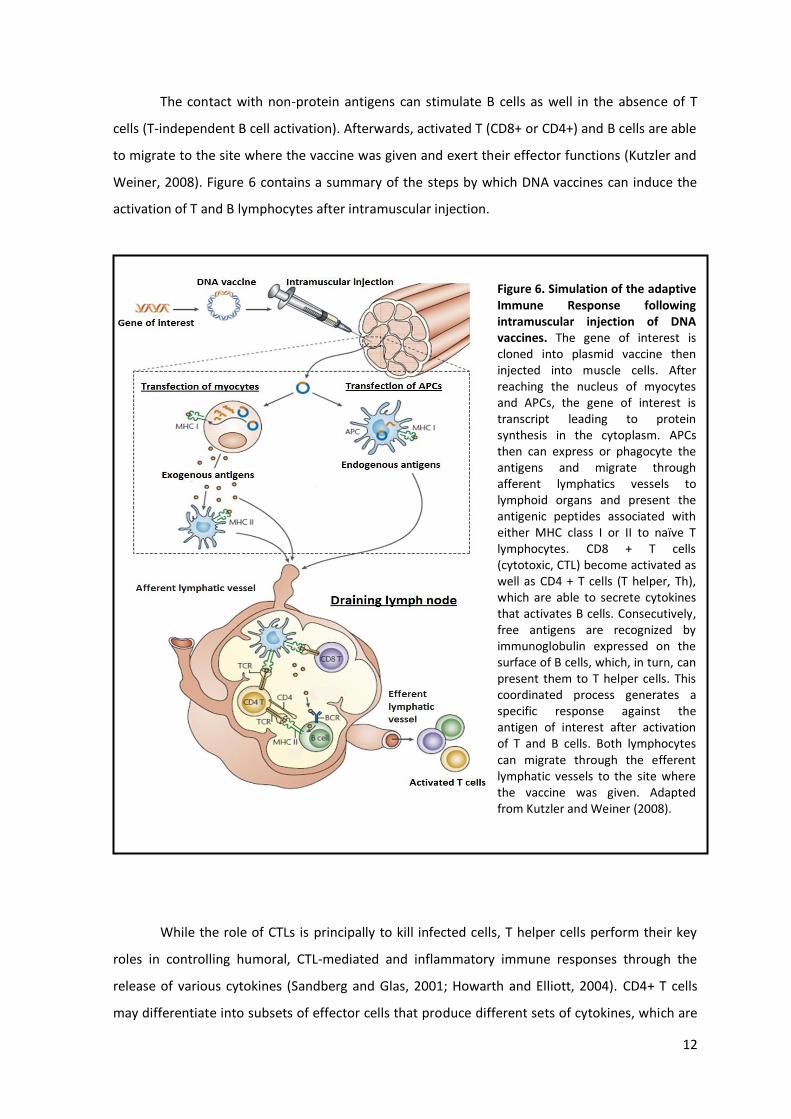
The contact with non-‐protein antigens can stimulate B cells as well in the absence of T
cells (T-‐independent B cell activation). Afterwards, activated T (CD8+ or CD4+) and B cells are able
to migrate to the site where the vaccine was given and exert their effector functions (Kutzler and
Weiner, 2008). Figure 6 contains a summary of the steps by which DNA vaccines can induce the
activation of T and B lymphocytes after intramuscular injection.
While the role of CTLs is principally to kill infected cells, T helper cells perform their key
roles in controlling humoral, CTL-‐mediated and inflammatory immune responses through the
release of various cytokines (Sandberg and Glas, 2001; Howarth and Elliott, 2004). CD4+ T cells
may differentiate into subsets of effector cells that produce different sets of cytokines, which are
Figure 6. Simulation of the adaptive Immune Response following intramuscular injection of DNA vaccines. The gene of interest is cloned into plasmid vaccine then injected into muscle cells. After reaching the nucleus of myocytes and APCs, the gene of interest is transcript leading to protein synthesis in the cytoplasm. APCs then can express or phagocyte the antigens and migrate through afferent lymphatics vessels to lymphoid organs and present the antigenic peptides associated with either MHC class I or II to naïve T lymphocytes. CD8 + T cells (cytotoxic, CTL) become activated as well as CD4 + T cells (T helper, Th), which are able to secrete cytokines that activates B cells. Consecutively, free antigens are recognized by immunoglobulin expressed on the surface of B cells, which, in turn, can present them to T helper cells. This coordinated process generates a specific response against the antigen of interest after activation of T and B cells. Both lymphocytes can migrate through the efferent lymphatic vessels to the site where the vaccine was given. Adapted from Kutzler and Weiner (2008).

13
responsible for their distinct functions. In the injection site, activated T helper cells can be
differentiated in the presence of interleukine-‐12 (IL-‐12) and IL-‐18 (both produced by local DCs)
into type 1 T helper cells (Th1) that secrete IL-‐2, IL-‐12, IFN-‐
hand, in the presence of IL-‐4 (expressed by DCs) these cells are differentiated into type 2 helper T
cells (Th2) that secrete mainly IL-‐4, IL-‐5 and IL-‐10. This initial polarization of the response is self-‐
perpetuating as Th1 cytokines enhance further Th1 responses and down-‐regulate Th2 cytokines,
and vice versa. It is still not clear how the initial polarization towards either Th1 or Th2 is
controlled, but it is thought to involve a number of factors including the route of immunization
and the type of APC that presents the antigen (DCs, macrophages or B cells), the pattern of
cytokines released by cells from the innate immune system, and the antigen
epitope/density/affinity (Abbas et al., 1996; Mosmann and Sad, 1996). Through the release of
instance, reactive oxygen intermediates and induce inflammation, favoring pathogen elimination.
Differently, Th2 cells express cytokines that activates for example eosinophils, cells associated
with allergy and asthma and responsible for combating multicellular parasites (Mosmann and Sad,
1996).
Different vectors and delivery modes of plasmid DNA were also found to stimulate
different T helper responses. Thus, there are increased efforts to develop specific vectors to
promote the type of T cell response (Th1 or Th2) for diverse applications, such as for prevention
of infectious diseases or asthma and diabetes (Liu, 2010; Liu, 2011). Naked DNA vaccines have
been shown to induce either Th1 or Th2 responses in experimental animals, depending on the
method of administration. When delivered by intramuscular injection, DNA vaccine tends to
induce Th1 responses. It was proved that CpG motifs in the bacterial DNA plasmids induce a local
inflammatory response, leading to the accumulation of pro-‐inflammatory cytokines, such as IL-‐12,
at the vaccination site.
B cells also confer immune protection after DNA vaccination by producing antibody
molecules, also known as immunoglobulins (Igs), which can recognize antigen through either low
or high After antigen recognition events in the lymph nodes (figure 6), B
cells are activated resulting in the proliferation of antigen-‐specific B cells (a process known as
clonal expansion). Depending on the stimuli, subsets of mature B lymphocytes can express
different surface Ig isotypes (i.e IgG1, IgG2a, IgE or IgA), migrate to the local site where the
vaccine was given and become immunoglobulin-‐secreting plasma cells (plasmocytes), capable of
secreting Ig (Mosmann and Sad, 1996; Kaplan et al., 2001). Antibody isotype is governed by the
cytokine environment and Th1-‐Th2 balance. For example, it was demonstrated that IL-‐4 favors the
production of IgG1, while IFN-‐

14
2001; Kaplan et al., 2001). Antibodies can exert their effector function by neutralizing toxins,
opsonizing or lysing circulating microorganisms, inducing cytotoxicity in cells infected or
expressing the antigen (Janeway, 2001).
1.4.5 Immune memory
After the primary immune response, a population of lymphocytes that mediate long-‐lived
immunological memory is produced. These lymphocytes, termed memory cells have, for many
years, been considered a dormant population, lacking the effector functions displayed by cells
during the acute phase of infection. However, immediately upon antigen contact, they can clone
rapidly and restore their effector functions (i.e B cells: secretion of antibodies; CTLs: lytic activity;
T helper: secretion of cytokines). The precise changes that distinguish naive, effector, and
memory lymphocytes are now a field of intense research. Protective immunity against reinfection
is one of the most important consequences of adaptive immunity. This means that secondary
exposure to an antigen produces a much more rapid immune response, turning the individual not
as badly affected compared to the first time the antigen appeared in the organism. Actually, it has
been demonstrated that if the preexisting memory cells are sufficiently numerous, they can
contain the infection immediately. A vaccine takes advantage of this effect, giving protection to
subsequent exposure to the same antigen (Justewicz and Webster, 1996; Leifert and Whitton,
2000; Janeway, 2001).
Compared to other types of immunization, DNA vaccines offer a unique opportunity to
enhance the duration of immune responses through their capacity for prolonged antigen
expression, thus favoring the formation of immune memory cells. Some studies have shown that
a single intramuscular DNA vaccination, when combined with electroporation (a delivery method,
significantly enhanced the onset and duration of the primary antibody response and maintained
immune memory (Tsang et al., 2007). Fazio and collaborators demonstrated that a single in utero
DNA immunization with a DNA vaccine based on hepatitis B virus (HBV) at two-‐thirds of pig
gestation produced, at birth, antibody titers considered protective in humans. The boost of
antibody titers following recall at 4 and 10 months demonstrated the establishment of immune
memory, illustrating the relevance of naked DNA-‐based vaccination aimed to prevent death
Another similar work was performed by Rigato and
co-‐workers in which they shown that the
intravenous injection led to in utero immunization. A DNA vaccine encoding LAMP-‐1 with Gag and
other Human Immunodeficiency Virus type 1 (HIV-‐1) antigens administrated in BALB/c mice
before conception or during pregnancy induced a long-‐lasting memory response (Rigato et al.,
2012). In order to stimulate the development of immune memory cells, immunization of DNA

15
along with adjuvants molecules are being performed, as recently described. It was observed that
the use of Hemokinin-‐1 (HK-‐1), a factor that activates B cells for proliferation, as an adjuvant
molecule enhanced the immunogenicity of HBsAg DNA vaccines, resulting in stronger humoral
and memory responses against hepatitis B infection (Chen et al., 2012).
1.5 Preclinical and clinical progress of DNA vaccines
To date, there have been several preclinical and clinical studies on DNA vaccines.
Although US FDA still did not approved DNA vaccines for use in humans, phase I clinical studies
have been reported for the prevention and/or treatment of HIV, malaria, hepatitis B, SARS and
many other infectious agents (Klinman et al., 2010). In addition to the initial demonstration of the
efficacy of DNA vaccines to protect against infectious challenge in a mouse model of influenza
virus, DNA vaccines have been shown to protect against influenza in ferrets, and primates;
lymphocytic choriomeningitis virus; herpes simplex virus in guinea pigs and mice; rabies virus;
cottontail rabbit papillomavirus; hepatitis B virus; malaria (Plasmodium falciparum); HIV in
nonhuman primates; and against other several bacterial pathogens (Srivastava and Liu, 2003).
What is remarkable is that in the past 6 years, four DNA vaccines for larger animals have
been licensed for use in the veterinary field. Two of them target infectious diseases such as West
Nile virus in horses, authorized for use in the United States (US); and aquatic rhabdovirus (also
termed as infectious hematopoietic necrosis, IHNV) in salmon, approved for use in Canada.
Another one is a cancer vaccine for melanoma in dogs; and the last one has a therapeutic purpose
in swine in which a plasmid encoded the growth hormone, when delivered before specific
vaccination, demonstrated enhanced protection against Mycoplasma hyopneumoniae; both of
them are authorized for use in the US and Australia. Although these animal disease models are
not completely similar to humans, past success with DNA vaccines turns this vaccine platform very
promising for use in humans (Faurez et al., 2010; Ingolotti et al., 2010; Findik and Çiftci, 2012)
(Table 1).

16
Table 1. Current licensed DNA therapies. Adapted from Kutzler and Weiner (2008).

17
Even though DNA immunization have evolved greatly over the last 20 years, the Achilles
heel of DNA vaccines proved to be their poor immunogenicity in humans and other large animals,
instead of the initial safety concerns. To date, the potency of the immune responses has been
disappointing in humans; nevertheless, humoral and cellular (T helper and cytolytic T cell)
responses were obtained (Srivastava and Liu, 2003; Bolhassani et al., 2011). Clearly regimens,
plasmid dose, timing of doses, adjuvants and routes of vaccination have been considered as well,
and wide variety of strategies will be developed to optimize DNA vaccine immunogenicity.
1.6 Improvement of DNA vaccines immunogenicity
The low immunogenicity of early DNA vaccines is hypothesized to stem, in part, from
inefficient uptake of the plasmids by cells due to inefficient delivery (Ferraro et al., 2011).
Nonetheless, the reasons for the failure of DNA vaccines to induce potent immune responses in
humans have not been elucidated (Bolhassani et al., 2011). Therefore, research has focused on
developing novel strategies to enhance transfection efficiency and improve other facets of the
DNA vaccination platform using several strategies (Kutzler and Weiner, 2008; Ferraro et al., 2011).
Reasonably, vector modifications that improve antigen expression are highly correlative
with improved immune responses. It has been shown that the use of altered transcriptional
elements, like (i) the modified CMV promoters (chimeric SV40-‐CMV promoters), (ii) mRNA
containing introns, (iii) gene of interest codon optimized to match with the target organism, (iv)
the use of a dual stop codon to limit read through translation, and (v) transcription
terminators/polyadenylation signals can significantly improve either antigen transcription or
translation by the host cell (Kutzler and Weiner, 2008; Williams et al., 2009). Optimization of the
initiation start site for protein synthesis (kozak consensus sequences) is also desirable as
endogenous sites of viruses and bacteria might not be optimal for expression in mammalian cells
(Kutzler and Weiner, 2008). The addition of leader sequences can also enhance the stability of
mRNA and contribute to translational efficiency. Considering the commercialization of DNA
vaccines, the use of high-‐efficiency origins of bacterial replication can also markedly improve the
quantity of plasmid product (Williams et al., 2009).
Of course the right choice of antigens is crucial for obtaining a satisfactory immune
response as well as the use of unmethylated CpG, which should be included in the plasmid
backbone in order to enhance the potency of a DNA vaccine (Glenting and Wessels, 2005).
Another strategy attempt to increase the magnitude of immune responses by co-‐expression of
cytokines, chemokines or co-‐stimulatory molecules, that can have a substantial effect on the
immune response (Liu, 2003; Kutzler and Weiner, 2008; Ferraro et al., 2011). Dam and dcm
methylation performed by bacteria should also be taken into account as this may affect antigen

18
expression or make it prone to recognition as foreign DNA by the host innate immune system
(Kutzler and Weiner, 2008; Williams et al., 2009). Another important effort to improve gene
expression includes the delivery method employed to introduce the DNA vaccine in the organism
(Bolhassani et al., 2011). These methods comprise physical and chemical approaches or the use of
viral and bacterial vectors. Figure 7 presents some of the improvements proposed to increase the
immunogenicity of DNA vaccines.
1.7 Non-‐biological delivery systems for DNA vaccines
1.7.1 Physical approaches
The physical methods of delivering a DNA plasmid are divided into (i) gene gun, (ii)
tattooing, (iii) ultrasound (US), (iv) electroporation (EP), (v) laser, (vi) dermal patches. Gene gun
-‐ is a biolistic method, originally designed for plant
transformation, which enables the DNA to be delivered directly to keratinocytes and epidermal
Langerhans cells, stimulating DCs. The vaccine plasmid linked to microscopic gold particles is
bombarded into the cells using a strong shock wave that accelerates DNA-‐coated gold particles to
high speeds. Recently, gene gun mediated transgene delivery system has been used for skin
vaccination against human melanoma as an experimental method (Yang et al., 2001; Bolhassani et
al., 2011; Ferraro et al., 2011). Nevertheless, gene gun technology has certain disadvantages
related to the adverse effects following gene transfer on animals. One major drawback is the lack
Figure 7: Ways in which antigen expression and immunogenicity can be improved for the DNA vaccine platform. (1) optimization of the transcriptional elements on the plasmid backbone; (2) strategies to improve protein expression of the gene of interest; (3) inclusion of formulation adjuvants or (4) immune plasmid adjuvants; and (5) the use of next-‐generation delivery methods (5). Adapted from Kutzler and Weiner (2008).

19
of sustained expression of the introduced genes in the target tissue as the DNA is superficially
penetrated into the tissue (Niidome and Huang, 2002). Therefore, multiple administrations are
required in many cases (Yang et al., 2001). More than just a fashion statement, tattoos can be
used to introduce DNA vaccines in the epidermal and dermal layers. DNA plasmids delivered by
tattooing have been shown to induce higher specific humoral and cellular immune responses
compared to intramuscularly injection of naked DNA. However, this is a very cost-‐effective
method that may be used when more rapid and more robust immune responses are required
(Pokorna et al., 2008; Bolhassani et al., 2011).
Ultrasound (US) can be used to transiently disrupt cell membranes to enable the
incorporation of DNA and this technology has been applied in clinical trials.
An alternative physical delivery system is a device that generates an electric current, in
vivo electroporation, to create temporary holes in the cellular membrane (Srivastava and Liu,
2003). That was initially studied to enhance the efficacy of chemotherapy agents (Ferraro et al.,
2011). Potent immune responses against hepatitis B surface antigen and HIV gag protein were
obtained by electroporation of muscle after intramuscular injection of naked plasmid DNA
(Niidome and Huang, 2002). However, the concerns about the chromosomal integration observed
in a safety study of electroporation mean that this technology needs careful evaluation for
humans (Liu, 2011).
In vitro studies have shown that laser beam can deliver a certain amount of energy onto a
target cell, modifying permeability through a local thermal effect, thus facilitating DNA
incorporation by the cell. This novel technology is currently being explored (Bolhassani et al.,
2011). Another new method is the dermal patch delivery (DermaVir). It is a self-‐adhesive patch
coated with multiple antigen or adjuvant encoding plasmids and a synthetic polymer that forms
pathogen-‐like nanoparticles (Ferraro et al., 2011).
It is worth highlighting that the method of delivering a DNA vaccine can drastically
influence the type of immune response induced by the vaccine. For instance, gene gun typically
induces Th2 reactions whereas needle inoculation triggers a Th1 response (Bolhassani et al., 2011).
Moreover, whilst each of these methods has contributed to incremental improvements in DNA
vaccine efficacy, more is still needed for human DNA vaccines success commercially (Bolhassani et
al., 2011; Liu et al., 2012). Nevertheless, these various delivery technologies merit continued
development and evaluation (Liu, 2011).
1.7.2 Chemical vectors
Chemical vectors that are mainly based on engineered DNA nanoparticles produced with
various range of macromolecules (Niidome and Huang, 2002). Generally, the major advantages

20
are (i) they can tightly compact and protect DNA, (ii) be recognized by specific cell-‐surface
receptors expressed in DCs or macrophages, (iii) disrupt the endosomal membrane to deliver DNA
plasmids to the nucleus after its destabilization when the pH of the medium is reduced to below 6
(Bolhassani et al., 2011). Usually, non-‐viral vectors include mainly DNA-‐liposome complexes and
DNA-‐polymer complexes. Nowadays DNA-‐peptide complexes are also being used as DNA carriers.
It has been shown that they are able to condense plasmid DNA, resulting in a highly stable
complex with potent transfection activity in vitro (Niidome and Huang, 2002). These vectors are
currently being evaluated in clinical trials (Zhang et al., 2012).
So far, lipid and polymer complexes are the most studied and used chemical vectors.
Negatively charged, DNA neutralizes both cationic liposomes and polymers leading to the
formation of electrostatic complexes with plasmid DNA. This condensation provides size reduction
and protection against nuclease degradation (Pichon et al., 2010; Bolhassani et al., 2011).
Moreover, the encapsulation of plasmid DNA into micro or nanospheres has proved to protect
plasmids from the environment and to target the genetic material to a specific cell type. For
example, it has been shown that some liposomes and polymers can effectively aid in directing
antigens to APCs by efficiently trafficking through local lymphoid tissue (Lu et al., 2009).
Liposome-‐based gene delivery comprises a large number of cationic lipids, such as quaternary
ammonium detergents and lipid derivatives of polyamines. Even though being capable of
accommodating large size plasmids and immunostimulatory agents, it was observed that the use
of liposomes sometimes leads to a transient expression of the transgene (Fenske and Cullis, 2008).
Polymers are also being explored as delivery vectors (Niidome and Huang, 2002). A very
interesting work was performed by Roy and collaborators in 1999. Mice that received
nanoparticles containing a dominant peanut allergen gene (pCMVArah2) produced secretory IgA
and serum IgG2a. Compared with non-‐immunized mice or mice treated with 'naked' DNA, mice
immunized with nanoparticles showed a substantial reduction in allergen-‐induced anaphylaxis
associated with reduced levels of IgE, plasma histamine and vascular leakage (Roy et al., 1999).
Nevertheless, the use of both liposomes and polymers has been shown to present some
limitations as they exhibited great toxicity in vivo (Fenske and Cullis, 2008; Bolhassani et al., 2011).
Consequently, nanoparticle-‐based vaccines require the development of increasingly safer
formulations (Xiang et al., 2010).
1.8 Biological delivery systems for DNA vaccines
1.8.1 Virus as DNA delivery vehicles
To date, live vectors, such as attenuated or non-‐pathogenic virus, have demonstrated the
feasibility of gene therapy/antigen delivery and remain one of the best vehicles to introduce

21
genes into cells (Pichon et al., 2010). Virus represents ideal nanoparticles due to their regular
geometries, well characterized surface properties and nanoscale dimensions (Bolhassani et al.,
2011).
The five main classes of viral vector can be categorized in two groups according to
whether their genomes integrate into host cellular chromatin (oncoretroviruses and lentiviruses)
or persist in the cell nucleus predominantly as extrachromosomal DNA molecule or episome
(AAVs, adenoviruses and herpes viruses) (Thomas et al., 2003). The basis for selecting a particular
viral vector includes how well the heterologous gene is expressed as the quantity of antigen may
affect the immune response. In addition, the type of cells y the virus vector that will
produce the antigen, may affect the nature and potency of immune response. Another factor
influencing the choice of vector is the persistence of the virus, which might be useful for
prolonging immunity (Liu, 2011). Figure 8 illustrates how a viral particle can transfect a
mammalian cell and deliver a gene of interest.
Oncoretrovirus vectors were the first class of viral vector to be developed and have, so far,
been the most widely used in clinical trials. The inconvenient is that they may cause genetic
diseases or favor the development of cancerous cells because they can integrate their genetic
material into host cellular genome (Thomas et al., 2003). Recombinant adenoviruses (Ads) are
also being extensively used for gene therapy because they are extremely efficient at delivering
DNA to target cells and have low potential for oncogenesis (Bolhassani et al., 2011; Liu, 2011).
Nonetheless, a severe fatal adverse effect have occurred during a gene therapy trial raising
serious safety concerns about the use of viral vectors (Pichon et al., 2010). In 1999 administration
of a recombinant adenoviral vector generated adverse reactions in a patient causing his death,
and in 2000 another study found that administration of retroviral vector induced leukemia in 2 of
11 patients who received the treatment (Check, 2002; Check, 2003). Besides these two tragic
events, viral vectors have several manufacturing issues as they are occasionally developed from
pathogenic species. Thus, the scientific community has boosted efforts to pursue an ideal non-‐
virus live vector to improve the effects of DNA vaccination (Kano et al., 2007; Pichon et al., 2010).

22
1.8.2 Bacteria-‐based vectors
Studies conducted in 1980 by Walter Schaffner demonstrated that the bacteria are
capable of transferring genetic material into mammalian cells in vitro. For this reason they have
been proposed as new vectors for plasmid vaccines transfer (Courvalin et al. 1995; Sizemore et al.
1995; Vassaux et al. 2006). Later, it was also demonstrated that Gram-‐positive bacteria, such as
Listeria monocytogenes was able to deliver plasmid DNA (Becker et al., 2008). Ever since,
attenuated or artificially engineered to be invasive bacteria have been tested as a vehicle for
transgene delivery (Seow and Wood, 2009).
Figure 8: Transduction using adenoviral vectors. Recombinant adenovirus enters cells via CAR-‐mediated binding allowing internalization via receptor-‐mediated endocytosis through clathrin-‐coated vesicles. Inside the cytoplasm, the endocytosed adenoviral vector escapes from the endosomes, disassembles the capsid and the viral DNA enter into the nucleus through the nuclear envelope pore complex. The viral DNA is not incorporated into the host cell genome, but rather assumes an epichromosomal location, where it can still use the transcriptional and translational machinery of the host cell to synthesize the gene of interest. Adapted from Pankajakshan and Agrawal (2013).

23
2. BACTERIAL VECTORS AS DNA DELIVERY VEHICLES
The use of bacterial systems for gene therapy/antigen delivery (also known as
bactofection), is especially interesting as some vectors based on pathogenic strains has an innate
tropism for specific tissues of the host which are indispensable for the development of an
immune response (Pilgrim et al. 2003). One attractive feature of bacterial vectors is their potential
for oral administration, which may stimulate both mucosal and systemic immune responses
(Izadpanah et al. 2001; Srivastava and Liu, 2003). Oral and nasal mucosa represents the first line
of defense against many pathogenic microorganisms (Seow and Wood, 2009). Mucosal
immunization can lead to the proliferation of T helper cells that stimulates the secretion of IgA
antibodies in the epithelium. Secretory IgA (SIgA) represents the most abundant immunoglobulin
in our body which plays a critical role in host defense as it is capable of neutralizing pathogens or
toxins that are in direct contact with the mucosal surfaces (Lamm, 1997; Macpherson et al. 2001).
Furthermore, WHO recommends the use of mucosal vaccines because of economic, logistic and
security reasons (Neutra and Kozlowski 2006; Wells, 2011). Furthermore, bacterial carrier can
specifically target and transfer main APCs, such as DCs. After bacterial uptake by DCs, they can
act as adjuvants due to the presence of some molecules unique to bacterial cell walls known as
microbe-‐associated molecular patterns (MAMPs) (Becker et al., 2008). The recognition of MAMPs
by the host can modulate innate immune responses, promoting, as a consequence, an efficient
and adaptive long-‐lasting immune response (Schoen et al., 2004). Another advantage consists on
the fact that bacteria are able to accommodate large-‐size plasmid, which allows the insertion of
multiple genes of interest, protecting the DNA plasmids against endonuclease degradation (Hoebe
et al., 2004; Seow and Wood, 2009). Finally, bacteria-‐based vectors are considered low cost since
plasmids can be amplified by the simple growth of bacterial cultures turning purification steps
unnecessary (Hoebe et al. 2004).
2.1 Basic Principles for Bacteria-‐Mediated DNA delivery at mucosal surfaces
Vectors based on attenuated pathogenic species can actively invade the mucosal
epithelium and translocate across the epithelial barrier turning possible the expression of the
transgene. Actually, it has been reported that some vectors, such as Agrobacterium tumefaciens,
are able to transfer expression cassettes even without invading the target cells (Weiss and
Chakraborty, 2001). Once inside the cells, bacterial vectors are able to escape from
phagolysosome vesicles by the secretion of a variety of phospholipases and pore-‐forming
cytolysins and enter into the cytoplasm of host cells. A part of bacteria that are degraded inside
the phagolysosome release the plasmid vaccine, which can reach the cytoplasm through the pores
made by the phospholipases and pore-‐forming cytolysins (Hoebe et al., 2004; Schoen et al., 2004).
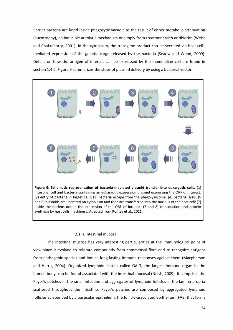
24
Carrier bacteria are lysed inside phagocytic vacuole as the result of either metabolic attenuation
(auxotrophy), an inducible autolytic mechanism or simply from treatment with antibiotics (Weiss
and Chakraborty, 2001). In the cytoplasm, the transgene product can be secreted via host cell
mediated expression of the genetic cargo released by the bacteria (Seaow and Wood, 2009).
Details on how the antigen of interest can be expressed by the mammalian cell are found in
section 1.4.2. Figure 9 summarizes the steps of plasmid delivery by using a bacterial vector.
2.1. 1 Intestinal mucosa
The intestinal mucosa has very interesting particularities at the immunological point of
view since it evolved to tolerate compounds from commensal flora and to recognize antigens
from pathogenic species and induce long-‐lasting immune responses against them (Macpherson
and Harris, 2004). Organized lymphoid tissues called GALT, the largest immune organ in the
human body, can be found associated with the intestinal mucosal (Neish, 2009). It comprises the
scattered throughout the intestine. Peyer's patches are composed by aggregated lymphoid
follicles surrounded by a particular epithelium, the follicle-‐associated epithelium (FAE) that forms
Figure 9: Schematic representation of bacteria-‐mediated plasmid transfer into eukaryotic cells. (1) Intestinal cell and bacteria containing an eukaryotic expression plasmid expressing the ORF of interest; (2) entry of bacteria in target cells; (3) bacteria escape from the phagolysosome; (4) bacterial lysis; (5 and 6) plasmids are liberated on cytoplasm and then are transferred into the nucleus of the host cell; (7) inside the nucleus occurs the expression of the ORF of interest; (7 and 8) transduction and protein synthesis by host cells machinery. Adapted from Pontes et al., 2011.

25
the interface between the GALT and the luminal microenvironment (Perdigón et al. 2001;
Perdigón et al. 2002; Hobson et al. 2003; Jung et al., 2010). Among the pathogenic bacteria with a
digestive tropism such as Escherichia coli, Yersinia, Mycobacterium avium paratuberculosis,
Listeria monocytogenes, Salmonella typhimurium and, Shigella flexneri, all of them have been
reported to invade the host by adhering with FAE M-‐cells (Jung et al., 2010).
Additionally, special mucosal DCs located in the lamina propria of the small intestine can
extend their dendrites between tight junctions (TJ) of epithelial cells and capture antigen directly
from the intestinal lumen (Rescigno et al. 2001). The binding between DCs and epithelial cells
occurs through the expression of TJ proteins by DCs which can establish TJ-‐like structures with
neighboring epithelial cells. Interestingly, lamina propria DCs can discriminate between
pathogenic and nonpathogenic bacteria. In fact, when the intestine is infected with
nonpathogenic bacteria, DCs do not receive any migratory signal and stay in situ, whereas
infection with pathogenic bacteria induces a large migration of DCs from the mucosa to the
mesenteric lymph nodes. Thus, DCs play an active role in bacterial handling across mucosal
surfaces (Rimoldi et al., 2004). After interacting with antigens, DCs can present them to naïve T
cells in the mesenteric lymph nodes, establishing a cross-‐talk between innate and adaptive
immunity. It has been shown that lamina propria DCs stimulated with commensal bacterial
antigens can induce T cell independent IgA class switching and soluble IgA (SIgA) production by
intestinal B cells (Barbosa and Rescigno, 2010). The IgA is the main class of immunoglobulin found
in intestinal secretions. Its role is to protect the intestinal epithelium against enteric pathogens
and toxins (Mantis and Forbes, 2010). Recent studies demonstrated that IgA are also implicated in
the regulation of gut commensal bacterial communities as it is able to control microbiota
expansion while maintaining gut homeostasis (Barbosa and Rescigno, 2010; Suzuki et al., 2010). In
conclusion, through a coordinated manner, T and B cells together with epithelial cells are capable
of creating a powerful line of defense against infectious agents and exert an important role in
regulating immune responses in the intestinal mucosa (Hobson et al. 2003; Izadpanah et al. 2001;
Kutzler & Weiner, 2008). The major components of the immune system associated with the
gastrointestinal tract are described in Figure 10.

26
2.1.2 Commensal bacteria and the intestinal mucosa
Commensals colonize the human gut soon after birth. Bacteria from the birth canal and
from maternal milk are the first colonizers and their persistence depends on the ability of strains
to interact among them and with the thick mucus layer that lines the intestinal lumen (Barbosa
and Rescigno, 2010). The trillions of commensal microorganisms that constitute the intestinal
microbiota are primarily composed of five bacterial phyla, Firmicutes, Bacteroidetes,
Actinobacteria, Proteobacteria, and Fusobacteria. Bacteroidetes and Firmicutes predominate and
represent approximately 90% of the total gut microbiota (Figure 11). Some of these
microorganisms can exert many benefits to the human health as they can promote plant
polysaccharide digestion and uptake of nutrients by host intestinal cells. Moreover, in addition to
nutritional benefits, commensal bacteria may protect their host through antimicrobial activity
against pathogenic bacteria and are able to regulate intestinal immune homeostasis (Yan and Polk,
2004; Jung et al., 2010; Kosiewicz et al., 2011).
Figure 10. Intestinal epithelium and the gut associated immune system. The IECs form a barrier that prevents biochemical and physical invasion of microorganisms into the body. M cells transports antigens from the intestinal lumen to antigen presenting cells such as DCs, which are located in Peyer's patches. DCs containing antigens travels through the afferent lymphatics vessels to the mesenteric lymph nodes and present them to naïve T cells, activating adaptive immune responses. Activated T cell and B lymphocytes are able to migrate from the lamina propria via the efferent lymphatic vessels. Figure adapted from Macpherson and Harris, 2004.

27
Commensals are not ignored by the intestinal immune system. Recent evidence shows
that they can interact with IECs and deliver tolerogenic signals that are transmitted to the
underlying cells of the immune system (Rescigno, 2009). Immunological tolerance against non-‐
pathogenic bacteria and antigens is a phenomenon observed along the gastrointestinal mucosa,
which avoids reactions against proteins and commensal bacteria. Oral tolerance is an active
process, leading to the generation of antigen-‐specific T lymphocytes that suppress further
immune stimulation. Consequently, mucosal tolerance protects the mucosa from detrimental
inflammatory immune responses (Jung et al., 2010). It has been shown that resident DCs
constantly traffic between mesenteric lymph nodes and gut mucosa, even in the absence of
inflammatory stimuli, and this recirculation is considered important to maintain tolerance to oral
antigens and to the microbiota (Iliev et al., 2007). Actually, exposure of immune cells to bacteria
has important consequences for gut health: excessive contact promotes exaggerated pro-‐
germ-‐free conditions, can dramatically impair immune development and function (Kelly and
Mulder, 2012). A break of this steady state is the major cause of inflammatory bowel diseases
(IBD), such as ulcerative colitis (UC) and Crohn's disease (CD) (Guarner and Malagelada, 2003).
commensals capable o
balancing the immune response in favor of regulation or by controlling bacteria that may directly
ic
diseases that may be impacted by the gut microbial community. Actually, allergic disease
Figure 11. Composition and luminal concentrations of dominant microbial species in various regions of the gastrointestinal tract. Adapted from Sartor, 2007

28
development, cancer, especially colon cancer, has been associated with alterations in the
intestinal microbiota (Kosiewicz et al., 2011).
2.3 Bacterial vectors used for gene transfer
2.3.1 Pathogenic bacterial DNA delivery
Enteropathogenic species are the most widely used bacterial delivery systems (Schoen et
al., 2004). The majority of the works describe the use of Shigella, Salmonella and Listeria strains,
being these very well-‐known examples of bacterial vectors. With respect to their localization
during the infective process, enteropathogenic bacteria employed as DNA vaccine carriers can be
subdivided into extracellular pathogens, such as some E. coli strains or Yersinia spp.,
intraphagosomal pathogens like Salmonella spp., and intracytosolic pathogens, Listeria
monocytogenes, Shigella spp (Loessner et al. 2007). In all cases, the bacteria used for such
proposed vaccines are attenuated strains. Their use is very interesting since they have a natural
tropism for dendritic cells and macrophages in the lymphoid tissue of the intestinal mucosal
surface, and thus naturally targets these inductive sites allowing the exploitation of this property
in the development of mucosal DNA vaccines (Becker et al. 2008). Attenuated pathogenic bacteria
as carriers for DNA vaccines has been applied in the prophylaxis and therapy of several infectious
diseases (Schoen et al. 2004) and tumors (Yu et al. 2004). Promising results have been obtained in
animal models, however, none of these strains were licensed for use in humans.
There are several strategies available to attenuate strains. Most of the vectors used for
genetic immunization contains a deletion of an essential gene in bacterial metabolism
(auxotrophic mutants), limiting their growth in vivo. Other attenuated bacteria are engineered to
express a phage lysine upon their entry into eukaryotic cells (Pilgrim et al. 2003; Loessner et al.
2007), turning them unable to perpetuate within the host and thereby cause an infection. There
are many enteropathogenic strains currently being used as DNA delivery vehicles and the main
examples are described below.
2.3.1.1 Extracellular pathogens
One great example of extracellular pathogenic bacteria that was used as a DNA vaccine
vector is Yersinia enterocolitica. It is a pleomorphic gram-‐negative bacillus that belongs to the
family Enterobacteriaceae. Yersinia survival is mediated by a virulence plasmid named pYV
encoding a series of virulence genes (Novoslavskij et al., 2012). The use of this bacterium as a
vector to deliver DNA was reported by Al-‐Mariri and co-‐workers. Following two intragastric
immunizations in BALB/c mice, the attenuated Yersinia vectors harboring a DNA vaccine encoding

29
Brucella abortus antigens elicited antigen-‐specific serum immunoglobulin and Th1-‐type responses
among splenocytes (Al-‐Mariri et al., 2002).
2.3.1.2 Intraphagosomal pathogens
Salmonella genus stands out among other intraphagosomal pathogens used to deliver
vaccine plasmids. Salmonella typhimurium, for example, has long served as a model organism for
genetic studies and are probably the most widely used bacteria for antigen delivery applications.
It is a gram-‐negative rod-‐shaped bacillus and approximately 2000 serotypes cause human disease
(Ohl and Miller, 2001; Parsa and Pfeifer, 2007). It has been shown that Salmonella pathogenicity
island 1 (SPI-‐1) encodes genes necessary for invasion of IECs and induction of intestinal secretory
and inflammatory responses. Differently, Salmonella pathogenicity island 2 (SPI-‐2) encodes genes
essential for intracellular replication (Ohl and Miller, 2001).
Recently, it was reported that the administration of an attenuated Salmonella
typhimurium strain delivering a DNA vaccine encoding duck enteritis virus UL24 could induce
systemic and mucosal immune responses in ducks, conferring a good protection against challenge
(Yu et al., 2012). Another interesting work was performed by Wen and collaborators (2012) where
they found that S. typhi loaded with an HIV gp140 DNA vaccine were readily taken up in vitro by
murine macrophage cells, and gp140 antigen was efficiently expressed in these cells. Peripheral
and intestinal mucosal anti-‐gp140 antibody responses in mice vaccinated were significantly higher
than those in mice immunized with naked DNA vaccine, demonstrating the bacteria carrying
gp140 plasmid could be used as a new strategy for development of HIV vaccines (Wen et al.,
2012).
2.3.1.3 Intracytosolic pathogens
Listeria monocytogenes represents another important example of a bacterial vector vastly
used for DNA delivery. This gram-‐positive bacterium is responsible for listeriosis, a severe human
infection with an overall 30% mortality rate. Similar to the gram-‐negative enteropathogenic
bacteria Salmonella, Shigella, and Yersinia spp., L. monocytogenes has the capacity to induce its
own uptake into nonphagocytic mammalian cells (Lecuit et al., 1997). Three tight barriers (the
intestinal, blood-‐brain, and placental barriers) can be crossed by this pathogen (Dussurget et al.,
2004) by the expression of two surface proteins, internalin A (or InlA) and InlB, encoded by two
genes (inlA and inlB) organized in an operon (Braun et al., 1997; Lecuit et al., 1997). The targets of
InlA and InlB on the surface of the host cell are the adhesion molecule E-‐cadherin and the
hepatocyte growth factor (HGF) receptor Met, respectively. Met is ubiquitously expressed,
allowing InlB to mediate bacterial internalization in a large number of cell types, whereas InlA
shows a more stringent cell tropism, as E-‐cadherin is only expressed by a limited number of cells
30
of epithelial origin (Bonazzi et al., 2009). InlA is a sortase anchored, cell wall protein which
contains 800 amino acids (aa) in length and composed of seven distinct domains and it is
considered one of the major virulence genes in L. monocytogenes (Monk et al., 2010). Recently, it
was demonstrated that E-‐cadherin is accessible from the lumen around mucus-‐expelling goblet
cells and around extruding enterocytes at the tip of villi. Thus these cells are preferentially
invaded by L. monocytogenes, which is rapidly transcytosed across the intestinal epithelium, and
released in the lamina propria by exocytosis from where it disseminates systemically (Nikitas et al.,
2011). Once inside the cell this bacterium is capable to resist early intravacuolar killing.
Phagosomal escape is largely mediated by the cholesterol-‐dependent cytolysin listeriolysin O
(LLO), which is essential for Listeria virulence. Rupture of the Listeria-‐containing phagosome is a
dynamic multistep process. After internalization, an LLO expression is up-‐regulated at low pH
inside the phagosome. The acidifying phagosome leads to LLO oligomerization of cholesterol-‐
bound monomers into a prepore complex, followed by insertion into the lipid bilayer. Maximizing
LLO activity in the vacuole but not in the cytosol is a critical aspect of the intracellular lifestyle of
Listeria because LLO mutations with increased expression or pore-‐forming activity destroy the
host cell and decrease virulence. Once in the cytosol, L. monocytogenes is capable of actin-‐based
motility and cell-‐to-‐cell spread without an extracellular phase (Cossart and Toledo-‐Arana, 2008;
Radtke et al., 2011). All this biological properties make L. monocytogenes a promising platform for
development as a vector for gene therapy, anti-‐cancer vaccine vector or antigen delivery vehicle.
Several preclinical studies have demonstrated the ability of this bacterium for intracellular gene or
protein delivery in vitro and in vivo, and this vector has also displayed a relatively safety and
efficacy in clinical trial (Tangney and Gahan, 2010) (Figure 12).
However, even though attractive, there is always the insecurity that attenuated vectors
might restore the ability to replicate and cause disease in the patient. This fact should be taken
into further consideration when it comes to the administration in infants and immuno-‐
compromised individuals. Actually, reversion to virulence, preexisting immunity, and
reactogenicity will always remain major concerns (Schoen et al., 2004). Thus, the scientific
community has recently been exploring the use of non-‐pathogenic bacteria, such as lactic acid
bacteria (LAB), as vectors for genetic immunization (Mercenier and Wells, 2008).

31
3. LACTIC ACID BACTERIA (LAB)
3.1 Taxonomy and characteristics
Phylogenetically, LAB belongs to the clostridial branch of the Gram-‐positive bacteria that
produce lactic acid as the major product of sugar fermentation. Species of Carnobacterium,
Enterococcus, Lactobacillus, Lactococcus, Leuconostoc, Oenococcus, Pediococcus, Streptococcus,
Tetragenococcus, Bifidobacterium, Vagococcus, and Weissella belong to the group of LAB. The
most representative is the Lactobacillus genus which contains approximately 80 species (Carr et al.
2002; Price et al., 2011). The individual species and strains of Lactobacillus differ by the profiles of
fermenting saccharides; however this property is not sufficient for the identification at the species
Figure 12: The pathogenesis of cellular Listeria monocytogenes infection. L. monocytogenes expresses cell-‐surface and secreted proteins that enable attachment to host cells, escape from the phagocytic vacuole and locomotion in the cytosol of the invaded cell. Internalin A (InlA) and InlB mediate the attachment of L. monocytogenes to the surface of host cells, and listeriolysin O (LLO) lyses the phagosomal membrane. The actin-‐assembly-‐inducing protein (ActA) is expressed in a polarized manner and catalyses actin polymerization, which propels bacteria through the cell and into neighbouring cells. To escape the secondary vacuole in the newly invaded cell, L. monocytogenes expresses the phosphatidylcholine-‐specific phospholipase PlcB, a secreted zinc metalloproteinase (Mpl) and LLO (Pamer 2004).
32
level. Some representatives of the genus are included in probiotic preparations with regard to
their beneficial health effects on the human and animal organisms (described later in this
manuscript). For example, they are able to interact with the immune system, to reduce the
manifestations of lactose intolerance by consuming fermented milk products and to produce
bacteriocins (antimicrobially active substances) (Bilková et al., 2011; Masood et al., 2011). Two
species, belonging to lactic bacteria, play a role in the production of fermented milks and dairy
products: Streptococcus thermophilus and Lactococcus lactis. However, not all LAB species have a
positive impact on human health or food production. Some can cause food spoilage, while several
important (human) pathogenic species are included in the LAB group (Felis and Dellaglio, 2007;
Price et al., 2011). The genus Streptococcus, for instance, contains about 60 species and a number
of them are known for their pathogenicity. The species S. thermophilus, however, is constituted
by non-‐pathogenic microorganisms, very essential in the dairy manufacturing (Felis and Dellaglio,
2007).
Lactic bacteria can tolerate mild acidic conditions of around pH 4 for several weeks and
are generally classified as facultative anaerobic, catalase-‐negative organisms. Their shapes are
primarily cocci (spheres), bacilli (rods), or ovoid. They may also be y-‐shaped, as in the case of
bifidobacteria, and usually they have less than 55 mol% G+C content in their DNA (Masood et al.,
2011). However, even though species from the Bifidobacterium genus present a high G+C content
in their DNA (55-‐57 mol%) and are not genetically closely related to LAB from the taxonomic point
of view, recently they have been grouped as LAB bacteria since they are able to produce lactic and
acetic acids (Felis and Dellaglio, 2007).
Lactic acid bacteria are distributed in different ecological niches; they are found in plants,
in fermented foods, and in the gastrointestinal tract of many animals, including humans, where
some species can live as commensal microorganisms (Steidler & Rottiers, 2006). Human beings
since ancient times and without understanding the scientific basis, use these bacteria for the
manufacture and maintenance of various types of foods such as cheese, wine, yogurt, fermented
milks, pickles, kefir, butter, koumiss among others. Nowadays it is known that food preservation is
due to acidification of the medium (pH 3.5 to 4.5), production of bacteriocins and organic
compounds. Due to the fact that they are used for centuries in food fermentation and
preservation, the FDA has granted this bacteria as generally regarded as safe (GRAS) for human
consumption (Van de Guchte et al. 2001).
3.1.1 Physiology of lactic acid bacteria into the human gastrointestinal tract
The physiology of lactic acid bacteria used for food fermentation is well studied and their
behavior for food production has received considerable attention. Nowadays, more attention has

33
been paid on the metabolic functions and phenotypic activities of enteric lactic bacteria during
their life into the human gastrointestinal tract as they have a significant impact on host
metabolism, participating in microbial-‐mammalian co-‐metabolism (Prakash et al., 2011). Species
of the genera Lactobacillus and Bifidobacterium are some of the most important taxa involved in
human nutrition. The upper portion of the gastrointestinal tract, made up of the stomach and the
duodenum, harbours very low numbers of microorganisms, with less than 1000 bacterial cells per
gram of contents, with the predominant microorganisms present being Lactobacilli and
Streptococci (Prakash et al., 2011), while bifidobacteria are largely present into the colon (Price et
al., 2011). Figure 13 illustrates some characteristics of the gastrointestinal tract and the
localization of various gut bacterial populations, termed microbiota.
3.1.2 The probiotic action
Live microorganisms which when administered in adequate
amounts confer a health benefit on the host
Research, 1997). The beneficial effects of LAB were revealed by a Russian Scientist E. Metchnikoff
Figure 13. The gastrointestinal tract characteristics (oxygen distribution, pH, bacterial populations, and bacterial cell counts) and the localization of the various gut bacterial populations, termed microbiota.

34
(1845 1919) who proposed that extended longevity of people of Balkan could be attributed to
their practice of ingesting fermented milk products (Metchnikoff, 1907; WHO, 2002; Masood et al.,
2011). It was vastly demonstrated that LAB can produce these beneficial effects by restoration of
normal intestinal flora, elimination of intestinal pathogens, reinforcement of intestinal barrier
capacity to foreign antigens, stimulation of nonspecific immunity such as phagocytosis,
stimulation of humoral immunity and production of antiinflammatory products (WHO, 2002;
Masood et al., 2011). Moreover, the deliberate ingestion of certain Lactobacillus strains has been
associated with prevention and/or recovery from various illnesses, ranging from gastrointestinal
infections to allergy (Vaughan et al., 2006; Prakash et al., 2011). Thus, nowadays the effect of LAB
as useful probiotics is being extensively explored.
Studies have shown that some species of LAB are able to compete with potential
pathogens for nutrient fixing sites, preventing in this way, infections. These bacteria can also
impede pathogens proliferation through the synthesis of bacteriocins, volatile organic acids and
hydrogen peroxide. The anticarcinogenic effects are attributed to inhibition of pro-‐carcinogens
enzymes or to the stimulation of the host immune system (Ewaschuk et al. 2008; Zoumpopoulou
et al., 2009). Karczewski and coworkers (2010) demonstrated that Lactobacillus plantarum is
capable of modifying the tight junctions of epithelial cells keeping them more closely together,
thereby strengthening the epithelial barrier. Two probiotic commensal bacteria, Lactobacillus
acidophilus and Streptococcus thermophilus were able to prevent enteroinvasive Escherichia coli
to disrupt the intestinal epithelial barrier in vitro (Resta-‐Lenert and Barrett, 2003). Recently, a very
interesting work was performed by Rocha and co-‐workers in which they observed that several L.
delbrueckii strains showed anti-‐inflammatory effects in vitro, to an extent that varied between
strains. These effects observed was attributed to bacterial surface exposed proteins that affected
the central part of the NF-‐ pro-‐
inflammatory immune responses. One of the selected strains could significantly reduce the
macroscopic and microscopic symptoms of DSS-‐induced colitis in the mouse intestinal tract,
diminished body weight loss, and improving survival (Santos Rocha et al., 2011).
Several probiotic strains or commensals, such as Lactobacillus GG (LGG), Bifidobacterium
breve, Streptococcus thermophilus, B. bifidum and Ruminococcus gnavus, secrete metabolites that
inhibit apoptosis and promote cell growth contributing for intestinal homeostasis (Ewaschuk et al.,
2008). Certain Lactobacilli have immune regulatory properties, including interactions with
immune cells of the intestinal mucosa, modulation of immune responses and induction of
humoral and cellular immunity. For example, Lactobacillus casei, L. paracasei, L. rhamnosus, L.
reuteri, L. plantarum and L. johnsonii may influence the maturation of dendritic cells, which plays
an important role in orchestrating the immune responses (Delcenserie et al. 2008).

35
However, despite the various results obtained in this research field, several mechanisms
still remain to be discovered given the enormous complexity of the relationships between
prokaryotic and eukaryotic cells from the intestine (Zoumpopoulou et al., 2009). A variety of LAB
strains are being evaluated for their potential to adhere to the epithelium in vitro and in vivo and
several models have been recently developed, which will undoubtedly help to better understand
the relationship between LAB, intestinal mucosa and activation of the immune system. This
knowledge is essential to the development and optimization lactic bacteria as a therapeutic tool
(Wang et al., 2011; Wells, 2011). Figure 14 summarizes the effects that probiotic species may
exert at the gastrointestinal tract.
3.2 The model LAB: Lactococcus lactis
Lactococcus lactis is probably the most economically important LAB species and also a
very important model organism for low GC gram-‐positive bacteria. It is still stimulating extensive
research efforts which have resulted in the genome sequencing of three strains of the species
Figure 14: Proposed effects for probiotics at the gastrointestinal tract. Adapted from Ciorba 2012
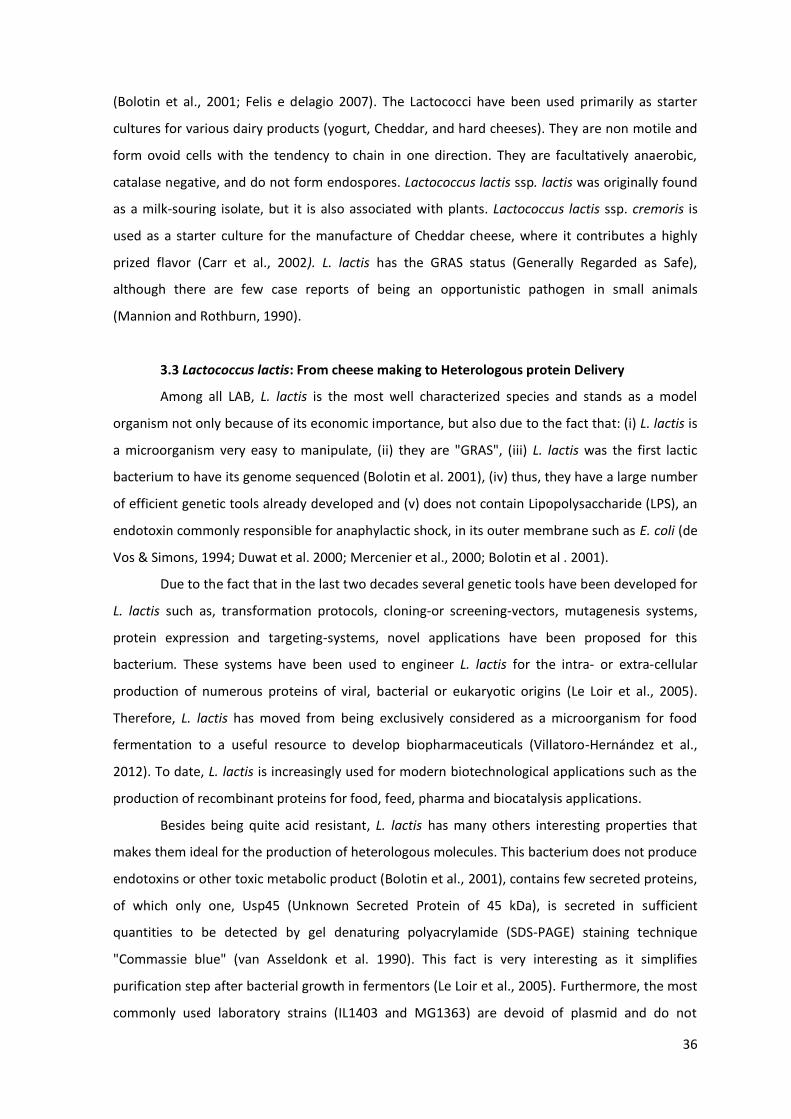
36
(Bolotin et al., 2001; Felis e delagio 2007). The Lactococci have been used primarily as starter
cultures for various dairy products (yogurt, Cheddar, and hard cheeses). They are non motile and
form ovoid cells with the tendency to chain in one direction. They are facultatively anaerobic,
catalase negative, and do not form endospores. Lactococcus lactis ssp. lactis was originally found
as a milk-‐souring isolate, but it is also associated with plants. Lactococcus lactis ssp. cremoris is
used as a starter culture for the manufacture of Cheddar cheese, where it contributes a highly
prized flavor (Carr et al., 2002). L. lactis has the GRAS status (Generally Regarded as Safe),
although there are few case reports of being an opportunistic pathogen in small animals
(Mannion and Rothburn, 1990).
3.3 Lactococcus lactis: From cheese making to Heterologous protein Delivery
Among all LAB, L. lactis is the most well characterized species and stands as a model
organism not only because of its economic importance, but also due to the fact that: (i) L. lactis is
a microorganism very easy to manipulate, (ii) they are "GRAS", (iii) L. lactis was the first lactic
bacterium to have its genome sequenced (Bolotin et al. 2001), (iv) thus, they have a large number
of efficient genetic tools already developed and (v) does not contain Lipopolysaccharide (LPS), an
endotoxin commonly responsible for anaphylactic shock, in its outer membrane such as E. coli (de
Vos & Simons, 1994; Duwat et al. 2000; Mercenier et al., 2000; Bolotin et al . 2001).
Due to the fact that in the last two decades several genetic tools have been developed for
L. lactis such as, transformation protocols, cloning-‐or screening-‐vectors, mutagenesis systems,
protein expression and targeting-‐systems, novel applications have been proposed for this
bacterium. These systems have been used to engineer L. lactis for the intra-‐ or extra-‐cellular
production of numerous proteins of viral, bacterial or eukaryotic origins (Le Loir et al., 2005).
Therefore, L. lactis has moved from being exclusively considered as a microorganism for food
fermentation to a useful resource to develop biopharmaceuticals (Villatoro-‐Hernández et al.,
2012). To date, L. lactis is increasingly used for modern biotechnological applications such as the
production of recombinant proteins for food, feed, pharma and biocatalysis applications.
Besides being quite acid resistant, L. lactis has many others interesting properties that
makes them ideal for the production of heterologous molecules. This bacterium does not produce
endotoxins or other toxic metabolic product (Bolotin et al., 2001), contains few secreted proteins,
of which only one, Usp45 (Unknown Secreted Protein of 45 kDa), is secreted in sufficient
quantities to be detected by gel denaturing polyacrylamide (SDS-‐PAGE) staining technique
"Commassie blue" (van Asseldonk et al. 1990). This fact is very interesting as it simplifies
purification step after bacterial growth in fermentors (Le Loir et al., 2005). Furthermore, the most
commonly used laboratory strains (IL1403 and MG1363) are devoid of plasmid and do not

37
produce extracellular proteases (Gasson et al. 1983; Chopin et al. 1984). Nowadays, there are
many recombinant L. lactis strains being used for high scale production of heterologous proteins
(Mierau et al., 2005). Recently this bacterium became famous as the first genetically modified
organism to be used alive for the treatment of chronic intestinal disease. Patients with Crohn's
disease received with genetically modified L. lactis (LL-‐Thy12) in which the thymidylate synthase
gene was replaced with a synthetic sequence encoding mature human interleukin-‐10. Treatment
with LL-‐Thy12 decreased disease activity and fecally recovered LL-‐Thy12 bacteria were dependent
on thymidine for growth and interleukin-‐10 production, indicating that the containment strategy
was effective (Braat et al., 2006).
3.3.1 Gene expression systems and heterologous protein production in
Lactococcus lactis
The expression of heterologous proteins in L. lactis was achieved by the development of
genetic knowledge, the improvement of molecular biology techniques and the studies focusing on
the regulatory elements of gene expression, such as promoters, inducers and repressors (Miyoshi
et al., 2004). Through this combination, high levels of a wide variety of heterologous proteins of
different origins were cloned in L. lactis by using several vectors containing constitutive or
inducible promoters (Langella and Le Loir, 1999; Pontes et al., 2011). Among all expression
systems available for use in L. lactis certainly Nisin Controlled Gene Expression (NICE) is the most
used (Figure 15). NICE system was developed at NIZO Food Research, Netherlands (NL), and has
played an important role in the development of recombinant L. lactis strains. It can be used for
over expression of homologous and heterologous genes for functional studies and to obtain large
quantities of specific gene products. The major advantages of this system over some others are
the expression of membrane proteins, secretion of proteins into the medium, tightly controlled
gene expression and simple scale up and downstream processing. NICE system is based on a
combination of the PnisA promoter with the regulatory genes NisRK (Wells et al. 1993). In the
presence of Nisin the promoter can induce transcription of the molecule of interest (Bermúdez-‐
Humarán et al. 2004). Nisin is a 34 amino anti-‐antimicrobial peptide that binds to lipids and then
forms small pores in the cytoplasmic membrane leading to subsequently to cell death. Because of
its broad host spectrum, it is widely used as a preservative in food. For the signal transduction
system the nisK and nisR genes were isolated from the nisin gene cluster and inserted into the
chromosome of L. lactis subsp. cremoris MG1363 (nisin-‐negative), creating the strain NZ9000
(nisin positive) (Kleerebezem et al., 1997; Mierau and Kleerebezem, 2005). When a gene of
interest is subsequently placed behind the inducible promoter PnisA on a plasmid or on the
chromosome, expression of the gene of interest can be induced by the addition of sub-‐inhibitory

38
amounts of nisin (0.1 5 ng/ml) to the culture medium. Depending on the presence or absence of
the corresponding targeting signals, the protein is expressed into the cytoplasm, into the
membrane or secreted into the medium. Many exogenous proteins have been expressed in L.
lactis through this system (Kuipers et al., 1993; de Ruyter et al. 1996).
In order to obtain a desired biological activity or depending on the nature of the target
protein expressed by L. lactis, it is necessary that, after their synthesis, they are modified, sorted
and dispatched towards their correct final destinations: (i) cytoplasm (ii) anchored to the cell wall
Figure 15: Schematic representation of the NICE system developed in L. lactis. A: Lack of expression of the gene cloned under control of the nisA promoter (P ), when no nisin is present in the environment. B: Response of the same cell (via NisK-‐NisR dependent signal transduction) after induction by the addition of nisin to the growth medium, resulting in the expression of the cloned gene (geneX). From Kleerebezem and co-‐workers 2000.

39
or (iii) secreted to the extracellular medium (Figure 16). The choice of the protein localization
depends on the application proposed for the strain. For example, the intracellular production
protects the recombinant protein from adverse environmental conditions (for example, low pH of
the stomach); however, cell lysis is required for protein delivery. The antigen/enzyme expressed
at the cell wall is also very interesting approach as it allows interaction with the environment
(digestive tract, for instance) but also limits their possible degradation by proteases. Finally, the
antigen released to the external medium is a nice strategy when it comes to the
commercialization of recombinant proteins without the contamination with LPS, as it facilitates
purification steps (Bermúdez-‐Humarán et al. 2004; Pontes et al., 2011).
Besides NICE, various expression systems that allow different cellular locations of the
gene of interest were developed for use in L. lactis, and other LAB strains as well (Norton et al.
1995; Dieye et al. 2001). Sugar-‐inducible expression systems have been developed and some of
them represent an alternative laboratorial tool for heterologous proteins production in L. lactis.
Among them, Xylose-‐Inducible Expression System (XIES) stands out as it can correctly address
Figure 16. Many protein expression and targeting systems were designed and have been used to engineer L. lactis to express a numerous proteins of viral or bacterial origins in the cytoplasm, anchored to their cell wall or secreted to the extracellular medium of. RBS: Ribosome Biding site (mRNA that is bound by the ribosome when initiating protein translation); SP: signal peptide (necessary to translocate the polypeptide chains across the plasma membrane allowing its secretion); CWA motif: Cell-‐wall-‐anchored motif. (This work).

40
recombinant proteins expressed by L. lactis to the intracellular medium, anchored to the cell wall
or secreted to the environment. Developed by Miyoshi and collaborators (Miyoshi et al., 2004),
this very interesting system is based on the use of a xylose-‐inducible lactococcal promoter, P(xylT)
from L. lactis NCDO2118 strain. In the presence of transported sugars (as glucose, fructose and/or
mannose), PxylT was shown to be tightly repressed; differently, in the presence of xylose, PxylT is
transcriptionally activated (Lokman et al., 1994; Jamet, 2001). The capacities of this system to
produce cytoplasmic and secreted proteins were tested using the Staphylococcus aureus nuclease
gene (nuc) fused or not to the lactococcal Usp45 signal peptide. Xylose-‐inducible nuc expression
was shown to be tightly controlled and resulted in high-‐level and long-‐term protein production
leading to a correct targeting either to the cytoplasm or to the extracellular medium. Furthermore,
this expression system demonstrated to be very versatile as it can be switched on or off easily by
adding either xylose or glucose, respectively (Miyoshi et al., 2004). Moreover, considering that
nisin is quite expensive, XIES is a very feasible system as it make use of two simple sugars, xylose
and glycose, facilitating its use in large-‐scale production. XIES system has been extensively
employed in the biotechnology field for production of different heterologous protein. In order to
study the effect of pure heat shock proteins (HSPs) on the immune system, recently De Azevedo
and co-‐workers (De Azevedo et al., 2012) constructed a recombinant L. lactis strains able to
produce and properly address the Mycobacterium leprae 65-‐kDa HSP (Hsp65) antigen to the
cytoplasm or to the extracellular medium, using the XIES system. Approximately 7 mg/L
recombinant Hsp65 was secreted and no protein degradation, related to lactococcal HtrA activity,
was observed. The Limulus amebocyte lysate assay demonstrated that the amount of LPS in the
recombinant Hsp65 preparations was 10-‐100 times lower than the permitted levels established by
the FDA, demonstrating the potential of these strains for a variety of biotechnological, medical
and therapeutic applications. Recently, the ORF encoding the S-‐layer protein (SlpA) of
Lactobacillus brevis was cloned into L. lactis under the transcriptional control of the XIES system.
SlpA was correctly secreted into the extracellular medium, as determined by immunoblotting.
Assays on the kinetics of SlpA production revealed the presence of glucose was sufficient to block
the transcription, even if xylose was present into the medium. The successful use of XIES to
express S-‐layer proteins in L. lactis opened new possibilities for an efficient production and
isolation of SlpA S-‐layer protein for its various applications in biotechnology and importantly as an
antigen-‐carrying vehicle (Hollmann et al., 2012).
There are other controlled expression systems besides NICE and XIES. An elegant system
has been developed on the basis of the middle promoter of the lactococcal bacteriophage 31,
which is induced by infection with 31. An expression plasmid containing the replication origin of
31 (ori31), which is induced upon bacteriophage infection, results in runaway plasmid

41
replication. The protein of interest is then produced also after bacteriophage infection, resulting
in the lysis of L. lactis. Because of the lysis that is ultimately realized, this system was termed
explosive expression. A drawback is the phage-‐dependent induction which may be difficult to
realize on an industrial scale. Moreover, there are the Thermoinduction and pH-‐dependent
systems, both very interesting but also presents some disadvantages (de Vos, 1999; Pontes et al.,
2011).
Still being GRAS organisms, the use of genetically modified L. lactis raises legitimate
concerns about their survival and propagation in the environment, and about the dissemination
of antibiotic selection markers or other genetic modifications to other microbes. Therefore, an
auxotrophic L. lactis strain was engineered to lack the thymidine synthase (thyA) gene. Thymine
starvation results in activation of the SOS repair system and DNA fragmentation, leading to
bacterial death (Lee, 2010). To accompany this purpose, a food-‐grade host/vector expression
system for L. lactis was recently constructed. It uses the alanine racemase gene (alr) as the
complementation marker. The alanine racemase auxotrophic mutant L. lactis was
obtained by double-‐crossover recombination using temperature-‐sensitive integration plasmid
pG(+)host9 and a food-‐grade vector pALR, containing entirely lactococcal DNA elements
(lactococcal replicon, nisin-‐inducible promoter PnisA and the alr gene from Lactobacillus casei
BL23) as a complementation marker. The green fluorescent protein and capsid protein of porcine
circovirus type II were successfully overexpressed under the nisin induction by using the new
food-‐grade host/vector system. This strain could be further used in as an excellent antigen
delivery vehicle, and could be also suitable for the use in the manufacture of ingredients for the
food industry as it is safe (Lu et al., 2012).
3.3.2 Lactococcus lactis as mucosal delivery vectors for therapeutic proteins
After the pioneering work of Wells and colleagues, increasing attention has been given to
L. lactis as a vehicle for the presentation of exogenous antigens at mucosal surfaces (for a review
see (Wells and Mercenier, 2008; Bermúdez-‐Humarán et al., 2011 and Pontes et al., 2011).
Iwaki and collaborators in 1990 were one of the first to attempt to use L. lactis as live
vaccines. Mice immunized orally with L. lactis strain expressing the Pac antigen of Streptococcus
mutans were able to produce both IgG and IgA Pac-‐specific antibodies. This result showed that L.
lactis could be used as a carrier for both presentation and delivery of antigens to the immune
system. Since then, several antigens derived from bacteria and viruses have been expressed in L.
lactis and immunization with these recombinant strains generated prophylactic and therapeutic
effects in several animal models (Wells & Mercenier, 2008; Wells, 2011).

42
Most studies concerning L. lactis as live vaccine were performed using the fragment C of
tetanus toxin (TTFC), a highly immunogenic antigen. Norton and colleagues (1995) found a
significant increase in the levels of IgA after oral immunization of mice with recombinant L. lactis
strains producing TTFC. Other studies have shown that animals vaccinated with L. lactis producing
the intracellular form of the antigen developed high levels of IgG and IgA antibodies specific for
TTFC. Subsequently, all animals became resistant to challenge with tetanus toxin (Wells et al.
1993; Robinson et al. 1997). Other several works confirmed the ability of L. lactis to present
antigens to the mucosa, generating specific immune response (Chatel et al. 2001). Encouraging
results were obtained after animal immunization with L. lactis expressing E7 antigen, derived from
human papillomavirus type-‐16 (HPV-‐16) virus, anchored to the cell wall. Vaccinated animals
showed humoral and cellular immune responses specific against the antigen (Bermúdez-‐Humarán
et al. 2004). Later, the administration of mice with live Lactococci expressing both E7 antigen and
IL-‐12 induced systemic and mucosal immune responses and protected the animals against HPV-‐16
induced tumors (Bermúdez-‐Humarán et al., 2005). Cortes-‐Perez and collaborators also developed
a safe mucosal vector for HPV-‐16 prophylactic vaccination based on L. lactis strains expressing
human papillomavirus type-‐16 L1 protein (Cortes-‐Perez et al., 2009). Table 2 summarizes some
studies that were performed in different laboratories around the world corroborating the ability
of L. lactis in inducing long-‐lasting immune responses.
ANTIGENS EXPRESSED
BY L. LACTIS
IMMUNE RESPONSE
OBSERVED
APPLICATION REFERENCE
EDIII antigen from
dengue virus type 2
Neutralization of the
virus in vitro
Dengue virus (DENV)
control strategy
Yes et al., 2008
Envelope protein of the
human
immunodeficiency
virus 1 (HIV-‐1)
High levels of IgG and
IgA antibodies against
the antigen observed
HIV vaccine
Xin et al., 2003
Antigenic protein of
Proteus mirabilis HBPM
Protection to the
animals against
challenge with P.
mirabilis virulent
strain
Control of urinary
tract infections
Scavone et al. 2007
PspA antigen derived
from Streptococcus
Better protected
against challenge with
Vaccine against
pneumonia
Hanniffy et al., 2007

43
Pneumoniae the virulent strain
Antigen lcrV from
Yersinia
pseudotuberculosis
Humoral and cellular
immune responses
were observed
conferring protection
against challenge
Vaccine against Far
East scarlet-‐like fever
Daniel et al., 2009
EspB antigen from the
type III secretion
system (T3SS) of E. coli
serotype O157:H7
Significant levels of
specific serum Ig and
faecal IgA were
detected
New strategie to fight
against
enterohemorrhagic E.
coli
Ahmed et al., 2012
Leishmania antigen
LACK and the
proinflammatory
cytokine IL-‐12
Protection against
challenge
Vaccine to combat
Leishmaniasis
Hugentobler et al.,
2012
Mature murine IFN-‐
gamma
-‐-‐ Adjuvant tool Bermúdez-‐Humarán
et al., 2008
Antigen envolved with
LPS transport (wzm)
from Vibrio cholerae
O1 strain
Augmentation of both
systemic and mucosal
immunity
Vaccine against
cholera
Zamri et al., 2012
Catalase-‐producing L.
lactis
Inhibition of
chemically induced
colon cancer
Cancer therapy De Moreno de
LeBlanc et al., 2008
IL-‐10
Anti-‐inflammatory
effects controlling
intestinal
inflammation
Treatment of allergy
and inflammatory
bowel diseases (IBD)
Cortes-‐Perez et al.,
2007; Braat et al.
2006 ; Marinho et al.,
2010
There are many others protection studies performed with recombinant L. lactis strains
and results are very encouraging as protection or partial protection after challenge was observed
(Pontes et al., 2011; Bermudez-‐Humaran et al., 2011; Tarahomjoo, 2012). A summary of the
current published applications of L. lactis delivery is illustrated in Figure 17. Nevertheless, even
though interesting, the strategy to express recombinant antigenic proteins in L. lactis presents
one problem associated with post-‐translational modifications performed on recombinant proteins
44
by L. lactis that might affect their bioactivity (Marreddy et al., 2011). Therefore, a novel
technology has emerged to circumvent this problem: delivery of cDNA encoding antigenic
proteins by L. lactis at the mucosal level. This very interesting approach will be discussed in the
next pages (Guimarães et al., 2006; Chatel et al., 2008; Pontes et al., 2011).
3.4 Lactococcus lactis: From protein to DNA delivery
3.4.1 Native L. lactis as DNA delivery vectors
Recently, the use of LAB as DNA vaccine delivery vehicles has been evaluated as an
alternative strategy for vaccination (Guimaraes et al., 2005; Chatel et al., 2008; Tao et al., 2011).
Guimaraes and co-‐workers (2006) were one of the first to explore the potential use of noninvasive
L. lactis strains as a DNA delivery vehicle. It was constructed two E. coli-‐L. lactis shuttle plasmids,
-‐
in vitro assays, pLIG:BLG1 plasmid
showed better transfection capacity and was then used to transform L. lactis MG1363, which
proved to be incapable of expressing BLG. After 3h of co-‐incubation with Caco-‐2 human colon
carcinoma cells, BLG expression was only found in cells incubated with MG1363 (LL-‐pLIG:BLG1)
strain and secretion started 48h after the beginning of the experiment. Native L. lactis was then
considered to be capable of transferring BLG cDNA into mammalian epithelial cells, and this work
demonstrated their potential to deliver in vivo a DNA vaccine (Guimaraes et al., 2006). In order to
evaluate the capacity of this bacterium to transfer DNA in vivo to mice IECs, Chatel and co-‐
workers orally immunized mice with L. lactis carrying the same eukaryotic expression plasmid. LL-‐
Figure 17. Current applications of L. lactis delivery. Illustration of the various molecules that have been produced in L. lactis and the animal models that were used. Adapted from Wells and Mercenier, 2008.

45
pLIG:BLG1 strain were able to translocate the epithelial cells of the intestinal membrane as BLG
cDNA was detected in the epithelial membrane of the small intestine of 40% of the mice
immunized and BLG was produced in 53% of the animals. Moreover, it was also observed a low
and transitory Th1-‐type immune response after immunization trials which conferred protection to
mice after sensitization with the allergen (Chatel et al., 2008). This study successfully
demonstrated that L. lactis can be used as vector for genetic immunization.
3.4.2 Recombinant invasive L. lactis as plasmid DNA delivery vehicles
In order to increase the capacity of lactococci to deliver cDNA, some strains of L.
lactis expressing invasins have been reported (Guimarães et al., 2005; Sleator and Hill, 2006). One
very interesting strategy peformed by Guimarães and collaborators was to engineer L. lactis to
express InlA (LL-‐InlA+) from L. monocytogenes. InlA gene was cloned and expressed under
transcriptional control of the native promoter. Western blot and immunofluorescence assays
revealed that recombinant lactococci efficiently displayed the cell wall anchored form of InlA.
Moreover, invasion rates of LL-‐InlA+ strain in Caco-‐2 cells was approximately 100-‐fold higher than
the wild type (wt) lactococci. The recombinant strain also proved to be able to enter intestinal
cells in vivo, after oral inoculation of guinea pigs. After internalization, LL-‐InlA+ was able to deliver
a functional eukaryotic gfp gene (green fluorescent protein) into epithelial Caco-‐2 cells
(Guimarães et al., 2005).
The results mentioned above were obtained through the use of a large plasmid called
PLIG (10 kb), resulted from the co-‐integration of two replicons: one from E. coli and the other
from L. lactis. Therefore, in order to circumvent this difficulty, a new plasmid (3742 bp), termed
pValac (Vaccination using Lactic acid bacteria) was constructed. The pValac is formed by fusion of
(i) cytomegalovirus promoter (CMV), that allows the expression of the antigen of interest in
eukaryotic cells, (ii) polyadenylation sequences from the bovine Growth Hormone (BGH), essential
to stabilize the RNA messenger transcript, (iii) origins of replication that allow its propagation in
both E. coli and L. lactis hosts, and (iv) a chloramphenicol resistance gene for selection of strains
harboring the plasmid. In order to evaluate pValac functionality, the gfp ORF was cloned into
pValac (pValac:gfp) and fluorescence was analyzed after transfection in PK15 cells. The
applicability of pValac was demonstrated by invasiveness assays of L. lactis inlA+ strain harbouring
pValac:gfp into Caco-‐2 cells. After transfection assays with pValac:gfp, it was observed that PK15
cells were able to express GFP. Moreover, L. lactis inlA+ strain were able to invade Caco-‐2 cells
and deliver pValac:gfp into epithelial cells (Guimaraes et al. 2009). Due to its small size, pValac
permit the cloning of large gene fragments representing, thus, a promising tool for genetic
immunization.

46
Nevertheless, even though interesting the use of LL-‐InlA+ strain presented some
bottlenecks: InlA cannot bind to its receptor in mice, murine E-‐cadherin, thus impeding in vivo
experiments in these animals. Therefore, it is only possible to study the effect of LL-‐InlA+ strain in
guinea pigs or transgenic mice, which may be laborious and/or expensive (Wollert et al., 2007).
For this reason, a new recombinant L. lactis strain expressing Fibronectin Biding Protein A (FnBPA)
(LL-‐FnBPA+) of Staphylococcus aureus (Que et al., 2001), was constructed and therefore evaluated
with the goal of improving the delivery of DNA to mammalian cells (Innocentin et al., 2009).
FnBPA naturally mediates adhesion of S. aureus to the host tissue and its entry into non-‐
phagocytic cells (Sinha et al., 1999). Thus, FnBPA utilization could increase the interactions of L.
lactis with IECs leading to a more efficient delivery of DNA vaccines. L. lactis FnBPA+, and L. lactis
InlA+ showed comparable internalization rates in Caco-‐2 cells and conventional or confocal
fluorescence microscopy demonstrated big clusters of L. lactis FnBPA+ and L. lactis InlA+ which
were uptaken by Caco-‐2 cells. Invasive lactococci were then used to deliver a eukaryotic GFP
expression plasmid (pValac:gfp) in Caco-‐2. After 3 hours co-‐incubation, invasive L. lactis were
capable of transferring gfp to Caco-‐2 cells more efficiently than the non-‐invasive strains
(Innocentin et al., 2009). Recently, another work was performed with LL-‐FnBPA+ strain in which
they used the BLG allergen and GFP to characterize the potential of this strain as an in vivo DNA
vaccine delivery vehicle. LL-‐FnBPA+ carrying the plasmid pValac:BLG (LL-‐FnBPA+ BLG) showed to
be more invasive than LL-‐BLG noninvasive strain, after co-‐incubation with Caco-‐2 cells. Then in
vitro experiments demonstrated that Caco-‐2 cells co-‐incubated with LL-‐FnBPA+ BLG could
produce up to 30 times more BLG than cells co-‐incubated with the noninvasive LL-‐BLG.
Furthermore, in vivo it was demonstrated that, in order to effectively deliver DNA, LL-‐FnBPA+
requires a pre-‐coating with Fetal Calf Serum before oral administration. A second interesting
observation concerns the fact that enterocytes were able to express cDNA of GFP or BLG without
regard to the strains used (invasive or not). Finally, the use of LL-‐FnBPA+ could increase the
number of mice producing BLG, but not the level of BLG produced (Pontes et al., 2012). Since
FnBPA requires an adequate local concentration of fibronectin to bind to integrins (Ozeri et al.,
1998; Dziewanowska et al., 2000), the interaction between FnBPA and epithelial cells may be a
complex process in vivo.
Tao and colleagues developed a different strategy attempt to improve DNA delivery by L.
lactis and S. gordonii in vitro without using virulent genes. They applied different chemical
treatments in order to weaken the bacterial cell wall, facilitating transfection of plasmids into cells
Caco-‐2. L. lactis NZ3900 when treated with glycine showed a higher frequency of eukaryotic
expression encoding red fluorescent protein (RFP) transfer to Caco-‐2 cells. Treatment with
penicillin and lizozime was showed to be more effective in S. gordonii (Tao et al., 2011). Even

47
though the mechanisms by which treated bacteria could be efficiently internalized into Caco-‐2
cells have not yet been elucidated, this strategy is interesting because virulence genes are not
involved (Pontes et al., 2011). However, the potential of these strains in delivering DNA vaccines
were not tested in vivo.
All of the L. lactis strains that have been developed to be used as vaccine DNA delivery
vectors are promising for mucosal immunization. Nevertheless, they all present some problems,
as mentioned above. Therefore, the use of other invasin gene that can easily bind to murine
epithelial cells from conventional mice as well as the use of other LAB specie that can stay longer
than L. lactis in the gut, are being considered viable strategies that will allow the use of LAB as
efficient DNA-‐vaccine delivery vehicles in near future (Bermúdez-‐Humarán et al., 2011; Pontes et
al., 2011).

49
CHAPTER 2
AIM OF THE STUDY
49
MAIN AIM OF THE STUDY
Understand the mechanisms by which the bacterium Lactococcus lactis is able to
deliver DNA vaccines to intestinal epithelial cells (IECs) both in vitro and in vivo, and to antigen
presenting cells, such as dendritic cells, by using a genetically modified strain of L. lactis
expressing Listeria monocytogenes mutated internalin A invasin. We also intended to
investigate the immunomodulatory properties of invasive or noninvasive L. lactis strains after
immunization trials.
SPECIFIC AIMS
-‐ Construct a novel invasive L. lactis strain expressing L. monocytogenes mutated
Internalin A (mInlA),
-‐ Detect the expression of the recombinant mInlA at L. lactis surface using flow
cytometry analysis,
-‐ Evaluate the capacity of L. lactis-‐mInlA to invade more efficiently than the wild type
strain the human intestinal epithelial cell line, Caco-‐2, non-‐differentiated or
differentiated in tissue culture inserts,
-‐ Investigate the ability of L. lactis-‐mInlA to deliver a cDNA of the major allergen in cows'
milk -‐lactoglobulin (BLG), in vitro to Caco-‐2 cells or in vivo to conventional mice
intestinal epithelial cells,
-‐ Evaluation of the immune response elicited by noninvasive or invasive L. lactis after
immunization trials,,
-‐ Recover bone marrow dendritic cells (BMDCs) to investigate if non-‐invasive or invasive
L. lactis strains are capable to deliver BLG cDNA directly to dendritic cells (DCs) after
co-‐incubation assays,
-‐ Evaluate if non-‐invasive or invasive L. lactis strains are capable to deliver BLG cDNA to
DCs trough a monolayer of differentiated Caco-‐2 cells after co-‐incubation assays.

CHAPTER 3 In vitro and in vivo characterization of DNA delivery using recombinant Lactococcus lactis expressing a mutated form of L. monocytogenes Internalin A

50
CHAPTER 3: In vitro and in vivo characterization of DNA delivery using recombinant Lactococcus
lactis expressing a mutated form of L. monocytogenes Internalin A
2.1 Introduction
DNA vaccination has gained a lot of attention due to its ability to induce long-‐lasting
humoral and cellular immune responses against an encoded antigen (Donnelly et al., 2000; Liu,
2010). This vaccine platform demonstrated to be highly efficient in rodents and mice, but not in
larger animals and humans as the DNA showed to be easily degraded in those hosts (Ledwith et al.,
2000). Therefore, the optimization of DNA vaccine vectors appeared to be very important to confer
protection to the genetic material and thus enhance gene transfer efficiency (Manam et al., 2000).
Several strategies have been explored to protect plasmids from degradation such as encapsulation
into synthetic particles (cationic liposomes or polymers) or the use of viral vectors (Manam et al.,
2000). Despite their potential some limitations and safety issues still remain; for example liposomes
are very complex to produce, can present some toxicity to mammalian cells and have limited
packaging capacity (Liu, 2011). Viral vectors, on the other hand, can have the capacity to randomly
integrate their genetic material into the host genome leading to tumour formation (Kang et al., 2003;
Thomas et al., 2003).
The use of bacteria as delivery vehicles for DNA vaccination has emerged as an interesting
alternative to overcome many of the problems associated with viral or liposomal delivery. Bacterial
carriers may be administered at mucosal surfaces eliciting secretory IgA responses, can increase and
expand the magnitude of immune responses against the vector-‐encoded antigen due to the natural
presence of Microbe-‐Associated Molecular Patterns (MAMPs) activating immune cells (Courvalin et
al., 1995; Becker et al., 2008). Attenuated pathogens such as Salmonella, Shigella, Listeria and
Yersinia have been used as experimental live delivery systems. However, a potential concern is the
risk of increased virulence in young or immunocompromised individuals (Loessner et al., 2008; Wells
and Mercenier, 2008).
The use of food-‐grade lactic acid bacteria (LAB) as DNA delivery vehicle represents an
alternative and attractive strategy (Wells and Mercenier, 2008). Actually, it has been extensively
demonstrated that the model LAB, Lactococcus lactis, is able to deliver a range of vaccine and
therapeutic molecules for applications in allergic, infectious or gastrointestinal diseases (Wells,
2011). A relatively new development, however, is their use as a vehicle for genetic immunization
(Pontes et al., 2011). Previous data have demonstrated that native L. lactis is capable to deliver DNA
vaccines both in vitro and in vivo to intestinal epithelial cells (IECs). However, its DNA transfer
efficiency seemed to be relative low (Chatel et al., 2008; Pontes et al., 2011). With the intent to
increase the persistence of L. lactis in the gastrointestinal tract which could, in theory, improve
51
plasmid transfer to IECs, our research group decided to construct recombinant invasive L. lactis
strains (Guimaraes et al., 2005; Innocentin et al., 2009; Pontes et al., 2012). Two strains were
designed, one expressing Internalin A (InlA) from L. monocytogenes (LL-‐InlA) and another producing
S. aureus Fibronectn Biding Protein A (FnBPA) (LL-‐FnBPA) (Guimaraes et al., 2005; Innocentin et al.,
2009). Both of them were capable to successfully express the recombinant proteins and deliver DNA
plasmids to IECs either in vitro or in vivo. Nevertheless, these two strains presented some
bottlenecks: (i) L. lactis expressing InlA cannot interact to its receptor in mice, murine E-‐cadherin,
being able to be administered only in transgenic mice and guinea pigs, and (ii) FnBPA requires an
adequate local concentration of fibronectin to bind to its receptors, integrins, which may hamper the
interpretation of in vivo studies (Ozeri et al., 1998; Dziewanowska et al., 2000). Hence, in this work
we have decided to construct a third recombinant invasive L. lactis strain expressing mInlA as it is
capable to bind to E-‐cadherin from conventional mice thus facilitating in vivo studies. Furthermore,
besides describing the construction and characterization of this novel L. lactis strain, we test it as a
-‐lactoglobulin (BLG) allergen to measure DNA transfer to IECs
both in vitro and in vivo.
2.2 Materials and Methods
In order to verify if the new strain was capable to deliver more efficiently a eukaryotic expression
vector than the noninvasive strain, wild type (wt) L. lactis (L. lactis NZ9000) was transformed by
electroporation with a plasmid harboring mInlA gene. FACS analysis was used to confirm the
expression of mInlA at L. Lactis surface (LL-‐mInlA+). The gentamicin survival assay into non-‐confluent
intestinal epithelial cells of the cell line Caco-‐2 cells was used to evaluate the invasivisity capacity of
LL-‐mInlA strain which was confirmed by confocal microscopies studies. Afterwards, LL-‐mInlA and LL-‐
wt were transformed by electroporation with a eukaryotic expression vector coding for the cDNA of
ß-‐ . Co-‐incubation assays of LL-‐mInlA+ strain
harboring pValac:BLG with non-‐confluent Caco-‐2 cells were performed in order to measure DNA
transfer. Culture supernatant and Caco-‐2 extracts were collected to be assayed for the presence of
BLG by ELISA. We then moved to in vivo experiments in which BALB/c mice, 3 to 6 weeks of age,
received 1x10⁹ (CFU) of invasive or non-‐invasive lactococci during three consecutive days. In the
fourth day, mice intestinal epithelial cells were purified and their extracts were used for ELISA assays
in order to quantify BLG production. Statistical significance between the groups was calculated using
the One Way ANOVA (and nonparametric) test, -‐test.
2.3 Results and Discussion

52
In this work we showed the ability of either noninvasive or recombinant invasive L. lactis
expressing Listeria monocytogenes mInlA as both in vitro and in vivo DNA delivery vehicle. Flow
cytometry analysis demonstrated with success that mInA was expressed and properly directed to the
surface of L. lactis. Previous studies have also shown that other invasins also derived Gram-‐positive
bacteria, such as Staphylococcus aureus FnBPA and InlA, were successfully expressed in L. lactis
confirming that the signal peptide for secretion and the anchoring signal are well recognized by the L.
lactis machinery (Guimaraes et al., 2006; Pontes et al., 2012). Afterwards, we moved to in vitro
assays in which we tested if the new invasive L. lactis (LL-‐mInlA+) were actually capable to internalize
intestinal epithelial cells (Caco-‐2 cells) using the gentamicin survival assay. We demonstrated that LL-‐
mInlA+ strain were almost 1000 times more invasive than the wild type strain. Moreover, a confocal
image taken after gentamicin assay showed clearly that LL-‐mInlA+is capable of adhering to and
entering in non-‐differentiated Caco-‐2 cells. Recombinant bacteria were found to be distributed at the
periphery of the Caco-‐2 cell were the mInlA receptor, E-‐cadherin, was accessible. Then, LL-‐mInlA+
and LL strains were then transformed with pValac: BLG plasmid, co-‐incubated with Caco-‐2 cells and
BLG expression was followed 72 h later by ELISA in order to measure the capacity of the strains in
transfer DNA plasmids to mammalian cells. Both LL-‐mInlA-‐BLG and LL-‐BLG were capable to deliver
pValac:BLG to Caco-‐2 cells, confirming prior observations (Guimarães et al., 2009), and the invasive
status improved this plasmid transfer. Cells were also capable of secreting the allergen, which may
facilitate antigen uptake and presentation by professional APCs through cross-‐priming pathways
(Donnelly et al., 2000). Subsequently, BALB/c mice received orally noninvasive and invasive L. lactis
carrying or not pValac: BLG for 3 consecutive days and on the day 4 enterocytes from the small
intestine were isolated and BLG production was measured by enzyme immunoassay (EIA). We
confirmed that noninvasive lactococci are able to transfer a functional plasmid in vivo in mice and LL-‐
mInlA+BLG strain slightly enhanced the number of mice expressing BLG. As no significant advantages
were observed by using LL-‐mInlA-‐BLG compared to LL-‐BLG, we hypothesize that interactions of
recombinant mInlA with their receptors were impeded somehow in mouse intestinal epithelium
hampering optimal plasmid transfer. Lack of invasion in vivo was also observed by another group
working with E. coli strain expressing invasin from Yersinia pseudotuberculosis (Critchley-‐Thorne et
al., 2006). It is thus possible that LL-‐mInlA+BLG strain, wich is not able to reach its receptor deeply
buried in the crypt, are sampled by are able to take
up particles/bacteria from the lumen (Azizi et al., 2010). Another possibility is the interaction of the
strains with other cells from the epithelial membrane such as dendritic cells, which are able to reach
the lumen to sample its content (Rescigno et al., 2001). Most likely, plasmid transfer in vivo using
either noninvasive or invasive lactococci is a combination of bacteria and released plasmid captures.

53
Taken together, we think that the use of another LAB, such as lactobacilli which persist longer in the
gut than lactococci could be a better option for DNA delivery.
2.4 Conclusions
The use of L. lactis as a DNA delivery vehicle has been demonstrated to be a very interesting
approach. Native lactococci have proved to be capable to transfer DNA plasmids to eukaryotic cells
to Caco-‐2 cells and to mice IECs. However, the manner by which this DNA transfer occurs remains to
be discovered. Therefore, we decided to express the mutated Internalin A (mInlA) invasin from L.
monocytogenes in L. lactis to elucidate a part of the mechanism of DNA transfer and to increase the
transfection ratio as in theory an invasive strain could stay longer in the host gastrointestinal tract.
Flow cytometry analysis demonstrated that mInlA was successfully expressed at the surface of L.
lactis NZ9000 strain. The final strain, named LL-‐mInlA+, revealed to be almost 1000 times more
invasive when compared to wild type (wt) L. lactis strain after gentamicin survival assays into non-‐
confluent Caco-‐2 cells. This invasiveness capacity was confirmed by confocal microscopy experiments
wherein LL-‐mInlA+ strain were found to be very attached to the periphery of Caco-‐2 cells where the
receptor for mInlA, E-‐cadherin, was exposed. Moreover, recombinant bacteria were also located to
be inside the cells meaning that they were actually capable to invade the cells. Nor adherence
neither invasion was observed for the wt strain. Afterwards, LL-‐mInlA+ was transformed with a
second plasmid, pValac:BLG, harboring the cDNA of the allergen BLG. Strains were incubated again
with Caco-‐2 cells and, after three hours, cell extract and culture supernatant were recovered to be
assayed for the presence of BLG by ELISA. More BLG was detected for cells which were incubated
with the invasive strain LLmInlA+. Therefore, we could conclude that the invasive status improved
plasmid transfer in vitro. We then moved to in vivo analysis to check weather intestinal epithelial
cells are the preferable cell type that can mediate plasmid transfer by using noninvasive and invasive
lactococci. After immunization trials, it was observed that the number of mice expressing BLG was
higher (n = 11) in the group immunized with invasive bacteria than with noninvasive bacteria (n = 8).
We could clearly see a higher tendency of mice expressing the allergen in the group immunized with
LLmInlA+. However, this difference observed was not statistically significant. Perhaps, IECs are not
the preferable cell type that can mediate DNA transfer in vivo as we did not see a drastic increase in
BLG expression by using recombinant invasive lactococci. Taken together, this data reinforce the use
of L. lactis, especially invasive LLmInlA+ as an alternative tool to deliver therapeutic plasmids either in
vitro or in vivo. We also intent to test if another lactic acid bacteria strain, such as the probiotic
Lactobacillus casei BL23, that can stay longer in the gastrointestinal tract, could represent an
advantage over the use of recombinant invasive L. lactis.

RESEARCH ARTICLE Open Access
In vitro and in vivo characterization of DNAdelivery using recombinant Lactococcus lactisexpressing a mutated form of L. monocytogenesInternalin AMarcela de Azevedo1,2,4, Jurgen Karczewski3, François Lefévre5, Vasco Azevedo4, Anderson Miyoshi4, Jerry M Wells3,Philippe Langella1,2 and Jean-Marc Chatel1,2*
Abstract
Background: The use of food-grade Lactic Acid Bacteria (LAB) as DNA delivery vehicles represents an attractivestrategy to deliver DNA vaccines at the mucosal surfaces as they are generally regarded as safe (GRAS). Wepreviously showed that either native Lactococcus lactis (LL) or recombinant invasive LL expressing FibronectinBinding Protein A of Staphylococcus aureus (LL-FnBPA+) or Internalin A of Listeria monocytogenes (LL-InlA+), wereable to deliver and trigger DNA expression by epithelial cells, either in vitro or in vivo. InlA does not bind to itsreceptor, the murine E-cadherin, thus limiting the use of LL-InlA+ in in vivo murine models. Moreover, FnBPA bindsto its receptors, integrins, via fibronectin introducing another limiting factor. In order to avoid the limitations ofLL-InlA+ and LL-FnBPA+, a new L. lactis strain was engineered to produce a previously described mutated form ofInlA (LL-mInlA+) allowing the binding of mInlA on murine E-cadherin.
Results: After showing the expression of mInLA at the surface of LL-mInlA+ strain, in vitro gentamycin survivalassay in Caco-2 cells showed that LL-mInlA+ is 1000 times more invasive than LL. LL-mInlA+ invasivity was alsovalidated by fluorescence microscopy. LL and LL-mInlA+ were transformed with pValacBLG, a plasmid containingthe cDNA of bovine !-Lactoglobulin (BLG), resulting in strains LL-BLG and LL-mInlA+BLG. The plasmid transferin vitro using LL-mInlA+BLG was increased 10 times compared to LL-BLG. Moreover, the number of mice producingBLG in isolated enterocytes after oral administration of LL-mInlA+BLG in vivo was slightly higher than after oraladministration of LL-BLG.
Conclusions: We confirmed in this study that the production of mInlA at the surface of L. lactis is a promisingstrategy for plasmid transfer in vitro and in vivo.
Keywords: Lactococcus lactis, Listeria monocytogenes, Mutated internalin A, Internalization, DNA delivery
BackgroundDNA vaccination has gained a lot of attention since itsability to induce long-lasting humoral and cellular im-mune responses against an encoded antigen was discov-ered [1]. In addition, DNA vaccination poses no dangerof integration into host cellular DNA thereby raising itssafety profile [2-4]. DNA vaccines can be easily isolated
to high purity, encode multiple antigens, and possess in-herent adjuvant activity due to the presence of unmethy-lated CpG motifs that are recognized in mammals byTLR9 [5]. So called purified “Naked” DNA vaccinationwas shown to be highly efficient in rodents and mice,but not in larger animals and humans [6]. Consequently,it is very important to optimize DNA vaccine vectorsand develop a delivery system to facilitate cellularuptake and enhance gene transfer efficiency and expres-sion in situ [7].
* Correspondence: [email protected], UMR1319 Micalis, Commensals and Probiotics-Host InteractionsLaboratory, Jouy-en-Josas, France2AgroParisTech, UMR Micalis, F-78350 Jouy-en-Josas, FranceFull list of author information is available at the end of the article
© 2012 de Azevedo et al.; licensee BioMed Central Ltd. This is an Open Access article distributed under the terms of theCreative Commons Attribution License (http://creativecommons.org/licenses/by/2.0), which permits unrestricted use,distribution, and reproduction in any medium, provided the original work is properly cited.
de Azevedo et al. BMC Microbiology 2012, 12:299http://www.biomedcentral.com/1471-2180/12/299

Several strategies have been explored to protect plas-mids from degradation, facilitating DNA uptake byphagocytic Antigen Presenting Cells (APCs) and therebyenhancing their immunological properties. This includesdelivery technologies based on encapsulation into syn-thetic particles (cationic liposomes or polymers) or theuse of viral vectors [7,8]. Despite their potential, somelimitations and safety issues still remain which can re-strict the application of gene therapy - e.g. the complex-ity of producing liposomes and their limited packagingcapacity [9]. Additionally, it was shown that some viralvectors have the capacity to randomly integrate theirgenetic material into the host genome causing inser-tional mutagenesis of a cellular oncogene, leading totumour formation [10].The use of bacteria as delivery vehicles for DNA vac-
cination has emerged as an interesting alternative toovercome many of the problems associated with viral orliposomal delivery [11]. W. Schaffner was the first to ob-serve genetic material transfer from bacteria to mamma-lian cells [12]. Since then, bacteria have been extensivelyexploited as vaccine delivery vehicles for vaccinationagainst bacterial and viral pathogens as well as cancerimmunotherapy [13-15]. The use of bacteria for mucosaldelivery of DNA vaccines may be advantageous due totheir potential to elicit secretory IgA responses as wellas systemic immunity, when compared to conventionalparenteral immunization [16]. Furthermore, bacterialcarriers can increase and expand the magnitude of im-mune responses against the vector-encoded antigen dueto the natural presence of Pathogen-Associated Molecu-lar Patterns (PAMPs) that bind to Toll-like receptors(TLRs) and activate immune cells [5].Presently, attenuated pathogens such as Salmonella,
Shigella, Listeria, Yersinia, as well as, non-pathogenicEscherichia coli have been used as experimental live de-livery systems [17,18]. An advantage of using attenuatedpathogens as DNA vaccine vehicles is that they possessmechanisms to adhere or invade host cells with a negli-gible risk of reversion to a virulent strain via gene trans-fer or mutation. However, a potential concern is the riskof increased virulence in young or immunocompromisedindividuals.The use of food-grade lactic acid bacteria (LAB) as
DNA delivery vehicle represents an alternative and at-tractive strategy to deliver DNA vaccines at the mucosalsurfaces (ref review by 19 and 20). The dietary group ofLAB, including Lactococcus lactis and many species ofLactobacillus, is generally regarded as safe (GRAS)organisms of which some are intestinal commensals ofhumans. Indeed, it has been extensively demonstratedthat these bacteria are able to deliver a range of vac-cine and therapeutic molecules for applications in aller-gic, infectious or gastrointestinal diseases [19,21,22]. A
relatively new development, however, is their use as avehicle for genetic immunization [23]. Previous ex-periments performed by our group showed that eithernative L. lactis (LL) or recombinant invasive LL ex-pressing Fibronectin Binding Protein A (LL-FnBPA+) ofStaphylococcus aureus or Internalin A (InlA) of Listeriamonocytogenes (LL-InlA+) [24,25], were able to deliverDNA in epithelial cells both in vitro and in vivo, dem-onstrating potential as gene transfer vehicles [24-27].However InlA does not bind to its murine receptor, E-cadherin, thus limiting the use of LL-InlA+ in in vivomurine model. On the other hand, FnBPA requires anadequate local concentration of fibronectin to bind to itsreceptors, integrins [28,29].In order to avoid the limitations of InlA and FnBPA
and improve our knowledge on the key steps by whichthe DNA is transferred to mammalian cells using L. lac-tis, LL was engineered to express a mutated form ofInternalin A (mInlA; Ser192Asn and Tyr369Ser) thatincreased binding affinity to murine and human E-cadherin [30,31] thus allowing for in vivo experiments inconventional mice. Herein, we describe the constructionand characterization of this novel L. lactis strain as aDNA delivery vector, using cow’s milk !-lactoglobulin(BLG) allergen, to measure DNA transfer to intestinalepithelial cells (IECs) in vitro and in vivo.Overall, the production of mInLA+at the surface of
Lactococcus lactis increased the invasisity of bacteriumand amount of plasmid transfer by 1000 and 10 fold, re-spectively. In vivo, BLG production was detected in iso-lated enterocytes after oral administration of LL-mInlA+BLG and was slightly higher than oral administrationof LL-BLG.
ResultsMutated internalin A is produced on the surface ofrecombinant L. lactis strainTo investigate surface expression and production ofmInlA, L. lactis NZ9000 and LL-mInlA+ strains wereincubated with specific anti-mInlA monoclonal antibodyand then with FITC-conjugated anti-Mouse IgG. Stainedcells were analyzed by flow cytometry. As shown inFigure 1, LL-mInlA+ strain (blue peak) showed a signifi-cant shift in the fluorescence intensity comparing to theNZ9000 strain (black peak). No shift was observed whenstrains were incubated with FITC-labeled anti-MouseIgG alone (data not shown). This experiment confirmedexpression of mInlA on the surface of L. lactis.
L. lactis producing mInlA is efficiently internalized byCaco-2 cellsNon-confluent Caco-2 cells were incubated for 1 h witheither NZ9000 or with LL-mInlA+. Non internalizedbacteria were killed by gentamicin and intracellular
de Azevedo et al. BMC Microbiology 2012, 12:299 Page 2 of 9http://www.biomedcentral.com/1471-2180/12/299

bacteria enumerated after lysis of the eukaryotic cells.The LL-mInlA+ strain exhibited 1000-fold greater inva-sion rate than NZ9000 strain (Figure 2).
LL-mInlA+ internalization analyzed by confocalmicroscopyLL-mInlA+ and NZ9000 strains were labeled with CFSEdye and then incubated with Caco-2 cells for 1 h. Cellswere fixed and confocal images were obtained. Very few
cell-associated bacteria could be detected after co-incubation with NZ9000 (Figure 3A). In contrast, theLL-mInlA+ strain strongly bound to the membrane ofcell clusters which is compatible with the known bindingof InlA to E-cadherin, a cell-cell adhesion molecule. Inaddition, LL-mInlA+ was located intracellularly in somecells (Figure 3C and B).
LL-mInlA+ can efficiently deliver in vitro a DNA vaccinecontaining !-lactoglobulin cDNATo test the ability of LL-mInlA+ to deliver a DNA vac-cine plasmid in vitro to IECs, we transformed LL-mInlA+ strain with pValac:BLG [32], a plasmid derived frompValac [23] containing the cDNA for BLG, under thecontrol of an eukaryotic promoter to generate strain LL-mInlA+BLG (Table 1).In order to monitor plasmid transfer and production
of BLG in Caco-2 cells extracts, non-confluent Caco-2cells were incubated with noninvasive L. lactis strains,LL and LL-BLG (see Table 1), or with LL-mInlA+BLGfor three hours. After incubation with these bacteria, cellsupernatant and proteins extracts from Caco-2 cellswere tested for BLG expression using an EIA. BLG pro-duction was measured in Caco-2 cells protein extractsincubated with either LL-BLG or LL-mInlA+BLG. How-ever, incubation with the LL-mInlA+BLG strain resultedin 10 fold higher levels of BLG compared to LL-BLGstrain demonstrating that surface expression of mInlAenhanced intracellular delivery of the DNA vaccineDNA (Figure 4A).Secreted levels of BLG were increased 2 fold after co-
incubation with LL-mInlA+BLG compared to LL-BLG(Figure 4B). These data shows that LL and LL-mInlA+,can mediate gene transfer of a DNA vaccine to Caco-2cells in vitro and that invasiveness significantly increasesthe efficiency of DNA delivery.
DNA delivery efficiency in vivo is slightly improved by theproduction of mInlAMice were intragastrically administrated with LL, LL-BLG or LL-mInlA+BLG for three consecutive days, andthe small intestine removed for isolation of IECs. BLGproduction was detected in protein extracts from IECsof mice administered with LL-BLG and LL-mInlA+BLGbut not with control mice (Figure 5). In both of the LL-BLG and LL-mInlA+BLG treated groups, some mice didnot show production of BLG suggesting that DNA deliv-ery may be a stochastic event depending on environ-mental factors. Even if this trend was not statisticallysignificant, the number of mice producing BLG (in eachof the three individual experiments) was systematicallyhigher (11 mice) in the group administered with invasivebacteria than with noninvasive bacteria (8 mice produ-cing BLG) suggesting that the LL-mInlA+strain is a
Cou
nt
Figure 1 Characterization of mInlA production at the surface ofL. lactis. Black peak corresponds to the negative control, the wildtype strain (LL) and the blue peak corresponds to L. lactis strainproducing mInlA (LL-mInlA+).
LL LL-mInlA+3
4
5
6 **
log
CFU
/mL
Figure 2 Evaluation of the LL-mInlA+ invasiveness capacity innon-confluent Caco-2 cells. Caco-2 cells were co-incubated withNZ9000 and LL-mInlA+ strains during 1 h and then treated withgentamicin for 2 h. Cells were lysed and the number of CFUinternalized was measured by plating. **, survival rates weresignificantly different (One-way ANOVA, Bonferroni’s multiplecomparison test, p < 0.05). Results are means standard deviations ofthree different experiments, each time done in triplicate.
de Azevedo et al. BMC Microbiology 2012, 12:299 Page 3 of 9http://www.biomedcentral.com/1471-2180/12/299

slightly better DNA delivery vehicle than non-invasivestrain.
DiscussionThere is a large body of research demonstrating that theuse of L. lactis is able to elicit humoral and cellular im-mune responses to an antigen produced in rodents (forreviews see [19-22]).Recently, we showed the ability of either native or re-
combinant invasive L. lactis as both in vitro and in vivoDNA delivery vehicle [24-27]. Recombinant invasive L.lactis strains were obtained by producing heterologousinvasins which are proteins expressed at the surfaceof pathogens responsible for their invasivity. We firstbuilt lactococci expressing Internalin A (InlA) fromListeria monocytogenes (LL-InlA+) and showed thatLL-InlA+ were able to 1) deliver a plasmid in vitro and 2)be invasive in vitro and in vivo in guinea pigs [24].
Nevertheless, the use of LL-InlA+ is restricted becauseInlA does not bind efficiently to its murine receptor, theE-cadherin [33]. Subsequently, we produced anotherinvasin, the Fibronectin Binding Protein A (FnBPA) fromStaphylococcus aureus and demonstrated that LL-FnBPA+ were invasive and able to transfer a plasmid in vitromore efficiently than non-invasive L. lactis [25]. How-ever, FnBPA requires an adequate local concentration offibronectin in order to bind to its receptors, integrins[28,29], and this limitation could be a problem in vivo.So, in this study we produced a mutated Internalin A(mInlA) at the surface of L. lactis. The two mutationsintroduced were demonstrated to allow the binding ofmInlA to murine E-cadherin thus permitting in vivoexperiments with conventional mice [30,31].We first checked that mInlA was expressed and prop-
erly directed to the surface of L. lactis. The shift offluorescence peak obtained for LL-mInlA+ in FACS
Caco-2LL LL-mInlA+ LL-mInlA+A B C
Figure 3 LL-mInlA+ internalization in Caco-2 cells analyzed by confocal microscopy. NZ9000 and L. lactis producing mutated internalin A(LL-mInlA+) were stained with CFSE dye (in green) and co-incubated with Caco-2 cells. Cell membranes were stained with DiI cell-labelingsolution (in red) and the fluorescent samples were analyzed by confocal microscopy as described in the methods. 3A. Non-internalization ofNZ9000 strain in Caco-2 cells. 3B. Intracellular localization of LL-mInlA+ in some cells. 3C. LL-mInlA+ bind to the membrane of cell clusters wheremInlA receptor, E-cadherin, is exposed.
Table 1 Bacterial strains and plasmids used in this workStrain/plasmid
Relevant characteristics Source/reference
Bacterialstrains
NZ9000 A derivative of L. lactis MG1363 wild type strain generated by the integration of the NisRK genes 45
LL L. lactis MG1363 containing pOri23 plasmid 40
LL-mInlA+ L. lactis NZ9000 strain containing pOri253:mInlA This work
LL-BLG L. lactis MG1363 strain containing pOri23 and pValac: BLG plasmid 32
LLmInlA+BLG L. lactis NZ9000 strain expressing mInlA gene and carrying pValac: BLG plasmid This work
Plasmids
pPL2:mInlA E. coli vector containing mInlA gene 30
pOri253Link L. lactis-E. coli shuttle vector, Eryr This work
pOri23 L. lactis-E. coli shuttle vector, Eryr 40
pValac: BLG L. lactis-E. coli shuttle vector carrying the BLG gene under the control of the eukaryotic promoter IE CMV, Cmr 32
pOri253:mInlA
L. lactis-E. coli shuttle vector carrying the mInlA gene under the control of the constitutive PrfA promoter protein andharboring the native cell wall anchoring signal
This work
Eryr Erythromycin resistant; Cmr Chloramphenicol resistant.
de Azevedo et al. BMC Microbiology 2012, 12:299 Page 4 of 9http://www.biomedcentral.com/1471-2180/12/299

analysis was significantly higher as compared to NZ9000strain thus confirming successful surface expression ofmInlA on L. lactis. Other invasins, from Gram-positivebacteria, such as InlA or FnBPA, have already beensuccessfully expressed in L. lactis confirming that thesignal peptide for secretion and the anchoring signalare well recognized by the L. lactis machinery. Produc-tion of invasins from Gram-negative bacteria, suchas Yersinia pseudotuberculosis invasin at the surface ofL. lactis has never been successful (Denis Mariat, per-sonal communication).The invasivity was assessed by gentamicin assay in non-
differentiated E-cadherin expressing human epithelial cellline Caco-2 cells. This experiment showed that LL-mInlA+strain is 1000-fold more invasive than NZ9000 strain.Wollert and collaborators (2007) observed a 2-foldin-crease in the adhesion and invasion efficiency of L.
monocytogenes strain producing mInlA compared to wild-type listeria expressing native InlA by using gentamicin-protection-invasion assays in Caco-2 cells [30]. A confocalimage taken after gentamicin assay showed clearly thatLL-mInlA+ is capable of adhering to and entering in non-differentiated Caco-2 cells. The preferential distribution ofrecombinant bacteria at the periphery of the Caco-2 cellislets can be explained by the fact that E-cadherin isaccessible only at the periphery. A similar type of bacterialdistribution, around the Caco-2 cell islets, was previouslyobserved when Caco-2 cells were co-incubated withLL-FnBPA+[25].LL-mInlA+ and LL strains were then transformed with
pValac: BLG plasmid, co-incubated with Caco-2 cellsand BLG expression was followed 72 h later by ELISA.BLG was detected in the cytoplasmic fraction of Caco-2cells which were co-incubated with noninvasive and in-vasive strains carrying pValac: BLG. This data confirmsprior observations that even noninvasive L. lactis cantransfer functional plasmids to Caco-2 cells [23]. Cellswere also capable of secreting the allergen, which isan interesting characteristic facilitating antigen uptakeand presentation by professional APCs through cross-priming pathways [1]. The use of LL-mInlA+ improvedBLG expression around ten times compared to noninva-sive strain. Our hypothesis is that invasive lactococci canenter in higher numbers inside epithelial cells and thusdeliver more plasmids.Noninvasive and invasive L. lactis, carrying pValac:
BLG or not, were orally administered for 3 consecutivedays in BALB/c mice. On the fourth day, enterocytesfrom the small intestine were isolated and BLG produc-tion was measured by enzyme immunoassay (EIA). Iso-lated enterocytes from mice administered with invasiveLL-mInlA+BLG produced the same amount of BLG ascompared to mice immunized with noninvasive LL-BLG.Thus, we confirmed that noninvasive lactococci are ableto transfer a functional plasmid in vivo in mice [27]. The
LL LL-BLG LL-mInlA+BLG0.00
0.05
0.10
0.15
**O
D40
5nm
/mgt
otal
pro
tein
LL LL-BLG LL-mInlA+BLG0.00
0.05
0.10
0.15
**
**
OD
405n
m/m
L m
ediu
m
A B
Figure 4 BLG production in Caco-2 cells after co-incubation with LL-mInlA+BLG or LL-BLG. Caco-2 cells were co-incubated with LL, LL-BLGor LL-mInlA+BLG during 3 h. BLG was assayed 72 h after co-incubation in cellular protein extracts (A) or medium (B). The results are expressed asmean ± SE values. Statistical significance between the groups was calculated using the One Way ANOVA followed by the “Bonferroni” post-test.Values of p < 0.05 were considered significant.
LL LL-BLG LL-mInlA+ BLG0.00
0.05
0.10
0.15
OD
405n
m/m
g to
tal p
rote
in
Figure 5 !-Lactoglobulin detection in mice isolated enterocytesafter oral administration of noninvasive and invasive lactococcistrains. Mice were orally administered 3 consecutive days with LL,LL-BLG or LL-mInlA+BLG. Seventy two hours after the last gavage,mice were sacrificed and BLG was assayed in protein extracts fromisolated small intestine enterocytes. Results showed the sum of twoindependent experiments.
de Azevedo et al. BMC Microbiology 2012, 12:299 Page 5 of 9http://www.biomedcentral.com/1471-2180/12/299

use of LL-mInlA+BLG enhanced slightly the number ofmice positive for plasmid transfer. Surprisingly, BLGproduction was not increased.These results partly confirmed what we published re-
cently with LL-FnBPA+BLG in vitro and in vivo [32].Oral administration in mice of LL-FnBPA+BLG or LL-FnBPA+GFP elicited a GFP or BLG production in enter-ocytes. As with LL-mInlA+ the BLG production was notincreased with LL-FnBPA+. However the number ofmice producing BLG was significantly higher after oraladministration of LL-FnBPA+BLG compared to non in-vasive LL-BLG. Considering these results it seems thatLL-FnBPA+strain is a better DNA delivery vehicle thanLL-mInLA+.As no significant advantages were observed by using
LL-mInlA+BLG compared to LL-BLG, we hypothesizethat interactions of recombinant mInlA with their recep-tors were impeded in mouse intestinal epithelium. Thislack of invasion in vivo was also observed by anothergroup working with E. coli strain expressing invasinfrom Yersinia pseudotuberculosis as an oral vaccine forcancer immunotherapy. They showed that invasive E.coli was unable to enter gut epithelial cells due to abasolateral localization of the receptor, B1-integrin [34].They demonstrated that invasive E. coli expressing Y.pseudotuberculosis invasin were selectively uptaken fromthe intestinal lumen into Peyer’s patches using anex vivo model. Similarly, E-cadherin, the mInlA receptor,is also expressed on the basolateral membrane of IECswhich are strongly linked to each other in the gut mak-ing E-cadherin less available. It has been shown recentlythat L. monocytogenes could enter the epithelial mem-brane through extruding epithelial cells at the top of thevilli but mainly through goblet cells which are locateddeeper in the crypt [35]. It is thus possible that LL-mInlA+BLG strain is not able to reach its receptordeeply buried in the crypt. The pathway whereby bac-teria could penetrate gut epithelial monolayers could bethrough Microfold (M) cells in Peyer’s patches. Thesecells are able to take up particles/bacteria from thelumen [36]. Nevertheless, we cannot exclude any possi-bility that lactococci can also interact with other cellsfrom the epithelial membrane such as dendritic cells.Some subset of dendritic cells is now well known to pro-duce dendrites, able to reach the lumen in order to sam-ple its content [37].The other hypothesis is that the plasmid would be
released in the lumen by lysed lactococci and then cap-tured by the enterocytes. It has been shown that lacto-cocci do not persist in the gut and are very sensitive toits physico-chemical condition [38]. Most likely, plasmidtransfer in vivo is a combination of both mechanisms,bacteria and released plasmid captures. Consideringthese data, the use of lactobacilli which persist longer in
the gut than lactococci could be a better option forDNA delivery.
ConclusionsMutated Internalin A protein was successfully expressedat the surface of L. lactis NZ9000, as demonstrated byFACS analysis. LL-mInlA+ strain was demonstrated tobe 1000 times more invasive as compared to NZ9000strain. This invasiveness capacity was confirmed by con-focal microscopy experiments wherein LL-mInlA+ wasfound to be attached to Caco-2 cells and intracellularlylocated. Assays of BLG detection after BLG expressionby eukaryotic cells revealed that the invasive statusimproved plasmid transfer in vitro. In vivo, the numberof mice expressing BLG was higher (n = 11) in the groupimmunized with invasive bacteria than with noninvasivebacteria (n = 8). Even though this difference was not sta-tistically significant, these study suggests that LL-mInlA+ strain can be used as a DNA delivery vehicle forin vitro or in vivo experiments. The use of other LABspecies which can persist longer in the gastrointestinaltract, such as lactobacilli, to mediate DNA transfer iscurrently being evaluated.
MethodsDNA manipulation and plasmids constructionProcedures for DNA manipulation were carried out asdescribed by Sambrook et al. (1989) [39], with a fewmodifications. Plasmids were purified by the alkalinelysis method after bacterial incubation for 30 min at37°C in TES solution (25% sucrose, 1 mM EDTA,50 mM Tris–HCl pH 8) containing lysozyme (10 mg/ml). The quality of the DNA, including its concentrationand purity, was estimated by measuring the absorbanceat 260 nm and 280 nm in spectrophotometer (Spectra-Fluor Plus, Tecan). Restriction and modification endonu-cleases were used according to recommendations of thesuppliers. Details concerning the plasmids used in thisstudy are found in Table 1.In order to construct pOri253Link:mInlA, mInlA
gene was excised from pPL2:mInlA vector (9438 bp)[30] with BamHI and NotI restriction enzymesand gel purified generating a 3000 bp DNA fragment.pOri253Link plasmid (5857 bp) was derived frompOri253 [40] by modifying the multiple cloning site.Two complementary oligos CCGGGGGATCCTCGAGACGCGTCCATGGCGGCCGCTGCA and CCCTAGGAGCTCTGCGCAGGTACCGCCGGCG introducingthe following restriction sites, BamhI, XhoI, MluI, NcoIand NotI were annealed and ligated into pOri253 previ-ously digested with XmaI and PstI (underlined). BamHI/NotI-digested and purified pOri253Link and mInlAfragments were ligated using T4 DNA ligase (Invitrogen)to obtain pOri253:mInlA vector (9175 bp) (Table 1).
de Azevedo et al. BMC Microbiology 2012, 12:299 Page 6 of 9http://www.biomedcentral.com/1471-2180/12/299

Finally, pOri253:mInlA was transformed in E. coli DH5"and in L. lactis NZ9000 strain as described in the nextsection.
Bacterial strains, media and growth conditionsBacterial strains are listed in Table 1. Briefly, L. lactisNZ9000 strain were grown in M17 medium containing0.5% glucose (GM17) at 30°C without agitation and10 #g/ml of erythromycin (Ery) or 5 #g/ml of chloram-phenicol (Cm) were added, when required. Electropor-ation of L. lactis NZ9000 with pOri253:mInlA and/orpValac: BLG [32] plasmids was performed as describedby Langella et al. (1993) [41]. Transformants were platedon GM17 agar plates containing Ery or Cm at the sameconcentration mentioned above and incubated at 30°Cfor two days before subsequent freezing or colony form-ing unit (CFU) counting. Positive clones were confirmedby colony PCR using specific oligos.
Mice handlingSpecific pathogen-free BALB/c mice (females, 6 weeksof age; Janvier, France) were maintained under normalhusbandry conditions in the animal facilities of theNational Institute of Agricultural Research (UEAR,INRA, Jouy-en-Josas, France). All animal experimentsbegan after allowing the animals 1 week for acclimationand were performed according to European Communityrules of animal care and with authorization 78-149 ofthe French Veterinary Services.
Detection of mInlA expression by L. lactis using flowcytometry analysisL. lactis NZ9000 and recombinant L. lactis expressingmInlA were centrifuged (5000 rpm), washed with phos-phate buffered saline (PBS) and then resuspended at aconcentration of approximately 1x10⁹ CFU/ml in 500 #lof PBS containing 0.5% of bovine serum albumin (BSA)and 10 #g/mL of monoclonal antibody anti-InlA kind-ly provided by Dr. Pascale Cossart (Cell Biology andInfection Department/Unité des Interactions Bactéries-Cellules, Pasteur Institute, Paris). After one hour incuba-tion at 4°C, the bacteria were pelleted by centrifugationwashed with PBS and then resuspended in 500 #l of PBSplus 0.5% of BSA containing fluorescein isothiocyanate(FITC)-conjugated AffiniPure Fab fragment Goat Anti-Mouse IgG (H+L) (Jackson Immuno Research). After 1 hincubation at 4°C, bacteria were washed once more withPBS and fixed in 2% paraformaldehyde for 30 min at 4°C.FITC labeled antibody binding to InlA was assessed byflow cytometry (Accuri C6 Flow CytometerW) using exci-tation at 494 nm and emission in the range of 510-530 nm(FL1-A channel). Data analysis was performed usingCFlow Software (Accuri Cytometers, Inc.). The result was
expressed as the average of three independent experi-ments performed in triplicate.
Invasion assay of bacteria into intestinal epithelial cellsThe human intestinal epithelial cell line Caco-2 (ATCCnumber HTB37) derived from a colon carcinoma wasused to measure invasion capacity of each strain. Caco-2cells were cultured in RPMI medium containing2 mM L-glutamine (BioWhittaker, Cambrex Bio Science,Verviers, Belgium) and 10% fetal calf serum in p-24plates (Corning Glass Works) until they reached 70-80%confluence. In the assays on non-confluent Caco-2 cells,approximately 4x10⁵ cells were present in each p-24well. Bacterial strains were grown to an OD600 of0.9–1.0, pelleted and washed in PBS, then added to theCaco-2 cell cultures at a multiplicity of infection (MOI)of approximately 1000 bacteria per eukaryotic cell. Thegentamicin survival assay was used to evaluate bacteriasurvival. In summary, recombinant or wild type L. lac-tis were applied in the apical side of eukaryotic cellsand co-incubated during one hour at 37°C, in 5% CO2.After this period, cells were washed in order to re-move bacteria in excess and then 150 #g/mL of genta-micin was added for 2 h to kill the extracellularbacteria. Cells were then lysed with 0.2% triton-X100 diluted in water. Finally, serial dilutions of thecell lysate were plated for bacterial counting. CFU ofintracellular bacteria were expressed as the averageof three independent gentamicin assays performed intriplicate. Invasion rate was calculated as the ratio ofCFU counts.
Confocal laser scanningBacteria were stained as described by Lee et al. (2004)[42]. Stationary phase culture of recombinant or wildtype L. lactis, were washed twice in PBS and stained with50 #M of green fluorescent dye carboxyfluorescein suc-cinimidyl ester (CFSE) at 37°C for 20 min under con-stant shaking in the dark. CFSE labeled bacteria wereused to perform the invasion assay as described abovein non-differentiated Caco-2 cells grown on filterinserts. After 1 h of infection, cells were washed threetimes with PBS and fixed using 4% paraformaldehyde.Cell membranes were stained with 1 #M VybrantW
CM-DiI cell-labeling solution (Invitrogen) for 1 h atroom temperature. Cells were mounted in Vectashieldsolution (Vector Labs, Burlingame, USA) to minimizephotobleaching. Confocal images were obtained using aZeiss LSM 510 system consisting of a Zeiss Axioskopwith a Zeiss Plan Neofluar 63x NA 1.3 oil objectives.Stacks of images were reconstructed using Zeiss LSMsoftware.
de Azevedo et al. BMC Microbiology 2012, 12:299 Page 7 of 9http://www.biomedcentral.com/1471-2180/12/299

!-Lactoglobulin (BLG) expression by human intestinalepithelial cells after incubation with bacteriaIn order to measure BLG expression and secretion byhuman epithelial cells the gentamicin survival assay wasperformed with Caco-2 cells as described above, how-ever, bacteria and Caco-2 cells were incubated for threehours. After gentamicin treatment, plates were main-tained for 72 h at 37°C, in 5% CO2. Supernatant was col-lected by centrifugation at 78.2 g (800 rpm) for 10 minand stored at -80°C. One mL of PBS supplemented witha cocktail of protease inhibitors (Roche) was then homo-genized by sonication (3 times 10 s). Samples were keptat -80°C and used to measure BLG production using anEnzyme ImmunoAssay (EIA) described in the nextsection.
Enzyme immunometric assay (EIA) for quantification ofbovine ! lactoglobulin in human epithelial cellsThe method used for BLG quantification is describedelsewhere [43]. In summary, 96 microtitre plates werecoated with 3.5 #g/ml of anti-BLG monoclonal antibody,diluted in 50 mM phosphate buffer (PB) pH 7.4, andincubated overnight at room temperature. After wash-ing, plates were blocked with EIA buffer (0.1 M PB pH7.4; 1 g/1 L BSA; 0.15 M NaCl; 0.001 M Na2EDTA;0.1 g/1 L sodium azide) and stored sealed at 4°C untiluse. Standard (recombinant BLG) and samples diluted inEIA buffer were added and kept at 4°C for 18 h. Afterthis time, plates were extensively washed and thenacetylcholinesterase conjugated monoclonal anti-BLGantibody (1 Ellman Unit/ml) was added for 18 h at 4°C.After washing, Ellman reagent was added and enzymaticreaction was measured at 405 nm in a spectrophotom-eter (SpectraFluor Plus, Tecan).
Oral administration of miceConventional BALB/c mice, 3 to 6 weeks of age werepurchased from INRA animal care facilities (Jouy-en-Josas, France), acclimatized for 1 week beforeimmunization under standard animal husbandry condi-tions in the animal facility (Unité d'ExpérimentationAnimale, Jouy-en-Josas, France). Mice (n = 8) were intra-gastrically administered with 1x10⁹ (CFU) of strains, LL,LL-BLG or LLmInlA-BLG on 3 consecutive days using agavage tube feeding. On the fourth day, the small intes-tine was collected for subsequent BLG quantification inisolated IECs.
Intestinal epithelial cells isolationMice were euthanized, and their small intestines wereremoved, rinsed with complete DMEM medium (con-taining 2 mM L-glutamine and 10% fetal calf serum).The length of intestine was opened and submerged inbuffer A (in mM: 120 NaCl, 4.7 KCl, 2.4 KCl, 1.2
KH2PO4, 1.2 Na2HP04, 25 NaHCO3, 10 HEPES, 5EDTA, 0.5 DTT, 0.25% BSA; at pH 7.4 warmed to 37°C)for 20 min with agitation at 240 rpm [44]. Cells werecollected by centrifugation (415.73 g – 2000 rpm – for5 min) at room temperature, washed once with PBS andlysed by sonication (3 times, 10 s). The cell lysate wascentrifuged for 10 min at 3143.98 g (5500 rpm), then thesupernatant was recovered and stored at -80°C. The EIAto detect BLG was performed as described above.
Statistical analysesThe results are expressed as mean ± standard error (SE)values. Statistical significance between the groups wascalculated using the One Way ANOVA (and nonpara-metric) test, followed by the “Bonferroni” post-test.Values of p < 0.05 were considered significant.
Competing interestsThe authors declare that they have no competing interests.
Authors’ contributionsThe work presented here was carried out in collaboration between allauthors. MA performed the main laboratory experiments and wrote thepaper. JK helped with the confocal microscopy experiment and data analysis.FL constructed, provided pOri253:mInlA plasmid and initiated the project. PL,AM, and VA defined the research theme, helped to orient the work andrevised the manuscript. JMC designed of the project, coordinated it, wroteand revised the manuscript. All authors have contributed to the writing ofthe paper and approved the final manuscript.
AcknowledgementsThe research leading to these results has received funding from theEuropean Community's Seventh Framework Programme (FP7/2007-2013)under grant agreement n°215553-2. Antibodies and reagents were kindlyprovided by Karine Adel Patient and Jean-Michel Wal (INRA, UR496, Unitéd'Immuno-Allergie Alimentaire, F-78352 Jouy-en-Josas, France; CEA, Institutde Biologie et de Technologie de Saclay, iBiTeC-S, Laboratoire d'Etudes et deRecherches en Immunoanalyse, F-91191 Gif-sur-Yvette, France). pPL2mInLAwas a kind gift of Dr. Schubert (Helmholtz Centre for Infection Research,Inhoffenstr. 7, D-38124 Braunschweig, Germany).
Author details1INRA, UMR1319 Micalis, Commensals and Probiotics-Host InteractionsLaboratory, Jouy-en-Josas, France. 2AgroParisTech, UMR Micalis, F-78350Jouy-en-Josas, France. 3Host Microbe Interactomics, Wageningen University,Wageningen, The Netherlands. 4Laboratorio de Genética Celular e Molecular,ICB, UFMG, Minas Gerais, Brazil. 5INRA, VIM, Jouy-en-Josas, France.
Received: 1 October 2012 Accepted: 14 December 2012Published: 19 December 2012
References1. Donnelly JJ, Liu MA, Ulmer JB: Antigen presentation and DNA vaccines.
Am J Respir Crit Care Med 2000, 162:190–193.2. Ledwith BJ, Manam S, Troilo PJ, Barnum AB, Pauley CJ, Griffiths TG, Harper
LB, Schock HB, Zhang H, Faris JE, Way PA, Beare CM, Bagdon WJ, NicholsWW: Plasmid DNA vaccines: assay for integration into host genomicDNA. Dev Biol 2000, 104:33–43.
3. Manam S, Ledwith BJ, Barnum AB, Troilo PJ, Pauley CJ, Harper LB, GriffithsTG 2nd, Niu Z, Denisova L, Follmer TT, Pacchione SJ, Wang Z, Beare CM,Bagdon WJ, Nichols WW: Plasmid DNA vaccines: tissue distribution andeffects of DNA sequence, adjuvants and delivery method on integrationinto host DNA. Intervirology 2000, 43:273–281.
4. Kang KK, Choi SM, Choi JH, Lee DS, Kim CY, Ahn BO, Kim BM, Kim WB:Safety evaluation of GX-12, a new HIV therapeutic vaccine: investigation
de Azevedo et al. BMC Microbiology 2012, 12:299 Page 8 of 9http://www.biomedcentral.com/1471-2180/12/299

of integration into the host genome and expression in the reproductiveorgans. Intervirology 2003, 46:270–276.
5. Liu MA: Immunologic basis of vaccine vectors. Immunity 2010, 33:504–515.6. Liu MA: DNA vaccines: an historical perspective and view to the future.
Immunol Rev 2011, 239:62–84.7. Liu MA, Ulmer JB: Human clinical trials of plasmid DNA vaccines.
Adv Genet 2005, 55:25–40.8. Kutzler MA, Weiner DB: DNA vaccines: ready for prime time? Nat Rev Genet
2008, 9:776–788.9. Seow Y, Wood MJ: Biological gene delivery vehicles: beyond viral vectors.
Mol Ther 2009, 17:767–777.10. Thomas CE, Ehrhardt A, Kay MA: Progress and problems with the use of
viral vectors for gene therapy. Nat Rev Genet 2003, 4:346–358.11. Becker PD, Noerder M, Guzmán CA: Genetic immunization: bacteria as
DNA vaccine delivery vehicles. Hum Vaccin 2008, 4:189–202.12. Schaffner W: Direct transfer of cloned genes from bacteria to mammalian
cells. Proc Natl Acad Sci 1980, 77:2163–2167.13. Courvalin P, Goussard S, Grillot-Courvalin C: Gene transfer from bacteria to
mammalian cells. C R Acad Sci III 1995, 318:1207–1212.14. Sizemore DR, Branstrom AA, Sadoff JC: Attenuated Shigella as a DNA
delivery vehicle for DNA-mediated immunization. Science 1995,270:299–302.
15. Vassaux G, Nitcheu J, Jezzard S, Lemoine NR: Bacterial gene therapystrategies. J Pathol 2006, 208:290–298.
16. Walker RI: New strategies for using mucosal vaccination to achieve moreeffective immunization. Vaccine 1994, 12:387–400.
17. Schoen C, Stritzker J, Goebel W, Pilgrim S: Bacteria as DNA vaccine carriersfor genetic immunization. Int J Med Microbiol 2004, 294:319–335.
18. Loessner H, Endmann A, Leschner S, Bauer H, Zelmer A, Zur Lage S,Westphal K, Weiss S: Improving live attenuated bacterial carriers forvaccination and therapy. Int J Med Microbiol 2008, 298:21–26.
19. Wells J: Mucosal vaccination and therapy with genetically modified lacticacid bacteria. Annu Rev Food Sci Technol 2011, 2:423–445.
20. Wells JM, Mercenier A: Mucosal delivery of therapeutic and prophylacticmolecules using lactic acid bacteria. Nat Rev Microbiol 2008, 6:349–362.
21. Bermúdez-Humarán LG, Kharrat P, Chatel JM, Langella P: Lactococci andlactobacilli as mucosal delivery vectors for therapeutic proteins and DNAvaccines. Microb Cell Fact 2011, 10(Suppl 1):1–10.
22. Pontes DS, de Azevedo MS, Chatel JM, Langella P, Azevedo V, Miyoshi A:Lactococcus lactis as a live vector: heterologous protein production andDNA delivery systems. Protein Expr Purif 2011, 79:165–175.
23. Guimarães V, Innocentin S, Chatel JM, Lefèvre F, Langella P, Azevedo V,Miyoshi A: A new plasmid vector for DNA delivery using lactococci.Genet Vaccines Ther 2009, 10:7–4.
24. Guimarães VD, Gabriel JE, Lefèvre F, Cabanes D, Gruss A, Cossart P,Azevedo V, Langella P: Internalin-expressing Lactococcus lactis is able toinvade small intestine of guinea pigs and deliver DNA into mammalianepithelial cells. Microbes Infect 2005, 7:836–844.
25. Innocentin S, Guimarães V, Miyoshi A, Azevedo V, Langella P, Chatel JM,Lefèvre F: Lactococcus lactis expressing either Staphylococcus aureusfibronectin-binding protein A or Listeria monocytogenes internalin A canefficiently internalize and deliver DNA in human epithelial cells.Appl Environ Microbiol 2009, 75:4870–4878.
26. Guimarães VD, Innocentin S, Lefèvre F, Azevedo V, Wal JM, Langella P,Chatel JM: Use of native lactococci as vehicles for delivery of DNA intomammalian epithelial cells. Appl Environ Microbiol 2006, 72:7091–7097.
27. Chatel JM, Pothelune L, Ah-Leung S, Corthier G, Wal JM, Langella P: In vivotransfer of plasmid from food-grade transiting lactococci to murineepithelial cells. Gene Ther 2008, 15:1184–1190.
28. Dziewanowska K, Carson AR, Patti JM, Deobald CF, Bayles KW, Bohach GA:Staphylococcal fibronectin binding protein interacts with heat shockprotein 60 and integrins: role in internalization by epithelial cells.Infect Immun 2000, 68:6321–6328.
29. Ozeri V, Rosenshine I, Mosher DF, Fässler R, Hanski E: Roles of integrins andfibronectin in the entry of Streptococcus pyogenes into cells via proteinF1. Mol Microbiol 1998, 30:625–637.
30. Wollert T, Pasche B, Rochon M, Deppenmeier S, van den Heuvel J,Gruber AD, Heinz DW, Lengeling A, Schubert WD: Extending the hostrange of Listeria monocytogenes by rational protein design. Cell 2007,129:891–902.
31. Monk IR, Casey PG, Hill C, Gahan CG: Directed evolution and targetedmutagenesis to murinize Listeria monocytogenes internalin A forenhanced infectivity in the murine oral infection model. BMC Microbiol2010, 10:1–13.
32. Pontes D, Innocentin S, del Carmen S, Almeida JF, Leblanc JG, de Morenode Leblanc A, Blugeon S, Cherbuy C, Lefevre F, Azevedo A, Miyoshi A,Langella P, Chatel JM: Production of Fibronectin Binding Protein A at theSurface of Lactococcus lactis Increases Plasmid Transfer In Vitro andIn Vivo. Plos One 2012, 7:1–6.
33. Lecuit M, Ohayon H, Braun L, Mengaud J, Cossart P: Internalin of Listeriamonocytogenes with an intact leucine-rich repeat region is sufficient topromote internalization. Infect Immun 1997, 65:5309–5319.
34. Critchley-Thorne RJ, Stagg AJ, Vassaux G: Recombinant Escherichia coliexpressing invasin targets the Peyer's patches: the basis for a bacterialformulation for oral vaccination. Mol Ther 2006, 14:183–191.
35. Nikitas G, Deschamps C, Disson O, Niault T, Cossart P, Lecuit M: Transcytosisof Listeria monocytogenes across the intestinal barrier upon specifictargeting of goblet cell accessible E-cadherin. J Exp Med 2011,208:2263–2277.
36. Azizi A, Kumar A, Diaz-Mitoma F, Mestecky J: Enhancing oral vaccinepotency by targeting intestinal M cells. PLoS Pathog 2010, 6:1–7.
37. Rescigno M, Urbano M, Valzasina B, Francolini M, Rotta G, Bonasio R,Granucci F, Kraehenbuhl JP, Ricciardi-Castagnoli P: Dendritic cells expresstight junction proteins and penetrate gut epithelial monolayers tosample bacteria. Nat Immunol 2001, 2:361–367.
38. Drouault S, Corthier G, Ehrlich DS, Renault P: Survival, physiology, and lysisof Lactococcus lactis in the digestive tract. Appl Environ Microbiol 1999,65:4881–4886.
39. Sambrook J, Fritsch EF, Maniatis T: Molecular Cloning.: A laboratory manualCold Spring Harbor Laboratory Press; 1989.
40. Que YA, Haefliger JA, Francioli P, Moreillon P: Expression of Staphylococcusaureus clumping factor A in Lactococcus lactis subsp. cremoris using anew shuttle vector. Infect Immun 2000, 68:3516–3522.
41. Langella P, Le Loir Y, Ehrlich SD, Gruss A: Efficient plasmid mobilization bypIP501 in Lactococcus lactis subsp. lactis J Bacteriol 1993, 175:5806–5813.
42. Lee YK, Ho PS, Low CS, Arvilommi H, Salminen S: Permanent colonizationby Lactobacillus casei is hindered by the low rate of cell division inmouse gut. Appl Environ Microbiol 2004, 70:670–674.
43. Negroni L, Bernard H, Clement G, Chatel JM, Brune P, Frobert Y, Wal JM,Grassi J: Two-site enzyme immunometric assays for determination ofnative and denatured b-lactoglobulin. J Immunol 1998, 220:25–37.
44. Tran Van Nhieu G, Ben-Ze'ev A, Sansonetti PJ: Modulation of bacterialentry into epithelial cells by association between vinculin and theShigella IpaA invasin. EMBO J 1997, 16:2717–2729.
doi:10.1186/1471-2180-12-299Cite this article as: de Azevedo et al.: In vitro and in vivo characterizationof DNA delivery using recombinant Lactococcus lactis expressing amutated form of L. monocytogenes Internalin A. BMC Microbiology 201212:299.
Submit your next manuscript to BioMed Centraland take full advantage of:
• Convenient online submission
• Thorough peer review
• No space constraints or color figure charges
• Immediate publication on acceptance
• Inclusion in PubMed, CAS, Scopus and Google Scholar
• Research which is freely available for redistribution
Submit your manuscript at www.biomedcentral.com/submit
de Azevedo et al. BMC Microbiology 2012, 12:299 Page 9 of 9http://www.biomedcentral.com/1471-2180/12/299

CHAPTER 4 Immune response elicited by DNA vaccination using Lactococcus lactis is modified by the production of
surface exposed pathogenic protein

76
Chapter 4: Immune response elicited by DNA vaccination using Lactococcus lactis is modified by
the production of surface exposed pathogenic protein
3.1 Introduction
Previously, it was demonstrated both in vitro and in vivo that L. lactis could transfer a plasmid
to mice intestinal epithelial cells (IECs) eliciting therefore a T helper cell type 1 (TH1) immune
response (Guimaraes et al., 2006; Chatel et al., 2008). In order to understand the mechanism of
plasmid transfer using lactococci and thus to improve it, we developed invasive L. lactis strains
producing invasins at their surface. The first strain were engineered to express Staphylococcus aureus
Fibronectin Biding Protein (LL-‐FnBPA+) which demonstrated to be a good vehicle to deliver
therapeutic plasmids to mammalian cells, such as the eukaryotic expression vector pValac containing
the cDNA of BLG (pValac:BLG) (Innocentin et al., 2009; Pontes et al., 2012). The other invasive
lactococci was designed to express Listeria monocytogenes Internalin A (InlA) (LL-‐InlA+) and we
showed that it could be used to transfer plasmid in vivo to guinea pigs (Guimaraes and others 2005).
Nevertheless, as InlA cannot efficiently bind to its receptor, E-‐cadherin, in mice we decided to
construct another invasive L. lactis strain expressing a mutated form of the InlA (mInlA). The use of
mInlA appeared to be very interesting as it can bind to murine E-‐cadherin thus facilitating in vivo
studies in conventional mice. Furthermore, we LL-‐mInlA+ could successfully deliver pValac:BLG to
IECs in vitro and in vivo it tended to increase plasmid transfer (De Azevedo et al., 2012).
This work had the intent to compare if the immune response elicited by L. lactis-‐mediated
DNA immunization could be modified by the surface-‐exposed production of invasins. Thus,
conventional BALB/c mice were administered with noninvasive and invasive strains, both carrying
pValac:BLG plasmid, and our results showed that either oral or nasal immunization with LL-‐FnBPA+
showed a tendency to induce a Th2 polarization while mice that received noninvasive lactococci
showed a tendency to polarize the immune response to a Th1 type. We then checked weather the
expression of an invasin at L. lactis surface could be the reason of this observed immune response.
Therefore, LL-‐mInlA+ was used to answer to this question. Mice that received noninvasive L. lactis
expressed a higher amount of INF-‐ , a cytokine characteristic for a Th1 immune profile. On the other
hand mice immunized with invasive strains (LL-‐FnBPA+ and LL-‐mInlA+) did not show any expression
of INF-‐ confirming what was observed before. The suppression of the Th1 type immune response
was then attributed to the expression of invasins at L. lactis cell wall as LL-‐FnBPA+ and LL-‐mInlA+
elicited almost the same immune response.
3.2 Materials and methods

77
Specific pathogen-‐free BALB/c mice were used for immunization assays. For measurement of
immune response mice received orally (1010 CFU) or intranasally (109 CFU) noninvasive (LL-‐BLG) or
invasive (LL-‐FnBPA+ and LL-‐mInlA+) L. lactis strains during three consecutive days. Two weeks after
the last administration mice received the same amount of bacteria again for 3 days consecutively.
Subsequent to the last administration mice were either killed and bled after one week or sensitized
by intraperitoneal injection of 5 µg BLG after two weeks (Adel-‐Patient et al., 2003). Then, two weeks
after BLG injection, sensitized mice were killed and bled. Serum of non-‐sensitized or sensitized mice
was collected to be assayed for the presence of BLG-‐specific IgE, IgG1 and IgG2a by ELISA. Spleens
were also collected from non-‐sensitized or sensitized mice; harvested and isolated splenocytes were
reactivated with BLG for cytokines secretion (IFN-‐ , IL-‐4 and IL-‐5) which was measured by ELISA as
well. All statistical tests were done using GraphPad Prism version 5.00 and data were analyzed using
-‐value less than 0.05 were considered significant.
3.3 Results
It has been shown that DNA immunization using invasive (LL-‐FnBPA+) or noninvasive (LL-‐BLG) L.
lactis strains elicits different BLG-‐specific primary immune responses. No BLG specific IgG1, IgG2a or
IgE could be detected in the serum of orally or intranasally immunized animals. Splenocytes of mice
administered with LL-‐FnBPA+ BLG strain exhibited a low TH2 BLG-‐specific immune response
(secretion of IL-‐4 and IL-‐5) whereas a classical TH1 BLG-‐specific immune response was observed in
mice treated with LL-‐BLG (secretion of INF-‐ . This TH1 profile which was induced by noninvasive L.
lactis could protect the animals from further BLG sensitization with a significant decrease of 45% of
BLG-‐specific IgE level, in contrast to mice pre-‐administered with LL-‐FnBPA+ BLG. In order to evaluate
if the same cytokine profile could be induced in sensitized animals which were previously immunized
with strains by oral or intranasal route, splenocytes were collected and assayed for the presence of
IFN-‐ , IL-‐4 and IL-‐5 cytokines. This analysis confirmed the Th2 orientation of immune response
elicited by the administration of LL-‐FnBPA+ BLG (secretion of IL-‐4 and IL-‐5). We then moved to in vivo
experiments with another invasive L. lactis (LL-‐mInlA+) to understand if this immune polarization
could be due to the expression of invasins at the surface of the bacterium. Splenocytes of mice
intranassally pre-‐treated with LL-‐BLG secreted only IFN-‐ whereas no cytokines were detected in
splenocytes of mice immunized with both invasive LL-‐FnBPA+ BLG or LL-‐mInlA+ BLG strains. This data
confirmed that invasive lactococci does not elicit a Th1 BLG-‐specific primary immune response. As
peptidoglycan (PG) of the cell wall plays an important role in bacterial adjuvanticity, we decided to
investigate whether its composition were modified due to the expression of invasins at the surface of
L. lactis. No differences could be detected.

78
3.4 Discussion
We demonstrated that L. lactis is able to transfer in vivo a fully functional plasmid to IECs
(Guimaraes et al., 2006; Chatel et al., 2008). In order to understand and improve the DNA delivery we
developed L. lactis strains producing invasins (LL-‐FnBPA+, LL-‐InlA+, LL-‐mInlA+) and we showed that,
at least in vitro, they are better delivery vehicles compared to the noninvasive L. Lactis (Innocentin et
al., 2009; De Azevedo et al., 2012; Pontes et al., 2012). In vivo, only LL-‐FnBPA+ strain could
significantly increase the number of mice expressing BLG.
We previously published that a Th1 immune response is elicited by DNA vaccination using non-‐
invasive lactococci (Chatel et al., 2008). In this work we observed the same immune polarization
(TH1), characterized by the secretion of INF-‐ , administration of LL-‐
FnBPA+ BLG strain elicited a Th2 immune response characterized by secretion of IL-‐4 or IL-‐5 from
splenocytes of mice pretreated with this strain. We investigated if this phenomenon was FnBPA-‐
specific or if it was linked to the invasivity properties of LL-‐FnBPA+ strain. Another invasive L. lactis,
LL-‐mInlA, was used to answer to this question and thus, together with the other ones, were
administered in mice for measurement of immune response. Neither IL-‐4 nor IL-‐5 was detected but
the level of IFN-‐ secreted in splenocytes from mice administered with LL-‐BLG was significantly
higher, suggesting that the expressing of both FnBPA and mInlA invasins direct the immune response
more toward a weak Th2 response. It has been recently described that production of invasins at the
surface of lactobacilli can modify their immunomodulatory properties (Fredriksen et al., 2012).
The majority of the works describes the use of attenuated pathogens as DNA delivery vector and,
in general, they elicit a TH1 type immune response (Pamer 2004; Schoen et al., 2004; Vassaux et al.,
2006; Abdul-‐Wahid and Faubert 2007). However, we have to point out that TH2 cytokines were not
assayed in those works. DNA vaccination using non-‐invasive lactococci elicited this "classical" Th1
immune response while the use of LL-‐FnBPA+ or LL-‐mInlA+ invasive strains led to a low Th2 immune
response. This difference in the immune response could be explained by the altered pathway that
the strains will present after its innoculation in mice. For example, it was demonstrated that E. coli
expressing an invasin from Y. pseudotuberculosis was mainly in the Peyer's patches of the animals
while noninvasive E. coli can gain also entry but with less efficiently (Critchley-‐Thorne et al., 2006).
Invasive L. lactis expressing InlA was able to enter in the epithelial membrane mainly through goblet
cells, as it was demonstrated for L. monocytogenes (Nikitas et al., 2011), while the noninvasive strain
which is not able to express neither InlA nor mInlA could cross the monolayer through a different
pathway. The different immune response observed for invasive and noninvasive lactococci could also
be explained by the composition of their cell wall. Some recent studies have revealed the crucial role
of cell wall components in immune response against bacteria (Grangette et al., 2005; Mazmanian et
al., 2005; Mazmanian et al., 2008). Therefore, we decided to check if the expression of invasins at the

79
surface of L. lactis could modify the peptidoglycan composition of our recombinant strain, but no
differences were detected. The mechanisms by which invasive lactococci induces an immune
response polarized to a TH2 profile remains to be discovered.
3.5 Conclusions
In this work we demonstrated that DNA immunization of conventional BALB/c mice with invasive
(LL-‐FnBPA+, LL-‐mInlA+) or noninvasive (LL-‐BLG) L. lactis strains induces different BLG-‐specific primary
immune responses. Mice administered with LL-‐FnBPA+ BLG strain revealed a low TH2 BLG-‐specific
immune response, characterized by the secretion of IL-‐4 and IL-‐5, while a TH1 BLG-‐specific immune
response was observed in mice treated with LL-‐BLG, characterized by the secretion of INF-‐
Furthermore, the induction of INF-‐ protected the animals from further BLG sensitization. This effect
was not observed in mice pre-‐treated with the invasive strain. The same immune profile was
obtained even in sensitized animals which were earlier immunized with strains by oral or intranasal
route, confirming previous results. Experiments using another invasive L. lactis (LL-‐mInlA+) had the
intent to clarify if the immune polarization could be due to the expression of invasins at the surface
of L. lactis. Mice intranassally immunized with LL-‐BLG secreted IFN-‐
detected in administered with LL-‐FnBPA+ BLG or LL-‐mInlA+ BLG strains. This data confirmed that
invasive lactococci does not elicit a Th1 BLG-‐specific primary immune response. Modification on the
peptidoglycan (PG) caused by the expression of invasins at bacterial cell wall was implicated as the
cause of this different immunogenicity observed between invasive and noninvasive L. lactis. No
differences on PG composition could be identified. Taken together our data clearly demonstrates
that the production of the invasins at L. lactis surface can modify their immunomodulatory
properties, although no mechanism has been elucidated in this work.

80
Immune response elicited by DN A vaccination using Lactococcus lactis is modified by 1
the production of surface exposed pathogenic protein 2
3
Daniela Pontes1,2,3, Marcela Azevedo2,3,4, Silvia Innocentin2,3,5, Sebastien Blugeon2,3, Francois 4
Lefévre6, Vasco Azevedo4, Anderson Miyoshi4, Pascal Courtin2,3, Marie-Pierre Chapot-5
Chartier2,3, Philippe Langella2,3 and Jean-Marc Chatel2,3. 6
7 1 Universidade Estadual da Paraíba, Campus V, departamento de Ciências Biológicas. João 8 Pessoa, PB - Brazil 9 2 INRA, UMR1319 Micalis, Domaine de Vilvert, F-78350 Jouy-en-Josas, France ; 10 3 AgroParisTech, UMR Micalis, F-78350 Jouy-en-Josas, France. 11 4 Institute of Biological Sciences, Federal University of Minas Gerais (UFMG-ICB), Belo 12 Horizonte MG, Brazil 13 5 Lymphocyte Signalling and Development Laboratory, Babraham Institute, Babraham 14 Research Campus, Cambridge, CB22 3AT UK. 15 6 INRA, VIM, Domaine de Vilvert, F-78352 Jouy-en-Josas, France. 16 17 Corresponding author: Chatel Jean-Marc, INRA, UMR1319 Micalis, Domaine de Vilvert, Bat 18 440 R-2, F-78350 Jouy-en-Josas, France; Tel : 33 134652468; Fax : 33 134652462; jean-19 [email protected] 20 21 Key words : DNA vaccination, invasin, lactic acid bacteria, immunomodulation 22
23 24 25 26 27 28 29 30 31 32 33 34 35 36 37 38 39 40 41 42 43 44 45 46
47

81
Abstract 1 Here, we compared immune responses elicited by DNA immunization using Lactococcus 2
lactis or L. lactis expressing the Staphylococcus aureus invasin Fibronectin Binding Protein 3
A (FnBPA) at its surface. Both strains carried pValac:BLG, a plasmid containing the cDNA 4
of Beta-Lactoglobulin (BLG), and were called respectively LL-BLG and LL-FnBPA+ BLG. 5
A low TH2 immune response characterized by the secretion of IL-4 and IL-5 in medium of 6
BLG reactivated splenocytes was detected after either oral or intranasal administration of LL-7
FnBPA+. In contrast, intranasal administration of LL-BLG elicited a strong TH1 immune 8
response. After BLG sensitization, mice previously intranasally administered with LL-BLG 9
showed a significantly lower concentration of BLG-specific IgE than the mice non-10
administered. Whereas administration LL-FnBPA+ BLG didn't modify the BLG-specific IgE 11
concentration obtained after sensitization confirming thus the TH2 orientation of the immune 12
response. To determine if the TH2-skewed immune response obtained with LL-FnBpA+ BLG 13
was FnBPA-specific or not, mice received intranasally another L. lactis strain producing a 14
mutated form of the Listeria monocytogenes invasin Internalin A and containing pValac:BLG 15
(LL-mInlA+ BLG). As with LL-FnBPA+ BLG, LL-mInlA+ BLG was not able to elicit a TH1 16
immune response. Furthermore, we observed that these difference were not due to the 17
peptidoglycan composition of the cell wall as LL-FnBPA+ BLG, LL-mInlA+ BLG and LL-18
BLG strains share the same one. Thus, DNA vaccination using LL-BLG elicited a pro-19
inflammatory TH1 immune response while using LL-FnBPA+ BLG or LL-mInlA+ BLG 20
elicited a low anti-inflammatory TH2 immune response. 21
22
23 24

82
Introduction 1 2
We previously developed an innovative strategy using Lactococus lactis, a food grade 3
bacterium, to deliver plasmids in vitro [1] and in vivo [2]. We demonstrated first that a non 4
invasive, transiting bacterium could transfer a plasmid to the epithelial membrane of the small 5
intestine in mice and such elicit a T helper cell type 1 (TH1) immune response [2]. TH1 6
immune is typical for DNA vaccination [3] even if various factors as antigen nature or mouse 7
strain could influence the immune response [4]. To understand the mechanism of plasmid 8
transfer and thus to improve it, we developed invasive L. lactis strains by expressing invasins 9
at their surface. 10
We constructed LL-FnBPA+, a recombinant L. lactis strain producing at its surface the 11
Staphylococcus aureus Fibronectin Binding protein A (FnBPA). We demonstrated that LL-12
FnBPA+ can be used to deliver plasmid DNA in vitro [5] and more recently that the use of 13
invasive LL-FnBPA+ increased plasmid transfer rate/efficacy in vivo [6]. We constructed also 14
a recombinant L. lactis strain producing Listeria monocytogenes Internalin A (LL-InlA) and 15
showed that LL-InlA could be used to transfer plasmid in vivo to guinea pigs [7]. As InlA 16
binds only poorly to murine E-cadherin, the use of this strain is thereby limited to either 17
transgenic mice expressing human E-cadherin [8] or guinea pigs. InlA has been mutated to 18
increase its binding to murine E-cadherin [9]. We produced the mutated mInlA at the surface 19
of L. lactis, LL-mInlA+, and showed that LL-mInlA+ did not increase significantly the 20
plasmid transfer in mice [10]. 21
In this paper, we evaluated if the immune response elicited by L. lactis-mediated DNA 22
immunization could be modified by the surface-exposed production of invasins. We first 23
demonstrated that intranasal or oral DNA administration using invasive LL-FnBPA carrying 24
pValacBLG, LL-FnBPA+ BLG, elicits a low TH2 primary immune response while the non 25
invasive strain LL-BLG elicited a classical TH1 immune response. These results were 26
confirmed using another L. lactis strain producing a mutated InlA and carrying pValacBLG, 27
LL-mInlA+ BLG. It was also shown that the differences between immune responses observed 28
after mucosal administration of invasive and non invasive strains were not due to a difference 29
in peptidoglycan composition. Our data thus evidenced that production of two unrelated 30
different invasins from pathogens led to the similar modification of the intrinsic 31
immunomodulatory properties of L. lactis. 32
33
34

83
Material and Methods. 1
Ethics statement 2
All procedures were carried out in accordance with European and French guidelines for the 3
care and use of laboratory animals. Permission 78-123 is a permit number dedicated to 4
P.Langella. MICALIS (Microbiologie de l'Alimentation au Service de laSante´) review board 5
specifically approved this study. 6
7
Bacterial strains, plasmids, media and growth conditions. Bacterial strains and plasmids 8
used in this study are listed in Table 1. L. lactis subsp. cremoris strains were grown in M17 9
medium containing 0.5% glucose (GM17) at 30°C. E . coli strains were grown in Luria10
Bertani medium and incubated at 37°C with vigorous shaking. Antibiotics were added at the 11
indicated concentrations as necessary: erythromycin, 500 g/ml for E . coli, and 5 g/ml for L. 12
lactis; chloramphenicol, 10 g/ml for E . coli and L. lactis. 13
14
Mice handling. 15
Specific pathogen-free BALB/c mice (females, 6 weeks of age; Janvier, France) were 16
maintained under normal husbandry conditions in the animal facilities of the National 17
Institute of Agricultural Research (UEAR, INRA, Jouy-en-Josas, France). All animal 18
experiments were started after the animals were allowed 2 weeks of acclimation and were 19
performed according to European Community rules of animal care and with authorization 78-20
149 of the French Veterinary Services. 21
Apparatus and reagents. All enzymatic immunoassays were performed in 96-well microtitre 22
plates (Immunoplate Maxisorb, Nunc, Roskilde, Denmark) using specialized Titertek 23
microtitration equipment from Labsystems (Helsinki, Finland). Unless otherwise stated, all 24
reagents were of analytical grade from Sigma (St Louis, MO, USA). BLG was purified from 25
[11]. 26
Quantification of B L G-specific IgE , IgG1 and IgG2a. Blood samples were obtained from 27
the retro-orbital venous plexus, centrifuged, 0.1% sodium azide was added as preservative, 28
and the sera were stored at -20 °C until further assay. Naïve mice were bled on the same days 29
to assess non-specific binding. Each immunization group was composed of 8 mice. BLG- 30
specific IgE, IgG1 and IgG2a were measured using immunoassays, as previously described, 31
allowing quantification of antibodies recognizing both native and denatured BLG [12]. 32

84
Quantification of specific IgE is preceded by removal of serum IgG using protein G 1
immobilized on porous glass (PROSEP, Bioprocessing, Consett, UK) avoiding thus IgG 2
interference in IgE detection [13]. The minimum detectable concentrations are 8 pg/ml for 3
IgE, 7 pg/ml for IgG1 and 12 pg/ml for IgG2a. 4
Cytokines production. Mice were humanely killed and spleens were harvested under sterile 5
conditions, and pooled per immunization group. After lysis of red blood cells (180 mM 6
NH4Cl, 17 mM Na2EDTA) and several washes, splenocytes were resuspended in RPMI-10 7
(RPMI supplemented with 10% foetal calf serum, 2 mM L-glutamine, 100 U penicillin, 100 8
mg/mL streptomycin). Cells were incubated for 60 h at 37°C (5% CO2) in 96-well culture 9
plates (106 cells/well) in the presence of BLG (20 µg/mL) or concanavalin A (1 µg/mL, 10
positive control). Incubations with PBS or ovalbumin (20 µg/mL) were done as negative 11
controls. Supernatants were then removed and stored at -80°C until further assay. IFN- , IL-4 12
and IL-5 was assayed using CytoSetsTM kits (BioSource International Europe, Nivelles, 13
Belgium). Limits of detection are respectively of 5 pg/ml, 3 pg/ml and 1 pg/ml for IL-4, IL-5 14
and IFN- . 15
Primary immune response elicited by oral or intranasal administration of bacteria 16
strains. Strains were grown to saturation (overnight (ON) cultures) as described above. 17
Before administration strains were centrifuged 10 min, 5,000 g at 4°C, and then resuspended 18
in PBS to wash the bacteria. Washed bacteria were centrifuged again and the pellet was 19
resuspended in PBS containing Fetal Calf Serum 10% to provide fibronectin during 2 hours at 20
4°C [14]. Then the strains were pelleted and resuspended in PBS. Group of mice (n=8) were 21
fed orally with 1010 CFU/mouse or received intranasally 109 CFU in 10 l per nostril during 3 22
days. Two weeks after the last administration mice received again 3 days consecutively 1010 23
CFU/mouse by oral administration or 109 CFU by intranasal administration. One week after 24
the last administration mice were killed and bled. Naïve mice were killed and bled the same 25
day to assess non-specific immune response. 26
Immune response after sensitization in mice intranasally or orally administered with 27
bacteria strains. Strains were grown to saturation (overnight (ON) cultures) as described 28
above. Before administration strains were centrifuged 10 min, 5,000 g at 4°C, and then 29
resuspended in PBS to wash the bacteria. Washed bacteria were centrifuged again and the 30

85
pellet was resuspended in PBS containing Fetal Calf Serum 10%, to provide fibronectin, 1
during 2 hours at 4°C [14]. Then the strains were pelleted and resuspended in PBS. Group of 2
mice (n=8) were fed orally with 1010 CFU/mouse or received intranasally 109 CFU in 10 l 3
per nostril during 3 days. Two weeks after the last administration mice received again 3 days 4
consecutively 1010 CFU/mouse by oral administration or 109 CFU by intranasal 5
administration. Two weeks after all mice were sensitized by i.p. injection of 5 µg BLG 6
adsorbed on alum (1 mg/mouse; Alhydrogel 3%, Superfos Biosector als, Denmark) [15]. 7
Injected volume was 0.2 ml per mouse. Two weeks after mice were humanely killed and bled. 8
Serum was collected to assessed BLG-specific IgE, IgG1 and IgG2a concentrations as 9
described above. Naïve mice (n=8) were bled the same day to assess non-specific immune 10
response. Naive sensitized mice received PBS orally or intranasally instead of bacteria. 11
Spleen were harvested and isolated splenocytes were reactivated for cytokines secretion as 12
described above. 13
Statistical analyses. All statistical calculations were done using GraphPad Prism version 5.00 14
for Windows (GraphPad Software, San Diego, CA). Data were analyzed using analysis of 15
-value less than 0.05 was 16
considered significant. 17
18
Peptidoglycan structural analysis. Peptidoglycan was extracted from exponential phase 19
cultures of the different L. lactis strains as described previously [16]. Peptidoglycan was then 20
hydrolyzed with mutanolysin and the reduced soluble muropeptides were then separated by 21
RP-HPLC with an Agilent UHPLC1290 system using ammonium phosphate buffer and 22
methanol linear gradient as described previously [17]. 23
24

86
1 Results 2
DN A immunization using L L-FnBPA+ B L G or L L B L G strains elicits different B L G-3
specific primary immune responses. 4
In order to know if the production of FnBPA at the surface of L. lactis could influence its 5
immunomodulatory properties, mice were orally or intranasally administered with LL-wt 6
(control), LL-BLG or LL-FnBPA+ BLG strains and the BLG specific primary immune 7
response was monitored. No BLG specific IgG1, IgG2a or IgE could be detected in mice sera 8
whatever the administration route (data not shown). Splenocytes from orally and intranasally 9
administered mice were reactivated with BLG and secreted cytokines, IFN- , IL-4 and IL-5 10
were assayed in the medium. Splenocytes of mice intranasally (Fig. 1) or orally immunized 11
(Fig. 2) with LL- BLG strain secreted only IFN- IL-4 and IL-5 were detected in medium of 12
BLG-reactivated splenocytes from mice receiving intranasally LL-FnBPA+ BLG (Fig. 1). 13
Moreover, only IL-5 cytokine secretion by BLG-reactivated splenocytes from mice orally 14
immunized with LL-FnBPA+ BLG was observed (Fig. 2). No IL-4 secretion was detected in 15
mice orally administered with LL-FnBPA+ BLG (data not shown). Thereby, mice treated 16
with LL-FnBPA+ BLG exhibit a low TH2 BLG-specific immune response whereas a classical 17
TH1 BLG-specific immune response was observed in mice treated with LL-BLG. 18
19
After intranasal administration with L L-B L G mice were protected from further B L G 20
sensitization in contrast to mice pre-administered with L L-FnBPA+ B L G . 21
After intranasal or oral administration of LL-wt, LL-BLG or LL-FnBPA+ BLG strains, mice 22
were sensitized with BLG in alum in order to elicit a Th2 immune response. The 23
concentrations of BLG-specific IgG1, IgG2a and IgE were monitored in sera of sensitized and 24
intranasally pre-treated mice (Fig. 3). Naïve sensitized mice (mice not receiving bacteria) 25
exhibited a TH2 immune response characterized by a high level of BLG-specific IgG1 and 26
IgE. Pre-treatment with LL-wt control strain had no effect on IgG1, IgG2a or IgE level. 27
Despite a decrease of 25% in average, there was no significant difference in IgE level 28
between naive sensitized mice and mice pre-treated with LL-FnBPA+ BLG (Fig. 3). In 29
contrast, mice pre-treated with LL-BLG showed a statistically significant decrease of 45% of 30
BLG-specific IgE level (Fig. 3). 31
No differences in the levels of IgG1, IgG2a or IgE could be detected between mice orally 32
immunized with LL-wt, LL-BLG or LL-FnBPA+ BLG strains and naive sensitized mice (data 33
not shown). 34

87
1
Cytokines Secreted by B L G reactivated splenocytes after B L G sensitization confirmed 2
the Th2 orientation of immune response elicited by the administration of L L-FnBPA+ 3
B L G . 4
Splenocytes of mice sensitized after intranasal or oral pre-treatment with either LL-FnBPA+ 5
BLG or LL-BLG strains were reactivated by BLG and IFN- , IL-4 and IL-5 cytokines were 6
assayed in medium. In mice pre-treated with LL-wt control strain, the IFN- (Fig. 4A, Fig. 7
5A) and IL-5 (Fig.. 4C, Fig. 5C) levels were lower compared to the naïve-sensitized group. 8
Therefore, a slight increase of IL-4 levels (Fig. 5B) was observed only for mice orally pre-9
treated with LL-wt strain. Intranasal and oral pre-treatments with LL-BLG did not modify the 10
levels of any cytokines. However, spleen cells from all sensitized mice pre-treated with LL-11
FnBPA+ BLG secreted higher amounts of IL-4 (Fig. 4B, Fig. 5B) and IL-5 (Fig. 4C, Fig. 5C) 12
than those of the naïve-sensitized group. 13
14
Intranasal administration of invasive L L-mInlA+ B L G does not elicit a Th1 B L G-15
specific primary immune response. 16
To study if the Th2 orientation of the immune response was or not FnBPA-specific, another 17
invasive strain, LL-mInLA+ BLG, was administered intranasally to mice. Splenocytes from 18
mice pre-treated with LL-BLG, LL-FnBPA+ BLG or LL-mInlA+ BLG were reactivated with 19
BLG and IFN- IL-4 and IL-5 were assayed in the medium. Splenocytes of mice pre-treated 20
with non invasive LL-BLG secreted only IFN- (Fig. 6) whereas no cytokines were detected 21
in media of mice pre-treated with both invasive LL-FnBPA+ BLG or LL-mInlA+ BLG 22
strains. 23
24
Peptidoglycan composition of cell wall from L L-B L G , L L-FnBPA+ B L G and L L-25
mInL A+ B L G is highly similar . 26
Peptidoglycan (PG) of cell wall is well known for its adjuvanticity role. We studied if the 27
production of heterologous proteins as invasins at the surface of L. lactis would modify its 28
composition. The PG structure of the different strains LL-BLG, LL-FnBPA+ BLG and LL-29
mInlA+ BLG was compared. For this purpose, PG was extracted from each strain, digested 30
with mutanolysin and the resulting muropeptides were separated by RP-HPLC. Comparison 31
of the obtained muropeptide profiles (Fig. 7) did not reveal any differences at the level of PG 32
structure between the strains. 33

88
1
Discussion 2 In this paper, we compared the immune response induced in mice by administration of 3
either non invasive or recombinant L. lactis strain producing FnBPA or mInLA. We 4
previously demonstrated in vitro and in vivo that L.lactis is able to transfer a fully functional 5
plasmid to eukaryotic cells of the murine epithelial membrane [1,2]. In order to understand 6
and improve the DNA delivery we developped recombinant strains producing invasins from 7
various pathogens, as FnBPA, InlA or a mutated form of InLA. In vitro, we showed that all 8
our invasive strains were better DNA delivery vectors than non invasive strains [7,10,14]. In 9
vivo, the use of the invasive LL-FnBPA+ BLG strain increased the number of mice producing 10
BLG but not the quantity of BLG produced [14] whereas the use of LL-mInlA+ BLG did not 11
change significantly none of both parameters [10]. Here, we would like to determine the 12
incidence of the invasin expression on the nature of the immune response elicited by 13
administration of such strains compared to on invasive ones. 14
Although since 1996 it was clearly shown [3] that Th1 was the main immune response 15
elicited by DNA immunization, several previously approaches had demonstrated that the 16
nature of the antigen [4,18,19], the route of administration [20,21,22] or the genetic 17
background of the host [4,23] could influence the immune response. 18
We previously published that a Th1 immune response is elicited by DNA vaccination 19
using non-invasive lactococci [2]. Here, we confirmed this result and we reported for the first 20
time intranasal DNA immunization using our recombinant invasive lactococci strains. After 21
intranasal administration, the wt LL-BLG strain can induce a Th1 immune response We 22
compared the immune responses observed with LL-BLG using different routes of 23
administration and showed that intranasal immunization was more effective than oral 24
immunization. Similar results have already been described for protein vaccination using LAB 25
[24,25]. 26
Surprisingly, intranasal and oral administration of LL-FnBPA+ BLG strain elicited a 27
Th2 immune response characterized by secretion of IL-4 or IL-5 by BLG reactivated 28
splenocytes in mice orally or intranasally pretreated with LL-FnBPA+ BLG. After BLG 29
sensitization, no decrease of IgE concentration was observed in mice. The results clearly 30
showed that DNA immunization using invasive LL-FnBPA+ BLG strain orientated the 31
immune response toward a low Th2-type in contrast to the Th1-type response elicited by 32
DNA vaccination using non invasive LL-BLG strain. We explored if this phenomenon was 33
FnBPA-specific or if it was linked to the invasivity properties of our strain. In order to answer 34

89
to this question, we performed the same type of experiments using a new recombinant 1
invasive strain producing a mutated form of Internalin A from Listeria monocytogenes , LL-2
mInlA+ BLG. Invasivity of LL-FnBPA+ BLG and LL-mInlA+ BLG were tested in vitro and 3
were comparable [10]. In vivo BLG expression detected in enterocytes was comparable 4
between mice orally administered with LL-BLG, LL-FnBPA+ BLG or LL-mInlA+ BLG (data 5
not shown). We were not able to detect any IFN in medium of BLG reactivated splenocytes 6
from mice treated with LL-mInlA+ BLG. In this experiment, neither IL-4 nor IL-5 was 7
detected but the level of IFN secreted by BLG reactivated splenocytes from mice 8
administered with LL-BLG was 10-times higher than in the previous experiments described in 9
this paper. This suggests that the production of both FnBPA and mInlA invasins at the surface 10
of L. lactis direct the immune response not toward a Th1 immune response but more toward a 11
weak Th2 response. 12
It has been recently described that production of invasins at the surface of lactobacilli 13
can modify their immunomodulatory properties. The production of invasin from Yersinia 14
pseudotuberculosis at the surface of Lactobacillus plantarum changed its profile from neutral 15
to pro-inflammatory [26]. They determined the immunomodulatory properties of their strain 16
only in vitro using monocytes stably transfected with NF- B reporter system. 17
Usually, most of the bacteria used as DNA delivery vector are attenuated pathogens 18
like L. monocytogenes or recombinant invasive E . coli [27,28]. L. monocytogenes infection is 19
accompanied by a strong innate immune response followed by a T-cell activation of Th1 type 20
[29]. Attenuated Salmonella typhimurium delivering a DNA vaccine coding for cyst wall 21
protein-2 (CWP-2) from Giardia lamblia was able to elicit a mixed CWP-2 specific cellular 22
Th1/Th2 immune response after oral administration [30]. Humoral immune response was 23
characterized by the presence of CWP-2 specific IgG2a in sera. The use of invasive E . coli 24
expressing the invasin from Y. pseudotuberculosis and Listeriolysin-O (LLO) from L. 25
monocytogenes could elicit a Th1 immune response characterized by secretion of IFN from 26
reactivated splenocytes. Same results were obtained when non invasive E . coli was used as 27
DNA delivery vector but the induction of immune response is less effective. It has to be noted 28
that no Th2 cytokines were assayed. Protection against challenge could be induced with a 29
better efficiency than naked DNA[31]. Using the same type of bacterial vector to deliver GFP, 30
Harms et al. observed a mixed Th1/Th2 immune response for both invasive and non invasive 31
E . coli. The secondary immune response provides a more defining Th1 cytokine profile [32]. 32
Our results with DNA vaccination using non-invasive lactococci elicited this "classical" Th1 33

90
immune response while the use of LL-FnBPA+ or LL-mInlA+ invasive strains led to a low 1
Th2 immune response. It has to be noticed that the immune response elicited by the use of 2
invasive L. lactis is a low level response. It is possible that the main immune response was a 3
tolerization characterized by a high number of Treg. 4
We know that L. lactis invasive or non invasive strains can transfer plasmids in vivo, 5
to enterocytes [14] but, other subset of epithelial cells could be targeted by the invasive LAB 6
thus modifying the immune response. Tropism of non invasive E . coli and invasive E . coli 7
expressing at its surface the invasin from Y. pseudotuberculosis, LLO from L. monocytogenes, 8
both also expressing GFP protein, were observed ex vivo in murine small intestine tissue. 9
Invasive E . coli was mainly detected in the Peyer's patches. Non invasive E . coli can gain also 10
entry to the Peyer's patches but with less efficiency [33]. Invasive bacteria were also assayed 11
in Peyer's patch antigen-presenting cells. 3% of the DCs and 0.5% of the leukocytes were 12
GFP positive [33]. E-cadherin, mInlA receptor, is expressed on the basolateral membrane of 13
epithelial cells which are strongly linked to each other in the gut turning E-cadherin less 14
available. It has been shown recently that L. monocytogenes could enter in the epithelial 15
membrane through extruding epithelial cells at the top of the villi but mainly through goblet 16
cells which are located deeper in the crypt [34]. 17
The difference of immune response between invasive and non invasive lactococci 18
could also be explained by a difference in the composition of the cell wall. Indeed some 19
recent studies have revealed the crucial role of cell wall components in immune response 20
against bacteria like Bacteroides fragilis [35,36] or probiotics [37,38]. Thus, we decided to 21
check if the expression of invasins at the surface of L. lactis could modify the peptidoglycan 22
composition of our recombinant strain, but no differences were detected. 23
To better understand why L. lactis recombinant invasive strains produced a low Th2 24
response, we need to go further in the characterization of the immune response, for example 25
the subset of T cell involved, the nature of cells taken up our bacteria or plasmid. It would be 26
interesting to know if the same bias is observed when we use our invasive bacteria to deliver 27
proteins instead of plasmid. The data described here will help us to choose the vehicle 28
depending on the nature of the desired immune response, pro-inflammatory or anti-29
inflammatory, to be induced by DNA delivery. 30
31
32
91
1 Acknowledgements 2
Antibodies and reagents were kindly provided by Karine Adel Patient and Jean-Michel Wal 3
(INRA, UR496, Unité d'Immuno-Allergie Alimentaire, F-78352 Jouy-en-Josas, France; CEA, 4
Institut de Biologie et de Technologie de Saclay, iBiTeC-S, Laboratoire d'Etudes et de 5
Recherches en Immunoanalyse, F-91191 Gif-sur-Yvette, France). 6
7
8
9

92
1 2 3 References 4 5 1. Guimaraes V D , Innocentin S, L efevre F , Azevedo V , Wal JM , et al. (2006) Use of 6
native lactococci as vehicles for delivery of DN A into mammalian epithelial cells. 7 ApplEnvironMicrobiol 72: 7091-7097. 8
2. Chatel JM , Pothelune L , Ah-L eung S, Corthier G , Wal JM , et al. (2008) In vivo 9 transfer of plasmid from food-grade transiting lactococci to murine epithelial 10 cells. Gene Ther 15: 1184-1190. 11
3. Raz E , T ighe H , Sato Y , Corr M , Dudler JA , et al. (1996) Preferential induction of a 12 Th1 immune response and inhibition of specific IgE antibody formation by 13 plasmid DN A immunization. Proc Natl A cad Sci U S A 93: 5141-5145. 14
4. Chatel JM , Song L , Bhogal B , O rson F M (2003) Various factors (allergen nature, 15 mouse strain, CpG/recombinant protein expressed) influence the immune 16 response elicited by genetic immunization. A llergy 58: 641-647. 17
5. Innocentin S, Guimaraes V , Miyoshi A , Azevedo V , Langella P, et al. (2009) 18 Lactococcus lactis expressing either Staphylococcus aureus fibronectin-binding 19 protein A or L isteria monocytogenes internalin A can efficiently internalize and 20 deliver DN A in human epithelial cells. Appl Environ Microbiol 75: 4870-4878. 21
6. Pontes D , Innocentin S, Del Carmen S, A lmeida JF , L eblanc JG , et al. Production of 22 F ibronectin Binding Protein A at the Surface of Lactococcus lactis Increases 23 Plasmid T ransfer In V itro and In V ivo. PLoS One 7: e44892. 24
7. Guimaraes V D , Gabriel JE , L efevre F , Cabanes D , G russ A , et al. (2005) Internalin-25 expressing Lactococcus lactis is able to invade small intestine of guinea pigs and 26 deliver DN A into mammalian epithelial cells. Microbes Infect 7: 836-844. 27
8. L ecuit M , Vandormael-Pournin S, L efort J, Huer re M , Gounon P, et al. (2001) A 28 transgenic model for listeriosis: role of internalin in crossing the intestinal 29 barrier . Science 292: 1722-1725. 30
9. Wollert T , Pasche B , Rochon M , Deppenmeier S, van den H euvel J, et al. (2007) 31 Extending the host range of L isteria monocytogenes by rational protein design. 32 Cell 129: 891-902. 33
10. de Azevedo M , K arczewski J, L efevre F , Azevedo V , Miyoshi A , et al. (2012) In vitro 34 and in vivo characterization of DN A delivery using recombinant Lactococcus 35 lactis expressing a mutated form of L . monocytogenes Internalin A . B M C 36 Microbiol 12: 299. 37
11. Negroni L , Bernard H , C lement G , Chatel JM , B rune P, et al. (1998) Two-site 38 enzyme immunometric assays for determination of native and denatured beta-39 lactoglobulin. J Immunol Methods 220: 25-37. 40
12. Adel-Patient K , C reminon C , Bernard H , C lement G , Negroni L , et al. (2000) 41 Evaluation of a high IgE-responder mouse model of allergy to bovine beta-42 lactoglobulin (B L G): development of sandwich immunoassays for total and 43 allergen-specific IgE , IgG1 and IgG2a in B L G-sensitized mice. J Immunol 44 Methods 235: 21-32. 45
13. Adel-Patient K , C reminon C , Boquet D , Wal JM , Chatel JM (2001) Genetic 46 immunisation with bovine beta-lactoglobulin cDN A induces a preventive and 47 persistent inhibition of specific anti-B L G IgE response in mice. IntA rchAllergy 48 Immunol 126: 59-67. 49

93
14. Pontes D , Innocentin S, Del Carmen S, A lmeida JF , L eblanc JG , et al. (2012) 1 Production of F ibronectin Binding Protein A at the surface of Lactococcus lactis 2 increases plasmid transfer in vitro and in vivo. PLoS One 7: e44892. 3
15. Adel-Patient K , Nahori M A , Proust B , Lapa e Silva JR , C reminon C , et al. (2003) 4 E licitation of the allergic reaction in beta-lactoglobulin-sensitized Balb/c mice: 5 biochemical and clinical manifestations differ according to the structure of the 6 allergen used for challenge. C lin Exp A llergy 33: 376-385. 7
16. Meyrand M , Boughammoura A , Courtin P, Mezange C , Guillot A , et al. (2007) 8 Peptidoglycan N-acetylglucosamine deacetylation decreases autolysis in 9 Lactococcus lactis. M icrobiology 153: 3275-3285. 10
17. Courtin P, Miranda G , Guillot A , W essner F , M ezange C , et al. (2006) Peptidoglycan 11 structure analysis of Lactococcus lactis reveals the presence of an L ,D-12 carboxypeptidase involved in peptidoglycan maturation. J Bacteriol 188: 5293-13 5298. 14
18. L ee SW , Sung Y C (1998) Immuno-stimulatory effects of bacterial-derived plasmids 15 depend on the nature of the antigen in intramuscular DN A inoculations. 16 Immunology 94: 285-289. 17
19. Hartl A , K iesslich J, W eiss R , Bernhaupt A , Mostbock S, et al. (1999) Isoforms of the 18 major allergen of birch pollen induce different immune responses after genetic 19 immunization. International A rchives of A llergy and Immunology 120: 17-29. 20
20. Feltquate D M , H eaney S, W ebster R G , Robinson H L (1997) Different T helper cell 21 types and antibody isotypes generated by saline and gene gun DN A 22 immunization. JImmunol 158: 2278-2284. 23
21. Scheiblhofer S, Stoecklinger A , G ruber C , Hauser-K ronberger C , A linger B , et al. 24 (2007) G ene gun immunization with clinically relevant allergens aggravates 25 allergen induced pathology and is contraindicated for allergen immunotherapy. 26 Mol Immunol 44: 1879-1887. 27
22. Toda M , Sato H , Takebe Y , Taniguchi T , Saito S, et al. (2000) Inhibition of 28 immunoglobulin E response to Japanese cedar pollen allergen (C ry j 1) in mice 29 by DN A immunization: different outcomes dependent on the plasmid DN A 30 inoculation method. Immunology 99: 179-186. 31
23. L i X , Huang C K , Schofield B H , Burks A W , Bannon G A , et al. (1999) Strain-32 dependent induction of allergic sensitization caused by peanut allergen DN A 33 immunization in mice. JImmunol 162: 3045-3052. 34
24. Bermudez-Humaran L G , K harrat P, Chatel JM , Langella P Lactococci and 35 lactobacilli as mucosal delivery vectors for therapeutic proteins and DN A 36 vaccines. Microb C ell Fact 10 Suppl 1: S4. 37
25. Cortes-Perez N G , L efevre F , Corthier G , Adel-Patient K , Langella P, et al. (2007) 38 Influence of the route of immunization and the nature of the bacterial vector on 39 immunogenicity of mucosal vaccines based on lactic acid bacteria. Vaccine 25: 40 6581-6588. 41
26. F redriksen L , K leiveland C R , O lsen Hult L T , L ea T , Nygaard CS, et al. Surface 42 Display of N-T erminally Anchored Invasin by Lactobacillus plantarum Activates 43 N F-kappaB in Monocytes. Appl Environ Microbiol 78: 5864-5871. 44
27. Schoen C , Stritzker J, Goebel W , Pilgrim S (2004) Bacteria as DN A vaccine car riers 45 for genetic immunization. IntJ MedMicrobiol 294: 319-335. 46
28. Vassaux G , Nitcheu J, Jezzard S, L emoine NR (2006) Bacterial gene therapy 47 strategies. J Pathol 208: 290-298. 48
29. Pamer E G (2004) Immune responses to L isteria monocytogenes. Nat Rev Immunol 49 4: 812-823. 50

94
30. Abdul-Wahid A , Faubert G (2007) Mucosal delivery of a transmission-blocking DN A 1 vaccine encoding Giardia lamblia C WP2 by Salmonella typhimurium 2 bactofection vehicle. Vaccine 25: 8372-8383. 3
31. B run P, Zumbo A , Castagliuolo I , Delogu G , Manfrin F , et al. (2008) Intranasal 4 delivery of DN A encoding antigens of Mycobacterium tuberculosis by non-5 pathogenic invasive Escherichia coli. Vaccine 26: 1934-1941. 6
32. Harms JS, Durward M A , Magnani D M , Splitter G A (2009) Evaluation of 7 recombinant invasive, non-pathogenic Eschericia coli as a vaccine vector against 8 the intracellular pathogen, B rucella. J Immune Based Ther Vaccines 7: 1. 9
33. C ritchley-Thorne RJ, Stagg AJ, Vassaux G (2006) Recombinant Escherichia coli 10 expressing invasin targets the Peyer's patches: the basis for a bacterial 11 formulation for oral vaccination. Mol Ther 14: 183-191. 12
34. Nikitas G , Deschamps C , Disson O , Niault T , Cossart P, et al. T ranscytosis of 13 L isteria monocytogenes across the intestinal bar rier upon specific targeting of 14 goblet cell accessible E-cadherin. J Exp Med 208: 2263-2277. 15
35. Mazmanian SK , Round JL , K asper D L (2008) A microbial symbiosis factor prevents 16 intestinal inflammatory disease. Nature 453: 620-625. 17
36. Mazmanian SK , L iu C H , Tzianabos A O , K asper D L (2005) An immunomodulatory 18 molecule of symbiotic bacteria directs maturation of the host immune system. 19 Cell 122: 107-118. 20
37. G rangette C , Nutten S, Palumbo E , Morath S, H ermann C , et al. (2005) Enhanced 21 antiinflammatory capacity of a Lactobacillus plantarum mutant synthesizing 22 modified teichoic acids. Proc Natl A cad Sci U S A 102: 10321-10326. 23
38. Macho Fernandez E , Valenti V , Rockel C , H ermann C , Pot B , et al. Anti-24 inflammatory capacity of selected lactobacill i in experimental colitis is driven by 25 N O D2-mediated recognition of a specific peptidoglycan-derived muropeptide. 26 Gut 60: 1050-1059. 27
39. Simon D , Chopin A (1988) Construction of a vector plasmid family and its use for 28 molecular cloning in Streptococcus lactis. Biochimie 70: 559-566. 29
40. de Azevedo M , K arczewski J, L efevre F , Azevedo V , Miyoshi A , et al. (2012) In vitro 30 and in vivo characterization of DN A delivery using recombinant Lactococcus 31 lactis expressing a mutated form of L . monocytogenes Internalin A . B M C 32 Microbiol 12: 299. 33
34 35
36

95
1 F igure legends. 2
3 F igure 1. B L G-specific cytokines secreted by B L G-reactivated splenocytes from mice 4
intranasally administered with L L-wt, L L-B L G or L L-FnBPA+ B L G . 5
Mice were intranasally administered with LL-wt, LL-BLG or LL-FnBPA+ BLG then 6
splenocytes were purified and reactivated with BLG. Secreted cytokines, IFN- , IL-4 (B) 7
and IL-5 (C) were assayed in medium. Sum of two independent experiments, 8 mice/group. 8
9
F igure 2. B L G-specific cytokines secreted by B L G-reactivated splenocytes from mice 10
orally administered with L L-wt, L L-B L G or L L-FnBPA+ B L G . 11
Mice were orally administered with LL-wt, LL-BLG or LL-FnBPA+ BLG then splenocytes 12
were purified and reactivated with BLG. Secreted cytokines, IFN and IL-5 (B) were 13
assayed in medium. 8 mice/group. 14
15
F igure 3. B L G-specific IgG1, IgG2a and IgE in sera of mice intranasally administered 16
with L L-wt, L L-B L G or L L-FnBPA+ B L G and then sensitized with B L G . BLG specific 17
IgG1 (A), IgG2a (B) and IgE (C) were assayed in sera of mice intranasally administered or 18
not (naive-sensitized, NS) with LL-wt, LL-BLG or LL-FnBPA+ BLG then sensitized with 19
BLG in Alum in order to elicit a Th2 immune response. Sum of two independent experiments, 20
8 mice/group.* indicates P< 0.05, ANOVA and Tukey's post test. 21
22
F igure 4. B L G-specific cytokines secreted by B L G-reactivated splenocytes from mice 23
intranasally administered with L L-wt, L L-B L G or L L-FnBPA+ B L G then B L G 24
sensitized. Mice were intranasally administered or not (NS) with LL-wt, LL-BLG or LL-25
FnBPA+ BLG then sensitized with BLG in Alum. Splenocytes were purified and reactivated 26
with BLG. Secreted cytokines, IFN- , IL-4 (B) and IL-5 (C) were assayed in medium. 27
The results presented here are from one experiment representative of two performed 28
independently, 8 mice/group. 29
30
F igure 5. B L G-specific cytokines secreted by B L G-reactivated splenocytes from mice 31
orally administered with L L-wt, L L-B L G or L L-FnBPA+ B L G then B L G sensitized. 32
Mice were orally administered or not (NS) with LL-wt, LL-BLG or LL-FnBPA+ BLG then 33
sensitized with BLG in Alum. Splenocytes were purified and reactivated with BLG. Secreted 34

96
cytokines, IFN- , IL-4 (B) and IL-5 (C) were assayed in medium. The results presented 1
here are from one experiment representative of two performed independently, 8 mice/group. 2
3
F igure 6. B L G-specific cytokines secreted by B L G-reactivated splenocytes from mice 4
intranasally administered with L L-wt, L L-B L G , L L-FnBPA+ B L G or L L-mInlA+ B L G . 5
Mice were intranasally administered with LL-wt, LL-BLG, LL-FnBPA+ BLG or LL-mInlA+ 6
BLG then splenocytes were purified and reactivated with BLG. Secreted IFN- was assayed 7
in medium. 8
9 F igure 7. Peptidoglycan composition of cell wall from lactococci recombinant strains 10
The PG structure of the different strains LL-BLG (A), LL-FnBPA+ BLG (B) and LL-mInlA+ 11
BLG (C) was compared. PG was extracted from each strain, digested with mutanolysin and 12
the resulting muropeptides were separated by RP-HPLC. Absorbance was monitored at 220 13
nm. 14
15

97
1 Table 1 Plasmids and strains used in this study 2
Strains Properties Reference
LL-wt MG1363 L. lactis strain carrying pIL253
plasmid, Eryr
[39]
LL- BLG MG1363 L. lactis strain carrying pIL253
and pValac:BLG plasmid, Eryr, Cmr
[14]
LL-FnBPA+ BLG MG1363 L. lactis strain expressing FnBPA
and carrying pValac:BLG, Eryr, Cmr
[14]
LL-mInlA+ BLG NZ9000 L. lactis strain expressing mInlA
and carrying pValac:BLG, Eryr, Cmr
[40]
Eryr, Erythromycin; Cmr, Chloramphenicol 3
4 5

98
FIGURE 1
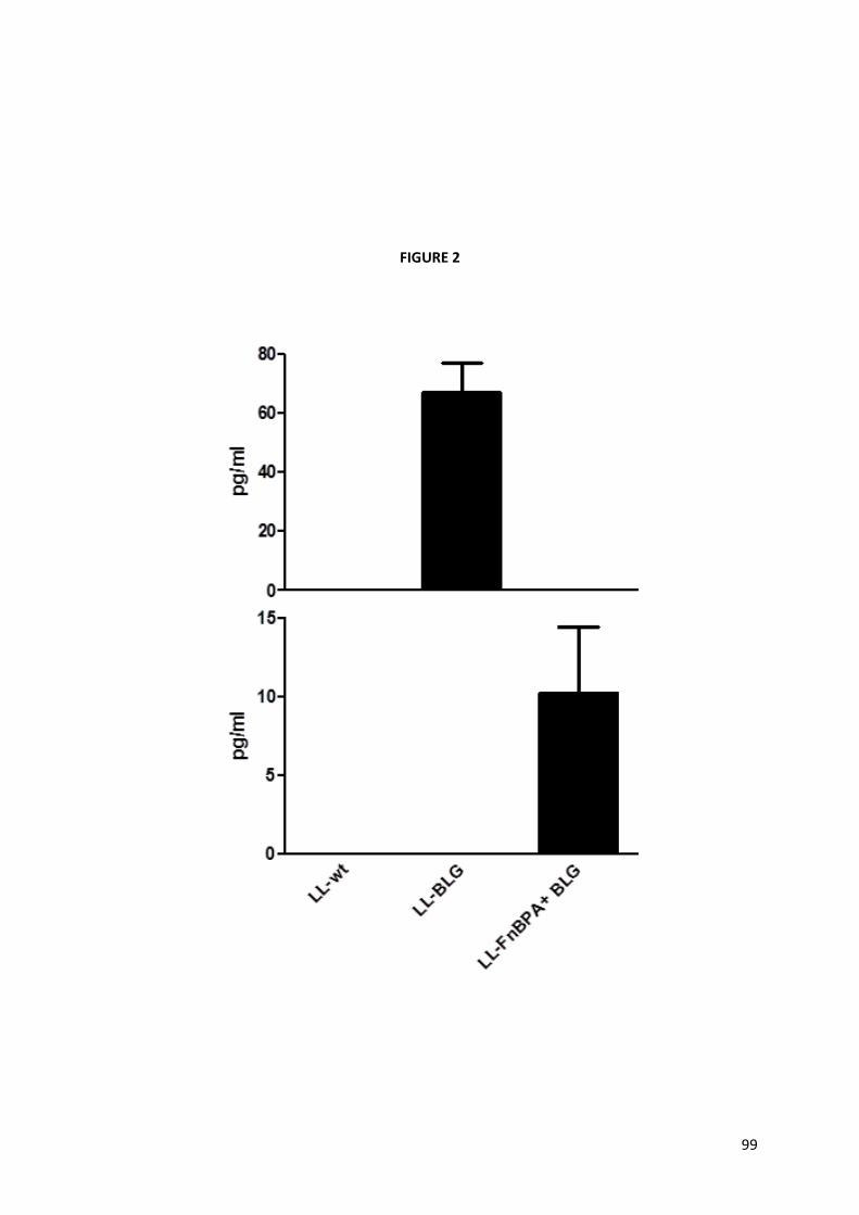
99
FIGURE 2

100
FIGURE 3

101
FIGURE 4

102
FIGURE 5

103
FIGURE 6

104
FIGURE 7

CHAPTER 5 Recombinant invasive Lactococcus lactis can transfer DNA vaccines either directly to dendritic cells or
across an epithelial cell monolayer

!"#$
$
!"#$%&'()*(+&,-./01#1%(012#302&(!"#$%#%##&'()"#$*'(,#1(%'#134&'(567(2#,,01&3(&0%"&'(80'&,%9:(
%-(8&18'0%0,(,&993(-'(#,'-33(#1(&$0%"&90#9(,&99(.-1-9#:&'(
(
);< =1%'-8>,%0-1(
%&'$ ()**+,)-+.,$ /)0$ 122,$ .3$ 452)-$ +,-2520-$ 0+,*2$ +-0$ 6+0*.(257$ .8+,4$ -.$ +-0$ )1+9+-7$ -.$
0-+:;9)-2$1.-/$*299<:26+)-26$),6$/;:.5)9$+::;,2$520=.,020$>?,4.9.--+$2-$)9@A$B"!"C@$D.82(25A$
.,2$.3$-/2$*.,*25,0$)1.;-$,)E26$%&'$+::;,+F)-+.,$+0$+-0$9.8$+::;,.42,+*+-7$)0$-/2$),-+42,$+0$
=5.6;*26$+,$(257$0:)99$):.;,-0$+,$9)5425$),+:)90!>G5+()0-)()$),6$H+;A$B""IC@$J20+620$-/+0A$+-$+0$
8299$E,.8,$-/)-$%&'<1)026$()**+,20$-)542-$=..597$=5.3200+.,)9$),-+42,<=5202,-+,4$*2990$>'KLCA$
0;*/$ )0$ 62,65+-+*$ *2990$ >%L0C$ >%)3-)5+),$ 2-$ )9@A$ B"!!C@$ D2,*2A$ 02(25)9$ 0-5)-24+20$ /)(2$ 122,$
620+4,26$-.$+,*52)02$-/2$=.-2,*7$.3$%&'$+::;,+F)-+.,$0;*/$)0$-/2$;02$.3$1)*-25+)$)0$)$(2*-.5$
3.5$ %&'$ 629+(257$ >G*/.2,$ 2-$ )9@A$ B""MC@$ N,2$ )--5)*-+(2$ 32)-;52$ .3$ 1)*-25+)9$ *)55+250$ +0$ -/2+5$
=.-2,-+)9$ 3.5$ .5)9$ )6:+,+0-5)-+.,$ 8/+*/$ :)7$ 0-+:;9)-2$ 1.-/$ :;*.0)9$ ),6$ 070-2:+*$ +::;,2$
520=.,020$ >G5+()0-)()$ ),6$ H+;A$ B""IC@$ ',.-/25$ )6(),-)42$ *.,0+0-0$ .,$ -/2$ 3)*-$ -/)-$ -/27$ *),$
=5.-2*-$ -/2$ %&'$ )4)+,0-$ 6245)6)-+.,$ 17$ 2,6.,;*92)020$ >G2.8$ ),6$ O..6A$ B""PC@$
Q,-25.=)-/.42,+*$ 0=2*+20$ /)(2$ 122,$ ;026$ )0$ 2R=25+:2,-)9$ 629+(257$ 070-2:0A$ /.82(25A$
52(250+.,$-.$-/2$(+5;92,-$=/2,.-7=2$52:)+,$)0$)$:)S.5$*.,*25,$>K.,-20$2-$)9@A$B"!!C@(
T2*2,-97A$-/2$=.-2,-+)9$;02$.3$"#$%&$&$$'(!)#$%*($>HHCA$-/2$:.629$H)*-+*$'*+6$J)*-25+)$>H'JCA$
3.5$=9)0:+6$%&'$629+(257$-.$2;E)57.-+*$*2990$+0$12+,4$2R=9.526$>J25:U62F<D;:)5V,$2-$)9@A$B"!!W$
K.,-20$2-$)9@$B"!!W$O2990A$B"!!C@$"+!)#$%*($+0$*.,0+62526$),$)6(),-)42.;0$(2*-.5$12*);02$+-$/)0$
),$20-)19+0/26$0)32-7$=5.3+92$42,25)-26$-/5.;4/$+-0$9.,4$;02$+,$-/2$6)+57$+,6;0-57$)0$0-)5-250$3.5$
3..6$ 325:2,-)-+.,0$ >K.,-20$ 2-$ )9@$ B"!!C@$ X;+:)5Y20$ ),6$ *.99)1.5)-.50$ 62:.,0-5)-26$ -/)-$ "+!
)#$%*($8)0$*)=)192$-.$-5),0325$)$Z<9)*-.49.1;9+,$>JHXC$2;E)57.-+*$2R=5200+.,$=9)0:+6$-.$/;:),$
+,-20-+,)9$2=+-/29+)9$*299$9+,2$L)*.<B$>X;+:)5Y20$2-$)9@A$B""#C@$H)-25A$L/)-29$2-$)9@$62:.,0-5)-26$
-/)-$-/+0$1)*-25+;:$8)0$)90.$)192$-.$-5),0325$JHX$*%&'$*,!-*-&$)3-25$.5)9$)6:+,+0-5)-+.,$+,$:+*2$
>L/)-29$2-$)9@A$B""[C@$&2(25-/29200A$=9)0:+6$-5),0325$5)-+.$.1025(26$8)0$9.8@$O+-/$-/2$+,-2,-$-.$
+,*52)02$ -/2$ *)=)*+-7$ .3$ "+! )#$%*($ -.$ -5),0325$ %&'$ =9)0:+60A$ 52*.:1+,),-$ 0-5)+,0$ 2R=5200+,4$
+,()0+,0$ 625+(26$ 35.:$ =)-/.42,+*$ 0=2*+20$ 8252$ *.,0-5;*-26@$ J.-/$ "+! )#$%*($ 2R=5200+,4$ 2+-/25$
"*(%./*#!0&,&$1%&2.,.($?,-25,)9+,$'$>?,9'C$.5$3%#451)&$&$$'(!#'/.'($\+15.,2*-+,$J+6+,4$K5.-2+,$
'$ >\,JK'C$ +:=5.(26$ %&'$ 629+(257$ *,! -*%/&! >X;+:)5Y20$ 2-$ )9@A$ B""]W$ ?,,.*2,-+,$ 2-$ )9@A$ B""PC@$$
D.82(25A$ 2(2,$ -/.;4/$ +,-2520-+,4A$ -/202$ -8.$ 0-5)-24+20$ =5202,-26$ 0.:2$1.--92,2*E0$ )0$ ?,9'$
+,()0+,$6.20$,.-$52*.4,+F2$ +-0$52*2=-.5$ +,$:+*2A$:;5+,2$Q<*)6/25+,$>O.9925-$2-$)9@A$B""^CA$),6$
\,JK'$ 52_;+520$ ),$ )62_;)-2$ 9.*)9$ *.,*2,-5)-+.,$ .3$ 3+15.,2*-+,$ -.$ 1+,6$ -.$ +,-245+,0$ $ -/;0$
9+:+-+,4$-/2$*,!-*-&$0-;6+20$+,$*.,(2,-+.,)9$:+*2$>%2$'F2(26.$2-$)9@A$B"!BC@$`/2523.52A$),.-/25$
52*.:1+,),-$+,()0+(2$"+!)#$%*($0-5)+,$2R=5200+,4$)$:;-)-26$3.5:$.3$?,9'$>HH:?,9'C$8)0$52*2,-97$

!"^$
$
620*5+126@$ 6,! -*%/&7$ +-$8)0$ 0/.8,$ -/)-$ -/2$ +,()0+(2$ 0-)-;0$ +:=5.(26$ 1.-/$ +,()0+(+-7$ ),6$%&'$
629+(257$*)=)*+-7$ -.$L)*.<B$*2990$),6A$ *,!-*-&A$ +-$8)0$62:.,0-5)-26$)$ -2,62,*7$.3$HH<:?,9'$ -.$
+,*52)02$-/2$=/2,.:2,.,$.3$=9)0:+6$-5),0325$-.$:+*2$2=+-/29+)9$*2990$>%2$)F2(26.$2-$)9@A$B"!BC@$
L.,0+625+,4$-/)-$*;552,-$E,.892642$)1.;-$%&'$()**+,)-+.,$;0+,4$"+! )#$%*($ +0$:.0-97$1)026$.,$
6)-)$ .1-)+,26$ +,$ 2R=25+:2,-0$ =253.5:26$ 8+-/$ +,-20-+,)9$ 2=+-/29+)9$ *2990$ >?QL0CA$ 82$ /)6$ -/2$
+,-2,-$-.$2()9;)-2$%&'$-5),0325$*)=)*+-7$.3$,.,+,()0+(2$.5$+,()0+(2$>HH<\,JK'$),6$HH<:?,9'C$"+!
)#$%*($ -.$%L0$)0$8299$8/+*/$)52$*.,0+62526$ -.$12$ -/2$:)S.5$'KL0$.3$ -/2$ +::;,2$070-2:@$O2$
)90.$:2)0;526$-/2$*)=)*+-7$.3$-/202$0-5)+,0$-.$629+(25$%&'$+,$)$`5),08299$*.<*;9-;52$:.629$),6$
82$2()9;)-26$%L0$+::;,2$520=.,02$)3-25$6+52*-$*.<+,*;1)-+.,$8+-/$1)*-25+)@$
$
);? @#%&'0#93(#18(.&%"-83(
J.,2<:)55.8$625+(26$62,65+-+*$*2990$>Ja%LC$35.:$*.,(2,-+.,)9$J'HJb*$8252$+0.9)-26$),6$
6+33252,-+)-26$8+-/$45),;9.*7-2<:)*5.=/)42$*.9.,7<0-+:;9)-+,4<3)*-.5$ >Xa<LG\C$123.52$12+,4$
6+52*-97$*.<+,*;1)-26$8+-/$+,()0+(2$>HH<\,JK'<JHXA$HH<:?,9'<JHXC$),6$,.,+,()0+(2$>HH<JHXCA$)99$
0-5)+,0$ *)557+,4$ -/2$ 2;E)57.-+*$ 2R=5200+.,$ (2*-.5$ =c)9)*dJHX@$O2$ -/2,$ ;026$ 39.8$ *7-.:2-57$
),)970+0$ +,$.5625$ -.$_;),-+37$ -/2$):.;,-$.3$6+33252,-+)-26$%L0@$'3-25$1)*-25+)9$ *.<+,*;1)-+.,A$
2R=5200+.,$.3$JHX$17$Ja%L0$8)0$_;),-+3+26$17$QH?G'$)0$8299$)0$02*52-+.,$.3$-/2$*7-.E+,20$?H<!B$
),6$ ?H<!"@$ ?,$ .5625$ -.$ 2()9;)-2$ -/2$ *)=)*+-7$ .3$ ,.,+,()0+(2$ .5$ +,()0+(2$ "+! )#$%*($ -.$ -5),0325$
=c)9)*dJHX$-.$%L0$45.8,$;,625$)$:.,.9)725$.3$6+33252,-+)-26$ ?QL0A$82$ 3+50-$45.8,$2=+-/29+)9$
*2990$.3$-/2$*299$9+,2$L)*.<B$)-$-/2$)=+*)9$0+62$),6$%L0$)-$-/2$1)0.9)-25)9$0+62$.3$=.5.;0$+,025-0@$
'3-25$ 1)*-25+)9$ *.<+,*;1)-+.,$ 8+-/$ -/2$ 070-2:A$ 82$ :2)0;526$ -/2$ *)=)*+-7$ .3$ -/2$ 0-5)+,0$ -.$
+,()62$ -/2$ :.,.9)725$ .3$ 2=+-/29+)9$ *2990$ ;0+,4$ -/2$ 42,-):+*+,$ 0;5(+()9$ )00)7@$ \+,)997A$ 82$
_;),-+3+26$JHX$=5.6;*-+.,$)-$1.-/$)=+*)9$),6$1)0.9)-25)9$0;=25,)-),-0$)0$8299$)0$+,$Ja%L0$),6$
L)*.<B$2R-5)*-0$17$QH?G'@$G-)-+0-+*)9$0+4,+3+*),*2$12-822,$-/2$45.;=0$8)0$*)9*;9)-26$;0+,4$-/2$
N,2$O)7$'&Nc'$>),6$,.,=)5):2-5+*C$-20-A$3.99.826$17$-/2$eJ.,3255.,+f$=.0-<-20-@$c)9;20$.3$
=g$"@"]$8252$*.,0+62526$0+4,+3+*),-@$
$
);A +&3>9%3(
Ja%L0$ 6+52*-97$ *.<+,*;1)-26$ 8+-/$ HH<\,JK'h$ 8252$ )192$ -.$ 2R=5200$ !@]$ -+:20$ /+4/25$
):.;,-0$.3$JHX$*.:=)526$-.$-/2$,.,+,()0+(2$>HH<JHXC$),6$8+96$-7=2$>8-C$0-5)+,0$8/+92$Ja%L0$
+,*;1)-26$8+-/$ HH:?,9'<JHX$ 0-5)+,$ *.;96$2R=5200$)9:.0-$B@]$ -+:20$452)-25$_;),-+-+20$.3$JHX@$
\;5-/25:.52A$)99$0-5)+,0$+,6;*26$-/2$2R=5200+.,$.3$-/2$=5.<+,39)::)-.57$*7-.E+,2$?H<!B$),6$,.5$
-/2$2R=5200+.,$.3$JHX$,2+-/25$-/2$+,()0+(2$0-)-;0$.3$0-5)+,0$6+6$,.-$)332*-$?H<!B$2R=5200+.,@$$O2$
-/2,$:.(26$-.$.-/25$ *,!-*%/&$)00)70$-.$0-;67$ +3$-/2$0-5)+,0$*.;96$-5),0325$=c)9)*dJHX$)*5.00$)$
:.,.9)725$.3$6+33252,-+)-26$?QL0@$`/2$3+50-$0-2=$8)0$-.$2()9;)-2$82)-/25$HH<\,JK'$.5$HH<:?,9'$

!"[$
$
+,()0+(2$ 9)*-.*.**+$ 8252$ )192$ -.$ +,()62$ -/2$ :.,.9)725$ ),6$ )3-258)560$ 20-)19+0/$ )$ *9.025$
*.,-)*-$8+-/$-/2$Ja%L0$45.8,$)-$-/2$1)0.9)-25)9$0+62$.3$-/2$+,025-@$$'3-25$*.<+,*;1)-+.,$8+-/$
1)*-25+)$ ),6$ -52)-:2,-$ 8+-/$ 42,-):+*+,A$ 82$ .1025(26$ -/)-$ 52*.:1+,),-$ HH<:?,9'$ ),6$ HH<
\,JK'$ 0-5)+,0$ )52$ )5.;,6$ !""$ -+:20$ :.52$ +,()0+(2$ -/),$ -/2$ 8+96$ -7=2$ 0-5)+,0$ >&iP"""$ ),6$
aX!I#IC@$'3-258)560A$82$:2)0;526$JHX$2R=5200+.,$+,$%L0$*.<*;9-;526$8+-/$-/2$:.,.9)725$.3$
L)*.<B$*2990@$'=+*)9$),6$1)0.9)-25)9$0;=25,)-),-0$8252$)90.$)00)726$3.5$ -/2$=5202,*2$.3$JHX@$
%L0$*.<*;9-;526$8+-/$-/2$?QL$:.,.9)725$=52(+.;097$*.<+,*;1)-26$8+-/$HH:?,9'<JHX$0-5)+,$*.;96$
2R=5200$ 0+4,+3+*),-$ /+4/25$ ):.;,-0$ .3$ JHX$ 8/2,$ *.:=)526$ -.$ HH$ 0-5)+,$ 9)*E+,4$ =c)9)*dJHX$
()**+,2@$\;5-/25:.52A$,.$JHX$2R=5200+.,$17$%L0$8)0$.1025(26$8+-/$*2990$*;9-;526$8+-/$HHA$HH<
JHX$.5$HH\,JK'<JHX$0-5)+,0$),6$,.$JHX$8)0$62-2*-26$,2+-/25$ +,$)=+*)9$.5$1)0.9)-25)9$*;9-;52$
0;=25,)-),-0@$$
$
);B 503,>330-1(
O2$/)(2$0/.8,$-/)-$"+!)#$%*($+0$*)=)192$-.$629+(25$*%&'$=9)0:+60$2+-/25$*,!-*%/&$.5$*,!-*-&$-.$
+,-20-+,)9$2=+-/29+)9$ *2990$ >?QL0C$ >X;+:)5Y20$2-$)9@A$ B""]W$L/)-29$2-$)9@A$ B""[W$ ?,,.*2,-+,$2-$)9@A$
B""PW$ %2$ 'F2(26.$ 2-$ )9@A$ B"!BC@$ T2*2,-$ 6)-)$ 62:.,0-5)-26$ -/)-$ -/2$ ;02$ .3$ 52*.:1+,),-$
+,()0+(2$ 0-5)+,0$ 2R=5200+,4$ +,()0+,0$ 35.:$ =)-/.42,+*$ 0=2*+20$ +,*52)026$ %&'$ -5),0325$ *,! -*%/&$
),6$0/.826$)$-2,62,*7$-.$+:=5.(2$%&'$629+(257$ *,!-*-&$-.$:+*2$?QL0$>K.,-20$2-$)9@A$B"!BW$%2$
'F2(26.$ 2-$ )9@A$ B"!BC@$ '99$ 2R=25+:2,-0$ =253.5:26$ 0.$ 3)5$ )+:26$ -.$ 2()9;)-2$ -/2$ -5),0325$ .3$
=c)9)*dJHX$=9)0:+6$;0+,4$,.,+,()0+(2$.5$+,()0+(2$"+!)#$%*($.,97$-.$?QL0@$?,$-/+0$8.5E$82$62*+626$
-.$:2)0;52$-/2$*)=)*+-7$.3$-/202$0-5)+,0$-.$629+(25$)$=9)0:+6$%&'$-.$%L0$)0$8299@$`/202$*2990$
)52$ +,$6+52*-$*.,-)*-$8+-/$ -/2$ ?QL$:.,.9)725$12+,4$*)=)192$ -.$;=-)E2$),-+42,0$6+9;-26$ +,$ -/2$
+,-20-+,)9$9;:2,A$),6$0-+:;9)-2$1.-/$:;*.0)9$),6$070-2:+*$+::;,2$520=.,020$>G-2+,:),$2-$)9@A$
B""BA$T+:.96+$2-$)9A$B""#C@$\.5$2R):=92A$0-+:;9)-26$%L0$*),$5292)02$+,-2592;E+,0$0;*/$)0$?H<!B$
-/)-$)*-+()-20$,)-;5)9$E+9925$*2990$=.9)5+F+,4$`$*2990$-.$-/2$=5.-2*-+(2$`/!$=/2,.-7=2$>a)*)-.,+)$
2-$)9@A$!PP]C@$
Ja%L0$*.<+,*;1)-26$8+-/$HH<\,JK'$),6$HH<:?,9'$8252$0/.8,$-.$2R=5200$452)-$):.;,-0$.3$
JHXA$62:.,0-5)-+,4$-/)-$1.-/$0-5)+,0$8252$*)=)192$-.$0;**2003;997$-5),0325$=c)9)*dJHX$=9)0:+6@$
`/2$ 3)*-$ -/)-$ HH:?,9'<JHX$ 0-5)+,$ *.;96$ -5),0325$:.52$ 233+*+2,-97$ -/2$ *%&'$.3$ JHX$ -.$ Ja%L0$
:)7$12$6;2$-.$-/2$2R=5200+.,$.3$Q<*)6/25+,A$52*2=-.5$.3$:?,9'A$+,$-/202$*2990$>J.5E.80E+$2-$)9@A$
!PPMC@$ $`/2$+:=5.(26$=9)0:+6$-5),0325$.1025(26$3.5$HH<\,JK'$0-5)+,$*.;96$12$)90.$6;2$-.$-/2$
=5202,*2$.3$52*2=-.50$2R=520026$)-$-/2$0;53)*2$.3$%L0$-/)-$)52$+,(.9(26$+,$-/2$2,6.*7-.0+0$.3$3+!
#'/.'($-/5.;4/$\,JK'$+,()0+,@$&2(25-/29200A$623+,+-+(2$+,3.5:)-+.,$)1.;-$/.8$\,JK'$+,-25)*-0$
8+-/$52*2=-.50$2R=520026$17$%L0$+0$9)*E+,4@$

!"P$
$
`.$ 2()9;)-2$ %L0$ +::;,2$ 520=.,02$ )4)+,0-$ ,.,+,()0+(2$ ),6$ +,()0+(2$ 9)*-.*.**+A$ +0.9)-26$
Ja%L0$8252$*.<+,*;1)-26$8+-/$0-5)+,0@$'3-25$-/+0$=25+.6A$?H<!BA$)$=5.<+,39)::)-.57$+,-2592;E+,$
+,(.9(26$+,$-/2$6+33252,-+)-+.,$.3$,)j(2$`$*2990$+,-.$`/!$*2990$>D0+2/$2-$)9@A$!PPIC$),6$?H<!"A$-/)-$
2R/+1+-0$ =92+.-5.=+*$ 2332*-0$ +,$ +::;,2<524;9)-+.,$ ),6$ +,39)::)-+.,$ 6.8,<524;9)-+,4$ -/2$
2R=5200+.,$ .3$ `/!$ *7-.E+,20$ >K20-E)$ 2-$ )9@A$ B""MCA$ 8252$ :2)0;526$ +,$ *;9-;52$ 0;=25,)-),-0@$$
Ja%L0$ *.<*;9-+()-26$ 8+-/$ 2+-/25$ ,.,+,()0+(2$ .5$ +,()0+(2$ 9)*-.*.**+$ 8252$ )192$ -.$ 5292)02$
292()-26$92(290$.3$?H<!B$>I""$=4b:HC$8/+92$,.$?H<!"$2R=5200+.,$8)0$.1025(26@$$
`/+0$6)-)$=)5-+)997$*.,3+5:0$8/)-$8)0$3.;,6$17$k):$),6$*.99)1.5)-.50$+,$B""[$8/252$-/27$
0/.826$-/)-$"+!)#$%*($&iP"""$0-5)+,$+,*;1)-26$8+-/$+0.9)-26$Ja%L0$+,6;*26$-/2$02*52-+.,$.3$?H<
!Z$>)$=5.<+,39)::)-.57$*7-.E+,2C$),6$?H<!B@$&.,2-/29200A$6+33252,-97$35.:$8/)-$8)0$.1-)+,26$
+,$ -/+0$8.5EA$ -/27$ 3.;,6$(257$0-5.,4$ +,6;*26$:T&'$ 92(290$.3$ ?H<!"$ >k):$2-$)9@A$B""[C@$N-/250$
/)(2$0/.8,$-/)-$-/2$-7=2$.3$+::;,2$520=.,02$29+*+-26$17$H'J$+0$0-5)+,$62=2,62,-$)0$-/27$:)7$
3)(.;5$2+-/25$)$`/!$520=.,02A$)$`/B$/;:.5)9$.5$-.925.42,+*A$.5$.,97$),$+,39)::)-.57$520=.,02$
>X.,299)$2-$)9@A$!PP[C@$$
?-$ 8)0$ ()0-97$ 62:.,0-5)-26$ -/)-$ -/2$ /2-25.9.4.;0$ 2R=5200+.,$ .3$ *25-)+,$ ),-+42,0$ 35.:$
=)-/.42,+*$0=2*+20$.,$"+!)#$%*($0;53)*2$*),$+,6;*2$)$620+526$-7=2$.3$+::;,2$520=.,02$>`/!$.5$
`/BC$>K.,-20$2-$)9@A$B"!!C@$ ?,$-/+0$8.5E$-/2$2R=5200+.,$.3$"+!0&,&$1%&2.,.($:?,9'$.5$3+!#'/.($
\,JK'$6+6$,.-$*/),426$?H<!B$=5.6;*-+.,$17$%L0@$`/2$2R=5200+.,$.3$JHX$17$-/2$Ja%L0$)90.$6+6$
,.-$:.6+3+26$?H<!B$92(290$+,$*;9-;52$0;=25,)-),-0@$$
`.$ 0-;67$ -/2$6+)9.4$12-822,$ 0-5)+,0A$6+33252,-+)-26$ ?QL0$),6$Ja%L0A$82$*/..02$ -.$8.5E$
8+-/$)$:.629$-/)-$:+:+*0$-/2$*,!-*-&$0+-;)-+.,$+,$8/+*/$L)*.<B$*2990$8252$*;9-;526$)-$-/2$)=+*)9$
0+62$.3$-/2$+,025-$),6$Ja%L0$8252$45.8,$)-$-/2$1)0.9)-25)9$0+62@$$'-$3+50-$82$*/2*E26$82)-/25$
52*.:1+,),-$ +,()0+(2$ "+! )#$%*($ 0-5)+,0A$ HH<\,JK'h$ ),6$ HH<:?,9'hA$ )52$ )192$ -.$ +,-25,)9+F2$ )$
:.,.9)725$ .3$ ?QL0$ :.52$ 233+*+2,-97$ -/),$ -/2$ 8-$ 0-5)+,0@$ `/2$ +,()0+.,$ 5)-2$ .1-)+,26$ 3.5$ -/2$
+,()0+(2$0-5)+,0$8252$0+:+9)5$),6$)1.;-$!""<3.96$/+4/25$-/),$-/2$+,()0+.,$5)-2$.1-)+,26$3.5$8-$
0-5)+,0@$%2$'F2(26.$),6$*.99)1.5)-.50$0/.826$-/)-$HH<:?,9'$0-5)+,$+0$!"""$-+:20$:.52$+,()0+(2$
-/),$-/2$8-$+,$2R=25+:2,-0$=253.5:26$8+-/$,.,<*.,39;2,-$L)*.<B$*2990@$`/2$9.825$+,()0+(2,200$
0*.52$ .1-)+,26$ ;0+,4$ -/2$ ?QL$ :.,.9)725$ 8)0$ =5.1)197$ 12*);02$ *2990$ 8252$ (257$ )--)*/26$ -.$
2)*/$ .-/25$ -;5,+,4$ Q<*)6/25+,$ 52*2=-.5$ 9200$ )**200+192@$ '3-258)560A$ 1)*-25+)$8252$ +,*;1)-26$
8+-/$-/2$*.<*;9-;52$070-2:$>Ja%L0$),6$L)*.<B$*2990C$ +,$.5625$-.$;,6250-),6$-/2$:2*/),+0:0$
17$8/+*/$"+!)#$%*($*),$-5),0325$%&'$()**+,20$*,!-*-&@$
$
);) !-1,9>30-1(
O2$62:.,0-5)-26$-/)-$52*.:1+,),-$+,()0+(2$"+!)#$%*($2R=5200+,4$2+-/25$3+!#'/.'($\,JK'$.5$
"+! 0&,&$1%&2.,.($ :?,9'$ )52$ *)=)192$ -.$ -5),0325$ %&'$ 6+52*-97$ -.$ Ja%L0@$ \;5-/25:.52A$ -/+0$

!!"$
$
6+52*-$*.,-)*-$92)6$-.$-/2$02*52-+.,$.3$?H<!B$17$-/2$*2990A$+,62=2,62,-$.3$-/2$+,()0+(2$0-)-;0@$&.$
?H<!"$=5.6;*-+.,$8)0$.1025(26@$O2$)90.$*/2*E26$-/)-$+,()0+(2$9)*-.*.**+$)52$)192$-.$+,()62$)$
:.,.9)725$.3$6+33252,-+)-26$L)*.<B$*2990$!""$-+:20$:.52$233+*+2,-97$-/),$-/2$8-$0-5)+,0@$l0+,4$)$
*.<*;9-;52$:.629$82$62:.,0-5)-26$-/)-$HH<:?,9'$0-5)+,$*),$-5),0325$=c)9)*dJHX$-.$%L0$)*5.00$
)$:.,.9)725$.3$2=+-/29+)9$*2990@$`)E2,$-.42-/25A$-/+0$0-;67$4+(20$,28$+,0+4/-0$.,$-/2$:2*/),+0:$
.3$9)*-.*.**+$;=-)E2$3.5$629+(257$.3$-/25)=2;-+*0@$
(

111
Recombinant invasive Lactococcus lactis can transfer DNA vaccines either directly to
dendritic cells or across an epithelial cell monolayer
Marcela de Azevedo4, Marjolein Meijerink3, Juliana Franco de Almeida5, Nico Taverne3, Vasco
Azevedo4, Anderson Miyoshi4, Philippe Langella1,2,Jerry M. Wells3, and Jean-‐Marc Chatel1,2
1 INRA, UMR1319 Micalis, Jouy-‐en-‐Josas, France.
2 AgroParisTech, UMR Micalis, F-‐78350 Jouy-‐en-‐Josas, France.
3 Host Microbe Interactomics, Wageningen University, The Netherlands.
4 Laboratorio de Genética Celular e Molecular, ICB, UFMG, Minas Gerais, Brazil.
5 Laboratório de Nanobiotecnologia, Universidade Federal de Uberlândia, Instituto de
Genética e Bioquímica, Minas Gerais, Brazil
* Corresponding author: e-‐mail: jean-‐[email protected]
Phone: 33 01 34 65 24 68
Fax: 33 01 34 65 24 62
Key words: Lactococcus lactis, mutated internalin A, Listeria monocytogenes, internalization,
dendritic cells, DNA delivery, -‐lactoglobulin

112
Abstract The use of bacterial carriers for oral delivery of DNA vaccines constitutes a
promising vaccination strategy. Lactococcus lactis (LL), a generally regarded as safe (GRAS)
bacterium (LAB) widely used in the dairy industry, has recently been explored as mucosal DNA
delivery vector. Due to its safe status, LL represents an attractive alternative to attenuated
pathogens which are the most commonly bacterial vectors used for DNA immunization.
Previous studies showed that either native or recombinant invasive LL are able to deliver and
trigger DNA expression by mammalian cells both in vitro and in vivo. However, current
knowledge about DNA vaccination using LL is mostly based on data obtained in studies with
intestinal epithelial cells (IECs). Considering that dendritic cells (DCs) serve as potent inducers
of specific cell-‐mediated immune responses acting as professional antigen presenting cells
(APCs), in this work we evaluated DNA transfer capacity of LL to DCs. We have shown for the
first time that invasive L. lactis strains expressing either Staphylococcus aureus Fibronectin
Binding Protein A (LL-‐FnBPA+), or Listeria monocytogenes mutated Internalin A (LL-‐mInlA+) can
transfect bone marrow-‐derived DCs (BMDCs) and deliver a plasmid DNA vaccine (pValac)
-‐lactoglobulin (BLG). BMDCs co-‐cultured with non-‐invasive
or invasive Lactococci were able to secrete elevated levels of the pro-‐inflammatory cytokine IL-‐
12. In order to understand the mechanism by which lactococci could transfer pValac:BLG in
vivo, differentiated Caco-‐2 cells were used to measure the capacity of strains to invade a
polarized monolayer of epithelial cells, mimicking the in vivo situation. Gentamycin survival
assay in these cells showed that LL-‐mInlA+ is 100 times more invasive than LL. We also
investigated the cross-‐talk between differentiated IECs, BMDCs and bacteria using an in vitro
Transwell co-‐culture model. Co-‐incubation of strains with the co-‐culture model has shown that
DCs maintained with LLmInlA-‐BLG strain could express significant higher levels of BLG. This
result suggest that in vivo DCs could sample bacteria containing the DNA vaccine across the
epithelial barrier; express the antigen and/or present it to naïve T cells stimulating adaptive
immune responses.
1. Introduction
DNA vaccination has been of great interest since its discovery in the 1990s owing to its
ability to stimulate both cell-‐mediated and humoral immune responses (Ingolotti et al., 2010).
It is now well proven that intramuscular injection of a plasmid DNA can induce antibodies,
helper T cell responses, cytotoxic T cell (CTL) responses, and protective immunity against
different viral, bacterial, and parasitic infections in various animal models (Donnelly et al.,
2000). Although US Food and Drug Administration (FDA) still did not approved DNA vaccines
for use in humans, phase I clinical studies have been reported for the prevention and/or

113
treatment of influenza, HIV, malaria, hepatitis B, SARS and many other infectious agents
(Klinman et al., 2011). Recently, four DNA vaccines have been licensed for administration in
horses, salmon, dogs and swine turning this vaccine platform very promising (Findik and Çiftci,
2012).
One of the concerns about naked DNA immunization is its low immunogenicity as the
antigen is produced in very small amounts in vivo due to its non-‐replicative nature (Srivastava
and Liu, 2003). Besides this, it is well know that DNA-‐based vaccines poorly target professional
antigen-‐presenting cells (APC) (Daftarian et al., 2011). Therefore, several strategies have been
designed to increase the potency of DNA immunization (Chen et al., 2012). The use of bacteria
as a vector for DNA delivery into eukaryotic cells has emerged as a potential approach to
enhance the immunogenicity of DNA vaccines (Schoen et al., 2004). One attractive feature of
bacterial carriers is their potential for oral administration which may stimulate both mucosal
and systemic immune responses (Srivastava and Liu, 2003). Some attenuated pathogenic
species contains an innate tropism for specific tissues of the host directing systemic responses
towards the mucosa (Schoen et al., 2004). Furthermore, besides protecting the plasmid against
degradation by endonucleases (Hoebe et al., 2004), bacteria can act as adjuvants due to the
presence of pathogen-‐associated molecular patterns (PAMPs) in their surface (Schoen et al.,
2004). Another advantage consists on the fact that bacterial vectors can accommodate large-‐
sized plasmids, which allows the insertion of multiple genes or chemokines of interest (Seow
and Wood, 2009). Finally, this vaccine platform is considered to be low cost as it does not
require further steps of plasmid purification, reducing cost and labor (Hoebe et al., 2004;
Schoen et al., 2004).
Shigella, Salmonella, and Listeria monocytogenes have been used as experimental delivery
systems. Even though the bacteria used for such proposed vaccines are attenuated strains,
preexisting immunity, reactogenicity and reversion to virulence remain major concerns,
especially considering their administration in children and immunosuppressed patients (Pontes
et al., 2011). Recently, the potential use of Lactococcus lactis (LL), the model Lactic Acid
Bacteria (LAB), for the production of biologically useful proteins and for plasmid DNA delivery
to eukaryotic cells is being explored (for a review see Bermúdez-‐Humarán et al., 2011; Pontes
et al. 2011; Wells, 2011). L. lactis is considered an advantageous vector because it has an
established safety profile generated through its long use in the dairy industry as starters for
food fermentations (Pontes et al. 2011). That is the reason why the FDA has been granted this
bacterium with the GRAS (Generally Recognized As Safe) status. Moreover, L. lactis is easy to
handle, does not produce lipopolysaccharides (LPS) in its outer membrane, contains a number
of genetic tools developed in the last couple of decades and does not induce strong host

114
immune responses (Bermúdez-‐Humarán et al., 2011; Pontes et al. 2011). Guimarães and
collaborators showed that L. lactis was capable to transfer a -‐lactoglobulin (BLG) eukaryotic
expression plasmid to human intestinal epithelial cell line Caco-‐2. Expression of BLG by Caco-‐2
cells was detected after co-‐culture with L. lactis carrying the expression plasmid (Guimarães et
al., 2006). Later, Chatel et al. demonstrated that this bacterium was also able to transfer BLG
cDNA in vivo after oral administration in mice (Chatel et al., 2008). With the intent to increase
the capacity of L. lactis to transfer DNA plasmids, recombinant strains expressing invasins
derived from pathogenic species were constructed. One was designed to express Listeria
monocytogenes Internalin A (InlA) (LLInlA) (Guimarães et al., 2005) and the other to produce
Staphylococcus aureus Fibronectin Binding Protein A (FnBPA) (LLFnBPA) (Innocentin et al.,
2009). In vitro, they showed a higher ability to invade mammalian cells compared to the wild
type (wt) Lactococci and, as a result, more GFP expression plasmid was delivered to Caco-‐2
cells. However, even though interesting, these two strategies presented some bottlenecks:
InlA invasin does not recognize its receptor in mice, murine E-‐cadherin (Wollert et al., 2007),
and FnBPA requires an adequate local concentration of fibronectin to bind to integrins (De
Azevedo et al., 2012) thus limiting the in vivo studies in conventional mice. Another
recombinant invasive L. lactis strain expressing a mutated form of Internalin A (mInlA) (LLmInlA)
was recently described (De azevedo et al., 2012). It was shown that the invasive status
improved both invasivity and DNA deliver capacity in vitro to Caco-‐2 cells. Moreover, it was
demonstrated a tendency of the invasive bacteria to increase the phenomenon of plasmid
transfer to mice epithelial cells.
Current knowledge about DNA vaccination using L. lactis is mostly based on data obtained
in experiments performed with intestinal epithelial cells (IECs). Considering that dendritic cells
(DCs) serve as potent inducers of specific cell-‐mediated immune responses, it is very
interesting to evaluate the capacity of L. lactis to transfer DNA vaccines to these cells as well.
Besides being in direct contact with the IEC monolayer, DCs are capable to uptake antigens,
other cells or bacteria carrying a DNA vaccine through the epithelial tight junctions (Rescigno
et al., 2001). They can also generate great amounts of MHC-‐peptide complexes, migrate to the
primary lymphoid organs and present antigens to naïve T cells, acting as professional antigen
presenting cells (APCs) (Joffre et al., 2012). Moreover, DCs are able to secrete interleukins,
such as IL-‐12, that recruits other DCs and T cells polarizing them to the protective Th1
phenotype (Steinman et al., 2002).
The purpose of this work was to measure the ability of noninvasive or invasive L. lactis
expressing either S. aureus FnBPA LL-‐FnBPA+ or L. monocytogenes mutated InlA LL-‐
mInlA+ to deliver a plasmid DNA vaccine (p -‐

115
lactoglobulin (BLG) (Guimarães et al., 2009) directly to bone marrow-‐derived DCs (BMDCs). The
capacity of these strains to deliver pValac:BLG in a Transwell co-‐culture model (where the
BMDCs were grown on the basolateral side of the IEC monolayer) was also investigated. Finally,
DCs immune response was measured after direct co-‐incubation with bacteria.
2. Materials and Methods
2.1 Bacterial strains, plasmids, media and growth conditions. All bacterial strains and
plasmids used in this study are listed in Table 1. Noninvasive L. lactis NZ9000 (Kleeberzem et al.,
1997), L. lactis MG1363 (Gasson, 1983), L. lactis carrying pOri23 plasmid (LL) (Que et al., 2000),
L. lactis harboring both pOri23 and pValac:BLG plasmids (LL-‐BLG) (Pontes et al., 2012) and
invasive L. lactis strains expressing S. aureus FnBPA (LL-‐FnBPA+) (Que et al., 2001) or L.
monocytogenes mInlA (LL-‐mInlA+) (De Azevedo et al., 2012) were grown in M17 medium
supplemented with 0.5% (wt/vol) glucose (GM17) at 30°C without agitation. Erythromycin (Ery)
( chloramphenicol (Cm) were added to the medium when needed.
DNA manipulation was performed following standard procedures (Sambrook et al., 1989), with
a few modifications. Plasmids were purified by lyses alkaline method after bacterial incubation
for 30 min at 37 °C in TES solution (25% sucrose, 1 mM EDTA, 50 mM Tris HCl pH 8) containing
lysozyme (10 mg/ml). The quality of the products obtained, including its concentration and
purity, were estimated measuring the absorbance at 260 nm and 280 nm in
spectrophotometer. Restriction and modification endonucleases were used in accordance with
(Invitrogen).
2.2 Polarized intestinal epithelial cell cultures on Transwell filters. The human intestinal
epithelial cell line Caco-‐2 (ATCC number HTB37) was maintained in DMEM medium (Sigma, St.
Louis, MO) containing 10% fetal bovine serum (FBS) (Omnilab, Switzerland) and 2 mM L-‐
glutamine (BioWhittaker, Cambrex Bio Science, Verviers, Belgium). The culture medium was
switched every 2 days, and cell viability was determined by trypan blue staining. Trypsin-‐
treated cells were seeded on permeable supports with microporous membranes (Corning
Glass Works, 0.4 µm pore size PET membrane 24 well formats) for 14 days into p-‐24 plates
(Corning Glass Works), to ensure cell polarization with a distinguished apical and basal side
(Jumarie and Malo., 1991). Cells were grown at 37°C in the presence of 5% CO2 and 95% air
atmosphere.

116
2.3 Invasion assay of bacteria into differentiated human intestinal epithelial cells.
Differentiated Caco-‐2 cells grown on Transwell filters were used to perform bacterial
internalization into human epithelial cells. Recombinant or wt L. lactis was grown to an OD600
of 0.9 1.0, pellet was recovered, washed two times with PBS 1X and diluted in order to obtain
a multiplicity of infection (MOI) of around 1000 bacteria per eukaryotic cell. Before infection,
polarized monolayers of Caco-‐2 cells were treated with 10mM EDTA buffer (Sigma-‐Aldrich, St.
Louis, MO) to disrupt tight junctions and the transepithelial electrical resistance (TER) was
monitored across the monolayers as described by Karczewski et al. (2010). The gentamicin
survival assay was used to evaluate bacteria survival (Isberg and Falkow, 1985). Briefly,
recombinant or wt L. lactis were applied in the apical side of eukaryotic cells and co-‐incubated
during one hour at 37°C, in 5% CO2. After this period, cells were washed in order to remove
bacteria in excess and then 150 µg/mL of gentamicin were added. Plates were maintained for
more 2 hours before cells were washed and lysed with 0.2% of triton. The quantity of live
intracellular bacteria was determined by plating serial dilutions of cell lysates and counting
colony-‐forming units (CFU).
2.4 Animals. Female BALB/c mice were used at six-‐to-‐eight weeks and housed in a light and
temperature controlled facility of Wageningen University to generate bone marrow-‐derived
dendritic cells (BMDCs). The study was approved by the Wageningen University Ethics
Committee (Research, Wageningen UR, Wageningen University).
2.5 Murine bone-‐marrow derived dendritic cells (BMDC) isolation. To obtain BMDC cells, 6-‐
were euthanized; femurs were isolated, washed and gently crushed in
10 ml of RPMI-‐1640 medium (zonder Hepes) supplemented with 1% penicilin/Streptomicin
(Sigma-‐Aldrich). Cells were filtered using a Steriflip® Filter Unit (Millipore) and stained with a
trypan blue staining solution to determine the number of live and dead cells. Around 2x10⁷ live
cells were seeded in a sterile petri dish in complete media (RPMI-‐1640 medium containing 10%
heat-‐inactivated fetal calf serum [FCS, Sigma-‐Aldrich], 1% penicilin/Streptomicin [Sigma-‐
Aldrich], 20 ng/ml of recombinant mouse granulocyte-‐macrophage colony-‐stimulating-‐factor
(GM-‐CSF) [R&D systems] and 0.05mM of -‐mercaptoethanol [Invitrogen]). Cells were
incubated at 37ºC in CO2 atmosphere and medium was changed every three days. At day six
BMDCs were removed from petri dishes plates to determine the number and size of cells using
fluorescence activated cell-‐sorting (FACS) analysis. 24-‐Well Cell Culture Plates (Corning)
containing 5x10⁵ BMDC per well were then prepared and after used for both DNA transfer and
BMDCs stimulation experiments.
2.6 Flow cytometry analysis. BMDCs cultured during six days were harvested from the plate,
centrifuged (300 ×g, 10 min, 4 °C), resuspended in appropriate medium and analyzed using a

117
BD Biosciences FACSCanto II flow cytometer (BD Biosciences, San Jose, CA) to measure CD
marker expression. Cells were stained with FITC-‐conjugated anti-‐mouse CD11c, PE/Cy7-‐
conjugated anti-‐mouse CD86 and PE-‐conjugated anti-‐mouse CD40 antibodies (all from BD
Biosciences) and suitable isotype control monoclonal antibodies. Data was analyzed with
either BD FACSDiva or Flowjo software and the level of expression was expressed as the
geometric mean of fluorescence intensity (MFI).
2.7 Bacterial incubation with BMDCs for -‐lactoglobulin (BLG) detection. In order to evaluate
the capacity that invasive or noninvasive L. lactis contains in transferring DNA vaccines to APCs,
BLG expression and secretion by isolated BMDCs were quantified after performing the
gentamicin survival assay. Briefly, noninvasive L. lacis (LL and LL-‐BLG) and invasive L. lactis (LL-‐
FnBPA+ and LL-‐mInlA+) (table 1) were incubated with BMDCs for one hour, 150 µg/mL of
gentamicin was added to kill extracellular bacteria, cells were washed after 2 hours and
gentamicin-‐treated cells were incubated during 72 hours at 37 °C, in 5% CO2. Plates were
centrifuged at 78.2 g for 10 minutes; supernatant was collected and stored at -‐80°C. 500 µl of
PBS containing a cocktail of protease inhibitors (Roche) was added to the cells and then
homogenized using an ultrasonic treatment (3 times 10 seconds). Samples were stored at -‐
80°C and thus used to measure BLG expression by ELISA.
2.8 In vitro transwell co-‐culture system of differentiated Caco-‐2 cells and BMDCs. The
method to prepare epithelial cell monolayers in contact with BMDCs is described elsewhere
(Rescigno et al., 2001; Zoumpopoulou et al., 2009). In summary, intestinal epithelial cell line
Caco-‐2 was cultured on the upper face of 3-‐µm pore Transwell filters (costar, polycarbonate
Membrane) for 10 15 days in a 24-‐well plate (Corning Glass Works) using DMEM medium
(Sigma, St. Louis, MO) containing 10% fetal bovine serum (FBS) (Omnilab, Switzerland) and 2
mM L-‐glutamine (BioWhittaker, Cambrex Bio Science, Verviers, Belgium). 4×10⁵ of isolated
BMDCs were seeded on the basolateral side of difereniated Caco-‐2 and maintained in RPMI-‐
1640 (Zonder Hepes), supplemented with 10% heat-‐inactivated FCS (Sigma-‐Aldrich) and 20
ng/ml of GM-‐CSF (R&D systems) during 18 h at 37 °C in a 5% CO2/95% air atmosphere. After
this period the medium from all compartments was changed and the system was used for
assessing BLG expression by epithelial and dendritic cells after bacterial co-‐incubation.
2.9 BLG detection in BMDCs and Caco-‐2 monolayers co-‐cultured on transwell inserts after
bacterial incubation. A co-‐culture system model was used to evaluate DNA transfer capacity of
invasive or noninvasive L. lactis strains to dendritic cells across an epithelial cell monolayer.
Inserts containing fully differentiated Caco-‐2 cells monolayers in the apical side and BMDCs
cultured in the basolateral side were prepared, as described in section 2.8. No EDTA treatment
was applied to disrupt tight junctions before co-‐incubation with strains. MOI of 1000 bacteria

118
per cell was incubated from the apical side of the epithelium at 37°C in 5% CO2 for 3 hours. All
compartments were washed and gentamicin (150 µg/mL) treatment was applied for 2 hours to
avoid bacteria overgrowth. Following washing, plate was incubated for 72 hours in 5% CO2 at
37°C; 250 µl of PBS supplemented with a cocktail of protease inhibitors (Roche) were added to
the wells; cells were scraped from the inserts and harvested using an ultrasonic treatment (3
times 10 seconds). Samples as well as apical and basolateral supernatant containing protease
inhibitors were stored at -‐80°C and assayed for BLG production by ELISA.
2.10 -‐ lactoglobulin in either BMDCs or differentiated Caco-‐2 cells using
an enzyme-‐linked immunosorbent assay (ELISA). Ninety-‐six-‐well microtiter plates (Nunc,
Roskilde, Denmark) were coated by adding 100 µl/well of sample. Plates were maintained at
4°C overnight and then washed three times with washing buffer (PBS 1X; 0.05% Tween 20) and
saturated with blocking buffer (PBS 1X; 1% BSA Sigma, Aldrich), sealed and kept at room
temperature for one hour. 3.5 µg/ml of anti BLG-‐antibody (Genway Biotech, Inc. Protein and
Antibody solutions) diluted in PBS 1X containing 0.1% BSA (Sigma-‐Aldrich) were added to the
plates and incubated at room temperature during 1 hour. After this period plates were washed
again by filling the wells with washing buffer. Rabbit anti-‐sheep IgG conjugated with Horse
Radish Peroxidase antibody (Bethyl) diluted 1:1000 in PBS 1X 0.1% BSA were added to the
wells. Plates were incubated for 1 hour at 37ºC in the dark and washed again. ABTS Peroxidase
Substrate (Roche Applied Science) was added in each well; plate was kept in the dark and,
after sufficient color development, reaction was stopped by adding 1M of H2SO4. Optical
density was measured at 405nm in a spectrophotometer (SpectraFluor Plus, Tecan). Standard
curves were fitted.
2.11 BMDCs bacterial stimulation and c . 4x10⁵
of isolated BMDCs were plated in 24-‐well plates (Corning Glass Works) as indicated in section
2.5 and bacteria were applied (or not) to the cells (MOI 40 bacteria/cell). Plate was maintained
at 37°C in 5% CO2 during 24 hours and supernatant was used to quantify mouse interleukine-‐
10 (IL-‐10) and IL-‐12 using commercially available ELISA kits (Mabtech, Stockholm, Sweden)
following the manufacturer's instructions.
2.12 Statistical analyses. The results are expressed as mean ± SE values. Statistical significance
between the groups was calculated using the One Way ANOVA (and nonparametric) test,
-‐test. Values of p< 0.05 were considered significant.
3. Results

119
3.1 Invasive L. lactis strains can transfer a -‐lactoglobulin cDNA directly to BMDCs. We
previously demonstrated that invasive L. lactis LL-‐FnBPA+ and LL-‐mInlA+ strains carrying
pValac:BLG (Pontes et al., 2012; De Azevedo et al., 2012), a plasmid derived from pValac
containing the cDNA for BLG under the control of an eukaryotic promoter, could transfer BLG
cDNA to IECs (De Azevedo et al., 2012). To measure the capacity of strains to deliver DNA to
DCs, BMDCs maturation was confirmed by FACS analysis (Figure 1). Next, matured DCs were
incubated with non-‐invasive (LL, LL-‐BLG) and invasive L. lactis carrying pValac:BLG vaccine
(LLFnBPA-‐BLG, LLmInlA-‐BLG). After three days, BLG was quantified in both culture
supernatants and BMDCs extracts. DCs co-‐incubated with LLFnBPA-‐BLG were able to express
1.5 times higher amounts of BLG compared to LL-‐BLG and LL strains while cells incubated with
LLmInlA-‐BLG strain could express almost 2.5 times greater quantities of BLG, compared to
noninvasive strains (Figure 2). No BLG secretion was observed in culture supernatants (data
not shown).
3.2 Lactococci induces the secretion of IL-‐12 in BMDCs. BMDCs were directly challenged by
addition (or not) of the bacteria at MOI 40:1. Plate was kept overnight at 37°C in 5% CO2/95%
air atmosphere and supernatants were assayed for the presence of IL-‐10 and IL-‐12. BMDCs co-‐
cultivated with either noninvasive (LL, LL-‐BLG) or invasive (LLmInlA-‐BLG, LLFnBPA-‐BLG) L. lactis
were able to secrete elevated levels of IL-‐12 compared to cells which were not incubated with
bacteria (medium). Moreover, expression of BLG by DCs as well as the invasive status of strains
did not affect IL-‐12 expression. The induction of IL-‐10 measured by ELISA was not observed in
culture supernatants incubated or not with bacteria (data not shown) (Figure 3).
3.3 Recombinant L. lactis expressing invasins are capable to internalize differentiated IECs.
The capacity of invasive (LL-‐FnBPA+ and LL-‐mInlA+) or noninvasive (NZ9000 and MG1363) L.
lactis to internalize a monolayer of Caco-‐2 cells was investigated. Before co-‐incubation with
bacteria, cell monolayer was treated with 10 mM EDTA solution to disrupt tight junctions (TJ)
and, therefore, expose mInlA receptor, E-‐cadherin, which is expressed below the TJ on the
basolateral side of the cell. 30 min of treatment with EDTA buffer was sufficient to completely
disrupt the transepithelial electrical resistance of the monolayer (data not shown).
Differentiated Caco-‐2 cells were incubated with strains for one hour, non-‐internalized bacteria
were killed by gentamicin and intracellular bacteria were enumerated after cell lysis.
Recombinant LL-‐mInlA+ and LL-‐FnBPA+ strains showed 100-‐fold greater invasion rate
compared to NZ9000 and MG1363 noninvasive strains as indicated in figure 4.

120
3.4 Invasive L. lactis are able to deliver pValac:BLG to BMDCs through the IEC monolayer.
With the intent to evaluate DNA transfer capacity of invasive or noninvasive L. lactis strains
using another model that better mimics the in vivo situation, BMDCs were co-‐cultured in the
basolateral side of the differentiated Caco-‐2 cells. The bacterial strains tested were added at
the apical surface of the IECs, after three hours gentamicin treatment was applied. For this
experiment, no EDTA treatment was applied before co-‐incubation with strains as we would like
to observe if DCs are able to cross the monolayer going through the tight junction between
adjacent epithelial cells and sample bacteria present in the apical compartment. After 72 hours,
cells were lysed, collected, as wells as basal and apical culture supernatants and all
compartments were used to investigate BLG expression by ELISA. As demonstrated in Figure 5,
DCs co-‐cultured with the IEC monolayer which were co-‐incubated with LLmInlA-‐BLG strain
could express significant higher amounts of BLG when compared to LL strain lacking
pValac:BLG vaccine. No BLG expression by DCs was observed with cells cultured with LL, LL-‐BLG
or LLFnBPA-‐BLG strains. Furthermore, BLG was detected neither in apical or basal culture
supernatants nor in Caco-‐2 extracts (data not shown).
4. Discussion
Several species of Lactic acid bacteria (LAB) have proved to be effective mucosal
delivery vehicles for vaccines and therapeutic molecules (Wells, 2011; Tarahomjoo, 2012).
Among all LAB species, the food-‐grade L. lactis is one of the most studied and over the last
decade efficient genetic tools have been designed allowing the expression of heterologous
proteins in this bacterium (Kleerebezem et al., 2002; Wells, 2011; Pontes et al., 2011).
Numerous antigenic proteins from bacterial, viral, and parasitic infective agents were
successfully expressed in L. lactis. Protection studies with this recombinant strains have been
performed and results are very encouraging as protection or partial protection after challenge
was observed (Pontes et al., 2011; Bermudez-‐Humaran et al., 2011; Tarahomjoo, 2012).
However, even though interesting, this strategy presents some problems associated with post-‐
translational modifications performed on recombinant proteins by L. lactis that affects the
bioactivity and function of these proteins (Marreddy et al., 2011). In order to circumvent those
problems, the use of L. lactis to deliver cDNA instead of recombinant proteins at the mucosal
level is being explored (Guimarães et al., 2006; Chatel et al., 2008; Pontes et al., 2011). We
have shown that L. lactis is capable to deliver cDNA plasmids either in vitro or in vivo to
intestinal epithelial cells (IECs) (Guimarães et al., 2005; Chatel et al., 2008; Innocentin et al.,
2009; De Azevedo et al., 2012). Recent data demonstrated that the use of recombinant
invasive strains expressing S. aures Fibronectin Binding Protein A (FnBPA) or L. monocytogenes
mutated Internalin A (mInlA) increased DNA transfer in vitro and showed a tendency to

121
improve DNA delivery in vivo to mice IECs (Pontes et al., 2012; De Azevedo et al., 2012). All
experiments performed so far aimed to evaluate the transfer of pValac:BLG plasmid using
noninvasive or invasive L. lactis only to IECs. In this work we decided to measure the capacity
of these strains to deliver a plasmid DNA to DCs as well as these cells are in direct contact with
the IEC monolayer being capable to cross the epithelial membrane through the tight junctions
(TJ) between adjacent epithelial cells and uptake antigens present in the lumen. Moreover,
they can generate great amounts of MHC-‐peptide complexes and present them to naïve T cells
stimulating both mucosal and systemic immune responses (Steinman et al., 2002, Rimoldi et al,
2006). Stimulated DCs can also release interleukins such as IL-‐12 that activates natural killer
cells polarizing T cells to the protective Th1 phenotype (Macatonia et al., 1995).
With the intent to study the capacity of noninvasive or invasive L. lactis to deliver a
DNA vaccine to DCs, LL, LL-‐FnBPA+ and LL-‐mInlA+ strains transformed with pValac:BLG vaccine
(Pontes et al., 2012; De Azevedo et al., 2012) were incubated with isolated BMDCs and after
72h BLG was assayed in culture supernatants and cell extracts by ELISA. Despite not being able
to secrete the allergen as no BLG was found in culture supernatants, DCs co-‐incubated with
either LLFnBPA-‐BLG or LLmInlA-‐BLG were able to produce significant higher amounts of BLG
compared to noninvasive (LL, LL-‐BLG) L. lactis (Figure 2). The fact that LLmInlA-‐BLG strain could
transfer more efficiently pValac:BLG plasmid to BMDCs can be explained by the fact that these
cells grown in GM-‐CSF-‐supplemented media express E-‐cadherin, receptor of mInlA (Borkowski
et al., 1994); fact that can facilitate bacterial adherence and invasion resulting in a more
efficient delivery of the DNA vaccine to the DCs. The improved plasmid transfer to BMDCs was
also observed for the other invasive L. lactis strain, LLFnBPA-‐BLG. Probably, some of the
surface receptors expressed by DCs that participate in receptor-‐mediated endocytosis could be
recognizing S. aureus FnBPA invasin. It is already known that S. aureus enters host cells
through FnBPA interaction with cell surface 5 1 integrins expressed in epithelial cells,
endothelial cells and keratinocytes (Sinha et al., 1999; Edwards et al., 2011). Recently, it was
demonstrated that DCs readily take up S. aureus in response to the recognition of
staphylococcal DNA resulting in the activation of TLR9 signaling (Parker and Prince, 2012).
Nevertheless, definitive information about how FnBPA interacts with receptors expressed by
DCs is lacking.
In this work, we have shown for the first time that recombinant invasive L. lactis are
able to transfect dendritic cells. The majority of the works about DNA transfer in DCs is based
on a technology that uses dendrimers as gene delivery vectors. It seems that this technology
provides favorable DNA condensation turning it easier to get inside the cells (Fant et al., 2010;
Daftarian et al., 2011; Jia et al., 2011). However, their application is limited due to its inherent
122
cytotoxicity to mammalian cells (Fenske and Cullis, 2008; Fant et al., 2010). Therefore, the use
of GRAS bacteria, such as L. lactis, is an alternative strategy particularly interesting.
To evaluate DCs immune response against noninvasive and invasive lactococci, isolated
BMDCs were co-‐incubated with strains for 24hours. After this time, we choose to quantify the
expression of IL-‐12, a pro-‐inflammatory interleukin which is involved in the differentiation of
naïve T cells into Th1 cells (Hsieh et al., 1993) and IL-‐10, that exhibits pleiotropic effects in
immune-‐regulation and inflammation down-‐regulating the expression of Th1 cytokines (Pestka
et al., 2004), in culture supernatants by ELISA. BMDCs co-‐cultivated with either noninvasive or
invasive lactococci were able to release elevated levels of IL-‐12 (300 pg/mL) compared to cells
not infected with bacteria while no IL-‐10 expression was observed, as shown in figure 3. This
data partially confirms what was found by Yam and collaborators in 2008 (Yam et al., 2008).
They incubated L. lactis NZ9000 strain with isolated BMDCs and observed that this bacterium
exhibited pro-‐inflammatory properties as it was capable of inducing significant levels of IL-‐1
(a pro-‐inflammatory cytokine) and IL-‐12 mRNA from DCs. Nonetheless, differently from what
was obtained in this work, they found very strong induced mRNA levels of IL-‐10 (Yam et al.,
2008). Others have shown that the type of immune response elicited by LAB is strain
dependent as they may favour either a Th1 response, a Th2 humoral or tolerogenic, or only an
inflammatory response (Gonella et al., 1998). For instance, in vivo it was observed an increase
of interleukins IL-‐10 and IL-‐4 in mice immunized with Lactobacillus delbrueckii ssp. bulgaricus
or Lactobacillus casei, while a significant induction of IL-‐2 and IL-‐12 was only detected with
mice fed with L. acidophilus (Perdigón et al., 2002). The exact mechanisms are currently a topic
of intensive research. It is know that the exposure of the DCs to microbial products can induce
the activation of the transcription factor NF-‐ -‐like receptor (TLR)
signaling, leading to IL-‐12 induction (Murphy et al., 1995). On the other hand, microbial
products can polarize DCs towards a Th2-‐ type immune response or towards peripheral
immune tolerance via the induction of regulatory T cells (Kushwah and Hu, 2011).
It was vastly demonstrated that the heterologous expression of certain antigens from
pathogenic species on L. lactis surface can induce a desired type of immune response (Th1 or
Th2) (For a review see Wells and Mercenier, 2008 and Pontes et al., 2011). In this study, the
expression of L. monocytogenes mInlA or S. aures FnBPA in L. lactis did not increased IL-‐12
production by DCs, when compared to levels obtained in cells incubated with non-‐invasive
bacteria. The expression of BLG, an allergen that usually drives a Th2-‐mediated immune
response, by DCs did not change IL-‐12 levels in culture supernatants as well.
Understanding how bacteria interact with the intestinal barrier is a fundamental topic
in the development of effective bacterial vaccine vectors (Canny and McCormick, 2008).
123
Therefore, we decided to study the dialog between strains, differentiated IECs and BMDCs. At
first we checked weather recombinant invasive L. lactis strains, LL-‐FnBPA+ and LL-‐mInlA+, are
able to internalize a monolayer of IECs more efficiently than the wt strains using the
gentamicin protection assay (Isberg and Falkow, 1985). We choose to work with Caco-‐2 cells
differentiated into villus like enterocytes that resembles intestinal epithelium both functionally
and structurally as this model better mimics the in vivo state. Before co-‐incubation with
bacteria, 10mM of EDTA was applied to the monolayer with the intent to disrupt tight
junctions (TJ) allowing E-‐cadherin exposure (Wollert et al., 2007). The invasion rate of LL-‐mInlA
and LL-‐FnBPA were similar and about 100-‐fold higher than the invasion rate obtained for wt
strains (NZ9000 and MG1363) (Figure 4). Previously data demonstrated that LL-‐mInlA+ were
1000 times more invasive than NZ9000 strain in experiments performed with non-‐confluent
Caco-‐2 cells (De Azevedo et al., 2012). The lower invasiveness score obtained after bacterial
co-‐incubation with the IEC monolayer was probably do to the fact that cells were very
attached to each other turning both E-‐cadherin 5 1 integrins receptors less accessible.
In order to understand the mechanisms by which L. lactis can transfer DNA vaccines in
vivo, bacteria was co-‐incubated with a transwell murine co-‐culture model mimicking the
intestinal barrier. DCs were grown on the basolateral side of the inserts while differentiated
IECs were cultivated on the apical side. It is important to mention that no EDTA treatment was
performed with this system because we would like to check the capacity of DCs to extend their
dendrites and penetrate the epithelial monolayer to sample bacteria present in the apical
compartment, as observed by Rescigno and collaborators (2001). After incubation with strains,
BLG production was assayed in apical and basolateral supernatant as well as in Caco-‐2 and DCs
extracts. BLG production was only found in DCs extracts co-‐cultured with the IEC monolayer
which were co-‐incubated with LLmInlA-‐BLG (Figure 5). In experiments performed with isolated
BMDCs cells we also observed a higher DNA transfer using this strain. This data shows for the
first time that DCs can sample an invasive L. lactis strain through the epithelium giving new
insights on how the DNA transfer process occurs in vivo. No BLG secretion by either DCs or
Caco-‐2 cells was observed, differently of what was obtained in previously work in which non-‐
confluent Caco-‐2 cells co-‐incubated with LLFnBPA-‐BLG and LLmInlA-‐BLG could secrete the
allergen (De Azevedo et al., 2012). As mentioned before, even with the use of EDTA to disrupt
the epithelial tight junctions, internalization score of strains is lower in experiments performed
in differentiated IECs. The fact that cells are very attached in a monolayer may hamper the
contact with bacteria, preventing efficient DNA transfer, and therefore lowering BLG
production and secretion. Follow-‐up studies will be performed in near future to evaluate
whether DCs could secrete BLG in vivo after oral immunization trials with non-‐invasive or

124
invasive L. lactis strains. It is also interesting to study the fate of these bacteria after
inoculation as this knowledge may help to elucidate or improve the mechanisms by which L.
lactis can transfer DNA vaccines to mammalian cells.
References
1. Bermúdez-‐Humarán LG, Kharrat P, Chatel JM, Langella P. (2011). Lactococci and
lactobacilli as mucosal delivery vectors for therapeutic proteins and DNA vaccines.
Microb Cell Fact. 10(Suppl 1):1-‐10
2. Borkowski TA, Van Dyke BJ, Schwarzenberger K, McFarland VW, Farr AG, Udey MC.
(1994). Expression of E-‐cadherin by murine dendritic cells: E-‐cadherin as a dendritic
cell differentiation antigen characteristic of epidermal Langerhans cells and related
cells. Eur J Immunol. 24(11):2767-‐74.
3. Chatel JM, Pothelune L, Ah-‐Leung S, Corthier G, Wal JM, Langella P. (2008). In vivo
transfer of plasmid from food-‐grade transiting lactococci to murine epithelial cells.
Gene Ther. 15:1184-‐1190.
4. Chen CH, Wang TL, Hung CF, Yang Y, Young RA, Pardoll DM, Wu TC. (2000).
Enhancement of DNA vaccine potency by linkage of antigen gene to an HSP70 gene.
Cancer Res. 60(4):1035-‐42.
5. Daftarian P, Kaifer AE, Li W, Blomberg BB, Frasca D, Roth F, Chowdhury R, Berg EA,
Fishman JB, Al Sayegh HA, Blackwelder P, Inverardi L, Perez VL, Lemmon V, Serafini P.
(2011). Peptide-‐conjugated PAMAM dendrimer as a universal DNA vaccine platform to
target antigen-‐presenting cells. Cancer Res. 15;71(24):7452-‐62.
6. Dang Z, Feng J, Yagi K, Sugimoto C, Li W, Oku Y. (2012). Mucosal Adjuvanticity of
Fibronectin-‐Binding Peptide (FBP) Fused with Echinococcus multilocularis Tetraspanin
3: Systemic and Local Antibody Responses. PLoS Negl Trop Dis. 6(9):e1842.
7. De Azevedo M, Karczewski J, Lefévre F, Azevedo V, Miyoshi A, Wells JM, Langella P,
Chatel JM.
(2012). In vitro and in vivo characterization of DNA delivery using recombinant
Lactococcus lactis expressing a mutated form of L. monocytogenes Internalin A.
Unpublished. Manuscript sent to BMC Microbiology.
8. Donnelly JJ, Liu MA, Ulmer JB. (2000). Antigen presentation and DNA vaccines. Am J
Respir Crit Care Med. 162: 190-‐193
9. Dresch C, Leverrier Y, Marvel J, Shortman K. (2012). Development of antigen cross-‐
presentation capacity in dendritic cells. Trends Immunol. 33(8):381-‐8.

125
10. Edwards AM, Potter U, Meenan NA, Potts JR, Massey RC. (2011). Staphylococcus
aureus keratinocyte invasion is dependent upon multiple high-‐affinity fibronectin-‐
binding repeats within FnBPA. PLoS One. 22;6(4):e18899.
11. (2010). Effects of
PEGylation and Acetylation of PAMAM Dendrimers on DNA Binding, Cytotoxicity and in
Vitro Transfection Efficiency. Mol Pharm. 2010 Aug 17.
12. Fenske DB, Cullis PR. (2008). Liposomal nanomedicines. Expert Opin Drug Deliv.
5(1):25-‐44.
13. Findik A, Çiftci A. (2012). Bacterial DNA Vaccines in Veterinary Medicine: A Review. J
Vet Adv. 2(4):139-‐148.
14. Gasson MJ. (1983). Plasmid complements of Streptococcus lactis NCDO 712 and other
lactic streptococci after protoplast-‐induced curing. J Bacteriol. 154(1):1-‐9.
15. Gonnella P, Chen Y, Inobe J, Komagata Y, Quarrulli M & Weiner H. (1998): In situ
immune response in gut-‐associated lymphoid tissue (GALT) following oral antigen in
TCR-‐transgenic mice. J. Immunol. 160, 4708 4718.
16. Guimarães VD, Gabriel JE, Lefèvre F, Cabanes D, Gruss A, Cossart P, Azevedo V,
Langella P. (2005). Internalin-‐expressing Lactococcus lactis is able to invade small
intestine of guinea pigs and deliver DNA into mammalian epithelial cells. Microbes
Infect. 7:836-‐844
17. Guimarães VD, Innocentin S, Lefèvre F, Azevedo V, Wal JM, Langella P, Chatel JM.
(2006). Use of native lactococci as vehicles for delivery of DNA into mammalian
epithelial cells. Appl Environ Microbiol. 72(11):7091-‐7.
18. Guimarães V, Innocentin S, Chatel JM, Lefèvre F, Langella P, Azevedo V, Miyoshi A.
(2009). A new plasmid vector for DNA delivery using lactococci. Genet Vaccines Ther.
10;7:4.
19. Hoebe K, Janssen E, Beutler B. (2004). The interface between innate and adaptive
immunity. Nat Immunol. 5(10):971-‐4
20. Hsieh CS, Macatonia SE, Tripp CS, Wolf SF, O'Garra A, Murphy KM. (1993).
Development of TH1 CD4+ T cells through IL-‐12 produced by Listeria-‐induced
macrophages. Science. 23;260(5107):547-‐9.
21. Ingolotti M, Kawalekar O, Shedlock DJ, Muthumani K, Weiner DB. (2010). DNA
vaccines for targeting bacterial infections. Expert Rev Vaccines. 9(7):747-‐63.
22. Innocentin S, Guimarães V, Miyoshi A, Azevedo V, Langella P, Chatel JM, Lefèvre F.
(2009). Lactococcus lactis expressing either Staphylococcus aureus fibronectin-‐binding

126
protein A or Listeria monocytogenes internalin A can efficiently internalize and deliver
DNA in human epithelial cells. Appl Environ Microbiol. 75:4870-‐4878
23. Isberg RR, Falkow S. (1985). A single genetic locus encoded by Yersinia
pseudotuberculosis permits invasion of cultured animal cells by Escherichia coli K-‐12.
Nature.19-‐25;317(6034):262-‐4.
24. Jia L, Xu JP, Wang H, Ji J. (2011). Polyamidoamine dendrimers surface-‐engineered with
biomimetic phosphorylcholine as potential drug delivery carriers. Colloids Surf B
Biointerfaces. 1;84(1):49-‐54. Epub 2010 Dec 14.
25. Jiao H, Pan Z, Yin Y, Geng S, Sun L, Jiao X. (2011). Oral and nasal DNA vaccines
delivered by attenuated Salmonella enterica serovar Typhimurium induce a protective
immune response against infectious bronchitis in chickens. Clin Vaccine Immunol.
18(7):1041-‐5.
26. Joffre OP, Segura E, Savina A, Amigorena S. (2012). Cross-‐presentation by dendritic
cells. Nat Rev Immunol. 13;12(8):557-‐69.
27. Jumarie C, Malo C. (1991). Caco-‐2 cells cultured in serum-‐free medium as a model for
the study of enterocytic differentiation in vitro. J Cell Physiol. 149(1):24-‐33.
28. Karczewski J, Troost FJ, Konings I, Dekker J, Kleerebezem M, Brummer RJ, Wells JM.
(2010). Regulation of human epithelial tight junction proteins by Lactobacillus
plantarum in vivo and protective effects on the epithelial barrier. Am J Physiol
Gastrointest Liver Physiol. 298(6):G851-‐9.
29. Kleeberzem M, Beerthuyzen MM, Vaughan EE, de Vos WM, Kuipers OP. (1997).
Controlled Gene Expression Systems for Lactic Acid Bacteria: Transferable Nisin-‐
Inducible Expression Cassettes for Lactococcus, Leuconostoc, and Lactobacillus spp.
Appl and Environ Microbiol. 63:4581 4584.
30. Kleerebezem M, Boels IC, Groot MN, Mierau I, Sybesma W, Hugenholtz J. (2002).
Metabolic engineering of Lactococcus lactis: the impact of genomics and metabolic
modelling. J Biotechnol. 25;98(2-‐3):199-‐213.
31. Klinman DM, Klaschik S, Tross D, Shirota H, Steinhagen F. (2010). FDA guidance on
prophylactic DNA vaccines: analysis and recommendations. Vaccine. 1;28(16):2801-‐5.
32. Kushwah R, Hu J. (2011). Role of dendritic cells in the induction of regulatory T cells.
Cell Biosci. 24;1(1):20.
33. Lutz MB, Suri RM, Niimi M, Ogilvie AL, Kukutsch NA, Rössner S, Schuler G, Austyn JM.
(2000). Immature dendritic cells generated with low doses of GM-‐CSF in the absence
of IL-‐4 are maturation resistant and prolong allograft survival in vivo. Eur J Immunol.
30(7):1813-‐22.

127
34. Macatonia SE, Hosken NA, Litton M, Vieira P, Hsieh CS, Culpepper JA, Wysocka M,
Trinchieri G, Murphy KM, O'Garra A. (1995). Dendritic cells produce IL-‐12 and direct
the development of Th1 cells from naive CD4+ T cells. J Immunol. 15;154(10):5071-‐9.
35. Marreddy RK, Pinto JP, Wolters JC, Geertsma ER, Fusetti F, Permentier HP, Kuipers
OP, Kok J, Poolman B. (2011). The response of Lactococcus lactis to membrane protein
production. PLoS One. 6(8):e24060.
36. Meijerink M, Ferrando ML, Lammers G, Taverne N, Smith HE, Wells JM. (2012).
Immunomodulatory effects of Streptococcus suis capsule type on human dendritic cell
responses, phagocytosis and intracellular survival. PLoS One. 7(4):e35849.
37. Murphy, T.L., Cleveland, M.G., Kulesza, P., Magram, J. & Murphy, K.M. (1995).
Regulation of interleukin 12 p40 expression through an NF-‐kappa B half-‐site. Mol. Cell.
Biol. 15, 5258 5267.
38. Parker D, Prince A. (2012). Staphylococcus aureus Induces Type I IFN Signaling in
Dendritic Cells Via TLR9. J Immunol. 15;189(8):4040-‐6.
39. Perdigón G, Maldonado Galdeano C, Valdez JC, Medici M. (2002). Interaction of lactic
acid bacteria with the gut immune system. Eur J Clin Nutr. 56 Suppl 4:S21-‐6.
40. Pestka S, Krause CD, Sarkar D, Walter MR, Shi Y, Fisher PB (2004). "Interleukin-‐10 and
related cytokines and receptors". Annu Rev Immunol. 22: 929 79.
41. Pontes DS, de Azevedo MS, Chatel JM, Langella P, Azevedo V, Miyoshi A. (2011).
Lactococcus lactis as a live vector: heterologous protein production and DNA delivery
systems. Protein Expr Purif. 79(2):165-‐75.
42. Pontes D, Innocentin S, Carmen SD, Almeida JF, LeBlanc JG, LeBlanc ADM, Blugeon S,
Cherbuy C, Lefévre F, Azevedo V, Miyoshi A, Langella P, Chatel JM. (2012). Production
of Fibronectin Binding Protein A at the Surface of Lactococcus lactis Increases Plasmid
Transfer In Vitro and In Vivo. Plos One 2012, 7:1-‐6.
43. Que YA, Haefliger JA, Francioli P, Moreillon P. (2000). Expression of Staphylococcus
aureus clumping factor A in Lactococcus lactis subsp. cremoris using a new shuttle
vector. Infect Immun. 68:3516-‐3522.
44. Que YA, François P, Haefliger JA, Entenza JM, Vaudaux P, Moreillon P. (2001).
Reassessing the role of Staphylococcus aureus clumping factor and fibronectin-‐binding
protein by expression in Lactococcus lactis. Infect Immun. 69(10):6296-‐302.
45. Rescigno M, Urbano M, Valzasina B, Francolini M, Rotta G, Bonasio R, Granucci F,
Kraehenbuhl JP, Ricciardi-‐Castagnoli P. (2001). Dendritic cells express tight junction
proteins and penetrate gut epithelial monolayers to sample bacteria. Nat Immunol.
2(4):361-‐7.

128
46. Rush CM, Mitchell TJ, Garside P. (2010). A detailed characterisation of the distribution
and presentation of DNA vaccine encoded antigen. Vaccine. 10;28(6):1620-‐34.
47. Sambrook J, Fritsch EF, Maniatis T. (1989). Molecular Cloning: A laboratory manual
Cold Spring Harbor Laboratory Press.
48. Seow WY, Yang YY, George AJ. (2009). Oligopeptide-‐mediated gene transfer into
mouse corneal endothelial cells: expression, design optimization, uptake mechanism
and nuclear localization. Nucleic Acids Res. 37(18):6276-‐89
49. Sinha B, François PP, Nüsse O, Foti M, Hartford OM, Vaudaux P, Foster TJ, Lew DP,
Herrmann M, Krause KH. (1999). Fibronectin-‐binding protein acts as Staphylococcus
aureus invasin via fibronectin bridging to integrin alpha5beta1. Cell Microbiol.
1(2):101-‐17.
50. Schoen C, Stritzker J, Goebel W, Pilgrim S. (2004). Bacteria as DNA vaccine carriers for
genetic immunization. Int J Med Microbiol. 294(5):319-‐35.
51. Srivastava IK, Liu MA. (2003). Gene vaccines. Ann Intern Med. 1;138(7):550-‐9. Review.
52. Steinman RM, Bona C, Inaba K. (2002). Dendritic Cells: Important Adjuvants During
DNA Vaccination. Chapter 2. In DNA vaccines book. Hildegund Ertl. Landes Bioscience,
December 2, 2002. 340 pages.
53. Tenbusch M, Ignatius R, Nchinda G, Trumpfheller C, Salazar AM, Töpfer K,
Sauermann U, Wagner R, Hannaman D, Tenner-‐Racz K, Racz P, Stahl-‐Hennig C, Uberla
K. (2012). Immunogenicity of DNA vaccines encoding simian immunodeficiency virus
antigen targeted to dendritic cells in rhesus macaques. PLoS One. 7(6):e39038.
54. Wells J. (2011). Mucosal vaccination and therapy with genetically modified lactic acid
bacteria. Annu Rev Food Sci Technol. 2:423-‐445.
55. Wollert T, Pasche B, Rochon M, Deppenmeier S, van den Heuvel J, Gruber AD, Heinz
DW, Lengeling A, Schubert WD. (2007). Extending the host range of Listeria
monocytogenes by rational protein design. Cell. 129:891-‐902.
56. Yam KK, Pouliot P, N'diaye MM, Fournier S, Olivier M, Cousineau B. (2008). Innate
inflammatory responses to the Gram-‐positive bacterium Lactococcus lactis. Vaccine.
26(22):2689-‐99.
57. Zelmer A, Krusch S, Koschinski A, Rohde M, Repp H, Chakraborty T, Weiss S. (2005).
Functional transfer of eukaryotic expression plasmids to mammalian cells by Listeria
monocytogenes: a mechanistic approach. J Gene Med. 7(8):1097-‐112.
58. Zoumpopoulou G, Tsakalidou E, Dewulf J, Pot B, Grangette C. (2009). Differential
crosstalk between epithelial cells, dendritic cells and bacteria in a co-‐culture model. Int
J Food Microbiol. 131(1):40-‐51.

129
Figure Legends
Figure 1. FACS analysis of surface CD11c, CD40 and CD86 receptors in isolated BMDCs.
BMDCs cultured during six days were analyzed using a BD Biosciences FACSCanto II flow to
measure CD marker expression. Cells were stained with FITC-‐conjugated anti-‐mouse CD11c,
PE/Cy7-‐conjugated anti-‐mouse CD86 and PE-‐conjugated anti-‐mouse CD40 antibodies and
results were analyzed with either BD FACSDiva or Flowjo software.
Figure 2. BLG detection in BMDCs extracts after co-‐incubation with non-‐invasive (LL and LL-‐
BLG) or invasive L. lactis (LLmInlA-‐BLG and LLFnBPA-‐BLG) strains carrying or not pValac:BLG.
BMDCs were isolated from BALB/c mice, cultivated with GM-‐CSF and incubated with strains
(MOI 1:1000) for one hour. Cells were washed, treated with gentamicin and BLG was assayed
in both cells extract and culture supernatants after three days. Data representative from two
independent experiments *p < 0.05.
Figure 3. IL-‐12 secretion by BMDCs after co-‐incubation with L. lactis. Isolated BMDCs were co-‐
incubated with non-‐invasive or invasive L. lactis (MOI 1:40) during 24 hours and culture
supernatant was tested for the presence of IL-‐12 using commercially available ELISA kits. Data
representative from two independent experiments.
Figure 4. Invasiveness scores of wild type or invasive recombinant Lactococci into
differentiated Caco-‐2 cells. Caco-‐2 cells were cultivated on inserts and after 14 days a
monolayer of fully differentiated IECs were obtained. MG1363, NZ9000, LL-‐mInlA+ and LL-‐
FnBPA+ strains were then co-‐incubated with cells during 1 hour and, after this period, treated
with gentamicin for 2 hours. Cells were lysed and the number of CFU internalized was
measured by plating. **, survival rates were significantly different (One-‐way ANOVA,
different experiments, each time done in triplicate.
Figure 5. BLG expression by BMDCs after incubation with non-‐invasive or invasive lactococci
using a co-‐culture model. IECs were apically challenged by addition of bacteria (MOI 1:1000) in
the presence BMDCs in the lower compartment. After bacterial stimulation, BLG expression or
secretion was measured in extract from IECs, BMDCs, apical or basolateral supernatants by
ELISA. Results are expressed as the average between two experiments. *p < 0.05

130
Table 1 Bacterial strains and plasmids used in this work
Strain/plasmid Relevant characteristics Source/reference
Bacterial strains
MG1363 L. lactis MG1363 wild type strain Gasson, 1983
NZ9000
A derivative of L. lactis MG1363
wild type strain generated by the
integration of the NisRK genes
Kleeberzem et al.,
1997
LL
L. lactis MG1363 containing pOri23
plasmid
Que et al., 2000
LL-‐mInlA+
L. lactis NZ9000 strain containing
pOri253:mInlA
De Azevedo et al.,
2012
LL-‐FnBPA+ L. lactis MG1363 strain harbouring
pOri23:FnBPA plasmid
Que et al., 2001
LL-‐BLG
L. lactis MG1363 strain containing
pOri23 and pValac:BLG plasmid
Pontes et al., 2012
LlmInlA-‐ BLG
L. lactis NZ9000 strain expressing
mInlA gene and carrying pValac:BLG
plasmid
De Azevedo et al.,
2012
LLFnBPA-‐BLG L. lactis MG1363 strain producing
FnBPA gene and carrying
pValac:BLG plasmid
Pontes et al., 2012
Plasmids
pPL2:mInlA E. coli vector containing mInlA gene Que et al., 2000
pOri253IInk
L. lactis-‐E. coli shuttle vector, Eryr
De Azevedo et al.,
2012
pOri23 L. lactis-‐E. coli shuttle vector, Eryr Que et al., 2000
pValac:BLG
L. lactis-‐E. coli shuttle vector
carrying the BLG gene under the
control of the eukaryotic promoter
IE CMV, Cmr
Pontes et al., 2012

131
pOri253:mInlA
L. lactis-‐E. coli shuttle vector
carrying the mInlA gene under the
control of the constitutive PrfA
promoter protein and harboring the
native cell wall anchoring signal,
Eryr
De Azevedo et al.,
2012
pOri23:FnBPA
L. lactis-‐E. coli shuttle vector
carrying the FnBPA gene of S.
aureus; Eryr
Que et al., 2001
Eryr, Erythromycin resistant; Cmr, Chloramphenicol resistant

132
FIGURE 1

133
FIGURE 2
DCs extract
OD 405nm
/0,01m
g protein
LL LL-‐BLG LLFnBPA-‐BLGLLmInlA-‐BLG0.0
0.5
1.0
1.5
2.0****
***

134
FIGURE 3
LL LL-‐BLG LLFnBPA-‐BLGLLmInlA-‐BLG Medium0
100
200
300
400
pg/m
L
IL-‐12
****
[

135
FIGURE 4
CFU/m
L
NZ9000 MG1363 LL-‐FnBPA+ LL-‐mInlA+3
4
5 *

136
FIGURE 5

CHAPTER 6 GENERAL DISCUSSION, MAIN CONCLUSIONS AND
DIRECTIONS FOR FUTURE WORK

138
GENERAL DISCUSSION, MAIN CONCLUSIONS AND DIRECTIONS FOR FUTURE WORK
Several studies demonstrated with success that L. lactis is capable to express heterologous
antigenic proteins eliciting mucosal, cellular, and humoral immunity against it (Wells and Mercenier,
2008; Bermudéz-‐Humaran et al., 2011). Ever since, recombinant L. lactis strains are being suggested
as an alternative strategy to combat infectious diseases (Pontes et al., 2011). More recently, a new
immunization system has been proposed in which L. lactis is used to deliver DNA vaccines instead of
heterologous antigenic proteins. Guimaraes and co-‐workers showed that non-‐invasive L. lactis could
deliver a eukaryotic expression vector in vitro to mammalian epithelial cells, demonstrating the
potential of this new vaccination strategy (Guimaraes et al., 2006). Later Chatel and collaborators
(2008) observed that L. lactis carrying the same eukaryotic expression plasmid were able to deliver it
in vivo to intestinal epithelial cells of conventional mice (Chatel et el., 2008). The in vivo data
demonstrated that L. lactis were actually able to successfully deliver DNA vaccines at mucosal
surfaces.
As L. lactis is not a pathogen/invasive bacterium, this feature could be, in theory, a limiting
factor for efficient DNA delivery. In order to better understand the mechanisms by which the DNA is
transferred to mammalian cells and to test if a more invasive lactococci could improve gene transfer,
our research group decided to construct recombinant invasive strains. In this work we constructed
with success L. lactis expressing one invasin named mutated Internalin A (mInlA) derived L.
monocytogenes. Although a L. lactis strain expressing a similar invasin, native InlA, have been already
described and demonstrated to be more invasive than the wt strain and able to deliver more
eukaryotic gfp gene into IECs, its use seemed to be limited in guinea pigs as InlA cannot bind to its
receptor (E-‐cadherin) in conventional mice (Guimaraes et al., 2005; Pontes et al., 2011). Therefore, in
order to facilitate in vivo studies we choose to work with mInlA invasin as it can bind to E-‐cadherin
from both human and conventional mice.
We reported that L. lactis were able to express mInlA under the transcriptional control of the
native promoter. FACS analysis revealed that recombinant lactococci efficiently displayed mInlA at its
cell wall. LL-‐mInlA strain also demonstrated to be almost 1000 times more invasive than the wt strain
(Azevedo et al., 2012) and equally invasive compared to other recombinant invasive L. lactis strains:
one expressing S. aureus Fibronecting Binding Protein A (LL-‐FnBPA) (Innocentin et al., 2009) and L.
lactis producing native InlA (LL-‐InlA) (Guimaraes et al., 2005) (data not shown). Confocal microscopy
studies confirmed the invasive status of LL-‐mInlA strain as internalized bacteria could be detected in
Caco-‐2 cells. Same data was previously obtained for other invasive lactococci, LL-‐FnBPA and LL-‐InlA
after conventional and confocal fluorescence microscopy. Big clusters of both strains were uptaken
by Caco-‐2 cells much more efficiently compared to the wt strain (Innocentin et al., 2009). Co-‐

139
incubation assays demonstrated that Caco-‐2 cells incubated with LL-‐mInlA strain carrying pValac:BLG
eukaryotic expression vector were able to express significantly more BLG when compared to cells
incubated with non-‐invasive strain harboring the same plasmid. The invasive status of lactococci
showed to be advantageous in vitro, as demonstrated before with LL-‐FnBPA strain used to deliver a
eukaryotic cGFP expression plasmid in Caco-‐2 cells which showed a highest percentage of
fluorescence when compared with the control strain (Innocentin et al., 2009). Studies comparing
plasmid transfer capacity in vitro between LL-‐mInlA, LL-‐InlA and LL-‐FnBPA strains are currently in
progress. Differently from the in vitro data, after mice oral immunization with invasive and non-‐
invasive lactococci we did not observe any significant increase on plasmid transfer by the use of LL-‐
mInlA strain, even though the number of mice expressing BLG was higher in the group immunized
with this strain. Pontes and collaborators in 2012 showed the same phenomenon using another
invasive Lactococci, LL-‐FnBPA. They observed that this strain when treated with fetal calf serum
could increase the number of mice producing BLG, but not the level of expressed BLG (Pontes et al.,
2012). As we did not see a significant increase on plasmid transfer by the use of recombinant invasive
lactococci we think that in vivo this is a complicated process where the complex structure of tissues
(endogenous microbiota, mucus layer, immune system) could hamper optimal interaction of strains
with mouse intestinal epithelium. Moreover, we believe that the main pathway whereby non-‐
invasive or invasive L. lactis penetrates gut epithelial monolayers to deliver plasmids could be
, instead of intestinal epithelial cells (IECs). Actually,
another research group have shown that invasive E. coli expressing Y. pseudotuberculosis invasin is
selectively uptaken ex vivo model
(Critchley-‐Thorne et al., 2006).
As major conclusions our results demonstrated the successful expression of mInlA at L. lactis
surface and that LL-‐mInlA+ strain is more invasive than the wt strain after co-‐incubation experiments
with non-‐confluent Caco-‐2 cells. The invasive status was confirmed by confocal microscopy analysis
which showed LL-‐mInlA+ strongly bound to the membrane of cell clusters and intracellular located in
some cells. Furthermore, co-‐incubation of noninvasive (LL) and invasive (LL-‐mInlA+) lactococci, both
carrying pValac:BLG eukaryotic expression plasmid, with non-‐confluent Caco-‐2 demonstrated that
both strains were able mediate gene transfer. We also concluded that the invasive status significantly
increased the efficiency of DNA delivery in vitro as more BLG was detected in Caco-‐2 cells extracts as
well as in the culture supernatants which were incubated with LL-‐mInlA+. After oral administration in
conventional BALB/c mice, we observed that L. lactis was able to transfer pValac:BLG to mice IECs
leading to a subsequent BLG production by these cells. The use of LL-‐mInlA+ BLG strain showed a
tendency to increase the number of mice expressing BLG suggesting that it is a slightly better DNA
delivery vehicle when compared to the noninvasive Lactococci. Thus, we think that plasmid transfer

140
in vivo is a combination of bacteria and released plasmid captures and that DNA delivery may be a
stochastic event depending on environmental factors. This research has thrown up many questions
in need of further investigation. We believe that studies about bacterial co-‐localization after
immunization trials using immunohistochemical techniques and the use of bacterial strains
expressing or carrying DNA vaccines with reporter genes could give us more information about the
fate of invasive or non-‐invasive lactococci. To study the influence of bacterium physiology on plasmid
transfer by using another lactic acid bacterium strain instead of L. lactis, such as the probiotic
Lactobacillus casei BL23 for example would be also very interesting.
Regarding the second chapter, we had the intent to compare the immune response elicited
by DNA vaccination using invasive or noninvasive recombinant lactococci strains in a mouse model of
food allergy. In this work we showed that DNA intranasal administration using invasive L. lactis FnBPA
expressing strain tended to induce a Th2 primary immune response, characterized by the secretion
of the cytokines IL-‐4 and IL-‐5, while the use of noninvasive strain tended to mount a Th1 response.
Chatel and co-‐workers showed a decrease of the IgE concentration after sensitization in mice
intranasally administered with noninvasive L. lactis (LL-‐BLG) (Chatel et al., 2008). Thus, reduction of
IgE levels previously observed could be due to an increase of Th1 population, observed in this work
by the increased secretion of after mice immunization with LL-‐BLG strain. In order to evaluate if
the expression of an invasin at L. lactis surface could change its immunogenicity, LLmInlA-‐BLG strain
were administrated in conventional mice as well. It has been recently described that production of
invasins at the surface of other LAB, such as lactobacilli may modify their immunomodulatory
properties turning them neutral to pro-‐inflammatory strain (Fredriksen et al., 2012). It has been
shown that immunized animals did not produce IF suppression of the pro-‐
inflammatory immune profile which was induced by the noninvasive lactococci.
After oral or intranasal administration, we have shown that invasive LL-‐FnBPA+ elicited a low
Th2 immune response while the intranasal administration of noninvasive L. lactis generated a Th1
immune response in the animals. It was also shown that sensitization of mice pre-‐treated with LL BLG
strain demonstrated lower concentration of BLG specific-‐IgE. The results of this investigation
demonstrated that mice orally or intranasally pre-‐treated with invasive L. lactis presented a Th2
immune polarization when compared with mice pre-‐treated with noninvasive L. lactis, confirming the
Th2 orientation of the immune response elicited by the invasive strain. Experiments performed with
LL-‐mInlA+ BLG reaffirmed that invasive lactococci do not polarize the immune response for a Th1
profile. The composition of the PeptidoGlycan (PG) that could be modified by the expression of
invasins at L. lactis cell wall was thought to be the reason of this skewed immune response. No
differences could be detected in PG composition from invasive and noninvasive L. lactis. We could
conclude that the DNA vaccination using noninvasive strain elicits a more pro-‐inflammatory response

141
after oral or intranasal administration while DNA vaccination using invasive strain elicits a more anti-‐
inflammatory immune response.
In order to understand the reasons why invasive lactococci elicits a Th2 immune profile and
non-‐invasive strain induces a Th1 immune response it would be interesting to assess the subsets of T
cells involved in the response against these strains by flow cytometry analysis. We also intent to
check if some bias is observed when recombinant proteins are delivered by invasive L. lactis instead
of plasmids. A future study investigating which innate immune receptors (Toll like receptors TLRs
or Nod like receptors NLRs) expressed by epithelial cells or dendritic cells are involved in the
recognition of non-‐invasive or invasive L. lactis using TLR-‐or NLR transfected HEK cells and bone
marrow derived macrophages (BMDM) from wt and TLR/NLR knockout mice would be very
interesting.
Considering the last chapter of this thesis, we had the curiosity to study DNA transfer in
dendritic cells (DCs) by using non-‐invasive and invasive L. lactis. Current knowledge about DNA
vaccination using L. lactis is mostly based on data obtained in studies with IECs. As DCs serve as
potent inducers of specific cell-‐mediated immune responses acting as professional antigen-‐
presenting cells (APCs), we evaluated DNA transfer capacity of this bacterium to DCs as well. The
purpose of this work was to measure the ability of noninvasive or invasive L. lactis to deliver
pValac:BLG directly to bone marrow-‐derived dendritic cells (BMDCs). It was also investigated the
capacity of these strains to deliver pValac:BLG in a Transwell co-‐culture model where BMDCs were
grown on the basolateral side of the IEC monolayer. Finally, immune response was also measured
after infection with bacteria. After direct co-‐incubation of DCs with LLFnBPA-‐BLG and LLmInlA-‐BLG
strains we have shown that cells were able to express significant higher amounts of BLG compared
with the ones previously incubated with noninvasive (LL-‐BLG) or L. lactis lacking pValac:BLG plasmid.
This data clearly demonstrates that L. lactis can transfect APCs besides IECs. Furthermore, differently
of what was observed for Caco-‐2 cells, we did not see any secretion of BLG by DCs (data not shown).
Other research groups have already demonstrated DNA transfer to DCs using bacteria. In 2005,
Kudela and co-‐workers performed a similar work in which they demonstrated that DCs incubated
with gram-‐negative bacterial ghosts loaded with eukaryotic expression plasmids encoding GFP
exhibited high expression levels of GFP (up to 85%) (Kudela et al., 2005). Another one performed by
Paglia and collabortors have shown that a auxotrophic mutant of Salmonella typhimurium carrying
vectors with GFP under the control of an eukaryotic promoter were able to transfect either
splenocytes of mice, macrophages or DCs as they were scored positive for GFP expression after
cytofluorometric analysis (Paglia et al., 1998). However, we show here for the first time, that DCs can
be directly and specifically transduced by a lactic acid bacterium, L. lactis, which harbors the GRAS
status and is considered to be safer than attenuated pathogens and gram-‐negative hosts (Pontes et

142
al., 2011). In order to evaluate if L. lactis can transfer DNA vaccines across the intestinal epithelium to
DCs we chose to work with an interesting in vitro model that mimic the in vivo situation. DCs were
grown on the basolateral side of permeable inserts and Caco-‐2 cells were polarized on its apical side.
This model have been used for studies attempt to understand the cross-‐talk between bacteria,
epithelial and dendritic cells (Zoumpopoulou et al., 2009). We observed that DCs co-‐cultured with
the IEC monolayer which were infected with LLmInlA-‐BLG strain could express significant higher
amounts of BLG when compared with L. lactis strain lacking pValac:BLG vaccine. The fact that we
could detect BLG only in DCs incubated with LLmInlA-‐BLG could be due to E-‐cadherin expression by
DCs (Bossche et al., 2012) which may have facilitated bacterial-‐cell interactions. Besides this,
LLmInlA-‐BLG were probably phagocytized by the DCs which were in contact with the monolayer of
epithelial cells, then pValac:BLG plasmid were delivered to their nucleus allowing BLG expression.
This data gives new insights on how the DNA transfer process occurs in vivo. Furthermore, the
monolayer of Caco-‐2 cells could not secrete the allergen as no BLG was detected in apical or basal
culture supernatants. Furthermore, no BLG was found in Caco-‐2 extracts. Differently, we previously
have shown that direct co-‐incubation of LLmInlA-‐BLG strain with non-‐confluent Caco-‐2 cells made
them capable of either express or secrete BLG (Azevedo et al., 2012). Thus, we think that when Caco-‐
2 cells are grown in a polarized fashion the interaction with both noninvasive and invasive L. lactis
may be hampered due to the presence of epithelial tight junctions. Actually, the receptor for mInlA,
E-‐cadherin, is expressed at the basolateral side of the epithelial cell and this fact may limit bacteria-‐
epithelial cell interaction, decrease pValac:BLG transfection to the cell nucleus and therefore
diminish BLG expression. We also observed that DCs co-‐cultivated with noninvasive or invasive
lactococci were able to secrete elevated levels of the pro-‐inflammatory cytokine IL-‐12 compared to
cells which were not incubated with bacteria (medium). It has been demonstrated that wild type (wt)
L. lactis can provide constant stimulation to immune-‐related cells at mucosal surfaces. For example,
L. lactis subsp. cremoris FC could induce the production of cytokines such as IL-‐10, IL-‐6, TNF-‐
high production of IL-‐12 from murine bone marrow DCs (Kosaka et al., 2012). Another recent work
has shown that the co-‐incubation of wt L. lactis MG1363 with murine induced elevated levels of
and IL-‐10 (Smelt et al., 2012). We also detect that the expression of BLG by DCs neither the
invasive status of strains did not change the level of IL-‐12 produced.
This study has shown that non-‐invasive or invasive L. lactis (LL-‐mInlA+ and LL-‐FnBPA+)
carrying pValac:BLG can mediate gene transfer in DCs as BLG was detected in BMDCs extracts after
direct co-‐incubation with strains. A higher amount of BLG was produced by DCs which were co-‐
incubated with the invasive strains demonstrating that the invasive status was advantageous in vitro.
Furthermore, DCs were not able to secrete the allergen. Another finding was that noninvasive or LL-‐
mInlA+ and LL-‐FnBPA+ strains induced the secretion of the proinflammatory cytokine IL-‐12 in BMDCs.

143
Invasive L. lactis demonstrated to be 100 times more invasive than the wt strain in a monolayer of
differentiated IECs. In a co-‐culture system where BMDCs were grown on the basolateral side of the
IEC monolayer we have also shown LLmInlA+ BLG strain was the only one capable to transfer
pValac:BLG to DCs across the monolayer of IECs providing additional evidence on how might be taken
up in vivo . The results of this investigation show new insights on the mechanism of lactococci uptake
for delivery of therapeutics. Further research might investigate in vitro activation of T lymphocytes
with DCs previously stimulated with invasive and non-‐invasive L. lactis in order to compare immune
response generated by both strains. Future research should also concentrate efforts to evaluate DCs
immune response elicited by non-‐invasive and invasive lactococci using a co-‐culture system and to
explore in vivo DNA transfer to DCs.

CHAPTER 7
REFERENCES

REFERENCES
1. Ackerman AL, Kyritsis C, Tampé R, Cresswell P. (2003). Early phagosomes in dendritic cells form a cellular compartment sufficient for cross presentation of exogenous antigens. Proc Natl Acad Sci U S A. 28;100(22):12889-‐94.
2. Abbas AK, Murphy KM, Sher A. (1996). Functional diversity of helper T lymphocytes. Nature. 31;383(6603):787-‐93.
3. Ahmed B, Loos M, Vanrompay D, Cox E. (2012). Mucosal priming of the murine immune system against enterohemorrhagic Escherichia coli O157:H7 using Lactococcus lactis expressing the type III secretion system protein EspB. Vet Immunol Immunopathol. S0165-‐2427(12)00349-‐2.
4. Al-‐Mariri A, Tibor A, Lestrate P, Mertens P, De Bolle X, Letesson JJ. (2002). Yersinia enterocolitica as a vehicle for a naked DNA vaccine encoding Brucella abortus bacterioferritin or P39 antigen. Infect Immun. 70(4):1915-‐23.
5. Amor K, Vaughan EE, de Vos WM. (2007). Advanced molecular tools for the identification of lactic acid bacteria. J Nutr. (3 Suppl 2):741S-‐7S
6. Artis D, Grencis RK. (2008). The intestinal epithelium: sensors to effectors in nematode infection. Mucosal Immunol. (4):252-‐64
7. Azizi A, Kumar A, Diaz-‐Mitoma F, Mestecky J. (2010). Enhancing oral vaccine potency by targeting intestinal M cells. PLoS Pathog. 11;6(11):e1001147
8. Barry ME, Pinto-‐González D, Orson FM, McKenzie GJ, Petry GR, Barry MA. (1999). Role of endogenous endonucleases and tissue site in transfection and CpG-‐mediated immune activation after naked DNA injection. Hum Gene Ther. 10;10(15):2461-‐80
9. Bahey-‐El-‐Din M, Gahan CG. (2010). Lactococcus lactis: from the dairy industry to antigen and therapeutic protein delivery. Discov Med. 2010 May;9(48):455-‐61
10. Barbosa T, Rescigno M. (2010). Host-‐bacteria interactions in the intestine: homeostasis to chronic inflammation. Wiley Interdiscip Rev Syst Biol Med. 2(1):80-‐97
11. Becker PD, Noerder M, Guzmán CA. (2008). Genetic immunization: bacteria as DNA vaccine delivery vehicles. Hum Vaccin. 4(3):189-‐202
12. Bermúdez-‐Humarán, L.G.; Corthier, G.; Langella, P. R. (2004). Recent advances in the use of Lactococcus lactis as live recombinant vector for the development of new safe mucosal vaccines. Recent Res. Devel. Microbiology. 8:147-‐160

13. Bermúdez-‐Humarán LG, Cortes-‐Perez NG, Lefèvre F, Guimarães V, Rabot S, Alcocer-‐Gonzalez JM, Gratadoux JJ, Rodriguez-‐Padilla C, Tamez-‐Guerra RS, Corthier G, Gruss A, Langella P. (2005). A novel mucosal vaccine based on live Lactococci expressing E7 antigen and IL-‐12 induces systemic and mucosal immune responses and protects mice against human papillomavirus type 16-‐induced tumors. J Immunol. 1;175(11):7297-‐302
14. Bermúdez-‐Humarán LG, Cortes-‐Perez NG, L'Haridon R, Langella P. (2008). Production of biological active murine IFN-‐gamma by recombinant Lactococcus lactis. FEMS Microbiol Lett. 280(2):144-‐9
15. Bermúdez-‐Humarán LG, Kharrat P, Chatel JM, Langella P. (2011). Lactococci and lactobacilli as mucosal delivery vectors for therapeutic proteins and DNA vaccines. Microb Cell Fact. 10 (Suppl 1):1-‐10
16. Bilková A, Sepova HK, Bilka F, Balázová A. (2011) Bacteriocins produced by lactic acid bacteria. Ceska Slov Farm. 60(2):65-‐72
17. Birmingham CL, Canadien V, Kaniuk NA, Steinberg BE, Higgins DE, Brumell JH. (2008). Listeriolysin O allows Listeria monocytogenes replication in macrophage vacuoles. Nature. 17;451(7176):350-‐4.
18. Blaskó B. (2010). World importance and present tendencies of dairy sector. Applied Studies in Agribusiness and Commerce. Agroinform Publishing House, Budapest. (4) 119-‐123
19. Bolotin A, Wincker P, Mauger S, Jaillon O, Malarme K, Weissenbach J, Ehrlich SD, Sorokin A. (2001). The complete genome sequence of the lactic acid bacterium Lactococcus lactis ssp. lactis IL1403. Genome Res. 11(5):731-‐53
20. Bolhassani A, Safaiyan S, Rafati S. (2011). Improvement of different vaccine delivery systems for cancer therapy. Mol Cancer. 7;10:3.
21. Bonazzi M, Lecuit M, Cossart P. (2009). Listeria monocytogenes internalin and E-‐cadherin: from bench to bedside. Cold Spring Harb Perspect Biol. 1(4):a003087.
22. Bower DM, Prather KL. (2009). Engineering of bacterial strains and vectors for the production of plasmid DNA. Appl Microbiol Biotechnol. 82(5):805-‐13.
23. Braat H, Rottiers P, Hommes DW, Huyghebaert N, Remaut E, Remon JP, van Deventer SJ, Neirynck S, Peppelenbosch MP, Steidler L. (2006). A phase I trial with transgenic bacteria expressing interleukin-‐10 in Crohn's disease. Clin Gastroenterol Hepatol. 4(6):754-‐9

24. Braun L, Dramsi S, Dehoux P, Bierne H, Lindahl G, Cossart P. (1997). InlB: an invasion protein of Listeria monocytogenes with a novel type of surface association. Mol Microbiol. 25(2):285-‐94
25. Brondyk WH. (2009). Selecting an appropriate method for expressing a recombinant protein. Methods Enzymol. 463:131-‐47
26. Budker V, Budker T, Zhang G, Subbotin V, Loomis A, Wolff JA. (2000). Hypothesis: naked plasmid DNA is taken up by cells in vivo by a receptor-‐mediated process. J Gene Med. Mar-‐Apr;2(2):76-‐88
27. Camejo A, Carvalho F, Reis O, Leitão E, Sousa S, Cabanes D. (2011). The arsenal of virulence factors deployed by Listeria monocytogenes to promote its cell infection cycle. Virulence. 2(5):379-‐94
28. Carr FJ, Chill D, Maida N. (2002). The Lactic Acid Bacteria: A Literature Survey. Critical Reviews in Microbiology. 28(4):281 370
29. Cerutti A, Puga I, Cols M. (2012). New helping friends for B cells. Eur J Immunol. (8):1956-‐68. doi: 10.1002/eji.201242594.
30. Chatel JM, Langella P, Adel-‐Patient K, Commissaire J, Wal JM, Corthier G. (2001). Induction of mucosal immune response after intranasal or oral inoculation of mice with Lactococcus lactis producing bovine beta-‐lactoglobulin. Clin Diagn Lab Immunol. 8(3):545-‐51
31. Chatel JM, Nouaille S, Adel-‐Patient K, Le Loir Y, Boe H, Gruss A, Wal JM, Langella P. (2003). Characterization of a Lactococcus lactis strain that secretes a major epitope of bovine beta-‐lactoglobulin and evaluation of its immunogenicity in mice. Appl Environ Microbiol. 69(11):6620-‐7
32. Chatel JM, Pothelune L, Ah-‐Leung S, Corthier G, Wal JM, Langella P. (2008). In vivo transfer of plasmid from food-‐grade transiting lactococci to murine epithelial cells. Gene Ther. 15:1184-‐1190.
33. Check E. (2002). Regulators split on gene therapy as patient shows signs of cancer. Nature. 419(6907):545-‐6.
34. Check E. (2003). Harmful potential of viral vectors fuels doubts over gene therapy. Nature. 423(6940):573-‐4.
35. Chelule PK, Mokoena MP, Gqaleni N. (2010). Advantages of traditional lactic acid bacteria fermentation of food in Africa. Current Research, Technology and Education Topics in Applied Microbiology and Microbial Biotechnology. 1160-‐1167

36. Chen X, Zhang W, Gao W, Zou Q, Feng C, Liu H, Zhou C, Zhang Y, Wang B. (2012). Hemokinin-‐1 as an adjuvant molecule enhancing humoral and memory responses to HBsAg DNA vaccination. Viral Immunol. 25(4):289-‐96
37. Chopin A, Chopin MC, Moillo-‐Batt A, Langella P. (1984). Two plasmid-‐determined restriction and modification systems in Streptococcus lactis. Plasmid. 11(3):260-‐3
38. Cortes-‐Perez NG, Ah-‐Leung S, Bermúdez-‐Humarán LG, Corthier G, Wal JM, Langella P, Adel-‐Patient K. (2007). Intranasal coadministration of live lactococci producing interleukin-‐12 and a major cow's milk allergen inhibits allergic reaction in mice. Clin Vaccine Immunol. 14(3):226-‐33
39. Cortes-‐Perez NG, Kharrat P, Langella P, Bermúdez-‐Humarán LG. (2009). Heterologous production of human papillomavirus type-‐16 L1 protein by a lactic acid bacterium. BMC Res Notes. 24;2:167.
40. Cossart P, Pizarro-‐Cerdá J, Lecuit M. (2003). Invasion of mammalian cells by Listeria monocytogenes: functional mimicry to subvert cellular functions. Trends Cell Biol. 13(1):23-‐31.
41. Cossart P, Toledo-‐Arana A. (2008). Listeria monocytogenes, a unique model in infection biology: an overview. Microbes Infect. 10(9):1041-‐50
42. Courvalin P, Goussard S, Grillot-‐Courvalin C. (1995). Gene transfer from bacteria to mammalian cells. C R Acad Sci III. 318(12):1207-‐12
43. Cranenburgh RM, Hanak JA, Williams SG, Sherratt DJ. (2001). Escherichia coli strains that allow antibiotic-‐free plasmid selection and maintenance by repressor titration. Nucleic Acids Res. 1;29(5):E26
44. Critchley-‐Thorne RJ, Stagg AJ, Vassaux G. (2006). Recombinant Escherichia coli expressing invasin targets the Peyer's patches: the basis for a bacterial formulation for oral vaccination. Mol Ther. 14(2):183-‐91
45. Daniel C, Sebbane F, Poiret S, Goudercourt D, Dewulf J, Mullet C, Simonet M, Pot B. (2009). Protection against Yersinia pseudotuberculosis infection conferred by a Lactococcus lactis mucosal delivery vector secreting LcrV. Vaccine. 27:1141 1144
46. De Azevedo MS, Rocha CS, Electo N, Pontes DS, Molfetta JB, Gonçalves ED, Azevedo V, Silva CL, Miyoshi A. (2012). Cytoplasmic and extracellular expression of pharmaceutical-‐grade mycobacterial 65-‐kDa heat shock protein in Lactococcus lactis. Genet Mol Res. 11(2):1146-‐57
47. Delcenserie V, Martel D, Lamoureux M, Amiot J, Boutin Y, Roy D. (2008). Immunomodulatory effects of probiotics in the intestinal tract. Curr Issues Mol Biol. 10(1-‐2):37-‐54

48. De Moreno de LeBlanc A, LeBlanc JG, Perdigón G, Miyoshi A, Langella P, Azevedo V, Sesma F. (2008). Oral administration of a catalase-‐producing Lactococcus lactis can prevent a chemically induced colon cancer in mice. J Med Microbiol. 2008 Jan;57(Pt 1):100-‐5.
49. De Ruyter PG, Kuipers OP, De Vos WM. (1996). Controlled gene expression systems for Lactococcus lactis with the food-‐grade inducer nisin. Appl. Environ. Microbiol. 62: 3662-‐3667
50. De Vos, W.M. & Simons, G.F.M. (1994). Gene cloning and expression systems in lactococci. In: M. J. Gasson and W. M. de Vos (Ed.). Genetics and biotechnology of lactic acid bacteria. Chapman & Hill, Glasgow. 52-‐105
51. De Vos WM. (1999). Gene expression systems for lactic acid bacteria. Curr Opin Microbiol. 2(3):289-‐95.
52. Dieye Y, Usai S; Clier F, Gruss A, Piard JC. (2001) Design of a protein-‐targeting system for lactic acid bacteria. J Bacteriol. 183:4157-‐4166
53. Donnelly JJ, Liu MA, Ulmer JB. (2000). Antigen presentation and DNA vaccines. Am J Respir Crit Care Med. 162: 190-‐193
54. Dresch C, Leverrier Y, Marvel J, Shortman K. (2012). Development of antigen cross-‐presentation capacity in dendritic cells. Trends Immunol. 33(8):381-‐8.
55. Dussurget O, Pizarro-‐Cerda J, Cossart P. (2004). Molecular determinants of Listeria monocytogenes virulence. Annu Rev Microbiol. 58:587-‐610.
56. Duwat P, Cesselin B, Sourice S, Gruss A. (2000) Lactococcus lactis, a bacterial model for stress responses and survival. Int. J. Food Microbiol. 55: 83-‐86
57. Dziewanowska K, Carson AR, Patti JM, Deobald CF, Bayles KW, Bohach GA. (2000). Staphylococcal fibronectin binding protein interacts with heat shock protein 60 and integrins: role in internalization by epithelial cells. Infect Immun. 68(11):6321-‐8
58. Eckburg PB, Bik EM, Bernstein CN, Purdom E, Dethlefsen L, Sargent M, Gill SR, Nelson KE, Relman DA. (2005). Diversity of the human intestinal microbial flora. Science. 308(5728):1635-‐8.
59. EMEA. (2006). Guideline on non-‐clinical studies required before first clinical use of gene therapy medicinal product. European Medicines Agency Doc. Ref. EMEA/CHMP/ GTWP/125459/2006
60. Ewaschuk JB, Diaz H, Meddings L, Diederichs B, Dmytrash A, Backer J, Looijer-‐van Langen M, Madsen KL. (2008). Secreted bioactive factors from Bifidobacterium

infantis enhance epithelial cell barrier function. Am J Physiol Gastrointest Liver Physiol. 2008 Nov;295(5):G1025-‐34. Epub 2008 Sep 11.
61. Faurez F, Dory D, Le Moigne V, Gravier R, Jestin A. (2010). Biosafety of DNA vaccines: New generation of DNA vectors and current knowledge on the fate of plasmids after injection. Vaccine. 21;28(23):3888-‐95
62. Fazio VM, Ria F, Franco E, Rosati P, Cannelli G, Signori E, Parrella P, Zaratti L, Iannace E, Monego G, Blogna S, Fioretti D, Iurescia S, Filippetti R, Rinaldi M. (2004). Immune response at birth, long-‐term immune memory and 2 years follow-‐up after in-‐utero anti-‐HBV DNA immunization. Gene Ther. 11(6):544-‐51
63. FDA. (2007). Guidance for industry: considerations for plasmid DNA vaccines for infectious disease indications. US Food and Drug Administration; 2007
64. Felis GE, Dellaglio F. (2007). Taxonomy of Lactobacilli and Bifidobacteria. Curr Issues Intest Microbiol. 8(2):44-‐61
65. Fenske DB, Cullis PR. (2008). Liposomal nanomedicines. Expert Opin Drug Deliv. 5(1):25-‐44
66. Ferraro B, Morrow MP, Hutnick NA, Shin TH, Lucke CE, Weiner DB. (2011). Clinical applications of DNA vaccines: current progress. Clin Infect Dis. 53(3):296-‐302
67. Findik A, Çiftci A. (2012). Bacterial DNA Vaccines in Veterinary Medicine: A Review. J Vet Adv. 2(4):139-‐148.
68. Fynan EF, Webster RG, Fuller DH, Haynes JR, Santoro JC, Robinson HL. (1993). DNA vaccines: protective immunizations by parenteral, mucosal, and gene-‐gun inoculations. Proc Natl Acad Sci USA. 15 90(24):11478-‐82
69. Fredriksen L, Kleiveland CR, Hult LT, Lea T, Nygaard CS, Eijsink VG, Mathiesen G. Surface display of N-‐terminally anchored invasin by Lactobacillus plantarum activates NF-‐ Appl Environ Microbiol. 78(16):5864-‐71
70. Gasson, MJ. (1983). Genetic transfer systems in lactic acid bacteria. Antonie Van Leeuwenhoek. 49(3):275-‐82
71. Gilbert C, Robinson K, Le Page RW, Wells JM. (2000). Heterologous expression of an immunogenic pneumococcal type 3 capsular polysaccharide in Lactococcus lactis. Infect Immun. 68(6):3251-‐60
72. Gill SR, Pop M, Deboy RT, Eckburg PB, Turnbaugh PJ, Samuel BS, Gordon JI, Relman DA, Fraser-‐Liggett CM, Nelson KE. (2006). Metagenomic analysis of the human distal gut microbiome. Science. 2;312(5778):1355-‐9
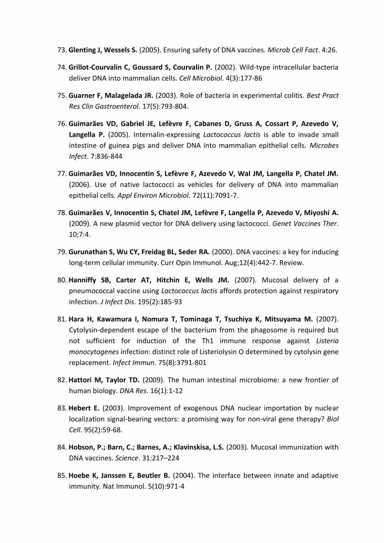
73. Glenting J, Wessels S. (2005). Ensuring safety of DNA vaccines. Microb Cell Fact. 4:26.
74. Grillot-‐Courvalin C, Goussard S, Courvalin P. (2002). Wild-‐type intracellular bacteria deliver DNA into mammalian cells. Cell Microbiol. 4(3):177-‐86
75. Guarner F, Malagelada JR. (2003). Role of bacteria in experimental colitis. Best Pract Res Clin Gastroenterol. 17(5):793-‐804.
76. Guimarães VD, Gabriel JE, Lefèvre F, Cabanes D, Gruss A, Cossart P, Azevedo V, Langella P. (2005). Internalin-‐expressing Lactococcus lactis is able to invade small intestine of guinea pigs and deliver DNA into mammalian epithelial cells. Microbes Infect. 7:836-‐844
77. Guimarães VD, Innocentin S, Lefèvre F, Azevedo V, Wal JM, Langella P, Chatel JM. (2006). Use of native lactococci as vehicles for delivery of DNA into mammalian epithelial cells. Appl Environ Microbiol. 72(11):7091-‐7.
78. Guimarães V, Innocentin S, Chatel JM, Lefèvre F, Langella P, Azevedo V, Miyoshi A. (2009). A new plasmid vector for DNA delivery using lactococci. Genet Vaccines Ther. 10;7:4.
79. Gurunathan S, Wu CY, Freidag BL, Seder RA. (2000). DNA vaccines: a key for inducing long-‐term cellular immunity. Curr Opin Immunol. Aug;12(4):442-‐7. Review.
80. Hanniffy SB, Carter AT, Hitchin E, Wells JM. (2007). Mucosal delivery of a pneumococcal vaccine using Lactococcus lactis affords protection against respiratory infection. J Infect Dis. 195(2):185-‐93
81. Hara H, Kawamura I, Nomura T, Tominaga T, Tsuchiya K, Mitsuyama M. (2007). Cytolysin-‐dependent escape of the bacterium from the phagosome is required but not sufficient for induction of the Th1 immune response against Listeria monocytogenes infection: distinct role of Listeriolysin O determined by cytolysin gene replacement. Infect Immun. 75(8):3791-‐801
82. Hattori M, Taylor TD. (2009). The human intestinal microbiome: a new frontier of human biology. DNA Res. 16(1):1-‐12
83. Hebert E. (2003). Improvement of exogenous DNA nuclear importation by nuclear localization signal-‐bearing vectors: a promising way for non-‐viral gene therapy? Biol Cell. 95(2):59-‐68.
84. Hobson, P.; Barn, C.; Barnes, A.; Klavinskisa, L.S. (2003). Mucosal immunization with DNA vaccines. Science. 31:217 224
85. Hoebe K, Janssen E, Beutler B. (2004). The interface between innate and adaptive immunity. Nat Immunol. 5(10):971-‐4

86. Hollmann A, Saviello M, Delfederico L, Saraiva TD, Barh D, Jain N, Tiwari S, Chandra S, Gupta K, Zambare V, Kumar A, Christopher L, Misra AN, Kumavath RN, Azevedo V, Semorile L, Miyoshi A. (2012). Tight controlled expression and secretion of Lactobacillus brevis SlpA in Lactococcus lactis. Biotechnol Lett. 34(7):1275-‐81
87. Howarth M, Elliott T. (2004). The processing of antigens delivered as DNA vaccines. Immunol Rev. 199:27-‐39
88. Hugentobler F, Di Roberto RB, Gillard J, Cousineau B. (2012). Oral immunization using live Lactococcus lactis co-‐expressing LACK and IL-‐12 protects BALB/c mice against Leishmania major infection. Vaccine. 30(39):5726-‐32
89. Iliev ID, Matteoli G, Rescigno M. (2007). The yin and yang of intestinal epithelial cells in controlling dendritic cell function. J Exp Med. 204(10):2253-‐7
90. Ingolotti M, Kawalekar O, Shedlock DJ, Muthumani K, Weiner DB. (2010). DNA vaccines for targeting bacterial infections. Expert Rev Vaccines. 9(7):747-‐63
91. Innocentin S, Guimarães V, Miyoshi A, Azevedo V, Langella P, Chatel JM, Lefèvre F. (2009). Lactococcus lactis expressing either Staphylococcus aureus fibronectin-‐binding protein A or Listeria monocytogenes internalin A can efficiently internalize and deliver DNA in human epithelial cells. Appl Environ Microbiol. 75:4870-‐4878
92. Izadpanah A, Dwinell MB, Eckmann L, Varki NM, Kagnoff MF. (2001). Regulated MIP-‐3alpha/CCL20 production by human intestinal epithelium: mechanism for modulating mucosal immunity. Am J Physiol Gastrointest Liver Physiol. 280:710-‐9
93. Iwaki M, Okahashi N, Takahashi I, Kanamoto T, Sugita-‐Konishi Y, Aibara K, Koga T. (1990). Oral immunization with recombinant Streptococcus lactis carrying the Streptococcus mutans surface protein antigen gene. Infect Immun. 58(9):2929-‐34
94. Jamet E. dans le métabolisme carboné chez Lactococcus lactis. Institut National Agronomique de Paris-‐Grignon
95. Janeway CA Jr. (2001). How the immune system protects the host from infection. Microbes Infect. 3(13):1167-‐71
96. Joffre OP, Segura E, Savina A, Amigorena S. (2012). Cross-‐presentation by dendritic cells. Nat Rev Immunol. 13;12(8):557-‐69.
97. Johansson ME, Phillipson M, Petersson J, Velcich A, Holm L, Hansson GC. (2008). The inner of the two Muc2 mucin-‐dependent mucus layers in colon is devoid of bacteria. Proc Natl Acad Sci USA. 30;105(39):15064-‐9
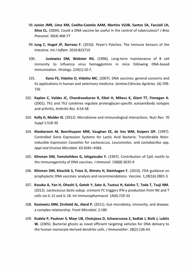
98. Junior JMR, Lima KM, Coelho-‐Castelo AAM, Martins VLDB, Santos SA, Faccioli LH, Silva CL. (2004). Could a DNA vaccine be useful in the control of tuberculosis? J Bras Pneumol. 30(4) 468-‐77
99. Jung C, Hugot JP, Barreau F. (2010). Peyer's Patches: The Immune Sensors of the Intestine. Int J Inflam. 2010:823710
100. Justewicz DM, Webster RG. (1996). Long-‐term maintenance of B cell immunity to influenza virus hemagglutinin in mice following DNA-‐based immunization. Virology. 224(1):10-‐7.
101. Kano FS, Vidotto O, Vidotto MC. (2007). DNA vaccines: general concerns and its applications in human and veterinary medicine. Semina:Ciências Agrárias. (4):709-‐726
102. Kaplan C, Valdez JC, Chandrasekaran R, Eibel H, Mikecz K, Glant TT, Finnegan A. (2001). Th1 and Th2 cytokines regulate proteoglycan-‐specific autoantibody isotypes and arthritis. Arthritis Res. 4:54-‐58
103. Kelly D, Mulder IE. (2012). Microbiome and immunological interactions. Nutr Rev. 70 Suppl 1:S18-‐30
104. Kleeberzem M, Beerthuyzen MM, Vaughan EE, de Vos WM, Kuipers OP. (1997). Controlled Gene Expression Systems for Lactic Acid Bacteria: Transferable Nisin-‐Inducible Expression Cassettes for Lactococcus, Leuconostoc, and Lactobacillus spp. Appl and Environ Microbiol. 63:4581 4584.
105. Klinman DM, Yamshchikov G, Ishigatsubo Y. (1997). Contribution of CpG motifs to the immunogenicity of DNA vaccines. J Immunol. 158(8):3635-‐9
106. Klinman DM, Klaschik S, Tross D, Shirota H, Steinhagen F. (2010). FDA guidance on prophylactic DNA vaccines: analysis and recommendations. Vaccine. 1;28(16):2801-‐5
107. Kosaka A, Yan H, Ohashi S, Gotoh Y, Sato A, Tsutsui H, Kaisho T, Toda T, Tsuji NM. (2012). Lactococcus lactis subsp. cremoris FC triggers IFN-‐cells via IL-‐12 and IL-‐18. Int Immunopharmacol. 14(4):729-‐33
108. Kosiewicz MM, Zirnheld AL, Alard P. (2011). Gut microbiota, immunity, and disease: a complex relationship. Front Microbiol. 2:180
109. Kudela P, Paukner S, Mayr UB, Cholujova D, Schwarczova Z, Sedlak J, Bizik J, Lubitz W. (2005). Bacterial ghosts as novel efficient targeting vehicles for DNA delivery to the human monocyte-‐derived dendritic cells. J Immunother. 28(2):136-‐43.

110. Kuipers OP, Beerthuyzen MM, Siezen RJ, De Vos WM. (1993). Characterization of the nisin gene cluster nisABTCIPR of Lactococcus lactis. Requirement of expression of the nisA and nisI genes for development of immunity. Eur J Biochem. 216(1):281-‐91
111. Kutzler MA, Weiner DB. (2008). DNA vaccines: ready for prime time? Nat Rev Genet. 9(10):776-‐88
112. Langella P, Le Loir Y. (1999). Heterologous protein secretion in Lactococcus lactis: a novel antigen delivery system. Braz J Med Biol Res. 32(2):191-‐8
113. Lafferty EI, Qureshi ST, Schnare M. (2010). The role of toll-‐like receptors in acute and chronic lung inflammation. J Inflamm (Lond). 25;7:57
114. Lamm ME. (1997). Interaction of antigens and antibodies at mucosal surfaces. Annu Rev Microbiol. 51:311-‐40
115. Le Loir Y, Azevedo V, Oliveira SC, Freitas DA, Miyoshi A, Bermudez-‐Humaran LG, Nouaille S, Ribeiro LA, Lececlercq S, Gabriel JE, Guimaraes VD, Oliveira MN, Charlier C, Gautier M, Langella P. (2005). Protein secretion in Lactococcus lactis: an efficient way to increase the overall heterologous protein production. Microb Cell Fact. 4(1):2
116. Lechardeur D, Sohn KJ, Haardt M, Joshi PB, Monck M, Graham RW, Beatty B, Squire J, O'Brodovich H, Lukacs GL. (1999). Metabolic instability of plasmid DNA in the cytosol: a potential barrier to gene transfer. Gene Ther. 6(4):482-‐97
117. Lecuit M, Ohayon H, Braun L, Mengaud J, Cossart P. (1997). Internalin of Listeria monocytogenes with an intact leucine-‐rich repeat region is sufficient to promote internalization. Infect Immun. 65(12):5309-‐19
118. Lee KC, Liu CF, Lin TH, Pan TM. (2010). Safety and risk assessment of the genetically modified Lactococci on rats intestinal bacterial flora. Int J Food Microbiol. 142(1-‐2):164-‐9
119. Lehmann MJ, Sczakiel G. (2005). Spontaneous uptake of biologically active recombinant DNA by mammalian cells via a selected DNA segment. Gene Ther. 12(5):446-‐51
120. Leifert JA, Whitton JL. (2000). Immune Responses to DNA Vaccines: Induction of CD8 T Cells. Madame Curie Bioscience Database. Austin (TX): Landes Bioscience
121. Levy MY, Barron LG, Meyer KB, Szoba Jr FC. (1996). Characterization of plasmid DNA transfer into mouse skeletal muscle: evaluation of uptake mechanism, expression and secretion of gene products into blood. Gene Ther. 3:201 11.
122. Li L, Saade F, Petrovsky N. (2012). The future of human DNA vaccines. J Biotechnol. S0168-‐1656(12)00614-‐1

123. Lichtor T, Glick RP. (2012). Immunogene therapy. Adv Exp Med Biol. 746:151-‐65.
124. Liu MA. (2003). DNA vaccines: a review. J Intern Med. 253(4):402-‐10
125. Liu MA, Ulmer JB. (2005). Human clinical trials of plasmid DNA vaccines. Adv Genet. 55:25-‐40
126. Liu MA. (2010). Immunologic basis of vaccine vectors. Immunity 33:504-‐515
127. Liu MA. (2011). DNA vaccines: an historical perspective and view to the future. Immunol Rev. 239(1):62-‐84
128. Loessner H, Endmann A, Leschner S, Bauer H, Zelmer A, zur Lage S, Westphal K, Weiss S. (2007). Improving live attenuated bacterial carriers for vaccination and therapy. Int J Med Microbiol. 298(1-‐2):21-‐6
129. Loessner H, Weiss S. (2004). Bacteria-‐mediated DNA transfer in gene therapy and vaccination. Expert Opin Biol Ther. 4(2):157-‐68
130. Lokman BC, Leer RJ, van Sorge R, Pouwels PH. (1994). Promoter analysis and transcriptional regulation of Lactobacillus pentosus genes involved in xylose catabolism. Mol Gen Genet. 245(1):117-‐25
131. Lu JM, Wang X, Marin-‐Muller C, Wang H, Lin PH, Yao Q, Chen C. (2009). Current advances in research and clinical applications of PLGA-‐based nanotechnology. Expert Rev Mol Diagn. 9:325-‐341.
132. Lu W, Kong J, Kong W. (2012). Construction and Application of a Food-‐Grade Expression System for Lactococcus lactis. Mol Biotechnol. Jun 7
133. Macauley-‐Patrick S, Fazenda ML, McNeil B, Harvey LM. (2005). Heterologous protein production using the Pichia pastoris expression system. Yeast. 22(4):249-‐70
134. Macpherson AJ, Hunziker L, McCoy K, Lamarre A. (2001). IgA responses in the intestinal mucosa against pathogenic and non-‐pathogenic microorganisms. Microbes Infect. 3(12):1021-‐35
135. Macpherson AJ, Harris NL. (2004). Interactions between commensal intestinal bacteria and the immune system. Nat Rev Immunol. 4(6):478-‐85
136. Mak TW, Saunders, ME. (2006). The Immune Response. Basic and Clinical Principles. Copyright © 2006 Elsevier Inc. ISBN: 978-‐0-‐12-‐088451-‐3
137. Malek TR, Castro I. (2010). Interleukin-‐2 receptor signaling: at the interface between tolerance and immunity. Immunity. 27;33(2):153-‐65

138. Mannion PT, Rothburn MM. (1990). Diagnosis of bacterial endocarditis caused by Streptococcus lactis and assisted by immunoblotting of serum antibodies. J Infect. 21(3):317-‐8
139. Mantis NJ, Forbes SJ. (2010). Secretory IgA: arresting microbial pathogens at epithelial borders. Immunol Invest. 39(4-‐5):383-‐406
140. Marinho FA, Pacífico LG, Miyoshi A, Azevedo V, Le Loir Y, Guimarães VD, Langella P, Cassali GD, Fonseca CT, Oliveira SC. (2010). An intranasal administration of Lactococcus lactis strains expressing recombinant interleukin-‐10 modulates acute allergic airway inflammation in a murine model. Clin Exp Allergy. 40(10):1541-‐51
141. Marra A, Isberg RR. (1996). Analysis of the role of invasin during Yersinia pseudotuberculosis infection of mice. Ann N Y Acad Sci. 25;797:290-‐2
142. Marreddy RK, Pinto JP, Wolters JC, Geertsma ER, Fusetti F, Permentier HP, Kuipers OP, Kok J, Poolman B. (2011). The response of Lactococcus lactis to membrane protein production. PLoS One. 6(8):e24060.
143. Masood MI, Qadir MI, Shirazi JH, Khan IU. (2011). Beneficial effects of lactic acid bacteria on human beings. Crit Rev Microbiol. 37(1):91-‐8
144. McCluskie MJ, Brazolot Millan CL, Gramzinski RA, Robinson HL, Santoro JC, Fuller JT, Widera G, Haynes JR, Purcell RH, Davis HL. (1999). Route and method of delivery of DNA vaccine influence immune responses in mice and non-‐human primates. Mol Med. 5(5):287-‐300
145. Mengaud J, Ohayon H, Gounon P, Mege R-‐M, Cossart P. (1996). E-‐cadherin is the receptor for internalin, a surface protein required for entry of L. monocytogenes into epithelial cells. Cell. 22;84(6):923-‐32
146. Mercenier A, Müller-‐Alouf H, Grangette C. (2000). Lactic acid bacteria as live vaccines. Curr Issues Mol Biol. 2(1):17-‐25
147. Wells JM, Mercenier A. (2008). Mucosal delivery of therapeutic and prophylactic molecules using lactic acid bacteria. Nat Rev Microbiol. 6(5):349-‐62
148. Metchnikoff, I.I. (1907). The Prolongation of Life, London.
149. Miyoshi A, Bermúdez-‐Humarán LG, Ribeiro LA, Le Loir Y, Oliveira SC, Langella P, Azevedo V. (2006). Heterologous expression of Brucella abortus GroEL heat-‐shock protein in Lactococcus lactis. Microbial Cell Factories. 5:1-‐8
150. Mierau I, Kleerebezem M. (2005). 10 years of the nisin-‐controlled gene expression system (NICE) in Lactococcus lactis. Appl Microbiol Biotechnol. 68(6):705-‐17

151. Miyoshi A, Jamet E, Commissaire J, Renault P, Langella P, Azevedo V. (2004). A xylose-‐inducible expression system for Lactococcus lactis. FEMS Microbiol Lett. 239(2):205-‐12
152. Mierau I, Leij P, van Swam I, Blommestein B, Floris E, Mond J, Smid EJ. (2005). Industrial-‐scale production and purification of a heterologous protein in Lactococcus lactis using the nisin-‐controlled gene expression system NICE: the case of lysostaphin. Microb Cell Fact. 27;4:15
153. Morello E, Nouaille S, Cortes-‐Perez NG, Blugeon S, Medina LF, Azevedo V, Gratadoux JJ, Bermúdez-‐Humarán LG, Le Loir Y, Langella P. (2012). Inactivation of the ybdD gene in Lactococcus lactis increases the amounts of exported proteins. Appl Environ Microbiol. 78(19):7148-‐51
154. Monk IR, Casey PG, Hill C, Gahan CG. (2010). Directed evolution and targeted mutagenesis to murinize Listeria monocytogenes internalin A for enhanced infectivity in the murine oral infection model. BMC Microbiol. 13;10:318
155. Mosmann TR, Sad S. (1996). The expanding universe of T-‐cell subsets: Th1, Th2 and more. Immunol Today. 17:138-‐46
156. Mulder DJ, Pooni A, Mak N, Hurlbut DJ, Basta S, Justinich CJ. (2011). Antigen presentation and MHC class II expression by human esophageal epithelial cells: role in eosinophilic esophagitis. Am J Pathol. 178(2):744-‐53
157. Nagabhushanam V, Cheers C. (2001). Non-‐major histocompatibility complex control of antibody isotype and Th1 versus Th2 cytokines during experimental infection of mice with Mycobacterium avium. Infect Immun. 69(3):1708-‐13
158. Neefjes J, Sadaka C. (2012). Into the intracellular logistics of cross-‐presentation. Front Immunol. 3:31
159. Neish AS. (2009). Microbes in gastrointestinal health and disease. Gastroenterology. 136(1):65-‐80
160. Neutra MR, Kozlowski PA. (2006). Mucosal vaccines: the promise and the challenge. Nat Rev Immunol. 6(2):148-‐58
161. Niidome T, Huang L. (2002). Gene therapy progress and prospects: nonviral vectors. Gene Ther. 9(24):1647-‐52
162. Nikitas G, Deschamps C, Disson O, Niault T, Cossart P, Lecuit M. (2011). Transcytosis of Listeria monocytogenes across the intestinal barrier upon specific targeting of goblet cell accessible E-‐cadherin. J Exp Med. 208(11):2263-‐77

163. Norton PM, Brown HW, Le Page RW. (1995). Mucosal and systemic responses following enteric exposure to lactic acid bacteria. Adv Exp Med Biol. 371B:1559-‐62
164. Nouaille S.; Ribeiro, L.A.; Miyoshi, A.; Pontes, D.; Le Loir, Y.; Oliveira, S.C.; Langella, P.; Azevedo, V. (2003) Heterologous protein production and delivery systems for Lactococcus lactis. Genet Mol Res. 31;2(1):102-‐11
165. L, Malakauskas A, Laukkanen-‐Ninios R, Korkeala H, Malakauskas M. (2012). Prevalence and genetic diversity of enteropathogenic Yersinia spp. in pigs at farms and slaughter in Lithuania. Res Vet Sci. S0034-‐5288(12)00299-‐8
166. Ohl ME, Miller SI (2001). Salmonella: AModel for Bacterial Pathogenesis. Annu. Rev. Med. 52:259 74
167. Ozeri V, Rosenshine I, Mosher DF, Fässler R, Hanski E. (1998). Roles of integrins and fibronectin in the entry of Streptococcus pyogenes into cells via protein F1. Mol Microbiol. 30:625-‐637
168. Paglia P, Medina E, Arioli I, Guzman CA, Colombo MP. (1998). Gene transfer in dendritic cells, induced by oral DNA vaccination with Salmonella typhimurium, results in protective immunity against a murine fibrosarcoma. Blood. 92(9):3172-‐6.
169. Parsa S., Pfeifer B. (2007). Engineering bacterial vectors for delivery of genes and proteins to antigen-‐presenting cells. Mol Pharm. 4:4-‐17
170. Perdigón, G., Fuller, R., Raya, R. (2001) Lactic acid bacteria and their effect on the immune system. Curr. Issues Intest. Microbiol. 2(1): 27-‐42
171. Perdigón G, Maldonado Galdeano C, Valdez JC, Medici M. (2002). Interaction of lactic acid bacteria with the gut immune system. Eur J Clin Nutr. 56 Suppl 4:S21-‐6
172. Pichon C, Billiet L, Midoux P. (2010). Chemical vectors for gene delivery: uptake and intracellular trafficking. Curr Opin Biotechnol. 21(5):640-‐5
173. Pilgrim S, Stritzker J, Schoen C, Kolb-‐Mäurer A, Geginat G, Loessner MJ, Gentschev I, Goebel W. (2003). Bactofection of mammalian cells by Listeria monocytogenes: improvement and mechanism of DNA delivery. Gene Ther. 10(24):2036-‐45
174. Plotkin SA. (2005). Vaccines: past, present and future. Nat Med. 11(4 Suppl):S5-‐11
175. Pokorna D, Rubio I, Müller M. (2008). DNA-‐vaccination via tattooing induces stronger humoral and cellular immune responses than intramuscular delivery supported by molecular adjuvants. Genet Vaccines Ther. 7;6:4

176. Pontes DS, de Azevedo MS, Chatel JM, Langella P, Azevedo V, Miyoshi A. (2011). Lactococcus lactis as a live vector: heterologous protein production and DNA delivery systems. Protein Expr Purif. 79(2):165-‐75
177. Pontes D, Innocentin S, Del Carmen S, Almeida JF, Leblanc JG, de Moreno de Leblanc A, Blugeon S, Cherbuy C, Lefèvre F, Azevedo V, Miyoshi A, Langella P, Chatel JM. (2012). Production of Fibronectin Binding Protein A at the Surface of Lactococcus lactis Increases Plasmid Transfer In Vitro and In Vivo. PLoS One. 7(9):e44892
178. Pool-‐Zobel BL, Neudecker C, Domizlaff I, Ji S, Schillinger U, Rumney C, Moretti M, Vilarini I, Scassellati-‐Sforzolini R, Rowland I. (1996). Lactobacillus-‐ and bifidobacterium-‐mediated antigenotoxicity in the colon of rats. Nutr Cancer. 26(3):365-‐80
179. Prakash S, Tomaro-‐Duchesneau C, Saha S, Cantor A. (2011). The gut microbiota and human health with an emphasis on the use of microencapsulated bacterial cells. J Biomed Biotechnol. 2011:981214
180. Preidis GA, Versalovic J. (2009). Targeting the human microbiome with antibiotics, probiotics, and prebiotics: gastroenterology enters the metagenomics era. Gastroenterology. 136(6):2015-‐31
181. Price CE, Zeyniyev A, Kuipers OP, Kok J. (2011). From meadows to milk to mucosa -‐ adaptation of Streptococcus and Lactococcus species to their nutritional environments. FEMS Microbiol. 36 (5) 949 971
182. Que YA, François P, Haefliger JA, Entenza JM, Vaudaux P, Moreillon P. (2001) Reassessing the role of Staphylococcus aureus clumping factor and fibronectin-‐binding protein by expression in Lactococcus lactis. Infect Immun. 69(10):6296-‐302
183. Radtke AL, Anderson KL, Davis MJ, DiMagno MJ, Swanson JA, O'Riordan MX. (2011). Listeria monocytogenes exploits cystic fibrosis transmembrane conductance regulator (CFTR) to escape the phagosome. Proc Natl Acad Sci U S A. 108(4):1633-‐8
184. Rescigno M, Urbano M, Valzasina B, Francolini M, Rotta G, Bonasio R, Granucci F, Kraehenbuhl JP, Ricciardi-‐Castagnoli P. (2001). Dendritic cells express tight junction proteins and penetrate gut epithelial monolayers to sample bacteria. Nat Immunol. 2(4):361-‐7
185. Rescigno M. (2009). Gut commensal flora: tolerance and homeostasis. F1000 Biol Rep. 1:9

186. Resta-‐Lenert S, Barrett KE. (2003). Live probiotics protect intestinal epithelial cells from the effects of infection with enteroinvasive Escherichia coli (EIEC). Gut. 52(7):988-‐97
187. Rigato PO, Maciel M Jr, Goldoni AL, Piubelli OG, Orii NM, Marques ET, August JT, Duarte AJ, Sato MN. (2012). Maternal LAMP/p55gagHIV-‐1 DNA immunization induces in utero priming and a long-‐lasting immune response in vaccinated neonates. PLoS One. 7(2):e31608
188. Rimoldi M, Chieppa M, Vulcano M, Allavena P, Rescigno M. (2004). Intestinal epithelial cells control dendritic cell function. Ann N Y Acad Sci. 1029:66-‐74
189. Robinson, K.; Chamberlain, L.M.; Schofield, K.M.; Wells, J.M.; Le Page, R.W. (1997). Oral Vaccination Of Mice Against Tetanus Using Recombinant Lactococcus Lactis. Nature Biotechnology. 15: 653-‐657
190. Sandberg JK, Glas R. (2001).Antigen Processing. Encyclopedia of life sciences/Nature Publishing Group. 1-‐6
191. Sansonetti PJ. (2004). War and peace at mucosal surfaces. Nat Rev Immunol. 4(12):953-‐64
192. Santos Rocha C, Lakhdari O, Blottière HM, Blugeon S, Sokol H, Bermúdez-‐Humarán LG, Azevedo V, Miyoshi A, Doré J, Langella P, Maguin E, van de Guchte M. (2011). Anti-‐inflammatory properties of dairy lactobacilli. Inflamm Bowel Dis. 18(4):657-‐66
193. Scavone P, Miyoshi A, Rial A, Chabalgoity A, Langella P, Azevedo V, Zunino P. (2007). Intranasal Immunisation With Recombinant Lactococcus Lactis Displaying Either Anchored Or Secreted Forms Of Proteus Mirabilis Mrpa Fimbrial Protein Confers Specific Immune Response And Induces A Significant Reduction Of Kidney Bacterial Colonisation In Mice. Microbes Infect. 9(7):821-‐8
194. Schoen C, Stritzker J, Goebel W, Pilgrim S. (2004). Bacteria as DNA vaccine carriers for genetic immunization. Int J Med Microbiol. 294(5):319-‐35
195. Seow WY, Yang YY, George AJ. (2009). Oligopeptide-‐mediated gene transfer into mouse corneal endothelial cells: expression, design optimization, uptake mechanism and nuclear localization. Nucleic Acids Res. 37(18):6276-‐89
196. Sinha B, François PP, Nüsse O, Foti M, Hartford OM, Vaudaux P, Foster TJ, Lew DP, Herrmann M, Krause KH. (1999). Fibronectin-‐binding protein acts as Staphylococcus aureus invasin via fibronectin bridging to integrin alpha5beta1. Cell Microbiol. 1(2):101-‐17
197. Sizemore DR, Branstrom AA, Sadoff JC. (1995). Attenuated Shigella as a DNA delivery vehicle for DNA-‐mediated immunization. Science. 13;270(5234):299-‐302

198. Sleator RD, Hill C. (2006). Patho-‐biotechnology: using bad bugs to do good things. Curr Opin Biotechnol. 17(2):211-‐6
199. Smelt MJ, de Haan BJ, Bron PA, van Swam I, Meijerink M, Wells JM, Faas MM, de Vos P. (2012). L. plantarum, L. salivarius, and L. lactis attenuate Th2 responses and increase Treg frequencies in healthy mice in a strain dependent manner. PLoS One. 7(10):e47244
200. Srivastava IK, Liu MA. (2003). Gene vaccines. Ann Intern Med. 1;138(7):550-‐9
201. Steidler L, Rottiers P. (2006). Therapeutic drug delivery by genetically modified Lactococcus lactis. Ann N Y Acad Sci. 1072:176-‐86
202. Suzuki K, Kawamoto S, Maruya M, Fagarasan S. (2010). GALT: organization and dynamics leading to IgA synthesis. Adv Immunol. 107:153-‐85
203. Tang DC, DeVit M, Johnston SA. (1992). Genetic immunization is a simple method for eliciting an immune response. Nature. 356:152-‐4
204. Tao L, Pavlova SI, Ji X, Jin L, Spear G. (2011). A novel plasmid for delivering genes into mammalian cells with noninvasive food and commensal lactic acid bacteria. Plasmid. 65(1):8-‐14
205. Tarahomjoo S. (2012). Development of vaccine delivery vehicles based on lactic acid bacteria. Mol Biotechnol. 51(2):183-‐99
206. Thomas CE, Ehrhardt A, Kay MA. (2003). Progress and problems with the use of viral vectors for gene therapy. Nat Rev Genet. 4(5):346-‐58
207. Tsang C, Babiuk S, van Drunen Littel-‐van den Hurk S, Babiuk LA, Griebel P. (2007). A single DNA immunization in combination with electroporation prolongs the primary immune response and maintains immune memory for six months. Vaccine. 25(30):5485-‐94
208. Tangney M, Gahan CG. (2010). Listeria monocytogenes as a vector for anti-‐cancer therapies. Curr Gene Ther. 2010 Feb;10(1):46-‐55.
209. Ulmer JB, Donnelly JJ, Parker SE, Rhodes GH, Felgner PL, Dwarki VJ, Gromkowski SH, Deck RR, DeWitt CM, Friedman A. (1993). Heterologous protection against influenza by injection of DNA encoding a viral protein. Science. 19 259(5102):1745-‐9
210. Vacik J, Dean BS, Zimmer WE, Dean DA. (1999). Cell-‐specific nuclear import of plasmid DNA. Gene Ther. 6: 1006
211. Vanniasinkam T, Reddy ST, Ertl HC. (2006). DNA immunization using a non-‐viral promoter. Virology. 344(2):412-‐20

212. Van Asseldonk M, Rutten G, Oteman M, Siezen RJ, de Vos WM, Simons G. (1990). Cloning of usp45, a gene encoding a secreted protein from Lactococcus lactis subsp. lactis MG1363. Gene. 95(1):155-‐60
213. Van den Bossche J, Malissen B, Mantovani A, De Baetselier P, Van Ginderachter JA. (2012). Regulation and function of the E-‐cadherin/catenin complex in cells of the monocyte-‐macrophage lineage and DCs. Blood. 119(7):1623-‐33
214. Van de Guchte M, Ehrlich SD, Maguin E. (2001). Production of growth-‐inhibiting factors by Lactobacillus delbrueckii. J Appl Microbiol. 91(1):147-‐53
215. Varkouhi AK, Mountrichas G, Schiffelers RM, Lammers T, Storm G, Pispas S, Hennink WE. (2012). Polyplexes based on cationic polymers with strong nucleic acid binding properties. Eur J Pharm Sci. 45(4):459-‐66
216. Vassaux G, Nitcheu J, Jezzard S, Lemoine NR. (2006). Bacterial gene therapy strategies. J Pathol. 208(2):290-‐8
217. Vaughan EE, Dean DA. (2006). Intracellular trafficking of plasmids during transfection is mediated by microtubules. Mol Ther. 13(2):422-‐8
218. Villatoro-‐Hernández J, Kuipers OP, Saucedo-‐Cárdenas O, Montes-‐de-‐Oca-‐Luna R. (2012). Heterologous protein expression by Lactococcus lactis. Methods Mol Biol. 824:155-‐65
219. Wang Z, Troilo PJ, Wang X, Griffiths TG, Pacchione SJ, Barnum AB. (2004). Detection of integration of plasmid DNA into host genomic DNA following intramuscular injection and electroporation. Gene Ther. 11:711 21
220. Wang Y, Wang J, Dai W. (2011). Use of GFP to trace the colonization of Lactococcus lactis WH-‐C1 in the gastrointestinal tract of mice. J Microbiol Methods. 86(3):390-‐2
221. Watterlot L, Meyrand M, Gaide N, Kharrat P, Blugeon S, Gratadoux JJ, Flores MJ, Langella P, Chapot-‐Chartier MP, Bermúdez-‐Humarán LG. (2010). Variations of N-‐acetylation level of peptidoglycan do not influence persistence of Lactococcus lactis in the gastrointestinal tract. Int J Food Microbiol. 144(1):29-‐34
222. Weiss S, Chakraborty T. (2001). Transfer of eukaryotic expression plasmids to mammalian host cells by bacterial carriers. Curr Opin Biotechnol. 12(5):467-‐72
223. Wells JM, Wilson PW, Norton PM, Gasson MJ, Le Page RW. (1993). Lactococcus Lactis: High-‐Level Expression Of Tetanus Toxin Fragment C And Protection Against Lethal Challenge. Mol. Microbiol. 8:1155-‐1162
224. Wells, J. (2011). Mucosal Vaccination and Therapy with Genetically Modified Lactic Acid Bacteria. Annu. Rev. Food Sci. Technol. 2:423-‐445

225. Wells, JM, Mercenier, A. (2008). Mucosal delivery of therapeutic and prophylactic molecules using lactic acid bacteria. Nat Rev Microbiol. 6:349-‐62
226. Wen J, Yang Y, Zhao G, Tong S, Yu H, Jin X, Du L, Jiang S, Kou Z, Zhou Y. (2012). Salmonella typhi Ty21a bacterial ghost vector augments HIV-‐1 gp140 DNA vaccine-‐induced peripheral and mucosal antibody responses via TLR4 pathway. Vaccine. 30(39):5733-‐9
227. Williams JA, Carnes AE, Hodgson CP. (2009). Plasmid DNA vaccine vector design: impact on efficacy, safety and upstream production. Biotechnol Adv. 27(4):353-‐70
228. WHO. (2007). Annex 1: Guidelines for assuring the quality and nonclinical safety evaluation of DNA vaccines. World Health Organization Technical Report Series No 941
229. WHO. (2002). Guidelines for the Evaluation of Probiotics in Food: Joint FAO/WHO Working Group meeting, London Ontario, Canada, 30 April-‐1 May
230. Wolff JA, Dowty ME, Jiao S, Repetto G, Berg RK, Ludtke JJ, Williams P, Slautterback DB. (1992). Expression of naked plasmids by cultured myotubes and entry of plasmids into T tubules and caveolae of mammalian skeletal muscle. J Cell Sci. 103
231. Wollert T, Pasche B, Rochon M, Deppenmeier S, van den Heuvel J, Gruber AD, Heinz DW, Lengeling A, Schubert WD. (2007). Extending the host range of Listeria monocytogenes by rational protein design. Cell. 129(5):891-‐902
232. Xiang SD, Selomulya C, Ho J, Apostolopoulos V, Plebanski M. (2010). Delivery of DNA vaccines: an overview on the use of biodegradable polymeric and magnetic nanoparticles. Wiley Interdiscip Rev Nanomed Nanobiotechnol. 2(3):205-‐18
233. Xin KQ, Hoshino Y, Toda Y, Igimi S, Kojima Y, Jounai N, Ohba K, Kushiro A, Kiwaki M, Hamajima K, Klinman D, Okuda K. (2003). Immunogenicity And Protective Efficacy Of Orally Administered Recombinant Lactococcus Lactis Expressing Surface-‐Bound Hiv Env. Blood. 102(1):223-‐8
234. Yang NS, Burkholder J, McCabe D, Neumann V, Fuller D. (2001). Particle-‐mediated gene delivery in vivo and in vitro. Curr Protoc Hum Genet. Chapter 12:Unit 12.6
235. Yan F, Polk DB. (2004). Commensal bacteria in the gut: learning who our friends are. Curr Opin Gastroenterol. 20(6):565-‐71
236. Yu DS, Lee CF, Hsieh DS, Chang SY. (2004). Antitumor effects of recombinant BCG and interleukin-‐12 DNA vaccines on xenografted murine bladder cancer. Urology. 63(3):596-‐601

237. Yu X, Jia R, Huang J, Shu B, Zhu D, Liu Q, Gao X, Lin M, Yin Z, Wang M, Chen S, Wang Y, Chen X, Cheng A. (2012). Attenuated Salmonella typhimurium delivering DNA vaccine encoding duck enteritis virus UL24 induced systemic and mucosal immune responses and conferred good protection against challenge. Vet Res. 43(1):56
238. Zanoni I, Granucci F. (2012). Regulation and dysregulation of innate immunity by NFAT signaling downstream of pattern recognition receptors (PRRs). Eur J Immunol. 42(8):1924-‐31
239. Zhang KQ, Yang F, Ye J, Jiang M, Liu Y, Jin FS, Wu YZ. (2012). A novel DNA/peptide combined vaccine induces PSCA-‐specific cytotoxic T-‐lymphocyte responses and suppresses tumor growth in experimental prostate cancer. Urology. 79(6):1410.e7-‐13
240. Zho F, Neutra MR. (2002). Antigen delivery to mucosa-‐associated lymphoid tissues using liposomes as a carrier. Biosci Rep. 22(2):355-‐69
241. Zoumpopoulou G, Tsakalidou E, Dewulf J, Pot B, Grangette C. (2009). Differential crosstalk between epithelial cells, dendritic cells and bacteria in a co-‐culture model. Int J Food Microbiol. 131(1):40-‐51
242. Zamri HF, Shamsudin MN, Rahim RA, Neela V. (2012). Oral vaccination with Lactococcus lactis expressing the Vibrio cholerae Wzm protein to enhance mucosal and systemic immunity. Vaccine. 30(21):3231-‐8

APPENDICE 1-‐ Recombinant Lactococcus lactis expressing both
Listeria monocytogenes Lysteriolysin O and mutated internalin A applied for DNA vaccination

161
Perspectives on the use of recombinant Lactococcus lactis expressing both Listeriolysin O and mutated
internalin A from Listeria monocytogenes as a tool for DNA vaccination
Marcela de Azevedo3, Clarissa Santos Rocha3, Vanessa Bastos Pereira3, Vasco Azevedo3, Jean-Marc Chatel1,2 and
Anderson Miyoshi3,*
1. Institut National de la Recherche Agronomique (INRA), MICALIS (UMR 1319), Domaine de Vilvert, F-78352 Jouy-en-
Josas, France
2. AgroParisTech, UMR Micalis, F-78350 Jouy-en-Josas, France.
3. Laboratório de Genética Celular e Molecular, Instituto de Ciências Biológicas, Universidade Federal de Minas Gerais
(ICB/UFMG), Belo Horizonte-MG, Brazil.
* Corresponding author: Anderson Miyoshi; Instituto de Ciências Biológicas (ICB), Universidade Federal de Minas Gerais
(UFMG). Av. Antônio Carlos, 6627, Pampulha, Belo Horizonte, MG, Brazil, CEP 31270-901. E-mail:
Abstract: Lactococcus lactis have been engineered in this work to express mutated internalin A at its surface and to
secrete great amounts of Listeriolysin O (LLO), both proteins derived from the food born pathogen Listeria
monocytogenes, in order to be used as a tool for DNA vaccination. Western Blot experiments demonstrated that the
bacterium was able to express LLO in both cytoplasmic and extracellular compartments, with a higher quantity found in
culture supernatants. Hemolytic assay have shown that the recombinant strain is capable of secreting 250 ng of LLO/mg
of total protein in active format. Finally, we believe that mInlA/LLO L. lactis producing this strain could have the potential
to be used as an alternative tool in DNA vaccination against a number of infectious diseases or in cancer therapy.
Key words: Lactic Acid Bacteria, Lactococcus lactis, Listeria monocytogenes, Listeriolysin O, Mutated internalin A

162
Introduction
The strategy to use GRAS (Generally Regarded as Safe) bacteria, such as the most economically important Lactic Acid
Bacteria (LAB), Lactococcus lactis, as a vector to deliver therapeutic plasmids seems a very interesting alternative
approach. L. lactis has been proposed as a safe and effective vaccine platform for the delivery of therapeutic molecules
to the immune system.1 It has been shown that wild type (wt) L. lactis is capable to deliver DNA vaccines both in vitro
and in vivo after oral inoculation of mice.2.3 Nevertheless, the ratio of DNA transferred to mammalian cells observed was
low. We hypothesized that this low efficiency detected could be due to the transient nature of L. lactis which might avoid
its optimal interaction with intestinal epithelial cells (IECs) from the gastrointestinal tract and therefore prevent DNA
translocation. Thus, strains of L. lactis expressing recombinant virulent genes were recently constructed in attempt to
improve their capacity to stay longer in the gastrointestinal tract and therefore deliver more DNA vaccines.4.5 Guimaraes
and co-workers were one of the first to explore the potential use of recombinant L. lactis strains engineered to express
invasins naturally produced by enteropathogenic species, such as Internalin A (InlA) from the food-borne pathogen L.
monocytogenes.2,6 InlA is a sortase anchored cell wall protein which contains 800 amino acids (aa) in length that
mediates bacterial entry into mammalian epithelial cells.7 Due to InlA, L. monocytogenes can invade mucosal surfaces in
-based immune system, including intestinal dendritic cells, which are
potent antigen presenting cells.8 InlA-expressing L. lactis (LL-InlA+) strain showed to be able to invade Caco-2 cells in
vitro and also intestinal cells after oral immunization in guinea pigs. Furthermore, LL-InlA+ strain were able to deliver a
functional eukaryotic gfp gene (green fluorescent protein) into epithelial Caco-2 cells more efficiently than the wt
lactococci demonstrating to be a good tool for gene delivery.2 Nevertheless, even though interesting, the use of LL-InlA+
strain presented some bottlenecks as InlA can only bind to its receptor (E-cadherin) in guinea pigs or transgenic mice,
turning in vivo studies laborious and/or expensive.9 For this reason, an L. lactis strain producing a mutated form of InlA
(LL-mInlA+), which can bind to E-cadherin from conventional mice was recently reported by our research group. Mutated
internalin A (mInlA) rendered L. lactis an invasive status, improving its interaction with intestinal epithelial cells, leading to
a higher level of DNA transfer in vitro.10 As in vivo the use of LL-mInlA+ did not statically increased the DNA delivery, we
decided to construct another strain able to express both mInlA and another virulence factor from L. monocytogenes, the
haemolysin listeriolysin O (LLO). In this pathogen LLO expression is up-regulated at low pH (~pH 5.5) inside de
phagosome which then oligomerize to form a pre-pore complex, rupturing the Listeria-containing phagosome.11.12
Therefore, the expression of LLO in L. lactis would in theory facilitate the escape of the plasmid DNA from the cytoplasm
to the nucleus. Thus, the result could be the cellular expression of the transfected gene in a higher efficiency. Actually, it
was already demonstrated that LLO incorporated in an anionic liposome was very suitable to enhance plasmid DNA
delivery.13 Besides increasing plasmid transfer, it was demonstrated that LLO-containing pH-sensitive liposomes
encapsulated with immunostimulatory CpG oligonucleotides was able to efficiently deliver Ovalbumin (OVA) antigen to
the cytosol of APCs and stimulate cytotoxic T lymphocytes (CTLs) driving Th1-type immune responses.14 Another
research group demonstrated that LLO-liposome-mediating cytosolic delivery of antigens in vivo could enhance antigen-
specific cytotoxic T lymphocyte frequency, activity conferring tumor protection.15 Thus, in the present work, food-grade L.

163
lactis NZ9000 strain was engineered to constitutively express both mInlA and LLO. This strain might then serve as a
vehicle for DNA vaccination in near future.
Results
L. lactis is able to successfully express mutated internalin A on its surface.
In this work we proposed two approaches thought to enhance plasmid transfer both in vitro and in vivo by expressing in
L. lactis two major immunodominant antigens, LLO and mInlA, derived from L monocytogenes. After transformation of L.
lactis NZ9000 strain with pLL31 plasmid by electroporation, flow cytometry analyses were performed to firstly detect
recombinant mInlA. Plasmid pLL31 harbours the native hlyA gene (encoding LLO) containing its constitutive promoter,
the signal peptide and the ribosomal binding site. It also contains the mutated internalin A ORF, the constitutive PrfA
promoter protein and the cell wall anchoring signal. L. lactis expressing native InlA, wt L. lactis NZ9000 and the strain
constructed in this work producing both LLO and mInlA (LL-mInlA+LLO+) were incubated with monoclonal antibody anti-
InlA and afterwards with (FITC)-conjugated AffiniPure Fab fragment Goat Anti-Mouse IgG (H+L) (Jackson Immuno
Research). Data was then analyzed by flow cytometry analysis. As demonstrated in Figure 1, strains producing mInlA
(blue and yellow peak) or native InlA (red peak) could significantly shift the distribution curve comparing to the wt strain
(black peak). Fluorescence intensity obtained for mInlA producing strain was two times higher compared to the wt strain
while LL-InlA could express four times more recombinant protein. This assay confirmed the expression of mInlA on the
surface of L. lactis.
L. monocytogenes Listeriolysin O is constitutively produced and secreted by L. lactis.
After confirming mInlA expression on L. lactis surface, western blotting experiments were conducted with the intent to
detect recombinant LLO in L. lactis cytoplasmic and extracellular cell compartments. Strains (wt and LL-mInlA+LLO+)
were grown for protein extraction. Both supernatant and cytoplasmic proteins were collected and submitted to Sodium
dodecyl sulfate polyacrylamide gel electrophoresis (SDS PAGE). Gels were then blotted against a nitrocellulose
membrane, which was incubated with primary anti-LLO antibody (abcam, France) and secondary goat Anti-
heavy chain specific) and signals of LLO were determined. Analysis of stationary-phase cell lysates showed the
presence of a band at the expected position, which is in accordance with the molecular weight of LLO (58 kDa) in both
soluble and insoluble cytoplasmic protein fractions as well as in culture supernatants, with a great amount of recombinant
protein found in this compartment (Figure 2). LLO was not detected in protein extracts from supernatant and cytoplasmic
compartments derived from the wt strain.
Recombinant Listeriolysin O is produced as an active pore-forming toxin in L. lactis.
Subsequently, the haemolytic assay was carried out to assess if LLO was being produced as an active protein. Culture
supernatant and cytoplasmic proteins derived from both wt and LL-mInlA+LLO+ strains were extracted and then used in
the assay. A purified recombinant LLO (rLLO) was used as a standard to measure protein activity and to quantify the
amount of LLO produced by L. lactis. Either rLLO or protein preparations from wt and L. lactis producing mInlA-LLO
strain were incubated with Red Blood Cells. Afterwards, hemolytic activity was measured at 540 nm in a

164
spectrophotometer and LLO production was quantified using the same approach. As demonstrated in Figure 3, protein
extracts (both soluble and insoluble) as well as culture supernatants derived from recombinant L. lactis revealed a strong
haemolytic activity when compared to the negative control (LLwt). We also observed that LLO from the insoluble
compartment has pore-forming activity similar to LLO derived from the soluble fraction. Furthermore, we also checked
that the highest hemolytic activity was found in the culture supernatant. Moreover, almost 250 ng/mg of total protein were
found to be constitutively secreted by L. lactis. This hemolytic assay clearly revealed that LLO is well secreted in L. lactis
as a highly active pore-forming toxin.
Discussion
Attenuated strains of L. monocytogenes have been proposed as a vehicle to deliver therapeutic plasmids to eukaryotic
cells. This bacterium has the capacity to induce its own uptake into nonphagocytic mammalian cells by the expression of
InlA and to form pores in phagolysosomal membranes due to the production of LLO facilitating DNA plasmid transfer to
the mammalian cell nucleus.8,16 Several preclinical studies have demonstrated the ability of L. monocytogenes for
intracellular gene delivery. Furthermore, this vector has also displayed a relatively safety and efficacy in some clinical
trial.16 However, even though attractive, there is always the insecurity that attenuated vectors might restore the ability to
replicate and cause disease in the patient. This fact should be taken into further consideration when it comes to the
administration in infants and immuno-compromised individuals. Actually, reversion to virulence, preexisting immunity,
and reactogenicity will always remain major concerns when pathogenic species are considered as a vaccine platform.17
Thus, the scientific community has recently been exploring the use of non-pathogenic bacteria, such as lactic acid
bacteria (LAB), as prophylactic or therapeutic tools and more recently as a DNA delivery vehicle for genetic
immunization.5,18 In 2008, Chatel and collaborators have shown that the model LAB, L. lactis, was able to deliver a
plasmid to the epithelial cells of the intestinal membrane of conventional mice. Besides this study has demonstrated that
L. lactis could be used as a vector for genetic immunization, the ratio of plasmid transfer observed was low.3 The
transient physiology of L. lactis was considered to be the cause of this limitation. Thus, recent research is focusing on the
construction of recombinant invasive lactococci strains as this feature could increase plasmid DNA delivery.4,5,10 One
very interesting strategy performed by Guimarães and collaborators was to engineer L. lactis to express InlA (LL-InlA+)
from L. monocytogenes. The recombinant strain proved to be able to enter intestinal cells in vivo, after oral inoculation of
guinea pigs. After internalization, LL-InlA+ was able to deliver a functional eukaryotic gfp (green fluorescent protein) into
epithelial Caco-2 cells.2 Nevertheless, the disadvantage was the limitation of the strain in in vivo experiments as InlA
does not bind to its receptor, E-cadherin, in conventional mice. Therefore, a mutated form of InlA that can bind to murine
E-cadherin, mInlA, was successfully expressed in L. lactis thus facilitating in vivo studies. De Azevedo et al.2
demonstrated that the expression of mInlA at L. lactis surface could improve its invasive capacity in experiments
performed with IECs. Moreover, the invasive characteristic increased the DNA vaccine delivery in vitro but not in vivo.10
In this work we decided to engineer L. lactis to express besides mInlA another virulent factor of L. monocytogenes, LLO,
as it could facilitate plasmid escape to the nucleus increasing the production of the gene of interest. We demonstrated

165
with success that mInlA was correctly expressed at L. lactis surface using flow cytometry analysis. The antibody used in
the assay was specific for native InlA what could explain the decreased detection of mInlA. After confirming mInlA
expression we moved to experiments designed to identify the production of LLO by L. lactis. Western Blot assay
exhibited a dual localization of LLO, being found in both extracellular and intracellular compartments. It is interesting to
notice that half of the LLO produced was found in the soluble fraction with a great amount correctly secreted to the
extracellular medium. The other half was detected in the insoluble fraction demonstrating that LLO was attached to the
cell envelope. This result could be likely due to the presence of inclusion bodies. It is known that recombinant proteins
expressed in L. lactis can be subjected to inadequate stability and/or solubility, leading to protein improper folding,
inclusion body formation and/or protein degradation.19 Thus, it is possible that a post-translational process could be
limiting LLO secretion, such as insoluble aggregates formation.
The hemolytic assay demonstrated that the protein is being produced as an active pore forming toxin. This is a
very important aspect when considering further experiments with the strain as it ensures that phagossomal membranes
certainly will be disrupted helping plasmid escape. Recently Vadia and collaborators showed that LLO can also serve as
an invasion factor sufficient to induce the internalization of noninvasive Listeria innocua.20 This might be another
advantage feature for L. lactis producing mInlA-LLO strain which would be able to establish a more intimate contact with
host cells from the gastrointestinal tract and then promote a higher plasmid transfer after oral inoculation. Other research
groups have reported the use of bacteria expressing recombinant LLO for vaccination proposals. L. lactis have been
engineered to produce this protein induced by an expensive peptide named nisin, or with the gene chromosomally
integrated as a vaccine against Listeriosis.21,22 The fact that LLO was engineered in this work to be constitutively
expressed might facilitate its commercialization and lower the cost of purification process, when compared to this other
LLO-L. lactis producing strain in which nisin is needed to induce gene expression.9
Perhaps the use of L. lactis expressing both virulent factors from L. monocytogenes, mInlA and LLO, as a tool to
deliver DNA plasmids may eventually represent an efficient strategy that could have significant applications in basic
research, cancer therapy or in the vaccinology field. Another application could be the use of the strain as a source of
pure LLO as L. lactis does not produce both inclusion bodies neither endotoxins (LPS), differently from recombinant E.
coli expressing LLO that contains LPS in its outer membrane.23 Finally, we are currently evaluating DNA transfer capacity
of this novel L. lactis strain in experiments performed with IECs in vitro. Afterwards our intent is to measure DNA transfer
in vivo after oral inoculation the strain in conventional BALB/c mice.
Material and Methods
Bacterial strains, media and growth conditions. Bacterial strains and plasmids used in this work are listed in table 1.
Lactococcus lactis NZ9000 was used as host strain for pLL31 plasmid, which was gently provided by MORU VAZE
Company. Electrocompetent L. lactis strains were prepared and transformed with the corresponding pLL31 expression
vector and plated into M17 agar supplemented with 0.5% glucose and 10 µg/mL of Erythromycin. Colonies were
screened by PCR analysis. Procedures for DNA manipulation were carried out as described by Sambrook et al.24

166
Detection of recombinant mInlA by flow cytometry analysis. Bacterial cells were collected and resuspended in PBS
containing 0.5% of bovine serum albumin (BSA) (Sigma) and 10 µg/mL of monoclonal antibody anti-mInlA. After
incubation at 4°C, suspension were centrifuged and pellets were resuspended in PBS containing 0.5% of BSA (Sigma)
and FITC-conjugated AffiniPure Fab fragment Goat Anti-Mouse IgG (H+L) (Jackson Immuno Research). Bacteria were
then fixed in 2% paraformaldehyde and flow cytometry was performed on Accuri's C6 Flow Cytometer® System. FITC
fluorescence was measured by using two-color excitation in the FL1-A channel. Data analysis was performed using
CFlow Software (Accuri Cytometers, Inc.).
Investigation of LLO production by Western blot analysis. Protein sample preparation from L. lactis cultures was
performed as previously described 25 and then used for immunodetection. Supernatant was collected as well soluble and
insoluble cytoplasmic protein extracts which were assayed for the presence of LLO. Sodium dodecyl sulfate
polyacrylamide gel electrophoresis (SDS PAGE) was performed and gels were blotted against a nitrocellulose
membrane and then blocked overnight at 4 C in 5% BSA (Sigma). Primary anti-LLO antibody (abcam, France) and
secondary goat Anti-
respectively. Blots were then scanned and the signals of rLLO were analyzed using a commercial protein standard.
Assessment of recombinant LLO haemolytic activity. In order to quantify and verify if LLO was being produced in
active form, the haemolytic activity of protein preparations were performed. Firstly, total protein was quantified by
Bradford assay and Assay Buffer (AB) was prepared to give a 1% Red Blood Cell (RBC) solution diluted in Low pH
Assay Buffer (LpHAB) [125 mM NaCl (Synth), 35 mM dibasic sodium phosphate (J.T.Baker), 0.5 mg/mL BSA (Sigma),
pH 5.5]. 1 mg of total protein derived from culture supernatants, 0.25 mg of total protein from soluble and insoluble
cytoplasmic extracts and 50 ng/ml of recombinant LLO diluted in LpHAB as well as the positive control (4
solution; 50% of LpHAB; 10% of Triton) were added to a 96 well microtiter plate-V-bottom format (Edge Bio). Negative
control (LpHAB buffer) was also included. Samples were serially diluted two times and plate was incubated at 37 C. After
spin down, 100 µL of the supernatant were transferred to a flat-bottomed microtiter plate (Edge Bio) and absorbance was
measured at 540 nm in a spectrophotometer. Data was quantitatively analyzed using a standard curve, which was fitted
based on rLLO.
Statistical analyses. Statistical significance between the groups was calculated using the One Way ANOVA test,
-test. Values of p< 0.05 were considered significant.
Acknowledgements
The research leading to these results has received funding from the European Community's Seventh Framework
Programme (FP7/2007-2013), under grant agreement n°215553-2, and from the French-Brazilian CAPES COFECUB
project n°720/11. We thank Dr. Catherine Grillot Courvalin (Associate Professor at the Pasteur Institute, Unit of
Antibacterial Agents, Paris, France) for providing the rLLO and valuable scientific input to the overall debate. We also
thank MORU VAZE Company for giving pLL31 plasmid.

167
References
1. Nouaille S, Ribeiro LA, Miyoshi A, Pontes D, Le Loir Y, Oliveira SC. Heterologous protein production and
delivery systems for Lactococcus lactis. Genet Mol 2003; 2:102 11
2. Guimarães VD, Gabriel JE, Lefèvre F, Cabanes D, Gruss A, Cossart P, et al. Internalin-expressing Lactococcus
lactis is able to invade small intestine of guinea pigs and deliver DNA into mammalian epithelial cells. Microbes
Infect 2005; 7:836-44
3. Chatel JM, Pothelune L, Ah-Leung S, Corthier G, Wal JM, Langella P. (2008). In vivo transfer of plasmid from
food-grade transiting lactococci to murine epithelial cells. Gene Ther. 15:1184-1190.
4. Innocentin S, Guimarães V, Miyoshi A, Azevedo V, Langella P, Chatel JM, et al. Lactococcus lactis Expressing
either Staphylococcus aureus Fibronectin-Binding Protein A or Listeria monocytogenes Internalin A Can
Efficiently Internalize and Deliver DNA in Human Epithelial Cells. Appl Environ Microbiol 2009; 75: 4870 4878
5. Pontes DS, de Azevedo MS, Chatel JM, Langella P, Azevedo V, Miyoshi A. Lactococcus lactis as a live vector:
Heterologous protein production and DNA delivery systems. Protein Expr Purif 2011; 79:165-75
6. Dussurget O, Pizarro-Cerda J, Cossart P. Molecular determinants of Listeria monocytogenes virulence. Annu
Rev Microbiol 2004; 58:587-610
7. Vazquez-Boland JA, Kuhn M, Berche P, Chakraborty T, Dominguez-Bernal G, Goebel W, et al. Listeria
pathogenesis and molecular virulence determinants. Clin Microbiol Rev 2001; 14: 584 640
8. Lecuit M, Ohayon H, Braun L, Mengaud J, Cossart P. Internalin of Listeria monocytogenes with an intact
leucine-rich repeat region is sufficient to promote internalization. Infect Immun 1997; 65:5309-19
9. Wollert T, Pasche B, Rochon M, Deppenmeier S, van den Heuvel J, Gruber AD, et al. Extending the host range
of Listeria monocytogenes by rational protein design. Cell 2007; 129(5):891-902
10. De Azevedo M, Karczewski J, Lefévre F, Azevedo V, Miyoshi A, Wells JM, et al. In vitro and in vivo
characterization of DNA delivery using recombinant Lactococcus lactis expressing a mutated form of L.
monocytogenes Internalin A. BMC Microb 2012; 12:299
11. Cossart P, Toledo-Arana A. Listeria monocytogenes, a unique model in infection biology: an overview. Microbes
Infect 2008; 10(9):1041-50
12. Radtke AL, Anderson KL, Davis MJ, DiMagno MJ, Swanson JA, O'Riordan MX. Listeria monocytogenes exploits
cystic fibrosis transmembrane conductance regulator (CFTR) to escape the phagosome. Proc Natl Acad Sci U
S A 2011; 108(4):1633-8
13. Lorenzi GL, Lee KD. Enhanced plasmid DNA delivery using anionic LPDII by listeriolysin O incorporation. J
Gene Med 2005; 7:1077-85
14. Andrews CD, Huh MS, Patton K, Higgins D, Van Nest G, Ott G, et al. Encapsulating immunostimulatory CpG
oligonucleotides in listeriolysin O-liposomes promotes a Th1-type response and CTL activity. Mol Pharm 2012;
7;9(5):1118-25

168
15. Mandal M, Lee KD. Listeriolysin O-liposome-mediated cytosolic delivery of macromolecule antigen in vivo:
enhancement of antigen-specific cytotoxic T lymphocyte frequency, activity, and tumor protection. Biochim
Biophys Acta 2002; 13;1563(1-2):7-17
16. Tangney M, Gahan CG. Listeria monocytogenes as a vector for anti-cancer therapies. Curr Gene Ther 2010;
10:46-55
17. Schoen C, Stritzker J, Goebel W, Pilgrim S. Bacteria as DNA vaccine carriers for genetic immunization. Int J
Med Microbiol 2004; 294(5):319-35
18. Wells JM, Mercenier A. Mucosal delivery of therapeutic and prophylactic molecules using lactic acid bacteria.
Nat Rev Microbiol 2008; 6(5):349-62
19. Le Loir Y, Azevedo V, Oliveira SC, Freitas DA, Miyoshi A, Bermúdez-Humarán LG et al. Protein secretion in
Lactococcus lactis: an efficient way to increase the overall heterologous protein production. Microb Cell Fact
2005; 4:2.Vadia S, Arnett E, Haghighat AC, Wilson-Kubalek EM, Tweten RK, Seveau S. The pore-forming toxin
listeriolysin O mediates a novel entry pathway of L. monocytogenes into human hepatocytes. PLoS Pathog
2011; 7(11):e1002356
20. Bahey-El-Din M, Griffin BT, Gahan CGM. Nisin inducible production of listeriolysin O in Lactococcus lactis
NZ9000. Microb Cell Fact 2008; 29:7:24
21. Bahey-El-Din M, Casey PG, Griffin BT, Gahan CG. Efficacy of a Lactococcus lactis
platform expressing chromosomally integrated hly from Listeria monocytogenes. Bioeng Bugs 2010; 1(1):66-74
22. Giammarini C, Andreoni F, Amagliani G, Casiere A, Barocci S, Magnani M. High-level expression of the Listeria
monocytog
Expr Purif 2003; 28:78 85
23. Sambrook J, Fritsch EF, Maniatis T. Molecular Cloning: A laboratory manual Cold Spring Harbor Laboratory.
1989
24. Le Loir Y, Gruss A, Ehrlich SD, Langella P. A Nine-Residue Synthetic Propeptide Enhances Secretion Efficiency
of Heterologous Proteins in Lactococcus lactis. J Bacteriol 1998; 180: 1895 1903
25. Kleeberzem M, Beerthuyzen MM, Vaughan EE, de Vos WM, Kuipers OP. Controlled Gene Expression Systems
for Lactic Acid Bacteria: Transferable Nisin-Inducible Expression Cassettes for Lactococcus, Leuconostoc, and
Lactobacillus spp. Appl and Environ Microbiol 1997; 63:4581 4584
Figure and table Legends
Fig.1 Detection of mutated InlA at L.lactis surface by fluorometry analysis. wt lactococci (black peak, negative control) or
recombinant Lactococcus lactis expressing Listeria monocytogenes Internalin A (InlA) (red peak, positive control),
mutated InlA (mInlA) (blue peak) or both Listeriolysin (LLO) and mInlA (yellow peak)
Fig.2 Western blot demonstrating the cytoplasmic and extracellular expression of Lysteriolisin O (LLO) in Lactococcus
lactis NZ9000. Protein extracts of recombinant or non-recombinant L. lactis strain were prepared from cell (left) and

169
supernatant (right) fractions. LLwt: L. lactis NZ9000, LL-mInlA+LLO+: L. lactis expressing both Listeria monocytogenes
mutated InlA and LLO, S: soluble cytoplasmic fraction, I: Insoluble cytoplasmic fraction
Fig.3 Haemolytic activity of Listeriolysin O (LLO) expressed in Lactococcus lactis. Sheep red blood cells were incubated
with cytoplasmic extracts and culture supernatants to evaluate LLO pore forming capacity. LLwt: L. lactis NZ9000,
LLMILys: L. lactis expressing both Listeria monocytogenes mutated InlA and LLO, S: soluble cytoplasmic fraction, I:
Insoluble cytoplasmic fraction
Table 1 Bacterial strains and plasmids used in this work
Strain/plasmid Relevant characteristics Source/reference
Bacterial strains
Lactococcus lactis MG1363 (LL
wt)
A derivative of L. lactis MG1363
wild type strain generated by the
integration of the NisRK genes
26
Lactococcus lactis expressing
both mutated internalin A (mInlA)
and Lysteriolisin O (LLO) (LL-
mInlA+LLO+)
L. lactis NZ9000 strain containing
pLL31 plasmid
This work
Plasmids
pLL31
L. lactis-Escherichia coli shuttle
vector carrying the mInlA gene
under the control of the
constitutive PrfA promoter protein
and harboring the native cell wall
anchoring signal and hlyA gene
(encoding LLO), containing its
constitutive promoter and own
signal peptide.
MORU VAZE
Company


ng L
LO/m
g to
tal p
rote
in
LL
LLMILys LL
LLMILys LL
LLMILys0
100
200
300
Culture supernatant Cytoplasmicsoluble fraction
Cytoplasmicinsoluble fraction

APPENDICE 2-‐
Immunotherapy of allergic diseases using probiotics
or recombinant probiotics

1
IMMUNOTHERAPY OF ALLERGIC DISEASES USING PROBIOTICS OR RECOMBINANT PROBIOTICS
Marcela Santiago Pacheco de Azevedo1, Silvia Innocentin2, Fernanda Alves Dorella1, Clarissa
Santos Rocha1, Daniela Santos Pontes3, Anderson Miyoshi1, Vasco Azevedo1, Philippe Langela2,
Jean-‐Marc Chatel2
1. Laboratório de Genética Celular e Molecular, Instituto de Ciências Biológicas, Universidade
Federal de Minas Gerais (ICB/UFMG), Belo Horizonte-‐MG, Brazil.
2. Institut National de la Recherche Agronomique (INRA), MICALIS (UMR 1319), Domaine de
Vilvert, F-‐78352 Jouy-‐en-‐Josas, France
3. Universidade Estadual da Paraíba, Campus V, departamento de Ciências Biológicas. João Pessoa,
PB -‐ Brazil
* Corresponding authors: emails ([email protected]; jean-‐[email protected])
-‐ Anderson Miyoshi, Professor and Reaseacher at Universidade Federal de Minas Gerais (UFMG),
Instituto de Ciencias Biologicas (ICB), Adress: Av. Antônio Carlos, 6627 -‐ Pampulha -‐ Belo Horizonte
-‐ MG -‐ CEP 31270-‐901 +55 (31) 34092873 Brazil.
-‐ Jean-‐Marc Chatel, Researcher at Institut National de la Recherche Agronomique (INRA), Adress :
Institut National de la Recherche Agronomique (MICALIS) Domaine de Vilvert -‐ 78352 JOUY-‐EN-‐
JOSAS cedex France Coordonnées
Key words: Recombinant probiotics, Lactic Acid Bacteria, allergic diseases, -‐lactoglobulin, allergy
treatment, Immunotherapy

2
SUMMARY Allergic diseases affect up to 30% of the western population and their prevalence is
increasing. Probiotics are able to modulate the mucosal immune response and clinical trials
demonstrated that specific strains, especially Lactic Acid Bacteria (LAB) ones, reduce allergic
symptoms. Moreover, the use of recombinant probiotics has been evaluated as possible strategies
for the immunotherapy of allergic diseases. The production and delivery of allergens by
recombinant LAB in concert with their ability to induce a Th1-‐type immune response have been
shown to be a promising mucosal vaccination strategy in mouse model. Aim of this paper is to
review the applications of probiotics and recombinant LAB in allergy immunotherapy with a
special focus on beta-‐
IMMUNOLOGICAL BASIS OF ALLERGIC DISEASES
Allergic manifestations are ranked as the fourth most important disease in the world and they are
responsible for a substantial health-‐care burden on society (Hong et al. 2012). The World Health
-‐mediated allergic
diseases and that their prevalence over the last 20-‐30 years is increasing dramatically (WHO/WAO
2002).
Allergies usually occur when the immune system reacts against harmless substances present in the
environment. These substances called allergens can enter in contact with the Immune System
through various routes such as inhalation, ingestion, skin contact or enter directly into the body
through an insect bite. The exposure to environmental allergens can cause a variety of symptoms
associated with a range of conditions including allergic asthma, allergic rhinitis, food allergies, and
dermatological problems such as allergic eczema and urticarial (Weiner et al. 2011).
The allergic immune response is a complex process beginning with the allergen being presented to
allergen-‐specific naïve CD4+ T lymphocytes by antigen presenting cells (APCs). After antigen

3
presentation, these cells become activated and can differentiate to T helper type 2 (Th2) cells. At
the second exposure to the same allergen, activated Th2 cells secrete interleukins such as IL-‐4, IL-‐5
and IL-‐13. These interleukins are able to stimulate B cell activation leading to the secretion of
allergen-‐specific IgE antibodies which induce cross-‐liking of the high-‐
mast-‐cells and basophiles. Th2 cell cytokines and IgE activated cells of the innate immune system
(eosinophils, mast cells, and basophiles) release histamine and other inflammatory mediators into
surrounding tissues. The resulting pathophysiological response includes vasodilation, mucous
secretion, nerve stimulation and smooth muscle contraction that manifests clinically as itchiness,
rhinorrhea, dyspnea, and anaphylaxis characterizing the type I allergic reaction (Gould and Sutton
2008; Bosnjak et al. 2011). The role of the Th2 cell-‐mediated immune response against innocuous
environmental allergens is well documented. Furthermore, it is important to mention that the
expression of the allergic phenotype also depends on the individual predisposition and the gene
environment interactions (Romagnani 2004).
Overall, allergic manifestations are complex diseases that result on the deregulation of the IgE
system homeostasis (Gould and Sutton 2008). The development of type I allergy reflects an
imbalance in the T-‐lymphocytes immunity associated with the dominance of Th2 response. Th2
cells secrete IL-‐4, IL-‐5 and IL-‐13 that trigger IgE production by B lymphocytes, as mentioned above.
In contrast, Th1 lymphocytes secrete IFN which antagonize IL-‐4 production and consequently,
Th2 cells development. inhibits IgE switching on B lymphocytes and promotes the
differentiation of naïve T cells towards Th1 subsets preventing Th2 cells proliferation (Pène et al.
1988).
In normal circumstances, allergic reactions against commensal micro-‐organisms or innocuous
antigens contained in food are prevented by the induction of immunological tolerance as the
peripheral immune system has evolved many strategies to maintain a state of tolerance to

4
harmless antigens (Akbari et al. 2001). In allergic individuals, the unresponsiveness to innocuous
antigens is breakdown. One of the major mechanisms of oral tolerance is the induction of
Regulatory T cells (Tregs cells), favoured by Resident Dendritic Cells (DC) of the mucosal tissue, and
the production by mucosal epithelial cells of TGF-‐ -‐10, that have been implicated in the
down-‐regulation of immune responses and, therefore, are considered to be key cytokines in
tolerance induction via the mucosa (Akbari et al. 2001; Weiner et al. 2011). By the production of
IL-‐10 and TGF-‐ , Tregs can suppress IgE production and Th1/Th2 proliferation (Akabari et al. 2003).
In the general population the prevalence of allergies has been estimated to be around 1-‐3% in
adults and 4-‐6% in children according to the Center for Disease Control (CDC). More than 70 foods
have been described to cause food allergies but several studies indicate that among children 75%
of allergic reactions are due to only a limited number of foods, namely: egg, peanut, milk, fish and
nut. Fruits, vegetables, nuts and peanuts are responsible for most allergic reactions among adults.
Moreover, the prevalence of reported food allergy increased 18% among children under age 18
from 1997 to 2007. Asthma is estimated by the World Health Organization to affect about 150
million people worldwide and is a major cause of hospitalization for chronic diseases in children in
the western population. In more than 50% of adults and in at least 80% of affected children,
asthma can be related to an allergic cause. Policies that promote early identification of the
disease, to certify adequate treatment and, particularly, improve air quality, are helping to reduce
this burden (WHO 2007).
Currently, the only treatment that has been reported to cure allergic diseases is allergen-‐specific
immunotherapy. It involves several injections at increasing doses of the allergen to achieve
immunological tolerance over time (Moote and Kim 2011). Even though being considered a
potentially disease-‐modifying therapy, is a problem, as allergen-‐specific
immunotherapy requires the repeated administration of gradually increasing amounts of an

5
allergen for nearly three years, and carries inherent risks of allergic reactions during the
treatment. More recently, a major breakthrough in allergy molecular medicine has been
represented by the development of Omalizumab (Xolair), an anti-‐human IgE monoclonal antibody
capable to block and prevent IgE binding to Fc RI (Chang 2000). Yet, also this therapeutic
intervention presents the discomfort of high doses injection, and is not sufficient to fully protect
from allergic manifestations. Recently, the U.S. Food and Drug Administration (FDA) is conducting
a safety review of Xolair due to a possible increased risk of heart attack, abnormal heart rhythm,
heart failure, and stroke in patients treated with Xolair (Nowak-‐ and Sampson 2011).
PROBIOTICS AND ALLERGY TREATMENT
Epidemiological data have demonstrated differences in the intestinal flora between allergic and
non-‐allergic children and, for this reason, it was postulated a possible association between allergy
and altered microbiota. In fact, it was observed that gut microbiota plays an important role in the
early development of the mucosal immune system and as an environmental factor in the
development of allergic diseases. Lack of early microbial stimulation and perturbation of the
gastrointestinal microbiota results in aberrant immune responses to innocuous antigens and
exerts a negative effect on the development of oral tolerance (Noverr and Huffnagle 2005). Infants
that showed higher levels of potentially pathogenic bacteria in their gastrointestinal tract, such as
and Staphylococcus aureus, were associated with increased risk of developing
allergy. On the other hand, the intestinal flora of non-‐allergic children was found to be more
colonized by Lactic Acid Bacteria (LAB), for example Lactobacillus and Bifidobacterium,
demonstrating that the enhanced presence of these bacteria in the gastrointestinal tract seems to
correlate with protection against allergic diseases (Forno et al. 2008; Ozdemir 2010).

6
In this context, the potential use of LAB to develop effective strategies for the immunotherapy of
type I allergies is being explored. LABs are food-‐grade bacteria with a long record of safe oral
consumption and are granted as generally regarded as safe (GRAS) by the FDA. Many LABs are
able to colonize the mucosa of the oral, urogenital and gastrointestinal tracts and some species
are commensal of mammal microbiota. Specific LAB strains that can provide a health benefit to
the host when administrated in adequate amount are defined as probiotics (Steidler and Rottiers
2006; Wells and Mercenier 2008). The first study that illustrates the potential of probiotic bacteria
to modulate immune response and prevent allergic diseases was conducted by Kalliomaki and
colleagues in 2001. The consumption of probiotic Lactobacillus ramnosus GG by either pregnant
woman for four weeks prior to labor or newborns, both having high allergy risk factors, decreased
significantly the prevalence of atopic eczema. Furthermore, atopic babies that received probiotic
strains exhibited reduced symptoms of the disease and higher levels of fecal IgA compared to the
control group (Kalliomäki et al. 2001). Although their role in allergy is controversial, IgA are
resistant to cleavage by secretory proteases and might block allergens penetration within the
mucosal tissue, slowing down the allergic reaction (Novak et al. 2004). A recent work reports that
administration of Bifidobacterium breve M16V in neonates
who underwent small intestine surgery could significantly reduce the incidence of CMPI after
surgical procedure (Ezaki et al. 2012). Even though L. rhamnosus GG is the strain that has been
most studied, the potential of several probiotic strains for the prevention or treatment of allergic
diseases have been investigated in different clinical trials (for a review see Özdemir 2010; Kim and
Ji 2012). However, despite these observations, several human probiotic trials against many forms
of allergy have yielded inconsistent results; probably reflecting the inherent complexity of the
allergic syndrome. Lack of alignment of clinical design e.g. different target populations, countries,
intervention schemes and the formulation of probiotic used in the study makes difficult to

7
compare results obtained with different probiotic strains (Prescott and Bjorksten 2007; Kalliomäki
2010). Moreover, it is important to remark that the variation in the dose, formulation and species
of probiotics used in this type of study should be taken into account (Wells 2011a; Kuitunen and
Boyle 2012). A recent study investigating the associations of L. rhamnosus HN001 and
Bifidobacterium animalis subsp lactis HN019 with allergic disease and atopy demonstrated that
the protective effect was due to the first strain while HN019 did not affect the prevalence of any
outcome (Kuitunen and Boyle 2012; Wickens et al. 2012). Indeed, Meijerink et al. showed that the
cytokine profile induced by different probiotic strains were highly variable after co-‐incubation with
peripheral blood mononuclear cells (PBMCs). These data demonstrates that probiotics can have
distinct immunomodulatory capacities which could affect the type of immune responses elicited in
the intestine changing their prophylactic/therapeutic properties (Meijerink et al. 2012).
In attempts to understand the underlying mechanisms of these anti-‐allergic effects, probiotic
bacteria were shown to modulate the balance of the different T-‐helper cells, skewing the immune
response to antigens/allergens from a pro-‐allergic Th2 toward a Th1 one. High levels of type 1
-‐12 secretion and up-‐regulate CD40 and CD86 expression were detected in
murine splenocytes co-‐incubated with Streptococcus thermophilus or with Lactobacillus casei
strains (Ongol et al. 2008) as well as in human myeloid dendritic cells (hmDCs) after co-‐incubation
with Lactobacillus gasseri, Lactobacillus johnsonii or Lactobacillus reuteri strains (Mohamadzadeh
et al. 2005). Niers et al. investigated the effect of probiotic-‐maturated DCs on T cell polarization.
DCs which were co-‐incubated with bifidum W23 could drive T cells towards a Th1
response leading to a higher secretion of IFN -‐10 and lower secretion of IL-‐4, when compared
to the control groups. This strain presented to be a good candidate for primary prevention of
allergic diseases (Niers et al. 2007). It was also evaluated anti-‐allergic effects of a Dahi (yogurt)
containing probiotic Lactobacillus acidophilus, L. casei and Lactococcus lactis biovar diacetylactis

8
(named probiotic Dahi) on ovalbumin (OVA) induced allergy in mice. Administration of the
probiotic Dahi supressed the production of total and OVA-‐specific IgE in mice serum. It was also
observed higher amounts of INF-‐ -‐12 and lower amounts of Th2 specific cytokines (IL-‐4 and
IL-‐6) in the cultured splenocytes from mice fed with probiotic Dahi (Jain et al. 2010). Wang et al.
(2012) performed a study using the same OVA-‐induced allergy mouse model. However, the
probiotic chosen for the study was a mixture named GM080 containing Lactobacillus paracasei, L.
fermentum and L. acidophilius. It was shown that the OVA treatment increased the signaling
proteins of inflammation and apoptosis in cardiomyocytes from mice and administration of
GM080 could ameliorate both inflammation and apoptosis in the cells (Wang et al. 2012). By using
an allergic poly-‐sensitization model to birch and grass pollen allergens, Schabussova and
collaborators (2011) found that the treatment with Bifidobacterium longum NCC 3001 and L.
paracasei could prevent inflammation in the lungs, a major target organ of allergic disease. Mice
administrated with the probiotic strains had increased levels of IgA in their Bronchoalveolar lavage
(BAL) fluids and both strains induced a general immunosuppression of T cell responses, rather
than a shift of the allergen-‐specific Th2 responses towards the Th1 phenotype. Moreover, IL-‐10
mRNA expression was elevated in the probiotic treated group and the beneficial effects of B.
longum NCC 3001 were maintained for a more prolonged period of time (Schabussova et al. 2011).
Co-‐incubation of PBMCs from allergic subjects with a variety of LAB strains inhibited allergen
stimulated Th2-‐cytokine release and increases the Th1-‐cytokine response (Ghadimi et al. 2008).
Recently, it was investigated the prophylactic potential of different probiotic strains using a peanut
sensitization model. The prophylactic treatment with both Lactobacillus salivarius HMI00 and L.
casei Shirota (LCS) decreased the production of IL-‐4 and/or IL-‐5 by mice splenocytes and mast cell
responses leading to a reduction on Th2 immune responses. On the contrary, L. plantarum WCFS1
strain appeared to increase the Th2 phenotype (Meijerink et al. 2012). It was also reported that a

9
mixture of eight live probiotic bacteria (VSL#3) when administrated orally in mice pre-‐sensitized
with Shrimp tropomyosin (ST) allergen were capable to suppress established Th2 responses and
generate regulatory T cells populations which were able to control allergic inflammation (Schiavi
et al. 2010). Thomas et al. showed that pigs pre-‐sensitized with Ascaris suum allergen (ASA) and
afterwards feed with L. rhamnosus HN001 strain had a decreasing in the severity of allergic skin
and lung reactions. This was related to probiotic-‐induced modulation of Th1 (INF-‐ ) and regulatory
(IL-‐10) cytokine expression (Thomas et al. 2011).
Many in vivo studies using animal models have shown positive effects of probiotics with respect to
the prevention of several allergic diseases. In order to transpose this results to humans, further
works should be carried out regarding the effective probiotic strains, optimal dose, timing and
duration of supplementation as well as the additive/synergistic effects between probiotics and
prebiotics. To date, the immunological mechanisms by which a probiotic strain can exert their
beneficial effects on humans still remain to be proven (Kim and Ji 2012).
RECOMBINANT PROBIOTICS AND IMMUNOTHERAPY OF TYPE I ALLERGY
It was shown that supplementation trials with probiotics can decrease allergic manifestations in
children. Some recent studies are also focusing on the use of selected probiotic strains as mucosal
antigen delivery vehicles for recombinant allergens, serving as attractive adjuvant systems for
improved allergy treatment (Schabussova and Wiedermann 2008). However, the current work on
prevention of allergic sensitization using recombinant LAB have been carried out only using
murine models (Wells 2011b).
Charng et al (2006) demonstrated that recombinant LAB is able to inhibit allergen-‐induced airway
allergic inflammation. In this study mice were intraperitoneally sensitized with Dermatophagoides
pteronyssinus group-‐5 allergen (Der p 5) and orally treated with recombinant LAB containing a

10
plasmid-‐encoded Der p 5 gene. After sensitization and challenge, it was observed a reduction in
the synthesis of Der p 5-‐specific IgE, and hyperreactivity, thus providing a basis for developing a
novel therapeutic method for allergic respiratory diseases (Charng et al. 2006). Parental
administration of anti-‐IgE monoclonal antibodies is currently used to treat allergic patients by
passive immunization but requires high doses of monoclonal antibodies resulting in high therapy
costs. In order to develop vaccine able to induce natural production of IgE auto-‐antibodies, L.
johnsonii have been engineered to express either anti-‐idiotipic scFv or IgE mimotopes fused on its
surface. Intranasal administration of the recombinant strains elicits the production of anti-‐IgE
antibodies showing its potential as mucosal vaccine (Scheppler et al. 2005).
Induction of oral tolerance, which can be defined as the process by which the immune system
does not respond to unwanted and potentially pathogenic innocuous molecules (Maillard and
Snapper 2007), is an attractive therapeutic strategy to treat atopic diseases, indeed it represent
the natural way for the body to prevent allergy development against food antigens. Therefore,
inducing a tolerogenic mechanism is suitable to prevent a hyperactive immune system associated
with atopic and autoimmune diseases. Recently, it has been shown that the use of genetically
modified L. lactis to deliver recombinant autoantigens or allergens can provide a novel therapeutic
approach for inducing tolerance (Huibregtse et al. 2007).
The potential of L. lactis to induce antigen-‐specific peripheral tolerance has been evaluated by
feeding Ovalbumin-‐sensitized OVA-‐T cell receptor transgenic mice with recombinant L. lactis
expressing OVA. Administration of this strain leads to OVA-‐specific tolerance by inducing
regulatory T cells in a TGF-‐ dependent manner (Huibregtse et al. 2007). This approach can also be
used to develop effective therapeutics for systemic and intestinal immune-‐mediated inflammatory
diseases.
11
In order to develop mucosal immunotherapy strategies able to modulate the T-‐cell-‐mediated
response towards a Th1 profile, LAB have been used to produce and deliver allergens at the
mucosal surfaces. Lactobacillus plantarum has been modified to produce the dust house mite
allergen Derp-‐1. The intranasal administration of the recombinant strain induces Derp-‐1-‐T-‐cell
specific proliferative response with low production and reduced IL-‐5 secretion. Although the
inhibitory effect on IL-‐5 production was shown to be specific to the recombinant strain, the effect
on was due to L. plantarum strain. No effect on immunoglobulin production was reported
(Kruisselbrink et al. 2001). Unlike, Rigaux et al. showed that prophylactic intranasal pre-‐treatment
of mice with recombinant L. plantarum strain producing Derp-‐1 prevents the development of Th2-‐
biased allergic response by a reduction of specific IgE and the induction of allergen-‐specific IgG2a
antibodies. Moreover, both wild-‐type and recombinant L. plantarum reduce airway eosinophilia
following aerosolized allergen exposure and IL-‐5 secretion upon allergen restimulation (Rigaux et
al. 2009).
Recombinant L. lactis and L. plantarum producing the birch pollen allergen Bet v1 have been also
reported to modulate the allergic immune response to Bet v1. In prophylaxis protocols, intranasal
administration of mice with recombinant strains expressing Bet v1 induces a non-‐allergic Th1
immune response specific to Bet v1. After sensitization mice pretreated with the recombinant
strains expressing Bet v1 showed a decreased level of specific IgE, of IL-‐5 detected in broncho-‐
alveolar fluids (BAL) as well as induction of Bet v1 specific IgA and IFN . Intranasal delivery was
more efficient in modulating the immune response to Betv1, while oral pre-‐treatment was
successful only with recombinant L. plantarum strain. The differences in the immune responses
induced after oral administration by the two recombinant strains might be explain due to the
lower amount of expressed antigen by recombinant L. lactis; however differences in
immunomodulatory capacities and in gut persistence of the two strains cannot be excluded

12
(Daniel e al. 2006; Daniel et al. 2007). Schwarzer and co-‐workers (2011) recently explored the
effect of neonatal colonization with a recombinant L. plantarum NCIMB8826 strain constitutively
producing Bet v 1 in a murine model of type I allergy. It has been shown that mono-‐colonization
with this strain induced a Th1-‐biased immune response at the cellular level upon stimulation with
Bet v 1 (Schwarzer and co-‐workers 2011), as demonstrated by Daniel and collaborators (Daniel
2006; Daniel 2007). Furthermore, after sensitization with Bet v 1, immunized mice displayed
suppressed IL-‐4 and IL-‐5 production in spleen and mesenteric lymph node cell cultures as well as
decreased allergen-‐specific antibody responses (IgG1, IgG2a, and IgE) in sera. This suppression was
associated with a significant up-‐regulation of the regulatory marker Foxp3 at the mRNA level in
the spleen cells (Schwarzer et al. 2011). The capacity of recombinant L. plantarum expressing
another allergen has been aslo investigated. The strain was engineered to express a major
Japanese cedar pollen allergen, Cry j 1, and prophylactic effects in vivo was recently evaluated. In
attempt to facilitate heterologous expression, the codon usage in the Cry j 1 gene was optimized
for L. plantarum NCL21 strain using the recursive PCR-‐based exhaustive site-‐directed mutagenesis.
The use of codon-‐optimized Cry j 1 cDNA and a lactate dehydrogenase gene fusion system led to a
successful production of recombinant Cry j 1 in the host strain. Moreover, it was demonstrated
that oral administration with L. plantarum expressing Cry j 1 suppressed allergen-‐specific IgE
response and nasal symptoms in a murine model of cedar pollinosis (Ohkouchi et al. 2012).
Marinho and collaborators also evaluate the immunomodulatory effect of L. lactis expressing
recombinant IL-‐10 in a cytoplasmic (LL-‐CYT) or secreted form (LL-‐SEC) using a mouse model of
ovalbumin (OVA)-‐induced acute airway inflammation. It was observed that mice immunized with
LL-‐CYT and LL-‐SEC strains had decreased levels of anti-‐OVA IgE and IgG1, reduced eosinophils
numbers and IL-‐4 and CCL3 production, when compared to the asthmatic group. Furthermore, LL-‐
CYT strain was more effective to suppress lung inflammation (Marinho et al. 2010). Taken

13
together, allergen-‐secreting LAB could be considered as an alternative strategy to prevent food
allergy in the future.
Cow's milk protein allergy (CMPA) is a complex disorder that affects 2 to 7.5% of children, with the
highest prevalence during the first year of age, and is capable of inducing adverse reactions, which
may involve skin, gastrointestinal (GI) tract or respiratory system (Caffarelli et al. 2010; Du Toit
2010). Generally, this allergy is an immunological reaction to four proteins in the casein fraction
-‐ -‐ -‐ -‐ -‐ -‐lactoglobulin, IgE or non-‐IgE associated
(Wal 2004; Solinas et al. 2010). CMPA develops early in infancy within 12 to 24 month of birth, but
80 to 90%
(Exl and Fritsché 2001; Crittenden and Bennett 2005) while 51% of adults develop tolerance.
Recent studies associated tolerance to cow's milk with a decreased epitope binding to milk
peptides by IgE and a simultaneous increase in corresponding epitope binding by IgG4 (Cerecedo
et al 2008; Pecora et al. 2009; Savilahti et al. 2010; Wang et al. 2010).
As described above LAB are attractive tools to deliver therapeutic molecules at the mucosal level
due to their GRAS status and immunomodulatory capacities (Pontes et al. 2011; Bermudéz-‐
Humaran et al. 2011). Indeed, the model LAB L. lactis, was engineered to express the major
milk allergen, -‐lactoglobulin (BLG). The recombinant allergen was produced predominantly in a
soluble, intracellular, and mostly denatured form. Mucosal administration of the recombinant
strain induced BLG specific fecal IgA, although allergen-‐specific IgE, IgA, IgG1 or IgG2a were not
detected in mice sera (Chatel et al. 2001). Similar immune response was reported after oral
administration of recombinant L. lactis secreting a T-‐cell determinant IgE epitope of BLG (Chatel et
al. 2003).

14
In a food-‐hypersensitivity mouse model, Adel-‐Patient and collaborators showed that oral
administration of recombinant strains producing different amounts of recombinant BLG partially
prevented mice from sensitization. Oral pre-‐treatment with recombinant strains prevented Th2-‐
type immune response due to a reduction of specific IgE and the induction of allergen-‐specific
IgG2a, imunoglobulins widely recognized as a characteristic of a Thl-‐type immune response, and
fecal IgA antibodies. The intensity of the Th1 immune response induced was correlated with the
amount of recombinant BLG produced: the more effective strains were those producing the
highest amount of BLG (Adel-‐Patient et al. 2005).
Intranasal delivery of L. lactis recombinant strain did not induce the secretion of BLG specific
antibodies but elicited IFN production in mice splenocytes after BLG re-‐stimulation. Intranasal
pre-‐treatment of mice with recombinant L. lactis strain producing BLG reduced the airway
eosinophilia influx and IL-‐5 secretion in BAL induced by intranasal allergen challenge. However,
unlike after oral administration, no difference in IgE or IgG2a between pre-‐treated or control mice
were detected. In the same study, intranasal co-‐administration of recombinant L. lactis strains
producing BLG or IL-‐12 elicited a protective Th1 immune-‐response, inhibiting the allergic response
in mice without affecting specific BLG IgE secretion (Cortes-‐Perez et al. 2007). The administration
of L. lactis expressing cytokines to avoid allergic reactions seems to be a promising approach. After
oral challenge with BLG, mice immunized with recombinant L. lactis expressing murine IL-‐10 (LL-‐
mIL10) showed a significant decrease in antigen specific IgE in their gastrointestinal tract
preventing anaphylaxis. Moreover, Th2-‐type response was completely abrogated, suggesting oral
tolerance against BLG. Mice that received LL-‐mIL10 strain had significantly increased levels of IgA
antibodies in their feces. Considering that T-‐cell dependent secretion of IgA antibodies by B cells
is largely induced by IL-‐10 and TGF-‐ t was hypothesized that the active secretion of IL-‐10 by L.
lactis up regulated IL-‐10-‐secreting cells increasing levels of plasmatic IL-‐10. This data suggests a

15
prime role for LL-‐mIL10 strain in the induction of IL-‐10 which, consequently, could improve IgA
secretion (Frossard et al. 2007). The effects of intranasal administration of recombinant L. lactis
expressing BLG were also tested in a therapeutic protocol. In oral sensitized mice, intranasal
administration of recombinant strain reduced IgG1 production but did not influence specific BLG
IgE or IgG2a secretion. After intranasal challenge, mild decreased in IL-‐4 and IL-‐5 secreted in BAL
was detected (Cortes-‐Perez et al. 2009).
Hazebrouck et al. (2006) investigated the effect of L. casei constitutively producing BLG when
established permanently in the gut of gnotiobiotic mice. After oral administration, the immune
response against BLG as well as BLG production was monitored for 10 weeks BLG-‐producing L.
casei strain was successfully able to colonize the gut of mice and BLG production was detected in
the animal feces. Although immunoglobulins were not detected in sera or feces, secretion of IFN-‐
gamma and IL-‐5 was observed in stimulated splenocytes (Hazebrouck et al. 2006).
The influence of the administration route on the immune response elicited by a recombinant BLG
Lb. casei producing strain was analysed (Hazebrouck et al. 2007; Hazebrouck et al. 2009).
Intranasal pre-‐administration of the BLG-‐producing Lb. casei enhance the BLG-‐specific IgG2a and
IgG1 responses in sensitized mice but did not influence BLG-‐specific IgE production. Unlikely, oral
pre-‐treatment led to a significant inhibition of BLG-‐specific IgE production in sensitized mice while
IgG1 and IgG2a responses were not elicited. The production of IL-‐17 cytokine by BLG-‐reactivated
splenocytes was similar after both oral and intranasal administrations. However, BLG-‐reactivated
splenocytes from mice intranasally pretreated showed an enhanced secretion of Th1 cytokines
(IFN-‐ and IL-‐12) and Th2 cytokines (IL-‐4 and IL-‐5) suggesting a mixed Th1/Th2 cell response,
whereas only production of Th1 cytokines, but not Th2 cytokines, was enhanced in BLG-‐
reactivated splenocytes from mice orally pretreated. Those results show that the route of

16
administration of recombinant LAB may be critical for their immunomodulatory effects
(Hazebrouck et al. 2009).
DNA DELIVERY BY RECOMBINANT LAB Recently, the potential of
LAB as mucosal DNA delivery vehicles has been investigated. The advantage of DNA vaccine relies
in their ability to induce both cellular and humoral Th1 immune responses, resulting in the specific
immune activation of the host against the delivered antigen (Tang et al. 1992; Ulmer et al. 1993;
Liu 2010). DNA immunization has shown great promise in rodent models of allergic disease and,
for this reason, had turned out as a promising novel type of immunotherapy against allergy (Li et
al. 2006).
Genetic immunization with a eukaryotic expression cassette encoding the BLG antigen elicited a
Th1 immune response in mice. Preventive immunization reduced BLG-‐specific IgE production and
induced IFN , IL-‐10 and BLG-‐specific IgG2a secretion in sensitized mice (Adel-‐Patient 2001). Unlike
bacterial delivery of recombinant proteins, bacterial mediated DNA delivery leads to the
expression by the host of post-‐translational modified antigens (Fouts 2003). Recombinant BLG is
expressed mainly in denaturated form by E. coli or L. lactis, whereas its production in eukaryotic
cells is in the native conformation (Chatel 1999; Chatel 2001). In vitro delivery of DNA into
mammalian cells by LAB was demonstrated employing L. lactis containing a BLG eukaryotic
expression plasmid. Production of BLG was detected in Caco-‐2 cells after co-‐culture with L. lactis
harbouring the expression plasmid. Interestingly, no BLG production was detected in cells
incubated with the purified plasmid mixed neither with L. lactis, suggesting that the expression
plasmid should be inside the bacteria to achieve efficient delivery (Guimaraes 2006). The efficiency
of DNA delivery to Caco-‐2 cells was strongly increased by the expression of invasive genes at the
surface of L. lactis. Recombinant invasive strains expressing either Listeria monocytogenes

17
Internalin A or Staphylococcus aureus Fibronectin Binding Protein A genes showed a higher ability
to be internalized into mammalian cells compared to the control strain. As a consequence,
recombinant invasive strains were more efficient in GFP expression plasmid delivery into Caco-‐2
cells resulting in higher number of GFP producing cells (Innocentin et al. 2009). In vivo, L. lactis
expressing L. monocytogenes InLA was able to invade guinea pig enterocytes after oral
administration (Guimaraes et al. 2005).
Chatel et al. demonstrated that L. lactis was able to transfer a BLG eukaryotic expression plasmid
in vivo. After oral administration of BLG delivery strain, BLG cDNA was detected in the epithelial
membrane of the small intestine of 40% of mice and BLG protein was produced in 53% of the
mice. Although only mild and transitory secretion of BLG specific IgG2a was detected in mice sera
after oral administration of L. lactis harbouring BLG expression plasmid, a significant decrease in
BLG specific IgE in pre-‐treated sensitized mice sera was reported. Furthermore, BLG reactivated
splenocytes of sensitized mice previously orally fed with the L. lactis BLG cDNA delivery strain
showed an increased IFN production and a decreased IL-‐5 secretion (Chatel et al. 2008).
CONCLUSION
Over the past decades, the use of recombinant LAB to develop possible strategies for mucosal
immunotherapy of allergic diseases has strongly increased. Growing amount of data regarding the
immuno-‐modulatory proprieties of specific LAB strains as well as their persistence in the gut tract
will provide rationale choice of LAB strain for specific therapeutic application. Furthermore,
availability of containment systems for genetically modified LAB (Steidler 2000), might support
progress towards human clinical trials of LAB based allergen-‐specific immunotherapy. Allergen-‐
delivering LAB have been implemented mainly in preventive vaccination protocols, but it remains

18
to be seen whether a LAB-‐based vaccine would be successful in a more realistic scenario, closer to
human allergic conditions, therefore in atopic individuals.
REFERENCES
1. Adel-‐Patient, K., Ah-‐Leung, S., Creminon, C., Nouaille, S., Chatel, J.M., Langella, P. and Wal, J.M. (2005) Oral administration of recombinant Lactococcus lactis expressing bovine beta-‐lactoglobulin partially prevents mice from sensitization. Clin Exp Allergy 35, 539-‐546
2. Adel-‐Patient, K., Creminon, C., Boquet, D., Wal, J.M. and Chatel, J.M. (2001) Genetic immunisation with bovine beta-‐lactoglobulin cDNA induces a preventive and persistent inhibition of specific anti-‐BLG IgE response in mice. Int Arch Allergy Immunol 126, 59-‐67
3. Akbari, O., DeKruyff, R.H. and Umetsu, D.T. (2001) Pulmonary dendritic cells producing IL-‐10 mediate tolerance induced by respiratory exposure to antigen. Nat Immunol 2, 725-‐731
4. Akbari, O., Stock, P., DeKruyff, R.H. and Umetsu, D.T. (2003) Role of regulatory T cells in allergy and asthma. Curr Opin Immunol 15, 627-‐633
5. Bermúdez-‐Humarán, L.G., Kharrat, P., Chatel, J.M. and Langella, P. (2011) Lactococci and lactobacilli as mucosal delivery vectors for therapeutic proteins and DNA vaccines. Microb Cell Fact 10, 1:S4
6. Bosnjak, B., Stelzmueller, B., Erb ,K.J. and Epstein, M.M. (2011) Treatment of allergic asthma: modulation of Th2 cells and their responses. Respir Res 12, 1-‐17
7. Caffarelli, C., Baldi, F., Bendandi, B., Calzone, L., Marani, M. and Pasquinelli, P. (2010) Cow's milk protein allergy in children: a practical guide. Ital J Pediatr 15, 36:5
8. Cerecedo, I., Zamora, J., Shreffler, W.G., Lin, J., Bardina, L., Dieguez, M.C., Wang, J., Muriel, A., de la Hoz, B. and Sampson, H.A. (2008) Mapping of the IgE and IgG4 sequential epitopes of milk allergens with a peptide microarray-‐based immunoassay. J Allergy Clin Immunol 122, 589-‐59
9. Chang, T.W. (2000) The pharmacological basis of anti-‐IgE therapy. Nat Biotechnol 18, 157-‐162
10. Charng, Y.C., Lin, C.C., Hsu, C.H. (2006) Inhibition of allergen-‐induced airway inflammation and hyperreactivity by recombinant lactic-‐acid bacteria. Vaccine 24, 5931-‐5936

19
11. Chatel, J.M., Adel-‐Patient, K., Créminon, C. and Wal, J.M. (1999) Expression of a lipocalin in prokaryote and eukaryote cells: quantification and structural characterization of recombinant bovine beta-‐lactoglobulin. Protein Expr Purif 16, 70-‐75
12. Chatel, J.M., Langella, P., Adel-‐Patient, K., Commissaire, J., Wal, J.M. and Corthier, G. (2001) Induction of mucosal immune response after intranasal or oral inoculation of mice with Lactococcus lactis producing bovine beta-‐lactoglobulin. Clin Diagn Lab Immunol 8, 545-‐551
13. Chatel, J.M., Pothelune, L., Ah-‐Leung, S., Corthier, G., Wal, J.M. and Langella, P. (2008) In vivo transfer of plasmid from food-‐grade transiting lactococci to murine epithelial cells. Gene Ther 15, 1184-‐1190
14. Chatel, J.M., Song, L., Bhogal, B. and Orson, F.M. (2003) Various factors (allergen nature, mouse strain, CpG/recombinant protein expressed) influence the immune response elicited by genetic immunization. Allergy 58, 641-‐647
15. Cortes-‐Perez, N.G., Ah-‐Leung, S., Bermúdez-‐Humarán, L.G., Corthier, G., Wal, J.M., Langella, P. and Adel-‐Patient, K. (2007) Intranasal coadministration of live lactococci producing interleukin-‐12 and a major cow's milk allergen inhibits allergic reaction in mice. Clin Vaccine Immunol 14, 226-‐233
16. Cortes-‐Perez, N.G., Ah-‐Leung, S., Bermúdez-‐Humarán, L.G., Corthier, G., Langella, P., Wal, J.M. and Adel-‐Patient, K. (2009) Allergy therapy by intranasal administration with recombinant Lactococcus lactis Producing bovine beta-‐lactoglobulin. Int Arch Allergy Immunol 150, 25-‐31
17. Crittenden, R.G. and Bennett, L.E. (2005) Cow's milk allergy: a complex disorder. J Am Coll Nutr 24, 582S-‐91S
18. Daniel, C., Repa, A., Mercenier, A., Wiedermann, U. and Wells, J. (2007) The European LABDEL project and its relevance to the prevention and treatment of allergies. Allergy 62, 1237-‐1242
19. Daniel, C., Repa, A., Wild, C., Pollak, A., Pot, B., Breiteneder, H., Wiedermann, U. and Mercenier, A. (2006) Modulation of allergic immune responses by mucosal application of recombinant lactic acid bacteria producing the major birch pollen allergen Bet v 1. Allergy 61, 812-‐819
20. Du Toit, G., Meyer, R., Shah, N., Heine, R.G., Thomson, M.A., Lack, G. and Fox, A.T. Identifying and managing cow's milk protein allergy. Arch Dis Child Educ Pract Ed 95, 134-‐144
21. Exl, B.M. and Fritsché, R. (2001) Cow's milk protein allergy and possible means for its prevention. Nutrition 17, 642-‐651
22. Ezaki, S., Itoh, K., Kunikata, T., Suzuki, K., Sobajima, H. and Tamura, M. (2012) Prophylactic probiotics reduce cow's milk protein intolerance in neonates after small intestine surgery

20
and antibiotic treatment presenting symptoms that mimics postoperative infection. Allergol Int 61, 107-‐113
23. Forno, E., Onderdonk, A.B., McCracken, J., Litonjua, A.A., Laskey, D., Delaney, M.L., Dubois, A.M., Gold, D.R., Ryan, L.M., Weiss, S.T. and Celedón, J.C. (2008) Diversity of the gut microbiota and eczema in early life. Clin Mol Allergy 22, 6-‐11
24. Fouts, T.R., DeVico, A.L., Onyabe,0 D.Y., Shata, M.T., Bagley, K.C., Lewis, G.K. and Hone, D.M. (2003) Progress toward the development of a bacterial vaccine vector that induces high-‐titer long-‐lived broadly neutralizing antibodies against HIV-‐1. FEMS Immunol Med Microbiol 37, 129-‐134
25. Frossard, C.P., Steidler, L. and Eigenmann, P.A. (2007) Oral administration of an IL-‐10-‐secreting Lactococcus lactis strain prevents food-‐induced IgE sensitization. J Allergy Clin Immunol 119, 952-‐959
26. Ghadimi, D., Fölster-‐Holst, R., de Vrese, M., Winkler, P., Heller, K.J. and Schrezenmeir, J. (2008) Effects of probiotic bacteria and their genomic DNA on TH1/TH2-‐cytokine production by peripheral blood mononuclear cells (PBMCs) of healthy and allergic subjects. Immunobiology 213, 677-‐692
27. Gould, H.J. and Sutton, B.J. (2008) IgE in allergy and asthma today. Nat Rev Immunol 8, 205-‐217
28. Guimarães, V.D., Gabriel, J.E., Lefèvre, F., Cabanes, D., Gruss, A., Cossart, P., Azevedo, V. and Langella, P. (2005) Internalin-‐expressing Lactococcus lactis is able to invade small intestine of guinea pigs and deliver DNA into mammalian epithelial cells. Microbes Infect 7, 836-‐844
29. Guimarães, V.D., Innocentin, S., Lefèvre, F., Azevedo, V., Wal, J.M., Langella, P. and Chatel, J.M. (2006) Use of native lactococci as vehicles for delivery of DNA into mammalian epithelial cells. Appl Environ Microbiol 72, 7091-‐7097
30. Hazebrouck, S., Oozeer, R., Adel-‐Patient, K., Langella, P., Rabot, S., Wal, J.M. and Corthier, G. (2006) Constitutive delivery of bovine beta-‐lactoglobulin to the digestive tracts of gnotobiotic mice by engineered Lactobacillus casei. Appl Environ Microbiol 72, 7460-‐7467
31. Hazebrouck, S., Pothelune, L., Azevedo, V., Corthier, G., Wal, J.M. and Langella, P. (2007) Efficient production and secretion of bovine beta-‐lactoglobulin by Lactobacillus casei. Microb Cell Fact 6, 6-‐12
32. Hazebrouck, S., Przybylski-‐Nicaise, L., Ah-‐Leung, S., Adel-‐Patient, K., Corthier, G., Langella, P. and Wal, J.M. (2009) Influence of the route of administration on immunomodulatory properties of bovine beta-‐lactoglobulin-‐producing Lactobacillus casei. Vaccine 27, 5800-‐5805

21
33. Hong, S., Son, D.K., Lim, W.R., Kim, S.H., Kim, H., Yum, H.Y. and Kwon, H. (2012) The prevalence of atopic dermatitis, asthma, and allergic rhinitis and the comorbidity of allergic diseases in children. Environ Health Toxicol 27, e2012006
34. Huibregtse, I.L., Snoeck, V., de Creus, A., Braat, H., De Jong, E.C., Van Deventer, S.J. and Rottiers, P. (2007) Induction of ovalbumin-‐specific tolerance by oral administration of Lactococcus lactis secreting ovalbumin. Gastroenterology 133, 517-‐528
35. Innocentin, S., Guimarães, V., Miyoshi, A., Azevedo, V., Langella, P., Chatel, J.M. and Lefèvre, F. (2009) Lactococcus lactis expressing either Staphylococcus aureus fibronectin-‐binding protein A or Listeria monocytogenes internalin A can efficiently internalize and deliver DNA in human epithelial cells. Appl Environ Microbiol 75, 4870-‐4878
36. Jain, S., Yadav, H., Sinha, P.R., Kapila, S., Naito, Y. and Marotta, F. (2010) Anti-‐allergic effects of probiotic Dahi through modulation of the gut immune system. Turk J Gastroenterol 21 244-‐250
37. Kalliomäki, M. (2010) Probiotics, prebiotics and synbiotics: a hope or hype in allergy? Clin Exp Allergy 40, 694-‐696
38. Kalliomäki, M., Salminen, S., Arvilommi, H., Kero, P., Koskinen, P. and Isolauri, E. (2001) Probiotics in primary prevention of atopic disease: a randomised placebo-‐controlled trial. Lancet 357, 1076-‐1079
39. Kim, N.Y. and Ji, G.E. (2012) Effects of probiotics on the prevention of atopic dermatitis. Korean J Pediatr 55 193-‐201
40. Kruisselbrink, A., Heijne Den Bak-‐Glashouwer, M.J., Havenith, C.E., Thole, J.E. and Janssen, R. (2001) Recombinant Lactobacillus plantarum inhibits house dust mite-‐specific T-‐cell responses. Clin Exp Immunol 126, 2-‐8
41. Kuitunen, M. and Boyle, R.J. (2012) Probiotics for the prevention of allergic disease. Clin Exp Allergy 42, 991-‐993
42. Li, G., Liu, Z., Zhong, N., Liao, B. and Xiong, Y. (2006) Therapeutic effects of DNA vaccine on allergen-‐induced allergic airway inflammation in mouse model. Cell Mol Immunol 3, 379-‐384
43. Liu, M.A. (2010) Immunologic basis of vaccine vectors. Immunity 33, 504-‐515
44. Maillard, M.H. and Snapper, S.B. (2007) Teaching tolerance with a probiotic antigen delivery system. Gastroenterology 133, 706-‐709
45. Marinho, F.A., Pacífico, L.G., Miyoshi, A., Azevedo, V., Le Loir, Y., Guimarães, V.D., Langella, P., Cassali, G.D., Fonseca, C.T. and Oliveira, S.C. (2010) An intranasal administration of Lactococcus lactis strains expressing recombinant interleukin-‐10 modulates acute allergic airway inflammation in a murine model. Clin Exp Allergy 40, 1541-‐1551

22
46. Meijerink, M., Wells, J.M., Taverne, N., De Zeeuw Brouwer, M.L., Hilhorst, B., Venema, K. and Van Bilsen, J. (2012) Immunomodulatory effects of potential probiotics in a mouse peanut sensitization model. FEMS Immunol Med Microbiol 65, 488-‐496
47. Mohamadzadeh, M., Olson, S., Kalina, W.V., Ruthel, G., Demmin, G.L., Warfield, K.L., Bavari, S. and Klaenhammer, T.R. (2005) Lactobacilli activate human dendritic cells that skew T cells toward T helper 1 polarization. Proc Natl Acad Sci USA 102, 2880-‐2885
48. Moote, W. and Kim, H. (2011) Allergen-‐specific immunotherapy. Allergy Asthma Clin Immunol 7, Suppl 1:S5
49. Niers, L.E., Hoekstra, M.O., Timmerman, H.M., Van Uden, N.O., De Graaf, P.M., Smits, H.H., Kimpen, J.L. and Rijkers, G.T. (2007) Selection of probiotic bacteria for prevention of allergic diseases: immunomodulation of neonatal dendritic cells. Clin Exp Immunol 149, 344-‐352
50. Novak, N., Allam, J.P., Hagemann, T., Jenneck, C., Laffer, S., Valenta, R., Kochan, J. and Bieber, T. (2004) Characterization of FcepsilonRI-‐bearing CD123 blood dendritic cell antigen-‐2 plasmacytoid dendritic cells in atopic dermatitis. J Allergy Clin Immunol 114, 364-‐370
51. Noverr, M.C. and Huffnagle, G.B. (2005) The 'microflora hypothesis' of allergic diseases. Clin Exp Allergy 35, 1511-‐1520
52. Nowak-‐Clin Immunol 127, 558-‐573
53. Ohkouchi, K., Kawamoto, S., Tatsugawa, K., Yoshikawa, N., Takaoka, Y., Miyauchi, S., Aki, T., Yamashita, M., Murooka, Y. and Ono, K. (2012) Prophylactic effect of Lactobacillus oral vaccine expressing a Japanese cedar pollen allergen. J Biosci Bioeng 113, 536-‐541
54. Ongol, M.P., Iguchi, T., Tanaka, M., Sone, T., Ikeda, H., Asano, K. and Nishimura, T. (2008) Potential of selected strains of lactic acid bacteria to induce a Th1 immune profile. Biosci Biotechnol Biochem 72, 2847-‐2857
55. Ozdemir, O. (2010) Various effects of different probiotic strains in allergic disorders: an update from laboratory and clinical data. Clin Exp Immunol 160, 295-‐304
56. Pecora, V., Nucera, E. and Schiavino, D. (2009) Evaluation of specific sequential IgE and IgG4-‐desensitization, using peptide microarray immunoassay. J Allergy Clin Immunol 123, S178
57. Pène, J., Rousset, F., Brière, F., Chrétien, I., Bonnefoy, J.Y., Spits, H., Yokota, T., Arai, N., Arai, K. and Banchereau, J. (1988) IgE production by normal human lymphocytes is induced by interleukin 4 and suppressed by interferons gamma and alpha and prostaglandin E2. Proc Natl Acad Sci USA 85, 6880-‐6884

23
58. Pontes, D.S., de Azevedo, M.S., Chatel, J.M., Langella, P., Azevedo, V. and Miyoshi, A. (2011) Lactococcus lactis as a live vector: heterologous protein production and DNA delivery systems. Protein Expr Purif 79, 165-‐175
59. Prescott, S.L. and Björkstén, B. (2007) Probiotics for the prevention or treatment of allergic diseases. J Allergy Clin Immunol 120, 255-‐262
60. Rigaux, P., Daniel, C., Hisbergues, M., Muraille, E., Hols, P., Pot, B., Pestel, J. and Jacquet, A. (2009) Immunomodulatory properties of Lactobacillus plantarum and its use as a recombinant vaccine against mite allergy. Allergy 64, 406-‐414
61. Romagnani, S. (2004) The increased prevalence of allergy and the hygiene hypothesis: missing immune deviation, reduced immune suppression, or both? Immunology 112, 352-‐363
62. Savilahti, E.M., Rantanen, V., Lin, J.S., Karinen, S., Saarinen, K.M., Goldis, M., Mäkelä, M.J., Hautaniemi, S., Savilahti, E. and Sampson, H.A. (2010) Early recovery from cow's milk allergy is associated with decreasing IgE and increasing IgG4 binding to cow's milk epitopes. J Allergy Clin Immunol 125, 1315-‐1321
63. Schabussova, I. and Wiedermann, U. (2008) Lactic acid bacteria as novel adjuvant systems for prevention and treatment of atopic diseases. Curr Opin Allergy Clin Immunol 8, 557-‐564
64. Schabussova, I., Hufnagl, K., Wild, C., Nutten, S., Zuercher, A.W., Mercenier, A., Wiedermann, U. (2011) Distinctive anti-‐allergy properties of two probiotic bacterial strains in a mouse model of allergic poly-‐sensitization. Vaccine 29, 1981-‐1990
65. Scheppler, L., Vogel, M., Marti, P., Müller, L., Miescher, S.M. and Stadler, B.M. (2005) Intranasal immunisation using recombinant Lactobacillus johnsonii as a new strategy to prevent allergic disease. Vaccine 23, 1126-‐1134
66. Schiavi, E., Barletta, B., Butteroni, C., Corinti, S., Boirivant, M. and Di Felice, G. (2011) Oral therapeutic administration of a probiotic mixture suppresses established Th2 responses and systemic anaphylaxis in a murine model of food allergy. Allergy 66, 499-‐508
67. Schwarzer, M., Repa, A., Daniel, C., Schabussova, I., Hrncir, T., Pot, B., Stepankova, R., Hudcovic, T., Pollak, A., Tlaskalova-‐Hogenova, H., Wiedermann, U. and Kozakova, H. (2011) Neonatal colonization of mice with Lactobacillus plantarum producing the aeroallergen Bet v 1 biases towards Th1 and T-‐regulatory responses upon systemic sensitization. Allergy 66, 368-‐375
68. Solinas, C., Corpino, M., Maccioni, R. and Pelosi, U. (2010) Cow's milk protein allergy. J Matern Fetal Neonatal Med 23, 76-‐79
69. Steidler, L. and Rottiers, P. (2006) Therapeutic drug delivery by genetically modified Lactococcus lactis. Ann N Y Acad Sci 1072, 176-‐186

24
70. Steidler, L., Hans, W., Schotte, L., Neirynck, S., Obermeier, F., Falk, W., Fiers, W. and Remaut, E. (2000) Treatment of murine colitis by Lactococcus lactis secreting interleukin-‐10. Science 289, 1352-‐1355
71. Tang, D.C., DeVit, M. and Johnston, S.A. (1992) Genetic immunization is a simple method for eliciting an immune response. Nature 356, 152-‐154
72. Thomas, D.J., Husmann, R.J., Villamar, M., Winship, T.R., Buck, R.H., Zuckermann, F.A. (2011) Lactobacillus rhamnosus HN001 attenuates allergy development in a pig model. PLoS One 6, e16577
73. Ulmer, J.B., Donnelly, J.J., Parker, S.E., Rhodes, G.H., Felgner, P.L., Dwarki, V.J., Gromkowski, S.H., Deck, R.R., DeWitt, C.M. and Friedman, A. (1993) Heterologous protection against influenza by injection of DNA encoding a viral protein. Science 259, 1745-‐1749
74. Wal, J.M. (2004) Bovine milk allergenicity. Ann Allergy Asthma Immunol 93, S2-‐11
75. Wang, H.F., Tseng, C.Y., Chang, M.H., Lin, J.A., Tsai, F.J., Tsai, C.H., Lu, Y.C., Lai, C.H., Huang, C.Y. and Tsai, C.C. (2012) Anti-‐inflammatory effects of probiotic Lactobacillus paracasi on ventricles of BALB/C mice treated with ovalbumin. Chin J Physiol 55, 37-‐46
76. Wang, J., Lin, J., Bardina, L., Goldis, M., Nowak-‐Wegrzyn, A., Shreffler, W.G. and Sampson, H.A. (2010) Correlation of IgE/IgG4 milk epitopes and affinity of milk-‐specific IgE antibodies with different phenotypes of clinical milk allergy. J Allergy Clin Immunol 125, 695-‐702
77. Weiner, H.L., Da Cunha, A.P., Quintana, F. and Wu H. (2011) Oral tolerance. Immunol Rev 241, 241-‐59
78. Wells, J.M. (2011b) Mucosal vaccination and therapy with genetically modified lactic acid bacteria. Annu Rev Food Sci Technol 2, 423-‐445
79. Wells, J.M. (2011a) Immunomodulatory mechanisms of lactobacilli. Microb Cell Fact 10, Suppl 1:S17
80. Wells, J.M. and Mercenier, A. (2008) Mucosal delivery of therapeutic and prophylactic molecules using lactic acid bacteria. Nat Rev Microbiol 6, 349-‐362
81. WHO. (2007) Asthma. Fact sheet No 3.07
82. WHO/WAO. (2002) Meeting on the prevention of allergy and allergic asthma, Geneva 8-‐9 January
83. Wickens, K., Black, P., Stanley, T.V., Mitchell, E., Barthow, C., Fitzharris, P., Purdie, G. and Crane, J. (2012) A protective effect of Lactobacillus rhamnosus HN001 against eczema in the first 2 years of life persists to age 4 years. Clin Exp Allergy 42, 1071-‐1079

25
AUTHORSHIP
All authors substantial contributed to conception and design of the review; critically revised the
manuscript, and approved the final submitted version.
COMPETING INTEREST
Marcela Santiago Pacheco de Azevedo and Silvia Innocentin wrote the review and would like to
share the first authorship.

In vitro/Ex-‐vivo studies with probiotics Strain (s) Experimental
model/Disease Immunological observations
Reference
Lactobacillus gasseri (ATCC no. 19992), Lactobacillus johnsonii (ATCC no. 33200) and Lactobacillus reuteri
(ATCC no. 23272)
Human myeloid dendritic cells
Induction of bioactive IL-‐12, IL-‐18, cell proliferation; up-‐regulation of T0LR-‐2 in
the cells
Mohamadzadeh et al.
2005
W23
Dendritic cells (derived from
umbilical cord blood obtained from
healthy children); Autologous
CD4+ T cells; Chinese
hamster ovary (CHO)-‐cell lines
In vitro-‐cultured
neonatal DC were able to drive Th1 responses; h
IL-‐10 and lower secretion of IL-‐4; TLR activation by probiotic
bacteria
Niers et al. 2007
L. rhamnosus GG, Lactobacillus gasseri PA,
MF, longum SP,
and L.gB.bB.l (a mix of L. gasseri, , and B.
longum)
PBMCs from allergic or healthy subjects
Modulation of Th1/Th2 response to allergens
Ghadimi et al. 2008
Probiotic effects in animal models of allergic disease
Streptococcus thermophiles 21072 and Lactobacillus casei
subsp. casei 027
C57BL/6 and BALB/c mice
from splenocytes, IL-‐12p70, IL-‐10 from peritoneal exudate
cells and expression of co-‐stimulatory
molecules in dendritic cells by LAB stimulation;
Ongol et al. 2008
L. acidophilus NCDC14, L.
casei NCDC19 and Lactococcus lactis biovar diacetylactis NCDC-‐60
Ovalbumin (OVA) induced allergy in
mice
OVA-‐specific IgE in mice serum; higher amounts of INF-‐IL-‐12; lower amounts of IL-‐4 and IL-‐6 in the cultured splenocytes
Jain et al. 2010
Lactobacillus paracasei, L. fermentum and L. acidophilius
OVA-‐induced allergy mouse model
Amelioration of both inflammation and
apoptosis in cardiomyocytes
Wang et al. 2012
L. paracasei NCC 2461 and B. longum NCC 3001
Mouse model of poly-‐sensitization
Supression of Th2 responses, up-‐
regulation of IL-‐10, TLR2
or TLR4 in the draining lymph nodes
Schabussova et al. 2011

L. plantarum WCFS1, L. plantarum NCIMB8826, L. salivarius HMI001, L. casei Shirota, 28 different strains comprising 12 species of
probiotics
Human PBMC (hPBMC); Mouse
peanut (PE) sensitization model
Induction of IL-‐10, IL-‐12 and IFN-‐
hPBMC; Modulation of PE-‐
responses by treatment with lactobacilli; ex
vivo cytokine response (increased amounts of IL-‐4, IL-‐5 and IL-‐10)
Meijerink et al. 2012
Probiotic VSL#3 (Lactobacillus acidophilus, L. delbrueckii
subsp. bulgaricus, L. casei, L. plantarum, longum, B. infantis, B. breve, Streptococcus salivarius subsp. thermophilus)
Mouse Shrimp tropomyosin (ST) sensitization model
Protection of mice against anaphylactic reactions; suppression of established Th2 responses and generation of
regulatory T cells populations
Schiavi et al. 2011
L. rhamnosus HN001
Pig sensitization with Ascaris suum allergen
(ASA)
Induction of INF-‐IL-‐10 cytokines in
PBMCs from the pigs; Diminished allergic skin flare and allergic
lung responses
Thomas et al. 2011
Human clinical studies with probiotics Lactobacillus ramnosus GG
Atopic eczema, allergic rhinitis, or
asthma
Reduced symptoms of the disease in patients and higher levels of
fecal IgA
Kalliomäki et al. 2001
Bifidobacterium breve M16V
intolerance (CMPI)
Reduction of CMPI symptoms in neonates after small intestine
surgery
Ezaki et al. 2012
L. rhamnosus HN001
Eczema
Protection against eczema, when given
life only, extended to at least 4 years
of age
Wickens et al. 2012
Table 1: Summary of some research studies concerning the use of different probiotic strains as a potential tool to treat/prevent allergic diseases.

Recombinant probiotic effects in animal models of allergic disease Strain (s) Recombinant allergen/
Localization Experimental model/Disease
Immunological observations
Reference
L. acidophilus ATCC 4356
S. thermophilus
Dermatophagoides pteronyssinus group-‐5
allergen (Der p 5 /Cytoplasmic expression
BALB/c mice sensitized with
Der p 5
Inhibition Der p 5-‐specific IgE
and airway hyperreactivity; eosinophilic and neutrophilic
Charng et al. 2006
L. johnsonii NCC2754
Anti-‐idiotypic scFv fragments
by anti-‐human IgE antibodies /anchored to the cell wall
BALB/c mice
Induction of an anti-‐IgE response after intranasal immunisation
Scheppler et al. 2005
L. lactis MG1363
Gallus gallus ovalbumin (OVA)/secreted to the extracellular medium
OVA-‐ -‐cell receptor
transgenic mice (DO11.10) on a
BALB/c background
Suppression of
local and systemic OVA-‐ -‐cell
responses
Huibregtse et al. 2007
Lactobacillus plantarum 256
Der p 1 of house dust mites/ Cytoplasmic expression
C57Bl/6 J (H-‐2b) mice
Inhibition of Th1 components
of the response and reduced
production of IL-‐5; Stimulation of
immunoregulatory mechan-‐ isms
Kruisselbrink et al. 2001
Lactobacillus plantarum NCIMB8826, L. plantarum EP007
Dust house mite allergen Derp-‐1/ Cytoplasmic
expression
Der p 1-‐sensitization murine model
Stimulation of dendritic cells
(TLR2-‐, TLR9-‐ and MyD88-‐dependent mechanism and via
MAPK and NF-‐kB activation)
Rigaux et al. 2009
L. lactis NZ9800 and L. plantarum
NCIMB8826
Birch pollen allergen Bet v1/ Cytoplasmic expression
Mouse model of birch pollen
allergy
Reduction of allergen-‐IgE; Augmentation of allergen-‐IgA at the mucosae
in mice
Daniel et al. 2006
Lactobacillus plantarum NCIMB8826
Birch pollen allergen Bet v1/ Cytoplasmic expression
Murine model of type I allergy
Induction of Th1-‐biased
immune response at the cellular
level; Induction of INF-‐
Schwarzer et al. 2011

splenocytes and suppression of IL-‐4 and IL-‐5 in spleen and mesenteric
lymph node
Lactobacillus plantarum NCL21
Japanese cedar pollen
allergen Cry j 1/ Cytoplasmic expression
Murine model of Japanese cedar
pollinosis
Suppression of allergen-‐specific IgE response; Amelioration of
the cedar pollinosis like clinical symptoms
Ohkouchi et al. 2012
L. lactis NCDO2118
Full-‐length Rattus norvegicus IL-‐10 with the codon usage of
L. lactis/ Cytoplasmic expression and secreted to the extracellular medium
Mouse model of ovalbumin
(OVA)-‐induced acute airway inflammation
Reduction in eosinophil
numbers, EPO, IgE anti-‐OVA levels, IL-‐
4 and CCL3 levels and pulmonary
mucus hypersecretion
Marinho et al. 2010
L. lactis NZ9000 C -‐lactoglobulin (BLG)/
Cytoplasmic and extracellular locations
BALB/c mice Induction of BLG specific fecal IgA
Chatel et al. 2001
L. lactis NZ9000
/ cytoplasmic and extracellular
expression
BLG sensitization murine model
Induction of
response down-‐regulating Th2
balance; Reduction of specific IgE responses
Adel-‐Patient et al. 2005
L. lactis NZ9000
IL-‐
BLG fused with the LEISSTCDA
Propeptide/secreted to the extracellular medium
BLG
sensitization murine model
Induction of a
immune response regulating systemic and local Th2 and effectors cells
Cortes-‐Perez et al. 2007
L. lactis wild type
Murine IL-‐10/secreted to the extracellular medium
Mouse model of food allergy using BLGn in the presence of cholera toxin
Inhibition of anaphylaxis and antigen-‐serum IgE, IgG1 production; increased
production of antigen-‐and IL-‐10 in the gut
Frossard et al. 2007
L. casei BL23
BLG/secreted to the
Germfree maturation of the
Hazebrouck

extracellular medium C3H/HeN mice gut immune system; Increased
levels of INF-‐ -‐5
et al. 2006
L. casei BL23
-‐lactoglobulin (BLG)
BALB/c mice
Stimulation of systemic IgG1 and IgG2a responses; Suppression of BLG-‐production
Hazebrouck et al. 2009
Table 2: Main research studies about the use of different recombinant probiotic strains to treat/prevent allergic diseases and major effects on the host immune response.