A characterization of the Anderson metal-insulator transport transition
Upload
independentCategory
view
12download
0
DNA Responds to Ionizing Radiation as an Insulator, Not as a“Molecular Wire”**
Michael G. Debije, Michael T. Milano, and William A. Bernhard*Department of Biochemistry and Biophysics, University of Rochester, Rochester, NY 14642 (USA)
KeywordsDNA structures; electron transfer; EPR spectroscopy; nucleotides; radicals
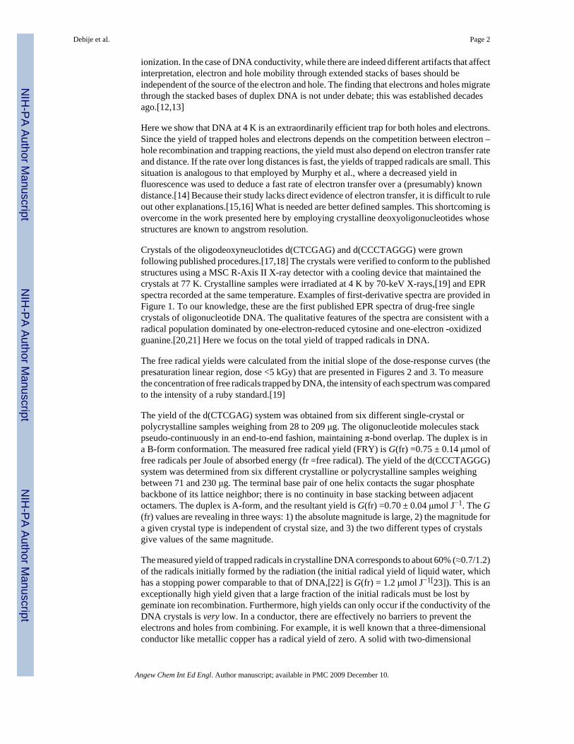
The understanding of electron and hole transfer in DNA is critical to predicting the biologicalconsequences of exposure to ionizing radiation. These processes are biologically relevant sinceabout 50% of the consequential damage is produced by direct-type events,[1] that is, from one-electron loss (holes) and one-electron gain directly by the DNA[2] or by fast transfer of holesand electrons to the DNA from adjacent solvent.[3] Transfer processes are chemically relevantsince the distribution of damage confronting the DNA repair enzymes is dependent on electrontransfer events occurring within a fraction of a second following energy deposition. Over thepast four decades, scientists working on the radiation physics and chemistry of DNA havegiven considerable attention to the question of DNA conductivity. This body of literatureprovides rather strong evidence, further discussed below, that the DNA polymer behaves as aninsulator and not as a “molecular wire”.
The possibility that DNA might possess properties of a molecular wire has been raised in recentyears by a series of experiments using photochemistry to initiate and detect electron transfer.In 1993 Murphy et al. concluded that electron transfer was long range (>4.0 nm) and fast(>109 s−1).[14] In this and subsequent work (see reference [4] for a review), it was concludedthat the dependence of the electron transfer rate on distance is relatively shallow; that is, theexponential factor β[5] is approximately 0.2 Å−1. The extremely small value of β was attributedto high electron and hole mobility through the “π-ways” of stacked bases. Other laboratoriesusing photochemistry concluded that the value of β is much larger. For electron transfer, Meadeand co-workers obtained β ≈1.0 Å−1,[6] and for hole transfer Lewis et al. obtained β ≈0.6Å−1.[7] Thus, there is a debate with regard to range, rate, and mechanism of electron transferin DNA.[8–11]
Relatively absent from this debate has been discussion of earlier findings largely from theradiation research community, findings which are difficult to reconcile with unusually fastelectron transfer rates over long distances (i.e., a β value an order of magnitude smaller than1.0 Å−1). The absence is, in part, due to concern over the possibility that the mechanism ofelectron transfer through DNA might depend on the means by which electrons or holes arecreated. Chemistry initiated by electronic excitation often differs from that initiated by
**We thank Loren Williams and Gary Hu for providing crystals of d(CGATCG):anthracycline, Tom Colby and Michael Strickler forassistance in verifying crystal structures, and Yurii Razskazovskii for his helpful discussions. The technical assistance of Kermit R.Mercer was invaluable. The investigation was supported by PHS Grant 2-R01-CA32546, awarded by the National Cancer Institute,DHHS. The contents of this paper are solely the responsibility of the authors and do not necessarily represent the official views of theNational Cancer Institute.*Fax: (+1)716-275-6007, [email protected].
NIH Public AccessAuthor ManuscriptAngew Chem Int Ed Engl. Author manuscript; available in PMC 2009 December 10.
Published in final edited form as:Angew Chem Int Ed Engl. 1999 September ; 38(18): 2752–2756.
NIH
-PA Author Manuscript
NIH
-PA Author Manuscript
NIH
-PA Author Manuscript
ionization. In the case of DNA conductivity, while there are indeed different artifacts that affectinterpretation, electron and hole mobility through extended stacks of bases should beindependent of the source of the electron and hole. The finding that electrons and holes migratethrough the stacked bases of duplex DNA is not under debate; this was established decadesago.[12,13]
Here we show that DNA at 4 K is an extraordinarily efficient trap for both holes and electrons.Since the yield of trapped holes and electrons depends on the competition between electron –hole recombination and trapping reactions, the yield must also depend on electron transfer rateand distance. If the rate over long distances is fast, the yields of trapped radicals are small. Thissituation is analogous to that employed by Murphy et al., where a decreased yield influorescence was used to deduce a fast rate of electron transfer over a (presumably) knowndistance.[14] Because their study lacks direct evidence of electron transfer, it is difficult to ruleout other explanations.[15,16] What is needed are better defined samples. This shortcoming isovercome in the work presented here by employing crystalline deoxyoligonucleotides whosestructures are known to angstrom resolution.
Crystals of the oligodeoxyneuclotides d(CTCGAG) and d(CCCTAGGG) were grownfollowing published procedures.[17,18] The crystals were verified to conform to the publishedstructures using a MSC R-Axis II X-ray detector with a cooling device that maintained thecrystals at 77 K. Crystalline samples were irradiated at 4 K by 70-keV X-rays,[19] and EPRspectra recorded at the same temperature. Examples of first-derivative spectra are provided inFigure 1. To our knowledge, these are the first published EPR spectra of drug-free singlecrystals of oligonucleotide DNA. The qualitative features of the spectra are consistent with aradical population dominated by one-electron-reduced cytosine and one-electron -oxidizedguanine.[20,21] Here we focus on the total yield of trapped radicals in DNA.
The free radical yields were calculated from the initial slope of the dose-response curves (thepresaturation linear region, dose <5 kGy) that are presented in Figures 2 and 3. To measurethe concentration of free radicals trapped by DNA, the intensity of each spectrum was comparedto the intensity of a ruby standard.[19]
The yield of the d(CTCGAG) system was obtained from six different single-crystal orpolycrystalline samples weighing from 28 to 209 μg. The oligonucleotide molecules stackpseudo-continuously in an end-to-end fashion, maintaining π-bond overlap. The duplex is ina B-form conformation. The measured free radical yield (FRY) is G(fr) =0.75 ± 0.14 μmol offree radicals per Joule of absorbed energy (fr =free radical). The yield of the d(CCCTAGGG)system was determined from six different crystalline or polycrystalline samples weighingbetween 71 and 230 μg. The terminal base pair of one helix contacts the sugar phosphatebackbone of its lattice neighbor; there is no continuity in base stacking between adjacentoctamers. The duplex is A-form, and the resultant yield is G(fr) =0.70 ± 0.04 μmol J−1. The G(fr) values are revealing in three ways: 1) the absolute magnitude is large, 2) the magnitude fora given crystal type is independent of crystal size, and 3) the two different types of crystalsgive values of the same magnitude.
The measured yield of trapped radicals in crystalline DNA corresponds to about 60% (≈0.7/1.2)of the radicals initially formed by the radiation (the initial radical yield of liquid water, whichhas a stopping power comparable to that of DNA,[22] is G(fr) = 1.2 μmol J−1[23]). This is anexceptionally high yield given that a large fraction of the initial radicals must be lost bygeminate ion recombination. Furthermore, high yields can only occur if the conductivity of theDNA crystals is very low. In a conductor, there are effectively no barriers to prevent theelectrons and holes from combining. For example, it is well known that a three-dimensionalconductor like metallic copper has a radical yield of zero. A solid with two-dimensional
Debije et al. Page 2
Angew Chem Int Ed Engl. Author manuscript; available in PMC 2009 December 10.
NIH
-PA Author Manuscript
NIH
-PA Author Manuscript
NIH
-PA Author Manuscript
conductivity provides a matrix that could trap one electron or hole per conducting plane. Toprovide a comparison with a two-dimensional conductor, we measured G(fr) for graphite at 4K by using a 0.5-mm pencil lead with a room-temperature conductivity of 1 S cm−1. The G(fr) value of graphite (<0.005 μmol J−1) is too small to detect in the presence of the quartzsample holder.
In sharp contrast, crystals of α-methylmannoside (a lattice of sugar molecules that is aninsulator) give a yield of 0.5 μmol J−1 at 4 K. As shown in Table 1, this is comparable to DNAirradiated in the form of films, powders, and crystals. Also, given the large body of work onelectron transfer in proteins (see for example reference [24]), we measured the free radicalyield of lysozyme crystals at 4 K. The G(fr) value of lysozyme (0.57 ± 0.03 μmol J−1) showsthat the probability of electron – hole combination within this protein lattice is essentially thesame as observed for DNA. The range of electrons and holes in lysozyme crystals, therefore,is comparable to the range in DNA.
If π-ways in DNA exist, the d(CTCGAG) hexamer crystal is optimized to detect them.Assuming base stacking continuity results in electrical continuity, the hexamer crystal wouldbe unable to trap more than two free radicals per continuous duplex column (i.e., one electronor hole per “strand”). Since each strand of stacked bases extends the length of the crystal, strandlength depends on crystal size and shape. Assuming one radical per strand of stacked bases ina cubic crystal containing 100 μg of d(CTCGAG),[25] the calculated free radical concentrationis less than 1015 frg−1. Observed concentrations exceed this value by more than three ordersof magnitude. The maximum measured concentration at free radical saturation is 7.2 × 10¹⁸
frg−1. Each strand of approximately 106 bases traps, on average, around 6000 free radicals.Viewed another way, the free radical density at dose saturation, assuming uniform trappingacross the entire crystal, is about one free radical per 57 bp, a through-base-stack distance of17 nm. Since energy deposition is not uniform,[26] the mean interradical distance is smaller.
The high density of holes and electrons trapped by d(CTCGAG) at dose saturation iscompelling evidence that no conductive states exist in the B-form helix. Indeed, if electron orhole transfer is long-range, then the FRY in d(CTCGAG) crystals should depend on crystalsize. Reducing the size of a cubic crystal by 10-fold should increase the FRY by a factor of2.154. Variations in crystal size, from less than 10 μg to greater than 100 μg, give FRYs withinthe observed standard deviation of ±20%, further indicating that the range at 4 K is limited.
A comparison between the two crystals provides an estimate of the electron range. The largestrun of stacked bases in d(CCCATGGG) is 8 bp, and yet G(fr) is the same as for d(CTCGAG),where base stacking is continuous. This observation of limited range is consistent with anumber of studies that indicate the mean range of a thermalized electron through DNA is short.EPR measurements at 4 – 77 K have provided evidence that the range of thermalized electronsand holes in DNA is at least 2 bp[19] with a mean between 3 – 11 bp.[27,28]
Further support of a short range comes from EPR measurements on single crystals of a hexameranthracycline complex.[19] The complex contains two intercalated anthracyclines per d(CGATCG) with intercalation occurring between each of the tandem CG base pairs. Basestacking is continuous through the crystal.[29] It is known from single-crystal EPR that holesand electrons trap only at anthracycline. No DNA-centered radicals are observed. This isexpected given the redox properties of anthracycline, which make it the deepest trapping sitefor both electrons and holes. It also proves that holes migrate through the stacked bases. Wehave measured the FRY of these crystals at 4 K and find that G(fr) =0.27 ± 0.03 μmolJ−1.[30] Thus, the high density of anthracycline traps does not increase yields. This can beunderstood if pure duplex DNA contains, inherently, a high density of traps such that the rangeof thermalized electrons and holes is limited to very short distances.
Debije et al. Page 3
Angew Chem Int Ed Engl. Author manuscript; available in PMC 2009 December 10.
NIH
-PA Author Manuscript
NIH
-PA Author Manuscript
NIH
-PA Author Manuscript
If DNA had a metallike band structure, one should expect an increased conductivity at lowtemperatures. This phenomenon has not been observed: in fact, the opposite is true. To date,we have measured G(fr) values and temperature effects on 12 different types of crystals,representing a wide range of conformation, packing, and sequence. We have observed that G(fr) of crystalline DNA irradiated at 4 K and annealed to 240 K has 20 – 40% of the yieldobtained at 4 K.[31,32] The EPR spectrum qualitatively changes in a manner consistent withan initial set of radicals that are shallowly trapped, accompanied by reversible protonation/deprotonation. These low-temperature radicals convert into a set of more deeply trappedspecies at higher temperatures, and these are typically irreversibly protonated/deprotonated.[33] A minimum and maximum distance for the thermally activated migration has beenestimated. The minimum is about 2 nm based on the absence of spectral features due to radicalpairs.[27] The maximum is on the order of 20 nm based on the free radical concentrationremaining after annealing at 240 K. Both boundaries are compatible with the results ofRazskazovskii et al.[28] With track structure calculations (currently underway), it should bepossible to determine the distribution of migration distances from our measurements oncrystalline DNA. As with other EPR studies that show a decrease in radical concentration uponannealing,[34–36] there is no doubt that electron transfer occurs through the stacked bases ofduplex DNA and that electrons migrate by thermally activated hopping.
The migration of charge through DNA is supported by experiments with solid-state EPR,[33]pulse radiolysis,[37] and product analysis.[38,39] In a paper by Razskazovskii et al., it isconcluded that the mean distance of electron migration at 77 K is 11 bp and that above 150 Kthermally activated migration extends to 30 bp or more.[28] Their results are consistent witha base-to-base hopping mechanism.
Our observations on crystalline DNA are well explained by a model in which the migration ofelectrons and holes is propagated primarily by hopping between shallow traps. At temperaturesless than 80 K, the deepest electron trap is cytosine (C),[20,40] where the rate of proton transferfrom N1 of guanine (G) to N3 of C•− (forming C(N3 + H)•)[41] competes effectively with therate of electron transfer.[42] Likewise, the deepest hole trap is guanine,[20,43] where protontransfer away from N1 of G•+ to N3 of C (forming G(N1 – H)•)[44] competes with hole transfer.[45] Steenken has written an excellent review of the role of proton transfer in moderatingelectron transfer.[42] Proton transfer separates the charge from the unpaired electron, leavinga neutral radical on one strand and a diamagnetic ion on the conjugate base of the opposingstrand and reduces the probability of electrons and holes combining.
In conclusion, crystalline oligodeoxynucleotides serve as well-defined samples that giveimproved precision in the measurement of free radical yields. Increased length of base stackingbeyond eight base pairs has no effect on free radical yields, and the yields are markedly high(0.7 μmol J−1). The high yields coupled with the absence of a dependence on long-range basestacking supports the conclusion that the range of holes and electrons at 4 K is short (2 – 8 bp).It follows that DNA is effectively an insulator.
References1. Krisch RE, Flick MB, Trumbore CN. Radiat Res 1991;126:251. [PubMed: 1850853]2. Wang W, Yan M, Becker D, Sevilla MD. Radiat Res 1994;137:2. [PubMed: 8265784]3. Mroczka N, Bernhard WA. Radiat Res 1993;135:155. [PubMed: 8396269]4. Holmlin R, Dandliker P, Barton J. Angew Chem 1997;109:2830.Angew Chem Int Ed Engl
1997;36:2714.5. At long distances, the tunneling electron transfer reactions are expected to drop off exponentially, that
is, kET ∝ exp[− β(R − R0)], where R0 is the donor – acceptor separation at the van der Waals contact.6. Meade TJ, Kayyem JF. Angew Chem 1995;107:358.Angew Chem Int Ed Engl 1995;34:352.
Debije et al. Page 4
Angew Chem Int Ed Engl. Author manuscript; available in PMC 2009 December 10.
NIH
-PA Author Manuscript
NIH
-PA Author Manuscript
NIH
-PA Author Manuscript
7. Lewis FD, Wu W, Zhang Y, Letsinger RL, Greenfield SR, Wasielewski MR. Science 1997;277:673.[PubMed: 9235887]
8. Harriman A. Angew Chem 1999;111:996.Angew Chem Int Ed 1999;38:945.9. Barbara, P.; Olson, E. Electron Transfer: From Isolated Molecules, Part Two. Jortner, J.; Bixon, M.,
editors. Vol. ch 13. Wiley; New York: 1999.10. Priyadarshy S, Risser S, Beratan D. J Biol Inorg Chem 1998;3:196.11. Turro N, Barton J. J Biol Inorg Chem 1998;3:201.12. Sevilla, M. Excited States in Organic and Biochemistry. Pullman, B.; Goldblum, N., editors. Reidel;
Boston: 1977. p. 1513. Gregoli S, Olast M, Bertinchamps A. Radiat Res 1982;89:238. [PubMed: 6278529]14. Murphy C, Arkin M, Jenkins Y, Ghatlia N, Bossmann S, Turro N, Barton J. Science 1993;262:1025.
[PubMed: 7802858]15. Krider E, Meade T. J Biol Inorg Chem 1998;3:222.16. Beratan DN, Priyadarshy S, Risser SM. Chem Biol 1997;4:3. [PubMed: 9070421]17. Wahl M, Rao S, Sundaralingam M. Biophys J 1996;70:2857. [PubMed: 8744323]18. Tippin DB, Sundaralingam M. Acta Crystallogr Sect D 1996;52:997. [PubMed: 15299609]19. Milano MT, Hu GG, Williams LD, Bernhard WA. Radiat Res 1998;150:101. [PubMed: 9650607]20. Sevilla M, Becker D, Yan M, Summerfield S. J Phys Chem 1991;95:3409.21. Bernhard, WA. The Early Effects of Radiation on DNA, Ser. H. Fielden, EM.; O’Neill, P., editors.
Vol. 54. Springer; Berlin: 1991. p. 14122. La Verne JA, Pimblott SM. Radiat Res 1995;141:208. [PubMed: 7838960]23. Holley, WR.; Chatterjee, A. The Early Effects of Radiation on DNA. Fielden, EM.; O’Neill, P.,
editors. Springer; Berlin: 1991. p. 19524. Moser C, Keske J, Warncke K, Ford R, Dutton P. Nature 1992;355:796. [PubMed: 1311417]25. The crystal habit of d(CTCGAG) is rodlike with no one dimension greater than five times the smallest
dimension.26. Pimblott SM, Verne JAL, Mozumder A, Green NJ. J Phys Chem 1990;94:488.27. Spalletta RA, Bernhard WA. Radiat Res 1992;130:7. [PubMed: 1313984]28. Razskazovskii Y, Swarts S, Falcone J, Taylor C, Sevilla M. J Phys Chem 1997;101:1460.29. Lipscomb LA, Peek ME, Zhou FX, Bertrand JA, van Derveer D, Williams LD. Biochemisty
1994;33:3649.30. Milano, M. PhD Thesis. University of Rochester; Rochester, New York: 1998. p. 27831. Debije MG. unpublished results32. There are two general mechanisms by which the trapped electrons and holes are lost upon annealing.
One is by free radical conversions, which require higher thermal energy and are activated at T> 150K. The other is by free radical combination reactions, which require lower thermal energy and areactivated over a wide range of temperatures (50 < T< 160 K). Radical combination reactions occurby detrapping the electron and/or hole.
33. Becker D, Sevilla MD. Adv Radiat Biol 1993;17:121.34. Bernhard WA, Mroczka N, Barnes J. Int J Radiat Biol 1994;66:491. [PubMed: 7983436]35. Wang W, Sevilla M. Radiat Res 1994;138:9. [PubMed: 8146305]36. Mroczka NE, Bernhard WA. Radiat Res 1995;144:251. [PubMed: 7494867]37. Anderson RF, Patel KB, Wilson WR. J Chem Soc Faraday Trans 1991;87:3739.38. Fuciarelli AF, Sisk EC, Miller JH, Zimbrick JD. Int J Radiat Biol 1994;66:505. [PubMed: 7983438]39. Swarts SG, Sevilla MD, Becker D, Tokar CJ, Wheeler KT. Radiat Res 1992;129:333. [PubMed:
1542721]40. Bernhard WA. J Phys Chem 1989;93:2187.41. Barnes J, Bernhard W. J Phys Chem 1994;98:887.42. Steenken S. Biol Chem 1997;378:1293. [PubMed: 9426189]43. Steenken S, Jovanovic SV. J Am Chem Soc 1997;119:617.44. Close D. Radiat Res 1993;135:1. [PubMed: 8392211]
Debije et al. Page 5
Angew Chem Int Ed Engl. Author manuscript; available in PMC 2009 December 10.
NIH
-PA Author Manuscript
NIH
-PA Author Manuscript
NIH
-PA Author Manuscript
45. Giese B, Wesseley S, Spormann M, Lindemann U, Meggers E, Michel-Beyerle ME. Angew Chem1999;111:1050.Angew Chem Int Ed 1999;38:996.
46. Milano MT, Bernhard WA. Radiat Res 1999;151:39. [PubMed: 9973082]
Debije et al. Page 6
Angew Chem Int Ed Engl. Author manuscript; available in PMC 2009 December 10.
NIH
-PA Author Manuscript
NIH
-PA Author Manuscript
NIH
-PA Author Manuscript
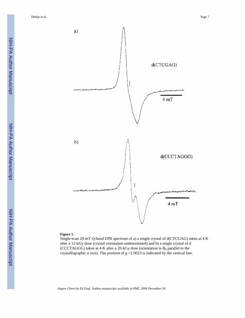
Figure 1.Single-scan 20-mT Q-band EPR spectrum of a) a single crystal of d(CTCGAG) taken at 4 Kafter a 12-kGy dose (crystal orientation undetermined) and b) a single crystal of d(CCCTAGGG) taken at 4 K after a 20-kGy dose (orientation is B0 parallel to thecrystallographic a axis). The position of g =2.0023 is indicated by the vertical line.
Debije et al. Page 7
Angew Chem Int Ed Engl. Author manuscript; available in PMC 2009 December 10.
NIH
-PA Author Manuscript
NIH
-PA Author Manuscript
NIH
-PA Author Manuscript
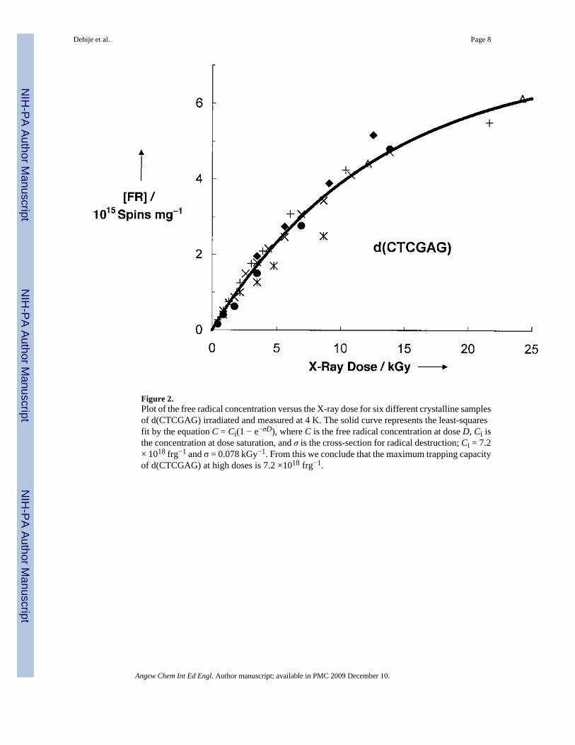
Figure 2.Plot of the free radical concentration versus the X-ray dose for six different crystalline samplesof d(CTCGAG) irradiated and measured at 4 K. The solid curve represents the least-squaresfit by the equation C = Ci(1 − e−σD), where C is the free radical concentration at dose D, Ci isthe concentration at dose saturation, and σ is the cross-section for radical destruction; Ci = 7.2× 10¹⁸ frg−1 and σ = 0.078 kGy−1. From this we conclude that the maximum trapping capacityof d(CTCGAG) at high doses is 7.2 ×10¹⁸ frg−1.
Debije et al. Page 8
Angew Chem Int Ed Engl. Author manuscript; available in PMC 2009 December 10.
NIH
-PA Author Manuscript
NIH
-PA Author Manuscript
NIH
-PA Author Manuscript
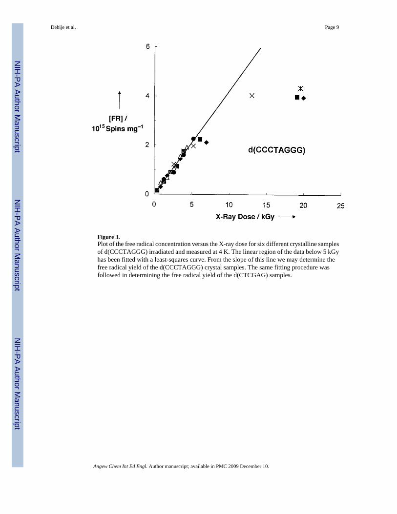
Figure 3.Plot of the free radical concentration versus the X-ray dose for six different crystalline samplesof d(CCCTAGGG) irradiated and measured at 4 K. The linear region of the data below 5 kGyhas been fitted with a least-squares curve. From the slope of this line we may determine thefree radical yield of the d(CCCTAGGG) crystal samples. The same fitting procedure wasfollowed in determining the free radical yield of the d(CTCGAG) samples.
Debije et al. Page 9
Angew Chem Int Ed Engl. Author manuscript; available in PMC 2009 December 10.
NIH
-PA Author Manuscript
NIH
-PA Author Manuscript
NIH
-PA Author Manuscript

NIH
-PA Author Manuscript
NIH
-PA Author Manuscript
NIH
-PA Author Manuscript
Debije et al. Page 10
Table 1
Free radical yields at 4 K of various materials exposed to ionizing radiation, and comparison of DNA samplesagainst known conductors and insulators.
Material Form Yield[μmolJ−1]
copper wire 0graphite rod pressed powder < 0.005α-methylmannoside crystalline 0.44 ± 0.03alysozyme crystalline 0.57 ± 9.04acalf thymus DNA film 0.4 – 0.5bcalf thymus DNA lyophilized powder 0.5 – 0.6bd(CCCTAGGG) crystalline 0.70 ± 0.04ad(CTCGAG) crystalline 0.75 ± 0.14ad(CGATCG):anthracycline crystalline 0.24 ± 0.03a
aQuoted errors are relative errors; absolute errors are higher (±25%).[46]
bHydration state of 16 waters per nucleotide.[46]
Angew Chem Int Ed Engl. Author manuscript; available in PMC 2009 December 10.
Copyright © 2022 FDOKUMEN