DNA-Binding Proteinsin Cells and Membrane Blebs of ... - NCBI
6
JOURNAL OF BACTERIOLOGY, Aug. 1989, p. 4196-4201 0021-9193/89/084196-06$02.00/0 Copyright © 1989, American Society for Microbiology DNA-Binding Proteins in Cells and Membrane Blebs of Neisseria gonorrhoeae Vol. 171. No. 8 DAVID W. DORWARD* AND CLAUDE F. GARON Laboratory of Pathobiology, Nationcal Institite of Allergy and Infectiolus Diseases, Rocky Moulntain Laboratories, Hamilton, Montana 59840 Received 22 March 1989/Accepted 12 May 1989 Naturally elaborated membrane bleb fractions BI and BIT of Neisseria gonorrhoeae contain both linear and circular DNAs. Because little is known about the interactions between DNA and blebs, studies were initiated to identify specific proteins that bind DNA in elaborated membrane blebs. Western immunoblots of whole-cell and bleb proteins from transformation-competent and DNA-uptake-deficient (dud) mutants were probed with single- or double-stranded gonococcal DNA, pBR322, or synthetic DNA oligomers containing intact or altered gonococcal transformation uptake sequences. The specificity and sensitivity of a nonradioactive DNA-binding protein assay was evaluated, and the assay was used to visualize DNA-protein complexes on the blots. The complexes were then characterized by molecular mass, DNA-binding specificity, and expression in bleb fractions. The assay effectively detected blotted DNA-binding proteins. At least 17 gonococcal DNA-binding proteins were identified; unique subsets occurred in BI and BIT. Certain DNA-binding proteins had varied affinities for single- and double-stranded DNA, and the intact transformation uptake sequence competitively displaced the altered sequence from a BI protein at 11 kilodaltons (kDa). A dud mutant, strain FA660, lacked DNA-binding activity at the 11-kDa protein in BI. The segregation of DNA-binding proteins within BI and BIT correlates with their distinct protein profiles and suggests that these vesicles may play different roles. Although the DNA-binding proteins expressed in BIT may influence the nuclease-resistant export of plasmids within BIT vesicles, the BI 11-kDa protein may bind transforming DNA. Neisseria gonorrhoeae is a gram-negative bacterial patho- gen that causes the sexually transmitted mucosal and dis- seminated infections of gonorrhea. This organism readily develops antimicrobial resistance and expresses several highly variable immunogenic cell surface determinants (5). Numerous studies have addressed the genetic bases of these observations, and mechanisms including directed intracellu- lar gene translocation, transformation, and conjugation are considered responsible (2, 3, 7, 14a, 15, 22, 24). Recently, an additional genetic exchange mechanism involving mem- brane-associated DNA was proposed (10). Log-phase cultures of N. gonorrhoeae release membrane vesicles termed blebs (5, 10, 11, 26), and bleb formation by gonococci in vivo has been reported (5). On sucrose density gradients, the blebs sediment into two fractions, BI and BIT. Fraction BI has a protein content characteristic of the inner membrane, and fraction BIT contains outer membrane pro- teins and lipopolysaccharide (10). Each fraction also con- tains chromosomal and plasmid DNAs, but only plasmids associated with outer membrane-derived BIT exhibit nucle- ase resistance (10). Genetic exchange experiments with blebs showed that recipient gonococci, incubated in cell-free broth culture filtrates that contained blebs from penicilli- nase-producing bacteria (bla+), naked DNA with streptomy- cin resistance (Strr) markers, and exogenous DNase effi- ciently incorporate and express plasmid-encoded bla markers without expressing chromosomal str genes. Equiv- alent results of experiments with a blati Str' bleb donor indicate that blebs can mediate intercellular transfer of nuclease-protected plasmids, but may be incapable of trans- ferring chromosomal DNA in a DNase-resistant state (10). Such differences suggest that specific mechanisms may exist * Corresponding author. for determining the association between DNA and blebs and the role of bleb DNA in genetic exchange. The present study was undertaken to identify and charac- terize DNA-binding proteins in blebs that could contribute to interactions between DNA and these membrane vesicles. A qualitative approach, developed by Wager and Stephens to examine DNA-binding proteins in Chlamydia spp. (27), was used. Western immunoblots of gonococcal whole-cell (WC) and bleb proteins were probed with DNA, and the resulting DNA-protein complexes were visualized by a recently de- scribed nonradioactive assay (9). The assay system uses digoxigenin-derivatized dUTP to label DNA probes in a Klenow enzyme-catalyzed polymerization. The label is then detected by a solid-phase enzyme-linked immunosorbent assay. In this study we first considered the specificity and sensi- tivity of the nonradioactive DNA-binding protein assay by comparing the detection of known DNA-binding proteins with that of proteins lacking DNA-binding activity and then examined DNA-binding proteins in gonococcal WC and blebs. Probes for DNA binding included single- and double- stranded gonococcal WC and plasmid DNA (ssDNA and dsDNA), pBR322, and synthetic oligomers containing either an intact (I) or an altered (A) gonococcal transformation uptake sequence (14). Both transformation-competent and DNA-uptake-deficient (du.d) (4) strains were examined. MATERIALS AND METHODS Bacteria. N. gonorrhoeae 31426 (transformation compe- tent) (American Type Culture Collection, Rockville, Md.) and FA660 (duid) (3) were maintained by daily single-colony transfers on gonococcal clear typing medium as previously described (11). For experiments, gonococcal cells and mem- brane blebs from 16-h cultures were harvested and separated 4196
-
Upload
khangminh22 -
Category
Documents
-
view
0 -
download
0
Transcript of DNA-Binding Proteinsin Cells and Membrane Blebs of ... - NCBI

JOURNAL OF BACTERIOLOGY, Aug. 1989, p. 4196-42010021-9193/89/084196-06$02.00/0Copyright © 1989, American Society for Microbiology
DNA-Binding Proteins in Cells and Membrane Blebsof Neisseria gonorrhoeae
Vol. 171. No. 8
DAVID W. DORWARD* AND CLAUDE F. GARON
Laboratory of Pathobiology, Nationcal Institite of Allergy and Infectiolus Diseases,Rocky Moulntain Laboratories, Hamilton, Montana 59840
Received 22 March 1989/Accepted 12 May 1989
Naturally elaborated membrane bleb fractions BI and BIT of Neisseria gonorrhoeae contain both linear andcircular DNAs. Because little is known about the interactions between DNA and blebs, studies were initiatedto identify specific proteins that bind DNA in elaborated membrane blebs. Western immunoblots of whole-celland bleb proteins from transformation-competent and DNA-uptake-deficient (dud) mutants were probed withsingle- or double-stranded gonococcal DNA, pBR322, or synthetic DNA oligomers containing intact or alteredgonococcal transformation uptake sequences. The specificity and sensitivity of a nonradioactive DNA-bindingprotein assay was evaluated, and the assay was used to visualize DNA-protein complexes on the blots. Thecomplexes were then characterized by molecular mass, DNA-binding specificity, and expression in blebfractions. The assay effectively detected blotted DNA-binding proteins. At least 17 gonococcal DNA-bindingproteins were identified; unique subsets occurred in BI and BIT. Certain DNA-binding proteins had variedaffinities for single- and double-stranded DNA, and the intact transformation uptake sequence competitivelydisplaced the altered sequence from a BI protein at 11 kilodaltons (kDa). A dud mutant, strain FA660, lackedDNA-binding activity at the 11-kDa protein in BI. The segregation of DNA-binding proteins within BI and BITcorrelates with their distinct protein profiles and suggests that these vesicles may play different roles. Althoughthe DNA-binding proteins expressed in BIT may influence the nuclease-resistant export of plasmids within BITvesicles, the BI 11-kDa protein may bind transforming DNA.
Neisseria gonorrhoeae is a gram-negative bacterial patho-gen that causes the sexually transmitted mucosal and dis-seminated infections of gonorrhea. This organism readilydevelops antimicrobial resistance and expresses severalhighly variable immunogenic cell surface determinants (5).Numerous studies have addressed the genetic bases of theseobservations, and mechanisms including directed intracellu-lar gene translocation, transformation, and conjugation areconsidered responsible (2, 3, 7, 14a, 15, 22, 24). Recently, anadditional genetic exchange mechanism involving mem-brane-associated DNA was proposed (10).Log-phase cultures of N. gonorrhoeae release membrane
vesicles termed blebs (5, 10, 11, 26), and bleb formation bygonococci in vivo has been reported (5). On sucrose densitygradients, the blebs sediment into two fractions, BI and BIT.Fraction BI has a protein content characteristic of the innermembrane, and fraction BIT contains outer membrane pro-teins and lipopolysaccharide (10). Each fraction also con-tains chromosomal and plasmid DNAs, but only plasmidsassociated with outer membrane-derived BIT exhibit nucle-ase resistance (10). Genetic exchange experiments withblebs showed that recipient gonococci, incubated in cell-freebroth culture filtrates that contained blebs from penicilli-nase-producing bacteria (bla+), naked DNA with streptomy-cin resistance (Strr) markers, and exogenous DNase effi-ciently incorporate and express plasmid-encoded blamarkers without expressing chromosomal str genes. Equiv-alent results of experiments with a blati Str' bleb donorindicate that blebs can mediate intercellular transfer ofnuclease-protected plasmids, but may be incapable of trans-ferring chromosomal DNA in a DNase-resistant state (10).Such differences suggest that specific mechanisms may exist
* Corresponding author.
for determining the association between DNA and blebs andthe role of bleb DNA in genetic exchange.The present study was undertaken to identify and charac-
terize DNA-binding proteins in blebs that could contribute tointeractions between DNA and these membrane vesicles. Aqualitative approach, developed by Wager and Stephens toexamine DNA-binding proteins in Chlamydia spp. (27), wasused. Western immunoblots of gonococcal whole-cell (WC)and bleb proteins were probed with DNA, and the resultingDNA-protein complexes were visualized by a recently de-scribed nonradioactive assay (9). The assay system usesdigoxigenin-derivatized dUTP to label DNA probes in aKlenow enzyme-catalyzed polymerization. The label is thendetected by a solid-phase enzyme-linked immunosorbentassay.
In this study we first considered the specificity and sensi-tivity of the nonradioactive DNA-binding protein assay bycomparing the detection of known DNA-binding proteinswith that of proteins lacking DNA-binding activity and thenexamined DNA-binding proteins in gonococcal WC andblebs. Probes for DNA binding included single- and double-stranded gonococcal WC and plasmid DNA (ssDNA anddsDNA), pBR322, and synthetic oligomers containing eitheran intact (I) or an altered (A) gonococcal transformationuptake sequence (14). Both transformation-competent andDNA-uptake-deficient (du.d) (4) strains were examined.
MATERIALS AND METHODS
Bacteria. N. gonorrhoeae 31426 (transformation compe-tent) (American Type Culture Collection, Rockville, Md.)and FA660 (duid) (3) were maintained by daily single-colonytransfers on gonococcal clear typing medium as previouslydescribed (11). For experiments, gonococcal cells and mem-brane blebs from 16-h cultures were harvested and separated
4196

GONOCOCCAL DNA-BINDING PROTEINS 4197
97.4-66.2- -
42.7- _
CBB
<
X X a:0 m
ss DNA.0
C <en a) > Cfu) c 0 m
ds DNA
,o <0Om (; ,ce 0 >% enX nc 0 m
em
p1g BSA
31.0- -
-4.,
ng SSBwe*
FIG. 1. Evaluation of the DNA-binding protein assay. A Coomassie brilliant blue-stained (CBB) SDS-12.5% polyacrylamide gel,containing molecular mass standards (lane stds) and 1 ,ug each of SSB, RecA, cyt c, or BSA was compared with bands exhibitingDNA-binding activity on Western blots made from similar gels. Blots were probed with digoxigenin-labeled single-stranded (ss DNA) or
double-stranded (ds DNA) pBR322, and DNA-protein complexes were detected as described in Materials and Methods. The Coomassiebrilliant blue stain showed effective resolution of the proteins, with dimers evident in the cyt c lane. Single-stranded probes showed strongreactivity with SSB and relatively weak reactivity with RecA and Cyt c. Detection of RecA and Cyt c was enhanced, and labeling of SSB was
reduced with dsDNA probes. Titrations of SSB and BSA showed that 10-ng loadings of SSB were detectable, although 1 mg of BSA was notdetected. In lanes labeled a and b, 0.1 and 10 ,ug was loaded, respectively. Molecular mass standards are given in kilodaltons.
by differential and sucrose density gradient centrifugation,as previously described (10).DNA preparation. WC DNA was purified from N. gonor-
rhoeae by the alkaline lysis and phenol-chloroform-etherextraction methods of Maniatis et al. (20). Ethanol-precipi-tated DNA was washed once with 70% ethanol and thendissolved at approximately 1 mg/ml in buffer consisting of 10mM Tris (pH 8.0) and 1 mM EDTA (TE).For gonococcal plasmid purifications, WC DNA extracts
were separated by electrophoresis in agarose tube gels(Bio-Rad Laboratories, Richmond, Calif.) and supercoiledforms of the 4.2-kilobase-pair cryptic plasmid were elutedfrom excised gel segments. Eluted plasmids were extractedonce with 3 volumes of TE-saturated n-butanol, precipitatedwith ethanol, and suspended in TE. The plasmids were
linearized by single-site restriction with HindIll (18) (NewEngland BioLabs Inc., Beverly, Mass.), as specified by themanufacturer.
Linearized pBR322 was purchased from BoehringerMannheim Biochemicals, Indianapolis, Ind., and syntheticDNA oligomers were produced on a DNA synthesizer(model 380 B; Applied Biosystems Inc., Foster City, Calif.)as specified by the manufacturer.
Purified protein preparations. Single-stranded-DNA-bind-ing protein (SSB) and RecA protein were purchased fromUnited States Biochemical Corp., Cleveland, Ohio. Cy-tochrome c (cyt c), purified from Saccharomyces cerevisiae,and bovine serum albumin fraction V (BSA) were purchasedfrom Sigma Chemical Co., St. Louis, Mo., and ICN Bio-medicals Inc., Costa Mesa, Calif., respectively.
Protein electrophoresis. Sodium dodecyl sulfate (SDS)-polyacrylamide gel electrophoresis was performed, with thediscontinuous buffer system of Laemmli (19), by methodspreviously described (16). Gels were typically run for 2.5 h at7.5 W (constant power) and then either stained withCoomassie brilliant blue or electroblotted onto nitrocellulosemembranes as previously described for DNA-binding pro-tein assays (27).DNA-binding protein assays. Assays for DNA binding to
electroblotted proteins were performed by using modifica-tions to previously published procedures (9, 27). All steps
were performed at room temperature. Nitrocellulose mem-branes with blotted proteins were blocked for 1 h with 0.1%(wt/vol) nonfat dry milk, dissolved in Dulbecco phosphate-buffered saline (pH 7.2) containing 0.05% Tween 20 (Sigma)(dPBS-Tween). The blots were then washed twice for 10 minin dPBS-Tween. Washed blots were probed for 2 h with 0.1p.g of DNA per ml in dPBS-Tween or incubated in dPBS-Tween alone. Probes were either labeled with digoxigenin-conjugated dUTP (Boehringer Mannheim), as specified bythe manufacturer, or left unlabeled. Probed blots wereblocked again for 30 min with 4% nonfat dry milk in bufferconsisting of 0.1 M Tris (pH 7.5) and 0.15 M NaCl (TN) andwashed twice for 10 min each in TN. Protein-DNA com-plexes were located with an anti-digoxigenin antibody con-jugated to alkaline phosphatase (Boehringer Mannheim) anddetected as specified by the manufacturer.
RESULTS
To optimize the specificity and sensitivity of this nonra-dioactive DNA-binding protein assay, we performed a seriesof procedural experiments. These involved titrating blockingreagent concentrations while maintaining appropriate condi-tions for DNA-protein interactions. As reported previouslyfor radioactive assays (27), dPBS-Tween proved an effectivebuffer system for DNA-protein binding. Membrane blockingwith dPBS-Tween alone, with dPBS supplemented withBSA, or with nonfat dry milk in dPBS, was evaluated (datanot shown). Optimal results were obtained when the proce-dure described above was used.The specificity of this system was evaluated by comparing
the reactivities of SSB, RecA, cyt c, and BSA (Fig. 1). BothSSB and RecA bind DNA in vivo. The highly basic cyt c canbind DNA ionically (13), and BSA has no known DNAinteraction. An SDS-12.5% polyacrylamide gel stained withCoomassie brilliant blue showed the migration of eachprotein and revealed a small proportion of dimers in the cytc preparation. When Western blots of such gels were probedwith single-stranded pBR322, labeled and detected as de-scribed above, strong labeling of SSB and relatively weak
0o goo
l u Y 0
21.5- --~
14.4--
VOL. 171, 1989

4198 DORWARD AND GARON
B. black
97.4 .66.2-
42.7-A
31:.
21 .5-. ....
1 4.4-
Labeled Unlabeled DNA-
0. 0 0- m O,m3D cn m m C : co co
-35
-11
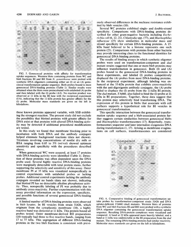
FIG. 2. Gonococcal DNA-binding proteins. Western blots containing proteins from WC and bleb fractions BI and BII of N. gonorrhoeae31426 were stained for total protein with buffalo black (B. black), probed with digoxigenin-labeled (Labeled) or unlabeled (Unlabeled)pBR322, or incubated in buffer lacking DNA (DNA-). A single protein, corresponding to PI at 35 kDa, reacted nonspecifically in the WC andBIT lanes of the negative controls. Seventeen bands that bound labeled DNA in the WC lane were observed. The apparent masses for theseand bleb-associated DNA-binding proteins are listed in Table 1. Molecular mass standards (lane stds) are given in kilodaltons.
labeling of RecA and cyt c were observed. Conversely,equivalent blots probed with double-stranded pBR322showed increased labeling of RecA and cyt c, with a corre-
sponding decrease in labeling of SSB. No label was observedin BSA lanes in either experiment. Samples containing 10 ngof SSB loaded on gels were detectable, whereas a 1-mgloading of BSA failed to bind the DNA probes.The assay system was then used to identify proteins
capable of binding DNA in N. gonorrhoeae WC and blebs
(Fig. 2). Western blots containing lysates of WC and frac-tions BI and BII from N. gonorrhoeae 31426 were stainedwith buffalo black, and probed with either control-labeledpBR322 (Boehringer Mannheim) or unlabeled pBR322 orincubated in dPBS-Tween alone. A single protein, corre-sponding to outer membrane protein I (PI) at 35 kilodaltons(kDa) in WC and BII lanes, showed a nonspecific reaction inthe unlabeled and dPBS-Tween control experiments. Atleast 17 gonococcal proteins, ranging from 11 to 97 kDa,
TABLE 1. DNA-binding proteins in N. gonorrhoeae
Fraction Molecular mass Protein present' in:(kDa) pBR322 WC dsDNA WC ssDNA Plasmid (I) probe (A) probe
WC 97 + + + + +84 + + + + +57 - - + +/- -
39 + + + + + +34 - - - - + +l-32.5 + + + + + +32 + + _+ +-29 + + + + + +27 + + + + + +26.5 + + + + + +21 + + +/- - +/- +17 + + + + + +16 + + + + + +15 + + + + + +14 + + + + + +13 + + + + - -11 + + + + - -
BI 22 - + - +/- - +11 ++ ++ ++ ++ ++ ++
BIT 57 - +/- - + -34 - - +l- - + -
32 + + + + + -27 + + + +/- +17 + +/- + + + +l-
"+/-, Light reaction product, not always reproducible; ±, reproducible detection; ++, strong reproducible reaction.
J. BACTERIOL.

GONOCOCCAL DNA-BINDING PROTEINS 4199
ssDNA
3 az' m9766-*
44-
31- -
22-
dsDNA
C, m m
plasmld3m
u_ -
FIG. 3. Molecular specificity of gonococcal DNA-binding pro-teins. Western blots containing proteins from WC and bleb fractionsBI and BIT or N. gonorrhoeae were probed with digoxigenin-labeledWC ssDNA, WC dsDNA, or linearized 4.2-kilobase-pair gonococcalcryptic plasmids (plasmid), and the resulting DNA-protein com-plexes were visualized. Several proteins showed enhanced bindingwith ssDNA. A 57-kDa WC band was detected only with ssDNAprobes (asterisk). A protein at 22 kDa in the WC lane was detectedonly with WC dsDNA. A highly reactive BI band at 11 kDa boundall three probes. Molecular mass standards are given on the left inkilodaltons.
exhibited DNA-binding activity in this assay. The apparentmolecular masses ofWC and bleb proteins that bound one ormore of six different DNA probes are listed in Table 1.Unique subsets of the DNA-binding proteins were observedin membrane blebs. The 11-kDa protein had strong reactivityand was the sole DNA-binding protein consistently resolvedin BI lysates from strain 31426. Three proteins between 17and 32 kDa in BIT lysates specifically bound pBR322. Con-trol blots probed with labeled pBR322 but incubated withoutthe antibody-alkaline phosphatase conjugate showed no re-action (data not shown).
Equivalent blots were probed with gonococcal WC dsDNA,WC ssDNA (denatured by boiling dsDNA), or 4.2-kilobase-pair cryptic plasmids extracted from strain 31426. The re-sults are shown in Fig. 3. Reactivity at 22 kDa appeared to bespecific for dsDNA, and a band at 32 kDa in WC lysates(arrow) was detected only with the dsDNA and the plasmidprobes. Most proteins were labeled more intensely withssDNA probes. One WC band at 57 kDa (asterisk) wasobserved only when ssDNA was used. The banding patternof blots probed with gonococcal cryptic plasmids was similarto the pBR322 profile shown in Fig. 2.To identify proteins possibly involved in binding and
uptake of transforming DNA by N. gonorrhoeae, we probed
blots with synthetic DNA oligomers containing either an (I)or an (A) transformation uptake sequence. The oligomersequences are given in Fig. 4. Figure 5 shows the results ofthe blotting experiments. The (I) probe contains the 10-baseinverted repeat (underline) recently correlated with optimalcompetitive inhibition of gonococcal transformation (Fig. 4)(14). The (A) probe has an identical base composition,except that every second base within the inverted repeat wassubstituted with the complementary base, to alter the se-quence while maintaining the physical structure.Both oligomers bound to proteins on the filters (Fig. 5) and
reacted strongly in the assay. When blots were preincubatedwith unlabeled (A) probes and then challenged with labeled(I) probes, few changes in banding occurred. Conversely,blots preincubated with unlabeled (I) probes and challengedwith (A) labels lacked a reaction product at 11 kDa in the BIlane. Nonspecific reactions at 35 kDa were disregarded (Fig.2).
In a final experiment to identify proteins associated withtransformation, we compared (I) probe binding with WC andbleb lysates from transformation-competent stain 31426 anddud mutant FA660 (Fig. 6). No significant differences inbanding between these strains were 'detected in WC and BITlanes. However, the FA660 BI preparation appeared to haveenhanced labeling at 22 kDa, and it lacked the strongDNA-binding activity at 11 kDa.
DISCUSSIONIn this study we identified several DNA-binding proteins
in N. gonorrhoeae, and showed that specific DNA-bindingproteins are located in membrane bleb fractions BI and BII.The data supported the effectiveness of nonradioactiveDNA-binding protein assays on Western blots of SDS-polyacrylamide gel electrophoresis protein separations andsuggested that a BI protein may actively bind transformingDNA.Recent studies showed that [32P]DNA specifically labeled
certain developmentally regulated Chlamydia DNA-bindingproteins after SDS-polyacrylamide gel electrophoresis sepa-ration and electrophoretic transfer to nitrocellulose (27) andthat a nonradioactive assay could effectively detect similarDNA-protein complexes (9). The present study confirmedthose findings and evaluated the specificity and sensitivity ofthe assay system. DNA-binding experiments with purifiedproteins showed that nanogram quantities of SSB, RecA,and cyt c were detectable by the assay, whereas no reactionoccurred with up to 1 mg of BSA. A 10-ng sample of SSB,corresponding to 0.45 pmol, was visualized although it isunlikely that this amount represents the minimum detectablequantity of DNA-binding protein in this assay. Electrotrans-fer efficiencies are variable (9), and the extent of proteinrenaturation after SDS-polyacrylamide gel electrophoresis isunknown (27). Furthermore, the DNA-binding properties of
Intact probe:
5'AATTCAACAACAACAACAGCCGTCTGAACCAAKITCAGACGGCAACAACAACAACA3'
Altered probe:
5'AATFCAAACAACAACAACAGOCCTGTCATCCAAAATGACAGGCCAACAACAACAACA3'FIG. 4. Sequences of synthetic oligomer probes. DNA-binding protein probes containing an (I) or an (A) gonococcal transformation
uptake sequence were synthesized. The underlined inverted repeats denote the uptake sequences.
VOL. 171, 1989
4200 DORWARD AND GARON
Au0
66-42-
AllU. _
, b
3# WE4 mcom a
I/AU5w 0-4
3 co X
-p.S-~~~~~~~~~ ~~~~~~~~~~~~~~~~~~~~~~~~~~~~~~~~~~~~~~~~..........
31 - :..:
22-
14- ...... ..... All*,3
FIG. 5. Gonococcal proteins with affinity for transformationuptake sequences. Western blots containing proteins from WC andbleb fractions BI and BII from N. gonorrhoeae were probed withsynthetic DNA oligomers containing either an (I) or an (A) gono-coccal transformation uptake sequence. Both probes bound to mostgonococcal DNA-binding proteins (Table 1). Similar results wereobtained when the blots were preincubated with unlabeled (A) probeand then labeled with the (I) probe (A/I). No reaction product wasobserved at 11 kDa in the BI lane with the reciprocal experiment(I/A), indicating that the 11-kDa protein has greater affinity for the(I) probe. Molecular mass standards are given on the left inkilodaltons.
these known proteins appeared variable, with SSB exhibit-ing the strongest reaction. The present study did not excludethe possibility that blotted proteins with greater affinity forDNA exist or that proteins with altered DNA-binding activ-ity may be detected if additional procedural modificationsare tested.
In this study we found that membrane blocking prior toincubation with both DNA and the antibody conjugatehelped eliminate background reactions (data not shown).Titrations involving concentrations of nonfat dry milk orBSA ranging from 0.05 to 5% (wt/vol) showed optimumsensitivity and specificity with the procedures describedabove.When gonococcal WC were assayed, at least 17 proteins
with DNA-binding activity were identified (Table 1). Detec-tion of these proteins was often dependent upon the DNAprobe used. Several highly reactive DNA-binding proteinswere marginally detectable with total protein stains, furthersupporting the selectivity and sensitivity of this assay. Outermembrane PI at 35 kDa was visualized nonspecifically incontrol experiments with unlabeled probes or lackingprobes. Additional control experiments lacking the antibodyconjugate revealed no bands (data not shown), indicatingthat blotted PI has no detectable alkaline phosphatase activ-ity. Thus, nonspecific labeling of PI was probably due toantibody cross-reactivity. Further experimentation with thisassay provided information on the expression, specificity,and possible functions of several gonococcal DNA-bindingproteins.A limited number of DNA-binding proteins were observed
in bleb lysates. In BI vesicles from strain 31426, whichoriginate from the cytoplasmic membrane (10), a highlyreactive band was detected at 11 kDa. This protein bound allprobes tested. Outer membrane-derived BII preparations(10) typically had three to five reactive bands, ranging from17 to 57 kDa. This segregation of different DNA-bindingproteins in the two bleb fractions is consistent with previ-
ously observed differences in the nuclease resistance exhib-ited by bleb vesicles (10).
Several WC proteins exhibited single- and double-strandspecificity. Comparisons with DNA-binding proteins de-scribed for other gram-negative bacteria including Esche-richia coli (6, 12, 21), Chiamydia spp. (27), and Haemophilusinfluenzae (25) show similarities in molecular mass andstrand specificity for certain DNA-binding proteins. A 15-kDa band believed to be a histone represents one suchprotein (23). Comparisons with proteins from other bacteriamay provide interesting clues to the functional identities forgonococcal DNA-binding proteins.The results of binding assays in which synthetic oligomer
probes were used on transformation-competent and dudmutant strains suggested that one or more bleb proteins mayinfluence transformation in gonococci. Both (I) and (A)probes bound to the gonococcal DNA-binding proteins inthese experiments, and labeled (I) probes competitivelydisplaced the (A) probes from most DNA-binding proteins.In the reciprocal experiment, although labeling was en-hanced at the 35-kDa protein that exhibits cross-reactivitywith the anti-digoxigenin antibody conjugate, the (A) probefailed to displace the (I) probe from the 11-kDa BI protein.The dud mutant, FA660, also failed to bind the (I) probe at 11kDa in BI preparations. Together, these data suggest thatthis protein may selectively bind transforming DNA. Theexpression of this protein in blebs that associate with cellsurfaces supports a hypothetical role for BI vesicles ingonococcal transformation.
This specific interaction between the gonococcal transfor-mation uptake sequence and a bleb-associated protein fur-ther suggests certain similarities between gonococcal blebsand Haemophilus transformasomes (17). Reportedly, trans-formasomes sequester and later internalize exogenous DNAduring transformation (1, 17). Arising as membrane evagina-tions on cell surfaces, transformasomes are considered
Competent
¢: m' m97.66- -
dud
¢: m m
43-
31-
22- .
1 4-
FIG. 6. Comparative binding of gonococcal transformation up-take probes by transformation-competent strain 31426 and DNAuptake-deficient FA660 (dud) mutants. Western blots of proteinsfrom WC and bleb fractions BI and BII were probed with a syntheticDNA oligomer containing the gonococcal transformation uptakesequence. Resulting DNA-protein complexes were visualized andcompared. A band at 22 kDa appeared more heavily labeled, and aband at 11 kDa was undetectable in the BI preparation from the dudmutant. The remaining DNA-binding proteins had similar reactivity.Molecular mass standards are given on the left in kilodaltons.
J. BACTERIOL.
GONOCOCCAL DNA-BINDING PROTEINS 4201
extensions of the outer membrane, although complete char-acterizations of transformasome protein and lipopolysac-charide contents are lacking (1, 8, 17). Gonococcal blebsoriginate as either cytoplasmic (BI) or outer (BII) mem-brane-derived vesicles (10). Vesicles in BII may contributeto DNase-resistant intercellular exchange or plasmids, but agenetic role for the DNA associated with extracellular BImembranes is unclear, since purified BI vesicles exhibitlittle, if any, nuclease resistance (10). Given evidence thattransformasome release from Haemophilius cells correlateswith loss of transformation competency (1, 8) and that the11-kDa protein in BI vesicles from N. gonorrhoeae activelybinds transforming DNA, experiments to examine whethercell surface-associated BI blebs function as transforma-somes in N. gonorrhoeae may be warranted.
In the present study we have evaluated a sensitive,nonradioactive assay for detecting DNA-protein interactionson nitrocellulose membranes, provided a list of DNA-binding proteins expressed in cells and blebs of N. gonor-rhoeae, and identified bleb-associated proteins that mayregulate the interactions of plasmids with BII vesicles andlinear DNA containing transformation uptake sequenceswith BI vesicles. Such information provides both a basis forfurther examination of genetic exchange regulation in gono-cocci and an effective, nonhazardous method for expandingthese studies to a variety of organisms.
ACKNOWLEDGMENTS
We thank Ralph Judd and Warren Simpson for critical review ofthe manuscript. We also thank Gary Hettrick and Robert Evans forphotographic support, Sylvia Perryman for oligomer synthesis, andBetty Kester for manuscript preparation.
LITERATURE CITED
1. Barouki, R., and H. 0. Smith. 1985. Reexamination of pheno-typic defects in rec-I and rec-2 mutants of Haemophilus infltl-enzae Rd. J. Bacteriol. 163:629-634.
2. Bergstrom, S., K. Robbins, J. M. Koomey, and J. Swanson.1986. Piliation control mechanisms in Neisseria gonorrhoeae.Proc. Natl. Acad. Sci. USA 83:3890-3894.
3. Biswas, G. D., S. A. Lacks, and P. F. Sparling. 1989. Transfor-mation-deficient mutants of piliated Neisseria gonorrhoeae. J.Bacteriol. 171:657-664.
4. Borst P., and D. R. Graves. 1987. Programmed gene rearrange-ments altering gene expression. Science 235:658-667.
5. Britigan, B. E., M. S. Cohen, and P. F. Sparling. 1985. Gono-coccal infection: a model of molecular pathogenesis. N. Engl. J.Med. 312:1683-1694.
6. Broyles, S. S., and D. E. Pettijohn. 1986. Interaction of theEscherichia coli HU protein with DNA: evidence for formationof nucleosome-like structures with altered DNA helical pitch. J.Mol. Biol. 187:47-60.
7. Cannon, J. G., and P. F. Sparling. 1984. The genetics of thegonococcus. Annu. Rev. Microbiol. 38:111-133.
8. Concino, M. F., and S. H. Goodgal. 1982. DNA-binding vesiclesreleased from the surface of a competence-deficient mutant ofHaemophiliis influenzae. J. Bacteriol. 152:441-450.
9. Dooley, S., J. Radtke, N. Blin, and G. Unteregger. 1988. Rapiddetection of DNA-binding factors using protein-blotting anddigoxygenine-dUTP marked probes. Nucleic Acids Res. 16:11839.
10. Dorward, D. W., C. F. Garon, and R. C. Judd. 1989. Export andintercellular transfer of penicillinase-specifying plasmids viamembrane blebs of Neisseria gonorrhoeae. J. Bacteriol. 171:2499-2505.
11. Dorward, D. W., and R. C. Judd. 1988. The isolation and partialcharacterization of naturally-evolved outer membrane blebs ofNeisseria gonorrhoeae, p. 349-356. In J. T. Poolman, H. C.Zanen, T. F. Meyer, J. E. Heckels, P. R. H. Makela, H. Smith,and E. C. Beuvery (ed.), Gonococci and meningococci. KluwerAcademic Publishers, Dordrecht, The Netherlands.
12. Drlica, K., and J. Rouviere-Yaniv. 1987. Histone-like proteins ofbacteria. Microbiol. Rev. 51:301-319.
13. Garon, C. F. 1986. Electron microscopy of nucleic acids, p.161-181. In H. C. Aldrich, and W. J. Todd (ed.), Ultrastructuretechniques for microorganisms. Plenum Publishing Corp., NewYork.
14. Goodman, S. D., and J. J. Scocca. 1988. Identification andarrangement of the DNA sequence recognized in specific trans-formation of Neisseria gonorrhoeae. Proc. Natl. Acad. Sci.USA 85:6982-6986.
14a.Graves, J. F., G. D. Biswas, and P. F. Sparling. 1982. Sequence-specific DNA uptake in transformation of Neisseria gonhor-rhoeae. J. Bacteriol. 152:1071-1077.
15. Hagblom, R., E. Segal, E. Billyard, and M. So. 1989. Intragenicrecombination leads to pilus antigenic variation in Neisseriagonorrhoeae. Nature (London) 315:156-158.
16. Judd, R. C. 1982. 1251-peptide mapping of protein III isolatedfrom four strains of Neisseria gonorrhoeae. Infect. Immun.37:622-631.
17. Kahn, M. E., F. Barany, and H. 0. Smith. 1983. Transforma-somes: specialized membranous structures that protect DNAduring Haemophilus transformation. Proc. Natl. Acad. Sci.USA 80:6927-6931.
18. Korch, C., P. Hagblom, H. Ohman, M. Goransson, and S.Normark. 1985. Cryptic plasmid of Neisseria gonorrhoeae:complete nucleotide sequence and genetic organization. J. Bac-teriol. 163:430-438.
19. Laemmli, U. K. 1970. Cleavage of structural proteins during theassembly of the head of bacteriophage T4. Nature (London)227:680-685.
20. Maniatis, T., E. F. Fritsch, and J. Sambrook. 1982. Molecularcloning: a laboratory manual. Cold Spring Harbor Laboratory,Cold Spring Harbor, N.Y.
21. Rouviere-Yaniv, J., and F. Gros. 1975. Characterization of anovel, low-molecular-weight DNA-binding protein from Esche-richia coli. Proc. Natl. Acad. Sci. USA 72:3428-3432.
22. Seiffert, H. S., R. S. Ajioka, C. Marchal, P. F. Sparling, and M.So. 1988. DNA transformation leads to pilin antigenic variationin Neisseria gonorrhoeae. Nature (London) 336:392-395.
23. Spassky, A., S. Rimsky, H. Garreau, and H. Buc. 1984. Hla, anE. c-oli DNA-binding protein which accumulates in stationaryphase, strongly compacts DNA in vitro. Nucleic Acids Res.12:5312-5340.
24. Stern, A., P. Nickel, and T. F. Meyer. 1986. Opacity genes inNeisseria gonorrhoeae: control of phase and antigenic varia-tion. Cell 47:61-72.
25. Sutrina, S. L., and J. J. Scocca. 1983. DNA-binding proteins ofHaemophilus influenzae: purification and characterization of amajor intracellular binding protein. J. Bacteriol. 155:246-253.
26. Swanson, J., S. J. Kraus, and E. C. Gotschlich. 1971. Studies ongonococcus infection. I. Pili and zones of adhesion: theirrelation to gonococcal growth patterns. J. Exp. Med. 134:886-906.
27. Wager E. A., and R. S. Stephens. 1988. Developmental-form-specific DNA-binding proteins in Chiamydia spp. Infect. Im-mun. 56:1678-1684.
VOL. 171, 1989