Application of Immediate Loaded Mini Dental Implants ... - MDPI
Upload
independentCategory
view
4download
0
Distinct Effect of Impact Rise Times onImmediate and Early NeuropathologyAfter Brain Injury in Juvenile Rats
Eric J. Neuberger,1 Radia Abdul Wahab,2 Archana Jayakumar,1 Bryan J. Pfister,2
and Vijayalakshmi Santhakumar1,3*1Department of Neurology and Neurosciences, Rutgers New Jersey Medical School, Newark, New Jersey2Department of Biomedical Engineering, New Jersey Institute of Technology, Newark, New Jersey3Department of Pharmacology and Physiology, Rutgers New Jersey Medical School, Newark, New Jersey
Traumatic brain injury (TBI) can occur from physicaltrauma from a wide spectrum of insults ranging fromexplosions to falls. The biomechanics of the trauma canvary in key features, including the rate and magnitude ofthe insult. Although the effect of peak injury pressure onneurological outcome has been examined in the fluid per-cussion injury (FPI) model, it is unknown whether differen-ces in rate of rise of the injury waveform modify cellularand physiological changes after TBI. Using a program-mable FPI device, we examined juvenile rats subjected toa constant peak pressure at two rates of injury: a stand-ard FPI rate of rise and a faster rate of rise to the samepeak pressure. Immediate postinjury assessment identi-fied fewer seizures and relatively brief loss of conscious-ness after fast-rise injuries than after standard-rise injuriesat similar peak pressures. Compared with rats injured atstandard rise, fewer silver-stained injured neuronal pro-files and degenerating hilar neurons were observed 4–6hr after fast-rise FPI. However, 1 week postinjury, bothfast- and standard-rise FPI resulted in hilar cell loss andenhanced perforant path-evoked granule cell field excit-ability compared with sham controls. Notably, the extentof neuronal loss and increase in dentate excitability werenot different between rats injured at fast and standardrates of rise to peak pressure. Our data indicate thatreduced cellular damage and improved immediate neuro-logical outcome after fast rising primary concussive inju-ries mask the severity of the subsequent cellular andneurophysiological pathology and may be unreliable as apredictor of prognosis. VC 2014 Wiley Periodicals, Inc.
Key words: traumatic brain injury; neuronal cell death;electrophysiology; dentate gyrus; neuroexcitation
Traumatic brain injury (TBI) is a rapidly growingsilent epidemic (Congeni, 2009; Vaishnavi et al., 2009),leading to neurological dysfunction, including impairedlearning and, memory, psychological disorders such asdepression (Stroke, 2002; Iverson et al., 2006), andincreased risk for epilepsy and Alzheimer’s and Parkin-son’s diseases (Engel and Pedley, 1998; Garga and Lowen-stein, 2006; Johnson et al., 2010; Christensen, 2012; Lee
et al., 2012; Chauhan, 2014). Brain injury is characterizedby a cascade of cellular and circuit changes (Lowensteinet al., 1992; Santhakumar et al., 2001; Winter et al.,2004; Cohen et al., 2007) leading to lasting neurologicalsequelae (Hicks et al., 1993; Herman, 2002; Creed et al.,2011). The biomechanics of the impact initiating TBI canvary greatly, such as a fall, automobile accident, sportsconcussion, or explosive blast. These variations in traumaare likely to determine the severity and progression ofinjury as well as neurological outcome.
The mechanical perturbation resulting in TBI candiffer in several physical attributes, including the maxi-mum force applied, the rate at which the force is deliv-ered, and the duration of the injurious force. For instance,we know that TBI can be caused from relatively slowevents like falls, during which injury can occur over hun-dreds of milliseconds (Zhang et al., 2006b; Wright andLaing, 2012). In contrast, high-rate shock waves fromexplosions attain peak pressure on the order of microsec-onds to a few milliseconds depending on the distancefrom the source (Graham et al., 2000; Chavko et al.,2007; Zhang et al., 2009a; Shridharani et al., 2012).Because brain tissue is viscoelastic (Prange and Margulies,2002; Vappou et al., 2007), tissue response has beenshown to be sensitive to both the rate and the magnitudeof mechanical trauma (Morrison et al., 2003; Rashidet al., 2012). For viscoelastic tissues, the more rapid the
Contract grant sponsor: NJCBIR, contract grant numbers:
CBIR11PJT003; 09.003-BIR1; Contract grant sponsor: NIH/NINDS,
contract grant number: NS069861; Contract grant sponsor: F.M, Kirby
Foundation (to V.S.).
E.J. Neuberger and R. Abdul Wahab contributed equally to this work.
*Correspondence to: Vijayalakshmi Santhakumar, PhD, Department of
Neurology and Neuroscience, Rutgers New Jersey Medical School,
MSB-H-512, 185 S. Orange Ave., Newark, NJ 07103. E-mail:
Received 11 December 2013; Revised 25 March 2014; Accepted 28
March 2014
Published online 5 May 2014 in Wiley Online Library
(wileyonlinelibrary.com). DOI: 10.1002/jnr.23401
VC 2014 Wiley Periodicals, Inc.
Journal of Neuroscience Research 92:1350–1361 (2014)
traumatic deformation, the higher the damaging forcedeveloped in the brain (Shuck and Advani, 1972; Arbo-gast et al., 1997; Magou et al., 2011). Accordingly, wehypothesized that traumatic forces generated by faster-rateinjuries would lead to a neuronal injury response differentfrom current head injury models.
To date, studies on concussive brain injury using thefluid percussion injury (FPI) model have primarilyfocused on how the peak magnitude of the pressurewaveform influences neuropathology (Dixon et al., 1987;Povlishock et al., 1994; Huh et al., 2011). In contrast, lit-tle is known about how injury rate influences the earlyand long-term histological and physiological changes afterTBI. Evidence from FPI models suggests that early post-traumatic anatomical and physiological changes contributeto progressive neurological dysfunction (Santhakumaret al., 2001; D’Ambrosio et al., 2004; Kharatishvili et al.,2006). However, the initial prognosis may not alwaysindicate longer-term outcome under different injury para-digms. Although the majority of practicing physiciansbelieve that an accurate early neurological assessment isimportant for medical management of TBI patients, fewerare confident that these measures reliably predict progno-sis (Collaborators et al., 2008; Steyerberg et al., 2008).Curiously, whether differences in early and later cellularneuropathology following injuries at dissimilar rates con-tribute to inconsistencies between early neurologicalassessment and subsequent prognosis has not been consid-ered. A recently developed voice-coil-driven FPI (VC-FPI) device (Abdul-Wahab, 2011), which allows for inde-pendent control of the magnitude and rate of the concus-sive waveform, enabled us to examine experimentally theeffect of injury rate on the brain. Here we address thecritical issue of how differences in the time of rise to peakimpact pressure affect measures of immediate neurologicalassessment and early cellular injury in the juvenile rat fol-lowing concussive brain trauma at the same peak impactpressures. We further examine whether the early cellularand neurological measures are reliable indicators of subse-quent cellular and physiological pathology after primaryconcussive brain injury at different rise rates.
MATERIALS AND METHODS
FPI
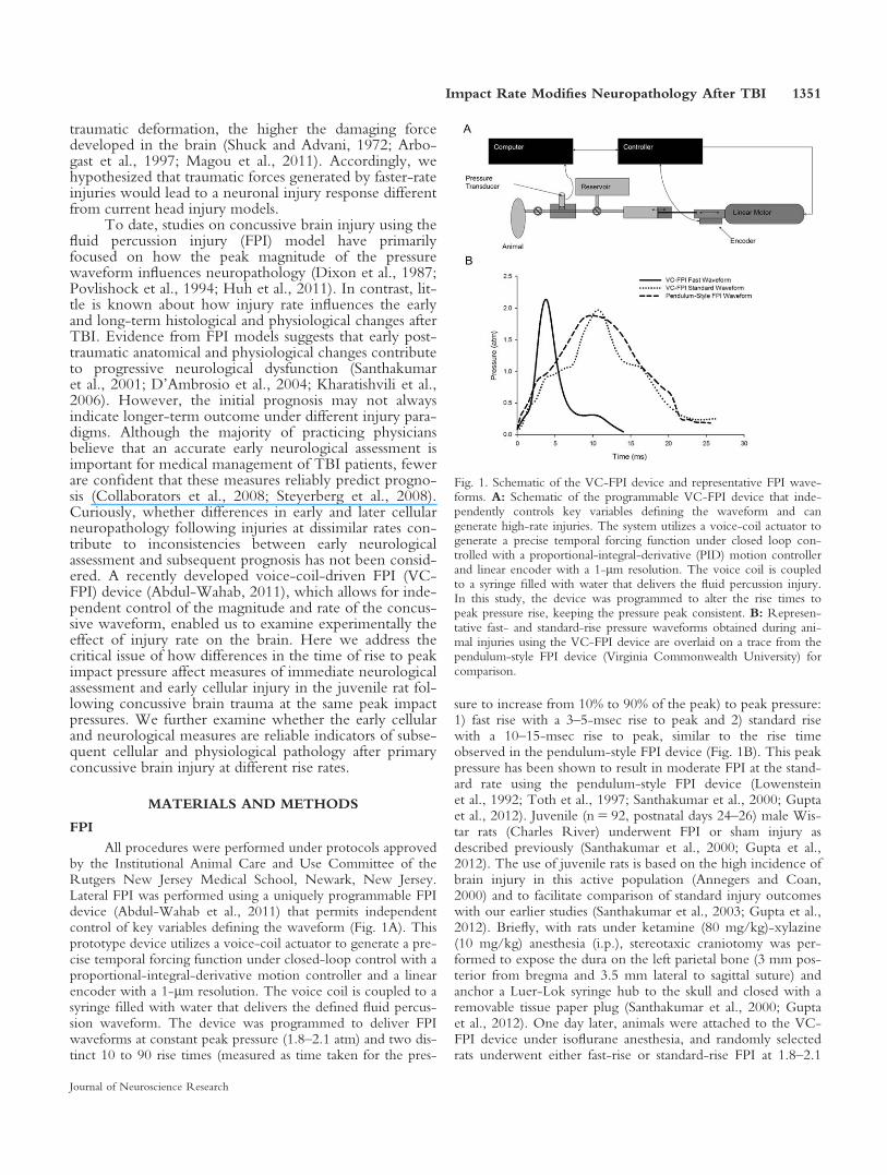
All procedures were performed under protocols approvedby the Institutional Animal Care and Use Committee of theRutgers New Jersey Medical School, Newark, New Jersey.Lateral FPI was performed using a uniquely programmable FPIdevice (Abdul-Wahab et al., 2011) that permits independentcontrol of key variables defining the waveform (Fig. 1A). Thisprototype device utilizes a voice-coil actuator to generate a pre-cise temporal forcing function under closed-loop control with aproportional-integral-derivative motion controller and a linearencoder with a 1-lm resolution. The voice coil is coupled to asyringe filled with water that delivers the defined fluid percus-sion waveform. The device was programmed to deliver FPIwaveforms at constant peak pressure (1.8–2.1 atm) and two dis-tinct 10 to 90 rise times (measured as time taken for the pres-
sure to increase from 10% to 90% of the peak) to peak pressure:1) fast rise with a 3–5-msec rise to peak and 2) standard risewith a 10–15-msec rise to peak, similar to the rise timeobserved in the pendulum-style FPI device (Fig. 1B). This peakpressure has been shown to result in moderate FPI at the stand-ard rate using the pendulum-style FPI device (Lowensteinet al., 1992; Toth et al., 1997; Santhakumar et al., 2000; Guptaet al., 2012). Juvenile (n 5 92, postnatal days 24–26) male Wis-tar rats (Charles River) underwent FPI or sham injury asdescribed previously (Santhakumar et al., 2000; Gupta et al.,2012). The use of juvenile rats is based on the high incidence ofbrain injury in this active population (Annegers and Coan,2000) and to facilitate comparison of standard injury outcomeswith our earlier studies (Santhakumar et al., 2003; Gupta et al.,2012). Briefly, with rats under ketamine (80 mg/kg)-xylazine(10 mg/kg) anesthesia (i.p.), stereotaxic craniotomy was per-formed to expose the dura on the left parietal bone (3 mm pos-terior from bregma and 3.5 mm lateral to sagittal suture) andanchor a Luer-Lok syringe hub to the skull and closed with aremovable tissue paper plug (Santhakumar et al., 2000; Guptaet al., 2012). One day later, animals were attached to the VC-FPI device under isoflurane anesthesia, and randomly selectedrats underwent either fast-rise or standard-rise FPI at 1.8–2.1
Fig. 1. Schematic of the VC-FPI device and representative FPI wave-forms. A: Schematic of the programmable VC-FPI device that inde-pendently controls key variables defining the waveform and cangenerate high-rate injuries. The system utilizes a voice-coil actuator togenerate a precise temporal forcing function under closed loop con-trolled with a proportional-integral-derivative (PID) motion controllerand linear encoder with a 1-lm resolution. The voice coil is coupledto a syringe filled with water that delivers the fluid percussion injury.In this study, the device was programmed to alter the rise times topeak pressure rise, keeping the pressure peak consistent. B: Represen-tative fast- and standard-rise pressure waveforms obtained during ani-mal injuries using the VC-FPI device are overlaid on a trace from thependulum-style FPI device (Virginia Commonwealth University) forcomparison.
Impact Rate Modifies Neuropathology After TBI 1351
Journal of Neuroscience Research
atm. Sham-injured control animals underwent the craniotomyand were attached to the FPI device under anesthesia withoutdelivery of the pressure wave (Santhakumar et al., 2003; Guptaet al., 2012). Immediately after injury, rats were monitored foroccurrence of behavioral seizures, duration of apnea (time tofirst breath following injury), time to recovery of response totoe pinch on the contralateral side, recovery of righting reflex,and mortality. Seizures were defined as the presence of Racinestage 3 or higher seizures, including forelimb or hind limb clo-nus or generalized tonic clonic seizures. When seizures werepresent, the first seizure typically occurred during the period ofposttraumatic apnea, and the rats recovered from apnea afterthe seizure. In some animals, additional seizures were observedafter recovery from apnea. Righting reflex is defined as thetime it takes for the animals to right itself when placed on theback. The syringe hub was plugged, and animals were returnedto the home cage with the implanted hub and no additionalsurgical procedures.
Histology and Immunohistochemistry
Gallyas silver stain (Gallyas et al., 1990; van den Pol andGallyas, 1990; Toth et al., 1997) and Fluoro-Jade C stainingwere performed on 40-lm sections from animals perfused with4% paraformaldehyde 4–6 hr postinjury. The range of time toperfusion after injury was kept constant among groups. Onlysections ipsilateral to the side of injury were examined. Sectionsprocessed for Gallyas stain were dehydrated in 50%, 75%, and100% 1-propanol for 5 min each; incubated at 56�C in 0.8%sulfuric acid in 1-propanol for 16 hr; and treated with 1% aceticacid for 5 min. Sections were then rehydrated and treated with3% acetic acid and developed in a silicotungstate solution (Gal-lyas et al., 1990; van den Pol and Gallyas, 1990). After develop-ment, sections were treated with 1% acetic acid for 30 min,dehydrated, and mounted on slides. For Fluoro-Jade C staining,sections mounted on gelatinized slides were air dried, hydrated,and incubated in 0.06% potassium permanganate before beingstained with 0.001% Fluoro-Jade C in 0.1% acetic acid in thedark for 30 min. Gallyas silver stain and Fluoro-Jade C stainingwere performed in separate groups of rats, which wereimplanted and injured by different investigators. For NeuNstaining, rats were perfused 1 week after sham or head injury.Sections (50 lm) were immunostained with anti-NeuN anti-body (MAB377, 1:1,000, mouse monoclonal; Millipore, Bed-ford, MA) and reacted with an appropriate secondary antibodyto reveal staining (Yu et al., 2013). Controls in which primaryantibody was omitted were routinely included.
Quantification was performed with randomized system-atic sampling protocols, selecting every tenth slice along theseptotemporal axis of the hippocampus on the injured side (Yuet al., 2013). Cell counts were performed with the OpticalFractionator probe of Stereo Investigator V.10.02 (MBF Bio-science) and an Olympus BX51 microscope and a 3100 oilobjective. In each section, the hilus was outlined by a contourtraced under a 310 objective. Sampling parameters were set at3100, counting frame 50 3 50 lm, dissector height 25 lm,and top guard zone 4 lm. Approximately 30 sites per contourwere sampled using randomized systematic sampling protocols.In each section, the number of labeled cells was estimated based
on planimetric volume calculations in Stereo Investigator (Westet al., 1991; West, 1993; Yu et al., 2013). Data are plotted asnumber of stained neurons per section.
In Vitro Electrophysiology
One week (7–10 days) after FPI or sham injury (Santhaku-mar et al., 2001; Gupta et al., 2012), rats were decapitated underisoflurane anesthesia. Horizontal brain slices (400 lm) were pre-pared in ice-cold sucrose-artificial cerebrospinal fluid (sucrose-aCSF) containing the following (in mM): 85 NaCl, 75 sucrose,24 NaHCO3, 25 glucose, 4 MgCl2, 2.5 KCl, 1.25 NaH2PO4,and 0.5 CaCl2 using a Leica VT1200S vibratome. Slices ipsilateralto the side of injury were incubated at 32�C 6 1�C for 30 min ina submerged holding chamber and subsequently held at roomtemperature (Gupta et al., 2012). The recording aCSF containedthe following (in mM): 126 NaCl, 2.5 KCl, 2 CaCl2, 2 MgCl2,1.25 NaH2PO4, 26 NaHCO3, and 10 D-glucose. All solutionswere saturated with 95% O2 and 5% CO2 and maintained at apH of 7.4 for 1–6 hr. Field recordings were performed in aninterface recording chamber (BSC2; Automate Scientific) per-fused with aCSF at 34�C. Recordings in the granule cell layer ofthe dentate gyrus were obtained by using patch pipettes (4 MX,KG-33 borosilicate glass capillary with 1.5 mm outer diameterfrom Harvard Apparatus, pulled using a Sutter P-1000 horizontalmicropipette puller) filled with recording aCSF as in earlier stud-ies (Toth et al., 1997; Gupta et al., 2012). Responses wereevoked by constant-current stimuli (0.5–6 mA, 50 lsec) deliv-ered at 0.1 Hz through a custom-made, parallel bipolar 90-lmtungsten stimulating electrode placed in the perforant path at thejunction of the dorsal blade and the crest as previously described(Santhakumar et al., 2000, 2001; Gupta et al., 2012). Recordingswere obtained with an AxoPatch200B amplifier and digitized at10 kHz with a DigiData 1440A (Molecular Devices, Sunnyvale,CA). Population spike amplitude was measured as the amplitudeof the first negative deflection overriding the field excitatorypostsynaptic potential (EPSP) waveform as described previously(Jester et al., 1995), (x 1 y)/2 – z in the fast- and standard-FPItraces in Figure 5A.
Statistical Analysis
Statistical analysis of behavioral and histological data wasperformed via Mann-Whitney U test or one-way ANOVA onranks (Kruskal-Wallis test) followed by pairwise multiple com-parison by Dunn’s method or Student-Newman-Keuls methodas appropriate. Categorical data were analyzed via v2 test. Two-way repeated-measures ANOVA was used to compare fieldrecording data between experimental groups. Statistical analysiswas performed in SigmaPlot 12.3. Significance was set toP< 0.05. Data are shown as mean 6 SEM and median andinterquartile range (IQR) where appropriate.
RESULTS
Differential Immediate Behavioral Response toFast- and Standard-Rise FPI
Previous studies using pendulum FPI devices haveshown distinctive immediate behavioral changes such asseizures and transient apnea following moderate
1352 Neuberger et al.
Journal of Neuroscience Research

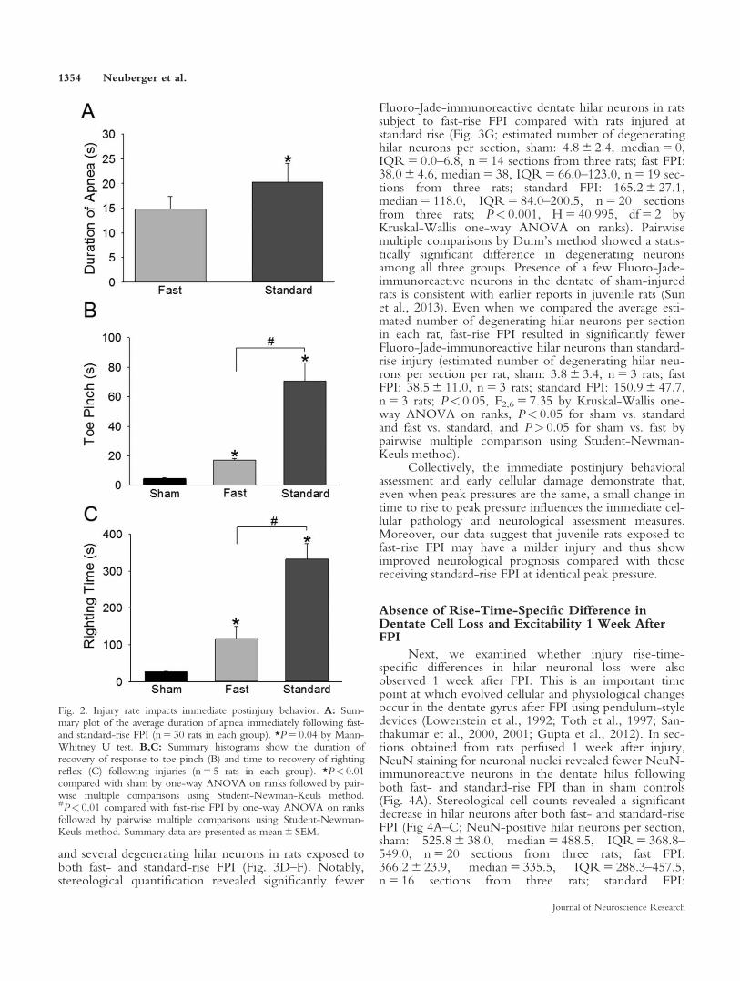
concussive brain injury (Dixon et al., 1987; Toth et al.,1997). Here we examine the immediate neurobehavioralresponses of juvenile rats to two different rise times of FPIdelivery. Using a moderate injury strength of 1.8–2.1 atmpeak pressure, rats were injured with a fast-rise (3–5 msecrise to peak) and standard-rise (10–15 msec rise to peak)FPI using our voice-coil-driven FPI (VC-FPI) device. Arandom sampling of the FPI waveforms used in the cur-rent study revealed a statistically significant difference inthe time of rise to peak pressure between fast- andstandard-rise FPI waveforms (Table I). Physiologically,sham controls showed no apnea (data not shown), andrats injured using the fast waveform demonstrated shorterduration of apnea than those injured using the standardwaveform (Fig. 2A; apnea in seconds, fast FPI:14.8 6 1.8, median 5 10.0, IQR 5 8.25–19.25, n 5 30;standard FPI: 20.27 6 2.77, median 5 15.0, IQR 5 12.0–19.75, n 5 30; P< 0.05 by Mann-Whitney U test, basedon pooled data from animals euthanized at the 4–6-hr and1-week time points).
None of the rats exposed to fast-rise FPI demon-strated postinjury seizures (0 of 16 rats subject to fast-riseFPI), whereas a majority of the rats exposed to standardrate FPI developed stage 3 or higher seizures (13 of 18rats subject to standard-rise FPI). The difference in thepercentage of rats that developed seizures following fast-and standard-rise injuries was statistically significant (TableI).
Injury with a standard rate of rise to peak pressureresulted in 22.2% mortality in the rats (four of 18 rats;two rats died from postinjury apnea and two rats diedfrom continuous stage 4 posttraumatic seizures followingrecovery from apnea). However, none of the rats exposedto fast-rise FPI at identical peak pressures died followinginjury, indicating reduced mortality following fast-riseinjury (Table I). A group of animals that survived wasperfused 4–6 hr after FPI for histology. Similar resultswere obtained with animals used for histological andphysiological studies 1 week after FPI (Table I).
Rats injured using either waveform showed increasein latency of response to toe pinch and recovery of right-ing reflex compared with controls. However, rats subjectto fast-rise FPI exhibited a significantly faster recovery ofresponse to toe pinch than rats that underwent standard-rise FPI (Fig. 2B; latency to recovery of response to toepinch in sec, sham: 4.6 6 0.4, median 5 4.0, IQR 5 4.0–5.5, n 5 5; fast FPI: 17.0 6 1.1, median 5 16.0,
IQR 5 15.0–19.5, n 5 5; standard FPI: 70.4 6 12.7,median 5 73.0, IQR 5 41.5–98.0, n 5 5; P< 0.05,H 5 12.61, df 5 2 by one-way ANOVA on ranks fol-lowed by P< 0.05 for all pairwise multiple comparisonsusing Student-Newman-Keuls method). Similarly, ratsthat sustained fast-rise FPI exhibited earlier return ofrighting reflex compared with rats injured at standard-riseFPI (Fig. 2C; time to return of righting reflex in sec,sham: 26.0 6 1.7, median 5 25.0, IQR 5 22.5–30.0,n 5 5; fast FPI: 115.80 6 33.5, median 5 89.0,IQR 5 63.0–182.0, n 5 5; standard FPI: 332.4 6 41.9,median 5 287.0, IQR 5 262.0–425.5, n 5 5; P< 0.05,H 5 12.61, df 5 2 by one-way ANOVA on ranks fol-lowed by P< 0.05 for all pairwise multiple comparisonsusing Student-Newman-Keuls method). Together theimmediate neurobehavioral data from juvenile rats suggestthat, even when peak injury pressures are similar, the fast-rise injuries result in reduced early neurological deficitscompared with standard-rise injuries.
Rate of FPI Modifies the Extent of Early HilarNeuronal Injury
Next, we examined differences in cellular responsesto injury at fast and standard rise times. Previous studieshave shown that moderate injuries at standard rise, usingthe pendulum-style FPI device, lead to instantaneousmechanical injury (Toth et al., 1997) and early degenera-tion (Gupta et al., 2012) of neurons in the hilus of thedentate gyrus. Similar to earlier studies (Toth et al.,1997), sections from sham-control rats treated with theGallyas silver stain (Gallyas et al., 1990; van den Pol andGallyas, 1990; Toth et al., 1997) revealed negligible hilarstaining (sham: nine sections from three rats), in contrastto the dense staining of hilar neuronal somata and den-drites following standard-rise injury (standard FPI: n 5 12sections from three rats). Importantly, the somatic silverstaining following fast-rise FPI was relatively sparse (fastFPI: n 5 12 sections from three rats), suggesting that theimmediate mechanical damage to dentate neurons isreduced compared with standard-rise FPI at similar peakpressures (Fig. 3A–C).
To confirm our findings, a second investigator sub-jected an additional group of rats to injuries at fast andstandard rates for examination of early postinjury neuronaldegeneration. Fluoro-Jade C staining performed 4–6 hrafter FPI revealed few labeled neurons in sham controls
TABLE I. Summary of Waveform Rise and Immediate Postinjury Response
Sham Fast FPI Standard FPI
10–90% Rise time (msec) N/A 4.67 6 0.3 (n 5 9) 11.88 6 1.11 (n 5 9)*
% Seizure (stage 3 or higher) 0 (n 5 7) 0 (n 5 16) 72.2% (n 5 18)†‡
% Mortality (4-hr study) 0 (n 5 7) 0 (n 5 16) 22.2% (n 5 18)†‡
% Mortality (1-week study) 0 (n 5 14) 0 (n 5 16) 22.7% (n 5 22)†‡
*P< 0.05 by Student’s t-test.†P< 0.05 compared with sham by v2 test.‡P< 0.05 compared with fast FPI by v2 test.
Impact Rate Modifies Neuropathology After TBI 1353
Journal of Neuroscience Research
and several degenerating hilar neurons in rats exposed toboth fast- and standard-rise FPI (Fig. 3D–F). Notably,stereological quantification revealed significantly fewer
Fluoro-Jade-immunoreactive dentate hilar neurons in ratssubject to fast-rise FPI compared with rats injured atstandard rise (Fig. 3G; estimated number of degeneratinghilar neurons per section, sham: 4.8 6 2.4, median 5 0,IQR 5 0.0–6.8, n 5 14 sections from three rats; fast FPI:38.0 6 4.6, median 5 38, IQR 5 66.0–123.0, n 5 19 sec-tions from three rats; standard FPI: 165.2 6 27.1,median 5 118.0, IQR 5 84.0–200.5, n 5 20 sectionsfrom three rats; P< 0.001, H 5 40.995, df 5 2 byKruskal-Wallis one-way ANOVA on ranks). Pairwisemultiple comparisons by Dunn’s method showed a statis-tically significant difference in degenerating neuronsamong all three groups. Presence of a few Fluoro-Jade-immunoreactive neurons in the dentate of sham-injuredrats is consistent with earlier reports in juvenile rats (Sunet al., 2013). Even when we compared the average esti-mated number of degenerating hilar neurons per sectionin each rat, fast-rise FPI resulted in significantly fewerFluoro-Jade-immunoreactive hilar neurons than standard-rise injury (estimated number of degenerating hilar neu-rons per section per rat, sham: 3.8 6 3.4, n 5 3 rats; fastFPI: 38.5 6 11.0, n 5 3 rats; standard FPI: 150.9 6 47.7,n 5 3 rats; P< 0.05, F2,6 5 7.35 by Kruskal-Wallis one-way ANOVA on ranks, P< 0.05 for sham vs. standardand fast vs. standard, and P> 0.05 for sham vs. fast bypairwise multiple comparison using Student-Newman-Keuls method).
Collectively, the immediate postinjury behavioralassessment and early cellular damage demonstrate that,even when peak pressures are the same, a small change intime to rise to peak pressure influences the immediate cel-lular pathology and neurological assessment measures.Moreover, our data suggest that juvenile rats exposed tofast-rise FPI may have a milder injury and thus showimproved neurological prognosis compared with thosereceiving standard-rise FPI at identical peak pressure.
Absence of Rise-Time-Specific Difference inDentate Cell Loss and Excitability 1 Week AfterFPI
Next, we examined whether injury rise-time-specific differences in hilar neuronal loss were alsoobserved 1 week after FPI. This is an important timepoint at which evolved cellular and physiological changesoccur in the dentate gyrus after FPI using pendulum-styledevices (Lowenstein et al., 1992; Toth et al., 1997; San-thakumar et al., 2000, 2001; Gupta et al., 2012). In sec-tions obtained from rats perfused 1 week after injury,NeuN staining for neuronal nuclei revealed fewer NeuN-immunoreactive neurons in the dentate hilus followingboth fast- and standard-rise FPI than in sham controls(Fig. 4A). Stereological cell counts revealed a significantdecrease in hilar neurons after both fast- and standard-riseFPI (Fig 4A–C; NeuN-positive hilar neurons per section,sham: 525.8 6 38.0, median 5 488.5, IQR 5 368.8–549.0, n 5 20 sections from three rats; fast FPI:366.2 6 23.9, median 5 335.5, IQR 5 288.3–457.5,n 5 16 sections from three rats; standard FPI:
Fig. 2. Injury rate impacts immediate postinjury behavior. A: Sum-mary plot of the average duration of apnea immediately following fast-and standard-rise FPI (n 5 30 rats in each group). *P 5 0.04 by Mann-Whitney U test. B,C: Summary histograms show the duration ofrecovery of response to toe pinch (B) and time to recovery of rightingreflex (C) following injuries (n 5 5 rats in each group). *P< 0.01compared with sham by one-way ANOVA on ranks followed by pair-wise multiple comparisons using Student-Newman-Keuls method.#P< 0.01 compared with fast-rise FPI by one-way ANOVA on ranksfollowed by pairwise multiple comparisons using Student-Newman-Keuls method. Summary data are presented as mean 6 SEM.
1354 Neuberger et al.
Journal of Neuroscience Research

Fig. 3. Differential early dentate neuronal injury and degenerationafter fast- and standard-rise FPI within 4 hr after injury. A–C: Rep-resentative sections from the dentate gyrus of rats fixed 4 hr aftersham, fast-rise, and standard-rise FPI and stained with the Gallyassilver stain to reveal neurons with mechanical injury. Note the lackof staining in controls (A), the sparse staining after fast-rise FPI (B),and the presence of numerous darkly stained cells in the dentatehilus and also a subset of the darkly stained cells in the granule celllayer (GCL) after standard-rise FPI (C). D–F: Fluoro-Jade C stainedhippocampal sections from rats perfused 4–6 hr after FPI show fewlabeled hilar neurons in the sham control (D), several labeled neu-
rons following fast-rise FPI (E), and numerous degenerating neuronsin the dentate hilus after standard-rise FPI (F). G: Summary histo-gram of stereological counts of Fluoro-Jade C-labeled hilar neuronsper section in animals subject to the injury paradigms (n 5 14 sec-tions from three rats in sham, 19 sections from three rats after fast-FPI, and 20 sections from three rats after standard FPI). *P< 0.05compared with sham; #P< 0.05 compared with fast-rise FPI byone-way ANOVA for ranks followed by post hoc pairwise compari-son by Dunn’s method. Summary data are presented as mean-6 SEM. [Color figure can be viewed in the online issue, which isavailable at wileyonlinelibrary.com.]

322.1 6 27.4, median 5 287.0, IQR 5 216.0–411.0,n 5 23 sections from three rats; P< 0.05, H 5 17.7,df 5 2 by Kruskal-Wallis one-way ANOVA on ranks).Pairwise multiple comparison by Dunn’s method showedthat differences in the number of surviving NeuN-positive neurons between sham and standard-rise FPI andbetween sham and fast-rise FPI were statistically signifi-cant. However, the number of NeuN-labeled neuronswas not different between fast- and standard-rise FPI 1week after injury. Similar results were obtained when sta-tistical analysis was conducted on the estimated number ofhilar neurons per section in each rat (estimated number ofNeuN-positive hilar neurons per section per rat, sham:520.7 6 13.9, n 5 3 rats; fast FPI: 371.7 6 48.5, n 5 3rats; standard FPI: 319.1 6 18.4, n 5 3 rats; F2,6 5 11.37,P< 0.05 by one-way ANOVA, P< 0.05 for sham vs.standard and sham vs. fast, and P> 0.05 for standard vs.fast by pairwise multiple comparison using Holm-Sidakmethod). Given the disparity between hilar neuronaldegeneration following fast- and standard-rise FPI at 4 hr,this could suggest recovery of some mechanically injuredneurons following standard-rise FPI (Toth et al., 1997).However, because Fluoro-Jade labels irrevocably degen-erating neurons, our data likely indicate a greater progres-sion of hilar cell loss within the week following fast-riseFPI. Fluoro-Jade staining conducted on sections from rats1 week after fast- or standard-rate FPI showed no labeling
of neuronal profiles (number of degenerating hilar neu-rons per section per rat, sham: 0, nine sections from threerats; fast FPI: 0, nine sections from three rats; standardFPI: 0, nine sections from three rats) indicating that thereare no actively degenerating neurons 1 week after fast-and standard-rate FPI.
Earlier studies, by us and by other groups, have con-sistently demonstrated an increase in dentate networkexcitability and altered inhibition 1 week after FPI whenusing the pendulum-style device (Lowenstein et al., 1992;Toth et al., 1997; Santhakumar et al., 2000, 2001; Guptaet al., 2012). The hilar neuronal loss and increase in den-tate excitability have been suggested to predict develop-ment of long-term neurological complications such asposttraumatic epilepsy (Lowenstein et al., 1992; Tothet al., 1997). Therefore, we examined the granule cellpopulation response to afferent activation to assess theeffect of injury rise time on neurophysiological outcomes1 week after FPI. As illustrated in Figure 5A, althoughthe granule cell population response evoked by perforantpath stimulation at 4 mA showed no population spikes inseven of 12 slices from sham-control rats, afferent activa-tion reliably elicited dentate population spikes in all slices1 week after fast- and standard-rise FPI. Summary datademonstrate that the afferent-evoked granule cell popula-tion spike amplitude in injured rats was significantlyenhanced compared with that in sham controls. Two-
Fig. 4. Dentate hilar neuronal loss 1 week after fast- and standard-rise FPI. A–C: Representative NeuN-stained hippocampal sectionsfrom rats perfused 1 week after injury show significantly more neu-rons in the hilus of sham controls (A) compared with rats exposedto either fast-rise (B) or standard-rise (C) FPI. D: Summary datacompare stereological counts of NeuN-labeled neurons per sectionsfrom sham controls and rats subject to fast- and standard-rise FPI
(n 5 20 sections from three rats in sham, 16 sections from three ratsafter fast FPI, and 23 sections from three rats after standard FPI).GCL, granule cell layer. *P< 0.05; N.S. indicates P> 0.05 by one-way ANOVA for ranks followed by post hoc pairwise comparisonby Dunn’s method. Summary data are presented as mean 6 SEM.[Color figure can be viewed in the online issue, which is availableat wileyonlinelibrary.com.]
1356 Neuberger et al.
Journal of Neuroscience Research

Fig. 5. Absence of rate-specific difference in dentate excitability 1week after FPI. A: Examples of dentate granule cell field responsesevoked by a 4-mA stimulus to the perforant path. Recordings wereperformed in control aCSF. Population spike amplitude was calcu-lated as (x 1 y)/2 – z (Jester et al., 1995). B: Summary plot of affer-ent evoked population spike amplitude in aCSF. C: Dentatepopulation responses evoked by perforant path stimulation at 4 mAwith the GABAA receptor antagonist gabazine (10 lM, SR95531).D: Summary plot of perforant path-evoked granule cell populationspike amplitude with gabazine (10 lM). In B,D, *P< 0.05 in fast
FPI, **P< 0.05 in standard FPI compared with sham by two-wayrepeated-measures ANOVA followed by post hoc Tukey’s test;#P< 0.05 in fast FPI, ##P< 0.05 in standard FPI compared withsham for effect of injury by two-way repeated-measures ANOVA.In A,C, triangles indicate truncated stimulus artifact. E: Summaryhistogram of the number of population spikes in response to 6-mAstimulation in the presence of gabazine. *P< 0.05 by one-wayANOVA on ranks followed by post hoc pairwise multiple compari-son using Dunn’s method. Summary data are presented asmean 6 SEM.

way repeated-measures ANOVA identified significanteffects of treatment (injury) at F2,179 5 7.42 (P< 0.01),stimulus intensity at F4,179 5 54.03 (P< 0.001), and inter-action between treatment and intensity at F8,179 5 6.43(P< 0.001). Pairwise multiple comparisons by Tukey’stest within each stimulus intensity level revealed that bothfast and standard FPI were significantly different fromsham at 2, 4, and 6 mA stimulation (population spikeamplitude in millivolts evoked by 4 mA stimulation:sham: 0.31 6 0.13; fast FPI: 1.42 6 0.34; standard FPI:1.2 6 0.18, 6 mA stimulation: sham: 0.35 6 0.14; fastFPI: 1.56 6 0.27; standard FPI: 1.53 6 0.13, n 5 12 sliceeach from five sham, six fast-FPI, and six standard-FPIrats). Interestingly, slices from rats injured with fast-rise orstandard-rise FPI produced similar postinjury populationspike amplitudes (Fig. 5B; P> 0.5 by two-way repeated-measures ANOVA followed by pairwise multiple com-parison within each stimulus intensity level by Tukey’stest).
Previous work has identified that brain injury withthe pendulum-style FPI device enhances excitability ofthe dentate excitatory network, measured in the presenceof a GABAA receptor (GABAAR) antagonist (Santhaku-mar et al., 2000). To determine whether injuries at fastand standard rise differentially impacted the excitability ofthe dentate excitatory network, we examined the perfo-rant path-evoked granule cell field responses in slices thatwere perfused with gabazine (10 lM, SR95531), aGABAAR antagonist. Once again, the amplitude of thefirst population spike in response to afferent activationwas greater in slices from head-injured rats comparedwith sham controls (Fig. 5C). Two-way repeated-meas-ures ANOVA identified significant effects of treatment(injury) at F2,224 5 4.53 (P< 0.05), stimulus intensity atF4,224 5 49.52 (P< 0.001), and interaction between treat-ment and intensity at F8,224 5 3.3 (P< 0.005). Pairwisemultiple comparisons within each stimulus intensity levelby Tukey’s test revealed significant differences betweensham and both fast and standard FPI at 4 and 6 mA stimu-lation (Fig. 5D; population spike amplitude in mV evokedby 4 mA stimulation: sham: 2.35 6 0.23, n 5 15 slicesfrom six rats; fast FPI: 2.85 6 0.35, n 5 15 slices from sixrats; standard FPI: 3.44 6 0.34, n 5 15 slices from six rats;6 mA stimulation: sham: 2.54 6 0.24 n 5 15 slices; fastFPI: 3.16 6 0.36 n 5 15 slices; standard FPI: 3.71 6 0.41n 5 15 slices). However, the population spike amplitudeswithin each intensity was not different between fast andstandard FPI (Fig. 5D; P> 0.5 by two-way repeated-measures ANOVA followed by pairwise multiple com-parisons within each stimulus intensity level by Tukey’stest). Additionally, the slices from rats subject to FPIshowed an increase in the number of afferent-evokedpopulation spikes (Fig. 5E). The number of populationspikes in response to perforant path stimulation at 6 mAwas significantly higher in both fast- and standard-FPIgroups compared with sham but similar in slices from ratsexposed to fast- and standard-rise FPI (Fig. 5E; number ofpopulation spikes in response to 6 mA simulation: sham:1.07 6 9.30, n 5 15 slices from six rats; fast FPI:
2.67 6 0.32, n 5 15 slices from six rats, standard FPI:3.07 6 0.34, n 5 15 slices from six rats; significance deter-mined by one-way ANOVA on ranks followed by posthoc pairwise multiple comparisons using Dunn’smethod).
Together our studies on fast- and standard-rise FPIat similar peak impact pressures in juvenile rats indicatethat, although the improved neurobehavioral outcomeand reduced cellular injury measures within hours follow-ing fast-rise injury suggest that fast-rate trauma leads to amilder TBI than slower-rate injury, the extent of cell lossand physiological dysfunction after fast-rise injury arecomparable to that of moderate TBI within a week afterinjury.
DISCUSSION
The use of a novel programmable VC-FPI device allowedus to examine a fundamental question concerningwhether differences in the rate of delivery of a fluid per-cussion wave alter neurological outcome following braininjury. We demonstrate, for the first time, that increasingthe rate of injury, by decreasing the rate of rise to thesame peak pressure, alters the immediate behavioral assess-ment as well as cellular injury and neuronal degenerationwithin 4 hr after injury. This initially suggests that fast-rate injuries lead to reduced neurological damage com-pared with slower-rate injuries. Studies performed 1 weekafter FPI, however, demonstrate a similar level of injuryas shown by the extent of dentate hilar cell loss andincrease in network excitability following fast- andslower-rise injuries.
Our findings in juvenile rats suggest that a betterimmediate neurological outcome following fast injurymay mask the severity of neuropathology at later timepoints. Strategies for evaluation of injury severity rely pri-marily on early clinical measures (Saatman et al., 2008), soour data suggest the need to consider the effect of injuryrate in shaping the cellular and physiological response atlater time points while managing patients sustaining fast-and slower-rate concussive injuries.
Brain injury can result from a wide spectrum ofinsults ranging from falls that occur at relatively slowimpact velocities (Wright and Laing, 2012) to sports inju-ries and traffic accidents, which represent higher-rateimpacts (Zhang et al., 2006a, 2009b). Extremely rapidshock waves generated by blasts represent the farthest endof the spectrum of high-rate impact (Zhang et al., 2009a;Shridharani et al., 2012). Indeed, fast-rate TBI followingexposure to explosive blasts is the signature injury of therecent wars (Elder and Cristian, 2009) and a growing issuefacing veterans. The primary impact waves of blast TBIhave a considerably faster rise to peak pressure than non-blast concussive TBI (Chavko et al., 2007; Zhang et al.,2009a; Cernak and Noble-Haeusslein, 2010; Shridharaniet al., 2012; Wright and Laing, 2012). Most contempo-rary studies use devices such as shock tubes to generatefast-rise injuries (Cernak et al., 2001; Risling et al., 2011;Rubovitch et al., 2011; Skotak et al., 2013). The more
1358 Neuberger et al.
Journal of Neuroscience Research

commonly employed FPI devices (Dixon et al., 1987;Garga and Lowenstein, 2006) are used to deliver fixedconcussive waveforms to model closed-head injury fromtrauma such as automobile accidents or falls. The inabilityto control rate and magnitude in the pendulum-style FPIdevice independently and the use of different devices tomodel injuries at drastically different rates render it diffi-cult directly to compare rise time-specific changes in neu-ropathology without differences in peak pressure anddevice-dependent confounding factors. The prototypeVC-FPI device (Abdul-Wahab et al., 2011) enabled us toovercome these technical obstacles and demonstrate that,even at constant peak pressures, the rate of fluid percus-sion directly affects the immediate neurological response.Our study presents the first demonstration that even smallchanges in injury rate can modify the ability of early neu-rological measures to predict the subsequentneuropathology.
Rate sensitivity to injury is likely because brain tis-sue behaves like a viscoelastic material (Prange and Mar-gulies, 2002; Vappou et al., 2007; Rashid et al., 2012).Indeed, some biomechanical investigations are beginningto suggest that the brain’s response to mechanical loadingis sensitive to both the rate and the magnitude of thetrauma (Morrison et al., 2003; Lusardi et al., 2004; Magouet al., 2011; Rashid et al., 2012). Viscoelastic tissues aresoft and compliant under slow mechanical loading, wheresmall forces can lead to large deformations in the brain.Under higher rates of mechanical loading, however, thetime dependence of the viscoelastic brain acts more like astiff solid, developing larger internal forces under the samedeformation constraints (Shuck and Advani, 1972; Arbo-gast et al., 1997; Magou et al., 2011). Accordingly, weshould assume that, with variations in the rate of mechan-ical trauma, the deformation and damaging forces withinthe brain tissue will differ. These variations in the biome-chanical loading conditions of the brain tissue likely con-tribute to the variety of neuronal damage andneurological outcome seen in head-injured patients. Simi-lar to how increases in the magnitude of trauma, such aspeak pressure in FPI, lead to increased injury, alterationsin loading rate should also lead to alterations in the injurysequelae. Our results suggest that increases in the rate ofinjury appear to inflict a milder injury acutely but progressinto neuronal injury and dysfunction similar to the lower-rate injury at the same injury magnitude.
A potential caveat is that, although the peak pres-sures are similar, the fast-injury waveform in the currentstudy had a shorter duration than the standard wave. Thusthe impulse, a measure of energy transfer determined asthe area under the pressure curve, in fast-rise FPI is lowerthan in standard-rise FPI. This difference is realistic, inthat high-rate injuries such as blast waves usually have abrief duration compared with the standard concussivewaveform modeled by the pendulum-style FPI device(Elder and Cristian, 2009; Zhang et al., 2009a). Previousstudies with the pendulum-style FPI device have demon-strated that dentate neuronal loss measured 1 week afterinjury increases progressively with increase in injury
severity, classified based on injury peak pressure frommild to moderate to severe (Lowenstein et al., 1992).Additionally, it has been shown that the cell loss afterbrain injury using the pendulum-style FPI device occursearly after injury and does not increase further beyond 1week postinjury (Toth et al., 1997). Thus, if the fast-rise,low-impulse injury leads to just a milder form of TBI, wewould expect that the dentate hilar cell loss 1 week afterFPI would be lower in rats exposed to fast-rise FPI thanthose subject to standard-rise FPI. However, it is notablethat fast-rise FPI produced cellular and physiologicalpathology at 1 week comparable to that of standard-rise,higher-impact injuries, indicating that fast-rise FPI is notmerely a milder version of standard-rise FPI. Moreover,although the immediate neurobehavioral compromiseafter fast-rise FPI is significantly milder than that afterstandard-rise FPI (Fig. 1B,C), animals examined 1 weekafter FPI showed similar dentate hilar cell loss and altera-tions in network excitability regardless of the injury rate(Figs. 4, 5). Measures of postinjury neurological responsessuch as the Glasgow Coma Scale are widely used to assessseverity of neurological injury and predict prognosis fol-lowing TBI (Sternbach, 2000; Saatman et al., 2008; Elderand Cristian, 2009). Our data indicate that early postin-jury criteria, which seem to predict prognosis reliably afterincreasing magnitudes of impact at similar rates (Dixonet al., 1987; Povlishock et al., 1994; Huh et al., 2011),may have to consider the effect of impact rate for fast-rateinjuries such as blast TBI. Moreover, our findings suggestthat early clinical assessment may not provide an accurateevaluation of the subsequent pathology and indicate thatfollowup assessments of patients conducted several daysafter the trauma could aid in evaluating long-term prog-nosis of brain-injured patients.
Our histological studies 4 hr after injury demonstratethat the immediate cellular injury and degeneration areconsiderably milder after fast-rise FPI than after standard-rise FPI. What are the mechanisms that could account forthe subsequent absence of rate-specific difference in hilarcell loss and network excitability 1 week after injury?Structural characteristics of cytoskeletal elements in soma-todendritic compartments may underlie the difference inmechanical damage induced by fast- and slower-rise FPI.It is possible that axons, which have distinctive cytos-keletal components (Stiess and Bradke, 2011), are morevulnerable to injury during fast-rise TBI. Consistent withthis possibility, diffuse axonal injury takes a longer time tomanifest (Leonard et al., 1997; Carbonell and Grady,1999) compared with considerable early somatodendriticinjury evident in silver-stained sections from rats exposedto standard-rise injury. Additionally, high-rate TBI is typ-ically associated with extensive white matter injury(Koliatsos et al., 2011; Jorge et al., 2012). An equally pos-sible alternative is that fast-rise injuries lead to delayedexcitotoxic cell death (Manev et al., 1989; Palmer et al.,1993; Yi and Hazell, 2006), leading to increased networkexcitability. However, the lack of Fluoro-Jade C-stainedneurons 1 week after both fast and standard FPI indicatesthat, as with standard injury (Toth et al., 1997), the cell
Impact Rate Modifies Neuropathology After TBI 1359
Journal of Neuroscience Research

loss after fast FPI may be complete by 1 week. Concussiveinjury leads to distinctive apoptotic responses in juvenileand aged rats (Sun et al., 2013), so it is possible that injuryrate modified the apoptotic responses in the juvenile ratsin the current study. Although the exact mechanismsremain to be elucidated, it is evident that the progressionof neuropathology following fast-rise FPI in juvenile ratsis different than that after slower-rise TBI. The observeddivergence between early postinjury assessment and sub-sequent cellular and physiological outcome following fast-and standard-rise FPI suggest that early behavioral meas-ures used to predict prognosis after slower-rate TBI mayunderestimate the severity of long-term neurological out-come after faster-rate injuries such as blast-related TBI.
CONCLUSIONS
We have demonstrated that the rate of rise to peak pres-sure modifies the immediate neurobehavioral and earlycellular response to concussive brain injury in juvenilerats. We show that, despite a better immediate neurologi-cal outcome following fast injury, the long term cellularand physiological pathology after injuries with fast- andslower-rising waveforms are not different. Our findingssuggest dissimilarities in the progression of cellular pathol-ogy after fast- and slow-rising injuries.
ACKNOWLEDGMENTS
We thank Mat Long, Bogumila Swietek, Akshay Gupta,Akshata Korgaonkar, and Fatima S. Elgammal for theirhelpful comments and technical assistance and Drs. StellaElkabes and Robert F. Heary for equipment and technicalsupport. The authors declare that there are no knownconflicts of interest.
REFERENCES
Abdul-Wahab R, Mina S, Sampath S, Santhakumar V, Pfister BJ. 2011.
Precisely controllable traumatic brain injury devices for rodent models.
Bioengineering Conference (NEBEC), 2011 IEEE 37th Annual North-
east. p 1–2.
Annegers JF, Coan SP. 2000. The risks of epilepsy after traumatic brain
injury. Seizure 9:453–457.
Arbogast KB, Thibault KL, Pinheiro BS, Winey KI, Margulies SS. 1997.
A high-frequency shear device for testing soft biological tissues. J Bio-
mechanics 30:757–759.
Carbonell WS, Grady MS. 1999. Regional and temporal characterization
of neuronal, glial, and axonal response after traumatic brain injury in
the mouse. Acta Neuropathol 98:396–406.
Cernak I, Noble-Haeusslein LJ. 2010. Traumatic brain injury: an over-
view of pathobiology with emphasis on military populations. J Cereb
Blood Flow Metab 30:255–266.
Cernak I, Wang Z, Jiang J, Bian X, Savic J. 2001. Ultrastructural and
functional characteristics of blast injury-induced neurotrauma. J Trauma
50:695–706.
Chauhan NB. 2014. Chronic neurodegenerative consequences of trau-
matic brain injury. Restor Neurol Neurosci (in press).
Chavko M, Koller WA, Prusaczyk WK, McCarron RM. 2007. Measure-
ment of blast wave by a miniature fiber optic pressure transducer in the
rat brain. J Neurosci Methods 159:277–281.
Christensen J. 2012. Traumatic brain injury: risks of epilepsy and implica-
tions for medicolegal assessment. Epilepsia 53(Suppl 4):43–47.
Cohen AS, Pfister BJ, Schwarzbach E, Grady MS, Goforth PB, Satin LS.
2007. Injury-induced alterations in CNS electrophysiology. Prog Brain
Res 161:143–169.
Collaborators MCT, Perel P, Arango M, Clayton T, Edwards P,
Komolafe E, Poccock S, Roberts I, Shakur H, Steyerberg E,
Yutthakasemsunt S. 2008. Predicting outcome after traumatic brain
injury: practical prognostic models based on large cohort of interna-
tional patients. Br Med J 336:425–429.
Congeni J. 2009. Management of the adolescent concussion victim. Ado-
lescent Med 20:41–56, viii.
Creed JA, DiLeonardi AM, Fox DP, Tessler AR, Raghupathi R. 2011.
Concussive brain trauma in the mouse results in acute cognitive deficits
and sustained impairment of axonal function. J Neurotrauma 28:547–563.
D’Ambrosio R, Fairbanks JP, Fender JS, Born DE, Doyle DL, Miller
JW. 2004. Post-traumatic epilepsy following fluid percussion injury in
the rat. Brain 127:304–314.
Dixon CE, Lyeth BG, Povlishock JT, Findling RL, Hamm RJ,
Marmarou A, Young HF, Hayes RL. 1987. A fluid percussion model
of experimental brain injury in the rat. J Neurosurg 67:110–119.
Elder GA, Cristian A. 2009. Blast-related mild traumatic brain injury:
mechanisms of injury and impact on clinical care. Mt Sinai J Med 76:
111–118.
Engel J, Pedley TA. 1998. Epilepsy: a comprehensive textbook. Philadel-
phia: Lippincott-Raven.
Gallyas F, Guldner FH, Zoltay G, Wolff JR. 1990. Golgi-like demonstra-
tion of “dark” neurons with an argyrophil III method for experimental
neuropathology. Acta Neuropathol 79:620–628.
Garga N, Lowenstein DH. 2006. Posttraumatic epilepsy: a major problem
in desperate need of major advances. Epilepsy Curr 6:1–5.
Graham DI, Raghupathi R, Saatman KE, Meaney D, McIntosh TK.
2000. Tissue tears in the white matter after lateral fluid percussion brain
injury in the rat: relevance to human brain injury. Acta Neuropathol
99:117–124.
Gupta A, Elgammal FS, Proddutur A, Shah S, Santhakumar V. 2012.
Decrease in tonic inhibition contributes to increase in dentate semilunar
granule cell excitability after brain injury. J Neurosci 32:2523–2537.
Herman ST. 2002. Epilepsy after brain insult: targeting epileptogenesis.
Neurology 59(Suppl 5):S21–S26.
Hicks RR, Smith DH, Lowenstein DH, Saint Marie R, McIntosh TK.
1993. Mild experimental brain injury in the rat induces cognitive defi-
cits associated with regional neuronal loss in the hippocampus. J Neuro-
trauma 10:405–414.
Huh JW, Widing AG, Raghupathi R. 2011. Differential effects of injury
severity on cognition and cellular pathology after contusive brain
trauma in the immature rat. J Neurotrauma 28:245–257.
Iverson GL, Brooks BL, Collins MW, Lovell MR. 2006. Tracking neu-
ropsychological recovery following concussion in sport. Brain Injury
20:245–252.
Jester JM, Campbell LW, Sejnowski TJ. 1995. Associative EPSP—spike
potentiation induced by pairing orthodromic and antidromic stimula-
tion in rat hippocampal slices. J Physiol 484:689–705.
Johnson VE, Stewart W, Smith DH. 2010. Traumatic brain injury and
amyloid-beta pathology: a link to Alzheimer’s disease? Nat Rev Neuro-
sci 11:361–370.
Jorge RE, Acion L, White T, Tordesillas-Gutierrez D, Pierson R, Crespo-
Facorro B, Magnotta VA. 2012. White matter abnormalities in veterans
with mild traumatic brain injury. Am J Psychiatry 169:1284–1291.
Kharatishvili I, Nissinen JP, McIntosh TK, Pitkanen A. 2006. A model
of posttraumatic epilepsy induced by lateral fluid-percussion brain injury
in rats. Neuroscience 140:685–697.
Koliatsos VE, Cernak I, Xu L, Song Y, Savonenko A, Crain BJ, Eberhart
CG, Frangakis CE, Melnikova T, Kim H, Lee D. 2011. A mouse model
of blast injury to brain: initial pathological, neuropathological, and behav-
ioral characterization. J Neuropathol Exp Neurol 70:399–416.
1360 Neuberger et al.
Journal of Neuroscience Research

Lee PC, Bordelon Y, Bronstein J, Ritz B. 2012. Traumatic brain injury,
paraquat exposure, and their relationship to Parkinson disease. Neurol-
ogy 79:2061–2066.
Leonard JR, Grady MS, Lee ME, Paz JC, Westrum LE. 1997. Fluid per-
cussion injury causes disruption of the septohippocampal pathway in
the rat. Exp Neurol 143:177–187.
Lowenstein DH, Thomas MJ, Smith DH, McIntosh TK. 1992. Selective
vulnerability of dentate hilar neurons following traumatic brain injury: a
potential mechanistic link between head trauma and disorders of the
hippocampus. J Neurosci 12:4846–4853.
Lusardi TA, Wolf JA, Putt ME, Smith DH, Meaney DF. 2004. Effect of
acute calcium influx after mechanical stretch injury in vitro on the via-
bility of hippocampal neurons. J Neurotrauma 21:61–72.
Magou GC, Guo Y, Choudhury M, Chen L, Hususan N, Masotti S,
Pfister BJ. 2011. Engineering a high throughput axon injury system. J
Neurotrauma 28:2203–2218.
Manev H, Favaron M, Guidotti A, Costa E. 1989. Delayed increase of
Ca21 influx elicited by glutamate: role in neuronal death. Mol Pharma-
col 36:106–112.
Morrison B 3rd, Cater HL, Wang CC, Thomas FC, Hung CT, Ateshian
GA, Sundstrom LE. 2003. A tissue level tolerance criterion for living
brain developed with an in vitro model of traumatic mechanical load-
ing. Stapp Car Crash J 47:93–105.
Palmer AM, Marion DW, Botscheller ML, Swedlow PE, Styren SD,
DeKosky ST. 1993. Traumatic brain injury-induced excitotoxicity
assessed in a controlled cortical impact model. J Neurochem 61:2015–
2024.
Povlishock JT, Hayes RL, Michel ME, McIntosh TK. 1994. Workshop
on animal models of traumatic brain injury. J Neurotrauma 11:723–
732.
Prange MT, Margulies SS. 2002. Regional, directional, and age-
dependent properties of the brain undergoing large deformation. J Bio-
mechanical Eng 124:244–252.
Rashid B, Destrade M, Gilchrist MD. 2012. Mechanical characterization
of brain tissue in compression at dynamic strain rates. J Mechanical
Behav Biomedical Materials 10:23–38.
Risling M, Plantman S, Angeria M, Rostami E, Bellander BM,
Kirkegaard M, Arborelius U, Davidsson J. 2011. Mechanisms of blast
induced brain injuries, experimental studies in rats. Neuroimage
54(Suppl 1):S89–S97.
Rubovitch V, Ten-Bosch M, Zohar O, Harrison CR, Tempel-Brami C,
Stein E, Hoffer BJ, Balaban CD, Schreiber S, Chiu WT, Pick CG.
2011. A mouse model of blast-induced mild traumatic brain injury.
Exp Neurol 232:280–289.
Saatman KE, Duhaime AC, Bullock R, Maas AI, Valadka A, Manley
GT, Workshop Scientific T, Advisory Panel M. 2008. Classification of
traumatic brain injury for targeted therapies. J Neurotrauma 25:719–
738.
Santhakumar V, Bender R, Frotscher M, Ross ST, Hollrigel GS, Toth
Z, Soltesz I. 2000. Granule cell hyperexcitability in the early post-
traumatic rat dentate gyrus: the “irritable mossy cell&rdquop; hypothe-
sis. J Physiol 524:117–134.
Santhakumar V, Ratzliff AD, Jeng J, Toth Z, Soltesz I. 2001. Long-term
hyperexcitability in the hippocampus after experimental head trauma.
Ann Neurol 50:708–717.
Santhakumar V, Voipio J, Kaila K, Soltesz I. 2003. Post-traumatic hyper-
excitability is not caused by impaired buffering of extracellular potas-
sium. J Neurosci 23:5865–5876.
Shridharani JK, Wood GW, Panzer MB, Capehart BP, Nyein MK,
Radovitzky RA, Bass CR. 2012. Porcine head response to blast. Front
Neurol 3:70.
Shuck LZ, Advani SH. 1972. Rheological response of human brain tissue
in shear. J Basic Eng 94:905–911.
Skotak M, Wang F, Alai A, Holmberg A, Harris S, Switzer RC,
Chandra N. 2013. Rat injury model under controlled field-relevant pri-
mary blast conditions: acute response to a wide range of peak overpres-
sures. J Neurotrauma 30:1147–1160.
Sternbach GL. 2000. The Glasgow Coma Scale. J Emerg Med 19:67–71.
Steyerberg EW, Mushkudiani N, Perel P, Butcher I, Lu J, McHugh GS,
Murray GD, Marmarou A, Roberts I, Habbema JD, Maas AI. 2008.
Predicting outcome after traumatic brain injury: development and inter-
national validation of prognostic scores based on admission characteris-
tics. PLoS Med 5:e165; discussion e165.
Stiess M, Bradke F. 2011. Neuronal polarization: the cytoskeleton leads
the way. Dev Neurobiol 71:430–444.
Stroke. 2002. Traumatic brain injury: hope through research. Bethesda,
MD: NIoNDa.
Sun D, McGinn M, Hankins JE, Mays KM, Rolfe A, Colello RJ. 2013.
Aging- and injury-related differential apoptotic response in the dentate
gyrus of the hippocampus in rats following brain trauma. Front Aging
Neurosci 5:95.
Toth Z, Hollrigel GS, Gorcs T, Soltesz I. 1997. Instantaneous perturba-
tion of dentate interneuronal networks by a pressure wave-transient
delivered to the neocortex. J Neurosci 17:8106–8117.
Vaishnavi S, Rao V, Fann JR. 2009. Neuropsychiatric problems after
traumatic brain injury: unraveling the silent epidemic. Psychosomatics
50:198–205.
van den Pol AN, Gallyas F. 1990. Trauma-induced Golgi-like staining of
neurons: a new approach to neuronal organization and response to
injury. J Comp Neurol 296:654–673.
Vappou J, Breton E, Choquet P, Goetz C, Willinger R, Constantinesco
A. 2007. Magnetic resonance elastography compared with rotational
rheometry for in vitro brain tissue viscoelasticity measurement. Magma
20:273–278.
West MJ. 1993. New stereological methods for counting neurons. Neu-
robiol Aging 14:275–285.
West MJ, Slomianka L, Gundersen HJ. 1991. Unbiased stereological esti-
mation of the total number of neurons in thesubdivisions of the rat hip-
pocampus using the optical fractionator. Anat Rec 231:482–497.
Winter CD, Pringle AK, Clough GF, Church MK. 2004. Raised paren-
chymal interleukin-6 levels correlate with improved outcome after trau-
matic brain injury. Brain 127:315–320.
Wright AD, Laing AC. 2012. The influence of headform orientation and
flooring systems on impact dynamics during simulated fall-related head
impacts. Med Eng Physics 34:1071–1078.
Yi JH, Hazell AS. 2006. Excitotoxic mechanisms and the role of astro-
cytic glutamate transporters in traumatic brain injury. Neurochem Int
48:394–403.
Yu J, Proddutur A, Elgammal FS, Ito T, Santhakumar V. 2013. Status
epilepticus enhances tonic GABA currents and depolarizes GABA
reversal potential in dentate fast-spiking basket cells. J Neurophysiol
109:1746–1763.
Zhang J, Yoganandan N, Pintar FA, Gennarelli TA. 2006a. Brain strains
in vehicle impact tests. Annu Proc Automotive Med 50:1–12.
Zhang J, Yoganandan N, Pintar FA, Gennarelli TA. 2006b. Role of
translational and rotational accelerations on brain strain in lateral head
impact. Biomed Sci Instrument 42:501–506.
Zhang J, Pintar FA, Yoganandan N, Gennarelli TA, Son SF. 2009a.
Experimental study of blast-induced traumatic brain injury using a
physical head model. Stapp Car Crash J 53:215–227.
Zhang J, Yoganandan N, Pintar FA. 2009b. Dynamic biomechanics of the
human head in lateral impacts. Annu Proc Automotive Med 53:249–256.
Impact Rate Modifies Neuropathology After TBI 1361
Journal of Neuroscience Research
Copyright © 2022 FDOKUMEN