Tabes Dorsalis Tachykinin Tachyphylaxis Tactile Allodynia ...
Upload
independentCategory
view
3download
0
Brain (2001), 124, 2287–2298
Dissociated active and passive tactile shaperecognition: a case study of pure tactile apraxiaNathalie Valenza,1 Radek Ptak,1 Ivan Zimine,2 Maryse Badan,1 Francois Lazeyras2 andArmin Schnider1
1Clinique de Reeducation and 2Departement de Radiologie, Correspondence to: Nathalie Valenza, HopitauxHopitaux Universitaires de Geneve, Geneve, Switzerland Universitaires de Geneve, Clinique de Reeducation
(Beau-Sejour), CH-1211 Geneve 14, SwitzerlandE-mail: [email protected]
SummaryDisorders of tactile object recognition (TOR) may resultfrom primary motor or sensory deficits or higher cognitiveimpairment of tactile shape representations or semanticmemory. Studies with healthy participants suggest theexistence of exploratory motor procedures directly linkedto the extraction of specific properties of objects. A puredeficit of these procedures without concomitant gnosticdisorders has never been described in a brain-damagedpatient. Here, we present a patient with a righthemispheric infarction who, in spite of intact sensorimotorfunctions, had impaired TOR with the left hand.Recognition of 2D shapes and objects was severelydeficient under the condition of spontaneous exploration.Tactile exploration of shapes was disorganized and
Keywords: tactile apraxia; exploratory procedures; tactile object recognition; stereognosis
Abbreviations: BA � Brodmann area; EP � exploratory procedure; fMRI � functional MRI; SI � primary sensory area;SII � secondary sensory area; TOR � tactile object recognition
IntroductionDisorders of tactile object recognition (TOR) are relativelycommon consequences of brain damage (Caselli, 1991b,1997). TOR is usually tested by asking the patient to namedifferent objects palpated out of sight. The failure to namea palpated object can be attributed to several causes, in mostcases a primary motor deficit, causing incapacity to exploreactively the object, or a sensory loss resulting in deficientsensation of 2D or three-dimensional object properties(Caselli, 1997). Other patients may have deficient TORbecause of inability to recognize the shape of objects despiteintact sensorimotor functions. This particular form of TORdeficit has been interpreted as a loss of tactile shaperepresentations (‘tactile apperceptive agnosia’) (Reed andCaselli, 1994; Reed et al., 1996). In some patients, aTOR deficit may result from impaired access to semanticrepresentations of palpated objects, despite intact shaperecognition (‘associative tactile agnosia’, affecting one hand
© Oxford University Press 2001
exploratory procedures, such as the contour-followingstrategy, which is necessary to identify the precise shapeof an object, were severely disturbed. However,recognition of 2D shapes under manually or verballyguided exploration and the recognition of shapes tracedon the skin were intact, indicating a dissociation in shaperecognition between active and passive touch. FunctionalMRI during sensory stimulation of the left hand showedpreserved activation of the spared primary sensory cortexin the right hemisphere. We interpret the deficit of ourpatient as a pure tactile apraxia without tactile agnosia,i.e. a specific inability to use tactile feedback to generatethe exploratory procedures necessary for tactile shaperecognition.
or both hands) (Endo et al., 1992; Platz, 1996; Nakamuraet al., 1998). Finally, naming of a palpated object may bedisrupted after tactile–verbal disconnection (tactile anomia).These patients demonstrate intact TOR, as evidenced bycorrect classification, but they are unable to name the objectsaccurately (Endo et al., 1992).
Studies with healthy subjects show that tactile recognitiondepends on the use of exploratory procedures, i.e. stereotypedhand movements that are elicited spontaneously when tryingto recognize an object by touch (Lederman and Klatzky,1987). These exploratory procedures differ for distinct objectqualities, e.g. rubbing when exploring texture and material,pressing when exploring hardness and static contact whenestimating the temperature of an object (Lederman and Klatzky,1987). With respect to the shape, enclosing an object briefly issufficient to extract some broad information about it, such asglobal shape and volume. Identification of the precise shape

2288 N. Valenza et al.
Table 1 Neuropsychological assessment
Score Percentile
Executive functionsVerbal fluency (Thurstone and Thurstone, 1963) 27 39Figural fluency (Regard et al., 1982) 22 24Stroop 21� (1 error) 47 (37)
Visuospatial testsBell’s test (Gauthier et al., 1989) 1 omission Normal rangeVisual Organization Test (Hooper, 1985) 18 25–50Line Orientation Test (Benton et al., 1983) 21 22Koh’s Cubes (WAIS-R) (Wechsler, 1987b) 22 22
Memory testsRey Auditory Verbal Learning Test (Rey, 1958b)
Trials 1–5 55 20Long-delay free recall 13 30Recognition 15 46
Rey Visual Learning Test (Rey, 1958a)Trial 1 6 47Trial 5 10 2Long-delay free recall 10 –Recognition 14 23
Rey–Osterrieth Complex Figure (Osterrieth, 1944)Copy 34 23Delayed recall 25 62
General intellectual functions
Progressive Matrices (Raven et al., 1998) 52 54
of an object requires the use of a contour-following strategy,i.e. a dynamic and time-consuming exploratory procedure inwhich the hand maintains contact with the contour of the object(Lederman and Klatzky, 1987). Despite the evidence fromstudies with healthy participants, some authors (e.g. Caselli,1991a, b, 1993) suggest that tactile exploration strategies arenot primordial for TOR, basing their assumption on clinicalobservation of patients with relatively severe motordisturbance but preserved TOR. Patients with tactile agnosiaalso seem to employ relatively normal exploratory movements(Endo et al., 1992; Platz, 1996; Reed et al., 1996; Nakamuraet al., 1998). Tactile agnosia has therefore been interpretedas inability to integrate the tactile features of a shape into amodality-specific tactile representation despite correctexploration (Endo et al., 1992; Platz, 1996; Nakamuraet al., 1998). As emphasized in a recent paper (Binkofski et al.,2001), the inverse deficit pattern, i.e. TOR failure based onselectively impaired exploratory procedures with preservedfeature extraction, has never been described.
Here, we present a patient showing a dissociation betweenimpaired shape and object identification when usingspontaneous exploratory procedures (active touch) and intactshape identification when exploration is guided by theexperimenter (passive touch).
Case reportA 28-year-old right-handed woman suffering from renalpolycystosis was admitted to rehabilitation 5 weeks after ableeding from an aneurysm of the right middle cerebral
artery. The aneurysm of the right middle cerebral artery anda second aneurysm of the posterior communicating arterywere clipped. The postoperative course was complicated bysevere arterial spasms and an infarction of the right middlecerebral artery. On admission, the neurological examinationrevealed slight paresis and slight sensory loss of the left sideof the face and the left arm. Neuropsychological examinationrevealed discrete left-sided neglect, disturbed visuospatialcapacities and a moderate deficit of non-verbal memory andexecutive functions. The patient was distractible and showedperseverative tendencies and a lack of insight into hercognitive deficits. Her most apparent deficit was an inabilityto name objects held in her left hand.
The neuropsychological deficits disappeared rapidly, exceptfor the left-sided disturbance of TOR. Two months afteradmission, motor functions had completely recovered andonly slight hypoaesthesia of the left arm persisted. The onlycomplaint mentioned by the patient at that time was thepersistence of her inability to recognize objects by touchwith the left hand, although she could feel their temperatureand substance and crudely recognized their size andsmoothness. For instance, she complained of her inability todiscriminate small wooden objects used in occupationaltherapy (e.g. a cone, a mushroom, a ring) when they wereput in a bag.
Neuropsychological evaluationA detailed neuropsychological examination was made4 months after hospital admission. General intellectual

Tactile apraxia without tactile agnosia 2289
Table 2 Somatosensory evaluation of the left and right hand according to the criteria ofCaselli (1997)
Left hand Right hand
Basic somaesthetic functionsLight touch 9/10 10/10Discrimination of 2 points (index finger) (mm) 10 1Localization of tactile stimulation 19/20 20/20Proprioception (perception of direction of movement) 8/10 10/10Vibratory sensation 7/8 8/8
Intermediate somaesthetic functionsWeight comparison 19/20 20/20Size comparison 15/18 17/18Texture comparison 12/12 12/12Substance identification 7/8 8/8Shape comparison 10/18 18/182D shape identification 2/9 9/93D shape identification 10/20 20/20
functions, tested with a short version of the ProgressiveMatrices of Raven (Raven et al., 1998) were within thenormal range. Language, arithmetic skills, praxis andperceptual functions were normal. Visual fields were intact.Assessment of anterograde memory revealed normal verbal(Rey, 1958b) but slightly impaired non-verbal memory(Osterrieth, 1944; Rey, 1958a). Verbal and non-verbal fluency(Thurstone and Thurstone, 1963; Regard et al., 1982) werenormal. Assessment of visuospatial and constructive functionsrevealed normal performance in the Benton Line OrientationTest (Benton et al., 1983), in the Hooper Visual OrganizationTest (Hooper, 1985) and in the Koh’s cubes of the WechslerIntelligence Scale—Revised (Wechsler, 1987). There wereno signs of neglect in a cancellation test (Gauthier et al.,1989) or in line bisection, and no visual extinction. Insummary, the patient showed only a minor non-verbal learningmemory deficit (Table 1).
The following experiments were conducted after the patienthad given written informed consent. The study was approvedby the Ethics Committee of the University of Geneva.
Evaluation of somaesthetic functionsPrimary somaesthetic functionsPrimary somaesthetic functions were tested according to thecriteria of Caselli (Caselli, 1997). In all examinations, theright hand served as a control for the left hand. The followingsomaesthetic functions were tested: light touch, positionsense, vibratory sensation and two-point discrimination. Therewere no differences between the right and left hands exceptfor moderately deficient two-point discrimination at thefingertips of the left hand (Table 2).
Intermediate somaesthetic functionsThere was no tactile extinction on double simultaneousstimulation. To test intermediate somaesthetic functions, the
patient was asked to compare two objects given successivelyin either the left or the right hand. Weight perception(discrimination of cylinders of the same size weighing 50,90, 150, 200 or 270 g), texture perception (discriminationof four grades of sandpaper) and dimension perception(discrimination of sticks of length varying between 1 and10 cm) were equivalent for both hands. Substance perception(naming of eight substances, such as metal, wood and plastic)did not differ between hands. In contrast, 3D form perception(discrimination of simple forms, such as a cube, a cone anda cylinder) was severely deficient for the left hand. Similarly,perception of 2D and 3D forms (tactile–visual matching offorms palpated with one hand) revealed a severe deficit forthe left hand (Table 2). In the latter tasks the patient madestriking errors; e.g. she confounded a hemisphere with a cone.
Tactile object recognitionThe patient was asked to name 14 objects (e.g. toothbrush,pen, penny, fork, lock) given successively to either hand.She was able to name 13 objects palpated with the righthand but only three objects palpated with the left hand.During this task, palpation with the left hand appeared to bequalitatively different from palpation with the right hand.When using her right hand, the patient first wrapped theobject with her hand, searched for its salient features (e.g.the hairs of a toothbrush) and named it very quickly. Incontrast, exploration by the left hand was characterized byslow and iterative rubbing movements with the index andthe thumb, often limited to non-discriminative features ofthe object. The patient hardly ever explored the whole objectand never appeared to search for its salient features.
Motor functions: writing with the left handTo assess fine motor functions, we asked the patient to writedown digits from 0 to 9 out of vision, with the hand covered

2290 N. Valenza et al.
Fig. 1 Writing without visual control: examples of digits drawnby the left and right hands.
by a wooden box. The different digits were dictated inrandom order. The patient was asked to write as quickly aspossible, with her left hand first.
Examples of her writing in this test are shown in Fig. 1.The patient was able to draw the digits with both her rightand her left hand. This residual capacity for writing out ofvisual control is evidence that fine motor control waspreserved in both hands.
In summary, detailed investigation of somatosensoryfunctions and of TOR showed that, despite essentially normalsensory functions in both hands, the patient was severelyimpaired in identifying simple geometric shapes and objectswhen using her left hand. The deficit could not be attributedto a motor deficit, as motor functions of the left hand hadrecovered almost completely. As visuospatial functions werenormal, the impaired TOR was not attributable to a moregeneral supramodal spatial impairment. The followingexperiments were motivated by the observation that thepatient appeared to use abnormal exploratory strategies withthe left hand.
Experimental study of tactile explorationThe experiments described below were conducted todetermine the nature of our patient’s tactile recognitiondeficit. In order to evaluate distinct stages of tactile shapeprocessing, the patient was asked to recognize shapes bytouch in different experimental conditions. In all experiments,the patient’s hands were hidden under a wooden box.
Experiment 1: recognition and exploration of2D shapesWe assessed the patient’s ability to recognize 2D shapes withher left and right hands. In all conditions, the left hand wastested first to prevent performance bias due to cueing withknowledge obtained by the right, intact hand.
MethodsForms of different significance were cut out of sandpaperand glued on to sheets of paper. The forms had an approximatesize of 8�4 cm. We used four categories of forms: 10 letters(A, B, H, L, N, O, S, T, V and Z), the 10 digits, five common2D forms (triangle, rectangle, cross, square and circle)
Fig. 2 (A) Examples of 2D sandpaper shapes used in differentrecognition tasks (Experiments 1, 4, 5 and 6). Items belonged todifferent categories: letters, digits, elementary geometrical formsand pseudoletters. (B) Exploratory trajectories performed with theleft and right hands during tactile recognition of these stimuli.
Table 3 Experiment 1: number of correct answers for theleft and right hands in the 2D shape recognition task
Hand Letters Digits Forms Pseudoletters
Left 0/10 2/10 3/10 0/10Right 9/10 9/10 10/10 9/10
and 10 pseudoletters (Fig. 2A). Every category was testedseparately. Within a category, items were presented randomlyand in a different order to the left and the right hand. Eachgeometrical form was presented twice. When tested withletters and digits, the patient was asked to respond verbally.When tested with forms and pseudoletters, the patient wasgiven a list with all items and asked to point with the otherhand to the item corresponding to the felt one.
During the exploration of digits, the patient’s hands werevideotaped using a video camera placed perpendicularly tothe table. A small coloured point at the centre of the nail ofher index finger served as a reference point. The patient wasasked to explore the 2D shapes using her index finger asoften as possible. The tracking movements of her indexfinger were subsequently traced on sheets of transparentpaper using picture-by-picture analysis of still frames.
ResultsResponse accuracy. Table 3 summarizes the results. Intotal, the patient gave only five correct answers in 40 trialswith the left hand compared with 37 correct responses withthe right hand (Fisher’s exact test, P � 0.003). There wasno obvious dissociation between the different categories; theleft hand performed badly in all conditions.
Qualitative analysis of explorative movements.Figure 2B shows the qualitative differences between the

Tactile apraxia without tactile agnosia 2291
Fig. 3 Examples of metallic unfamiliar objects used inExperiment 2 and of the patient’s drawings after palpation of theobject with the left and the right hands.
exploratory movements of the left hand and the right handin the digit-recognition task. The trajectories of the righthand were precise and followed the entire contour of thedigit. In contrast, the movements of the left hand seemedchaotic and the trajectories were short and disorganized.Exploration was often limited to one part of a digit andjumped suddenly to another part. Moreover, exploration bythe left hand was much slower than that by the right hand.
Experiment 2: tactile representation ofunfamiliar objectsThe previous experiment showed severely impaired tactilerecognition of 2D shapes in the left hand, associated with animportant alteration of exploratory procedures. According toseveral studies, agnosic patients are able to draw objectspalpated by their agnosic hand correctly but nevertheless failto recognize them (Endo et al., 1992; Caselli, 1997; Nakamuraet al., 1998). This observation suggests that manualexploratory movements made by these patients allow themto elaborate an adequate representation of an object’s shape.We assumed that our patient’s defective manual explorationof objects prevented her from building an adequaterepresentation of the shape of palpated items. Therefore, weasked her to palpate objects and to draw them immediatelyafterwards.
MethodsWe used a set of five metallic objects consisting of differenttypes of drainpipe; examples are shown in Fig. 3. Each objectcould be enclosed entirely by one hand. The objects wereplaced singly into the patient’s left and right hands. Thepatient was allowed to palpate the object without constraint,and was then asked to draw it with her right (dominant) hand.
Table 4 Experiment 3: number of correct answers for theleft and right hands in the passive recognition task(graphaesthesia)
Hand Letters Digits Forms Pseudoletters
Left 7/10 8/10 8/10 7/10Right 8/10 10/10 8/10 10/10
ResultsA selection of drawings is presented in Fig. 3. After palpationwith the left hand, the patient generally failed to reproducethe global shape of the palpated objects and, although shewas able to extract some details, she failed to place themaccurately. In contrast, after palpation with her right handshe reproduced adequately the global configuration of theexplored object and some of its internal details.
Thus, Experiments 1 and 2 show that our patient presentsa unimanual disorder of object exploration that might preventher from extracting an integrated representation of the shapesof objects palpated by her left hand. The followingexperiments were designed to determine whether our patienthad a specific deficit of active tactile exploration or of highercognitive stages of TOR, in particular whether she failedto access stored tactile shape representations or lexicalrepresentations of tactile knowledge.
Experiment 3: identification of passivelyexplored 2D shapesThis experiment was designed to determine whether tactileshape recognition with the left hand was preserved whenactive exploration was not required. If our patient’s TORimpairment is caused by deficient exploration strategiesduring palpation, elimination of the active exploration processshould improve identification of shapes by touch. In order totest this hypothesis, we applied the procedure usually usedwhen testing graphaesthesia.
MethodsThe same items (letters, digits, shapes, pseudoletters) as thoseused in Experiment 1 were used. The forms were tracedhead-down by the experimenter on the patient’s skin using awooden stick. As in Experiment 1, the different categoriesof stimuli were tested separately and presented randomlywithin each category. The patient either named the items(letters, digits) or pointed to the corresponding picture(pseudoletters, geometrical shapes).
ResultsTable 4 summarizes the results. When the left hand wasstimulated, the patient gave 30 correct responses out of 40trials; with the right hand, she correctly identified 36 stimuli

2292 N. Valenza et al.
Table 5 Experiment 4A: number of correct answers for theleft and right hands in the 2D shape recognition task undermanual guidance of exploration
Hand Letters Digits Pseudoletters
Left 9/10 10/10 9/10Right 10/10 10/10 8/10
(Fisher’s exact test, P � 0.139). Comparison of the lefthand’s performance in the present experiment with the resultsof Experiment 1 showed that passive shape recognition wasmarkedly better than active shape recognition (Fisher’s exacttest, P � 0.001).
Thus, passive identification of 2D forms did not differbetween the left hand and the right hand. Considering theresults of Experiment 2, it may be concluded that the patientpresents a dissociation in tactile shape recognition betweenactive and passive touch. The preservation of passiverecognition suggests that tactile representations of shapes areintact and appeared to be easily accessible by the left hand.Moreover, it confirms that sensory functions of the left handare sufficiently preserved to allow correct identificationof tactile stimuli, providing further evidence that sensoryimpairment cannot explain our patient’s TOR deficit.However, graphaesthesia may not be an adequate comparisonfor active shape exploration. The following experiments weredesigned to test passive tactile recognition of 2D shapes inconditions closer to those of active tactile recognition.
Experiment 4A: recognition of 2D shapes undermanual guidance of explorationIs deficient active TOR secondary to inability to integratethe direction and the succession of exploratory movements?To answer this question, we asked the patient to identify 2Dshapes under manually guided exploration, i.e. with her lefthand guided by the experimenter.
MethodsThirty sandpaper shapes (letters, pseudoletters and digits)that had been used in Experiment 1 were presented separately,first to the left and then to the right hand. As in the previousexperiments, the patient had to name the letters and digitsand to point to the corresponding pictorial representation ofthe palpated pseudoletters. Stimuli were presented in randomorder to each hand. The experimenter guided the patient’sindex finger over the shapes in a trajectory that followed thegeneral pattern of up–down and left–right.
ResultsTable 5 summarizes the results. The patient correctlyidentified 28 out of 30 stimuli with both her left hand andher right hand.
It is evident that manually guided exploration significantlyreduces the left hand deficit and shows that the left hand isable to access tactile shape representations. This result alsoimplies that our patient is able to derive shape informationfrom hand movements. Importantly, the good performanceof the left hand in this experimental condition confirms theexistence of a clear-cut dissociation in tactile shaperecognition between active and passive touch.
Experiment 4B: recognition of 2D shapes underverbal guidance of explorationThis experiment was designed to determine whether ourpatient was able to execute the exploratory movementsnecessary to recognize 2D tactile shapes on demand. Toanswer this question, we asked her to identify 2D shapesunder verbally guided exploration.
MethodsThe stimuli used in this experiment consisted of the fiveletters and five pseudoletters presented in Experiment 1. Thefollowing procedure was applied. A trajectory that followedthe pattern of up–down and left–right was defined for eachitem. After placing the patient’s index finger at the topleft corner of the shape, the examiner indicated movementdirection to the patient. For example, when presenting theletter L, the examiner placed the patient’s finger at the topof the letter and ordered her to move her finger down. Asthe patient reached the inferior border of the letter, theexaminer told her to stop and to move her finger to the right.Immediately after the end of the movement, she was askedto name letters or to point to the picture representing thepseudoletter. Stimuli were presented in random order withineach category, first to the left hand and then to the right hand.
ResultsThe patient correctly recognized all 10 stimuli with bothhands. The patient often answered before the end of stimulusexploration. Her answers were rapid even when theexploration was carried out by the left hand.
This experiment shows that when free exploration is notrequired, the left hand demonstrates intact recognition of 2Dshapes. The normalization of the left hand’s performanceunder verbally guided exploration may be explained in twoways. The patient might have translated the verbal ordersinto a visual representation of the explored form. Thus, herdecision did not require tactile information. Alternatively,her tactile exploration might have been improved sufficientlyby verbal guidance to allow an informative tactilerepresentation of shape.
Experiment 5: active recognition of 2D shapeswith a preliminary hypothesisThe three previous experiments showed that when activeexploration was not required (Experiment 3) and when

Tactile apraxia without tactile agnosia 2293
Table 6 Experiment 5: number of correct responses for theleft and right hands in the 2D shape recognition task witha preliminary hypothesis
Hand Letters Digits Pseudoletters
Left 5/10 6/10 7/10Right 10/10 10/10 10/10
exploratory movements were guided by the experimenter(Experiments 4 and 5), 2D shapes were recognized by theleft hand as well as by the right hand. Studies aboutobject exploration in healthy subjects show that exploratorymovements are guided by internal knowledge about theobjects (top-down influences) as well as by the ongoingelaboration of hypotheses during exploration (Klatzky andLederman, 1993). In this experiment, we investigated whethergiving the patient a valid or an invalid cue about stimulusidentity would subsequently affect her tactile exploration, i.e.whether top-down information would modulate the patient’sexploratory patterns.
MethodsThe sandpaper digits, letters and pseudoletters used inExperiment 1 were presented to the patient. Items werepresented randomly within each category, first to the lefthand and then to the right hand. At the beginning of eachtrial, before the patient started the exploration of the stimulus,the examiner proposed a possible answer, verbally for thedigits and the letters and by pointing to the correspondingpictorial representation for the pseudoletters. The proposedanswer was correct (valid cue) in 50% of trials and incorrect(invalid cue) in 50% of trials. The patient was asked toaccept or reject verbally our proposition as soon as she hadcompleted stimulus exploration.
ResultsTable 6 presents the number of correct answers for each handand every stimulus type. The patient answered correctly in18 out of 30 trials when exploring stimuli with her left hand.This performance was not different from chance. In contrast,when using her right hand, her answers were correct in 100%of the trials (right–left difference, P � 0.001).
The results of this experiment reveal that the left hand’sperformance was not improved by a preliminary hypothesisconcerning the identity of the stimulus. Top-down processesdid not affect tactile recognition of the left hand, suggestingthat the patient was unable to carry out knowledge-drivenstimulus exploration.
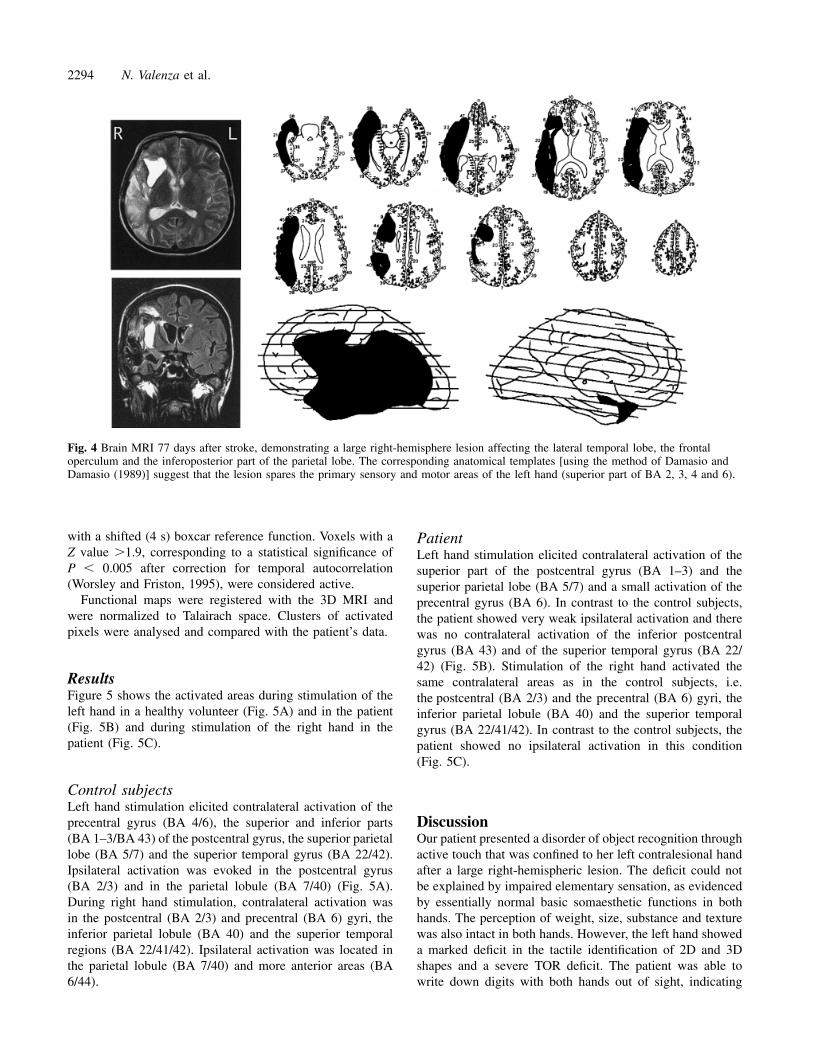
Neuroimaging studyMRI performed 77 days after the stroke showed a large right-hemisphere lesion affecting the lateral temporal lobe, the
frontal operculum and the inferoposterior part of the parietallobe. Lesion reconstruction (Damasio and Damasio, 1989)suggested preservation of the superior right pre- andpostcentral gyri [superior portion of Brodmann areas (BA)2, 3, 4 and 6], areas that correspond to the primary motorand the primary sensory (SI) representation of the lefthand (Fig. 4).
Since data derived from lesion reconstruction seemed tocorroborate our clinical observation of intact sensorimotorfunctions of the left hand, we used functional imaging toassess whether the preserved cortical area in our patientcorresponded to the sensorimotor representation of her lefthand. We used functional MRI (fMRI) to measure activationelicited by sensory stimulation of the right and left hands.Our main objective was to examine whether the intact portionof the somatosensory cortex in the right hemisphere wasactivated by sensory stimulation of the left hand. Secondly,we were interested in the differences between the activatedregions in the patient and those in control subjects exposedto the same stimulation.
Material and methodsThe patient and two healthy, age-matched, right-handedwomen participated in the fMRI study. We used a blockparadigm consisting of four cycles of 24 s of activationfollowed by 24 s of rest. In the activation condition, thepalm and the fingers of each hand were stimulated with asmall wooden stick which the experimenter moved randomlyon the subjects’ skin. The subjects closed their eyes duringimage acquisition. In the control condition, no sensorystimulation was delivered. Total fMRI acquisition time was3 min 12 s. The right and left hands were stimulated inseparate runs.
MRIA 1.5 T Eclipse system (Marconi Medical Systems,Cleveland, OH, USA), equipped with fast gradients (27 mT/mwith a slew rate of 72 mT/m/ms) with a standard head coilwas used. The fMRI data were obtained from 19 contigu-ous axial slices of 5 mm parallel to the AC–PC (anteriorcommissure–posterior commissure) line using single-shotechoplanar imaging [TR (repetition time)/TE (echo time)/flip angle � 2 s/40 ms/80°) with in-plane resolution of1.95 � 1.95 mm2. Anatomical MRI was obtained in the samesession, using gradient echo 2D and high-resolution 3Dacquisitions.
fMRI analysisThe data were analysed off-line using MedX3.3 software(Sensor Systems, Sterling, Va., USA) on a Unix workstation.All studies were first corrected for head motion (Woodset al., 1998). Activation maps report Z-score values computedfrom cross-correlation analysis of each voxel time course
2294 N. Valenza et al.
Fig. 4 Brain MRI 77 days after stroke, demonstrating a large right-hemisphere lesion affecting the lateral temporal lobe, the frontaloperculum and the inferoposterior part of the parietal lobe. The corresponding anatomical templates [using the method of Damasio andDamasio (1989)] suggest that the lesion spares the primary sensory and motor areas of the left hand (superior part of BA 2, 3, 4 and 6).
with a shifted (4 s) boxcar reference function. Voxels with aZ value �1.9, corresponding to a statistical significance ofP � 0.005 after correction for temporal autocorrelation(Worsley and Friston, 1995), were considered active.
Functional maps were registered with the 3D MRI andwere normalized to Talairach space. Clusters of activatedpixels were analysed and compared with the patient’s data.
ResultsFigure 5 shows the activated areas during stimulation of theleft hand in a healthy volunteer (Fig. 5A) and in the patient(Fig. 5B) and during stimulation of the right hand in thepatient (Fig. 5C).
Control subjectsLeft hand stimulation elicited contralateral activation of theprecentral gyrus (BA 4/6), the superior and inferior parts(BA 1–3/BA 43) of the postcentral gyrus, the superior parietallobe (BA 5/7) and the superior temporal gyrus (BA 22/42).Ipsilateral activation was evoked in the postcentral gyrus(BA 2/3) and in the parietal lobule (BA 7/40) (Fig. 5A).During right hand stimulation, contralateral activation wasin the postcentral (BA 2/3) and precentral (BA 6) gyri, theinferior parietal lobule (BA 40) and the superior temporalregions (BA 22/41/42). Ipsilateral activation was located inthe parietal lobule (BA 7/40) and more anterior areas (BA6/44).
PatientLeft hand stimulation elicited contralateral activation of thesuperior part of the postcentral gyrus (BA 1–3) and thesuperior parietal lobe (BA 5/7) and a small activation of theprecentral gyrus (BA 6). In contrast to the control subjects,the patient showed very weak ipsilateral activation and therewas no contralateral activation of the inferior postcentralgyrus (BA 43) and of the superior temporal gyrus (BA 22/42) (Fig. 5B). Stimulation of the right hand activated thesame contralateral areas as in the control subjects, i.e.the postcentral (BA 2/3) and the precentral (BA 6) gyri, theinferior parietal lobule (BA 40) and the superior temporalgyrus (BA 22/41/42). In contrast to the control subjects, thepatient showed no ipsilateral activation in this condition(Fig. 5C).
DiscussionOur patient presented a disorder of object recognition throughactive touch that was confined to her left contralesional handafter a large right-hemispheric lesion. The deficit could notbe explained by impaired elementary sensation, as evidencedby essentially normal basic somaesthetic functions in bothhands. The perception of weight, size, substance and texturewas also intact in both hands. However, the left hand showeda marked deficit in the tactile identification of 2D and 3Dshapes and a severe TOR deficit. The patient was able towrite down digits with both hands out of sight, indicating

Tactile apraxia without tactile agnosia 2295
Fig. 5 Ipsilateral and contralateral activations elicited by sensory stimulation of the left hand in acontrol subject (A) and the patient (B), and ipsilateral and contralateral activations elicited by sensorystimulation of the right hand in the patient (C). Areas of activation are shown in white.
that motor functions were sufficiently preserved to allowtactile exploration. It is unlikely that our patient’s TOR deficitwas caused by initial cognitive impairments, such as leftspatial neglect or difficulties in visual-spatial tasks, as noneof these deficits was present at the time of testing. Thus, herleft unimanual TOR deficit was attributable neither to anelementary sensorimotor dysfunction nor to a more generalsupramodal spatial impairment, which has been proposed asan explanation of TOR deficits (Semmes, 1965).
The experiments reported here showed a left-handdissociation in 2D shape recognition between active andpassive touch. In recognition tasks based on active touch,i.e. when the patient explored shapes actively, she consistentlyfailed to identify the stimuli with her left hand, whereas herright hand performed perfectly. Exploratory movements ofthe left hand were disorganized and incomplete. Drawingsof objects palpated with the left hand were unrecognizable,suggesting that altered exploration precluded adequaterepresentation of the objects’ shape. In contrast, in situationsof passive touch, e.g. in recognition tasks not necessitatingactive exploration of the stimuli, the performance of the lefthand was normal. For example, shapes traced on the skin of
the left hand were recognized accurately. In addition, undermanually and verbally guided exploration, the patient wasable to recognize correctly the same shapes she failed torecognize with active exploration. The dissociation betweenactive and passive shape recognition suggests that ourpatient’s difficulty in identifying shapes with her left hand isdue to deficient active exploratory movements. The presenceof this dissociation excludes an explanation of the TORdeficit in terms of primary sensory impairment, such as two-point discrimination and proprioception.
Our patient’s deficit differs from the known tactilerecognition deficits reported in the literature. Reed andcolleagues recently reported a patient with TOR impairmentrestricted to the right hand (Reed et al., 1996). A detailedexperimental investigation of her difficulties revealed aspecific deficit of shape recognition which could not beattributed to deficient active exploration. The authorsinterpreted the deficit as apperceptive tactile agnosia resultingfrom an impairment of modality-specific shape representa-tions. In contrast to our patient, their patient did not improveher performance in passive shape recognition. Another patientcould not name, describe or demonstrate the use of objects

2296 N. Valenza et al.
placed in his left hand (Platz, 1996). A qualitative analysisof his spontaneous finger movements during TOR revealeda slight alteration in the exploratory movements of his agnosichand. However, he identified shapes accurately by touch, incontrast to our patient. Platz interpreted the deficit as a pureassociative agnosia due to defective integration of distincttactile features into a coherent tactile percept (Platz, 1996).He stressed that the slight deficit in exploration might be theconsequence, rather than the cause, of the recognition deficit.In other cases of associative tactile agnosia (Endo et al.,1992; Nakamura et al., 1998), there was apparently normalexploration of objects and the capacity to analyse tactilefeatures of objects (including their shape) and to drawpalpated objects was intact. Despite preserved high-leveltactile perception, these patients failed to attribute meaningto correctly explored objects, as evidenced by their inabilityto classify objects according to their function out of vision.Our patient also differs from patients with tactile anomiawho are able to recognize objects tactually, as illustrated bytheir ability to match them according to their category orfunction (Beauvois et al., 1978; Endo et al., 1992). However,they are unable to name the recognized object.
Recently, two studies reported a deficit of tactile explorationin patients with deficient TOR and severely impaired tactilediscrimination consecutive to posterior parietal lesions (Pauseet al., 1989; Binkofski et al., 2001). The patients describedin these studies showed complete destruction of the temporalcharacteristics of the finger movements during active touchdespite normal frequencies of repetitive finger movementsand almost normal force. The authors concluded that theposterior parietal cortex plays a crucial role in the conceptionand generation of the motor programmes required for thecollection of somatosensory information. In contrast to ourpatient, their patients did not demonstrate intact recognitionin either of the conditions tested. However, their passiverecognition (e.g. through graphaesthesia) was not tested. Adeficit of tactile shape representation may explain theirimpairment of exploration and TOR. Thus, none of thepatients described previously showed the dissociationbetween active and passive recognition found in our patient.
The different clinical expressions of TOR disorders suggestthat the tactile identification of objects depends on severalprocesses, the organization of which is at least partlyhierarchical. In our patient, the main known causes of TORdeficit can be rejected. As already stressed, an explanationin terms of a primary sensory impairment can be eliminated.Neither is inability to execute the exploratory movementsthe cause of the alteration of shape exploration, since thepatient is perfectly able to perform all movements underverbal guidance. Impaired access to tactile shaperepresentations may not explain her deficient tactile shapeidentification, since she recognizes forms in experimentalsituations that do not require the spontaneous generation ofexploratory movements. The latter observation also permitsus to reject an explanation in terms of a tactile–verbal
disconnection, since our patient can correctly name tactileshapes presented passively to her left hand.
As both basic somatosensory functions and tactile shaperepresentations, as well as their links to the lexical memories,are spared in our patient, the deficit in tactile recognitionmust be located at an intermediate level between basicsomatosensory functions and the higher cognitive stages ofTOR. The dissociation between active recognition and passiverecognition suggests that our patient’s deficit depends onprocesses linked to the elaboration of exploratory strategiesduring TOR. Manual exploratory movements during TORhave been studied extensively in healthy subjects (Ledermanand Klatzky, 1987). These authors assume that theperception of the precise shape of an object depends on theuse of a particular exploratory procedures (EP) (the ‘contour-following EP’), during which the hand maintains contactwith the contour of the object. According to their observations,the contour-following strategy is the only necessary EP thathas to be evoked by subjects to allow correct identificationof the exact shape of an object; the remaining EPs are definedas interchangeable (Lederman and Klatzky, 1987). Thecontour-following EP can thus be conceptualized as a highlyspecialized EP that occupies a special position among theEP repertoire. Our patient has a selective deficit of shaperecognition through active touch but recognizes other tactileproperties of objects (substance, length, weight, size) withoutdifficulty. This pattern of deficit is compatible with a selectiveloss of the contour-following EP and corroborates thesuggestion that contour-following is distinct among EPs andmay be selectively impaired after brain damage.
Why is our patient unable to elaborate a correct tactileexploration of shapes? One possible explanation arises fromthe notion that exploration is built on continuous interactionsbetween sensory input and stored tactile knowledge aboutshapes (Klatzky and Lederman, 1993; Platz, 1996). Successfulrecognition takes place when the perceived information canbe matched with stored tactile shape representations. From apurely cognitive point of view, it would be predicted thatdisconnection between the haptic system and the tactile shaperepresentation system should lead to specific impairment oftactile shape exploration, although the haptic system andtactile shape representation are intact. The hypothesis of sucha disconnection is corroborated by the observation that ourpatient does not profit from a preliminary hypothesis aboutthe identity of shapes. However, this explanation is difficultto match with recent neurophysiological and neuroimagingdata about active touch in animals and humans. For instance,Binkofski and colleagues suggest that the parietal lobe isresponsible both for ‘the pragmatic and the cognitive aspectsof somatosensation’ (Binkofski et al., 2001). A disconnectionbetween two systems would thus be problematic, since bothsystems would be located within the same brain area. Acompatible interpretation of our patient’s dissociation wouldposit a deficit of the process of sensorimotor transformation(Pause and Freund, 1989), a deficit which would preclude

Tactile apraxia without tactile agnosia 2297
the necessary interactions between sensorimotor input andtactile shape representations that lead to successful TOR.
Our patient’s relatively large brain lesion precludes preciseanalysis of the brain systems involved in active TOR.However, fMRI showed that sensory stimulation of thepatient’s left hand activated areas of preserved lateral cortexin the right hemisphere. Sensory stimulation of the left handelicited bilateral activation of SI, stronger on the contralateralthan on the ipsilateral side, and contralateral activation ofthe superior parietal lobe. These activated regions havebeen reported to play a role in the discrimination of basicsomatosensory attributes (Srinivas and Ogas, 1999), althoughSI may be the first and most important cortical relay forelementary sensation. The activation in the preserved cortexof the right hemisphere supports the idea that this brain areaconveys the preserved basic somatosensory functions of theleft hand. The major difference between the patient and thehealthy subjects was the absence of activation in regions atthe parietotemporal junction (BA 22/42/43), which have beenregarded as the human analogues of the secondary sensoryarea (SII). Its functions and its relationship to SI remainpoorly understood and human data about the effects ofselective lesion of SII are lacking. Some studies have shownthat complete destruction of SII in monkeys evokesimpairments in texture and shape discrimination and altersdiscrimination of size and roughness (Paulesu et al., 1997).Our data suggest that SII may also play an important role inthe integration of the tactile information acquired duringexploration.
Our case demonstrates a pure tactile apraxia withoutconcomitant gnostic deficit, i.e. a sensorimotor integrationdisorder, originally described clinically by Delay (Delay,1935) and Klein (Klein, 1931). This disturbance of handfunction has been defined as ‘a disorder of the explorationand manipulation of objects, whereas intransitive, expressiveand symbolic movements are preserved’ (Pause and Freund,1989; Binkofski et al., 2001). We propose that tactile apraxiaresults from impaired integration of sensorimotor feedbackwith stored tactile shape information—integration that isnecessary to generate the exploratory procedures requiredfor successful TOR. Our case study emphasizes that theelaboration of the specific hand movements required for theextraction of distinctive somatosensory information is crucialfor successful TOR, and provides new evidence that theseexploratory procedures may be selectively impaired afterbrain damage.
AcknowledgementsThis work was supported by grants 32-50882.97 and 3238-062769.00 from the Swiss National Science Foundationto A.S.
ReferencesBeauvois M-F, Saillant B, Meininger V, Lhermitte F. Bilateral tactileaphasia: a tacto-verbal dysfunction. Brain 1978; 101: 381–401.
Benton AL, Hamsher KDS, Varney NR, Spreen O. Contributionsto neuropsychological assessment. New York: Oxford UniversityPress; 1983.
Binkofski F, Kunesch E, Classen J, Seitz RJ, Freund H-J. Tactileapraxia. Unimodal apractic disorder of tactile object explorationassociated with parietal lobe lesions. Brain 2001; 124: 132–44.
Caselli RJ. Bilateral impairment of somesthetically mediated objectrecognition in humans. Mayo Clin Proc 1991a; 66: 357–64.
Caselli RJ. Rediscovering tactile agnosia. [Review]. Mayo ClinProc 1991b; 66: 129–42.
Caselli RJ. Ventrolateral and dorsomedial somatosensory associationcortex damage produces distinct somesthetic syndromes in humans.Neurology 1993; 43: 762–71.
Caselli RJ. Tactile agnosia and disorders of tactile perception.In: Feinberg TE, Farah MJ, editors. Behavorial neurology andneuropsychology. New York: McGraw-Hill; 1997. p. 277–88.
Damasio H, Damasio AR. Lesion analysis in neuropsychology. NewYork: Oxford University Press; 1989.
Delay J. Les astereognosies: pathologie du toucher. Clinique,physiologie, topographie. Paris: Masson & Cie; 1935.
Endo K, Miyasaka M, Makishita H, Yanagisawa N, Sugishita M.Tactile agnosia and tactile aphasia: symptomatological andanatomical differences. Cortex 1992; 28: 445–69.
Gauthier L, Dehaut F, Joanette Y. The Bells Test: a quantitative andqualitative test for visual neglect. Int J Clin Neuropsychol 1989;11: 49–54.
Hooper EH. Hooper Visual Organization Test. Los Angeles: WesternPsychological Services; 1985.
Klatzky RL, Lederman SJ. Toward a computational model ofconstraint-driven exploration and haptic object identification.Perception 1993; 22: 597–621.
Klein R. Zur Symptomatologie des Parietallapens. Z Ges NeurolPsychiat 1931; 135: 589–608.
Lederman SJ, Klatzky RL. Hand movements: a window into hapticobject recognition. Cognit Psychol 1987; 19: 342–68.
Nakamura J, Endo K, Sumida T, Hasegawa T. Bilateral tactileagnosia: a case report. Cortex 1998; 34: 375–88.
Osterrieth PA. Le test de copie d’une figure complexe: contributiona l’etude de la perception et de la memoire. Arch Psychol 1944;30: 286–356.
Paulesu E, Frackowiak RSJ, Bottini G. Maps of somatosensorysystems. In: Frackowiak RSJ, Friston KJ, Frith CD, Dolan RJ,Mazziotta JC, editors. Human brain function. San Diego: AcademicPress; 1997. p. 183–242.
Pause M, Freund HJ. Role of the parietal cortex for sensorimotortransformation. Brain Behav Evol 1989; 33: 136–40.
Pause M, Kunesch E, Binkofski F, Freund H-J. Sensorimotordisturbances in patients with lesions of the parietal cortex. Brain1989; 112: 1599–625.

2298 N. Valenza et al.
Platz T. Tactile agnosia. Casuistic evidence and theoretical remarkson modality-specific meaning representations and sensorimotorintegration. Brain 1996; 119: 1565–74.
Raven JC, Court JH, Raven J, editors. Coloured progressive matrices.Paris: Editions & Applications Psychologiques; 1998.
Reed CL, Caselli RJ. The nature of tactile agnosia: a case study.Neuropsychologia 1994; 32: 527–39.
Reed CL, Caselli RJ, Farah MJ. Tactile agnosia. Underlyingimpairment and implications for normal tactile object recognition.Brain 1996; 119: 875–88.
Regard M, Strauss E, Knapp P. Children’s production on verbal andnon-verbal fluency tasks. Percept Mot Skills 1982; 55: 839–44.
Rey A. Test de copie et de reproduction de memoire de figuresgeometriques complexes. Paris: Centre de Psychologie; 1958a.
Rey A. L’examen clinique en psychologie. Paris: Presse Universitairede France; 1958b.
Semmes J. A non-tactual factor in astereognosis. Neuropsychologia1965; 3: 295–314.
Srinivas K, Ogas J. Disorders of somesthetic recognition: a theo-retical review. Neurocase 1999; 5: 83–93.
Thurstone LL, Thurstone TG. Chicago Test of Primary MentalAbilities. Chicago: Research Associates; 1963.
Wechsler D. Wechsler Memory Scale—Revised manual. SanAntonio: Psychological Corporation; 1987.
Woods RP, Grafton ST, Holmes CJ, Cherry SR, Mazziotta JC.Automated image registration: I. General methods and intrasubject,intramodality validation. J Comput Assist Tomogr 1998; 22: 139–52.
Worsley KJ, Friston KJ. Analysis of fMRI time-series revisited—again. Neuroimage 1995; 2: 173–81.
Received March 5, 2001. Revised June 10, 2001.Accepted June 25, 2001
Copyright © 2022 FDOKUMEN