Diffusion tensor studies dissociated two fronto-temporal pathways in the human memory system
12
Diffusion tensor studies dissociated two fronto-temporal pathways in the human memory system Emi Takahashi, a, ⁎ Kenichi Ohki, b and Dae-Shik Kim a a Department of Anatomy and Neurobiology, Center for Biomedical Imaging, 715 Albany Street, X-B01, Boston University School of Medicine, Boston, MA 02118, USA b Department of Neurobiology, Harvard Medical School, Boston, MA 02115, USA Received 31 January 2006; revised 6 October 2006; accepted 9 October 2006 Recent functional neuroimaging studies have shown that multiple cortical areas are involved in memory encoding and retrieval. However, the underlying anatomical connections among these memory- related areas in humans remain elusive due to methodological limitations. Diffusion tensor imaging (DTI) is a technique based on detecting the diffusion of water molecules from magnetic resonance images. DTI allows non-invasive mapping of anatomical connections and gives a comprehensive picture of connectivity throughout the entire brain. By combining functional magnetic resonance imaging (fMRI) and DTI, we show that memory-related areas in the left dorsolateral prefrontal cortex (DLPFC) and the left ventrolateral prefrontal cortex (VLPFC) each connect with memory-related areas in the left temporal cortex. This result suggests there are two pathways between prefrontal cortex and temporal cortex related to the human memory system. © 2006 Elsevier Inc. All rights reserved. Keywords: Functional magnetic resonance imaging; Diffusion tensor imaging; Long-term memory; Prefrontal cortex; Temporal cortex; Connection Introduction The human memory system is supported by multiple processes (Squire and Zola-Morgan, 1991; Cabeza and Nyberg, 2000; Tulving, 2002; Habib et al., 2003). Recent functional neuroima- ging studies have shown that areas in the prefrontal, parietal and temporal cortices are involved in memory encoding and retrieval (Tulving et al., 1994; Demb et al., 1995; Buckner et al., 1998, 1999; Buckner and Koutstaal, 1998; Wagner et al., 1998; Lepage et al., 2000). Among these memory-related areas, prefrontal cortex (PFC) is thought to subserve cognitive control processes (Goldman-Rakic, 1987; Schacter, 1987; Shimamura, 1995; Fuster, 1997; Takahashi and Miyashita, 2002), and send top-down signals to posterior cortices (Incisa della Rocchetta and Milner, 1993; Gazzaniga, 1995; Tomita et al., 1999; Takahashi and Miyashita, 2002). Recently, a two-stage model of PFC has been proposed (Petrides, 1994a,b; 1996; Owen et al., 1996). In this model, ventrolateral prefrontal cortex (VLPFC) interacts with posterior cortices such as temporal cortex for active (or controlled) encoding and retrieval of information, whereas dorsolateral prefrontal cortex (DLPFC) monitors and manipulates maintained information in VLPFC. Anatomical studies in nonhuman primates have shown direct corticocortical connections from VLPFC to temporal cortex (Petrides and Pandya, 2002), but direct connections from DLPFC to temporal cortex are still controversial (Seltzer and Pandya, 1989; Petrides and Pandya, 1994, 1999; Petrides, 2005). Based on these studies we hypothesized two possibilities for the anatomical connections between PFC and temporal cortex in humans (Fig. 1A); (i) Serial Pathway Model: DLPFC connects with VLPFC, and VLPFC connects with temporal cortex, but DLPFC does not connect with temporal cortex, (ii) Parallel Pathway Model: DLPFC and VLPFC both connect with temporal cortex. In this study, we performed diffusion tensor imaging (DTI) fiber tracking from functionally defined memory areas to test these two connectivity models. DTI is a new technique based on the diffusion properties of water molecules as detected from diffusion-weighted magnetic resonance images (Basser et al., 1994). A number of fiber tracking algorithms have been developed to visualize white matter fiber tracts from DTI images (Mori et al., 1999; Jones et al., 1999; Conturo et al., 1999). Although many studies have shown anatomical connections per se in the human brain (Conturo et al., 1999; Basser et al., 2000; Stieltjes et al., 2001; Xu et al., 2002; Behrens et al., 2003; Lehericy et al., 2004; Powell et al., 2004), no studies to date have related memory functions with anatomical www.elsevier.com/locate/ynimg NeuroImage 34 (2007) 827 – 838 ⁎ Corresponding author. Department of Psychiatry, Massachusetts General Hospital, Harvard Medical School, Boston, MA 02215, USA. Fax: +1 617 726 4078. E-mail address: [email protected] (E. Takahashi). Available online on ScienceDirect (www.sciencedirect.com). 1053-8119/$ - see front matter © 2006 Elsevier Inc. All rights reserved. doi:10.1016/j.neuroimage.2006.10.009
-
Upload
independent -
Category
Documents
-
view
3 -
download
0
Transcript of Diffusion tensor studies dissociated two fronto-temporal pathways in the human memory system
www.elsevier.com/locate/ynimg
NeuroImage 34 (2007) 827–838Diffusion tensor studies dissociated two fronto-temporal pathways inthe human memory system
Emi Takahashi,a,⁎ Kenichi Ohki,b and Dae-Shik Kima
aDepartment of Anatomy and Neurobiology, Center for Biomedical Imaging, 715 Albany Street, X-B01, Boston University School of Medicine, Boston,MA 02118, USAbDepartment of Neurobiology, Harvard Medical School, Boston, MA 02115, USA
Received 31 January 2006; revised 6 October 2006; accepted 9 October 2006
Recent functional neuroimaging studies have shown that multiplecortical areas are involved in memory encoding and retrieval.However, the underlying anatomical connections among these memory-related areas in humans remain elusive due to methodologicallimitations. Diffusion tensor imaging (DTI) is a technique based ondetecting the diffusion of water molecules from magnetic resonanceimages. DTI allows non-invasive mapping of anatomical connectionsand gives a comprehensive picture of connectivity throughout the entirebrain. By combining functional magnetic resonance imaging (fMRI)and DTI, we show that memory-related areas in the left dorsolateralprefrontal cortex (DLPFC) and the left ventrolateral prefrontal cortex(VLPFC) each connect with memory-related areas in the lefttemporal cortex. This result suggests there are two pathways betweenprefrontal cortex and temporal cortex related to the human memorysystem.© 2006 Elsevier Inc. All rights reserved.
Keywords: Functional magnetic resonance imaging; Diffusion tensorimaging; Long-term memory; Prefrontal cortex; Temporal cortex;Connection
Introduction
The human memory system is supported by multiple processes(Squire and Zola-Morgan, 1991; Cabeza and Nyberg, 2000;Tulving, 2002; Habib et al., 2003). Recent functional neuroima-ging studies have shown that areas in the prefrontal, parietal andtemporal cortices are involved in memory encoding and retrieval(Tulving et al., 1994; Demb et al., 1995; Buckner et al., 1998,
⁎ Corresponding author. Department of Psychiatry, Massachusetts GeneralHospital, Harvard Medical School, Boston, MA 02215, USA. Fax: +1 617726 4078.
E-mail address: [email protected] (E. Takahashi).Available online on ScienceDirect (www.sciencedirect.com).
1053-8119/$ - see front matter © 2006 Elsevier Inc. All rights reserved.doi:10.1016/j.neuroimage.2006.10.009
1999; Buckner and Koutstaal, 1998; Wagner et al., 1998; Lepage etal., 2000).
Among these memory-related areas, prefrontal cortex (PFC) isthought to subserve cognitive control processes (Goldman-Rakic,1987; Schacter, 1987; Shimamura, 1995; Fuster, 1997; Takahashiand Miyashita, 2002), and send top-down signals to posteriorcortices (Incisa della Rocchetta and Milner, 1993; Gazzaniga,1995; Tomita et al., 1999; Takahashi and Miyashita, 2002).Recently, a two-stage model of PFC has been proposed (Petrides,1994a,b; 1996; Owen et al., 1996). In this model, ventrolateralprefrontal cortex (VLPFC) interacts with posterior cortices such astemporal cortex for active (or controlled) encoding and retrieval ofinformation, whereas dorsolateral prefrontal cortex (DLPFC)monitors and manipulates maintained information in VLPFC.Anatomical studies in nonhuman primates have shown directcorticocortical connections from VLPFC to temporal cortex(Petrides and Pandya, 2002), but direct connections from DLPFCto temporal cortex are still controversial (Seltzer and Pandya, 1989;Petrides and Pandya, 1994, 1999; Petrides, 2005).
Based on these studies we hypothesized two possibilities for theanatomical connections between PFC and temporal cortex inhumans (Fig. 1A); (i) Serial Pathway Model: DLPFC connectswith VLPFC, and VLPFC connects with temporal cortex, butDLPFC does not connect with temporal cortex, (ii) ParallelPathway Model: DLPFC and VLPFC both connect with temporalcortex. In this study, we performed diffusion tensor imaging (DTI)fiber tracking from functionally defined memory areas to test thesetwo connectivity models.
DTI is a new technique based on the diffusion properties ofwater molecules as detected from diffusion-weighted magneticresonance images (Basser et al., 1994). A number of fiber trackingalgorithms have been developed to visualize white matter fibertracts from DTI images (Mori et al., 1999; Jones et al., 1999;Conturo et al., 1999). Although many studies have shownanatomical connections per se in the human brain (Conturo etal., 1999; Basser et al., 2000; Stieltjes et al., 2001; Xu et al., 2002;Behrens et al., 2003; Lehericy et al., 2004; Powell et al., 2004), nostudies to date have related memory functions with anatomical
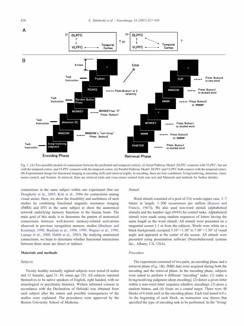
Fig. 1. (A) Two possible models of connections between the prefrontal and temporal cortices. (i) Serial Pathway Model: DLPFC connects with VLPFC, but notwith the temporal cortex, and VLPFC connects with the temporal cortex, (ii) Parallel PathwayModel: DLPFC and VLPFC both connect with the temporal cortex.(B) Experimental design for functional imaging in encoding (left) and retrieval (right). In encoding, there are four conditions: living/nonliving, detection, visuo-motor control, and fixation. In retrieval, there are retrieval trials and visuo-motor control trials (see text and Materials and methods for further details).
828 E. Takahashi et al. / NeuroImage 34 (2007) 827–838
connections in the same subject within one experiment (but seeDougherty et al., 2005; Kim et al., 2006 for connections amongvisual areas). Here, we show the feasibility and usefulness of suchstudies by combining functional magnetic resonance imaging(fMRI) and DTI in the same subject to show the anatomicalnetwork underlying memory functions in the human brain. Themain goal of this study is to determine the pattern of anatomicalconnections between well-known memory-related activationsobserved in previous recognition memory studies (Buckner andKoutstaal, 1998; Buckner et al., 1998, 1999; Wagner et al., 1998;Lepage et al., 2000; Habib et al., 2003). By studying anatomicalconnections, we hope to determine whether functional interactionsbetween these areas are direct or indirect.
Materials and methods
Subjects
Twenty healthy normally sighted subjects were tested (8 malesand 12 females, aged 21–39, mean age 25). All subjects reportedthemselves to be native speakers of English, right handed, with noneurological or psychiatric histories. Written informed consent inaccordance with the Declaration of Helsinki was obtained fromeach subject after the nature and possible consequences of thestudies were explained. The procedures were approved by theBoston University School of Medicine.
Stimuli
Word stimuli consisted of a pool of 216 words (upper case, 3–7letters in length, 1–200 occurrences per million (Kucera andFrancis, 1967)). We also used non-word stimuli (alphabeticalstimuli) and the number sign (####) for control tasks. Alphabeticalstimuli were made using random sequences of letters having thesame length as the word stimuli. All stimuli were presented on atangential screen 1.1 m from the subjects. Words were white on ablack background, occupied 3.10°×1.30° to 7.30°×1.30° of visualangle and appeared at the center of the screen. All stimuli werepresented using presentation software (Neurobehavioral systemsInc., Albany, CA, USA).
Procedure
The experiment consisted of two parts, an encoding phase and aretrieval phase (Fig. 1B). fMRI data were acquired during both theencoding and the retrieval phase. In the encoding phase, subjectswere asked to perform 4 different “encoding” tasks: (1) make aliving/nonliving judgment (deep encoding), (2) detect a given letterwithin a non-word letter sequence (shallow encoding), (3) press arandom button, and (4) fixate on a central target. There were 48blocks of 6 trials each in the encoding phase. Each trial lasted 4.0 s.At the beginning of each block, an instruction was shown thatspecified the type of encoding task to be performed. In the “living/
829E. Takahashi et al. / NeuroImage 34 (2007) 827–838
nonliving” blocks, subjects decided whether or not each word wasanimate. In the “detection” blocks, they decided whether eachword contained “E”. In both the “living/nonliving” and “detection”blocks, subjects reported their response by pressing one of twobuttons held in the right hand. In the “random button press” (visuo-motor control) blocks, they looked at each non-word random lettersequence and pressed one of the two buttons held in the right hand.In the fixation blocks, they looked at the fixation cross and did notpress any buttons. Each word was presented only once throughoutthe encoding phase. The length, the percentage of living words,and the percentage of words containing “E” were balanced acrossall the living/nonliving and detection blocks. The ordering ofliving/nonliving and detection blocks, and stimuli used in bothblocks were counterbalanced across subjects. The encoding phaselasted approximately 25 min.
The retrieval phase (4 runs) started about 20 min after the endof the encoding phase. During the interval between the encodingand the retrieval phases, subjects performed a distracter task (whitecircle detection out of 4 circles) to disengage various strategies forencoding. In the retrieval phase, subjects performed randomlyintermixed retrieval trials, visuo-motor control trials, and fixationtrials. Each run consisted of 72 trials. In the retrieval trials, subjectsmade yes/no recognition memory judgments for previously studiedand new stimuli. Subjects reported their response by pressing oneof the two buttons held in the right hand. Half of the words fromthe encoding phase were presented again (72 words: 36 wordswere deep-encoded, the other 36 words were shallow-encoded),along with new words (72 words). In the visuo-motor controltrials, they looked at the number sign (####) and pressed thethird button that was specifically assigned for this trial type. Inthe fixation trials, they looked at the fixation cross and did notpress any buttons. Each trial was 4.0 s long, and the four trialtypes occurred with equal probability across the experiment inpseudo-random sequence. The retrieval phase lasted approximately20 min.
Image acquisition
A 3 Tesla whole body scanner (Intera, Philips) was used toacquire T1-weighted anatomical images, gradient-echoEPI, andspin-echo EPI diffusion-weighted images for the DTI data sets.
Functional imagingParameters for functional image acquisition were as follows:
repetition time (TR)=4 s; echo time (TE)=35 ms; flip angle=90°;in-plane resolution 1.8×1.8 mm2; FOV=230; number of slices 36;slice thickness 4 mm. Slice orientation was axial, and the imagingvolume was aligned to cover the whole brain. For each subject,conventional T1-weighted structural images were obtained toprovide anatomical information. Each scanning run commencedwith the acquisition of 2 dummy volumes, allowing tissuemagnetization to achieve a steady state, after which functionalvolumes were acquired (85 volumes for each encoding run, and 73volumes for each retrieval run).
Diffusion tensor imagingDiffusion-weighted images (DWI) were acquired using multi-
slice spin-echo echo-planar imaging (SE-EPI). Parameters for DTIacquisition were as follows: TR=17.1 s, TE=80 ms, Matrix size128×128, FOV 230, fat suppression, number of slices=96, slicethickness=1.5 mm, b=1000 s/mm2, 15 directions, gradient
strength=0.2 G/mm, SENSE (sensitivity encoding) reductionfactor=2.0. Diffusion tensor images were taken in the identicalfield of view (FOV) with functional images to simplify post hocspatial registration. For each subject, 16 data sets were acquired (15diffusion-weighted + 1 non-diffusion-weighted images). Fromthese data, diffusion tensors were calculated for all image pixels. Atotal of 4 signal averages were collected to ensure a sufficientsignal-to-noise ratio (SNR) for high-quality tensor mapping. Inorder to compensate for motion, scan was acquired separately andthen coregistered with the others before averaging. The sensitivityencoding technique (SENSE) was used, which is known to reducesusceptibility artifacts significantly (Jaermann et al., 2004). Pulsegating was not performed.
fMRI and DTI data analyses
All functional images were analyzed with SPM99 (WellcomeDepartment of Neurology, UK). For each subject, the acquiredimages were realigned to the first volume to correct for headmovement. Differences in acquisition timing between each slicewere corrected for using sinc interpolation.
T1 images were acquired in the same FOV as EPI and DWIimages and was normalized to the standard Talairach space(Talairach and Tournoux, 1988) by an affine transformation. Thesame transformation was applied to EPI and DWI images (seebelow). After spatial normalization, each EPI volume wasresampled to 2-mm cubic voxels, smoothed spatially with aGaussian kernel of 8 mm full-width half-maximum (FWHM), andthe time-series was smoothed temporally with a 4-s FWHMGaussian kernel. Slow signal drifs were removed by high passfiltering using cut-off periods of 300 s for encoding and 128 s forretrieval. For each voxel, data were best-fitted (least square) using alinear combination of regressors. The regressors were constructed tocorrespond to each trial type for each subject and then convolvedwith the standard hemodynamic response function (HRF). In theencoding phase, we did not separate correct and incorrect trialswithin blocks. Instead, we analyzed all trials with three regres-sors, corresponding to deep encoding, shallow encoding andvisuo-motor control. In the retrieval phase, we separated correctand incorrect trials (“Hit”, “Correct Rejection”, “Miss”, and“False Alarm”), and made five regressors, corresponding to thesetrial types and “visuo-motor control”. Trials in which the subjectdid not report a response by pressing a button in the retrievalphase (9 trials) were eliminated from further analysis. Contrastswere first performed at the single subject level and then the result-ing images were taken up to the group level using t-tests. Thestatistical threshold was set to p<0.001 as an initial height thres-hold and to p<0.05 corrected for whole-brain multiple compar-isons at cluster level, according to the SPM99 standard procedures(Friston et al., 1994). The location of each cluster was indicatedby peak voxels on the normalized structural images and labeledusing the nomenclature of Talairach and Tournoux (Talairach andTournoux, 1988).
DTI images were realigned using the diffusion toolbox inSPM2. The 1st images of each run were realigned to the first imageof the first run. This procedure removed eddy current-induceddistortions. Then all the images were averaged across the 4 runs.For each voxel, the diffusion tensor and fractional anisotropy (FA)were calculated using standard procedures (Basser et al., 1994). Weremoved voxels that had extremely large residuals after fitting the15 DWIs by an ellipsoid tensor.
830 E. Takahashi et al. / NeuroImage 34 (2007) 827–838
Using the T maps generated by SPM99, which are the result ofthe random effect analysis of 20 subjects, we created startingpoints for DTI fiber tracking. We used a threshold p<0.001(uncorrected) and p<0.05 (corrected at cluster level) as criteria formaking the starting points. The starting points for fiber trackingwere set in intervals of 1 mm in the foci of fMRI activationclusters. The coordinates of the activated clusters in Talairachcoordinates were reverse normalized into each subject's coordi-nates, and used as the basis for fiber tracking and determining thecoordinates of the end points of the fibers. Then we normalizedthe end points' coordinates, averaged the data across all thesubjects, and superimposed the resulting maps onto the normalizedT1-weighted images. The reverse normalization and normalizationof the coordinates were performed in the following way. We tookT1 and DWI scans in the same FOV, and using SPM99, wenormalized our T1 image to the template brain. For each subject,we applied reverse conversion of normalization to the seed points,using in-house Matlab (The Mathworks Inc., Natick, MA, USA)programs. The normalization is an affine transformation, whichcan be expressed as: y=C(x−x0)+C0+y0. The reverse normal-ization was obtained as: x=C−1(y−y0−C0)+x0, where x is thecoordinate before normalization, x0 is the origin before normal-ization, y is the coordinate after normalization, y0 is the origin afternormalization, C is the linear transformation matrix obtained bySPM99, and C0 is the translation obtained by SPM99. Thisapproach is better than normalizing DWIs directly because theresolution of DWIs was reduced when we resampled DWIs duringnormalization.
Fiber tracking
Diffusion tensors, fractional anisotropy (FA), and fiber tractswere calculated using custom-made Matlab programs. We used afiber tracking algorithm based on the method described in Basser etal. (2000). At every position along the fiber trajectory a diffusiontensor is interpolated and eigenvectors are computed. Theeigenvector associated with the greatest eigenvalue indicates theprincipal direction of water diffusion. The fiber tract is propagatedalong this direction over a small distance (0.5 mm) to the nextpoint where a new diffusion tensor is interpolated (linear). Fibertracking terminates when the angle between two consecutiveeigenvectors is greater than a given threshold (60°), or when theFA value is smaller than a given threshold (0.14). The criteria ofFA<0.14–0.15 are reported to provide the best tradeoff betweenfewer erroneous tracts and penetration into the white matter(Thottakara et al., 2006).
This streamline approach is based on the assumption thatdiffusion is locally uniform and can be accurately described by asingle eigenvector. Unfortunately, this approach fails to describecrossing fibers (Alexander et al., 2001). To overcome this problem,tensorline approaches (e.g., tensor deflection) have been developedthat use the entire tensor information instead of reducing it to a singleeigenvector (Lazar et al., 2003). In this study, both algorithms, i.e.,streamline (Basser et al., 2000) and tensor deflection (Lazar et al.,2003), were used to reconstruct 3D DTI fibers.
It is important to take into consideration that DTI fiber trackingfrom area A to area B does not necessarily signify a fiber connectionfrom area A to area B. There is noway to knowwhether the fibers arerunning from A to B or B to A, because the diffusion of a watermolecule does not yield directional information on the transmissionof neural signals.
Group study in DTI fiber tracking
For Fig. 3 and Supplementary Figs. S2–S5, we performed DTIfiber tracking from all voxels in each activated cluster. We obtainedthe terminal points of each subject, summed them up, and super-imposed them onto normalized T1-weighted anatomical images.Given the possibility of missing fibers that pass through afunctionally active area, we also traced fibers between pairs ofactivation clusters.
Estimation of tractography error by the bootstrap method
In this study, the dispersion errors in white matter tractographywere estimated by a statistical nonparametric bootstrap method(Lazar and Alexander, 2005). In each iteration, two samples wererandomly selected with replacement from the pool of fourindependent DTI acquisitions, and were averaged. The procedurewas repeated for each diffusion-encoding of the brain volume (15directions) and for one non-diffusion-weighted volume, resultingin one bootstrap DTI volume sample. The dispersion errors for thetractography were obtained by running the tractography algorithmover 100 iterations of the bootstrap DTI data set.
We made histograms of the probability of a connection toDLPFC for each and every voxel in STS/FG (Fig. 5A). Using 100bootstrap samples, we performed fiber tracking 100 times fromeach and all the seed points in the temporal activation. For eachseed point, we examined how many times out of 100 the fibersconverged onto the DLPFC activation.
A probabilistic map from a single seed point (Fig. 5E) wasobtained by counting how many boot-trac fibers passed each voxel.A probabilistic map from multiple seeds (Fig. 5F and Supplemen-tary Fig. S7) was obtained as follows. First, we performed fibertracking from all the seed points, for each bootstrap sample. Wedefined a voxel which was passed by at least one fiber as “1”, and avoxel which was not passed by any fiber as “0”. When more thanone fiber passed a voxel, we defined it as “1”. We performed theseprocedures for 100 bootstrap samples, obtained probabilities foreach voxel to be “1”, and defined this as a probabilistic map.
The probability of connections between two clusters wasobtained as follows. We performed the fiber tracking for eachbootstrap sample from all the seed points in cluster 1. Among thosefibers for each bootstrap sample, a connection was defined when atleast one fiber terminated in cluster 2, and no connection wasdefined when no fiber terminated in cluster 2. These procedureswere done with all the 100 bootstrap samples and both directions(from clusters 1 to 2 and from clusters 2 to 1), and the probabilitiesbetween the two clusters were calculated based on how many timesout of 100 iterations a connection was found.
One possible reason for observing relatively low probabilitiesof connections in bootstrapping was that we selected only two outof four DTI scans to estimate the fiber tracking error. This mighthave caused lower signal-to-noise ratio of the images.
Estimation of connection specificity by arbitrarily defined areas
We estimated the specificity in the connections between twoactivated clusters in the left DLPFC and the left temporal cortex inencoding. For this purpose, we examined the degree ofconnectivity between the DLPFC activation and arbitrarily definednon-activating regions. If the connection was specific, the degreeof connectivity between DLPFC and arbitrary regions should be
831E. Takahashi et al. / NeuroImage 34 (2007) 827–838
significantly lower than the connectivity between DLPFC andtemporal cortex activation. However, the degree of connectivitycould be affected by many factors, such as distances betweenregions, and shapes of regions. Thus, we restricted the arbitraryregions to those satisfying the following conditions: (1) all voxelswere inside the brain, (2) there was no overlap with the activatedregions, (3) the volume and shape of the region were exactly the
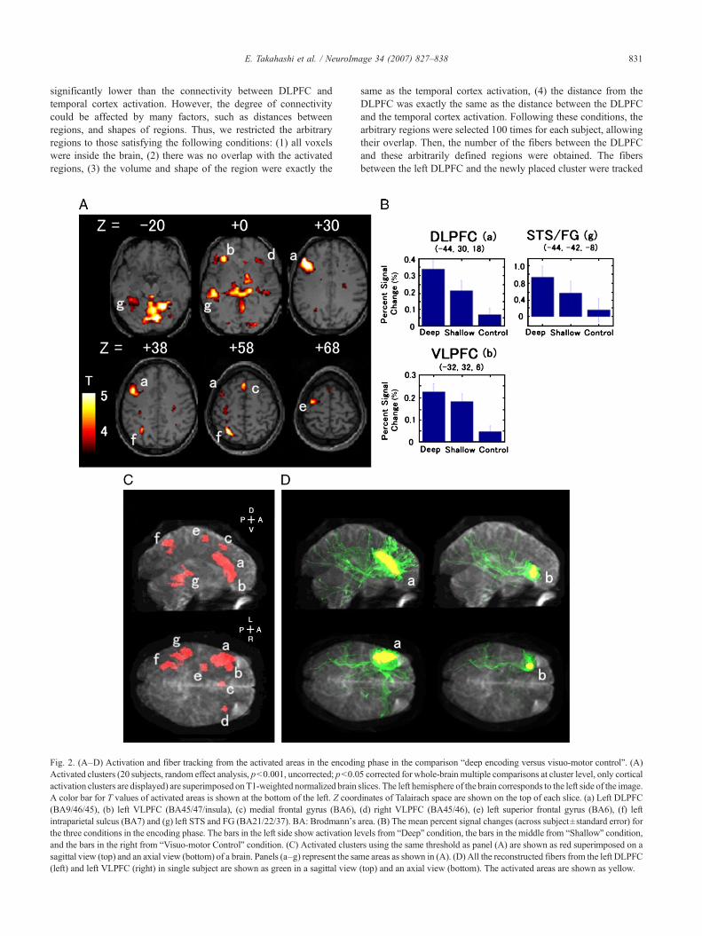
Fig. 2. (A–D) Activation and fiber tracking from the activated areas in the encodinActivated clusters (20 subjects, random effect analysis, p<0.001, uncorrected; p<0.0activation clusters are displayed) are superimposed onT1-weighted normalized brainA color bar for T values of activated areas is shown at the bottom of the left. Z coord(BA9/46/45), (b) left VLPFC (BA45/47/insula), (c) medial frontal gyrus (BA6),intraparietal sulcus (BA7) and (g) left STS and FG (BA21/22/37). BA: Brodmann'sthe three conditions in the encoding phase. The bars in the left side show activation leand the bars in the right from “Visuo-motor Control” condition. (C) Activated clustesagittal view (top) and an axial view (bottom) of a brain. Panels (a–g) represent the sa(left) and left VLPFC (right) in single subject are shown as green in a sagittal view
same as the temporal cortex activation, (4) the distance from theDLPFC was exactly the same as the distance between the DLPFCand the temporal cortex activation. Following these conditions, thearbitrary regions were selected 100 times for each subject, allowingtheir overlap. Then, the number of the fibers between the DLPFCand these arbitrarily defined regions were obtained. The fibersbetween the left DLPFC and the newly placed cluster were tracked
g phase in the comparison “deep encoding versus visuo-motor control”. (A)5 corrected for whole-brain multiple comparisons at cluster level, only corticalslices. The left hemisphere of the brain corresponds to the left side of the image.inates of Talairach space are shown on the top of each slice. (a) Left DLPFC(d) right VLPFC (BA45/46), (e) left superior frontal gyrus (BA6), (f) leftarea. (B) The mean percent signal changes (across subject±standard error) forvels from “Deep” condition, the bars in the middle from “Shallow” condition,rs using the same threshold as panel (A) are shown as red superimposed on ame areas as shown in (A). (D) All the reconstructed fibers from the left DLPFC(top) and an axial view (bottom). The activated areas are shown as yellow.

Table 1Regions activated in the encoding phase (“deep encoding” versus “control”)
Clustersize
Tvalue
Coordinate (mm) L/R Brodmann's area (BA)
x y z
1347 7.12 −44 30 18 L BA9/46/45 Middle frontal gyrus (DLPFC)606 5.73 −44 −42 −8 L BA21/22/37 Superior temporal sulcus and fusiform gyri (STS/FG)306 5.27 −34 −58 58 L BA7 Intraparietal sulcus (IPS)277 5.21 −32 32 6 L BA46/10/47 Inferior frontal gyrus (VLPFC)107 4.70 −34 −4 70 L BA6 Superior frontal gyrus (SFG)93 4.61 −2 24 58 L/R BA6 Medial frontal gyrus (SMA)89 4.60 40 28 14 R BA45/46 Inferior frontal gyrus (VLPFC)69 4.56 28 −88 0 R BA17/18 Inferior occipital gyrus
Only clusters with a significant activity of p<0.05 corrected for whole-brain multiple comparisons are reported.The coordinates and their T values are at the peak voxels in each cluster, and the coordinates and approximate Brodmann's area (BA) are indicated in theTalairach and Tournoux atlas space. 1 voxel=8 mm3.
832 E. Takahashi et al. / NeuroImage 34 (2007) 827–838
from any voxel in one cluster to any voxel in the other cluster inboth directions.
Results
The mean percent correct in the encoding phase was 96±4%(mean±SEM; n=20) for the living/nonliving judgment task, and97±3% for the detection task. They were not significantly different(p>0.05; two-tailed t-test). Reaction times were 1.17±0.05 s forthe living/nonliving judgments task, 1.07±0.05 s for thedetection task, and 0.80±0.05 s for the visuo-motor control task(mean±SEM). These reaction times were significantly different(F(2,54)=13.8, p<10−4, one-way ANOVA). In the post hocTukey's t-test, the visuo-motor control task was significantlydifferent from both the living/nonliving judgments and the de-tection tasks (p<0.05), but the living/nonliving and the detectiontasks were not significantly different (p>0.05).
Fig. 3. The average results of 20 subjects' terminal points of DTI fiber tracking. Thterminals of the tracked fibers are shown as yellow to green. The colors indicate thethe voxel (see color bar). Terminal points of fibers from encoding activation in the leThe end points of fiber tracking converged in (a) left inferior frontal gyrus (BA47formation, (d) bilateral brain stem, (e) left inferior occipital gyrus (BA18), (f) left Sfrontal operculum (BA47), (i) left striatum, (j) left thalamus, (k) right inferior fronoccipital gyrus (BA19), (n) left medial frontal gyrus (BA9), (o) left supplementar(BA6), (r) left inferior parietal gyrus (BA40), (s) left supplementary motor area (B
In the retrieval phase, subjects made yes/no recognitionmemory judgments for previously studied and new words (seeMaterials and methods for details). The percent correct for wordsencoded with living/nonliving judgment task was 77±4% (mean±SEM; n=20), which was significantly higher than the percentcorrect for words encoded with detection task (69±3%; p<0.005;two-tailed paired t-test), thus confirming that the words were moredeeply encoded in living/nonliving judgment task than in thedetection task. The reaction time was not significantly differentbetween deeply encoded words (1.23±0.04 s) and shallowlyencoded words (1.21±0.05 s; p=0.3; two-tailed paired t-test).
Functional MRI results
Figs. 2A and C show activated brain areas (n=20 subjects)found in the encoding phase (“deep encoding” versus “visuo-motorcontrol”) superimposed on T1-weighted anatomical slices (Fig.
e left hemisphere of the brain corresponds to the left side of the image. Thenumber of subjects whose fiber tracking from any seed points terminated inft DLPFC (top row), left VLPFC (middle row) and the left STS (bottom row).), (b) left anterior to posterior insula, (c) left hippocampus/parahippocampalTS and FG (BA21/22/37), (g) left inferior frontal gyrus (BA44/45), (h) lefttal gyrus (BA46), (l) left superior to middle frontal gyri (BA9/10), (m) lefty motor area (BA8), (p) left precuneus (BA7), (q) left middle frontal gyrusA6), (t) left superior frontal gyrus (BA6), and (u) left postcentral gyrus.
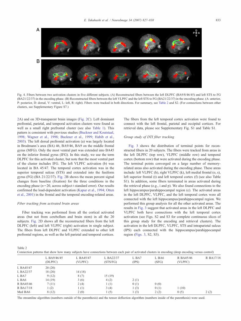
Fig. 4. Fibers between two activation clusters in five different subjects. (A) Reconstructed fibers between the left DLPFC (BA9/8/46/45) and left STS to FG(BA21/22/37) in the encoding phase. (B) Reconstructed fibers between the left VLPFC and the left STS to FG (BA21/22/37) in the encoding phase. (A: anterior,P: posterior, D: dorsal, V: ventral, L: left, R: right). Fibers were tracked in both directions. For summary, see Table 2 and S2. (For connections between otherclusters, see Supplementary Figure S7.)
833E. Takahashi et al. / NeuroImage 34 (2007) 827–838
2A) and on 3D-transparent brain images (Fig. 2C). Left dominantprefrontal, parietal, and temporal activation clusters were found aswell as a small right prefrontal cluster (see also Table 1). Thispattern is consistent with previous studies (Buckner and Koutstaal,1998; Wagner et al., 1998; Buckner et al., 1999; Habib et al.,2003). The left dorsal prefrontal activation (a) was largely locatedin Brodmann's area (BA) 46, BA9/46, BA9 on the middle frontalgyrus (MFG). Only the most ventral part was extended into BA45on the inferior frontal gyrus (IFG). In this study, we use the termDLPFC for this activated cluster, but note that the most ventral partof the cluster includes IFG. The left VLPFC activation (b) waslocated in BA 45/47. The temporal cortex activation was in thesuperior temporal sulcus (STS) and extended into the fusiformgyrus (FG) (BA 21/22/37). Fig. 2B shows the mean percent signalchanges from baseline (fixation) for the three conditions in theencoding phase (n=20, across subject±standard error). Our resultsconfirmed the load-dependent activation (Kapur et al., 1994; Ottenet al., 2001) in the frontal and the temporal encoding-related areas.
Fiber tracking from activated brain areas
Fiber tracking was performed from all the cortical activatedareas (but not from cerebellum and brain stem) in all the 20subjects. Fig. 2D shows all the reconstructed fibers from the leftDLPFC (left) and left VLPFC (right) activation in single subject.The fibers from left DLPFC and VLPFC extended to other leftprefrontal regions, as well as the left parietal and temporal cortices.
Table 2Connection patterns that show how many subjects have connections between each
L BA9/46/45(DLPFC)
L BA45/47(VLPFC)
L BA22/37(STS/FG)
L BA45/47 20 (20)L BA22/37 18 (20) 14 (18)L BA7 9 (12) 8 (7) 15 (19)L BA6 14 (19) 3 (6) 4 (2)R BA45/46 7 (11) 2 (4) 1 (1)R BA17/18 1 (2) 1 (1) 3 (4)Med BA6 8 (12) 2 (0) 1 (5)
The streamline algorithm (numbers outside of the parenthesis) and the tensor defl
The fibers from the left temporal cortex activation were found toconnect with the left frontal, parietal and occipital cortices. Forretrieval data, please see Supplementary Fig. S1 and Table S1.
Group study of DTI fiber tracking
Fig. 3 shows the distribution of terminal points for recon-structed fibers in 20 subjects. The fibers were tracked from areas inthe left DLPFC (top row), VLPFC (middle row) and temporalcortex (bottom row) that were activated during the encoding phase.The terminal points converged on a large number of memory-related areas also activated during the encoding phase. These areasinclude: left VLPFC (h), right VLPFC (k), left medial frontal (o, s),left superior frontal (t) and left temporal cortex (f) (see also TableS1). In addition, some fibers terminated in areas activated duringthe retrieval phase (e.g., i and p). We also found connections to theleft hippocampus/parahippocampal region (c). The activated areasin the left DLPFC, VLPFC, and the left temporal cortex were allconnected with the left hippocampus/parahippocampal region. Weperformed this group analysis for all the other activated areas. Theresults in Fig. 3 suggest that activated areas in the left DLPFC andVLPFC both have connections with the left temporal cortexactivation (see Figs. S2 and S3 for complete continuous slices ofthis group study for the encoding and retrieval clusters). Theactivation in the left DLPFC, VLPFC, STS and intraparietal sulcus(IPS) each connected with the hippocampus/parahippocampalregion (Figs. 3, S2, S3).
pair of activated clusters in encoding (deep encoding versus control)
L BA7(IPS)
L BA6(SFG)
R BA45/46(VLPFC)
R BA17/18
2 (1)0 (1) 0 (0)1 (3) 0 (1) 1 (10)1 (3) 2 (2) 0 (5) 2 (2)
ection algorithm (numbers inside of the parenthesis) were used.
834 E. Takahashi et al. / NeuroImage 34 (2007) 827–838
Connections between memory-related areas
We examined connections between pairs of activation clusters ineach subject (Figs. 4A, B, Table 2). Fibers were obtained using anyvoxel in the starting cluster (as a seed) to any voxel within the endcluster, going in either direction. Fig. 4A shows the connectionsfound between the activated areas in the left DLPFC and the lefttemporal cortex for the encoding phase. Fig. 4B shows theconnections between the left VLPFC and the left temporal cortexactivation for the encoding phase. We performed this analysis on thedata obtained from all 20 subjects. The most dorsal part (BA 9) ofthe left DLPFC cluster connected with the dorsal part (BA21, 22) ofthe left temporal cortex cluster. In some subjects, there were alsoconnections between the ventral part of the DLPFC cluster and thetemporal cortex cluster. Fibers between VLPFC and STS also
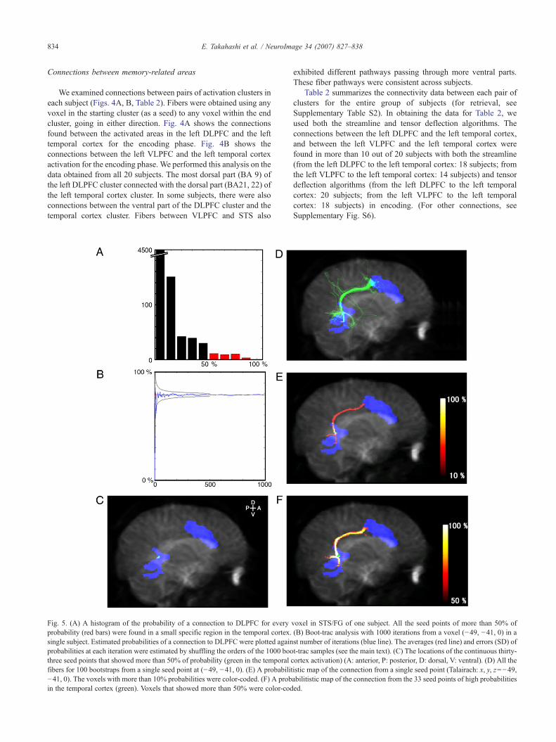
Fig. 5. (A) A histogram of the probability of a connection to DLPFC for everyprobability (red bars) were found in a small specific region in the temporal cortex.single subject. Estimated probabilities of a connection to DLPFC were plotted againprobabilities at each iteration were estimated by shuffling the orders of the 1000 boothree seed points that showed more than 50% of probability (green in the temporalfibers for 100 bootstraps from a single seed point at (−49, −41, 0). (E) A probabilit−41, 0). The voxels with more than 10% probabilities were color-coded. (F) A probin the temporal cortex (green). Voxels that showed more than 50% were color-cod
exhibited different pathways passing through more ventral parts.These fiber pathways were consistent across subjects.
Table 2 summarizes the connectivity data between each pair ofclusters for the entire group of subjects (for retrieval, seeSupplementary Table S2). In obtaining the data for Table 2, weused both the streamline and tensor deflection algorithms. Theconnections between the left DLPFC and the left temporal cortex,and between the left VLPFC and the left temporal cortex werefound in more than 10 out of 20 subjects with both the streamline(from the left DLPFC to the left temporal cortex: 18 subjects; fromthe left VLPFC to the left temporal cortex: 14 subjects) and tensordeflection algorithms (from the left DLPFC to the left temporalcortex: 20 subjects; from the left VLPFC to the left temporalcortex: 18 subjects) in encoding. (For other connections, seeSupplementary Fig. S6).
voxel in STS/FG of one subject. All the seed points of more than 50% of(B) Boot-trac analysis with 1000 iterations from a voxel (−49, −41, 0) in ast number of iterations (blue line). The averages (red line) and errors (SD) oft-trac samples (see the main text). (C) The locations of the continuous thirty-cortex activation) (A: anterior, P: posterior, D: dorsal, V: ventral). (D) All theistic map of the connection from a single seed point (Talairach: x, y, z=−49,abilitistic map of the connection from the 33 seed points of high probabilitiesed.

Fig. 6. (A) A histogram of the numbers of the fibers between the DLPFCcluster and each of the 100 random clusters for one subject. (B) Histogramsof actual numbers of fibers from DLPFC to the temporal cortex (black bars)and median numbers of fibers from DLPFC to random clusters (white bars)for 18 subjects.
835E. Takahashi et al. / NeuroImage 34 (2007) 827–838
Estimations of tractography error
We estimated the error of fiber tracking between the leftDLPFC and the left temporal cortex using a bootstrap method(Lazar and Alexander, 2005) (see Materials and methods). First, wemade histograms of the probability of a connection to DLPFC foreach and every voxel in STS/FG (Fig. 5A; see Materials andmethods). Thirty-three continuous seed points in the temporalcortex (a green region in Fig. 5C) showed greater than 50%probability of a connection to DLPFC. The seed cluster volumewas 33 mm3 (1-mm sampling of seed points), corresponding to0.7% of the entire temporal cortex activation (4848 mm3). It isclear that these seed points did not distribute diffusively across thewhole activated cluster, but were rather confined in a specificregion. The Talairach coordinates of these 33 voxels were locatedin STS (x, y, z=−54±5, −40±2, −1±2). These results indicate thatthe locations in temporal cortex that had connections with DLPFCwere very specific.
We showed the results of boot-trac from a single seed point(Talairach: x, y, z=−49, −41, 0; from a green region in Fig. 5C) intwo ways. First, all the fibers for 100 bootstraps are shown in Fig.5D. Most of the fibers (78%) went to the DLPFC, graduallydiverging by the distance from the seed point in the temporalcortex activation. To assess uncertainty of this probability, weperformed boot-trac analysis 1000 times. By shuffling the orders ofthe 1000 boot-trac samples 1000 times, an error for each iterationwas obtained (Fig. 5B). At 100 iterations of boot-trac, the error wasless than 5%. Second, the boot-trac from the same seed is shown asa probabilistic map (Fig. 5E; see Materials and methods). Thisfigure shows how many times, out of 100, the boot-trac fiberspassed through each voxel. The probabilities were almost 100%around the seed point in the temporal cortex activation, but soonwent down less than 50%. This is because of the divergence of thetracked fibers, which depends on the distance from the seed point,as displayed in Fig. 5D.
We obtained a probabilistic map of connections from all 33 seedpoints with a connection probability greater than 50% (Fig. 5F; seeMaterials and methods). These seed points are located in restrictedlocations, so they could compensate for the low probability of oneseed point. The results indicate that the fiber tracking from those 33seed points reached the DLPFC activation with a probability of morethan 80%. The terminal location was the posterior end of the DLPFCcluster (around x, y, z=−49, 3, 36 in the Talairach coordinate),around the precentral sulcus to the middle frontal gyrus. This resultindicates that not only the seed points, but also the terminal pointswere in very restricted locations within the activation. Fig. 5F showsthe probabilities of fibers from 33 voxels in the temporal cortex toanywhere in the whole brain. Although we did not selectively showthe fibers that reached the DLPFC activation, high probabilitieswere found only along the pathway from the temporal cortex toDLPFC. Thus, the specificity of this pathway was remarkable. Wealso obtained probabilistic maps (Supplementary Fig. S7, upperrow) from all the voxels in the temporal cortex activation, similar toFig. 5F. All subjects' results (n=20) were averaged after normal-ization (Supplementary Fig. S7, lower row).
The boot-tracked fibers were diverging by the distance from theseed points, but in many cases, as shown in Fig. 5D, most of thefibers reached the DLPFC activation. To see this observationquantitatively, we examined the probability of connectionsbetween any voxel in the temporal cortex and any voxel in theDLPFC (see Materials and methods). We found the connectionwith a probability of 100% in this subject.
We performed this latter analysis for 18 subjects who showedconnections between these regions. The existence of this pathwaywas crucial to distinguishing between the two models (the Serialand Parallel Pathway Models) mentioned above. Ten subjects outof the 18 subjects showed more than 50% probabilities for theconnections between the left DLPFC and the left temporal cortex.The probability for the existence of this connection was very highin these 10 subjects (86.3±18.0%, n=10). This result validates ourmajor finding on the direct pathway between DLPFC and thetemporal cortex activation.
Estimations of the connection specificity using arbitrarydefined areas
We also examined the specificity of the connections betweenDLPFC and the temporal cortex. Arbitrary regions satisfying theselection criteria (see Materials and methods) were used 100 times
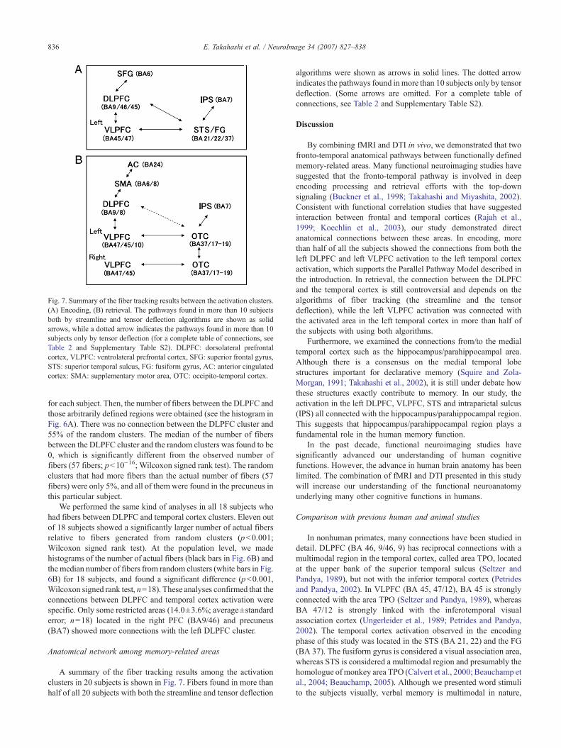
Fig. 7. Summary of the fiber tracking results between the activation clusters.(A) Encoding, (B) retrieval. The pathways found in more than 10 subjectsboth by streamline and tensor deflection algorithms are shown as solidarrows, while a dotted arrow indicates the pathways found in more than 10subjects only by tensor deflection (for a complete table of connections, seeTable 2 and Supplementary Table S2). DLPFC: dorsolateral prefrontalcortex, VLPFC: ventrolateral prefrontal cortex, SFG: superior frontal gyrus,STS: superior temporal sulcus, FG: fusiform gyrus, AC: anterior cingulatedcortex: SMA: supplementary motor area, OTC: occipito-temporal cortex.
836 E. Takahashi et al. / NeuroImage 34 (2007) 827–838
for each subject. Then, the number of fibers between the DLPFC andthose arbitrarily defined regions were obtained (see the histogram inFig. 6A). There was no connection between the DLPFC cluster and55% of the random clusters. The median of the number of fibersbetween the DLPFC cluster and the random clusters was found to be0, which is significantly different from the observed number offibers (57 fibers; p<10−16; Wilcoxon signed rank test). The randomclusters that had more fibers than the actual number of fibers (57fibers) were only 5%, and all of them were found in the precuneus inthis particular subject.
We performed the same kind of analyses in all 18 subjects whohad fibers between DLPFC and temporal cortex clusters. Eleven outof 18 subjects showed a significantly larger number of actual fibersrelative to fibers generated from random clusters (p<0.001;Wilcoxon signed rank test). At the population level, we madehistograms of the number of actual fibers (black bars in Fig. 6B) andthe median number of fibers from random clusters (white bars in Fig.6B) for 18 subjects, and found a significant difference (p<0.001,Wilcoxon signed rank test, n=18). These analyses confirmed that theconnections between DLPFC and temporal cortex activation werespecific. Only some restricted areas (14.0±3.6%; average±standarderror; n=18) located in the right PFC (BA9/46) and precuneus(BA7) showed more connections with the left DLPFC cluster.
Anatomical network among memory-related areas
A summary of the fiber tracking results among the activationclusters in 20 subjects is shown in Fig. 7. Fibers found in more thanhalf of all 20 subjects with both the streamline and tensor deflection
algorithms were shown as arrows in solid lines. The dotted arrowindicates the pathways found in more than 10 subjects only by tensordeflection. (Some arrows are omitted. For a complete table ofconnections, see Table 2 and Supplementary Table S2).
Discussion
By combining fMRI and DTI in vivo, we demonstrated that twofronto-temporal anatomical pathways between functionally definedmemory-related areas. Many functional neuroimaging studies havesuggested that the fronto-temporal pathway is involved in deepencoding processing and retrieval efforts with the top-downsignaling (Buckner et al., 1998; Takahashi and Miyashita, 2002).Consistent with functional correlation studies that have suggestedinteraction between frontal and temporal cortices (Rajah et al.,1999; Koechlin et al., 2003), our study demonstrated directanatomical connections between these areas. In encoding, morethan half of all the subjects showed the connections from both theleft DLPFC and left VLPFC activation to the left temporal cortexactivation, which supports the Parallel Pathway Model described inthe introduction. In retrieval, the connection between the DLPFCand the temporal cortex is still controversial and depends on thealgorithms of fiber tracking (the streamline and the tensordeflection), while the left VLPFC activation was connected withthe activated area in the left temporal cortex in more than half ofthe subjects with using both algorithms.
Furthermore, we examined the connections from/to the medialtemporal cortex such as the hippocampus/parahippocampal area.Although there is a consensus on the medial temporal lobestructures important for declarative memory (Squire and Zola-Morgan, 1991; Takahashi et al., 2002), it is still under debate howthese structures exactly contribute to memory. In our study, theactivation in the left DLPFC, VLPFC, STS and intraparietal sulcus(IPS) all connected with the hippocampus/parahippocampal region.This suggests that hippocampus/parahippocampal region plays afundamental role in the human memory function.
In the past decade, functional neuroimaging studies havesignificantly advanced our understanding of human cognitivefunctions. However, the advance in human brain anatomy has beenlimited. The combination of fMRI and DTI presented in this studywill increase our understanding of the functional neuroanatomyunderlying many other cognitive functions in humans.
Comparison with previous human and animal studies
In nonhuman primates, many connections have been studied indetail. DLPFC (BA 46, 9/46, 9) has reciprocal connections with amultimodal region in the temporal cortex, called area TPO, locatedat the upper bank of the superior temporal sulcus (Seltzer andPandya, 1989), but not with the inferior temporal cortex (Petridesand Pandya, 2002). In VLPFC (BA 45, 47/12), BA 45 is stronglyconnected with the area TPO (Seltzer and Pandya, 1989), whereasBA 47/12 is strongly linked with the inferotemporal visualassociation cortex (Ungerleider et al., 1989; Petrides and Pandya,2002). The temporal cortex activation observed in the encodingphase of this study was located in the STS (BA 21, 22) and the FG(BA 37). The fusiform gyrus is considered a visual association area,whereas STS is considered a multimodal region and presumably thehomologue of monkey area TPO (Calvert et al., 2000; Beauchamp etal., 2004; Beauchamp, 2005). Although we presented word stimulito the subjects visually, verbal memory is multimodal in nature,

837E. Takahashi et al. / NeuroImage 34 (2007) 827–838
which is consistent with temporal activation in visual andmultimodal association areas. In humans, we found connectionsbetween DLPFC (BA 46, 9) and temporal cortex activations. Thefibers ended mostly in STS (Fig. 5A). This appears to behomologues to connections between DLPFC and area TPO in themonkey brain. Human VLPFC (BA 45, 47) was also connected withthe STS activation, which is also consistent with previous monkeystudies. Our findings suggest that there are two pathways betweenPFC and temporal cortex related to human verbal memory: onebetween DLPFC and temporal cortex, and another between VLPFCand temporal cortex. The similar pathways between PFC and thetemporal multimodal region in the monkey brain suggest that suchdual control systems might also be important for multimodalmemory function in nonhuman primates.
We showed that left DLPFC is connected with STS, theintraparietal sulcus (IPS), and the anterior cingulate cortex. Inmonkeys, connections with the prefrontal region around theprincipal sulcus (PS) were found in the STS, IPS, and the cingulatecortex (Pandya and Kuypers, 1969). Similarly, connections werealso found between IPS and the temporal cortex in monkeys(Pandya and Kuypers, 1969; Mesulam et al., 1977; Cavada andGoldman-Rakic, 1989), which is consistent with our results. Fromthe occipital cortex, we showed fascicles to IPS. These pathwaysare also known in monkeys between the IPS and inferior occipitalgyrus (Seltzer and Pandya, 1980).
We showed hippocampal connections with a focus on activationidentified in the DLPFC, VLPFC, STS and IPS. In monkeys,hippocampal connections were also found with the dorsolateralprefrontal cortex (BA9/46) (Goldman-Rakic et al., 1984; Nauta,1964), the inferior parietal sulcus (BA7) and the inferior temporalcortex (Seltzer and Pandya, 1983; Van Hoesen et al., 1979).
Roles of the connections between DLPFC and the temporal cortex
On the basis of lesion studies in nonhuman primates (Petrides,1991), neuroimaging studies in humans (Petrides et al., 1993a,b), andconnection studies in nonhuman primates (Petrides and Pandya,1994), the two-stage model of PFC (Petrides, 1994a,b; 1996; Owen etal., 1996) was suggested: the ventrolateral prefrontal cortex (VLPFC)is thought to interact with posterior cortices such as the temporalcortex for active (or controlled) encoding and retrieval of information,while the dorsolateral prefrontal cortex (DLPFC) is hypothesized tomonitor and manipulate the maintained information in VLPFC. Inthis study, we found that there are two fronto-temporal pathwaysbetween the left DLPFC and the left temporal cortex, and between theleft VLPFC and the left temporal cortex. This result suggests thatthere is a direct interaction between DLPFC and temporal cortex.
Acknowledgments
We thank Dorothe A. Poggel and Itamar Ronen for their helpfulcomments on this paper, and Jeff Thompson, Elizabeth Applebyand Kim Ono for their helpful editorial comments. This work wassupported by NIH (NS44825), and the Human Frontiers ScienceProgram. The first author (E.T.) was supported by the UeharaMemorial Foundation (Japan).
Appendix A. Supplementary data
Supplementary data associated with this article can be found, inthe online version, at doi:10.1016/j.neuroimage.2006.10.009.
References
Alexander, A.L., Hasan, K.M., Lazar, M., Tsuruda, J.S., Parker, D.L., 2001.Analysis of partial volume effects in diffusion-tensor MRI. Magn.Reson. Med. 45, 770–780.
Basser, P.J., Mattiello, J., LeBihan, D., 1994. MR diffusion tensorspectroscopy and imaging. Biophys. J. 66, 259–267.
Basser,P.J.,Pajevic,S.,Pierpaoli,C.,Duda,J.,Aldroubi,A.,2000. InvivofibertractographyusingDT-MRIdata.Magn.Reson.Med.44,625–632.
Behrens, T.E., Johansen-Berg, H., Woolrich, M.W., Smith, S.M., Wheeler-Kingshott, C.A., Boulby, P.A., Barker, G.J., Sillery, E.L., Sheehan, K.,Ciccarelli, O., Thompson, A.J., Brady, J.M., Matthews, P.M., 2003.Non-invasive mapping of connections between human thalamus andcortex using diffusion imaging. Nat. Neurosci. 6, 750–757.
Beauchamp, M.S., 2005. See me, hear me, touch me: multisensoryintegration in lateral occipital–temporal cortex. Curr. Opin. Neurobiol.15, 145–153.
Beauchamp, M.S., Argall, B.D., Bodurka, J., Duyn, J.H., Martin, A., 2004.Unraveling multisensory integration: patchy organization within humanSTS multisensory cortex. Nat. Neurosci. 7, 1190–1192.
Buckner, R.L., Koutstaal, W., 1998. Functional neuroimaging studies ofencoding, priming, and explicit memory retrieval. Proc. Natl. Acad. Sci.U. S. A. 95, 891–898.
Buckner, R.L., Koutstaal, W., Schacter, D.L., Wagner, A.D., Rosen, B.R.,1998. Functional–anatomic study of episodic retrieval using fMRI: I.Retrieval effort versus retrieval success. NeuroImage 7, 151–162.
Buckner, R.L., Kelley, W.M., Petersen, S.E., 1999. Frontal cortexcontributes to human memory formation. Nat. Neurosci. 2, 311–314.
Cabeza, R., Nyberg, L., 2000. Imaging cognition II: an empirical review of275 PET and fMRI studies. J. Cogn. Neurosci. 12, 1–47.
Cavada, C., Goldman-Rakic, P.S., 1989. Posterior parietal cortex in rhesusmonkey: I. Parcellation of areas based on distinctive limbic and sensorycorticocortical connections. J. Comp. Neurol. 287, 393–421.
Calvert, G.A., Campbell, R., Brammer, M.J., 2000. Evidence fromfunctional magnetic resonance imaging of crossmodal binding in thehuman heteromodal cortex. Curr. Biol. 10, 649–657.
Conturo, T.E., Lori, N.F., Cull, T.S., Akbudak, E., Snyder, A.Z., Shimony,J.S., McKinstry, R.C., Burton, H., Raichle, M.E., 1999. Trackingneuronal fiber pathways in the living human brain. Proc. Natl. Acad.Sci. U. S. A. 96, 10422–10427.
Demb, J.B., Desmond, J.E., Wagner, A.D., Vaidya, C.J., Glover, G.H.,Gabrieli, J.D., 1995. Semantic encoding and retrieval in the left inferiorprefrontal cortex: a functional MRI study of task difficulty and processspecificity. J. Neurosci. 15, 5870–5878.
Dougherty, R.F., Ben-Shachar, M., Bammer, R., Brewer, A.A., Wandell,B.A., 2005. Functional organization of human occipito-callosal fibertracts. Proc. Natl. Acad. Sci. U. S. A. 102, 7350–7355.
Friston, K.J., Worsley, K.J., Frackowiak, R.S.J., Mazziotta, J.C., Evans,A.C., 1994. Assessing the significance of focal activations using theirspatial extent. Hum. Brain Mapp. 1, 214–220.
Fuster, J.M., 1997. The Prefrontal Cortex: Anatomy, Physiology, andNeuropsychology of the Frontal Lobe. Lippincott-Raven, Philadelphia.
Gazzaniga, M.S., 1995. Principles of human brain organization derived fromsplit-brain studies. Neuron 14, 217–228.
Goldman-Rakic, P.S., 1987. Circuitry of primate prefrontal cortex andregulation of behavior by representational memory. In: Plum, F.,Mountcastle, V. (Eds.), Handbook of Physiology: Section 1. TheNervous System. Vol. 5. Higher Functions of the Brain. Part 1. AmericanPhysiological Society, Bethesda, MD, pp. 373–417.
Goldman-Rakic, P.S., Selemon, L.D., Schwartz, M.L., 1984. Dual pathwaysconnecting the dorsolateral prefrontal cortex with the hippocampalformation and parahippocampal cortex in the rhesus monkey. Neurosci.12, 719.
Habib, R., Nyberg, L., Tulving, E., 2003. Hemispheric asymmetries ofmemory: the HERA model revisited. TRENDS Cogn. Sci. 7, 241–245.
Incisa della Rocchetta, A., Milner, B., 1993. Strategic search and retrievalinhibition: the role of the frontal lobes. Neuropsychologia 31, 503–524.
838 E. Takahashi et al. / NeuroImage 34 (2007) 827–838
Jaermann, T., Crelier, G., Pruessmann, K.P., Golay, X., Netsch, T., vanMuiswinkel, A.M., Mori, S., van Zijl, P.C., Valavanis, A., Kollias, S.,Boesiger, P., 2004. SENSE-DTI at 3 T. Magn. Reson. Med. 51,230–236.
Jones, D.K., Horsfield, M.A., Simmons, A., 1999. Optimal strategies formeasuring diffusion in anisotropic systems by magnetic resonanceimaging. Magn. Reson. Med. 42, 515–525.
Kapur, S., Craik, F.I., Tulving, E., Wilson, A.A., Houle, S., Brown, G.M.,1994. Neuroanatomical correlates of encoding in episodic memory:levels of processing effect. Proc. Natl. Acad. Sci. U. S. A. 91, 2008–2011.
Kim, M., Ducros, M., Carlson, T., Ronen, I., He, S., Ugurbil, K., Kim, D.-S.,2006. Anatomical correlates of the functional organization in the humanoccipitotemporal cortex. Magn. Reson. Imaging. 24, 583–590.
Koechlin, E., Ody, C., Kouneiher, F., 2003. The architecture of cognitivecontrol in the human prefrontal cortex. Science 302, 1181–1185.
Kucera, H., Francis, W.N., 1967. Computational Analysis of Present-dayAmerican English. Brown Univ. Press, Providence, RI.
Lazar, M., Alexander, A.L., 2005. Bootstrap white matter tractography(BOOT-TRAC). NeuroImage 24, 524–532.
Lazar, M., Weinstein, D.M., Tsuruda, J.S., Hasan, K.M., Arfanakis, K.,Meyerand, M.E., Badie, B., Rowley, H.A., Haughton, V., Field, A.,Alexander, A.L., 2003. White matter tractography using diffusion tensordeflection. Hum. Brain Mapp. 18, 306–321.
Lehericy, S., Ducros, M., Krainik, A., Francois, C., Van de Moortele, P.F.,Ugurbil, K., Kim, D.S., 2004. 3-D diffusion tensor axonal trackingshows distinct SMA and pre-SMA projections to the human striatum.Cereb. Cortex 14, 1303–1309.
Lepage, M., Ghaffar, O., Nyberg, L., Tulving, E., 2000. Prefrontal cortexand episodic memory retrieval mode. Proc. Natl. Acad. Sci. U. S. A. 97,506–511.
Mesulam, M.M., Van Hoesen, G.W., Pandya, D.N., Geschwind, N., 1977.Limbic and sensory connections of the inferior parietal lobule (area PG)in the rhesus monkey: a study with a new method for horseradishperoxidase histochemistry. Brain Res. 136, 393–414.
Mori, S., Crain, B.J., Chacko, V.P., van Zijl, P.C.M., 1999. Three-dimensional tracking of axonal projections in the brain by magneticresonance imaging. Annal. Neurol. 45, 265–269.
Nauta, W.J.H., 1964. In: Warren, J.M., Akert, K. (Eds.), The FrontalGranular Cortex and Behavior. McGraw-Hill, New York, pp. 397–409.
Otten, L.J., Henson, R.N.A., Rugg, M.D., 2001. Depth of processing effectson neural correlates of memory encoding. Brain 124, 399–412.
Owen, A.M., Evans, A.C., Petrides, M., 1996. Evidence for a two-stagemodel of spatial working memory processing within the lateral frontalcortex: a positron emission tomography study. Cereb. Cortex 6, 31–38.
Pandya, D.N., Kuypers, H.G.J.M., 1969. Cortico-cortical connections in therhesus monkey. Brain Res. 13, 13–36.
Petrides, M., 1991. Monitoring of selections of visual stimuli and theprimate frontal cortex. Proc. R. Soc. London, Ser. B 246, 293–298.
Petrides, M., 1994a. Frontal lobes and behaviour. Curr. Opin. Neurobiol. 4,207–211.
Petrides, M., 1994b. Frontal lobes and working memory: evidence frominvestigations of the effects of cortical excisions in nonhuman primates.In: Boller, F., Grafman, J. (Eds.), Handbook of Neuropsychology, Vol. 9.Elsevier, Amsterdam, pp. 59–82.
Petrides, M., 1996. Specialized systems for the processing of mnemonicinformation within the primate frontal cortex. Philos. Trans. R. Soc., B351, 1455–1462.
Petrides, M., 2005. Lateral prefrontal cortex: architectonic and functionalorganization. Philos. Trans. R. Soc., B 360, 781–795.
Petrides, M., Pandya, D.N., 1994. Comparative architectonic analysis of thehuman and the macaque frontal cortex. In: Boller, F., Grafman, J. (Eds.),Handbook of Neuropsychology, vol. 9. Elsevier, Amsterdam, pp. 17–58.
Petrides, M., Pandya, D.N., 1999. Dorsolateral prefrontal cortex: compara-tive cytoarchitectonic analysis in the human and the macaque brain andcorticocortical connection patterns. Eur. J. Neurosci. 11, 1011–1036.
Petrides, M., Pandya, D.N., 2002. Comparative cytoarchitectonic analysis of
the human and the macaque ventrolateral prefrontal cortex andcorticocortical connection patterns in the monkey. Eur. J. Neurosci.16, 291–310.
Petrides, M., Alivisatos, B., Evans, A.C., Meyer, E., 1993a. Dissociation ofhuman mid-dorsolateral from posterior dorsolateral frontal cortex inmemory processing. Proc. Natl Acad. Sci. U. S. A. 90, 873–877.
Petrides, M., Alivisatos, B., Meyer, E., Evans, A.C., 1993b. Functionalactivation of the human frontal cortex during the performance of verbalworking memory tasks. Proc. Natl Acad. Sci. U. S. A. 90, 878–882.
Powell, H.W., Guye, M., Parker, G.J., Symms, M.R., Boulby, P., Koepp,M.J., Barker, G.J., Duncan, J.S., 2004. Noninvasive in vivodemonstration of the connections of the human parahippocampalgyrus. NeuroImage 22, 740–747.
Rajah, M.N., McIntosh, A.R., Grady, C.L., 1999. Frontotemporal interac-tions in face encoding and recognition. Cogn. Brain Res. 8, 259–269.
Schacter, D.L., 1987. Memory, amnesia, and frontal lobe dysfunction.Psychophysiology 17, 568–576.
Seltzer, B., Pandya, D.N., 1980. Converging visual and somatic sensorycortical input to the intraparietal sulcus of the rhesus monkey. Brain Res.192, 339–351.
Seltzer, B., Pandya, D.N., 1983. The distribution of posterior parietal fibersin the corpus callosum of the rhesus monkey. Exp. Brain Res. 49,147–150.
Seltzer, B., Pandya, D.N., 1989. Frontal lobe connections of the superiortemporal sulcus in the rhesus monkey. J. Comp. Neurol. 281,97–113.
Shimamura, A.P., 1995. Memory and frontal lobe function. In: Gazzaniga,M.S. (Ed.), The Cognitive Neurosciences. MIT press, Cambridge, MA,pp. 803–813.
Squire, L.R., Zola-Morgan, S., 1991. The medial temporal lobe memorysystem. Science 253, 1380–1386.
Stieltjes, B., Kaufmann, W.E., van Zijl, P.C., Fredericksen, K., Pearlson,G.D., Solaiyappan, M., Mori, S., 2001. Diffusion tensor imaging andaxonal tracking in the human brainstem. NeuroImage 14, 723–735.
Takahashi, E., Miyashita, Y., 2002. In: Squire, L.R., Schacter, D.L.(Eds.), Neuropsychology of Memory. The Guilford Press, NewYork, pp. 301–310.
Takahashi, E., Ohki, K., Miyashita, Y., 2002. The role of the parahippo-campal gyrus in source memory for external and internal events.NeuroReport 13, 1951–1956.
Talairach, J., Tournoux, P., 1988. Co-Planar Stereotaxic Atlas of the HumanBrain. Thieme, New York.
Thottakara, P., Lazar, M., Johnson, C., Alexander, L., 2006. Application ofBrodmann's area templates for ROI selection in white mattertractography studies. NeuroImage 29, 868–878.
Tomita, H., Ohbayashi, M., Nakahara, K., Hasegawa, I., Miyashita, Y.,1999. Top-down signal from prefrontal cortex in executive control ofmemory retrieval. Nature 401, 699–703.
Tulving, E., Kapur, S., Craik, F.I., Moscovitch, M., Houle, S., 1994.Hemispheric encoding/retrieval asymmetry in episodic memory:positron emission tomography findings. Poc. Natl. Acad. Sci. U. S. A.91, 2016–2020.
Tulving, E., 2002. Episodic memory: from mind to brain. Annu. Rev.Psycol. 53, 1–25.
Ungerleider, L.G., Gaffan, D., Pelak, V.S., 1989. Projections from infectiortemporal cortex to prefrontal cortex via the uncinate fascicle in rhesusmonkeys. Exp. Brain Res. 76, 473–484.
Van Hoesen, G.W., Rosene, D.L., Mesulam, M.M., 1979. Subicular inputfrom temporal cortex in the rhesus monkey. Science 205, 608–610.
Wagner, A.D., Schacter, D.L., Rotte, M., Koutstaal, W., Maril, A., Dale,A.M., Rosen, B.R., Buckner, R.L., 1998. Building memories:remembering and forgetting of verbal experiences as predicted bybrain activity. Science 281, 1188–1191.
Xu, D., Mori, S., Solaiyappan, M., van Zijl, P.C., Davatzikos, C., 2002. Aframework for callosal fiber distribution analysis. NeuroImage 17,1131–1143.