Discovery of a Conjugative Megaplasmid in Bifidobacterium breve
11
Discovery of a Conjugative Megaplasmid in Bifidobacterium breve Francesca Bottacini, a Mary O’Connell Motherway, a,b Eoghan Casey, b Brian McDonnell, b Jennifer Mahony, b Marco Ventura, c Douwe van Sinderen a,b Alimentary Pharmabiotic Centre, National University of Ireland, Cork, Ireland a ; School of Microbiology, National University of Ireland, Cork, Ireland b ; Laboratory of Probiogenomics, Department of Life Sciences, University of Parma, Parma, Italy c Bifidobacterium breve is a common and sometimes very abundant inhabitant of the human gut. Genome sequencing of B. breve JCM 7017 revealed the presence of an extrachromosomal element, designated pMP7017 consisting of >190 kb, thus representing the first reported bifidobacterial megaplasmid. In silico characterization of this element revealed several genomic features sup- porting a stable establishment of the megaplasmid in its host, illustrated by predicted CRISPR-Cas functions that are known to protect the host against intrusion of foreign DNA. Interestingly, pMP7017 is also predicted to encode a conjugative DNA trans- fer apparatus and, consistent with this notion, we demonstrate here the conjugal transfer of pMP7017 to representative strains of B. breve and B. longum subsp. longum. We also demonstrate the presence of a megaplasmid with homology to pMP7017 in three B. longum subsp. longum strains. B ifidobacterium breve is a common inhabitant of the infant gut, in addition to Bifidobacterium longum subsp. longum, Bifido- bacterium longum subsp. infantis, Bifidobacterium pseudocatenu- latum, Bifidobacterium bifidum (1), and the less frequently encountered Bifidobacterium kashiwanohense (2). Furthermore, representatives of this species have been isolated in other environ- ments, including human milk, the adult gut, and human vagina (3). The presence of bifidobacteria (being members of the Actino- bacteria phylum) in the gut has been correlated with particular health-promoting effects, such as enforcement of the intestinal barrier, activation/modulation of the host’s immune response and protection against particular infections (4), and for this reason representatives of this genus have been included as active ingredi- ents in functional foods (5). Since the first genome sequence of B. longum subsp. longum NCC2705 was released in 2002, further sequencing endeavors have produced additional bifidobacterial genome sequences in an effort to elucidate the mode of action of their beneficial effects. In this respect genome sequencing and subsequent functional anal- yses have revealed that members of this genus possess genetic fea- tures that support the process of colonization and adhesion to the intestinal mucosa (6), evasion/stimulation of the host’s immune system (7), and a metabolic ability to use host-derived glycans, all of which are believed to contribute to competitive exclusion of pathogens (4) and perhaps other beneficial activities. The presence of plasmids among representatives of the ge- nus Bifidobacterium has been reported for certain bifidobacte- rial species (e.g., Bifidobacterium pseudolongum subsp. globosum, Bifidobacterium indicum, Bifidobacterium asteroides, Bifidobacte- rium bifidum, Bifidobacterium catenulatum, Bifidobacterium pseudocatenulatum, Bifidobacterium kashiwanohense, Bifidobacte- rium breve, Bifidobacterium longum subsp. longum, and Bifidobac- terium longum subsp. infantis)(8–11). Typically, when plasmids are found, just a single plasmid per isolate is present, with some notable exceptions: B. kashiwanohense JCM 15439, B. longum subsp. longum DJO10A, B. longum subsp. infantis 157F, and B. longum subsp. longum KACC 91563, which each harbor two such genetic elements (2, 8). Among the identified bifidobacterial plas- mids, most have been isolated from strains of B. longum subsp. longum (8). The bifidobacterial plasmids that have been charac- terized thus far exhibit a size not exceeding 10.5 kb and the possi- ble presence of extrachromosomal genetic elements larger than 100 kb, which are designated megaplasmids (12), in Bifidobacte- rium has only been suggested for two porcine cecal isolates (13). Bacterial conjugation is one of the possible means of horizontal transfer of genetic material and it has been intensively studied in Gram-negative bacteria, in particular in the plant pathogen Agro- bacterium tumefaciens due to its pathogenic and biotechnological relevance (e.g., tumor-inducing plasmid Ti) (14–16). Conjuga- tion has been described for Gram-positive bacteria, with the dif- ference that a somewhat simpler conjugative machinery appears to be required due to the fact that DNA traverses just a single membrane layer (17). With regard to bifidobacteria, recent ge- nome analyses for members of this genus has indeed provided evidence of horizontal transfer events (3, 6, 18–20), where pro- phages are believed to be one of the principal vehicles of such DNA transfer phenomena (21). The presence of (conjugative) megaplasmids in representatives of the phylum Actinobacteria has thus far only been observed for members of the Gordonia, Rhodococcus, Brevibacterium, Nocardi- opsis, and Mycobacterium spp. (with sizes ranging between 100 and 700 kb) (12, 22), of which p1CP from Rhodococcus opacus PD630 (23), pREA400, pREA250, and pREA100 from Rhodococ- cus erythropolis AN12 (24), and pKB1 from Gordonia westfalica Received 3 September 2014 Accepted 13 October 2014 Accepted manuscript posted online 17 October 2014 Citation Bottacini F, O’Connell Motherway M, Casey E, McDonnell B, Mahony J, Ventura M, van Sinderen D. 2015. Discovery of a conjugative megaplasmid in Bifidobacterium breve. Appl Environ Microbiol 81:166 –176. doi:10.1128/AEM.02871-14. Editor: M. W. Griffiths Address correspondence to Douwe van Sinderen, [email protected]. F.B. and M.O.M. are co-senior authors. Supplemental material for this article may be found at http://dx.doi.org/10.1128 /AEM.02871-14. Copyright © 2015, American Society for Microbiology. All Rights Reserved. doi:10.1128/AEM.02871-14 166 aem.asm.org January 2015 Volume 81 Number 1 Applied and Environmental Microbiology
Transcript of Discovery of a Conjugative Megaplasmid in Bifidobacterium breve

Discovery of a Conjugative Megaplasmid in Bifidobacterium breve
Francesca Bottacini,a Mary O’Connell Motherway,a,b Eoghan Casey,b Brian McDonnell,b Jennifer Mahony,b Marco Ventura,c
Douwe van Sinderena,b
Alimentary Pharmabiotic Centre, National University of Ireland, Cork, Irelanda; School of Microbiology, National University of Ireland, Cork, Irelandb; Laboratory ofProbiogenomics, Department of Life Sciences, University of Parma, Parma, Italyc
Bifidobacterium breve is a common and sometimes very abundant inhabitant of the human gut. Genome sequencing of B. breveJCM 7017 revealed the presence of an extrachromosomal element, designated pMP7017 consisting of >190 kb, thus representingthe first reported bifidobacterial megaplasmid. In silico characterization of this element revealed several genomic features sup-porting a stable establishment of the megaplasmid in its host, illustrated by predicted CRISPR-Cas functions that are known toprotect the host against intrusion of foreign DNA. Interestingly, pMP7017 is also predicted to encode a conjugative DNA trans-fer apparatus and, consistent with this notion, we demonstrate here the conjugal transfer of pMP7017 to representative strains ofB. breve and B. longum subsp. longum. We also demonstrate the presence of a megaplasmid with homology to pMP7017 in threeB. longum subsp. longum strains.
Bifidobacterium breve is a common inhabitant of the infant gut,in addition to Bifidobacterium longum subsp. longum, Bifido-
bacterium longum subsp. infantis, Bifidobacterium pseudocatenu-latum, Bifidobacterium bifidum (1), and the less frequentlyencountered Bifidobacterium kashiwanohense (2). Furthermore,representatives of this species have been isolated in other environ-ments, including human milk, the adult gut, and human vagina(3). The presence of bifidobacteria (being members of the Actino-bacteria phylum) in the gut has been correlated with particularhealth-promoting effects, such as enforcement of the intestinalbarrier, activation/modulation of the host’s immune response andprotection against particular infections (4), and for this reasonrepresentatives of this genus have been included as active ingredi-ents in functional foods (5).
Since the first genome sequence of B. longum subsp. longumNCC2705 was released in 2002, further sequencing endeavorshave produced additional bifidobacterial genome sequences in aneffort to elucidate the mode of action of their beneficial effects. Inthis respect genome sequencing and subsequent functional anal-yses have revealed that members of this genus possess genetic fea-tures that support the process of colonization and adhesion to theintestinal mucosa (6), evasion/stimulation of the host’s immunesystem (7), and a metabolic ability to use host-derived glycans, allof which are believed to contribute to competitive exclusion ofpathogens (4) and perhaps other beneficial activities.
The presence of plasmids among representatives of the ge-nus Bifidobacterium has been reported for certain bifidobacte-rial species (e.g., Bifidobacterium pseudolongum subsp. globosum,Bifidobacterium indicum, Bifidobacterium asteroides, Bifidobacte-rium bifidum, Bifidobacterium catenulatum, Bifidobacteriumpseudocatenulatum, Bifidobacterium kashiwanohense, Bifidobacte-rium breve, Bifidobacterium longum subsp. longum, and Bifidobac-terium longum subsp. infantis) (8–11). Typically, when plasmidsare found, just a single plasmid per isolate is present, with somenotable exceptions: B. kashiwanohense JCM 15439, B. longumsubsp. longum DJO10A, B. longum subsp. infantis 157F, and B.longum subsp. longum KACC 91563, which each harbor two suchgenetic elements (2, 8). Among the identified bifidobacterial plas-mids, most have been isolated from strains of B. longum subsp.longum (8). The bifidobacterial plasmids that have been charac-
terized thus far exhibit a size not exceeding 10.5 kb and the possi-ble presence of extrachromosomal genetic elements larger than100 kb, which are designated megaplasmids (12), in Bifidobacte-rium has only been suggested for two porcine cecal isolates (13).
Bacterial conjugation is one of the possible means of horizontaltransfer of genetic material and it has been intensively studied inGram-negative bacteria, in particular in the plant pathogen Agro-bacterium tumefaciens due to its pathogenic and biotechnologicalrelevance (e.g., tumor-inducing plasmid Ti) (14–16). Conjuga-tion has been described for Gram-positive bacteria, with the dif-ference that a somewhat simpler conjugative machinery appearsto be required due to the fact that DNA traverses just a singlemembrane layer (17). With regard to bifidobacteria, recent ge-nome analyses for members of this genus has indeed providedevidence of horizontal transfer events (3, 6, 18–20), where pro-phages are believed to be one of the principal vehicles of such DNAtransfer phenomena (21).
The presence of (conjugative) megaplasmids in representativesof the phylum Actinobacteria has thus far only been observed formembers of the Gordonia, Rhodococcus, Brevibacterium, Nocardi-opsis, and Mycobacterium spp. (with sizes ranging between 100and 700 kb) (12, 22), of which p1CP from Rhodococcus opacusPD630 (23), pREA400, pREA250, and pREA100 from Rhodococ-cus erythropolis AN12 (24), and pKB1 from Gordonia westfalica
Received 3 September 2014 Accepted 13 October 2014
Accepted manuscript posted online 17 October 2014
Citation Bottacini F, O’Connell Motherway M, Casey E, McDonnell B, Mahony J,Ventura M, van Sinderen D. 2015. Discovery of a conjugative megaplasmid inBifidobacterium breve. Appl Environ Microbiol 81:166 –176.doi:10.1128/AEM.02871-14.
Editor: M. W. Griffiths
Address correspondence to Douwe van Sinderen, [email protected].
F.B. and M.O.M. are co-senior authors.
Supplemental material for this article may be found at http://dx.doi.org/10.1128/AEM.02871-14.
Copyright © 2015, American Society for Microbiology. All Rights Reserved.
doi:10.1128/AEM.02871-14
166 aem.asm.org January 2015 Volume 81 Number 1Applied and Environmental Microbiology
Kb1 (25) have conclusively been shown to be conjugative. Withregard to conjugative transfer in bifidobacteria, only a recentstudy proposed a new shuttle vector pDOJHR-WD2 based on theconjugative machinery of Escherichia coli WM3064(pBB109),which was successfully used for DNA transfer between E. coli anddifferent Bifidobacterium species (26); however, no functional, na-tive bifidobacterial conjugation system has as yet been identified.
In this study we present the sequence and in silico annotation ofa megaplasmid present in B. breve JCM 7017. This 190.17-kb plas-mid, designated pMP7017, the first and largest megaplasmid to beidentified among a member of the genus Bifidobacterium and ac-tinobacterial gut commensals in general, is able to transfer toother bifidobacteria by an apparent conjugative mechanism. In-terestingly, and consistent with the horizontal transfer ability ofpMP7017, we also detected homologs of this megaplasmid inthree representatives of B. longum subsp. longum.
MATERIALS AND METHODSSequencing and annotation. DNA sequencing of pMP7017 was per-formed using protocols developed by the sequencing provider Macrogen(Seoul, Republic of Korea) and employing a combination of two next-generation sequencing (NGS) platforms (see below). An initial 454 runusing a Roche genome sequencer FLX Titanium instrument employing along-tag, paired-end library (average read length of 400 bp) resulted in theconstruction of an initial scaffold, which was subsequently improved withan additional Illumina Hiseq 2000 paired-end sequencing run with anaverage read length of 101 bp.
The resulting sequences were then assembled in a hybrid 454-Illuminaapproach using a combination of Newbler v2.9 (98.83% of aligned readsand 0.04% inferred read error) (454 Sequencing; Roche) for long readsand Abyss v1.3.4 (http://www.bcgsc.ca/) for short reads, resulting in ahigh-quality and ungapped final consensus sequence, where the averagesequence coverage of each base pair was �200-fold.
Replicon analyses and bioinformatic analysis. Open reading frame(ORF) prediction was performed with a combined approach of the toolProdigal v2.0 (http://prodigal.ornl.gov). Functional assignment of thepredicted ORFs was first performed on the basis of BLASTP v2.2.26 (27)searches against the nonredundant protein database curated by the Na-tional Centre for Biotechnology Information (ftp://ftp.ncbi.nih.gov/blast/db/) and further refined, when necessary, employing BLASTX v2.2.26searches (27).
Artemis v.14 (http://www.sanger.ac.uk/resources/software/artemis/)was used to inspect the results of the ORF prediction and used for manualediting, if necessary, of the start codon of a predicted gene. Gene annota-tions were further integrated with the information retrieved from alter-native databases, e.g., protein family (Pfam) (http://pfam.sanger.ac.uk).
TRNA genes were identified using tRNAscan-SE v1.4 (28) and thepresence of Restriction-Modification (R-M) systems was investigated us-ing BLASTP (27) alignments against the downloaded REBASE database(http://rebase.neb.com/rebase/rebase.html; cutoff E value of 0.0001 andshowing at least 30% similarity across at least 80% of the complete lengthof the deduced amino acid sequence).
The presence of CRISPR loci was inspected using the CRISPR findertool (http://crispr.u-psud.fr), while codon usage and amino acid fre-quency were computed using the GCUA (29) tool and visualized withscatter3D module of the statistical package R (http://www.r-project.org/).Sequence comparisons at the protein level were performed using an bidi-rectional BLASTP alignment (27) (cutoff E value of 0.0001, with at least50% identity across at least 50% of either protein sequence).
Bacterial strains, plasmids, and culture conditions. Bacterial strainsand plasmids used in the present study are listed in Table 1. Bifidobacterialstrains were routinely cultured in reinforced clostridial medium (RCM[Oxoid, Ltd., Basingstoke, Hampshire, United Kingdom]) or modified deMan, Rogosa, and Sharpe medium (mMRS) prepared from first princi-
ples (30). Prior to inoculation, mMRS was supplemented with cysteine-HCl (0.05% [wt/vol]) and lactose (1.0% [wt/vol]) (Sigma). Bifidobacte-rial cultures were incubated at 37°C under anaerobic conditions, whichwere maintained using an anaerobic hood (Davidson and Hardy, Belfast,Ireland). E. coli was cultured in Luria-Bertani (LB) broth (31) at 37°C withagitation. Where appropriate, growth media contained tetracycline (10�g ml�1), ampicillin (100 �g ml�1 for E. coli), erythromycin (100 �gml�1 for E. coli), or kanamycin (50 �g ml�1 for E. coli). Recombinant E.coli cells containing pORI19 were selected on LB agar containing erythro-mycin and supplemented with X-Gal (5-bromo-4-chloro-3-indolyl-�-D-galactopyranoside) at 40 �g ml�1 and 1 mM IPTG (isopropyl-�-D-galac-topyranoside).
DNA manipulations. Chromosomal DNA was isolated from B. breveJCM 7017 as previously described (32). Minipreparation of plasmid DNAfrom E. coli was achieved using a QIAprep spin plasmid miniprep kit(Qiagen GmBH, Hilden, Germany). Single-stranded oligonucleotideprimers (see Table S1 in the supplemental material) used in the presentstudy were synthesized by Eurofins (Ebersberg, Germany). StandardPCRs were performed using TaqPCR mastermix (Qiagen), whereas high-fidelity PCR was achieved using KOD polymerase (Novagen, Darmstadt,Germany). B. breve colony PCRs were performed as described previously(33). PCR fragments were purified using the Qiagen PCR purification kit.Electroporation of plasmid DNA into E. coli was performed as describedby Sambrook et al. (31), and into B. breve JCM 7017 as described by Mazeet al. (34).
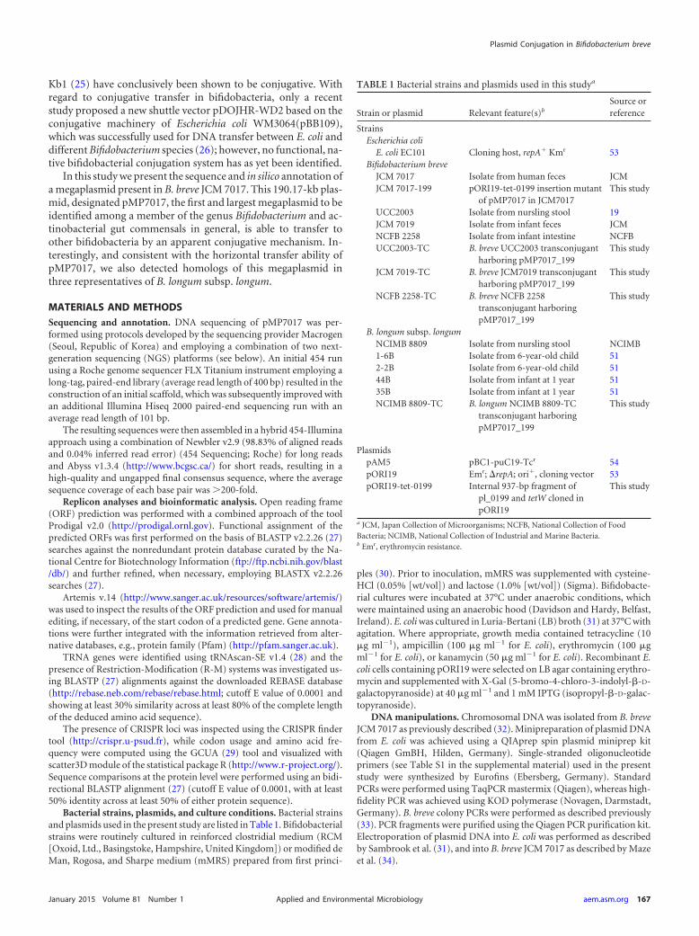
TABLE 1 Bacterial strains and plasmids used in this studya
Strain or plasmid Relevant feature(s)b
Source orreference
StrainsEscherichia coli
E. coli EC101 Cloning host, repA� Kmr 53Bifidobacterium breve
JCM 7017 Isolate from human feces JCMJCM 7017-199 pORI19-tet-0199 insertion mutant
of pMP7017 in JCM7017This study
UCC2003 Isolate from nursling stool 19JCM 7019 Isolate from infant feces JCMNCFB 2258 Isolate from infant intestine NCFBUCC2003-TC B. breve UCC2003 transconjugant
harboring pMP7017_199This study
JCM 7019-TC B. breve JCM7019 transconjugantharboring pMP7017_199
This study
NCFB 2258-TC B. breve NCFB 2258transconjugant harboringpMP7017_199
This study
B. longum subsp. longumNCIMB 8809 Isolate from nursling stool NCIMB1-6B Isolate from 6-year-old child 512-2B Isolate from 6-year-old child 5144B Isolate from infant at 1 year 5135B Isolate from infant at 1 year 51NCIMB 8809-TC B. longum NCIMB 8809-TC
transconjugant harboringpMP7017_199
This study
PlasmidspAM5 pBC1-puC19-Tcr 54pORI19 Emr; �repA; ori�, cloning vector 53pORI19-tet-0199 Internal 937-bp fragment of
pl_0199 and tetW cloned inpORI19
This study
a JCM, Japan Collection of Microorganisms; NCFB, National Collection of FoodBacteria; NCIMB, National Collection of Industrial and Marine Bacteria.b Emr, erythromycin resistance.
Plasmid Conjugation in Bifidobacterium breve
January 2015 Volume 81 Number 1 aem.asm.org 167Applied and Environmental Microbiology

Antibiotic marking of pMP7017 through creation of an insertionmutant of pMP7017_0199. An internal 937-bp fragment of ORFpMP7017_0199 was amplified by PCR using B. breve JCM 7017 chromo-somal DNA as a template and primer pairs IM199F and IM199R (seeTable S1 in the supplemental material). The insertion mutation in B. breveJCM 7017 was generated essentially as described previously (35) to pro-duce B. breve JCM 7017-199. Site-specific recombination in tetracyclin-resistant isolates was confirmed by colony PCR using the primer combi-nation tetWFw and tetWRv to verify tetW gene integration and the primercombination 0199-FW (positioned upstream of the selected internal frag-ment of pMP7017_0199) and pORI19For to confirm integration at thecorrect genetic location.
Conjugal matings. Conjugations were performed by inoculatingovernight cultures of donor and recipient at 1% into mMRS broth sup-plemented with cysteine-HCl (0.05% [wt/vol]) and lactose (1.0% [wt/vol]) and growing at 37°C under anaerobic conditions for 7 h to late logphase. Next, 1 ml of each culture (donor and recipient) was mixed andconcentrated by centrifugation, and the resultant pellet was resuspendedin 200 �l of RCM prior to spread plating on reinforced clostridial agar(RCA) plates. RCA plates were incubated overnight at 37°C under anaer-obic conditions. Subsequently, the conjugation mixture was recoveredfrom the plates and resuspended in 2 ml of phosphate-buffered saline andserially diluted. Donor and recipient cells in each conjugation mixturewere enumerated based on selective carbohydrate utilization by the donoror recipient strain on mMRS agar, whereas transconjugants were isolatedby adopting the appropriate carbohydrate to select for recipient cells incombination with tetracycline (10 �g ml�1) (Table 2).
PFGE plug preparation. Agarose gel plugs of high-molecular-weightDNA for pulsed-field gel electrophoresis (PFGE) were prepared accordingto previously described protocols (36, 37), with minor modifications, asoutlined below. All bifidobacterial strains were grown in mMRS brothsupplemented with 0.05% cysteine and 1% (wt/vol) lactose to early sta-tionary phase. A volume of culture containing �109 bacteria (equivalentto an optical density at 600 nm of 2.0) was centrifuged (5,000 g for 1min), washed once with 1 ml of NT buffer (1 M NaCl, 10 mM Tris-HCl[pH 7.6]), and repelleted (5,000 g for 1 min). The cell pellet was resus-pended in 300 �l of NT buffer, to which an equal volume of melted 2%(wt/vol) low-melting-point (LMP) agarose, prepared in 0.125 M EDTA(pH 7.6) and maintained at 50°C, was added. The cell suspension andLMP agarose were mixed carefully without introducing bubbles. Gel plugswere formed by pipetting 300-�l volumes into plug molds and allowed tosolidify at 4°C for 20 min. Subsequently, two plugs per strain were addedto 2 ml of EC buffer (1 M NaCl, 6 mM Tris-HCl, 100 mM EDTA [pH 7.6])containing 1% (wt/vol) Sarkosyl, 10 mg ml�1 lysozyme, and 40 U ml�1
mutanolysin, followed by incubation at 37°C for 24 h. The lysozyme so-lution was replaced with 5 ml of 0.5 M EDTA (pH 8.0) containing 1%(wt/vol) Sarkosyl and 0.5 mg/ml proteinase K and then incubated at 37°Cfor 24 h. Plugs were then washed with 5 ml of TE buffer (10 mM Tris-HCl,1 mM EDTA [pH 8.0]) containing 1 mM PMSF (phenylmethylsulfonylfluoride; freshly prepared) at 37°C for 1 h to inactivate the proteinase K.This was followed by three 30-min incubations in 5 ml of TE buffer atroom temperature to remove the PMSF. Plugs were stored in 10 mMTris-HCl–100 mM EDTA (pH 8.0) at 4°C.
Treatment with S1 nuclease or restriction endonucleases. Singleslices (2 mm by 2 mm) were soaked in either 200 �l of S1 buffer (50 mM
NaCl, 30 mM sodium acetate [pH 4.5], 5 mM ZnSO4) or an appropriaterestriction buffer at room temperature for 30 min. The S1 buffer wasreplaced with another 200 �l of S1 buffer containing 1 unit of Aspergillusoryzae S1 nuclease and then incubated at 37°C for 45 min. The restrictionbuffer was replaced with fresh restriction buffer containing 15 U of theappropriate restriction enzyme (SpeI and AscI), followed by incubation at37°C for 16 h. Reactions were stopped by suspending the treated plugs in200 �l of 0.5 M EDTA (pH 8.0) and held at room temperature for 10 min.The 0.5 M EDTA was replaced with 200 �l of TE and left at room temper-ature for at least 30 min before loading onto a gel.
PFGE. Plug slices were loaded directly into the wells of a 1% (wt/vol)PFGE agarose gel melted in 0.5 TBE (89 mM Tris-borate, 2 mM EDTA[pH 8.3]) buffer. The wells were sealed with molten agarose in 0.5 TBEbuffer. DNA fragments were resolved using a CHEF-DR III pulsed-fieldsystem (Bio-Rad Laboratories, Hercules, CA) at 6 V/cm for 18 h with 0.5TBE running buffer maintained at 14°C. Linear ramped pulse times wereselected depending on the size of DNA fragments to be resolved; for the S1nuclease-treated samples, a linear ramped pulse time of 3 to 50 s was used;for SpeI- and AscI-digested samples, a linear ramped pulse time of 1 to 15s was used. A molecular size Chef DNA lambda ladder was included ineach gel (number 170-3635; Bio-Rad Laboratories). The gels were stainedin distilled water containing ethidium bromide for 120 min under light-limited conditions and destained in distilled water for 60 min. The gelswere visualized, and images were captured under UV light.
Nucleotide sequence accession numbers. The sequences generated inthis study have been submitted to GenBank database with the accessionnumber (GenBank accession no. KM406416). All of the sequences usedfor our analysis have been retrieved from NCBI database with the follow-ing accession numbers: B. breve UCC2003 (GenBank accession no.NC_020517), B. breve JCM 7017 (GenBank accession no. CP006712), B.breve NCFB 2258 (GenBank accession no. CP006714)], B. breve JCM 7019(GenBank accession no. CP006713), B. longum subsp longum 1-6B(GenBank accession no. AJTF00000000), B. longum subsp. longum2-2B (GenBank accession no. AJTJ00000000), B. longum subsp. longum35B (GenBank accession no. AJTI00000000), and B. longum subsp.longum 44B (GenBank accession no. AJTM00000000).
RESULTS AND DISCUSSIONSequence analysis and general features of pMP7017. During thegenome sequencing and assembly of the Bifidobacterium brevestrain JCM 7017 (3), a sequence scaffold of 190,178 bp was de-tected, corresponding to a DNA element that was not part of thechromosome, thereby providing preliminary evidence for thepresence of a megaplasmid in this strain. Sequence analysis of thispMP7017 replicon showed an overall G�C mol% of 57.43, com-parable with the G�C content of its host (58.65 G�C mol%) andof bifidobacterial genomes in general (9). However, the fact that aslightly different G�C content was observed for about half of itssequence, roughly corresponding to nucleotide coordinates22,000 to 125,000 (Fig. 1), suggests that this megaplasmid is theresult of cointegration of two originally separate modules (havinga G�C mol% of either 60.1 or 55.3) (Fig. 1). The pMP7017 plas-mid harbors 228 ORFs, of which 70% do not have significant hits
TABLE 2 Conjugation between B. breve JCM 7017 and representative B. breve and B. longum subsp. longum strains
DonorCarbohydrateselection for donor Recipient
Carbohydrateselection for recipient
Conjugationefficiencya
B. breve JCM 7017_199 Sorbitol B. breve UCC2003 Ribose 5.1 10�9
B. breve JCM 7017_199 Sorbitol B. breve NCFB 2258 Ribose 1.3 10�8
B. breve JCM 7017_199 Cellobiose B. breve JCM 7019 Melezitose 1.33 10�6
B. breve JCM 7017_199 Sorbitol B. longum NCIMB 8809 Arabinose 4.7 10�8
a Expressed as the average number of transconjugants per recipient from triplicate conjugation experiments.
Bottacini et al.
168 aem.asm.org January 2015 Volume 81 Number 1Applied and Environmental Microbiology

in database searches, thus representing hypothetical genes whichis a common genetic feature of plasmids, while the remainingORFs are associated with predicted functions that are principallyinvolved in DNA replication, DNA methylation, partitioning,DNA transfer, and colonization (Fig. 1).
The first (arbitrary) half of the pMP7017 sequence (corre-sponding to locus tag names pMP7017_0001 through topMP7017_0100) contains ORFs which, among others, represent asortase-dependent pilus cluster (cluster I) encoding a predictedsortase (pMP7017_0021) and three putative surface-anchoredproteins (pMP7017_0022-24). Interestingly, this cluster showssignificant homology (above 50% of similarity in a BLASTP align-ment) with predicted pilus clusters of particular B. longum strains,though it is not significantly similar to those identified in otherbifidobacteria (19, 38, 39). This portion of pMP7017 also containstwo genes encoding a potential type II R/M system (pMP7017_0033-34), followed by genetic functions putatively involved inplasmid segregation and DNA transfer, encompassing a predictedchromosome partitioning genes parA (pMP7017_0053), as well asadjacent genes predicted to encode a mating-pair formation com-plex (Mpf) for DNA translocation represented by two VirD4-B4ATPases (pMP7017_0059 and pMP7017_0062, respectively) anda transmembrane pore-forming VirB6 (pMP7017_0056) (Table3). The VirD4-B4 and VirB6 proteins in the plant pathogen Agro-bacterium tumefaciens are known to form the structural scaffoldand motor of a type IV secretion system (T4SS) that acts as a DNAsecretion channel (40–42). The observed similarity may thus in-dicate a similar role in the conjugal transmission of pMP7017between bifidobacterial species (see below). Finally, two adjacent
CRISPR/Cas loci (corresponding to locus tags pMP7017_0080-85and pMP7017_0086-90) are present in this section of the mega-plasmid, which are known to provide immunity against invadingphages and plasmids in bacteria (43, 44). Further discussion onthese CRISPR/Cas systems will follow below.
The second half of the replicon (locus tags pMP7017_0101through to pMP7017_0146) encodes the presumed replicative ap-paratus consisting of a possible relaxase (pMP7017_0124) and apredicted replication protein (pMP7017_0146) (Pfam PF04796.5),as well as several genes that specify coupling proteins (CP) of atype IV secretion system associated with the Mpf complex direct-ing DNA translocation (40–42): FtsK/SpoIIIE family proteins(pMP7017_0114-0116 and pMP7010_0125-26) and a DivIVA do-main-containing protein (pMP7017_0134) (Table 3). This secondportion of the megaplasmid also specifies biosynthetic functionsfor a putative adhesion locus encoding two predicted surface pro-teins (pMP7017_0199-0200), of which the corresponding genesare located in the vicinity of a sortase-encoding gene (pMP7017_0184), as well as two genes that each encode a putative methyl-transferase (pMP7017_0174/0197); this region also harbors thelargest density of hypothetical genes.
Interestingly, the pMP7017 replicon also specifies threepredicted toxin-antitoxin (T-A) systems (pMP7017_0014-15,pMP7017_101-102 and pMP7017_164-165), as well as a putativeantidote protein (pMP7017_0019). Such T-A systems are knownto be involved in ensuring plasmid maintenance (45).
Finally, downstream of the gene encoding the predicted repli-cation protein (pMP7017_0146), a cluster of 14 tRNA genes (base
FIG 1 pMP7017 general features. Genome atlas representing the circular sequence of the magaplasmid pMP7017 with the relative ORFs organization is shown.Highlighted are the four main functional regions (REG1 to -4) with relative genetic features. A green line also divides pMP7017 into two putative cointegrates.
Plasmid Conjugation in Bifidobacterium breve
January 2015 Volume 81 Number 1 aem.asm.org 169Applied and Environmental Microbiology

positions 121532 to 125960) is present, and their possible role willbe discussed below.
Host adaptation: codon usage and amino acid frequency.The evolutionary process that drives codon usage bias across agenome and indirectly determines the overall tRNA abundance ina given organism is believed to be mediated by natural selection infavor of those codons for which sufficient cognate tRNA is presentin order to optimize protein translation efficiency (46, 47).
In order to explore the possible relationship between the B.breve JCM 7017 chromosome and its coexisting megaplasmidpMP7017 a multivariate analysis on codon usage by plasmid andchromosome-encoded proteins was performed (see Fig. S1a in thesupplemental material). The resulting tridimensional plot dem-onstrated that the pMP7017 replicon possesses a codon bias that iscompatible with that of the chromosome, while it also revealedthat plasmid-borne ORFs with a deviating codon bias mostly spec-ify hypothetical proteins (see Table S2 in the supplemental mate-rial). This result indicates that pMP7017 and its host DNA cur-rently coexist following convergent evolution that has led tooptimal production of chromosome- and plasmid-encoded func-tions.
The presence of 14 clustered tRNA genes on pMP7017 suggestsa correspondence between codon usage and tRNA abundance. Inorder to investigate this notion, amino acid frequencies forpMP7017 and B. breve JCM 7017-encoded ORFs were computedand the resulting heatmap, based on hierarchical clustering,showed that concordance exists between frequently used codonsin the plasmid and the host and that this is consistent with thetRNA genes present in the plasmid (Fig. 2a and b). For this reasonis tempting to argue that pMP7017 followed an evolutionary pro-cess which made its codon usage fitting the one of the host allow-ing efficient protein translation, but also that its maintenance andexpression is supported by a selected set of tRNA genes that pro-vides translational backup to the host.
Protection against DNA invasion: CRISPRs. Plasmid pMP7017is predicted to encode several methyltransferases (pMP7017_0033-34, pMP7017_0174, and pMP7017_0197) which may be part of R-M
systems, while it also encompasses two adjacent CRISPR/Cas systems(pMP7017_0080-85 and pMP7017_0086-90) (Fig. 1). These featuresmay promote the exclusion of (other) invading genetic elements inconcert with the chromosomally encoded protection systems (con-stituted by two R-M systems and a CRISPR/Cas locus) (3), thus pro-viding mutual benefit.
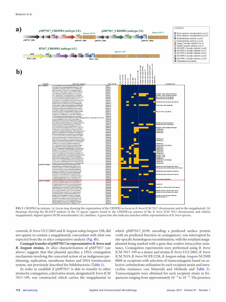
Comparative analysis of the three type CRISPR/Cas systemsencoded by B. breve JCM 7017 revealed a subtype I-C system pres-ent on the JCM 7017 chromosome and two subtype I-E systemsharbored by pMP7017, and named B7017-CRISPR1, pMP7017-CRISPR1, and pMP7017-CRISPR2, respectively. In silico analysisof these three type I CRISPR/Cas systems allowed the identifica-tion of 74 nonredundant spacer sequences (Fig. 3a and b), whichtogether are believed to constitute a genetic memory of previouschallenges by phages (or other invading DNA elements) mountedagainst this B. breve strain or against previous hosts of the conju-gative pMP7017 megaplasmid.
Of these three systems, B7017-CRISPR1 located on the B. breveJCM 7017 chromosome (�18 kb in size with gene coordinates1632794 to 1650595) possesses a total of 49 nonredundant spac-ers, of which the distal end (spacers 27 to 49) returned several hitswith CRISPR-associated regions of other B. breve representatives(in particular B. breve S27, B. breve ACS-071-V-Sch8b, and B.breve NCFB 2258), while the proximal end (spacers 1 to 26) ap-pears to be more variable, suggesting that this CRISPR/Cas systemwas originally acquired by certain representatives of B. breve anddiversified over time as a result of subsequent phage infections andconsequent spacer acquisition events (Fig. 3b).
The presence of at least one cas-1 and cas-2 gene couple in theB7017_CRISPR1 and pMP7017_CRISPR2, being a signature of anactive CRISPR/Cas system (48), suggests that they constitute fullyfunctional CRISPR systems with proto-spacer processing/acqui-sition activity, as a crucial part of the adaptation stage. In contrast,the pMP7017_CRISPR1 locus only appears to encode endonu-clease activity and may therefore be active solely in the CRISPR/Cas-mediated immunity phase (49, 50) or rely on the cas-1 andcas-2 activities of the other two systems. Interestingly, BLASTN-
TABLE 3 Segregation and conjugal transfer genes in pMP7017
Cellular process ORF Annotation Pfam no.
Segregation pMP7017_0053 Chromosome partitioning protein parA PF01656DNA translocation pMP7017_0062 VirB4 PF01656
pMP7017_0056 VirB6 PF04610pMP7017_0059 VirD4 PF10412pMP7017_0136 Protein translocase subunit secE PF00584pMP7017_0138 Protein translocase subunit secG PF03840
Conjugal transfer couplingproteins (CP)
pMP7017_0114 Ftsk/SpoIIIE family protein PF01580pMP7017_0115 CHAP possible extracellular subunitpMP7017_0116 Hypothetical protein with CHAP domain PF05257pMP7017_0125 Ftsk/SpoIIIE family protein PF01580pMP7017_0126 Ftsk/SpoIIIE family protein PF01580pMP7017_0127 Hypothetical protein with flagellar basal body-associated domain
(putative VirB10)PF03739
pMP7017_0134 DivIVA domain containing protein (putative VirC1) PF05103
Replication and DNAprocessing
pMP7017_0124 Putative relaxase PF04796pMP7017_0073 Replicative DNA helicase dnaB PF03796pMP7017_0108 Dna polymerase III alpha subunit dnaE PF00929pMP7017_0146 Predicted repA replicase protein PF04796
Bottacini et al.
170 aem.asm.org January 2015 Volume 81 Number 1Applied and Environmental Microbiology

based searches of the identified spacer sequences against the Na-tional Center for Biotechnology Information (NCBI) nonredun-dant (nr) database returned a number of significant hits againstphage-related sequences (i.e., located in predicted bifidobacterialprophage regions) and other CRISPR-associated spacers, as wellas against sequences that are associated with horizontal genetransfer (HGT) (such as portions of putative integrative conjuga-tive elements or integrated plasmids) of various bifidobacteria,mostly those that are commonly found in the infant gut (e.g., B.breve, B. longum subsp. longum, B. longum subsp. infantis, and B.bifidum) (48) (Fig. 3b). These results support the notion thatCRISPR/Cas systems not only constitute a barrier to phage infec-tion but also to other DNA invasion events that promote HGTmediated by mobilizable elements (44).
Presence of a megaplasmid in B. breve JCM 7017 and strainsof B. longum subsp. longum. The process of gene annotation,followed by an in silico comparative analysis conducted on theORFeome of pMP7017, allowed the identification of 90 to 100 kbof homologous sequences in the draft genomes (deposited as avarying number of contigs) of B. longum subsp. longum strains1-6B, 2-2B, and 44B (51). These homologous sequences (whentranslated) display an overall conservation of BLASTP similaritybetween 60 and 100%, whereas at nucleotide level (BLASTN),high levels of identity (80 to 100%) were only observed for por-tions of these contigs. In order to investigate synteny between thepMP7017 plasmid sequence and the above-mentioned B. longumsubsp. longum contigs, a dot plot alignment was performed, show-ing that a degree of synteny is observed for the pMP7017 region
corresponding to the approximate nucleotide coordinates 20,000to 125,000 (Fig. 4a), which also specifies the predicted conjugativeapparatus (Fig. 1) and which supports the above-mentioned no-tion that this megaplasmid represents a cointegrate of two plas-mids.
The comparative analysis at protein level showed that ca. 70%of the pMP7017 ORFs returned significant similarities to proteinsencoded by B. longum subsp. longum (Fig. 4b). The ORFs thatwere shown to be similar between pMP7017 and the draft genomesequence of one or more of these three B. longum subsp. longumstrains include those that encode sortase-dependent pilus clustersI and II, the ORFs predicted to be involved in plasmid replicationand conjugation, and the characteristic 14 clustered tRNA genesand CRISPR/cas genes, the latter showing 80 to 100% similarity atamino acid level (see Fig. S2 in the supplemental material) andidentical repeats (see Table S3 in the supplemental material),while the spacers appeared different. However, the draft nature ofthese B. longum subsp. longum sequences prevented a full-lengthalignment of these contigs with pMP7017, while it was also notclear whether these sequences were associated with a megaplas-mid. For this reason, in order to not only verify the presence ofpMP7017 in B. breve JCM 7017 but also evaluate the presence of amegaplasmid in the three aforementioned strains of B. longumsubsp. longum, S1 nuclease PFGE was performed (Fig. 4c). Thisindeed confirmed the presence of pMP7017 in B. breve JCM 7017and also revealed the presence of a megaplasmid, of equivalentsize, in B. longum subsp longum 1-6B, B. longum subsp. longum2-2B and B. longum subsp. longum 44B. In contrast, the negative
FIG 2 Amino acid usage comparison and tRNA genes. (a) Heatmap and hierarchical clustering showing the amino acid frequencies computed for the predictedproteome of pMP7017 and the host. For representative reasons, a sample of equal numbers of 227 randomly selected ORFs of B. breve JCM 7017 and pMP7017are displayed in this heatmap. (b) Bidimensional barplot representing the cumulative amino acid frequency in pMP7017 and the host with locus map indicatingthe 14 consecutive tRNA genes encountered in pMP7017 sequence. A table with the total number of tRNA in the plasmid and chromosome is also included.
Plasmid Conjugation in Bifidobacterium breve
January 2015 Volume 81 Number 1 aem.asm.org 171Applied and Environmental Microbiology
controls, B. breve UCC2003 and B. longum subsp longum 35B, didnot appear to contain a megaplasmid, concordant with what wasexpected from the in silico comparative analysis (Fig. 4b).
Conjugal transfer of pMP7017 to representative B. breve andB. longum strains. In silico characterization of pMP7017 (seeabove) suggests that this plasmid specifies a DNA conjugationmechanism involving the concerted action of an indigenous par-titioning, replication, membrane fusion and DNA translocationsystem, not previously described for bifidobacteria (Table 3).
In order to establish if pMP7017 is able to transfer to otherstrains by conjugation, a derivative strain, designated B. breve JCM7017-199, was constructed which carries the megaplasmid in
which pMP7017_0199, encoding a predicted surface protein(with no predicted function in conjugation), was interrupted bysite-specific homologous recombination, with the resultant mega-plasmid being marked with a gene that confers tetracycline resis-tance. Conjugation experiments were performed using B. breveJCM 7017-199 as a donor and strains B. breve UCC2003, B. breveJCM 7019, B. breve NCFB 2258, B. longum subsp. longum NCIMB8809 as recipients with selection of transconjugants based on se-lective carbohydrate utilization by each recipient strain and tetra-cycline resistance (see Materials and Methods and Table 2).Transconjugants were obtained for each recipient strain at fre-quencies ranging from approximately 10�9 to 10�6. The presence
FIG 3 CRISPR/Cas systems. (a) Locus map showing the organization of the CRISPR/cas locus in B. breve JCM 7017 chromosome and in the megaplasmid. (b)Heatmap showing the BLASTP analysis of the 74 spacer regions found in the CRISPR/cas systems of the B. breve JCM 7017 chromosome and relativemegaplasmid, aligned against NCBI nonredundant (nr) database. A green line also indicates matches within representatives of B. breve species.
Bottacini et al.
172 aem.asm.org January 2015 Volume 81 Number 1Applied and Environmental Microbiology

of pMP7017_199 in the transconjugants was confirmed by PCRtargeting genes on the megaplasmid and, for each recipient strain,genes for selective carbohydrate utilization. Furthermore, addi-tional primer pairs based on B. breve JCM 7017 chromosomalgenes were also included and as expected, these gave no PCR prod-uct for the transconjugant strains (see Fig. S3 in the supplementalmaterial). To further verify conjugative transfer of pMP7017_199to B. breve and B. longum PFGE of restricted total DNA was per-formed on representative B. breve UCC2003_pMP7017_199 andB. longum NCIMB 8809_pMP7017_199 transconjugants. Adopt-ing the restriction enzymes AscI for B. breve UCC2003 and SpeIfor B. longum subsp. longum NCIMB 8809, we clearly demonstratethe presence of expected pMP7017_199 restriction fragments inthe presumed transconjugants and also the correct recipient strainrestriction profile thereby confirming the conjugal transfer of
pMP7017_199 from B. breve JCM 7017_199 to strains of B. breveand B. longum (Fig. 5).
Conclusions. The presence of plasmids does not represent atypical feature of B. breve and B. longum genomes and all currentlyavailable bifidobacterial plasmids are considered cryptic as theirencoded functions (besides, in some cases, those involved in rep-lication) are unknown. In this respect the pMP7017 megaplasmid,identified in B. breve JCM 7017, represents an exceptional bifido-bacterial plasmid, which not only represents the largest reportedconjugative plasmid identified thus far in the genus Bifidobacte-rium and actinobacterial gut commensals (at �190 kb) but alsopossesses a number of genomic features indicating the presence ofparasite-benefactor balance reached over time through conver-gent evolution with the host. In addition, it also provides twoCRISPR/Cas systems to counter phage infections and other invad-
FIG 4 Presence of pMP7017 in B. breve and B. longum subsp. longum. (a) Dot plot alignment showing the comparison of pMP7017 megaplasmid with the contigsof B. longum subsp. longum of which ORFs displayed significant match in BLASTP alignment (e.g., B. longum subsp. longum 1-6B, B. longum subsp. longum 2-2B,and B. longum subsp. longum 44B). Delimited by green bars are the regions displaying the most of synteny. (b) BLAST-based genome atlas showing thecomparative analysis conducted on pMP7017 against the draft sequence of B. longum subsp longum 1-6B, B. longum subsp. longum 2-2B, and B. longum subsp.longum 44B. From the outer to the inner circle and shaded in gradient from dark red to orange: B. breve JCM 7017 pMP7017, B. longum subsp. longum 44B, B.longum subsp. longum 2-2B, and B. longum subsp. longum 1-6B. (c) PFGE experiment showing the presence of a megaplasmid of a size comparable to that ofpMP7017 (about 200 kb) also in B. longum subsp. longum 1-6B, B. longum subsp. longum 2-2B, and B. longum subsp. longum 44B, but absent in B. longum subsp.longum 35B and B. breve UCC2003 as a negative control.
Plasmid Conjugation in Bifidobacterium breve
January 2015 Volume 81 Number 1 aem.asm.org 173Applied and Environmental Microbiology

ing DNA elements. Plasmid pMP7017 also possesses a number ofgenes whose products are similar to proteins that are known to bepart of a conjugative machinery present in both Gram-positiveand Gram-negative bacteria (40–42).
The capacity of this element of being horizontally transferablebetween different species was first suspected due to the findingthat elements similar to pMP7017 of B. breve JCM 7017 were alsopresent in several members of the B. longum subsp. longum taxon.
Plasmid-mediated conjugation in other bacteria is known toinvolve the transfer of a protein-single-strand DNA complex(called the relaxosome) pumped through an aqueous pore of atype IV secretion system from the donor to the recipient cell (40–42). In bifidobacteria, a gene specifying a protein similar to thetype IV secretion protein has been observed in the chromosome ofB. bifidum MIMBb75 (52). The structural scaffold of such DNA
transfer system in pMP7017 is believed to be represented by aVirB6 structural scaffold, which is coupled to aVirD4-B4 ATPases,to provide the energy for DNA translocation. The functionality ofthe predicted conjugative machinery in pMP7017 megaplasmidwas conclusively demonstrated by conjugation experimentsshowing that this element is capable to spread and persist in dif-ferent bifidobacterial strains and species.
One of the conditions for initiating a conjugative transfer isthe establishment of cell-to-cell contact, which in Gram-nega-tive bacteria is accomplished by proteinaceous filaments calledsex pili, while in Gram-positive bacteria such structures havenot yet been clearly identified. The fact that pMP7017 ispredicted to encode two different sortase-dependent pili(pMP7017_0021 to pMP7017_0024 and pMP7017_0184-pMP7017_0199-pMP7017_0200) which have been shown to
FIG 5 Conjugal transfer of pMP7017. PFGE experiments for B. breve UCC2003 or B. longum NCIMB 8809 transconjugants conducted with AscI (a) and SpeI(b) enzymes, respectively. The presence of megaplasmid in the parent strain, B. breve JCM 7017, and the donor strain, B. breve JCM 7017_199, is depicted in eachpanel. The presence of pMP7017 in B. breve UCC2003 trasconjugants is evidenced by a 111-kb fragment in total DNA digests with AscI, while the megaplasmidpresence in B. longum NCIMB 8809 transconjugants is verified by the presence of a 196-kb fragment.
Bottacini et al.
174 aem.asm.org January 2015 Volume 81 Number 1Applied and Environmental Microbiology

promote bifidobacterial coaggregation (39) suggests a role inbacterial conjugation.
B. breve and B. longum subsp. longum both represent commoninhabitants of the infant gut (1), which supports the hypothesisthat the cooccurrence of apparent homologs of this conjugativeelement in both of these species may be a result of previous DNAexchanges, followed by diversification and stable maintenance ofthis element over generations. The fact that pMP7017 representsthe first native plasmid capable of conjugal transmission betweenbifidobacterial species is highly relevant as it may confer this rep-licon a relevant biotechnological potential for improving the fu-ture genetic manipulation of members of this genus.
ACKNOWLEDGMENTS
This study was supported by a Science Foundation Ireland grant (SFI/12/RC/2273) and an HRB postdoctoral fellowship (grant PDTM/2011/9)awarded to M.O.M.
We declare that we have no competing interests.
REFERENCES1. Turroni F, Peano C, Pass DA, Foroni E, Severgnini M, Claesson MJ,
Kerr C, Hourihane J, Murray D, Fuligni F, Gueimonde M, Margolles A,De Bellis G, O’Toole PW, van Sinderen D, Marchesi JR, Ventura M.2012. Diversity of bifidobacteria within the infant gut microbiota. PLoSOne 7:e36957. http://dx.doi.org/10.1371/journal.pone.0036957.
2. Takahata M, Toh H, Nakano A, Takagi M, Murakami M, Ishii Y,Takizawa T, Tanabe S, Morita H. 2014. Complete sequence analysis oftwo cryptic plasmids from Bifidobacterium kashiwanohense JCM 15439(type strain) isolated from healthy infant feces. Anim Sci J 85:158 –163.http://dx.doi.org/10.1111/asj.12095.
3. Bottacini F, O’Connell Motherway M, Kuczynski J, O’Connell KJ,Serafini F, Duranti S, Milani C, Turroni F, Lugli GA, Zomer A, ZhurinaD, Riedel C, Ventura M, van Sinderen D. 2014. Comparative genomicsof the Bifidobacterium breve taxon. BMC Genomics 15:170. http://dx.doi.org/10.1186/1471-2164-15-170.
4. Ventura M, Turroni F, Lugli GA, van Sinderen D. 2014. Bifidobacteriaand humans: our special friends, from ecological to genomics perspec-tives. J Sci Food Agric 94:163–168. http://dx.doi.org/10.1002/jsfa.6356.
5. Stanton C, Ross RP, Fitzgerald GF, Van Sinderen D. 2005. Fermentedfunctional foods based on probiotics and their biogenic metabolites. CurrOpin Biotechnol 16:198 –203. http://dx.doi.org/10.1016/j.copbio.2005.02.008.
6. Turroni F, Bottacini F, Foroni E, Mulder I, Kim JH, Zomer A, SanchezB, Bidossi A, Ferrarini A, Giubellini V, Delledonne M, Henrissat B,Coutinho P, Oggioni M, Fitzgerald GF, Mills D, Margolles A, Kelly D,van Sinderen D, Ventura M. 2010. Genome analysis of Bifidobacteriumbifidum PRL2010 reveals metabolic pathways for host-derived glycan for-aging. Proc Natl Acad Sci U S A 107:19514 –19519. http://dx.doi.org/10.1073/pnas.1011100107.
7. Fanning S, Hall LJ, van Sinderen D. 2012. Bifidobacterium breveUCC2003 surface exopolysaccharide production is a beneficial trait me-diating commensal-host interaction through immune modulation andpathogen protection. Gut Microbes 3:420 – 425. http://dx.doi.org/10.4161/gmic.20630.
8. Lee JH, O’Sullivan DJ. 2010. Genomic insights into bifidobacteria. Mi-crobiol Mol Biol Rev 74:378 – 416. http://dx.doi.org/10.1128/MMBR.00004-10.
9. Ventura M, Canchaya C, Tauch A, Chandra G, Fitzgerald GF, ChaterKF, van Sinderen D. 2007. Genomics of Actinobacteria: tracing the evo-lutionary history of an ancient phylum. Microbiol Mol Biol Rev 71:495–548. http://dx.doi.org/10.1128/MMBR.00005-07.
10. Cronin M, Knobel M, O’Connell-Motherway M, Fitzgerald GF, vanSinderen D. 2007. Molecular dissection of a bifidobacterial replicon.Appl Environ Microbiol 73:7858 –7866. http://dx.doi.org/10.1128/AEM.01630-07.
11. Mayo B, van Sinderen D. 2010. Bifidobacteria: genomics and molecularaspects. Caister Academic Press, Norfolk, United Kingdom.
12. Schwartz E. 2009. Microbial megaplasmids. Springer, Berlin, Germany.13. Simpson PJ, Stanton C, Fitzgerald GF, Ross RP. 2003. Genomic diversity
and relatedness of bifidobacteria isolated from a porcine cecum. J Bacte-riol 185:2571–2581. http://dx.doi.org/10.1128/JB.185.8.2571-2581.2003.
14. Piper KR, Beck von Bodman S, Farrand SK. 1993. Conjugation factor ofAgrobacterium tumefaciens regulates Ti plasmid transfer by autoinduc-tion. Nature 362:448 – 450. http://dx.doi.org/10.1038/362448a0.
15. Lang S, Gruber K, Mihajlovic S, Arnold R, Gruber CJ, Steinlechner S,Jehl MA, Rattei T, Frohlich KU, Zechner EL. 2010. Molecular recogni-tion determinants for type IV secretion of diverse families of conjugativerelaxases. Mol Microbiol 78:1539 –1555. http://dx.doi.org/10.1111/j.1365-2958.2010.07423.x.
16. Wetzel ME, Kim KS, Miller M, Olsen GJ, Farrand SK. 2014. Quorum-dependent mannopine-inducible conjugative transfer of an Agrobacte-rium opine-catabolic plasmid. J Bacteriol 196:1031–1044. http://dx.doi.org/10.1128/JB.01365-13.
17. Grohmann E, Muth G, Espinosa M. 2003. Conjugative plasmid transferin gram-positive bacteria. Microbiol Mol Biol Rev 67:277–301. http://dx.doi.org/10.1128/MMBR.67.2.277-301.2003.
18. Ventura M, Turroni F, Zomer A, Foroni E, Giubellini V, Bottacini F,Canchaya C, Claesson MJ, He F, Mantzourani M, Mulas L, Ferrarini A,Gao B, Delledonne M, Henrissat B, Coutinho P, Oggioni M, Gupta RS,Zhang Z, Beighton D, Fitzgerald GF, O’Toole PW, van Sinderen D.2009. The Bifidobacterium dentium Bd1 genome sequence reflects its ge-netic adaptation to the human oral cavity. PLoS Genet 5:e1000785. http://dx.doi.org/10.1371/journal.pgen.1000785.
19. O’Connell Motherway M, Zomer A, Leahy SC, Reunanen J, Bottacini F,Claesson MJ, O’Brien F, Flynn K, Casey PG, Munoz JA, Kearney B,Houston AM, O’Mahony C, Higgins DG, Shanahan F, Palva A, de VosWM, Fitzgerald GF, Ventura M, O’Toole PW, van Sinderen D. 2011.Functional genome analysis of Bifidobacterium breve UCC2003 revealstype IVb tight adherence (Tad) pili as an essential and conserved host-colonization factor. Proc Natl Acad Sci U S A 108:11217–11222. http://dx.doi.org/10.1073/pnas.1105380108.
20. Bottacini F, Milani C, Turroni F, Sanchez B, Foroni E, Duranti S,Serafini F, Viappiani A, Strati F, Ferrarini A, Delledonne M, HenrissatB, Coutinho P, Fitzgerald GF, Margolles A, van Sinderen D, Ventura M.2012. Bifidobacterium asteroides PRL2011 genome analysis reveals cluesfor colonization of the insect gut. PLoS One 7:e44229. http://dx.doi.org/10.1371/journal.pone.0044229.
21. Ventura M, Lee JH, Canchaya C, Zink R, Leahy S, Moreno-Munoz JA,O’Connell-Motherway M, Higgins D, Fitzgerald GF, O’Sullivan DJ, vanSinderen D. 2005. Prophage-like elements in bifidobacteria: insights fromgenomics, transcription, integration, distribution, and phylogenetic anal-ysis. Appl Environ Microbiol 71:8692– 8705. http://dx.doi.org/10.1128/AEM.71.12.8692-8705.2005.
22. Dib JR, Wagenknecht M, Hill RT, Farias ME, Meinhardt F. 2010. Novellinear megaplasmid from Brevibacterium sp. isolated from extreme envi-ronment. J Basic Microbiol 50:280 –284. http://dx.doi.org/10.1002/jobm.200900332.
23. Konig C, Eulberg D, Groning J, Lakner S, Seibert V, Kaschabek SR,Schlomann M. 2004. A linear megaplasmid, p1CP, carrying the genes forchlorocatechol catabolism of Rhodococcus opacus 1CP. Microbiology 150:3075–3087. http://dx.doi.org/10.1099/mic.0.27217-0.
24. Yang JC, Lessard PA, Sengupta N, Windsor SD, O’Brien XM, BramucciM, Tomb JF, Nagarajan V, Sinskey AJ. 2007. TraA is required formegaplasmid conjugation in Rhodococcus erythropolis AN12. Plasmid 57:55–70. http://dx.doi.org/10.1016/j.plasmid.2006.08.002.
25. Broker D, Arenskotter M, Steinbuchel A. 2008. Transfer of megaplasmidpKB1 from the rubber-degrading bacterium Gordonia westfalica strainKb1 to related bacteria and its modification. Appl Microbiol Biotechnol77:1317–1327. http://dx.doi.org/10.1007/s00253-007-1262-8.
26. Dominguez W, O’Sullivan DJ. 2013. Developing an efficient and repro-ducible conjugation-based gene transfer system for bifidobacteria. Micro-biology 159:328 –338. http://dx.doi.org/10.1099/mic.0.061408-0.
27. Altschul SF, Gish W, Miller W, Myers EW, Lipman DJ. 1990. Basic localalignment search tool. J Mol Biol 215:403– 410. http://dx.doi.org/10.1016/S0022-2836(05)80360-2, http://dx.doi.org/10.1006/jmbi.1990.9999.
28. Lowe TM, Eddy SR. 1997. tRNAscan-SE: a program for improved detec-tion of transfer RNA genes in genomic sequence. Nucleic Acids Res 25:955–964. http://dx.doi.org/10.1093/nar/25.5.0955.
29. McInerney JO. 1998. GCUA: general codon usage analysis. Bioinformat-ics 14:372–373. http://dx.doi.org/10.1093/bioinformatics/14.4.372.
30. De Man JC, Rogosa M, Sharpe ME. 1960. A medium for the cultivation
Plasmid Conjugation in Bifidobacterium breve
January 2015 Volume 81 Number 1 aem.asm.org 175Applied and Environmental Microbiology

of lactobacilli. J Appl Bacteriol 23:130 –135. http://dx.doi.org/10.1111/j.1365-2672.1960.tb00188.x.
31. Sambrook J, Russell DW, Fritsch EF, Maniatis T. 2001. Molecularcloning: a laboratory manual, 3rd ed. Cold Spring Harbor LaboratoryPress, Cold Spring Harbor, NY.
32. O’Riordan K, Fitzgerald GF. 1998. Evaluation of bifidobacteria for theproduction of antimicrobial compounds and assessment of performancein cottage cheese at refrigeration temperature. J Appl Microbiol 85:103–114. http://dx.doi.org/10.1046/j.1365-2672.1998.00474.x.
33. Motherway MO, Fitzgerald GF, Neirynck S, Ryan S, Steidler L, vanSinderen D. 2008. Characterization of ApuB, an extracellular type II amy-lopullulanase from Bifidobacterium breve UCC2003. Appl Environ Micro-biol 74:6271– 6279. http://dx.doi.org/10.1128/AEM.01169-08.
34. Maze A, O’Connell-Motherway M, Fitzgerald GF, Deutscher J, vanSinderen D. 2007. Identification and characterization of a fructose phos-photransferase system in Bifidobacterium breve UCC2003. Appl EnvironMicrobiol 73:545–553. http://dx.doi.org/10.1128/AEM.01496-06.
35. Motherway MO, O’Driscoll J, Fitzgerald GF, van Sinderen D. 2009.Overcoming the restriction barrier to plasmid transformation and tar-geted mutagenesis in Bifidobacterium breve UCC2003. Microb Biotechnol2:321–332. http://dx.doi.org/10.1111/j.1751-7915.2008.00071.x.
36. Li Y, Canchaya C, Fang F, Raftis E, Ryan KA, van Pijkeren JP, vanSinderen D, O’Toole PW. 2007. Distribution of megaplasmids in Lacto-bacillus salivarius and other lactobacilli. J Bacteriol 189:6128 – 6139. http://dx.doi.org/10.1128/JB.00447-07.
37. Wall R, Fitzgerald G, Hussey S, Ryan T, Murphy B, Ross P, Stanton C.2007. Genomic diversity of cultivable Lactobacillus populations residing inthe neonatal and adult gastrointestinal tract. FEMS Microbiol Ecol 59:127–137. http://dx.doi.org/10.1111/j.1574-6941.2006.00202.x.
38. Foroni E, Serafini F, Amidani D, Turroni F, He F, Bottacini F,O’Connell Motherway M, Viappiani A, Zhang Z, Rivetti C, van Sin-deren D, Ventura M. 2011. Genetic analysis and morphological identifi-cation of pilus-like structures in members of the genus Bifidobacterium.Microb Cell Fact 10(Suppl 1):S16. http://dx.doi.org/10.1186/1475-2859-10-S1-S16.
39. Turroni F, Serafini F, Foroni E, Duranti S, Motherway MO, TavernitiV, Mangifesta M, Milani C, Viappiani A, Roversi T, Sanchez B, SantoniA, Gioiosa L, Ferrarini A, Delledonne M, Margolles A, Piazza L, PalanzaP, Bolchi A, Guglielmetti S, van Sinderen D, Ventura M. 2013. Role ofsortase-dependent pili of Bifidobacterium bifidum PRL2010 in modulatingbacterium-host interactions. Proc Natl Acad Sci U S A 110:11151–11156.http://dx.doi.org/10.1073/pnas.1303897110.
40. Jakubowski SJ, Krishnamoorthy V, Cascales E, Christie PJ. 2004. Agro-bacterium tumefaciens VirB6 domains direct the ordered export of a DNAsubstrate through a type IV secretion system. J Mol Biol 341:961–977.http://dx.doi.org/10.1016/j.jmb.2004.06.052.
41. Christie PJ, Cascales E. 2005. Structural and dynamic properties of bac-terial type IV secretion systems. Mol Membr Biol 22:51– 61. http://dx.doi.org/10.1080/09687860500063316.
42. Kahrstrom CT. 2014. Structural biology: solving the T4SS structural mys-tery. Nat Rev Microbiol 12:312. http://dx.doi.org/10.1038/nrmicro3254.
43. Barrangou R, Fremaux C, Deveau H, Richards M, Boyaval P, MoineauS, Romero DA, Horvath P. 2007. CRISPR provides acquired resistanceagainst viruses in prokaryotes. Science 315:1709 –1712. http://dx.doi.org/10.1126/science.1138140.
44. van der Oost J, Jore MM, Westra ER, Lundgren M, Brouns SJ. 2009.CRISPR-based adaptive and heritable immunity in prokaryotes. TrendsBiochem Sci 34:401– 407. http://dx.doi.org/10.1016/j.tibs.2009.05.002.
45. Goeders N, Van Melderen L. 2014. Toxin-antitoxin systems as mul-tilevel interaction systems. Toxins 6:304 –324. http://dx.doi.org/10.3390/toxins6010304.
46. Ikemura T. 1985. Codon usage and tRNA content in unicellular andmulticellular organisms. Mol Biol Evol 2:13–34.
47. Akashi H. 1994. Synonymous codon usage in Drosophila melanogaster:natural selection and translational accuracy. Genetics 136:927–935.
48. Makarova KS, Haft DH, Barrangou R, Brouns SJJ, Charpentier E,Horvath P, Moineau S, Mojica FJM, Wolf YI, Yakunin AF, van derOost J, Koonin EV. 2011. Evolution and classification of the CRISPR-Cas systems. Nat Rev Microbiol 9:467– 477. http://dx.doi.org/10.1038/nrmicro2577.
49. Dong HJ, Nilsson L, Kurland CG. 1996. Co-variation of tRNA abun-dance and codon usage in Escherichia coli at different growth rates. J MolBiol 260:649 – 663. http://dx.doi.org/10.1006/jmbi.1996.0428.
50. Novoa EM, de Pouplana LR. 2012. Speeding with control: codon usage,tRNAs, and ribosomes. Trends Genet 28:574 –581. http://dx.doi.org/10.1016/j.tig.2012.07.006.
51. Shkoporov AN, Efimov BA, Khokhlova EV, Chaplin AV, Kafarskaya LI,Durkin AS, McCorrison J, Torralba M, Gillis M, Sutton G, Weibel DB,Nelson KE, Smeianov VV. 2013. Draft genome sequences of two pairs ofhuman intestinal Bifidobacterium longum subsp. longum strains, 44B and1-6B and 35B and 2-2B, consecutively isolated from two children after a5-year time period. Genome Announce 1:e00234-13. http://dx.doi.org/10.1128/genomeA.00234-13.
52. Guglielmetti S, Balzaretti S, Taverniti V, Miriani M, Milani C, ScarafoniA, Corona S, Ciranna A, Arioli S, Santala V, Iametti S, Bonomi F,Ventura M, Mora D, Karp M. 2014. TgaA, a VirB1-like componentbelonging to a putative type IV secretion system of Bifidobacterium bifi-dum MIMBb75. Appl Environ Microbiol 80:5161–5169. http://dx.doi.org/10.1128/AEM.01413-14.
53. Law J, Buist G, Haandrikman A, Kok J, Venema G, Leenhouts K. 1995.A system to generate chromosomal mutations in Lactococcus lactis whichallows fast analysis of targeted genes. J Bacteriol 177:7011–7018.
54. Alvarez-Martín P, O’Connell-Motherway M, Van Sinderen D, Mayo B.2007. Functional analysis of the pBC1 replicon from Bifidobacteriumcatenulatum L48. Appl Microbiol Biotechnol 76:1395–1402. http://dx.doi.org/10.1007/s00253-007-1115-5.
Bottacini et al.
176 aem.asm.org January 2015 Volume 81 Number 1Applied and Environmental Microbiology