Differential roles of p80- and p130-angiomotin in the switch between migration and stabilization of...
9
Differential roles of p80- and p130-angiomotin in the switch between migration and stabilization of endothelial cells Mira Ernkvist, Olivier Birot 1 , Indranil Sinha, Niina Veitonmaki, Staffan Nyström, Karin Aase, Lars Holmgren ⁎ Cancer Centrum Karolinska, Department of Oncology and Pathology, Karolinska Institutet, CCK R8:03, 171 76 Stockholm, Sweden Received 18 July 2007; received in revised form 21 November 2007; accepted 26 November 2007 Available online 8 December 2007 Abstract We have previously shown that angiomotin (Amot) plays an important role in growth factor-induced migration of endothelial cells in vitro. Genetic knock-down of Amot in zebrafish also results in inhibition of migration of intersegmental vessels in vivo. Amot is expressed as two different isoforms, p80-Amot and p130-Amot. Here we have analyzed the expression of the two Amot isoforms during retinal angiogenesis in vivo and demonstrate that p80-Amot is expressed during the migratory phase. In contrast, p130-Amot is expressed during the period of blood vessel stabilization and maturation. We also show that the N-terminal domain of p130-Amot serves as a targeting domain responsible for localization of p130-Amot to actin and tight junctions. We further show that the relative expression levels of p80-Amot and p130-Amot regulate a switch between a migratory and a non-migratory cell phenotype where the migratory function of p80-Amot is dominant over the stabilization and maturation function of p130-Amot. Our data indicates that homo-oligomerization of p80-Amot and hetero-oligomerization of both isoforms are critical for this regulation. © 2007 Elsevier B.V. All rights reserved. Keywords: Actin; Angiogenesis; Calcium switch; Retina; Tight junction 1. Introduction Angiogenesis is a process involving the formation of new blood vessels by sprouting from pre-existing vessels and occurs during embryogenesis, organ growth and other physiological settings. Dysregulated blood vessel formation contributes to the pathogenesis of many diseases including diabetic retinopathy, atherosclerosis, endometriosis and tumor progression [1,2]. We previously identified angiomotin (Amot) by its binding to the angiogenesis inhibitor angiostatin [3]. Amot is primarily expressed by endothelial cells (ECs) but is also expressed by epithelial structures such as the branchial arches and Rathke's pouch in the developing mouse embryo [4]. Amot plays a critical role in physiological angiogenesis during zebrafish and mouse embryogenesis. For instance, knock-down of Amot in zebrafish severely impairs the migration of intersegmental vessels during embryogenesis. Furthermore, Amot knock-out mice exhibit vascular defects in the intersomitic vessels, dilated vessels in the brain and die between E11 and 11.5 [4]. In addition, targeting Amot with a DNA vaccination strategy inhibits pathological angiogenesis and tumor growth in BALB/ C mice [5]. Amot is characterized by a conserved coiled-coil domain and a C-terminal PDZ-binding motif [6], and is expressed as two different isoforms, i.e. p80-Amot and p130-Amot. The longer isoform, i.e. p130-Amot, contains an extended N-terminal domain. These two isoforms possess distinct functions in vitro where the shorter p80-Amot isoform enhances cell migration and the longer p130-Amot isoform associates with actin and affects cell shape [3,7,8]. Recent data demonstrated that Amot binds to Rich1, a Rho GTPase activating protein (GAP), and to a protein complex containing the PDZ-domain proteins Pals1, Patj, and Par-3 at tight junctions [9]. In this study we demonstrate that the p80 and p130 Amot isoforms are differentially expressed during specific stages of mouse retina vascularization. We also provide evidence that Available online at www.sciencedirect.com Biochimica et Biophysica Acta 1783 (2008) 429 – 437 www.elsevier.com/locate/bbamcr ⁎ Corresponding author. Tel.: +46 8 51779317; fax: +46 8 339031. E-mail address: [email protected] (L. Holmgren). 1 Present address: University of Montreal, Department of Kinesiology, CEPSUM, C.P.6128 Succursalle Centre-Ville, Montreal H3C 3J7, QC, Canada. 0167-4889/$ - see front matter © 2007 Elsevier B.V. All rights reserved. doi:10.1016/j.bbamcr.2007.11.018
-
Upload
icfaiuniversity -
Category
Documents
-
view
5 -
download
0
Transcript of Differential roles of p80- and p130-angiomotin in the switch between migration and stabilization of...
Available online at www.sciencedirect.com
a 1783 (2008) 429–437www.elsevier.com/locate/bbamcr
Biochimica et Biophysica Act
Differential roles of p80- and p130-angiomotin in the switch betweenmigration and stabilization of endothelial cells
Mira Ernkvist, Olivier Birot 1, Indranil Sinha, Niina Veitonmaki,Staffan Nyström, Karin Aase, Lars Holmgren ⁎
Cancer Centrum Karolinska, Department of Oncology and Pathology, Karolinska Institutet, CCK R8:03, 171 76 Stockholm, Sweden
Received 18 July 2007; received in revised form 21 November 2007; accepted 26 November 2007Available online 8 December 2007
Abstract
We have previously shown that angiomotin (Amot) plays an important role in growth factor-induced migration of endothelial cells in vitro.Genetic knock-down of Amot in zebrafish also results in inhibition of migration of intersegmental vessels in vivo. Amot is expressed as two differentisoforms, p80-Amot and p130-Amot. Here we have analyzed the expression of the two Amot isoforms during retinal angiogenesis in vivo anddemonstrate that p80-Amot is expressed during the migratory phase. In contrast, p130-Amot is expressed during the period of blood vesselstabilization and maturation. We also show that the N-terminal domain of p130-Amot serves as a targeting domain responsible for localization ofp130-Amot to actin and tight junctions. We further show that the relative expression levels of p80-Amot and p130-Amot regulate a switch between amigratory and a non-migratory cell phenotype where the migratory function of p80-Amot is dominant over the stabilization and maturation functionof p130-Amot. Our data indicates that homo-oligomerization of p80-Amot and hetero-oligomerization of both isoforms are critical for this regulation.© 2007 Elsevier B.V. All rights reserved.
Keywords: Actin; Angiogenesis; Calcium switch; Retina; Tight junction
1. Introduction
Angiogenesis is a process involving the formation of newblood vessels by sprouting from pre-existing vessels and occursduring embryogenesis, organ growth and other physiologicalsettings. Dysregulated blood vessel formation contributes to thepathogenesis of many diseases including diabetic retinopathy,atherosclerosis, endometriosis and tumor progression [1,2].
We previously identified angiomotin (Amot) by its bindingto the angiogenesis inhibitor angiostatin [3]. Amot is primarilyexpressed by endothelial cells (ECs) but is also expressed byepithelial structures such as the branchial arches and Rathke'spouch in the developing mouse embryo [4]. Amot plays acritical role in physiological angiogenesis during zebrafishand mouse embryogenesis. For instance, knock-down of Amotin zebrafish severely impairs the migration of intersegmental
⁎ Corresponding author. Tel.: +46 8 51779317; fax: +46 8 339031.E-mail address: [email protected] (L. Holmgren).
1 Present address: University of Montreal, Department of Kinesiology,CEPSUM, C.P.6128 Succursalle Centre-Ville, Montreal H3C 3J7, QC, Canada.
0167-4889/$ - see front matter © 2007 Elsevier B.V. All rights reserved.doi:10.1016/j.bbamcr.2007.11.018
vessels during embryogenesis. Furthermore, Amot knock-outmice exhibit vascular defects in the intersomitic vessels, dilatedvessels in the brain and die between E11 and 11.5 [4]. Inaddition, targeting Amot with a DNA vaccination strategyinhibits pathological angiogenesis and tumor growth in BALB/C mice [5].
Amot is characterized by a conserved coiled-coil domain anda C-terminal PDZ-binding motif [6], and is expressed as twodifferent isoforms, i.e. p80-Amot and p130-Amot. The longerisoform, i.e. p130-Amot, contains an extended N-terminaldomain. These two isoforms possess distinct functions in vitrowhere the shorter p80-Amot isoform enhances cell migrationand the longer p130-Amot isoform associates with actin andaffects cell shape [3,7,8].
Recent data demonstrated that Amot binds to Rich1, a RhoGTPase activating protein (GAP), and to a protein complexcontaining the PDZ-domain proteins Pals1, Patj, and Par-3 attight junctions [9].
In this study we demonstrate that the p80 and p130 Amotisoforms are differentially expressed during specific stages ofmouse retina vascularization. We also provide evidence that
430 M. Ernkvist et al. / Biochimica et Biophysica Acta 1783 (2008) 429–437
p80-Amot can undergo homo-oligomerization and hetero-oligomerization with p130-Amot, and speculate that thismechanism regulates the switch between the migratory cellphenotype induced by p80-Amot and the non-migratory cellphenotype induced by p130-Amot.
2. Materials and methods
2.1. Cell lines and transfections
Chinese hamster ovary (CHO) cells, mouse aortic epithelial (MAE) cells andMadin–Darby canine kidney (MDCK) cells were grown in Dulbecco'sModified Eagle's Medium supplemented with 10% fetal bovine serum, glu-tamine and antibiotics.
Cells were transfected with Lipofectamine 2000 according to themanufacturer's protocol (Life Technologies).
2.2. Immunoprecipitation
Immunoprecipitation was performed as described previously [10]. Briefly,cells were transfected as described above and 48 h later lyzed with lysis bufferand centrifuged. The supernatant was pre-cleared and incubated with theappropriate antibody for 6 h followed by incubation with sepharose A. Pelletswere washed with lysis buffer, re-suspended in 1x sample buffer and analyzedby Western blot.
2.3. Protein extraction and Western blot
Western blots were performed as described previously [8]. For extraction oftotal mouse retinal proteins, eyes were fixed for 3–5 min at RT in 4%paraformaldehyde and rinsed in PBS. Retinas were dissected as previouslydescribed [10] and homogenized in lysis buffer (1% Triton X-100, 1%deoxycholic acid, 50 mM Tris (pH 8.0), 100 mM NaCl, 5 mM EDTA). Bandspresent on Western blots were quantified using Image J 1.33 software (NationalInstitutes of Health, USA).
2.4. Immunofluorescence
Cultured cells were plated in chamber slides and stained as describedpreviously [8]. The stained cells were viewed using a Zeiss Axioplan 2microscope, and images collected using an AxioCam HRm Camera and theAxiovision 4.5 software. The confocal images were collected using a Leica SP5DMI6000 microscope and the Leica microsystem LAS AF TCS SO5 software.The images were then analyzed using the Image J 1.33 software.
For Amot staining in the mouse retina, eyes were removed, fixed in 4%paraformaldehyde at 4 °C for 2–3 h, washed in PBS and the retinas dissected asdescribed above. Retinas were incubated for 2 h in PBB buffer (PBS containing1% BSA, 0.5% Triton X-100 and 5% goat serum) and incubated at 4 °Covernight with C-terminal Amot antibodies. After washing, retinas wereincubated for 2 h with FITC-conjugated swine anti-rabbit antibody (Dako#F0205, Sweden) in PBS containing 0.5% BSA, 0.25% Triton X-100 and 5%goat serum. The retinal vasculature was stained in whole mount retina accordingto the protocol published by Gerhardt et al. [11].
2.5. Antibodies
Amot antibodies raised against the C-terminal of both p80-Amot and p130-Amot were generated as described [8]. Antibodies and reagents used forimmunofluorescent staining were ZO-1 (Zymed), E-cadherin and CD31 (BDTransduction Laboratories), phalloidin (Molecular Probes), and isolectin (SigmaChemical Co.). Secondary antibodies were purchased from Dako, Vector andMolecular Probes. Antibodies used for Western blots were myc-tag (clone 9E10from Santa Cruz), Flag (Sigma Chemical Co.), GST (Santa Cruz), and actin(Sigma Chemical Co.). HRP-conjugated secondary antibodies were purchasedfrom Amersham.
2.6. Migration assay
Migration assays were performed in a modified Boyden chamber using a 48-well chemotaxis chamber (Neuroprobe Inc.) as described previously [3,8].
2.7. Calcium switch experiments
MDCK cells were transfected with different constructs 24 h before theexperiments. The cells were grown in calcium-free medium with 7 mM EGTAfor 1.5 h to dissociate cell–cell contacts. At different time points after re-additionof calcium rich medium (i.e. 0, 1, 3, 6 and 16 h), cells were prepared forimmunostaining.
2.8. Statistical analysis
The Student t-test was used to determine statistical significance betweencontrol and treatment groups. All analyses were performed using Statview 5.0.1where ⁎⁎⁎, Pb0.001, ⁎⁎, Pb0.01 and ⁎, Pb0.05.
3. Results and discussion
3.1. The Amot isoforms are differently expressed duringangiogenesis
Our previous results indicate that the two Amot isoformsplay distinct roles during angiogenesis. For instance, p80-Amotaffects cell migration [3] whereas p130-Amot affects cell shape[8]. To investigate their respective roles in vivo, we examinedtheir expression pattern during retinal angiogenesis.
The retinal vascular network develops postnatally and beginsas a sprout from the optic disc which forms a primitive vascularplexus that is then rapidly remodeled and matures [12,13]. Thisprocess is well characterized, and it is known precisely wheneach of the different angiogenic events occur [11]. Therefore,retinal angiogenesis in neonatal mice provides a valuable invivo model system for investigating the various events ofangiogenesis. However, very little is known of the mechanismsresponsible for the switch that causes ECs to stop migrating andinstead stabilize and mature.
Retinas from postnatal days (P) 3 and 7 were stained withisolectin B4 that binds to ECs (Fig. 1A). Between P1 and P5,ECs proliferate and migrate towards the periphery of the retina,thus forming the initial vascular plexus (Fig. 1A). At P7, theblood vessels reached the periphery of the retina and most of theECs shift from migration and undergo an active phase ofstabilization, maturation and differentiation (Fig. 1A). PositiveAmot staining was detected in the retina where it co-localizedwith the endothelial specific marker CD31 (Fig. 1A), illustrat-ing that Amot is expressed by the ECs during retinalangiogenesis.
Retinas from different postnatal stages were dissected andAmot isoform expression analyzed by Western blot. Only p80-Amot was clearly expressed between P3 and P5 (Fig. 1B), andstill weakly expressed at P7. However, the predominant isoformexpressed after P7 was p130-Amot (Fig. 1B). In the adult mouseretina, where blood vessels are mature and stabilized, onlyp130-Amot was expressed (Fig. 1B). Thus P7, which cor-responds to the time point where blood vessels have reachedthe periphery of the retina, represents a critical time point when
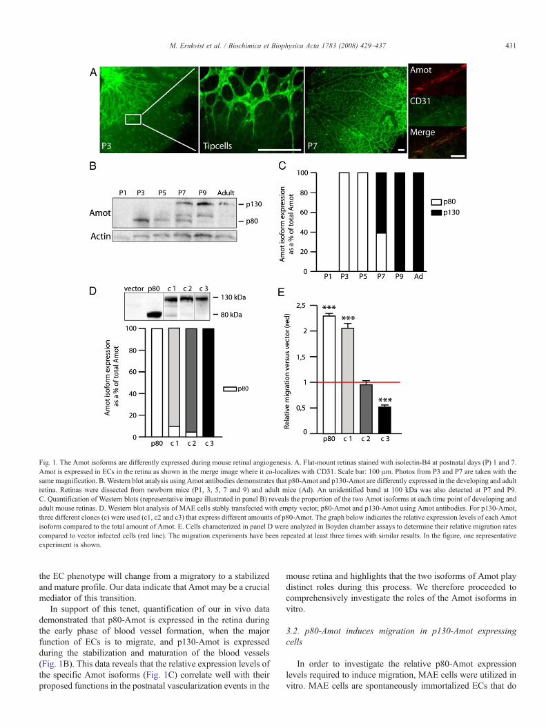
Fig. 1. The Amot isoforms are differently expressed during mouse retinal angiogenesis. A. Flat-mount retinas stained with isolectin-B4 at postnatal days (P) 1 and 7.Amot is expressed in ECs in the retina as shown in the merge image where it co-localizes with CD31. Scale bar: 100 μm. Photos from P3 and P7 are taken with thesame magnification. B. Western blot analysis using Amot antibodies demonstrates that p80-Amot and p130-Amot are differently expressed in the developing and adultretina. Retinas were dissected from newborn mice (P1, 3, 5, 7 and 9) and adult mice (Ad). An unidentified band at 100 kDa was also detected at P7 and P9.C. Quantification of Western blots (representative image illustrated in panel B) reveals the proportion of the two Amot isoforms at each time point of developing andadult mouse retinas. D. Western blot analysis of MAE cells stably transfected with empty vector, p80-Amot and p130-Amot using Amot antibodies. For p130-Amot,three different clones (c) were used (c1, c2 and c3) that express different amounts of p80-Amot. The graph below indicates the relative expression levels of each Amotisoform compared to the total amount of Amot. E. Cells characterized in panel D were analyzed in Boyden chamber assays to determine their relative migration ratescompared to vector infected cells (red line). The migration experiments have been repeated at least three times with similar results. In the figure, one representativeexperiment is shown.
431M. Ernkvist et al. / Biochimica et Biophysica Acta 1783 (2008) 429–437
the EC phenotype will change from a migratory to a stabilizedand mature profile. Our data indicate that Amot may be a crucialmediator of this transition.
In support of this tenet, quantification of our in vivo datademonstrated that p80-Amot is expressed in the retina duringthe early phase of blood vessel formation, when the majorfunction of ECs is to migrate, and p130-Amot is expressedduring the stabilization and maturation of the blood vessels(Fig. 1B). This data reveals that the relative expression levels ofthe specific Amot isoforms (Fig. 1C) correlate well with theirproposed functions in the postnatal vascularization events in the
mouse retina and highlights that the two isoforms of Amot playdistinct roles during this process. We therefore proceeded tocomprehensively investigate the roles of the Amot isoforms invitro.
3.2. p80-Amot induces migration in p130-Amot expressingcells
In order to investigate the relative p80-Amot expressionlevels required to induce migration, MAE cells were utilized invitro. MAE cells are spontaneously immortalized ECs that do
432 M. Ernkvist et al. / Biochimica et Biophysica Acta 1783 (2008) 429–437
not endogenously express Amot and can therefore be infec-ted with p80-Amot or p130-Amot alone to investigate theindividual functions of the two isoforms [3,8]. MAE cells stablyinfected with p130-Amot also express small amounts of p80-Amot. We have previously reported that the p130-Amotsequence does not contain any potential proteolytic cleavagesites that could mediate the conversion of p130-Amot to p80-Amot [8], indicating that the p130-Amot open reading frameproduces both isoforms.
We generated three MAE cell clones infected with p130-Amot that expressed either 10% p80-Amot compared to p130-Amot (clone 1), 5% p80-Amot (clone 2) or only p130-Amot(clone 3) (Fig. 1D). As controls, MAE cells infected with p80-Amot or empty vector were used. The capacity of the cells tomigrate was tested multiple times in a Boyden chamber assaywhere the cells were allowed to migrate towards bFGF. Asillustrated in Fig. 1E (one representative experiment), clone 3,which expressed only p130-Amot exhibited a significantdecrease in migration of approximately 50% compared to vectorcells. Interestingly, clone 2, which expresses 5% p80-Amotcompared to p130-Amot, exhibited approximately the samemigratory rate as vector cells. Furthermore, clone 1, whichexpresses 10% p80-Amot compared to p130-Amot, exhibited asimilar migratory rate to that of p80-Amot only-expressing cells,with a significant two-fold increase in migration compared tovector transfected cells. Since a ratio of 1:10 p80-Amot:p130-Amot favors p80-Amot function, this data suggests that if ECsexpress only p130-Amot, migration will be repressed, whereas arelatively small amount of p80-Amot is sufficient to dominateEC function and induce a migratory phenotype.
3.3. p80-Amot induces a migratory phenotype in MDCK cells
MDCK cells are epithelial cells derived from the distaltubules of canine kidney and widely used for studies ofepithelial cell polarity and cell junctions.
p80-Amot has been shown to play a role in cell polarization[9] and both Amot isoforms have been shown to localize tocell–cell contacts [9,14]. We used MDCK cells that do notexpress any isoform of endogenous Amot (determined byWestern blot and immunofluorescence, data not shown) toinvestigate the localization of transfected p80-Amot, p130-Amot and the N-terminal domain of p130-Amot (Fig. 2A). Wealso transfected MDCK cells with constructs of p80-Amot andp130-Amot lacking the last three amino acids in their C-ter-minal PDZ-binding domain (ΔPDZ-bind) (Fig. 2A) which hasbeen shown to inhibit the migratory function of p80-Amot inprevious studies [7].
We demonstrated that p80-Amot is highly expressed inMDCK cells that appear migratory (Fig. 2C). The p80-Amotexpressing cells displayed extensions, lamellipodia (Fig. 2G)and a different morphology compared to the other, non-transfected polarized MDCK cells that exhibited regular tightjunctions. This change in morphology was also found in p80-Amot expressing cells in contact with other p80-Amotexpressing cells (S Fig. 1A). In addition, weak expression ofp80-Amot was detected in cell–cell contacts (Fig. 2B).
Staining of MDCK cells transfected with p130-Amot re-vealed that p130-Amot localized to cell–cell contacts, re-gardless of whether neighboring cells expressed p130-Amot(Fig. 2D) or not (S Fig. 1B). In addition, p130-Amot wasfound to be expressed in a vesicular pattern, as previouslyshown [8]. In contrast to the expression of p80-Amot, theexpression of p130-Amot did not change the cell morphologytowards a migratory phenotype. MDCK cells were also trans-fected with the p130-Amot N-terminal domain, resulting inexpression in cell–cell contacts and in the edges of the cellswhere it co-localized with actin (Fig. 2E). The N-terminalfragment also overlapped with actin fibers (S Fig. 1C), aspreviously demonstrated in MAE cells [8], indicating that theN-terminal domain of p130-Amot co-localizes to actin in sev-eral cell types.
Cells transfected with p80ΔPDZ-bind exhibited cytoplasmicexpression of p80-Amot in the absence of cell–cell contacts(S Fig. 1D) whereas cells transfected with p130ΔPDZ-bindexhibited an identical staining pattern compared to p130-Amottransfected cells (S Fig. 1E).
These results demonstrate that p80-Amot expression inducesa migratory cell phenotype characterized by extensions andlamellipodia whereas expression of p130-Amot, or its N-ter-minal domain, at cell–cell contacts in association with actindoes not change the cell morphology towards a migratoryphenotype. To quantify the frequency with which p80-Amotover expression results in a migratory phenotype, we deter-mined the number of migratory cells present followingtransfection with p80-Amot, p130-Amot or GFP (S Fig. 1F).As illustrated in Fig. 2F, 52% of cells expressing p80-Amotwere migratory compared to only 30% of GFP expressing cellsand 20% of p130-Amot expressing cells.
In contrast to p80-Amot, this data suggests that p130-Amotis not dependent on its PDZ-binding domain for tight junctionlocalization. Instead, its N-terminal domain appears to be acritical targeting domain for localization. These findings areconsistent with the temporal expression of Amot observed invivo where p80-Amot is predominantly expressed during themigratory phase of angiogenesis and p130-Amot is primarilyexpressed during the blood vessel stabilization phase.
3.4. The N-terminal domain of p130-Amot directs localizationto tight junctions
To explore the respective roles of the different angiomotinisoforms during cell–cell contact formation, we examined thelocalization of p80-Amot, p130-Amot, the specific p130-AmotN-terminal domain, p80ΔPDZ-bind and p130ΔPDZ-bind inMDCK cells during polarization and cell–cell contact forma-tion. In low calcium medium, the tight junctions in MDCK cellswere disrupted, the cells became unpolarized and the Amotisoforms and mutants concentrated in the cytoplasm where theyco-localized with ZO-1 (i.e. p80-Amot, p80ΔPDZ-bind, p130-Amot and p130ΔPDZ-bind) or phalloidin (i.e. N-terminaldomain) (S Fig. 2A, B).
We then examined the localization of the constructs at 1 h,3 h, 6 h and 16 h after re-addition of calcium. MDCK cells
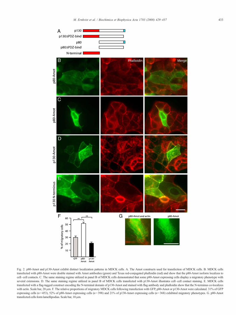
Fig. 2. p80-Amot and p130-Amot exhibit distinct localization patterns in MDCK cells. A. The Amot constructs used for transfection of MDCK cells. B. MDCK cellstransfected with p80-Amot were double stained with Amot antibodies (green) and Texas red-conjugated phalloidin (red) and show that the p80-Amot isoform localizes tocell–cell contacts. C. The same staining regime utilized in panel B of MDCK cells demonstrated that some p80-Amot expressing cells display a migratory phenotype withseveral extensions. D. The same staining regime utilized in panel B of MDCK cells transfected with p130-Amot illustrates cell–cell contact staining. E. MDCK cellstransfected with a flag-tagged construct encoding the N-terminal domain of p130-Amot and stained with flag antibody and phalloidin show that the N-terminus co-localizeswith actin. Scale bar, 20 μm. F. The relative proportions of migratory MDCK cells following transfection with GFP, p80-Amot or p130-Amot were calculated. 31% of GFPexpressing cells (n=453), 52% of p80-Amot expressing cells (n=398) and 21% of p130-Amot expressing cells (n=368) exhibited migratory phenotypes. G. p80-Amottransfe cted cells form lamellipodia s. Scale bar, 10 μm.
433M. Ernkvist et al. / Biochimica et Biophysica Acta 1783 (2008) 429–437
434 M. Ernkvist et al. / Biochimica et Biophysica Acta 1783 (2008) 429–437
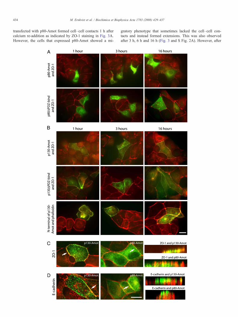
transfected with p80-Amot formed cell–cell contacts 1 h aftercalcium re-addition as indicated by ZO-1 staining in Fig. 3A.However, the cells that expressed p80-Amot showed a mi-
gratory phenotype that sometimes lacked the cell–cell con-tacts and instead formed extensions. This was also observedafter 3 h, 6 h and 16 h (Fig. 3 and S Fig. 2A). However, after
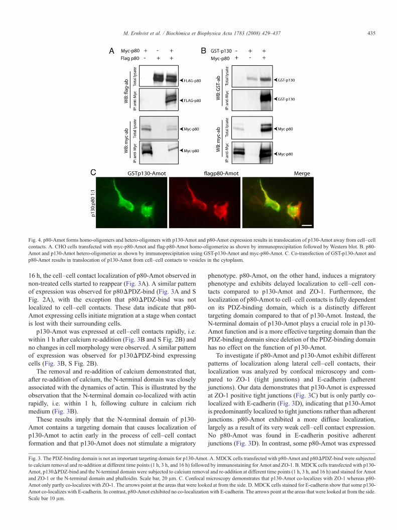
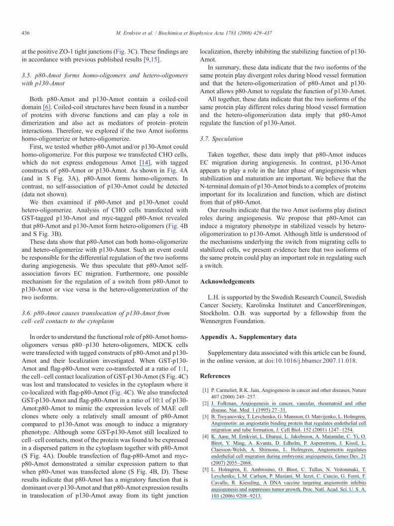
Fig. 4. p80-Amot forms homo-oligomers and hetero-oligomers with p130-Amot and p80-Amot expression results in translocation of p130-Amot away from cell–cellcontacts. A. CHO cells transfected with myc-p80-Amot and flag-p80-Amot homo-oligomerize as shown by immunoprecipitation followed by Western blot. B. p80-Amot and p130-Amot hetero-oligomerize as shown by immunoprecipitation using GST-p130-Amot and myc-p80-Amot. C. Co-transfection of GST-p130-Amot andp80-Amot results in translocation of p130-Amot from cell–cell contacts to vesicles in the cytoplasm.
435M. Ernkvist et al. / Biochimica et Biophysica Acta 1783 (2008) 429–437
16 h, the cell–cell contact localization of p80-Amot observed innon-treated cells started to reappear (Fig. 3A). A similar patternof expression was observed for p80ΔPDZ-bind (Fig. 3A and SFig. 2A), with the exception that p80ΔPDZ-bind was notlocalized to cell–cell contacts. These data indicate that p80-Amot expressing cells initiate migration at a stage when contactis lost with their surrounding cells.
p130-Amot was expressed at cell–cell contacts rapidly, i.e.within 1 h after calcium re-addition (Fig. 3B and S Fig. 2B) andno changes in cell morphology were observed. A similar patternof expression was observed for p130ΔPDZ-bind expressingcells (Fig. 3B, S Fig. 2B).
The removal and re-addition of calcium demonstrated that,after re-addition of calcium, the N-terminal domain was closelyassociated with the dynamics of actin. This is illustrated by theobservation that the N-terminal domain co-localized with actinrapidly, i.e. within 1 h, following culture in calcium richmedium (Fig. 3B).
These results imply that the N-terminal domain of p130-Amot contains a targeting domain that causes localization ofp130-Amot to actin early in the process of cell–cell contactformation and that p130-Amot does not stimulate a migratory
Fig. 3. The PDZ-binding domain is not an important targeting domain for p130-Amotto calcium removal and re-addition at different time points (1 h, 3 h, and 16 h) followeAmot, p130ΔPDZ-bind and the N-terminal domain were subjected to calcium removaand ZO-1 or the N-terminal domain and phalloidin. Scale bar, 20 μm. C. Confocal mAmot only partly co-localizes with ZO-1. The arrows point at the areas that were lookAmot co-localizes with E-cadherin. In contrast, p80-Amot exhibited no co-localizationScale bar 10 μm.
phenotype. p80-Amot, on the other hand, induces a migratoryphenotype and exhibits delayed localization to cell–cell con-tacts compared to p130-Amot and ZO-1. Furthermore, thelocalization of p80-Amot to cell–cell contacts is fully dependenton its PDZ-binding domain, which is a distinctly differenttargeting domain compared to that of p130-Amot. Instead, theN-terminal domain of p130-Amot plays a crucial role in p130-Amot function and is a more effective targeting domain than thePDZ-binding domain since deletion of the PDZ-binding domainhas no effect on the function of p130-Amot.
To investigate if p80-Amot and p130-Amot exhibit differentpatterns of localization along lateral cell–cell contacts, theirlocalization was analyzed by confocal microscopy and com-pared to ZO-1 (tight junctions) and E-cadherin (adherentjunctions). Our data demonstrates that p130-Amot is expressedat ZO-1 positive tight junctions (Fig. 3C) but is only partly co-localized with E-cadherin (Fig. 3D), indicating that p130-Amotis predominantly localized to tight junctions rather than adherentjunctions. p80-Amot exhibited a more diffuse localization,largely as a result of its very weak cell–cell contact expression.No p80-Amot was found in E-cadherin positive adherentjunctions (Fig. 3D). In contrast, some p80-Amot was expressed
. A. MDCK cells transfected with p80-Amot and p80ΔPDZ-bind were subjectedd by immunostaining for Amot and ZO-1. B. MDCK cells transfected with p130-l and re-addition at different time points (1 h, 3 h, and 16 h) and stained for Amoticroscopy demonstrates that p130-Amot co-localizes with ZO-1 whereas p80-
ed at from the side. D. MDCK cells stained for E-cadherin show that some p130-with E-cadherin. The arrows point at the areas that were looked at from the side.
436 M. Ernkvist et al. / Biochimica et Biophysica Acta 1783 (2008) 429–437
at the positive ZO-1 tight junctions (Fig. 3C). These findings arein accordance with previous published results [9,15].
3.5. p80-Amot forms homo-oligomers and hetero-oligomerswith p130-Amot
Both p80-Amot and p130-Amot contain a coiled-coildomain [6]. Coiled-coil structures have been found in a numberof proteins with diverse functions and can play a role indimerization and also act as mediators of protein–proteininteractions. Therefore, we explored if the two Amot isoformshomo-oligomerize or hetero-oligomerize.
First, we tested whether p80-Amot and/or p130-Amot couldhomo-oligomerize. For this purpose we transfected CHO cells,which do not express endogenous Amot [14], with taggedconstructs of p80-Amot or p130-Amot. As shown in Fig. 4A(and in S Fig. 3A), p80-Amot forms homo-oligomers. Incontrast, no self-association of p130-Amot could be detected(data not shown).
We then examined if p80-Amot and p130-Amot couldhetero-oligomerize. Analysis of CHO cells transfected withGST-tagged p130-Amot and myc-tagged p80-Amot revealedthat p80-Amot and p130-Amot form hetero-oligomers (Fig. 4Band S Fig. 3B).
These data show that p80-Amot can both homo-oligomerizeand hetero-oligomerize with p130-Amot. Such an event couldbe responsible for the differential regulation of the two isoformsduring angiogenesis. We thus speculate that p80-Amot self-association favors EC migration. Furthermore, one possiblemechanism for the regulation of a switch from p80-Amot top130-Amot or vice versa is the hetero-oligomerization of thetwo isoforms.
3.6. p80-Amot causes translocation of p130-Amot fromcell–cell contacts to the cytoplasm
In order to understand the functional role of p80-Amot homo-oligomers versus p80–p130 hetero-oligomers, MDCK cellswere transfected with tagged constructs of p80-Amot and p130-Amot and their localization investigated. When GST-p130-Amot and flag-p80-Amot were co-transfected at a ratio of 1:1,the cell–cell contact localization of GST-p130-Amot (S Fig. 4C)was lost and translocated to vesicles in the cytoplasm where itco-localized with flag-p80-Amot (Fig. 4C). We also transfectedGST-p130-Amot and flag-p80-Amot in a ratio of 10:1 of p130-Amot:p80-Amot to mimic the expression levels of MAE cellclones where only a relatively small amount of p80-Amotcompared to p130-Amot was enough to induce a migratoryphenotype. Although some GST-p130-Amot still localized tocell–cell contacts, most of the protein was found to be expressedin a dispersed pattern in the cytoplasm together with p80-Amot(S Fig. 4A). Double transfection of flag-p80-Amot and myc-p80-Amot demonstrated a similar expression pattern to thatwhen p80-Amot was transfected alone (S Fig. 4B, D). Theseresults indicate that p80-Amot has a migratory function that isdominant over p130-Amot and that p80-Amot expression resultsin translocation of p130-Amot away from its tight junction
localization, thereby inhibiting the stabilizing function of p130-Amot.
In summary, these data indicate that the two isoforms of thesame protein play divergent roles during blood vessel formationand that the hetero-oligomerization of p80-Amot and p130-Amot allows p80-Amot to regulate the function of p130-Amot.
All together, these data indicate that the two isoforms of thesame protein play different roles during blood vessel formationand the hetero-oligomerization data imply that p80-Amotregulate the function of p130-Amot.
3.7. Speculation
Taken together, these data imply that p80-Amot inducesEC migration during angiogenesis. In contrast, p130-Amotappears to play a role in the later phase of angiogenesis whenstabilization and maturation are important. We believe that theN-terminal domain of p130-Amot binds to a complex of proteinsimportant for its localization and function, which are distinctfrom that of p80-Amot.
Our results indicate that the two Amot isoforms play distinctroles during angiogenesis. We propose that p80-Amot caninduce a migratory phenotype in stabilized vessels by hetero-oligomerization to p130-Amot. Although little is understood ofthe mechanisms underlying the switch from migrating cells tostabilized cells, we present evidence here that two isoforms ofthe same protein could play an important role in regulating sucha switch.
Acknowledgements
L.H. is supported by the Swedish Research Council, SwedishCancer Society, Karolinska Institutet and Cancerföreningen,Stockholm. O.B. was supported by a fellowship from theWennergren Foundation.
Appendix A. Supplementary data
Supplementary data associated with this article can be found,in the online version, at doi:10.1016/j.bbamcr.2007.11.018.
References
[1] P. Carmeliet, R.K. Jain, Angiogenesis in cancer and other diseases, Nature407 (2000) 249–257.
[2] J. Folkman, Angiogenesis in cancer, vascular, rheumatoid and otherdisease, Nat. Med. 1 (1995) 27–31.
[3] B. Troyanovsky, T. Levchenko, G. Mansson, O. Matvijenko, L. Holmgren,Angiomotin: an angiostatin binding protein that regulates endothelial cellmigration and tube formation, J. Cell Biol. 152 (2001) 1247–1254.
[4] K. Aase, M. Ernkvist, L. Ebarasi, L. Jakobsson, A. Majumdar, C. Yi, O.Birot, Y. Ming, A. Kvanta, D. Edholm, P. Aspenstrom, J. Kissil, L.Claesson-Welsh, A. Shimono, L. Holmgren, Angiomotin regulatesendothelial cell migration during embryonic angiogenesis, Genes Dev. 21(2007) 2055–2068.
[5] L. Holmgren, E. Ambrosino, O. Birot, C. Tullus, N. Veitonmaki, T.Levchenko, L.M. Carlson, P. Musiani, M. Iezzi, C. Curcio, G. Forni, F.Cavallo, R. Kiessling, A DNA vaccine targeting angiomotin inhibitsangiogenesis and suppresses tumor growth, Proc. Natl. Acad. Sci. U. S. A.103 (2006) 9208–9213.

437M. Ernkvist et al. / Biochimica et Biophysica Acta 1783 (2008) 429–437
[6] A. Bratt, W.J. Wilson, B. Troyanovsky, K. Aase, R. Kessler, E.G. VanMeir, L. Holmgren, Angiomotin belongs to a novel protein family withconserved coiled-coil and PDZ binding domains, Gene 298 (2002) 69–77.
[7] T. Levchenko, K. Aase, B. Troyanovsky, A. Bratt, L. Holmgren, Loss ofresponsiveness to chemotactic factors by deletion of the C-terminal proteininteraction site of angiomotin, J. Cell Sci. 116 (2003) 3803–3810.
[8] M. Ernkvist, K. Aase, C. Ukomadu, J. Wohlschlegel, R. Blackman, N.Veitonmaki, A. Bratt, A. Dutta, L. Holmgren, p130-Angiomotin associatesto actin and controls endothelial cell shape, Febs J. 273 (2006) 2000–2011.
[9] C.D. Wells, J.P. Fawcett, A. Traweger, Y. Yamanaka, M. Goudreault, K.Elder, S. Kulkarni, G. Gish, C. Virag, C. Lim, K. Colwill, A. Starostine, P.Metalnikov, T. Pawson, A Rich1/Amot complex regulates the Cdc42GTPase and apical-polarity proteins in epithelial cells, Cell 125 (2006)535–548.
[10] T. Chan-Ling, Glial, vascular, and neuronal cytogenesis in whole-mountedcat retina, Microsc. Res. Tech. 36 (1997) 1–16.
[11] H. Gerhardt, M. Golding, M. Fruttiger, C. Ruhrberg, A. Lundkvist, A.Abramsson, M. Jeltsch, C. Mitchell, K. Alitalo, D. Shima, C. Betsholtz,VEGF guides angiogenic sprouting utilizing endothelial tip cell filopodia,J. Cell Biol. 161 (2003) 1163–1177.
[12] P.A. Campochiaro, Retinal and choroidal neovascularization, J. Cell.Physiol. 184 (2000) 301–310.
[13] A. Uemura, S. Kusuhara, H. Katsuta, S. Nishikawa, Angiogenesis in themouse retina: a model system for experimental manipulation, Exp. CellRes. 312 (2006) 676–683.
[14] A. Bratt, O. Birot, I. Sinha, N. Veitonmaki, K. Aase, M. Ernkvist, L.Holmgren, Angiomotin regulates endothelial cell–cell junctions and cellmotility, J. Biol. Chem. 280 (2005) 34859–34869.
[15] Y. Sugihara-Mizuno, M. Adachi, Y. Kobayashi, Y. Hamazaki, M.Nishimura, T. Imai, M. Furuse, S. Tsukita, Molecular characterization ofangiomotin/JEAP family proteins: interaction with MUPP1/Patj and theirendogenous properties, Genes Cells 12 (2007) 473–486.