Differential distribution of DNA methylation within the RASSF1A CpG island in breast cancer
10
[CANCER RESEARCH 63, 6178 – 6186, October 1, 2003] Differential Distribution of DNA Methylation within the RASSF1A CpG Island in Breast Cancer 1 Pearlly S. Yan, Huidong Shi, Farahnaz Rahmatpanah, Timothy H-C. Hsiau, Andrew H-A. Hsiau, Yu-Wei Leu, Joseph C. Liu, and Tim Hui-Ming Huang 2 Human Cancer Genetics Program, Department of Molecular Virology, Immunology and Medical Genetics, Comprehensive Cancer Center, The Ohio State University, Columbus, Ohio 43210 [P. S. Y., Y-W. L., J. C. L., T. H-M. H.], and Department of Pathology and Anatomical Sciences, Ellis Fischel Cancer Center, University of Missouri School of Medicine, Columbia, Missouri 65203 [H. S., F. R., T. H-C. H., A. H-A. H.] ABSTRACT Aberrant DNA methylation of promoter CpG islands is associated with transcriptionally repressive heterochromatin in neoplasia. The dynamics of this epigenetic process in mediating the transition from an active to an inactive state of transcription remains to be elucidated, however. Here, we used the methylation-specific oligonucleotide microarray to map the methylation patterns of a CpG island, located within the promoter and the first exon regions of RASSF1A, in normal breast tissue controls, primary tumors, and breast cancer cell lines. Oligonucleotide pairs, spaced along the CpG island region, were designed to discriminate between methylated and unmethylated alleles of selected sites. The methylation-specific oligo- nucleotide data indicate that the majority of test samples show widespread methylation in the first exon of RASSF1A. In contrast, the promoter area was usually undermethylated in normal controls and in 32% of the primary tumors tested, whereas the rest of the primary tumors and breast cancer cell lines showed various degrees of methylation in the region. Methylation profiling of individual tumors further suggest that DNA methylation progressively spreads from the first exon into the promoter area of this gene. Functional analysis indicates that increased density of RASSF1A promoter methylation is associated with altered chromatin, marked by a depletion of acetylated histones and methylated histone 3-lysine 4 and an enrichment of methylated histone 3-lysine 9 in the studied area. The combination of these epigenetic modifications may engender a stable silencing of the gene in breast cancer cells. Thus, this study underscores the importance of detailed mapping of methylation patterns within a CpG island locus that may provide insights into the progressive nature of aberrant DNA methylation and its relationship with transcriptional silencing during the neoplastic process. INTRODUCTION The CpG island, a stretch of GC-rich DNA sequence, is frequently located at the 5-end regulatory regions of a gene, extending from the promoter to the first exon region (1). This area of genomic sequence is subject to epigenetic modifications, including DNA methylation and histone acetylation and methylation, which are known to play an important role in regulating gene expression (2). In normal cells, the majority of promoter CpG islands are protected from this epigenetic event and are unmethylated (2, 3). In cancer cells, it has been shown that widespread DNA methylation occurs in many promoter CpG islands, resulting in a closed, repressive chromatin configuration that disables transcription initiation of the associated genes (3). Although CpG island hypermethylation has been observed in many types of solid tumors and leukemias (4, 5), the dynamics of this epigenetic process in mediating the transition from an unmethylated, active state to a densely methylated, inactive state of a gene promoter remains largely unknown. Using cultured cells overexpressing DNA methyltransferase DNMT1, Graff et al. (6) observed that de novo methylation is a gradual process and progressively accumulated within a susceptible CpG island. The in vitro observation of this so-called methylation spread phenomenon still awaits proof in vivo. To conduct such a study in primary tumors, detailed mapping of methylated sites across a CpG island of interest is needed. The frequently used assays, methylation-specific PCR (7), or COBRA 3 (8), which detect point methylation on only a few sites within a CpG island, may not be suitable for such a task. The bisulfite sequencing method (9) is the choice for comprehensive methylation mapping but is more labor intensive for analyzing a large panel of primary tumors representing different stages of methylation progression. Toward this end, we recently developed an oligonucleotide-based microarray technique, MSO microarray (10), which can improve the resolution of methylation mapping in a defined CpG island region. In the MSO assay, DNA targets are hybridized to a series of arrayed oligonucleotide probes, designed to be closely spaced along an inter- rogating sequence. Each pair of oligonucleotides is capable of dis- criminating between methylated and unmethylated alleles at specific sites. Herein we used the MSO microarray to assess methylation profiles of the RASSF1A CpG island region in a panel of breast normal tissue controls, primary tumors, and cancer cell lines. This CpG island, spanning the promoter and the first exon regions of the gene, is hypermethylated in 42– 65% of primary breast tumors (11–14) and in many other cancer types (15–17). Recent evidence suggests that the methylation-associated silencing of RASSF1A transcript can disrupt RAS and its related signaling pathways, leading to uncontrolled tumor growth (11). The frequent observation of RASSF1A hypermethylation in breast cancer therefore provides an ideal model for examining the methyla- tion spread phenomenon in vivo. Our MSO results demonstrate that DNA methylation is differentially distributed within the RASSF1A CpG island in breast cancer. This disparity observed in breast tumors may reflect different stages of methylation progressively spreading from the nearby first exon into the promoter of the RASSF1A gene. Additional analysis suggests that RASSF1A promoter methylation is associated with altered chromatin configuration, marked by specific patterns of histone acetylation and methylation. MATERIALS AND METHODS Sample Preparation, SssI Treatment, and Bisulfite Modification. Thir- ty-seven breast tumors were obtained from patients undergoing mastectomy before chemotherapy at the Ellis Fischel Cancer Center (Columbia, MO), in compliance with the Institutional Review Board. Except for two invasive lobular carcinomas (T301 and T313), the rest of the tumors analyzed were classified as infiltrating ductal carcinomas. Ten tumor-free breast parenchymas were obtained to serve as controls. Breast cancer cell lines MDA-MB-435s, Received 1/29/03; revised 6/26/03; accepted 7/17/03. The costs of publication of this article were defrayed in part by the payment of page charges. This article must therefore be hereby marked advertisement in accordance with 18 U.S.C. Section 1734 solely to indicate this fact. 1 Supported in part by National Cancer Institute Grants R21 CA-94441 and RO1 CA-69065. 2 To whom requests for reprints should be addressed, at Room 514B, Human Cancer Genetics Program, Medical Research Facility, The Ohio State University, 420 W. 12 th Avenue, Columbus, OH 43210. Phone: (614) 688-8277; Fax: (614) 688-3981; E-mail: [email protected]. 3 The abbreviations used are: COBRA, combined bisulfite restriction analysis; RT- PCR, reverse transcription-PCR; MSO, methylation-specific oligonucleotide; acetyl-H3, acetylated histone 3; methyl-H3-K9, dimethylated histone 3-lysine 9; methyl-H3-K4, dimethylated histone 3-lysine 4. 6178 on July 22, 2015. © 2003 American Association for Cancer Research. cancerres.aacrjournals.org Downloaded from
-
Upload
independent -
Category
Documents
-
view
0 -
download
0
Transcript of Differential distribution of DNA methylation within the RASSF1A CpG island in breast cancer

[CANCER RESEARCH 63, 6178–6186, October 1, 2003]
Differential Distribution of DNA Methylation within the RASSF1A CpG Island inBreast Cancer1
Pearlly S. Yan, Huidong Shi, Farahnaz Rahmatpanah, Timothy H-C. Hsiau, Andrew H-A. Hsiau, Yu-Wei Leu,Joseph C. Liu, and Tim Hui-Ming Huang2
Human Cancer Genetics Program, Department of Molecular Virology, Immunology and Medical Genetics, Comprehensive Cancer Center, The Ohio State University, Columbus,Ohio 43210 [P. S. Y., Y-W. L., J. C. L., T. H-M. H.], and Department of Pathology and Anatomical Sciences, Ellis Fischel Cancer Center, University of Missouri School ofMedicine, Columbia, Missouri 65203 [H. S., F. R., T. H-C. H., A. H-A. H.]
ABSTRACT
Aberrant DNA methylation of promoter CpG islands is associated withtranscriptionally repressive heterochromatin in neoplasia. The dynamicsof this epigenetic process in mediating the transition from an active to aninactive state of transcription remains to be elucidated, however. Here, weused the methylation-specific oligonucleotide microarray to map themethylation patterns of a CpG island, located within the promoter and thefirst exon regions of RASSF1A, in normal breast tissue controls, primarytumors, and breast cancer cell lines. Oligonucleotide pairs, spaced alongthe CpG island region, were designed to discriminate between methylatedand unmethylated alleles of selected sites. The methylation-specific oligo-nucleotide data indicate that the majority of test samples show widespreadmethylation in the first exon of RASSF1A. In contrast, the promoter areawas usually undermethylated in normal controls and in 32% of theprimary tumors tested, whereas the rest of the primary tumors and breastcancer cell lines showed various degrees of methylation in the region.Methylation profiling of individual tumors further suggest that DNAmethylation progressively spreads from the first exon into the promoterarea of this gene. Functional analysis indicates that increased density ofRASSF1A promoter methylation is associated with altered chromatin,marked by a depletion of acetylated histones and methylated histone3-lysine 4 and an enrichment of methylated histone 3-lysine 9 in thestudied area. The combination of these epigenetic modifications mayengender a stable silencing of the gene in breast cancer cells. Thus, thisstudy underscores the importance of detailed mapping of methylationpatterns within a CpG island locus that may provide insights into theprogressive nature of aberrant DNA methylation and its relationship withtranscriptional silencing during the neoplastic process.
INTRODUCTION
The CpG island, a stretch of GC-rich DNA sequence, is frequentlylocated at the 5�-end regulatory regions of a gene, extending from thepromoter to the first exon region (1). This area of genomic sequenceis subject to epigenetic modifications, including DNA methylationand histone acetylation and methylation, which are known to play animportant role in regulating gene expression (2). In normal cells, themajority of promoter CpG islands are protected from this epigeneticevent and are unmethylated (2, 3). In cancer cells, it has been shownthat widespread DNA methylation occurs in many promoter CpGislands, resulting in a closed, repressive chromatin configuration thatdisables transcription initiation of the associated genes (3).
Although CpG island hypermethylation has been observed in manytypes of solid tumors and leukemias (4, 5), the dynamics of thisepigenetic process in mediating the transition from an unmethylated,active state to a densely methylated, inactive state of a gene promoter
remains largely unknown. Using cultured cells overexpressing DNAmethyltransferase DNMT1, Graff et al. (6) observed that de novomethylation is a gradual process and progressively accumulatedwithin a susceptible CpG island. The in vitro observation of thisso-called methylation spread phenomenon still awaits proof in vivo.To conduct such a study in primary tumors, detailed mapping ofmethylated sites across a CpG island of interest is needed. Thefrequently used assays, methylation-specific PCR (7), or COBRA3
(8), which detect point methylation on only a few sites within a CpGisland, may not be suitable for such a task. The bisulfite sequencingmethod (9) is the choice for comprehensive methylation mapping butis more labor intensive for analyzing a large panel of primary tumorsrepresenting different stages of methylation progression.
Toward this end, we recently developed an oligonucleotide-basedmicroarray technique, MSO microarray (10), which can improve theresolution of methylation mapping in a defined CpG island region. Inthe MSO assay, DNA targets are hybridized to a series of arrayedoligonucleotide probes, designed to be closely spaced along an inter-rogating sequence. Each pair of oligonucleotides is capable of dis-criminating between methylated and unmethylated alleles at specificsites. Herein we used the MSO microarray to assess methylationprofiles of the RASSF1A CpG island region in a panel of breast normaltissue controls, primary tumors, and cancer cell lines. This CpGisland, spanning the promoter and the first exon regions of the gene,is hypermethylated in 42–65% of primary breast tumors (11–14) andin many other cancer types (15–17). Recent evidence suggests that themethylation-associated silencing of RASSF1A transcript can disruptRAS and its related signaling pathways, leading to uncontrolled tumorgrowth (11).
The frequent observation of RASSF1A hypermethylation in breastcancer therefore provides an ideal model for examining the methyla-tion spread phenomenon in vivo. Our MSO results demonstrate thatDNA methylation is differentially distributed within the RASSF1ACpG island in breast cancer. This disparity observed in breast tumorsmay reflect different stages of methylation progressively spreadingfrom the nearby first exon into the promoter of the RASSF1A gene.Additional analysis suggests that RASSF1A promoter methylation isassociated with altered chromatin configuration, marked by specificpatterns of histone acetylation and methylation.
MATERIALS AND METHODS
Sample Preparation, SssI Treatment, and Bisulfite Modification. Thir-ty-seven breast tumors were obtained from patients undergoing mastectomybefore chemotherapy at the Ellis Fischel Cancer Center (Columbia, MO), incompliance with the Institutional Review Board. Except for two invasivelobular carcinomas (T301 and T313), the rest of the tumors analyzed wereclassified as infiltrating ductal carcinomas. Ten tumor-free breast parenchymaswere obtained to serve as controls. Breast cancer cell lines MDA-MB-435s,
Received 1/29/03; revised 6/26/03; accepted 7/17/03.The costs of publication of this article were defrayed in part by the payment of page
charges. This article must therefore be hereby marked advertisement in accordance with18 U.S.C. Section 1734 solely to indicate this fact.
1 Supported in part by National Cancer Institute Grants R21 CA-94441 and RO1CA-69065.
2 To whom requests for reprints should be addressed, at Room 514B, Human CancerGenetics Program, Medical Research Facility, The Ohio State University, 420 W. 12th
Avenue, Columbus, OH 43210. Phone: (614) 688-8277; Fax: (614) 688-3981; E-mail:[email protected].
3 The abbreviations used are: COBRA, combined bisulfite restriction analysis; RT-PCR, reverse transcription-PCR; MSO, methylation-specific oligonucleotide; acetyl-H3,acetylated histone 3; methyl-H3-K9, dimethylated histone 3-lysine 9; methyl-H3-K4,dimethylated histone 3-lysine 4.
6178
on July 22, 2015. © 2003 American Association for Cancer Research.cancerres.aacrjournals.org Downloaded from
ZR-75-1, Hs578t, MDA-MB-453, MCF7, MDA-MB-231, and T47D wereroutinely maintained in our laboratory as described (18). Genomic DNA wasisolated using the QIAamp Tissue Kit (Qiagen). For the preparation of 100%methylated DNA, a blood DNA sample was treated with M. SssI methyltrans-ferase that methylates all cytosine residues of CpG dinucleotides in thegenome. Sodium bisulfite modification of the test samples and M. SssI-treatedDNA samples was then performed using the CpGenome DNA Modification kit(Intergen). This treatment converts unmethylated, but not methylated, cytosineto uracil in the genome.
MSO Microarray. Preparation of MSOs was carried out essentially asdescribed (10). These oligonucleotides were used to interrogate bisulfite-treated sense strand DNA in regions covering the promoter and first exon ofRASSF1A. Each oligonucleotide (final concentration: 50 pmol/�l) was sus-pended in microspotting solution (Telechem) and printed in triplicate asmicrodots (�1 nl; 100 �m diameter) on the superaldehyde-coated glass slides(Telechem) using a GMS 417 microarrayer. Slide processing was performedfollowing the manufacturer’s protocol (Telechem). For DNA target prepara-tion, bisulfite-treated DNA was amplified by PCR from three regions (P1, P2,and E1) located in the RASSF1A CpG island (Fig. 1). PCR conditions were thesame as those described previously (10). Primers used for bisulfite DNAamplification were: P1, sense strand, 5�-GTA GGT TAA GTG TGT TGT TTTAGT and antisense strand, 5�-TTA CCC TTC CTT CCC TCC TT; P2, sensestrand, 5�-AGG AGG GAA GGA AGG GTA AG and antisense strand,5�-TAA CTT TAA ACR CTA ACA AAC; and E1, sense strand, 5�-AAG TYGGGG TTY GTT TTG TGG TTT and antisense strand, 5�-CCC CAA ATAAAA TCR CCA CAA AAA T (R � mixture of A and G; Y � mixture of Gand T). Purified PCR products were labeled at the 3� terminus with Cy5-dCTP(Amersham Pharmacia) by terminal transferase (New England Biolabs). Theselabeled products were resuspended in the Unihybridization Solution(Telechem), denatured at 95°C for 5 min, and applied to MSO slides. Hybrid-ization was conducted in a moist chamber at 50°C for 4–5 h. Slides were rinsedand washed once at 50°C with 2 � SSC-0.2% SDS for a total of 15 min,followed by washing once with 2 � SSC at room temperature for 5 min, anddrying by centrifugation at 500 rpm for 5 min. These microarray slides werescanned with a GenePix 4000A scanner (Axon Instruments). The fluorescenceimages were analyzed with GenePix Pro3.0 software. Signal intensities ofindividual spots were obtained and exported to Excel spreadsheets for furtheranalysis. MSO data were first normalized according to a global ratio in eachmicroarray image, and the intensity ratio of M/(M�U) (M: methylated alleleand U: unmethylated allele) for each probe set was then derived. Statisticalanalyses were conducted using SigmaStat 2.0 software (Jandel Scientific).
COBRA. Bisulfite-treated genomic DNA (�1 �g) was used as a templatefor PCR with specific primers located in the RASSF1A CpG island regions
(Fig. 1). Primers used for amplification were as follows: promoter sense strand,5�-AGG AGG GAA GGA AGG GTA AG and antisense strand, 5�-TAA CTTTAA ACR CTA ACA AAC-3�; first exon sense strand, 5�-AAG TYG GGGTTY GTT TTG TGG TTT and antisense strand, 5�-CCC CAA ATA AAATCR CCA CAA AAA T. After amplification, 32P-incorporated PCR productswere digested with the restriction enzyme BstUI (New England Biolabs),which recognizes sequences unique to the methylated and bisulfite-uncon-verted alleles. The digested and undigested control DNA samples were sepa-rated in parallel on 8% polyacrylamide gels and subject to autoradiographyusing a PhosphorImager (Amersham-Pharmacia).
Bisulfite Sequencing. The P2 and E1 regions (see Fig. 1) within theRASSF1A CpG were amplified from bisulfite-treated genomic DNA by PCRusing the same primer pairs described earlier. Amplified products were sub-cloned using the TOPO-TA Cloning System (Invitrogen). Plasmid DNA of10–15 insert-positive clones were isolated by the QIAprep Spin Miniprep kit(Qiagen) and sequenced using the ABI sequencing system (Applied Biosys-tems).
Semiquantitative RT-PCR. Total RNA (1 �g) was extracted, reversetranscribed, and used to amplify cDNA from a 5�-end region of RASSF1A orfrom �-actin (control) with AmpliTaq Gold polymerase (Applied Biosystems).The primers used for PCR were: RASSF1A, sense strand, 5�-ACC TCT GTGGCG ACT TCA TCT and antisense strand, 5�-AGG TGA ACT TGC AATGCG C; �-actin, sense strand, 5�-GGC ATG GGT CAG AAG GAT T andantisense strand, 5�-ATG TCG TCC CAG TTG GTG A. After 25 cycles ofamplification, 32P-incorporated PCR products were run on 8% polyacrylamidegels and subject to autoradiography using a PhosphorImager (Amersham-Pharmacia). Densitometric analysis of the observed bands was performedusing ImageQuant (Molecular Dynamics). The relative expression level ofRASSF1A was normalized with that of �-actin (control) in each sample.
ChIP-PCR. The assay was conducted using a kit from Upstate Biotech-nology and followed the manufacturer’s protocols with some modifications.Breast cancer cells (2 � 106/assay) were cross-linked using 1% formaldehyde.Cell nuclei were isolated and lysed in an SDS buffer containing proteinaseinhibitors. The samples were sonicated and precleared with salmon spermDNA/Protein A agarose beads (Upstate Biotechnology). The soluble chromatinfraction was collected, and 8 �l of antibodies for acetyl-H3, acetyl-H4,methyl-H3-K4, or 12 �l of methyl-H3-K9, or no antibody, was added andincubated overnight with rotation. After elution from the beads, the immuno-complex was further digested with proteinase K to release bound chromatinDNA. DNA was then recovered by phenol extraction, ethanol precipitated, andresuspended in water. Primers were designed to separately amplify threeregions in the RASSF1A promoter area (Fig. 1). One additional primer set wasdesigned to amplify a 300-bp fragment of the �-actin gene as an internalcontrol (19). Primer sequences used for ChIP-PCR were: RASSF1A region 1,sense strand, 5�-GCT TCA GCA AAC CGG ACC AGG and antisense strand,4 Internet address: www.cbrc.jp/htbin/nph-tfsearch.
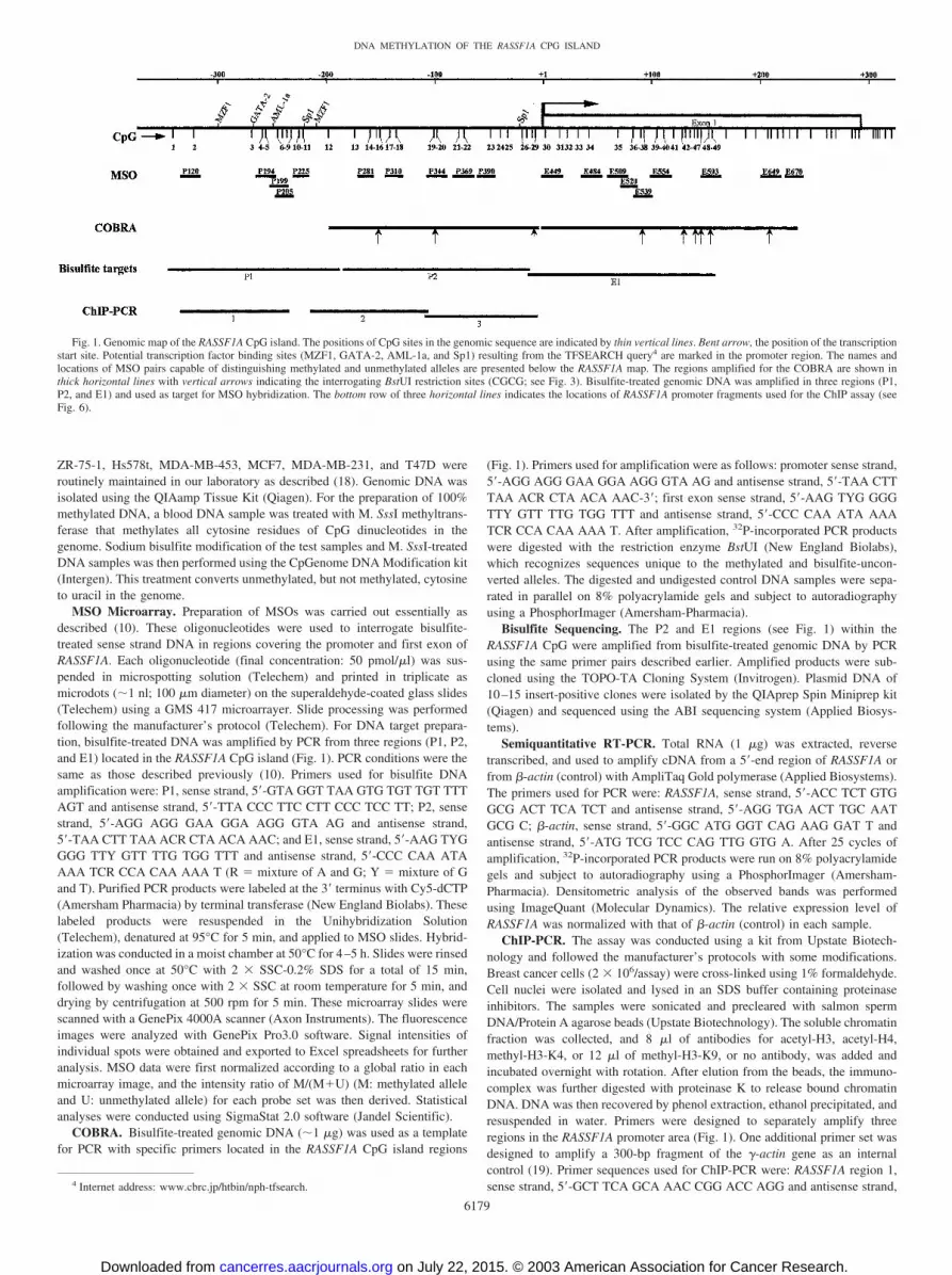
Fig. 1. Genomic map of the RASSF1A CpG island. The positions of CpG sites in the genomic sequence are indicated by thin vertical lines. Bent arrow, the position of the transcriptionstart site. Potential transcription factor binding sites (MZF1, GATA-2, AML-1a, and Sp1) resulting from the TFSEARCH query4 are marked in the promoter region. The names andlocations of MSO pairs capable of distinguishing methylated and unmethylated alleles are presented below the RASSF1A map. The regions amplified for the COBRA are shown inthick horizontal lines with vertical arrows indicating the interrogating BstUI restriction sites (CGCG; see Fig. 3). Bisulfite-treated genomic DNA was amplified in three regions (P1,P2, and E1) and used as target for MSO hybridization. The bottom row of three horizontal lines indicates the locations of RASSF1A promoter fragments used for the ChIP assay (seeFig. 6).
6179
DNA METHYLATION OF THE RASSF1A CPG ISLAND
on July 22, 2015. © 2003 American Association for Cancer Research.cancerres.aacrjournals.org Downloaded from

5�-CCG GAC GGC CAC AAC GA; RASSF1A region 2, sense strand, 5�-TGGGGT GTG AGG AGG GGA CGA and antisense strand, 5�-AGA GCC GCGCAA TGG A; RASSF1A region 3, sense strand, 5�-GTT TCC ATT GCG CGGCTC T and antisense strand, 5�-CTG GCT TTG GGC GCT AGC AAG; and�-actin, uGACE (19), 5�-CAT GTA CGT GGC CAT CCA GG and dGACE,5�-GTG GCC ATC TCC TGC TCG AA. Diplex PCR for simultaneouslyamplifying both test and control fragments was performed using the AmpliTaqGold polymerase (Perkin-Elmer). After 25–30 cycles of amplification, 32P-incorporated PCR products were run on 8% polyacrylamide gels and subjectedto autoradiography. Densitometric analysis of the observed bands was per-formed using ImageQuant (Molecular Dynamics). Fold-enrichment was de-rived from the ratio between the intensities of test and control fragments fromthe bound sample to that of the total input sample.
RESULTS
Standardization of MSO Microarray for Quantitative Methyl-ation Analysis. A panel of 56 oligonucleotide probes were initiallydesigned to map the methylation status of a 650-bp CpG island region,spanning the promoter and the first exon of RASSF1A. Sequences ofthese oligonucleotide probes were derived from the bisulfite-treatedsense strand DNAs in the region. Each probe set was designed toassess the methylation status of one to three nearby CpG sites in aninterrogating sequence. These paired probes affixed on a microscopicslide surface at their 5�-ends were expected to form a perfect matchwith either methylated or unmethylated DNA targets (10, 20). As
Fig. 2. Methylation status of the RASSF1A CpGisland determined by MSO microarray. A summaryof the MSO results is presented for 10 breast nor-mals (prefix with an N), 37 primary tumors (prefixwith an T), and 7 breast cancer cell lines. A total of19 oligonucleotide pairs (10 in the promoter regionand 9 in the first exon region) was used to surveythe methylation status of this CpG island in ahigh-throughput manner. The locations of theseoligonucleotide pairs are presented in Fig. 1. �,BstUI restriction sites present in the nucleotidesequences. ✦, bisulfite sequencing results pre-sented in Fig. 4. The scale at left represents thepercentage of methylation levels as determinedfrom the calibration curves for the test CpG sites(see description in the text). In cases where MSOhybridization signals could not be analyzed prop-erly, a diagonal line was presented in place ofpercentage of methylation. The outcomes of theCOBRA on all of the test samples are shown atright with (�) indicating the presence of methyl-ated BstUI sites and (�) indicating the absence ofsuch methylation (see additional details in Fig. 3).P, RASSF1A promoter; E, RASSF1A first exon. Forprimary tumors T191 and T217, the presence ofvery faint restriction bands rendered their �/�status. Clinicopathological information of primarybreast tumors is also shown at right.
6180
DNA METHYLATION OF THE RASSF1A CPG ISLAND
on July 22, 2015. © 2003 American Association for Cancer Research.cancerres.aacrjournals.org Downloaded from

described in our previous study (10), cross-hybridization likely occursbetween imperfect match probes and targets, especially in a stretch ofDNA containing too many consecutive Ts, Gs, As, or Cs or CGs. Inaddition, bias of PCR amplification on either the methylated or theunmethylated DNA molecules may occur in the bisulfite-treated sam-
ples, leading to an inaccurate estimate of methylation in target DNAs(21). Therefore, to test the accuracy and reproducibility of the MSOprobes, targets were first prepared from a series of mixed samplescontaining 100, 66, 33, and 0% SssI-methylated DNAs and subjectedto MSO hybridization. For an optimal probe set, levels of DNA
Fig. 3. Methylation status of the RASSF1A promoter (P) andfirst exon (E) determined by COBRA. The COBRA analysisinvolved the amplification of bisulfite-treated genomic DNA inthe presence of 32P as described in “Materials and Methods.”The fragments were digested (D) and mock digested (U) withBstUI. The digests were separated on 8% polyacrylamide gel,which was dried and exposed to a PhosphorImager. The obser-vation of BstUI restriction indicates the presence of methylationin the original DNA. A, nonneoplastic breast tissue controls anda positive control; B and C, primary breast tumors; D, breastcancer cell lines. Our data showed that methylation seldomoccurred in the promoter region of normal breast tissue controlsor a subset of primary breast tumors but occurred frequently inthe remainder of the tumors and in all of the breast cancer celllines studied. In contrast to the promoter region, the first exonregion is methylated in almost all of the samples examined. Notethat minor bands occasionally occurred in some of the digestedand/or undigested samples below the amplified fragment be-cause of nonspecific PCR amplification.
6181
DNA METHYLATION OF THE RASSF1A CPG ISLAND
on July 22, 2015. © 2003 American Association for Cancer Research.cancerres.aacrjournals.org Downloaded from

methylation are expected to be proportional to the calculated intensityratios of M/(M�U). Of the 56 (or 28 paired) oligonucleotide probesinitially tested, 68% (19 paired) of the probes were capable of distin-guishing methylated versus unmethylated alleles from these mixedsamples. The locations of these optimal probes in the RASSF1A CpGisland region are shown in Fig. 1. Standardization curves generatedfor these optimal probe sets were subsequently used to derive levels ofRASSF1A methylation in the test DNA samples (10).
Methylation Patterns of the RASSF1A CpG Island in BreastCancer. We used this MSO microarray to determine the methylationpatterns of RASSF1A in 10 normal breast tissue controls, 37 primarytumors, and 7 breast cancer cell lines. The aforementioned standard-ization curves were applied to calculate the degree of methylation inthe test samples. Fig. 2 presents the overall methylation pattern ofthese samples, listed from top to bottom according to their increasedmethylation abilities (i.e., percentage of methylation in each interro-gating site), in the RASSF1A CpG island. Within the normal controlgroup, we observed a demarcation along the border of the promoterand first exon of this gene, showing little or no methylation in theformer but low-to-moderate levels (9–44%) of methylation in thelatter. Similarly, this demarcation line was clearly seen in the first 12primary tumors (T221 to T195) analyzed. Further down the row,methylation patterns of the remaining primary tumors seem to impli-cate a progressive nature of RASSF1A methylation. In these cases, thedemarcation line became less distinct, e.g., tumors T321, T237, T191,T217, and T201 showed moderate levels (34–46%) of methylation in
the first exon, accompanied by an increase in the number of methyl-ated CpG sites in the promoter area. Advancing further to the bottomof this panel, tumors T103, T43, T311, and T243 showed extensivemethylation in both areas. The breast cancer cell lines did not exhibitthis progressive trend, however. Except for MDA-MB-435s, both thepromoter and first exon were heavily methylated in all of the cell linesanalyzed.
Verification of MSO Findings by COBRA and Bisulfite Se-quencing. Next, we verified the MSO findings by COBRA, whichwas designed to detect DNA methylation in three BstUI sites locatedin the promoter region and six BstUI sites in the first exon ofRASSF1A (see Fig. 1). In COBRA, bisulfite-treated DNAs wereindividually amplified by PCR and then treated with BstUI, whichrestricts CGCG recognition sites in the samples. Unmethylated siteswere converted to TGTG after PCR and resisted BstUI digestion,whereas methylated sites remained unconverted and thus restricted bythis endonuclease. The digested fragments displayed in gel electro-phoresis are indicative of methylated BstUI sites in the test region.Fig. 3 shows representative examples of COBRA, and the results ofall samples tested are summarized in parallel with the MSO findingsin Fig. 2. The methylation findings by COBRA significantly corrob-orate those by MSO (�2 test, P � 0.001). In all but two normalcontrols and 32% (12 of 37) of the primary tumors analyzed, BstUImethylation was not detected in the promoter but was detected in thefirst exon of RASSF1A. In the rest of 21 primary tumors and all cancercell lines, BstUI methylation was present in both regions. Four of the
Fig. 4. Methylation status of RASSF1A promoterand first exon determined by bisulfite sequencing.Genomic DNA was bisulfite treated and PCR am-plified using primer sets for the P2 and E1 regions(see Fig. 1). The amplification products were sub-cloned and sequenced. Each row represents an in-dependent subclone. E, unmethylated CpG site; F,methylated CpG site. The nucleotide position ofeach CpG site is indicated at the top.
6182
DNA METHYLATION OF THE RASSF1A CPG ISLAND
on July 22, 2015. © 2003 American Association for Cancer Research.cancerres.aacrjournals.org Downloaded from

primary tumors, T321, T237, T191, and T217, that fell between thesetwo categories showed discrepancies in their promoter methylationlevels as determined by MSO versus COBRA. These samples, havinglight methylation in the promoter region, fell into a region wherebythe sharp methylation demarcation observed in the preceding sampleswas less distinct (see Fig. 2). These discrepancies could be attributedto the differential sensitivity of methodologies used for the detectionof subtle methylation changes. In this regard, COBRA is restricted todetecting complete or partial methylation on a few BstUI sites andcould not accurately reflect a quantitative measurement of DNAmethylation within the survey region by MSO. Alternatively, it sug-gests a potential false result determined by MSO in these samples,which may have little impact on the observation of differential meth-ylation profiles uncovered in this study.
The intent of the MSO analysis is to provide an overall assessmentof DNA methylation in one to three CpG sites represented by indi-vidual oligonucleotide sets. Thus far, only the bisulfite sequencingapproach is capable of determining the methylation status of everyCpG site in an interrogating region. We further compared the levels ofRASSF1A methylation determined by bisulfite sequencing with thoseobtained by MSO in five DNA samples (N212, T177, T103, T47D,and MDA-MB-435s). As shown in Fig. 5, methylation of cloned DNAstrands in each sample analyzed cumulatively reflects the overallmethylation profiles determined by MSO (see Fig. 2). Additionally,bisulfite sequencing data indicate that the majority of DNA strandsaffected by aberrant methylation were presented in an all-or-nonemanner. However, mosaic patterns of DNA methylation were seen insome promoter strands in T103, T47D, and MDA-MB-435s.
Functional Analysis of the RASSF1A CpG Island in BreastCancer Cells. To establish a functional relationship between DNAhypermethylation and the silencing of RASSF1A transcript, semiquan-
titative RT-PCR was conducted in 12 primary breast tumors and 7breast cancer cell lines. A normal tissue panel and 7 normal breastsamples were used as controls for determining the basal levels ofRASSF1A mRNA. As shown in Fig. 5, the expression levels ofRASSF1A are significantly higher in normal breast tissue controls thanthose of primary tumors (P � 0.003, Mann-Whitney rank-sum test).Within the tumor group tested, although not statistically significant,we found a trend that tumors showing higher levels of RASSF1Aexpression usually had lower overall promoter methylation. Threetumors (T187, T185, and T293) that had high levels of promotermethylation (30–40%) exhibited a significant amount of RASSF1Atranscript, which might have been attributed to the presence of resid-ual normal cells in these samples. Except for MDA-MB-435s, thistranscript was not detected in breast cancer cell lines ZR-75-1,Hs578t, MDA-MB-453, MCF7, or MDA-MB-231, which showedrelatively high levels (�60%) of promoter methylation in RASSF1Aby MSO and bisulfite sequencing. MDA-MB-435s differs from theremaining cell lines in that a low level (�20%) of promoter methyl-ation was observed in this cell line. Nonetheless, high levels (60–90%) of first exon methylation were seen in all of the cell lines,including MDA-MB-435s. The detection of RASSF1A transcript byRT-PCR therefore suggests that promoter methylation, rather thanfirst exon methylation, in this CpG island region is responsible for thesilencing of this gene.
The association of promoter methylation and gene silencing inrelation to chromatin remodeling has not been reported previously inRASSF1A. To establish this functional link, we focused the analysison two breast cancer cell lines, MDA-MB-435s and T47D. As indi-cated earlier, the former had an overall low level of promoter meth-ylation in RASSF1A, whereas the latter showed extensive methylationin the CpG island. We performed ChIP assays on MDA-MB-435s and
Fig. 5. Semiquantitative reverse transcription-PCR for RASSF1A expression. Total RNA isolatedfrom samples of a multitissue panel and normalbreast controls (A), primary breast tumors (B), andbreast cancer cell lines (C). cDNAs from thesesamples were generated by reverse transcriptionand were subject to PCR using a primer pair spe-cific for the RASSF1A transcript. The level of itsexpression from each sample was further comparedand normalized with that of �-actin. Average meth-ylation (i.e., overall promoter methylation) was de-rived from the MSO results in Fig. 2.
6183
DNA METHYLATION OF THE RASSF1A CPG ISLAND
on July 22, 2015. © 2003 American Association for Cancer Research.cancerres.aacrjournals.org Downloaded from

T47D cells. The histone-associated DNAs, immunoprecipitated withantibodies against acetyl-H3, acetyl-H4, methyl-H3-K4, or methyl-H3-K9, were individually amplified with three primer sets spanning a350-bp region of the RASSF1A promoter (Fig. 1). The �-actin genewas used as a control, with the assumption that the level of thesehistone modifications was the same in the two cell lines analyzed (19).The results shown in Fig. 6 display marked differences in the levels ofhistone acetylation and methylation between the MDA-MB-435s andT47D (t test, P � 0.05). Enhancement of histone acetylation andH3-K4 methylation was observed in the less methylated and transcrip-tionally active promoter in MDA-MB-435s cells. In contrast, the levelof H3-K9 methylation is inversely correlated with those of the acety-lated histones and H3-K4 methylation and is associated with heavilymethylated and transcriptionally inactive promoter in T47D cells.These findings support the idea that DNA methylation-mediated genesilencing is closely linked to a few repressive elements of the histonecode at a gene promoter in cancer cells (3, 22).
DISCUSSION
Aberrant DNA methylation of the RASSF1A CpG island and itsassociated gene silencing have been reported in many tumor types,including breast cancer (15–17). However, the dynamics of this epi-genetic event in relation to breast tumorigenesis remain to be eluci-dated. Fine mapping of methylation patterns in the Rb (23), p15INK4B
(24), GSTP-1 (25), CDKN2A (26), and TMS1 (27) CpG islands hasprovided useful insights into the mechanisms of aberrant DNA meth-ylation in cancer. In this study, we used MSO to generate methylation
profiles of the RASSF1A CpG island in breast normal controls, pri-mary tumors, and breast cancer cell lines. The island includes a650-bp region, encompassing the entire promoter and almost all of thefirst exon of this gene. The MSO data, which were independentlyconfirmed by COBRA, showed that the distribution of methylatedsites was not equally represented within this CpG island. The majorityof the test samples (�80%) showed widespread methylation in thefirst exon region. In contrast, the promoter area was usually under-methylated in normal controls and some primary tumors (32%),whereas the rest of the primary tumors showed various degrees ofmethylation in the region. The extensive methylation of both thepromoter and the first exon observed in almost all of the breast cancercell lines analyzed could be attributed to the long-term culture effecton these cells (28).
Our methylation profiling implicates the progressive nature ofRASSF1A methylation in breast cancer. Such a view is further sup-ported by a recent observation that RASSF1A methylation was pro-gressively accumulated in sequential passages of human mesothelialcells transfected with SV40 (29). In addition, an earlier study reportedthe methylation spreading of the E-cad and VHL CpG islands incultured fibroblasts overexpressing DNA methyltransferase DNMT1(6). This in vitro methylation spread occurred in a time-dependentmanner, likely starting from the outer flanks of the CpG islands andgradually moving toward the transcription start sites of these genes(6). The in vivo proof of this progressive model may have to comefrom a longitudinal study, which requires methylation analysisthrough a series of tumor samples obtained from the same patient.
Fig. 6. ChIP assay on the RASSF1A CpG island. Chromatin DNAwas immunoprecipitated with antibodies specific for acetyl-H3, acetyl-H4, dimethyl-H3-K4 (methyl-H3-K4), or dimethyl-H3-K9 (methyl-H3-K9), respectively. DNA fragments corresponding to RASSF1Apromoter regions 1, 2, and 3 (see Fig. 1) were amplified by PCR.Images on left are PCR analyses of ChIP on MDA-MB-435s and T47Din the three regions. Diplex PCR was performed on immunoprecipi-tated DNA and total input DNA. The corresponding signal intensityenrichment of each ChIP-PCR analysis for the three regions is plottedon the right. Enrichment levels are derived from fold-changes betweenthe normalized RASSF1A intensity in each immunoprecipitated DNAand the normalized RASSF1A intensity in the input DNA. Note that aminor band occasionally occurred in Lane 2 directly below the ampli-fied �-actin fragment because of nonspecific PCR amplification.
6184
DNA METHYLATION OF THE RASSF1A CPG ISLAND
on July 22, 2015. © 2003 American Association for Cancer Research.cancerres.aacrjournals.org Downloaded from

This type of study would be difficult to conduct. In the present study,we suggest an alternative approach to examine in vivo methylationprogression. As de novo methylation is somatically heritable (3), theoverall methylation profiles reconstructed from the 37 primary tumorsanalyzed in this study would approximate the progression ofRASSF1A methylation in breast cancer. On the basis of this rationale,we propose that a methylation wave occurs, progressively extendingfrom the first exon to the promoter area of the RASSF1A gene in breasttumors. This epigenetic event could occur early in breast tumorigen-esis (14). In most of the normal breast tissue controls analyzed in thisseries, methylated CpG sites have already been seen in the first exonbut not the promoter area. Whether this is an age-related phenomenon(30) of RASSF1A in normal breast tissues remains to be determined.In neoplastic cells, the border that separates the methylated first exonfrom the unmethylated promoter no longer exists. We have yet todetermine how this protective barrier is lost in the RASSF1A CpGisland. One possibility is that a decrease in the occupancy of tran-scription factors on one of the putative Sp1 [(T/G)(G/A)GGC(T/G)G(G/A)(G/A) (C/T)] sites, located within �10-bp upstream of thetranscription start site, renders an easy access of DNA methyltrans-ferase to CpG sites located in the promoter. Disruption of the Sp1elements has been implicated previously as a factor in de novomethylation in gene promoters (6, 31, 32). The process may be slow,resulting in a subtle decrease in the level of RASSF1A transcription.This decrease in transcription might then foster an additional decreasein the protection of this CpG island, allowing for additional methyl-ation to accumulate in the promoter area. Increased density of DNAmethylation in the promoter may attract the binding of methyl-CpG-binding proteins, such as MeCP2 and other repressors, which maydirectly interact with histone methyltransferases or deacetylases, re-sulting in an alteration of the histone code (3). As evidenced by ourfunctional analysis, densely methylated promoter in T47D cells istightly linked to a depletion of acetylated histones and methyl-H3-K4and an enhancement of methyl-H3-K9, in the studied area. In contrast,the active promoter in MDA-MB-435s cells reveals an inverse patternof these chromatin modifications. Our data therefore indicate thataberrant DNA methylation, in partnership with a combination ofhistone modifications, sets up a repressive heterochromatin to estab-lish stable silencing of the RASSF1A gene in breast cancer cells.
Recent evidence has indicated that increased density of methylationwithin a susceptible CpG island is associated with more advancedstages of tumors (24, 26). In this study, there was no apparentstatistical association of methylation progression in RASSF1A withclinicopathological parameters (see Fig. 3, right panel) of breastcancer patients, likely attributable to the smaller sample size used inthis study. Alternatively, because there is also an indication thatconcurrent methylation of multiple CpG islands is linked to moremalignant features of tumors (33, 34), the clinicopathological associ-ation may become more obvious when the progression profiles ofDNA methylation are simultaneously analyzed in multiple loci byMSO in future studies.
In conclusion, detailed mapping of methylation patterns of suscep-tible CpG islands in cancer cells is increasingly important for epige-netic analysis. Ideally, the bisulfite sequencing approach should beused, yielding methylation levels of every CpG site within a givenlocus. Here, we have shown that MSO microarray, which can surveyDNA methylation in the genomic sequence of interest in a high-throughput manner, is an effective alternative to bisulfite sequencing.Such a thorough analysis is expected to provide insights into theprogressive nature of aberrant DNA methylation and its relationshipto transcriptional silencing in the neoplastic process.
ACKNOWLEDGMENTS
We thank Diane Peckham and Deiter J. Duff for their technical support andDr. Christoph Plass for critical reading of this manuscript.
REFERENCES
1. Bird, A. P. CpG-rich islands and the function of DNA methylation. Nature (Lond.),321: 209–213, 1986.
2. Bird, A. DNA methylation patterns and epigenetic memory. Genes Dev., 16: 6–21,2002.
3. Jones, P. A., and Baylin, S. B. The fundamental role of epigenetic events in cancer.Nat. Rev. Genet., 3: 415–428, 2002.
4. Esteller, M., Corn, P. G., Baylin, S. B., and Herman, J. G. A gene hypermethylationprofile of human cancer. Cancer Res., 61: 3225–3229, 2001.
5. Costello, J. F., Fruhwald, M. C., Smiraglia, D. J., Rush, L. J., Robertson, G. P., Gao,X., Wright, F. A., Feramisco, J. D., Peltomaki, P., Lang, J. C., Schuller, D. E., Yu, L.,Bloomfield, C. D., Caligiuri, M. A., Yates, A., Nishikawa, R., Su Huang, H., Petrelli,N. J., Zhang, X., O’Dorisio, M. S., Held, W. A., Cavenee, W. K., and Plass, C.Aberrant CpG-island methylation has non-random and tumour-type-specific patterns.Nat. Genet., 24: 132–138, 2000.
6. Graff, J. R., Herman, J. G., Myohanen, S., Baylin, S. B., and Vertino, P. M. Mappingpatterns of CpG island methylation in normal and neoplastic cells implicates bothupstream and downstream regions in de novo methylation. J. Biol. Chem., 272:22322–22329, 1997.
7. Herman, J. G., Graff, J. R., Myohanen, S., Nelkin, B., and Baylin, S. B. Methylation-specific PCR: A novel PCR assay for methylation status of CpG islands. Proc. Natl.Acad. Sci. USA, 93: 9821–9826, 1996.
8. Xiong, A., and Laird, P. W. COBRA: a sensitive and quantitative DNA methylationassay. Nucleic Acids Res., 25: 2532–2534, 1997.
9. Frommer, M., McDonald, L. E., Millar, D. S., Collis, C. M., Watt, F., Grigg, G. W.,Molloy, P. L., and Paul, C. L. A genomic sequencing protocol that yields a positivedisplay of 5-methylcytosine residues in individual DNA strands. Proc. Natl. Acad.Sci. USA, 89: 1827–1831, 1992.
10. Gitan, R. S., Shi, H., Yan, P. S., and Huang, T. H-M. Methylation-specific oligonu-cleotide microarray: A new potential for high-throughput methylation analysis. Ge-nome Res., 12: 158–164, 2001.
11. Burbee, D. G., Forgacs, E., Zochbauer-Muller, S., Shivakumar, L., Fong, K., Gao, B.,Randle, D., Kondo, M., Virmani, A., Bader, S., Sekido, Y., Latif, F., Milchgrub, S.,Toyooka, S., Gazdar, A. F., Lerman, M. I., Zabarovsky, E., White, M., and Minna,J. D. Epigenetic inactivation of RASSF1A in lung and breast cancers and malignantphenotype suppression. J. Natl. Cancer Inst. (Bethesda), 93: 691–699, 2001.
12. Dammann, R., Yang, G., and Pfeifer, G. P. Hypermethylation of the CpG island ofRas association domain family 1A (RASSF1A), a putative tumor suppressor genefrom the 3p21.3 locus, occurs in a large percentage of human breast cancers. CancerRes., 61: 3105–3109, 2001.
13. Honorio, S., Agathanggelou, A., Schuermann, M., Pankow, W., Viacava, P., Maher,E. R., and Latif, F. Detection of RASSF1A aberrant promoter hypermethylation insputum form chronic smokers and ductal carcinoma in situ from breast cancerpatients. Oncogene, 22: 147–150, 2003.
14. Lehmann, U., Langer, F., Feist, H., Glockner, S., Hasemeier, B., and Kreipe, H.Quantitative assessment of promoter hypermethylation during breast cancer develop-ment. Am J. Pathol., 160: 605–612, 2002.
15. Liu, L., Yoon, J. H., Dammann, R., and Pfeifer, G. P. Frequent hypermethylation ofthe RASSF1A gene in prostate cancer. Oncogene, 21: 6835–6840, 2002.
16. Lo, K. W., Kwong, J., Hui, A. B., Chan, S. Y., To, K. F., Chan, A. S., Chow, L. S.,Teo, P. M., Johnson, P. J., and Huang, D. P. High frequency of promoter hyper-methylation of RASSF1A in nasopharyngeal carcinoma. Cancer Res., 61: 3877–3881,2001.
17. Agathanggelou, A., Honorio, S., Macartney, D. P., Martinez, A., Dallol, A., Rader, J.,Fullwood, P., Chauhan, A., Walker, R., Shaw, J. A., Hosoe, S. L., Lerman, M. I.,Minna, J. D., Maher, E. R., and Latif, F. Methylation associated inactivation ofRASSF1A from region 3p21.3 in lung, breast and ovarian tumours. Oncogene, 20:1509–1518, 2001.
18. Huang, T. H-M., Perry, M. R., and Laux, D. E. Methylation profiling of CpG islandsin human breast cancer cells. Hum. Mol. Genet., 8: 459–470, 1999.
19. Richon, V. M., Sandhoff, T. W., Rifkind, R. A., and Marks, P. A. Histone deacetylaseinhibitor selectively induces p21WAF1 expression and gene-associated histone acety-lation. Proc. Natl. Acad. Sci. USA, 97: 10014–10019, 2000.
20. Adorjan, P., Distler, J., Lipscher, E., Model, F., Muller, J., Pelet, C., Braun, A., Florl,A. R., Gutig, D., Grabs, G., Howe, A., Kursar, M., Lesche, R., Leu, E., Lewin, A.,Maier, S., Muller, V., Otto, T., Scholz, C., Schulz, W. A., Seifert, H. H., Schwope, I.,Ziebarth, H., Berlin, K., Piepenbrock, C., and Olek, A. Tumour class prediction anddiscovery by microarray-based DNA methylation analysis. Nucleic Acids Res., 30:e21, 2002.
21. Warnecke, P. M., Stirzaker, C., Melki, J. R., Millar, D. S., Paul, C. L., and Clark, S. J.Detection and measurement of PCR bias in quantitative methylation analysis ofbisulphite-treated DNA. Nucleic Acids Res., 25: 4422–4426, 1997.
22. Fahrner, J. A., Eguchi, S., Herman, J. G., and Baylin, S. B. Dependence of histonemodifications and gene expression on DNA hypermethylation in cancer. Cancer Res.,62: 7213–7218, 2002.
23. Stirzaker, C., Millar, D. S., Paul, C. L., Warnecke, P. M., Harrison, J., Vincent, P. C.,Frommer, M., and Clark, S. J. Extensive DNA methylation spanning the Rb promoterin retinoblastoma tumors. Cancer Res., 57: 2229–2237, 1997.
6185
DNA METHYLATION OF THE RASSF1A CPG ISLAND
on July 22, 2015. © 2003 American Association for Cancer Research.cancerres.aacrjournals.org Downloaded from

24. Cameron, E. E., Baylin, S. B., and Herman, J. G. p15(INK4B) CpG island methyl-ation in primary acute leukemia is heterogeneous and suggests density as a criticalfactor for transcriptional silencing. Blood, 94: 2445–2451, 1999.
25. Millar, D. S., Paul, C. L., Molloy, P. L., Clark, S. J. A distinct sequence (ATAAA)nseparates methylated and unmethylated domains at the 5�-end of the GSTP1 CpGisland. J. Biol. Chem., 275: 24893–24899, 2000.
26. Nosaka, K., Maeda, M., Tamiya, S., Sakai, T., Mitsuya, H., and Matsuoka, M.Increasing methylation of the CDKN2A gene is associated with the progression ofadult T-cell leukemia. Cancer Res., 60: 1043–1048, 2000.
27. Levine, J. J., Stimson-Crider, K. M., and Vertion, P. M. Effects of methylation onexpression of TMS1/ASC in human breast cancer cells. Oncogene, 22: 3475–3488,2003.
28. Smiraglia, D. J., Rush, L. J., Fruhwald, M. C., Dai, Z., Held, W. A., Costello, J. F.,Lang, J. C., Eng, C., Li, B., Wright, F. A., Caligiuri, M. A., and Plass, C. ExcessiveCpG island hypermethylation in cancer cell lines versus primary human malignancies.Hum. Mol. Genet., 10: 1413–1419, 2001.
29. Toyooka, S., Carbone, M., Toyooka, K. O., Bocchetta, M., Shivapurkar, N.,Minna, J. D., and Gazdar, A. F. Progressive aberrant methylation of the RASSF1A
gene in simian virus 40 infected human mesothelial cells. Oncogene, 21: 4340 –4344, 2002.
30. Issa, J. P. Aging, DNA methylation and cancer. Crit. Rev. Oncol. Hematol., 32:31–43, 1999.
31. Macleod, D. J., Charlton, J., Mullins, J., and Bird, A. P. Sp1 site in the mouse aprtgene promoter are required to prevent methylation of the CpG island. Genes Dev., 8:2282–2292, 1995.
32. Brandeis, M., Frank, D., Keshet, I., Siegfried, Z., Mendelsohn, M., Nemes, A.,Temper, V., Razin, R., and Cedar, H. Sp1 elements protect a CpG island from de novomethylation. Nature (Lond.), 371: 435–438, 1995.
33. Salem, C., Liang, G., Tsai, Y. C., Coulter, J., Knowles, M. A., Feng, A. C., Groshen,S., Nichols, P. W., and Jones, P. A. Progressive increases in de novo methylation ofCpG islands in bladder cancer. Cancer Res., 60: 2473–2476, 2000.
34. Nass, S. J., Herman, J. G., Gabrielson, E., Iversen, P. W., Parl, F. F., Davidson, N. E.,and Graff, J. R. Aberrant methylation of the estrogen receptor and E-cadherin 5� CpGislands increases with malignant progression in human breast cancer. Cancer Res., 60:4346–4348, 2000.
6186
DNA METHYLATION OF THE RASSF1A CPG ISLAND
on July 22, 2015. © 2003 American Association for Cancer Research.cancerres.aacrjournals.org Downloaded from

2003;63:6178-6186. Cancer Res Pearlly S. Yan, Huidong Shi, Farahnaz Rahmatpanah, et al.
CpG Island in Breast Cancer RASSF1ADifferential Distribution of DNA Methylation within the
Updated version
http://cancerres.aacrjournals.org/content/63/19/6178
Access the most recent version of this article at:
Cited articles
http://cancerres.aacrjournals.org/content/63/19/6178.full.html#ref-list-1
This article cites 32 articles, 21 of which you can access for free at:
Citing articles
http://cancerres.aacrjournals.org/content/63/19/6178.full.html#related-urls
This article has been cited by 23 HighWire-hosted articles. Access the articles at:
E-mail alerts related to this article or journal.Sign up to receive free email-alerts
Subscriptions
Reprints and
To order reprints of this article or to subscribe to the journal, contact the AACR Publications
Permissions
To request permission to re-use all or part of this article, contact the AACR Publications
on July 22, 2015. © 2003 American Association for Cancer Research.cancerres.aacrjournals.org Downloaded from