Different Myrosinase and Idioblast Distribution in Arabidopsis and Brassica napus
14
Different Myrosinase and Idioblast Distribution in Arabidopsis and Brassica napus 1 Erik Andre ´asson*, Lise Bolt Jørgensen, Anna-Stina Ho ¨ glund, Lars Rask, and Johan Meijer Department of Plant Physiology, Institute of Molecular Biology, University of Copenhagen, Oester Farimagsgade 2A, DK–1353 Copenhagen, Denmark (E.A.); Department of Evolutionary Botany, Botanical Institute, University of Copenhagen, Gothersgade 140, DK–1123 Copenhagen, Denmark (L.B.J.); Department of Plant Biology, Uppsala Genetic Center, Swedish University of Agricultural Sciences, Box 7080, S–750 07 Uppsala, Sweden (A.-S.H., J.M.); and Department of Medical Biochemistry and Microbiology, Biomedical Centre, Box 582, Uppsala University, S–751 23 Uppsala, Sweden (L.R.) Myrosinase (EC 3.2.3.1) is a glucosinolate-degrading enzyme mainly found in special idioblasts, myrosin cells, in Brassi- caceae. This two-component system of secondary products and degradative enzymes is important in plant-insect interac- tions. Immunocytochemical analysis of Arabidopsis localized myrosinase exclusively to myrosin cells in the phloem parenchyma, whereas no myrosin cells were detected in the ground tissue. In Brassica napus, myrosinase could be detected in myrosin cells both in the phloem parenchyma and in the ground tissue. The myrosin cells were similar in Arabidopsis and B. napus and were found to be different from the companion cells and the glucosinolate-containing S-cells present in Arabidopsis. Confocal laser scanning immunomicroscopy analysis of myrosin cells in B. napus embryos showed that the myrosin grains constitute a continuous reticular system in the cell. These findings indicate that in the two species studied, initial cells creating the ground tissue have different potential for making idioblasts and suggest that the myrosinase- glucosinolate system has at least partly different functions. Several myrosinases in B. napus extracts are recovered in complex together with myrosinase-binding protein (MBP), and the localization of MBP was therefore studied in situ. The expression of MBP was highest in germinating seedlings of B. napus and was found in every cell except the myrosin cells of the ground tissue. Rapid disappearance of the MBP from the non-myrosin cells and emergence of MBP in the myrosin cells resulted in an apparent colocalization of MBP and myrosinase in 7-d-old seedlings. Glucosinolates constitute a group of secondary me- tabolites characteristic of the order Capparales but mainly found in the family Brassicaceae (Rodman et al., 1996; Rask et al., 2000). These compounds consist of a thioglucoside moiety linked to a variety of amino acid-derived side chains (Chew, 1988; Louda and Mole, 1991; Bones and Rossiter, 1996; Rosa et al., 1997). Whereas glucosinolates are sometimes re- garded as being involved in intermediary metabo- lism as storage substances or precursors, the myrosinase-glucosinolate system is more often re- garded as a defense system against generalist herbi- vores (Rask et al., 2000). The enzyme myrosinase (-thioglucoside glucohydrolase, EC 3.2.3.1) cata- lyzes cleavage of glucosinolates to aglucons that de- compose to form toxic products such as isothiocya- nates, thiocyanates, nitriles, or epithionitriles (Fig. 1). In general, glucosinolates and myrosinase are thought to be brought together to interact (see be- low), either by a transport mechanism or by follow- ing tissue disruption, e.g. wounding caused by insect herbivory, breaking cellular boundaries. In Arabidopsis, two expressed myrosinase genes have been found (Xue et al., 1995). A more complex array of myrosinase genes has been reported for Bras- sica napus, which contains at least 20 genes divided into three subfamilies, MA, MB, and MC (Rask et al., 2000). In extracts from B. napus seeds and seedlings, myrosinases in the subfamilies MB and MC are found in complexes together with myrosinase- binding proteins (MBP; Lenman et al., 1990; Falk et al., 1995a; Taipalensuu et al., 1996; Geshi and Brandt, 1998). The levels of MBP transcripts are, like certain glucosinolates, induced in response to wounding and jasmonate treatment (Bodnaryk, 1992; Doughty et al., 1995; Taipalensuu et al., 1997a, 1997b). Myrosinase has been found in all investigated or- gans of B. napus plants, mainly in idioblasts, also called myrosin cells (Thangstad et al., 1990, 1991; Ho ¨ glund et al., 1991, 1992). Idioblasts are specialized cells that are scattered at low frequency and often as single cells among the other major cells in a tissue. Myrosin cells are anatomically characterized by a high protein content in the vacuole and thus are prone to react cytochemically with certain protein reagents. Ultrastructurally, the vacuolar content is 1 This study was supported by grants from the Nordic Joint Committee for Agricultural Research, from the Swedish Council for Forestry and Agricultural Research, from the Swedish Foun- dation for Strategic Research, from Nilsson-Ehles, and from Lamms Stiftelse. * Corresponding author; e-mail [email protected]. ku.dk; fax 45–353–221–28. Article, publication date, and citation information can be found at www.plantphysiol.org/cgi/doi/10.1104/pp.010334. 1750 Plant Physiology, December 2001, Vol. 127, pp. 1750–1763, www.plantphysiol.org © 2001 American Society of Plant Biologists
-
Upload
independent -
Category
Documents
-
view
4 -
download
0
Transcript of Different Myrosinase and Idioblast Distribution in Arabidopsis and Brassica napus

Different Myrosinase and Idioblast Distribution inArabidopsis and Brassica napus1
Erik Andreasson*, Lise Bolt Jørgensen, Anna-Stina Hoglund, Lars Rask, and Johan Meijer
Department of Plant Physiology, Institute of Molecular Biology, University of Copenhagen, OesterFarimagsgade 2A, DK–1353 Copenhagen, Denmark (E.A.); Department of Evolutionary Botany, BotanicalInstitute, University of Copenhagen, Gothersgade 140, DK–1123 Copenhagen, Denmark (L.B.J.); Departmentof Plant Biology, Uppsala Genetic Center, Swedish University of Agricultural Sciences, Box 7080, S–750 07Uppsala, Sweden (A.-S.H., J.M.); and Department of Medical Biochemistry and Microbiology, BiomedicalCentre, Box 582, Uppsala University, S–751 23 Uppsala, Sweden (L.R.)
Myrosinase (EC 3.2.3.1) is a glucosinolate-degrading enzyme mainly found in special idioblasts, myrosin cells, in Brassi-caceae. This two-component system of secondary products and degradative enzymes is important in plant-insect interac-tions. Immunocytochemical analysis of Arabidopsis localized myrosinase exclusively to myrosin cells in the phloemparenchyma, whereas no myrosin cells were detected in the ground tissue. In Brassica napus, myrosinase could be detectedin myrosin cells both in the phloem parenchyma and in the ground tissue. The myrosin cells were similar in Arabidopsisand B. napus and were found to be different from the companion cells and the glucosinolate-containing S-cells present inArabidopsis. Confocal laser scanning immunomicroscopy analysis of myrosin cells in B. napus embryos showed that themyrosin grains constitute a continuous reticular system in the cell. These findings indicate that in the two species studied,initial cells creating the ground tissue have different potential for making idioblasts and suggest that the myrosinase-glucosinolate system has at least partly different functions. Several myrosinases in B. napus extracts are recovered in complextogether with myrosinase-binding protein (MBP), and the localization of MBP was therefore studied in situ. The expressionof MBP was highest in germinating seedlings of B. napus and was found in every cell except the myrosin cells of the groundtissue. Rapid disappearance of the MBP from the non-myrosin cells and emergence of MBP in the myrosin cells resulted inan apparent colocalization of MBP and myrosinase in 7-d-old seedlings.
Glucosinolates constitute a group of secondary me-tabolites characteristic of the order Capparales butmainly found in the family Brassicaceae (Rodman etal., 1996; Rask et al., 2000). These compounds consistof a thioglucoside moiety linked to a variety of aminoacid-derived side chains (Chew, 1988; Louda andMole, 1991; Bones and Rossiter, 1996; Rosa et al.,1997). Whereas glucosinolates are sometimes re-garded as being involved in intermediary metabo-lism as storage substances or precursors, themyrosinase-glucosinolate system is more often re-garded as a defense system against generalist herbi-vores (Rask et al., 2000). The enzyme myrosinase(�-thioglucoside glucohydrolase, EC 3.2.3.1) cata-lyzes cleavage of glucosinolates to aglucons that de-compose to form toxic products such as isothiocya-nates, thiocyanates, nitriles, or epithionitriles (Fig. 1).In general, glucosinolates and myrosinase are
thought to be brought together to interact (see be-low), either by a transport mechanism or by follow-ing tissue disruption, e.g. wounding caused by insectherbivory, breaking cellular boundaries.
In Arabidopsis, two expressed myrosinase geneshave been found (Xue et al., 1995). A more complexarray of myrosinase genes has been reported for Bras-sica napus, which contains at least 20 genes dividedinto three subfamilies, MA, MB, and MC (Rask et al.,2000). In extracts from B. napus seeds and seedlings,myrosinases in the subfamilies MB and MC arefound in complexes together with myrosinase-binding proteins (MBP; Lenman et al., 1990; Falk etal., 1995a; Taipalensuu et al., 1996; Geshi and Brandt,1998). The levels of MBP transcripts are, like certainglucosinolates, induced in response to wounding andjasmonate treatment (Bodnaryk, 1992; Doughty et al.,1995; Taipalensuu et al., 1997a, 1997b).
Myrosinase has been found in all investigated or-gans of B. napus plants, mainly in idioblasts, alsocalled myrosin cells (Thangstad et al., 1990, 1991;Hoglund et al., 1991, 1992). Idioblasts are specializedcells that are scattered at low frequency and often assingle cells among the other major cells in a tissue.Myrosin cells are anatomically characterized by ahigh protein content in the vacuole and thus areprone to react cytochemically with certain proteinreagents. Ultrastructurally, the vacuolar content is
1 This study was supported by grants from the Nordic JointCommittee for Agricultural Research, from the Swedish Councilfor Forestry and Agricultural Research, from the Swedish Foun-dation for Strategic Research, from Nilsson-Ehles, and fromLamms Stiftelse.
* Corresponding author; e-mail [email protected]; fax 45–353–221–28.
Article, publication date, and citation information can be foundat www.plantphysiol.org/cgi/doi/10.1104/pp.010334.
1750 Plant Physiology, December 2001, Vol. 127, pp. 1750–1763, www.plantphysiol.org © 2001 American Society of Plant Biologists

fairly electron dense and the cytoplasm contains dis-tended rough endoplasmic reticulum (rER) having alumen with a similar electron density as the vacuoles(Jørgensen, 1981). In mature embryos of members ofthe Brassicaceae, myrosin cells can be distinguishedfrom the surrounding cells by the absence of globoidsin the protein bodies (Rest and Vaughan, 1972; Bonesand Iversen, 1985; Hoglund et al., 1992). Myrosinasehas also been suggested to be present in other celltypes, e.g. Bones et al. (1991) reported thatmyrosinase-containing cells in the vascular tissuemost likely were companion cells.
The only glucosinolate that has been localized im-munohistochemically is the highly abundant ali-phatic glucosinolate sinigrin in Brassica juncea em-bryos (Kelly et al., 1998). Using light microscopy(LM) analysis, sinigrin was found to be present invacuoles of aleurone-like cells but not in myrosin cellidioblasts. In Arabidopsis, glucosinolates have beenfound to be highly enriched in certain sulfur contain-ing “S-cells” in the pedicel (flower stalk), locatedexternally to the vascular system (Koroleva et al.,2000). The S-cells are giant cells that line the phloembetween the vascular bundles and the endodermis(also denoted the starch sheath).
Today, Arabidopsis is the most useful model sys-tem in plant research. The recently available genomeinformation further supports studies of various pro-cesses in this model system and will call for thoroughinvestigations into special characteristics, e.g. themyrosinase-glucosinolate system. In this paper, wedescribe the cellular localization of myrosinase to beexclusively in myrosin cells in the phloem paren-chyma in Arabidopsis and report ultrastructuralcharacterization of those cells and the S-cells. We also
show the cellular and subcellular localization of my-rosinase in B. napus in myrosin cells in the phloemand the ground tissue. Finally, MBP was immunohis-tochemically detected in seedlings.
RESULTS
Myrosinase Expression in Arabidopsis
Using the previously characterized monoclonal an-tibody 3D7 specific to different plant myrosinases,we detected one band by western-blot analysis ofprotein extracts from leaves of Arabidopsis (Fig. 2a,lane 1). In the same tissue, we detected myrosinaseactivity (Fig. 2b, column 1). However, no western-blot signal was detected in extracts from Arabidopsisseeds (Fig. 2a, lane 2), and only very low myrosinaseactivity was found (Fig. 2b, column 2). As expected,myrosinase from B. napus seeds was readily detectedby western-blot analysis (Fig. 2a, lane 3) and enzymeactivity measurements (Fig. 2b, column 3). Thus, the
Figure 1. General structure of glucosinolates and their possible prod-ucts after myrosinase cleavage. R denotes amino acid-derived sidechains. Epithiospecifier protein (ESP) together with the pH and otherfactors are critical parameters determining which product is formedfrom the aglucone.
Figure 2. Western-blot analysis (a) and myrosinase activity measure-ments (b) of protein extracts from Arabidopsis and B. napus. Themonoclonal antibody 3D7 was used to detect myrosinase in leaves(a, lane 1) and seeds (lane 2) from Arabidopsis and seeds from B.napus (lane 3). The monoclonal antibody 34:14 was used to detectMBP from Arabidopsis leaves (lane 4) and B. napus seeds (lane 5). b,Specific myrosinase activity in leaves (column 1) and seeds (column2) from Arabidopsis, and seeds from B. napus (column 3).
Myrosinase Localization in Arabidopsis and Brassica napus
Plant Physiol. Vol. 127, 2001 1751
activity measurements correlated with the western-blot analysis. The anti-MBP antibody did not recog-nize any protein in Arabidopsis leaf extracts (Fig. 2a,lane 4), whereas the positive control (B. napus seedextract) showed MBP expression (Fig. 2a, lane 5).
The spatial distribution of myrosinase in Arabidop-sis was investigated using both the monoclonal anti-body 3D7 and the polyclonal rabbit antiserum K505.Both antibodies showed the same staining character-istics confined to idioblastic cells in the phloem pa-renchyma of all tissues examined. In leaves from25-d-old plants, myrosinase-containing cells often oc-curred symmetrically in the phloem parenchyma(Fig. 3, a and b). In flower buds, some cells in thedeveloping petals and sepals contained myrosinase(Fig. 3c). In 10-d-old siliques, the myrosinase-containing phloem parenchyma cells were larger(Fig. 3d). In contrast, no myrosinase-containing cellswere detected in the developing embryo or in nearmature seeds. Five-day-old seedlings showed onlysmall myrosinase-containing phloem parenchymacells in the axis (Fig. 3e). In 9-d-old seedlings, it waspossible to detect myrosinase also in large cells in thephloem parenchyma of the cotyledon. The stainingwas more granulated in myrosinase-containing cellsfrom older tissues such as siliques (Fig. 3g) comparedwith younger tissues such as sepals (Fig. 3f), wherethe staining was more homogeneous. No stainingcould be observed in the negative control.
Myrosinase expression has been reported in youngguard cells from B. napus (Hoglund et al., 1991).However, radioactive in situ hybridization analysiswith the myrosinase TGG1 probe (Xue et al., 1995)and immunohistochemical analysis with the anti-myrosinase antibodies 3D7 and K505 of tissue sec-tions of Arabidopsis cotyledons and leaves of differ-ent ages did not reveal any signal from the transcriptor the protein in the guard cells (results not shown).
Cytochemical and UltrastructuralAnalysis in Arabidopsis
A correlation between staining with Millon’s pro-tein reagent (Jensen, 1962) of myrosin cells and im-munostaining for myrosinase has been shown for B.napus seeds (Hoglund et al., 1991). In an Arabidopsisleaf, the aniline blue black (ABB) protein reagent(Fisher, 1968) stained the material in the vacuoleof the myrosin cell (Fig. 4a), and the same myrosincell reacted with the antimyrosinase antibody 3D7(Fig. 4b) in consecutive sections. This, together withthe immunoelectron microscopy (IEM) study (Fig. 5a,see below), shows that the idioblastic myrosin cellspresent in the phloem parenchyma are themyrosinase-containing cells.
The presence of structurally defined myrosin cellsin the phloem parenchyma was investigated in plast-embedded young material of pedicels, leaves, anddeveloping embryos from Arabidopsis. The myrosin
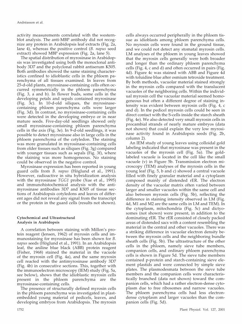
cells always occurred peripherally in the phloem tis-sue as idioblasts among phloem parenchyma cells.No myrosin cells were found in the ground tissue,and we could not detect any stomatal myrosin cells.LM analyses of the phloem in young leaves showedthat the myrosin cells generally were both broaderand longer than the ordinary phloem parenchymacells (Fig. 4, c and d) and often occurred in pairs (Fig.4d). Figure 4c was stained with ABB and Figure 4dwith toluidine blue after osmium tetroxide treatment.By both methods, vacuolar material stained stronglyin the myrosin cells compared with the translucentvacuoles of the neighboring cells. Within the individ-ual myrosin cell the vacuolar material seemed homo-geneous but often a different degree of staining in-tensity was evident between myrosin cells (Fig. 4, cand d). In the pedicel myrosin cells could be seen indirect contact with the S-cells inside the starch sheath(Fig. 4e). We also detected very small myrosin cells inprocambial strands of nearly mature embryos (datanot shown) that could explain the very low myrosi-nase activity found in Arabidopsis seeds (Fig. 2b,column 2).
An IEM study of young leaves using colloidal goldlabeling indicated that myrosinase was present in thevacuoles of the myrosin cell (Fig. 5a). The gold-labeled vacuole is located in the cell like the smallvacuole (v) in Figure 5b. Transmission electron mi-croscopy (TEM) analyses of the myrosin cells in theyoung leaf (Fig. 5, b and c) showed a central vacuolefilled with finely granular material and a cytoplasmcomposed mainly of distended rER. The electrondensity of the vacuolar matrix often varied betweenlarger and smaller vacuoles within the same cell andalso between myrosin cells (Fig. 5b), reflecting thedifference in staining intensity observed in LM (Fig.4d, M1 and M2 are the same cells in LM and TEM). Inthe cytoplasm, mitochondria (Fig. 5c) and dictyo-somes (not shown) were present, in addition to thedominating rER. The rER consisted of closely packedareas of distended sacs with a content resembling thematerial in the central and other vacuoles. There wasa striking difference in vacuolar electron density be-tween the myrosin cells and the neighboring bundlesheath cells (Fig. 5b). The ultrastructure of the othercells in the phloem, namely sieve tube members,companion cells, and ordinary phloem parenchymacells is shown in Figure 5d. The sieve tube memberscontained p-protein and starch-containing sieve ele-ment plastids and were connected by simple sieveplates. The plasmodesmata between the sieve tubemembers and the companion cells were characteris-tically branched (data not shown) toward the com-panion cells, which had a rather electron-dense cyto-plasm due to free ribosomes and narrow vacuoles.The phloem parenchyma cells had less electron-dense cytoplasm and larger vacuoles than the com-panion cells (Fig. 5d).
Andreasson et al.
1752 Plant Physiol. Vol. 127, 2001

Figure 3. Immunohistochemical analysis of myrosinase in Arabidopsis using the monoclonal antibody 3D7. In fully expandedleaves from 25-d-old plants, myrosinase were found in often pair-wise-occurring idioblastic cells of the phloem parenchyma (aand b). b, Larger magnification of a. In flower buds, some cells contained myrosinase (c and f), even in the developing petal andsepal (c). In 10-d-old siliques, the myrosinase containing idioblastic phloem parenchyma cells were larger and the staining wasmore granulated (d and g). d, Arrows indicate long myrosinase-expressing cells. e, The yellowish staining outside the endospermis retained unoxidized substrate and is therefore regarded as background. In 5-d-old seedlings, developing myrosinase-containingphloem cells were only found in the axis. Xylem is marked with � and the size bars correspond to 10 �m.
Myrosinase Localization in Arabidopsis and Brassica napus
Plant Physiol. Vol. 127, 2001 1753

In the pedicel, S-cells were located outside thephloem (Fig. 4, e and f). S-cells had a large diameterin transverse section and were very long, withpointed ends (data not shown). A thin lining of cy-toplasm surrounded a huge, electron-lucent centralvacuole. Organelles in the cytoplasm were sparse,but organelle-like dilated cisterna (DC) of the ER(Bonnett and Newcomb, 1965; Behnke and Eschlbeck,1978) containing homogeneous, granular protein sur-rounded by a ribosome-studded membrane weresometimes found. DCs were also found in ordinaryphloem parenchyma cells (Fig. 5d).
Immunohistochemical Analysis ofMyrosinase in B. napus
Immunohistochemical analyses of myrosinase ex-pression in various B. napus organs were carried outby use of the monoclonal antibody 3D7, emphasizingorgans and tissues that have not been carefully in-vestigated earlier. Myrosinase was found in ground-tissue myrosin cells, as expected, but also in myrosincells of the phloem parenchyma. In very youngflower buds, no staining could be detected, but in thepedicel phloem-specific expression was present (Fig.6b). In the seedling shoot (Fig. 6a), old flower buds(Fig. 6d), siliques (Fig. 6g), and leaves (Fig. 6, h and i),myrosinase could be detected in idioblastic cells inthe ground tissue and also in the phloem paren-chyma. In mature petals (Fig. 6e) and stems (Fig. 6, cand f) only phloem-specific expression was found. Inroots, idioblasts containing myrosinase were presentin the cortex (Fig. 6j). Myrosinase was detected inapproximately 0.1% of the cells in the mesophyll ofthe seedling cotyledons and at a frequency of approx-imately 0.05% of the cells in the leaves. Further,expression of myrosinase in some of the guard cellsin cotyledons of germinating embryos and in youngleaves was found, using the antimyrosinase mono-clonal antibody 3D7 and nonradioactive in situ hy-bridization with a myrosinase B-specific probe (datanot shown). All observations are in agreement withan earlier study (Hoglund et al., 1991), except that we
Figure 4. LM studies of Arabidopsis myrosin cells in young rosetteleaf (a–d) and in pedicel (e). TEM study of S-cells in pedicel (f). a, ABBstaining of paraffin-embedded material that included a myrosin cell(arrow) peripherally in the phloem (P). b, Immunohistochemicalanalysis of myrosinase on a section consecutive to the one shown ina. The antimyrosinase antibody 3D7 indicated myrosinase expres-sion in the same myrosin cell (arrow) as in a. The leaf blade in c wasembedded in glycol methacrylate, sectioned paradermally, and
stained with ABB. It shows the phloem (P), including two long andrelatively broad myrosin cells (M), and mesophyll cells. A parader-mal section of a leaf blade (d) embedded in epoxy resin and stainedwith toluidine blue, including a vascular strand cut slightly obliquelylongitudinally, showed phloem (P) and two adjacent myrosin cells(M1 and M2) located in the phloem parenchyma. e, Transversesection treated as d of a pedicel, with xylem (X), phloem (P), S-cells(S-C), and myrosin cells (M). The S-cells are located between thephloem and the cells (asterisks) of the starch sheath. The myrosin cellto the right is in direct contact with an S-cell. f, Two S-cells (S-C) andthe outer part of the phloem with mature sieve-tube members (ST),immature members (iST), companion cells (CC), and phloem paren-chyma cell (PPC) are shown in transverse section. The S-cells hadlarge, empty vacuoles and a thin cytoplasmic layer in which a dilatedcisternae (DC) of the endoplasmic reticulum (ER) was located. Sizebars in a through e correspond to 10 �m and in f to 1 �m.
Andreasson et al.
1754 Plant Physiol. Vol. 127, 2001

Figure 5. TEM analysis of Arabidopsis phloem of rosette leaf (a–c) and pedicel (d). a, Immunogold labeling with the 3D7antibody in a peripheral vacuole (V) surrounded by cytoplasm in a myrosin cell. Abundant ribosomes in the cytoplasm weresometimes organized as polysomes (R). To the left, two neighboring cells and the common cell wall (CW) are shown. b, Themyrosin cells M1 and M2, also shown in Figure 4d, both exhibited the characteristic high protein content of vacuoles (V)in myrosin cells. The cytoplasm had abundant distended rER and mitochondria. Next to M1, two bundle sheath cells withcell sap in the vacuoles were located. c, Higher magnification of a myrosin cell with cell wall (CW), cytoplasm withmitochondria (Mi), distended rER (ER), and the vacuole (V) with electron-dense homogeneous, granular material. Thedistended ER had abundant ribosomes on the membrane, and the moderately electron-dense material inside the ERresembled the most peripheral part of the vacuolar material. d, The phloem with sieve tube members (ST), companion cell(CC), and phloem parenchyma cell (PPC). The sieve tube members were connected by a sieve plate (SP), and the uppermember showed three sieve element plastids and P protein. The companion cells had a fairly electron-dense cytoplasm andlong, narrow vacuoles. In the ordinary phloem parenchyma cell, the vacuole filled most of the cell lumen and in thecytoplasm was a small DC of the ER with granular content and ribosomes on the membrane. Size bars correspond to 1 �m.
Myrosinase Localization in Arabidopsis and Brassica napus
Plant Physiol. Vol. 127, 2001 1755

Figure 6. Immunohistochemical analysis of myrosinase in B. napus by use of the monoclonal antibody 3D7. In undevelopedflower buds, no staining could be detected, but in the pedicel, phloem-specific expression could be found (b). In the shootapex (a), old flower buds (d), siliques (g), and leaves (h and i), myrosinase could be detected in myrosin cells in the groundtissue and the phloem. In mature petals (e) and stems (c and f), only phloem-specific expression was found. c, Transversesection of a stem; f, Longitudinal section with the outside uppermost. In root, ground tissue idioblasts containing myrosinasewere visible (j). Xylem is marked with � and the size bars correspond to 10 �m.
Andreasson et al.
1756 Plant Physiol. Vol. 127, 2001

could not detect any consistent staining in the xylemin petals and stem.
The Myrosin Grains Constitute a Continuous ReticularSystem in B. napus
TEM analyses have shown that myrosin cells inembryos contain several vacuoles (Rest andVaughan, 1972; Bones and Iversen, 1985). Thesevacuoles contain myrosinase as revealed by IEM(Thangstad et al., 1991; Hoglund et al., 1992) and aredenoted as myrosin grains. Here, we applied confo-cal laser scanning immunomicroscopy analysis tostudy the spatial organization of the myrosin grainsin myrosin cells of the mature embryo. This analysisrevealed that the myrosin grains within one cellseemed to constitute a continuous reticular system, a“myrosin body” (Fig. 7). The sizes of the differentparts of the reticulum varied considerably, and theconnections were sometimes very narrow. The my-rosin body was present throughout the cell and com-prised a significant portion of the cellular volume.The myrosinase staining was not homogeneousthroughout the structure, and central portions withweak or no staining could sometimes be observed.
MBP and Myrosinase Expression in B. napus Seedlings
Immunohistochemical analyses of MBP expressionin B. napus organs were carried out using the mono-clonal antibody S4C6. In consecutive sections, bothmyrosinase (Fig. 8, a, b, and e) and MBP (Fig. 8, c, d,and f) were detected. In 2-d-old seedlings, myrosi-nase was found in idioblastic myrosin cells (Fig. 8, a
and b) and MBP was found in all nonidioblasticground tissue cells, but not in the vascular tissue,epidermis, or idioblastic myrosin cells (arrows indi-cate identical myrosin cell; Fig. 8, c and d). Thus, in2-d-old seedlings, myrosinase and MBP were notcolocalized. However, in 7-d-old seedlings, myrosi-nase (Fig. 8e) and MBP (Fig. 8f) were colocalized inidioblastic myrosin cells in the hypocotyl. Thus, infully developed seedlings, MBP was confined to id-ioblastic structures, which suggests that myrosinase-MBP complexes might exist in vivo. However, it islikely that the myrosinase-MBP complexes do notexist in vivo in seeds.
DISCUSSION
We have shown that myrosinase is present in sim-ilar idioblasts called myrosin cells in both Arabidop-sis and B. napus. However, there is a difference in thelocalization of myrosinase between the two species.In Arabidopsis, myrosinase is confined to cells of thephloem parenchyma, whereas in B. napus myrosinaseis expressed in both the ground tissue and thephloem tissue. The expression pattern of myrosinasein Arabidopsis matched in situ hybridization analy-sis of the published myrosinase genes in Arabidop-sis, TGG1 and TGG2 (Xue et al., 1995). This expres-sion pattern is also in agreement with an earlyinvestigation of myrosin cell localization, findingthese cells in the vascular bundles of Arabidopsis(von Hayek, 1911). Our present data are also inagreement with recent microarray data (Ruan et al.,1998), in which myrosinase was found to be one ofthe highest expressed genes in the flower bud and the
Figure 7. Confocal microscopy analysis of a my-rosin cell in B. napus. Myrosinase was detected byuse of the 3D7 monoclonal antibody. A, Myrosincell at high magnification showing the distributionof myrosinase in a reticular system rather than sep-arate myrosin grains. This part is presented as astereo anaglyph achieved as surface-extracted max-imum intensity projection of a series of confocalimages. B, Single confocal image presenting anoverview of part of a cotyledon from a matureembryo. The arrow indicates the single myrosin cellshown in a. Size bars correspond to 5 �m (A) and50 �m (B).
Myrosinase Localization in Arabidopsis and Brassica napus
Plant Physiol. Vol. 127, 2001 1757

flower. The antibody used recognizes all character-ized myrosinase subfamilies in B. napus (Lenman etal., 1990, 1993; Falk et al., 1995a), and in Arabidopsis,the TGG1 and TGG2 isoforms react with the 3D7antibody, whereas the earlier suggested third my-rosinase gene, tgg3 (Xue et al., 1992), was recentlycharacterized and found to be a pseudogene (J.Zhang, B. Pontoppidan, J. Xue, L. Rask, and J. Meijer,unpublished data).
The glucosinolate-containing S-cells (Koroleva etal., 2000) with translucent vacuoles are structurallydifferent from the phloem myrosin cells with protein-containing vacuoles. This probably means that my-rosinase and glucosinolates are not colocalized in thesame cells, at least not in pedicels of Arabidopsis.Kelly et al. (1998) also showed the presence of glu-cosinolates in non-myrosin cells in imbibed seeds ofB. juncea. This is in contrast to the “mustard oil
Figure 8. Immunohistochemical analysis of myrosinase and MBP expression in B. napus seedlings using the monoclonalantibodies 3D7 and S4C6, respectively. Myrosinase was detected in a, b, and e and MBP in consecutive sections in c, d, andf. In 2-d-old seedlings, myrosinase was found in myrosin cells (a and b) and MBP was found in all cells of the ground tissueexcept for the myrosin cells (c and d). In 7-d-old seedlings, myrosinase and MBP were colocalized (e and f). Size barscorrespond to 10 �m. Arrows indicate the same myrosin cell in consecutive sections.
Andreasson et al.
1758 Plant Physiol. Vol. 127, 2001

bomb” theory (Luthy and Matile, 1984), which pre-dicted myrosinase to be inactivated and localized inthe same cell as glucosinolates. Actually, a recentstudy of uptake of radioactive glucosinolate precur-sors indicated accumulation of at least desulfoglu-cosinolates to occur in the myrosin cells present inthe ground tissue of B. napus developing embryos(Thangstad et al., 2001). Because there is a differencein ontogenetic stage of the investigated tissues in thetwo Brassica spp. used by Thangstad et al. (2001) andKelly et al. (1998), it is possible that during embryodevelopment desulfoglucosinolates are transportedinto the myrosin cells and later the intact glucosino-lates are transported into other cells or become de-graded by myrosinase. Myrosinase probably doesnot degrade desulfoglucosinolates, and thus they canbe colocalized without any reaction occurring. Thiscould explain the available data and is in line withthe transport of alkaloid-precursors to idioblasts sug-gested by St-Pierre et al. (1999). During germination,the glucosinolates may be transported with assis-tance of MBP to the myrosin cells for use as metab-olites (see below). However, further experiments areneeded to clarify this, e.g. in situ expression analysisof glucosinolate biosynthetic genes and cloning of thetransporters.
The presence in Arabidopsis of glucosinolates inthe S-cells and myrosinase in myrosin cells, with thecells adjacent and sometimes in contact with eachother, is an important structural aspect consideringthe proposed functions of the system in nutrition anddefense. In both cases, products may need to berapidly distributed throughout the plant. Transportof secondary compounds have been shown in severalspecies, e.g. for alkaloids (Hashimoto and Yamada,1994; St-Pierre et al., 1999) and glucosinolates(Lykkesfeldt and Møller, 1993; Merritt, 1996; Bru-denell et al., 1999). Glucosinolates have been pro-posed to be transported by the phloem and not by thexylem because myrosinase has been claimed to bepresent in the latter tissue (Brudenell et al., 1999).However, although we found no evidence for my-rosinase in the xylem, myrosin cells in the phloemcontained myrosinase. Ultrastructural studies didnot indicate any differences between phloem andground-tissue myrosin cells (Werker and Vaughan,1976; Jørgensen et al., 1977; Jørgensen, 1981; this pa-per). It has been suggested that the myrosinase-containing cells in B. napus found in the vasculartissue are companion cells (Bones et al., 1991). Thiswould imply a direct plasmodesmatal connection be-tween the sieve tubes and myrosinase-containingcells. However, we found that the phloem myrosincells are different from companion cells, which havesmall vacuoles and free ribosomes in the cytoplasm,whereas myrosin cells have large protein-rich vacu-oles and mainly ER-bound ribosomes. On the otherhand, plasmodesmata occur in myrosin cells (Werkerand Vaughan, 1974; Burmeister et al., 1977; L.B. Jør-
gensen, unpublished data) making symplastic glu-cosinolate transport to the myrosin cell possible. Al-though the cells may have cytoplasmic connection,there is a need for tonoplast transporters becauseboth glucosinolates and myrosinase are reported tobe located in vacuoles (Thangstad et al., 1991; Ho-glund et al., 1992; Kelly et al., 1998).
It is generally believed that idioblasts have evolvedto have different functions in different plant taxa(Mauseth, 1988). For example, tobacco (Nicotianatabacum) idioblasts contain pathogenesis-related pro-teins (Dixon et al., 1991), and calcium-accumulatingidioblasts are present in water lettuce (Pistia stratiotes;Franceschi et al., 1993). In Arabidopsis leaves, theidioblasts often occur next to each other (Figs. 3a and4d), indicating that after idioblast determination, ad-ditional cell division events occur. This is also thecase during differentiation of stomatal myrosin cells(Jørgensen, 1995). Different initial cells in the meris-tem give rise to the vascular system and the groundtissue. The ground tissue initial cells in Arabidopsismay not have the capacity to form idioblasts, andtherefore myrosinase-containing cells are lacking inthis tissue. The analysis of the subcellular organiza-tion of myrosinase in myrosin cells of B. napus em-bryos showed that the myrosin grains actuallyformed a continuous reticulum throughout the cell.Therefore, we suggest denoting the myrosinase-containing structure as a myrosin body rather thanmyrosin grains (Rest and Vaughan, 1972; Werker andVaughan, 1974). This organization of the mature my-rosin body may simply be a consequence of aprotein-accumulating vacuole that slowly expandsirregularly in the cell as proteins are accumulating.The relative concentration of myrosinase as judgedfrom fluorescence intensity varied throughout thestructure suggesting that additional molecules arepresent. Also, there seems to be an age-dependenteffect on the reticular system in the mature plantbecause older myrosin cells from the siliques ap-peared to be more granulated than younger ones.
In general, members of the Brassicaceae are fastinvaders that have many specialist enemies, i.e. in-sects that only attack plants from this family. Thus, itis logical to consider the special secondary metabo-lites, the glucosinolates, in this context. However,Arabidopsis produces many small seeds and oftencompletes the life cycle often early in the season,thereby avoiding massive insect attack and accord-ingly can be regarded as a herbivory escaper. Thereare also differences in the myrosinase gene familybetween Arabidopsis and larger summer-living Bras-sicaceae members in the tribus Brassiceae such as B.napus and Sinapis alba (Xue et al., 1992). Arabidopsisseems to have only two myrosinase genes that areexpressed exclusively in the phloem parenchyma,whereas the larger Brassicaceae members have ap-proximately 20 genes expressed in both the groundtissue and the phloem parenchyma (Rask et al., 2000;
Myrosinase Localization in Arabidopsis and Brassica napus
Plant Physiol. Vol. 127, 2001 1759

Eriksson et al., 2001; this paper). The difference isespecially pronounced in the seed wherein hardlyany myrosinase is expressed in Arabidopsis, whereasin B. napus, members of all three myrosinase genefamilies are transcribed. Actually, the seed is the onlyorgan in which all three myrosinase gene families areexpressed in B. napus. In the Brassiceae (also includ-ing Raphanus sp., Brassica oleracea, and wild Rhyncho-sinapis cheiranthos), the levels of the myrosinase-glucosinolate system correlate better with the cost ofherbivory than in Arabidopsis, if the seed is consid-ered to be a costly organ to lose because it is essentialfor survival. In Arabidopsis seeds, the glucosinolatesmay be regarded as storage compounds used duringlater stages of germination, as suggested in B. napusby James and Rossiter (1991), because there is hardlyany myrosinase present to produce toxic compoundsalthough glucosinolates are abundant (Hogge et al.,1988; Haughn et al., 1991).
We have shown that the idioblastic myrosin cellschange composition during seedling developmentbecause MBP expression changes considerably. Glu-cosinolates and MBP are probably colocalized in theB. napus seed (Kelly et al., 1998; this paper), butduring germination MBP disappears outside myrosincells, concominant with a dramatic decrease ofmainly aliphatic glucosinolates (e.g. Clossais-Besnard and Larher, 1991). This is also in agreementwith the observation by James and Rossiter (1991)that the myrosinase activity increased during thisperiod. This might suggest a general storage protein-related function for MBP but also that MBP could beinvolved in the degradation of glucosinolates by be-ing a part of the transport mechanism to the myrosincells in which myrosinases degrade the glucosino-lates into nutritional components. The presence ofMBP in myrosin cells later during development maysupport the latter idea. However, MBPs constitute agene family with several members in which the dif-ferent genes are structurally distinct although certainmotifs are shared (Taipalensuu et al., 1997b) Theexistence of MBP in Arabidopsis is not clear at themoment. Neither has any cross-reaction been de-tected with the B. napus anti-MBP antibody 34:14 inwestern-blot analysis (Fig. 2, Falk et al., 1995b), norhas any specific Southern-blot hybridization signalusing two MBP probes (Taipalensuu et al., 1997b)been detected in Arabidopsis. A thorough analysisof the Arabidopsis genome for MBP homologs hasindicated the presence of several genes containingvarious domains present in B. napus MBP butno full-length homolog (B. Pontoppidan and JohanMeijer, unpublished data). These results make it un-likely that a functional MBP homolog exists in Ara-bidopsis, and maybe MBPs are correlated to the pres-ence of ground tissue myrosin cells, because B. napus,Brassica nigra, S. alba, and R. cheiranthos all containedMBPs and ground-tissue myrosin cells (Falk et al.,1995b; E. Andreasson and M. Lenman, unpublished
data). This investigation calls for further studies toidentify species-specific structures, regulation, andfunctions of the glucosinolate-myrosinase system,which may limit the usefulness of Arabidopsis as amodel system for Brassicaceae.
MATERIALS AND METHODS
Plant Material
Brassica napus L. cv Karat (Svalof Weibull AB, Svalov,Sweden) and Arabidopsis ecotype Columbia (NottinghamArabidopsis Stock Centre, Nottingham, UK) were used.The plants were grown in standard soil under greenhouseconditions or in a climate chamber with 70% relative hu-midity using a 16-h daylight/21°C and 8-h darkness/17°Cregime. Investigations of the pedicel were carried out inecotype Wassiljewskija because the reported characteriza-tion of S-cells was carried out using this ecotype (Korolevaet al., 2000).
Western-Blot Analysis
Extracts of seeds and leaves were prepared in SDS buffer(1.3 mm Tris, pH 8.8, 0.003% [w/v] bromphenol blue, 9%[w/v] Suc, 1.3% [v/v] SDS, and 8 mm dithiothreitol). Sep-aration by SDS-PAGE (Minigel, Bio-Rad Laboratories,Hemel Hempstead, UK) and semidry transfer (Bio-RadLaboratories) of the proteins to Hybond C nitrocellulosemembrane (Amersham-Pharmacia Biotech, Uppsala) werecarried out according to the manufacturer’s instructions. A3D7 mouse hybridoma supernatant at the dilution 1:300was used to detect myrosinase and a 34:14 antibody hy-bridoma supernatant at the dilution 1:1,000 was used todetect MBP (Lenman et al., 1990; Falk et al., 1995b). Thefurther detection was carried out as described (Chen andHalkier, 1999), except that the developing reagent waspurchased from Pierce (Rockford, IL).
Myrosinase Activity Assay
Leaves and seeds were extracted in 0.5 mL of 50 mmTris-HCl, pH 7.5, 0.2 m NaCl, 1 mm EDTA, and 1 mmphenylmethylsulfonyl fluoride for 1 h, and the supernatantwas passed through a Sephadex G-25 column (Amersham-Pharmacia Biotech) equilibrated with the extraction buffer.Myrosinase activity was measured by monitoring Glc re-lease after sinigrin hydrolysis using a kit (Randox Labora-tories Ltd, Ardmore, UK) in 50 mm citrate buffer, pH 4.5,and 0.3 mm ascorbate. Protein concentration was deter-mined by the Bradford method according to the manufac-turer using bovine serum albumin as the standard (Bio-RadLaboratories).
Immunohistochemical Analysis
The plant material was fixed for 16 to 48 h at 4°C in4% (w/v) paraformaldehyde and 0.25% (v/v) glutaralde-hyde. Paraffin embedding, binding of the anti-myrosinaseantibody, and staining using the peroxidase-antiperoxidase
Andreasson et al.
1760 Plant Physiol. Vol. 127, 2001

procedure were carried out as previously described (Hog-lund et al., 1991), except that 4% (w/v) bovine serumalbumin was used as blocking agent. The ascites propagatedantibodies 3D7 at the dilution 1:500 to 1:2,000 and S4C6 atthe dilution 1:100 were used to detect myrosinase and MBP,respectively (Lenman et al., 1990; Geshi et al., 1998). Theantimyrosinase polyclonal antisera K505 was diluted1:12,000 (Lenman et al., 1990; Hoglund et al., 1991). Negativecontrol stainings with an ascites anticruciferin antibody withthe same total protein amount as the specific antibodieswere also analyzed. Sections, consecutive to the 3D7 anti-body treated, were stained with ABB (see below) and dehy-drated in ethanol before mounting. These 3D7 antibodytreated sections were processed with DAKO AEC�Highsensitivity substrate-chromogen system (DAKO, Carpinte-ria, CA) for visualization of the peroxidase-antiperoxidasereaction before mounting in glycerin gelatin.
Cytochemical and Ultrastructural Analysis
Standard methods were used for ultrastructural analysisby TEM. Small pieces of tissue were fixed overnight at 5°Cin 2.5% (v/v) glutaraldehyde and 2% (w/v) paraformalde-hyde in 0.1 m phosphate buffer, pH 7.0, rinsed in buffer,and postfixed in 1% (w/v) OsO4 in the same buffer for 2 hat room temperature. After rinsing, the material was dehy-drated in a graded acetone series before infiltration andembedding in Spurr’s epoxy resin (Spurr, 1969). Ultrathinsections were cut on a Supernova ultramicrotome(Reichert-Jung, Heidelberg), collected on grids, and con-trasted with uranyl acetate and lead citrate before beingexamined in a JEM 100CX microscope (JEOL, Tokyo). ForLM analysis with a Reichert-Jung Polyvar microscope, 1- to2-�m-thick sections of the material embedded in Spurrwere stained in 0.05% (w/v) toluidine blue (ToluidinblauO, Merck, Darmstadt, Germany) in 1% (w/v) borax (sodi-um tetra borate), pH 8.4. Also, 3-�m-thick sections of tissueembedded in glycol methacrylate were analyzed after fix-ation as above in the aldehyde-mixture and dehydrationwith methoxyethanol, ethanol, propanol, and butanol be-fore infiltration with the plastic (Feder and O’Brien, 1968).These sections were stained with the protein reagent ABB(Naphtol Blue Black, Sigma, St. Louis) for cytochemicalidentification of myrosin cells in green tissue as describedby Jørgensen et al. (1977) and Jørgensen (1995).
IEM
The fixation and incubation with the 3D7 antibody wasdone according to Hoglund et al. (1992), except that thedilution of the 3D7 antibody was 1:300. The blocking wasachieved in 4% (w/v) bovine serum albumin and collodialgold-labeled protein A (20 nm) from Sigma was used todetect antibody bounding. The grids were contrasted withuranyl acetate and lead citrate and examined in a JEOLJEM 100CX microscope.
Immunofluorescence Staining and Confocal LaserScanning Microscopy Analysis
Whole seeds of B. napus (a dihaploid line 20516-K of cvKarat) were fixed in 4% (w/v) paraformaldehyde inphosphate-buffered saline (PBS) for 12 h at 4°C. Sampleswere dehydrated through a graded series of ethanol fol-lowed by incubation in xylene before infiltration and em-bedding in Histowax (Histolab, Goteborg, Sweden). Sec-tions (5 �m thick) were placed on chromogelatine-coatedcoverslips. Before staining, sections were xylene treated,followed by rehydration in decreasing concentrations ofethanol and distilled water followed by a wash in PBS.After treatment with blocking buffer (normal goat serum1:10 in PBS with 4% [w/v] bovine serum albumin) for 30min, sections were incubated for 30 min with the monoclo-nal antibody 3D7 directed against myrosinase (1:400) andfollowed by an incubation with a tetramethyl rhodamineisothiocyanate-labeled rabbit anti-mouse immunoglobulinantiserum (1:200; Dakopatts AB, Alvsjo, Sweden) for 30min. Sections were mounted in Moviol. Images were re-corded using a system equipped with a Nikon invertedmicroscope and argon/krypton laser, purchased from Mo-lecular Dynamics (Sunnyvale, CA). Fluorochrome-labeledsamples were excited by a 543-nm line laser.
ACKNOWLEDGMENTS
We are grateful to Ulla Pihlgren and Lis Munk Frederik-sen for technical assistance and Leif Bolding for graphicassistance. Bo Pontoppidan is thanked for Figure 1.
Received April 9, 2001; returned for revision June 18, 2001;accepted August 29, 2001.
LITERATURE CITED
Behnke HD, Eschlbeck G (1978) Dilated cisternae in Cap-parales: an attempt towards the characterization of aspecific endoplasmic reticulum. Protoplasma 97: 351–363
Bodnaryk RP (1992) Effects of wounding on glucosinolatesin the cotyledons of oilseed rape and mustard. Phyto-chemistry 31: 2671–2677
Bones A, Iversen TH (1985) Myrosin cells and myrosinase.Isr J Bot 34: 351–376
Bones A, Rossiter JT (1996) The myrosinase-glucosinolatesystem, its organization and biochemistry. Physiol Plant97: 194–208
Bones A, Thangstad OP, Haugen OA, Espevik T (1991)Fate of myrosin cells: characterization of monoclonalantibodies against myrosinase. J Exp Bot 42: 1541–1549
Bonnett HT, Newcomb EH (1965) Polyribosomes and cis-ternal accumulations in root cells of radish. J Cell Biol 27:423–432
Brudenell AJP, Griffiths H, Rossiter JT, Baker DA (1999)The phloem mobility of glucosinolates. J Exp Bot 50:745–756
Burmeister WP, Cottaz S, Driguez H, Iori R, Palmieri S,Henrissat B (1997) The crystal structures of Sinapis albamyrosinase and a covalent glycosyl-enzyme intermedi-
Myrosinase Localization in Arabidopsis and Brassica napus
Plant Physiol. Vol. 127, 2001 1761

ate provide insights into the substrate recognition andactive-site machinery of an S-glycosidase. Structure 5:663–675
Chen S, Halkier BA (1999) Functional expression and char-acterization of the myrosinase MYR1 from Brassica napusin Saccharomyces cerevisiae. Prot Exp Purif 17: 414–420
Chew FS (1988) Biological effects of glucosinolates. In HGCutler, ed, Biologically Active Natural Products for Po-tential Use in Agriculture. American Chemical Society,Washington, DC, pp 155–181
Clossais-Besnard N, Larher F (1991) Physiological role ofglucosinolates in Brassica napus: concentration and dis-tribution pattern of glucosinolates among plant organsduring a complete life cycle. J Sci Food Agric 56: 25–38
Dixon DC, Cutt JR, Klessig DF (1991) Differential target-ing of the tobacco PR-1 pathogenesis-related proteins tothe extracellular space and vacuoles of crystal idioblasts.EMBO J 10: 1317–1324
Doughty K, Kiddle G, Pye B, Wallsgrove R, Pickett J(1995). Selective induction of glucosinolates in oilseedrape leaves by methyl jasmonate. Phytochemistry 38:347–350
Eriksson S, Ek B, Xue J, Rask L, Meijer J (2001) Identifi-cation and characterization of soluble and insoluble my-rosinases in different organs of Sinapis alba. Physiol Plant111: 353–364
Falk A, Ek B, Rask L (1995a) Characterization of a newmyrosinase in Brassica napus. Plant Mol Biol 27: 863–874
Falk A, Taipalensuu J, Ek B, Lenman M, Rask L (1995b)Characterization of rapeseed myrosinase-binding pro-tein. Planta 195: 387–395
Feder N, O’Brien TP (1968) Plant microtechnique: someprinciples and new methods. Am J Bot 55: 123–142
Fisher DB (1968) Protein staining of ribboned epon sec-tions for light microscopy. Histochemistry 16: 92–96
Franceschi VR, Li X, Zhang D, Okita TW (1993) Caseques-trinlike calcium-binding proteins is expressed incalcium-accumulating cells of Pistia stratiotes. Proc NatlAcad Sci USA 90: 6986–6990
Geshi N, Andreasson E, Meijer J, Rask L, Brandt A (1998)Myrosinase and myrosinase-binding proteins are co-localized in grains of myrosin cells in cotyledon of Bras-sica napus L. seedlings. Plant Physiol Biochem 36:583–590
Geshi N, Brandt A (1998) Two jasmonate-induciblemyrosinase-binding proteins from Brassica napus L. seed-lings with homology to jacalin. Planta 204: 295–304
Hashimoto T, Yamada Y (1994) Alkaloid biogenesis: mo-lecular aspects. Annu Rev Plant Physiol Plant Mol Biol45: 257–285
Haughn GW, Davin L, Giblin M, Underhill EW (1991)Biochemical genetics of plant secondary metabolites inArabidopsis thaliana: the glucosinolates. Plant Physiol 97:217–226
Hogge LR, Reed DW, Underhill EW, Haughn GW (1988)HPLC separation of glucosinolates from leaves and seedsof Arabidopsis thaliana and their identification using ther-mospray liquid chromatography/mass spectrometry.J Chromatogr Sci 26: 551–556
Hoglund AS, Lenman M, Falk A, Rask L (1991) Distribu-tion of myrosinase in rapeseed tissues. Plant Physiol 95:213–221
Hoglund AS, Lenman M, Rask L (1992) Myrosinase islocalized to the interior of myrosin grains and is notassociated to the surrounding tonoplast membrane.Plant Sci 85: 165–170
James DC, Rossiter JT (1991) Development and character-istics of myrosinase in Brassica napus during early seed-ling growth. Physiol Plant 82: 163–170
Jensen WA (1962) Botanical Histochemistry: Principles andPractice. W.H. Freeman, San Francisco, pp 228–229
Jørgensen LB (1981) Myrosin cells and dilated cisternae ofthe endoplasmic reticulum in the order Capparales.Nord J Bot 1: 433–445
Jørgensen LB (1995) Stomatal myrosin cells in Caricaceae:taxonomic implications for a glucosinolate-containingfamily. Nord J Bot 15: 523–540
Jørgensen LB, Behnke HD, Mabry TJ (1977) Protein-accumulating cells and dilated cisternae of the endoplas-mic reticulum in three glucosinolate-containing genera:Armoracia, Capparis, Drypetes. Planta 137: 215–224
Kelly PJ, Bones A, Rossiter JT (1998) Sub-cellular immu-nolocalization of the glucosinolate sinigrin in seedlingsof Brassica juncea. Planta 206: 370–377
Koroleva OA, Davies A, Deeken R, Thorpe MR, TomosAD, Hedrich R (2000) Identification of a newglucosinolate-rich cell type in Arabidopsis flower stalk.Plant Physiol 124: 599–608
Lenman M, Falk A, Rodin J, Hoglund AS, Ek B, Rask L(1993) Differential expression of myrosinase gene fami-lies. Plant Physiol 103: 703–711
Lenman M, Rodin J, Josefsson LG, Rask L (1990) Immu-nological characterization of rapeseed myrosinase. EurJ Biochem 194: 747–753
Louda S, Mole S (1991) Glucosinolates, chemistry andecology. In GA Rosenthal, MR Berenbaum, eds, Herbi-vores: Their Interactions with Secondary Plant Metabo-lites. Academic Press, San Diego, pp 123–164
Luthy B, Matile P (1984) The mustard oil bomb: rectifiedanalysis of the subcellular organisation of the myrosi-nase system. Biochem Physiol Pflanz 179: 5–12
Lykkesfeldt J, Møller BL (1993) Synthesis of benzylglu-cosinolate in Tropaeolum majus L.: isothiocyanates as po-tent enzyme inhibitors. Plant Physiol 102: 609–613
Mauseth JD (1988) Plant Anatomy. Benjamin/Cummings,Menlo Park, CA
Merritt SZ (1996) Within plant variation in concentrationsof amino acids, sugar, and sinigrin in phloem sap ofblack mustard, Brassica nigra (L) Koch (Cruciferae).J Chem Ecol 22: 1133–1145
Rask L, Andreasson E, Ekbom B, Eriksson S, Pontoppi-dan B, Meijer J (2000) Myrosinase: gene family evolutionand herbivory defense in Brassicaceae. Plant Mol Biol 42:93–113
Rest JA, Vaughan GJ (1972) The development of proteinand oil bodies in the seed of Sinapis alba L. Planta 105:245–262
Andreasson et al.
1762 Plant Physiol. Vol. 127, 2001

Rodman JE, Karol KG, Price RA, Sytsma KJ (1996) Mole-cules, morphology, and Dahlgren’s expanded order Cap-parales. Syst Bot 21: 289–307
Rosa EAS, Heaney RK, Fenwick GR, Portas CAM (1997)Glucosinolates in crop plants. Hortic Rev 19: 99–215
Ruan Y, Gilmore J, Conner T (1998) Towards Arabidopsisgenome analysis: monitoring expression profiles of 1400genes using cDNA microarrays. Plant J 15: 821–833
Spurr AR (1969) A low-viscosity epoxy resin embeddingmedium for electron microscopy. J Ultrastr Res 26: 31–43
St-Pierre B, Vazquez-Flota A, De Luca V (1999) Multicel-lular compartmentation of Catharanthus roseus alkaloidbiosynthesis predicts intercellular translocation of path-way intermediate. Plant Cell 11: 887–900
Taipalensuu J, Eriksson S, Rask L (1997a) The myrosinase-binding protein from Brassica napus seeds possesses lec-tin activity and has a highly similar vegetatively ex-pressed wound-inducible counterpart. Eur J Biochem250: 680–688
Taipalensuu J, Falk A, Ek B, Rask L (1997b) Myrosinase-binding proteins are derived from a large wound-inducible and repetitive transcript. Eur J Biochem 243:605–611
Taipalensuu J, Falk A, Rask L (1996) A wound- andmethyl jasmonate-inducible transcript coding for amyrosinase-associated protein with similarities to anearly nodulin. Plant Physiol 110: 483–491
Thangstad OP, Bones AM, Holtan S, Moen L, Rossiter J(2001) Microautoradiographic localization of a glucosi-nolate precursor to specific cells in Brassica napus L.embryos indicates a separate transport pathway into my-rosin cells. Planta 213: 207–213
Thangstad OP, Evjen K, Bones A (1991) Immunogold-EMlocalization of myrosinase in Brassicaceae. Protoplasma161: 85–93
Thangstad OP, Iversen T-H, Slupphaug G, Bones A (1990)Immunocytochemical localization of myrosinase in Bras-sica napus L. Planta 180: 245–248
von Hayek A (1911) Entwurf eines Cruciferen-Systems aufphylogenetischer Grundlage. Beihefte Bot Centralblatt27: 127–335
Werker E, Vaughan JG (1974) Anatomical and ultrastruc-tural changes in aleurone and myrosin cells of Sinapisalba during germination. Planta 116: 243–255
Werker E, Vaughan JG (1976) Ontogeny and distributionof myrosin cells in the shoot of Sinapis alba L.: a light- andelectron-microscope study. Isr J Bot 25: 140–151
Xue J, Jørgensen M, Pihlgren U, Rask L (1995) The myrosi-nase gene family in Arabidopsis thaliana: gene organization,expression and evolution. Plant Mol Biol 27: 911–922
Xue J, Lenman M, Falk A, Rask L (1992) The glucosinolate-degrading enzyme myrosinase in Brassicaceae is en-coded by a gene family. Plant Mol Biol 18: 387–398
Myrosinase Localization in Arabidopsis and Brassica napus
Plant Physiol. Vol. 127, 2001 1763