Dietary sophorolipid accelerates growth by modulation of gut ...
9
RESEARCH Open Access Dietary sophorolipid accelerates growth by modulation of gut microbiota population and intestinal environments in broiler chickens Min-Jin Kwak 1† , Min-Young Park 1† , Yong-Soon Choi 1 , Junghwan Cho 1 , Duleepa Pathiraja 1 , Jonggun Kim 1 , Hanbae Lee 2 , In-Geol Choi 1 and Kwang-Youn Whang 1* Abstract Background: Gut is a crucial organ for the host’s defense system due to its filtering action of the intestinal membrane from hazardous foreign substances. One strategy to strengthen the gut epithelial barrier function is to upregulate beneficial microflora populations and their metabolites. Sophorolipid (SPL), which is a glycolipid bio- surfactant, could increase beneficial microflora and decrease pathogenic bacteria in the gastrointestinal tract. Therefore, herein, we conducted an experiment with broiler chickens to investigate the fortifying effects of SPL on the host’s gut defense system by modulating the microbiota population. Methods: A total of 540 1-day-old chicks (Ross 308) were used, and they were immediately allotted into three treatment groups (6 replications with 30 chicks/pen) according to their initial body weight. The dietary treatments consisted of CON (basal diet), BAM (10 mg/kg bambermycin), and SPL (10 mg/kg SPL). During the experiment, birds freely accessed feed and water, and body weight and feed intake were measured at the end of each phase. On d 35, birds (one bird/pen) were sacrificed to collect jejunum and cecum samples. Results: Dietary SPL and BAM supplementation significantly accelerated birds’ growth and also significantly improved feed efficiency compared to CON. Intestinal microbial community was significantly separated by dietary SPL supplementation from that of CON, and dietary SPL supplementation significantly increased Lactobacillus spp. and Akkermansia muciniphila. Moreover, birds fed with dietary SPL also showed the highest concentration of cecal butyrate among all treatment groups. Gut morphological analysis showed that dietary SPL significantly increased villus height, ratio of villus height to crypt depth, goblet cell numbers, and the gene expression levels of claudin-1 and mucin 2. Additionally, dietary SPL significantly decreased the mRNA expression level of pro-inflammatory cytokine, interleukin-6, and increased that of anti-inflammatory cytokine, interleukin-10, compared to other treatments. © The Author(s). 2021 Open Access This article is licensed under a Creative Commons Attribution 4.0 International License, which permits use, sharing, adaptation, distribution and reproduction in any medium or format, as long as you give appropriate credit to the original author(s) and the source, provide a link to the Creative Commons licence, and indicate if changes were made. The images or other third party material in this article are included in the article's Creative Commons licence, unless indicated otherwise in a credit line to the material. If material is not included in the article's Creative Commons licence and your intended use is not permitted by statutory regulation or exceeds the permitted use, you will need to obtain permission directly from the copyright holder. To view a copy of this licence, visit http://creativecommons.org/licenses/by/4.0/. The Creative Commons Public Domain Dedication waiver (http://creativecommons.org/publicdomain/zero/1.0/) applies to the data made available in this article, unless otherwise stated in a credit line to the data. * Correspondence: [email protected] † Min-Jin Kwak and Min-Young Park contributed equally to this work. 1 Department of Biotechnology, Korea University, 145 Anam-ro, Seoul 02841, Republic of Korea Full list of author information is available at the end of the article Kwak et al. Journal of Animal Science and Biotechnology (2021) 12:81 https://doi.org/10.1186/s40104-021-00606-x
-
Upload
khangminh22 -
Category
Documents
-
view
0 -
download
0
Transcript of Dietary sophorolipid accelerates growth by modulation of gut ...

RESEARCH Open Access
Dietary sophorolipid accelerates growth bymodulation of gut microbiota populationand intestinal environments in broilerchickensMin-Jin Kwak1†, Min-Young Park1†, Yong-Soon Choi1, Junghwan Cho1, Duleepa Pathiraja1, Jonggun Kim1,Hanbae Lee2, In-Geol Choi1 and Kwang-Youn Whang1*
Abstract
Background: Gut is a crucial organ for the host’s defense system due to its filtering action of the intestinalmembrane from hazardous foreign substances. One strategy to strengthen the gut epithelial barrier function is toupregulate beneficial microflora populations and their metabolites. Sophorolipid (SPL), which is a glycolipid bio-surfactant, could increase beneficial microflora and decrease pathogenic bacteria in the gastrointestinal tract.Therefore, herein, we conducted an experiment with broiler chickens to investigate the fortifying effects of SPL onthe host’s gut defense system by modulating the microbiota population.
Methods: A total of 540 1-day-old chicks (Ross 308) were used, and they were immediately allotted into threetreatment groups (6 replications with 30 chicks/pen) according to their initial body weight. The dietary treatmentsconsisted of CON (basal diet), BAM (10 mg/kg bambermycin), and SPL (10 mg/kg SPL). During the experiment, birdsfreely accessed feed and water, and body weight and feed intake were measured at the end of each phase.On d 35, birds (one bird/pen) were sacrificed to collect jejunum and cecum samples.
Results: Dietary SPL and BAM supplementation significantly accelerated birds’ growth and also significantlyimproved feed efficiency compared to CON. Intestinal microbial community was significantly separated bydietary SPL supplementation from that of CON, and dietary SPL supplementation significantly increasedLactobacillus spp. and Akkermansia muciniphila. Moreover, birds fed with dietary SPL also showed the highestconcentration of cecal butyrate among all treatment groups. Gut morphological analysis showed that dietarySPL significantly increased villus height, ratio of villus height to crypt depth, goblet cell numbers, and thegene expression levels of claudin-1 and mucin 2. Additionally, dietary SPL significantly decreased the mRNAexpression level of pro-inflammatory cytokine, interleukin-6, and increased that of anti-inflammatory cytokine,interleukin-10, compared to other treatments.
© The Author(s). 2021 Open Access This article is licensed under a Creative Commons Attribution 4.0 International License,which permits use, sharing, adaptation, distribution and reproduction in any medium or format, as long as you giveappropriate credit to the original author(s) and the source, provide a link to the Creative Commons licence, and indicate ifchanges were made. The images or other third party material in this article are included in the article's Creative Commonslicence, unless indicated otherwise in a credit line to the material. If material is not included in the article's Creative Commonslicence and your intended use is not permitted by statutory regulation or exceeds the permitted use, you will need to obtainpermission directly from the copyright holder. To view a copy of this licence, visit http://creativecommons.org/licenses/by/4.0/.The Creative Commons Public Domain Dedication waiver (http://creativecommons.org/publicdomain/zero/1.0/) applies to thedata made available in this article, unless otherwise stated in a credit line to the data.
* Correspondence: [email protected]†Min-Jin Kwak and Min-Young Park contributed equally to this work.1Department of Biotechnology, Korea University, 145 Anam-ro, Seoul 02841,Republic of KoreaFull list of author information is available at the end of the article
Kwak et al. Journal of Animal Science and Biotechnology (2021) 12:81 https://doi.org/10.1186/s40104-021-00606-x
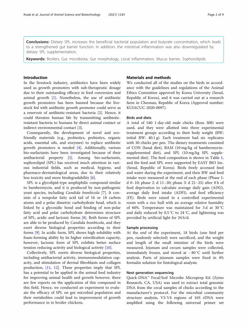
Conclusions: Dietary SPL increases the beneficial bacterial population and butyrate concentration, which leadsto a strengthened gut barrier function. In addition, the intestinal inflammation was also downregulated bydietary SPL supplementation.
Keywords: Broilers, Gut microbiota, Gut morphology, Local inflammation, Mucus barrier, Sophorolipids
IntroductionIn the livestock industry, antibiotics have been widelyused as growth promoters with sub-therapeutic dosagedue to their outstanding efficacy in feed conversion andanimal growth [1]. Nonetheless, the use of antibioticgrowth promoters has been banned because the live-stock fed with antibiotic growth promoter could serve asa reservoir of antibiotic-resistant bacteria [2]. Hence, itcould threaten human life by transmitting antibiotic-resistant bacteria to humans by direct animal contact orindirect environmental contact [3].Consequently, the development of novel and eco-
friendly materials (e.g., probiotics, prebiotics, organicacids, essential oils, and enzymes) to replace antibioticgrowth promoters is needed [4]. Additionally, variousbio-surfactants have been investigated because of theirantibacterial property [5]. Among bio-surfactants,sophorolipid (SPL) has received much attention in vari-ous industrial fields, such as medical, hygiene, andpharmaco-dermatological areas, due to their relativelyless toxicity and more biodegradability [6].SPL is a glycolipid-type amphiphilic compound similar
to bambermycin, and it is produced by non-pathogenicyeast species, including Candida bombicola [7]. It con-sists of a nonpolar fatty acid tail of 16 or 18 carbonatoms and a polar dimetric carbohydrate head, which islinked by a glycosidic bond and binding of non-polarfatty acid and polar carbohydrate determines structureof SPL, acidic and lactonic forms [8]. Both forms of SPLare able to be produced by Candida bombicola, and theyexert diverse biological properties according to theirforms [9]. In acidic form, SPL shows high solubility withfoam-forming ability by its higher esterification capacity,however, lactonic form of SPL exhibits better surfacetension reducing activity and biological activity [10].Collectively, SPL exerts diverse biological properties,
including antibacterial activity, immunomodulation cap-acity, and stimulation of dermal fibroblasts and collagenproduction, [11, 12]. These properties imply that SPLhas a potential to be applied in the animal feed industryfor improving animal health and growth however, thereare few reports on the application of this compound inthis field. Hence, we conducted an experiment to evalu-ate the efficacy of SPL on gut microbial population andtheir metabolites could lead to improvement of growthperformance in in broiler chickens.
Materials and methodsWe conducted all of the studies on the birds in accord-ance with the guidelines and regulations of the AnimalEthics Committee approved by Korea University (Seoul,Republic of Korea), and it was carried out at a researchfarm in Cheonan, Republic of Korea (Approval number:KUIACUC-2020-0097).
Birds and dietsA total of 540 1-day-old male chicks (Ross 308) wereused, and they were allotted into three experimentaltreatment groups according to their body weight (BW;initial BW: 40.1 g). Each treatment had six replicateswith 30 chicks per pen. The dietary treatments consistedof CON (basal diet), BAM (10mg/kg of bambermycin-supplemented diet), and SPL (10 mg/kg SPL supple-mented diet). The feed composition is shown in Table 1,and the feed and SPL were supported by EASY BIO Inc.(Seoul, Republic of Korea). Birds freely accessed feedand water during the experiment, and their BW and feedintake were measured at the end of each phase (Phase 1:d 0–10; phase 2: d 11–20; phase 3: d 21–35) after 8 h offeed deprivation to calculate average daily gain (ADG),average daily feed intake (ADFI), and feed efficiency(FE). Birds were raised in a controlled experimentalroom with a rice hull with an average relative humidityof 60%. Temperature was maintained for 3 d at 30 °Cand daily reduced by 0.5 °C to 24 °C, and lightening wasprovided by artificial light for 24 h/d.
Sample processingAt the end of the experiment, 18 birds (one bird perpen, randomly selected) were sacrificed, and the weightand length of the small intestine of the birds weremeasured. Jejunum and cecum samples were collected,immediately frozen, and stored at − 80 °C until furtheranalysis. Parts of jejunum samples were fixed in 4%formalin solution for histological analysis.
Next generation sequencingQuick-DNA™ Fecal/Soil Microbe Microprep Kit (ZymoResearch, CA, USA) was used to extract total genomicDNA from the cecal samples of chicks according to themanufacturer’s protocol. For the microbial communitystructure analysis, V3-V4 regions of 16S rDNA wereamplified using the following universal primer set
Kwak et al. Journal of Animal Science and Biotechnology (2021) 12:81 Page 2 of 9

(forward 5′-CCTACGGGNGGCWGCAG-3′, reverse 5′-GACTACHVGGGTATCTAATCC-3′), synthesized byIntegrated DNA Technologies (IDT, Singapore). PCR con-ditions for amplifying the V3-V4 regions of 16S rDNAwere as follows; 5-min initial denaturation at 98 °C,followed by 20 amplification cycles (30 s at 98 °C, 30 s at56 °C, 1 min 30 s at 72 °C), 5 min final extension at 72 °C.The resulting DNA amplicons were purified using mag-netic beads (TopQ XSEP MagBead, CELLMICS, USA). A
second PCR was performed to attach the Illumina univer-sal p5/p7 overhang sequence and sample-specific bar-codes. Second PCR conditions were as follows; 1-mininitial denaturation at 98 °C, followed by 10 amplificationcycles (30 s at 98 °C, 30 s at 60 °C, 1min at 72 °C), final ex-tension at 72 °C 3min. The sequencing-ready librarieswere purified using magnetic beads (TopQ XSEP Mag-Bead, CELLMICS, USA). Size distribution of the sequen-cing ready libraries were determined using the QSepfragment analyzer (Qsep Inc., Taiwan). DNA quantifica-tion is performed using the Quit dsDNA HS assay kit(Thermo Scientific, USA). Finally, all the libraries werepooled in equimolar quantities and diluted as necessary.Sequencing was performed on the Illumina MiSeq plat-form (Illumina, USA) using MiSeq Reagent Kit V3 (2 ×300 PE) (Illumina, USA). Mock DNA libraries were pre-pared with the ZymoBiomics microbial community DNAstandard (Zymo Research, USA) using the amplicon li-brary preparation procedure described above and se-quenced together. This control library allowed us toevaluate potential biases and errors associated with theamplification and sequencing steps. Taxonomy profilingwas performed using the open software program Quanti-tative Insights Into Microbial Ecology (QIIME) version1.9.0 with NCBI 16S DB and BLASTN v2.3.0 [13]. We alsoperformed a closed-reference operational taxonomic unitpicking process to assign taxonomy using NCBI 16S DBand BLASTN (v2.3.0).
Gas chromatography–mass spectrometryThe concentration of short-chain fatty acids (SCFA) inthe cecum contents was determined by gas chromatog-raphy–mass spectrometry (GC–MS). Briefly, 10mg ofcecal contents were homogenized with extraction solutionconsisting of 100 μL of internal standard (100 μmol/Lcrotonic acid), 100 μL hydrochloric acid, and 200 μL ofether. After vigorous vortexing for 10min, the homoge-nates were centrifuged at 1,000×g for 10min, and 80 μL ofsupernatants were transferred into new glass vials. Ali-quots were mixed with 16 μL of N-tert-butyldimethylsilyl-N-methyltrifluoroacetamide (MTBSTFA) and sealedtightly. The glass vials were heated at 80 °C for 20min in awater bath, and then left at room temperature for 48 h forderivatization. The derivatized samples were run througha 6890N Network GC System with an HP-5MS columnand 5973N network mass selective detector. Pure heliumwas used as carrier gas and delivered at a 1.2 mL/min flowrate. The head pressure was set at 97 kPa with a 20:1 split.The inlet temperature was 250 °C, and the transfer linetemperature was 260 °C. The temperature program was asfollows: 60 °C for 3min, 60–120 °C (5 °C per min), and 120–300 °C (20 °C per min). The run time was 30min, and SCFAconcentrations were quantified by comparing their peakareas with standards.
Table 1 Ingredients and nutritional values of basal diets
Ingredients, % Starter Grower Finisher
Corn 60.92 56.29 62.07
Soybean meal 26.53 18.25 10.63
Fermented soybean meal 5.00 0.00 0.00
DDGS 0.00 5.00 5.00
Unpolished rice 0.00 4.00 3.00
Rice bran polish 0.00 1.00 1.50
Rapeseed mineral 0.00 4.00 3.00
Sesameseed meal 0.00 0.00 0.50
Poultry meal 2.50 5.50 8.00
Animal fat 0.00 1.72 2.00
L-Lysine sulfate (55%) 0.62 0.72 0.75
L-Methionine (90%) 0.47 0.35 0.31
Threonine (98%) 0.22 0.19 0.19
L-Tryptophan (99%) 0.01 0.03 0.03
Choline chloride (50%) 0.10 0.11 0.14
MCP 1.54 1.08 0.81
Limestone 1.45 1.23 1.55
Salt 0.25 0.25 0.25
NaHCO3 0.05 0.05 0.05
Vitamin premixa 0.20 0.14 0.11
Mineral premixb 0.15 0.12 0.12
Total 100.00 100.00 100.00
Calculated value
ME, kcal/kg 2851.00 2945.00 3040.00
CP, % 21.85 20.40 19.00
Ca, % 1.00 0.90 1.04
P, % 0.77 0.70 0.64
Lysine, % 1.49 1.32 1.19
Methionine, % 0.75 0.62 0.56
Threonine, % 1.02 0.94 0.89
Tryptophan, % 0.25 0.23 0.20aProvided per kilogram of complete diet: vitamin A, 6300 IU; vitamin D, 2,800IU; vitamin E, 35 mg; vitamin K3, 1.75 mg; vitamin B1, 2 mg; vitamin B2, 6 mg;vitamin B6, 3 mg; vitamin B12, 13 μg; biotin, 0.1 mg; calcium pantothenic acid,15 mg; folic acid, 1.5 mg; niacin, 50 mgbProvided per kilogram of complete diet: Mn, 100 mg; Cu, 17 mg; Zn, 92 mg;Fe, 50 mg; I, 1.5 mg; Co, 0.15 mg; Se, 0.3 mg
Kwak et al. Journal of Animal Science and Biotechnology (2021) 12:81 Page 3 of 9

Gut histological assayJejunum samples, fixed in formalin, were embedded intoparaffin blocks to prepare 5-μm cross sections using aRotary Microtome CUT 5062 (SLEE MAINZ, Mainz,Germany). These sections were stained with hematoxylinand eosin and Alican blue staining methods. Total 10villi and 10 crypts were randomly selected per experi-mental unit. A single observer measured villus height(VH) and crypt depth (CD) and counted the goblet cellsnumbers.
qRT-PCR analysisTotal RNA from jejunum samples was extracted usingTrizol® (Invitrogen, Grand Island, NY, USA) accordingto the manufacturer’s procedure, and the concentrationand purity of RNA were determined using Nanodropspectrophotometer (Thermo Scientific, Wilmington, DE,USA). Subsequently, cDNA samples were synthesizedwith the High-Capacity cDNA Reverse Transcription kit(Applied Biosystems, Carlsbad, CA, USA) according tothe manufacturer’s instructions. Target gene expressionlevels were determined using RealHelix™ Premier qPCRkit (NanoHelix, Daejun, Korea) with a StepOnePlusReal-Time PCR System (Applied Biosystems, Carlsbad,CA, USA). Primers for the target genes are listed inTable 2, and the expression level of glyceraldehyde-3-phosphate dehydrogenase (GAPDH) is used as a house-keeping gene. The 2-ΔΔCT method was used to quantifyrelative mRNA expression levels.
Statistical analysisAll data were analyzed using analysis of variance(ANOVA) with Statistical System 9.4 (SAS Institute,Cary, NC, USA). Significant differences between treat-ments were determined using Duncan’s multiple-rangetests and were defined at the P < 0.05 level.
ResultsGrowth performanceAs indicated in Table 3, birds in the BAM and SPLgroups showed significantly higher BW and ADG(P < 0.05) compared to those in the CON group. Therewere no significant differences in average feed intake be-tween treatments, however, feed efficiency was signifi-cantly higher (P < 0.05) in the BAM group compared toCON group.
Intestinal microbial populationDietary BAM and SPL supplementation significantly in-creased ACE and Chao 1 indexes compared to CONgroup (P < 0.05), however, Shannon and Simpson in-dexes were not different among treatments (Fig. 1a–d).Cecal microbial communities of BAM and SPL groupswere partially and completely separated from that ofCON group (Fig. 1e).At phylum level, dietary SPL supplementation in-
creased Firmicutes population and decreased Bacteroi-detes population (Fig. 2a). Additionally, dietary SPLsupplementation significantly increased (P < 0.05) thegenus level of Lactobacillus and decreased (P < 0.05) thatof Streptococcs (Fig. 2b). At the species level, dietary SPLsupplementation significantly increased (P < 0.05) thepopulations of Lactobacillus helveticus and Lactobacillussalivarius in the SPL group compared to their popula-tions in the CON group (Fig. 2c), and the SPL groupalso showed a significantly higher (P < 0.05) level ofAkkermansia muciniphila compared to the othertreatment groups (Fig. 2d). The population of theStreptococcus gallolyticus group was significantly de-creased (P < 0.05) by dietary BAM and SPL supplemen-tation (Fig. 2e).
Table 2 Oligonucleotide primers used in jejunal qRT-PCRanalysisa
Gene name Sequence (forward, reverse) Reference
GAPDH 5′-GAGGGTAGTGAAGGCTGCTG-3’ [14]
5′-CCACAACACGGTTGCTGTAT-3’
CLDN1 5′-TGGAGGATGACCAGGTGAAGA-3’ [15]
5′-CGAGCCACTCTGTTGCCATA-3’
MUC2 5′-TTCATGATGCCTGCTCTTGTG-3’ [15]
5′-CCTGAGCCTTGGTACATTCTTGT-3’
IL-6 5′-GACGAGGAGAAATGCCTGACG-3’ [16]
5′-CCGAGTCTGGGATGACCACTTC-3’
IL-10 5′-TCTACACAGATGAGGTCCTGCC-3’ [16]
5′-AGGTGAAGAAGCGGTGACAG-3’aAbbreviations: CLDN1 claudin-1, GAPDH glyceraldehyde-3-phosphatedehydrogenase, IL-6 interleukin-6, IL-10 interleukin-10, MUC2 mucin 2, OCLDoccludin, ZO-1 zonula occludens-1
Table 3 Effects of sophorolipid on growth performance ofbroilers1,2,3
Treatment CON BAM SPL SEM P-value
Initial BW, g 40.16 40.12 40.19 0.045 0.853
Final BW, g 1911.8b 2011.6a 2010.3a 18.587 0.035
ADG, g/d 53.48b 56.33a 56.29a 0.531 0.035
ADFI, g/d 88.62 87.89 90.45 0.719 0.344
FE 0.60b 0.64a 0.62ab 0.014 0.0101Treatments: CON, control group fed with basal diet; BAM, group fed with 10mg/kg of bambermycin supplemented diet; SPL, group fed with 10 mg/kg ofsophorolipid supplemented diet2Abbreviations: ADFI average daily feed intake, ADG average daily gain, BWbody weight, FE feed efficiency, SEM standard error of means3A pen is an experimental unit; 6 replications (pens) per treatment; 30 chicksper pena, b Mean values within a row have different superscript letters weresignificantly different (P < 0.05)
Kwak et al. Journal of Animal Science and Biotechnology (2021) 12:81 Page 4 of 9

Cecal short-chain fatty acid concentrationAs listed in Table 4, dietary SPL supplementation signifi-cantly increased (P < 0.05) the total concentration ofSCFA, and those of acetate and butyrate in the SPLgroup compared to that in the BAM group. In addition,the ratio of propionate was significantly lowered(P < 0.05) in SPL group compared to BAM group.
Intestinal characteristics and histological analysisAs shown in Table 5, birds fed with the SPL-supplemented diet (SPL group) showed significantly re-duced (P < 0.05) intestinal weight compared to those ofthe other treatment groups; however, gut weight perlength was not changed by SPL supplementation.Hence, dietary SPL supplementation significantly
Fig. 1 Cecal microbial community of broiler chickens fed experimental diets. a-b Dietary effects of bambermycin and sophorolipid on speciesrichness indexes (ACE and Chao1); c-d Dietary effects of bambermycin and sophorolipid on diversity indexes (Shannon and Simpson); e Principalcomponent Analysis ordination plots of microbial communities in the CON, BAM, and SPL groups based on the Jensen-Shannon distance metric.*P < 0.05 compared with CON group. Treatment groups: CON, control group fed with basal diet; BAM, group fed with 10 mg/kg of bambermycinsupplemented diet; SPL, group fed with 10 mg/kg of sophorolipid-supplemented diet
Fig. 2 Gut microbiota population of broilers fed with experimental diets. a Intestinal microflora at phylum; b Intestinal microflora at genus level;b–d Specific bacterial populations at the species level in the cecum of birds (Lactobacillus family, Akkermansia muciniphila, and Streptococcusgallolyticus group). *P < 0.05 compared with CON group. a, bMean values within a row have different superscript letters were significantly different(P < 0.05). Treatment groups: CON, control group fed with basal diet; BAM, group fed with 10 mg/kg of bambermycin supplemented diet; SPL,group fed with 10 mg/kg of sophorolipid-supplemented diet
Kwak et al. Journal of Animal Science and Biotechnology (2021) 12:81 Page 5 of 9

increased (P < 0.05) VH compared to that of birds inthe other treatment groups without affecting CD(Table 5). The ratio of VH to CD in the birds fedwith the SPL-supplemented diet was the highest (P <0.05) compared to that of birds in CON group. More-over, the goblet cell numbers per 1 μm of villus were
also significantly increased (P < 0.05) in the SPL treat-ment group compared to that in the other groups.
Gene expression levels related to inflammation, tightjunction, and mucin secretionAs presented in Fig. 3, dietary SPL supplementation sig-nificantly downregulated (P < 0.05) the expression levelof interleukin-6 (IL-6) and upregulated (P < 0.05) that ofinterleukin-10 (IL-10) compared to the other treatments.The expression level of claudin-1 (CLDN1) significantlyincreased (P < 0.05) in the SPL group compared to theother groups. Moreover, the BAM and SPL groupsshowed significantly increased (P < 0.05) expression levelof mucin 2 (MUC2) compared to the CON group.
DiscussionFirst, the results of this study indicated that SPL couldaccelerate the growth of broilers as much as bambermy-cin, which is in agreement with the reports of Boontiamet al. [17] which demonstrated that surfactant supple-mentation with a low energy and crude protein dietcould improve average daily gain and feed efficiency dur-ing the overall experimental period without affectingaverage daily feed intake. Furthermore, both glycolipid-type antibiotics and biosurfactant increased thebeneficial microbiota L. heliveticus and L. salivarius anddecreased the pathogenic bacteria S. gallolyticus. In ac-cordance with our results, Abu Hafsa and Ibrahim [18]also demonstrated that similar modulation of gut micro-biota (increased Lactobacillus and decreased
IL-6 IL-10 CLDN1 MUC2Fig. 3 mRNA expression levels of genes related to inflammation, tight junction, and mucin in the jejunum of birds fed with experimental diets. a,bMean values within a row have different superscript letters were significantly different (P < 0.05). Treatment groups: CON, control group fed withbasal diet; BAM, group fed with 10 mg/kg of bambermycin supplemented diet; SPL, group fed with 10mg/kg of sophorolipid-supplemented diet.Abbreviations: IL-6, interleukin-6; IL-10, interleukin-10; CLDN1, claudin-1; MUC2, mucin 2
Table 4 Effects of sophorolipid on cecal SCFA concentration ofbroilers1,2,3
Treatment CON BAM SPL SEM P-value
Absolute concentration of SCFA, μmol/g
Total SCFA 226.43a 179.47b 249.61a 10.802 0.012
Acetate 185.91a 143.14b 206.06a 9.984 0.017
Propionate 17.62 15.73 17.18 0.676 0.526
iso-butyrate 3.69 3.84 4.01 0.208 0.850
Butyrate 19.20ab 16.76b 22.36a 0.978 0.049
Percentage of SCFA, %
Acetate 82.11 78.98 82.48 0.852 0.187
Propionate 7.76ab 8.91a 6.88b 0.322 0.021
iso-butyrate 1.66 2.17 1.60 0.130 0.140
Butyrate 8.47 9.95 9.04 0.615 0.6471Treatments: CON, control group fed with basal diet; BAM, group fed with 10mg/kg of bambermycin supplemented diet; SPL, group fed with 10 mg/kg ofsophorolipid supplemented diet2Abbreviations: SCFA short-chain fatty acid, SEM standard error of means3A chick is an experimental unit; 6 replications per treatmenta, b Mean values within a row have different superscript letters weresignificantly different (P < 0.05)
Kwak et al. Journal of Animal Science and Biotechnology (2021) 12:81 Page 6 of 9

Sterptococcus) by dietary probiotic supplementation sig-nificantly improved the growth of birds. Various reportsrelated Lacobacillus species have suggested that L. heli-veticus is known as the beneficial microbes in bone andmental health and L. salivarius has a positive relation-ship with growth of birds [19–21]. Collectively, micro-flora communities modulated by bambermycin and SPLwere similar, however SPL have seemed to accelerate theshift of the community compared to that of bambermy-cin. Also, these results suggested that this microbial shifthas a potential to improve the growth and health ofbirds by modulation of their metabolites.On the other hand, SCFA are the main metabolites of
microbial fermentation, and they have been widely stud-ied to elucidate the specific mechanism of antibioticgrowth promoter linking the host and its intestinalmicrobiota [22]. In 2019, Guinan et al. demonstratedthat water supply with antibiotic could downregulatedthe SCFA concentration in cecal contents by increasedcolonization of Candida albicans [23]. In agreementwith this study, the results of our study demonstratedthat dietary bambermycin supplementation significantlyreduced the concentration of total SCFA compared tobirds fed control diet and SPL supplemented diet.Additionally, both acetate and butyrate were increasedin the SPL group than BAM group, which might bedue to the increased populations of butyrate-producing bacteria, L. helveticus and mucin-degradingbacteria, A. muciniphila [24, 25]. An in vitro studywith L. helveticus showed that this probiotics strainsignificantly increased butyrate concentration, and itmay be due to the upregulated conversion ratio oflactate to butyrate by lactating-utilizing bacteria [26].And A. muciniphila supplemented with mucin couldproduce acetate and ethanol from mucin fermentation[27]. Collectively, these results suggest that dietarySPL treatment maintain the cecal SCFA concentrationby intestinal colonization of SCFA-producing bacteriaunlike bambermycin supplementation.
The gastrointestinal tract is the main region that playsa role in a protective system because it is the first organto meet external substances with a large contact area[28]. Hence, the gut defense strategy employs variousphysiological factors (e.g., mucus barrier, tight junctions,and immune cytokines) to maintain the intestinalhomeostatic balance [29]. Our results suggest thatdietary SPL supplementation could enhance mucin-presenting capacity and villus turnover balance by in-creasing beneficial bacterial populations. At phylumlevel, there were higher portion of Firmicutes, and lowerthat of Bacteroidetes in SPL treatment group comparedto the other groups. Bacteroides, known as the LPS-generating bacteria group when they are lysed, may playa critical role to destruct intestinal integrity and morph-ology by lowering intestinal permeability [30], resultedin the lower Firmicutes/Bacteroidetes ratio is consideredas the favorable index of weight loss [31]. At specieslevel, Akkermansia muciniphila, has received attentionbecause of its specific biological properties including im-mune modulation, wound healing, and SCFA production[32]. Moreover, various studies have demonstrated thatA. muciniphila has the potential to act as a probiotic be-cause it could exert a glucose-lowering effect by regulat-ing gut barrier integrity through increased expressionlevels of tight junction proteins [33, 34]. Similar to theresults of these studies, we also found an increasedpopulation of A. muciniphila and strengthened gut epi-thelial integrity and mucus secretion capacity.Additionally, our results demonstrated that SPL could
relieve the local gut immune response and strengthenthe intestinal epithelial barrier by modulating the gutmicrobiota population. S. gallolyticus, a gram-positivepathogenic bacterium, which is commonly found in vari-ous animals and humans, and it is a potential transmis-sion bacterium with antibiotic resistance [35, 36]. Inaddition, Li et al. [37] demonstrated that inoculation ofS. gallolyticus in duckling induced macrophage necrop-tosis in spleens by bacterial infection and increased the
Table 5 Effects of sophorolipid on gut characteristic and histological analysis of broilers1,2,3
Treatment CON BAM SPL SEM P-value
Intestinal, g/100 g body weight 3.18a 3.08ab 2.96b 0.037 0.038
Intestinal weight/length, g/m 30.60 31.12 30.54 0.319 0.735
Villus height, μm 371.27b 398.17ab 422.50a 7.483 0.010
Crypt depth, μm 110.58 104.40 105.50 1.366 0.142
Villus height/crypt depth 3.37b 3.82a 4.01a 0.099 0.013
Goblet cells/villus height, /μm 0.22b 0.21b 0.34a 0.016 < 0.0011Treatments: CON, control group fed with basal diet; BAM, group fed with 10 mg/kg of bambermycin supplemented diet; SPL, group fed with 10 mg/kg ofsophorolipid supplemented diet2Abbreviation: SEM standard error of means3A chick is an experimental unit; 6 replications per treatmenta, b Mean values within a row have different superscript letters were significantly different (P < 0.05)
Kwak et al. Journal of Animal Science and Biotechnology (2021) 12:81 Page 7 of 9

expression levels of IL-6, and that decreased populationof S. gallolyticus achieved in response to dietary oreganopowder has a negative relationship with the increase ofanti-inflammatory cytokine IL-10 [38]. In accordancewith the results of these previous studies, decreasedpopulation of pathogenic bacteria (S. gallolyticus) bysophorolipid supplementation has the potential to allevi-ate immune responses by improving pro- (IL-6) andanti-inflammatory (IL-10) cytokine production. On theother hands, our results also found that the higher popu-lation of Lactobacillus genus in BAM and SPL groupscompared to CON group. Lactobacillus is the genera ofgram positive and facultative anaerobes and it can in-hibit the pathogen growth by establishment of low pHenvironment through lactic acid producing capacity [39].Therefore, our results demonstrated that microbial shiftby SPL might be able to modulate immune response inbroilers’ intestine.
ConclusionsDietary sophorolipid supplementation in broiler feedcould modulate the intestinal microbiota population andshort-chain fatty acid levels. Hence, the promoted gutenvironment could improve gut defense integrity, in-cluding intensified mucus layer and tight junction, andalleviate local inflammation, resulting in the accelerationof chick growth. Further studies are required to elucidatethe precise relationship between sophorolipids and theireffects on intestinal health.
AbbreviationsADFI: Average daily feed intake; ADG: Average daily gain; ANOVA: Analysis ofvariations; BW: Body weight; CD: Crypt depth; CLDN1: Claudin-1; FE: Feedefficiency; GAPDH: Glyceraldehyde 3-phosphate dehydrogenase; GC–MS: Gaschromatography–mass spectrometry; IL-6: Interleukin-6; IL-10: Interleukin-10;MTBSTFA: N-tert-butyldimethylsilyl-N-methyltrifluoroacetamide ; MUC2: Mucin2; QIIME: Quantitative Insights Into Microbial Ecology; SPL: Sophorolipid;SCFA: Short-chain fatty acid; VH: Villus height
AcknowledgementsNot applicable.
Authors’ contributionsMJK, MYP, JGK and KYW conceived and designed the experiments; MJK andMYP mainly performed the experiments; MJK and YSC analyzed the data; JC,DP, HBL and IGC contributed reagents/materials/analysis tools; MJK wrotethe manuscript. All authors read and approved the final manuscript.
FundingThis work was financially supported by EASYBIO Inc. and Korea University.
Availability of data and materialsNot applicable.
Declarations
Ethics approvalAll of works related to animal was conducted in accordance with theguidelines and regulations for the care and the use of experimental animalswas approved by the Korea University Institutional Animal Care & UseCommittee (Permission No. KUIACUC-2020-0097).
Consent for publicationNot applicable.
Competing interestsThe authors declare that they have no conflict of interest.
Author details1Department of Biotechnology, Korea University, 145 Anam-ro, Seoul 02841,Republic of Korea. 2Pathway Intermediates, Seoul 02841, Republic of Korea.
Received: 12 January 2021 Accepted: 13 May 2021
References1. Danzeisen JL, Kim HB, Isaacson RE, Tu ZJ, Johnson TJ. Modulations of the
chicken cecal microbiome and metagenome in response to anticoccidialand growth promoter treatment. PLoS One. 2011;6(11):e27949. https://doi.org/10.1371/journal.pone.0027949.
2. Bates J, Jordens JZ, Griffiths DT. Farm animals as a putative reservoir forvancomycin-resistant enterococcal infection in man. J AntibicrobChemother. 1994;34(4):507–14. https://doi.org/10.1093/jac/34.4.507.
3. Munk P, Knudsen BE, Lukjacenko O, Duarte ASR, Luiken RE, Van Gompel L,et al. Abundance and diversity of the fecal resistome in slaughter pigs andbroilers in nine European countries. Nature. 2017;3:898–908.
4. Wang JP, Lee JH, Yoo JS, Cho JH, Kim HJ, Kim IH. Effects of phenyllactic acidon growth performance, intestinal microbiota, relative organ weight, bloodcharacteristics, and meat quality of broiler chicks. Poult Sci. 2010;89(7):1549–55. https://doi.org/10.3382/ps.2009-00235.
5. Roy A, Haldar S, Mondal S, Ghosh TP. Effects of supplemental exogenousemulsifier on performance, nutrient metabolism, and serum lipid profile inbroiler chickens. Vet Med Int. 2010;11:262604.
6. Chen C, Jung B, Kim WK. Effects of lysophospholipid on growthperformance, carcass yield, intestinal development, and bone quality inbroilers. Poult Sci. 2019;98(9):3902–23. https://doi.org/10.3382/ps/pez111.
7. Cho KJ, Kim YB, Kim EK. Production and application of sophorolipid, amicrobial surfactant. Korean Soc Biotechnol Bioeng J. 1999;14:747–53.
8. Develter DW, Lauryssen LM. Properties and industrial applications ofsophorolipid. Eur J Lipid Sci Technol. 2010;112(6):628–38. https://doi.org/10.1002/ejlt.200900153.
9. Borsaniyiova M, Patil A, Mukherji R, Prabhune A, Bopegamage S. Biologicalactivity of sophorolipids and their possible use as antiviral agents. FoliaMicrobiol. 2016;61(1):85–9. https://doi.org/10.1007/s12223-015-0413-z.
10. Kwak MJ, Park MY, Kim J, Lee H, Whang KY. Curative effects of sophorolipidon physical wounds: in vitro and in vivo studies. Vet Med Sci. 2021;00:1–9.https://doi.org/10.1002/vms3.481.
11. Hardin R, Pierre J, Schulze R, Mueller CM, Fu SL, Wallner SR, et al.Sophorolipids improve sepsis survival: effects of dosing and derivatives. JSurg Res. 2008;142:314–9.
12. Concaix FB. Use of sophorolipids comprising diacetyl lactones as agent forstimulating skin fibroblast metabolism. U S Patent No 6,596,265. 2003.
13. Yoon SH, Ha SM, Kwon S, Lim J, Kim Y, Seo H, et al. Introducing EzBioCloud:a taxonomically united database of 16S rRNA and whole genomeassemblies. Int J Syst Evol Microbiol. 2017;67(5):1613–7. https://doi.org/10.1099/ijsem.0.001755.
14. Li X, McFarland DC, Velleman SG. Effect of Smad3-mediated transforminggrowth factor-β1 signaling on satellite cell proliferation and differentiationin chickens. Poult Sci. 2008;87(9):1823–33. https://doi.org/10.3382/ps.2008-00133.
15. Song B, Li H, Wu Y, Zhen W, Wang Z, Xia Z, et al. Effect ofmicroencapsulated sodium butyrate dietary supplementation on growthperformance and intestinal barrier function of broiler chickens infected withnecrotic enteritis. Anim Feed Sci Technol. 2017;232:6–15. https://doi.org/10.1016/j.anifeedsci.2017.07.009.
16. Heidari M, Wang D, Delekta P, Sun S. Marek’s disease virusimmunosuppression alters host cellular reponses and immune geneexpression in the skin of infected chickens. Vet Immunol Immunopathol.2016;180:21–8. https://doi.org/10.1016/j.vetimm.2016.08.013.
17. Boontiam W, Hyun YK, Jung B, Kim YY. Effects of lysophospholipidsupplementation to reduced energy, crude protein, and amino acid dietson growth performance, nutrient digestibility, and blood profiles in broilerchickens. Poult Sci. 2019;98(12):6693–701. https://doi.org/10.3382/ps/pex005.
Kwak et al. Journal of Animal Science and Biotechnology (2021) 12:81 Page 8 of 9

18. Abu Hafsa SH, Ibrahim SA. Effect of dietary polyphenol-rich grape seed ongrowth performance, antioxidant capacity and ileal microflora in broilerchicks. J Anim Physiol Anim Nutr. 2018;102(1):268–75. https://doi.org/10.1111/jpn.12688.
19. Ohland CL, Kish L, Bell H, Thiesen A, Hotte N, Pankiv E, et al. Effects ofLactobacillus helveticus on murine behavior are dependent on diet andgenotype and correlate with alterations in the gut microbiome.Psychoneuroendocrinology. 2013;38(9):1738–47. https://doi.org/10.1016/j.psyneuen.2013.02.008.
20. Liang S, Wang T, Hu X, Luo J, Li W, Wu X, et al. Administration ofLactobacillus helveticus NS8 improves behavioral, cognitive, andbiochemical aberrations caused by chronic restraint stress. Neuroscience.2015;310:561–77. https://doi.org/10.1016/j.neuroscience.2015.09.033.
21. Zhu NH, Zhang RJ, Wu H, Zhang B. Effects of Lactobacillus cultures ongrowth performance, xanthophyll deposition, and color of the meat andskin of broilers. J Appl Poult Res. 2009;18(3):570–8. https://doi.org/10.3382/japr.2009-00012.
22. Le Poul E, Loison C, Struyf S, Springael JY, Lannoy V, Decobecq ME, et al.Functional characterization of human receptors for short chain fatty acidsand their role in polymorphonuclear cell activation. J Biol Chem. 2003;278(28):25481–9. https://doi.org/10.1074/jbc.M301403200.
23. Guinan J, Wang S, Hazbun TR, Yadav H, Thangamani S. Antibiotic-induceddecreases in the levels of microbial-derived short-chain fatty acids correlatewith increased gastrointestinal colonization of Candida albicans. Sci Rep.2019;9:1–11.
24. Parada Venegas D, De la Fuente MK, Landskron G, González MJ, Quera R,Dijkstra G, et al. Short chain fatty acid (SCFA)-mediated gut epithelial andimmune regulation and its relevance in inflammatory bowel diseases. FrontImmunol. 2019;10:277–92.
25. Taverniti V, Guglielmetti S. Health-promoting properties of Lactobacillushelveticus. Front Microbiol. 2012;3:392–404.
26. Vitali B, Ndagijimana M, Maccaferri S, Blagi E, Guerzoni ME, Brigidi P. Anin vitro evaluation of the effect of probiotics and prebioitcs on themetabolic profile of human microbiota. Anaerobe. 2012;18(4):386–91.https://doi.org/10.1016/j.anaerobe.2012.04.014.
27. Derrien M, Vaughan EE, Plugge CM, de Vos WM. Akkermansia muciniphilagen. nov., sp. Sp. Nov., a human intestinal mucin-degrading bacterium. Int JSyst Evol Microbiol. 2004;54:1469–76.
28. Artis D. Epithelial-cell recognition of commensal bacteria and maintenanceof immune homeostasis in the gut. Nat Rev Immunol. 2008;8(6):411–20.https://doi.org/10.1038/nri2316.
29. Schroeder BO. Fight them or feed them: how the intestinal mucus layermanages the gut microbiota. Gastroenterol Rep. 2019;7(1):3–12. https://doi.org/10.1093/gastro/goy052.
30. Chen K, Zhao H, Shu L, Xing H, Wang C, Lu C, et al. Effect of resveratrol onintestinal tight junction proteins and the gut microbiome in high-fat diet-fed insulin resistant mice. Int J Food Sci Nutr. 2020;71(8):965–78. https://doi.org/10.1080/09637486.2020.1754351.
31. Qu L, Liu Q, Zhang Q, Tuo X, Fan D, Deng J, et al. Kiwifruit seed oil preventsobesity by regulating inflammation, thermogenesis, and gut microbiota inhigh-fat diet-induced obese C57BL/6 mice. Food Chem Toxicol. 2019;125:85–94. https://doi.org/10.1016/j.fct.2018.12.046.
32. Earley H, Lennon G, Balfe Á, Coffey JC, Winter DC, O’Connell PR. Theabundance of Akkermansia muciniphila and its relationship with sulphatedcolonic mucins in health and ulcerative colitis. Sci Rep. 2019;9:1–9.
33. Ashrafian F, Behrouzi A. Comparative study of effect of Akkermansiamuciniphila and its extracellular vesicles on toll-like receptors and tightjunction. Gastroenterol Hepatol Bed Bench. 2019;12(2):163–8.
34. Su H, Mo J, Ni J, Ke H, Bao T, Xie J, et al. Andrographolide Exertsantihyperglycemic effect through strengthening intestinal barrier functionand increasing microbial composition of Akkermansia muciniphila. OxidMed Cell. 2020;6538930:1–20. https://doi.org/10.1155/2020/6538930.
35. Dumke J, Hinse D, Vollmer T, Schulz J, Knabbe C, Dreier J. Potentialtransmission pathways of Streptococcus gallolyticus subsp gallolyticus. PLoSOne. 2015;10:e0126507.
36. Nomoto R, Le HTT, Sekizaki T, Osawa R. Antimicrobial susceptibility ofStreptococcus gallolyticus isolated from humans and animals. Jpn J InfectDis. 2013;66(4):334–6. https://doi.org/10.7883/yoken.66.334.
37. Li M, Liu B, Gu C, Zhang W, Yang J, Cheng G, et al. Necroptosis of splenicmacrophages induced by Streptococcus gallolyticus subsp. pasteurianus.Avian Dis. 2017;61(1):115–22. https://doi.org/10.1637/11449-061216-Reg.
38. Bauer BW, Gangadoo S, Bajagai YS, Van TTH, Moore RJ, Stanley D. Oreganopowder reduces Streptococcus and increases SCFA concentration in amixed bacterial culture assay of chicken. PloS One. 2019;13(3):e0216853.https://doi.org/10.1371/journal.pone.0216853.
39. Rodriguez-Cebezas ME, Camuesco D, Arribas B, Garrido-Mesa N, ComaladaM, Bailon E, et al. The combination of fructooligosaccharides and resistantstarch shows prebiotic additive effects in rats. Clin Nutr. 2010;29(6):832–9.https://doi.org/10.1016/j.clnu.2010.05.005.
Kwak et al. Journal of Animal Science and Biotechnology (2021) 12:81 Page 9 of 9