Three-dimensional structure of myosin subfragment-1: a molecular motor
2178 DIABETES, VOL. 49, DECEMBER 2000
Diabetes in the Goto-Kakizaki Rat Is Accompaniedby Impaired Insulin-Mediated Myosin-BoundPhosphatase Activation and Vascular SmoothMuscle Cell RelaxationOana A. Sandu, Louis Ragolia, and Najma Begum
Our laboratory has demonstrated that insulin rapidlystimulates myosin-bound phosphatase (MBP) activity invascular smooth muscle cells (VSMCs). In this study, weexamined whether diabetes is accompanied by alter-ations in MBP activation and elucidated the compo-nents of the signaling pathway that mediate the effectsof diabetes. VSMCs isolated from Goto-Kakizaki (GK)diabetic rats (a model for type 2 diabetes) exhibitedmarked impairment in MBP activation by insulin thatwas accompanied by failure of insulin to decrease thephosphorylation of a regulatory myosin-bound subunit(MBS) of MBP and inhibit Rho kinase activity resultingin increased myosin light-chain (MLC)20 phosphoryla-tion and VSMC contraction. In VSMCs isolated fromcontrol rats, insulin inactivates Rho kinase anddecreases MBS phosphorylation, leading to MBP acti-vation. In addition to this pathway, insulin also appearsto activate MBP by stimulating the phosphatidylinosi-tol (PI) 3-kinase/nitric oxide (NO)/cGMP signalingpathway because treatment with the syntheticinhibitors of PI 3-kinase, NO synthase (NOS), andcGMP all blocked insulin’s effect on MBP activation,whereas cGMP agonists and sodium nitroprusside(SNP) mimicked insulin’s effect on MBP activation.VSMCs from diabetic GK rats exhibit reductions ininsulin-mediated induction of inducible NOS proteinexpression and cGMP generation but normal MBP acti-vation in response to SNP and cGMP agonist. Thisobservation led us to examine the effect of diabetes onthe activation status of the upstream insulin-signalingcomponents. Although GK diabetes did not affectinsulin-stimulated tyrosine phosphorylation of theinsulin receptor or its content, insulin-stimulatedinsulin receptor substrate (IRS)-1 tyrosine phospho-rylation was severely impaired. This was accompanied
by marked reductions in IRS-1–associated PI 3-kinaseactivity. We conclude that insulin stimulates MBP via itsregulatory subunit, MBS, by inactivating Rho kinase andstimulating NO/cGMP signaling via PI 3-kinase as part ofa complex signaling network that controls MLC20 phos-phorylation and VSMC contraction. Defective signalingvia Rho kinase and the IRS-1/PI 3-kinase/NOS/cGMPpathway may mediate the inhibitory effects of hypergly-cemia and diabetes on MBP activation in this experi-mental model. Diabetes 49:2178–2189, 2000
Diabetes and obesity are associated with anincreased risk of hypertension, atherosclerosis,and cardiovascular disease. Numerous epidemi-ologic studies indicate that insulin resistance as
well as hyperinsulinemia associated with type 2 diabetescontribute largely to the development of hypertension andatherosclerotic lesions (1–4). The mechanisms linking thesediseases, however, are not well understood. Vascular smoothmuscle cells (VSMCs) are the major constituent of bloodvessel walls responsible for the maintenance of vasculartone. These cells also play an important role in the patho-genesis of type 2 diabetes, hypertension, and cardiovasculardiseases. Increased contractility of VSMCs, an abnormal vas-cular tone, and defective vasorelaxation are earliest abnor-malities observed in these disease states (1–4). Insulin caninhibit VSMC contraction, migration, and growth in the nor-mal vasculature (5–7), and insulin’s failure to do so in insulin-resistant states may contribute to enhanced atherosclero-sis/restenosis in these clinical conditions. Studies in humansubjects indicate that hypertension and type 2 diabetes areassociated with an impaired vasodilatory response to acetyl-choline and sodium nitroprusside (SNP), suggestingimpaired VSMC responsiveness to endothelium-derivednitric oxide (EDNO) (8–10). However, these studies do not dis-tinguish between hyperglycemia, insulin deficiency, orinsulin resistance as possible causes of any EDNO abnor-malities observed. Given the importance of EDNO as a reg-ulator of vascular tone (11), platelet adhesiveness (12),thrombosis (13), and VSMC mitogenesis (14), it appears thatinsulin resistance at the level of endothelium may have pro-found implications with respect to the increased risk ofmacrovascular disease seen in insulin-resistant humans. Anunderstanding of the exact mechanism of insulin’s inhibitionof contraction in normal VSMCs is necessary to explore the
From the Diabetes Research Laboratory (O.A.S., L.R., N.B.), Winthrop Uni-versity Hospital, Mineola; and the School of Medicine (L.R., N.B.), State Uni-versity of New York at Stony Brook, Stony Brook, New York.
Address correspondence and reprint requests to Najma Begum, Dia-betes Research Laboratory, 222 Station Plaza North, Suite 511B, Mineola,NY 11501. E-mail: [email protected].
Received for publication 10 February 2000 and accepted in revised form24 August 2000.
AII, angiotensin II; ECL, enhanced chemiluminescence; ENDO, endothe-lium-derived nitric oxide; HRP, horseradish peroxidase; iNOS, inducibleNOS; IR, insulin receptor; IRS, IR substrate; L-NMMA, NG-monomethyl-L-argi-nine; MAPK, mitogen-activated protein kinase; MBP, myosin-bound phos-phate; MBS, myosin-bound subunit; MLC, myosin light-chain; MLCK, MLCkinase; NO, nitric oxide; NOS, NO synthase; PI, phosphatidylinositol; PKC,protein kinase C; PP-1C�, �-isoform of the catalytic subunit of proteinphosphate 1; PVDF, polyvinylidene difluoride; SNP, sodium nitroprusside;VSMC, vascular smooth muscle cell.
DIABETES, VOL. 49, DECEMBER 2000 2179
O.A. SANDU, L. RAGOLIA, AND N. BEGUM
pathophysiology of the vascular complications associatedwith diabetes.
Smooth muscle contraction is regulated by the level ofmyosin phosphorylation that is catalyzed by myosin light-chain kinase (MLCK) and the dephosphorylation by amyosin-bound phosphatase (MBP) (7). MBP holoenzyme puri-fied from smooth muscle is a heterotrimer consisting of 130-,38-, and 20-kDa subunits (15,16). The 38-kDa subunit is iden-tified as a �-isoform of the catalytic subunit of protein phos-phatase 1 (PP-1C�). The 130-kDa subunit has been shown tobind myosin and also interact with the catalytic subunit (15).The holoenzyme shows a higher activity toward myosinwhen compared with the catalytic subunit alone (15). Thesefindings showed that the 130-kDa subunit is a regulatory andmyosin-targeting myosin-bound subunit (MBS) of MBP.Recently, MBS has been shown to be located downstream ofthe small GTP protein Rho (17). Rho kinase, which is activatedby GTP-RhoA (17), inhibits MBP activity by phosphorylationof MBS (18), resulting in an increase in 20-kDa myosin light-chain (MLC)20 phosphorylation and contraction of smoothmuscle. Although these results clearly explain how activatedRho increases Ca2+ sensitivity of both MLC20 phosphorylationand contraction (the pathway that leads from receptor, toG proteins, and then to the ultimate inhibition of the phos-phatase) is not known. Arachidonic acid has been suggestedas a possible secondary messenger because it dissociatesthe trimeric structure of the phosphatase leading to its inhi-bition (19). In addition, Ikebe and Brozovich (20) reported thatagonist-induced activation of protein kinase C can alsoincrease VSMC contraction via an inhibition of MBP.
Given that insulin’s vasodilatory effects are mediated vianitric oxide (NO) (21) and cGMP inhibits MLC20 phospho-rylation (22), it can be hypothesized that insulin may inhibitVSMC contraction by activating MBP via a NO/cGMP signal-ing pathway. Whether or not dephosphorylation of MBS viaagonist-induced signal transduction pathways causes acti-vation of MBP remains unknown.
We have recently demonstrated that insulin causes a rapidactivation of MBP in primary cultures of VSMCs isolatedfrom control rats. The present study was undertaken toinvestigate whether hyperglycemia associated with diabetesis accompanied by alterations in MBP activation and to elu-cidate the components of the signaling pathway that mediatethe effects of hyperglycemia using VSMCs isolated from theaortas of Goto-Kakizaki (GK) diabetic rats, a model for type 2diabetes (23).
Nonobese insulin-resistant GK rats are a highly inbred strainof Wistar (WKY) rats that spontaneously develop type 2 diabetes(24). This genetic rat model is particularly relevant to under-standing human type 2 diabetes because the defects in glu-cose-stimulated insulin secretion, peripheral insulin resistance,hyperinsulinemia, and hyperglycemia are seen as early as 4weeks after birth (24). Therefore, this model provides a valuabletool for dissecting the pathogenesis of insulin resistance.
The results of this study indicate that hyperglycemia asso-ciated with diabetes is accompanied by marked impairmentof MBP activation by insulin. Furthermore, insulin resistanceof MBP activation is accompanied by increased MBS phos-phorylation and Rho kinase activation and a decrease ininsulin receptor substrate (IRS)-1–associated phosphatidyli-nositol (PI) 3-kinase activity leading to impaired NO/cGMP sig-naling and MBP activation. Inhibition of MBP seems to be
coordinated with enhanced VSMC contraction because ofan increase in MLC20 phosphorylation in VSMCs isolatedfrom diabetic GK rats.
RESEARCH DESIGN AND METHODS
Cell culture reagents, fetal bovine serum, phosphorylase kinase, and phos-phorylase b were purchased from Life Technologies (Grand Island, NY).[�-32P]ATP (specific activity ≥3,000 Ci/mmol) and 32P-orthophosphoric acidwere purchased from Dupont/New England Nuclear (Boston, MA). 8-bromo-cGMP, NG-monomethyl-L-arginine (L-NMMA), wortmannin, and Rp-guanosine-3�,5�-cyclic monophosphorothioate (RpcGMP) were purchased from BiomolResearch (Plymouth Meeting, PA). Electrophoresis and protein assayreagents were from Bio-Rad (Richmond, CA). Okadaic acid was from MoanaBioproducts (Honolulu, HI). Porcine insulin was a gift from Eli Lilly (Indi-anapolis, IN). Type-1 collagenase was from Worthington Biochemical (Free-hold, NJ). Antibody against the 160-kDa rho kinase (ROK)-� was purchasedfrom Transduction Laboratories (San Diego, CA). Antibodies against insulinreceptor (IR) and Rho A were purchased from Santa Cruz Biotechnology(Santa Cruz, CA). Antibodies against the plekstrin homology domain of IRS-1and p85 PI 3-kinase were obtained from Upstate Biotechnology (Lake Placid,NY). Monoclonal phosphotyrosine antibody was purchased from Zymed (SanFrancisco, CA). MLC20 antibody, anti-mouse IgG-Agarose, protein A-Sepharose CL-4B, protease inhibitors, calmodulin, sodium orthovanadate,angiotensin II (AII), thrombin, and all other reagents were purchased fromSigma (St. Louis, MO). Anti-MBS antibody and MLCK were a gift from Dr.Hartshorne (Tucson, AZ). MLCs were prepared from chicken gizzards accord-ing to the published protocol (25).Culture of VSMCs and treatment with insulin. A colony of type II diabeticGK rats was established at this institute with the animals supplied by Dr.Robert V. Farese (VA Hospital, Tampa, FL) as detailed earlier (23). VSMCs inprimary culture were obtained by enzymatic digestion of the aortic media iso-lated from 7- to 8-week-old GK and WKY male rats as described in our recentpublications (26,27). Primary VSMC cultures were maintained in �–minimalessential medium containing 10% fetal bovine serum and 1% antibiotic/antimy-cotic mixture. VSMCs isolated from diabetic GK rats were maintained inmedium containing 20 mmol/l glucose to mimic a hyperglycemic condition. Ini-tial studies established that VSMCs from diabetic GK rats maintain their phe-notype up to the sixth passage. Therefore, subcultures of VSMCs at passagefive were used in this study. Experiments were performed on highly conflu-ent 9- to 11-day cells. Before each experiment, cells were serum starved for24 h in serum-free �–minimal essential medium containing 1% antibiotics and5.5 mmol/l glucose for WKY rats and 20 mmol/l glucose for GK rats. The nextday, cells were exposed to insulin (0–100 nmol/l) for 0–30 min. In some exper-iments, VSMCs were pretreated with various inhibitors for 30 min followed byexposure to insulin, as detailed in the figure legends.Preparation of myosin-enriched fractions. Myosin-enriched fractions ofVSMCs were prepared as described previously (28). Okadaic acid at a 1 nmol/lconcentration was included during the enzyme assay to inhibit any residualprotein phosphatase 2A activity (29,30).Measurement of MBP activity. Phosphatase activity in myosin-enriched frac-tions was assayed using [32P]-labeled phosphorylase as well as [32P]-labeledMLCs as substrates (31). [32P]-labeled phosphorylase a was prepared by incu-bating [�-32P]ATP with purified phosphorylase kinase and phosphorylase b (32).[32P]-labeled MLC was prepared according to the published protocol (33) byincubating MLC (0.8 mg/ml) with purified MLCK (50 µg/ml), 0.1 mg/mlcalmodulin, and 50 µmol/l [�-32P]ATP.Metabolic labeling of VSMCs and measurement of MBS phosphorylation
by immunoprecipitation and Western blot analyses. Serum-starvedVSMCs were metabolically labeled with 32P-orthophosphoric acid (0.3 mCi/ml)for 4 h and exposed to various agonists, followed by insulin, as detailed in thefigure legends. Equal amounts of precleared lysate proteins (0.25 mg) with 5 µlanti-MBS antibody were prebound to 100 µl protein A-Sepharose (29). Theimmunoprecipitates were washed exhaustively with the lysis buffer, sepa-rated by SDS-PAGE, and transferred to the polyvinylidene difluoride (PVDF)membrane followed by autoradiography. The level of phosphorylation wasmeasured by densitometric scanning of the autoradiograms. To overcomevariations in proteins due to immunoprecipitation, the membranes wereprobed with an anti-MBS antibody followed by incubation with horseradish per-oxidase (HRP)-conjugated secondary antibodies and detection by enhancedchemiluminescence (ECL). The extent of MBS phosphorylation was quantitatedby dividing the intensity of the radioactive signal with the protein signal.Immunoprecipitation and in vitro assay of Rho kinase activity in the
immunocomplexes. Equal amounts of precleared lysate proteins (100 µg) wereimmunoprecipitated overnight at 4°C with the anti–ROK-� antibody (6 µg/tube) with
2180 DIABETES, VOL. 49, DECEMBER 2000
INSULIN RESISTANCE AND MYOSIN PHOSPHATE INHIBITION
constant shaking. Rho kinase activity in the immunoprecipitates was assayedaccording to the published protocol with slight modifications (34) using amyosin-enriched fraction isolated from control cells or purified myosin as asubstrate in the presence of 100 µmol/l [�-32P]ATP. The reaction was stopped bytransferring 25-µl aliquots of the reaction mixture to phosphocellulose paper thatwas washed extensively with 75 mmol/l orthophosphoric acid and air dried, and32P incorporation was determined by liquid scintillation chromatography.Analyses of agonist-induced Rho translocation to the plasma membranes.
Membrane and cytosolic fractions were prepared according to the publishedprotocol (35). Equal amounts of proteins from membrane fractions were sub-jected to SDS-PAGE, transferred to PVDF membrane, and probed with mouseanti–Rho A antibody followed by incubation with HRP-labeled secondaryantibody and subsequent detection with ECL.Immunoprecipitation of IR and IRS-1 and Western blot analyses.
VSMCs were treated with and without insulin (100 nmol/l) for 10 min. Celllysates were precleared as detailed (29) and equal amounts of lysate proteins(4 mg) were immunoprecipitated with anti-insulin receptor antibody (10 µg) andanti–IRS-1 antibody (10 µg) overnight. The immunoprecipitates were separatedon 7.0% SDS polyacrylamide gel followed by Western blot analyses with theantiphosphotyrosine antibody. To overcome variations in proteins due toimmunoprecipitation, blots were stripped and reprobed with anti-IR andanti–IRS-1 antibody. The extent of tyrosine phosphorylation of IR and IRS-1was quantitated by dividing the intensity of the phosphotyrosine signal withthe protein signal.Immunoprecipitation and in vitro assay of PI 3-kinase activity in the
IRS-1 immunoprecipitates. Equal amounts of precleared lysate proteins(100 µg) were immunoprecipitated with rabbit anti–IRS-1 antibody. PI 3-kinase activity was assayed in the IRS-1 immunoprecipitates as detailed in ourrecent publication (24).MLC phosphorylation. MLC20 phosphorylation was analyzed by urea and byglycerol-PAGE separation of the mono- and diphosphorylated forms of MLC20
as detailed previously (36). MLC20 was detected by immunoblot analysis withMLC20 antibody followed by treatment with HRP-conjugated secondary anti-bodies and detection by ECL. The autoradiograms were scanned and quanti-fied, and the percent maximal MLC20 phosphorylation was determined bydividing the sum of fast migrating diphosphorylated MLC20 area and themonophosphorylated MLC20 area by the total of phosphorylated and non-phosphorylated areas (36).Measurement of VSMC contraction. VSMC contraction was measured byanalyzing the insulin-mediated decrease in thrombin-stimulated transvascularHRP diffusion according to the published protocol (37). Briefly, VSMCs wereplated on collagen-coated polyethylene trephtalate cell culture inserts (3-µmpore size, Becton Dickinson). Serum-starved VSMCs were treated in quadru-plicate with and without insulin (100 nmol/l) for 30 min followed by the addi-tion of 500 µl serum-free medium containing thrombin (0.5 U/ml). For controls,thrombin and insulin were omitted. After 15 min, the lower compartment wasfilled with 500 µl serum-free medium, and the medium in the upper compart-ment was replaced with fresh medium containing HRP (0.34 mg/ml). After 1 min,60 µl of medium from the lower compartment was transferred to a tube andmixed with 860 µl of reaction buffer containing 50 mmol/l NaH2PO4, 5 mmol/lguaiacol, and 0.6 mmol/l of freshly made H2O2. The absorbance was measuredat 470 nm after a 15-min incubation at room temperature.Protein assay. Proteins in the cellular extracts and lysates were quantitatedby the bicinchoninic acid (38) or by the Bradford technique (39).Statistical analysis. The results are presented as means ± SE of four to sixindependent experiments, each performed in triplicate at different times.Paired Student’s t test was used to compare the basal and insulin-treatedpreparations. Unpaired t test or analysis of variance was used to compare themean values between treatments. A P value of <0.05 was considered statisti-cally significant.
RESULTS
Characteristics of diabetic GK rats. As reported previ-ously by us and others (23,24,40), diabetic GK rats exhibit atwofold increase in postprandial plasma glucose and insulinlevels when compared with WKY controls (plasma glucose19.5 ± 2.6 vs. 8.9 ± 2.8 mmol/l; plasma insulin 1.3 ± 0.1 vs. 0.7 ±0.1 nmol/l, blood collected at the time of death at 8:00 A.M.).In contrast, body weight was comparable between the age-matched WKY control and GK rats (193 ± 13.7 vs. 185 ± 9.2 g).Similar results were obtained by other groups (40). Meanfasting plasma glucose and insulin levels were not measured
in this study, but other studies (41) have reported elevated lev-els of fasting plasma glucose (10.1 ± 0.21 vs. 6.7 ± 0.1 mmol/l)and insulin (1,100 ± 65 vs. 675 ± 43) in this rat model startingfrom 6 weeks after birth.Diabetes causes reductions in the basal and insulin-
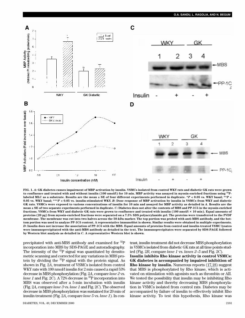
stimulated MBP activation. Our recent studies haveshown that physiological concentrations of insulin rapidlyincrease MBP activity in myosin-enriched fractions isolatedfrom control WKY rats. To further understand the impor-tance of MBP in VSMC function, we examined the activity ofthis enzyme in VSMCs isolated from diabetic GK rats. Diabetesresulted in a 43% decrease in basal MBP activity when com-pared with control VSMCs isolated from WKY rats (Fig. 1A).Whereas incubation with 100 nmol/l insulin for 10 mincaused a 63% increase in MBP activity in WKY rats, VSMCsfrom diabetic GK rats exhibited only a 15% increase in MBPactivity over the basal values (Fig. 1A).
In WKY rats, 1 nmol/l insulin caused a half-maximal stim-ulation of MBP, with a maximum response seen with10 nmol/l insulin after 10 min (Fig. 1B). The observed reduc-tions in MBP activation by insulin in VSMCs from diabetic ratswere due to a marked decrease in insulin responsiveness atall concentrations of insulin (Fig. 1B). Maintenance of GKVSMCs in medium containing normal concentrations of glu-cose did not restore cellular responsiveness to insulin interms of MBP activation, whereas incubation of VSMCs iso-lated from WKY rats in medium containing high glucose didresult in the inhibition of insulin’s effect on MBP activation(data not shown). This observation suggests that defectiveMBP activation seen in diabetic GK rat VSMCs may be due toa genetic difference between diabetic GK and WKY ratherthan hyperglycemia or other metabolic defects secondary todiabetes. Furthermore, the observed effect of high glucose onMBP inhibition was not due to changes in osmolality becausetreatment with mannitol did not inhibit insulin’s effect onMBP activation in WKY rats.
Western blot analysis indicated that GK diabetes did notalter the contents of either MBS or PP-1C� examined in themyosin-enriched fraction (Fig. 1C) as well as the MBSimmunoprecipitates (Fig. 1D). Furthermore, insulin treat-ment did not increase the amount of PP-1C� bound to MBSin WKY rats. This observation suggests that insulin may beactivating the enzyme already bound to the myosin regulatorysubunit (MBS), and the observed reductions in MBP activityin VSMCs isolated from diabetic GK rats may be due toimpaired activation of MBP bound to MBS.Insulin fails to decrease MBS phosphorylation in
VSMCs isolated from diabetic GK rats. Recent studies sug-gest that the large 110/130-kDa MBS of MBP gets phosphory-lated by agonists that stimulate smooth muscle contraction(17,18). Furthermore, phosphorylation of MBS results in inac-tivation of MBP resulting in a decrease in MBP activity (18).Our observations that physiological concentrations of insulinrapidly stimulate MBP suggest that insulin may be activatingthe phosphatase either by altering MBS phosphorylation sta-tus and/or activating the catalytic subunit bound to MBS viaanother mechanism. Therefore, we examined the effect ofinsulin on MBS phosphorylation status in VSMCs isolatedfrom control rats as well as diabetic GK rats. Serum-starvedVSMCs were labeled with 32P-orthophosphoric acid (0.3 mCi/ml)for 4 h to attain steady-state equilibrium (29) followed by theaddition of insulin for 0–30 min. Cell lysates were immuno-
DIABETES, VOL. 49, DECEMBER 2000 2181
O.A. SANDU, L. RAGOLIA, AND N. BEGUM
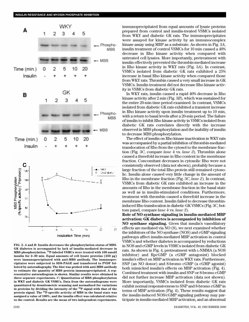
precipitated with anti-MBS antibody and examined for 32Pincorporation into MBS by SDS-PAGE and autoradiography.The intensity of the 32P signal was quantitated by densito-metric scanning and corrected for any variations in MBS pro-tein by dividing the 32P signal with the protein signal. Asshown in Fig. 2A, treatment of VSMCs isolated from controlWKY rats with 100 nmol/l insulin for 2 min caused a rapid 53%decrease in MBS phosphorylation (Fig. 2A, compare lane 2 vs.lane 1 and Fig. 2C). A 72% decrease in 32P incorporation intoMBS was observed after a 5-min incubation with insulin(Fig. 2A, compare lane 3 vs. lane 1 and Fig. 2C). The observeddecrease in MBS phosphorylation was sustained for 20 min ofinsulin treatment (Fig. 2A, compare lane 5 vs. lane 1). In con-
trast, insulin treatment did not decrease MBS phosphorylationin VSMCs isolated from diabetic GK rats at all time points stud-ied (Fig. 2B, compare lane 1 vs. lanes 2–5 and Fig. 2C).Insulin inhibits Rho kinase activity in control VSMCs:
GK diabetes is accompanied by impaired inhibition of
Rho kinase by insulin. Numerous reports (17,18) suggestthat MBS is phosphorylated by Rho kinase, which is acti-vated on stimulation with agonists such as thrombin or AII.We tested the possibility that insulin may be inhibiting Rhokinase activity and thereby decreasing MBS phosphoryla-tion in VSMCs isolated from control rats. Diabetes may beaccompanied by failure of insulin to effectively inhibit Rhokinase activity. To test this hypothesis, Rho kinase was
FIG. 1. A: GK diabetes causes impairment of MBP activation by insulin. VSMCs isolated from control WKY rats and diabetic GK rats were grown
to confluence and treated with and without insulin (100 nmol/l) for 10 min. MBP activity was assayed in myosin-enriched fractions using 32P-
labeled MLC as a substrate. Results are the mean ± SE of four different experiments performed in duplicate. *P < 0.05 vs. WKY basal; **P <
0.05 vs. WKY basal; ***P < 0.05 vs. insulin-stimulated WKY. B: Dose response of MBP activation by insulin in VSMCs from WKY and diabetic
GK rats. VSMCs were exposed to various concentrations of insulin for 10 min and assayed for MBP activity as detailed in A. Results are the
mean ± SE of two separate experiments performed in duplicate. C: Diabetes does not alter the contents of MBS and PP-1C� in the myosin-enriched
fractions. VSMCs from WKY and diabetic GK rats were grown to confluence and treated with insulin (100 nmol/l � 10 min). Equal amounts of
proteins (20 µg) from myosin-enriched fractions were separated on a 7.5% SDS polyacrylamide gel. The proteins were transferred to the PVDF
membrane. The membrane was cut into two halves across the 50-kDa marker. The top portion was probed with anti-MBS antibody, and the bot-
tom portion was used to analyze PP-1C� content. A representative immunoblot is shown. Similar results were obtained in multiple experiments.
D: Insulin does not increase the association of PP-1C� with the MBS. Equal amounts of proteins from control and insulin-treated VSMC lysates
were immunoprecipitated with the anti-MBS antibody as detailed in the text. The immunoprecipitates were separated by SDS-PAGE followed
by Western blot analysis as detailed in C. A representative Western blot is shown.
A
B
C
D
2182 DIABETES, VOL. 49, DECEMBER 2000
INSULIN RESISTANCE AND MYOSIN PHOSPHATE INHIBITION
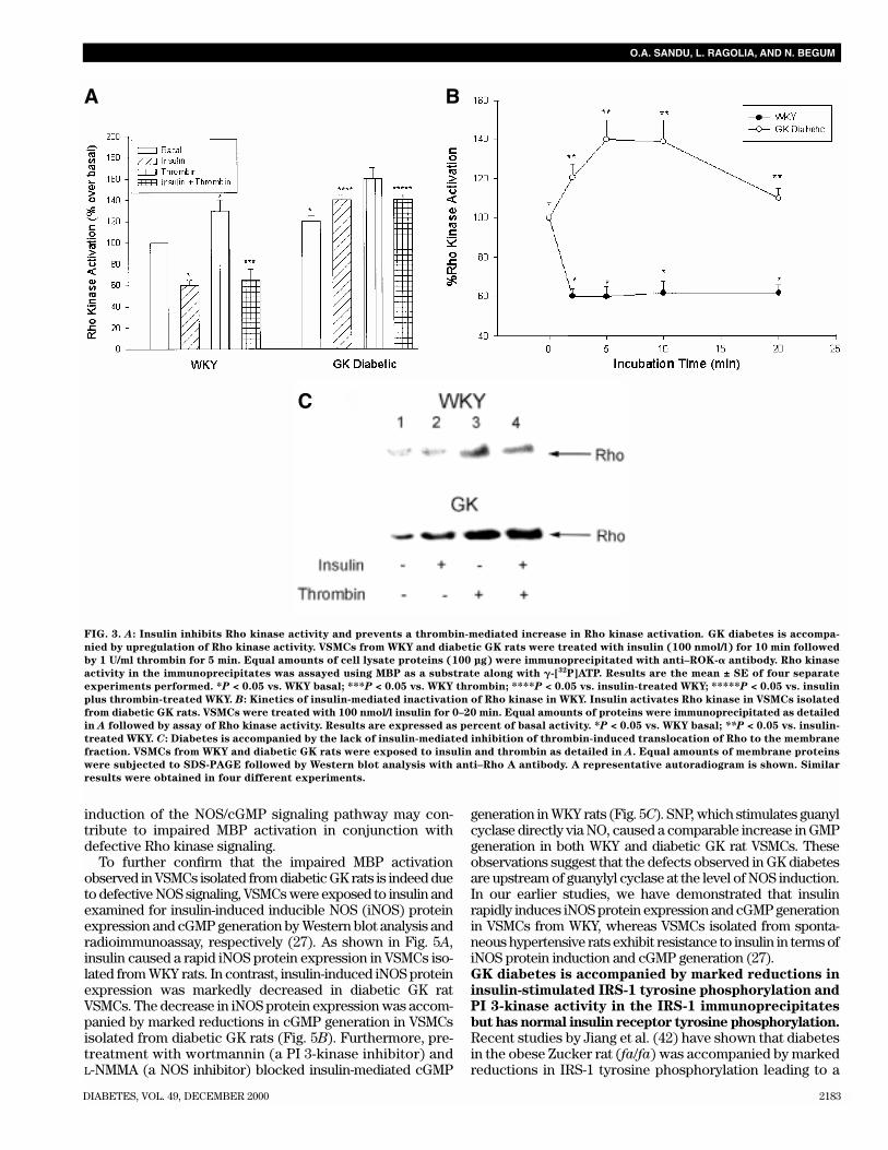
immunoprecipitated from equal amounts of lysate proteinsprepared from control and insulin-treated VSMCs isolatedfrom WKY and diabetic GK rats. The immunoprecipitateswere assayed for kinase activity by an immunocomplexkinase assay using MBP as a substrate. As shown in Fig. 3A,insulin treatment of control VSMCs for 10 min caused a 40%decrease in Rho kinase activity when compared withuntreated cell lysates. More importantly, pretreatment withinsulin effectively prevented the thrombin-mediated increasein Rho kinase activity in WKY rats (Fig. 3A). In contrast,VSMCs isolated from diabetic GK rats exhibited a 25%increase in basal Rho kinase activity when compared thosefrom WKY rats. Thrombin caused a very small increase in GKVSMCs. Insulin treatment did not decrease Rho kinase activ-ity in VSMCs from diabetic GK rats.
In WKY rats, insulin caused a rapid 40% decrease in Rhokinase activity after 2 min (Fig. 3B), which was sustained forthe entire 20-min time period examined. In contrast, VSMCsisolated from diabetic GK rats exhibited a transient increasein Rho kinase activity upon insulin treatment up to 10 min,with a return to basal levels after a 20-min period. The failureof insulin to inhibit Rho kinase activity in VSMCs isolated fromdiabetic GK rats correlates directly with the increaseobserved in MBS phosphorylation and the inability of insulinto decrease MBS phosphorylation.
The effect of insulin on Rho kinase inactivation in WKY ratswas accompanied by a partial inhibition of thrombin-mediatedtranslocation of Rho from the cytosol to the membrane frac-tion (Fig. 3C, compare lane 4 vs. lane 3). Thrombin alonecaused a threefold increase in Rho content in the membranefraction. Concomitant decreases in cytosolic Rho were notconsistently observed (data not shown), probably because alarge fraction of the total Rho protein still remained cytoso-lic. Insulin alone caused very little change in the amount ofRho in the membrane fraction (Fig. 3C, lane 2). In contrast,VSMCs from diabetic GK rats exhibited an increase in theamounts of Rho in the membrane fraction in the basal stateas well as in insulin-stimulated conditions. Furthermore,treatment with thrombin caused a threefold increase in themembrane Rho content. Insulin failed to decrease thrombin-induced Rho translocation in diabetic GK VSMCs (Fig. 3C, bot-tom panel, compare lane 4 vs. lane 3).Role of NO synthase signaling in insulin-mediated MBP
activation: GK diabetes is accompanied by inhibition of
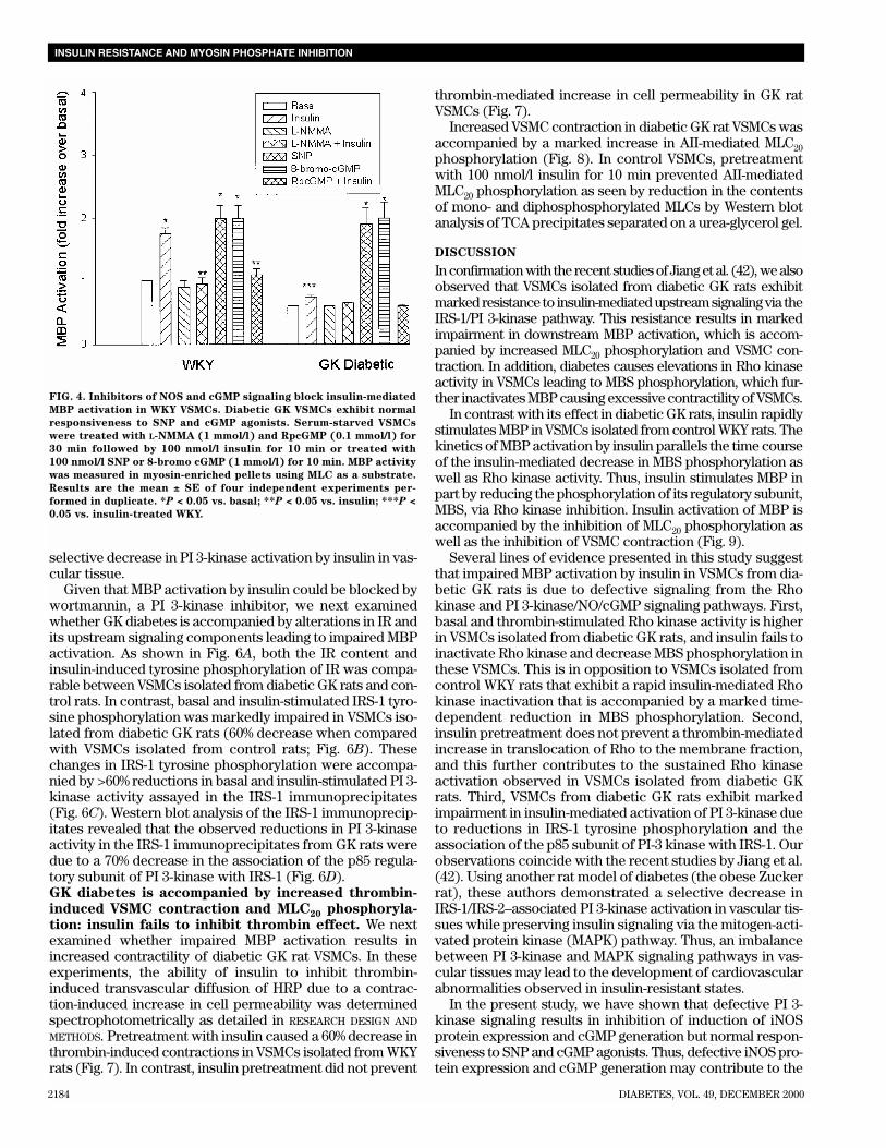
NO synthase signaling. Given that insulin’s vasodilatoryeffects are mediated via NO (8), we next examined whetherthe inhibitors of the NO synthase (NOS) and cGMP signalingpathways affect insulin-mediated MBP activation in controlVSMCs and whether diabetes is accompanied by reductionsin NOS and cGMP levels in VSMCs isolated from diabetic GKrats. As shown in Fig. 4, pretreatment with L-NMMA (a NOSinhibitor) and RpcGMP (a cGMP antagonist) blockedinsulin’s effect on MBP activation in WKY rats. Furthermore,SNP (an NO donor) and 8-bromo cGMP (a cGMP agonist)both mimicked insulin’s effects on MBP activation (Fig. 4).Combined treatment with insulin and SNP or 8-bromo cGMPdid not further increase MBP activation (data not shown).More importantly, VSMCs isolated from diabetic GK ratsexhibit normal responsiveness to SNP and 8-bromo cGMP interms of MBP activation (Fig. 4). These results suggest thatthe insulin-induced NOS/cGMP signaling pathway may par-ticipate in insulin-mediated MBP activation, and an abnormal
FIG. 2. A and B: Insulin decreases the phosphorylation status of MBS.
GK diabetes is accompanied by lack of insulin-mediated decrease in
MBS phosphorylation. 32P-labeled VSMCs were treated with 100 nmol/l
insulin for 2–30 min. Equal amounts of cell lysate proteins (250 µg)
were immunoprecipitated with anti-MBS antibody. The immunopre-
cipitates were subjected to SDS-PAGE and transferred to PVDF fol-
lowed by autoradiography. The blot was probed with anti-MBS antibody
to estimate the quantity of MBS protein immunoprecipitated. A rep-
resentative autoradiogram is shown. Similar results were obtained in
three separate experiments. C: Quantitation of MBS phosphorylation
in WKY and diabetic GK VSMCs. Data from the autoradiograms were
quantitated by densitometric scanning and normalized for variations
in proteins by dividing the intensity of the 32P signal with that of the
protein signal. The 32P-specific activity of MBS in the basal state was
assigned a value of 100%, and the insulin effect was calculated relative
to the control. Results are the mean of two independent experiments.
DIABETES, VOL. 49, DECEMBER 2000 2183
O.A. SANDU, L. RAGOLIA, AND N. BEGUM
induction of the NOS/cGMP signaling pathway may con-tribute to impaired MBP activation in conjunction withdefective Rho kinase signaling.
To further confirm that the impaired MBP activationobserved in VSMCs isolated from diabetic GK rats is indeed dueto defective NOS signaling, VSMCs were exposed to insulin andexamined for insulin-induced inducible NOS (iNOS) proteinexpression and cGMP generation by Western blot analysis andradioimmunoassay, respectively (27). As shown in Fig. 5A,insulin caused a rapid iNOS protein expression in VSMCs iso-lated from WKY rats. In contrast, insulin-induced iNOS proteinexpression was markedly decreased in diabetic GK ratVSMCs. The decrease in iNOS protein expression was accom-panied by marked reductions in cGMP generation in VSMCsisolated from diabetic GK rats (Fig. 5B). Furthermore, pre-treatment with wortmannin (a PI 3-kinase inhibitor) andL-NMMA (a NOS inhibitor) blocked insulin-mediated cGMP
generation in WKY rats (Fig. 5C). SNP, which stimulates guanylcyclase directly via NO, caused a comparable increase in GMPgeneration in both WKY and diabetic GK rat VSMCs. Theseobservations suggest that the defects observed in GK diabetesare upstream of guanylyl cyclase at the level of NOS induction.In our earlier studies, we have demonstrated that insulinrapidly induces iNOS protein expression and cGMP generationin VSMCs from WKY, whereas VSMCs isolated from sponta-neous hypertensive rats exhibit resistance to insulin in terms ofiNOS protein induction and cGMP generation (27).GK diabetes is accompanied by marked reductions in
insulin-stimulated IRS-1 tyrosine phosphorylation and
PI 3-kinase activity in the IRS-1 immunoprecipitates
but has normal insulin receptor tyrosine phosphorylation.
Recent studies by Jiang et al. (42) have shown that diabetesin the obese Zucker rat (fa/fa) was accompanied by markedreductions in IRS-1 tyrosine phosphorylation leading to a
FIG. 3. A: Insulin inhibits Rho kinase activity and prevents a thrombin-mediated increase in Rho kinase activation. GK diabetes is accompa-
nied by upregulation of Rho kinase activity. VSMCs from WKY and diabetic GK rats were treated with insulin (100 nmol/l) for 10 min followed
by 1 U/ml thrombin for 5 min. Equal amounts of cell lysate proteins (100 µg) were immunoprecipitated with anti–ROK-� antibody. Rho kinase
activity in the immunoprecipitates was assayed using MBP as a substrate along with �-[32P]ATP. Results are the mean ± SE of four separate
experiments performed. *P < 0.05 vs. WKY basal; ***P < 0.05 vs. WKY thrombin; ****P < 0.05 vs. insulin-treated WKY; *****P < 0.05 vs. insulin
plus thrombin-treated WKY. B: Kinetics of insulin-mediated inactivation of Rho kinase in WKY. Insulin activates Rho kinase in VSMCs isolated
from diabetic GK rats. VSMCs were treated with 100 nmol/l insulin for 0–20 min. Equal amounts of proteins were immunoprecipitated as detailed
in A followed by assay of Rho kinase activity. Results are expressed as percent of basal activity. *P < 0.05 vs. WKY basal; **P < 0.05 vs. insulin-
treated WKY. C: Diabetes is accompanied by the lack of insulin-mediated inhibition of thrombin-induced translocation of Rho to the membrane
fraction. VSMCs from WKY and diabetic GK rats were exposed to insulin and thrombin as detailed in A. Equal amounts of membrane proteins
were subjected to SDS-PAGE followed by Western blot analysis with anti–Rho A antibody. A representative autoradiogram is shown. Similar
results were obtained in four different experiments.
A B
C
2184 DIABETES, VOL. 49, DECEMBER 2000
INSULIN RESISTANCE AND MYOSIN PHOSPHATE INHIBITION
selective decrease in PI 3-kinase activation by insulin in vas-cular tissue.
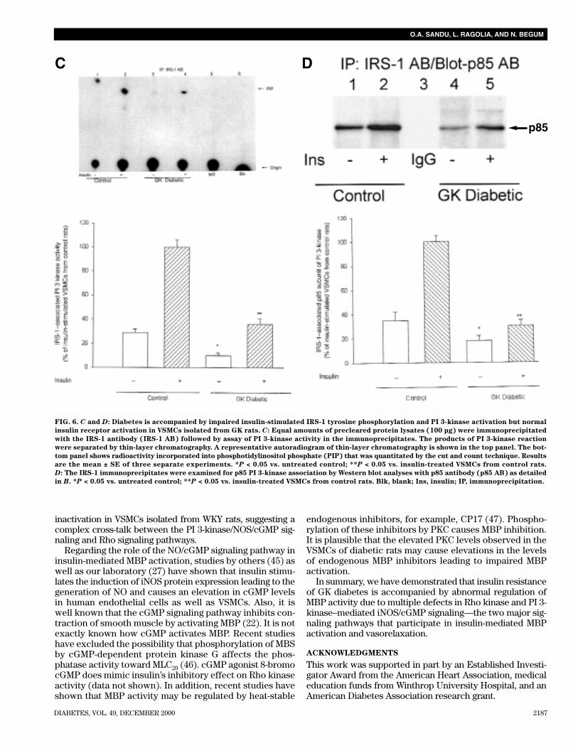
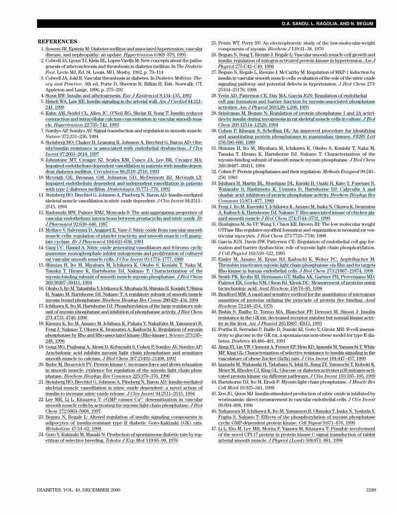
Given that MBP activation by insulin could be blocked bywortmannin, a PI 3-kinase inhibitor, we next examinedwhether GK diabetes is accompanied by alterations in IR andits upstream signaling components leading to impaired MBPactivation. As shown in Fig. 6A, both the IR content andinsulin-induced tyrosine phosphorylation of IR was compa-rable between VSMCs isolated from diabetic GK rats and con-trol rats. In contrast, basal and insulin-stimulated IRS-1 tyro-sine phosphorylation was markedly impaired in VSMCs iso-lated from diabetic GK rats (60% decrease when comparedwith VSMCs isolated from control rats; Fig. 6B). Thesechanges in IRS-1 tyrosine phosphorylation were accompa-nied by >60% reductions in basal and insulin-stimulated PI 3-kinase activity assayed in the IRS-1 immunoprecipitates(Fig. 6C). Western blot analysis of the IRS-1 immunoprecip-itates revealed that the observed reductions in PI 3-kinaseactivity in the IRS-1 immunoprecipitates from GK rats weredue to a 70% decrease in the association of the p85 regula-tory subunit of PI 3-kinase with IRS-1 (Fig. 6D).GK diabetes is accompanied by increased thrombin-
induced VSMC contraction and MLC20 phosphoryla-
tion: insulin fails to inhibit thrombin effect. We nextexamined whether impaired MBP activation results inincreased contractility of diabetic GK rat VSMCs. In theseexperiments, the ability of insulin to inhibit thrombin-induced transvascular diffusion of HRP due to a contrac-tion-induced increase in cell permeability was determinedspectrophotometrically as detailed in RESEARCH DESIGN AND
METHODS. Pretreatment with insulin caused a 60% decrease inthrombin-induced contractions in VSMCs isolated from WKYrats (Fig. 7). In contrast, insulin pretreatment did not prevent
thrombin-mediated increase in cell permeability in GK ratVSMCs (Fig. 7).
Increased VSMC contraction in diabetic GK rat VSMCs wasaccompanied by a marked increase in AII-mediated MLC20phosphorylation (Fig. 8). In control VSMCs, pretreatmentwith 100 nmol/l insulin for 10 min prevented AII-mediatedMLC20 phosphorylation as seen by reduction in the contentsof mono- and diphosphosphorylated MLCs by Western blotanalysis of TCA precipitates separated on a urea-glycerol gel.
DISCUSSION
In confirmation with the recent studies of Jiang et al. (42), we alsoobserved that VSMCs isolated from diabetic GK rats exhibitmarked resistance to insulin-mediated upstream signaling via theIRS-1/PI 3-kinase pathway. This resistance results in markedimpairment in downstream MBP activation, which is accom-panied by increased MLC20 phosphorylation and VSMC con-traction. In addition, diabetes causes elevations in Rho kinaseactivity in VSMCs leading to MBS phosphorylation, which fur-ther inactivates MBP causing excessive contractility of VSMCs.
In contrast with its effect in diabetic GK rats, insulin rapidlystimulates MBP in VSMCs isolated from control WKY rats. Thekinetics of MBP activation by insulin parallels the time courseof the insulin-mediated decrease in MBS phosphorylation aswell as Rho kinase activity. Thus, insulin stimulates MBP inpart by reducing the phosphorylation of its regulatory subunit,MBS, via Rho kinase inhibition. Insulin activation of MBP isaccompanied by the inhibition of MLC20 phosphorylation aswell as the inhibition of VSMC contraction (Fig. 9).
Several lines of evidence presented in this study suggestthat impaired MBP activation by insulin in VSMCs from dia-betic GK rats is due to defective signaling from the Rhokinase and PI 3-kinase/NO/cGMP signaling pathways. First,basal and thrombin-stimulated Rho kinase activity is higherin VSMCs isolated from diabetic GK rats, and insulin fails toinactivate Rho kinase and decrease MBS phosphorylation inthese VSMCs. This is in opposition to VSMCs isolated fromcontrol WKY rats that exhibit a rapid insulin-mediated Rhokinase inactivation that is accompanied by a marked time-dependent reduction in MBS phosphorylation. Second,insulin pretreatment does not prevent a thrombin-mediatedincrease in translocation of Rho to the membrane fraction,and this further contributes to the sustained Rho kinaseactivation observed in VSMCs isolated from diabetic GKrats. Third, VSMCs from diabetic GK rats exhibit markedimpairment in insulin-mediated activation of PI 3-kinase dueto reductions in IRS-1 tyrosine phosphorylation and theassociation of the p85 subunit of PI-3 kinase with IRS-1. Ourobservations coincide with the recent studies by Jiang et al.(42). Using another rat model of diabetes (the obese Zuckerrat), these authors demonstrated a selective decrease inIRS-1/IRS-2–associated PI 3-kinase activation in vascular tis-sues while preserving insulin signaling via the mitogen-acti-vated protein kinase (MAPK) pathway. Thus, an imbalancebetween PI 3-kinase and MAPK signaling pathways in vas-cular tissues may lead to the development of cardiovascularabnormalities observed in insulin-resistant states.
In the present study, we have shown that defective PI 3-kinase signaling results in inhibition of induction of iNOSprotein expression and cGMP generation but normal respon-siveness to SNP and cGMP agonists. Thus, defective iNOS pro-tein expression and cGMP generation may contribute to the
FIG. 4. Inhibitors of NOS and cGMP signaling block insulin-mediated
MBP activation in WKY VSMCs. Diabetic GK VSMCs exhibit normal
responsiveness to SNP and cGMP agonists. Serum-starved VSMCs
were treated with L-NMMA (1 mmol/l) and RpcGMP (0.1 mmol/l) for
30 min followed by 100 nmol/l insulin for 10 min or treated with
100 nmol/l SNP or 8-bromo cGMP (1 mmol/l) for 10 min. MBP activity
was measured in myosin-enriched pellets using MLC as a substrate.
Results are the mean ± SE of four independent experiments per-
formed in duplicate. *P < 0.05 vs. basal; **P < 0.05 vs. insulin; ***P <
0.05 vs. insulin-treated WKY.

DIABETES, VOL. 49, DECEMBER 2000 2185
O.A. SANDU, L. RAGOLIA, AND N. BEGUM
impaired MBP activation observed in VSMCs isolated from dia-betic GK rats. We have recently demonstrated that inhibitionof PI 3-kinase with wortmannin blocks insulin-mediated iNOSinduction (27), cGMP generation (27), and MBP activation inVSMCs isolated from WKY rats. Furthermore, inhibition ofNOS activity with L-NMMA and blockade of cGMP signalingwith RpcGMP attenuates the effect of insulin on MBP activa-tion in WKY rats. The fact that SNP and cGMP agonists stim-ulate MBP activation in VSMCs isolated from diabetic GKrats to levels seen in WKY rats suggests that the signalingdefects in diabetic GK rat VSMCs lie at the level of NOS dueto an inhibition of PI 3-kinase activation that mediates theinduction of iNOS in VSMCs.
Figure 9 depicts the putative signaling pathways involvedin MBP activation by insulin in normal VSMCs. Our results
suggest that defects in both the Rho kinase and the PI 3-kinase–mediated NOS signaling pathways contribute to theinhibitory effects of hyperglycemia and diabetes on MBPactivation. To our knowledge, this is the first study to demon-strate MBP activation by insulin in VSMCs in vivo via reduc-tions in MBS phosphorylation status and its abnormal regu-lation in the insulin-resistant state associated with diabetes.Other studies have demonstrated an inhibitory regulation ofMBP by a G-protein–dependent mechanism (22). Thus, aninhibition of MBP activity by thrombin (37), arachidonic acid(19), protein kinase C (PKC) (20), and guanosine-5�-O-(2-thiotriphosphate), tetralithium salt (GTP�S) (15–17) hasbeen reported. This study adds a new dimension to the aboveobservations by demonstrating that insulin’s stimulatoryeffect on MBP activation is in part due to a reduction in the
FIG. 5. A: GK diabetes is accompanied by impaired insulin-induced iNOS protein expression. VSMCs were stimulated with and without insulin
(100 nmol/l � 10 min). Equal amounts of proteins (50 µg) isolated from WKY and diabetic GK VSMCs were subjected to SDS-PAGE followed
by Western blot analysis with anti-iNOS antibody and detection with ECL. The intensity of signal in each lane was quantitated by densitometric
analysis. Results are the mean ± SE of four separate experiments. *P < 0.05 vs. WKY basal; **P < 0.05 vs. WKY basal and insulin-treated. B:
GK diabetes is accompanied by marked impairment in insulin-stimulated cGMP generation. VSMCs were stimulated with insulin (100 nmol/l)
for the indicated times followed by the extraction with 70% ethanol. The ethanol extracts were analyzed for cGMP content by radioimmunoassay
as detailed in RESEARCH DESIGN AND METHODS. Results are the mean ± SE of three separate experiments. *P < 0.05 vs. WKY. C: Wortmannin (Wort)
and L-NMMA block insulin’s effect on GMP generation. Diabetic GK VSMCs exhibit normal responsiveness to SNP in terms of cGMP produc-
tion. VSMCs were treated with wortmannin (100 nmol/l) and L-NMMA (1 mmol/l) for 30 min before treatment with insulin (Ins) or treated with
SNP (100 nmol/l) as detailed in Fig. 4. cGMP levels were measured in ethanol supernatants. Results are the mean ± SE of three separate exper-
iments. *P < 0.05 vs. basal; **P < 0.05 vs. insulin; ***P < 0.05 vs. WKY basal.
A B
C

2186 DIABETES, VOL. 49, DECEMBER 2000
INSULIN RESISTANCE AND MYOSIN PHOSPHATE INHIBITION
MBS phosphorylation state as well as because of the con-comitant activation of the enzyme bound to MBS via cGMPsignaling, thereby causing MLC20 dephosphorylation andinsulin-mediated vasorelaxation. Thus, the impaired vasore-laxation observed in patients with diabetes may be due toinherent reductions in MBP activity resulting from defectiveregulation of MBP activation in response to insulin. Given theknowledge that PKC levels are elevated in VSMCs isolatedfrom diabetic rat aortas (43) and the fact that PKC can acti-vate Rho and inhibit MBP, it is tempting to speculate that theexaggerated Rho kinase signaling and impaired insulin acti-
vation of MBP observed in diabetes may be due to an eleva-tion in PKC activity via excessive release of arachidonic acidby phospholipase A2 (44). Arachidonic acid could increaseRho kinase activity as well as interact directly with MBS,causing dissociation of the holoenzyme and thereby reducingMBP activity, or it could activate a kinase that phosphorylatesMBS and inhibits MBP. Alternatively, the PI 3-kinase pathwaymay regulate Rho kinase activation. However, in our prelim-inary studies, we did not observe a blockade of insulin’sinhibitory effects on Rho kinase with wortmannin, but L-NMMA and RpcGMP did prevent insulin-mediated Rho kinase
FIG. 6. A and B: Diabetes is accompanied by impaired insulin-stimulated IRS-1 tyrosine phosphorylation and PI 3-kinase activation but nor-
mal insulin receptor activation in VSMCs isolated from GK rats. A: VSMCs were treated with and without insulin for 10 min. Equal amounts
of precleared protein lysates (4 mg) were immunoprecipitated with the insulin receptor antibody (IR AB) (2 µg) or IgG followed by Western
blot analyses with phosphotyrosine (pTY) antibody (top panel). The blots were stripped and reprobed with the IR antibody (middle panel).
Data from three separate experiments were quantitated by densitometric analyses of the autoradiograms and corrected for variations in IR
protein by dividing the phosphotyrosine signal with the signal obtained with the IR antibody (specific activity). Results are expressed as per-
cent of insulin-stimulated VSMCs isolated from control rats, assigning a value of 100% to the mean specific activity (bottom panel). B: Pre-
cleared protein lysates (4 mg) from control and insulin-treated VSMCs were immunoprecipitated with the IRS-1 antibody (IRS-1 AB) (10 µg)
followed by Western blot analyses with phosphotyrosine (pTY) antibody (top panel). Blots were stripped and reprobed with IRS-1 antibody
(middle panel). Data from three separate experiments were quantitated by densitometric analyses of the autoradiograms and corrected for
variations in IRS-1 protein as detailed in A. Results are expressed as percent of insulin-stimulated VSMCs isolated from control rats, assign-
ing a value of 100% to the mean specific activity (bottom panel). *P < 0.05 vs. untreated control; **P < 0.05 vs. insulin-treated VSMCs from
control rats. +ve C, positive control; Ins, insulin; IP, immunoprecipitation.
A
B
DIABETES, VOL. 49, DECEMBER 2000 2187
O.A. SANDU, L. RAGOLIA, AND N. BEGUM
inactivation in VSMCs isolated from WKY rats, suggesting acomplex cross-talk between the PI 3-kinase/NOS/cGMP sig-naling and Rho signaling pathways.
Regarding the role of the NO/cGMP signaling pathway ininsulin-mediated MBP activation, studies by others (45) aswell as our laboratory (27) have shown that insulin stimu-lates the induction of iNOS protein expression leading to thegeneration of NO and causes an elevation in cGMP levelsin human endothelial cells as well as VSMCs. Also, it iswell known that the cGMP signaling pathway inhibits con-traction of smooth muscle by activating MBP (22). It is notexactly known how cGMP activates MBP. Recent studieshave excluded the possibility that phosphorylation of MBSby cGMP-dependent protein kinase G affects the phos-phatase activity toward MLC20 (46). cGMP agonist 8-bromocGMP does mimic insulin’s inhibitory effect on Rho kinaseactivity (data not shown). In addition, recent studies haveshown that MBP activity may be regulated by heat-stable
endogenous inhibitors, for example, CP17 (47). Phospho-rylation of these inhibitors by PKC causes MBP inhibition.It is plausible that the elevated PKC levels observed in theVSMCs of diabetic rats may cause elevations in the levelsof endogenous MBP inhibitors leading to impaired MBPactivation.
In summary, we have demonstrated that insulin resistanceof GK diabetes is accompanied by abnormal regulation ofMBP activity due to multiple defects in Rho kinase and PI 3-kinase–mediated iNOS/cGMP signaling—the two major sig-naling pathways that participate in insulin-mediated MBPactivation and vasorelaxation.
ACKNOWLEDGMENTS
This work was supported in part by an Established Investi-gator Award from the American Heart Association, medicaleducation funds from Winthrop University Hospital, and anAmerican Diabetes Association research grant.
p85
C D
FIG. 6. C and D: Diabetes is accompanied by impaired insulin-stimulated IRS-1 tyrosine phosphorylation and PI 3-kinase activation but normal
insulin receptor activation in VSMCs isolated from GK rats. C: Equal amounts of precleared protein lysates (100 µg) were immunoprecipitated
with the IRS-1 antibody (IRS-1 AB) followed by assay of PI 3-kinase activity in the immunoprecipitates. The products of PI 3-kinase reaction
were separated by thin-layer chromatography. A representative autoradiogram of thin-layer chromatography is shown in the top panel. The bot-
tom panel shows radioactivity incorporated into phosphotidylinositol phosphate (PIP) that was quantitated by the cut and count technique. Results
are the mean ± SE of three separate experiments. *P < 0.05 vs. untreated control; **P < 0.05 vs. insulin-treated VSMCs from control rats.
D: The IRS-1 immunoprecipitates were examined for p85 PI 3-kinase association by Western blot analyses with p85 antibody (p85 AB) as detailed
in B. *P < 0.05 vs. untreated control; **P < 0.05 vs. insulin-treated VSMCs from control rats. Blk, blank; Ins, insulin; IP, immunoprecipitation.

2188 DIABETES, VOL. 49, DECEMBER 2000
INSULIN RESISTANCE AND MYOSIN PHOSPHATE INHIBITION
FIG. 7. Insulin fails to inhibit thrombin-induced VSMC contraction in dia-
betic GK VSMCs. Serum-starved VSMCs were treated with and without
insulin (100 nmol/l) for 30 min followed by treatment with thrombin
(0.5 U/ml � 15). Cell contraction was measured as detailed in the text.
Results are the mean ± SE of three experiments. *P < 0.05 vs. thrombin-
treated WKY; ***P < 0.05 vs. insulin plus thrombin-treated WKY.
FIG. 8. GK diabetes is accompanied by failure of insulin to inhibit
AII-mediated MLC20 phosphorylation (MLCP). VSMCs were treated
with and without insulin (10 nmol/l) for 10 min followed by the addi-
tion of AII (100 nmol/l) for 5 min followed by glycerol PAGE and
immunobloting with anti-MLC20 antibody. A representative autoradiogram
is shown.
FIG. 9. Schematic representation of the proposed pathways for insulin activation of MBP. Insulin-stimulated receptor tyrosine kinase medi-
ates the activation of IRS-1/IRS-2/PI 3-kinase and the NO/cGMP signaling pathway, which activates MBP in part by decreasing MBS phospho-
rylation. In addition, insulin inhibits Rho kinase activation, thereby preventing MBP inactivation due to MBS phosphorylation by Rho kinase.
The NOS/cGMP signaling pathway may regulate Rho kinase activation as well. GK diabetes is accompanied by upregulation of Rho kinase activ-
ity as well as inhibition of PI 3-kinase activation leading to decreased generation of NO/cGMP, which causes impaired MBP activation partly
due to excessive MBS phosphorylation by Rho kinase.
DIABETES, VOL. 49, DECEMBER 2000 2189
O.A. SANDU, L. RAGOLIA, AND N. BEGUM
REFERENCES
1. Sowers JR, Epstein M: Diabetes mellitus and associated hypertension, vasculardisease, and nephropathy: an update. Hypertension 6:869–879, 1995
2. Colwell JA, Lyons TJ, Klein RL, Lopes-Virella M: New concepts about the patho-genesis of atherosclerosis and thrombosis in diabetes mellitus. In The Diabetic
Foot. Levin MJ, Ed. St. Louis, MO, Mosby, 1992, p. 79–1143. Colwell JA, Jokl R: Vascular thrombosis in diabetes. In Diabetes Mellitus: The-
ory and Practice. 5th ed. Porte D, Sherwin R, Rifkin H, Eds. Norwalk, CT,Appleton and Lange, 1996, p. 275–292
4. Stout RW: Insulin and atherogenesis. Eur J Epidemiol 8:134–135, 19925. Hsueh WA, Law RE: Insulin signaling in the arterial wall. Am J Cardiol 84:21J–
24J, 19996. Kahn AM, Seidel CL, Allen JC, O’Neil RG, Shelat H, Song T: Insulin reduces
contraction and intracellular calcium concentration in vascular smooth mus-cle. Hypertension 22:735–742, 1993
7. Somlyo AP, Somlyo AV: Signal transduction and regulation in smooth muscle.Nature 372:231–236, 1994
8. Steinberg HO, Chaker H, Leaming R, Johnson A, Brechtel G, Baron AD: Obe-sity/insulin resistance is associated with endothelial dysfunction. J Clin
Invest 97:2601–2610, 19979. Johnstone MT, Creager SJ, Scales KM, Cusco JA, Lee BK, Creager MA:
Impaired endothelium-dependent vasodilation in patients with insulin-depen-dent diabetes mellitus. Circulation 88:2510–2516, 1993
10. Mcveigh GE, Brennan GM, Johnston GD, McDermott BJ, McGrath LT:Impaired endothelium dependent and independent vasodilation in patientswith type 2 diabetes mellitus. Diabetologia 35:771–776, 1992
11. Steinberg HO, Brechtel G, Johnson A, Fineberg N, Baron AD: Insulin-mediatedskeletal muscle vasodilation is nitric oxide dependent. J Clin Invest 94:2511–2515, 1994
12. Radomski MW, Palmer RMJ, Moncada S: The anti-aggregating properties ofvascular endothelium: interactions between prostacyclin and nitric oxide. Br
J Pharmacol 92:639–646, 198713. Mollace V, Salvemini D, Anggard E, Vane J: Nitric oxide from vascular smooth
muscle cells: regulation of platelet reactivity and smooth muscle cell guany-late cyclase. Br J Pharmacol 104:633–638, 1991
14. Garg UC, Hassid A: Nitric oxide generating vasodilators and 8-bromo cyclicguanosine monophosphate inhibit mitogenesis and proliferation of culturedrat vascular smooth muscle cells. J Clin Invest 93:1774–1777, 1989
15. Shimizu H, Ito M, Miyahara M, Ichikawa K, Okubo S, Konishi T, Naka M,Tanaka T, Hirano K, Hartshorne DJ, Nakano T: Characterization of themyosin-binding subunit of smooth muscle myosin phosphatase. J Biol Chem
269:30407–30411, 199416. Okubo S, Ito M, Takashiba Y, Ichikawa K, Miyahara M, Shimizu H, Konishi T, Shima
H, Nagao M, Hartshorne DJ, Nakano T: A regulatory subunit of smooth musclemyosin bound phosphatase. Biochem Biophys Res Comm 200:429–434, 1994
17. Ichikawa K, Ito M, Hartshorne DJ: Phosphorylation of the large regulatory sub-unit of myosin phosphatase and inhibition of phosphatase activity. J Biol Chem
271:4733–4740, 199618. Kimura K, Ito M, Amano M, Ichihara K, Fukata Y, Nakafuku M, Yamamori B,
Feng J, Nakano T, Okawa K, Iwamatsu A, Kaibuchi K: Regulation of myosinphosphatase by Rho and Rho-associated kinase (Rho-kinase). Science 273:245–248, 1996
19. Gong MG, Fuglsang A, Alessi D, Kobayashi S, Cohen P, Somlyo AV, Somlyo AP:Arachidonic acid inhibits myosin light chain phosphatase and sensitizessmooth muscle to calcium. J Biol Chem 267:21492–21498, 1992
20. Ikebe M, Brozovich FV: Protein kinase C increases force and slows relaxationin smooth muscle: evidence for regulation of the myosin light chain phos-phatase. Biochem Biophys Res Commun 225:370–376, 1996
21. Steinberg HO, Brechtel G, Johnson A, Fineberg N, Baron AD: Insulin-mediatedskeletal muscle vasodilation is nitric oxide dependent: a novel action ofinsulin to increase nitric oxide release. J Clin Invest 94:2511–2515, 1994
22. Lee MR, Li L, Kitazawa T: cGMP causes Ca2+ desensitization in vascularsmooth muscle cells by activating the myosin light chain phosphatase. J Biol
Chem 272:5063–5068, 199723. Begum N, Regale L: Altered regulation of insulin signaling components in
adipocytes of insulin-resistant type II diabetic Goto-Kakizaki (GK) rats.Metabolism 47:54–62, 1998
24. Goto Y, Kakizaki M, Masaki N: Production of spontaneous diabetic rats by rep-etition of selective breeding. Tohoku J Exp Med 119:85–90, 1976
25. Perrie WT, Perry SV: An electrophoretic study of the low-molecular-weightcomponents of myosin. Biochem J 119:31–38, 1970
26. Begum N, Song Y, Rienzie J, Regale L: Vascular smooth muscle cell growth andinsulin regulation of mitogen-activated protein kinase in hypertension. Am J
Physiol 275:C42–C49, 199827. Begum N, Regale L, Rienzie J, McCarthy M: Regulation of MKP-1 induction by
insulin in vascular smooth muscle cells: evaluation of the role of the nitric oxidesignaling pathway and potential defects in hypertension. J Biol Chem 273:25164–25170, 1998
28. Verin AD, Patterson CE, Day MA, Garcia JGN: Regulation of endothelialcell gap formation and barrier function by myosin-associated phosphataseactivities. Am J Physiol 269:L99–L108, 1995
29. Srinivasan M, Begum N: Regulation of protein phosphatase 1 and 2A activi-ties by insulin during myogenesis in rat skeletal muscle cells in culture. J Biol
Chem 269:12514–12520, 199430. Cohen P, Klumpp S, Schelling DL: An improved procedure for identifying
and quantitating protein phosphatases in mammalian tissues. FEBS Lett
250:596–600, 198931. Shimizu H, Ito M, Miyahara M, Ichikawa K, Okubo S, Konishi T, Naka M,
Tanaka T, Hirano K, Hartshorne DJ, Nakano T: Characterization of themyosin-binding subunit of smooth muscle myosin phosphatase. J Biol Chem
269:30407–30411, 199432. Cohen P: Protein phosphatases and their regulation. Methods Enzymol 99:243–
250, 198333. Ishihara H, Martin BL, Brautigan DL, Karaki H, Ozaki H, Kato Y, Fusetani N,
Watanabe S, Hashimoto K, Uemura D, Hartshorne DJ: Calyculin A andokadaic acid: inhibitors of protein phosphatase activity. Biochem Biophys Res
Commun 15:871–877, 198934. Feng J, Ito M, Kureishi Y, Ichikawa K, Amano M, Isaka N, Okawa K, Iwamatsu
A, Kaibuch K, Hartshorne DJ, Nakano T: Rho associated kinase of chicken giz-zard smooth muscle J Biol Chem 274:3744–3752, 1999
35. Hoshijima M, Sa VP, Wang Y, Chien KR, Brown JH: The low molecular weightGTPase Rho regulates myofibril formation and organization in neonatal rat ven-tricular myocytes. J Biol Chem 273:7725–7730, 1998
36. Garcia JGN, Davis HW, Patterson CE: Regulation of endothelial cell gap for-mation and barrier dysfunction: role of myosin light chain phosphorylation.J Cell Physiol 163:510–522, 1995
37. Essler M, Amano M, Kruse HJ, Kaibuchi K, Weber PC, Aepfelbacher M:Thrombin inactivates myosin light chain phosphatase via Rho and its targetsRho kinase in human endothelial cells. J Biol Chem 273:21867–21874, 1998
38. Smith PK, Krohn RI, Hermanso GT, Mallia AK, Gartner FH, Provenzano MD,Fujimot EK, Goeke NM, Olson BJ, Klenk DC: Measurement of proteins usingbicinchoninic acid. Anal Biochem 150:76–85, 1998
39. Bradford MM: A rapid and sensitive method for the quantitation of microgramquantities of proteins utilizing the principle of protein dye binding. Anal
Biochem 72:248–254, 197640. Bisbis S, Bailbe D, Tormo MA, Blanchot FP, Derouet M, Simon J: Insulin
resistance in the GK rat: decreased receptor number but normal kinase activ-ity in the liver. Am J Physiol 265:E807–E813, 1993
41. Portha B, Serradas P, Baille D, Suzuki KI, Goto Y, Giroix MH: B-cell insensi-tivity to glucose in the GK rat, a spontaneous non-obese model for type II dia-betes. Diabetes 40:486–491, 1991
42. Jiang ZY, Lin YW, Clemont A, Feener EP, Hein KD, Igarashi M, Yamauchi T, WhiteMF, King GL: Characterization of selective resistance to insulin signaling in thevasculature of obese Zucker (fa/fa) rats. J Clin Invest 104:447–457, 1999
43. Igarashi M, Wakasaki H, Takahara N, Ishii H, Jiang ZY, Yamauchi T, Kuboki K,Meier M, Rhodes CJ, King GL: Glucose or diabetes activates p38 mitogen-acti-vated protein kinase via different pathways. J Clin Invest 103:185–195, 1999
44. Hartshorne DJ, Ito M, Erodi F: Myosin light chain phosphatase. J Muscle Res
Cell Motil 19:325–341, 199845. Zen JG, Quon MJ: Insulin-stimulated production of nitric oxide is inhibited by
wortmannin: direct measurement in vascular endothelial cells. J Clin Invest
98:894–898, 199646. Nakamura M, Ichikawa K, Ito M, Yamamori B, Ohinaka T, Isaka N, Yoshida Y,
Fugita S, Nakano T: Effects of the phosphorylation of myosin phosphatasecyclic GMP-dependent protein kinase. Cell Signal 9:671–676, 1999
47. Li L, Eto M, Lee MR, Morita F, Yazawa M, Kitazawa T: Possible involvementof the novel CPI-17 protein in protein kinase C signal transduction of rabbitarterial smooth muscle. J Physiol (Lond) 508:871–881, 1998
Copyright © 2022 FDOKUMEN


![Cardiotonic bipyridine amrinone slows myosin-induced actin filament sliding at saturating [MgATP]](https://static.fdokumen.com/doc/165x107/63437f1709a7e2992b0e5f82/cardiotonic-bipyridine-amrinone-slows-myosin-induced-actin-filament-sliding-at-saturating.jpg)