Schwann cell behavior after nerve repair by means of tissue-engineered muscle-vein combined guides
Upload
independentCategory
view
1download
0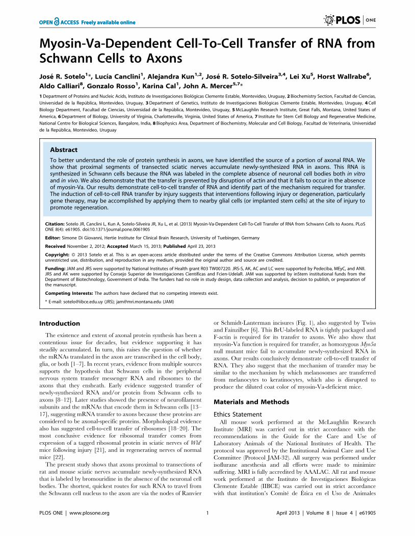
Myosin-Va-Dependent Cell-To-Cell Transfer of RNA fromSchwann Cells to AxonsJose R. Sotelo1*, Lucıa Canclini1, Alejandra Kun1,2, Jose R. Sotelo-Silveira3,4, Lei Xu5, Horst Wallrabe6,
Aldo Calliari8, Gonzalo Rosso1, Karina Cal1, John A. Mercer5,7*
1 Department of Proteins and Nucleic Acids, Instituto de Investigaciones Biologicas Clemente Estable, Montevideo, Uruguay, 2 Biochemistry Section, Facultad de Ciencias,
Universidad de la Republica, Montevideo, Uruguay, 3 Department of Genetics, Instituto de Investigaciones Biologicas Clemente Estable, Montevideo, Uruguay, 4 Cell
Biology Department, Facultad de Ciencias, Universidad de la Republica, Montevideo, Uruguay, 5 McLaughlin Research Institute, Great Falls, Montana, United States of
America, 6 Department of Biology, University of Virginia, Charlottesville, Virginia, United States of America, 7 Institute for Stem Cell Biology and Regenerative Medicine,
National Centre for Biological Sciences, Bangalore, India, 8 Biophysics Area, Department of Biochemistry, Molecular and Cell Biology, Facultad de Veterinaria, Universidad
de la Republica, Montevideo, Uruguay
Abstract
To better understand the role of protein synthesis in axons, we have identified the source of a portion of axonal RNA. Weshow that proximal segments of transected sciatic nerves accumulate newly-synthesized RNA in axons. This RNA issynthesized in Schwann cells because the RNA was labeled in the complete absence of neuronal cell bodies both in vitroand in vivo. We also demonstrate that the transfer is prevented by disruption of actin and that it fails to occur in the absenceof myosin-Va. Our results demonstrate cell-to-cell transfer of RNA and identify part of the mechanism required for transfer.The induction of cell-to-cell RNA transfer by injury suggests that interventions following injury or degeneration, particularlygene therapy, may be accomplished by applying them to nearby glial cells (or implanted stem cells) at the site of injury topromote regeneration.
Citation: Sotelo JR, Canclini L, Kun A, Sotelo-Silveira JR, Xu L, et al. (2013) Myosin-Va-Dependent Cell-To-Cell Transfer of RNA from Schwann Cells to Axons. PLoSONE 8(4): e61905. doi:10.1371/journal.pone.0061905
Editor: Simone Di Giovanni, Hertie Institute for Clinical Brain Research, University of Tuebingen, Germany
Received November 2, 2012; Accepted March 15, 2013; Published April 23, 2013
Copyright: � 2013 Sotelo et al. This is an open-access article distributed under the terms of the Creative Commons Attribution License, which permitsunrestricted use, distribution, and reproduction in any medium, provided the original author and source are credited.
Funding: JAM and JRS were supported by National Institutes of Health grant R03 TW007220. JRS-S, AK, AC and LC were supported by Pedeciba, MEyC, and ANII.JRS and AK were supported by Consejo Superior de Investigaciones Cientıficas and Fcien-UdelaR. JAM was supported by inStem institutional funds from theDepartment of Biotechnology, Government of India. The funders had no role in study design, data collection and analysis, decision to publish, or preparation ofthe manuscript.
Competing Interests: The authors have declared that no competing interests exist.
* E-mail: [email protected] (JRS); [email protected] (JAM)
Introduction
The existence and extent of axonal protein synthesis has been a
contentious issue for decades, but evidence supporting it has
steadily accumulated. In turn, this raises the question of whether
the mRNAs translated in the axon are transcribed in the cell body,
glia, or both [1–7]. In recent years, evidence from multiple sources
supports the hypothesis that Schwann cells in the peripheral
nervous system transfer messenger RNA and ribosomes to the
axons that they ensheath. Early evidence suggested transfer of
newly-synthesized RNA and/or protein from Schwann cells to
axons [8–12]. Later studies showed the presence of neurofilament
subunits and the mRNAs that encode them in Schwann cells [13–
17], suggesting mRNA transfer to axons because these proteins are
considered to be axonal-specific proteins. Morphological evidence
also has suggested cell-to-cell transfer of ribosomes [18–20]. The
most conclusive evidence for ribosomal transfer comes from
expression of a tagged ribosomal protein in sciatic nerves of Wlds
mice following injury [21], and in regenerating nerves of normal
mice [22].
The present study shows that axons proximal to transections of
rat and mouse sciatic nerves accumulate newly-synthesized RNA
that is labeled by bromouridine in the absence of the neuronal cell
bodies. The shortest, quickest routes for such RNA to travel from
the Schwann cell nucleus to the axon are via the nodes of Ranvier
or Schmidt-Lanterman incisures (Fig. 1), also suggested by Twiss
and Fainzilber [6]. This BrU-labeled RNA is tightly packaged and
F-actin is required for its transfer to axons. We also show that
myosin-Va function is required for transfer, as homozygous Myo5a
null mutant mice fail to accumulate newly-synthesized RNA in
axons. Our results conclusively demonstrate cell-to-cell transfer of
RNA. They also suggest that the mechanism of transfer may be
similar to the mechanism by which melanosomes are transferred
from melanocytes to keratinocytes, which also is disrupted to
produce the diluted coat color of myosin-Va-deficient mice.
Materials and Methods
Ethics StatementAll mouse work performed at the McLaughlin Research
Institute (MRI) was carried out in strict accordance with the
recommendations in the Guide for the Care and Use of
Laboratory Animals of the National Institutes of Health. The
protocol was approved by the Institutional Animal Care and Use
Committee (Protocol JAM-32). All surgery was performed under
isoflurane anesthesia and all efforts were made to minimize
suffering. MRI is fully accredited by AAALAC. All rat and mouse
work performed at the Instituto de Investigaciones Biologicas
Clemente Estable (IIBCE) was carried out in strict accordance
with that institution’s Comite de Etica en el Uso de Animales
PLOS ONE | www.plosone.org 1 April 2013 | Volume 8 | Issue 4 | e61905
(CEUA-IIBCE) under law 18.611 of the Republica Oriental del
Uruguay. The specific protocol was approved by the CEUA-
IIBCE (Protocol Sotelo-013/09/2011). All surgery was performed
under pentobarbital anesthesia and all efforts were made to
minimize suffering.
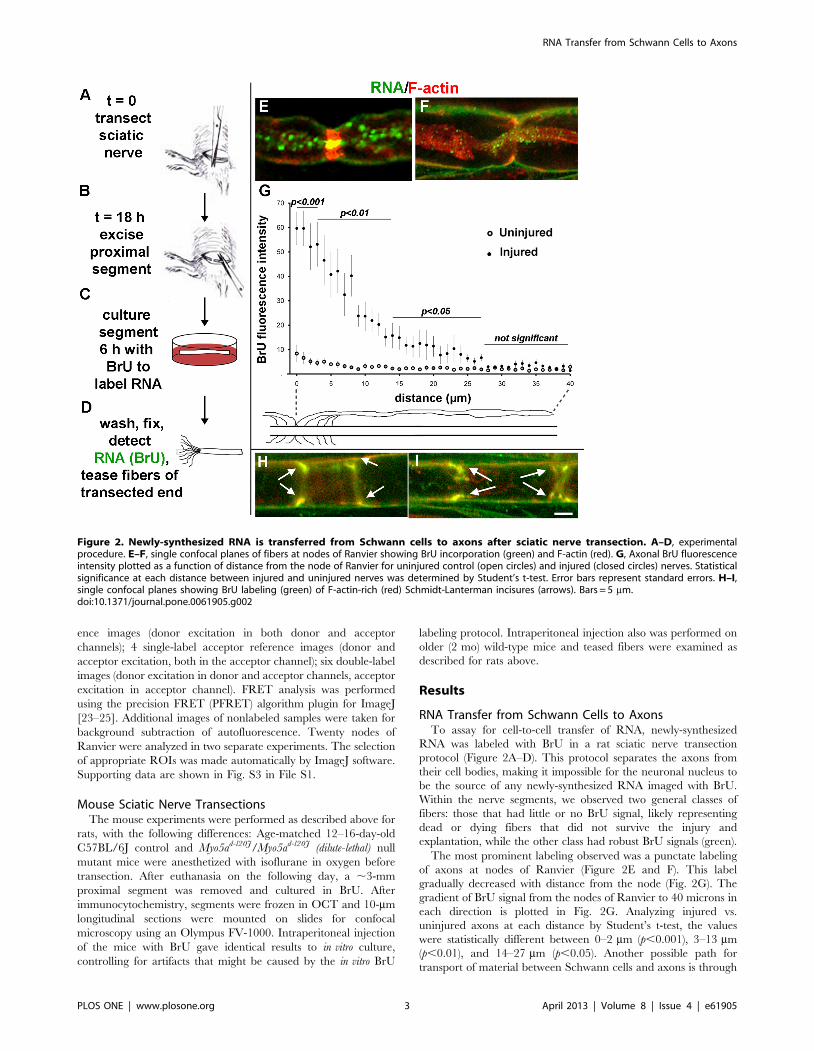
Sciatic Nerve TransectionAdult Sprague-Dawley or Wistar rats were anesthetized with
50 mg/kg pentobarbital. An incision was made at mid-thigh and
the sciatic nerve was transected (diagram, Fig. 2A). Incisions were
closed with cyanoacrylate glue. After 18 h recovery, the rats were
euthanized and a 2-cm sciatic nerve segment proximal to the
transection was removed (Fig. 2B); equivalent contralateral
uninjured segments were used as negative controls. The segments
were incubated in Neurobasal medium (Invitrogen) containing
2.5 mM bromouridine (BrU, Sigma) for 1, 3 or 6 h at 37uC, 5%
CO2 (Fig. 2C). Representative nodes of Ranvier for all three time
points are shown in Fig. S1 in File S1. Only 6-h incubations are
shown in all other figures. A negative control in which transected
nerve segments were incubated for 6 h in Neurobasal medium
lacking BrU also was performed. As an in situ control for artifacts
that might be caused by explanting the nerve segments for BrU
labeling, transection of both sciatic nerves was followed by a
proximal crush injury (achieving axonotmesis) after 18 h, instead
of the second transection and explantation shown in Fig. 2. BrU
was then applied in situ to the left sciatic nerve in the thigh for 3 h
under anesthesia [10]. Meanwhile, the injured contralateral nerve
was explanted and incubated in BrU for 3 h. In all experiments,
segments were washed 10 times for 5 min each in ice-cold PHEM
buffer (60 mM PIPES, 25 mM HEPES, 10 mM EGTA, 2 mM
MgCl2) to remove unincorporated BrU, then fixed for 30 min in
3% paraformaldehyde in PHEM at room temperature. Segments
were treated for 1 h at 37uC with 0.2 mg/ml collagenase (Sigma)
in PHEM with 5 mM CaCl2 and without EGTA. The nerve fibers
were released from epineurium with #5 forceps and teased at the
injured end with 26-gauge needles (Fig. 2D). The segments were
permeabilized with 0.1% triton X-100 in PHEM buffer for 30 min
at room temperature.
ImmunocytochemistryThe incubation buffer for all steps was 0.1% BSA and 50 mM
glycine in PHEM buffer. Nerve segments were prepared for
immunocytochemistry by blocking in 5% normal goat serum for
30 min at 37uC. Permeabilized fibers were incubated with anti-
BrdU (Sigma, 1:300), anti-CASPR (Abcam, 1:300), anti-myosin
Va (kindly supplied by Roy Larson, 1:100), or antiserum against
purified ribosomes [20] (1:1000) for 1 h at 37uC. Fibers were
washed 6 times 5 min each. Secondary antibodies (goat anti-
mouse or goat anti-rabbit conjugated with Alexa 488, 546, or 633,
all from Invitrogen, all 1:1000) were incubated for 45 min at 37uC.
F-actin was detected using fluorescent phalloidin (Invitrogen)
added together with secondary antibodies. Fibers were then
washed 6 times 5 min each. Finally, individual fibers were teased
and mounted in ProLong Antifade (Invitrogen).
a-Amanitin TreatmentRNA polymerase II was inhibited by adding 10 mg/ml a-
amanitin (Sigma) during the BrU labeling step described above.
Ribonuclease TreatmentAfter the wash step to remove soluble BrU, sciatic nerve
segments were incubated with RNAse in PHEM buffer at 5 or
10 mg/ml for 1 h, at 37uC. Segments were washed 10 times 5 min
in PHEM at room temperature.
Latrunculin A TreatmentF-actin was depolymerized by the addition of 0.07, 0.2, 0.6, or
1.8 mg/ml Latrunculin A (Sigma) during the BrU labeling step.
In situ HybridizationIncubations were performed at room temperature unless
otherwise stated. Frozen 10-mm sections of uninjured mouse
sciatic nerves were blocked with 0.03% H2O2 for 1 h, washed 3
times 5 min in 4X SSC, and prehybridized in 4X SSC, 50%
formamide, 10% dextran sulfate, 0.1 mg/ml tRNA, and 0.5 mg/
ml sheared salmon sperm DNA for 2 h at 54uC. Hybridization
was carried out for four hours at 54uC in the same buffer plus
0.5 ng/ml of in vitro transcribed digoxigenin labeled probe
complementary to the small subunit of neurofilament mRNA
(nucleotides 1858 to 1959, NM_010910). Sections were washed
twice for 10 min in 4X SSC plus 30% formamide at 54uC, then
twice for 5 min each in 2X, 1X, 0.5X, and 0.25X SSC. Sections
were postfixed in 3% paraformaldehyde in PHEM for 5 min and
washed three times for 5 min in PHEM. Blocking was performed
as described for immunocytochemistry above. Incubation with
primary antibodies (mouse anti-BrdU, HRP-Sheep anti-digoxi-
genin) was performed overnight at 4uC. Sections were washed 3
times for 10 min in PHEM and then incubated in tyramide
amplification reagent according to the instructions of the
manufacturer (Invitrogen) for 10 min. Excess tyramide was
removed by washing 3 times for 5 min with PHEM. Secondary
antibody (Goat anti-mouse Alexa 546 and goat anti-rabbit Alexa
633, Invitrogen) incubations were performed for two hours. Three
washes for 5 min with PHEM were performed before mounting in
ProLong (Invitrogen).
Confocal MicroscopyTeased fibers were visualized with an Olympus FV-300 confocal
microscope, equipped with a Plan Apo N 60X oil NA 1.42 lens
and 488, 543 and 633 nm laser lines. Images were processed with
Fluoview and ImageJ software. Nodes of Ranvier chosen for
quantitative analysis were all within 100 mm of the injured end.
FRET AnalysisTo estimate the distance between myosin-Va and newly-
synthesized RNA, we performed quantitative fluorescence reso-
nance energy transfer (FRET) between the secondary antibodies
recognizing the primary antibodies described above. Images were
collected for FRET analysis using single-labeled donor or acceptor
samples and double-labeled samples: 4 single-label donor refer-
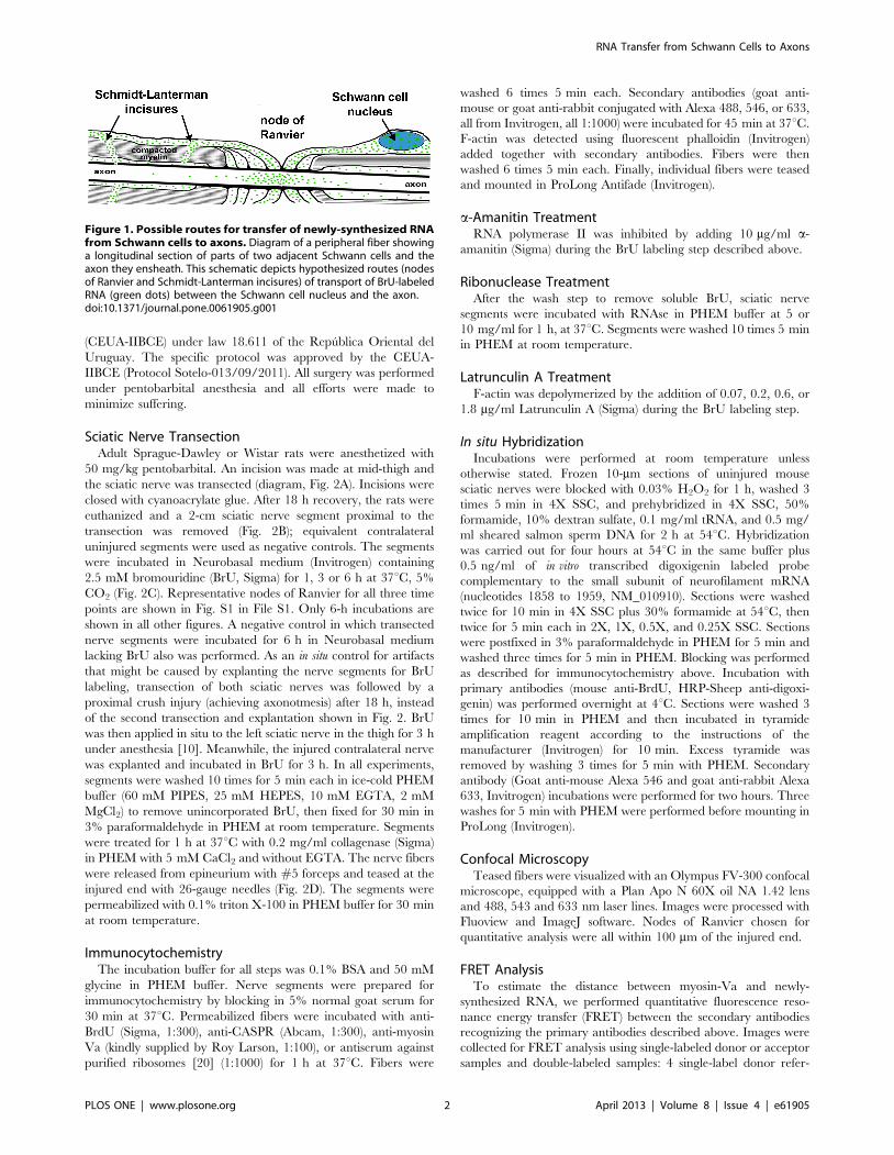
Figure 1. Possible routes for transfer of newly-synthesized RNAfrom Schwann cells to axons. Diagram of a peripheral fiber showinga longitudinal section of parts of two adjacent Schwann cells and theaxon they ensheath. This schematic depicts hypothesized routes (nodesof Ranvier and Schmidt-Lanterman incisures) of transport of BrU-labeledRNA (green dots) between the Schwann cell nucleus and the axon.doi:10.1371/journal.pone.0061905.g001
RNA Transfer from Schwann Cells to Axons
PLOS ONE | www.plosone.org 2 April 2013 | Volume 8 | Issue 4 | e61905
ence images (donor excitation in both donor and acceptor
channels); 4 single-label acceptor reference images (donor and
acceptor excitation, both in the acceptor channel); six double-label
images (donor excitation in donor and acceptor channels, acceptor
excitation in acceptor channel). FRET analysis was performed
using the precision FRET (PFRET) algorithm plugin for ImageJ
[23–25]. Additional images of nonlabeled samples were taken for
background subtraction of autofluorescence. Twenty nodes of
Ranvier were analyzed in two separate experiments. The selection
of appropriate ROIs was made automatically by ImageJ software.
Supporting data are shown in Fig. S3 in File S1.
Mouse Sciatic Nerve TransectionsThe mouse experiments were performed as described above for
rats, with the following differences: Age-matched 12–16-day-old
C57BL/6J control and Myo5ad-l20J/Myo5ad-l20J (dilute-lethal) null
mutant mice were anesthetized with isoflurane in oxygen before
transection. After euthanasia on the following day, a ,3-mm
proximal segment was removed and cultured in BrU. After
immunocytochemistry, segments were frozen in OCT and 10-mm
longitudinal sections were mounted on slides for confocal
microscopy using an Olympus FV-1000. Intraperitoneal injection
of the mice with BrU gave identical results to in vitro culture,
controlling for artifacts that might be caused by the in vitro BrU
labeling protocol. Intraperitoneal injection also was performed on
older (2 mo) wild-type mice and teased fibers were examined as
described for rats above.
Results
RNA Transfer from Schwann Cells to AxonsTo assay for cell-to-cell transfer of RNA, newly-synthesized
RNA was labeled with BrU in a rat sciatic nerve transection
protocol (Figure 2A–D). This protocol separates the axons from
their cell bodies, making it impossible for the neuronal nucleus to
be the source of any newly-synthesized RNA imaged with BrU.
Within the nerve segments, we observed two general classes of
fibers: those that had little or no BrU signal, likely representing
dead or dying fibers that did not survive the injury and
explantation, while the other class had robust BrU signals (green).
The most prominent labeling observed was a punctate labeling
of axons at nodes of Ranvier (Figure 2E and F). This label
gradually decreased with distance from the node (Fig. 2G). The
gradient of BrU signal from the nodes of Ranvier to 40 microns in
each direction is plotted in Fig. 2G. Analyzing injured vs.
uninjured axons at each distance by Student’s t-test, the values
were statistically different between 0–2 mm (p,0.001), 3–13 mm
(p,0.01), and 14–27 mm (p,0.05). Another possible path for
transport of material between Schwann cells and axons is through
Figure 2. Newly-synthesized RNA is transferred from Schwann cells to axons after sciatic nerve transection. A–D, experimentalprocedure. E–F, single confocal planes of fibers at nodes of Ranvier showing BrU incorporation (green) and F-actin (red). G, Axonal BrU fluorescenceintensity plotted as a function of distance from the node of Ranvier for uninjured control (open circles) and injured (closed circles) nerves. Statisticalsignificance at each distance between injured and uninjured nerves was determined by Student’s t-test. Error bars represent standard errors. H–I,single confocal planes showing BrU labeling (green) of F-actin-rich (red) Schmidt-Lanterman incisures (arrows). Bars = 5 mm.doi:10.1371/journal.pone.0061905.g002
RNA Transfer from Schwann Cells to Axons
PLOS ONE | www.plosone.org 3 April 2013 | Volume 8 | Issue 4 | e61905
Schmidt-Lanterman incisures [6]. We saw extensive BrU labeling
of these as well (Fig. 2H and I, arrows).
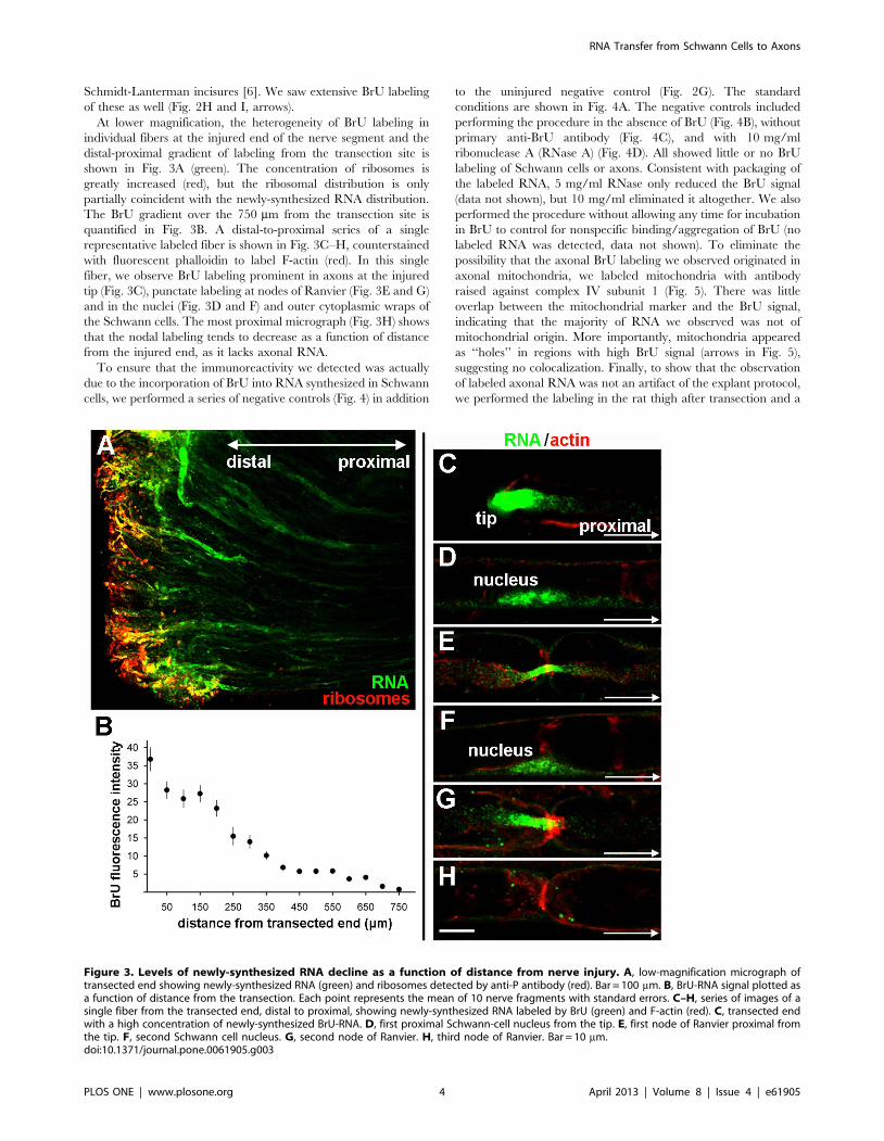
At lower magnification, the heterogeneity of BrU labeling in
individual fibers at the injured end of the nerve segment and the
distal-proximal gradient of labeling from the transection site is
shown in Fig. 3A (green). The concentration of ribosomes is
greatly increased (red), but the ribosomal distribution is only
partially coincident with the newly-synthesized RNA distribution.
The BrU gradient over the 750 mm from the transection site is
quantified in Fig. 3B. A distal-to-proximal series of a single
representative labeled fiber is shown in Fig. 3C–H, counterstained
with fluorescent phalloidin to label F-actin (red). In this single
fiber, we observe BrU labeling prominent in axons at the injured
tip (Fig. 3C), punctate labeling at nodes of Ranvier (Fig. 3E and G)
and in the nuclei (Fig. 3D and F) and outer cytoplasmic wraps of
the Schwann cells. The most proximal micrograph (Fig. 3H) shows
that the nodal labeling tends to decrease as a function of distance
from the injured end, as it lacks axonal RNA.
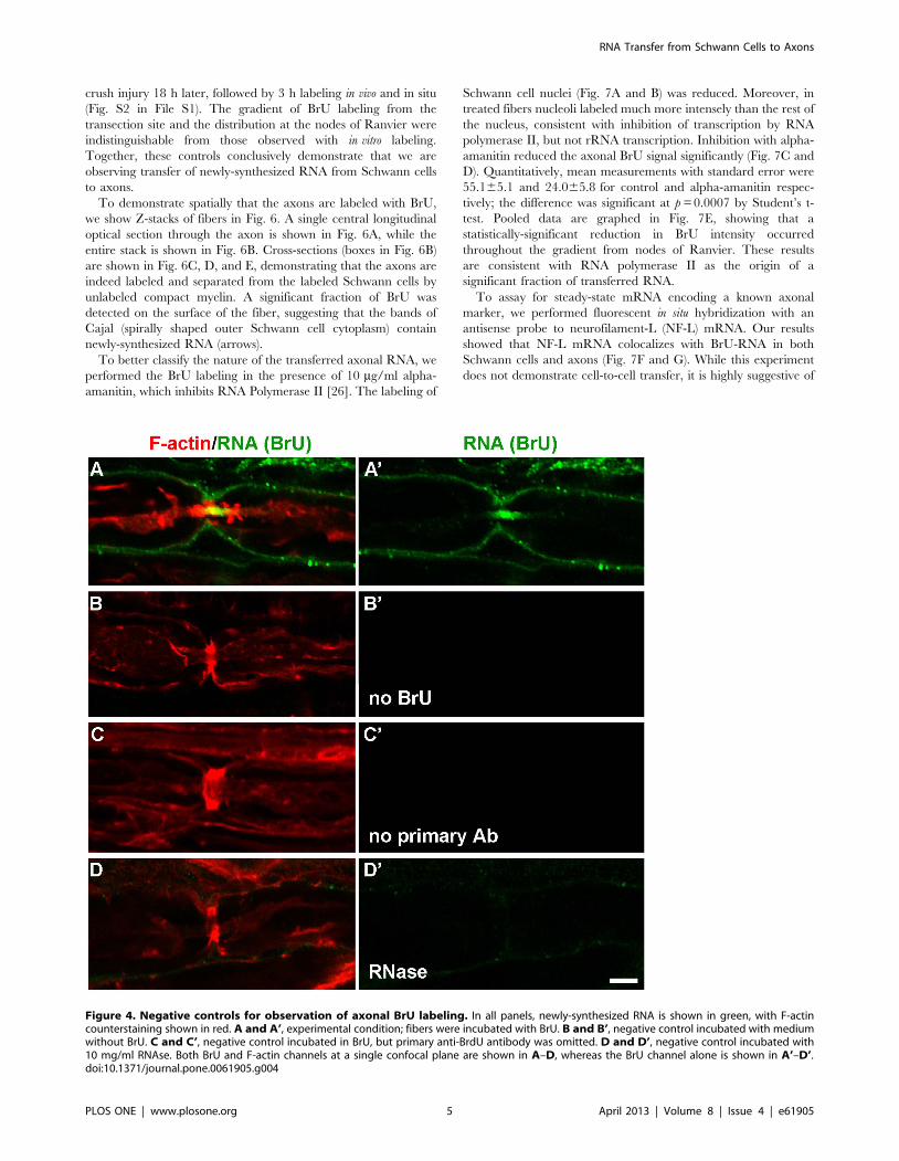
To ensure that the immunoreactivity we detected was actually
due to the incorporation of BrU into RNA synthesized in Schwann
cells, we performed a series of negative controls (Fig. 4) in addition
to the uninjured negative control (Fig. 2G). The standard
conditions are shown in Fig. 4A. The negative controls included
performing the procedure in the absence of BrU (Fig. 4B), without
primary anti-BrU antibody (Fig. 4C), and with 10 mg/ml
ribonuclease A (RNase A) (Fig. 4D). All showed little or no BrU
labeling of Schwann cells or axons. Consistent with packaging of
the labeled RNA, 5 mg/ml RNase only reduced the BrU signal
(data not shown), but 10 mg/ml eliminated it altogether. We also
performed the procedure without allowing any time for incubation
in BrU to control for nonspecific binding/aggregation of BrU (no
labeled RNA was detected, data not shown). To eliminate the
possibility that the axonal BrU labeling we observed originated in
axonal mitochondria, we labeled mitochondria with antibody
raised against complex IV subunit 1 (Fig. 5). There was little
overlap between the mitochondrial marker and the BrU signal,
indicating that the majority of RNA we observed was not of
mitochondrial origin. More importantly, mitochondria appeared
as ‘‘holes’’ in regions with high BrU signal (arrows in Fig. 5),
suggesting no colocalization. Finally, to show that the observation
of labeled axonal RNA was not an artifact of the explant protocol,
we performed the labeling in the rat thigh after transection and a
Figure 3. Levels of newly-synthesized RNA decline as a function of distance from nerve injury. A, low-magnification micrograph oftransected end showing newly-synthesized RNA (green) and ribosomes detected by anti-P antibody (red). Bar = 100 mm. B, BrU-RNA signal plotted asa function of distance from the transection. Each point represents the mean of 10 nerve fragments with standard errors. C–H, series of images of asingle fiber from the transected end, distal to proximal, showing newly-synthesized RNA labeled by BrU (green) and F-actin (red). C, transected endwith a high concentration of newly-synthesized BrU-RNA. D, first proximal Schwann-cell nucleus from the tip. E, first node of Ranvier proximal fromthe tip. F, second Schwann cell nucleus. G, second node of Ranvier. H, third node of Ranvier. Bar = 10 mm.doi:10.1371/journal.pone.0061905.g003
RNA Transfer from Schwann Cells to Axons
PLOS ONE | www.plosone.org 4 April 2013 | Volume 8 | Issue 4 | e61905
crush injury 18 h later, followed by 3 h labeling in vivo and in situ
(Fig. S2 in File S1). The gradient of BrU labeling from the
transection site and the distribution at the nodes of Ranvier were
indistinguishable from those observed with in vitro labeling.
Together, these controls conclusively demonstrate that we are
observing transfer of newly-synthesized RNA from Schwann cells
to axons.
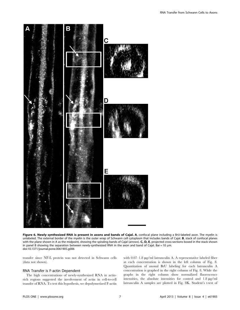
To demonstrate spatially that the axons are labeled with BrU,
we show Z-stacks of fibers in Fig. 6. A single central longitudinal
optical section through the axon is shown in Fig. 6A, while the
entire stack is shown in Fig. 6B. Cross-sections (boxes in Fig. 6B)
are shown in Fig. 6C, D, and E, demonstrating that the axons are
indeed labeled and separated from the labeled Schwann cells by
unlabeled compact myelin. A significant fraction of BrU was
detected on the surface of the fiber, suggesting that the bands of
Cajal (spirally shaped outer Schwann cell cytoplasm) contain
newly-synthesized RNA (arrows).
To better classify the nature of the transferred axonal RNA, we
performed the BrU labeling in the presence of 10 mg/ml alpha-
amanitin, which inhibits RNA Polymerase II [26]. The labeling of
Schwann cell nuclei (Fig. 7A and B) was reduced. Moreover, in
treated fibers nucleoli labeled much more intensely than the rest of
the nucleus, consistent with inhibition of transcription by RNA
polymerase II, but not rRNA transcription. Inhibition with alpha-
amanitin reduced the axonal BrU signal significantly (Fig. 7C and
D). Quantitatively, mean measurements with standard error were
55.165.1 and 24.065.8 for control and alpha-amanitin respec-
tively; the difference was significant at p = 0.0007 by Student’s t-
test. Pooled data are graphed in Fig. 7E, showing that a
statistically-significant reduction in BrU intensity occurred
throughout the gradient from nodes of Ranvier. These results
are consistent with RNA polymerase II as the origin of a
significant fraction of transferred RNA.
To assay for steady-state mRNA encoding a known axonal
marker, we performed fluorescent in situ hybridization with an
antisense probe to neurofilament-L (NF-L) mRNA. Our results
showed that NF-L mRNA colocalizes with BrU-RNA in both
Schwann cells and axons (Fig. 7F and G). While this experiment
does not demonstrate cell-to-cell transfer, it is highly suggestive of
Figure 4. Negative controls for observation of axonal BrU labeling. In all panels, newly-synthesized RNA is shown in green, with F-actincounterstaining shown in red. A and A’, experimental condition; fibers were incubated with BrU. B and B’, negative control incubated with mediumwithout BrU. C and C’, negative control incubated in BrU, but primary anti-BrdU antibody was omitted. D and D’, negative control incubated with10 mg/ml RNAse. Both BrU and F-actin channels at a single confocal plane are shown in A–D, whereas the BrU channel alone is shown in A’–D’.doi:10.1371/journal.pone.0061905.g004
RNA Transfer from Schwann Cells to Axons
PLOS ONE | www.plosone.org 5 April 2013 | Volume 8 | Issue 4 | e61905
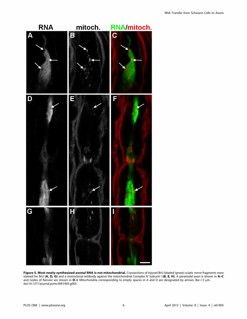
Figure 5. Most newly-synthesized axonal RNA is not mitochondrial. Cryosections of injured BrU-labeled (green) sciatic nerve fragments werestained for BrU (A, D, G) and a monoclonal antibody against the mitochondrial Complex IV Subunit I (B, E, H). A paranodal axon is shown in A–Cand nodes of Ranvier are shown in D–I. Mitochondria corresponding to empty spaces in A and D are designated by arrows. Bar = 5 mm.doi:10.1371/journal.pone.0061905.g005
RNA Transfer from Schwann Cells to Axons
PLOS ONE | www.plosone.org 6 April 2013 | Volume 8 | Issue 4 | e61905
transfer since NF-L protein was not detected in Schwann cells
(data not shown).
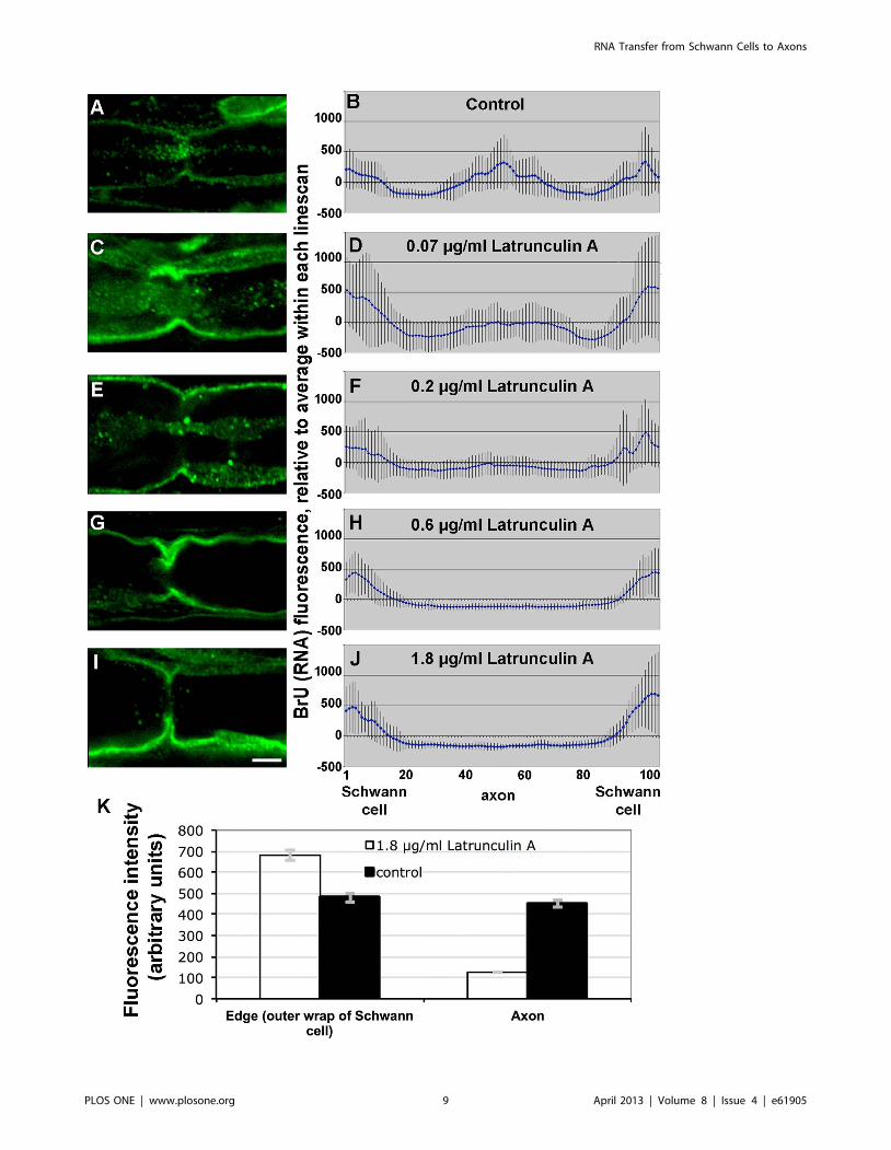
RNA Transfer is F-actin DependentThe high concentrations of newly-synthesized RNA in actin-
rich regions suggested the involvement of actin in cell-to-cell
transfer of RNA. To test this hypothesis, we depolymerized F-actin
with 0.07–1.8 mg/ml latrunculin A. A representative labeled fiber
at each concentration is shown in the left column of Fig. 8.
Quantitation of axonal BrU labeling for each latrunculin A
concentration is graphed in the right column of Fig. 8. While the
graphs in the right column show normalized fluorescence
intensities, the absolute intensities for control and 1.8 mg/ml
latrunculin A samples are plotted in Fig. 8K. Student’s t-test of
Figure 6. Newly synthesized RNA is present in axons and bands of Cajal. A, confocal plane including a BrU-labeled axon. The myelin isunlabeled. The external border of the myelin is the outer wrap of Schwann cell cytoplasm that includes bands of Cajal. B, stack of confocal planeswith the plane shown in A as the midpoint, showing the spiraling bands of Cajal (arrows). C, D, E, projected cross-sections boxed in the stack shownin panel B showing the separation between newly-synthesized RNA in the axon and band of Cajal. Bar = 10 mm.doi:10.1371/journal.pone.0061905.g006
RNA Transfer from Schwann Cells to Axons
PLOS ONE | www.plosone.org 7 April 2013 | Volume 8 | Issue 4 | e61905
control vs. experimental intensities in edges and axons were
significant with p = 0.02 and p,0.0001 respectively. In other
words, the relative decrease of BrU signal in the axon was
complemented by an increase of signal in the Schwann cells,
consistent with inhibition of transport from the latter to the
former.
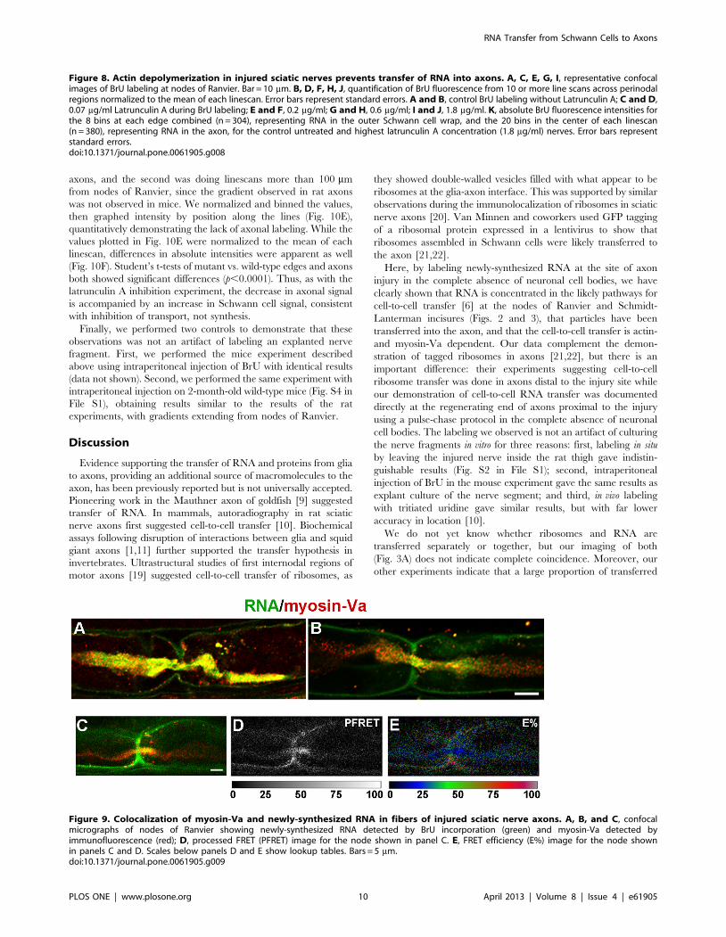
RNA Transfer is Myosin-Va-dependentThe requirement for actin in turn suggested a role for myosin
motors, so we performed immunofluorescent detection of myosin-
Va after transection. We observed significant colocalization of
myosin-Va with newly-synthesized RNA (Fig. 9A–C). To estimate
the distance between myosin-Va and newly-synthesized RNA, we
performed quantitative fluorescence resonance energy transfer
(FRET) between the secondary antibodies detecting the anti-
myosin-Va and anti-BrU primary antibodies. The spectral
bleedthrough-corrected processed FRET (PFRET) signal [24]
was observed in axons and Schwann cell cytoplasm at the nodes of
Ranvier (Fig. 8D). Specific FRET signals, as demonstrated by E%,
an expression of distances between fluorophores of 1–10 nm, were
enriched in axons near the nodes of Ranvier (Fig. 8E, Fig. S3 in
File S1). Thus, our data are consistent with a close association of
myosin-Va with BrU-RNA in axons.
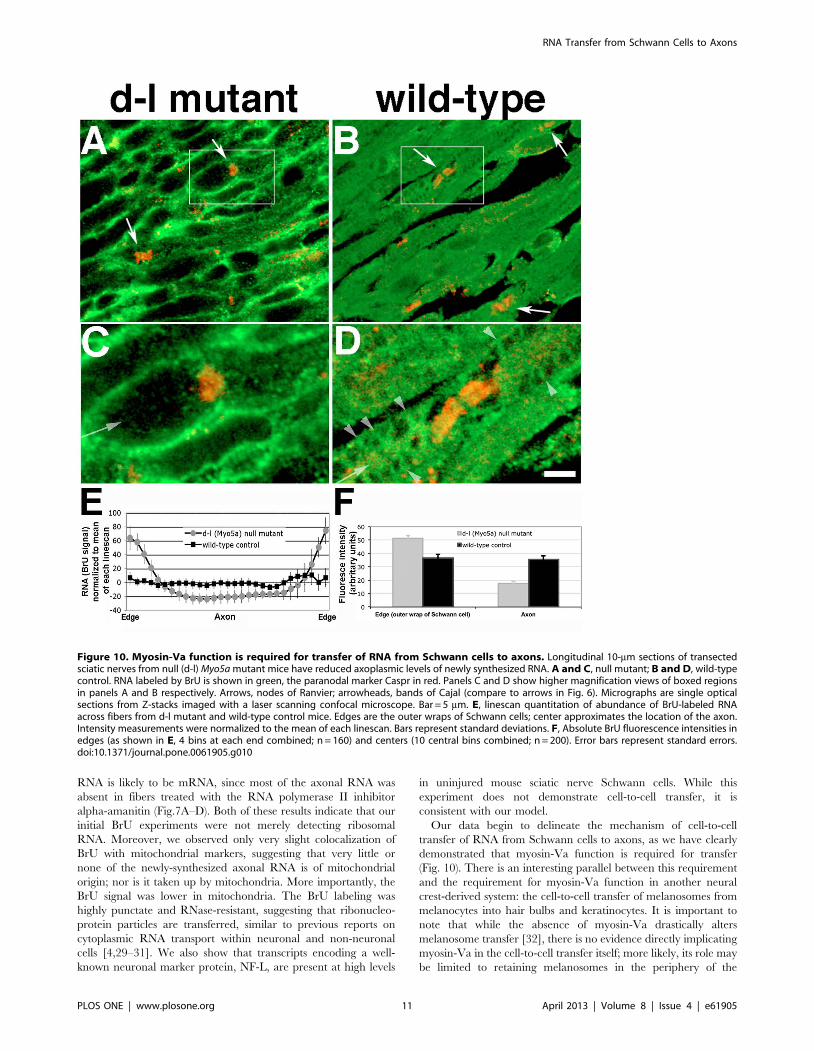
As a genetic test for a requirement for myosin-Va function in
cell-to-cell transfer of RNA, we modified the sciatic nerve
transection and BrU labeling procedure developed for adult rats
for 12–17-day-old mice, allowing us to perform the experiment on
dilute-lethal (Myo5ad-l20J/Myo5ad-l20J) null mutant pups. These mice
lack myosin-Va, which causes them to die at 19–21 days of age
[27]. To compensate for the smaller diameter of mouse fibers,
instead of teasing whole-mount preparations, the segments
proximal to the transection were frozen and longitudinally
sectioned. The results (Fig. 10) were striking: while wild-type
littermate controls (Fig. 10B) had fibers and axons filled with BrU,
as well as prominent labeling of bands of Cajal (Fig. 10D,
arrowheads), axons of mutant mice had no detectable BrU
labeling (Fig. 10A and C). Nodes of Ranvier (arrows) were
identified by immunofluorescent detection of the paranodal
marker Caspr [28]. To quantify the difference between mutant
and wild-type fibers, we measured fluorescence intensities using
20-pixel wide linescans across 50 fibers chosen blindly from 5 mice
of each genotype. There were two criteria: the first was greater
width, to ensure a bias toward measuring diameters that included
Figure 7. Most of the newly-synthesized RNA is produced by RNA Polymerase II. A–E, injured control nerves without a-amanitin (A andC), and injured nerves treated with a-amanitin during the BrU labeling period (B and D) were stained for BrU (green) and F-actin with phalloidin(red). A and B, Schwann cell nuclei; C and D, nodes of Ranvier. Bar = 10 mm. E, BrU-RNA fluorescence intensities plotted as a function of distancefrom the node of Ranvier for controls without a-amanitin (circles) and nerves treated with 10 mg/ml a-amanitin (triangles). Statistical significance ateach distance was determined by Student’s t-test. Error bars represent standard errors. F, Neurofilament L (NF-L) mRNA is found in both Schwanncells and axons by in situ hybridization (red) and BrU-RNA (green). Arrows are pointing to axons. G, negative control NF-L sense probe. Bar = 5 mm.doi:10.1371/journal.pone.0061905.g007
RNA Transfer from Schwann Cells to Axons
PLOS ONE | www.plosone.org 8 April 2013 | Volume 8 | Issue 4 | e61905
RNA Transfer from Schwann Cells to Axons
PLOS ONE | www.plosone.org 9 April 2013 | Volume 8 | Issue 4 | e61905
axons, and the second was doing linescans more than 100 mm
from nodes of Ranvier, since the gradient observed in rat axons
was not observed in mice. We normalized and binned the values,
then graphed intensity by position along the lines (Fig. 10E),
quantitatively demonstrating the lack of axonal labeling. While the
values plotted in Fig. 10E were normalized to the mean of each
linescan, differences in absolute intensities were apparent as well
(Fig. 10F). Student’s t-tests of mutant vs. wild-type edges and axons
both showed significant differences (p,0.0001). Thus, as with the
latrunculin A inhibition experiment, the decrease in axonal signal
is accompanied by an increase in Schwann cell signal, consistent
with inhibition of transport, not synthesis.
Finally, we performed two controls to demonstrate that these
observations was not an artifact of labeling an explanted nerve
fragment. First, we performed the mice experiment described
above using intraperitoneal injection of BrU with identical results
(data not shown). Second, we performed the same experiment with
intraperitoneal injection on 2-month-old wild-type mice (Fig. S4 in
File S1), obtaining results similar to the results of the rat
experiments, with gradients extending from nodes of Ranvier.
Discussion
Evidence supporting the transfer of RNA and proteins from glia
to axons, providing an additional source of macromolecules to the
axon, has been previously reported but is not universally accepted.
Pioneering work in the Mauthner axon of goldfish [9] suggested
transfer of RNA. In mammals, autoradiography in rat sciatic
nerve axons first suggested cell-to-cell transfer [10]. Biochemical
assays following disruption of interactions between glia and squid
giant axons [1,11] further supported the transfer hypothesis in
invertebrates. Ultrastructural studies of first internodal regions of
motor axons [19] suggested cell-to-cell transfer of ribosomes, as
they showed double-walled vesicles filled with what appear to be
ribosomes at the glia-axon interface. This was supported by similar
observations during the immunolocalization of ribosomes in sciatic
nerve axons [20]. Van Minnen and coworkers used GFP tagging
of a ribosomal protein expressed in a lentivirus to show that
ribosomes assembled in Schwann cells were likely transferred to
the axon [21,22].
Here, by labeling newly-synthesized RNA at the site of axon
injury in the complete absence of neuronal cell bodies, we have
clearly shown that RNA is concentrated in the likely pathways for
cell-to-cell transfer [6] at the nodes of Ranvier and Schmidt-
Lanterman incisures (Figs. 2 and 3), that particles have been
transferred into the axon, and that the cell-to-cell transfer is actin-
and myosin-Va dependent. Our data complement the demon-
stration of tagged ribosomes in axons [21,22], but there is an
important difference: their experiments suggesting cell-to-cell
ribosome transfer was done in axons distal to the injury site while
our demonstration of cell-to-cell RNA transfer was documented
directly at the regenerating end of axons proximal to the injury
using a pulse-chase protocol in the complete absence of neuronal
cell bodies. The labeling we observed is not an artifact of culturing
the nerve fragments in vitro for three reasons: first, labeling in situ
by leaving the injured nerve inside the rat thigh gave indistin-
guishable results (Fig. S2 in File S1); second, intraperitoneal
injection of BrU in the mouse experiment gave the same results as
explant culture of the nerve segment; and third, in vivo labeling
with tritiated uridine gave similar results, but with far lower
accuracy in location [10].
We do not yet know whether ribosomes and RNA are
transferred separately or together, but our imaging of both
(Fig. 3A) does not indicate complete coincidence. Moreover, our
other experiments indicate that a large proportion of transferred
Figure 8. Actin depolymerization in injured sciatic nerves prevents transfer of RNA into axons. A, C, E, G, I, representative confocalimages of BrU labeling at nodes of Ranvier. Bar = 10 mm. B, D, F, H, J, quantification of BrU fluorescence from 10 or more line scans across perinodalregions normalized to the mean of each linescan. Error bars represent standard errors. A and B, control BrU labeling without Latrunculin A; C and D,0.07 mg/ml Latrunculin A during BrU labeling; E and F, 0.2 mg/ml; G and H, 0.6 mg/ml; I and J, 1.8 mg/ml. K, absolute BrU fluorescence intensities forthe 8 bins at each edge combined (n = 304), representing RNA in the outer Schwann cell wrap, and the 20 bins in the center of each linescan(n = 380), representing RNA in the axon, for the control untreated and highest latrunculin A concentration (1.8 mg/ml) nerves. Error bars representstandard errors.doi:10.1371/journal.pone.0061905.g008
Figure 9. Colocalization of myosin-Va and newly-synthesized RNA in fibers of injured sciatic nerve axons. A, B, and C, confocalmicrographs of nodes of Ranvier showing newly-synthesized RNA detected by BrU incorporation (green) and myosin-Va detected byimmunofluorescence (red); D, processed FRET (PFRET) image for the node shown in panel C. E, FRET efficiency (E%) image for the node shownin panels C and D. Scales below panels D and E show lookup tables. Bars = 5 mm.doi:10.1371/journal.pone.0061905.g009
RNA Transfer from Schwann Cells to Axons
PLOS ONE | www.plosone.org 10 April 2013 | Volume 8 | Issue 4 | e61905
RNA is likely to be mRNA, since most of the axonal RNA was
absent in fibers treated with the RNA polymerase II inhibitor
alpha-amanitin (Fig.7A–D). Both of these results indicate that our
initial BrU experiments were not merely detecting ribosomal
RNA. Moreover, we observed only very slight colocalization of
BrU with mitochondrial markers, suggesting that very little or
none of the newly-synthesized axonal RNA is of mitochondrial
origin; nor is it taken up by mitochondria. More importantly, the
BrU signal was lower in mitochondria. The BrU labeling was
highly punctate and RNase-resistant, suggesting that ribonucleo-
protein particles are transferred, similar to previous reports on
cytoplasmic RNA transport within neuronal and non-neuronal
cells [4,29–31]. We also show that transcripts encoding a well-
known neuronal marker protein, NF-L, are present at high levels
in uninjured mouse sciatic nerve Schwann cells. While this
experiment does not demonstrate cell-to-cell transfer, it is
consistent with our model.
Our data begin to delineate the mechanism of cell-to-cell
transfer of RNA from Schwann cells to axons, as we have clearly
demonstrated that myosin-Va function is required for transfer
(Fig. 10). There is an interesting parallel between this requirement
and the requirement for myosin-Va function in another neural
crest-derived system: the cell-to-cell transfer of melanosomes from
melanocytes into hair bulbs and keratinocytes. It is important to
note that while the absence of myosin-Va drastically alters
melanosome transfer [32], there is no evidence directly implicating
myosin-Va in the cell-to-cell transfer itself; more likely, its role may
be limited to retaining melanosomes in the periphery of the
Figure 10. Myosin-Va function is required for transfer of RNA from Schwann cells to axons. Longitudinal 10-mm sections of transectedsciatic nerves from null (d-l) Myo5a mutant mice have reduced axoplasmic levels of newly synthesized RNA. A and C, null mutant; B and D, wild-typecontrol. RNA labeled by BrU is shown in green, the paranodal marker Caspr in red. Panels C and D show higher magnification views of boxed regionsin panels A and B respectively. Arrows, nodes of Ranvier; arrowheads, bands of Cajal (compare to arrows in Fig. 6). Micrographs are single opticalsections from Z-stacks imaged with a laser scanning confocal microscope. Bar = 5 mm. E, linescan quantitation of abundance of BrU-labeled RNAacross fibers from d-l mutant and wild-type control mice. Edges are the outer wraps of Schwann cells; center approximates the location of the axon.Intensity measurements were normalized to the mean of each linescan. Bars represent standard deviations. F, Absolute BrU fluorescence intensities inedges (as shown in E, 4 bins at each end combined; n = 160) and centers (10 central bins combined; n = 200). Error bars represent standard errors.doi:10.1371/journal.pone.0061905.g010
RNA Transfer from Schwann Cells to Axons
PLOS ONE | www.plosone.org 11 April 2013 | Volume 8 | Issue 4 | e61905
melanocyte. We propose a similar mechanism in this case, with
myosin-Va helping to retain RNA in the regions of the Schwann
cell cytoplasm from which the transferred RNA is taken or
donated. Whether the Schwann cell, axon, or both play the active
role of cell-to-cell transfer remains an entirely open question.
There are three primary differences between the mouse data
and the rat data. The first is the lack of any gradient of BrU
immunoreactivity spreading out from the nodes of Ranvier. This is
likely caused by a higher metabolic rate in the very young mice
relative to that of the adult rats; shortening of the BrU labeling
period to as little as 20 min did not produce a gradient (data not
shown). Consistent with this hypothesis, labeling the injured sciatic
nerves of 2-month-old, wild-type mice yielded similar results to the
rat experiments (Fig. S4 in File S1). The second is the difficulty in
distinguishing axons in the wild-type fibers, again due to the young
age of the mice. The third difference is the thickness and
raggedness of the Schwann-cell labeling in the mutant mice, likely
because myelination is in progress at this age.
While the data presented here are from injured nerves (with the
exception of the comparison of BrU gradients in Injured to
Uninjured (Fig. 2) and in situ hybridization data in Fig. 7), they are
provocative when combined with previous observations in
uninjured axons: depolymerization of F-actin by cytochalasin B
inhibits axonal protein synthesis [33], and that myosin-Va and the
mRNA encoding it are present in periaxoplasmic ribosomal
plaques in uninjured axons [34]. This raises interesting questions:
first, is myosin-Va function required for axonal protein synthesis
from mRNAs that originate in the neuronal soma; and second,
does cell-to-cell transfer of RNA occur developmentally? We are
addressing both questions using transgenic and knock-in mice with
tissue-specific expression of tagged mRNAs and proteins.
In summary, these data confirm and extend our understanding
of the complex relationship between glia and the axons they
ensheath. This relationship is crucial in understanding mecha-
nisms underlying responses to injury and neurodegeneration, as
well as in designing therapeutic strategies that exploit intercellular
transport for both retrograde signaling to the cell body [35] and
controlling regeneration. The close associations and complex
topologies of Schwann cell and axonal plasma membranes make
assessment of intercellular transfer mechanisms difficult; however,
our data suggest essential roles for both F-actin and myosin-Va in
this mechanism.
Supporting Information
File S1 Contains Figures S1, S2, S3, and S4 withlegends.
(DOCX)
Acknowledgments
We thank Mike Kavanaugh and Dave Bonislawski for assistance with
confocal microscopy, supported by National Institutes of Health grant P20
RR015583. We thank Tejas Gupte, Farah Haque, John Bermingham, and
members of Jim Spudich’s laboratory (Stanford) for critiquing the
manuscript.
Author Contributions
Conceived and designed the experiments: JRS LC AK JRS-S HW JAM.
Performed the experiments: JRS LC AK LX HW GR KC AC. Analyzed
the data: JRS LC AK LX JRS-S HW. Contributed reagents/materials/
analysis tools: HW. Wrote the paper: JRS LC AK JRS-S JAM.
References
1. Lasek RJ, Gainer H, Barker JL (1977) Cell-to-cell transfer of glial proteins to thesquid giant axon. The glia-neuron protein transfer hypothesis. J Cell Biol 74:
501–523.
2. Alvarez J (2001) The autonomous axon: a model based on local synthesis ofproteins. Biol Res 34: 103–109.
3. Brittis PA, Lu Q, Flanagan JG (2002) Axonal protein synthesis provides a
mechanism for localized regulation at an intermediate target. Cell 110: 223–235.
4. Sotelo-Silveira JR, Calliari A, Kun A, Koenig E, Sotelo JR (2006) RNA
trafficking in axons. Traffic 7: 508–515.
5. Van Horck FP, Holt CE (2008) A cytoskeletal platform for local translation inaxons. Sci Signal 1: pe11.
6. Twiss JL, Fainzilber M (2009) Ribosomes in axons–scrounging from the
neighbors? Trends Cell Biol 19: 236–243.
7. Crispino M, Cefaliello C, Kaplan B, Giuditta A (2009) Protein synthesis in nerve
terminals and the glia-neuron unit. Results Probl Cell Differ 48: 243–267.
8. Edstrom JE, Eichner D, Edstrom A (1962) The ribonucleic acid of axons andmyelin sheaths from Mauthner neurons. Biochim Biophys Acta 61: 178–184.
9. Jakoubek B, Edstrom JE (1965) RNA changes in the Mauthner axon and myelin
sheath after increased functional activity. J Neurochem 12: 845–849.
10. Benech C, Sotelo JR, Menendez J, Correa-Luna R (1982) Autoradiographic
study of RNA and protein synthesis in sectioned peripheral nerves. Exp Neurol
76: 72–82.
11. Cutillo V, Montagnese P, Gremo F, Casola L, Giuditta A (1983) Origin of
axoplasmic RNA in the squid giant fiber. Neurochem Res 8: 1621–1634.
12. Eyman M, Cefaliello C, Ferrara E, De Stefano R, Lavina ZS, et al. (2007) Localsynthesis of axonal and presynaptic RNA in squid model systems. Eur J Neurosci
25: 341–350.
13. Sotelo JR, Benech CR, Kun A (1992) Local radiolabeling of the 68 kDaneurofilament protein in rat sciatic nerves. Neurosci Lett 144: 174–176.
14. Roberson MD, Toews AD, Goodrum JF, Morell P (1992) Neurofilament and
tubulin mRNA expression in Schwann cells. J Neurosci Res 33: 156–162.
15. Kelly BM, Gillespie CS, Sherman DL, Brophy PJ (1992) Schwann cells of the
myelin-forming phenotype express neurofilament protein NF-M. J Cell Biol 118:
397–410.
16. Fabrizi C, Kelly BM, Gillespie CS, Schlaepfer WW, Scherer SS, et al. (1997)
Transient expression of the neurofilament proteins NF-L and NF-M by
Schwann cells is regulated by axonal contact. J Neurosci Res 50: 291–299.
17. Sotelo-Silveira JR, Calliari A, Kun A, Benech JC, Sanguinetti C, et al. (2000)
Neurofilament mRNAs are present and translated in the normal and severedsciatic nerve. J Neurosci Res 62: 65–74.
18. Pearce J, Lnenicka GA, Govind CK (2003) Regenerating crayfish motor axons
assimilate glial cells and sprout in cultured explants. J Comp Neurol 464: 449–
462.
19. Li YC, Li YN, Cheng CX, Sakamoto H, Kawate T, et al. (2005) Subsurface
cisterna-lined axonal invaginations and double-walled vesicles at the axonal-
myelin sheath interface. Neuroscience research 53: 298–303.
20. Kun A, Otero L, Sotelo-Silveira JR, Sotelo JR (2007) Ribosomal distributions in
axons of mammalian myelinated fibers. J Neurosci Res 85: 2087–2098.
21. Court FA, Hendriks WT, Macgillavry HD, Alvarez J, van Minnen J, et al. (2008)
Schwann cell to axon transfer of ribosomes: toward a novel understanding of the
role of glia in the nervous system. J Neurosci 28: 11024–11029.
22. Court FA, Midha R, Cisterna BA, Grochmal J, Shakhbazau A, et al. (2011)
Morphological evidence for a transport of ribosomes from Schwann cells to
regenerating axons. Glia 59: 1529–1539.
23. Wallrabe H, Chen Y, Periasamy A, Barroso M (2006) Issues in confocal
microscopy for quantitative FRET analysis. Microsc Res Tech 69: 196–206.
24. Chen Y, Periasamy A (2006) Intensity range based quantitative FRET data
analysis to localize protein molecules in live cell nuclei. Journal of Fluorescence
16: 95–104.
25. Elangovan M, Wallrabe H, Chen Y, Day RN, Barroso M, et al. (2003)
Characterization of one- and two-photon excitation fluorescence resonance
energy transfer microscopy. Methods 29: 58–73.
26. Bensaude O (2011) Inhibiting eukaryotic transcription: Which compound to
choose? How to evaluate its activity? Transcription 2: 103–108.
27. Searle AG (1952) A lethal allele of dilute in the house mouse. Heredity 6: 395–
401.
28. Einheber S, Zanazzi G, Ching W, Scherer S, Milner TA, et al. (1997) The
axonal membrane protein Caspr, a homologue of neurexin IV, is a component
of the septate-like paranodal junctions that assemble during myelination. J Cell
Biol 139: 1495–1506.
29. Sossin WS, DesGroseillers L (2006) Intracellular trafficking of RNA in neurons.
Traffic 7: 1581–1589.
30. Bassell GJ, Oleynikov Y, Singer RH (1999) The travels of mRNAs through all
cells large and small. Faseb J 13: 447–454.
31. Kosik KS, Krichevsky AM (2005) The elegance of the microRNAs: a neuronal
perspective. Neuron 47: 779–782.
32. Silvers WK (1979) The coat colors of mice. New York: Springer-Verlag. 379 p.
33. Sotelo-Silveira J, Crispino M, Puppo A, Sotelo JR, Koenig E (2008) Myelinated
axons contain beta-actin mRNA and ZBP-1 in periaxoplasmic ribosomal
RNA Transfer from Schwann Cells to Axons
PLOS ONE | www.plosone.org 12 April 2013 | Volume 8 | Issue 4 | e61905
plaques and depend on cyclic AMP and F-actin integrity for in vitro translation.
J Neurochem 104: 545–547.34. Sotelo-Silveira JR, Calliari A, Cardenas M, Koenig E, Sotelo JR (2004) Myosin
Va and kinesin II motor proteins are concentrated in ribosomal domains
(periaxoplasmic ribosomal plaques) of myelinated axons. J Neurobiol 60: 187–
196.35. Rishal I, Fainzilber M (2010) Retrograde signaling in axonal regeneration. Exp
Neurol 223: 5–10.
RNA Transfer from Schwann Cells to Axons
PLOS ONE | www.plosone.org 13 April 2013 | Volume 8 | Issue 4 | e61905
Copyright © 2022 FDOKUMEN