Development and analytical application of a glucose biosensor based on glucose oxidase/...
6
Biosensors and Bioelectronics 25 (2010) 2238–2243 Contents lists available at ScienceDirect Biosensors and Bioelectronics journal homepage: www.elsevier.com/locate/bios Development and analytical application of a glucose biosensor based on glucose oxidase/O-(2-hydroxyl)propyl-3-trimethylammonium chitosan chloride nanoparticle-immobilized onion inner epidermis Fei Wang a , Jun Yao a,* , Mohammad Russel a , Huilun Chen a , Ke Chen a , Yong Zhou a , Brunello Ceccanti b , Gyula Zaray c , Martin M.F. Choi d,** a Key Laboratory of Biogeology and Environmental Geology of Chinese Ministry of Education & Sino-Hungarian Joint Laboratory of Environmental Science and Health, China University of Geosciences, 430074 Wuhan, PR China b Institute of Ecosystem Studies (ISE), Italian National Research Council (ICT-CNR), Pisa, Italy c Department of Chemical Technology and Environmental Chemistry, Eötvös University, H-1518 Budapest, P.O. Box 32, Hungary d Department of Chemistry, Hong Kong Baptist University, Kowloon Tong, Hong Kong, PR China article info Article history: Received 20 December 2009 Received in revised form 5 February 2010 Accepted 26 February 2010 Available online 6 March 2010 Keywords: O-HTCC nanoparticle Enzyme immobilization Onion membrane Glucose oxidase abstract A glucose biosensor comprising a glucose oxidase/O-(2-hydroxyl)propyl-3-trimethylammonium chi- tosan chloride nanoparticle (O-HTCC NP)-immobilized onion inner membrane and a dissolved oxygen (O 2 ) sensor has been successfully developed. The detection scheme is based on the depletion of dissolved O 2 content upon exposure to glucose. The decrease in O 2 level was monitored and related to the glucose concentration. The biosensor shows linear response to glucose from 0.0 to 0.60 mM with a detection limit of 50 M(S/N = 3). The effect of O-HTCC NP and enzyme loading, pH, temperature, and phosphate buffer concentration on the sensitivity of the biosensor was studied in detail. The biosensor exhibits fast response time (70 s), good repeatability (3.2%, n = 10) and storage stability (90% of initial sensitivity after 3-week storage). Common interferents including acetic acid, lactic acid, propionic acid, butyric acid, folic acid, methanol, glycine, dl--alanine and dl-cysteine do not cause significant interferences on the biosensor. The proposed biosensor method was successfully applied to determine the glucose content in real samples such as orange juice, red wine and tea drink and the results were comparable to that obtained from a spectrophotometric method. The glucose recovery test demonstrates that the proposed glucose biosensor offers an excellent, accurate and precise method for the determination of glucose in real samples. © 2010 Elsevier B.V. All rights reserved. 1. Introduction In recent years there is a growing interest in the development of glucose biosensor based on immobilized enzyme since rapid, accurate and in situ determination of glucose in food industry, med- ical and bioanalysis fields is of paramount importance (Mizutani and Yabuki, 1997; Wang, 1999; Choi, 2005). Considerable atten- tion has been focused on immobilizing glucose oxidase (GO x ) on different solid matrices which are subsequently positioned on a transducer surface to produce workable biosensors. This enzyme immobilization process is crucial as it can determine the analyti- cal performance of a glucose biosensor (Zhang et al., 2005). As such, finding the compatible enzyme matrices and effective cross-linking * Corresponding author. Tel.: +86 27 67883036; fax: +86 27 87436235. ** Corresponding author. Tel.: +852 34117839; fax: +852 34117348. E-mail addresses: [email protected] (J. Yao), [email protected] (M.M.F. Choi). agents for improving the functionality, sensitivity and repeatability of biosensors has always been an important subject for researchers. An excellent immobilization support and opportune technological design are essential to preserve the biological activities of enzymes (Wink et al., 1997). Enzyme immobilization is commonly performed by physical adsorption (Niculescu et al., 2004), covalent attachment (Li et al., 1998), entrapment (Kwan et al., 2004; Tag et al., 2000), or cross- linking (Yang et al., 2006; Wen et al., 2007; Zhang et al., 2007). On the other hand, various inorganic materials (Hasse and Scholz, 2004; Manesh et al., 2008; Wang et al., 2009), organic polymers (Rauf et al., 2006; Li and Lin, 2007) and natural biomaterials (Yang et al., 2006; Wen et al., 2007; Kumar and D’Souza, 2009) have been exploited for immobilization of enzymes and their use in biosen- sor fabrication. Among these methods and matrices, the effective ways to improve stability, repeatability, reproducibility and activ- ity of immobilized enzymes are to incorporate the enzymes into biomaterials by cross-linking as they are more biocompatible with each other since biomaterials in living organisms are composed 0956-5663/$ – see front matter © 2010 Elsevier B.V. All rights reserved. doi:10.1016/j.bios.2010.02.033
Transcript of Development and analytical application of a glucose biosensor based on glucose oxidase/...
Biosensors and Bioelectronics 25 (2010) 2238–2243
Contents lists available at ScienceDirect
Biosensors and Bioelectronics
journa l homepage: www.e lsev ier .com/ locate /b ios
Development and analytical application of a glucose biosensor based on glucoseoxidase/O-(2-hydroxyl)propyl-3-trimethylammonium chitosan chloridenanoparticle-immobilized onion inner epidermis
Fei Wanga, Jun Yaoa,!, Mohammad Russela, Huilun Chena, Ke Chena, Yong Zhoua, Brunello Ceccantib,Gyula Zarayc, Martin M.F. Choid,!!
a Key Laboratory of Biogeology and Environmental Geology of Chinese Ministry of Education & Sino-Hungarian Joint Laboratory of Environmental Science and Health,China University of Geosciences, 430074 Wuhan, PR Chinab Institute of Ecosystem Studies (ISE), Italian National Research Council (ICT-CNR), Pisa, Italyc Department of Chemical Technology and Environmental Chemistry, Eötvös University, H-1518 Budapest, P.O. Box 32, Hungaryd Department of Chemistry, Hong Kong Baptist University, Kowloon Tong, Hong Kong, PR China
a r t i c l e i n f o
Article history:Received 20 December 2009Received in revised form 5 February 2010Accepted 26 February 2010Available online 6 March 2010
Keywords:O-HTCC nanoparticleEnzyme immobilizationOnion membraneGlucose oxidase
a b s t r a c t
A glucose biosensor comprising a glucose oxidase/O-(2-hydroxyl)propyl-3-trimethylammonium chi-tosan chloride nanoparticle (O-HTCC NP)-immobilized onion inner membrane and a dissolved oxygen(O2) sensor has been successfully developed. The detection scheme is based on the depletion of dissolvedO2 content upon exposure to glucose. The decrease in O2 level was monitored and related to the glucoseconcentration. The biosensor shows linear response to glucose from 0.0 to 0.60 mM with a detectionlimit of 50 !M (S/N = 3). The effect of O-HTCC NP and enzyme loading, pH, temperature, and phosphatebuffer concentration on the sensitivity of the biosensor was studied in detail. The biosensor exhibitsfast response time (70 s), good repeatability (3.2%, n = 10) and storage stability (90% of initial sensitivityafter 3-week storage). Common interferents including acetic acid, lactic acid, propionic acid, butyric acid,folic acid, methanol, glycine, dl-"-alanine and dl-cysteine do not cause significant interferences on thebiosensor. The proposed biosensor method was successfully applied to determine the glucose contentin real samples such as orange juice, red wine and tea drink and the results were comparable to thatobtained from a spectrophotometric method. The glucose recovery test demonstrates that the proposedglucose biosensor offers an excellent, accurate and precise method for the determination of glucose inreal samples.
© 2010 Elsevier B.V. All rights reserved.
1. Introduction
In recent years there is a growing interest in the developmentof glucose biosensor based on immobilized enzyme since rapid,accurate and in situ determination of glucose in food industry, med-ical and bioanalysis fields is of paramount importance (Mizutaniand Yabuki, 1997; Wang, 1999; Choi, 2005). Considerable atten-tion has been focused on immobilizing glucose oxidase (GOx) ondifferent solid matrices which are subsequently positioned on atransducer surface to produce workable biosensors. This enzymeimmobilization process is crucial as it can determine the analyti-cal performance of a glucose biosensor (Zhang et al., 2005). As such,finding the compatible enzyme matrices and effective cross-linking
! Corresponding author. Tel.: +86 27 67883036; fax: +86 27 87436235.!! Corresponding author. Tel.: +852 34117839; fax: +852 34117348.
E-mail addresses: [email protected] (J. Yao), [email protected](M.M.F. Choi).
agents for improving the functionality, sensitivity and repeatabilityof biosensors has always been an important subject for researchers.An excellent immobilization support and opportune technologicaldesign are essential to preserve the biological activities of enzymes(Wink et al., 1997).
Enzyme immobilization is commonly performed by physicaladsorption (Niculescu et al., 2004), covalent attachment (Li et al.,1998), entrapment (Kwan et al., 2004; Tag et al., 2000), or cross-linking (Yang et al., 2006; Wen et al., 2007; Zhang et al., 2007).On the other hand, various inorganic materials (Hasse and Scholz,2004; Manesh et al., 2008; Wang et al., 2009), organic polymers(Rauf et al., 2006; Li and Lin, 2007) and natural biomaterials (Yanget al., 2006; Wen et al., 2007; Kumar and D’Souza, 2009) have beenexploited for immobilization of enzymes and their use in biosen-sor fabrication. Among these methods and matrices, the effectiveways to improve stability, repeatability, reproducibility and activ-ity of immobilized enzymes are to incorporate the enzymes intobiomaterials by cross-linking as they are more biocompatible witheach other since biomaterials in living organisms are composed
0956-5663/$ – see front matter © 2010 Elsevier B.V. All rights reserved.doi:10.1016/j.bios.2010.02.033
Ke Chen
5Y IF=5.4
F. Wang et al. / Biosensors and Bioelectronics 25 (2010) 2238–2243 2239
of biomolecules such as carbohydrates, lipids and proteins. Sofar, bamboo inner shell membrane, eggshell membrane and onioninner epidermal membrane have been proved to be useful bio-materials for enzyme immobilization (Yang et al., 2006; Zhang etal., 2006; Kumar and D’Souza, 2009). For effective enzyme immo-bilization, it is essential to find a suitable cross-linking agent toprevent enzyme leaching from the solid support. Glutaraldehydeand chitosan have been successfully used as a gluey agent, whilethe sensitivity of the biosensor with chitosan is two times moresensitive than glutaraldehyde (Wen et al., 2007). The use of chi-tosan, however, is generally limited to acidic conditions since ithas poor solubility above pH 6.5 where chitosan loses its cationicnature (Jae et al., 2003). In order to circumvent this problem, chi-tosan can be functionalized with quaternary ammonium salt toform cationic chitosan. For instances, choline dichloride compris-ing a trimethylammonium moiety reacted with chitosan to yieldhighly cationic chitosan (Curti et al., 2003.). Chitosan was convertedto O-(2-hydroxyl)propyl-3-trimethylammonium chitosan chloride(O-HTCC) nanoparticle (NP) and this O-HTCC NP was proved to bea useful protein carrier material (Sun and Wan, 2007).
In this study, we explore the use of O-HTCC NP for immobi-lization of an enzyme for biosensing. The major advantage of ourproposed method is to employ a new alternative cross-linkingagent and biomaterial as an enzyme immobilization platform. Theinner epidermal membrane of the onion bulk scales was chosen asthe bio-platform to immobilize enzyme since it possesses excel-lent gas and water permeability for substrates and products todiffuse in and out of the membrane; very similar to those of theabove-mentioned biomaterials. Onion membranes mainly consistof elongated tubular cells, blunt or tapering ends along with numer-ous guard cells (Scott et al., 1958; Bruce and Hepworth, 2004). Thestructural features of the inner epidermis cell wall can possiblyprovide a biocompatible environment for enzyme immobilization.This natural membrane is mechanically stronger than other naturalmembranes for biosensors because of its microfibrillar cellulosicstructure. Thus, it should be an ideal biocompatible platform forenzyme immobilization (Kumar and D’Souza, 2009).
Glucose oxidase (GOx) was selected for investigation since itcan recognize glucose target molecules quickly and accuratelyin complicated systems. In addition, GOx has been widely stud-ied and applied in the field of biosensor (Newman and Turner,2005; Manesh et al., 2008; Wang et al., 2009). In this work, thequantitative determination of glucose in real samples is based onthe enzymatic oxidation of glucose by dissolved oxygen (O2) asdepicted in Eq. (1). GOx is a flavoprotein which catalyzes the oxi-dation of #-d-glucose by molecular O2 to $-gluconolactone whichsubsequently spontaneously hydrolyzes to gluconic acid (Zoldak etal., 2004):
d-glucose + O2 + H2OGOx"#d-gluconic acid + H2O2 (1)
The aim of this work is to explore the use of GOx/O-HTCCNP-immobilized onion inner membrane for quantitative deter-mination of glucose. The enzyme-immobilized membrane wasfabricated by gelating GOx with O-HTCC NP on an inner epidermalonion membrane and then positioned it on the surface of an O2 sen-sor. The biosensor response is defined as the decrease in dissolvedO2 concentration which is linearly related to the glucose concentra-tion in the standard solutions. The effects of pH, phosphate bufferconcentration and temperature on the response of the biosensorwere studied in detail. To our knowledge, this is the first reporton using onion membrane to immobilize both GOx and O-HTCCNP to achieve higher enzyme activity, better detection sensitivityand stability. Our proposed biosensor method has been success-fully applied to the determination of glucose in some beverage andwine samples.
2. Experimental
2.1. Materials and reagents
Glucose oxidase (EC 1.1.3.4 from Aspergillus niger) with a spe-cific activity of 245 units per milligram of solid was obtainedfrom Sigma (St. Louis, MO, USA). Benzaldehyde, chitosan, gly-cidyltrimethylammonium chloride, glutaraldehyde solution (25%,w/w) and sodium tripolyphosphate were obtained from Aldrich(Milwaukee, WI, USA). All other reagents were of analytical-reagentgrade and used without further purification. The buffer solution forpreparing the glucose solution was 200 mM monosodium dihydro-gen phosphate–disodium hydrogen phosphate solution at pH 7.4.A glucose stock standard solution (0.50 M) was prepared in pH 7.4phosphate buffer (200 mM) and was allowed to mutarotate for 24 hat room temperature before use. All solutions were prepared withdoubly distilled water. Fresh onions (Allium cepa L.) were purchasedfrom a local market.
2.2. Preparation of O-HTCC nanoparticle
The synthesis of O-HTCC NP was based on a previous method(Sun and Wan, 2007) with slight modifications. 3.0 g of chitosanwas dissolved in 100 mL of 10% acetic acid solution. After theaddition of benzaldehyde (15.8 g), the solution was stirred for anhour and then left for 20 h in vacuum (55–60 $C). The resultingsolution was adjusted to pH 7.0 with a 1 M NaOH solution andfiltered. The N-benzylidene chitosan precipitate was washed withmethanol several times. 9.0 g of N-benzylidene chitosan and 9.0 g ofglycidyltrimethylammonium chloride were added to 50 mL of iso-propyl alcohol. The resulting mixture was stirred at 70 $C for 16 h toprecipitate the o-quaternary ammonium-N-benzylidene chitosan.The o-quaternary ammonium-N-benzylidene chitosan was col-lected and washed with methanol and acetone successively. 3.0 gof o-quaternary ammonium-N-benzylidene chitosan was added to50 mL of 0.25 M ethanolic HCl solution and then stirred for 24 hat room temperature. After most of the ethanol was removed byvacuum, 15 mL of H2O was added to the mixture and followed byadding excess acetone to precipitate the O-HTCC. The solvent wasremoved under vacuum at 80 $C to obtain O-HTCC. The crude O-HTCC product was further purified: 1.5 g of O-HTCC was dissolvedin 40 mL of water and then the solution was poured into acetone toobtain the O-HTCC precipitate. The precipitate was collected andwashed with acetone several times. The precipitate was then dis-solved in distilled water again and dialyzed against water for 3 days,concentrated, precipitated in acetone, and dried under vacuum at80 $C for 48 h to get the purified O-HTCC product.
10 mg of the purified O-HTCC was dissolved in 10 mL of aceticacid. Then 0.80 mL of sodium tripolyphosphate aqueous solution(4.0 mg mL"1) was added to the stirred O-HTCC solution at roomtemperature. Opalescent suspension immediately appeared andthe solution was filtered to obtain the O-HTCC NP. The O-HTCCNP was dried under vacuum and kept in a dry box until further use.The size of O-HTCC NPs was determined to be ca. 35.3 nm in sodiumtripolyphosphate aqueous solution (Sun and Wan, 2007).
2.3. Immobilization of GOx/O-HTCC NP on onion membrane
The procedure to immobilize GOx and O-HTCC NP on an onioninner epidermis membrane was similar to a previous report (Kumarand D’Souza, 2009). An onion was cut into half, bulb scales wereseparated and inner epidermis from outer fleshy scale was stripped.Onion membranes were placed in a clean watch glass, cut into acircular pieces of 1.5 cm diameter and stored at 4 $C. An aliquot of100 !L of 0.8% (w/v) GOx (prepared in 200 mM phosphate bufferat pH 7.4) was added on the onion membrane. After 30 min, 20 !L
2240 F. Wang et al. / Biosensors and Bioelectronics 25 (2010) 2238–2243
of 0.3% (w/v) O-HTCC NP solution as a gelation agent was droppedonto the surface of the membrane to mix with GOx uniformly by aglass rod. The GOx/O-HTCC NP-immobilized membrane was driedat room temperature for 1 h. The GOx/O-HTCC NP-immobilizedmembrane was immersed in and washed with a phosphate buffer(pH 7.4) for 5 min to remove any loosely bound enzyme and thenkept at 4 $C until further use.
2.4. Scanning electron microscopy of onion membrane
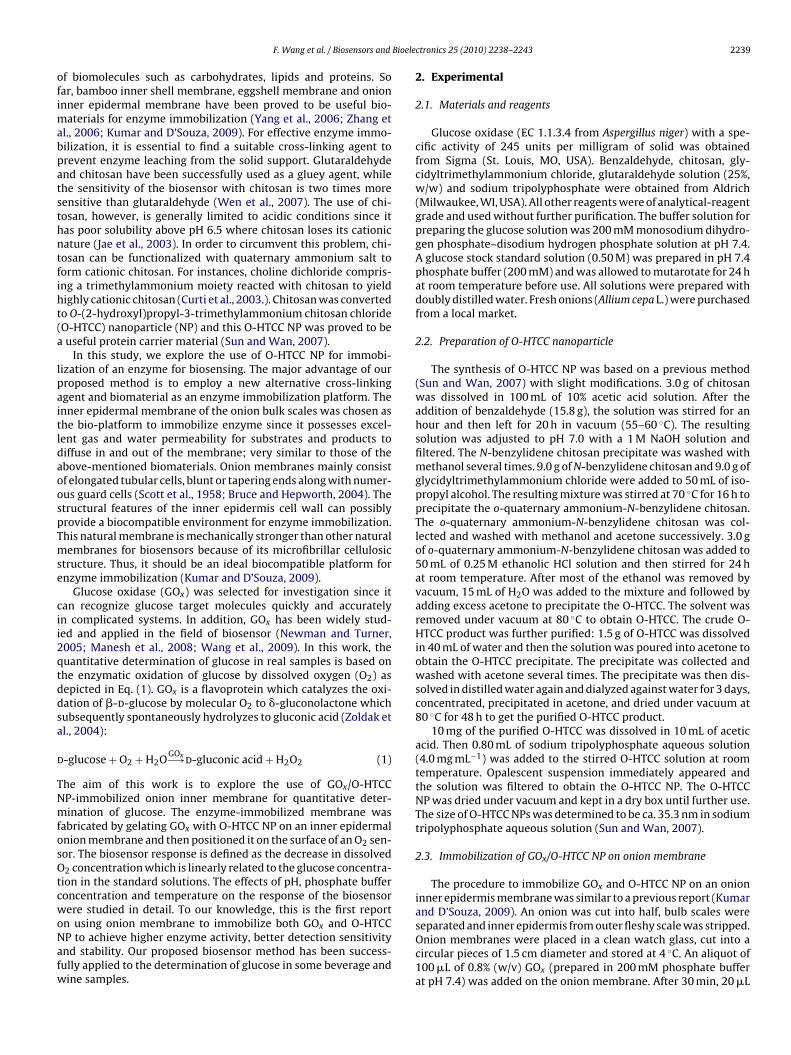
The scanning electron micrographs (SEM) of the onion inner epi-dermis with and without the immobilized GOx/O-HTCC NP werecaptured by a Quanta 200 scanning electron microscope (FEI Com-pany, Hillsboro, OR, USA).
2.5. Assembly of glucose biosensor and determination
The GOx/O-HTCC NP-immobilized onion inner epidermis waspositioned on the surface of a Pasco CI-6542 O2 sensor (Pasco Scien-tific, Roseville, CA, USA) and kept in a steady position by an O-ring.The O2 sensor was immersed into a stirred 5.0 mL phosphate buffersolution (200 mM, pH 7.4). A series of volumes of standard (0.50 M)or sample glucose solution were injected into the phosphate bufferwith a syringe. The dissolved O2 signal was captured and processedby a datalogger system consisting of a Science Workshop 500 inter-face, serial cables, a power supply, and control software (PascoScientific, Roseville, CA, USA). The data were logged in a computerfor real-time display and processing. The working principle of O2sensor has been previously described by Wen et al. (2007).
3. Results and discussion
3.1. Microstructure of onion inner membranes without and withGOx/O-HTCC NP
The microstructures of onion inner membranes without andwith immobilized GOx/O-HTCC NP were studied. An onion innermembrane is mainly composed of regular fibrillar lacunaes withclear stripes as depicted in Fig. 1a. When the onion membranewas immobilized with GOx/O-HTCC NP, the onion fibers still retaintheir integrity which is more or less the same as the fresh one. Itis clear that lumps of GOx/O-HTCC NP are deposited on the onionfibers (Fig. 1b). The SEM supports that GOx/O-HTCC were success-fully immobilized onto the onion membrane. It is well known thatenzyme can be immobilized on solid substrates via the electro-static interaction between the positively charged polyelectrolytesand the negatively charged enzyme molecules (Wei et al., 2003;Choi, 2005; Miscoria et al., 2006). Fig. S1 (Supplementary Material)depicts the chemical structure of O-HTCC which is a quaternizedchitosan derivative possessing a positive charge per monomer unit.The immobilization of GOx on O-HTCC NP can be interpreted by thestrong electrostatic attraction between the cationic O-HTCC NP andthe anionic GOx. The major advantages of our enzyme immobiliza-tion method are three folds: first, O-HTCC NP have larger specificsurface area which means more enzyme molecules loading; sec-ond, O-HTCC NP could still maintain its cationic nature at pH > 6.5as compared to chitosan (Jae et al., 2003), inferring that GOx/O-HTCC NP would be stable at a wider pH range; finally, the enzymemolecules immobilized on the O-HTCC NP/onion membrane couldretain their bioactivity because of the good biocompatibility ofonion membrane with enzyme.
3.2. Response behavior of glucose biosensor
The principal detection mechanism of the glucose biosensorrelies on the enzymatic oxidation of glucose by O2 as shown in Eq.
Fig. 1. Scanning electron micrographs of the onion inner membranes without (a)and with (b) immobilized GOx/O-HTCC NP.
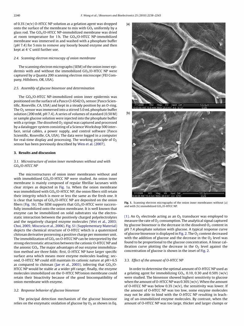
(1). An O2 electrode acting as an O2 transducer was employed tomeasure the rate of O2 consumption. The analytical signal capturedby glucose biosensor is the decrease in the dissolved O2 content inpH 7.4 phosphate solution with glucose. A typical response curveof glucose biosensor is displayed in Fig. 2. The O2 content decreasedwith the addition of glucose and the decrease in the O2 level wasfound to be proportional to the glucose concentration. A linear cal-ibration curve plotting the decrease in the O2 level against theconcentration of glucose is shown in the inset of Fig. 2.
3.3. Effect of the amount of O-HTCC NP
In order to determine the optimal amount of O-HTCC NP used asa gelating agent for immobilizing GOx, 0.10, 0.30 and 0.50% (w/v)were studied. The biosensor showed highest sensitivity to glucosewhen the amount of O-HTCC NP was 0.30% (w/v). When the amountof O-HTCC NP was below 0.3% (w/v), the sensitivity was lower. Ifthe amount of O-HTCC NP was too low, some enzyme moleculesmay not be able to bind with the O-HTCC NP resulting in leach-ing of un-immobilized enzyme molecules. By contrast, when theamount of O-HTCC NP was too large, thicker and larger clumps or
F. Wang et al. / Biosensors and Bioelectronics 25 (2010) 2238–2243 2241
Fig. 2. Typical response curve of the glucose biosensor at pH 7.4 phosphate buffer(200 mM) after each successive addition of various volumes of 0.50 M glucose stan-dard: (0) 0.0; (1 and 2) 1.0; and (3 and 4) 2.0 !L. The inset displays the linearcalibration curve for glucose from 0.00 to 0.60 mM. Error bars are 95% confidenceintervals calculated from three replicate measurements.
clusters of GOx/O-HTCC NP were deposited on the onion fibers ofthe membrane which slow down the gas and water permeability ofmembrane and then eventually prolong the response time (>100 s).
3.4. Effect of enzyme loading
Since the response of the glucose biosensor depends strongly onthe amount of the immobilized enzyme, any change in enzyme con-centration would influence its sensitivity (Wen et al., 2007). Variousamounts of GOx (0.20, 0.40, 0.60, 0.80, 1.0, 1.2, and 1.5%, w/v) wereimmobilized on the onion inner membrane to determine its opti-mal response sensitivity. The sensitivity, that is the decrease indissolved O2, was measured by exposing the biosensor to 0.20 mMglucose in phosphate buffer at pH 7.4. In general, it was found thatthe sensitivity of the glucose biosensor increased with the increaseof enzyme loading on the onion inner membrane. However, notmuch increase in the sensitivity of the biosensor was observedwhen the concentration of GOx was higher than 0.80% (w/v). It isobvious that larger enzyme loading will improve sensitivity as moreGOx molecules are immobilized on the onion membrane. However,when O-HTCC NP is overloaded with enzyme, the excess GOx willnot bind with O-HTCC NP; as a result, they just simply leach outof the membrane. As such, the optimal concentration for enzymeimmobilization was chosen as 0.80% (w/v) in this work.
3.5. Comparison of performance of GOx immobilized onionmembrane using O-HTCC NP, chitosan and glutaraldehyde
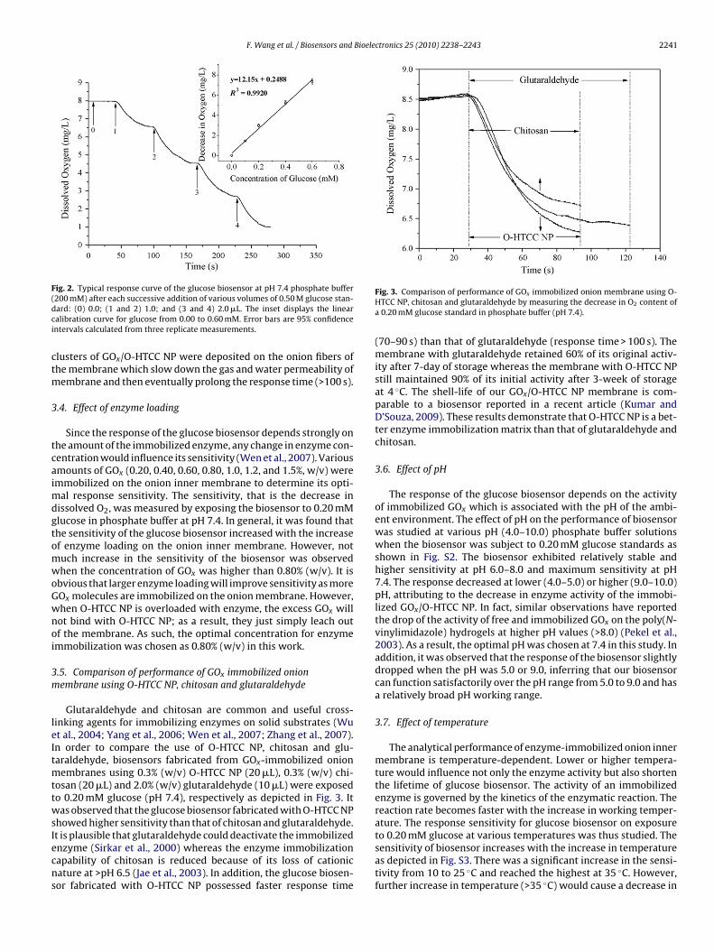
Glutaraldehyde and chitosan are common and useful cross-linking agents for immobilizing enzymes on solid substrates (Wuet al., 2004; Yang et al., 2006; Wen et al., 2007; Zhang et al., 2007).In order to compare the use of O-HTCC NP, chitosan and glu-taraldehyde, biosensors fabricated from GOx-immobilized onionmembranes using 0.3% (w/v) O-HTCC NP (20 !L), 0.3% (w/v) chi-tosan (20 !L) and 2.0% (w/v) glutaraldehyde (10 !L) were exposedto 0.20 mM glucose (pH 7.4), respectively as depicted in Fig. 3. Itwas observed that the glucose biosensor fabricated with O-HTCC NPshowed higher sensitivity than that of chitosan and glutaraldehyde.It is plausible that glutaraldehyde could deactivate the immobilizedenzyme (Sirkar et al., 2000) whereas the enzyme immobilizationcapability of chitosan is reduced because of its loss of cationicnature at >pH 6.5 (Jae et al., 2003). In addition, the glucose biosen-sor fabricated with O-HTCC NP possessed faster response time
Fig. 3. Comparison of performance of GOx immobilized onion membrane using O-HTCC NP, chitosan and glutaraldehyde by measuring the decrease in O2 content ofa 0.20 mM glucose standard in phosphate buffer (pH 7.4).
(70–90 s) than that of glutaraldehyde (response time > 100 s). Themembrane with glutaraldehyde retained 60% of its original activ-ity after 7-day of storage whereas the membrane with O-HTCC NPstill maintained 90% of its initial activity after 3-week of storageat 4 $C. The shell-life of our GOx/O-HTCC NP membrane is com-parable to a biosensor reported in a recent article (Kumar andD’Souza, 2009). These results demonstrate that O-HTCC NP is a bet-ter enzyme immobilization matrix than that of glutaraldehyde andchitosan.
3.6. Effect of pH
The response of the glucose biosensor depends on the activityof immobilized GOx which is associated with the pH of the ambi-ent environment. The effect of pH on the performance of biosensorwas studied at various pH (4.0–10.0) phosphate buffer solutionswhen the biosensor was subject to 0.20 mM glucose standards asshown in Fig. S2. The biosensor exhibited relatively stable andhigher sensitivity at pH 6.0–8.0 and maximum sensitivity at pH7.4. The response decreased at lower (4.0–5.0) or higher (9.0–10.0)pH, attributing to the decrease in enzyme activity of the immobi-lized GOx/O-HTCC NP. In fact, similar observations have reportedthe drop of the activity of free and immobilized GOx on the poly(N-vinylimidazole) hydrogels at higher pH values (>8.0) (Pekel et al.,2003). As a result, the optimal pH was chosen at 7.4 in this study. Inaddition, it was observed that the response of the biosensor slightlydropped when the pH was 5.0 or 9.0, inferring that our biosensorcan function satisfactorily over the pH range from 5.0 to 9.0 and hasa relatively broad pH working range.
3.7. Effect of temperature
The analytical performance of enzyme-immobilized onion innermembrane is temperature-dependent. Lower or higher tempera-ture would influence not only the enzyme activity but also shortenthe lifetime of glucose biosensor. The activity of an immobilizedenzyme is governed by the kinetics of the enzymatic reaction. Thereaction rate becomes faster with the increase in working temper-ature. The response sensitivity for glucose biosensor on exposureto 0.20 mM glucose at various temperatures was thus studied. Thesensitivity of biosensor increases with the increase in temperatureas depicted in Fig. S3. There was a significant increase in the sensi-tivity from 10 to 25 $C and reached the highest at 35 $C. However,further increase in temperature (>35 $C) would cause a decrease in
2242 F. Wang et al. / Biosensors and Bioelectronics 25 (2010) 2238–2243
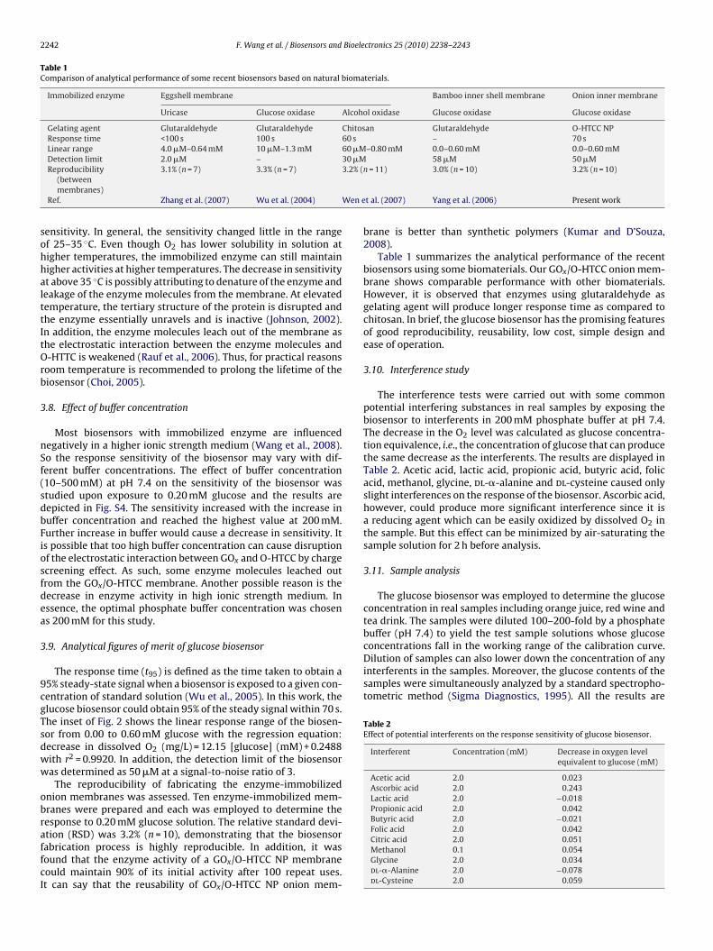
Table 1Comparison of analytical performance of some recent biosensors based on natural biomaterials.
Immobilized enzyme Eggshell membrane Bamboo inner shell membrane Onion inner membrane
Uricase Glucose oxidase Alcohol oxidase Glucose oxidase Glucose oxidase
Gelating agent Glutaraldehyde Glutaraldehyde Chitosan Glutaraldehyde O-HTCC NPResponse time <100 s 100 s 60 s – 70 sLinear range 4.0 !M–0.64 mM 10 !M–1.3 mM 60 !M–0.80 mM 0.0–0.60 mM 0.0–0.60 mMDetection limit 2.0 !M – 30 !M 58 !M 50 !MReproducibility
(betweenmembranes)
3.1% (n = 7) 3.3% (n = 7) 3.2% (n = 11) 3.0% (n = 10) 3.2% (n = 10)
Ref. Zhang et al. (2007) Wu et al. (2004) Wen et al. (2007) Yang et al. (2006) Present work
sensitivity. In general, the sensitivity changed little in the rangeof 25–35 $C. Even though O2 has lower solubility in solution athigher temperatures, the immobilized enzyme can still maintainhigher activities at higher temperatures. The decrease in sensitivityat above 35 $C is possibly attributing to denature of the enzyme andleakage of the enzyme molecules from the membrane. At elevatedtemperature, the tertiary structure of the protein is disrupted andthe enzyme essentially unravels and is inactive (Johnson, 2002).In addition, the enzyme molecules leach out of the membrane asthe electrostatic interaction between the enzyme molecules andO-HTTC is weakened (Rauf et al., 2006). Thus, for practical reasonsroom temperature is recommended to prolong the lifetime of thebiosensor (Choi, 2005).
3.8. Effect of buffer concentration
Most biosensors with immobilized enzyme are influencednegatively in a higher ionic strength medium (Wang et al., 2008).So the response sensitivity of the biosensor may vary with dif-ferent buffer concentrations. The effect of buffer concentration(10–500 mM) at pH 7.4 on the sensitivity of the biosensor wasstudied upon exposure to 0.20 mM glucose and the results aredepicted in Fig. S4. The sensitivity increased with the increase inbuffer concentration and reached the highest value at 200 mM.Further increase in buffer would cause a decrease in sensitivity. Itis possible that too high buffer concentration can cause disruptionof the electrostatic interaction between GOx and O-HTCC by chargescreening effect. As such, some enzyme molecules leached outfrom the GOx/O-HTCC membrane. Another possible reason is thedecrease in enzyme activity in high ionic strength medium. Inessence, the optimal phosphate buffer concentration was chosenas 200 mM for this study.
3.9. Analytical figures of merit of glucose biosensor
The response time (t95) is defined as the time taken to obtain a95% steady-state signal when a biosensor is exposed to a given con-centration of standard solution (Wu et al., 2005). In this work, theglucose biosensor could obtain 95% of the steady signal within 70 s.The inset of Fig. 2 shows the linear response range of the biosen-sor from 0.00 to 0.60 mM glucose with the regression equation:decrease in dissolved O2 (mg/L) = 12.15 [glucose] (mM) + 0.2488with r2 = 0.9920. In addition, the detection limit of the biosensorwas determined as 50 !M at a signal-to-noise ratio of 3.
The reproducibility of fabricating the enzyme-immobilizedonion membranes was assessed. Ten enzyme-immobilized mem-branes were prepared and each was employed to determine theresponse to 0.20 mM glucose solution. The relative standard devi-ation (RSD) was 3.2% (n = 10), demonstrating that the biosensorfabrication process is highly reproducible. In addition, it wasfound that the enzyme activity of a GOx/O-HTCC NP membranecould maintain 90% of its initial activity after 100 repeat uses.It can say that the reusability of GOx/O-HTCC NP onion mem-
brane is better than synthetic polymers (Kumar and D’Souza,2008).
Table 1 summarizes the analytical performance of the recentbiosensors using some biomaterials. Our GOx/O-HTCC onion mem-brane shows comparable performance with other biomaterials.However, it is observed that enzymes using glutaraldehyde asgelating agent will produce longer response time as compared tochitosan. In brief, the glucose biosensor has the promising featuresof good reproducibility, reusability, low cost, simple design andease of operation.
3.10. Interference study
The interference tests were carried out with some commonpotential interfering substances in real samples by exposing thebiosensor to interferents in 200 mM phosphate buffer at pH 7.4.The decrease in the O2 level was calculated as glucose concentra-tion equivalence, i.e., the concentration of glucose that can producethe same decrease as the interferents. The results are displayed inTable 2. Acetic acid, lactic acid, propionic acid, butyric acid, folicacid, methanol, glycine, dl-"-alanine and dl-cysteine caused onlyslight interferences on the response of the biosensor. Ascorbic acid,however, could produce more significant interference since it isa reducing agent which can be easily oxidized by dissolved O2 inthe sample. But this effect can be minimized by air-saturating thesample solution for 2 h before analysis.
3.11. Sample analysis
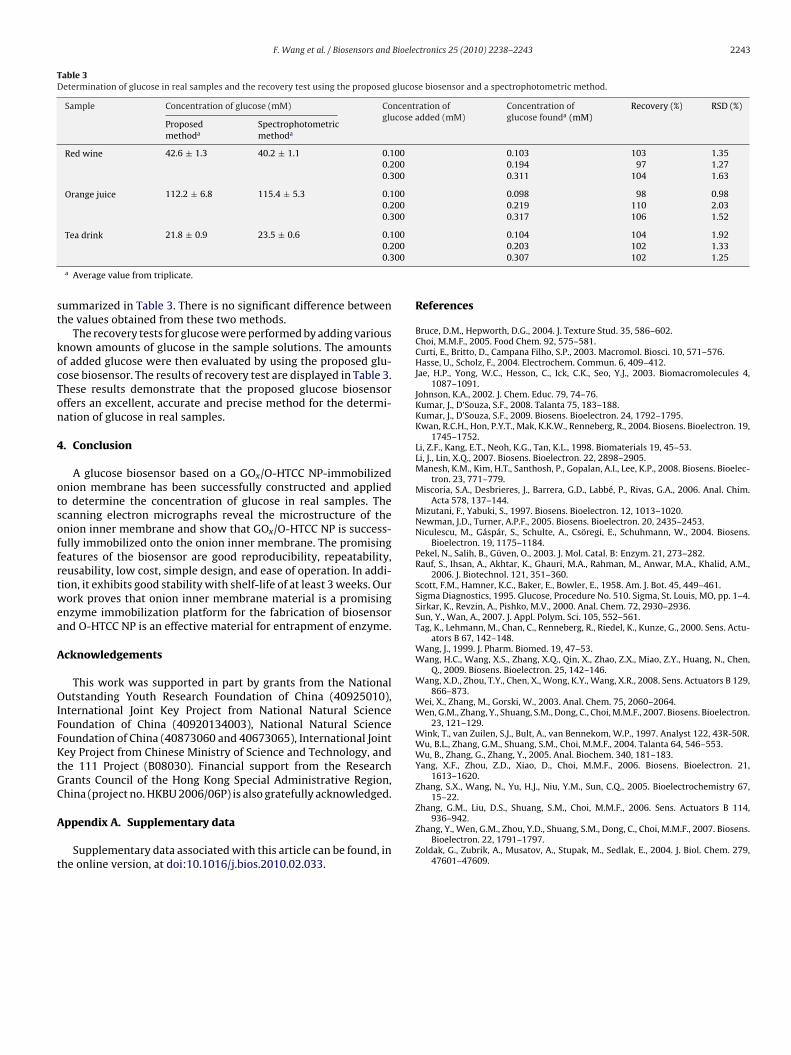
The glucose biosensor was employed to determine the glucoseconcentration in real samples including orange juice, red wine andtea drink. The samples were diluted 100–200-fold by a phosphatebuffer (pH 7.4) to yield the test sample solutions whose glucoseconcentrations fall in the working range of the calibration curve.Dilution of samples can also lower down the concentration of anyinterferents in the samples. Moreover, the glucose contents of thesamples were simultaneously analyzed by a standard spectropho-tometric method (Sigma Diagnostics, 1995). All the results are
Table 2Effect of potential interferents on the response sensitivity of glucose biosensor.
Interferent Concentration (mM) Decrease in oxygen levelequivalent to glucose (mM)
Acetic acid 2.0 0.023Ascorbic acid 2.0 0.243Lactic acid 2.0 "0.018Propionic acid 2.0 0.042Butyric acid 2.0 "0.021Folic acid 2.0 0.042Citric acid 2.0 0.051Methanol 0.1 0.054Glycine 2.0 0.034dl-"-Alanine 2.0 "0.078dl-Cysteine 2.0 0.059
F. Wang et al. / Biosensors and Bioelectronics 25 (2010) 2238–2243 2243
Table 3Determination of glucose in real samples and the recovery test using the proposed glucose biosensor and a spectrophotometric method.
Sample Concentration of glucose (mM) Concentration ofglucose added (mM)
Concentration ofglucose founda (mM)
Recovery (%) RSD (%)
Proposedmethoda
Spectrophotometricmethoda
Red wine 42.6 ± 1.3 40.2 ± 1.1 0.100 0.103 103 1.350.200 0.194 97 1.270.300 0.311 104 1.63
Orange juice 112.2 ± 6.8 115.4 ± 5.3 0.100 0.098 98 0.980.200 0.219 110 2.030.300 0.317 106 1.52
Tea drink 21.8 ± 0.9 23.5 ± 0.6 0.100 0.104 104 1.920.200 0.203 102 1.330.300 0.307 102 1.25
a Average value from triplicate.
summarized in Table 3. There is no significant difference betweenthe values obtained from these two methods.
The recovery tests for glucose were performed by adding variousknown amounts of glucose in the sample solutions. The amountsof added glucose were then evaluated by using the proposed glu-cose biosensor. The results of recovery test are displayed in Table 3.These results demonstrate that the proposed glucose biosensoroffers an excellent, accurate and precise method for the determi-nation of glucose in real samples.
4. Conclusion
A glucose biosensor based on a GOx/O-HTCC NP-immobilizedonion membrane has been successfully constructed and appliedto determine the concentration of glucose in real samples. Thescanning electron micrographs reveal the microstructure of theonion inner membrane and show that GOx/O-HTCC NP is success-fully immobilized onto the onion inner membrane. The promisingfeatures of the biosensor are good reproducibility, repeatability,reusability, low cost, simple design, and ease of operation. In addi-tion, it exhibits good stability with shelf-life of at least 3 weeks. Ourwork proves that onion inner membrane material is a promisingenzyme immobilization platform for the fabrication of biosensorand O-HTCC NP is an effective material for entrapment of enzyme.
Acknowledgements
This work was supported in part by grants from the NationalOutstanding Youth Research Foundation of China (40925010),International Joint Key Project from National Natural ScienceFoundation of China (40920134003), National Natural ScienceFoundation of China (40873060 and 40673065), International JointKey Project from Chinese Ministry of Science and Technology, andthe 111 Project (B08030). Financial support from the ResearchGrants Council of the Hong Kong Special Administrative Region,China (project no. HKBU 2006/06P) is also gratefully acknowledged.
Appendix A. Supplementary data
Supplementary data associated with this article can be found, inthe online version, at doi:10.1016/j.bios.2010.02.033.
References
Bruce, D.M., Hepworth, D.G., 2004. J. Texture Stud. 35, 586–602.Choi, M.M.F., 2005. Food Chem. 92, 575–581.Curti, E., Britto, D., Campana Filho, S.P., 2003. Macromol. Biosci. 10, 571–576.Hasse, U., Scholz, F., 2004. Electrochem. Commun. 6, 409–412.Jae, H.P., Yong, W.C., Hesson, C., Ick, C.K., Seo, Y.J., 2003. Biomacromolecules 4,
1087–1091.Johnson, K.A., 2002. J. Chem. Educ. 79, 74–76.Kumar, J., D’Souza, S.F., 2008. Talanta 75, 183–188.Kumar, J., D’Souza, S.F., 2009. Biosens. Bioelectron. 24, 1792–1795.Kwan, R.C.H., Hon, P.Y.T., Mak, K.K.W., Renneberg, R., 2004. Biosens. Bioelectron. 19,
1745–1752.Li, Z.F., Kang, E.T., Neoh, K.G., Tan, K.L., 1998. Biomaterials 19, 45–53.Li, J., Lin, X.Q., 2007. Biosens. Bioelectron. 22, 2898–2905.Manesh, K.M., Kim, H.T., Santhosh, P., Gopalan, A.I., Lee, K.P., 2008. Biosens. Bioelec-
tron. 23, 771–779.Miscoria, S.A., Desbrieres, J., Barrera, G.D., Labbé, P., Rivas, G.A., 2006. Anal. Chim.
Acta 578, 137–144.Mizutani, F., Yabuki, S., 1997. Biosens. Bioelectron. 12, 1013–1020.Newman, J.D., Turner, A.P.F., 2005. Biosens. Bioelectron. 20, 2435–2453.Niculescu, M., Gáspár, S., Schulte, A., Csöregi, E., Schuhmann, W., 2004. Biosens.
Bioelectron. 19, 1175–1184.Pekel, N., Salih, B., Güven, O., 2003. J. Mol. Catal. B: Enzym. 21, 273–282.Rauf, S., Ihsan, A., Akhtar, K., Ghauri, M.A., Rahman, M., Anwar, M.A., Khalid, A.M.,
2006. J. Biotechnol. 121, 351–360.Scott, F.M., Hamner, K.C., Baker, E., Bowler, E., 1958. Am. J. Bot. 45, 449–461.Sigma Diagnostics, 1995. Glucose, Procedure No. 510. Sigma, St. Louis, MO, pp. 1–4.Sirkar, K., Revzin, A., Pishko, M.V., 2000. Anal. Chem. 72, 2930–2936.Sun, Y., Wan, A., 2007. J. Appl. Polym. Sci. 105, 552–561.Tag, K., Lehmann, M., Chan, C., Renneberg, R., Riedel, K., Kunze, G., 2000. Sens. Actu-
ators B 67, 142–148.Wang, J., 1999. J. Pharm. Biomed. 19, 47–53.Wang, H.C., Wang, X.S., Zhang, X.Q., Qin, X., Zhao, Z.X., Miao, Z.Y., Huang, N., Chen,
Q., 2009. Biosens. Bioelectron. 25, 142–146.Wang, X.D., Zhou, T.Y., Chen, X., Wong, K.Y., Wang, X.R., 2008. Sens. Actuators B 129,
866–873.Wei, X., Zhang, M., Gorski, W., 2003. Anal. Chem. 75, 2060–2064.Wen, G.M., Zhang, Y., Shuang, S.M., Dong, C., Choi, M.M.F., 2007. Biosens. Bioelectron.
23, 121–129.Wink, T., van Zuilen, S.J., Bult, A., van Bennekom, W.P., 1997. Analyst 122, 43R-50R.Wu, B.L., Zhang, G.M., Shuang, S.M., Choi, M.M.F., 2004. Talanta 64, 546–553.Wu, B., Zhang, G., Zhang, Y., 2005. Anal. Biochem. 340, 181–183.Yang, X.F., Zhou, Z.D., Xiao, D., Choi, M.M.F., 2006. Biosens. Bioelectron. 21,
1613–1620.Zhang, S.X., Wang, N., Yu, H.J., Niu, Y.M., Sun, C.Q., 2005. Bioelectrochemistry 67,
15–22.Zhang, G.M., Liu, D.S., Shuang, S.M., Choi, M.M.F., 2006. Sens. Actuators B 114,
936–942.Zhang, Y., Wen, G.M., Zhou, Y.D., Shuang, S.M., Dong, C., Choi, M.M.F., 2007. Biosens.
Bioelectron. 22, 1791–1797.Zoldak, G., Zubrik, A., Musatov, A., Stupak, M., Sedlak, E., 2004. J. Biol. Chem. 279,
47601–47609.