A pipeline for neuron reconstruction based on spatial sliding volume filter seeding
Detection of Ligands in Regions Anatomically Connected toNeurons Expressing the Eph Receptor Bsk: Potential Roles inNeuron–Target Interaction
Jian-Hua Zhang,1 Douglas P. Cerretti,2 Tian Yu,1 John G. Flanagan,3 and Renping Zhou1
1Laboratory for Cancer Research, Department of Chemical Biology, College of Pharmacy, Rutgers University, Piscataway,New Jersey 08855, 2Immunex Research and Development Corporation, Seattle, Washington 98101, and 3Department ofCell Biology, Harvard Medical School, Boston, Massachusetts 02115
Neuron–target interaction is a key feature in the establish-ment of neuronal networks. However, the underlying mech-anism remains unclear. We have shown that at the time oftarget innervation, Bsk, an eph family receptor, is expressedat high levels in several brain regions including the hip-pocampus, olfactory bulb, and retina. To study whether theligands are expressed in the target tissues, we investigatedthe expression of Bsk ligands using a ligand-affinity probe,Bsk-AP, which consisted of the extracellular domain of Bskfused in frame with a human placental alkaline phosphatase.These analyses showed that the ligands were expressed athigh levels in the developing septum, hypothalamus, olfac-tory neural epithelium, and tectum. In situ hybridization stud-ies revealed that at least three different factors were respon-
sible for the Bsk-AP binding. In the septum, Elf-1, Lerk3(Efl-2), and AL-1/Lerk7 were transcribed. In the hypothala-mus, AL-1/Lerk7 was the ligand detected by Bsk-AP. In theolfactory system, high levels of Lerk3 were detected in thesensory neurons. Both Elf-1 and AL-1/Lerk7 were present inthe tectum. These ligand-positive areas are known to beanatomically connected to Bsk-expressing regions. Theseobservations strongly suggest that Bsk and the ligands par-ticipate in neuron–target interactions in multiple systems andprovide support for their involvement in topographicprojection.Key words: brain-specific kinase; growth factor receptor; eph
family; alkaline phosphatase tagging; neuronal targeting; topo-graphic projection
Neuron–target interaction is a key feature in the establishment ofneuronal networks and plays a critical role in the development oftopographic connections. Appropriate targeting by neurons re-quires that the guidance cues match terminals with specific cellu-lar targets, a requirement accommodated by matching fixed tagson afferents and corresponding targets. Complementarity of mo-lecular tags on afferents and targets was first postulated by Sperryin his chemoaffinity theory more than half a century ago (Sperry,1943, 1963).Only recently have specific candidate molecules been identified
(Cheng et al., 1995; Drescher et al., 1995). Elf-1, a membrane-anchored eph family ligand, and one of its putative receptors,Mek4, are expressed in complementary gradients in the chickenretina and tectum, respectively, fulfilling a long-standing predic-tion of Sperry’s chemoaffinity theory (Cheng et al., 1995). Fur-thermore, an Elf-1-related molecule, the repulsive axonal guid-ance signal (RAGS), is expressed in the chicken tectum in ananteroposterior gradient and repels the growth of retinal axons(Drescher et al., 1995). The human homolog of RAGS, AL-1/Lerk7, has been shown previously to be a ligand of the eph familyreceptor Rek7 (Winslow et al., 1995; Cerretti et al., 1996), a rathomolog of mouse Bsk (Zhou et al., 1994). These studies stronglyimplicate the eph family ligands and receptors in the developmentof topographic mapping.
At least seven candidate ligands of the eph family have beenisolated (Bartley et al., 1994; Beckmann et al., 1994; Cheng et al.,1994; Davis et al., 1994; Shao et al., 1994; Bergemann et al., 1995;Drescher et al., 1995; Kozlosky et al., 1995; Winslow et al., 1995).The ligands can be divided into two subfamilies, one with mole-cules anchored on the membrane through GPI anchors and theother with molecules containing a transmembrane domain. Fourof the five ligands with GPI anchors—B61, Lerk3 (also namedEFL-2; Davis et al., 1994), Elf-1, and AL-1/Lerk7—have beenshown to bind to Bsk or its species homologs in vitro (Davis et al.,1994; Shao et al., 1995; Winslow et al., 1995; Gao et al., 1996).However, it is not known whether any of these molecules areaccessible to Bsk in vivo, because no detailed in situ expressionstudy has been published. Here we report studies on the expres-sion of Bsk and its ligands during the development of mousenervous system. Our studies suggest that Bsk may interact withmultiple ligands in vivo and play important roles in the establish-ment and maintenance of projections in multiple systems.
MATERIALS AND METHODSAnimals. CD-1 mice from embryonic day 8 (E8) to E18 and postnatal day1 (P1) through P180 were used in this study. At least two mice at each agewere analyzed. The occurrence of a vaginal plug was defined as E1, andthe day of birth was defined as P1. Embryos and brains were dissectedunder carbon dioxide anesthesia and immediately frozen on powdereddry ice. Coronal and sagittal sections of 14 mm thickness were cut on acryostat at 2208C and thaw-mounted onto slides pretreated withtriethoxy-3-aminopropyl silane (Sigma, St. Louis, MO; see Ligand-Binding Assay). These slides were then stored at 2808C until use (up to2 months).Construction and expression of Bsk-AP. The affinity probe Bsk-AP was
created as follows. An MroI restriction site was first introduced at the
Received April 19, 1996; revised Aug. 19, 1996; accepted Aug. 21, 1996.This research was supported by National Science Foundation Grant IBN-9409930.
We thank Y. Yue for critical comments on this manuscript.Correspondence should be addressed to Renping Zhou, Laboratory for Cancer
Research, College of Pharmacy, Rutgers University, Piscataway, NJ 08855.Copyright q 1996 Society for Neuroscience 0270-6474/96/167182-11$05.00/0
The Journal of Neuroscience, November 15, 1996, 16(22):7182–7192
junction of the extracellular and transmembrane domain by site-directedmutagenesis using an oligonucleotide 59-GATCAAAGCCAGATT-CCGGACATCATTGCAGTGTCAG-39 corresponding to nucleotide po-sition 636–669 of Bsk (Zhou et al., 1994) with a GGA addition to createan MroI site. An EcoRI–MroI DNA fragment containing the entirecoding region of the extracellular domain from the initiation codon to thelast codon before the transmembrane domain of Bsk was ligated to asecreted form of human placental alkaline phosphatase in the APtag-1
Vector (Flanagan and Leder, 1990) using a BglII linker. The vectorcontaining the fusion construct was cotransfected into National Institutesof Health-3T3 cells with pSV2neo plasmid containing the aminoglycosidephosphotransferase gene, which confers G418 resistance. The transfectedcells were then selected with 400 mg/ml G418 (Life Technologies, Gaith-ersburg, MD), and neo-resistant colonies were first screened for heat-resistant alkaline phosphatase activity in the culture supernatant (Flana-gan and Leder, 1990) and then for metabolically labeled Bsk-AP protein
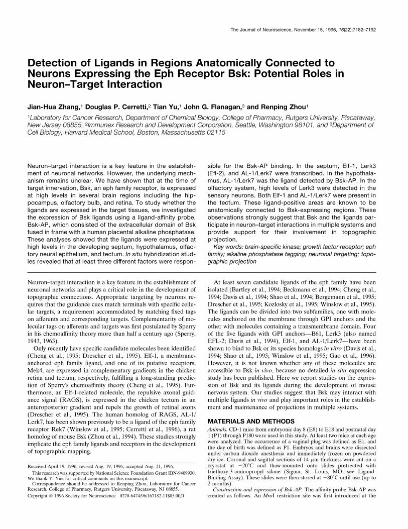
Figure 1. Differential Bsk expression along the mediolateral hippocampalaxis. Bsk mRNA levels detected by in situ hybridization at different posi-tions along the mediolateral axis are shown. A, B, Dark- and bright-fieldphotomicrographs of a medial (septal) hippocampal section. C, D, Dark-and bright-field photomicrographs of a section at an intermediate medio-lateral level. E, F, Dark- and bright-field pictures of a lateral (temporal)hippocampal section. Slides shown here and in the following figures werecounterstained with thionin. G, Quantitative analysis of Bsk in situ hybrid-ization signals at different mediolateral hippocampal levels. S, Subiculum;DG, dentate gyrus. Scale bars, 200 mm.
Zhang et al. • Expression of Bsk Ligands in the Developing Brain J. Neurosci., November 15, 1996, 16(22):7182–7192 7183
using immunoprecipitation with a monoclonal antibody against the hu-man placental alkaline phosphatase (Medix Biotech). A protein with anexpected molecular weight of 110 kDa was detected only in the trans-fected cells in both the media and the cell lysates. The clone with thehighest level of expression secreted ;800 OD units z ml21 z hr21 heat-resistant alkaline phosphatase activity.To examine whether the secreted Bsk-AP was active in ligand binding,
COS-7 cells transiently transfected with Elf-1, a putative ligand of Bsk,were stained with Bsk-AP. These experiments showed that Elf-1-expressing cells, but not the cells transfected with the vector alone,stained positive with Bsk-AP, indicating that the fusion protein retainedligand-binding activity.Ligand-binding assay. Frozen tissue sections of 14 mm were mounted on
slides coated with 3-aminopropyltrimethoxy silane (Sigma). Coated slideswere prepared by washing first in acetone for 5 min, followed with 100%ethanol. The slides were then dried at room temperature and coated bydipping in 2% 3-aminopropyltrimethoxy silane in acetone for 15 sec.After coating, the slides were washed in acetone and distilled H2O anddried at room temperature overnight. Ligand detection was performedbasically as described by Cheng et al. (1995). Briefly, frozen sections onthe coated slides were incubated with Bsk-AP-containing tissue culturemedia for 90 min at room temperature and washed five times with HBSS(Sigma). The sections were then fixed for 30 sec in 60% acetone, 3%formaldehyde, 20 mM HEPES, pH 7.5, and washed twice with 150 mMNaCl, 20 mM HEPES, pH 7.5. Because the human placental alkalinephosphatase in the probe is heat-stable, the slides were heated at 658C for15 min to inactivate endogenous phosphatases. After heat inactivation,the slides were rinsed with 100 mM Tris-HCl, pH 9.5, 100 mM NaCl, and5 mM MgCl2, and stained for 24 hr in the same buffer containing 10 mML-homoarginine, 0.17 mg/ml BCIP, and 0.33 mg/ml NBT. After staining,the slides were mounted with coverslip and photographed.In situ hybridization. In situ hybridization using Bsk and the ligand
probes was performed as described previously (Zhou et al., 1994). Ligandprobes are as described in the following sections.Elf-1 probes. Two antisense oligonucleotide probes were used to detect
Elf-1 expression. Elf-1-04, 59-CTTGAAGCCTCGCTGCCGGTGGTC-
ACAGGAGGCGTGGCCCTCACC-39, corresponded to nucleotide po-sition 283–328 in the Elf-1 coding region (Cheng et al., 1994). Elf-1-06,59-ACCTCATCCCTGTGGCTTGTTCCCTTCCCAGTGTCACCAGC-AATGT-39, corresponded to nucleotide position 926–971 in the 39-end-noncoding region of Elf-1 (Cheng et al., 1994). Neither oligonucleotideprobe shared any significant homology with other eph family ligands orany other genes in the gene bank. The two probes gave similar patternsof hybridization. The corresponding sense probes were used as controlsand gave no specific signals.Lerk3 probes. Lerk3 is a mouse homolog of the human EFL-2/LERK3,
a ligand isolated through binding to the rat homolog of Bsk, Ehk1 (Daviset al., 1994; Kozlosky et al., 1995). Lerk3 expression was detected with anantisense riboprobe and an oligonucleotide probe. For the generation ofriboprobe, a mouse Lerk3 cDNA fragment was isolated using degeneratePCR primers from two conserved regions of the eph ligand family. Theupstream primer, 59-A(C,T)AT(A,C,T)TA(C,T)TG(C,T)CCI CA(C,T)-TA-39, corresponded to amino acid sequence DIY/ICPH, and the downstream primer, 59-(T,G,A)AT(G,A)TA(G,A)TA(G,A)TA(T,C)TC-(G,A)TG, corresponded to amino acid sequence HEYYYI. The 235-bp-long Lerk3 was subcloned into the TA cloning vector (Invitrogen, SanDiego, CA). Antisense probe and sense control were generated by SP6 orT7 in vitro transcription with [35S]ATP labeling. To confirm the resultsobtained with the riboprobe, an antisense oligonucleotide probe,59-GCGCTGTAACGCTGGAACTTCTCGGAGAACTTGATGGGG-CTG-39, corresponded to nucleotide position 355–397 of Lerk3 cDNAsequence (Davis et al., 1994) was also used. Both the riboprobe and theoligonucleotide probe gave similar patterns of hybridization.AL-1/Lerk7 probe. AL-1/LERK7 is a human homolog of the chicken
RAGS, the repulsive axon guidance signal (Drescher et al., 1995; Win-slow et al., 1995; Cerretti et al., 1996). Antisense riboprobe was tran-scribed from a 0.7 kb human AL-1/LERK7 cDNA cloned in pBluescript.The ligands of the eph family from different species generally share over90% homology in the nucleotide level. The probe detects mouse Lerk7specifically and does not hybridize with other eph family ligands asdemonstrated by Southern blot analysis of mouse genomic DNA (D.
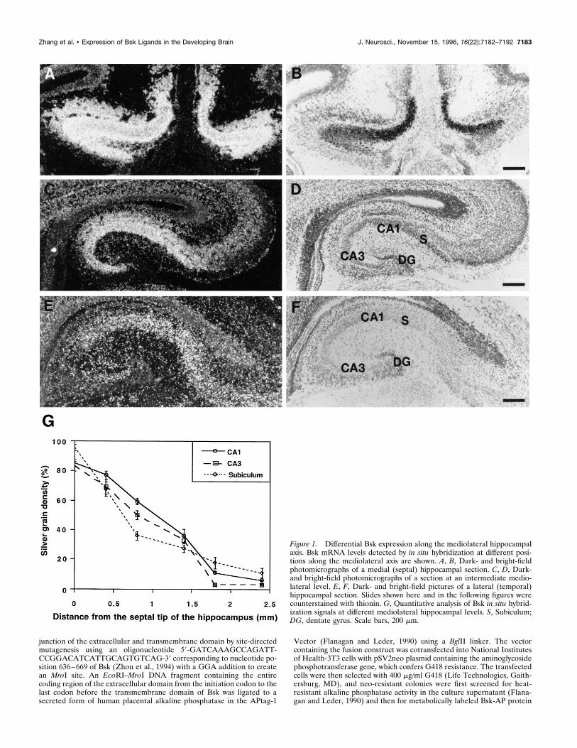
Figure 2. Expression of Bsk ligand(s) in the adult septum. A, E, Bsk-AP binding activity in the septum. B, F, Elf-1 expression. C, G, Lerk3 expression.D, H, AL-1/Lerk7 expression. A–D, Coronal sections. E–H, Higher magnifications of the septal regions, showing that the expression of the ligands wasrestricted to the ventral lateral septum. ac, Anterior commissure; cc, corpus callosum; f, fornix; BST, bed nucleus of the stria terminalis; Cpu, caudateputamen; Ctx, cerebral cortex; LS, lateral septum; MS, medial septum; Se, septum; VDB, vertical diagonal band; Scale bars: A, 1.2 mm; E, 0.6 mm.
7184 J. Neurosci., November 15, 1996, 16(22):7182–7192 Zhang et al. • Expression of Bsk Ligands in the Developing Brain
Cerretti, unpublished data). Sense probe was used as a control andrevealed no specific binding.Quantitation. Relative quantitative analysis of Bsk hybridization signals
in the hippocampus and the olfactory bulb was performed using Im-agePro image analysis software from Nikon. To avoid bias introduced bythe variations of cell density in different regions, only areas withinindividual cells were quantitated for silver grain density, expressed aspercent of area covered (area–fraction analysis).
RESULTSTo investigate the roles that the Eph family receptor Bsk mayplay during the development of the nervous system, we exam-ined the expression of Bsk and its ligands in several systemsduring embryogenesis and postnatal life. Bsk mRNA was de-tected using antisense cRNA probes (Zhou et al., 1994). Theligands of Bsk were first examined with an affinity probe,Bsk-AP, the human placental alkaline phosphatase-tagged Bskextracellular domain. Bsk-AP contains the entire extracellulardomain of Bsk (for details, see Materials and Methods). Be-cause of the promiscuity of the ligand–receptor interaction inthe Eph family, Bsk-AP may bind to multiple ligands. Toidentify further the molecular nature of the ligands detectedwith Bsk-AP, cRNA and oligonucleotide probes against knownligands of the Eph family were used in in situ hybridizationexperiments. These studies indicate that Bsk and its ligands areexpressed in distinct but synaptically connected regions of thebrain, suggesting potential roles in neuron–target interactions.
The expression of Bsk and its ligands in thehippocamposeptal systemWe have shown previously that Bsk is expressed at high levels inthe hippocampus (Zhou et al., 1994). Careful examination of BskmRNA levels indicated that Bsk transcripts were distributed in agradient (Fig. 1). The highest level of expression was found in themedial hippocampus, whereas the lowest level was found in thelateral hippocampus. Quantitation analysis revealed a steady de-crease of Bsk mRNA levels from the medial to the lateral hip-pocampus in all of the CA fields, as well as in the subiculum (Fig.1G). The hippocampal gradient was observed in E18 (Fig. 1), P7,P14, and adult mice (data not shown).Analysis using Bsk-AP probe showed that the ligands were
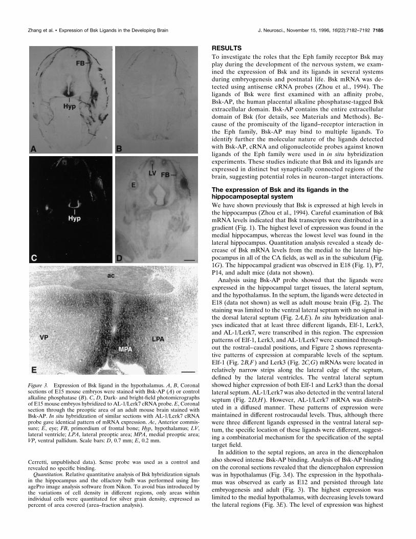
expressed in the hippocampal target tissues, the lateral septum,and the hypothalamus. In the septum, the ligands were detected inE18 (data not shown) as well as adult mouse brain (Fig. 2). Thestaining was limited to the ventral lateral septum with no signal inthe dorsal lateral septum (Fig. 2A,E). In situ hybridization anal-yses indicated that at least three different ligands, Elf-1, Lerk3,and AL-1/Lerk7, were transcribed in this region. The expressionpatterns of Elf-1, Lerk3, and AL-1/Lerk7 were examined through-out the rostral–caudal positions, and Figure 2 shows representa-tive patterns of expression at comparable levels of the septum.Elf-1 (Fig. 2B,F) and Lerk3 (Fig. 2C,G) mRNAs were located inrelatively narrow strips along the lateral edge of the septum,defined by the lateral ventricles. The ventral lateral septumshowed higher expression of both Elf-1 and Lerk3 than the dorsallateral septum. AL-1/Lerk7 was also detected in the ventral lateralseptum (Fig. 2D,H). However, AL-1/Lerk7 mRNA was distrib-uted in a diffused manner. These patterns of expression weremaintained in different rostrocaudal levels. Thus, although therewere three different ligands expressed in the ventral lateral sep-tum, the specific location of these ligands were different, suggest-ing a combinatorial mechanism for the specification of the septaltarget field.In addition to the septal regions, an area in the diencephalon
also showed intense Bsk-AP binding. Analysis of Bsk-AP bindingon the coronal sections revealed that the diencephalon expressionwas in hypothalamus (Fig. 3A). The expression in the hypothala-mus was observed as early as E12 and persisted through lateembryogenesis and adult (Fig. 3). The highest expression waslimited to the medial hypothalamus, with decreasing levels towardthe lateral regions (Fig. 3E). The level of expression was highest
Figure 3. Expression of Bsk ligand in the hypothalamus. A, B, Coronalsections of E15 mouse embryos were stained with Bsk-AP (A) or controlalkaline phosphatase (B). C, D, Dark- and bright-field photomicrographsof E15 mouse embryos hybridized to AL-1/Lerk7 cRNA probe. E, Coronalsection through the preoptic area of an adult mouse brain stained withBsk-AP. In situ hybridization of similar sections with AL-1/Lerk7 cRNAprobe gave identical pattern of mRNA expression. Ac, Anterior commis-sure; E, eye; FB, primordium of frontal bone; Hyp, hypothalamus; LV,lateral ventricle; LPA, lateral preoptic area; MPA, medial preoptic area;VP, ventral pallidum. Scale bars: D, 0.7 mm; E, 0.2 mm.
Zhang et al. • Expression of Bsk Ligands in the Developing Brain J. Neurosci., November 15, 1996, 16(22):7182–7192 7185
in the rostral end of the hypothalamus (preoptic area) (Fig. 3E)and decreased to very low levels toward the caudal end (mamillarynuclei; data not shown).In situ analysis revealed that AL-1/Lerk7 was responsible for
the ligand expression in the hypothalamus (Fig. 3C). Very highlevels of AL-1/Lerk7 expression were detected in embryonic hy-pothalamus (Fig. 3C). However, the expression decreased tomoderate levels in adult hypothalamus (data not shown). Thedistribution of AL-1/Lerk7 in both the embryonic and the adulthypothalamus is identical to that of Bsk-AP staining.AL-1/Lerk7 expression was also detected, in addition to the
septum and hypothalamus, in many other regions. Significantexpression was found in the tectum (Fig. 8), motor cortex, thala-mus, and deep cerebellar nuclei, whereas no expression wasdetected in the hippocampus (data not shown).
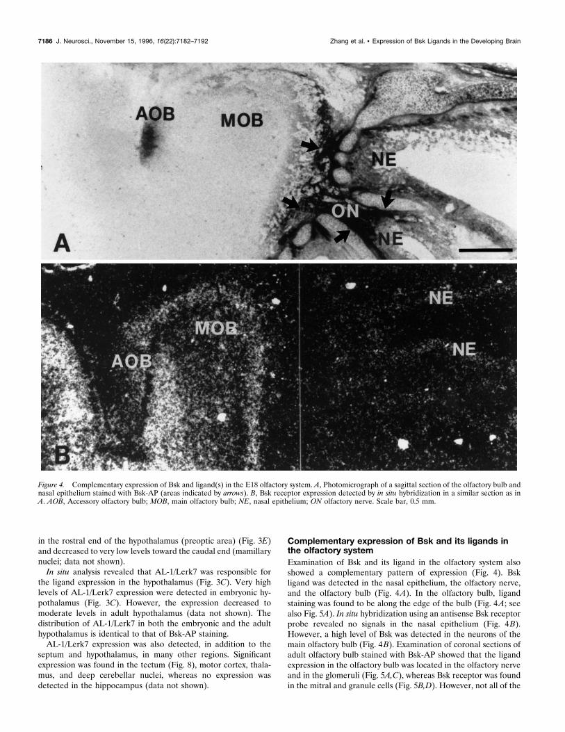
Complementary expression of Bsk and its ligands inthe olfactory systemExamination of Bsk and its ligand in the olfactory system alsoshowed a complementary pattern of expression (Fig. 4). Bskligand was detected in the nasal epithelium, the olfactory nerve,and the olfactory bulb (Fig. 4A). In the olfactory bulb, ligandstaining was found to be along the edge of the bulb (Fig. 4A; seealso Fig. 5A). In situ hybridization using an antisense Bsk receptorprobe revealed no signals in the nasal epithelium (Fig. 4B).However, a high level of Bsk was detected in the neurons of themain olfactory bulb (Fig. 4B). Examination of coronal sections ofadult olfactory bulb stained with Bsk-AP showed that the ligandexpression in the olfactory bulb was located in the olfactory nerveand in the glomeruli (Fig. 5A,C), whereas Bsk receptor was foundin the mitral and granule cells (Fig. 5B,D). However, not all of the
Figure 4. Complementary expression of Bsk and ligand(s) in the E18 olfactory system. A, Photomicrograph of a sagittal section of the olfactory bulb andnasal epithelium stained with Bsk-AP (areas indicated by arrows). B, Bsk receptor expression detected by in situ hybridization in a similar section as inA. AOB, Accessory olfactory bulb; MOB, main olfactory bulb; NE, nasal epithelium; ON olfactory nerve. Scale bar, 0.5 mm.
7186 J. Neurosci., November 15, 1996, 16(22):7182–7192 Zhang et al. • Expression of Bsk Ligands in the Developing Brain
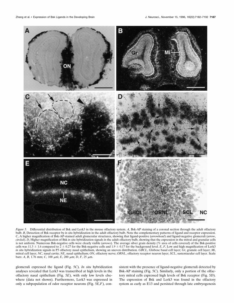
glomeruli expressed the ligand (Fig. 5C). In situ hybridizationanalyses revealed that Lerk3 was transcribed at high levels in theolfactory nasal epithelium (Fig. 5E), with only low levels else-where (data not shown). Furthermore, Lerk3 was expressed inonly a subpopulation of odor receptor neurons (Fig. 5E,F), con-
sistent with the presence of ligand-negative glomeruli detected byBsk-AP staining (Fig. 5C). Similarly, only a portion of the olfac-tory mitral cells expressed high levels of Bsk receptor (Fig. 5D).The expression of Bsk and Lerk3 was found in the olfactorysystem as early as E13 and persisted through late embryogenesis
Figure 5. Differential distribution of Bsk and Lerk3 in the mouse olfactory system. A, Bsk-AP staining of a coronal section through the adult olfactorybulb. B, Detection of Bsk receptor by in situ hybridization in the adult olfactory bulb. Note the complementary patterns of ligand and receptor expression.C, A higher magnification of Bsk-AP-stained adult glomerular structures, showing that ligand-positive (arrowhead) and ligand-negative glomeruli (arrow,circled). D, Higher magnification of Bsk in situ hybridization signals in the adult olfactory bulb, showing that the expression in the mitral and granular cellsis not uniform. Numerous Bsk-negative cells were clearly visible (arrows). The average silver grain density (% area of cells covered) of the Bsk-positivecells was 11.3 6 1.6 compared to 2 6 0.27 for the Bsk-negative cells and 1.9 6 0.17 for the background level. E, F, Low and high magnification of Lerk3in situ hybridization signals in P3 olfactory nasal epithelium, showing an uneven distribution. GBCL, Globose basal cell layer; Gr, granule cell layer; Mi,mitral cell layer; NC, nasal cavity; NE, nasal epithelium; ON, olfactory nerve; ORNL, olfactory receptor neuron layer; SCL, sustentacular cell layer. Scalebars: A, B, 1.76 mm; C, 100 mm; E, 200 mm; D, F, 25 mm.
Zhang et al. • Expression of Bsk Ligands in the Developing Brain J. Neurosci., November 15, 1996, 16(22):7182–7192 7187
(Fig. 4) and adult (Fig. 5 and data not shown). No Elf-1 and onlylow levels of AL-1/Lerk7 expression were detected in this region(data not shown). These observations further demonstrate thatalthough Bsk and its ligands were expressed in distinct neuronalpopulations, the ligand- and Bsk-positive regions are synapticallyconnected, suggesting an important role of these ligand andreceptor molecules in neuron–target interaction.
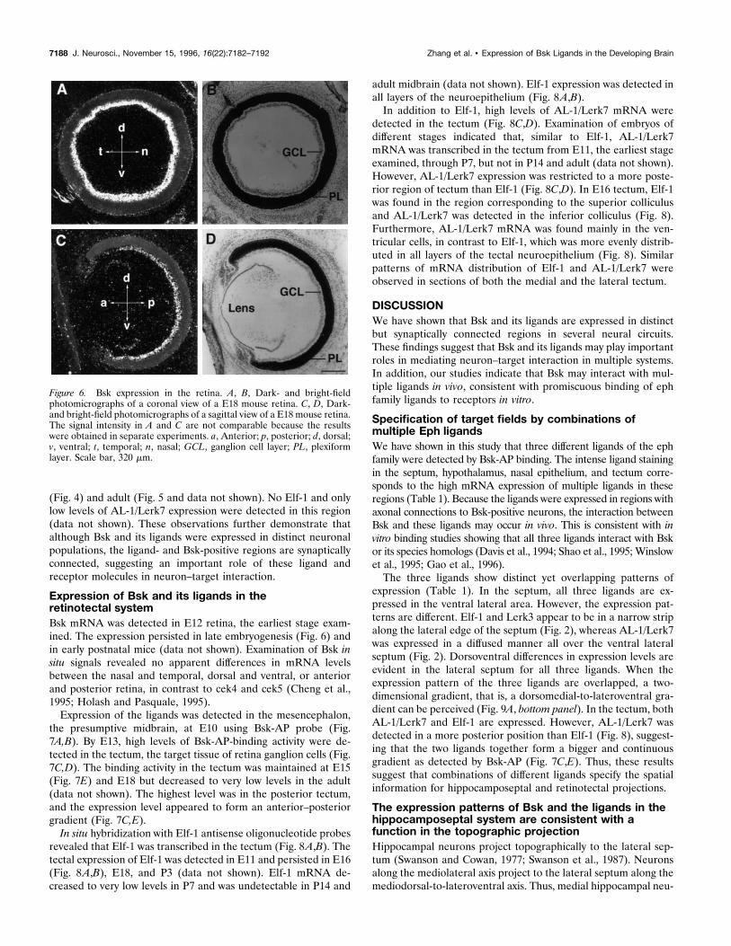
Expression of Bsk and its ligands in theretinotectal systemBsk mRNA was detected in E12 retina, the earliest stage exam-ined. The expression persisted in late embryogenesis (Fig. 6) andin early postnatal mice (data not shown). Examination of Bsk insitu signals revealed no apparent differences in mRNA levelsbetween the nasal and temporal, dorsal and ventral, or anteriorand posterior retina, in contrast to cek4 and cek5 (Cheng et al.,1995; Holash and Pasquale, 1995).Expression of the ligands was detected in the mesencephalon,
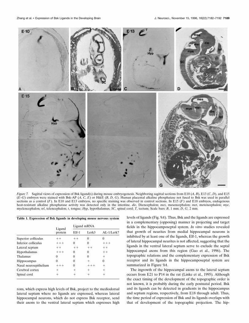
the presumptive midbrain, at E10 using Bsk-AP probe (Fig.7A,B). By E13, high levels of Bsk-AP-binding activity were de-tected in the tectum, the target tissue of retina ganglion cells (Fig.7C,D). The binding activity in the tectum was maintained at E15(Fig. 7E) and E18 but decreased to very low levels in the adult(data not shown). The highest level was in the posterior tectum,and the expression level appeared to form an anterior–posteriorgradient (Fig. 7C,E).In situ hybridization with Elf-1 antisense oligonucleotide probes
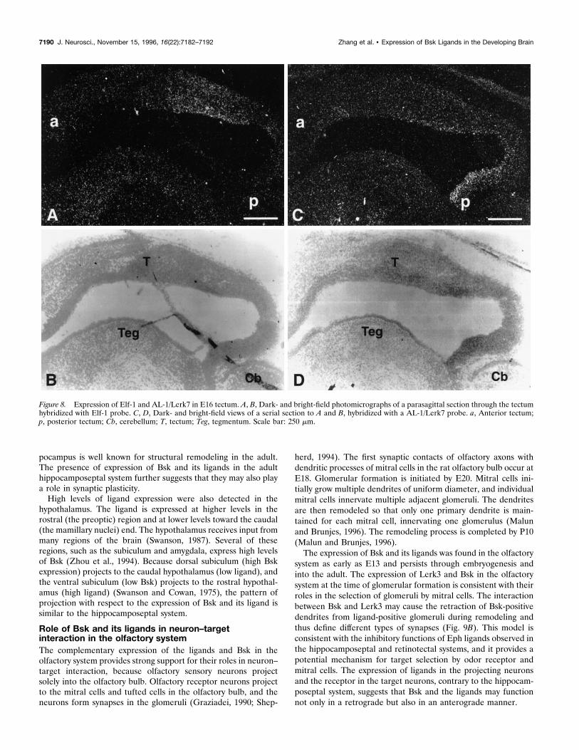
revealed that Elf-1 was transcribed in the tectum (Fig. 8A,B). Thetectal expression of Elf-1 was detected in E11 and persisted in E16(Fig. 8A,B), E18, and P3 (data not shown). Elf-1 mRNA de-creased to very low levels in P7 and was undetectable in P14 and
adult midbrain (data not shown). Elf-1 expression was detected inall layers of the neuroepithelium (Fig. 8A,B).In addition to Elf-1, high levels of AL-1/Lerk7 mRNA were
detected in the tectum (Fig. 8C,D). Examination of embryos ofdifferent stages indicated that, similar to Elf-1, AL-1/Lerk7mRNA was transcribed in the tectum from E11, the earliest stageexamined, through P7, but not in P14 and adult (data not shown).However, AL-1/Lerk7 expression was restricted to a more poste-rior region of tectum than Elf-1 (Fig. 8C,D). In E16 tectum, Elf-1was found in the region corresponding to the superior colliculusand AL-1/Lerk7 was detected in the inferior colliculus (Fig. 8).Furthermore, AL-1/Lerk7 mRNA was found mainly in the ven-tricular cells, in contrast to Elf-1, which was more evenly distrib-uted in all layers of the tectal neuroepithelium (Fig. 8). Similarpatterns of mRNA distribution of Elf-1 and AL-1/Lerk7 wereobserved in sections of both the medial and the lateral tectum.
DISCUSSIONWe have shown that Bsk and its ligands are expressed in distinctbut synaptically connected regions in several neural circuits.These findings suggest that Bsk and its ligands may play importantroles in mediating neuron–target interaction in multiple systems.In addition, our studies indicate that Bsk may interact with mul-tiple ligands in vivo, consistent with promiscuous binding of ephfamily ligands to receptors in vitro.
Specification of target fields by combinations ofmultiple Eph ligandsWe have shown in this study that three different ligands of the ephfamily were detected by Bsk-AP binding. The intense ligand stainingin the septum, hypothalamus, nasal epithelium, and tectum corre-sponds to the high mRNA expression of multiple ligands in theseregions (Table 1). Because the ligands were expressed in regions withaxonal connections to Bsk-positive neurons, the interaction betweenBsk and these ligands may occur in vivo. This is consistent with invitro binding studies showing that all three ligands interact with Bskor its species homologs (Davis et al., 1994; Shao et al., 1995; Winslowet al., 1995; Gao et al., 1996).The three ligands show distinct yet overlapping patterns of
expression (Table 1). In the septum, all three ligands are ex-pressed in the ventral lateral area. However, the expression pat-terns are different. Elf-1 and Lerk3 appear to be in a narrow stripalong the lateral edge of the septum (Fig. 2), whereas AL-1/Lerk7was expressed in a diffused manner all over the ventral lateralseptum (Fig. 2). Dorsoventral differences in expression levels areevident in the lateral septum for all three ligands. When theexpression pattern of the three ligands are overlapped, a two-dimensional gradient, that is, a dorsomedial-to-lateroventral gra-dient can be perceived (Fig. 9A, bottom panel). In the tectum, bothAL-1/Lerk7 and Elf-1 are expressed. However, AL-1/Lerk7 wasdetected in a more posterior position than Elf-1 (Fig. 8), suggest-ing that the two ligands together form a bigger and continuousgradient as detected by Bsk-AP (Fig. 7C,E). Thus, these resultssuggest that combinations of different ligands specify the spatialinformation for hippocamposeptal and retinotectal projections.
The expression patterns of Bsk and the ligands in thehippocamposeptal system are consistent with afunction in the topographic projectionHippocampal neurons project topographically to the lateral sep-tum (Swanson and Cowan, 1977; Swanson et al., 1987). Neuronsalong the mediolateral axis project to the lateral septum along themediodorsal-to-lateroventral axis. Thus, medial hippocampal neu-
Figure 6. Bsk expression in the retina. A, B, Dark- and bright-fieldphotomicrographs of a coronal view of a E18 mouse retina. C, D, Dark-and bright-field photomicrographs of a sagittal view of a E18 mouse retina.The signal intensity in A and C are not comparable because the resultswere obtained in separate experiments. a, Anterior; p, posterior; d, dorsal;v, ventral; t, temporal; n, nasal; GCL, ganglion cell layer; PL, plexiformlayer. Scale bar, 320 mm.
7188 J. Neurosci., November 15, 1996, 16(22):7182–7192 Zhang et al. • Expression of Bsk Ligands in the Developing Brain
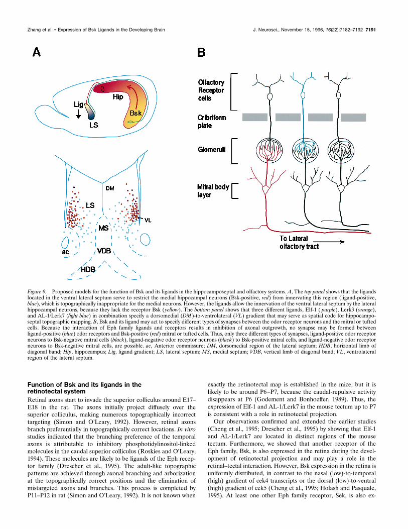
rons, which express high levels of Bsk, project to the mediodorsallateral septum where no ligands are expressed, whereas lateralhippocampal neurons, which do not express Bsk receptor, sendtheir axons to the ventral lateral septum which expresses high
levels of ligands (Fig. 9A). Thus, Bsk and the ligands are expressedin a complementary (opposing) manner in projecting and targetfields in the hippocamposeptal system. In vitro studies revealedthat growth of neurites from medial hippocampal neurons isinhibited by at least one of the ligands, Elf-1, whereas the growthof lateral hippocampal neurites is not affected, suggesting that theligands in the ventral lateral septum serve to exclude the septalhippocampal axons from this region (Gao et al., 1996). Thetopographic relations and the complementary expression of Bskreceptor and its ligands in the hippocamposeptal system aresummarized in Figure 9A.The ingrowth of the hippocampal axons to the lateral septum
occurs from E21 to P14 in the rat (Linke et al., 1995). Althoughthe exact timing of the development of the topographic order isnot known, it is probably during the early postnatal period. Bskand its ligands can be detected in gradients in the hippocampusand septum regions, respectively, from E18 through adult. Thus,the time period of expression of Bsk and its ligands overlaps withthat of development of the topographic projection. The hip-
Figure 7. Sagittal views of expression of Bsk ligand(s) during mouse embryogenesis. Neighboring sagittal sections from E10 (A, B), E13 (C, D), and E15(E–G) embryos were stained with Bsk-AP (A, C, E) or H&E (B, D, G). Human placental alkaline phosphatase not fused to Bsk was used in parallelsections as a control (F ). In E10 and E13 embryos, no specific staining was observed in control sections. In E15 (F ) and E18 embryos, endogenousheat-resistant alkaline phosphatase activity was detected only in the intestine. die, Diencephalon; mes, mesencephalon; met, metencephalon; mye,myelencephalon; tel, telencephalon; t, tongue; Hyp, hypothalamus; SC, spinal cord; T, tectum; Scale bars: B, 1 mm; D, G, 2 mm.
Table 1. Expression of Bsk ligands in developing mouse nervous system
Ligandprotein
Ligand mRNA
Elf-1 Lerk3 AL-1/Lerk7
Superior colliculus 11 11 0 0Inferior colliculus 111 0 0 111
Lateral septum 11 11 11 11
Hypothalamus 111 0 0 11
Thalamus 0 0 0 1
Hippocampus 0 0 1 0Nasal neuroepithelium 111 0 111 1
Cerebral cortex 1 1 1 1
Spinal cord 1 1 1 1
Zhang et al. • Expression of Bsk Ligands in the Developing Brain J. Neurosci., November 15, 1996, 16(22):7182–7192 7189
pocampus is well known for structural remodeling in the adult.The presence of expression of Bsk and its ligands in the adulthippocamposeptal system further suggests that they may also playa role in synaptic plasticity.High levels of ligand expression were also detected in the
hypothalamus. The ligand is expressed at higher levels in therostral (the preoptic) region and at lower levels toward the caudal(the mamillary nuclei) end. The hypothalamus receives input frommany regions of the brain (Swanson, 1987). Several of theseregions, such as the subiculum and amygdala, express high levelsof Bsk (Zhou et al., 1994). Because dorsal subiculum (high Bskexpression) projects to the caudal hypothalamus (low ligand), andthe ventral subiculum (low Bsk) projects to the rostral hypothal-amus (high ligand) (Swanson and Cowan, 1975), the pattern ofprojection with respect to the expression of Bsk and its ligand issimilar to the hippocamposeptal system.
Role of Bsk and its ligands in neuron–targetinteraction in the olfactory systemThe complementary expression of the ligands and Bsk in theolfactory system provides strong support for their roles in neuron–target interaction, because olfactory sensory neurons projectsolely into the olfactory bulb. Olfactory receptor neurons projectto the mitral cells and tufted cells in the olfactory bulb, and theneurons form synapses in the glomeruli (Graziadei, 1990; Shep-
herd, 1994). The first synaptic contacts of olfactory axons withdendritic processes of mitral cells in the rat olfactory bulb occur atE18. Glomerular formation is initiated by E20. Mitral cells ini-tially grow multiple dendrites of uniform diameter, and individualmitral cells innervate multiple adjacent glomeruli. The dendritesare then remodeled so that only one primary dendrite is main-tained for each mitral cell, innervating one glomerulus (Malunand Brunjes, 1996). The remodeling process is completed by P10(Malun and Brunjes, 1996).The expression of Bsk and its ligands was found in the olfactory
system as early as E13 and persists through embryogenesis andinto the adult. The expression of Lerk3 and Bsk in the olfactorysystem at the time of glomerular formation is consistent with theirroles in the selection of glomeruli by mitral cells. The interactionbetween Bsk and Lerk3 may cause the retraction of Bsk-positivedendrites from ligand-positive glomeruli during remodeling andthus define different types of synapses (Fig. 9B). This model isconsistent with the inhibitory functions of Eph ligands observed inthe hippocamposeptal and retinotectal systems, and it provides apotential mechanism for target selection by odor receptor andmitral cells. The expression of ligands in the projecting neuronsand the receptor in the target neurons, contrary to the hippocam-poseptal system, suggests that Bsk and the ligands may functionnot only in a retrograde but also in an anterograde manner.
Figure 8. Expression of Elf-1 and AL-1/Lerk7 in E16 tectum. A, B, Dark- and bright-field photomicrographs of a parasagittal section through the tectumhybridized with Elf-1 probe. C, D, Dark- and bright-field views of a serial section to A and B, hybridized with a AL-1/Lerk7 probe. a, Anterior tectum;p, posterior tectum; Cb, cerebellum; T, tectum; Teg, tegmentum. Scale bar: 250 mm.
7190 J. Neurosci., November 15, 1996, 16(22):7182–7192 Zhang et al. • Expression of Bsk Ligands in the Developing Brain
Function of Bsk and its ligands in theretinotectal systemRetinal axons start to invade the superior colliculus around E17–E18 in the rat. The axons initially project diffusely over thesuperior colliculus, making numerous topographically incorrecttargeting (Simon and O’Leary, 1992). However, retinal axonsbranch preferentially in topographically correct locations. In vitrostudies indicated that the branching preference of the temporalaxons is attributable to inhibitory phosphotidylinositol-linkedmolecules in the caudal superior colliculus (Roskies and O’Leary,1994). These molecules are likely to be ligands of the Eph recep-tor family (Drescher et al., 1995). The adult-like topographicpatterns are achieved through axonal branching and arborizationat the topographically correct positions and the elimination ofmistargeted axons and branches. This process is completed byP11–P12 in rat (Simon and O’Leary, 1992). It is not known when
exactly the retinotectal map is established in the mice, but it islikely to be around P6–P7, because the caudal-repulsive activitydisappears at P6 (Godement and Bonhoeffer, 1989). Thus, theexpression of Elf-1 and AL-1/Lerk7 in the mouse tectum up to P7is consistent with a role in retinotectal projection.Our observations confirmed and extended the earlier studies
(Cheng et al., 1995; Drescher et al., 1995) by showing that Elf-1and AL-1/Lerk7 are located in distinct regions of the mousetectum. Furthermore, we showed that another receptor of theEph family, Bsk, is also expressed in the retina during the devel-opment of retinotectal projection and may play a role in theretinal–tectal interaction. However, Bsk expression in the retina isuniformly distributed, in contrast to the nasal (low)-to-temporal(high) gradient of cek4 transcripts or the dorsal (low)-to-ventral(high) gradient of cek5 (Cheng et al., 1995; Holash and Pasquale,1995). At least one other Eph family receptor, Sek, is also ex-
Figure 9. Proposed models for the function of Bsk and its ligands in the hippocamposeptal and olfactory systems. A, The top panel shows that the ligandslocated in the ventral lateral septum serve to restrict the medial hippocampal neurons (Bsk-positive, red) from innervating this region (ligand-positive,blue), which is topographically inappropriate for the medial neurons. However, the ligands allow the innervation of the ventral lateral septum by the lateralhippocampal neurons, because they lack the receptor Bsk ( yellow). The bottom panel shows that three different ligands, Elf-1 ( purple), Lerk3 (orange),and AL-1/Lerk7 (light blue) in combination specify a dorsomedial (DM )-to-ventrolateral (VL) gradient that may serve as spatial code for hippocampo-septal topographic mapping. B, Bsk and its ligand may act to specify different types of synapses between the odor receptor neurons and the mitral or tuftedcells. Because the interaction of Eph family ligands and receptors results in inhibition of axonal outgrowth, no synapse may be formed betweenligand-positive (blue) odor receptors and Bsk-positive (red) mitral or tufted cells. Thus, only three different types of synapses, ligand-positive odor receptorneurons to Bsk-negative mitral cells (black), ligand-negative odor receptor neurons (black) to Bsk-positive mitral cells, and ligand-negative odor receptorneurons to Bsk-negative mitral cells, are possible. ac, Anterior commissure; DM, dorsomedial region of the lateral septum; HDB, horizontal limb ofdiagonal band; Hip, hippocampus; Lig, ligand gradient; LS, lateral septum; MS, medial septum; VDB, vertical limb of diagonal band; VL, ventrolateralregion of the lateral septum.
Zhang et al. • Expression of Bsk Ligands in the Developing Brain J. Neurosci., November 15, 1996, 16(22):7182–7192 7191
pressed in the retina uniformly (Cheng et al., 1995). Thus, it islikely that Bsk or Sek serves other functions rather than as asurface tag for topographic mapping in this system.The uniform expression of Bsk and Sek in the retina raises a
difficult issue in explaining retinotectal topographic ordering usinga model in which the interaction between the Eph ligands andreceptors results in inhibition of axonal growth or branching(Cheng et al., 1995; Drescher et al., 1995; Brambilla and Klein,1996; Friedman and O’Leary, 1996). One would predict thatbecause Bsk and Sek are expressed in the nasal retina, the pres-ence of high levels of Eph ligands in the caudal tectum wouldprevent nasal axonal targeting to this region. In fact, the growth ofnasal axons is indeed inhibited by RAGS in vitro (Drescher et al.,1995). A solution to this paradox may be that the topographicorder is achieved by the combined action of Eph family-repulsivecues and other attractive signals. Chemoattractants or trophicfactors may be preferentially expressed in the caudal tectum andattract the growth and promote the survival or branching of nasalaxons at their correct topographic target. In vitro studies showedthat membrane preparations from caudal tectum prolong substan-tially the survival of nasal neurites (Boxberg et al., 1995), confirm-ing the existence of trophic factors for homing nasal axons. Theexistence of caudal tectum-specific trophic factors also providesan answer to the question of why nasal axons do not target therostral tectum. A balance between repulsion and attraction maybe critical for the establishment of topographic ordering in boththe retinotectal and the hippocamposeptal systems.In summary, our studies showed that high levels of Bsk and
ligands are expressed in complementary patterns in the projectingand target neurons in several neural pathways, suggesting that Bskand its ligands mediate neuronal targeting in multiple systems.
REFERENCESBartley TD, Hunt RW, Welcher AA, Boyle WJ, Parker VP, Lindberg RA,Lu HS, Colombero AM, Elliott RL, Guthrie BA, Holst PL, Skrine JD,Toso RJ, Zhang M, Fernandez E, Trail G, Varnum B, Yarden Y,Hunter T, Fox GM (1994) B61 is a ligand for the ECK receptorprotein-tyrosine kinase. Nature 368:558–560.
Beckmann MP, Cerretti DP, Baum P, Bos TV, James L, Farrah T,Kozlosky C, Hollingsworth T, Shilling H, Maraskovsky C, Fletcher FA,Lhotak V, Pawson T, Lyman SD (1994) Molecular characterization ofa family of ligands for Eph-related tyrosine kinase receptors. EMBO J13:3757–3762.
Bergemann AD, Cheng HJ, Brambilla R, Klein R, Flanagan JG (1995)Elf-2, a new member of the Eph ligand family, is segmentally expressedin mouse embryos in the region of the hindbrain and newly formingsomites. Mol Cell Biol 15:4921–4929.
Boxberg YV, Deiss S, Schwarz U (1993) Guidance and topographicstablization of nasal chick retinal axons on target-derived components invitro. Neuron 10:345–357.
Brambilla R, Klein R (1996) Telling axons where to grow: a role for Ephreceptor tyrosine kinases in guidance. Mol Cell Neurosci 6:487–495.
Cerretti DP, Copeland NG, Gilbert DJ, Jenkins NA, Kuefer MU, Valen-tine V, Shapiro DN, Cui X, Morris SW (1996) The gene encodingLERK-7 (EPLG7, Epl7), a ligand for the eph-related receptor tyrosinekinases, maps to human chromosome 5 at band q21 and the mousechromosome 17. Genomics 35:376–379.
Cheng H-J, Flanagan JG (1994) Identification and cloning of ELF-1, adevelopmentally expressed ligand for the Mek4 and Sek receptor ty-rosine kinases. Cell 79:157–168.
Cheng H-J, Nakamoto M, Bergemann AD, Flanagan JG (1995) Comple-mentary gradients in expression and binding of ELF-1 and Mek4 indevelopment of the topographic retinotectal projection map. Cell82:371–381.
Davis S, Gale NW, Aldrich TH, Maisonpierre PC, Lhotak V, Pawson T,Goldfarb M, Yancopoulos G (1994) Ligands for EPH-related receptor
tyrosine kinases that require membrane attachment or clustering foractivity. Science 266:816–819.
Drescher U, Kremoser C, Handwerker C, Loschinger J, Masaharu N,Bonhoeffer F (1995) In vitro guidance of retinal ganglion cell axons byRAGS, a 25 kDa tectal protein related to ligands for Eph receptortyrosine kinases. Cell 82:359–370.
Flanagan JG, Leder P (1990) The kit ligand: a cell surface moleculealtered in steel mutant fibroblasts. Cell 63:185–194.
Friedman GC, O’Leary DDM (1996) Eph receptor tyrosine kinases andtheir ligands in neural development. Curr Opin Neurobiol 6:127–133.
Gao P-P, Zhang J-H, Yokoyama M, Racey B, Dreyfus CF, Black IB, ZhouR (1996) Regulation of topographic projection in the brain: Elf-1 inthe hippocamposeptal system. Proc Natl Acad Sci USA 93:11161–11166.
Godement P, Bonhoeffer F (1989) Cross-species recognition of tectalcues by retinal fibers in vitro. Development 106:313–320.
Graziadei PP (1990) Olfactory development. In: Development of sensorysystems in mammals (Coleman JR, ed), pp 616–666. New York: Wiley.
Holash JA, Pasquale EB (1995) Polarized expression of the receptorprotein-tyrosine kinase Cek5 in the developing avian visual system. DevBiol 172:683–693.
Kozlosky CJ, Maraskovsky E, McGrew JT, VandenBos T, Teepe M,Lyman SD, Srinivasan S, Fletcher FA, Gayle III RB, Cerretti DP,Beckmann MP (1995) Ligands for the receptor tyrosine kinases hekand elk: isolation of cDNAs encoding a family of proteins. Oncogene10:299–306.
Linke R, Pabst T, Frotscher M (1995) Development of the hippocampo-septal projection in the rat. J Comp Neurol 351:602–616.
Malun D, Brunjes PC (1996) Development of olfactory glomeruli: tem-poral and spatial interactions between olfactory receptor axons andmitral cells in opossums and rats. J Comp Neurol 368:1–16.
Roskies AL, O’Leary DDM (1994) Control of topographic retinal axonbranching by inhibitory membrane-bound molecules. Science265:799–803.
Shao H, Lou L, Pandey A, Pasquale EB, Dixit VM (1994) cDNA cloningand characterization of a ligand for the Cek5 receptor protein-tyrosinekinase. J Biol Chem 269:26606–26609.
Shao H, Lou L, Pandey A, Verderame MF, Siever DA, Dixit VM (1995)cDNA cloning and characterization of a Cek7 receptor protein-tyrosinekinase ligand that is identical to the ligand (ELF-1) for the Mek-4 andSek receptor protein-tyrosine kinases. J Biol Chem 270:3467–3470.
Shepherd GM (1994) Discrimination of molecular signals by the olfac-tory receptor neuron. Neurons 13:771–790.
Simon DK, O’Leary DDM (1992a) Development of topographic order inthe mammalian retinocollicular projection. J Neurosci 12:1212–1232.
Simon DK, O’Leary DDM (1992b) Responses of retinal axons in vivoand in vitro to position-encoding molecules in the embryonic superiorcolliculus. Neuron 9:977–989.
Sperry RW (1943) Visuomotor coordination in the newt (Triturus viride-scens) after regeneration of the optic nerve. J Comp Neurol 79:33–55.
Sperry RW (1963) Chemoaffinity in the orderly growth of nerve fiberpatterns and connections. Proc Natl Acad Sci USA 50:703–710.
Swanson LW (1987) The hypothalamus. In: Handbook of chemical neu-roanatomy, Vol 5, Integrated systems of the CNA, Part I (Bjorklund A,Hokfelt T, Swanson LW, eds), pp 1–124. Amsterdam: Elsevier Science.
Swanson LW, Cowan WM (1975) Hippocampo-hypothalamic connec-tions: origin in subicular cortex, not Ammon’s horn. Science189:303–304.
Swanson LW, Cowan WM (1977) An autoradiographic study of the or-ganization of the efferent connections of the hippocampal formation inthe rat. J Comp Neurosci 172:49–84.
Swanson LW, Kohler C, Bjorklund A (1987) The limbic region. I. Theseptohippocampal system. In: Handbook of chemical neuroanatomy,Vol 5, Integrated systems of the CNA, Part I (Bjorklund A, Hokfelt T,Swanson LW, eds), pp 124–278. Amsterdam: Elsevier Science.
Winslow JW, Moran P, Valverde J, Shih A, Yuan JQ, Wong SC, Tsai SP,Goddard A, Henzei WJ, Hefti F, Beck KD, Caras IW (1995) Cloningof AL-1, a ligand for an Eph-related tyrosine kinase receptor involvedin axon bundle formation. Neuron 14:937–981.
Zhou R, Copeland TD, Kromer LF, Schulz NT (1994) Isolation andCharacterization of Bsk, a growth factor receptor-like kinase associatedwith the limbic system. J Neurosci Res 37:129–143.
7192 J. Neurosci., November 15, 1996, 16(22):7182–7192 Zhang et al. • Expression of Bsk Ligands in the Developing Brain
Copyright © 2022 FDOKUMEN