Eph-Dependent Tyrosine Phosphorylation of Ephexin1 Modulates Growth Cone Collapse
14
Neuron, Vol. 46, 191–204, April 21, 2005, Copyright ©2005 by Elsevier Inc. DOI 10.1016/j.neuron.2005.01.030 Eph-Dependent Tyrosine Phosphorylation of Ephexin1 Modulates Growth Cone Collapse Mustafa Sahin, 1,2,7 Paul L. Greer, 1,7 Michael Z. Lin, 1,7 Introduction Heather Poucher, 3 Johann Eberhart, 4 The establishment of precisely wired neuronal networks Susanne Schmidt, 5 Tracy M. Wright, 1 between the approximately 10 11 neurons in the central Steven M. Shamah, 1 Sinead O’Connell, 4 nervous system (CNS) presents a tremendous challenge Christopher W. Cowan, 1 Linda Hu, 1 for mammalian development. During ontogeny, newly Jeffrey L. Goldberg, 6 Anne Debant, 5 Gabriel Corfas, 1 generated neurons must migrate to their appropriate Catherine E. Krull, 3 and Michael E. Greenberg 1, * location within the brain. As they reach their final desti- 1 Neurobiology Program nation, these cells extend axons and form synapses Children’s Hospital and onto their targets to generate appropriate neuronal cir- Departments of Neurology and Neurobiology cuits. Each of these steps depends on the ability of Harvard Medical School neurons to respond to a complex set of environmental Boston, Massachusetts 02115 cues and modify their intrinsic maturation program. 2 Department of Neurology Over the last decade, significant progress has been Children’s Hospital made in identifying both the extracellular factors and Boston, Massachusetts 02115 neuronal receptors that regulate neuronal development. 3 Cell and Developmental Biology Such factors include secreted molecules such as sem- University of Michigan aphorins, slits, and netrins, as well as membrane-asso- Ann Arbor, Michigan 48109 ciated molecules such as ephrins (Dickson, 2002; 4 Biological Sciences Huber et al., 2003). Neurons and their growth cones re- University of Missouri-Columbia spond to these guidance factors through the coordi- Columbia, Missouri 65211 nated regulation of a variety of intracellular processes 5 Centre de Recherches en Biochimie including endocytosis, protein and lipid transport, actin Macromoléculaire cytoskeletal remodeling, the modulation of microtubule FRE 2593 dynamics, and protein synthesis and degradation. A Centre National de la Recherche Scientifique key, unresolved issue is how cell surface receptors pro- 1919 Route de Mende mote these diverse cellular processes to elicit specific 34293 Cedex 5 Montpellier cellular responses such as neuronal migration, growth France cone guidance, and synapse formation. 6 Bascom Palmer Eye Institute To begin to address this question, we investigated University of Miami the signaling mechanisms by which the Eph family of Miami, Florida 33136 receptor tyrosine kinases mediates cellular responses. With 13 members in the mouse and human genomes, Ephs can be divided into two subfamilies (EphA and EphB) that together constitute the largest known family Summary of receptor tyrosine kinases. The Ephs and their li- gands, the ephrins, are broadly expressed throughout Ephs regulate growth cone repulsion, a process con- the developing nervous system (Flanagan and Vander- trolled by the actin cytoskeleton. The guanine nucleo- haeghen, 1998; Palmer and Klein, 2003). Eph signaling tide exchange factor (GEF) ephexin1 interacts with contributes to a wide variety of developmental pro- EphA4 and has been suggested to mediate the effect cesses including early patterning events responsible of EphA on the activity of Rho GTPases, key regula- for hindbrain segmentation as well as the migration of tors of the cytoskeleton and axon guidance. Using cul- neural crest cells during development. The best-char- tured ephexin1 -/- mouse neurons and RNA interfer- acterized role of Eph signaling involves the guidance of ence in the chick, we report that ephexin1 is required axons during neural development. Gene knockout ex- for normal axon outgrowth and ephrin-dependent periments have demonstrated a role for ephrin-Eph sig- axon repulsion. Ephexin1 becomes tyrosine phos- naling in the pathfinding of axons, including axons of phorylated in response to EphA signaling in neurons, the corticospinal tract, the corpus callosum, and the and this phosphorylation event is required for growth anterior commissure, as well as retinotectal map forma- cone collapse. Tyrosine phosphorylation of ephexin1 tion, motor axon projections to the periphery, and thala- enhances ephexin1’s GEF activity toward RhoA while mocortical projections (Palmer and Klein, 2003). not altering its activity toward Rac1 or Cdc42, thus Ephs likely mediate aspects of neural development changing the balance of GTPase activities. These by controlling cytoskeletal function through regulation findings reveal that ephexin1 plays a role in axon of the Rho family GTPases, RhoA, Rac1, and Cdc42 guidance and is regulated by a switch mechanism (Penzes et al., 2003; Wahl et al., 2000; Winning et al., that is specifically tailored to control Eph-mediated 2002). These small, monomeric GTPases function as growth cone collapse. molecular switches, cycling between an inactive GDP bound state and an active GTP bound form in which they activate downstream effectors (Van Aelst and *Correspondence: [email protected] 7 These authors contributed equally to this work. D’Souza-Schorey, 1997). Each of the Rho family mem-
-
Upload
independent -
Category
Documents
-
view
0 -
download
0
Transcript of Eph-Dependent Tyrosine Phosphorylation of Ephexin1 Modulates Growth Cone Collapse
Neuron, Vol. 46, 191–204, April 21, 2005, Copyright ©2005 by Elsevier Inc. DOI 10.1016/j.neuron.2005.01.030
Eph-Dependent Tyrosine Phosphorylationof Ephexin1 Modulates Growth Cone Collapse
Mustafa Sahin,1,2,7 Paul L. Greer,1,7 Michael Z. Lin,1,7
Heather Poucher,3 Johann Eberhart,4
Susanne Schmidt,5 Tracy M. Wright,1
Steven M. Shamah,1 Sinead O’Connell,4
Christopher W. Cowan,1 Linda Hu,1
Jeffrey L. Goldberg,6 Anne Debant,5 Gabriel Corfas,1
Catherine E. Krull,3 and Michael E. Greenberg1,*1Neurobiology ProgramChildren’s Hospital andDepartments of Neurology and NeurobiologyHarvard Medical SchoolBoston, Massachusetts 021152Department of NeurologyChildren’s HospitalBoston, Massachusetts 021153Cell and Developmental BiologyUniversity of MichiganAnn Arbor, Michigan 481094Biological SciencesUniversity of Missouri-ColumbiaColumbia, Missouri 652115Centre de Recherches en BiochimieMacromoléculaireFRE 2593Centre National de la Recherche Scientifique1919 Route de Mende34293 Cedex 5 MontpellierFrance6Bascom Palmer Eye InstituteUniversity of MiamiMiami, Florida 33136
Summary
Ephs regulate growth cone repulsion, a process con-trolled by the actin cytoskeleton. The guanine nucleo-tide exchange factor (GEF) ephexin1 interacts withEphA4 and has been suggested to mediate the effectof EphA on the activity of Rho GTPases, key regula-tors of the cytoskeleton and axon guidance. Using cul-tured ephexin1−/− mouse neurons and RNA interfer-ence in the chick, we report that ephexin1 is requiredfor normal axon outgrowth and ephrin-dependentaxon repulsion. Ephexin1 becomes tyrosine phos-phorylated in response to EphA signaling in neurons,and this phosphorylation event is required for growthcone collapse. Tyrosine phosphorylation of ephexin1enhances ephexin1’s GEF activity toward RhoA whilenot altering its activity toward Rac1 or Cdc42, thuschanging the balance of GTPase activities. Thesefindings reveal that ephexin1 plays a role in axonguidance and is regulated by a switch mechanismthat is specifically tailored to control Eph-mediatedgrowth cone collapse.
*Correspondence: [email protected]
7 These authors contributed equally to this work.Introduction
The establishment of precisely wired neuronal networksbetween the approximately 1011 neurons in the centralnervous system (CNS) presents a tremendous challengefor mammalian development. During ontogeny, newlygenerated neurons must migrate to their appropriatelocation within the brain. As they reach their final desti-nation, these cells extend axons and form synapsesonto their targets to generate appropriate neuronal cir-cuits. Each of these steps depends on the ability ofneurons to respond to a complex set of environmentalcues and modify their intrinsic maturation program.Over the last decade, significant progress has beenmade in identifying both the extracellular factors andneuronal receptors that regulate neuronal development.Such factors include secreted molecules such as sem-aphorins, slits, and netrins, as well as membrane-asso-ciated molecules such as ephrins (Dickson, 2002;Huber et al., 2003). Neurons and their growth cones re-spond to these guidance factors through the coordi-nated regulation of a variety of intracellular processesincluding endocytosis, protein and lipid transport, actincytoskeletal remodeling, the modulation of microtubuledynamics, and protein synthesis and degradation. Akey, unresolved issue is how cell surface receptors pro-mote these diverse cellular processes to elicit specificcellular responses such as neuronal migration, growthcone guidance, and synapse formation.
To begin to address this question, we investigatedthe signaling mechanisms by which the Eph family ofreceptor tyrosine kinases mediates cellular responses.With 13 members in the mouse and human genomes,Ephs can be divided into two subfamilies (EphA andEphB) that together constitute the largest known familyof receptor tyrosine kinases. The Ephs and their li-gands, the ephrins, are broadly expressed throughoutthe developing nervous system (Flanagan and Vander-haeghen, 1998; Palmer and Klein, 2003). Eph signalingcontributes to a wide variety of developmental pro-cesses including early patterning events responsiblefor hindbrain segmentation as well as the migration ofneural crest cells during development. The best-char-acterized role of Eph signaling involves the guidance ofaxons during neural development. Gene knockout ex-periments have demonstrated a role for ephrin-Eph sig-naling in the pathfinding of axons, including axons ofthe corticospinal tract, the corpus callosum, and theanterior commissure, as well as retinotectal map forma-tion, motor axon projections to the periphery, and thala-mocortical projections (Palmer and Klein, 2003).
Ephs likely mediate aspects of neural developmentby controlling cytoskeletal function through regulationof the Rho family GTPases, RhoA, Rac1, and Cdc42(Penzes et al., 2003; Wahl et al., 2000; Winning et al.,2002). These small, monomeric GTPases function asmolecular switches, cycling between an inactive GDPbound state and an active GTP bound form in whichthey activate downstream effectors (Van Aelst andD’Souza-Schorey, 1997). Each of the Rho family mem-
Neuron192
bers promotes the formation of distinct actin structures qgin cells. For example, in neuronal growth cones, acti-wvated RhoA causes growth cone repulsion by enhanc-ring actin cytoskeleton contractility, while Rac1 andeCdc42 promote extension by inducing the formation ofcactin-based lamellipodia and filopodia, respectively.
Rho GTPase activation is controlled by the opposingoactions of guanine nucleotide exchange factors (GEFs),fwhich catalyze the exchange of GDP for GTP, andpGTPase activating proteins (GAPs), which promote hy-edrolysis of GTP to GDP. Recently, we identified a Dblwfamily GEF, termed ephexin1, which interacts with thercytoplasmic domain of EphA4 (Shamah et al., 2001).eEphexin1 is highly expressed in the CNS during devel-popment and is enriched in neuronal growth cones.tWhen overexpressed in cells, ephexin1 activates RhoA,iRac1, and Cdc42. The coexpression of activatedrEphA4 with ephexin1 enhances the ability of ephexin1mto activate RhoA relative to its ability to activate Rac1iand Cdc42, thereby promoting growth cone collapse.
However, the relative importance for axon guidance ofRephexin1 compared to other GEFs and the mechanismGby which EphA signaling regulates ephexin1 activity re-wmained to be determined.fIn this study, we find that, in the absence of ephrin stim-aulation, ephexin1 promotes axon growth. We also demon-mstrate that ephexin1 is required for ephrin-A-inducedsgrowth cone collapse of retinal ganglion cells (RGC)(and the Eph/ephrin-mediated stalling of spinal motor1axons at the base of the limb in vivo. Thus, ephexin1 isbinvolved in the opposing processes of axonal outgrowthtand growth cone collapse/repulsion. We have eluci-vdated the mechanism by which Ephs control ephex-0in1’s activity to mediate these processes. We find that,iin the absence of ephrin stimulation, ephexin1 activates
RhoA, Rac1, and Cdc42, thus leading to a balance ofmGTPase activity that promotes outgrowth. Ephrin en-cgagement of Ephs leads to phosphorylation of ephex-ein1 on Tyr87. This phosphorylation event preferentiallypactivates ephexin1’s exchange activity toward RhoAcbut not Rac1 and Cdc42, thus switching the substrateepreference of ephexin1 and leading to growth cone col-plapse. These findings reveal a new mode of phosphory-p
lation-dependent GEF regulation and provide insighte
into the mechanism by which EphA signaling controlsa
the actin cytoskeleton via ephexin1 phosphorylation. Rm
Results eg
Ephexin1-Deficient RGCs Have a Deficit Uin Ephrin-A-Induced Growth Cone Collapse mWe have previously shown that EphA4 and ephexin1 linteract when expressed in HEK293T cells (Shamah et eal., 2001). In addition, overexpression of a dominant in- eterfering form of ephexin1 in RGCs leads to an inhibi- wtion of ephrin-induced growth cone collapse. Nonethe- 3less, we have been unable to detect an interaction lbetween endogenous EphAs and ephexin1 in neurons nby coimmunoprecipitation, leaving open the possibility othat, when overexpressed in neurons, inactive ephexin1 foccludes the binding of a GEF other than ephexin1 that lunder physiological conditions is the actual mediator of g
tephrin-A-induced growth cone collapse. To address the
uestion of whether ephexin1 mediates ephrin-A-inducedrowth cone collapse under physiological conditions,e generated mice in which the ephexin1 gene is dis-
upted and asked whether, in ephexin1−/− neurons,phrin-A is still effective at inducing RGC growth coneollapse.ephexin1−/− mice were generated by standard meth-
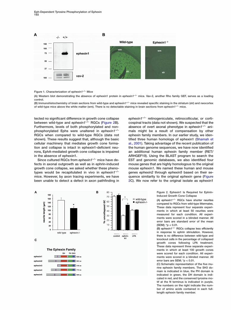
ds (Figure S1A), were born at the expected Mendelianrequency, and were viable, fertile, and overtly normal,ossibly due to the presence of other members of thephexin family (see below). Western blot analyses ofhole brain lysates with any of four distinct antibodies
aised against either the N- or C-terminal domains ofphexin1 failed to detect the presence of ephexin1rotein in ephexin1−/− mice (Figure 1A; Figure S1B). Fur-
hermore, immunohistochemistry of wild-type and ephex-n1−/− brain sections revealed strong, specific immuno-eactivity in the striatum and neocortex of wild-typeice and no immunoreactivity in the brains of ephex-
n1−/− mice (Figure 1B).As ephexin1 has previously been shown to activate
hoA, Rac1, and Cdc42 (Shamah et al., 2001) and theseTPases are known to be involved in axon extension,e investigated whether ephexin1−/− RGCs have de-
ects in axon extension. We measured the length of thexon in RGC cultures from wild-type and ephexin1−/−
ice. Axons from ephexin1−/− RGCs were significantlyhorter than their counterparts from wild-type RGCsFigure 2A; wild-type 151.9 ± 13.8 �m versus ephexin1−/−
10.7 ± 8.4 �m; p < 0.01 by ANOVA). However, the num-er of neurites per cell was unaltered between wild-ype and ephexin1−/− neurons (wild-type 3.87 ± 0.22ersus ephexin1−/− 4.19 ± 0.11 neurites per cell; p =.32 by ANOVA). These results suggest that ephex-
n1 plays a crucial role in axon outgrowth.Our previous study had suggested that ephexin1ight play a role in regulating ephrin-induced growth
one collapse in RGCs. To assess the importance ofphexin1 for EphA-mediated axon guidance, we com-ared the ability of ephrin-A to induce growth coneollapse of RGCs obtained from either wild-type orphexin1−/− mice. We used purified RGCs for these ex-eriments, because both EphAs and ephexin1 are ex-ressed in the ganglion cell layer of embryonic andarly postnatal retina, and the ability of ephrins to medi-te growth cone collapse has been well established inGCs (Feldheim et al., 1998; Jurney et al., 2002; Sha-ah et al., 2001). Purified RGCs from wild-type and
phexin1−/− mice have a similar percentage of intactrowth cones prior to ephrin treatment (Figure 2B).pon exposure to ephrin-A1, RGCs from wild-typeice show a significant increase in the number of col-
apsed growth cones. By contrast, significantly fewerphexin1−/− RGC growth cones collapse in response tophrin-A1 (Figure 2B; percent uncollapsed mean ± SEMith ANOVA: wild-type, 30.4 ± 1.4; ephexin1−/−, 53.3 ±.2; p < 0.01). This reduction in the percentage of col-
apsed growth cones in ephexin1−/− versus wild-typeeurons does not reflect an intrinsic defect in the abilityf RGCs to respond to repulsive factors; when RGCsrom wild-type and ephexin1−/− mice were exposed toysophosphatidic acid (LPA), an agent that inducesrowth cone collapse via a G protein-coupled receptorhat triggers RhoA activation (Moolenaar, 1999), we de-
Eph-Dependent Tyrosine Phosphorylation of Ephexin193
Figure 1. Characterization of ephexin1−/− Mice
(A) Western blot demonstrating the absence of ephexin1 protein in ephexin1−/− mice. Vav-2, another Rho family GEF, serves as a loadingcontrol.(B) Immunohistochemistry of brain sections from wild-type and ephexin1−/− mice revealed specific staining in the striatum (str) and neocortexof wild-type mice above the white matter (wm). There is no detectable staining in brain sections from ephexin1−/− mice.
2C). We now refer to the original isolate as ephexin1been unable to detect a defect in axon pathfinding in
Figure 2. Ephexin1 Is Required for Ephrin-Induced Growth Cone Collapse
(A) ephexin1−/− RGCs have shorter neuritescompared to RGCs from wild-type littermates.These data represent four separate experi-ments in which at least 50 neurites weremeasured for each condition. All experi-ments were scored in a blinded manner. Allerror bars are standard error of the mean(SEM); *p < 0.01.(B) ephexin1−/− RGCs collapse less efficientlyin response to ephrin stimulation. However,there is no difference between wild-type andknockout cells in the percentage of collapsedgrowth cones following LPA treatment.These data represent three separate experi-ments in which at least 100 growth coneswere scored for each condition. All experi-ments were scored in a blinded manner. Allerror bars are SEM; *p < 0.01.(C) Schematic representation of the five mu-rine ephexin family members. The SH3 do-main is indicated in blue, the PH domain isindicated in green, the DH domain is indi-cated in red, and the conserved tyrosine mo-tif at the N terminus is indicated in purple.The numbers on the right indicate the num-ber of amino acids contained in each full-length ephexin family member.
tected no significant difference in growth cone collapsebetween wild-type and ephexin1−/− RGCs (Figure 2B).Furthermore, levels of both phosphorylated and non-phosphorylated Ephs were unaltered in ephexin1−/−
RGCs when compared to wild-type RGCs (data notshown). These results suggest that, although the basiccellular machinery that mediates growth cone forma-tion and collapse is intact in ephexin1-deficient neu-rons, EphA-mediated growth cone collapse is impairedin the absence of ephexin1.
Since cultured RGCs from ephexin1−/− mice have de-fects in axonal outgrowth as well as in ephrin-inducedgrowth cone collapse, we asked whether these pheno-types would be recapitulated in vivo in ephexin1−/−
mice. However, by axon tracing experiments, we have
ephexin1−/− retinogeniculate, retinocollicular, or corti-cospinal tracts (data not shown). We suspected that theabsence of overt axonal phenotype in ephexin1−/− ani-mals might be a result of compensation by otherephexin family members. In our earlier study, we iden-tified three human homologs of ephexin1 (Shamah etal., 2001). Taking advantage of the recent publication ofthe human genome sequences, we have now identifiedan additional human ephexin family member (RET/ARHGEF19). Using the BLAST program to search theEST and genomic databases, we also identified fourmouse genes that are highly homologous to the originalmouse ephexin1. We named these human and mousegenes ephexin2 through ephexin5 based on their se-quence similarity to the original ephexin gene (Figure
Neuron194
and use the term ephexin family to refer to this group stof five mammalian genes. A comparison of the se-
quence of the five ephexin family members reveals that ttthey share the same overall structure, including a Dbl
homology (DH) domain followed by a pleckstrin homol- epogy (PH) domain, and a C-terminal SH3 domain. How-
ever, the various members of the ephexin family have rtunique N-terminal regions, which are variable in length
and have little sequence similarity. Examination of the bntissue distribution of the ephexin family members by
Northern blot and in situ hybridization revealed that Cfonly ephexin1 and ephexin5 (also known as Vsm-Rho-
GEF [Ogita et al., 2003]) mRNA are detected at signifi- cgcant levels in P7 and P21 mouse brain (Figure S2).EfEphexin Is Expressed Differentiallyrin Chick Motor Neuron ColumnsdSince our analysis of ephexin1−/− mice appears to beicomplicated by issues of compensation by other ephexin
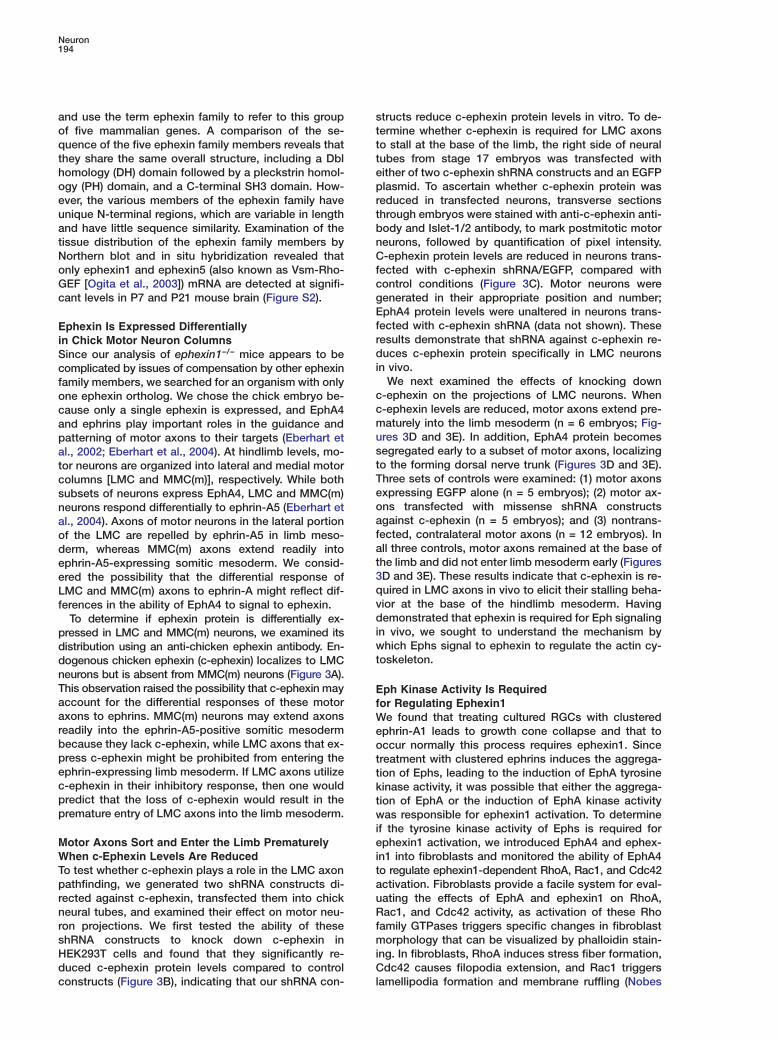
family members, we searched for an organism with onlycone ephexin ortholog. We chose the chick embryo be-ccause only a single ephexin is expressed, and EphA4mand ephrins play important roles in the guidance andupatterning of motor axons to their targets (Eberhart etsal., 2002; Eberhart et al., 2004). At hindlimb levels, mo-ttor neurons are organized into lateral and medial motorTcolumns [LMC and MMC(m)], respectively. While bothesubsets of neurons express EphA4, LMC and MMC(m)oneurons respond differentially to ephrin-A5 (Eberhart etaal., 2004). Axons of motor neurons in the lateral portionfof the LMC are repelled by ephrin-A5 in limb meso-aderm, whereas MMC(m) axons extend readily intotephrin-A5-expressing somitic mesoderm. We consid-3ered the possibility that the differential response ofqLMC and MMC(m) axons to ephrin-A might reflect dif-vferences in the ability of EphA4 to signal to ephexin.dTo determine if ephexin protein is differentially ex-ipressed in LMC and MMC(m) neurons, we examined itswdistribution using an anti-chicken ephexin antibody. En-tdogenous chicken ephexin (c-ephexin) localizes to LMC
neurons but is absent from MMC(m) neurons (Figure 3A).This observation raised the possibility that c-ephexin may Eaccount for the differential responses of these motor faxons to ephrins. MMC(m) neurons may extend axons Wreadily into the ephrin-A5-positive somitic mesoderm ebecause they lack c-ephexin, while LMC axons that ex- opress c-ephexin might be prohibited from entering the tephrin-expressing limb mesoderm. If LMC axons utilize tc-ephexin in their inhibitory response, then one would kpredict that the loss of c-ephexin would result in the tpremature entry of LMC axons into the limb mesoderm. w
ieMotor Axons Sort and Enter the Limb Prematurely
When c-Ephexin Levels Are Reduced itTo test whether c-ephexin plays a role in the LMC axon
pathfinding, we generated two shRNA constructs di- aurected against c-ephexin, transfected them into chick
neural tubes, and examined their effect on motor neu- Rfron projections. We first tested the ability of these
shRNA constructs to knock down c-ephexin in miHEK293T cells and found that they significantly re-
duced c-ephexin protein levels compared to control Clconstructs (Figure 3B), indicating that our shRNA con-
tructs reduce c-ephexin protein levels in vitro. To de-ermine whether c-ephexin is required for LMC axonso stall at the base of the limb, the right side of neuralubes from stage 17 embryos was transfected withither of two c-ephexin shRNA constructs and an EGFPlasmid. To ascertain whether c-ephexin protein was
educed in transfected neurons, transverse sectionshrough embryos were stained with anti-c-ephexin anti-ody and Islet-1/2 antibody, to mark postmitotic motoreurons, followed by quantification of pixel intensity.-ephexin protein levels are reduced in neurons trans-
ected with c-ephexin shRNA/EGFP, compared withontrol conditions (Figure 3C). Motor neurons wereenerated in their appropriate position and number;phA4 protein levels were unaltered in neurons trans-
ected with c-ephexin shRNA (data not shown). Theseesults demonstrate that shRNA against c-ephexin re-uces c-ephexin protein specifically in LMC neurons
n vivo.We next examined the effects of knocking down
-ephexin on the projections of LMC neurons. When-ephexin levels are reduced, motor axons extend pre-aturely into the limb mesoderm (n = 6 embryos; Fig-
res 3D and 3E). In addition, EphA4 protein becomesegregated early to a subset of motor axons, localizingo the forming dorsal nerve trunk (Figures 3D and 3E).hree sets of controls were examined: (1) motor axonsxpressing EGFP alone (n = 5 embryos); (2) motor ax-ns transfected with missense shRNA constructsgainst c-ephexin (n = 5 embryos); and (3) nontrans-ected, contralateral motor axons (n = 12 embryos). Inll three controls, motor axons remained at the base ofhe limb and did not enter limb mesoderm early (FiguresD and 3E). These results indicate that c-ephexin is re-uired in LMC axons in vivo to elicit their stalling beha-ior at the base of the hindlimb mesoderm. Havingemonstrated that ephexin is required for Eph signaling
n vivo, we sought to understand the mechanism byhich Ephs signal to ephexin to regulate the actin cy-
oskeleton.
ph Kinase Activity Is Requiredor Regulating Ephexin1
e found that treating cultured RGCs with clusteredphrin-A1 leads to growth cone collapse and that toccur normally this process requires ephexin1. Sincereatment with clustered ephrins induces the aggrega-ion of Ephs, leading to the induction of EphA tyrosineinase activity, it was possible that either the aggrega-ion of EphA or the induction of EphA kinase activityas responsible for ephexin1 activation. To determine
f the tyrosine kinase activity of Ephs is required forphexin1 activation, we introduced EphA4 and ephex-
n1 into fibroblasts and monitored the ability of EphA4o regulate ephexin1-dependent RhoA, Rac1, and Cdc42ctivation. Fibroblasts provide a facile system for eval-ating the effects of EphA and ephexin1 on RhoA,ac1, and Cdc42 activity, as activation of these Rho
amily GTPases triggers specific changes in fibroblastorphology that can be visualized by phalloidin stain-
ng. In fibroblasts, RhoA induces stress fiber formation,dc42 causes filopodia extension, and Rac1 triggers
amellipodia formation and membrane ruffling (Nobes
Eph-Dependent Tyrosine Phosphorylation of Ephexin195
Figure 3. Chick Motor Neuron Axons Sortand Enter the Hindlimb Prematurely WhenEphexin Is Knocked Down
(A) Expression of chick ephexin in the neuraltube (nt) using an anti-chick ephexin anti-body. Labeling localizes to motor neurons inthe lateral motor column (LMC) but not themedial motor column [MMC(m)]. C-ephexinprotein is also present in the forming dorsalroot ganglia (DRG) and the myotome.(B) shRNAs directed against c-ephexin re-duce c-ephexin protein levels in HEK293Tcells. Cells were transfected with c-ephexin(lane 1), c-ephexin and shRNA vector (lane2), c-ephexin and shRNA 461 (lane 3), orc-ephexin and shRNA 218 (lane 4). Lysateswere probed with antibodies againstc-ephexin and actin, which serves as a load-ing control.(C) shRNA directed against c-ephexin re-duces c-ephexin protein levels in chick neu-ral tubes. Average pixel intensity followingimmunohistochemistry with anti-c-ephexinantibody for stage 23 presumptive LMC mo-tor neurons was calculated for transfectedand untransfected sides of chick embryos.shRNA directed against chick ephexin (“E”;n = 5), missense shRNA (“M”; n = 4), andEGFP-expressing vector (“V”; n = 2) werecompared for ephexin expression. The y axisdenotes the ratio of average pixel intensitybetween transfected and untransfected sides.shRNAs directed against chicken ephexinspecifically reduce ephexin protein levels. Allerror bars are SEM; *p < 0.0001.(D) Top: Motor axons that express ephexinshRNAs sort and project into the hindlimbprematurely. White dashed line demarcates
the base of the limb, which was determined by the expression of EphA4 in limb mesoderm. (Left) Motor axons treated with ephexin shRNAsexpress EGFP (green); (middle) EphA4 protein (red) decorates the forming dorsal nerve trunk; (right) neurofilament antibody labeling (blue)marks all motor axons. Bottom: Nontransfected motor axons stall at the base of the limb. EphA4 protein (middle; red) and neurofilamentantibody (right; blue) mark all motor axons in the spinal nerve.(E) Three optical sections acquired from the same embryo, treated with ephexin shRNAs on the right neural tube (nt). Left neural tube is nottransfected. White dashed line indicates base of hindlimb; no, notochord. Top: EGFP expression (green) localizes to most cells in the ventralneural tube and several motor axons that have projected to the limb. Middle: EphA4 protein (red) becomes segregated to a subset of axons(arrow), in the presence of ephexin shRNAs. Note that all motor axons on the nontransfected side express EphA4. Bottom: Neurofilamentantibody (blue) labels ephexin shRNA-transfected and nontransfected motor axons.
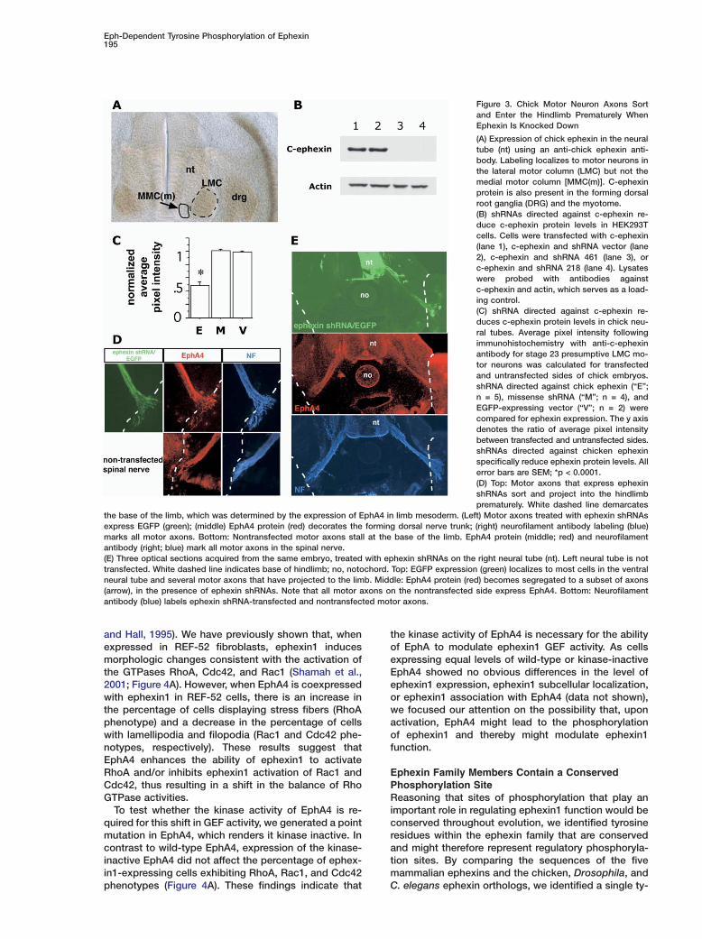
and Hall, 1995). We have previously shown that, whenexpressed in REF-52 fibroblasts, ephexin1 inducesmorphologic changes consistent with the activation ofthe GTPases RhoA, Cdc42, and Rac1 (Shamah et al.,2001; Figure 4A). However, when EphA4 is coexpressedwith ephexin1 in REF-52 cells, there is an increase inthe percentage of cells displaying stress fibers (RhoAphenotype) and a decrease in the percentage of cellswith lamellipodia and filopodia (Rac1 and Cdc42 phe-notypes, respectively). These results suggest thatEphA4 enhances the ability of ephexin1 to activateRhoA and/or inhibits ephexin1 activation of Rac1 andCdc42, thus resulting in a shift in the balance of RhoGTPase activities.
To test whether the kinase activity of EphA4 is re-quired for this shift in GEF activity, we generated a pointmutation in EphA4, which renders it kinase inactive. Incontrast to wild-type EphA4, expression of the kinase-inactive EphA4 did not affect the percentage of ephex-in1-expressing cells exhibiting RhoA, Rac1, and Cdc42phenotypes (Figure 4A). These findings indicate that
the kinase activity of EphA4 is necessary for the abilityof EphA to modulate ephexin1 GEF activity. As cellsexpressing equal levels of wild-type or kinase-inactiveEphA4 showed no obvious differences in the level ofephexin1 expression, ephexin1 subcellular localization,or ephexin1 association with EphA4 (data not shown),we focused our attention on the possibility that, uponactivation, EphA4 might lead to the phosphorylationof ephexin1 and thereby might modulate ephexin1function.
Ephexin Family Members Contain a ConservedPhosphorylation SiteReasoning that sites of phosphorylation that play animportant role in regulating ephexin1 function would beconserved throughout evolution, we identified tyrosineresidues within the ephexin family that are conservedand might therefore represent regulatory phosphoryla-tion sites. By comparing the sequences of the fivemammalian ephexins and the chicken, Drosophila, andC. elegans ephexin orthologs, we identified a single ty-
Neuron196
Figure 4. Phosphorylation of a Conserved N-Terminal Tyrosine Residue of Ephexin1
(A) REF-52 cells transfected with wild-type or kinase-dead EphA4 and ephexin1. Cells were stained with fluorescent phalloidin and scored ina blinded manner for the presence of stress fibers (white bars), lamellipodia (hatched bars), and filopodia (black bars). All transfections wererepeated three times, and at least 120 cells were scored in a blinded manner for each condition. All error bars are SEM.(B) Sequence alignment of the Mus musculus, Gallus gallus, Drosophila melanogaster, and Caenorhabditis elegans ephexin family membersreveals a conserved N-terminal motif (highlighted in red). The first tyrosine in this motif is conserved in all family members.(C) In vitro kinase assays with Src and trkB. Wild-type ephexin1 or ephexin1-Y87F were used as substrates for the kinases. Samples wereelectrophoresed on an SDS-PAGE gel, which was subsequently stained with Coomassie blue to demonstrate equal loading and exposed tofilm. Autoradiograph (top); Coomassie staining (bottom).(D) Verification that Tyr87 is phosphorylated in ephexin1. Ephexin1 that was phosphorylated in vitro was subjected to tryptic digestion andEdman degradation. Graph represents scintillation count associated with each amino acid as it was released by Edman degradation.
rosine corresponding to amino acid 87 (Tyr87) of ephex- amin1 that is present in the unique N-terminal region of
ephexin1 and is conserved between all ephexin family cwmembers (Figure 4B). Analysis using the Scansite phos-
phorylation prediction program revealed that Tyr87 lies ttwithin a stretch of amino acids that form a consensus site
for Src family kinase phosphorylation (http://scansite. gpmit.edu/). As EphA4 is similar to Src in its substrate
preference, we considered the possibility that ephexin1 tTyr87 is a site of phosphorylation by the Eph kinaseand/or a Src family member. t
oTo test if Tyr87 can be phosphorylated by Src orEphA4, we incubated recombinant Src or the EphA4 ki- p
pnase domain in the presence of [γ-32P]ATP and eitherwild-type ephexin1 or a mutant form of ephexin1 in t
awhich Tyr87 was mutated to phenylalanine (ephexin1-Y87F). Under these conditions, wild-type ephexin1 was p
tefficiently phosphorylated by both Src and the EphA4kinase domain, but not by trkB (Figure 4C; Figure S3). f
bMutation of Tyr87 to phenylalanine abolishes ephex-in1’s ability to be phosphorylated by EphA4 and Src. t
wAlthough these results suggest that Tyr87 is the siteof EphA4/Src phosphorylation on ephexin1, it remained l
tpossible that mutation of Tyr87 to phenylalanine interfereswith the phosphorylation of ephexin1 on a neighboring e
mino acid. Therefore, we used direct sequencingethods to identify which residue on ephexin1 be-
omes phosphorylated following in vitro incubationith Src. To this end, ephexin1 was phosphorylated in
he presence of [γ-32P]ATP and Src in vitro, and theryptic peptide carrying the radioactive phosphateroup was isolated by HPLC. Analysis of this trypticeptide by Edman degradation confirmed that Tyr87 is
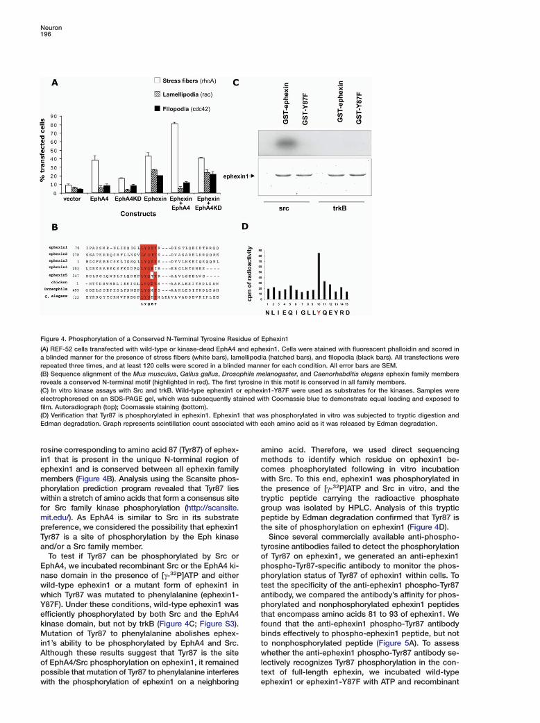
he site of phosphorylation on ephexin1 (Figure 4D).Since several commercially available anti-phospho-
yrosine antibodies failed to detect the phosphorylationf Tyr87 on ephexin1, we generated an anti-ephexin1hospho-Tyr87-specific antibody to monitor the phos-horylation status of Tyr87 of ephexin1 within cells. To
est the specificity of the anti-ephexin1 phospho-Tyr87ntibody, we compared the antibody’s affinity for phos-horylated and nonphosphorylated ephexin1 peptides
hat encompass amino acids 81 to 93 of ephexin1. Weound that the anti-ephexin1 phospho-Tyr87 antibodyinds effectively to phospho-ephexin1 peptide, but noto nonphosphorylated peptide (Figure 5A). To assesshether the anti-ephexin1 phospho-Tyr87 antibody se-
ectively recognizes Tyr87 phosphorylation in the con-ext of full-length ephexin, we incubated wild-typephexin1 or ephexin1-Y87F with ATP and recombinant
Eph-Dependent Tyrosine Phosphorylation of Ephexin197
Figure 5. Characterization of the Anti-Ephexin1 Phospho-Tyrosine 87-Specific Antibody
(A) Slot blot analysis of phosphorylated (P) and nonphosphorylated (NP) peptides using the anti-ephexin1 phospho-tyrosine 87-specificantibody. Each slot represents a 10-fold higher concentration of peptide over the one to its left.(B) Western blots of recombinant ephexin1 phosphorylated in vitro by Src, using anti-ephexin1 phospho-tyrosine 87-specific antibody. Immu-noreactivity is detected with Src and ephexin1, but not ephexin1 alone or ephexin1-Y87F and Src, demonstrating the specificity of the anti-ephexin1 phospho-tyrosine 87 antibody.(C) Ephexin1 is phosphorylated in neurons in response to ephrin treatment. Primary rat striatal neurons were cultured for 4 days and stimulatedfor the indicated amount of time with ephrin-A1. Lysates were probed with anti-ephexin1 phospho-tyrosine 87-specific (top panel) and totalephexin1 (bottom panel) antibodies. Ephexin1 tyrosine phosphorylation can be detected as early as 1 min after stimulation.(D) Cultured primary striatal neurons from wild-type and ephexin1−/− mice were stimulated for the indicated amount of time with ephrin-A1.Lysates were probed with anti-ephexin1 phospho-tyrosine 87-specific antibody. Ephexin1 phosphorylation can be detected in wild-typeneurons but not in ephexin1−/− neurons.(E) Ephexin1 is phosphorylated specifically by ephrins. Primary rat striatal neurons were cultured for 4 days and stimulated with the indicatedagents. Lysates were probed with anti-ephexin1 phospho-tyrosine 87-specific antibody, total ephexin1, and phospho-EphA antibodies.(F) Inhibitors of Src family kinases block ephexin1 phosphorylation on Tyr87 without blocking activation of the Eph tyrosine kinases. Primaryrat striatal neurons were cultured for 4 days and stimulated with Fc alone (first lane) or with ephrin-A1-Fc in the presence of no inhibitor(second lane), PP3, PP2, DMSO, or SU6656 (SU). Western blots were probed with anti-ephexin1 phospho-tyrosine 87-specific antibody,phospho-EphA, and total ephexin1 antibodies.
Src and examined the anti-ephexin1 phospho-Tyr87 an-tibody’s binding to ephexin1 by Western blotting. Wefound that the anti-ephexin1 phospho-Tyr87 antibodyspecifically detects Tyr87 phosphorylated ephexin1,but not the ephexin1-Y87F mutant or nonphosphory-lated wild-type ephexin1 (Figure 5B).
Ephrin Stimulation Results in Rapid TyrosinePhosphorylation of Ephexin1 in NeuronsGiven the importance of ephexin1 for ephrin-A-inducedgrowth cone collapse in cultured neurons, we utilizedthe anti-ephexin1 phospho-Tyr87 antibody to deter-mine whether ephexin1 is phosphorylated in responseto EphA signaling in primary neuronal cultures. Theanti-ephexin1 phospho-Tyr87 antibody recognized aprotein of the appropriate molecular weight (75 kDa) inephrin-A1-treated neurons (Figure 5C). To confirm thatthis protein is ephexin1, we compared lysates fromwild-type and ephexin1−/− neurons. Importantly, no 75
kDa band is seen in the neuronal lysates from ephex-in1−/− mice, even though Ephs are activated to thesame extent as in wild-type neurons (Figure 5D; datanot shown), suggesting that the 75 kDa band recog-nized by the anti-ephexin1 phospho-Tyr87 antibodyupon ephrin-A treatment is the Tyr87 phosphorylatedform of ephexin1. Using this antibody, we found thatephrin-A1 stimulation induces the phosphorylation ofTyr87 on ephexin1 in cultured neurons within 1 min (Fig-ure 5C). Ephexin1 phosphorylation peaks at 5 min andbegins to diminish by 30 min (Figure 5C; data notshown), paralleling the time course of growth cone col-lapse and reextension in these cultures. To determinewhether ephexin1 Tyr87 phosphorylation can be in-duced by extracellular stimuli in addition to ephrin-A,we characterized the phosphorylation of ephexin1 onTyr87 after exposure of neurons to several other extra-cellular factors. Ephrin-A1 and ephrin-B3 stimulation in-duced ephexin1 phosphorylation, while LPA, Sema-3a,
Neuron198
and Nogo did not induce the phosphorylation of this iasite, despite the ability of these agents to induce
growth cone collapse (Figure 5E). Furthermore, other lpstimuli, such as BDNF, EGF, PDGF, and forskolin, did
not result in the phosphorylation of ephexin1 on Tyr87 cd(data not shown). These observations demonstrate that
ephexin1 Tyr87 phosphorylation occurs specifically in pgresponse to ephrin stimulation and correlates with the
onset of ephrin-induced growth cone collapse, sug- spgesting a specific role for this phosphorylation event in
the process of Eph-dependent growth cone navigation. qetEphexin1 Is Phosphorylated in Neuronscby Src Family Kinases
In vitro, both recombinant Src and the EphA4 kinasesdomain are able to phosphorylate ephexin1. To assesstthe importance of Src activation for ephrin-A1-inducedsephexin1 phosphorylation in vivo, we took advantage ofetwo small molecule inhibitors of the Src family of tyrosine6kinases, PP2 and SU6656. Both PP2 and SU6656, butlnot PP3, the inactive analog of PP2, inhibited ephrin-wA1-induced ephexin1 Tyr87 phosphorylation in culturedirat striatal neurons (Figure 5F). To ensure that, at thegconcentrations used, the pharmacological inhibitorsuwere not inhibiting Eph tyrosine kinases, we titrated the
concentration of the inhibitors to a level that did notcaffect ephrin-induced Eph phosphorylation in culturedpneurons. We found that PP2 at 0.5 �M and SU6656 atp1 �M were specifically able to inhibit ephexin1 phos-tphorylation on Tyr87 but did not inhibit Eph tyrosinepphosphorylation (Figure 5F). These findings suggesttthat ephrin-A1 binding to EphA leads to phosphoryla-etion of ephexin1 at Tyr87 by activating Src family ki-Enases. Consistent with our findings, Drescher and col-fleagues have recently reported that Src family kinasesAcan phosphorylate ephexin1 in cultured neurons in re-asponse to ephrins (Knoll and Drescher, 2004).t
Tyrosine Phosphorylation of Ephexin1 Contributesto Growth Cone Collapse T
PTo determine whether phosphorylation of ephexin1 atTyr87 is required for ephrin/Eph-dependent growth W
pcone collapse, we transfected RGCs with wild-type andnonphosphorylatable forms (Y87F) of ephexin1 and an- u
Ealyzed the effect of these GEFs on ephrin-inducedgrowth cone collapse. We have previously shown that m
aectopic expression of ephexin1 significantly potenti-ates ephrin-mediated growth cone collapse and that t
lthe GEF activity of ephexin1 is necessary for this effect(Shamah et al., 2001). While expression of wild-type t
wephexin1 or ephexin1-Y87F did not significantly alterthe percentage of collapsed growth cones in unstim- o
eulated RGCs, RGCs expressing wild-type ephexin1 ex-hibited an increased percentage of collapsed growth d
acones in response to ephrin-A1 as compared to RGCsexpressing vector alone (Figure 6A). In contrast, ephrin r
cstimulation of RGCs expressing ephexin1-Y87F re-sulted in no increase in the percentage of growth cones i
othat collapse when compared to RGCs expressing vec-tor alone (Figure 6A). To determine whether an indepen- a
sdent way of phosphorylating ephexin1 was sufficientto induce growth cone collapse, we sought to identify t
tconditions under which ephexin1 was phosphorylated
ndependently of ephrin stimulation. Since no othergent that we tested was able to induce the phosphory-
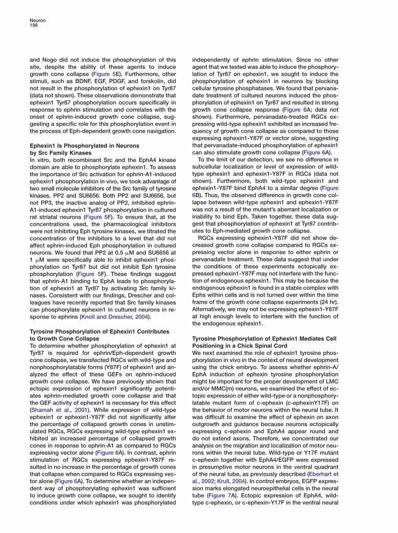
ation of Tyr87 on ephexin1, we sought to induce thehosphorylation of ephexin1 in neurons by blockingellular tyrosine phosphatases. We found that pervana-ate treatment of cultured neurons induced the phos-horylation of ephexin1 on Tyr87 and resulted in strongrowth cone collapse response (Figure 6A; data nothown). Furthermore, pervanadate-treated RGCs ex-ressing wild-type ephexin1 exhibited an increased fre-uency of growth cone collapse as compared to thosexpressing ephexin1-Y87F or vector alone, suggestinghat pervanadate-induced phosphorylation of ephexin1an also stimulate growth cone collapse (Figure 6A).To the limit of our detection, we see no difference in
ubcellular localization or level of expression of wild-ype ephexin1 and ephexin1-Y87F in RGCs (data nothown). Furthermore, both wild-type ephexin1 andphexin1-Y87F bind EphA4 to a similar degree (FigureB). Thus, the observed difference in growth cone col-
apse between wild-type ephexin1 and ephexin1-Y87Fas not a result of the mutant’s aberrant localization or
nability to bind Eph. Taken together, these data sug-est that phosphorylation of ephexin1 at Tyr87 contrib-tes to Eph-mediated growth cone collapse.RGCs expressing ephexin1-Y87F did not show de-
reased growth cone collapse compared to RGCs ex-ressing vector alone in response to either ephrin orervanadate treatment. These data suggest that underhe conditions of these experiments ectopically ex-ressed ephexin1-Y87F may not interfere with the func-ion of endogenous ephexin1. This may be because thendogenous ephexin1 is found in a stable complex withphs within cells and is not turned over within the time
rame of the growth cone collapse experiments (24 hr).lternatively, we may not be expressing ephexin1-Y87Ft high enough levels to interfere with the function ofhe endogenous ephexin1.
yrosine Phosphorylation of Ephexin1 Mediates Cellositioning in a Chick Spinal Corde next examined the role of ephexin1 tyrosine phos-
horylation in vivo in the context of neural developmentsing the chick embryo. To assess whether ephrin-A/phA induction of ephexin tyrosine phosphorylationight be important for the proper development of LMC
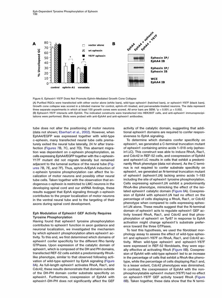
nd/or MMC(m) neurons, we examined the effect of ec-opic expression of either wild-type or a nonphosphory-atable mutant form of c-ephexin (c-ephexinY17F) onhe behavior of motor neurons within the neural tube. Itas difficult to examine the effect of ephexin on axonutgrowth and guidance because neurons ectopicallyxpressing c-ephexin and EphA4 appear round ando not extend axons. Therefore, we concentrated ournalysis on the migration and localization of motor neu-ons within the neural tube. Wild-type or Y17F mutant-ephexin together with EphA4/EGFP were expressed
n presumptive motor neurons in the ventral quadrantf the neural tube, as previously described (Eberhart etl., 2002; Krull, 2004). In control embryos, EGFP expres-ion marks elongated neuroepithelial cells in the neuralube (Figure 7A). Ectopic expression of EphA4, wild-ype c-ephexin, or c-ephexin-Y17F in the ventral neural
Eph-Dependent Tyrosine Phosphorylation of Ephexin199
Figure 6. Ephexin1-Y87F Does Not Promote Ephrin-Mediated Growth Cone Collapse
(A) Purified RGCs were transfected with either vector alone (white bars), wild-type ephexin1 (hatched bars), or ephexin1-Y87F (black bars).Growth cone collapse was scored in a blinded manner for control, ephrin-A1-treated, and pervanadate-treated neurons. The data representthree separate experiments in which at least 100 growth cones were scored. All error bars are SEM; *p < 0.001; p < 0.002.(B) Ephexin1-Y87F interacts with EphA4. The indicated constructs were transfected into HEK293T cells, and anti-ephexin1 immunoprecipi-tations were performed. Blots were probed with anti-EphA4 and anti-ephexin1 antibodies.
tube does not alter the positioning of motor neurons(data not shown; Eberhart et al., 2002). However, whenEphA4/EGFP was expressed together with wild-typec-ephexin, many transfected neural tube cells prema-turely exited the neural tube laterally, 24 hr after trans-fection (Figures 7B, 7C, and 7D). This aberrant migra-tion was dependent on c-ephexin phosphorylation, ascells expressing EphA4/EGFP together with the c-ephexin-Y17F mutant did not migrate laterally but remainedadjacent to the lumenal surface of the neural tube (Fig-ures 7B, 7E, and 7F). Thus, ephrin-A/EphA induction ofc-ephexin tyrosine phosphorylation can affect the lo-calization of motor neurons and possibly other neuraltube cells. Taken together with the observation that en-dogenous c-ephexin is restricted to LMC neurons in thedeveloping spinal cord and our shRNA findings, theseresults suggest that EphA signaling through c-ephexincontributes to the proper localization of motor neuronsin the ventral neural tube and to the targeting of theiraxons during spinal cord development.
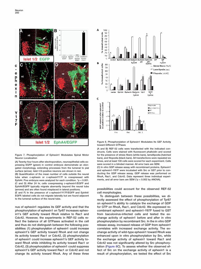
Eph Modulation of Ephexin1 GEF Activity RequiresTyrosine PhosphorylationHaving found that ephexin1 tyrosine phosphorylationcontributes to ephexin1 function in axon guidance andneuronal localization, we investigated the mechanismby which ephexin1 phosphorylation alters ephexin1 ac-tivity. To this end, we first determined which domains ofephexin1 confer specificity for the different Rho familyGTPases. Upon expression of the catalytic domain ofephexin1, which is composed of the DH and PH domains,transfected REF-52 cells exhibit a predominantly RhoA-like phenotype, similar to that observed following acti-vation of wild-type ephexin1 by EphA signaling (Figure8A). As full-length ephexin1 activates RhoA, Rac1, andCdc42, these results demonstrate that domains outsideof the DH-PH domain confer substrate specificity onephexin1. Furthermore, coexpression of EphA4 withephexin1-DH-PH does not significantly affect the GEF
activity of the catalytic domain, suggesting that addi-tional ephexin1 domains are required to confer respon-siveness to EphA signaling.
To determine which domains confer specificity onephexin1, we generated a C-terminal truncation mutantof ephexin1 containing amino acids 1–518 only (ephex-in1�C). This construct was able to induce RhoA, Rac1,and Cdc42 in REF-52 cells, and coexpression of EphA4and ephexin1�C results in cells that exhibit a predomi-nantly RhoA phenotype (data not shown). As the C termi-nus is not required to confer substrate specificity onephexin1, we generated an N-terminal truncation mutantof ephexin1 (ephexin1�N) lacking amino acids 1–183including the site of ephexin1 tyrosine phosphorylation.Cells expressing ephexin1�N exhibit a predominantlyRhoA-like phenotype, mimicking the effect of the iso-lated ephexin1 catalytic domain (Figure 8A). Coexpres-sion of EphA4 with ephexin1�N had no effect on thepercentage of cells displaying a RhoA, Rac1, or Cdc42phenotype when compared to cells expressing ephex-in1�N alone. These results suggest that the N-terminaldomain of ephexin1 acts to regulate ephexin1 GEF ac-tivity toward RhoA, Rac1, and Cdc42 and that phos-phorylation of ephexin1 on Tyr87 in response to EphAactivation might change ephexin1’s substrate prefer-ence toward the three GTPases.
To test this hypothesis, we used the fibroblast mor-phology assay to assess the effect of wild-type ephex-in1 and ephexin1-Y87F on RhoA, Rac1, and Cdc42 ac-tivity. When wild-type ephexin1 and ephexin1-Y87Fwere expressed in REF-52 fibroblasts, they were equ-ally effective at activating RhoA (Figure 8B). Coexpres-sion of EphA4 with wild-type ephexin1 led to an increasein the percentage of cells that exhibit a RhoA-like pheno-type, while the percentage of cells displaying Rac1 and,to a lesser extent, Cdc42 phenotypes was diminished.In contrast, the coexpression of EphA4 with the non-phosphorylatable ephexin1 mutant (Y87F) had no effecton ephexin1-Y87F GEF activity toward RhoA (Figure8B). Taken together, these data show that the N termi-
Neuron200
Ft
(sfFigure 7. Phosphorylation of Ephexin1 Modulates Spinal MotorbNeuron Localizationt(A) Twenty-four hours after electroporation, neuroepithelial cells ex-wpressing EGFP (green) in control embryos demonstrate an elon-(gated morphology, extending processes from the lumenal to pialasurface (arrow). Islet-1/2-positive neurons are shown in red.d(B) Quantification of the mean number of cells outside the neuralRtube when c-ephexin or c-ephexinY17F is coexpressed withmEphA4. Five embryos were analyzed for each condition. *p < 0.001.
(C and D) After 24 hr, cells coexpressing c-ephexin1/EGFP andEphA4/EGFP typically migrate aberrantly beyond the neural tube
p(arrows) and are often found misplaced in lateral positions.(E and F) In the presence of c-ephexin1Y17F/EGFP and EphA4/ cEGFP, labeled cells do not migrate laterally but are found adjacentto the lumenal surface of the neural tube. r
o
igure 8. Phosphorylation of Ephexin1 Modulates Its GEF Activityoward Different GTPases
A and B) REF-52 cells were transfected with the indicated con-tructs. Cells were stained with fluorescent phalloidin and scoredor the presence of stress fibers (white bars), lamellipodia (hatchedars), and filopodia (black bars). All transfections were repeated six
imes, and at least 100 cells were scored for each experiment. Cellsere scored in a blinded manner. All error bars are SEM.
C) In vitro GDP release assay with recombinant proteins. Ephexin1nd ephexin1-Y87F were incubated with Src or GST prior to con-ucting the GDP release assay. GDP release was performed onhoA, Rac1, and Cdc42. Data represent three individual experi-ents, and all error bars are SEM (*p < 0.002 by ANOVA).
fnus of ephexin1 regulates its GEF activity and that thephosphorylation of ephexin1 on Tyr87 increases ephex- c
fin1’s GEF activity toward RhoA relative to Rac1 andCdc42. However, the experiments in REF-52 cells re- c
pflect the balance of all GTPases activated in the celland thus do not distinguish between the following pos- r
csibilities: (1) phosphorylation of ephexin1 could increaseephexin1’s GEF activity toward RhoA and not change c
eits activity toward Rac1 or Cdc42, (2) phosphorylationof ephexin1 could increase ephexin1’s GEF activity to- t
Cward RhoA while inhibiting its activity toward Rac1 orCdc42, (3) phosphorylation of ephexin1 could suppress l
fephexin1’s GEF activity toward Rac1 or Cdc42 and notchange its activity toward RhoA. Any of these three r
ossibilities could account for the observed REF-52ell morphologies.To distinguish between these possibilities, we di-
ectly assessed the effect of phosphorylation of Tyr87n ephexin1’s ability to catalyze the exchange of GDPor GTP on RhoA, Rac1, and Cdc42. We expressed re-ombinant ephexin1 and ephexin1-Y87F fused to GSTrom baculovirus-infected cells and tested the ex-hange activity of ephexin1 before and after in vitrohosphorylation by recombinant Src. In the in vitro GDP
elease assay, increased release of GDP from ephexin1orrelates with increased exchange activity. The ex-hange activity of wild-type ephexin1 toward RhoA wasnhanced upon in vitro phosphorylation by Src, whilehe exchange activity of ephexin1 toward Rac1 anddc42 was not significantly altered by Src phosphory-
ation (Figure 8C). To assess whether the observed ef-ect of Src on the exchange activity of ephexin1 is aesult of phosphorylation, we tested the effect of Src
Eph-Dependent Tyrosine Phosphorylation of Ephexin201
on the ability of ephexin1-Y87F to catalyze exchangeof GTP for GDP on RhoA, Rac1, and Cdc42. We foundthat Src had no effect on the exchange activity of ephex-in1-Y87F.
To ensure that what we observe in vitro accuratelyreflects what occurs in cells, we performed GDP re-lease assays with lysates from transfected cells. Wefind that lysates from cells expressing ephexin1 andEphA4 exhibit higher exchange activity toward RhoAthan lysates from cells expressing ephexin1 alone. Thisincrease in exchange activity in the presence of EphA4expression does not occur in lysates from cells ex-pressing ephexin1-Y87F (data not shown). Thus, phos-phorylation of ephexin1 at Tyr87 results specifically inan increase in ephexin1’s GEF activity toward RhoA.Although this is the first example of a GEF changingits substrate preference in response to an extracellularstimulus, there are some recent examples of GAPsdemonstrating such a switch. MgcRacGAP, which is aGAP for Rac/Cdc42, is converted to a RhoGAP uponphosphorylation by Aurora B kinase (Minoshima et al.,2003). Phosphorylation of mgcRacGAP by Aurora ki-nase increases the GAP activity of mgcRacGAP towardRhoA but not toward Rac1 or Cdc42.
We conclude that the phosphorylation of ephexin1at Tyr87 within the N terminus of ephexin1 modulatesephexin1’s GEF activity toward RhoA, Rac1, andCdc42. However, rather than acting to generally sup-press GEF activity, the ephexin1 N-terminal region reg-ulates the substrate preference of ephexin1. Phosphor-ylation of ephexin1 on Tyr87 in response to ephrin-A/EphA signaling potentiates ephexin1-mediated activa-tion of RhoA relative to Rac1 and Cdc42, therebychanging the local balance of Rho GTPase activitywithin cells. The switch in specificity may allow ephex-in1 to promote axonal outgrowth in the absence ofephrin and growth cone repulsion when an Eph-expressing growth cone contacts ephrin-expressingcells. Consistent with this idea, ephexin1−/− neuronshave reduced axonal outgrowth in the absence ofephrin and impaired growth cone collapse when treatedwith clustered ephrins.
Discussion
In the current study, we have tested the hypothesis thatephexin1 is required for ephrin-A-induced growth conecollapse and have elucidated a mechanism by whichephrin-A triggers ephexin1-dependent axon guidance.We find that chicken motor neurons in which ephexinprotein has been knocked down by RNAi project pre-maturely into the hindlimb, indicating that ephexinplays a critical role in Eph-mediated axon guidance invivo. In addition, we find that cultured RGCs fromephexin1−/− mice have significantly shorter axons, andtheir growth cones show reduced growth cone collapsein response to ephrin-A1 stimulation as compared withwild-type RGCs. These seemingly contrasting results—that ephexin1 is required for axonal outgrowth as wellas for growth cone repulsion/collapse—are reconciledby our findings regarding the regulation of ephexin1 ac-tivity.
In the absence of ephrin, ephexin1 Tyr87 is not phos-
phorylated, and under these conditions, Eph boundephexin1 activates RhoA, Rac1, and Cdc42, leading toa balance of GTPase activation that promotes axonaloutgrowth. Consistent with this idea, RGCs that lackephexin1 have shorter axons than wild-type RGCs.Ephexin1 could influence axonal outgrowth in one oftwo possible ways. Ephexin1 could be required eitherfor the initiation of neurite development or for the pro-motion of axon outgrowth. As ephexin1−/− RGCs havea similar number of neurites when compared to wild-type RGCs, it is unlikely that the initiation of neuriteoutgrowth is regulated by ephexin. Furthermore, ephex-in1 is tethered to the Eph at the plasma membrane ofthe growth cone, localizing ephexin1 to the properplace to regulate the rate of axonal extension.
While in the absence of ephrins ephexin1 promotesaxonal outgrowth, ephrin stimulation of Ephs inducesSrc-dependent phosphorylation of ephexin1 on an evo-lutionarily conserved tyrosine, and this leads to growthcone collapse. The tyrosine phosphorylation of ephex-in1 enhances ephexin1’s exchange activity specificallytoward RhoA, thus promoting growth cone collapse/re-pulsion. Consistent with this idea, we find that ephexin1is important for ephrin-mediated growth cone collapsein vitro and axon guidance in vivo. Chicken motor neu-ron axons in which ephexin has been knocked downenter the hindlimb prematurely. Since motor neuronprojections into the hindlimb have previously beenshown to be controlled by ephrin/Eph interactions (Eb-erhart et al., 2002), this result strongly suggests thatephexin is required for Eph/ephrin-mediated axon guid-ance in vivo. This defect in axon guidance most likelyreflects impairment in the ability of ephexin-deficientaxonal growth cones to collapse in response to ephrinsin limb mesoderm, as the growth cones of culturedRGCs from ephexin1−/− mice display a defective col-lapse response when exposed to ephrin.
In addition to demonstrating that ephexin1 plays akey role in ephrin-mediated collapse/repulsion, ourfindings identify a mechanism by which Ephs regulateephexin1, leading to growth cone collapse. The direc-tional control of axon pathfinding in vivo is thought toresult from local modulation of the actin cytoskeletonsuch that growth cones turn away from repellents andtoward attractants. The discoveries that ephexin1 is re-quired for axon outgrowth, is regulated by tyrosinephosphorylation, and when tyrosine phosphorylatedpromotes growth cone collapse suggest a mechanismby which Rho GTPases could control the actin cy-toskeleton in the highly localized manner that is re-quired for accurate growth cone guidance. Sinceephexin1 is constitutively bound to Ephs, its effects onRho GTPases may remain confined to the vicinity of theEph complex. Thus, in the absence of ephrin stimula-tion, Eph/ephexin1 complexes might promote localizedgrowth cone extension. When Ephs are activated in aportion of the growth cone by ephrins, local tyrosinephosphorylation of ephexin1 at Tyr87 could tip the localbalance of GTPase activity toward RhoA, leading to ac-tin cytoskeletal changes that result in local retraction. Inregions of the growth cone that have not made specificcontact with ephrin, ephexin1 might still promotegrowth by activating RhoA, Rac1, and Cdc42. Thiscould result in directed movement of the growth cone
Neuron202
away from ephrin-expressing zones and toward its ulti- Dmate target. C
If ephexin1 bound to Ephs is to play the dual role Nof promoting outgrowth in the absence of ephrins and iretraction in the presence of ephrins, one might predict dthat ephexin1 may have evolved to specifically interactwith Ephs and not other guidance receptors. For exam- sple, if ephexin1 interacted with Ephs and plexins (a re- mceptor for the repulsive guidance factor semaphorin) at ithe same time and in the same region of the growth scone, conflicting signals might occur depending on the epresence of ephrins and semaphorins. If ephrin were wpresent and semaphorin were not, ephexin1 bound to oEphs could signal the growth cone to retract, while sephexin1 bound to plexins would signal the growth ccone to extend. To avoid this problem, ephexins appear eto have evolved so that they specifically mediate iephrin/Eph signaling. Consistent with this idea, we find gthat, of the growth and guidance factors tested, only bephrin is able to induce ephexin1 phosphorylation on rTyr87. No other guidance molecules (e.g., BDNF, LPA, eSema3A, or Nogo66) or growth factors (e.g., PDGF, aEGF, or insulin) tested led to ephexin1 tyrosine phos- aphorylation, suggesting that ephexin1 may be specifi- scally designed to respond to ephrin signals. Since dephexin1 can promote outgrowth in the absence of rephrin and repulsion upon encountering ephrins, ephex-in1 is tailored to mediate ephrin-dependent growth ccone guidance. r
To better understand the process by which Ephs me- pdiate axon guidance, we investigated how tyrosine aphosphorylation of ephexin1 switches ephexin1’s ex- ichange activity. Through mutagenesis of ephexin1, we sfound that the isolated DH/PH domain possesses tstrong exchange activity toward RhoA and is ineffective dat activating Rac1 and Cdc42. In the context of full- plength ephexin1 when Tyr87 is not phosphorylated, the eDH/PH domain is capable of activating RhoA, Rac1, Wand Cdc42. This suggests that, in the absence of phos- aphorylation, other domains of ephexin1 modulate the cactivity of the DH/PH domain so that it no longer prefer- nentially activates RhoA. Further mutagenesis revealed pthat it is the N terminus that possesses the modulatory pfunction. Tyr87 phosphorylation in response to ephrin
happears to suppress the modulatory effect of the N ter-
tminus, resulting in an ephexin1 DH/PH domain that is
2highly selective toward RhoA at the expense of Rac1sand Cdc42. Consistent with such a model, we find thattfull-length ephexin1 when not phosphorylated in vitroEis capable of activating RhoA, Rac1, and Cdc42. Whendphosphorylated at Tyr87 in vitro, ephexin1’s activity forwRhoA is specifically enhanced, while its activity towardoRac1 and Cdc42 is unchanged. This suggests thattwithin cells the phosphorylation of ephexin1 in re-psponse to Eph activation leads to a potentiation of
RhoA activity and possibly enhances binding to RhoAso that ephexin1 is unavailable to activate Rac1 and ECdc42. Further experimentation will be required to as-
Isess binding affinities of nonphosphorylated and phos-Rphorylated ephexin1 for RhoA relative to Rac1 andd
Cdc42. While it is presently not clear how tyrosine (phosphorylation specifically suppresses the N-terminal 5modulatory effect on the DH/PH domain, one likely ef- t
cfect is that the N terminus interacts directly with the
H/PH domain to modulate DH/PH domain function.onsistent with this possibility, we find that the isolatedterminus of ephexin is capable of interacting with an
solated fragment of ephexin1 consisting of the DH/PHomain and the C terminus (data not shown).While our experiments reveal that ephexin1 plays a
ignificant role in Eph-mediated axon guidance, it re-ains to be determined whether ephexin1 plays a role
n other Eph-dependent biological processes. One pos-ibility is that ephexin1 may mediate Eph-dependentffects on cell migration and localization. Consistentith this possibility, we have found that overexpressionf chicken ephexin leads to aberrant migration of pre-umptive motor neurons out of the neural tube, a pro-ess that is dependent on the phosphorylation ofphexin. In addition to migration, Ephs have also been
mplicated in dendritic spine formation. Similar torowth cones, dendritic spines are highly motile actin-ased structures that rapidly alter their morphology in
esponse to various extracellular stimuli (includingphrins). Thus, ephexin family GEFs may mediate somespects of ephrin-dependent dendritic spine formationnd/or maintenance. In this regard, our preliminary re-ults suggest that ephexin5 is highly enriched in den-ritic spines and thus might play an important role in
egulating dendritic spine formation.While ephexin1 appears capable of modulating actin
ytoskeletal dynamics during growth cone guidance,emodeling of the cytoskeleton is not the only cellularrocess that is required for regulation of axon guid-nce. The process of axon guidance is a complex event
nvolving endocytosis of plasma membrane and cellurface proteins, reorganization of the actin cytoskele-on and microtubules, and local protein translation andegradation (van Horck et al., 2004). The signalingathways that link cell surface receptor activation toach of these processes are not yet well understood.hile we have shown that ephexin1 regulates the bal-
nce of GTPase activation, thereby affecting the actinytoskeleton during the process of axon guidance, it isot yet clear if ephexin1 is involved in other steps in therocess of axon guidance such as endocytosis, localrotein translation, and local protein degradation. Weave recent evidence that another GEF, Vav2, is impor-ant for Eph-dependent axon guidance (Cowan et al.,005 [this issue of Neuron]). In response to ephrintimulation, Vav2 associates with the Ephs, becomesyrosine phosphorylated, and plays a role in ephrin/ph-dependent endocytosis that is a key step in Eph-ependent axon guidance. It remains to be determinedhether or not ephexin and Vav function independentlyf each other and how these two GEFs and other pro-eins that interact with Ephs coordinate the complexrocess of axon guidance.
xperimental Procedures
n Vitro Kinase Assaysecombinant ephexin was incubated with either GST-EphA4 kinaseomain or His-tagged Src (Upstate Biotechnology) in kinase buffer
20 mM Tris 7.5, 8 mM MgCl2, 3 mM MnCl2) with 200 µM ATP andµCi [γ-32P]ATP for 30 min at 30°C. For trkB kinase assays, Flag-
agged trkB was transfected into HEK293T cells and immunopre-ipitated with anti-Flag epitope antibody before incubating with
Eph-Dependent Tyrosine Phosphorylation of Ephexin203
ephexin1 in kinase buffer. Samples were electrophoresed on anSDS-PAGE gel that was subsequently stained with Coomassie blueand exposed to film.
Fibroblast Morphology AssayREF-52 cells were maintained in DMEM with 10% newborn calfserum, glutamine, and Pen/Strep (Invitrogen) and transfected usingFugene (Roche). Twenty-four to forty-eight hours after transfection,cells were fixed with 4% paraformaldehyde, permeabilized with0.1% Triton, and stained with Alexa594-conjugated phalloidin (Mo-lecular Probes). Cells were scored in a blinded manner.
Growth Cone Collapse Assay and Axon Length AnalysisRGCs were purified and cultured as previously described (Meyer-Franke et al., 1995). Expression plasmids were electroporated intoRGCs with the rat neuron Nucleofector kit (Amaxa). RGCs that werecultured for 1–2 days were stimulated with aggregated ephrins for30 min, fixed in 4% paraformaldehyde, stained with Alexa594-con-jugated phalloidin (Molecular Probes), and scored for growth conecollapse in a blinded manner. Ephrin-A1-Fc was aggregated at 5�g/ml 45 min prior to use with 0.45 mg/ml goat anti-human Fc anti-body (Jackson Laboratories) in Dulbecco’s phosphate-buffered sa-line (Invitrogen). Pervanadate was prepared as previously de-scribed (Sun et al., 2000), and RGCs were treated with 10 �Mpervanadate for 30 min.
For neurite length analysis of dissociated neurons, neurons werefixed after 24–36 hr in culture and stained with phalloidin-Texasred (Molecular Probes). Images blinded to the experimenter wereobtained on a fluorescent microscope and were analyzed usingMetamorph software (Universal Imaging Corporation) by manuallytracing the length of the neurite for at least 50 neurons per condi-tion. Four independent experiments were conducted for eachmouse strain.
RNA InterferenceThe full-length chicken ephexin sequence was copied into thesiRNA search engine (http://jura.wi.mit.edu/siRNAext/). Two oligosequences against chicken ephexin were generated: 218 and 461.These regions were not homologous to any other known genes, asdetermined by BLAST search. Each sequence was then enteredinto a siRNA converter (http://www.ambion.com/techlib/misc/psilencer_converter.html/), to generate top and bottom oligo se-quences that were acquired (Sigma-Genosys). C-ephexin 461 topsequence, 5#-GGCTGTTAGTGAGCGCTTCTTCAAGAGAGAAGCGCTCACTAACAGCCTTTTTT-3#; c-ephexin 461 bottom sequence, 5#-AATTAAAAAAGGCTGTTAGTGAGCTTCTCTCTTGAAGAAGCGCTCACTAACAGCCGGCC-3#; c-ephexin 218 top sequence, 5#-GCTCCAACTCCAGGTTCAACTTCAAGAGAGTTGAACCTGGAGTTGGAGTTTTTT-3# ; c-ephexin 218 bottom sequence, 5#-AATTAAAAAACTCCAACTCCAGGTTCAACTCTCTTGAAGTTGAACCTGGAGTTGGAGCGGCC-3#. Briefly, the complementary oligonucleotides wereannealed and inserted into the ApaI/EcoRI sites of the pSilencer1.0 U6 vector (Ambion). Missense shRNAs were generated bychanging three bases in the c-ephexin 218 and 461 constructs.Mutated bases are underlined: 218 missense, 5#-CTGCAACTACAGGTTCACC-3#; 461 missense, 5#-GCCTGTTAGGGAGCGCTAC-3#.These missense sequences were then subjected to BLAST andBBSRC query; no similarities to chicken or mammalian sequenceswere identified. Missense oligos were annealed and placed in pSi-lencer 1.0 U6, as described above. All shRNAs were confirmed bysequence analysis.
Quantification of Ephexin Protein LevelsTo quantify ephexin protein levels, the average pixel intensity ofpresumptive LMC neurons in unsaturated single optical sectionsacquired by confocal microscopy was determined using Fluoviewsoftware (Olympus). Sections acquired from three different treat-ment conditions were measured: (1) ephexin shRNA-expressing; (2)missense shRNA-expressing; and (3) pCAX (EGFP)-expressing.Three readings were performed for each LMC domain on the trans-fected side and nontransfected side.
In Ovo ElectroporationFertilized White Leghorn chicken eggs (Hy-Line International,Spencer, IA; Bilbie Aviaries, Ann Arbor, MI) were incubated at 38°Cin a humidified incubator until stage 17 of development. For trans-fection, 2.0–2.5 �g/�l of shRNA was coelectroporated with pCAX/EGFP (2–2.5 �g/�l) into a ventral quadrant of the neural tube ofstage 17 embryos, as previously described (Eberhart et al., 2002).For electroporations to express EGFP (2 �g/�l) or to coexpressc-ephexin/EGFP or c-ephexin-Y17F/EGFP with EphA4/EGFP, stage15–17 chick embryos were coelectroporated with 2–2.5 �g/�l ofeach construct. Five 14V (Protech Int., TX) or 19V–22V pulses (BTX/Genetronics, CA) of 50 ms duration were applied, and several dropsof Ringer’s buffer were added, after which each egg was sealedwith tape and incubated for 6, 12, 24, or 32 hr.
GDP Release AssaysIn vitro guanine nucleotide release assays were performed as de-scribed elsewhere (Debant et al., 1996) using recombinant full-length ephexin1 with RhoA-His, Rac1-His, and Cdc42-His (Cy-toskeleton). Briefly, the GTPases were first loaded with 3H-GDP inexchange buffer (50 mM Tris [pH 7.5], 50 mM NaCl, 5 mM EDTA, 1mM DTT, 1 mg/ml BSA). After incubation, MgCl2 was added to afinal concentration of 5 mM, and proteins were maintained on ice.Recombinant ephexin1 was incubated with the GTPase in 50 mMTris (pH 7.5), 1 mM GTP, 2 mM MgCl2 at room temperature for 15min. In the presentation of data, the intrinsic GDP release rate ofthe GTPase is taken into account. Thus, “100%” corresponds tothe amount of protein bound 3H-GDP retained on the filter in thepresence of the GTPase alone.
Supplemental DataThe Supplemental Data include three supplemental figures andSupplemental Experimental Procedures and can be found with thisarticle online at http://www.neuron.org/cgi/content/full/46/2/191/DC1/.
Acknowledgments
The authors thank Margaret Thompson, Yiping Zhou, and Hong Yefor assistance in generating ephexin1−/− mice; Pieter Dikkes for as-sistance with immunohistochemistry and in situ hybridization ex-periments; Sara Vasquez for assistance with neuronal cell cultures;David L. Brautigan for providing REF-52 cells; Rudiger Klein andJoaquim Egea for sharing unpublished results; and Eric Griffith,Janine Zieg, Ben Barres, Mitsuhiro Makino, Beth Perkins, andmembers of the Greenberg lab for critical discussions. M.E.G. ac-knowledges the generous support of the F.M. Kirby Foundation tothe Children’s Hospital Neurobiology Program. This work was sup-ported by National Institute of Child Health and Human Develop-ment grant K08 HD01384 (M.S.); the William Randolph Hearst Fund(M.S.); a National Defense Science and Engineering graduate fel-lowship (M.Z.L.); a National Science Foundation predoctoral fellow-ship (P.L.G.); a Muscular Dystrophy Association postdoctoral fel-lowship (S.O.); National Institute of Mental Health grant R01MH59894 (C.E.K.); National Institute of Neurological Disorders andStroke grant R01 NS35884 (G.C.); Mental Retardation ResearchCenter grant HD18655 and National Institutes of Health grantNS45500 (M.E.G.); and a grant from Daiichi Pharmaceuticals(M.E.G.).
Received: July 9, 2004Revised: December 3, 2004Accepted: January 18, 2005Published: April 20, 2005
References
Cowan, C.W., Shao, Y.R., Sahin, M., Shamah, S.M., Lin, M.Z., Greer,P.L., Gao, S., Griffith, E.C., Brugge, J.S., and Greenberg, M.E.(2005). Vav family GEFs link activated Ephs to endocytosis andaxon guidance. Neuron 46, 205–217.
Neuron204
Debant, A., Serra-Pages, C., Seipel, K., O’Brien, S., Tang, M., Park, W(S.H., and Streuli, M. (1996). The multidomain protein Trio binds the
LAR transmembrane tyrosine phosphatase, contains a protein ki- rnase domain, and has separate rac-specific and rho-specific gua- Wnine nucleotide exchange factor domains. Proc. Natl. Acad. Sci. EUSA 93, 5466–5471. pDickson, B.J. (2002). Molecular mechanisms of axon guidance. Sci-ence 298, 1959–1964.
Eberhart, J., Swartz, M.E., Koblar, S.A., Pasquale, E.B., and Krull,C.E. (2002). EphA4 constitutes a population-specific guidance cuefor motor neurons. Dev. Biol. 247, 89–101.
Eberhart, J., Barr, J., O’Connell, S., Flagg, A., Swartz, M.E., Cramer,K.S., Tosney, K.W., Pasquale, E.B., and Krull, C.E. (2004). Ephrin-A5exerts positive or inhibitory effects on distinct subsets of EphA4-positive motor neurons. J. Neurosci. 24, 1070–1078.
Feldheim, D.A., Vanderhaeghen, P., Hansen, M.J., Frisen, J., Lu, Q.,Barbacid, M., and Flanagan, J.G. (1998). Topographic guidance la-bels in a sensory projection to the forebrain. Neuron 21, 1303–1313.
Flanagan, J.G., and Vanderhaeghen, P. (1998). The ephrins and Ephreceptors in neural development. Annu. Rev. Neurosci. 21, 309–345.
Huber, A.B., Kolodkin, A.L., Ginty, D.D., and Cloutier, J.F. (2003).Signaling at the growth cone: ligand-receptor complexes and thecontrol of axon growth and guidance. Annu. Rev. Neurosci. 26,509–563.
Jurney, W.M., Gallo, G., Letourneau, P.C., and McLoon, S.C. (2002).Rac1-Mediated Endocytosis during Ephrin-A2- and Semaphorin3A-Induced Growth Cone Collapse. J. Neurosci. 22, 6019–6028.
Knoll, B., and Drescher, U. (2004). Src family kinases are involvedin EphA receptor-mediated retinal axon guidance. J. Neurosci. 24,6248–6257.
Krull, C.E. (2004). A primer on using in ovo electroporation to ana-lyze gene function. Dev. Dyn. 229, 433–439.
Meyer-Franke, A., Kaplan, M.R., Pfrieger, F.W., and Barres, B.A.(1995). Characterization of the signaling interactions that promotethe survival and growth of developing retinal ganglion cells in cul-ture. Neuron 15, 805–819.
Minoshima, Y., Kawashima, T., Hirose, K., Tonozuka, Y., Kawajiri, A.,Bao, Y.C., Deng, X., Tatsuka, M., Narumiya, S., May, W.S., et al.(2003). Phosphorylation by Aurora B Converts MgcRacGAP to aRhoGAP during Cytokinesis. Dev. Cell 4, 549–560.
Moolenaar, W.H. (1999). Bioactive lysophospholipids and their Gprotein-coupled receptors. Exp. Cell Res. 253, 230–238.
Nobes, C.D., and Hall, A. (1995). Rho, rac, and cdc42 GTPases reg-ulate the assembly of multimolecular focal complexes associatedwith actin stress fibers, lamellipodia, and filopodia. Cell 81, 53–62.
Ogita, H., Kunimoto, S., Kamioka, Y., Sawa, H., Masuda, M., andMochizuki, N. (2003). EphA4-mediated Rho activation via Vsm-Rho-GEF expressed specifically in vascular smooth muscle cells. Circ.Res. 93, 23–31.
Palmer, A., and Klein, R. (2003). Multiple roles of ephrins in morpho-genesis, neuronal networking, and brain function. Genes Dev. 17,1429–1450.
Penzes, P., Beeser, A., Chernoff, J., Schiller, M.R., Eipper, B.A.,Mains, R.E., and Huganir, R.L. (2003). Rapid induction of dendriticspine morphogenesis by trans-synaptic ephrinB-EphB receptor ac-tivation of Rho-GEF kalirin. Neuron 37, 263–274.
Shamah, S.M., Lin, M.Z., Goldberg, J.L., Estrach, S., Sahin, M., Hu,L., Bazalakova, M., Neve, R.L., Corfas, G., Debant, A., andGreenberg, M.E. (2001). EphA receptors regulate growth cone dy-namics through the novel guanine nucleotide exchange factorephexin. Cell 105, 233–244.
Sun, Q.L., Wang, J., Bookman, R.J., and Bixby, J.L. (2000). Growthcone steering by receptor tyrosine phosphatase delta defines a dis-tinct class of guidance cue. Mol. Cell. Neurosci. 16, 686–695.
Van Aelst, L., and D’Souza-Schorey, C. (1997). Rho GTPases andsignaling networks. Genes Dev. 11, 2295–2322.
van Horck, F.P., Weinl, C., and Holt, C.E. (2004). Retinal axon guid-ance: novel mechanisms for steering. Curr. Opin. Neurobiol. 14,61–66.
ahl, S., Barth, H., Ciossek, T., Aktories, K., and Mueller, B.K.2000). Ephrin-A5 induces collapse of growth cones by activatingho and rho kinase. J. Cell Biol. 149, 263–270.
inning, R.S., Ward, E.K., Scales, J.B., and Walker, G.K. (2002).phA4 catalytic activity causes inhibition of RhoA GTPase in Xeno-us laevis embryos. Differentiation 70, 46–55.