Toxic activity of Bacillus Thuringiensis isolates to Aedes Aegypti (L.) (Diptera: Culicidae) larvae
Upload
independentCategory
view
1download
0
This article appeared in a journal published by Elsevier. The attachedcopy is furnished to the author for internal non-commercial researchand education use, including for instruction at the authors institution
and sharing with colleagues.
Other uses, including reproduction and distribution, or selling orlicensing copies, or posting to personal, institutional or third party
websites are prohibited.
In most cases authors are permitted to post their version of thearticle (e.g. in Word or Tex form) to their personal website orinstitutional repository. Authors requiring further information
regarding Elsevier’s archiving and manuscript policies areencouraged to visit:
http://www.elsevier.com/copyright
Author's personal copy
Detection of genes encoding antimicrobial peptides in Mexicanstrains of Trichoplusia ni (Hubner) exposed to Bacillus thuringiensis
P. Tamez-Guerra a,*, J.A. Valadez-Lira a, J.M. Alcocer-Gonzalez a, B. Oppert b,R. Gomez-Flores a, R. Tamez-Guerra a, C. Rodrıguez-Padilla a
a Departmento de Microbiologıa e Inmunologıa, Facultad de Ciencias Biologicas, Universidad Autonoma de Nuevo Leon (FCB-UANL),
AP 46-F, 66450 San Nicolas de los Garza, N.L., Mexicob USDA-ARS Grain Marketing and Production Research Center, 1515 College Avenue, Manhattan, KS 66502, USA
Received 15 October 2007; accepted 13 February 2008Available online 20 February 2008
Abstract
The systemic immune response of Trichoplusia ni after Bacillus thuringiensis (Bt) exposure was evaluated by comparing the expressionof genes encoding antimicrobial peptides (AMPs) in Bt-susceptible and -resistant T. ni strains that were either exposed or not to Xen-Tari� (Bt-XT). AMP genes were detected by RT-PCR using primers for attacin, gloverin, lebocin, lysozyme, and peptidoglycan recog-nition peptide (PGRP). In general, AMP genes were detected more frequently in Mexican field strains previously exposed to Bt (SALXand GTOX) than in a Mexican laboratory strain (NL), but expression was similar to the AMP expression in USA laboratory strains (USand USX). Among the AMPs, transcripts for lebocin were the least detected (11.7%) and those for lysozyme were the most detected(84.8%) in all samples. Lebocin was detected only in 2nd instar and pupa. All untreated controls expressed attacin. Attacin and gloverinwere not detected in any midgut sample, and their highest detection was in pupa. Lysozyme was rarely detected in 2nd instar larvae fromany strain or treatment but was detected in almost all midgut and hemolymph samples. Overall, AMPs were found more in T. ni strainspreviously exposed to Bt-XT, especially lebocin and globerin (1.8-fold increase) and PGRP (3.8-fold increase). The data suggest that theexpression of AMPs in T. ni correlates to previous Bt exposure.� 2008 Elsevier Inc. All rights reserved.
Keywords: Trichoplusia ni immune response; Bacillus thuringiensis; Antimicrobial peptides; Bt-susceptibility
1. Introduction
Insects represent three-fourths of all animal species andhave confronted many potentially pathogenic microorgan-isms, including those used in pest control. However, insectshave developed protective mechanisms to evade the patho-genic effects of microbes (Vilmos and Kurucz, 1998; Gilles-pie et al., 1997). Insect defense mechanisms are diverse andinvolve cellular and systemic type reactions (Jarosz, 1996;Hultmark et al., 1983; Marchini et al., 1993). Cellular-med-iated reactions mainly involve phagocyte cells and the for-mation of a capsule produced by hemolymph cells (Jarosz,1996). The systemic responses usually involve a rapid syn-
thesis of small cationic peptides, such as defensins, cecro-pins, and attacins (Marchini et al., 1993; Natori, 1995).Following a bacterial infection, antimicrobial peptides areproduced in the insect fat body (analogous to the liver inmammals) and in hemolymph cells, and accumulate inthe hemolymph of the infected insect (Gillespie et al.,1997; Hoffmann, 1997).
Bacillus thuringiensis (Bt) has been the most commer-cially used bioinsecticide among entomopathogenic micro-organisms. The role of insect immune protectivemechanisms to evade Bt infection is unknown, but the pro-duction of inhibitory factors from Bt strains can interferewith the insect immune response (Edlund et al., 1976).Insect immunity was reported to play an important rolein the overall pathogenicity of another bacterium, Serratia
marcescens (Flyg et al., 1980). However, S. marcensces
0022-2011/$ - see front matter � 2008 Elsevier Inc. All rights reserved.
doi:10.1016/j.jip.2008.02.008
* Corresponding author. Fax: +5281 8352 4212.E-mail address: [email protected] (P. Tamez-Guerra).
www.elsevier.com/locate/yjipa
Available online at www.sciencedirect.com
Journal of Invertebrate Pathology 98 (2008) 218–227
Journal ofINVERTEBRATE
PATHOLOGY

Author's personal copy
spreads through the hemolymph, whereas Bt entersthrough the digestive system.
Insects with different Bt-susceptibility have been demon-strated to have variations in (1) toxin receptors in the mid-gut (DeMaagd et al., 2001), or (2) the production ofdigestive proteases resulting in differences in enzymaticactivity levels (Oppert et al., 1997). However, other protec-tive mechanisms may be responsible for varying suscepti-bility to bacteria harboring Bt toxins in insects(Tabashnik et al., 1997). Therefore, we compared theexpression of genes encoding antibacterial peptides(AMPs) in strains of the cabbage looper Trichoplusia ni
(Hubner) with varying susceptibility to Bt protoxins andtoxins.
2. Materials and methods
2.1. Insects
Trichoplusia ni strains were selected based on differencesin susceptibility to Bt toxins and protoxins (Tamez-Guerraet al., 2006) (Table 1). Strains included NL (a Mexicanstrain collected in Nuevo Leon and reared in our labora-tory for 3 years), US (kindly provided by Dr. Behle fromthe National Centre for Agriculture Utilization Research,United States Department of Agriculture, Peoria, IL),and two wild strains from east central Mexico, GuanajuatoState (SAL and GTO). Selected strains were exposed in thelaboratory to XenTari� (Bt-XT) (Valent Biosciences Cor-poration, Libertyville, Illinois, E.U.A.; imported and dis-tributed in Mexico by DuPont Mexico, S.A. de C.V.)over several generations (Tamez-Guerra et al., 2006) andwere coded with an ‘‘X” (USX, SALX, and GTOX). Allinsect populations were reared on artificial diet (McGuire
et al., 1997) at 25 �C ± 2 �C, 55 ± 10% relative humidityand 16 h light/8 h dark photoperiod.
2.2. Bioassays
We determined the presence of genes encoding AMPs ineach T. ni strain under different Bt toxin exposures, usingtwo bioassays: an overlayer bioassay and a stained drop-let-feeding bioassay (Tamez-Guerra et al., 2006; Behleet al., 2000). Bt-XT treatments consisted of five doses ofXenTari�, based on international units (IU), as follows:0, 500, 500* (exposed to Bt-XT until larvae reached a spe-cific instar and analyzed 20 h after exposure), 2500, or3500 IU/ml. Other treatments consisted of a fresh cultureor a mixture of spore–crystals from Bt var thuringiensisBtUANL01001 (Btt) and Escherichia coli strain DH5-a(EC), either alone or with 500 IU/ml Bt-XT. The only T.
ni strain that was susceptible to both Cry protoxin andtoxin was US (Tamez-Guerra et al., 2006), and thereforethe response of US to E. coli exposure was consistentlytested for microbial peptide production. We tested freshculture and spore–crystals of Btt to compare the infectivestage (fresh culture) versus the insecticidal toxins presentin crystals. Escherichia coli was tested because it is aGram-negative bacterium commonly used as a positivecontrol for AMP detection by PCR (Sugiyama et al.,1995; Axen et al., 1997; Lundstrom et al., 2002; Haraand Yamakawa, 1995; Spies et al., 1986). Btt and E. coli
were kindly provided by the Laboratory of Immunology,Biological Sciences College, Autonomous University ofNuevo Leon, MX. Btt was originally isolated from soil inGuanajuato, MX, whereas E. coli was originally obtainedfrom ATCC (Manassas, VA). Escherichia coli and Btt cul-tures (100 ll) were at an optical density600 nm of 0.6 U whenharvested (Gene Quant Pro, Amersham Bioscience, Brazil).To obtain a spore–crystal mixture from a slant sample, Bttwas inoculated in a Petri dish on nutrient agar (Spectrum)and incubated at 28 �C. After 5 days of culture, if sporesand crystals were observed, culture was collected into30 ml of sterile saline solution (0.1 M sodium chlorideand 0.01% Triton-X 100 in distilled water) and washedthree times, mixing it in distilled water and centrifugingat 20,000g for 30 min (Beckman, Avanti, J-25I). Spore–crystals were lyophilized (Labconco 77500-20, KansasCity, MO). The dried spore–crystal mixture was tested ata dose of 100 mg/ml.
Treatments of bacterial challenge consisted of Btt, Btt-EC, Bt-XT, and EC in the overlayer bioassay in selectedstrains and developmental stages. Treatments were mixedin PBS, and 50 ll of each treatment were applied to2 cm2 artificial diet mixed with 0.1% bovine serum albumin(BSA) in 12-well trays (Costar). PBS only was applied as acontrol (Tamez-Guerra et al., 2006). Doses were air-driedfor 2 h and infested with one T. ni larva per well. For theBt-XT treatments, larvae were fed artificial diet until theyreached a specific instar, when they were transferred totreated diet and were collected and analyzed after 20 h
Table 1Trichoplusia ni strains used in this study, and comparison of relativesusceptibilities to Cry 1Aa, Cry1Ab, and Cry1Ac toxins and protoxinsa
Strain codeb Source Resistance ratioc
NL Laboratory UANL-FCB, Mexico 91, pAa vs. US
US USDA-ARS, Peoria, IL, USA NoneUSX 4.5, pAa vs. US
21, pAc vs. NL
SALX Field, Salvatierra, Gto. Mexico 6.2, Ab vs. US16, Ab vs. US
GTOX Field, San Luis de la Paz, Gto., Mexico 16, pAa vs. US40, Ab vs. US50, pAb vs. NL87, pAc vs. NL22, pAc vs. US
a Data from Tamez-Guerra et al., 2006.b Strains that were selected in the laboratory with XenTari were denoted
by the designation of the strain followed by an ‘‘X”.c Resistance ratio was based on confidence limits of the LC50 value. Aa,
Ab, or Ac, and pAa, pAb, and pAc = resistance to Cry1Aa, Cry1Ab, orCry1Ac toxins or protoxins, respectively, compared with laboratorystrains (NL or US), as indicated.
P. Tamez-Guerra et al. / Journal of Invertebrate Pathology 98 (2008) 218–227 219
Author's personal copy
exposure, based on previous reports of AMP gene expres-sion time analysis (Chowdhury et al., 1995; Liu et al.,2000). For pupae analysis, larvae were fed on treated dietthroughout larval development and were collected as pupa.Each bioassay was performed on different days with tripli-cate determinations.
Neonates were exposed to Bt-XT in a droplet-feedingbioassay. Neonates were fed on a colored solution with dis-tilled water or a solution containing specific doses of eachtreatment. Positively treated larvae were selected by colorfrom each treatment and were incubated in 14:10 light:darkphotoperiod at 28 �C to the specific instar or pupae stage.
2.3. Antimicrobial peptide (AMP) gene detection
RT-PCR was used to detect genes encoding AMP in T.
ni exposed to different bacterial treatments. Samplesincluded the whole body of 2nd, 3rd, and 4th instar larvaeand pupae (Kang et al., 1996a), and hemolymph or midgutof 4th instar larvae, exposed or not to the treatmentsdescribed above (Wang et al., 2004; Hara and Yamakawa,1995). The fat body was not analyzed separately, but wasconsidered as the contributing factor if hemolymph andmidgut were negative. In general, whole body, hemolymphor midgut samples were obtained after fasting larvae for 2–4 h, as recommended for midgut enzyme detection (Lamet al., 2000). For whole larvae or pupae, three individualsfrom each selected instar were homogenized in 1.5 ml ofpre-chilled, 50 mM Tris–HCl buffer pH 8.0, containing0.01 M CaCl2 (PRO-250, PRO Scientific, Monroe, CT).From these samples, 50–100 mg were homogenized with1 ml of TRI reagent (Molecular Research Center, Inc.,Connecticut, OH). Midgut mRNA was obtained by a pre-vious procedure (Chomczynski and Sacchi, 1987). In brief,three midguts with food bolus were dissected from 4thinstar larvae and homogenized and added to 1 ml of TRIreagent. Hemolymph was obtained by a syringe from sam-ples after the midgut was removed and added to pre-chilledTRI reagent (1:5 v:v). Samples were incubated 5 min atroom temperature. In each sample, 0.2 ml of chloroformwas added and vigorously mixed for 15 s and incubatedat room temperature for 2–3 min. Samples were then cen-trifuged at 12,000g for 8 min at 2–8 �C. The upper layer(transparent phase) was isolated and transferred into anew tube, 500 ll of isopropanol was added, and the samplewas mixed in a vortex, incubated at room temperature for5–10 min, and centrifuged at 12,000g for 8 min. The super-natant was discarded, and the remaining pellet with RNAwas washed with 1 ml of ethanol 75% in DEPC water (milliQ water mixed vigorously with 0.1% diethylpyrocarbonatefor 2 h and autoclaved). The sample was centrifuged for5 min at 7500g. The supernatant was discarded and the pel-let was air-dried for 5–10 min. The pellet was dissolved bypipetting in 50–200 ll of DEPC water and was incubated at55–60 �C for 10 min. AMP gene detection was performedin triplicate from randomly selected larvae from the samegeneration.
RT-PCR was used to synthesize complementary DNA(cDNA) from RNA. In each tube, 10 ll of 5� reaction buf-fer (250 mM Tris–HCL, pH 8.3, 375 mM KCl, and 1.5 mMMgCl2), 1 ll of 50 mM dithiothreitol, 1 ll of 1 U of RNA-ase inhibitor, 2 ll of 800 lM of dNTPs, 2 ll of 2.5 lM ofprimer dT12-18, and 1 ll of 200 U of Moloney murine leu-kaemia virus (MMLV) reverse transcriptase (PROMEGA)were added to 5 lg of RNA samples. This mixture wasadjusted to 50 ll with DEPC water and was incubated at37 �C for 2 h. The enzyme was inactivated by increasingthe temperature to 60 �C for 10 min.
To identify transcripts of the constitutive ribosomal pro-tein S3a (RPS3a, as positive internal expression gene) andAMPs (attacin, gloverin, lebocin, lysozyme, and peptidogly-can recognition peptide, PGRP), specific internal gene prim-ers were used in a polymerase chain reaction (PCR). In a finalvolume of 50 ll, 1� buffer (200 mM Tris–HCl at pH 8.4,500 mM KCl), 5 ll of template (cDNA), 3 ll 1.5 mM MgCl2,1 ll of 800 lM of dNTP’s, and 10 pmol of each primer weremixed with 1 U of DNA taq polymerase (Bioline). RPS3awas amplified using the primers RPS3a-1-AGGCACCGTCTAGTTCACC-, RPS3a-2-GCCAGCGAGACTTCAAAAAC-) (Borovsky and Wuyts, 2001). Attacin was ampli-fied using the primers ATA1-CAAATTGATTTTGGGATTGG and ATA2-CACTTATTACCAAAAGACCGGC,for an expected product of 750 bp (GenBank: U46130; Sug-iyama et al., 1995; Kang et al., 1996b). Gloverin was ampli-fied using the primers GLO1-GAATCGTTCACCATGCAGTC, and GLO2-TCCTCATTTTAACCATACACGAAA, for an expected product of 808 bp (GenBank:AF233590; Lundstrom et al., 2002). Lebocin was amplifiedusing the primers LEB1-TCTGGTGTTGTGTGTGCTCTC, and LEB2-GGACAAAAACAGAAAAGTGCAA,for an expected product of 857 bp (GenBank: AF233589;Yamakawa and Tanaka, 1999). Lysozyme was isolated fromT. ni (Kang et al., 1996a) using the primers LISHv-ATTCGCTAACCAGTGGTCGT, and LISHv2-GGTACAGTGCCTTTTTAATTTGC, and these primers were used todetect an expected product of 925 bp (GenBank: U50551;Spies et al., 1986). PGRP was amplified using the primersPGRP1-GACTGTGAGTGGAGATTGCG, and PGRP2-TTTTGGTCTATTTCACCATTTACG, to obtain a605 bp product (GenBank: AF076481).
Preliminary tests were performed to select the amplifica-tion cycle number for each sample. AMP transcripts wereamplified for 25 cycles for whole body, 35 cycles for mid-gut, and 40 cycles for hemolymph in a thermocycler(Touchgene Techne, Cambridge, England). Each cyclehad a denaturing step of 94 �C/1 min, an annealing stepof 55 �C/1 min, and an extension step of 72 �C/2 min anda final extension of 72 �C/7 min. Ten microliters of theamplified sample was analyzed in a 1.5% agarose gel andethidium bromide stain under UV light using a UVPtrans-illuminator (VWR, USA). Optical densities of theDNA bands were detected using a UVP spectrometer (Bio-Spectrum Imaging Systems, UVP, Inc. Upland, CA). Foreach transcript, the RPS3 detection was used as internal
220 P. Tamez-Guerra et al. / Journal of Invertebrate Pathology 98 (2008) 218–227

Author's personal copy
control. If inconclusive results were obtained in any repli-cate, samples were retested using more amplification cycles(up to 40).
3. Results
To detect genes that encode antimicrobial peptides(AMPs), mRNA was isolated from larval and pupal wholebody and 4th instar larval hemolymph and midgut, andwas used as a template for cDNA amplification by RT-PCR. The products of typical amplification reactions withUS and GTOX are shown in Fig. 1. Constitutive RPS3gene was used as internal positive control and was detectedin all samples.
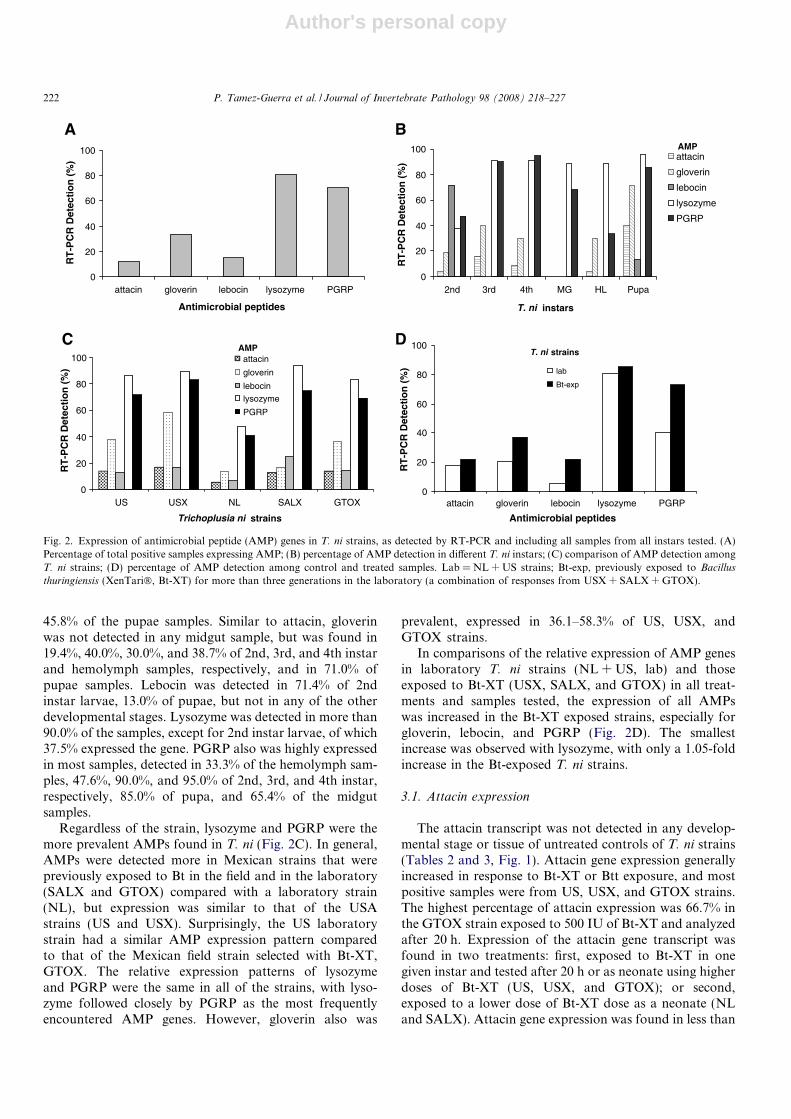
When all of the samples were combined from all devel-opmental stages, the most frequently detected AMP geneswere lysozyme and PGRP, found in 80.5% and 70.2% ofthe samples, respectively (Fig. 2A). Gloverin was foundin 33.0% of samples, whereas attacin and lebocin were inless than 20.0% of all samples.
The expression of AMP genes (attacin, gloverin, lebocin,lysozyme, or PGRP) differed within the various develop-mental stages of the T. ni strains (Fig. 2B). However, lyso-zyme and PGRP were the most frequently detected AMPgenes in all but 2nd instar larvae. Attacin was not detectedin the midgut tissue of any strain, and was found in only4.17% of 2nd instar and hemolymph samples, 16.0 and8.70% of 3rd and 4th instar larvae, respectively, but
GLOVERIN
2nd 3rd 4th. Pupae HEM MG
LEBOCIN
LYSOZYME
Instars Tissue
PGRP
ATTACIN
CTL
EXP
CTL
EXPGTOX
CTL
EXP
CTL
EXP
CTL
EXP
CTL
EXP
CTL
EXP
CTL
EXP
US
GTOX
US
GTOX
US
GTOX
US
GTOX
US CTL
EXP
CTL
EXP
RPS3
GTOX
US CTL
EXP
CTL
EXP
StrainsAMP
Fig. 1. Comparision of relative expression of antimicrobial peptide genes (attacin, gloverin, lebocin, lysozyme, and PGRP) in two T. ni strains (GTOX andUS) in different larval instars and tissue samples by RT-PCR. CTL, unexposed control; EXP, exposed to Bt-XT (500 IU/mL) until larvae reached theindicated instar and analyzed 20 h after exposure; HL, hemolymph; MG, midgut.
P. Tamez-Guerra et al. / Journal of Invertebrate Pathology 98 (2008) 218–227 221
Author's personal copy
45.8% of the pupae samples. Similar to attacin, gloverinwas not detected in any midgut sample, but was found in19.4%, 40.0%, 30.0%, and 38.7% of 2nd, 3rd, and 4th instarand hemolymph samples, respectively, and in 71.0% ofpupae samples. Lebocin was detected in 71.4% of 2ndinstar larvae, 13.0% of pupae, but not in any of the otherdevelopmental stages. Lysozyme was detected in more than90.0% of the samples, except for 2nd instar larvae, of which37.5% expressed the gene. PGRP also was highly expressedin most samples, detected in 33.3% of the hemolymph sam-ples, 47.6%, 90.0%, and 95.0% of 2nd, 3rd, and 4th instar,respectively, 85.0% of pupa, and 65.4% of the midgutsamples.
Regardless of the strain, lysozyme and PGRP were themore prevalent AMPs found in T. ni (Fig. 2C). In general,AMPs were detected more in Mexican strains that werepreviously exposed to Bt in the field and in the laboratory(SALX and GTOX) compared with a laboratory strain(NL), but expression was similar to that of the USAstrains (US and USX). Surprisingly, the US laboratorystrain had a similar AMP expression pattern comparedto that of the Mexican field strain selected with Bt-XT,GTOX. The relative expression patterns of lysozymeand PGRP were the same in all of the strains, with lyso-zyme followed closely by PGRP as the most frequentlyencountered AMP genes. However, gloverin also was
prevalent, expressed in 36.1–58.3% of US, USX, andGTOX strains.
In comparisons of the relative expression of AMP genesin laboratory T. ni strains (NL + US, lab) and thoseexposed to Bt-XT (USX, SALX, and GTOX) in all treat-ments and samples tested, the expression of all AMPswas increased in the Bt-XT exposed strains, especially forgloverin, lebocin, and PGRP (Fig. 2D). The smallestincrease was observed with lysozyme, with only a 1.05-foldincrease in the Bt-exposed T. ni strains.
3.1. Attacin expression
The attacin transcript was not detected in any develop-mental stage or tissue of untreated controls of T. ni strains(Tables 2 and 3, Fig. 1). Attacin gene expression generallyincreased in response to Bt-XT or Btt exposure, and mostpositive samples were from US, USX, and GTOX strains.The highest percentage of attacin expression was 66.7% inthe GTOX strain exposed to 500 IU of Bt-XT and analyzedafter 20 h. Expression of the attacin gene transcript wasfound in two treatments: first, exposed to Bt-XT in onegiven instar and tested after 20 h or as neonate using higherdoses of Bt-XT (US, USX, and GTOX); or second,exposed to a lower dose of Bt-XT dose as a neonate (NLand SALX). Attacin gene expression was found in less than
0
20
40
60
80
100
US USX NL SALX GTOX
Trichoplusia ni strains
RT
-PC
R D
etec
tio
n (
%)
attacin
gloverin
lebocin
lysozyme
PGRP
AMP
0
20
40
60
80
100
attacin gloverin lebocin lysozyme PGRP
Antimicrobial peptides
RT
-PC
R D
etec
tio
n (
%)
lab
Bt-exp
T. ni strains
0
20
40
60
80
100
attacin gloverin lebocin lysozyme PGRP
Antimicrobial peptides
RT
-PC
R D
etec
tio
n (
%)
0
20
40
60
80
100
2nd 3rd 4th MG HL Pupa
T. ni instars
RT
-PC
R D
etec
tio
n (
%)
attacin
gloverin
lebocin
lysozyme
PGRP
AMP
Fig. 2. Expression of antimicrobial peptide (AMP) genes in T. ni strains, as detected by RT-PCR and including all samples from all instars tested. (A)Percentage of total positive samples expressing AMP; (B) percentage of AMP detection in different T. ni instars; (C) comparison of AMP detection amongT. ni strains; (D) percentage of AMP detection among control and treated samples. Lab = NL + US strains; Bt-exp, previously exposed to Bacillus
thuringiensis (XenTari�, Bt-XT) for more than three generations in the laboratory (a combination of responses from USX + SALX + GTOX).
222 P. Tamez-Guerra et al. / Journal of Invertebrate Pathology 98 (2008) 218–227

Author's personal copy
half of pupae samples but not the hemolymph, except forNL, where it only was found in the hemolymph but notthe pupae (Fig. 3A). After bacterial challenge trials, USand USX strains exposed to either fresh Btt culture orBtt-SC expressed attacin only in pupae stage, whereas theGTOX strain did not (data not shown).
3.2. Gloverin expression
Gloverin was detected in pupae samples of the untreatedcontrols of US and USX (16.7%) and SALX (50.0%)(Tables 2 and 3, Fig. 1). SALX, USX, and GTOX strainshad higher levels of gloverin when they were exposed to500 IU of Bt-XT and tested after 20 h exposure, or when
given a higher dose (2500 IU). Similar to attacin, gloveringene expression was generally at higher percentage in sam-ples exposed to bacterial challenge. Bacterial challenge byBt-XT and EC induced the highest gloverin expression inthe USX strain (Table 2). Gloverin was expressed in allinstars, hemolymph, and pupae of US, USX, and GTOX,but expression was variable (Fig. 3B). SALX was the onlystrain that did not express gloverin in the hemolymph.
3.3. Lebocin expression
Increased lebocin gene expression was noted only in fieldstrains SALX and GTOX exposed to 500 IU of Bt-XT andtested after 20 h exposure, or when given a higher dose
Table 2Relative expression of antimicrobial peptides (AMPs) in American Trichoplusia ni strains fed control or different diet treatments
T. ni strains AMP Treatmentsa
None Doses of Xen-Tari� (Bt-XT, IU/ml) EC Btt Btt-SC
500 500* 2500 500 + EC
US Attacin 0.0 0.0 16.7 33.3 0.0 0.0 16.7 16.7Gloverin 16.7 33.3 33.3 33.3 50.0 6.7 33.3 33.3Lebocin 16.7 0.0 0.0 16.7 16.7 16.7 6.7 16.7Lysozyme 83.3 75.0 83.3 83.3 100 83.3 100 83.3PGRP 100.0 25.0 66.7 33.3 83.3 100 66.7 83.3
USX Attacin 0.0 33.3 33.3 16.7 16.7 16.7Gloverin 16.7 66.7 50.0 83.3 66.7 50.0 66.7
Lebocin 16.7 16.7 16.7Lysozyme 83.3 66.7 100 100 100.0
PGRP 100.0 50.0 83.3 83.3
Data represent the percentage of all insects tested within each strain (including all developmental stages and tissues).a Treatments are detailed in Section 2. Numbers in bold show detection values at higher percentages in the same treatment/AMP comparing US versus
USX. EC, Escherichia coli. Btt, fresh culture of Bacillus thuringiensis serovar thuringiensis. Btt-SC, spores and crystals of B. thuringiensis serovarthuringiensis. Blanks indicate that results were either inconclusive or were not tested.
Table 3Relative expression of antimicrobial peptides in Mexican Trichoplusia ni strains fed control or different diet treatments
T. ni strains AMP Treatmentsa
None Doses of Xen-Tari� (Bt-XT, IU/ml) Btt Btt-SC
500 500* 2500
NL Attacin 0.0 16.7 0.0Gloverin 0.0 50.0 0.0 16.7Lebocin 0.0 16.7Lysozyme 33.3 66.7 100 100 0.0PGRP 50.0 50.0 0.0 0.0
SALX Attacin 0.0 25.0
Gloverin 16.7 5.0Lebocin 16.7 33.3Lysozyme 100.0 100 75.0 100
PGRP 80.0 0.0 100 0.0
GTOX Attacin 0.0 66.7 16.7Gloverin 0.0 16.7 66.7 50.0 50.0Lebocin 0.0 0.0 33.3 33.3Lysozyme 66.7 66.7 100 83.3 83.3 100PGRP 66.7 66.7 83.3 66.7 83.3 50.0
Data represent the percentage of all insects tested within each strain (including all developmental stages and tissues).a Treatments are detailed in Section 2. Numbers in bold show detection values at higher percentages in the same treatment/AMP comparing NL versus
SALX and GTOX strains. EC, Escherichia coli. Btt, fresh culture of Bacillus thuringiensis serovar. thuringiensis. Btt-SC, spores and crystals of B.
thuringiensis serovar. thuringiensis.
P. Tamez-Guerra et al. / Journal of Invertebrate Pathology 98 (2008) 218–227 223

Author's personal copy
(2500 IU/ml) (Table 3). No differences in lebocin expres-sion were observed in US and USX after bacterial chal-lenge (Table 2). Lebocin gene expression was found inthe 2nd instar of all strains, except by NL and GTOuntreated controls. Lebocin was found only in pupae sam-ples of the field strains SALX and GTOX (Fig. 3C). Of the2nd instar samples, untreated controls of the US strainwere positive but treatments with 500 or 500* IU/ml forwere negative for lebocin, whereas the opposite wasobserved with GTOX (Fig. 1). None of the other develop-mental stages or tissues samples expressed lebocin.
3.4. Lysozyme expression
Lysozyme expression was found in all strains (Tables 2and 3). Increased lysozyme expression was observed inNL and GTOX, in particular when exposed to 500 IUof Bt-XT and assayed 20 h later. Increasing the Bt-XTdose to 2500 IU generally caused an increased lysozymegene expression in all strains except US. In general, afterbacterial challenge the USX, NL, and GTOX strainsexpressed lysozyme in higher percentage compared with
US and SALX strains. Whereas attacin, gloverin, andlebocin transcripts were not detected in midgut from 4thinstar larvae of any T. ni strain, lysozyme was detectedin many of the midgut samples (Fig. 3D). In contrast,only the midgut and hemolymph samples of NL hadhigher lysozyme expression, and lysozyme was notdetected in NL 2nd instar larvae.
3.5. PGRP expression
PGRP was found in all US and USX untreated controls,but bacterial challenge resulted in a decrease in PGRPexpression (Table 2 and Fig. 1). The only samples thatdemonstrated increased PGRP gene expression were fromSALX and GTOX larvae exposed to Bt-XT at 500 IU/mland analyzed after 20 h. PGRP was not detected in hemo-lymph samples from SALX and GTOX nor in 2nd instarlarvae of NL (Fig. 3E). Almost all midgut GTOX samplesexpressed PGRP, with the exception of those exposed tothe Btt-SC (data not shown). Pupae from US and USXstrains did not have PGRP after Btt challenge (data notshown).
T. ni instars
0
20
40
60
80
100
US USX NL SALX GTOX
T. ni strains
T. ni strains
T. ni strains
T. ni strains
T. ni strains
Atta
cin
dete
ctio
n (%
)
2nd
3rd
4th
HL
Pupa
T. ni instars
0
20
40
60
80
100
US USX NL SALX GTOX
Glo
verin
de
tect
ion
(%)
2nd
3rd
4th
HL
Pupa
T. ni instars
0
20
40
60
80
100
US USX NL SALX GTOX
Lebo
cin
dete
ctio
n (%
)
2nd
Pupa
T. ni instars
0
20
40
60
80
100
US USX NL SALX GTOX
Lyso
zym
e de
tect
ion
(%)
2nd3rd4thMGHLPupa
T. ni instars
0
20
40
60
80
100
US USX NL SALX GTOX
PG
RP
det
ectio
n (%
)
2nd
3rd
4th
MG
HL
Pupa
Fig. 3. Detection of antimicrobial peptides (AMPs) by RT-PCR among instars of T. ni strains, as a percentage of the total samples tested. (A) Attacin; (B)gloverin; (C) lebocin; (D) lysozyme; (E) peptidoglycan recognition protein (PGRP).
224 P. Tamez-Guerra et al. / Journal of Invertebrate Pathology 98 (2008) 218–227

Author's personal copy
4. Discussion
Innate immune systems in insects recognize microorgan-isms through a series of pattern recognition receptors thatare highly conserved in evolution. For example, in Dro-
sophila, the activation of the innate immune responseinvolves recognition of the infectious agent and subsequentactivation of cellular and humoral responses (Kurata,2004). Humoral reactions depend on a primary response,which is mediated by the activation of cascades of constitu-tive proteins present in the hemolymph, and a secondaryresponse, which requires transcriptional activation ofAMPs (Kurata, 2004). These peptides accumulate in theinfected insect’s hemolymph and have the fundamental roleof protecting the insect from the pathogenic microorganism(Hoffmann, 1997).
Because AMPs are transcriptionally induced peptides,the secondary humoral response of insects may be definedby a set of peptides that are absent in the naıve state, whichhas been especially noted with attacin. Bt is more than aninfectious agent, because it produces insecticidal toxins thatinteract with specific receptors in the insect midgut that eli-cit secondary effects. Based on previous immunity studies(Gillespie et al., 1997), we hypothesized that the productionof AMP in Bt exposed insects was the result of a secondaryhumoral response that result in differences in susceptibilityto an infection. Similar results were reported by Crowleyand Houck (2002), who found an increased immuneresponse and, more specifically, the expression of cecropinB in pupae of Calliphora vicina after E. coli exposure. Inthis study, several comparisons were made based on theresults from untreated diets. For example, NL and GTOXstrains on untreated diets did not induce gloverin in thepupae stage, and lebocin in the 2nd instar, whereas otherstrains did.
It was previously reported that attacin expression byBombyx mori larvae was rapidly induced by the injectionof E. coli bacilli into larvae, and continued at least for48 h, mainly in fat bodies and hemocytes (Sugiyamaet al., 1995). Similarly, 5th instar B. mori larvae injectedwith lipopolysaccharide from E. coli showed a significantinduction of attacin-like polypeptides, but the leveldecreased 48 h after injection (Wang et al., 2004). In addi-tion, attacin was not detected in the midgut of lipopolysac-charide injected B. mori (Wang et al., 2004) or inimmunized Samia cynthia ricini larvae (Kishimoto et al.,2002). Whereas attacin has been reported in midgut of Dro-
sophila (Tzou et al., 2000), we did not detect attacin in themidgut samples of any T. ni strain. Expression of attacin inbacterial challenges of T. ni larvae was previously reportedfrom hemolymph samples (Kang et al., 1996a). In the pres-ent study, attacin was expressed infrequently in T. ni strainsexposed to bacteria, but it was induced in GTOX if thesample was analyzed following 20 h of exposure to Bt-XT.
Gloverin is an inducible antibacterial insect protein iso-lated from pupae of Hyalophora cecropia and suppressesE. coli growth at very low concentrations (Axen et al.,
1997). In our study, we observed that the expression ofgloverin was, in many aspects, similar to that of attacin,as previously reported by Axen et al. (1997). We found thatgloverin expression was related to insect stage, where thelowest expression was found in 2nd instar, increasing in3rd and 4th instar larvae, and the highest at the pupaestage. In many cases, gloverin was induced in US orUSX larvae under bacterial challenge. Other tests with T.
ni have demonstrated that gloverin is expressed in hemo-lymph and hemocytes of bacteria-immunized larvae (Lund-strom et al., 2002). Based on these results, the gloverinexpression in T. ni appears to be very specific.
Early studies of lebocin detection in B. mori indicated agene expression pattern typical of an insect antibacterialpeptide (Chowdhury et al., 1995). In this study, lebocinwas the least detected AMP transcript, only detected in2nd instar larvae and in three pupae samples. GTOX wasthe only strain that demonstrated an increase in lebocinin response to bacterial challenge. In a previous study withT. ni, a Northern blot analysis demonstrated that lebocinexpression was inducible upon bacterial challenge, mainlyin fat body and hemocytes, and that its expression reachedits highest level after 20 h challenge and continued at leastuntil 60 h after bacterial injection (Liu et al., 2000). Simi-larly, B. mori studies showed that lebocin gene expressionoccurred tissue-specifically in the fat bodies and continuedat least 48 h post bacterial injection (Chowdhury et al.,1995). This may explain our detection of lebocin transcriptprimarily in 2nd instar larvae. These results also suggest alonger induction time for attacin and gloverin gene expres-sion (and longer expression period) compared with that oflebocin.
One of the most recognized AMPs is lysozyme. Lyso-zyme has been detected in H. cecropia (Hultmark et al.,1983) and Manduca sexta larvae (Spies et al., 1986) afterE. coli exposure. In the present study, lysozyme was themost detected AMP and was constitutively expressed.Lysozyme was detected at high levels in most instars,pupae, and tissues, and its expression levels fluctuated inresponse to bacterial challenge.
PGRPs are pattern recognition molecules that recognizebacteria and peptidoglycan molecules (Dziarski, 2004).PGRP expression has been reported after the exposure ofGram-positive bacteria or bacterial cell wall fragments ina differential display screen for up-regulated immune genesin T. ni (Kang et al., 1998). PGRPs are differentiallyexpressed in various cells and tissues, their expression isoften up-regulated by bacteria, and they mediate hostresponses to bacterial infections (Dziarski, 2004). In Dro-
sophila, PGRP are the initial sensors of infecting bacteriathat then trigger a cascade leading to the expression of anti-microbial peptides (Girardin and Philpott, 2004). Recentdata suggested that in diverse PGRPs are involved in dis-tinguishing between invading bacteria and activation ofappropriate immune reactions (Kurata, 2004). Similar toobservations with lysozyme, PGRP gene expression wasconstitutively expressed and fluctuated in response to bac-
P. Tamez-Guerra et al. / Journal of Invertebrate Pathology 98 (2008) 218–227 225
Author's personal copy
terial challenge in the T. ni strains. PGRP was not detectedin any hemolymph sample of SALX or GTOX, but wasdetected in all Bt-exposed midgut treatments, suggestingthat it is expressed as a response to bacteria challenge oran immune signaling molecule.
Overall, the expression of attacin, gloverin, and lebocingene in different stages of selected T. ni strains was relatedto bacterial challenge and instar/tissue, whereas lysozymeand PGRP were not. Different patterns of AMP geneexpression were found in strains from the US and Mexico.However, all T. ni strains previously exposed to Bt demon-strated an increased AMP gene expression. More studieswith individual AMP and Bt resistant strains are neededto determine a conclusive relationship between Bt-suscepti-bility and insect immunity.
Acknowledgments
We want to thank Ludek Zurek at Kansas State Univer-sity and Pablo Zapata at University of Nuevo Leon fortheir thoughtful comments to improve the manuscript.This study was supported by the research and scientific ex-change division, United States Department of Agriculture-Agricultural Research Services, Grant 210-22310-003-03S,PAICYT-UANL CN1355-06, and CONACyT 36815-B.Mention of trade names or commercial products in thispublication is solely for the purpose of providing specificinformation and does not imply recommendation orendorsement by the US. Department of Agriculture.
References
Axen, A., Carlsson, A., Engstrom, A., Bennich, H., 1997. Gloverin, anantibacterial protein from the immune hemolymph of Hyalophora
pupae. Eur. J. Biochem. 247, 614–619.Behle, R.W., McGuire, M.R., Tamez-Guerra, P., 2000. Effect of light
energy on alkali released virions from Anagrapha falcifera nucleopoly-hedrovirus. J. Invertebr. Pathol. 76, 120–126.
Chomczynski, P., Sacchi, N., 1987. Single-step method of RNA isolationby acid guanidinium thiocyanate–phenol–chloroform extraction. Anal.Biochem. 162, 156–159.
Chowdhury, S., Taniai, K., Hara, S., Kadono-Okuda, K., Kato, Y.,Yamamoto, M., Xu, J., Choi, S.K., Debnath, N.C., Choi, H.K., et al.,1995. cDNA cloning and gene expression of lebocin, a novel memberof antibacterial peptides from the silkworm, Bombyx mori. Biochem.Biophys Res Commun. 214, 271–278.
Crowley, L.D., Houck, M.A., 2002. The immune response of larvae andpupae of Calliphora vicina (Diptera: Calliphoridae), upon administeredinsult with Escherichia coli. J. Med. Entomol. 39, 931–934.
DeMaagd, R.A., Bravo, A., Crickmore, N., 2001. How Bacillus thurin-
giensis has evolved specific toxins to colonize the insect world. Rev.Trends Genet. 17, 193–199.
Dziarski, R., 2004. Peptidoglycan recognition proteins (PGRPs). Mol.Immunol. 40, 877–886.
Edlund, T., Siden, I., Boman, H.G., 1976. Evidence for two immuneinhibitors from Bacillus thuringiensis interfering with the humoraldefense system of saturniid pupae. Infect. Immun. 14, 934–941.
Flyg, C., Kenne, K., Boman, H.G., 1980. Insect pathogenic properties ofSerratia marcescens: phage-resistant mutants with a decreased resis-tance to Cecropia immunity and a decreased virulence to Drosophila. J.Gen. Microbiol. 120, 173–181.
Gillespie, J.P., Kanost, M.R., Trenczek, T., 1997. Biological mediators ofinsect immunity. Annu. Rev. Entomol. 42, 611–643.
Girardin, S.E., Philpott, D.J., 2004. Mini-review: the role of peptidoglycanrecognition in innate immunity. Eur. J. Immunol. 34, 1777–1778.
Hara, S., Yamakawa, M., 1995. A novel antibacterial peptide familyisolated from the silkworm, Bombyx mori. Biochem. J. 310, 651–656.
Hoffmann, J.A., 1997. Immune responsiveness in vector insects. Proc.Natl. Acad. Sci. USA 94, 11152–11153.
Hultmark, D., Engstrom, A., Anderson, K., Steiner, H., Bennich, H.,Boman, H.G., 1983. Insect immunity. Attacins, a family of antibac-terial proteins from Hyalophora cecropia. EMBO J. 2, 571–576.
Jarosz, J., 1996. Anti-infective defense strategies and methods of escapefrom entomologic pathogens under immunologic control of insects.Wiad. Parazytol. 42, 3–27.
Kang, D., Liu, G., Gunne, H., Steiner, H., 1996a. PCR differential displayof immune gene expression in Trichoplusia ni. Insect Biochem. Mol.Biol. 26, 177–184.
Kang, D., Liu, G., Lundstrom, A., Gelius, E., Steiner, H., 1998. Apeptidoglycan recognition protein in innate immunity conserved frominsects to humans. Proc. Natl. Acad. Sci. USA 95, 10078–10082.
Kang, D., Lundstrom, A., Steiner, H., 1996b. Trichoplusia ni attacin A, adifferentially displayed insect gene coding for an antibacterial protein.Gene 174, 245–249.
Kishimoto, K., Fujimoto, S., Matsumoto, K., Yamano, Y., Morishima, I.,2002. Protein purification, cDNA cloning and gene expression ofattacin, an antibacterial protein, from eri-silkworm, Samia cynthia
ricini. Insect Biochem. Mol. Biol. 32, 881–887.Kurata, S., 2004. Recognition of infectious non-self and activation of
immune responses by peptidoglycan recognition protein (PGRP)-family members in Drosophila. Dev. Comp. Immunol. 28, 89–95.
Lam, W., Cosat, G.M., Rayne, R.C., 2000. Characterization of multipletrypsin from the midgut of Locusta migratoria. Insect Biochem. Mol.Biol. 30, 85–94.
Liu, G., Kang, D., Steiner, H., 2000. Trichoplusia ni lebocin, an inducibleimmune gene with a downstream insertion element. Biochem. Biophys.Res. Commun. 269, 803–807.
Lundstrom, A., Liu, G., Kang, D., Berzins, K., Steiner, H., 2002.Trichoplusia ni gloverin, an inducible immune gene encoding anantibacterial insect protein. Insect Biochem. Mol. Biol. 32, 795–801.
Marchini, D., Giordano, P.C., Amons, R., Bernini, L.F., Dallai, R., 1993.Purification and primary structure of ceratotoxin A and B, twoantibacterial peptides from the female reproductive accessory glands ofthe med fly Ceratitis capitata (Insecta: Diptera). Insect Biochem. Mol.Biol. 23, 591–598.
McGuire, M.R., Galan-Wong, L.J., Tamez-Guerra, P., 1997. Bacteria:bioassay of Bacillus thuringiensis against lepidopteran larvae. In:Lacey, L. (Ed.), Manual of Techniques in Insect Pathology. AcademicPress, New York, pp. 91–99.
Natori, S., 1995. Antimicrobial proteins of insect and their clinicalapplication. Nippon Rinsho 52, 1297–1304.
Oppert, B., Kramer, K.L., Beeman, R.W., Johnson, D., McGaughey,W.H., 1997. Proteinase-mediated resistance to Bacillus thuringiensis
insecticidal toxins. J. Biol. Chem. 272, 23473–23476.Spies, A.G., Karlinsey, J.E., Spence, K.D., 1986. Antibacterial hemolymph
proteins of Manduca sexta. Comp. Biochem. Physiol. B 83, 125–133.Sugiyama, M., Kuniyoshi, H., Kotani, E., Taniai, K., Kadono-Okuda, K.,
Kato, Y., Yamamoto, M., Shimabukuro, M., Chowdhury, S., Xu, J.,et al., 1995. Characterization of a Bombyx mori cDNA encoding anovel member of the attacin family of insect antibacterial proteins.Insect Biochem. Mol. Biol. 25, 385–392.
Tabashnik, B.E., Liu, Y.-B., Malvar, T., Heckel, D.G., Masson, L., Ballester,V., Granero, F., Mensua, J.L., Ferre, J., 1997. Global variation in thegenetic and biochemical basis of diamondback moth resistance to Bacillus
thuringiensis. Proc. Natl. Acad. Sci. USA 94, 12780–12785.Tamez-Guerra, P., Damas, G., Iracheta, M.M., Gomez-Flores, R.,
Oppert, B., Rodrıguez-Padilla, C., 2006. Differences in susceptibilityand physiological fitness of Trichoplusia ni (Hubner) strains to Bacillus
thuringiensis exposure. J. Econ. Entomol. 99, 937–945.
226 P. Tamez-Guerra et al. / Journal of Invertebrate Pathology 98 (2008) 218–227

Author's personal copy
Tzou, P., Ohresser, S., Ferrandon, D., Capovilla, M., Reichhart, J.M.,Lemaitre, B., Hoffmann, J.A., Imler, J.L., 2000. Tissue-specificinducible expression of antimicrobial peptide genes in Drosophila
surface epithelia. Immunity 13, 737–748.Vilmos, P., Kurucz, E., 1998. Mini review. Insect immunity: evolutionary
roots of the mammalian innate immune system. Immunol. Lett. 62,59–66.
Wang, Y., Zhang, P., Fujii, H., Banno, Y., Yamamoto, K., Aso, Y.,2004. Proteomic studies of lipopolysaccharide-induced polypeptidesin the silkworm, Bombyx mori. Biosci. Biotechnol. Biochem. 68,1821–1823.
Yamakawa, M., Tanaka, H., 1999. Immune proteins and their geneexpression in the silkworm, Bombyx mori. Dev. Comp. Immunol. 23,281–289.
P. Tamez-Guerra et al. / Journal of Invertebrate Pathology 98 (2008) 218–227 227
Copyright © 2022 FDOKUMEN